Note: Descriptions are shown in the official language in which they were submitted.
CA 03015455 2018-08-22
WO 2017/144877 PCT/GB2017/050458
1
DIETARY PRODUCT DEVOID OF AT LEAST TWO NON ESSENTIAL AMINO ACIDS
Field of the Invention
The present invention generally relates to the field of dietary therapies for
treating cancer.
More particularly, the present invention relates to altering the levels of
amino acids in the
diet so as to treat cancers and improve existing cancer therapies. The
invention also
relates to biomarkers for identifying patients that will benefit from dietary
therapies for
treating cancer and methods and kits using said biomarkers.
Background of the Invention
Cancer is a disease where cells undergo uncontrolled growth, growing and
dividing
beyond the normal limits of cell growth. These cells can invade and destroy
surrounding
tissues. Furthermore, cancer cells can metastasize, where they can spread to
other areas
of the body via the blood or lymphatic system.
Cancer treatment can involve surgery to remove the tumours, radiotherapy to
reduce
tumour size, or pharmacotherapy/chemotherapy, using drugs or other medicines
to treat
the cancers. Survival rates for cancers vary between cancer types; however,
for cancer
which has metasized rates are especially low. A key reason for this is the
fact that
pharmacotherapeutic/chemotherapeutic treatments often fail, rarely completely
eradicating
the cancer. Normal, non-cancerous, cells can only tolerate a certain dose of
the
pharmacotherapeutic/chemotherapeutic agent, resulting in sub-optimal doses for
treating
cancers being utilised to prevent too many adverse side effects. To compound
the
problem, agents have limited selectivity for cancerous cells over normal
cells, and
cancerous cells can become resistant to the
pharmacotherapeutic/chemotherapeutic
agents over treatment periods. Surviving cancer cells are typically still able
to undergo
uncontrolled proliferation, and the cancer persists. This has led to
researchers looking for
novel methods of treating cancers.
In recent years, attention has turned to cancer metabolism, and, in
particular, how
cancerous cells differ to normal cells so as to present the rapid,
uncontrolled cell growth
typically associated with disease. It is evident that cancers can reprogram
their metabolism
in order to be able to grow, generate new cells and adapt to metabolic stress.
In treating
cancers, specific metabolic pathway enzymes can be targeted, or alternatively,
the
chemicals and/or metabolites utilised in the pathways can be targeted.
Proteins are a key
component of cells, and protein synthesis pathways are key to the growth of
cancerous
cells.
CA 03015455 2018-08-22
WO 2017/144877 PCT/GB2017/050458
2
Proteins can be synthesised from amino acids, and there are 20 known
biologically active
amino acids in mammals. These can be synthesised in the body (non-essential
amino
acids), but those which cannot be are essential components of the diet
(essential amino
acids). Cancerous cells are highly dependent on utilisation of non-essential
amino acids to
support proliferation. Some cancers can synthesise these de novo to support
proliferation,
while others rely on uptake of exogenous amino acids. (Jason W. Locasale,
Nature
Reviews Cancer, 2013, 13, 572-583; R. Possemato et al, Nature, 2011 476, 346-
350; 0.
Maddocks et al, Nature, 2013, 493, 542-546; C. Labuschagne et al, Cell Rep.
2014, 22,
7(4), 1248-58). Non-essential amino acids are used for protein synthesis and
also many
other anabolic processes necessary for cancer cell growth.
The reliance of cancers on exogenous amino acids has the potential to be
exploited to
treat cancers, by modulating the amount of exogenous amino acids which they
can obtain,
through limiting levels of amino acids in the diet. This starvation of
cancerous cells of
essential components required for grow and survive may have the effect of
preventing
cancer growth or inducing cancerous cell death. This could be used alone as a
therapy, or
in conjunction with other strategies such
as radiotherapy and
pharmacotherapy/chemotherapy.
Such strategies will require improved methods of stratifying cancer types so
as to identify
patients and patient populations that may benefit from such therapies.
Accordingly there remains a need for improved methods of identifying patients
and patient
populations that will benefit from metabolically targeted therapies.
BRIEF SUMMARY OF THE DISCLOSURE
In accordance with the present inventions there is provided a dietary product
comprising a
plurality of amino acids, wherein the dietary product comprises all the
essential amino
acids and wherein the dietary product is substantially devoid of at least two
non-essential
amino acids. Suitably, the dietary product may comprise at least 9 amino
acids.
At least one of the substantially devoid non-essential amino acids may be
selected from
the group consisting of: glycine, serine, cysteine, tyrosine and arginine.
The at least two substantially devoid non-essential amino acids may comprise
two or more
of the following amino acids: glycine, serine, cysteine, tyrosine and
arginine. Suitably, the
dietary product may be substantially devoid of:
a. Glycine, serine and cysteine;
b. Glycine serine and arginine;
c. Glycine serine and tyrosine;
CA 03015455 2018-08-22
WO 2017/144877 PCT/GB2017/050458
3
d. Glycine, serine, arginine and cysteine;
e. Glycine, serine, tyrosine and cysteine;
f. Cysteine and arginine;
g. Cysteine and tyrosine;
h. Cysteine and glycine;
Cysteine, tyrosine and arginine; or
j. Glycine, serine, arginine, tyrosine and cysteine.
Suitably, the dietary product may further comprise one or more macronutrients
and/or one
or more micronutrients.
The dietary product may further comprise methionine at a level of less than
25mg/kg body
weight of the subject/day or less than 20mg/kg/day or less than 18mg/kg/day or
less than
16mg/kg/day.
A dietary product of the invention may be formulated to provide at least the
recommended
daily intake of essential amino acids (with the optional exception of
methionine) based on
average daily total protein consumption.
The dietary product of the invention may be in the form of a solid, or a
beverage.
The present invention further provides, a process of preparing a dietary
product of the
invention, wherein the components are dissolved or dispersed in water and
spray dried.
In another aspect the present invention provides a pharmaceutical composition
comprising
a dietary product of the invention or a dietary product produced in accordance
with the
invention and a pharmaceutically acceptable carrier, excipient or diluent.
Suitably, the pharmaceutical composition of the invention may further comprise
a
therapeutic agent selected from: an inhibitor of cancer cell growth, a
radiotherapeutic
agent and a chemotherapeutic agent. The therapeutic agent may inhibit OXPHOS
and/or
may increase reactive oxygen species and/or may decrease anti-oxidant defence.
In a further aspect, the present invention provides a dietary product of the
invention or
produced in accordance with a process of the invention or a pharmaceutical
composition of
the invention for use in the treatment of cancer.
The cancer may be selected from the group consisting of: intestinal,
colorectal, liver, lung,
osteosarcoma, lymphoma, leukaemia and breast cancer.
The cancer may be positive for wild-type KRAS.
The cancer may have deregulated cMyc expression.
CA 03015455 2018-08-22
WO 2017/144877 PCT/GB2017/050458
4
The dietary product may be substantially devoid of serine and/or glycine.
The cancer may be associated with a downregulation of MTAP expression and,
optionally,
the dietary product may have a reduced level or be substantially devoid of
cysteine.
Suitably, the dietary product for use in the treatment of cancer may be used
in combination
with a therapeutic agent selected from: an inhibitor of cancer cell growth, a
radiotherapeutic agent, a chemotherapeutic agent, an inhibitor of amino acid
metabolism/turnover/inter-conversion, an inhibitor of non-essential amino acid
biosynthesis, an inhibitor of amino acid transport, an enzyme or drug which
promotes
amino acid degradation or substance which sequesters amino acid(s).
The therapeutic agent may inhibit OXPHOS and/or may increase reactive oxygen
species
and/or may decrease anti-oxidant defence.
In another aspect, the present invention provides the use of dietary product
of the
invention or a dietary product produced in accordance with the invention or a
pharmaceutical composition of the invention in the manufacture of a medicament
for use
the treatment of cancer.
Suitably, the cancer may be selected from the group consisting of: colorectal,
lymphoma,
liver, lung, osteosarcoma and breast cancer.
The cancer may be positive for wild-type KRAS and/or the cancer may have
deregulated
cMyc expression and/or downregulated MTAP expression.
Suitably, the dietary product of the invention may be substantially devoid of
serine, glycine
or serine and glycine.
Suitably, the dietary product of the invention may have a reduced level or may
be
substantially devoid of cysteine.
Suitably, the dietary product may be used in combination with one or more
therapeutic
agent(s) selected from: an inhibitor of cancer cell growth, a radiotherapeutic
agent and a
chemotherapeutic agent.
The therapeutic agent may inhibit OXPHOS and/or may increase reactive oxygen
species
and/or may decrease anti-oxidant defence.
In another aspect, the present invention relates to a method of treating
cancer in a subject,
comprising administering a therapeutically effective amount of a dietary
product of the
invention or a dietary product produced in accordance with the invention or a
pharmaceutical composition of the invention.
CA 03015455 2018-08-22
WO 2017/144877 PCT/GB2017/050458
Suitably, the cancer may be selected from the group consisting of: colorectal,
liver,
osteosarcoma, lung, lymphoma and breast cancer.
The cancer may be positive for wild-type KRAS and/or may have deregulated cMyc
expression.
5 The dietary product may be substantially devoid of serine and/or glycine.
The dietary product may be used in combination with one or more therapeutic
agent(s)
selected from: an inhibitor of cancer cell growth, a radiotherapeutic agent
and a
chemotherapeutic agent.
The therapeutic agent may inhibit OXPHOS and/or may increase reactive oxygen
species
and/or may decrease anti-oxidant defence.
Suitably, in all aspects of the invention, the dietary product may be the sole
source of
nutrition for the subject.
The treatment is administered over a period of at least 24 hours or until a
therapeutic
endpoint is observed.
The dietary product may be administered between 1 and 6 times a day.
Suitably, at least the recommended daily amount of essential amino acids may
be met by
the administration regimen each day.
The present invention further provides the use of KRAS and/or MTAP as a
biomarker to
identify a patient or patient population responsive to or sensitive to a
cancer treatment
comprising a diet substantially devoid of serine and/or glycine.
Suitably, the cancer treatment may comprise a diet substantially devoid of
serine and
glycine.
Suitably, the cancer treatment may further comprise administration of a
therapeutic agent
selected from: an inhibitor of cancer cell growth, a radiotherapeutic agent
and/or a
chemotherapeutic agent.
In another aspect, the present invention provides a method of identifying a
subject having
a decreased likelihood of responsiveness or sensitivity to a cancer treatment
comprising a
diet substantially devoid of serine comprising:
a) determining the level of Kras expression or activity in a biological sample
isolated from the subject;
b) comparing the level of Kras expression or activity in the biological sample
to a
control sample or to a predetermined reference level of Kras expression or
activity,
CA 03015455 2018-08-22
WO 2017/144877 PCT/GB2017/050458
6
wherein an increased level of Kras expression or activity the biological
sample compared
to the control sample or compared to the predetermined reference level is
indicative of
non-responsiveness or insensitivity to said cancer treatment.
In a further aspect, the present invention provides a method of identifying a
subject having
an increased likelihood of responsiveness or sensitivity to a cancer treatment
comprising a
diet substantially devoid of serine comprising:
a) determining the level of Kras expression or activity in a biological sample
isolated from the subject;
b) comparing the level of Kras expression or activity in the biological sample
to a
control sample or to a predetermined reference level of Kras expression or
activity,
wherein an decreased level of Kras expression or activity in the biological
sample
compared to the control sample or compared to the predetermined reference
level, or a
level of Kras expression or activity which is substantially the same as the
control sample or
the predetermined reference level is indicative of responsiveness or
sensitivity to said
cancer treatment.
In another aspect, the present invention provides a method of identifying a
subject who
may benefit from a cancer treatment comprising a diet substantially devoid of
serine
comprising:
a) determining the level of Kras expression or activity in a biological sample
isolated from the subject;
b) comparing the level of Kras expression or activity in the biological sample
to a
control sample or to a predetermined reference level of Kras expression or
activity,
wherein an decreased level of Kras expression or activity in the biological
sample
compared to the control sample or compared to the predetermined reference
level, or a
level of Kras expression or activity which is substantially the same as the
control sample or
the predetermined reference level indicates that the patient may benefit from
said cancer
treatment.
In a further aspect, the present invention provides a method of identifying a
subject having
an increased likelihood of responsiveness or sensitivity to a cancer treatment
comprising a
diet i) substantially devoid of serine and/or ii) and/or ii) with a restricted
level of cysteine
comprising:
a) determining the level of MTAP expression or activity in a biological sample
isolated from the subject;
CA 03015455 2018-08-22
WO 2017/144877 PCT/GB2017/050458
7
b) comparing the level of MTAP expression or activity in the biological sample
to a
control sample or to a predetermined reference level of MTAP expression or
activity,
wherein an decreased level of MTAP expression or activity in the biological
sample
compared to the control sample or compared to the predetermined reference
level, or a
level of MTAP expression or activity which is substantially the same as the
control sample
or the predetermined reference level is indicative of responsiveness or
sensitivity to said
cancer treatment.
In another aspect, the present invention provides a method of identifying a
subject who
may benefit from a cancer treatment comprising a diet: i) substantially devoid
of serine
and/or ii) with a restricted level of cysteine comprising:
a) determining the level of MTAP expression or activity in a biological sample
isolated from the subject;
b) comparing the level of MTAP expression or activity in the biological sample
to a
control sample or to a predetermined reference level of MTAP expression or
activity,
wherein an decreased level of MTAP expression or activity in the biological
sample
compared to the control sample or compared to the predetermined reference
level, or a
level of MTAP expression or activity which is substantially the same as the
control sample
or the predetermined reference level indicates that the patient may benefit
from said
cancer treatment. Suitably, in all aspects, in the biological sample may be a
cancer cell or
cancerous tissue. Likewise, in all aspects, the control sample may be a normal
cell or
tissue sample. The normal cell or tissue sample may be of the same cell or
tissue type as
the cancer cell or cancerous tissue.
In a further aspect, the present invention provides a method of treating a
subject having a
cancer comprising:
a) determining if the level of Kras expression or activity in a biological
sample
isolated from the subject is indicative of responsiveness or sensitivity to a
cancer treatment
comprising a diet substantially devoid of serine; and
b) administering to the subject the cancer treatment, where the
level of Kras
expression or activity in the biological sample is indicative of
responsiveness or sensitivity
to said cancer treatment.
In a further aspect, the present invention provides a method of treating a
subject having a
cancer comprising:
CA 03015455 2018-08-22
WO 2017/144877 PCT/GB2017/050458
8
a) determining if the level of MTAP expression or activity in a biological
sample
isolated from the subject is indicative of responsiveness or sensitivity to a
cancer treatment
comprising a diet i) substantially devoid of serine and/or ii) restricted in
cysteine; and
b) administering to the subject the cancer treatment, where the level of
MTAP
expression or activity in the biological sample is indicative of
responsiveness or sensitivity
to said cancer treatment.
Suitably, said cancer treatment may comprise a diet substantially devoid of
serine and
glycine.
Suitably, said cancer treatment may further comprise administration of a
therapeutic agent
selected from: an inhibitor of cancer cell growth, a radiotherapeutic agent
and/or a
chemotherapeutic agent.
Determining if the level of Kras expression or activity in a biological sample
isolated from
the subject is indicative of responsiveness or sensitivity to a cancer
treatment comprising a
diet substantially devoid of serine may comprise:
a) determining the level of Kras expression or activity in a biological sample
isolated from the subject;
b) comparing the level of Kras expression or activity in the biological sample
to a
control sample or to a predetermined reference level of Kras expression or
activity,
wherein an increased level of Kras expression or activity in the biological
sample
compared to a control sample or compared to a predetermined reference level is
indicative
of non-responsiveness or insensitivity to the subject to said cancer
treatment, and wherein
an decreased level of Kras expression or activity in the biological sample
compared to a
control sample or compared to a predetermined reference level, or a level of
Kras
expression or activity which is substantially the same as a control sample or
a
predetermined reference level, is indicative of responsiveness or sensitivity
of the subject
to said cancer treatment.
Determining if the level of MTAP expression or activity in a biological sample
isolated from
the subject is indicative of responsiveness or sensitivity to a cancer
treatment comprising:
i) a diet substantially devoid of serine, and/or ii) a diet restricted in
cysteine may comprise:
a) determining the level of MTAP expression or activity in a biological sample
isolated from the subject;
b) comparing the level of MTAP expression or activity in the biological sample
to a
control sample or to a predetermined reference level of Kras expression or
activity,
CA 03015455 2018-08-22
WO 2017/144877 PCT/GB2017/050458
9
wherein an decreased level of MTAP expression or activity in the biological
sample
compared to a control sample or compared to a predetermined reference level,
or a level
of MTAP expression or activity which is substantially the same as a control
sample or a
predetermined reference level, is indicative of increased responsiveness or
sensitivity of
the subject to said cancer treatment.
In another aspect, the present invention provides a kit for use in identifying
a subject who
would benefit from a cancer treatment comprising a diet substantially devoid
of serine
and/or glycine comprising:
a. an agent for determining the expression or activity of Kras;
and
b. reagents for the assay.
Suitably, the kit may further comprise an agent for determining the expression
or activity of
MTAP.
The kit may further comprise instructions that an increased level of Kras
expression or
activity in a biological sample compared to a control sample or compared to a
predetermined reference level is indicative of non-responsiveness or
insensitivity of the
subject to said cancer treatment, and wherein a decreased level of Kras
expression or
activity in the biological sample compared to the control sample or compared
to a
predetermined reference level, or a level of Kras expression or activity which
is
substantially the same as the control sample or the predetermined reference
level, is
indicative of responsiveness or sensitivity to the subject to said cancer
treatment.
In another aspect, the present invention provides a kit for use in identifying
a subject who
would benefit from a cancer treatment comprising a diet: i) substantially
devoid of serine
and/or glycine; and/or ii) restricted in cysteine comprising:
a. an agent for determining the expression or activity of MTAP;
and
b. reagents for the assay.
The kit may further comprise instructions that a decreased level of MTAP
expression or
activity in the biological sample compared to the control sample or compared
to a
predetermined reference level, or a level of MTAP expression or activity which
is
substantially the same as the control sample or the predetermined reference
level, is
indicative of responsiveness or sensitivity to the subject to said cancer
treatment.
BRIEF DESCRIPTION OF THE DRAWINGS
Embodiments of the invention are further described hereinafter with reference
to the
accompanying drawings, in which:
CA 03015455 2018-08-22
WO 2017/144877 PCT/GB2017/050458
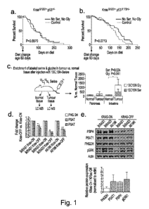
Figure la. PDAC Kras G12D/+ p53+/-, and lb. PDAC Kras G12D/+ p53R172H/+: Mice
placed on diet at -60 days of age, taken until clinical end-point (PDAC
related survival).
Survival calculated from change of diet (not birth). P value calculated using
mantel-cox
test.
5 Figure lc. Mice were injected in the tail vein with 100p1 of 100pM
130315Ni serine and left
for 2h. After sacrifice tissues were frozen then homogenised in metabolite
extraction buffer
& quantified by LCMS. P values calculated using paired T-test.
Figure Id. Kras inducible cell lines (iKRAS1, iKRAS3 and AK196) were grown in
complete
medium with doxycycline (KRAS-ON) or without doxycycline (KRAS-OFF). mRNA
10 expression of serine synthesis pathway enzymes was analysed by qRT-PCR.
Error bars
=SEM.
Figure le. Three Kras inducible cell lines (iKRAS1, iKRAS3 and AK196) were
grown for 3
days, protein expression was analysed by western blot. Relative changes of
Kras-
ON/Kras-OFF (measured by LiCor infra-red quantification) in expression of SSP
and
Phospho-ERK1 protein averaged across iKRAS1, iKRAS3 and AK196 cells; the
quantified
bands are those shown in the western blot. Error bars = STDEV.
Figure If. Kras inducible cell lines were grown in medium either containing or
lacking
serine and glycine (+SG / -SG) and counted after 48 and 96 hours. Error bars
=SEM.
Figure 2a. APCmin / APCmin KRAS organoids were grown with or without serine &
glycine for 24-48h.
Figure 2b. APCmin / APCmin KRAS organoids grown without serine & glycine for 5
days
then seeded into medium containing serine and glycine and grown for a further
24-
72h.Figure 2c. qRT-PCR on mRNA extracted from APCmin / APCmin KRAS organoids
grown with or without serine & glycine.
Figure 2d. APCmin / APCmin KRAS organoids grown in the presence of 1306-
glucose for
5 hours, metabolites were extracted and analysed by LCMS. P values calculated
using
TTEST unpaired. Error bars =STDEV.
Figure 3a and 3b. Effect of serine / glycine free diet on serum amino acid
levels in two
mice models of pancreatic cancer measured by mass spec analysis of serum
samples.
Statistical comparisons detailed in figure. a. Pdx1cre; KRasG12;p53+/- mice
received
normal chow until 60 days of age, then were transferred to either a control
diet containing
serine and glycine (Ctr) or a matched diet lacking serine and glycine (-SG)
until clinical
end-point. Serum isolated from terminal bleeds was analysed by LCMS. Relative
quantity
of metabolites are shown (x-axis = peak area). Error bars = STDEV. P values
were
calculated for each amino acid by T-test (unpaired, two tails), P values below
0.05 are
CA 03015455 2018-08-22
WO 2017/144877 PCT/GB2017/050458
11
shown. b. Pdx1c1e; KRasG12;p53R172H/+ mice received control or SG-free (-SG)
diet at 60
days of age until clinical end-point. Serum isolated from terminal bleeds was
analysed by
LCMS. Relative quantity of metabolites are shown (x-axis = peak area). Error
bars =
STDEV. P values were calculated for each amino acid by T-test (unpaired, two
tails), P
values below 0.05 are shown.
Figure 4a. Growth rate of tumours formed from HCT116 cells (human colorectal
cancer,
either p53 wt or null). Tumours grew rapidly in mice fed a control diet, but a
serine and
glycine free diet (-SG) significantly attenuated tumour growth. b. The
survival rate of the
mice from the experiment shown in Figure 4a.The serine free diet significantly
improved
the survival of the mice.
Figure 5. Effect of serine starvation on the growth inhibitory effects of anti-
cancer drugs
with HCT116, DLD1 and 5W480 cell lines. A significant proportion of the
chemotherapies
show an enhanced anti-proliferative effect when given at the same time as
serine and
glycine starvation.
Figure 6. Effect of dietary serine & glycine restriction on levels of amino
acids in serum
samples from mice. 057BI6 mice were either fed a control diet containing all
20 amino
acids, or a diet lacking serine and glycine but containing all 18 other amino
acids. Serum
samples were analysed by LCMS, relative quantities of all non-essential amino
acids are
shown. VVith the diet, reduced serine, glycine and cysteine levels are seen.
Figure 7. Cysteine uptake from cell culture medium in multiple cell lines
(A549, HCT116,
5W480, RKO, MCF7, MDA MB 231 and MDA MB 468). Cancer cell lines are shown to
avidly consume exogenous cysteine.
Figure 8. Effects of cysteine starvation in multiple cell lines (HCT116,
HepG2, MDA MB
231, RKO and U205). Base medium = all amino acids added except: serine,
glycine,
cysteine. S = Serine 0.8mM G = Glycine 0.4mM C = Cysteine 0.4mM Medium
replaced
every 24h.
Figure 9. Effects of the combined and separate starvation of cysteine and
serine & glycine
on cell numbers of three cell lines (HCT116, 5W480 and DLD1). Cells were
seeded in
media with varying concentrations of serine, glycine and cysteine (but replete
for all other
amino acids) and counted after 48h.
Figure 10. Mechanism for serine and cysteine interdependence. Homocysteine
efflux
prevents depletion of serine pools in two ways; 1. Serine-derived one-carbons
are not
used for re-methylation, which allows the serine derived one-carbon pool to be
used for
nucleotide (DNA, RNA) synthesis instead 2. Serine is not needed to make
cysteine.
However, homocysteine efflux means cysteine can no longer be synthesized de
novo, so
CA 03015455 2018-08-22
WO 2017/144877 PCT/GB2017/050458
12
must come from outside of the cancer cell. To meet the high anabolic demands
for
nucleotide and glutathione (GSH) synthesis, cancer cells require uptake of
exogenous
serine and cysteine.
Figure 11. Summary of systemic metabolism and tumour metabolism.
Figure 12. Effect of withdrawal of non-essential amino acids, in addition to
serine and
glycine, on the growth of HCT116 and RKO cells. Showing an improvement to the
anti-
cancer effect of a serine and glycine free diet by modulating other amino
acids. a. Serine
and glycine starvation alone decreases proliferation rate. In addition to
serine and glycine
removal, withdrawing certain other non-essential amino acids (aspartic acid,
glutamic acid,
proline and asparagine) has a minor further additional effect on cell
proliferation rate at 2
days. b. Serine and glycine starvation alone decreases proliferation rate.
Furthermore,
removal of tyrosine, arginine or cysteine individually has a greater anti-
proliferative effect.
Figure 13. Effect of the different combinations of serine, glycine, cysteine,
arginine and
tyrosine starvation on the cell growth and cell death (shown by a % change in
cell
numbers) of HCT116 cells over 4 days. The cell growth for complete medium (the
control)
was +1400%. Tyrosine starvation alone; tyrosine, serine and glycine
starvation; arginine
starvation alone; and arginine, serine and glycine starvation all resulted in
growth
inhibition. Cysteine starvation alone; cysteine, serine and glycine
starvation; serine,
glycine, cysteine, and arginine starvation resulted in growth inhibition and
cell death.
Figure 14. Serine synthesis pathway enzyme expression is a determinant of
sensitivity to
serine starvation. Tumours with elevated expression or enhanced activity of
serine
synthesis pathway enzymes (PHGDH, PSAT1, PSPH) are less sensitive to serine
starvation. Serine synthesis pathway activity may be increased by multiple
mechanisms in
cancer, including gene copy number amplification, transcriptional activation
(e.g. by
oncogenic Kras), or by epigenetic means, or potentially by other mechanisms,
e.g.
allosteric activation.
Figure 15. Measurement of the release of cysteine precursors / homocysteine
dimers in 4
cell lines (5W480, DLD1, HCT116 and RKO) cultured in complete media over 48
hours.
Homocysteine is a precursor for de novo synthesis of cysteine, however,
homocysteine is
released from cancer cells and detected a homodimer, i.e. homocystine.
Figure 16. Measurement of the release of cysteine precursors / homocysteine
dimers in 2
cell lines (HCT116 and RKO), under serine and serine & glycine starvation
conditions.
homoC-cys = homocysteine + cysteine dimer. Homocystine = homocysteine +
homocysteine dimer.
CA 03015455 2018-08-22
WO 2017/144877 PCT/GB2017/050458
13
Figure 17. Measurement of the release of cysteine precursors / homocysteine
dimers in 2
cell lines (A549 and MDA MB 231), under serine and serine & glycine starvation
conditions. homoC-cys = homocysteine + cysteine dimer. Homocystine =
homocysteine +
homocysteine dimer
Figure 18. Serine and glycine free diet is an effective therapeutic
intervention in GEMMs
for lymphoma and intestinal cancer. a. Ep-Myc mice received normal chow until -
60 days
of age, then were transferred to either a control diet (containing serine and
glycine) or a
matched diet lacking serine and glycine (No Ser, No Gly) until clinical end-
point
(lymphoma-related survival). Survival was calculated from change of diet (not
birth). P
value calculated by Mantel-Cox test. b. APCMin/+ mice received normal chow
until -80
days of age, then were transferred to either a control diet (containing serine
and glycine) or
a matched diet lacking serine and glycine (No Ser, No Gly) until clinical end-
point
(intestinal tumour related survival). Survival was calculated from change of
diet (not birth).
P value calculated by Mantel-Cox test. c. Serum from Ep-Myc and d. APCMin/+
cohorts
was analysed by LCMS, relative abundance (by metabolite peak area) is shown.
Error
bars = STDEV, P values were calculated by T-test (unpaired, 2 tails,
*=P<0.0005). See
Figure 21 for relative quantification of all amino acids. e. Serum
concentration for serine
and glycine in the APCMin/+ cohort was determined using 6-point calibration
curves with
13015N-serine & glycine diluted in serum. Error bars = STDEV. f. Lgr5-creER
APCfl/f1
mice were induced at 7-10 weeks of age, diet was changed seven days after
first
tamoxifen treatment and maintained until clinical end-point (intestinal tumour-
related
survival). Survival is calculated from first tamoxifen treatment. P value
calculated by
Mantel-Cox test.
Figure 19. Manipulation of anti-oxidant response enhanced diet-induced anti-
cancer
effect. a. Ep-Myc mice received control or serine and glycine free diet (No
Ser, No Gly)
with 100mg/kg/day Phenformin (Phen.) by gavage at -60 days of age and taken to
clinical
end-point. Lymphoma-related survival was calculated from change of diet, not
birth. b.
APCMin/+ mice were transferred to Control or serine and glycine free diet (No
Ser, No
Gly) at -80 days of age, then four days later received Metformin (Metf.)
200mg/kg/day in
drinking water. Intestinal tumour-related survival calculated from change of
diet, not birth.
P value calculated by Mantel-Cox test. See Figure 22a for complete comparison
of survival
curves. c. Comparison of diet-only tumour burden data with metformin + diet
tumour
burden. Post-mortem count of tumour number was performed on the small
intestines (SI)
of APCmini+ mice. P values calculated by T-test (unpaired, 2 tails). See
Figure 22c for
tumour area data. Diet-only data replicated in (a) and (b) above. d.
Intestinal tumour
organoids derived from a VillincreER; APCflifl mouse were grown +/- SG, +/-
metformin at the
CA 03015455 2018-08-22
WO 2017/144877 PCT/GB2017/050458
14
stated concentrations for two days. Relative change (versus `-drug') in
organoid diameter
is plotted. Data are average of four independent experiments, error bars =
SEM. P
values calculated by T-test (unpaired, two tails, with correction for multiple
comparisons).
e. APCflifl organoids were grown +/- SG +/- metformin for two days, then fixed
and
immuno-stained for lipid peroxidation product malondialdehyde (MDA). Data is
average of
three independent experiments, error bars = SEM. P values calculated by T-test
(unpaired,
two tails, corrected for multiple comparisons). f. Ep-Myc mice were crossed
with Tigar-/-
mice, cohorts were placed on diets at -60 days of age and taken until clinical
end-point
(lymphoma-related survival). Survival was calculated from change of diet (not
birth). P
value calculated by Mantel-Cox test.
Figure 20. Effect of serine and glycine free diet on tumour burden in APCmini+
mice.
APCmini+ mice received normal chow until 80 days of age, then were transferred
to either a
control diet (containing serine and glycine) or a matched diet lacking serine
and glycine
(No Ser, No Gly; -SG) until clinical end-point (intestinal tumour related
survival). Post-
mortem tumour measurement was performed on intestinal tissue at time of diet
change
(80 days) or clinical endpoint. P values calculated by T-test (unpaired, two
tails, with
correction for multiple comparisons).
Figure 21. Effect of serine and glycine free diet on serum amino acids a. Ep-
myc and b.
APCmini+ mice received normal chow until -60 & -80 days of age respectively,
then were
transferred to either a control diet containing serine and glycine (Ctr) or a
matched diet
lacking serine and glycine (-SG) until clinical end-point. Serum isolated from
terminal
bleeds was analysed by LCMS. Relative quantity of metabolites are shown (x-
axis = peak
area). Error bars = STDEV. P values were calculated by T-Test (unpaired).
Figure 22. Metformin treatment did not enhance the anti- cancer effect of
serine and
glycine free diet in APCmini+ mice. Mice were transferred to serine and
glycine free diet (No
Ser, No Gly) (a) or Control diet (b) at -80 days of age, then four days later
received
Metformin (Metf.) 200mg/kg/day in drinking water. Intestinal tumour-related
survival
calculated from change of diet, not birth. P value calculated by Mantel-Cox
test. c.
Comparison of diet-only tumour burden data with metformin + diet tumour
burden. Post-
mortem tumour area measurement was performed on the small intestine (SI) of
APCmini+
mice. P values were calculated by T Test (unpaired, two tails) "Diet only"
data is replicated
from Figure 20.
Figure 23. In vivo metformin levels had little impact on systemic metabolism
and were too
low to potentiate the anti-cancer effect of the serine & glycine free diet. a.
APCmini+ mice
were transferred to Control or serine and glycine free diet ( - SG) then
received Metformin
200mg/kg/day in drinking water. Serum isolated from terminal bleeds was
analysed by
CA 03015455 2018-08-22
WO 2017/144877 PCT/GB2017/050458
LCMS. Error bars = STDEV. b. Tissue samples from metformin treated mice were
analysed by LCMS. NC=normal colon, NSI= normal small intestine, TC=tumour
colon,
TSI=tumour small intestine. Error bars = STDEV c. For mice where matching
serum and
tumour (SI or colon) tissue samples were available (Ctr diet n=7, -SG diet
n=6), serum
5 versus tumour metformin concentrations are plotted. Metformin
concentrations were
determined in all samples using a six-point calibration curve using the
relevant biological
matrix (tissue/serum). d. Serum from APCmini+ mice treated with metformin was
analysed
for glucose and lactate levels using an Agilent 2100 Bioanalyser. e. Human
colorectal
cancer cells DLD1 and 5W480, which express truncated APC, were grown in
varying
10 concentrations of Metformin either without serine and glycine (No Ser,
No Gly) or in low
serine and glycine (10pM) for three days after which cell number was counted.
Data are
averages of triplicate wells, error bars = STDEV.
Figure 24. S-plot of unbiased metabolomics analysis (OPLS-DA; orthogonal
partial least
15 squares discriminant analysis) of Ep-myc tumour tissue (tumour bearing
spleens) (Ctr
n=20, ¨SG n=13). The detected metabolites showing the greatest decrease due to
diet are
serine and glycine. Decreased levels of carnitine-related and choline-related
metabolites
were also observed. Increased levels of phosphatidylcholine (PC) metabolites
and alanine
and threonine were also seen. SG starvation is known to influence glycolysis
and
OXPHOS (potentially explaining changes in carnitine and alanine levels), and
one-carbon
metabolism (potentially explaining changes in choline related metabolites).
Figure 25. Show the effects of the ¨SG diet on Eu-myc tumour cells. a.
Lymphoma
cell.were isolated from Ep-myc mice and expanded in culture. Cells were
injected sub-
cutaneously (5x10"5/flank) into nude mice and allowed to form tumours. Once
tumours
(Ctr n=4, -SG n=4) were visible and measurable, mice were transferred to
control (Ctr) or
serine & glycine free diet (-SG). Mice were sacrificed and tumours excised at
single
temporal end-point (6 days on diet). Average tumour volume (as percentage of
starting
tumour volume is shown, error bars= STDEV. b, To assess cell number per sub-
cutaneous
Ep-myc tumour, two separate cell counts per tumour (using H&E stained cross-
sections)
were performed and averaged, mean of means is shown, error bars = SEM. P
values
calculated by T-test (unpaired, one-tail). c, Whole sub-cutaneous Ep-myc
tumour tissue
sections (Ctr n=3, -SG n=4) were immuno-stained for cleaved caspase-3 (CC3)
and BrdU.
Image analysis of non-necrotic regions of whole tumours allowed quantitative
evaluation of
% cleaved caspase-3 positive cells per tumour and % BrdU positive cells per
per tumour.
Data are averages, error bars=STDEV. d. Ep-myc tumour (as described in a-c
above)
cross-sections were H&E stained, the scale bar for each image is 4mm,
demonstrative
CA 03015455 2018-08-22
WO 2017/144877 PCT/GB2017/050458
16
necrotic regions marked with arrows. Additional tumour tissue sections (marked
with *) are
included for comparison from tumours which developed after diet change (these
three
tumours were measurable two days post diet change and were present for 4 days
on diet
before end-point). e. Necrosis was quantified by image analysis of necrotic &
non-
necrotic surface area of H&E stains for the sections shown in (d). Error bars=
STDEV, P-
value was calculated by T-test (unpaired, one tail, Ctr, n=5;-SG,n=6). f,
APCmini+ mice
were placed on control diet (Ctr,n=3) or serine & glycine free diet (-SG,n=3)
at 80 days of
age. At a single temporal end-point (14 days on diet) mice were sacrificed and
the small
intestine was removed for histological analysis. Tissue sections were immuno-
stained for
cleaved caspase-3 and BrdU. Image analysis of whole intestines allowed
quantitative
evaluation of cell number per adenoma, % 003 positive cells and % BrdU
positive cells
per adenoma. Data are averages of all adenomas identified in each small
intestine section,
error bars=SEM, P-values calculated by T-test (unpaired, one tail). For all
analyses (a-f), P
values below 0.1 are shown.
Figure 26. Expression of SSP enzymes in tumour tissue from PDAC and Ep-myc
models.
Protein lysates of PDAC tumours and tumour bearing spleens from Ep-Myc mice
that
received the control or SG-free diet were analysed for SSP enzyme expression
by western
blot quantified using a Li-Cor scanner. Relative expression (versus control
diet) of SSP
enzymes is shown. Error bars = STDEV. Each tissue sample was taken from a
different
mouse, numbers of mice/tumours are shown above the bars.
Figure 27. Shows that a ¨SG diet led to decreased serine and glycine levels,
and
decreased GSH/GSSG ratio in Eu-myc tumours but no decrease in glycine or
GSH/GSSG
ratio in PDAC tumours. Pancreatic tumours from Pdx1c1e;KRasG12;p53+/- mice and
tumour bearing spleens from Ep-myc mice were analysed by LCMS for serine,
glycine,
GSH (reduced glutathione) and GSSG (oxidised glutathione). P values calculated
by T-
test, unpaired, two tails. Error bars = STDEV.
.. Figure 28. Tumour-organoids expressing Kras were more resistant to serine
and glycine
starvation. VillincreERAPC flit' and VillincreER, APCflifl;KRasG12 /+
intestinal tumour organoids
(from n=3 mice per genotype) were grown without serine & glycine for five days
then
dissociated and seeded into complete growth medium. Organoid diameter was
measured
for each day in complete (recovery) medium. Data are averages of organoids
from three
mice, obtained in a single experiment. Error bars = SEM
CA 03015455 2018-08-22
WO 2017/144877 PCT/GB2017/050458
17
Figure 29 shows that a diet devoid in glycine and serine decreases growth of
xenograft
tumours already formed in vivo, decreases intra-tumour serine and glycine
levels and that
such levels translate to slower cancer cell proliferation in vitro a. HCT116
cells were
injected bilaterally (3x10"6 per flank) and allowed to form tumours. Once
tumours were
visible and measurable by calipers mice were transferred to control diet or
serine and
glycine free diet (-SG). Tumours were measured three times per week and
average weekly
tumour volume is plotted, Error bars =SEM. P values were calculated by T-test
(unpaired,
one tail). b. HCT116 tumours (taken at clinical end-point) were analysed by
LCMS for
absolute concentration of serine and glycine (1-3 pieces of each tumour were
analysed).
Data are averages, bars are STDEV. P values were calculated by T-test
(unpaired, one
tail). c. HCT116 cells were grown in vitro (24-well plates) in the intra-
tumoural serine and
glycine concentrations displayed in
Medium was replaced every 24 hours and cell
counts were performed on the stated days. Data are averages of 12 replicate
wells for
each condition from an individual experiment, error bars = STDEV. d. HCT116
cells were
grown in vitro (24-well plates, 12 replicate wells for each condition) in the
intra-tumoural
serine and glycine concentrations displayed in
Medium was replaced every 24 hours
and cell counts were performed after four days. Data are averages of three
independent
experiments, error bars = SEM. P values were calculated by T-test (unpaired,
one tail).
Figure 30. Kras expressing cells obtain serine and glycine by de novo serine
and glycine
synthesis, not by an increase in micropinocytosis. Macropinocytosis in iKRas
cells was
assessed using TMR-labelled dextran uptake assay. Cells were initially grown
+/-
doxycycline for 48h then seeded +/- doxycycline, +/- SG for 40h (final 16h
without FBS),
then given TM R dextran/FBS in matched medium for 30 minutes. Error bars &
lines show
average and STDEV.
Figure 31a. Daunorubicin complements serine and glycine starvation.
VillincreER; APCflifl
mouse were grown +/- serine and glycine, +/- daunorubicin at the stated
concentrations for
two days. Relative change (versus `-drug') in organoid diameter is plotted.
Data is average
of three independent experiments, error bars = SEM. b. VillincreER; APCflifl
organoids were
grown +/- serine and glycine +/- daunorubicin for two days, then fixed with
and stained for
malondialdehyde (MDA), data is average of three independent experiments, error
bars =
SEM. P values calculated by T-test (unpaired, two tails, with correction for
multiple
comparisons).
CA 03015455 2018-08-22
WO 2017/144877 PCT/GB2017/050458
18
Figure 32. Shows a simplified schematic diagram illustrating de novo cysteine
synthesis
and polyamine synthesis in humans. Metabolites are shown in normal text,
enzymes are
shown in boxes.
Figure 33. Shows that MTA Efflux (which is an indicator of MTAP
deletion/inactivation)
correlates with enhanced sensitivity to cysteine starvation. a. Pancreatic
cancer cell lines
were grown in 24-well plates in formulated medium (based on RPM! medium)
lacking
cysteine but containing all 19 other essential and non-essential amino acids
for three days.
Cell numbers were counted using a CASY TT cell counter. Methylthioadenosine
(MTA)
levels in the cell culture medium were measured after 3 days by analysing
samples of
medium by liquid chromatography-mass spectrometry. b. Colorectal and breast
cancer cell
lines were grown in 24-well plates in formulated medium (based on RPM! medium)
lacking
cysteine but containing all 19 other essential and non-essential amino acids
for three days.
Cell numbers were counted using a CASY TT cell counter. Methylthioadenosine
(MTA)
levels in the cell culture medium were measured after 24 hours by analysing
samples of
medium by liquid chromatography-mass spectrometry. R2 = correlation
coefficient (with
Log trend-line) computed by MS Excel.
Figure 34. Shows that MDA-MB-231 cells have a higher rate of MTA and
spermidine
synthesis indicating that large amounts of methionine are diverted into the
polyamine
pathway in these cells. By contrast, in HCT116 and 5W480 cells less methionine
is
diverted into the polyamine pathway and more methionine reaches homocysteine/
cystathionine which can be converted into cysteine. This helps to explain the
better
survival of HCT116 and 5W480 cells during cysteine starvation. 5W480 and MDA-
MB-231
(M231) cells were grown in formulated medium (based on RPM! medium lacking
carbon-
12 methionine, supplemented with carbon-13 labelled methionine and containing
all 19
other essential and non-essential amino acids for two days. Cell lysates were
analysed by
liquid chromatography-mass spectrometry. The most abundant isotopomers are
shown.
MTA= methylthioadenosine, Met = methionine, Hc = homocysteine, SAM = 5-
adenosylmethionine. M+x = mass plus x units.
Figure 35. Shows that HCT116 and 5W480 cells are able to recycle MTA back to
methionine, but that MDA-MB-231 cells (which efflux MTA) are unable to recycle
MTA
back to methionine. Metabolite tracing with carbon-13 labelled methionine
shows that
unlike M DA-MB-231 cells, 5W480 and HCT116 cells are able to recycle
methionine which
has been used in the polyamine pathway (via MTA), which appears as `m+1'
methionine.
HCT116, 5W480 and MDA-MB-231 (M231) cells were grown in formulated medium
CA 03015455 2018-08-22
WO 2017/144877 PCT/GB2017/050458
19
(based on RPM! medium lacking carbon-12 methionine, supplemented with carbon-
13
labelled methionine and containing all 19 other essential and non-essential
amino acids for
two days. Cell lysates were analysed by liquid chromatography-mass
spectrometry. Major
methionine isotopomers are shown.
Figure 36. Shows that MDA-MB-231 (M231) cells have significant efflux of MTA
compared
with HCT116 and 5W480 cells, and even continue to efflux MTA during cysteine
starvation.
HCT116, 5W480 and MDA-MB-231 (M231) cells were grown in formulated medium
(based on RPM! medium), with or without cysteine, lacking carbon-12
methionine,
supplemented with carbon-13 labelled methionine and containing all 19 other
essential
and non-essential amino acids for two days. Metabolite extracts were prepared
from
medium samples (taken at the specified time-points) and were analysed by
liquid
chromatography-mass spectrometry. MTA= methylthioadenosine. M+x= mass plus x-
units.
There is no data for MDA-MB-231 cells at 48h in cysteine starvation because no
live cells
were remaining by that time-point.
Figure 37. Shows that by knocking-out MTAP gene expression leads to induction
of MTA
efflux. HCT116 cells were transfected with CRISPR/Cas9 and targeting sequences
(Seq 1
& 2) for MTAP or with a non-targeting control sequence (NTC). Several clones
were
isolated from each sequence and grown in complete medium for 4 days. Protein
lysates
were analysed for MTAP expression by western blot. Samples of medium were
analysed
for MTA content by liquid-chromatography mass spectrometry.
Figure 38. Shows that HCT116 efflux homocysteine (an upstream precursor of
cysteine)
but they are able to re-uptake homocysteine during cysteine starvation. HCT116
cells are
still sensitive to cysteine starvation, but less so than MDA-MB-231 (M231)
cells. HCT116,
5W480 and MDA-MB-231 (M231) cells were grown in formulated medium (based on
RPM!
medium), with or without cysteine, lacking carbon-12 methionine, supplemented
with
carbon-13 labelled methionine and containing all 19 other essential and non-
essential
amino acids for two days. Metabolite extracts were prepared from medium
samples (taken
at the specified time-points) and were analysed by liquid chromatography-mass
spectrometry. HC =homocysteine. M+x = mass plus x-units. There is no data for
MDA-MB-
231 cells at 48h in cysteine starvation because no live cells were remaining
by that time-
point.
Figure 39. Shows that cells can be rescued from cysteine starvation by
supplementation
with homocysteine this demonstrates that the enzymes CTH and CBS are
expressed,
CA 03015455 2018-08-22
WO 2017/144877 PCT/GB2017/050458
active and able to conduct de novo cysteine synthesis when the precursor
supply is
adequate. This data supports the idea that precursor shortage (rather than, or
in addition
to, defective/inadequate 0TH and CBS enzyme expression) contributes to
sensitivity to
cysteine starvation. Colorectal and breast cancer cell lines were grown in 24-
well plates.
5 Basal medium was formulated (based on RPM! medium) lacking cysteine but
containing all
19 other essential and non-essential amino acids. This basal medium was
supplemented
with the stated components; cysteine 0.4 mM (+Cys), homocysteine 0.2mM & 0.8mM
(HC)
and grown for three days. Data are average of three wells. Error bars = STDEV.
10 Figure 40. Shows that inhibition of AMD1 (the enzyme which diverts
methionine-derived
SAM into the polyamine synthesis pathway) protects cells from acute
sensitivity (i.e. cell
death) to cysteine starvation. MDA-MB-231 cells were initially seeded in
complete
(DMEM) medium in 24-well plates. After 24h, cells were treated with AMD1
inhibitor
Sardomozide (20uM) for 16h or left untreated (Ctr). Cells were then washed
with PBS and
15 given medium lacking cysteine but containing all 19 other amino acids.
Images (a) were
captured using a light microscope, and cell counts (b) were performed using a
CASY TT
cell counter. Data are average of three wells. Error bars = STDEV.
DETAILED DESCRIPTION
The inventors have surprisingly found that a diet substantially devoid of at
least two non-
20 essential amino acids can have utility in the treatment of cancer or a
proliferative disorder.
Without wishing to be bound by theory, by substantially removing an amino acid
required
for tumour cell proliferation and growth, metabolic remodelling to provide a
source of the
substantially devoid amino acid diverts resources and can reduce the amount of
the amino
acid available for rapid proliferation, thereby slowing down, or even
inhibiting, the growth
of, or causing the death of cancer cells.
Suitably, the present invention may involve partly or completely substituting
the normal diet
of a subject suffering from cancer with a prescribed diet substantially devoid
of at least two
non-essential amino acids. Such a diet may potentially be achieved by the
provision of a
dietary product as detailed herein, or by two or more dietary supplements
which can be
.. administered simultaneously or sequentially. Potentially, such a diet may
be further
supplemented through proper foods selection, using ingredients currently
available such
that the diet remains substantially devoid of two or more non-essential amino
acids.
DIETARY PRODUCT
In a first aspect of the present invention, there is provided a dietary
product comprising a
plurality of amino acids, wherein the dietary product comprises all the
essential amino
CA 03015455 2018-08-22
WO 2017/144877 PCT/GB2017/050458
21
acids and wherein the dietary product is substantially devoid of at least two
non-essential
amino acids.
By "essential amino acids" it is meant methionine, leucine, phenylalanine,
isoleucine,
valine, lysine, threonine, histidine and tryptophan.
"Dietary product" refers to a composition comprising one or more essential
amino acids or
salts or esters thereof, that is used in a food product, or used or consumed
in combination
with a food product, to provide a desired level of the amino acid(s) or salt
or esters thereof
to the subject consuming the supplement. The dietary ingredients in these
products may
include: vitamins, minerals, herbs or other botanicals, amino acids, and
substances such
as enzymes, organ tissues, glandulars, and metabolites. In some embodiments,
the
dietary product is the sole source of exogenous amino acids consumed by the
subject as
part of their diet. Suitably, in some aspects, the dietary product may be
intended to
substantially or solely replace a subject's diet. Hence, in some aspects, the
dietary product
may be a complete meal replacement for the subject.
Advantageously, replacement of consumption of usual sources of amino acids
such as
protein with a dietary product of the invention will yield a diet
substantially devoid of at
least two non-essential amino acids. This may provide therapeutically benefits
to a cancer
subject.
As used herein, in accordance with all aspects of the invention, the term
"subject"
preferably refers to a mammalian animal, including a human, a veterinary or
farm animal, a
domestic animal or pet, and animals normally used for clinical research,
including non-
human primates, dogs and mice. More specifically, the subject of the present
invention
may be a human.
Suitably, the dietary product may comprise at least 9 amino acids. Suitably,
the dietary
product may comprise at least 10 or at least 11 or at least 12 or at least 13
or at least 14 or
at least 15 or at least 16 or at least 17 or 18 amino acids. Suitably, the
dietary product may
comprise 9 to 18 amino acids or12-18 amino acids, or 12-17 amino acids or 13-
17 amino
acids or 14-17 amino acids, for example.
Suitably, the at least two substantially devoid amino acids comprise (or
consist essentially
thereof or consist of) two or more of the following amino acids: glycine,
serine, cysteine,
tyrosine, proline and arginine. Alternatively, the dietary product may be
devoid of at least
three or at least four or at least five or at least six or at least seven of
the following amino
acids: glycine, serine, cysteine, tyrosine, proline, arginine, alanine,
aspartic acid, glutamic
acid, glutamine and asparagine. Suitably, the dietary product may be devoid of
seven
amino acids, wherein the dietary product is devoid of serine and glycine and
five of the
CA 03015455 2018-08-22
WO 2017/144877 PCT/GB2017/050458
22
following amino acids: cysteine, tyrosine, proline, arginine, alanine,
aspartic acid, glutamic
acid, glutamine and asparagine. Suitably the dietary may be substantially
devoid or may
comprise a restricted level of cysteine.
In this context, by "consist essentially thereof" it is meant that that the
dietary product may
not lack further amino acids which have a material effect on the dietary
product on the
invention. By "material effect" it is meant a significant therapeutic effect
which may be
measured as one of the following: a) a significant effect on the specificity
for cancer as
opposed to healthy cells; b) a significant effect on the inhibition of cell
proliferation; c) a
significant effect on the toxicity of cancer cells or d) any combination of a)-
c). In some
aspects, this may be measured by comparing the dietary product with and
without a
particular amino acid and determining whether the lack of the amino acid has a
material
effect.
1. Method for measuring the effect of amino acid starvation on cell
proliferation in
vitro:
Cells are seeded into multiple replicate 24-well cell culture plates at a
density of 1 x 10A4
to 1 x 10"5 cells per well in complete medium and allowed to adhere overnight.
After
overnight adherence cells should be 5-20% confluent. Cells are washed once
with PBS
and receive various cell culture media specifically formulated to contain or
lack a specific
amino acid / amino acids, including a control medium which contains all amino
acids. The
medium is replaced with fresh matched medium every 24 hours. At multiple time-
points
after the initial medium change (e.g. 1 day, 2 days, 3 days, 4 days and 5
days) plates are
used for cell counts. At least three wells (i.e. triplicate) per condition
should be used and
average calculated. Cells are counted using a Casy TT cell counter, or by
fixing cells,
staining with DAPI and counting with an Operetta scanner. Cell numbers under
the
different amino acid conditions at the different time-points will be compared.
A significant
effect due to changed amino acid composition of the medium is deemed as
greater than
5% change in cell number compared with the control medium, which is
statistically
significant when compared by appropriate TTEST (where P<0.05 qualifies as
significant
effect) over at least three independent experiments.
2. Methods for measuring effect of amino acid composition of diet on cancer
cell
proliferation / tumour growth & survival in vivo using mouse xenograft /
allograft /
orthotopic models
An appropriate cancer cell line should be selected which forms tumours when
grafted sub-
cutaneously into flanks of nude mice (e.g. HCT116). An appropriate number of
cells to
CA 03015455 2018-08-22
WO 2017/144877 PCT/GB2017/050458
23
form a tumour (e.g. 3 x 10A6) are injected sub-cutaneously into the mouse
flanks. At least
mice per group should be used, either with both flanks injected or single
flanks. The
same day as the mice are injected they should be transferred from normal chow
onto
experimental diets which are specifically formulated to lack a specific amino
acid / amino
5 acids. A control group which receive a diet containing all amino acids
should be included.
Tumour length and width are measured at least twice per week until death and
used to
calculate tumour volume. Mice should be allowed to live until clinical
endpoint where a pre-
determined maximal tumour volume (allowed by local ethics) is reached then
culled. The
average tumour volume at each time-point of measurement before the first mouse
dies/is
10 culled should be compared. A significant effect on tumour volume is
assessed by an
appropriate TTEST, where P<0.05 qualifies as significant effect. A significant
change in
survival is calculated using a Mantel-Cox (log rank) statistical test, where
P<0.05 qualifies
as significant effect.
Alternatively, the above assay can be performed where mice are kept on a
normal chow
diet after injection with grafted cells and only assigned to the experimental
diets once
measurable tumours are detected. In this case tumour volume can be compared
either as
absolute volume, or as a percentage of starting tumour volume at time of diet
change.
Alternatively an allograft or orthotopic model can be used in the same way as
described
above.
3. Methods for measuring effect of amino acid composition of diet on cancer
cell
proliferation / tumour growth & survival in vivo using genetically engineered
mouse
.. models (GEM Ms)
An appropriate GEMM should be selected (e.g. APC niin/+ or Ep-myc); mice
should be fed
normal chow until diet change. Age at diet change should be later in life
(once tumour
initiation has occurred) but before death due to clinical end-point (tumour
related survival)
has occurred. E.g. 80 days in APC niin/+ mice and 60 days in Ep-myc mice. At
the specified
age mice should be transferred from normal chow onto experimental diets which
are
specifically formulated to lack a specific amino acid / amino acids. A control
group, which
receive a diet containing all amino acids should be included. If possible
tumour growth
should be measured (e.g. by tumour measurement, or by biomarker analysis e.g.
fluorescent signal from fluorescent protein marker in tumour), and mice
allowed to reach
clinical end-point (tumour related survival). At this time tumour burden
should also be
assessed (e.g. by counting / weighing / measuring tumours). The average tumour
volume
CA 03015455 2018-08-22
WO 2017/144877 PCT/GB2017/050458
24
at each time-point of measurement before the first mouse dies/is culled should
be
compared. A significant effect on tumour volume is assessed by an appropriate
TTEST,
where P<0.05 qualifies as significant effect. A significant change in survival
is calculated
using a Mantel-Cox (log rank) statistical test, where P<0.05 qualifies as
significant effect.
For end-point tumour burden a significant effect on tumour burden is assessed
by an
appropriate TTEST, where P<0.05 qualifies as significant effect.
Alternatively the diet can be changed earlier in life, e.g. 10 days / 20 days
/ 40 days, and
the same outcomes described above (3) are measured and compared.
Suitably, the dietary product may be substantially devoid of serine. Cancer
cells may
rapidly utilise large amounts of exogenous serine to support their rapid
proliferation. When
serine is depleted cancer cells are forced to channel glycolytic intermediates
through the
serine synthesis pathway. Advantageously, this may result in reduced
proliferation and/or
reduced cell survival.
Suitably, the dietary product may be substantially devoid of glycine. This may
reduce blood
levels of both glycine and serine, as serine is utilised to synthesise
glycine.
Advantageously, the present invention has shown that a diet substantially
devoid of both
serine and glycine may be particularly effective.
Suitably, the dietary product may be substantially devoid of cysteine. The
present invention
has surprisingly shown that numerous cancer cell lines (such as lung,
colorectal and
breast) avidly consume exogenous cysteine. Surprisingly, a diet substantially
devoid of
cysteine may inhibit cell growth and may cause cancer cell death as shown in
colorectal
cell lines, for example. Suitably, a dietary product substantially devoid of
cysteine or having
a restricted level of cysteine may be particularly effective for a subject
having
downregulated expression of MTAP.
Suitably, the dietary product may be substantially devoid of tyrosine. The
present invention
has surprisingly found that restriction of tyrosine can reduce cancer cell
growth either
alone or in combination with other non-essential amino acids.
Suitably, the dietary product is substantially devoid of:
a. Glycine, serine and cysteine;
b. Glycine serine and arginine;
c. Glycine serine and tyrosine;
d. Glycine, serine, arginine and cysteine;
e. Glycine, serine, tyrosine and cysteine;
CA 03015455 2018-08-22
WO 2017/144877 PCT/GB2017/050458
f. Cysteine and arginine;
g. Cysteine and tyrosine;
h. Cysteine and glycine;
Cysteine, tyrosine and arginine; or
5 j. Glycine, serine, arginine, tyrosine and cysteine.
Advantageously, the present invention has surprisingly shown that such
combinations are
particularly effective at inhibiting cell proliferation and/or inducing cancer
cell death.
In one aspect, the dietary product is substantially devoid of glycine, serine
and cysteine.
This combination has been shown by the present invention to be surprisingly
effective in
10 inhibiting cancer cell proliferation and increasing cancer cell death in
numerous cancer cell
lines including colorectal (such as in HCT116 and RKO), liver (HepG2),
osteosarcoma
(U20S) and breast (MDA MB 231) cancer, for example.
In one aspect, the dietary product is substantially devoid of glycine, serine
and arginine.
This combination has been shown by the present invention to be surprisingly
effective in
15 inhibiting cancer cell proliferation and/or increasing cancer cell death
in colorectal cells
lines (such as RKO and HCT116).
In one aspect, the dietary product is substantially devoid of glycine, serine
and tyrosine.
This combination has been shown by the present invention to be surprisingly
effective in
inhibiting cancer cell proliferation and/or increasing cancer cell death in
colorectal cells
20 lines (such as RKO and HCT116).
In one aspect, the dietary product is substantially devoid of glycine, serine,
arginine and
cysteine. Surprisingly, this combination has been shown to be particular
effective in
inducing cell death in a colorectal cell line.
In one aspect, the dietary composition may be substantially devoid of glycine,
serine,
25 arginine, tyrosine and cysteine.
Suitably, in all aspects, the dietary product may comprise any one of or any
combination
of: methionine, glutamine and leucine. Advantageously, leucine and glutamine.
The dietary product may further comprises methionine at a level of less than
25mg/kg body
weight of the subject/day or less than 20mg/kg/day or less than 18mg/kg/day or
less than
1 6mg/kg/day.
A dietary product of the invention may be formulated to provide at least the
recommended
daily intake of essential amino acids based on average daily total protein
consumption,
unless otherwise stated herein.
CA 03015455 2018-08-22
WO 2017/144877 PCT/GB2017/050458
26
The recommended daily intake of essential amino acids by the Institute of
Medicine, as
based on average daily total protein consumption, is: Histidine 18mg/g protein
consumed;
isoleucine 25mg/g protein; leucine 55mg/g protein, lysine 51mg/g protein,
methionine and
cysteine combined 25mg/g protein; phenylalanine and tyrosine combined 47 mg/g
protein,
threonine 27 mg/g protein, tryptophan 7mg/g protein and valine 32 mg/g
protein. Tyrosine
and cysteine are non-essential amino acids. Where a dietary product of the
invention is
substantially devoid of either tyrosine and/or cysteine, the dietary product
is formulated to
provide levels of phenylalanine and methionine in the dietary product will be
adjusted such
that the dietary product is formulated to provide methionine in an amount of
at least 25
mg/g protein and phenylalanine in an amount of at least 47 mg/g protein based
on average
daily protein consumption.
Suitably, a dietary product "restricted" in cysteine is one which provides
less that is
formulated to provide less than the recommended daily intake of cysteine based
on
average daily protein consumption. For example, dietary product restricted in
cysteine is
may be one which provides less than 20mg/g protein or less than 15 mg/g
protein or less
than 10mg/g protein or less than 5mg/g protein.
Suitably, the dietary product may be formulated to provide a restricted level
of total non-
essential amino acids per gram of protein consumption. For example, the
combined daily
intake of non-essential amino acids may be equivalent to the diet being
substantially
devoid of at least one or at least two or at least three or at least four of
at least five or at
least six or at least seven non-essential amino acids compared with the
recommended
daily intake of total non-essential amino acids per gram of protein consumed.
The institute of medicine recommends that protein is consumed at a rate of 0.8
grams per
kilogram per day of body weight for adults for example. The dietary product
may be
formulated to provide at least 0.8 grams protein per kg body weight during
recommended
daily consumption of the product.
Suitably, the dietary product of the invention may be formulated to provide
these above
recommended levels. For example, one or more amino acids may be formulated in
the
dietary product to provide at least 2, 3, 4, 5, or 6 times the daily average
intake based on
average daily total protein consumption.
Suitably, the amino acids present in the dietary product of the invention may
be amino
acids in free form, in prodrug form, salts or amino acid esters. Amino acids
with one or
more N-terminal or C-terminal modification, and homopolymer, homodimer,
heteropolymer
and heterodimer forms may also be contemplated.
CA 03015455 2018-08-22
WO 2017/144877 PCT/GB2017/050458
27
Suitably, the dietary product may be formulated to be administered from once
to eight
times daily. Preferably, once to four times daily. Thus, the dietary product
may be
formulated to an appropriate unit dosage form.
The dietary product of the invention may further comprise one or more
macronutrients
and/or micronutrients.
Guidance on macronutrients and suggested recommended daily amounts may be
found in
the Dietary Reference Intakes for Energy, Carbohydrate, Fiber, Fat, Fatty
Acids,
cholesterol, protein and amino acids released by the Institute of Medicine
September
2002.
A non- exhaustive list of macronutrients which may be additional components of
the
dietary product include: carbohydrate, fiber and fat (such as n-6
polyunsaturated fatty
acids, n-3 polyunsaturated fatty acids, saturated and trans fatty acids and
cholesterol).
A non-exhaustive list of micronutrients includes Vitamin A, Vitamin C, Vitamin
D, Vitamin
E, Vitamin K, Thiamin, Riboflavin, Niacin, Vitamin B6, folate, Vitamin B12,
Pantothenic
acid, biotin, choline, calcium, chromium, copper, fluoride, iodine, iron,
magnesium,
molybedenum, phosphorus, selenium, zinc, potassium, sodium, and chloride.
Suitably the
dietary product may be formulated to provide these in acceptable or
recommended daily
intake amounts as detailed in the publication "Dietary Reference Intakes: RDA
and Al for
Vitamins and Elements", NAS. IOM. Food and Nutrition Board.
The diets contain an imbalance of amino acids generally in the form of a
deficiency of two
or more non-essential amino acids, optionally complemented by a surplus of one
or more
other amino acids. For example, a substantially devoid amino acid may be at
least 10, 15,
20, 30, 45, 50, 100, or 1000 times lower than the average abundance of the
other amino
acids. Foods that are low in protein but rich in other nutrients, such as
fruits, vegetables
and certain nuts can be consumed following a dietician's recommendation,
making sure
the dietary amino acid intake ratios are kept at the intended ratios. This
diet is intended to
be consumed alone or in combination with drug therapies, such as those that
have anti-
cancer activity.
In some embodiments, the dietary product of the invention is formulated across
two or
more dietary supplements which together provide a dietary product of the
invention. These
may be administered simultaneously or sequentially to said subject, such that
the
combined average diet provided by the dietary supplements provides a dietary
product of
the invention. This may be advantageous to add variety to the subject's diet.
Dietary products may be provided in the form of a powder, a gel, a solution, a
suspension,
a paste, a solid, a liquid, a liquid concentrate, a powder which may be
reconstituted, a
CA 03015455 2018-08-22
WO 2017/144877 PCT/GB2017/050458
28
shake, a concentrate, a pill, a bar, a tablet, a capsule or a ready-to-use
product. It is
contemplated that a dietary product can also be a pharmaceutical composition
when the
supplement is in the form of a tablet, pill, capsule, liquid, aerosol,
injectable solution, or
other pharmaceutically acceptable formulation. Suitably, the dietary product
may be a
beverage. Suitably, the beverage may be administered 2 to 6 times a day.
Suitably, the dietary product may not be a naturally occurring food.
Suitably, the dietary product may comprise additional compounds to the
specified amino
acids. Suitably such additional compounds may not aid de novo synthesis of the
substantially devoid amino acids.
As used herein "substantially devoid" in reference to an amino acid means
completely or
very nearly free (such as trace amounts) of that amino acid.
Optionally, administration will be by the intravenous route. Optionally,
parenteral
administration may be provided in a bolus or by infusion.
Suitably, the dietary product may be:
a) A tube fed enteral nutritional product (such as a naso-gastric nutritional
product,
which may be administered via a NG tube; a naso-jejunal nutritional product,
which
may be administered via a NJ tube; or a PEG (percutaneous endoscopic
gastrostomy) tube nutritional product);
b) a parenteral nutrition product (which may be administered by central venous
administration, e.g. via dedicated lumen on a venous catheter); or
c) an IV infusion product.
Preferably the administration may be via tube-fed enteral nutrition.
In certain embodiments, the diet or dietary product of the invention is
administered over a
time period of at least 1 day, 2 days, 3 days, 4 days, 5 days, 6 days, 7 days,
at least 2
weeks, 3 weeks, 4 weeks, 5 weeks, 6 weeks, 7 weeks, 8 weeks, 9 weeks, 10
weeks, 11
weeks, 12 weeks, 13 weeks, 14 weeks, 15 weeks, 16 weeks, 17 weeks, 18 weeks,
19
weeks, 20 weeks, 21 weeks, 22 weeks, 23 weeks, 24 weeks, 25 weeks, 26 weeks,
or until
a therapeutic endpoint is observed, e.g., tumor shrinkage is observed.
The present invention further provides a process of preparing a dietary
product of the
invention, wherein the amino acids are dissolved or dispersed in water and
spray dried.
Suitably, the amino acids may be mixed with additional components such as
macronutrients and micronutrients. Binders, emulsifiers or other ingredients
suitable for
human or animal consumption may be added as desired.
CA 03015455 2018-08-22
WO 2017/144877 PCT/GB2017/050458
29
PHARMACEUTICAL COMPOSITION
In another aspect, the present invention provides a pharmaceutical composition
comprising a dietary product of the invention or a dietary product produced in
accordance
with the invention and a pharmaceutically acceptable carrier, excipient or
diluent.
Conventional procedures for the selection and preparation of suitable
pharmaceutical
formulations are described in, for example, "Pharmaceuticals - The Science of
Dosage
Form Designs", M. E. AuIton, Churchill Livingstone, 1988.
The compositions of the invention may be in a form suitable for oral use (for
example as
tablets, lozenges, hard or soft capsules, aqueous or oily suspensions,
emulsions,
dispersible powders or granules, syrups or elixirs).
The compositions of the invention may be obtained by conventional procedures
using
conventional pharmaceutical excipients, well known in the art. Thus,
compositions
intended for oral use may contain, for example, one or more colouring,
sweetening,
flavouring and/or preservative agents.
Suitably, the pharmaceutical composition is formulated to provide a
therapeutically
effective amount of the dietary product of the invention.
An effective amount for use in therapy of a condition is an amount sufficient
to
symptomatically relieve in a warm-blooded animal, particularly a human the
symptoms of
the condition or to slow the progression of the condition.
The term "therapeutically effective amount" encompasses the amount of a
compound or
composition that, when administered, is sufficient to prevent development of,
or alleviate to
some extent, one or more of the symptoms of the condition, disorder or disease
being
treated. The term "therapeutically effective amount" also encompasses the
amount of a
compound or composition that is sufficient to elicit the biological or medical
response of a
cell, tissue, organ, system, animal or human, which is being sought by a
researcher,
medical doctor or clinician. An appropriate "effective" amount in any
individual case may
be determined by one of ordinary skill in the art using routine
experimentation. It will be
understood that the specific dose level and frequency of administration for
any particular
patient may be varied and will depend upon a variety of factors, including the
activity of the
specific compound employed; the bioavailability, metabolic stability, rate of
excretion and
length of action of that compound; the mode and time of administration of the
compound;
the age, body weight, general health, sex and diet of the patient; and the
severity of the
particular condition being treated.
The terms "treat", "treating" and "treatment" encompass alleviating or
abrogating a
condition, disorder or disease, or one or more of the symptoms associated with
the
CA 03015455 2018-08-22
WO 2017/144877 PCT/GB2017/050458
condition, disorder or disease, and encompass alleviating or eradicating the
cause(s) of
the condition, disorder or disease itself. In certain embodiments, the terms
"treat",
"treating", and "treatment" refer to administration of a compound, a
pharmaceutical
composition or a pharmaceutical dosage form to a subject for the purpose of
alleviating,
5 abrogating or preventing a condition, disorder or disease, or symptom(s)
associated
therewith, or cause(s) thereof.
Suitably, the pharmaceutical composition of the invention may further comprise
a
therapeutic agent selected from: an inhibitor of cancer cell growth, a
radiotherapeutic
agent, a chemotherapeutic agent, an inhibitor of amino acid
metabolism/turnover/inter-
10 conversion, an inhibitor of non-essential amino acid biosynthesis, an
inhibitor of amino acid
transport, an enzyme or drug which promotes amino acid degradation or
substance which
sequesters amino acid(s). The therapeutic agent may inhibits OXPHOS and/or may
increase reactive oxygen species and/or may decrease anti-oxidant defence.
CANCERS AND PROLIFERTIVE DISORDERS
15 In one aspect, the present invention provides a dietary product of the
invention or
produced in accordance with a process of the invention or a pharmaceutical
composition of
the invention for use in a medicament.
For example, the present invention provides a dietary product of the invention
or produced
in accordance with a process of the invention or a pharmaceutical composition
of the
20 invention for use in the treatment of cancer.
In another aspect, the present invention provides the use of dietary product
of the
invention or a dietary product produced in accordance with the invention or a
pharmaceutical composition of the invention in the manufacture of a medicament
for use
the treatment of cancer.
25 In a further aspect, the present invention provides a method of treating
cancer in a subject,
comprising administering a therapeutically effective amount of a dietary
product to the
subject.
For all aspects, exemplary cancers include, but are not limited to,
adrenocortical
carcinoma, AIDS-related cancers, AIDS-related lymphoma, anal cancer, anorectal
cancer,
30 cancer of the anal canal, appendix cancer, childhood cerebellar
astrocytoma, childhood
cerebral astrocytoma, basal cell carcinoma, skin cancer (non-melanoma),
biliary cancer,
extrahepatic bile duct cancer, intrahepatic bile duct cancer, bladder cancer,
urinary bladder
cancer, bone and joint cancer, osteosarcoma and malignant fibrous
histiocytoma, brain
cancer, brain tumor, brain stem glioma, cerebellar astrocytoma, cerebral
astrocytoma/malignant glioma, ependymoma, medulloblastoma, supratentorial
primitive
CA 03015455 2018-08-22
WO 2017/144877 PCT/GB2017/050458
31
neuroectodeimal tumors, visual pathway and hypothalamic glioma, breast cancer,
bronchial adenomas/carcinoids, carcinoid tumor, gastrointestinal, nervous
system cancer,
nervous system lymphoma, central nervous system cancer, central nervous system
lymphoma, cervical cancer, childhood cancers, chronic lymphocytic leukemia,
chronic
myelogenous leukemia, chronic myeloproliferative disorders, colon cancer,
colorectal
cancer, cutaneous T-cell lymphoma, lymphoid neoplasm, mycosis fungoides,
Seziary
Syndrome, endometrial cancer, esophageal cancer, extracranial germ cell tumor,
extragonadal germ cell tumor, extrahepatic bile duct cancer, eye cancer,
intraocular
melanoma, retinoblastoma, gallbladder cancer, gastric (stomach) cancer,
gastrointestinal
carcinoid tumor, gastrointestinal stromal tumor (GIST), germ cell tumor,
ovarian germ cell
tumor, gestational trophoblastic tumor glioma, head and neck cancer,
hepatocellular (liver)
cancer, Hodgkin lymphoma, hypopharyngeal cancer, intraocular melanoma, ocular
cancer,
islet cell tumors (endocrine pancreas), Kaposi Sarcoma, kidney cancer, renal
cancer,
kidney cancer, laryngeal cancer, acute lymphoblastic leukemia, acute myeloid
leukemia,
chronic lymphocytic leukemia, chronic myelogenous leukemia, hairy cell
leukemia, lip and
oral cavity cancer, liver cancer, lung cancer, non-small cell lung cancer,
small cell lung
cancer, AIDS-related lymphoma, non-Hodgkin lymphoma, primary central nervous
system
lymphoma, Waldenstram macroglobulinemia, meduUoblastoma, melanoma, intraocular
(eye) melanoma, merkel cell carcinoma, mesothelioma malignant, mesothelioma,
metastatic squamous neck cancer, mouth cancer, cancer of the tongue, multiple
endocrine
neoplasia syndrome, mycosis fungoides, myelodysplasia
syndromes,
myelodysplastic/myeloproliferative diseases, chronic myelogenous leukemia,
acute
myeloid leukemia, multiple myeloma, chronic myeloproliferative disorders,
nasopharyngeal
cancer, neuroblastoma, oral cancer, oral cavity cancer, oropharyngeal cancer,
ovarian
cancer, ovarian epithelial cancer, ovarian low malignant potential tumor,
pancreatic cancer,
islet cell pancreatic cancer, paranasal sinus and nasal cavity cancer,
parathyroid cancer,
penile cancer, pharyngeal cancer, pheochromocytoma, pineoblastoma and
supratentorial
primitive neuroectodermal tumors, pituitary tumor, plasma cell
neoplasm/multiple
myeloma, pleuropulmonary blastoma, prostate cancer, rectal cancer, renal
pelvis and
ureter, transitional cell cancer, retinoblastoma, rhabdomyosarcoma, salivary
gland cancer,
ewing family of sarcoma tumors, Kaposi Sarcoma, soft tissue sarcoma, uterine
cancer,
uterine sarcoma, skin cancer (non-melanoma), skin cancer (melanoma), merkel
cell skin
carcinoma, small intestine cancer, soft tissue sarcoma, squamous cell
carcinoma, stomach
(gastric) cancer, supratentorial primitive neuroectodermal tumors, testicular
cancer, throat
cancer, thymoma, thymoma and thymic carcinoma, thyroid cancer, transitional
cell cancer
of the renal pelvis and ureter and other urinary organs, gestational
trophoblastic tumor,
urethral cancer, endometrial uterine cancer, uterine sarcoma, uterine corpus
cancer,
CA 03015455 2018-08-22
WO 2017/144877 PCT/GB2017/050458
32
vaginal cancer, vulvar cancer, and VVilm's Tumor. In some embodiments, the
cancer is
selected from the group consisting of colorectal, liver, osteosarcoma,
lymphoma and
breast cancer and lymphoma.
The cancer may be positive for wild-type KRAS.
The cancer may have deregulated cMyc expression.
The cancer may be a tumour which may have downregulated MTAP expression. The
tumour may be a solid tumour or a haematological malignancy.
Exemplary solid tumours include, but are not limited to, mesothelioma, lung
cancer, non-
small cell lung cancer (NSCLC), adenocarcinoma, squamous cell carcinoma,
gliomas,
pancreatic tumour, pancreatic cancer, ampullary cancer, biliary cancer,
biliary tract
cancer, soft tissue sarcoma, esophageal cancer, endometrial cancer,
chondrosarcoma,
osteosarcoma, gastrointestinal stromal tumour and chordoma, primary malignant
melanoma, metastatic melanoma and primary breast cancer.
Exemplary hematologic malignancies include, but are not limited to, diffuse
large cell
lymphoma, low-grade lymphoma, B-lineage acute lymphocytic leukemia, mantle
cell
lymphoma, T-cell acute leukemia, adult T cell leukemia, lymphomas of T-cell
origin. The
lymphomas may optionally be transformed.
Suitably, the dietary products of the invention may have utility in treating
diseases or
disorders in which aberrant or otherwise undesired proliferation of cells can
lead to a
debilitating disorder.
The dietary product may be substantially devoid of cysteine. Suitably, a diet
substantially
devoid of cysteine may have utility in cancers which rely avidly consume
exogenous
cysteine such as lung, colorectal and breast cancer. Suitably, a diet
substantially devoid of
cysteine may have utility in cancers where there is a downregulated expression
of MTAP.
The dietary product may be substantially devoid of serine and/or glycine.
Suitably, a diet
substantially devoid of serine and;/or glycine may have utility in cancers
which rely avidly
consume exogenous serine and/or glycine such as lung, colorectal and breast
cancer,
lymphoma, colorectal cancer, liver cancer, osteosarcoma and breast cancer.
The dietary product may be substantially devoid of arginine and/or tyrosine.
Suitably, a diet
substantially devoid of arginine may have utility in cancers such as
colorectal cancer.
COMBINATION THERAPY
The dietary products or pharmaceutical compositions of the invention may be
used alone
to provide a therapeutic effect. Suitably, the dietary products or
pharmaceutical
CA 03015455 2018-08-22
WO 2017/144877 PCT/GB2017/050458
33
compositions of the invention may also be used in combination with one or more
additional
chemotherapeutic agent and/or radiotherapy.
Such chemotherapy may include one or more of the following categories of anti-
cancer
agents:
(i) antiproliferative/antineoplastic drugs and combinations thereof, such
as alkylating
agents (for example cis platin, oxaliplatin, carboplatin, cyclophosphamide,
nitrogen
mustard, uracil mustard, bendamustin, melphalan, chlorambucil, chlormethine,
busulphan,
temozolamide, nitrosoureas, ifosamide, melphalan, pipobroman, triethylene-
melamine,
triethylenethiophoporamine, carmustine, lomustine, stroptozocin and
dacarbazine);
antimetabolites (for example gemcitabine and antifolates such as
fluoropyrimidines like 5
fluorouracil and tegafur, raltitrexed, methotrexate, pemetrexed, cytosine
arabinoside,
floxuridine, cytarabine, 6-mercaptopurine, 6-thioguanine, fludarabine
phosphate,
pentostatine, and gemcitabine and hydroxyurea); antibiotics (for example
anthracyclines
like adriamycin, bleomycin, doxorubicin, daunomycin, epirubicin, idarubicin,
mitomycin-C,
dactinomycin and mithramycin); antimitotic agents (for example vinca alkaloids
like
vincristine, vinblastine, vindesine and vinorelbine and taxoids like taxol and
taxotere and
polokinase inhibitors); proteasome inhibitors, for example carfilzomib and
bortezomib;
interferon therapy; and topoisomerase inhibitors (for example
epipodophyllotoxins like
etoposide and teniposide, amsacrine, topotecan, irinotecan, mitoxantrone and
camptothecin); bleomcin, dactinomycin, daunorubicin, doxorubicin, epirubicin,
idarubicin,
ara-C, paclitaxel (Taxol TM), nabpaclitaxel, docetaxel, mithramycin, deoxyco-
formycin,
mitomycin-C, L-asparaginase, interferons (especially IFN-alpha), etoposide,
teniposide,
DNA-demethylating agents, (for example, azacitidine or decitabine); and
histone de-
acetylase (HDAC) inhibitors (for example vorinostat, MS-275, panobinostat,
romidepsin,
valproic acid, mocetinostat (MGCD0103) and pracinostat 5B939);
(ii) cytostatic agents such as antiestrogens (for example tamoxifen,
fulvestrant,
toremifene, raloxifene, droloxifene and iodoxyfene), antiandrogens (for
example
bicalutamide, flutamide, nilutamide and cyproterone acetate), LHRH antagonists
or LHRH
agonists (for example goserelin, leuprorelin and buserelin), progestogens (for
example
megestrol acetate), aromatase inhibitors (for example as anastrozole,
letrozole, vorazole
and exemestane) and inhibitors of 5*-reductase such as finasteride; and
navelbene, CPT-
II, anastrazole, letrazole, capecitabine, reloxafme, cyclophosphamide,
ifosamide, and
droloxafine;
(iii) anti-invasion agents, for example dasatinib and bosutinib (SKI-606),
and
metalloproteinase inhibitors, inhibitors of urokinase plasminogen activator
receptor function
or antibodies to Heparanase;
CA 03015455 2018-08-22
WO 2017/144877 PCT/GB2017/050458
34
(iv)
inhibitors of growth factor function: for example such inhibitors include
growth factor
antibodies and growth factor receptor antibodies, for example the anti erbB2
antibody
trastuzumab [HerceptinTm], the anti-EGFR antibody panitumumab, the anti erbB1
antibody
cetuximab, tyrosine kinase inhibitors, for example inhibitors of the epidermal
growth factor
family (for example EGFR family tyrosine kinase inhibitors such as gefitinib,
erlotinib, 6-
acrylamido-N-(3-chloro-4-fluorophenyI)-7-(3-morpholinopropoxy)-quinazolin-4-
amine (Cl
1033), afatinib, vandetanib, osimertinib and rociletinib) erbB2 tyrosine
kinase inhibitors
such as lapatinib) and antibodies to costimulatory molecules such as CTLA-4, 4-
IBB and
PD-I, or antibodies to cytokines (IL-10, TGF-beta); inhibitors of the
hepatocyte growth factor
family; inhibitors of the insulin growth factor family; modulators of protein
regulators of cell
apoptosis (for example BcI-2 inhibitors); inhibitors of the platelet-derived
growth factor
family such as imatinib and/or nilotinib (AMN107); inhibitors of
serine/threonine kinases
(for example Ras/Raf signalling inhibitors such as farnesyl transferase
inhibitors, sorafenib,
tipifarnib and lonafarnib), inhibitors of cell signalling through MEK and/or
AKT kinases, c-kit
inhibitors, abl kinase inhibitors, P13 kinase inhibitors, Plt3 kinase
inhibitors, CSF-1R kinase
inhibitors, IGF receptor, kinase inhibitors; aurora kinase inhibitors and
cyclin dependent
kinase inhibitors such as CDK2 and/or CDK4 inhibitors; CCR2, CCR4 or CCR6
antagonists; and RAF kinase inhibitors such as those described in
W02006043090,
W02009077766, W02011092469 or W02015075483.
(v) antiangiogenic agents such as those which inhibit the effects of
vascular
endothelial growth factor, [for example the anti-vascular endothelial cell
growth factor
antibody bevacizumab (AvastinTm)]; thalidomide; lenalidomide; and for example,
a VEGF
receptor tyrosine kinase inhibitor such as vandetanib, vatalanib, sunitinib,
axitinib and
pazopanib;
(vi) gene therapy approaches, including for example approaches to replace
aberrant
genes such as aberrant p53 or aberrant BRCA1 or BRCA2;
(vii)
immunotherapy approaches, including for example antibody therapy such as
alemtuzumab, rituximab, ibritumomab tiuxetan (Zevaline) and ofatumumab;
interferons
such as interferon a; interleukins such as IL-2 (aldesleukin); interleukin
inhibitors for
example IRAK4 inhibitors; cancer vaccines including prophylactic and treatment
vaccines
such as HPV vaccines, for example Gardasil, Cervarix, Oncophage and Sipuleucel-
T
(Provenge); gp100;dendritic cell-based vaccines (such as Ad.p53 DC); toll-like
receptor
modulators for example TLR-7 or TLR-9 agonists; PD-1, PD-L1, PD-L2 and CTL4-A
modulators (for example Nivolumab), antibodies and vaccines; other IDO
inhibitors (such
as indoximod); anti-PD-1 monoclonal antibodies (such as MK-3475 and
nivolumab); anti-
CA 03015455 2018-08-22
WO 2017/144877 PCT/GB2017/050458
PDL1 monoclonal antibodies (such as MEDI-4736 and RG-7446); anti-PDL2
monoclonal
antibodies; and anti-CTLA-4 antibodies (such as ipilumumab); and
(viii) cytotoxic agents for example fludaribine (fludara), cladribine,
pentostatin
(NipentTM);
5 (ix) targeted therapies, for example PI3K inhibitors, for example
idelalisib and
perifosine; SMAC (second mitochondriaderived activator of caspases) mimetics,
also
known as Inhibitor of Apoptosis Proteins (IAP) antagonists (IAP antagonists).
These
agents act to supress IAPs, for example XIAP, clAP1 and clAP2, and thereby re-
establish
cellular apoptotic pathways. Particular SMAC mimetics include Birinapant
(TL32711,
10 TetraLogic Pharmaceuticals), LCL161 (Novartis), AEG40730 (Aegera
Therapeutics), SM-
164 (University of Michigan), LBW242 (Novartis), ML101 (Sanford-Burnham
Medical
Research Institute), AT-406 (Ascenta Therapeutics/University of Michigan), GDC-
0917
(Genentech), AEG35156 (Aegera Therapeutic), and HGS1029 (Human Genome
Sciences); and agents which target ubiquitin proteasome system (UPS), for
example,
15 bortezomib, carfilzomib, marizomib (NPI-0052), and MLN9708; and
(xii) chimeric antigen receptors, anticancer vaccines and arginase inhibitors.
Suitably, the composition of the present invention may be used in combination
with one or
more therapeutic enzymes which deplete amino acids. Such therapeutic enzymes
may be
correlated with the composition of the present invention. For example, for
compositions
20 which are substantially devoid of arginine, a therapeutic enzyme such as
arginase may be
used.
In addition, or in the alternative, the composition of the present invention
may be used in
combination with one or more compounds involved in the inhibition of de novo
synthesis of
25 amino acids. Such compounds may be correlated with the composition of
the present
invention. For example, for compositions which are substantially devoid of
serine,
compounds which inhibit de novo synthesis of serine may be used, such as PHGDH
inhibitors, PSAT1 inhibitors and PSPH inhibitors.
The therapeutic agent used in the present methods can be a single agent or a
combination
30 of agents. Preferred combinations will include agents that have
different mechanisms of
action.
Herein, where the term "combination" is used it is to be understood that this
refers to
simultaneous, separate or sequential administration. In one aspect of the
invention
"combination" refers to simultaneous administration. In another aspect of the
invention
35 "combination" refers to separate administration. In a further aspect of
the invention
CA 03015455 2018-08-22
WO 2017/144877 PCT/GB2017/050458
36
"combination" refers to sequential administration. Where the administration is
sequential or
separate, the delay in administering the second component should not be such
as to lose
the beneficial effect of the combination.
The term "administered in combination with" and grammatical equivalents or the
like, as
used herein, are meant to encompass administration of the selected therapeutic
agents to
a single patient, and are intended to include treatment regimens in which the
agents are
administered by the same or different route of administration or at the same
or different
times. In some embodiments the compounds described herein will be co-
administered with
other agents. These terms encompass administration of two or more agents to an
animal
so that both agents and/or their metabolites are present in the animal at the
same time.
They include simultaneous administration in separate compositions,
administration at
different times in separate compositions, and/or administration in a
composition in which
both agents are present.
The agents disclosed herein may be administered by any route, including
intradermally,
subcutaneously, orally, intraarterially or intravenously.
In some embodiments in which a combination treatment is used, the amount of
the dietary
product or pharmaceutical composition of the invention and the amount of the
other
pharmaceutically active agent(s) are, when combined, therapeutically effective
to treat a
targeted disorder in the patient. In this context, the combined amounts are
"therapeutically
effective amount" if they are, when combined, sufficient to reduce or
completely alleviate
symptoms or other detrimental effects of the disorder; cure the disorder;
reverse,
completely stop, or slow the progress of the disorder; or reduce the risk of
the disorder
getting worse. Typically, such amounts may be determined by one skilled in the
art by, for
example, starting with the dosage range described in this specification for
the compound of
the invention and an approved or otherwise published dosage range(s) of the
other
pharmaceutically active compound(s).
According to a further aspect of the invention there is provided a dietary
product or
pharmaceutical composition of the invention as defined hereinbefore and an
additional
anti-cancer agent as defined hereinbefore, for use in the conjoint treatment
of cancer.
According to a further aspect of the invention there is provided a method of
treatment of a
human or animal subject suffering from a cancer comprising administering to
the subject a
therapeutically effective amount of a dietary product or pharmaceutical
composition of the
invention, simultaneously, sequentially or separately with an additional anti-
cancer agent
as defined hereinbefore.
CA 03015455 2018-08-22
WO 2017/144877 PCT/GB2017/050458
37
According to a further aspect of the invention there is provided a dietary
product or
pharmaceutical composition of the invention for use simultaneously,
sequentially or
separately with an additional anti-cancer agent as defined hereinbefore, in
the treatment of
a cancer.
The dietary product or pharmaceutical composition the invention may also be
used be
used in combination with radiotherapy. Suitable radiotherapy treatments
include, for
example X-ray therapy, proton beam therapy or electron beam therapies.
Radiotherapy
may also encompass the use of radionuclide agents, for example 1311, 32P, 90Y,
895r,
1535m or 223Ra. Such radionuclide therapies are well known and commercially
available.
According to a further aspect of the invention there is provided a dietary
product or
pharmaceutical composition of the invention, or a pharmaceutically acceptable
salt thereof
as defined hereinbefore for use in the treatment of cancer conjointly with
radiotherapy.
According to a further aspect of the invention there is provided a method of
treatment of a
human or animal subject suffering from a cancer comprising administering to
the subject a
therapeutically effective amount of a dietary product or pharmaceutical
composition of the
invention, or a pharmaceutically acceptable salt thereof simultaneously,
sequentially or
separately with radiotherapy.
Suitably, the present invention has surprisingly found that the combination of
a diet
substantially devoid of at least one amino acid in combination at least one
chemotherapeutic agent or radiotherapy may be more than merely additive.
Suitably, in some embodiments, the present invention may provide a synergistic
combination of a dietary product or pharmaceutical composition of the present
invention in
combination with at least one chemotherapeutic agent or radiotherapy.
In one embodiment, the dietary product or pharmaceutical composition of the
invention
may be combined with one or more classes of chemotherapeutic agents selected
from the
group consisting of: HDAC inhibitors, MTOR inhibitors, Tyrosine kinase
inhibitors and
proteasome inhibitors.
HDAC inhibitors
Suitably, the chemotherapeutic agent may be one or more histone deacetylase
(HDAC)
inhibitors. Inhibitors of HDACs modulate transcription and induce cell growth
arrest,
differentiation, and apoptosis. HDAC inhibitors (HDACIs) also enhance the
cytotoxic
effects of therapeutic agents used in cancer treatment, including radiation
and
chemotherapeutic drugs.
CA 03015455 2018-08-22
WO 2017/144877 PCT/GB2017/050458
38
The term "HDAC" refers to a family of enzymes that remove acetyl groups from a
protein,
for example, the c-amino groups of lysine residues at the N-terminus of a
histone. The
HDAC can be a human HDAC, including, HDAC1, HDAC2, HDAC3, HDAC4, HDAC5,
HDAC6, HDAC7, HDAC8, HDAC9, HDAC10, and HDAC11. The HDAC also can be
derived from a protozoal or fungal source.
HDAC inhibitors (HDACIs) typically contain three structural elements which are
analogous
to the structure of acetyllysine. These three structural elements are a zinc
binding group
(M), which is responsible for chelation of zinc in the active site, a linker
region (L), which
binds to the hydrophobic channel that connects the active site to the outer
enzyme
surface, and a capping group (Cap), which interacts with residues at the outer
enzyme
surface.
Examples of HDAC inhibitors include: SAHA, Romidepsin, Valproic Acid, P01-
24781, lTF-
2357, M3275, Panbinoastat, Belinostat, Vorinostat, MGCD0103 and EVP-0334.
The present invention has surprisingly found that HDAC inhibitors such as
Romidepsin and
Vorinsostat can work in synergy with a dietary product or pharmaceutical
composition of
the present invention.
Suitably, a dietary product or pharmaceutical composition of the present
invention may be
used in combination with a HDAC inhibitor for any indication which HDAC
inhibitors have
utility in treating. HDAC inhibitors have been approved for or clinical trials
are underway in
at least the following: T-cell lymphoma, multiple myeloma, renal cancer,
Hodgkins
lymphoma, Follicular lymphoma, leukemia, acute myeloid leukemia, melanoma, non
small
cell lung cancer, solid tumours, prostate cancer, diffuse large B-cell
lymphoma and
mesothelioma, for example. Preferably, the HDAC inhibitor is Romidepsin and/or
Vorinsostat,
mTOR inhibitors
Suitably, the chemotherapeutic agent may be one or more mammalian target of
rapamycin
(mTOR) inhibitors. The phrase "mTOR inhibitor" as used herein, includes but is
not limited
to compounds, proteins or antibodies which target/inhibit the activity of
members of the
mTOR kinase family. Inhibitors of mTOR activity e.g. include rapamycin of
formula:
CA 03015455 2018-08-22
WO 2017/144877 PCT/GB2017/050458
39
41
HO
42
37
H3C0 39 36 CH3
ss CH3
4 = =
35 32
33 31 30
3 34 =
6 7 6 0 29 28 OH
N H3C
= 0 8 0 .= 27 RAPA
0
0 H3CO
9OH 26
H3C 25
1110 0 OCH3 H3C 24
18 20
12 17 23
14 16 22 .
13 15 19 21
CH3 CH3
and rapamycin derivatives, e.g. including
40-0-substituted rapamycin derivatives, such as
40-0-alkyl-rapamycin derivatives, such as 40-0-hydroxyalkyl-rapamycin
derivatives, such
5 as 40-0-(2-hydroxy)-ethyl-rapamycin (everolimus),
32-deoxo-rapamycin derivatives and 32-hydroxy-rapamycin derivatives, such as
32-
deoxorapamycin,
16-0-substituted rapamycin derivatives such as 16-pent-2-ynyloxy-32-
deoxorapamycin,
16-pent-2-ynyloxy-32(S or R) -dihydro-rapamycin, 16-pent-2-ynyloxy-32(S or R)-
dihydro-
40-0-(2-hydroxyethyl)-rapamycin,
rapamycin derivatives which are acylated at the oxygen group in position 40,
e.g. 4043-
hydroxy-2-(hydroxy-methyl)-2-methylpropanoate]-rapamycin (also known as
00I779),
rapamycin derivatives which are substituted in 40 position by heterocyclyl,
e.g. 40-epi-
(tetrazoly1)-rapamycin (also known as ABT578),
the so-called rapalogs, e. g. as disclosed in W09802441 or W00114387 , e.g.
such as 40-
0-phospho-containing rapamycin derivatives, e.g. 40-0-dimethylphosphinyl-
rapamycin,
including AP23573, and
40-0-alkoxy-alkyl-rapamycin derivatives, such as compounds as disclosed under
the
name biolimus (biolimus A9), including 40-0-(2-ethoxy)-ethyl-rapamycin
(everolimus), and
compounds disclosed under the name TAFA-93, AP23464, AP23675 or AP23841.
CA 03015455 2018-08-22
WO 2017/144877 PCT/GB2017/050458
The present invention has surprisingly found that mTOR inhibitors such as
Ternsirolimus
and Everolimus can work in synergy with a dietary product or pharmaceutical
composition
of the present invention.
Suitably, a dietary product or pharmaceutical composition of the present
invention may be
5 used in combination with an mTOR inhibitor for any indication which mTOR
inhibitors have
utility in treating. mTOR inhibitors have been approved for or clinical trials
are underway in
at least the following: lymphatic leukemia, colon and mammary cancers,
melanocarcinoma
and ependymoblastoma; U.S. skin carcinomas, central nervous system neoplasms;
Renal
cell carcinoma, Mantle cell lymphoma, Breast and Pancreatic Neuroendocrine,
for
10 example. Preferably, the mTOR inhibitor is Temsirolimus and/or
Everolimus.
Tyrosine kinase inhibitors
Suitably, the chemotherapeutic agent may be one or more tyrosine kinase
inhibitors.
Tyrosine kinases function in cellular signal transduction. Cell proliferation,
differentiation,
migration, metabolism and programmed death are examples of tyrosine kinase-
mediated
15 cellular responses. Various tyrosine kinase inhibitors are known to have
utility in the
treatment of cancer and, in one embodiment, any known tyrosine kinase
inhibitor may be
used. Such inhibitors include commercially available inhibitors and inhibitors
under
development.
Small molecule inhibitors, such as curcumin, difluorinated curcumin (DFC), [3-
{544-
20 (cyclopentyloxy)-2-hydroxybenzoyI]-2-[(3-hydroxy-1 ,2-
benzisoxazol-6-y1)
methoxy]phenyllpropionic acid] (T5224, Roche), nordihydroguaiaretic acid
(NDGA),
dihydroguaiaretic acid (DH GA), [(E,E,Z,E)-3-methy1-7-(4-methylpheny1)-9-
(2,6,6-trimethyl-
1- cyclohexen-1-yI)-2,4,6,8-nonatetraenoic acid (SR1 1302, Tocris
Biosciences), (EJ-2-
benzylidene-3- (cyclohexylamino)-2,3-dihydro-1 H-inden-1-one (BC!), TPI-2, TPI-
3,
25 triptolide, lapatinib, erlotinib, sunitinib, and vemurafenib (PLX4032)
are encompassed. In
one embodiment, inhibitors of c-Fos used in the composition are curcumin,
difluorinated
curcumin (DFC), [3-{5[4-(cyclopentyloxy)-2- hydroxybenzoyI]-2-[(3-hydroxy-1 ,2-
benzisoxazol-6-y1) methoxy]phenyllpropionic acid] (T5224, Roche),
nordihydroguaiaretic
acid (NDGA), dihydroguaiaretic acid (DHGA), and [(E,E,Z,E)-3-methyl-7-(4-
methylphenyI)-
30 9-(2,6,6-trimethy1-1-cyclohexen-1 -yI)-2,4,6,8-nonatetraenoic acid (SR1
1302, Tocris
Biosciences). In one embodiment, inhibitors of Dusp-1 are (EJ-2-benzylidene-3-
(cyclohexylamino)- 2,3-dihydro-1 H-inden-1-one (BCD, also known as NSC 1501
17, TPI-
2, TPI-3, and triptolide. In one embodiment, inhibitors of tyrosine kinase are
lapatinib,
erlotinib, sunitinib, and vemurafenib
35 Further examples of Tyrosine kinase inhibitors which may be used as a
chemotherapeutic
agent in accordance with the present invention include: Afatinib (GiotriO,
Axitinib (Inlyta),
CA 03015455 2018-08-22
WO 2017/144877 PCT/GB2017/050458
41
Bosutinib (Bosulif), Crizotinib (Xalkori), Dasatinib (Sprycel), Erlotinib
(Tarceva), Gefitinib
(lressa), matinib (Glivec), Lapatinib (Tyverb), Nilotinib (Tasigna), Pazopanib
(Votrient),
Regorafenib (Stivarga), Sorafenib (Nexavar) and Sunitinib (Sutent),
The present invention has surprisingly found that tyrosine kinase inhibitors
such as
Dasatnib and Regorafenib can work in synergy with a dietary product or
pharmaceutical
composition of the present invention.
Suitably, a dietary product or pharmaceutical composition of the present
invention may be
used in combination with tyrosine kinase inhibitor for any indication which
tyrosine kinase
inhibitors have utility in treating. Tyrosine inhibitors have been approved
for or clinical trials
are underway in at least the following: non small cell lung cancer, kidney
cancer, soft
tissue sarcoma, thyroid cancer, chronic myeloid leukaemia (CML), lung cancer,
acute
myeloid leukaemia, acute lymphoblastic leukaemia, gastro intestinal stromal
tumour
(GIST), sarcoma, chronic eosinophilic leukaemia, bowel cancer, liver cancer
and
pancreatic cancer for example. Preferably, the Tyrosine kinase inhibitor is
Dasatnib and/or
Regorafenib.
Suitably, the cancer to be treated when using Dasatnib and/or Regorafenib is
colorectal
cancer, chronic myeloid leukaemia (CML), acute myeloid leukaemia, acute
lymphoblastic
leukaemia, bowel cancer or GIST.
Proteasome inhibitors
Suitably, the chemotherapeutic agent may be one or more proteasome inhibitors.
Proteasome inhibitors refer to inhibitors of the ubiquitin proteasome system
(UPS). UPS is
a non-lysomal protein degradation pathway. The conjugation of ubiquitin to
protein
surfaces is a multistep process in which ubiquitin is activated by the El
conjugating
enzyme and is transferred is mediated by ubiquitin conjugases (E2) and E3
ubiquitin
ligases, Such inhibitors may include commercially available inhibitors and
inhibitors under
development.
Examples include: bortezomib (Velcade) and analogs thereof (such as boronic
acid
derivatives, benzylmalonic- and amino acid-based derivatives and boronic
ester),
salinosporamide A (NPI-0052), PR-171, El- conjugating enzyme inhibitors, POSH
inhibitors, MDM2-p53 inhibitors and deubiquitylating enzyme inhibitors.
The present invention has surprisingly found that proteasome inhibitors such
as
Carfilzomib can work in synergy with a dietary product or pharmaceutical
composition of
the present invention.
Suitably, a dietary product or pharmaceutical composition of the present
invention may be
used in combination with a proteasome inhibitor for any indication which
proteasome
CA 03015455 2018-08-22
WO 2017/144877 PCT/GB2017/050458
42
inhibitors have utility in treating. Non-limiting examples of solid tumors
that can be treated
with the disclosed proteasome inhibitors include pancreatic cancer; bladder
cancer;
colorectal cancer; breast cancer, including metastatic breast cancer; prostate
cancer,
including androgen-dependent and androgen-independent prostate cancer; renal
cancer,
including; e.g., metastatic renal cell carcinoma; hepatocellular cancer; lung
cancer;
including, e.g., non-small cell lung cancer (NSCLC), bronchioloalveolar
carcinoma (BAC),
and adenocarcinoma of the lung; ovarian cancer, including, e.g., progressive
epithelial or
primary peritoneal cancer; cervical cancer; gastric cancer; esophageal cancer;
head and
neck cancer, including, e.g.; squamous cell carcinoma of the head and neck;
melanoma;
neuroendocrine cancer, including metastatic neuroendocrine tumors; brain
tumors,
including, e,g,, glioma, anaplastic oligodendroglioma, adult glioblastoma
multiforme, and
adult anaplastic astrocytoma; bone cancer; and soft tissue sarcoma.
Non-limiting examples of hematologic malignancies that can be treated with the
disclosed
proteasome inhibitors include acute myeloid leukemia (AML); chronic
myelogenous
leukemia (CML), including accelerated CML and CML blast phase (CML-BP); acute
lymphoblastic leukaemia (ALL); chronic lymphocytic leukaemia (CLL); Hodgkin's
disease
(HD); non-Hodgkin's lymphoma (NHL), including follicular lymphoma and mantle
cell
lymphoma; B-cell lymphoma; T-cell lymphoma; multiple myeloma (MM);
Waldenstrom's
macroglobulinemia; myelodysplastic syndromes (MDS), including refractory
anemia (RA),
.. refractory anemia with ringed siderblasts (RARS), (refractory anemia with
excess blasts
(RAEB), and RAEB in transformation (RAEB-T); and myeloproliferative syndromes.
Preferably, the proteasome inhibitor is Carfilzomib,
Suitably, the cancer to be treated when using Carfilzomib is multiple myeloma
or T-cell
lymphoma.
EGFR inhibitors
Suitably, the chemotherapeutic agent may be one or more epidermal growth
factor
receptor (EGFR) inhibitors. EGFR (also known as ErbB-1 or HER-1) inhibitors
refer to
inhibitors of the cell-surface receptor for members of the EGF-family of
extracellular protein
ligands. EGFRs play an important role in controlling normal cell growth,
apoptosis and
other cellular functions. Mutations of EGFRs can lead to continual or abnormal
activation
of the receptors causing unregulated cell division, which can account for some
types of
cancers.
In one aspect, the term 'EGFR" refers to HER2/c-neu (ErbB-2), HER 3 (ErbB-3)
and HER
4 (ErbB-4) as well as EGFR (ErbB-1),
CA 03015455 2018-08-22
WO 2017/144877 PCT/GB2017/050458
43
The present invention has surprisingly found that EGFR inhibitors such as
cetimuxab can
wor kin synergy with a dietary product or pharmaceutical composition of the
present
invention.
Suitably, a dietary product or pharmaceutical composition of the present
invention may be
used in combination with an EGFR inhibitor for any indication which EGFR
inhibitors have
utility in treating. Non-limiting examples of solid tumors that can be treated
with the
disclosed EGFR inhibitors include non-small-cell lung cancer, pancreatic
cancer, breast
cancer; colon cancer and some other cancers that are caused by epidermal
growth factor
receptor up-regulation.
Examples of EGFR inhibitors include: cetuximab, gefitinib, erlotinib,
iapatinib,
panitumumab, vandetanib, necitumumab and osimertinib.
Preferably, the proteasome inhibitor is cetuximab.
Other Chemotherapeutic agents of interest.
In one aspect; the dietary product or pharmaceutical composition of the
invention may be
combined with one or more chemotherapeutic agents selected from the group
consisting
of: Tamoxifen citrate, Metformin, Erlotinib hydrochloride, Dasatinib,
Estramustine
phosphate sodium, Daunorubicin hydrochloride, Vorinostat, Cabozantinib_
ldelalisib,
Vinorelbine tartrate; Temsirolimus, Hydroxyurea, Melphalan hydrochloride,
Valrubicin,
Everolimus, Amifostine, Tretinoin, Fludarabine phosphate, Dacarbazine,
Vemurafenib,
Ceritinib, Arsenic trioxide, Temozolomide, Dexrazoxane, Reaorafenib,
Sorafenib,
Exemestane, Romidepsin, Bosutinib, Capecitabine, Lenalidomide, Allopurinol,
Streptozocin, Altretamine, Cisplatin, Doxorubicin hydrochloride, Nilotinib,
imiquimod,
Carfilzomib, Vandetanib, Vismodegib, Fluorouracil, Olaparib, Mitotane,
Anastrozole,
Epirubicin hydrochloride, Raloxifene, Lapatinib, Pazopanib hydrochloride,
Fulvestrant,
Uracil mustard, Afatinib, lfosfamide, Etoposide, Triethylenemeiamine,
Ponatinib and
analogues thereof.
Advantageously, the present invention has shown that these chemotherapeutic
agents
have more than additive (i.e. synergistic effects) when combined with a diet
in which serine
and glycine are restricted.
Accordingly, in one aspect, the present invention provides a synergistic
combination of a
dietary product or pharmaceutical composition of the invention and one or more
chemotherapeutic agents for use in the treatment of cancer. Any dose of
chemotherapeutic agent which results in a synergistic combination may be used.
CA 03015455 2018-08-22
WO 2017/144877 PCT/GB2017/050458
44
Suitably, the chemotherapeutic agent may be daunorubicin. A combination of a
dietary
product or pharmaceutical composition of the invention and daunorubicin may be
used in
the treatment of acute myeloid leukaemia, acute lymphocytic leukaemia, chronic
myelogenous leukaemia and Kaposki's sarcoma, for example.
In one aspect, the dose of each chemotherapeutic agent (or total combined dose
of
chemotherapeutic agents) may be equivalent to at least 0,1 g/Kg body weight of
patient
per day, preferably at least 0.2g/Kg per day or 0.3 g/Kg per day or 0,4 g/Kg
per day or 0.5
g/Kg per day. Suitably, the dose of chemotherapeutic agent (or combined
combinations of
chemotherapeutic agents) may be equivalent to at least 1 g/Kg per day;
preferably 2g/Kg
per day.
For example; when the subject is a human for metformin the dose may be
equivalent to at
least lg per day, preferably 2g per day or an equivalent dose for a non-human.
Further, in another aspect, the present invention provides a method of
treating cancer in a
subject comprising administering a synergistically effective combination of:
a) a dietary
product of the invention and b) a chemotherapeutic agent. Suitably, the
components of the
synergistic combination may be administered simultaneously or sequentially.
Suitably, in accordance with all aspect of the invention, the chemotherapeutic
agents may:
a) inhibit OXPHOS; b) increase reactive oxygen species (ROS); c) decrease anti-
oxidant
defence, or d) provide any combination of a)-c).
Suitably, the chemotherapeutic agent may inhibit OXPHOS. For example, the
chemotherapeutic agent may be a biguanide. Without wishing to be bound by
theory, it is
believed that a dietary product or pharmaceutical composition of the invention
(particularly
a dietary product or pharmaceutical composition substantially devoid of at
least serine) will
improve the anti-tumour effects of biguanides,
Suitably, the chemotherapeutic agent may increase ROS levels. Without wish to
be bound
by theory, it is believed that a dietary product or pharmaceutical composition
of the
invention (particularly a dietary product or pharmaceutical composition
substantially devoid
of at least serine) will have an enhanced effect when used in combination with
a
compound which increases ROS levels. Cancer cells utilise large amounts of
exogenous
serine to support rapid proliferations in order to deal with elevated ROS
levels. Without
wishing to be bound by theory, it is believed that when exogenous serine is
depleted,
cancer cells are forced to channel glycolytic intermediates through the serine
synthesis
pathway, and the metabolic remodelling may result in reduced proliferation and
cell
survival.
CA 03015455 2018-08-22
WO 2017/144877 PCT/GB2017/050458
KRAS
The inventors have surprisingly identified that the level of Kras expression
or activity in
cancerous cells / tissues is indicative of likelihood of responsiveness or
sensitivity of a
patient to a cancer treatment comprising a diet substantially devoid of serine
(and/or
5 glycine). The level of Kras expression or activity can be used to
identify cancer cells, for
example tumours, in a subject that will be responsive to a cancer treatment
comprising a
diet substantially devoid of serine. The biomarker can also be used to
identify a subject
having an increased likelihood or decreased likelihood of responsiveness or
sensitivity to a
cancer treatment comprising a diet substantially devoid of serine. The
biomarker can also
10 be used to aid in the selection of a treatment for a patient's cancer.
In this regard the
invention provides biomarkers, and use thereof, including methods and kits
comprising use
of the biomarker.
In one aspect the invention provides use of KRAS as a biomarker to identify a
patient
population responsive to or sensitive to a cancer treatment comprising a diet
substantially
15 devoid of serine. The term "biomarker" or "marker" refers to an organic
biomolecule which
is differentially present in a sample taken from a subject of one phenotypic
status as
compared with another phenotypic status. A biomarker is differentially present
between
different phenotypic statuses if the difference in the mean or median
expression levels of
the biomarker in the different groups is calculated to be statistically
significant. Biomarkers,
20 alone or in combination, provide measures of relative risk that a
subject belongs to one
phenotypic status or another. For the purpose of this invention, biomarkers
are the
markers for predicting likelihood of responsiveness or sensitivity to a cancer
treatment
comprising a diet substantially devoid of serine. In some embodiments, the
biomarkers are
the genes disclosed herein (e.g. nucleic acids). In some other embodiments,
the
25 biomarkers are the product of the genes (e.g. proteins).
As used herein, the term "KRAS" refers to the human cellular homolog of a
transforming
gene isolated from the Kirsten rat sarcoma virus. KRAS gene belongs to a class
of genes
known as oncogenes. When mutated, oncogenes have the potential to cause normal
cells
to become cancerous. The KRAS gene is in the Ras family of oncogenes, which
also
30 includes two other genes: HRAS and NRAS. The proteins produced from
these three
genes are GTPases. These proteins play important roles in cell division, cell
differentiation,
and the self-destruction of cells (apoptosis).
KRAS belongs to the RAS family of proteins with a molecular weight of about 21
kDa and
GTP hydrolytic activity. KRAS is found inside the cell membrane, and has a
role to transmit
35 signals into cells in response to the binding of extracellular growth
factors such as
CA 03015455 2018-08-22
WO 2017/144877 PCT/GB2017/050458
46
Epidermal Growth Factor (EGF) with the receptors. Activating mutations can be
found in
KRAS, and they are found in about 20% of human cancer.
As used herein, the term "KRAS" is used to refer to both polypeptides and
nucleic acid
molecules.
Preferably the KRAS is a human KRAS polypeptide or nucleic acid molecule.
As used herein, the term "nucleic acid molecule" includes DNA molecules (e.g.,
a cDNA or
genomic DNA) and RNA molecules (e.g., a mRNA) and analogs of the DNA or RNA
generated, e.g., by the use of nucleotide analogs. The nucleic acid molecule
can be
single-stranded or double-stranded, but preferably is double-stranded DNA.
The nucleic acid sequence information of human KRAS can be found under the
Ensembl
accession number ENSG00000133703. In one specific embodiment, the KRAS gene of
the present invention comprises KRAS nucleic acid (e.g. Ensembl accession
number
ENSG00000133703) or contiguous fragment thereof, or sequences at least 60%,
65%,
70%, 75%, 80%, 85%, 90%, 95%, 98% or at least 99% identical to the nucleic
acid
sequence of Ensembl accession number ENSG00000133703 or the contiguous
fragment
thereof.
As used herein the term "wild-type" KRAS refers to a KRAS polypeptide or
nucleic acid
containing no mutation, e.g. no mutation compared to the KRAS nucleic acid or
polypeptide found under the Ensembl accession number ENSG00000133703.The
nucleic
acid sequences of KRAS of mammalian or non-mammalian species other than the
herein
provided sequences for human KRAS can be identified by the skilled person
using
methods known in the art, e.g. by nucleic acid sequencing or using
hybridization assays or
by using alignments, either manually or by using computer programs such as
those
mentioned herein below in connection with the definition of the term
"hybridization" and
degrees of homology.
Hybridization assays for the characterization of orthologs of known nucleic
acid
sequences/promoters are well known in the art; see e.g. Sambrook, Russell
"Molecular
Cloning, A Laboratory Manual", Cold Spring Harbor Laboratory, N.Y. (2001):
Ausubel,
"Current Protocols in Molecular Biology", Green Publishing Associates and
Wiley
lnterscience, N.Y. (1989). The term "hybridization" or "hybridizes" as used
herein may
relate to hybridizations under stringent or non-stringent conditions. If not
further specified,
the conditions are preferably non-stringent. Said hybridization conditions may
be
established according to conventional protocols described, e.g. in Sambrook
(2001) loc.
cit.; Ausubel (1989) loc. cit., or Higgins and Hames (Eds.) "Nucleic acid
hybridization, a
practical approach" IRL Press Oxford, Washington D.C., (1985). The setting of
conditions
CA 03015455 2018-08-22
WO 2017/144877 PCT/GB2017/050458
47
is well within the skill of the artisan and can be determined according to
protocols
described in the art. Thus, the detection of only specifically hybridizing
sequences will
usually require stringent hybridization and washing conditions such as, for
example, the
highly stringent hybridization conditions of 0.1xSSC, 0.1% SDS at 65 C. or
2xSSC, 60
C., 0.1% SDS. Low stringent hybridization conditions for the detection of
homologous or
not exactly complementary sequences may, for example, be set at 6xSSC, 1% SDS
at 65
C. As is well known, the length of the probe and the composition of the
nucleic acid to be
determined constitute further parameters of the hybridization conditions
As used herein, the terms "homology" and "identity" are used interchangeably.
Calculations of sequence homology or identity between sequences are performed
as
follows.
To determine the percent identity of two amino acid sequences, or of two
nucleic acid
sequences, the sequences are aligned for optimal comparison purposes (e.g.
gaps can be
introduced in one or both of a first and a second amino acid or nucleic acid
sequence for
optimal alignment and non-homologous sequences can be disregarded for
comparison
purposes). In a preferred embodiment, the length of a reference sequence
aligned for
comparison purposes is at least 30%, preferably at least 40%, more preferably
at least
50%, even more preferably at least 60%, and even more preferably at least 70%,
75%,
80%, 82%, 84%, 85%, 86%, 87%, 88%, 89%, 90%, 91%, 92%, 93%, 94%, 95%, 96%,
97%, 98%, 99%, or 100% of the length of the reference sequence. The amino acid
residues or nucleotides at corresponding amino acid positions or nucleotide
positions are
then compared. When a position in the first sequence is occupied by the same
amino acid
residue or nucleotide as the corresponding position in the second sequence,
then the
molecules are identical at that position (as used herein amino acid or nucleic
acid "identity"
is equivalent to amino acid or nucleic acid "homology"). The percent identity
between the
two sequences is a function of the number of identical positions shared by the
sequences,
taking into account the number of gaps, and the length of each gap, which need
to be
introduced for optimal alignment of the two sequences.
The comparison of sequences and determination of percent identity between two
sequences can be accomplished using a mathematical algorithm. In a preferred
embodiment, the percent identity between two amino acid sequences is
determined using
the Needleman et al. (1970) J. Mol. Biol. 48:444-453) algorithm which has been
incorporated into the GAP program in the GCG software package (available at
http://www.gcg.com), using either a BLOSUM 62 matrix or a PAM250 matrix, and a
gap
weight of 16, 14, 12, 10, 8, 6, or 4 and a length weight of 1, 2, 3, 4, 5,
0r6. In yet another
preferred embodiment, the percent identity between two nucleotide sequences is
CA 03015455 2018-08-22
WO 2017/144877 PCT/GB2017/050458
48
determined using the GAP program in the GCG software package (available at
http://www.gcg.com), using a NWSgapdna.CMP matrix and a gap weight of 40, 50,
60, 70,
or 80 and a length weight of 1, 2, 3, 4, 5, or 6. A particularly preferred set
of parameters
(and the one that should be used if the practitioner is uncertain about what
parameters
should be applied to determine if a molecule is within a sequence identity or
homology
limitation of the invention) are a BLOSUM 62 scoring matrix with a gap penalty
of 12, a gap
extend penalty of 4, and a frameshift gap penalty of 5.
Alternatively, the percent identity between two amino acid or nucleotide
sequences can be
determined using the algorithm of Meyers et al. (1989) CAB/OS 4:11-17) which
has been
incorporated into the ALIGN program (version 2.0), using a PAM120 weight
residue table,
a gap length penalty of 12 and a gap penalty of 4.
As used herein, the term "contiguous fragment" refers to a non-interrupted
sequence of
nucleic acids or amino acids also occurring in the same order in the sequence
referred to.
Particularly envisaged are contiguous fragments having a length of at least
25%, 50%,
70%, 75%, 80% or 90% of the length of the reference sequence, and contiguous
fragments having typically at least 25 nucleic acids or at least 8 amino
acids.
In one embodiment, a nucleic acid fragment comprises or consists of a sequence
corresponding to a domain, region, or functional site of KRAS. Alternatively a
nucleic acid
fragment of KRAS encodes an epitope bearing region of a KRAS polypeptide.
In an alternative embodiment, KRAS may be selected from the group consisting
of, but not
limited to, human KRAS (NP_004976.2, NP_203524.1, etc.), mouse KRAS
(NP_067259.4,
etc.), zebrafish KRAS (NP_001003744.1, etc.), frog KRAS (NP_001095209.1), cow
KRAS
(NP_001103471.1), chicken KRAS (NP_001243091.1), monkey KRAS (NP_001248441.1),
NP 001028153.1, NP 113703.1, and NP 001008034.1, or a variant or mutation
thereof.
The polypeptide sequence information of human KRAS can be found under the
Ensembl
accession number ENSG00000133703.
In one specific embodiment, the KRAS
polypeptide of the present invention comprises KRAS (e.g. Ensembl accession
number
ENSG00000133703) or contiguous fragment thereof, or sequences at least 60%,
65%,
70%, 75%, 80%, 85%, 90%, 95%, 98% or at least 99% identical to the polypeptide
sequence of Ensembl accession number ENSG00000133703 or the contiguous
fragment
thereof.
The KRAS polypeptide may be an allelic variant of the KRAS polypeptide
sequence of
Ensembl accession number ENSG00000133703. The KRAS polypeptide may be an
epitope bearing region of a KRAS polypeptide of the KRAS polypeptide sequence
of
Ensembl accession number ENSG00000133703. The KRAS polypeptide may be a
CA 03015455 2018-08-22
WO 2017/144877 PCT/GB2017/050458
49
fragment, for example a biologically active fragment, of the KRAS polypeptide
sequence of
Ensembl accession number ENSG00000133703.
As used herein, a "biologically active fragment" of a KRAS polypeptide
includes peptides
comprising amino acid sequences sufficiently homologous to or derived from the
amino
acid sequence of a KRAS polypeptide, e.g., polypeptide sequence of Ensembl
accession
number ENSG00000133703, which include fewer amino acids than the full length
KRAS
polypeptide, and exhibit at least one activity of a KRAS polypeptide. For
example, a
biologically active fragment of a KRAS polypeptide can be a polypeptide which
comprises
or consists of 10, 25, 50, 100, 200 or more contiguous amino acids a KRAS
polypeptide of
the KRAS polypeptide sequence of Ensembl accession number ENSG00000133703.
In the context of the determination of the activity of KRAS, the term
"activity" used herein
comprises, for example, determining the enzymatic activity at the protein
level and/or the
determination of the expression level (e.g. mRNA or protein). Methods for
determining the
activity as defined herein are well known in the art and also described herein
below.
In one embodiment the KRAS is a mutant KRAS, for example an activating KRAS
mutant.
The term "activating mutation" used herein refers to a mutation in a gene, in
particular in
the KRAS gene, which leads to an increased activity of the corresponding gene
product,
i.e. the protein, in particular the KRAS protein compared to wild type.
Methods for
measuring the (increased) activity of a protein, in particular the KRAS
protein, are known in
the art and also described herein below. Mutations in the KRAS gene, can be
detected by
methods known in the art. Such methods are, for example described in
(Papadopoulos et
al., 2006; Shendure et al., 2004).
The invention provides methods of identifying a subject having a decreased
likelihood of
responsiveness or sensitivity to a cancer treatment comprising a diet
substantially devoid
of serine comprising:
a) determining the level of Kras expression or activity in a biological sample
isolated from the subject;
b) comparing the level of Kras expression or activity in the biological sample
to a
control sample or to a predetermined reference level of Kras expression or
activity,
wherein an increased level of Kras expression or activity in the biological
sample
compared to the control sample or compared to the predetermined reference
level is
indicative of non-responsiveness or insensitivity to said cancer treatment.
The invention also provides methods of identifying a subject having an
increased likelihood
of responsiveness or sensitivity to a cancer treatment comprising a diet
substantially
devoid of serine comprising:
CA 03015455 2018-08-22
WO 2017/144877 PCT/GB2017/050458
a) determining the level of Kras expression or activity in a biological sample
isolated from the subject;
b) comparing the level of Kras expression or activity in the biological sample
to a
control sample or to a predetermined reference level of Kras expression or
activity,
5 wherein an decreased level of Kras expression or activity in the
biological sample
compared to the control sample or compared to the predetermined reference
level, or a
level of Kras expression or activity which is substantially the same as the
control sample or
the predetermined reference level is indicative of responsiveness or
sensitivity to said
cancer treatment.
10 The invention also provides methods of identifying a subject who may
benefit from a
cancer treatment comprising a diet substantially devoid of serine comprising:
a) determining the level of Kras expression or activity in a biological sample
isolated from the subject;
b) comparing the level of Kras expression or activity in the biological sample
to a
15 control sample or to a predetermined reference level of Kras expression
or activity,
wherein an decreased level of Kras expression or activity in the biological
sample
compared to the control sample or compared to the predetermined reference
level, or a
level of Kras expression or activity which is substantially the same as the
control sample or
the predetermined reference level indicates that the patient may benefit from
said cancer
20 treatment.
MTAP
The inventors have surprisingly identified that the level of
methylthioadenosine
phosphorylase (MTAP) expression or activity in cancerous cells / tissues is
indicative of
likelihood of responsiveness or sensitivity of a patient to a cancer treatment
comprising a
25 diet substantially devoid of: i) cysteine and/or ii) serine. The level
of MTAP expression or
activity can be used to identify cancer cells, for example tumours, in a
subject that will be
responsive to a cancer treatment comprising a diet substantially devoid of
cysteine and/or
cysteine. The biomarker can also be used to aid in the selection of a
treatment for a
patient's cancer. In this regard the invention provides biomarkers, and use
thereof,
30 including methods and kits comprising use of the biomarker.
In a further aspect the invention provides use of MTAP as a biomarker to
identify a patient
population responsive to or sensitive to a cancer treatment comprising a diet
substantially
devoid of cysteine and/or serine.
The invention also provides methods of identifying a subject who may benefit
from a
35 cancer treatment comprising a diet substantially devoid of cysteine
comprising:
CA 03015455 2018-08-22
WO 2017/144877 PCT/GB2017/050458
51
a) determining the level of MTAP expression or activity in a biological sample
isolated from the subject;
b) comparing the level of MTAP expression or activity in the biological sample
to a
control sample or to a predetermined reference level of MTAP expression or
activity,
wherein an decreased level of MTAP expression or activity in the biological
sample
compared to the control sample or compared to the predetermined reference
level, or a
level of MTAP expression or activity which is substantially the same as the
control sample
or the predetermined reference level indicates that the patient may benefit
from said
cancer treatment.
As used herein, the term "MTAP" refers to S-methyl-5'thioadenosine
phosphorylase which
catalyses the reversible phosphorylation of S-methyl-5'-thioadnosine (MTA) to
adenine and
5-methylthioribose-1-phosphate. This enzyme plays a major role in polyamine
metabolism
and is important for the salvage of both adenosine and methionine. MTAP is
known to be
deficient in many cancers, this is often due to the co-deletion of the MTAP
gene with the
tumour suppressor gene p16.
As used herein, the term "MTAP" is used to refer to both polypeptides and
nucleic acid
molecules.
Preferably the MTAP is a human MTAP polypeptide or nucleic acid molecule.
The nucleic acid sequence information of human MTAP gene can be found under
the
Ensembl accession number ENSG00000099810. In one specific embodiment, the MTAP
gene of the present invention comprises a MTAP nucleic acid (e.g. Ensembl
accession
number ENSG00000099810) or contiguous fragment thereof, or sequences at least
60%,
65%, 70%, 75%, 80%, 85%, 90%, 95%, 98% or at least 99% identical to the
nucleic acid
sequence of Ensembl accession number ENSG00000099810 or the contiguous
fragment
thereof.
The nucleic acid sequences of MTAP of mammalian or non-mammalian species other
than
the herein provided sequences for human MTAP can be identified by the skilled
person
using methods known in the art, e.g. by nucleic acid sequencing or using
hybridization
assays or by using alignments, either manually or by using computer programs
such as
those mentioned herein below in connection with the definition of the term
"hybridization"
and degrees of homology.
In one embodiment, a nucleic acid fragment comprises or consists of a sequence
corresponding to a domain, region, or functional site of MTAP. Alternatively a
nucleic acid
fragment of MTAP encodes an epitope bearing region of a MTAP polypeptide.
CA 03015455 2018-08-22
WO 2017/144877 PCT/GB2017/050458
52
In an alternative embodiment, MTAP may be a nucleotide sequence encoding a
human
MTAP polypeptide, or a variant or mutation thereof.
The polypeptide sequence information of human MTAP can be found under
UniProtKB ¨
Q13126. In one specific embodiment, the MTAP polypeptide of the present
invention
comprises human MTAP or contiguous fragment thereof, or sequences at least
60%, 65%,
70%, 75%, 80%, 85%, 90%, 95%, 98% or at least 99% identical to the polypeptide
sequence of UniProtKB ¨ Q13126 (e.g. Q13126-1) or the contiguous fragment
thereof.
The MTAP polypeptide may be an allelic variant of the MTAP polypeptide
sequence of
UniProtKB ¨ Q13126. The MTAP polypeptide may be an epitope bearing region of a
MTAP
polypeptide sequence of UniProtKB ¨ Q13126. The MTAP polypeptide may be a
fragment,
for example a biologically active fragment, of the MTAP polypeptide sequence
of
UniProtKB ¨ Q13126.
As used herein, a "biologically active fragment" of a MTAP polypeptide
includes peptides
comprising amino acid sequences sufficiently homologous to or derived from the
amino
.. acid sequence of a MTAP polypeptide, e.g., polypeptide sequence of
UniProtKB ¨
Q131263, which include fewer amino acids than the full length MTAP
polypeptide, and
exhibit at least one activity of a MTAP polypeptide. For example, a
biologically active
fragment of a MTAP polypeptide can be a polypeptide which comprises or
consists of 10,
25, 50, 100, 200 or more contiguous amino acids a MTAP polypeptide of the MTAP
polypeptide sequence of UniProtKB accession number Q13126 (e.g. Q13126-1)..
In the context of the determination of the activity of MTAP, the term
"activity" used herein
comprises, for example, determining the enzymatic activity at the protein
level and/or the
determination of the expression level (e.g. mRNA or protein). Methods for
determining the
activity as defined herein are well known in the art and also described herein
below.
As used herein, a subject is "responsive" or "sensitive to" to a cancer
treatment comprising
a diet substantially devoid of serine if the treatment slows cancer or tumor
growth,
prevents cancer or tumor growth, or reduces one or more symptoms of the cancer
or
tumor, for example, tumor burden, after or following the treatment. Therefore,
in a
preferred embodiment, the subject is responsive, if the treatment reduces
tumor burden
during or after treatment. In some embodiments, a subject is responsive to the
treatment if
the tumor or cancer goes into remission or is eradicated.
As used herein, a subject is "non-responsive" or "insensitivity" to a cancer
treatment
comprising a diet substantially devoid of serine if the treatment does not
slow cancer or
tumor growth, prevents cancer or tumor growth, or does not reduce one or more
symptoms
of the cancer or tumor, for example, tumor burden, after or following the
treatment.
CA 03015455 2018-08-22
WO 2017/144877 PCT/GB2017/050458
53
Therefore, in a preferred embodiment, the subject is non-responsive, if tumor
burden is
increased during or after treatment. In some embodiments, a subject is non-
responsive to
the treatment if the tumor or cancer expands, spreads, or metastasizes, or if
one or more
symptoms of the cancer worsen during or after treatment.
The disclosed methods of the invention typically include detecting the
expression level of
KRAS or MTAP in a biological sample obtain from a subject. As used herein, the
term
"biological sample" and "sample isolated from a subject" are used
interchangeably to refer
to tissues, cells and biological fluids isolated from a subject, as well as
tissues, cells and
fluids present within a subject.
In preferred embodiments the subject is a subject with cancer and more
preferably the
sample is a sample of cancer cells or cancer tissue. The biological sample can
include a
single cancer cell, or preferable includes multiple cancer cells.
In some embodiments, the biological sample includes cancer cells obtained from
a tumor.
In some embodiments, the biological sample includes cancer cells that are not
obtained
from a tumor. For example, in some embodiments, the cancer cells are
circulating cancer
cells. The biological sample can include other components or cells that are
not cancer
cells. For example, the sample can include non-cancerous cells, tissue, etc.
In preferred
embodiments, the biological sample includes cancer cells that isolated or
separated away
from normal tissue. In some embodiments, the biological sample is obtained
from a
cancerous tissue or organ.
A biological sample can be obtained from a subject using a variety of methods
that are
known in the art. In some embodiments, the sample is a tissue biopsy, for
example a
punch biopsy. The sample should be handled in accordance with the method of
detection
that will be employed. In some embodiments, a biological sample that is of
tissue or
cellular origin can be solubilized in a lysis buffer optionally containing one
or more of a
chaotropic agent, detergent, reducing agent, buffer, and salts. The conditions
for handling
biological samples that are analyzed for mRNA level may be different than the
conditions
for handling biological samples that are analyzed for protein level, and such
conditions are
known in the art. If the sample is a blood sample that include clotting
factors (e.g., a whole
blood sample), the preparation may include an anti-coagulant.
The sample can be concentrated, or diluted with a suitable diluent before the
sample is
analyzed. The sample can be frozen, fresh, fixed (e.g. formalin fixed),
centrifuged, and/or
embedded (e.g. paraffin embedded), etc. The cell sample can be subjected to a
variety of
well-known post-collection preparative and storage techniques (e.g., nucleic
acid and/or
protein extraction, fixation, storage, freezing, ultrafiltration,
concentration, evaporation,
centrifugation, etc.) prior to assessing the amount of the marker in the
sample. Likewise,
CA 03015455 2018-08-22
WO 2017/144877 PCT/GB2017/050458
54
biopsies may also be subjected to post-collection preparative and storage
techniques, e.g.,
fixation.
The types of cancer that can be assayed and treated with the methods of the
invention
include, but are not limited to, the following: colorectal, liver,
osteosarcoma, lymphoma and
.. breast cancer.
In the context of the present invention, the expression of KRAS/MTAP means the
gene or
protein expression level of the KRAS/MTAP gene or protein as measured by any
suitable
methods.
Typically, the level of expression of a particular gene may be reflected at
the transcription
level by measuring the level of mRNA transcribed from the KRAS/MTAP gene in a
cell or
tissue, or at the translation level by measuring the protein level in a cell
or tissue. The
methods can be cell-based or cell-free assays.
Methods of detecting the level of expression of KRAS or MTAP in a sample in
accordance
with the present invention are provided. The expression level of KRAS or MTAP
may be
determined by measuring the mRNA or protein level of KRAS or MTAP in the
sample.
Methods for measuring mRNA in a sample include, for example, quantitative
polymerase
chain reaction (qPCR), reverse transcription PCR (RT-PCR), reverse
transcription real-
time PCR (RT-qPCR), transcriptome analysis using next-generation sequencing,
array
hybridization analysis, digital PCR, Northern analysis, dot-blot, in situ
hybridization, and
RNase protection assay.
Quantitative real-time PCR is particularly suitable for determining a
particular mRNA level
in a cell or tissue sample, in which case mRNA is first reverse transcribed
into cDNA,
which is then amplified by PCR using gene-specific oligonucleotide PCR
primers. This
qRT-PCR method is well-known in the art. Next-generation sequencing or
microarray may
also be used for detecting mRNA levels. Additionally, in situ hybridization
may also be
used to detect in situ the mRNA level of KRAS in a cell or tissue sample,
e.g., in a FFPE
tissue sample.
In some embodiments, the expression of KRAS and/r MTAP may be determined using
PCR, (e.g., qPCR, RT-PCR, RT-qPCR, etc.). Such PCR assays are well known in
the art.
.. For example, in some embodiments, a method for detecting mRNA from
KRAS/MTAP in a
biological sample includes producing cDNA from the sample by reverse
transcription using
at least one primer; amplifying the cDNA so produced; and detecting the
presence of the
amplified cDNA. In addition, such methods can include one or more steps that
allow one to
determine the levels of mRNA in a biological sample (e.g., by simultaneously
examining
the levels a comparative control mRNA sequence of a "housekeeping" gene such
as an
CA 03015455 2018-08-22
WO 2017/144877 PCT/GB2017/050458
actin family member). Optionally, the sequence of the amplified cDNA can be
determined.
Northern blot analysis is a conventional technique well known in the art and
is described,
for example, in Sambrook, et al., Molecular Cloning, a Laboratory Manual,
third edition,
Cold Spring Harbor Press, NY (2000) 11803-2500.
5 In some embodiments, the KRAS/MTAP genes can be detected by, for example,
a probe
or primer. The term "probe" as used herein refers to an oligonucleotide,
polynucleotide or
nucleic acid, either RNA or DNA, whether occurring naturally as in a purified
restriction
enzyme digest or produced synthetically, which is capable of annealing with or
specifically
hybridizing to a nucleic add with sequences complementary to the probe. A
probe may be
10 either single-stranded or double-stranded. The exact length of the probe
will depend upon
many factors, including temperature, source of probe and use of the method.
For example,
for diagnostic applications, depending on the complexity of the target
sequence, the
oligonucleotide probe typically contains 15-25 or more nucleotides, although
it may contain
fewer nucleotides.
15 In some embodiments, the biological sample contains a low quantity of
cells, or is a single
cell. Methods of amplifying cDNA and analyzing mRNA expression levels in low
quantities
of cells (e.g., 1,000 to 10 cells) and single cells, are well known in the
art. Such methods
can include, for example, semirandom primed PCR and phi29-based cDNA
amplification
steps.
20 These and other suitable methods for binding (specific) mRNA are well
known in the art
and are, for example, described in Sambrook and Russell (2001, loc. cit.). A
skilled person
is capable of determining the amount of the component, in particular said gene
products,
by taking advantage of a correlation, preferably a linear correlation, between
the intensity
of a detection signal and the amount of the gene product to be determined.
25 For detecting the KRAS/MTAP protein expression in a cell or tissue
sample, any known
methods for measuring protein level in cells or tissue samples may be used for
the present
invention.
Methods for measuring KRAS/MTAP protein expression in a sample include, for
example,
immunoassay, ligand binding assay, mass spectroscopy, or high performance
liquid
30 chromatography (HPLC). Some methods include immunoassays whereby an
antibody
specifically immunoreactive with a KRAS/MTAP protein is contacted with a cell
or tissue
sample under conditions to allow immunoreaction with KRAS/MTAP proteins in the
sample, and the amount of bound antibody is measured. Exemplary immunoassays
include, but are not limited to radioimmunoassays, ELISAs, immunoprecipitation
assays,
35 Western blot, fluorescent immunoassays, and immunohistochemistry, flow
cytometry,
protein arrays, multiplexed bead arrays, magnetic capture, in vivo imaging,
fluorescence
CA 03015455 2018-08-22
WO 2017/144877 PCT/GB2017/050458
56
resonance energy transfer (FRET), and fluorescence recovery/localization after
photobleaching (FRAP/FLAP). In other preferred embodiments, the presence or
absence
of KRAS in a cell or tissue sample, is determined by I HC.
It will be appreciated that some immunoassays, for example ELISAs, can require
two
different biomarker specific antibodies or ligands (e.g., a capture ligand or
antibody, and a
detection ligand or antibody). In certain embodiments, the KRAS is captured
with a ligand
or antibody on a surface and the protein biomarker is labeled with an enzyme.
In one
example, a detection antibody conjugated to biotin or streptavidin¨to create a
biotin-
streptavidin linkage to an enzyme that contains biotin or streptavidin. A
signal is generated
by the conversion of the enzyme substrate into a colored molecule and the
intensity of the
color of the solution is quantified by measuring the absorbance with a light
sensor.
Contemplated assays may utilize chromogenic reporters and substrates that
produce an
observable color change to indicate the presence of the protein biomarker.
Fluorogenic,
electrochemiluminescent, and real-time PCR reporters are also contemplated to
create
quantifiable signals.
Some assays optionally including fixing one or more antibodies to a solid
support to
facilitate washing and subsequent isolation of the complex, prior to
contacting the antibody
with a sample. Examples of solid supports include glass or plastic in the form
of, e.g., a
microtiter plate, a stick, a bead, or a microbead. Antibodies can also be
attached to a
probe, substrate or a ProteinChip array.
Flow cytometry is a laser based technique that may be employed in counting,
sorting, and
detecting protein biomarkers by suspending particles in a stream of fluid and
passing them
by an electronic detection apparatus. A flow cytometer has the ability to
discriminate
different particles on the basis of color. Differential dyeing of particles
with different dyes,
emitting in two or more different wavelengths allows the particle to be
distinguished.
Multiplexed analysis, such as FLOWMETRIXTm is discussed in Fulton, et al.,
Clinical
Chemistry, 43(9):1749-1756 (1997) and can allow one to perform multiple
discrete assays
in a single tube with the same sample at the same time.
In another preferred embodiment, the expression of KRAS and/or MTAP of the
present
invention is detected by mass spectrometry. Multidimensional HPLC (High
Performance
Liquid Chromatography) can be combined with mass spectrometry to separate
KRAS.
Also, the presence, absence or level of expression of the KRAS and/or MTAP
gene or
polypeptide in the patient's cancer can be detected in vivo or in vitro. In
some
.. embodiments, expression is detected in vitro, in a biological sample
containing genetic
material that is isolated from the patient. In some other embodiments,
expression of the
CA 03015455 2018-08-22
WO 2017/144877 PCT/GB2017/050458
57
marker gene can be carried out in vivo, for example using techniques such as
"Quantum
Dot" labeling or CT scan.
The activity of KRAS may, not only be determined by measuring the expression
level but
also, be determined, for example, by measuring GTPase activity of KRAS or by
measuring
the activation of downstream signaling pathway members, e.g., by determining
the level of
phospho-Akt or phospho-Erk. in case of KRAS. Means and methods for determining
the
activity of said proteins are well known in the art and may, for example, be
deduced from
Lottspeich (Spektrum Akademischer Verlag, 1998). KRAS activation assay kits
that detect
cellular Ras-GTP are well known in the art e.g. Jena Bioscience's Ras
activation Kit and
Cell Biolabs, Inc K-Ras activation assay kit).
The activity of MTAP may, not only be determined by measuring the expression
level but
also, be determined, for example, by measuring the cellular efflux of
methylthioadenosine
(MTA).
Suitably, MTA may be used as a biomarker in accordance with the present
invention. In
one embodiment, the method and uses relating to MTAP may be substituted with
MTA, in
this embodiment the correlation between MTA and "responsiveness" or
"sensitivity" to
treatment with a diet i) substantially devoid of serine amd/or ii) restricted
in cysteine is
reversed. An enhanced efflux of MTA is indicative of responsiveness or
sensitivity to such
treatment.
The term "activity" as used herein refers to the activity of a protein (e.g.
KRAS), whereas
the term expression level refers to expression on a protein level (e.g. to be
determined by
Western Blots and the like) or transcriptional level (e.g. spliced, unspliced
or partially
spliced mRNA, which may be determined by Northern Blots, Real time PCR and the
like).
As used herein, the term "increase" can refer to an increase of 5%, 10%, 20%,
25%, 30%,
40%, 50%, 60%, 70%, 80%, 85%, 90%, 95%, 96%, 97%, 98%, 99% or greater, in
biomarker level detected (e.g. expression or activity) by the methods
described herein, as
compared to the level of the same biomarker from a control or in a reference
level. In
certain embodiments, the term increase refers to the increase in biomarker
level, wherein
the increased level is 0.1, 0.5, 1, 2, 3, 4, 5-fold or more higher compared to
the level of the
biomarker in the control or reference level.
As used herein, the term "decrease" can refer to a reduction of 5%, 10%, 20%,
25%, 30%,
40%, 50%, 60%, 70%, 80%, 85%, 90%, 95%, 96%, 97%, 98%, 99% or greater, in
biomarker level detected (e.g. expression or activity) by the methods
described herein, as
compared to the level of the same biomarker from a control or in a reference
level. In
certain embodiments, the term decrease refers to the decrease in biomarker
level, wherein
CA 03015455 2018-08-22
WO 2017/144877 PCT/GB2017/050458
58
the decreased level is 0.1, 0.5, 1, 2, 3, 4, 5-fold or more lower compared to
the level of the
biomarker in the control or reference level.
As used herein, the phrases or "substantially the same" denotes a sufficiently
high degree
of similarity between two numeric values (for example, one associated with
KRAS
expression or activity in a test biological sample and the other associated
with KRAS
expression or activity in a control sample), such that one of skill in the art
would consider
the difference between the two values to be of little or no biological and/or
statistical
significance within the context of the biological characteristic measured by
said values.
The difference between said two values is, for example, less than about 50%,
less than
about 40%, less than about 30%, less than about 20%, and/or less than about
10% as a
function of the reference/comparator value.
The methods disclosed herein include comparing the level of the biomarker
(e.g. KRAS
and/or MTAP) detected in a sample isolated from the subject to a control or
predetermined
reference level.
As used herein "control", refers to a sample having a normal level of
biomarker expression,
for example a sample from a healthy subject not having or suspected of having
cancer or,
in the case of KRAS, a sample not having or suspected of having a KRAS
mutation.
Preferably, the control sample is a normal (e.g. non-diseased) cell or tissue
sample.
Preferably, where the biomarker to be measured in KRAS, the control sample is
positive
for a wild type KRAS. The control sample may be the same tissue or cell type
as the
sample isolated from the subject.
As used herein, the term "reference level" refers to a biomarker level (i.e. a
KRAS level)
that is the same as the level of the same biomarker, detected by the methods
described
herein, from a control sample. Alternatively, the reference level may be
comprised of a
biomarker (e.g, KRAS) expression level from a reference database, which may be
used to
generate a pre-determined cut off value, i.e. a diagnostic score that is
statistically
predictive of a symptom or disease or lack thereof or may be a pre-determined
reference
level based on a standard population sample, or alternatively, a pre-
determined reference
level based on a subject's base line level of expression, i.e. prior to organ
transplantation.
Preferably biological sample isolated from the subject is assayed using the
same testing
platform (e.g., analysis of mRNA by RT-PCT, analysis of protein by
immunoassay, etc.) as
was used to obtain the reference value.
Alternatively, predictions may be based on the normalized expression level of
the
biomarker (e.g. KRAS). Expression levels are normalized by correcting the
absolute
expression level of the biomarker (e.g., KRAS) in a sample by comparing its
expression to
the expression of a reference nucleic acid that is not a marker, e.g., an
mRNA, such as an
CA 03015455 2018-08-22
WO 2017/144877 PCT/GB2017/050458
59
mRNA that is constitutively expressed. This normalization allows the
comparison of the
expression level in one sample to another sample, or between samples from
different
sources. This normalized expression can then optionally be compared to a
reference level
or control.
"Aln one aspect the invention provides a method of treating a subject having a
cancer
comprising:
a)
determining if the level of Kras expression or activity in a biological sample
isolated from the subject is indicative of responsiveness or sensitivity to a
cancer treatment
comprising a diet substantially devoid of serine; and
b)
administering to the subject the cancer treatment, where the level of Kras
expression or activity in the biological sample is indicative of
responsiveness or sensitivity
to said cancer treatment.
In certain embodiment the diet substantially devoid of serine comprises or
consists of a
dietary product. As used herein, the term "dietary product" refers to a
composition
comprising one or more essential amino acids or salts or esters thereof, that
is used in a
food product, or used or consumed in combination with a food product, to
provide a
desired level of the amino acid(s) or salt or esters thereof to the subject
consuming the
supplement. The dietary ingredients in these products may include: vitamins,
minerals,
herbs or other botanicals, amino acids, and substances such as enzymes, organ
tissues,
glandulars, and metabolites.
Dietary products may be provided in the form of a powder, a gel, a solution, a
suspension,
a paste, a solid, a liquid, a liquid concentrate, a reconstitutable powder, a
shake, a
concentrate, a pill, a bar, a tablet, a capsule or a ready-to-use product. It
is contemplated
that a dietary product can also be a pharmaceutical composition when the
supplement is in
the form of a tablet, pill, capsule, liquid, aerosol, injectable solution, or
other
pharmaceutically acceptable formulation.
As used herein "substantially devoid" means completely or very nearly free of
serine. In
various embodiments, the diet or dietary product is substantially devoid of
serine.
In one embodiment said cancer treatment comprises a diet substantially devoid
of serine
and glycine.
In some embodiments, said cancer treatment comprises a diet substantially
devoid of
serine is administered to a cancer patient during a chemotherapeutic or
radiotherapeutic
regimen.
Preferably, said cancer treatment further comprises administration of a
therapeutic agent selected from: an inhibitor of cancer cell growth, a
radiotherapeutic
agent and a chemotherapeutic agent.
CA 03015455 2018-08-22
WO 2017/144877 PCT/GB2017/050458
As used herein an inhibitor of cancer cell growth, a radiotherapeutic agent
and a
chemotherapeutic agent and/or radiotherapy.
Such chemotherapy may include one or more of the following categories of anti-
cancer
agents:
5 (i) antiproliferative/antineoplastic drugs and combinations thereof,
such as alkylating
agents (for example cis platin, oxaliplatin, carboplatin, cyclophosphamide,
nitrogen
mustard, uracil mustard, bendamustin, melphalan, chlorambucil, chlormethine,
busulphan,
temozolamide, nitrosoureas, ifosamide, melphalan, pipobroman, triethylene-
melamine,
triethylenethiophoporamine, carmustine, lomustine, stroptozocin and
dacarbazine);
10 antimetabolites (for example gemcitabine and antifolates such as
fluoropyrimidines like 5
fluorouracil and tegafur, raltitrexed, methotrexate, pemetrexed, cytosine
arabinoside,
floxuridine, cytarabine, 6-mercaptopurine, 6-thioguanine, fludarabine
phosphate,
pentostatine, and gemcitabine and hydroxyurea); antibiotics (for example
anthracyclines
like adriamycin, bleomycin, doxorubicin, daunomycin, epirubicin, idarubicin,
mitomycin-C,
15 dactinomycin and mithramycin); antimitotic agents (for example vinca
alkaloids like
vincristine, vinblastine, vindesine and vinorelbine and taxoids like taxol and
taxotere and
polokinase inhibitors); proteasome inhibitors, for example carfilzomib and
bortezomib;
interferon therapy; and topoisomerase inhibitors (for example
epipodophyllotoxins like
etoposide and teniposide, amsacrine, topotecan, irinotecan, mitoxantrone and
20 camptothecin); bleomcin, dactinomycin, daunorubicin, doxorubicin,
epirubicin, idarubicin,
ara-C, paclitaxel (Taxol TM), nabpaclitaxel, docetaxel, mithramycin, deoxyco-
formycin,
mitomycin-C, L-asparaginase, interferons (especially IFN-alpha), etoposide,
teniposide,
DNA-demethylating agents, (for example, azacitidine or decitabine); and
histone de-
acetylase (HDAC) inhibitors (for example vorinostat, MS-275, panobinostat,
romidepsin,
25 valproic acid, mocetinostat (MGCD0103) and pracinostat 5B939);
(ii) cytostatic agents such as antiestrogens (for example tamoxifen,
fulvestrant,
toremifene, raloxifene, droloxifene and iodoxyfene), antiandrogens (for
example
bicalutamide, flutamide, nilutamide and cyproterone acetate), LHRH antagonists
or LHRH
agonists (for example goserelin, leuprorelin and buserelin), progestogens (for
example
30 megestrol acetate), aromatase inhibitors (for example as anastrozole,
letrozole, vorazole
and exemestane) and inhibitors of 5*-reductase such as finasteride; and
navelbene, CPT-
II, anastrazole, letrazole, capecitabine, reloxafme, cyclophosphamide,
ifosamide, and
droloxafine;
(iii) anti-invasion agents, for example dasatinib and bosutinib (SKI-606),
and
35 metalloproteinase inhibitors, inhibitors of urokinase plasminogen
activator receptor function
or antibodies to Heparanase;
CA 03015455 2018-08-22
WO 2017/144877 PCT/GB2017/050458
61
(iv)
inhibitors of growth factor function: for example such inhibitors include
growth factor
antibodies and growth factor receptor antibodies, for example the anti erbB2
antibody
trastuzumab [HerceptinTm], the anti-EGFR antibody panitumumab, the anti erbB1
antibody
cetuximab, tyrosine kinase inhibitors, for example inhibitors of the epidermal
growth factor
family (for example EGFR family tyrosine kinase inhibitors such as gefitinib,
erlotinib, 6-
acrylamido-N-(3-chloro-4-fluorophenyI)-7-(3-morpholinopropoxy)-quinazolin-4-
amine (Cl
1033), afatinib, vandetanib, osimertinib and rociletinib) erbB2 tyrosine
kinase inhibitors
such as lapatinib) and antibodies to costimulatory molecules such as CTLA-4, 4-
IBB and
PD-I, or antibodies to cytokines (IL-10, TGF-beta); inhibitors of the
hepatocyte growth factor
family; inhibitors of the insulin growth factor family; modulators of protein
regulators of cell
apoptosis (for example BcI-2 inhibitors); inhibitors of the platelet-derived
growth factor
family such as imatinib and/or nilotinib (AMN107); inhibitors of
serine/threonine kinases
(for example Ras/Raf signalling inhibitors such as farnesyl transferase
inhibitors, sorafenib,
tipifarnib and lonafarnib), inhibitors of cell signalling through MEK and/or
AKT kinases, c-kit
inhibitors, abl kinase inhibitors, P13 kinase inhibitors, Plt3 kinase
inhibitors, CSF-1R kinase
inhibitors, IGF receptor, kinase inhibitors; aurora kinase inhibitors and
cyclin dependent
kinase inhibitors such as CDK2 and/or CDK4 inhibitors; CCR2, CCR4 or CCR6
antagonists; and RAF kinase inhibitors such as those described in
W02006043090,
W02009077766, W02011092469 or W02015075483.
(v) antiangiogenic agents such as those which inhibit the effects of
vascular
endothelial growth factor, [for example the anti vascular endothelial cell
growth factor
antibody bevacizumab (AvastinTm)]; thalidomide; lenalidomide; and for example,
a VEGF
receptor tyrosine kinase inhibitor such as vandetanib, vatalanib, sunitinib,
axitinib and
pazopanib;
(vi) gene therapy approaches, including for example approaches to replace
aberrant
genes such as aberrant p53 or aberrant BRCA1 or BRCA2;
(vii)
immunotherapy approaches, including for example antibody therapy such as
alemtuzumab, rituximab, ibritumomab tiuxetan (Zevaline) and ofatumumab;
interferons
such as interferon a; interleukins such as IL-2 (aldesleukin); interleukin
inhibitors for
example IRAK4 inhibitors; cancer vaccines including prophylactic and treatment
vaccines
such as HPV vaccines, for example Gardasil, Cervarix, Oncophage and Sipuleucel-
T
(Provenge); gp100;dendritic cell-based vaccines (such as Ad.p53 DC); toll-like
receptor
modulators for example TLR-7 or TLR-9 agonists; PD-1, PD-L1, PD-L2 and CTL4-A
modulators (for example Nivolumab), antibodies and vaccines; other IDO
inhibitors (such
as indoximod); anti-PD-1 monoclonal antibodies (such as MK-3475 and
nivolumab); anti-
CA 03015455 2018-08-22
WO 2017/144877 PCT/GB2017/050458
62
PDL1 monoclonal antibodies (such as MEDI-4736 and RG-7446); anti-PDL2
monoclonal
antibodies; and anti-CTLA-4 antibodies (such as ipilumumab; and
(viii) cytotoxic agents for example fludaribine (fludara), cladribine,
pentostatin
(NipentTM);
(ix) targeted therapies, for example PI3K inhibitors, for example
idelalisib and
perifosine; SMAC (second mitochondriaderived activator of caspases) mimetics,
also
known as Inhibitor of Apoptosis Proteins (IAP) antagonists (IAP antagonists).
These
agents act to supress IAPs, for example XIAP, clAP1 and clAP2, and thereby re-
establish
cellular apoptotic pathways. Particular SMAC mimetics include Birinapant
(TL32711,
TetraLogic Pharmaceuticals), LCL161 (Novartis), AEG40730 (Aegera
Therapeutics), SM-
164 (University of Michigan), LBW242 (Novartis), ML101 (Sanford-Burnham
Medical
Research Institute), AT-406 (Ascenta Therapeutics/University of Michigan), GDC-
0917
(Genentech), AEG35156 (Aegera Therapeutic), and HGS1029 (Human Genome
Sciences); and agents which target ubiquitin proteasome system (UPS), for
example,
.. bortezomib, carfilzomib, marizomib (NPI-0052), and MLN9708; and
(xii) chimeric antigen receptors, anticancer vaccines and arginase inhibitors.
The therapeutic agent used in the present methods can be a single agent or a
combination
of agents. Preferred combinations will include agents that have different
mechanisms of
action.
The term "administered in combination with" and grammatical equivalents or the
like, as
used herein, are meant to encompass administration of the selected therapeutic
agents to
a single patient, and are intended to include treatment regimens in which the
agents are
administered by the same or different route of administration or at the same
or different
times. In some embodiments the compounds described herein will be co-
administered with
.. other agents. These terms encompass administration of two or more agents to
an animal
so that both agents and/or their metabolites are present in the animal at the
same time.
They include simultaneous administration in separate compositions,
administration at
different times in separate compositions, and/or administration in a
composition in which
both agents are present. Thus, in some embodiments, the compounds of the
invention and
the other agent(s) are administered in a single composition.
The agents disclosed herein may be administered by any route, including
intradermally,
subcutaneously, orally, intraarterially or intravenously. Preferably,
administration will be by
the intravenous route. Preferably parenteral administration may be provided in
a bolus or
by infusion.
CA 03015455 2018-08-22
WO 2017/144877 PCT/GB2017/050458
63
The concentration of a therapeutic agent to be administered in accordance with
the
invention will vary depending on several factors, including the dosage of the
compound to
be administered, the pharmacokinetic characteristics of the compound(s)
employed, and
the route of administration. The agent may be administered in a single dose or
in repeat
doses. Treatments may be administered daily or more frequently depending upon
a
number of factors, including the overall health of a patient, and the
formulation and route of
administration of the selected compound(s).
Preferably, said cancer treatment further comprises administration of a
therapeutically
effective amount of said therapeutic agent. The term "therapeutically
effective amount" as
used herein, refer to an amount of at least one agent or compound being
administered that
is sufficient to treat or prevent the particular disease or condition. The
result can be
reduction and/or alleviation of the signs, symptoms, or causes of a disease,
or any other
desired alteration of a biological system. For example, an "effective amount"
for
therapeutic uses is the amount of the composition comprising a compound as
disclosed
herein required to provide a clinically significant decrease in a disease. An
appropriate
"effective" amount in any individual case may be determined using techniques,
such as a
dose escalation study.
In certain embodiments, the diet is administered over a time period of at
least 1 day, 2
days, 3 days, 4 days, 5 days, 6 days, 7 days, at least 2 weeks, 3 weeks, 4
weeks, 5
weeks, 6 weeks, 7 weeks, 8 weeks, 9 weeks, 10 weeks, 11 weeks, 12 weeks, 13
weeks,
14 weeks, 15 weeks, 16 weeks, 17 weeks, 18 weeks, 19 weeks, 20 weeks, 21
weeks, 22
weeks, 23 weeks, 24 weeks, 25 weeks, 26 weeks, or until a therapeutic endpoint
is
observed, e.g., tumor shrinkage is observed.
Where the diet the diet substantially devoid of serine comprises or consists
of a dietary
product, the dietary product is administered from one to ten times daily.
The invention also includes kits for detecting the presence of KRAS and/or
MTAP in a
sample. The kits of the invention have particular use in identifying subject
who would
benefit from a cancer treatment comprising a diet substantially devoid of
serine. For
example, the kit can include a compound or agent capable of detecting the
expression or
activity of a KRAS polypeptide or nucleic acid in a biological sample. The kit
can include a
compound or agent capable of detecting the expression or activity of a MTAP
polypeptide
or nucleic acid in a biological sample. The compound(s) or agent(s) can be
packaged in a
suitable container. The kit can further comprise instructions for using the
kit to detect
KRAS and/or MTAP protein or nucleic acid molecule.
In one aspect, the invention provides a kit for use in identifying a subject
who would benefit
from a cancer treatment comprising a diet substantially devoid of serine
comprising:
CA 03015455 2018-08-22
WO 2017/144877 PCT/GB2017/050458
64
a. an agent for determining the expression or activity of Kras; and
b. reagents for the assay.
The kit may further comprise an agent for determining the expression or
activity of MTAP.
In another aspect the invention provides a kit for use in identifying a
subject who would
benefit from a cancer treatment comprising a diet: i) substantially devoid of
serine and/or ii)
restricted in cysteine comprising:
a. an agent for determining the expression or activity of MTAP; and
b. reagents for the assay.
In the kits of the invention, the agent may be an antibody or a nucleic acid
molecule.
For antibody-based kits, the kit can include: (1) a first antibody (e.g.,
attached to a solid
support) which specifically binds to to a polypeptide marker of the invention
(e.g. KRAS or
MTAP); and, optionally, (2) a second, different antibody which binds to either
the
polypeptide marker or the first antibody and is conjugated to a detectable
agent.
For oligonucleotide-based kits, the kit can include: (1) a nucleotide probe,
e.g., a
detectably labeled primer, which hybridizes to a biomarker (e.g. KRAS or MTAP)
nucleic
acid molecule or (2) a pair of primers or amplifying a biomarker nucleic acid
molecule.
The kits can also include components necessary for detecting the detectable
agent (e.g.,
an enzyme or a substrate). The kits can also contain a control sample or a
series of
control samples which can be assayed and compared to the test sample
contained.
The kit may also comprising instructions for use.
In one embodiment, when the kit determine KRAS expression or activity, the kit
comprises
comprising instructions that an increased level of Kras expression or activity
in a biological
sample compared to a control sample or compared to a predetermined reference
level is
indicative of non-responsiveness or insensitivity of the subject to said
cancer treatment,
and wherein a decreased level of Kras expression or activity in the biological
sample
compared to the control sample or compared to a predetermined reference level,
or a level
of Kras expression or activity which is substantially the same as the control
sample or the
predetermined reference level, is indicative of responsiveness or sensitivity
to the subject
to said cancer treatment.
In one embodiment, when the kit determines MTAP expression or activity, the
kit
comprises instructions that a decreased level of MTAP expression or activity
in the
biological sample compared to the control sample or compared to a
predetermined
reference level, or a level of MTAP expression or activity which is
substantially the same
CA 03015455 2018-08-22
WO 2017/144877 PCT/GB2017/050458
as the control sample or the predetermined reference level, is indicative of
responsiveness
or sensitivity to the subject to said cancer treatment.
Examples
EXAMPLE 1 (Figures 1, 2, 3, 4, 18, 19, 20, 21, 22 &23)
5 Methods
Cell lines & Cell Culture.
DLD1, and SW480 cells were obtained from ATCC and authenticated using Promega
GenePrint 10. iKRAS cells (iKRAS1, iKRAS3, AK196) were kindly supplied by Ron
DePinho (Ying et al., Cell, 2012) , (The University of Texas MD Anderson
Cancer Center).
10 Cell culture media were purchased from GIBCO, product numbers are shown
in
parenthesis. SW480 & iKRAS (DMEM - 21969) and DLD1 (RPMI-1640 - 31870) were
maintained in the stated media supplemented with 10% FBS (10270), penicillin-
streptomycin & amphotericin with L-glutamine at final concentration of 2mM.
Stock iKRAS
cells were grown in the presence of doxycycline 2ug/m1 (KRAS-ON) and in medium
with /
15 without doxycycline (KRAS-ON/OFF) for experiments. Cells were maintained
in 37 C, 5%
CO2 humidified incubators. Cultured cells were routinely tested for mycoplasma
using
Mycoalert detection kit (Lonza).
Proliferation assays.
iKRas cell were seeded in complete DMEM medium + doxycycline 2ug/m1 in 24-well
20 plates and allowed to adhere overnight. Cells were then washed with PBS
and received
either assay medium with or without serine and glycine, with or without
doxycycline 2ug/ml.
Triplicate wells were counted (using Casy TT cell counter, lnnovatis, Roche
Applied
Science) at 48h and 96h, using a "time=0" plate to calculate relative cell
number from time
of medium change. Data presented are from three independent experiments.
25 Metformin in vitro assays
DLD1 and 5W480 cells were seeded into 24-well plates and allowed to adhere
overnight.
Cells were washed with PBS and received either assay medium with or without
serine and
glycine, with or without metformin at the stated doses and allowed to grow for
three days.
Representative wells were photographed using a light microscope and counted
using a
30 Casy TT cell counter (Fig 19d). For the dose-response experiment (Figure
23e) cells were
seeded in the same way, either in assay medium without serine and glycine, or
in assay
medium with low serine and glycine (10uM), triplicate wells were counted after
three days.
Organoid culture.
ADF = Advanced DMEM F/12, with 2mM glutamine, 1% of penicillin/streptomycin
solution,
CA 03015455 2018-08-22
WO 2017/144877 PCT/GB2017/050458
66
0.1% of AlbuMAX I BSA, 10 mM HEPES (all Gibco/Life Technologies). Adenomas
were
removed from the small intestine of mice and cut into smaller pieces and
washed 5 times
with ice cold PBS. Pieces were incubated in 5 mM EDTA for 10 min at 4 C on a
roller.
Crypts were washed 2 times with ice cold PBS to remove EDTA and incubated in
10x
trypsin for 30 min at 37 C. The crypt-enriched supernatant was collected and
washed
approximately 5 times with 5 ml ADF through mechanical pipetting. Crypts were
pelleted
via centrifugation at 1,200 rpm for 5 min. Crypts were re-suspended in growth
factor
reduced matrigel (BD Biosciences) and 20 pl was plated per well in a 12-well
plate.
Matrigel was allowed to solidify for 30 min in a 37 C incubator before
appropriate ADF
was added supplemented with 0.05 pg/ml EGF and 0.1pg/m1 noggin (Total volume
per well
1 ml). Crypts were split by harvesting in ice cold PBS and spun down at 600
rpm for 3 min.
Supernatant was aspirated and the pellet dissociated with 100p1 ice cold PBS
using
mechanical pipetting. 5 ml of PBS was added to tube and spun own at 600 rpm
for 3 min,
repeated until supernatant was clear of debris. The final crypt pellet was re-
suspended
.. with growth factor reduced matrigel and plated as before. For
serine/glycine starvation,
amino acid free Advanced DMEM F/12 (Gibco/Life Technologies) was used to
construct
assay medium for organoids with or without serine and glycine, containing all
other amino
acids.
Diets.
From weaning, mice received 'normal chow' (Rat and Mouse Breeder and Grower,
801730, Special Diet Services, SDS, UK) and water ad libitum. On normal chow,
dietary
amino acids are derived from whole proteins contained in the raw ingredients
(wheat,
wheatfeed, barley, de-hulled extracted toasted soya, maize and fish meal),
with a small
amount of purified lysine added as a supplement. Two sets of experimental
diets were
.. used, both based on Baker Purified Amino Acid Diet (Hirakawa et al Nutr.
Res. 1984) from
TestDiet (Richmond, IN): "Diet 1-Control" contained all essential amino acids
plus serine,
glycine, glutamine, arginine, cystine, and tyrosine; "Diet 1-No Ser, No Gly"
was the same
as Diet 1-Control, but without serine and glycine, with the other amino acid
levels
increased proportionally to achieve the same total amino acid content. These
"Diet 1"
formulations were used previously (Maddocks et al, Nature, 2013, and see under
"Xenografts" below). "Diet 2-Control" contained all essential amino acids plus
serine,
glycine, glutamine, arginine, cystine, tyrosine, alanine, proline, glutamate
and asparagine;
"Diet 2-No Ser, No Gly/ Diet 2-SG-free" was the same as Diet 2-Control, but
without serine
and glycine, with the other amino acid levels increased proportionally to
achieve the same
total amino acid content. "Diet 2" formulations were used for the Ep-Myc-Tigar-
/- cohort
(Figure 19f). All other cohorts received the previously published "Diet 1"
formulations.
CA 03015455 2018-08-22
WO 2017/144877 PCT/GB2017/050458
67
Mice.
All animal work was carried out in line with the Animals (Scientific
Procedures) Act 1986
and the EU Directive 2010 and was sanctioned by the local ethical review
process
(University of Glasgow). Mus Musculus cohorts were housed in a barriered
facility
proactive in environmental enrichment. The Ep-Myc11, ApcMin/+, Lgr5creER;
Apcfl/fl and
Pdx1cre; KrasG12D; Trp53f1/+ or Trp53R172H/+ mice/models have been previously
described. Mixed male and female populations were used for each genotype. The
number
of mice (or number of samples from individual mice) is shown in each Figure /
Figure
Legend. Ep-Myc, and ApcMin/+ mice were at least 20 generations C57BLJ6J. Ep-
Myc;
Tigarfl/fl mice were at least 50% C57BLJ6J. Mice were put on the appropriate
diet at the
following times: Ep-Myc pure bred 60 days post-natal, Ep-Myc; Tigarfl/fl 55
days post-
natal, ApcMin/+ 80 days post-natal, Lgr5creER; Apcfl/fl 7 days post-induction,
Pdx1cre;
KrasG12D; Trp53f1/+ or Trp53R172H/+ 60 days post-natal. Recombination by
Lgr5creER
was induced with two intraperitoneal injections of 120mg/kg tamoxifen, with a
day's rest
between the injections. For the phenformin experiment, Ep-Myc mice were
gavaged daily
with 100mg/kg mouse body weight, starting the same day as the diet change. For
the
metformin experiment, ApcMin/+ mice were given 200mg/kg/day in their drinking
water,
starting 4 days after the diet change. All mice were taken to clinical end-
point. Intestines
from ApcMin/+ mice were fixed in methacarn (4:2:1 ratio of methanol,
chloroform, acetic
acid) to facilitate scoring of tumour number and area (width x length).
Sample sizes for mouse studies were estimated from previous experience with
these
models where potential differences in survival are tested by Mantel-Cox (Log
Rank)
analysis. After data was collected for the first experimental groups (e.g. Ep-
Myc and
APCMin/+ on diet only) subsequent groups were reduced in size to minimize
animal
numbers used (e.g. Phenformin and Metformin treatment groups. In all
experiments, only
mice with overt phenotype at time of enrolment into the study were excluded
(i.e. not
enrolled): e.g. enlarged lymph nodes or signs of enlarged thymus in the Ep-Myc
cohorts,
anemia in the APCMin/+ cohorts. Animals that died due to illness unrelated to
tumour(s)
were included as censored observations. Mice were allocated into the
experimental groups
according to a randomized block design: as mice became available through
breeding, they
were split into blocks based on gender and then randomly assigned to a
treatment. Care
was taken to keep the male/female ratio similar, in order to remove gender as
a potential
source of variability. The investigator allocating mice to the experimental
groups and
collecting the endpoint data was not blinded.
Liquid chromatography mass spectrometry (LCMS).
CA 03015455 2018-08-22
WO 2017/144877 PCT/GB2017/050458
68
Samples were prepared in cold (-20 C) lysis solvent (LS) consisting of
methanol,
acetonitrile, and H20 (50:30:20). Serum samples of 10p1 were added to 490p1 of
LS and
vortexed, precipitated protein was cleared by centrifugation. Organoid
extracts were
prepared by washing wells with PBS then adding 250p1 LS per well and shaking
at 4oC for
10 minutes, LS was removed from wells and then proteins cleared by
centrifugation.
Tissue samples were snap frozen and stored at -80 C. Prior to lysis, frozen
samples were
weighed then homogenized in 1m1 cold LS using a Precellys homogeniser (Bertin
Technologies). Lysates were cleared of protein by centrifugation and lysate
concentrations
were normalized post-homogenisation with LS based on weight. Extracts were
analysed
on an LCMS platform consisting of an Accela 600 LC system and an Exactive mass
spectrometer (Thermo Scientific). A SeQuant ZIC-pHILIC column (2.1mm x 150mm,
5pm)
(Merck) was used to separate the metabolites with the mobile phase mixed by
A=Ammonium carbonate 20mM (adjusted to pH 9.4) and B=Acetonitrile. A gradient
program starting at 20% of A and after 2 mins linearly increasing to 80% at 17
min was
used followed by washing and re-equilibration steps. The total run time of the
method was
min. The LC stream was desolvated and ionised in the HESI probe. The Exactive
mass
spectrometer was operated in full scan mode over a mass range of 75-1,000 m/z
at a
resolution of 50,000 with polarity switching. The raw data was analysed by
LCquan
(Thermo Scientific) and MZMine 2.10 for metabolite identification and
quantification.
20 .. Western Blot.
Western blots on cells were performed as described previously (Maddocks et al,
Nature,
2013; Labuschagne et al., Cell. Rep., 2014; Maddocks et al, Mo/. Cell, 2016 ),
briefly,
whole-cell protein lysates were prepared in RIPA-buffer supplemented with
complete
protease inhibitors (Roche), sodium orthovanadate, and sodium fluoride (both
Sigma).
25 Tissue samples were lysed in RIPA buffer supplemented with protease and
phosphatase
inhibitor cocktail (Pierce/Thermo Scientific) using a TissueLyser 11 (Qiagen).
Lysates were
cleared by centrifugation and separated using precast 4-12% `NuPAGE' or 'Bolt'
gels
(Invitrogen, Life Technologies) and transferred to nitrocellulose membranes.
Proteins were
detected and quantified using a Li-Cor Odyssey Infrared scanner and software
(Li-Cor
Biosciences). Secondary antibodies for the relevant species were IRDye680 and
IRDye800 conjugated (Li-Cor Biosciences). Primary antibodies used were: PHGDH
(Sigma Life Science, HPA021241), PSAT1 (Novus Biologicals, NBP1-32920), PSPH
(Santa Cruz, sc-98683), Actin 1-19-R (Santa-Cruz, sc-1616-R), pERK [Phospho-
p44/p42
MAPK (Erk1/2) (Thr202/Tyr204)] (Cell Signalling Technology 9101), AMPKa1 (R&D
Systems, AF3197) and Phospho-AMPK T172 (Cell Signalling Technology 2535).
CA 03015455 2018-08-22
WO 2017/144877 PCT/GB2017/050458
69
qRT-PCR.
RNA was extracted using RNeasy kit with DNase (both Qiagen) to remove DNA. qRT-
PCR
was performed as described previously (Maddocks et al, Nature, 2013) using an
Applied
Biosystems 7500 Fast Real-Time PCR system with SYBR Green master mix (Applied
Biosystems). Primers (5'-3'): Mouse PHGDH For TGGCCTCGGCAGAATTGGAAG;
Mouse PHGDH Rev TGTCATTCAGCAAGCCTGTGGT; Mouse PSAT1 For
GATGAACATCCCATTTCGCATTGG; Mouse PSAT1
Rev
GCGTTATACAGAGAGGCACGAATG; Mouse PSPH
For
GAGATGGAGCTACGGACATGGAAG; Mouse PSPH
Rev
CTCCTCCAGTTCTCCCAGCAGCTC. Mouse ActinB purchased from Primer Design (HK-
SY-mo-900 ACTB). Sequences synthesized and purified by Eurofins MWG Operon.
Statistics.
Statistical comparisons for survival data were calculated with Graphpad Prism
(v6)
software using Mantel-Cox (Log Rank) test. T-tests were either performed using
Microsoft
excel (v14.6.1) or Graphpad Prism (v7). Type-1/paired (samples taken from the
same
animal) and type-2/unpaired (samples taken from different animals) T-tests
were used.
Where no prediction was made about the direction of potential difference a two-
sided/2
tailed T-test was used (e.g. across all amino acid levels in serum samples,
Figure 21).
Where pre-existing data supported a prediction in the direction of difference
between
samples a one-sided/one tailed T-test was used (e.g. de novo serine synthesis,
Figure 2d).
Where data presented is the mean of individual data-points error bars are
STDEV, where
data is a mean of means error bars are SEM. In each instance the relevant type
of T-test
or error bar is specified in the figure legend. Where T-tests were performed
with multiple
comparisons, P values were corrected using the Holm-Sidak method using
Graphpad
Prism (v7) software.
Xenog rafts
Bilateral subcutaneous injections of 3 x 10⁶ H0T116 cells were carried out on
8 week CD-
1-Foxn1nu female mice (Charles River); p53+/+ on right flank and p53-/- (1ex)
on the left.
Immediately following injection mice were placed either on control diet (n=10)
(containing
serine and glycine as part of the amino-acid mix) or diet deficient in serine
& glycine (n=10)
(Test Diet, International Product Supplies) ¨ formulations as follows:
Control diet ingredients: sucrose (25.9%), corn starch (41.8%), corn oil
(5.0%), Baker
amino acid vitamin mix (0.2%), Baker amino acid mineral mix (10.0%), sodium
bicarbonate
(1.0%), DL-alpha tocopheryl acetate (0.004%), ethoxyquin (preservative,
0.019%), choline
chloride (0.1%), amino acid premix (16.0%). Amino acid pre-mix: L-arginine-
HCL (1.60%),
CA 03015455 2018-08-22
WO 2017/144877 PCT/GB2017/050458
L-cystine (0.64%), L-glutamine (1.60%), glycine (1.33%), L- histidine-HCL
(0.80%), L-
isoleucine (1.07%), L-leucine (1.60%), L-lysine-HCL (1.87%), L-methionine
(0.80%), L-
phenylalanine (1.07%), L-serine (1.33%), L-threonine (1.07%), L-tryptophan
(0.27%), L-
tyrosine (0.53%), L-valine (1.07%). The serine- and glycine-free diet has the
same basic
5 formulation as the control diet, but the amino acid mix lacks serine and
glycine. Serine-
and glycine-free diet ingredients: sucrose (25.9%), corn starch (41.8%), corn
oil (5.0%),
Baker amino acid vitamin mix (0.2%), Baker amino acid mineral mix (10.0%),
sodium
bicarbonate (1.0%), DL-alpha tocopheryl acetate (0.004%), ethoxyquin
(preservative,
0.019%), choline chloride (0.1%), amino acid premix (16.0%). Amino acid pre-
mix: L-
10 arginine-HCL (1.60%), L-cystine (0.64%), L-glutamine (1.60%), L-
histidine- HCL (0.96%),
L-isoleucine (1.28%), L-leucine (1.92%), L-lysine-HCL (2.24%), L-methionine
(0.96%), L-
phenylalanine (1.28%), L-threonine (1.28%), L-tryptophan (0.32%), L-tyrosine
(0.64%), L-
valine (1.28%).
The diets had equal calorific value and equal total amino acid content.
Animals were
15 housed in sterile IVC cages, monitored thrice weekly and humanely
sacrificed when
tumours reached clinical endpoint of predetermined size (volume = (length x
width2)/2) or
ulceration. All animal work was approved by the Ethical Review Process
(University of
Glasgow) and undertaken in line with the UK Animals (Scientific Procedures)
Act of 1986
(PPL 60/4181) and the EU directive 2010.
20 Results
We tested two mouse models of pancreas cancer driven by activation of KRas and
either
loss (KrasG12Dp53+/-) or mutation (KrasG12Dp53R172H) of p53. Surprisingly, no
significant
change in survival was observed in response to serine/glycine free diet in
either model
(Fig la & b), despite a clear decrease in serum serine and glycine levels
(Fig. 3a &b).
25 Intra-venous injection of 130-15N-labeled serine revealed that serine
uptake was
comparable in pancreatic normal and tumour tissue, whereas intestinal tumours
in the
APCmin model took up significantly more serine (as measured by label in serine
and
glycine derived from serine) compared to normal tissue (Fig. 1c). These
results are
consistent with an increased requirement of APCmin tumours for exogenous
serine, and
30 thus their sensitivity to dietary serine restriction. The pancreatic
tumours, however,
appeared to be less reliant on exogenous serine, explaining their resistance
to the diet.
An obvious difference between the lymphoma/intestinal tumour models and the
pancreatic
models is the presence of activated KRas in the latter. Activated Ras has been
shown to
increase the ability of cells to access extracellular protein through
macropinocytosis, a
35 mechanism that could make cells less dependent on free circulating
serine levels.
However, overexpression of the SSP pathway enzymes can also remove dependence
on
CA 03015455 2018-08-22
WO 2017/144877 PCT/GB2017/050458
71
extracellular serine, prompting us to examine the effect of KRasG12
expression on the
ability of these cells to carry out de novo serine synthesis. Using pancreatic
cells with
doxycycline inducible KrasG12 we found a consistent decrease in SSP enzyme
expression
at both RNA and protein level following down-regulation of KRasG12 (Fig. 1d &
e). Cells
expressing KrasG12 were completely resistant to serine and glycine
starvation.
Inactivation of KrasG12 slowed the proliferation rate of these cells in
complete medium
and, importantly, cells without KrasG12 regained sensitivity to serine
starvation, showing a
further decrease in proliferation in serine and glycine free medium (Fig. 1f).
We also tested whether KrasG12 expression could confer resistance to serine
sensitive
intestinal tumour cells, using the organoid culture model. APCmin intestinal
organoids
grow as spheres in vitro, and consistent with our in vivo studies the growth
of these
organoids was impeded by serine and glycine removal (Fig. 2a). By contrast
APCmin/
KrasG12 organoids were much less affected by serine starvation (Fig. 2a).
Furthermore,
growth in serine and glycine free conditions for five days severely impaired
the ability of
APCmin organoids to recover after re-seeding into complete medium, whereas
KrasG12
expressing organoids made a rapid recovery (Fig. 2b and Fig. 28). These
phenotypic
changes were reflected by higher basal expression of SSP enzymes in the
KrasG12
expressing intestinal cells (Fig. 2c). The SSP utilizes glycolytic
intermediates to make
serine, so to test SSP activity in these cells, we grew organoids in medium
containing 13C-
labeled glucose and measured levels of labelled glucose and serine
(synthesised from
glucose) (Fig. 2d). While labelled glucose levels were comparable in APCmin
and APCmin
KrasG12 cells, indicating equal ability to take up glucose in these cells,
labelled serine
levels were significantly higher in the KrasG12 expressing cells, supporting
increased rates
of serine synthesis in these cells (Fig. 2d).
Analysis of serum amino acid levels in the GEM models for PDAC showed that the
diet
significantly decreased the systemic levels of serine and glycine in both
models (Fig. 3a &
b), whereas other amino acids levels were either unchanged or showed
minor/inconsistent
variation. Despite this systemic decrease in serine and glycine the PDAC
tumours were
resistant to serine/glycine starvation, due to their ability to up-regulate de
novo serine
synthesis as described above (Figs. 1 & 2). In contrast, tumours formed in
nude mice from
a xenografted human colorectal cancer cell line (HCT116) were sensitive to
dietary
serine/glycine starvation. In the xenograft model the serine/glycine free diet
caused a
significant decrease in tumour volume (Fig. 4a) and significantly increased
the survival of
the mice (Fig. 4b).
CA 03015455 2018-08-22
WO 2017/144877 PCT/GB2017/050458
72
To assess whether the observations made in the xenograft model translated to
autochthonous tumours we used well-established models for lymphoma (based on
Ep-Myc
expression) and intestinal tumours (based on defective APC expression). Mice
were
transferred from normal chow diet to experimental diet 60-80 days after birth,
following the
development of premalignant lesions (adenoma initiation occurs days after
birth APCmini+
mice, Ep-Myc mice develop pre-neoplastic lesions within 28-42 days after
birth), to mimic
a therapeutic (rather than preventative) intervention. In both genotypes the
serine and
glycine free diet significantly extended survival (Fig. 18a & b). Tumour area
in APCmini+
mice indicated there was also a small but significant trend for smaller tumour
size in mice
on the serine/glycine free diet at clinical endpoint, but no significant
difference in the
number of tumours in mice on the serine/glycine free diet at clinical endpoint
(Fig. 20).
Liquid chromatography-mass spectrometry (LCMS) analysis of serum samples
indicated
that diet reproducibly caused a significant decrease in serine and glycine
levels with
minimal or inconsistent impact on other amino acids (Fig. 18c&d, Fig. 21a&b).
These
changes translated to a reduction in serum serine and glycine from around
150pM on
control diet, to 65pM on the serine and glycine free diet (Fig 18e). We
further validated the
survival effect of the diet using an inducible intestinal tumour model (Lgr5-
creER APO");
in this case mice were transferred to diet a week after tumour induction was
initiated.
Again, the diet caused a significant increase in survival compared to control
diet
(containing purified amino acids) or normal chow (containing whole protein as
a source of
amino acids) (Fig 18f).
Serine starvation activates de novo serine synthesis, diverting glycolytic
intermediates
away from energy production. Cells respond by increasing OXPHOS to maintain
ATP
levels, and inhibiting OXPHOS can enhance the anti-proliferative effect of
serine
starvation. To test whether these observations would translate to an
autochthonous tumour
model we combined serine and glycine starvation with the biguanide phenformin
(a
complex I inhibitor) in Ep-Myc mice. A maximal dose of phenformin
(100mg/kg/day) was
tolerated by mice on normal diet, but elicited significant toxicity (symptoms
resembling
dyschezia) in mice receiving the serine and glycine deficient diet. While this
forced us to
curtail recruitment into this study, mice that were already recruited and did
not succumb to
toxicity (7/14) did not suffer further adverse effects. These mice were
maintained on diet
with phenformin and showed a trend for improved survival compared to animals
on the
serine and glycine free diet alone. However, due to initial toxicity, too few
mice survived to
make this effect statistically significant (Fig. 19a). These results are
consistent with a
CA 03015455 2018-08-22
WO 2017/144877 PCT/GB2017/050458
73
previous study showing cooperation between serine deprivation and biguanide
treatment
in a tumour allograft system.
To further explore the potential synergy between biguanide treatment and
serine starvation
we turned to metformin, which has lower toxicity, is widely used in the clinic
as an anti-
diabetic agent and is being trialled as an anti-cancer agent. While systemic
availability of
oral metformin is generally poor, some tissues (including the intestine)
express OCT1
transporters that facilitate metformin uptake, making APCmini+ mice a viable
model. Guided
by previous studies, we selected dose of metformin (200mg/kg/day) in mice
equivalent (by
body surface-area calculation) to a daily human dose of 1g/day. Doses of 0.5-
1g/day have
been used in multiple clinical trials of metformin in colorectal cancer, hence
we selected a
clinically relevant dose, but chose a sub-maximal dose to avoid toxicity seen
with
phenformin. However, we failed to detect a significant impact of metformin on
the survival
of APCmini+ mice - although the beneficial effect of serine starvation
persisted (Fig. 19b &
Fig. 22a&b). Intriguingly, metformin actually increased the number of tumours
present in
both diet groups (statistically significant for the serine and glycine free
diet group) (Fig.
19c), without a substantial change in average tumour area (Fig. 22c). While
this result was
surprising given the ability of metformin (1000uM) to synergise with serine
starvation in
intestinal tumour organoids derived from a VillincreER ;APO"' mouse (Fig.
19d), the tumour-
organoid data also showed that low dose metformin antagonizes serine and
glycine
starvation, protecting tumour cells from starvation (Fig. 19d). These dose
dependent
effects of metformin (to either protect from or potentiate the effects of
serine and glycine
starvation) are likely to be related to the effect of metformin on reactive
oxygen species
(ROS) levels, which decrease with low dose metformin, but increase with high
dose
metformin (Fig. 19e).
To investigate why metformin treatment didn't appear effective in
serine/glycine-starved
mice, we analysed serum and tissues by mass spectrometry. Analysis of serum
and tissue
from metformin treated mice (Fig. 23a, 23b & 23c) showed that metformin levels
were
relatively low, and in the range expected to antagonize (rather than
potentiate) the anti-
proliferative effect of serine and glycine starvation. Analysis of serum
glucose and lactate
showed that these low levels of metformin had minimal impact on systemic
metabolism
(Fig. 23d). At these concentrations metformin did not have a synergistic
effect with
serine/glycine starvation APC deficient organoids (Fig. 19d, as discussed
above) or in
APC-truncated colorectal cell lines, at 20pM showing a trend for increased
cell number
(Fig. 23e). Metformin has long-established anti-oxidant properties including
up-regulation
of thioredoxin, and we have shown that anti-oxidants improve cell survival
during serine
CA 03015455 2018-08-22
WO 2017/144877 PCT/GB2017/050458
74
and glycine starvation by protecting from ROS. The present study therefore
suggests that
despite a clinically relevant dose and tissue penetration, metformin levels
were too low to
inhibit tumour growth. This contrasts with a previous study showing a moderate
decrease
in tumour area (without change in tumour number) in APCmini+ mice on
metformin, albeit at
higher dose.
We showed previously that serine depletion makes cells in culture more
sensitive to ROS,
so to test directly whether increasing ROS levels in vivo could enhance the
anti-tumour
effect of the serine depleted diet, we used mice deleted of Tigar. The TIGAR
protein has
been shown to support tumour development by limiting ROS. While Ep-Myc
expression on
this mixed strain background caused mice to die of lymphoma more rapidly than
the pure
BI6 Ep-Myc (shown in Fig 18a), as expected, we found increased survival
following Tigar
deletion (Fig. 19f). Importantly, a combination of Tiger deletion with serine
and glycine free
diet had an improved effect, producing a significant overall increase in
survival (Fig. 19f).
These data support the concept that increasing tumour ROS levels will result
in improved
survival when combined with serine and glycine free diet. As many
chemotherapeutics and
radiotherapy induce ROS, there is excellent potential to combine this diet
with standard
anti-cancer treatments.
EXAMPLE 2 (Figure 5 and Table 1)
Methods
HCT116 (6,000 cells per well), DLD1 (6,000 cells per well) and SW480 (10,000
cells per
well) cells were seeded in 96-well plates either in medium containing or
lacking serine and
glycine (but containing all other amino acids). After 6 hours the stated drugs
at the stated
doses (ranging from 0.1 ¨ 10uM) were added to the plates and cells were
incubated for a
further 48 hours. After this time cells were fixed in formalin (4%) solution
and stained with
DAPI nuclear stain. Cell counts were performed using an Operetta system. The
results are
shown in Figure 5.
Human cell lines HCT116, DLD1 and SW480 were seeded into 96-well plates either
in
complete medium containing serine, glycine and cysteine at 100uM, with all
other amino
acids present, or low serine, glycine, cysteine (17-23uM) medium with all
other amino
acids present. After 6 hours the stated chemotherapeutic agents were added (at
doses of
0.01, 0.1 and 1uM) to the wells and cells were incubated for a further 48
hours. After this
time cells were formalin fixed and stained with a fluorescent nuclear stain
for cell counting
on an automated (Operetta) plate reader. Cell number data was used to derive a
synergy
score to calculate which drugs had a synergistic (i.e. greater than additive
effect) when
CA 03015455 2018-08-22
WO 2017/144877 PCT/GB2017/050458
given in combination with low serine/glycine/cysteine (see below). The results
are shown in
Table 1.
The data shown in Figure 5 demonstrate that multiple anti-cancer
chemotherapeutic
agents (from multiple drug classes) have enhanced anti-proliferative activity
in human
5 cancer cells when combined with serine and glycine starvation. This data
therefore
suggest that combining a serine and glycine free diet with conventional
chemotherapies in
cancer patients could enhance the anti-tumour activity of the chemotherapies,
and/or allow
them to be used at lower doses.
Table 1 shows the average synergy score for three colorectal cells lines for a
combination
10 of the specified drug with a reduction of serine, glycine and cysteine
in the medium.
Table 1
DRUG SYNERGY
SCORE
Tamoxifen citrate 9.12
Cetuximab 7.68
Metformin 7.53
Erlotinib hydrochloride 6.66
Dasatinib 6.61
Estramustine phosphate sodium 5.81
Daunorubicin hydrochloride 5.29
Vorinostat 4.94
Cabozantinib 4.51
ldelalisib 4.05
Vinorelbine tartrate 3.96
Temsirolimus 3.90
Hydroxyurea 3.88
Melphalan hydrochloride 3.53
Valrubicin 3.36
Everolimus 3.33
Amifostine 3.01
Tretinoin 2.85
Fludarabine phosphate 2.76
Dacarbazine 2.65
Vemurafenib 2.57
Ceritinib 2.56
Arsenic trioxide 2.52
Temozolomide 2.47
Dexrazoxane 2.31
Regorafenib 2.29
Sorafenib 2.26
Exemestane 2.14
Romidepsin 2.03
CA 03015455 2018-08-22
WO 2017/144877 PCT/GB2017/050458
76
Bosutinib 1.95
Capecitabine 1.94
Lenalidomide 1.94
Allopurinol 1.85
Streptozocin 1.81
Altretamine 1.81
Cisplatin 1.79
Doxorubicin hydrochloride 1.76
Nilotinib 1.70
lmiquimod 1.68
Carfilzomib 1.65
Vandetanib 1.61
Vismodegib 1.53
Fluorouracil 1.48
Olaparib 1.46
Mitotane 1.43
Anastrozole 1.43
Epirubicin hydrochloride 1.40
Raloxifene 1.38
Lapatinib 1.36
Pazopanib hydrochloride 1.32
Fulvestrant 1.26
Uracil mustard 1.21
Afatinib 1.18
lfosfamide 1.16
Etoposide 1.07
Triethylenemelamine 1.03
Ponatinib 1.00
The data shown in Table 1 show that human cancer cells exposed to low
concentrations of
serine, glycine and cysteine (as would occur in vivo when a serine & glycine
free, or
serine, glycine & cysteine free diet is taken) are more sensitive to the anti-
proliferative
effects of multiple chemotherapeutic agents. For the chemotherapeutic agents
listed in
Table 1 there is a synergistic effect on anti-proliferative activity when
combined with low
serine, glycine & cysteine; i.e. the anti-proliferative effect of each agent
in combination with
amino acid limitation is greater that than the sum of the anti-proliferative
the agent given
alone, or the effect of amino acid limitation alone. This data therefore
suggest that
combining a serine and glycine / serine, glycine and cysteine free diet with
conventional
chemotherapies in cancer patients could enhance the anti-tumour activity of
the
chemotherapies, and/or allow them to be used at lower doses.
EXAMPLE 3 (Figures 6, 7, 10 & 11)
Methods
CA 03015455 2018-08-22
WO 2017/144877 PCT/GB2017/050458
77
057BI6 mice (n=3 per diet group) were either fed a control diet (see "Diet 2-
Control"
above), or a diet lacking serine and glycine ("Diet 2-No Ser, No Gly" above)
for six weeks.
Terminal serum samples were analysed by LCMS as described above, relative
quantities
of all non-essential amino acids are shown. *P<0.05 unpaired t test. The
results are shown
in Figure 6.
Cell Culture: HCT116 and RKO cells were a gift of Prof. Bert Vogelstein.
SW480, A549,
MDA-MB-231, MDA-MB-468, and MCF7 cells were obtained from ATCC. Cell-culture
products were obtained from Gibco unless otherwise stated; catalog numbers are
shown in
parentheses. Cells were grown in a humidified atmosphere of 5% CO2 in air at
37 C. Stock
cells were maintained in McCoy's 5A medium (26600) supplemented with 10% fetal
bovine
serum (FBS; 10270) and penicillin-streptomycin, or in DMEM (21969)
supplemented with
10% FBS (G10270), 2mM L-glutamine, and penicillin-streptomycin. For starvation
experiments, "assay medium" lacking serine and glycine was formulated with MEM
(21090) supplemented with dialysed FBS (HyClone, Thermo Scientific), 2mM L-
glutamine,
D-glucose (Sigma-Aldrich; final concentration 17mM), MEM vitamins (11120), and
penicillin-streptomycin.
Uptake/release assay: The stated cells were seeded in 6-well plates (at
appropriate
seeding density to be ¨90% confluent by the end of the assay) in complete
medium and
allowed to grow for 48 hr (medium was refreshed after 24 hr). At the start of
the assay,
cells were washed with PBS and received 1.5 ml per well of assay medium
supplemented
with both serine and glycine (0.4mM). At the stated time points, 10 pl of
medium was
removed and added to 490 pl ice-cold methanol/acetonitrile/H20 (50:30:20).
These
samples were prepared for LC-MS as described below.
Liquid Chromatography-Mass Spectrometry: Samples were shaken at 4 C for 10
min, then
centrifuged for 15 min at 16,000 x g, and the supernatant was collected and
analyzed by
LC-MS. Analytes were separated using hydrophilic interaction liquid
chromatography with
a SeQuant ZIC-pHILIC column (2.1 x 150 mm, 5 pm) (Merck) and detected with
high-
resolution, accurate-mass mass spectrometry using an Orbitrap Exactive in line
with an
Accela autosampler and an Accela 600 pump (Thermo Scientific). The elution
buffers were
acetonitrile for buffer A and 20 mM (NH4)2CO3 and 0.1% NH40H in H20 for buffer
B. A
linear gradient was programed starting from 80% buffer A and ending at 20%
buffer A after
20 min, followed by wash (20% buffer A) and re-equilibration (80% buffer A)
steps with a
flow rate of 100 pl/min. The mass spectrometer was fitted with an electrospray-
ionization
probe and operated in full-scan and polar-switching mode with the positive
voltage at 4.5
kV and negative voltage at 3.5 kV. Serine and glycine levels were quantified
using five-
point calibration curves spiked in cell lysates and media. Metabolite
identification and data
CA 03015455 2018-08-22
WO 2017/144877 PCT/GB2017/050458
78
analysis were carried out using LCQUAN software (Thermo Scientific). The
results are
shown in Figure 7.
Results
The data shown in Figure 7 demonstrates that removing serine and glycine from
the diet
can also result in a depletion of cysteine/cystine levels in vivo, even when
dietary
cysteine/cystine is present. This effect is likely to occur because serine is
used to
synthesise cysteine de novo (See Figures 10 & 11). This data also suggests
that dietary
limitation of methionine (an essential amino acid that is also a precursor for
cysteine in
vivo) could further deplete systemic cysteine levels in vivo. The data shown
in Figure 7
show that cancer cell lines of multiple forms of cancer avidly consume
exogenous
cysteine/cystine, this suggests that cancer cells require exogenous cysteine
to grow and
may be defective for de novo cysteine synthesis (See Figures 10 & 11).
EXAMPLE 4 (Figures 8, 9, 12 & 13)
Methods
The stated cells were seeded into 24-well plates at 2x10"4 to 1x10"5 cells per
well and
allowed to adhere overnight. Cells were then washed once with PBS and
Experimental
growth medium was added. Medium was either complete (+SGC) containing serine,
glycine, cysteine and all other amino acids, or lacked only serine and glycine
(-SG), or
lacked only cysteine (-C). A separate "time-zero" counting plate was used to
record
.. starting cell number. Media were changed every 24 hours, and plates were
counted after 2
and 4 days. Relative cell number was calculated by comparison to cell number
at "time-
zero." For counting, cells were trypsinized, re-suspended in PBS-EDTA, and
counted with
a CASY Model TT Cell Counter (Innovatis, Roche Applied Science). Data are
averages of
triplicate wells, error bars are standard deviation. The results are shown in
Figure 8.
The stated cells were seeded into 24-well plates (at 2x10"4 to 1x10"5 cells
per well) in
media with the stated concentrations (ranging from 500uM to 0.16uM) of serine,
glycine
and cysteine (but replete for all other amino acids) and counted after 48h.
For counting,
cells were trypsinized, re-suspended in PBS-EDTA, and counted with a CASY
Model TT
Cell Counter (Innovatis, Roche Applied Science). The results are shown in
Figure 9.
The stated cells were seeded into 24-well plates at 2x10"4 to 1x10"5 cells per
well and
allowed to adhere overnight. Cells were then washed once with PBS and
Experimental
growth medium was added. Medium was either complete with all amino acids (+All
AA), or
lacked only serine and glycine (-Ser Gly), or lacked serine, glycine,
asparagine, aspartic
acid, proline and glutamic acid (-Ser Gly Asn Asp Pro Glu), or lacked serine,
glycine &
tyrosine (-Ser Gly Tyr), or lacked serine, glycine & cysteine (-Ser Gly Cys),
or lacked
CA 03015455 2018-08-22
WO 2017/144877 PCT/GB2017/050458
79
serine, glycine & arginine (-Ser Gly Arg). A separate "time-zero" counting
plate was used
to record starting cell number. Media were changed every 24 hours, and plates
were
counted after 2 and 3 days. Relative cell number was calculated by comparison
to cell
number at "time-zero." For counting, cells were trypsinized, re-suspended in
PBS-EDTA,
and counted with a CASY Model TT Cell Counter (Innovatis, Roche Applied
Science).
Data are averages of triplicate wells, error bars are standard deviation. The
results are
shown in Figure 12.
HCT116 cells were seeded into 24-well plates at 4 x 10A4 cells per well and
allowed to
adhere overnight. Cells were then washed once with PBS and Experimental growth
medium was added. Medium was either complete will all 20 amino acids or lacked
the
stated individual amino acids (Tyrosine / Arginine / Cysteine) or lacked
combinations of the
stated amino acids (Tyrosine / Arginine / Cysteine / Serine / Glycine). A
separate "time-
zero" counting plate was used to record starting cell number. Media were
changed every
24 hours, and plates were counted after 4 days. Change in cell number was
calculated by
comparison to cell number at "time-zero" and calculated as a percentage (time-
zero =
100%). E.g. for cells in complete medium cell number after four days was 1400%
vs. time-
zero. Cell with negative % change from time-zero were subject to cell death
and appear
below the x-axis. For counting, cells were trypsinized, re-suspended in PBS-
EDTA, and
counted with a CASY Model TT Cell Counter (Innovatis, Roche Applied Science).
Data are
averages of triplicate wells, error bars are standard deviation. The results
are shown in
Figure 13.
Results
The data in Figures 8 & 9 show that removal of exogenous cysteine inhibits the
growth of
cancer cells from multiple types of cancer and that cysteine depletion in
vitro is highly
effective at inhibiting cancer cell proliferation, achieving a greater
inhibition of proliferation
than serine and glycine starvation alone. Figure 12 shows that the specific
combination of
exogenous non-essential amino acids that are removed (i.e. that cells are
starved of)
determines the degree to which proliferation is inhibited in cancer cells. The
anti-
proliferative effect of removing serine and glycine alone is minimally
enhanced by
removing aspartate, asparagine, proline and glutamate. Whereas, the additional
removal
of tyrosine, arginine or cysteine individually in combination with serine and
glycine has a
more dramatic impact on proliferation. Figure 13 further shows that when
cysteine or
specific combinations of non-essential amino acids are removed from cancer
cells a
cytotoxic effect (i.e. beyond mere anti-proliferative activity) can be
achieved, and this is
greatest when multiple non-essential amino acids are removed.
CA 03015455 2018-08-22
WO 2017/144877 PCT/GB2017/050458
EXAMPLE 5 (Figure 14)
Methods
Expression of serine synthesis pathway enzymes (PHGDH, PSAT1 and PSPH) were
determined by western blot (as described above) in the stated cancer cell
lines (top left
5 panel) grown with or without serine and glycine for 48h. The stated cells
were seeded into
24-well plates at 2x10"4 to 1x10"5 cells per well and allowed to adhere
overnight. Cells
were then washed once with PBS and growth medium either containing or lacking
serine
and glycine was added (both media contained all essential amino acids and
cysteine,
arginine, glutamine and tyrosine). A separate "time-zero" counting plate was
used to
10 record starting cell number. Media were changed every 24 hours, and
plates were counted
after 2 and 4 days. Relative cell number was calculated by comparison to cell
number at
"time-zero." For counting, cells were trypsinized, re-suspended in PBS-EDTA,
and counted
with a CASY Model TT Cell Counter (Innovatis, Roche Applied Science). Data are
averages of triplicate wells, error bars are standard error of mean. The
results are shown
15 in Figure 14.
Results
The data in Figure 14 shows that cancer cells have varying levels of
expression of
enzymes that undertake de novo serine synthesis, and that expression of these
proteins
impacts the sensitivity of cells to serine and glycine starvation. Hence cells
expressing high
20 levels of serine synthesis enzymes are resistant to the anti-
proliferative effect of serine and
glycine starvation, but those with low expression are sensitive.
EXAMPLE 6 (Figure 15, 16 & 17)
Methods
The stated cells were seeded in 6-well plates (at appropriate seeding density
to be ¨90%
25 confluent by the end of the assay) in complete medium and allowed to
grow for 48 hr
(medium was refreshed after 24 hr). At the start of the assay, cells were
washed with PBS
and received 1.5 ml per well of assay medium (containing all essential amino
acids,
glutamine, arginine, tyrosine and cysteine) supplemented with both serine and
glycine
(0.4mM) or serine only (0.4mM). At the stated time points, 10p1 of medium was
removed
30 and added to 490p1 ice-cold methanol/acetonitrile/H20 (50:30:20). These
samples were
prepared for and analysed by LC-MS as described above. The results are shown
in
Figures 16 and 17.
Results
The data in Figure 15, 16 & 17 show that cancer cells show net efflux (i.e.
release)
35 precursors for the de novo synthesis of cysteine. Homocysteine is
derived from methionine
CA 03015455 2018-08-22
WO 2017/144877 PCT/GB2017/050458
81
and is essential for the de novo synthesis of cysteine in mammalian cells (see
also Figures
& 11). Homocysteine is lost from these cancer cells ¨ potentially contributing
to their
inability to make cysteine and therefore sensitivity to cysteine starvation ¨
in various forms,
and can be detected by mass spectrometry as a the unchanged molecule
(homocysteine)
5 or as a homodimer (homocystine) and heterodimer (with cysteine).
EXAMPLE 7 (Figures 24, 25, 26, 27, 28, 29, 30, 31)
Cell lines & Cell Culture
HCT116 cells were obtained from ATCC and authenticated using Promega GenePrint
10.
10 iKRAS cells (iKRAS1, iKRAS3, AK196) were kindly supplied by Prof. Ronald
DePinho
(Ying et al., Cell, 2012), (The University of Texas MD Anderson Cancer
Center). Cell
culture media were purchased from GIBCO, product numbers are shown in
parenthesis.
iKRAS (DMEM - 21969) and HCT116 cells (RPMI-1640 - 31870) were maintained in
the
stated media supplemented with 10% FBS (10270), penicillin-streptomycin &
amphotericin
with L-glutamine at final concentration of 2mM. Stock iKRAS cells were grown
in the
presence of doxycycline 2pg/m1 (KRAS-ON) and in medium with / without
doxycycline
(KRAS-ON/OFF) for experiments. Cells were maintained in 37 C, 5% CO2
humidified
incubators. Cultured cells were routinely tested for mycoplasma using
Mycoalert detection
kit (Lonza).
Proliferation assays
HCT116 cells (2.5 x 10"4 per well) were seeded in complete RPM! medium in 24-
well
plates and allowed to adhere overnight. Cells were then washed with PBS and
received
modified MEM medium supplemented with various concentrations of serine and
g1ycine2.
Medium was replaced with fresh medium every 24 hours. Wells were counted
(using Casy
TT cell counter, lnnovatis, Roche Applied Science) at the stated time-points,
using a
"time=0" plate to calculate relative cell number from time of medium change.
Tumour Organoid culture
ADF = Advanced DMEM/F-12, with 2mM glutamine, 1% penicillin/streptomycin
solution,
0.1% AlbuMAX I BSA, 10 mM HEPES (all Gibco/Life Technologies). Adenomatous
small
intestine tissue was excised and cut into smaller pieces and washed 5 times
with ice cold
PBS. Pieces were incubated in 5 mM EDTA for 10 min at 4 C on a roller. Crypts
were
washed 2 times with ice cold PBS to remove EDTA and incubated in 10x trypsin
for 30 min
at 37 C. The crypt-enriched supernatant was collected and washed
approximately 5 times
with 5 ml ADF through mechanical pipetting. Crypts were pelleted via
centrifugation at
1,200 rpm for 5 min. Crypts were re-suspended in growth factor reduced
matrigel (BD
CA 03015455 2018-08-22
WO 2017/144877 PCT/GB2017/050458
82
Biosciences) and 20 pl was plated per well in a 12-well plate. Matrigel was
allowed to
solidify for 30 min in a 37 C incubator before appropriate ADF was added
supplemented
with 0.05 pg/ml EGF and 0.1pg/m1 noggin (Total volume per well 1 ml). Crypts
were split
by harvesting in ice cold PBS and spun down at 600 rpm for 3 min. Supernatant
was
aspirated and the pellet dissociated with 100p1 ice cold PBS using mechanical
pipetting. 5
ml of PBS was added to tube and spun down at 600 rpm for 3 min, repeated until
supernatant was clear of debris. The final crypt pellet was re-suspended with
growth factor
reduced matrigel and plated as before. For SG starvation, amino acid free
Advanced
DMEM/F-12 (Gibco/Life Technologies) was used to construct assay medium for
organoids
with or without serine and glycine (0.2mM), containing all other amino acids.
For LCMS
analysis organoids were grown in 12-well plates in complete medium for three
days.
Medium was aspirated and organoids were washed with PBS. The medium was
replaced
with glucose-free Advanced DMEM/F-12 (Gibco/Life Technologies) supplemented
with
15mM 1306-glucose (OK-Gas/Cambridge Isotopes). After five hours media was
aspirated,
organoids were briefly washed in PBS and metabolites were extracted as
described below.
Organoid Imaging
Organoids were seeded in the stated media with or without metformin /
daunorubicin (both
from Sigma) and allowed to grow for two days. Images for size quantification
(performed
using ImageJ software) were taken using a light microscope then organoids were
fixed in
4% paraformaldehyde. ROS damage was assessed by immunostaining organoids with
Anti-malondialdehyde (MDA) (Abcam, ab6463), with Alexa Fluor 594 secondary
antibody
(Thermo Fisher Scientific). Images were captured on an Olympus FV1000 inverted
laser
scanning confocal microscope and MDA staining was quantified using ImageJ
software.
Liquid chromatography mass spectrometry (LCMS)
Samples were prepared in cold (-20 C) lysis solvent (LS) consisting of
methanol,
acetonitrile, and H20 (50:30:20). Serum (isolated from terminal bleeds &
stored at -80 C)
samples of 10p1 were added to 490p1 of LS and vortexed, precipitated protein
was cleared
by centrifugation (15000 rpm for 10mins at 4oC). Organoid extracts were
prepared by
breifly washing wells with excess PBS then adding 250p1 LS per well and
placing on a
rocking shaker at 4 C for 10 minutes, LS was removed from wells (without
mechanical
disruption of organoids/matrigel) and then vortexed and cleared by
centrifugation. Tissue
samples were snap frozen and stored at -80 C. Prior to lysis, frozen samples
were
weighed. Tissues were then homogenized in 1m1 cold LS using a Precellys
homogeniser
(Bertin Technologies) or a TissueLyser 11 (Qiagen). Lysates were cleared of
protein by
centrifugation and lysate concentrations normalized post-homogenisation with
LS to
CA 03015455 2018-08-22
WO 2017/144877 PCT/GB2017/050458
83
10mg/m1 based on original tissue weight.
Extracts were analysed on an LCMS platform consisting of an Accela 600 LC
system and
an Exactive mass spectrometer (Thermo Scientific). Two LC methods were applied
for
metabolite separation prior to MS detection. Method 1 employed a SeQuant ZIC-
pHILIC
column (2.1mm x 150mm, 5pm) (Merck) with the mobile phase mixed by A=Ammonium
carbonate 20mM (adjusted to pH 9.4) and B=Acetonitrile. A gradient program
starting at
20% of A and after 2 mins linearly increasing to 80% at 17 min was used
followed by
washing and re-equilibration steps. The total run time of the method 1 was 25
min. Method
2 employed a ZIC-HILIC column (4.6mm x 150mm, 3.5pm) (Merck) with the mobile
phase
mixed by A = water with 0.1% formic acid (v/v) and B= acetonitrile with 0.1%
formic acid. A
gradient program starting at 20% of A and linearly increasing to 80% at 30min
was used
followed by washing and re-equilibration steps. The total run time of the
method 2 was 46
min. The LC stream was desolvated and ionised in the HESI probe. The Exactive
mass
spectrometer was operated in full scan mode over a mass range of 75-1,000 m/z
at a
resolution of 50,000 with polarity switching. LCMS quantification of serine
and glycine was
achieved with 6-point standard curves using 130-15N-labelled amino acids
(Sigma) diluted
in a relevant matrix matched to the analytical sample. The raw data was
analysed by
LCquan (Thermo Scientific) and MZMine 2.10 for metabolite identification and
quantification.
Unbiased metabolomics
Raw LCMS data was converted into mzML files using ProteoWizard and imported
into
MZMine 2.10 for peak extraction and sample alignment. The generated .CSV file
was
imported into an in-house macro (Microsoft Excel 2010) for metabolite
identification and
removal of background signals. The detailed procedure and setting parameters
are
previously described (Zhang et al., PLoS One, 2013). SIMCA 14 (Umetrics) was
used for
multivariate analysis. The S-plots were produced in OPLS-DA (orthogonal
partial least
squares discriminant analysis) models for targeting the most influential
metabolites.
Diets
From weaning, mice received 'normal chow' (Rat and Mouse Breeder and Grower,
801730, Special Diet Services, SDS, UK) and water ad libitum. On normal chow,
dietary
amino acids are derived from whole proteins contained in the raw ingredients
(wheat,
wheatfeed, barley, de-hulled extracted toasted soya, maize and fish meal),
with a small
amount of purified lysine added as a supplement. Two sets of experimental
diets were
used, both based on Baker Purified Amino Acid Diet (Hirakawa et al., Nutr.
Res. 1984)
CA 03015455 2018-08-22
WO 2017/144877 PCT/GB2017/050458
84
from TestDiet (Richmond, IN): "Diet 1-Control" contained all essential amino
acids plus
serine, glycine, glutamine, arginine, cystine, and tyrosine; "Diet 1-SG-free"
was the same
as Diet 1-Control, but without serine and glycine, with the other amino acid
levels
increased proportionally to achieve the same total amino acid content. These
"Diet 1"
formulations were used previously (Maddocks et al., Nature, 2013). "Diet 2-
Control"
contained all essential amino acids plus serine, glycine, glutamine, arginine,
cystine,
tyrosine, alanine, proline, glutamate and asparagine; "Diet 2-SG-free" was the
same as
Diet 2-Control, but without serine and glycine, with the other amino acid
levels increased
proportionally to achieve the same total amino acid content. "Diet 2"
formulations were
used for the Ep-Myc;Tigar-/- cohort (Fig. 2f). All other cohorts received the
previously
published "Diet 1" formulations.
Mice¨GEM models
All animal work was carried out in line with the Animals (Scientific
Procedures) Act 1986
.. and the EU Directive 2010 (PPLs 60/4181, PPL70/8645 & 70/8646) and was
sanctioned
by the local ethical review process (University of Glasgow). Mus Muscu/us
cohorts were
housed in a barrier facility proactive in environmental enrichment. The Ep-Myc
(Adams et
al., Nature 1985), Apcmini+(Moser et al., Science, 1990; Su et al., Science,
1992)
Lgr5c1eER;Apcflifl (Barker et al., Nature, 1990) and Pdx1c1e;KraSG12D/+;Trp53"
or
.. Pdx1cre;KrasG12;Trp53R172H/+ (Hingorani et al., Cancer Cell, 2005; Morten
et al., Proc.
Natl. Acad. Sci. USA, 2010) mice/models have been previously described. Mixed
male and
female populations were used for each genotype. The number of mice (or number
of
samples from individual mice) is shown in each Figure / Figure Legend. Ep-Myc,
and
Apcmini+ mice were at least 20 generations C57BLJ6J (BI6). Ep-Myc;Tigar-/-
mice were a
mixed strain but at least 50% C57BLJ6J. Pancreatic (PDAC) cohorts were on a
mixed
strain background but all cohorts compromised of litter-matched controls. Mice
were put
on the appropriate diet at the following times: Ep-Myc (BI6) 60 days post-
natal, Ep-
Myc;Tigar-/- 55 days post-natal, Apcmini+ 80 days post-natal, Lgr5c1eER;Apcf"
7 days post-
induction, Pdx1cre;KrasG12D;Trp53" or Pdx1cre;KrasG12D;Trp53R172H/+ 60 days
post-natal.
Recombination by Lgr5c1eER was induced with two intraperitoneal injections of
120mg/kg
tamoxifen, with a day's rest between the injections. For the phenformin
experiment, Ep-
Myc mice were gavaged daily with 100mg/kg mouse body weight, starting the same
day as
the diet change. For the metformin experiment, Apcmini+ mice were given
200mg/kg/day in
their drinking water, starting four days after the diet change.
Villinc1eER;APC";KrasG12Di+
.. mice [C57BI/6J N10] were placed on experimental diet at 6-8 weeks of age,
kept on diet
for two weeks, and then induced with a single IP injection of tamoxifen
(80mg/kg).
Intestines were fixed in methacarn (4:2:1 ratio of methanol, chloroform,
acetic acid) to
CA 03015455 2018-08-22
WO 2017/144877 PCT/GB2017/050458
facilitate scoring of tumour number and area (width x length). Apart from n=6
APCmini+
mice, used for BrdU & Caspase staining, all other GEM mice were taken to
humane
clinical end-point.
5 Sample sizes for mouse studies were estimated from previous experience
with these
models where potential differences in survival are tested by Mantel-Cox (Log
Rank)
analysis. After data was collected for the first experimental groups (e.g. Ep-
Myc and
APCmini+ on diet only, Fig 18a & 18b) subsequent groups were reduced in size
to minimize
animal numbers used (e.g. Phenformin and Metformin treatment groups, Fig. 19a
& 19b).
10 In all experiments mice with overt phenotype at time of enrolment into
the study were
excluded (i.e. not enrolled): e.g. enlarged lymph nodes or signs of enlarged
thymus in the
Ep-Myc cohorts, anemia in the APCmini+ cohorts. Animals that died due to
illness unrelated
to tumour(s) were included as censored observations. Mice were allocated into
the
experimental groups according to a randomized block design: as mice became
available
15 through breeding, they were split into blocks based on gender and then
randomly assigned
to a treatment arm. Care was taken to keep the male/female ratio similar in
order to
remove gender as a potential source of variability. The investigator
allocating mice to the
experimental groups and collecting the endpoint data was not blinded.
20 Mice ¨ Xenografts/allografts
HCT116 cells were implanted by bilateral sub-cutaneous injections (3 x 10"6
cells per
flank) into CD1-Foxnlnu (CD1-Nude) female mice (Charles River, UK). Mice were
maintained on normal chow diet and monitored daily until visible, measurable
tumours had
formed. Tumour bearing mice were placed onto control (n=8 mice) or SG-free
diet (n=8
25 mice), tumours were measured with calipers three times per week, any
opposing flank
tumours which developed subsequent to diet change were excluded from the
analysis.
Average tumour volumes are plotted for the first five weeks on diet. Tumour
volumes were
calculated using the formula; volume = (length x width2)/2.
30 Ep-Myc lymphoma cells were isolated from tumour bearing lymph nodes of
mixed
background Ep-Myc mice by FACS. These cells were initially expanded in cell
culture with
irradiated mouse embryonic fibroblasts (MEFs) and passaged until they could
grow
independently. Culture medium was DMEM/F-12 (Gibco/Life Technologies)
supplemented
with 10% FBS, 50 pM beta-mercaptoethanol, penicillin, streptomycin, gentamycin
and
35 amphotericin. Cells were implanted by bilateral sub-cutaneous injections
(5 x 10"5 cells
per flank) into CD1-Foxnlnu female mice (Charles River, UK). Mice were
maintained on
normal chow diet and monitored daily until visible, measurable tumours had
formed.
CA 03015455 2018-08-22
WO 2017/144877 PCT/GB2017/050458
86
Tumour bearing mice were placed onto control or SG-free diets, tumours were
measured
with calipers every 2/3 days. Once the first mouse in the cohort reached
clinical end-point
(maximum permitted tumour volume) all mice in the cohort were killed and
tumours
removed (this occurred after 6 days on diet). Tumours were fixed in formalin,
paraffin
embedded and sections cut for histology.
BrdU and Caspase staining and necrosis quantification
Two hours before sacrifice, mice were injected with 250u1 of cell
proliferation labeling
reagent containing BrdU (RPN201, Amersham/GE Heathcare). Antibodies used:
Cleaved
caspase 3 ASP-175 (Cell Signaling Technology, 9661), anti-BrdU (BD
Biosciences,
347580) and EnVision anti-rabbit (Dako, K4003). Tissue sections were
counterstained with
Haematoxylin Z (CellPath). Stained slides were scanned using a Leica SCN400F
scanner
and analysed using HALO Image analysis software (Indica Labs). For Ep-Myc
tumours cell
number and BrdU and caspase staining were quantified across the whole tumour
with
necrotic areas excluded. For APC niin/+ mice, single cross sections of the
entire small
intestine were analysed, adenomas were manually identified and cell number,
caspase
and BrdU staining in each adenoma was quantified and averaged for each mouse.
Necrosis was quantified using H&E stained whole tumour cross sections,
necrotic areas
were manually defined using HALO software and total necrotic versus non-
necrotic surface
area were calculated.
Glucose and Lactate Quantification
Serum (from terminal blood samples) from mouse cohorts were analysed for
glucose and
lactate levels using an Agilent 2100 Bioanalyser (Agilent Technologies)
according to the
manufacturers instructions.
Macropinocytosis Assay
Analysis of macropinocytosis was based on a previously descried protocol
(Commisso et
al., Nature, 2013). Initially, iKRAS cells were grown with (KRas-ON) or
without (KRas-OFF)
doxycycline for 48h. Cells were then seeded onto glass coverslips in medium +/-
doxycycline and +/- SG. After 24h the medium was replaced with matched medium
lacking
FBS and left for a further 16h. Finally, medium was replaced with matched
medium
containing 10% FBS and Tetramethylrhodamine labeled dextran (TMR-dextran,
Thermo
Fisher Scientific) particles (0.5mg/m1). After 30 minutes with dextran, cells
were washed
with PBS and fixed in 4% formaldehyde. Cells were counterstained with DAPI and
green
Whole Cell Stain (Thermo Scientific) and mounted in Vectasheild Hardset
(Vector
Laboratories). Images were captured on an Olympus FV1000 inverted laser
scanning
CA 03015455 2018-08-22
WO 2017/144877 PCT/GB2017/050458
87
confocal microscope and dextran uptake was quantified using ImageJ/Fiji image
analysis
software.
Western Blot
Western blots on cells were performed as described previously (Maddocks et al,
Nature,
2013; Labuschagne et al., Cell. Rep., 2014; Maddocks et al, Mo/. Cell, 2016),
briefly,
whole-cell protein lysates were prepared in RIPA-buffer supplemented with
complete
protease inhibitors (Roche), sodium orthovanadate, and sodium fluoride (both
Sigma).
Tissue samples were lysed in RIPA buffer supplemented with protease and
phosphatase
inhibitor cocktail (Pierce/Thermo Scientific) using a TissueLyser II (Qiagen).
Lysates were
cleared by centrifugation and separated using precast 4-12% `NuPAGE' or 'Bolt'
gels
(Invitrogen, Life Technologies) and transferred to nitrocellulose membranes.
Proteins were
detected and quantified using a Li-Cor Odyssey Infrared scanner and software
(Li-Cor
Biosciences). Secondary antibodies for the relevant species were IRDye680 and
IRDye800 conjugated (Li-Cor Biosciences). Primary antibodies used were: PHGDH
(Sigma Life Science, HPA021241), PSAT1 (Novus Biologicals, NBP1-32920), PSPH
(Santa Cruz, sc-98683), Actin I-19-R (Santa-Cruz, sc-1616-R), pERK [Phospho-
p44/p42
MAPK (Erk1/2) (Thr202/Tyr204)] (Cell Signalling Technology 9101), AMPKa1 (R&D
Systems, AF3197) and Phospho-AMPK T172 (Cell Signalling Technology 2535).
Statistics
Statistical comparisons for survival data were calculated with Graphpad Prism
(v6)
software using Mantel-Cox (Log Rank) test. T-tests were either performed using
Microsoft
excel (v14.6.1) or Graphpad Prism (v7). Type-1/paired (e.g. samples taken from
the same
.. animal) and type-2/unpaired (e.g. samples taken from different animals) T-
tests were used.
Where no prediction was made about the direction of potential difference a two-
sided/two
tailed T-test was used (e.g. across all amino acid levels in serum samples,
Fig.
1c/Extended Data Fig. 2a). Where pre-existing data supported a prediction in
the direction
of difference between samples a one-sided/one tailed T-test was used (e.g. de
novo serine
synthesis, Fig. 4c). Where data presented is the mean of individual data-
points error bars
are STDEV, where data is a mean of means error bars are SEM. In each instance
the
relevant type of T-test or error bar is specified in the figure legend. Where
T-tests were
performed with multiple comparisons, P values were corrected using the Holm-
Sidak
method using Graphpad Prism (v7) software.
CA 03015455 2018-08-22
WO 2017/144877 PCT/GB2017/050458
88
Results
Unbiased metabolomics showed that the most decreased metabolites in Ep-Myc
tumour
tissues (tumour bearing spleens) on the ¨SG diet were serine and glycine,
which
demonstrated that the diet specifically lowered tumour levels of serine and
glycine (Fig.
24).
The effects of the ¨SG diet on Ep-myc tumour cells in vivo showed that in some
tumours
there was an increase in apoptosis (as indicated by an increase in cleaved
capsase-3
(003)) and in other tumours there was an increase in necrosis, both effects
lead to
inhibition or slowing of tumour growth (Fig. 25).
Expression of SSP enzymes in tumour tissue from PDAC and Eu-myc models was
also
analysed when grown on control and ¨SG diets. These results suggested that
Kras
controls SSP in vivo. (Fig. 26). Moreover, tumour-organoids (3D cell cultures)
which
express Kras were observed to be more resistant to serine and glycine
starvation, which
also indicated that Kras controls SSP (Fig. 28).
Moreover, a ¨SG diet led to decreased serine and glycine levels, and decreased
.. GSH/GSSG ratio (sign of oxidative stress) in Eu-myc tumours (which are
sensitive to the -
SG diet). But, in PDAC tumours (which harbour Kras mutations, and are
resistant to the
diet) the ¨SG diet did not lower glycine levels or GSH/GSSG ratio (Fig. 27).
The ¨SG diet decreased growth of xenografted HCT116 tumours that had already
formed
in vivo (Fig. 29a), decreased intra-tumour serine and glycine levels (Fig.
29b). The lower
tumour-levels of serine and glycine translated to slower cancer cell
proliferation in vitro
(Fig. 29c & 29d).
The data in Fig. 30 showed that the ability of Kras (in iKRAS1 iKRAS3 and
AK196 cell
lines) to obtain serine and glycine could not be explained by an increase in
micropinocytosis (a form of nutrient scavenging), which further supports the
idea that Kras
expressing cells obtained additional serine and glycine by de novo serine and
glycine
synthesis. Macropinocytosis allows cells to capture and use extracellular
nutrients by
engulfing extracellular molecules (such as proteins, which can be catabolised
into amino
acids). In cultured cells, up-regulation of macropinocytosis corresponds with
an increase in
uptake of labelled dextran, and an increase in the % (cell) area with dextran
staining.
Across all three Kras-inducible cell lines, the uptake of labelled dextran was
not increased
CA 03015455 2018-08-22
WO 2017/144877 PCT/GB2017/050458
89
during serine and glycine starvation, indicating that serine and glycine
starvation did not
result in increased macropinocytosis.
Data shown in Fig. 31a and 31b shows that Daunorubicin (a conventional anti-
cancer
agent) worked with serine & glycine starvation to increase reactive oxygen
species levels
in tumour-organoids and decrease tumour organoid growth.
EXAMPLE 7 (Figures 32, 33, 34, 35, 36, 37, 38, 39, 40)
Methods
Cell lines & Cell Culture
HCT116, SW480, MDA-MB-231, Panc10.05, CFPAC-1, SW1990, BxPC-3, AsPC-1,
PANC-1, MIA PaCa-2 cells were originally obtained from ATCC and subsequently
authenticated using Promega GenePrint 10. Breast and colorectal cancer cells
were grown
in DMEM (Gibco-21969) supplemented with 10% FBS (10270), penicillin-
streptomycin &
amphotericin with L-glutamine at final concentration of 2mM. Pancreatic cancer
cell lines
were grown in RPMI-1640 (Gibco-31870) medium supplemented with 10% FBS
(10270),
penicillin-streptomycin & amphotericin with L-glutamine at final concentration
of 2mM and
insulin-transferrin selenium solution (Gibco) 1:500. Cells were maintained in
37 C, 5% CO2
humidified incubators. Cultured cells were routinely tested for mycoplasma
using Mycoalert
detection kit (Lonza).
Guide RNAs used to delete MTAP and non-targeting control
MTAP_gRNA_1F sequence ONE CACCGGTTTTGCCCCAAAACGAGAG
MTAP_gRNA_1R sequence ONE AAACCTCTCGTTTTGGGGCAAAACC
MTAP_gRNA_2F sequence TWO CACCGGCCTGGTAGTTGACCTTTGA
MTAP_gRNA_2R sequence TWO AAACTCAAAGGTCAACTACCAGGCC
NTC_g RNA_1F CACCGAAAATAGCAGTAAACTCAAC
NTC_g R N A_1 R AAACG TTG AG TTTACTG CTATTTTC
The gRNA sequences were used together with the scaffold RNA to make a sgRNA in
accordance with Ran et al (2013).
Proliferation assays
Cells (4 x 10"4 - 1 x 10A5) were seeded in complete RPM! or DMEM medium in 24-
well
plates and allowed to adhere overnight. Cells were then washed with PBS and
received
assay medium supplemented with the stated amino acids / metabolites / drugs.
Assay
medium was formulated based on RPMI-1640 medium but lacking amino acids, which
were added individually depending on the assay. Assay medium was also
supplemented
CA 03015455 2018-08-22
WO 2017/144877 PCT/GB2017/050458
with additional vitamin B6 (20uM), a co-factor for cysteine synthesis. Cells
were counted
(using Casy TT cell counter, lnnovatis, Roche Applied Science) at the stated
time-points,
using a "time=0" plate to calculate relative cell number from time of medium
change.
5 Microscopy
Images were captured using a Zeiss light microscope at 20x magnification
coupled to a
Zeiss Axiocam digital camera with Zeiss Zen software.
Liquid chromatography mass spectrometry (LCMS)
10 Cells were grown in assay medium supplemented with the stated amino
acids /
metabolites. Universally labeled carbon-13 methionine was purchased from
Cambridge
Isotopes/CKGas. Cell extracts and media samples were prepared in cold (-20 C)
lysis
solvent (LS) consisting of methanol, acetonitrile, and H20 (50:30:20). Lysates
were
equalized based on cell number by counting replicate well before lysis.
Following addition
15 of LS to cells / medium samples, protein was allowed to precipitate and
cleared by
centrifugation. Extracts were analysed on an LCMS platform consisting of an
Accela 600
LC system and an Exactive mass spectrometer (Thermo Scientific).
Chromatography
employed a ZIC-HILIC column (4.6mm x 150mm, 3.5pm) (Merck) with the mobile
phase
mixed by A = water with 0.1% formic acid (v/v) and B= acetonitrile with 0.1%
formic acid. A
20 gradient program starting at 20% of A and linearly increasing to 80% at
30min was used
followed by washing and re-equilibration steps. The total run time of the
method 2 was 46
min. The LC stream was desolvated and ionised in the HESI probe. The Exactive
mass
spectrometer was operated in full scan mode over a mass range of 75-1,000 m/z
at a
resolution of 50,000 with polarity switching. The raw data was analysed by
LCquan
25 (Thermo Scientific) and MZMine 2.10 for metabolite identification and
quantification.
Western Blot
Whole-cell protein lysates were prepared in RIPA-buffer supplemented with
protease and
phosphate inhibitor cocktail (Pierce/Thermo Scientific). Lysates were cleared
by
30 centrifugation and separated using precast 4-12% 'Bolt' gels (Invitrogen,
Life
Technologies) and transferred to nitrocellulose membranes. Proteins were
detected and
quantified using a Li-Cor Odyssey Infrared scanner and software (Li-Cor
Biosciences).
Secondary antibodies for the relevant species were IRDye680 and IRDye800
conjugated
(Li-Cor Biosciences). Primary antibodies used were: rabbit anti-MTAP (Abcam).
Data presentation
Data is plotted as averages with error bars showing standard deviation.
CA 03015455 2018-08-22
WO 2017/144877 PCT/GB2017/050458
91
Overview of Cysteine Synthesis (Fig. 32)
Cysteine synthesis begins with the essential amino acid methionine, which is
converted
through multiple enzymatic steps into cysteine. Polyamines are crucial
molecules for cell
growth and proliferation, and polyamine synthesis has been found to be up-
regulated in
cancer. Polyamine synthesis requires the methionine derived metabolite dcSAM,
which is
converted into MTA during polyamine (spermine and spermidine) synthesis. MTA
can be
recycled back to methionine via a multi-step enzymatic pathway which includes
the
enzyme methylthioadenosine phosphorylase (MTAP). When MTAP is present,
recycling of
.. MTA produced in polyamine synthesis provides efficient methionine
utilization. However,
when MTAP is deleted (as frequently occurs in cancer) MTA cannot be recycled
back to
methionine, and is released from the cell. This means a constant supply of
methionine is
converted into MTA and ejected from the cell. This constant diversion of
methionine into
polyamine synthesis can prevent the utilization of methionine for other
purposes such as
.. cysteine synthesis.
Results
We have found that all cancer cell lines we have tested are to some extent
sensitive to
cysteine deprivation. Notably, certain cell lines such as MDA-MB-231 were
found to be
extremely sensitive to cysteine starvation, which causes dramatic cell death
(Fig. 33, 40).
.. Often nutrient deprivation (such as amino acid starvation) slows
proliferation but doesn't
necessarily induce acute cell death, so we investigated why certain cell lines
were highly
sensitive.
Whilst it has been previously reported that cysteine starvation can be harmful
to cancer
cells and that this sensitivity may be due to inactivation of genes for the
synthesis of
cysteine (e.g.methylation of the gene for CBS) our results surprisingly show
that
supplementing cells with a metabolic precursor upstream of CBS (homocysteine)
achieves
significant rescue from cysteine starvation (Fig. 39). This suggests that the
enzymes for
cysteine synthesis are present and active, but that there is a problem with
supply of the
upstream precursors (such as homocysteine) for cysteine synthesis.
Whilst it has been suggested that expression of the enzymes for de novo
cysteine
synthesis, particularly CBS and CTH can determine sensitivity to cysteine
starvation, we
have surprisingly found that the cells which are most acutely sensitive to
cysteine
starvation are those that efflux the methionine derived metabolite MTA (Fig.
33, 34, 36,
32). MTA efflux closely correlates with acute sensitivity to cysteine
starvation (Fig. 33, 39,
CA 03015455 2018-08-22
WO 2017/144877 PCT/GB2017/050458
92
36). MTA efflux is caused by inactivation or deletion of the gene encoding the
enzyme
MTAP (Fig. 37). MTAP functions to recycle MTA back to methionine and therefore
provides efficient methionine metabolism (Fig. 34, 35). In the absence of MTA
large
amounts of methionine is diverted into the polyamine pathway and not to the
cysteine
synthesis pathway (Fig. 34). Consistent with this finding is that inhibition
of AMD1 (the
enzyme which diverts methionine-derived SAM into the polyamine synthesis
pathway) is
able to protect cells from acute sensitivity (i.e. cell death) to cysteine
starvation (Fig.40).
In the context of using diet to treat cancer; based on our in vitro work, most
cancer cell
lines are sensitive to cysteine starvation, but a subset are particularly
sensitive (Fig. 33).
Our work shows that loss of MTAP expression strongly correlates with acute
sensitivity to
cysteine starvation (Fig. 33). Cells with loss of MTAP display a diversion of
metabolic
precursors upstream of cysteine synthesis (Fig. 34). MTAP is a commonly
deleted/inactivated in cancer/tumour cells (Bertino etal. 2011). Our findings
suggest that
tumours lacking MTAP expression will be particularly sensitive to cysteine
starvation.
Throughout the description and claims of this specification, the words
"comprise" and
"contain" and variations of them mean "including but not limited to", and they
are not
intended to (and do not) exclude other moieties, additives, components,
integers or steps.
Throughout the description and claims of this specification, the singular
encompasses the
plural unless the context otherwise requires. In particular, where the
indefinite article is
used, the specification is to be understood as contemplating plurality as well
as singularity,
unless the context requires otherwise.
Features, integers, characteristics, compounds, chemical moieties or groups
described in
conjunction with a particular aspect, embodiment or example of the invention
are to be
understood to be applicable to any other aspect, embodiment or example
described herein
unless incompatible therewith. All of the features disclosed in this
specification (including
any accompanying claims, abstract and drawings), and/or all of the steps of
any method or
process so disclosed, may be combined in any combination, except combinations
where at
least some of such features and/or steps are mutually exclusive. The invention
is not
restricted to the details of any foregoing embodiments. The invention extends
to any novel
one, or any novel combination, of the features disclosed in this specification
(including any
accompanying claims, abstract and drawings), or to any novel one, or any novel
combination, of the steps of any method or process so disclosed.
The reader's attention is directed to all papers and documents which are filed
concurrently
with or previous to this specification in connection with this application and
which are open
CA 03015455 2018-08-22
WO 2017/144877 PCT/GB2017/050458
93
to public inspection with this specification, and the contents of all such
papers and
documents are incorporated herein by reference.
References
Ying, H. et al. Oncogenic Kras maintains pancreatic tumors through regulation
of anabolic
glucose metabolism. Cell 149, 656-670, doi:10.1016/j.ce11.2012.01.058 (2012)
Labuschagne, C.F., van den Broek, N.J., Mackay, G.M., Vousden, K.H., and
Maddocks,
O.D. (2014). Serine, but not glycine, supports one-carbon metabolism and
proliferation of
cancer cells. Cell Rep 7, 1248-1258.
Maddocks, 0.D., Berkers, C.R., Mason, S.M., Zheng, L., Blyth, K., Gottlieb,
E., and
Vousden, K.H. (2013). Serine starvation induces stress and p53-dependent
metabolic
remodelling in cancer cells. Nature 493, 542-546.
Bunz, F., Dutriaux, A., Lengauer, C., Waldman, T., Zhou, S., Brown, J.P.,
Sedivy,
J.M., Kinzler, K.W., and Vogelstein, B. (1998). Requirement for p53 and p21 to
sustain G2
arrest after DNA damage. Science 282, 1497-1501.
Donehower, L.A., Harvey, M., Slagle, B.L., McArthur, M.J., Montgomery, C.A.,
Jr., Butel,
J.S., and Bradley, A. (1992). Mice deficient for p53 are developmentally
normal but
susceptible to spontaneous tumours. Nature 356, 215-221.
Vigneron, A.M., Ludwig, R.L., and Vousden, K.H. Cytoplasmic ASPP1 inhibits
apoptosis
through the control of YAP. Genes Dev 24, 2430-2439.
Hirakawa, D. A., Olson, L. M. & Baker, D. H. Comparative Utilization of a
Crystalline
Amino-Acid Diet and a Methionine-Fortified Casein Diet by Young-Rats and Mice.
Nutr
Res 4, 891-895, doi:Doi 10.1016/S0271-5317(84)80064-0 (1984).
Zhang, T. et al. Application of Holistic Liquid Chromatography-High Resolution
Mass
Spectrometry Based Urinary Metabolomics for Prostate Cancer Detection and
Biomarker
Discovery. PLoS One 8, e65880, doi:10.1371/journal.pone.0065880 (2013).
Adams, J. M. et al. The c-myc oncogene driven by immunoglobulin enhancers
induces
lymphoid malignancy in transgenic mice. Nature 318, 533-538 (1985).
Moser, A. R., Pitot, H. C. & Dove, W. F. A dominant mutation that predisposes
to multiple
intestinal neoplasia in the mouse. Science 247, 322-324 (1990).
Su, L. K. et al. Multiple intestinal neoplasia caused by a mutation in the
murine homolog of
the APC gene. Science 256, 668-670 (1992).
CA 03015455 2018-08-22
WO 2017/144877 PCT/GB2017/050458
94
Barker, N. et al. Crypt stem cells as the cells-of-origin of intestinal
cancer. Nature 457,
608-611, doi:10.1038/nature07602 (2009).
Hingorani, S. R. et al. Trp53R172H and KrasG12D cooperate to promote
chromosomal
instability and widely metastatic pancreatic ductal adenocarcinoma in mice.
Cancer Cell 7,
469-483, doi:10.1016/j.ccr.2005.04.023 (2005).
Morton, J. P. et al. Mutant p53 drives metastasis and overcomes growth
arrest/senescence in pancreatic cancer. Proc Natl Acad Sci U S A 107, 246-251,
doi:10.1073/pnas.0908428107 (2010).
Commisso, C. et al. Macropinocytosis of protein is an amino acid supply route
in Ras-
transformed cells. Nature 497, 633-637, doi:10.1038/nature12138 (2013).
Maddocks, 0. D., Labuschagne, C. F., Adams, P. D. & Vousden, K. H. Serine
Metabolism
Supports the Methionine Cycle and DNA/RNA Methylation through De Novo ATP
Synthesis in Cancer Cells. Mol Cell 61, 210-221,
doi:10.1016/j.molce1.2015.12.014 (2016).
Bertino, J.R., Waud, R.W., Parker, W.B., and Lubin, M. Targeting tumours that
lack
methylthioadenosine phosphorylase (MTAP) actiity. Cancer Biology & Therapy
11:8, 627-
632, doi: 10.461/cbt.11.714948
Ran, F.A., Hsu, P.D., Wright, J., Agarwala, V., Scott, D.A., and Zhang, F.
Genome
engineering using the CRISPR -Cas9 system. Nauter Protocols. 8, 11,
doi:
10.1038/npr0t2013.143 (2013)