Note: Descriptions are shown in the official language in which they were submitted.
CA 03015792 2018-08-24
WO 2017/147458
PCT/US2017/019407
NOVEL VACCINES AGAINST ZIKA VIRUS
CROSS-REFERENCE TO RELATED APPLICATIONS
The present application is entitled to priority to U.S. Provisional
Application No. 62/300,030,
filed February 25, 2016, U.S. Provisional Application No. 62/305,183, filed
March 8,2016,
U.S. Provisional Application No. 62/396,742, filed September 19, 2016, U.S.
Provisional
Application No. 62/417,100, filed November 3, 2016, and U.S. Provisional
Application No.
62/462,249, filed February 22, 2017, each of which is incorporated by
reference herein in its
.. entirety.
FIELD OF THE INVENTION
The present invention relates to Zika vaccines, improved methods for inducing
immune
.. responses, and for prophylactically and/or therapeutically immunizing
individuals against
Zika virus.
BACKGROUND
.. Zika virus (ZIKAV) is a small, enveloped, positive-stranded RNA virus that
belongs to the
Flavivirus genus of the Flaviviridae family. The virus is known to be
transmitted by daytime-
active Aedes mosquitoes, such as A. aegypti and A. albopictus. Its name comes
from the Zika
Forest of Uganda, where the virus was first isolated in 1947.
The infection, known as Zika fever, often causes no or only mild symptoms,
similar to a mild
form of dengue fever. Since the 1950s, it has been known to occur within a
narrow
equatorial belt from Africa to Asia. The virus spread eastward across the
Pacific Ocean
between 2013 and 2014 to French Polynesia, New Caledonia, the Cook Islands,
and Easter
Island, and in 2015 to Mexico, Central America, the Caribbean, and South
America, where
the Zika outbreak has reached pandemic levels. As of 2016, the illness cannot
be prevented
by drugs or vaccines. As of February 2016, there is evidence that Zika fever
in pregnant
women can cause abnormal brain development in their fetuses by mother-to-child
transmission, which may result in miscarriage or microcephaly.
1
CA 03015792 2018-08-24
WO 2017/147458
PCT/US2017/019407
The combination of the increasing spread of the virus, globally, and the
absence of any
treatment or vaccine against the virus causes the Zika virus to be a global
health concern.
Therefore, there remains a need to develop a vaccine that provides broad
immunity against
the Zika virus, and preferably a vaccine that is economical and effective
across all serotypes.
Further, there remains a need for an effective method of administering
vaccines, such as
DNA vaccines or DNA plasmid vaccines, to a mammal in order to provide
immunization
against Zika virus, either prophylatically or therapeutically.
SUMMARY OF THE INVENTION
One aspect of the present invention provides nucleic acid constructs capable
of expressing a
polypeptide that elicits an immune response in a mammal against Zika virus.
The nucleic acid
constructs are comprised of an encoding nucleotide sequence and a promoter
operably linked
to the encoding nucleotide sequence. The encoding nucleotide sequence
expresses the
polypeptide, wherein the polypeptide includes consensus Zika antigens,
including pre-
membrane-envelope (prM+Env or prME). The promoter regulates expression of the
polypeptide in the mammal.
Another aspect of the present invention provides DNA plasmid vaccines that are
capable of
generating in a mammal an immune response against a Zika virus. The DNA
plasmid
vaccines are comprised of a DNA plasmid capable of expressing a consensus Zika
antigen in
the mammal and a pharmaceutically acceptable excipient. The DNA plasmid is
comprised of
a promoter operably linked to a coding sequence that encodes the consensus
Zika antigen.
The consensus Zika antigen is comprised of consensus prME.
Another aspect of the present invention provides methods of eliciting an
immune response
against Zika virus in a mammal, comprising delivering a DNA plasmid vaccine to
tissue of
the mammal, the DNA plasmid vaccine comprising a DNA plasmid capable of
expressing a
consensus antigen of the Zika virus in a cell of the mammal to elicit an
immune response in
the mammal, and electroporating cells of the tissue to permit entry of the DNA
plasmids into
the cells.
2
CA 03015792 2018-08-24
WO 2017/147458
PCT/US2017/019407
BRIEF DESCRIPTION OF THE DRAWINGS
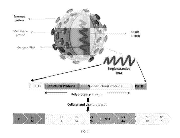
Figure 1 displays an illustration of a Zika virus particle, the Zika RNA
genome, and its
translated genes.
Figure 2 displays a plasmid map for a Zika vaccine, showing the site of the
location for the
insert (expression cassette) that encodes the Zika antigens.
Figure 3 displays drawings that show the linear structure of various Zika
antigen designs.
Figure 4 displays an annotated amino acid sequence for a Zika antigen ¨ leader
sequence+prME.
Figures 5 and 6 display the genetic relationship between various Zika virus
strains: Figure 5
shows genetic distance between isolates, and Figure 6 displays a genetic tree.
Figure 7 displays a plasmid map for a Zika vaccine, showing the site of the
location for the
insert (expression cassette) that encodes Zika-prM+Env.
Figure 8 displays a gel electrophoresis image that shows the presence of
expression cassette.
Figures 9A and 9B displays western blot gels that show Zika-envelope protein:
Figure 9A
showing nonspecific binding to anti-sera in the cell lysates; Figure 9B
showing specific
binding to anti-pan-flavivirus in the cell lysates.
Figures 10A displays an SDS-PAGE gel that shows purification of Zika-envelope
protein.
Figure 10B displays a western blot gel that shows purification of Zika-
envelope protein.
Figures 11 and 12 display bar graphs showing spike-specific CD8 T-lymphocyte
responses
assessed by IFN-gamma ELISpot assay against peptide pools covering pre-
M+envelope
antigen. Figure 11 of individual mice. Figure 12 group averages. Mean
responses in each
group one week after the third immunization.
3
CA 03015792 2018-08-24
WO 2017/147458
PCT/US2017/019407
Figures 13A and 13B display a graph that represents binding ELISA of samples,
showing
Zika prM+Env vaccination of mice elicits a positive antibody response which
reacts with
Zika-envelope antigen.
Figures 14A and 14B displays graphs that show that ZV-prME immunogen elicits a
considerable antibody response which reacts specifically with Zika-Envelope
antigen. The
cross reactivity of the ZpME sera against Dengue 1, 2, 3, and 4 antigen Envs
were negative,
while against Zika Env showed strong binding.
Figures 15A ¨ 15E display an analysis indicating that ZV-prME vaccine
generated sera does
not cross-react with Dengue 1-4 recombinant Envs. Analysis supports that anti-
CHIKV
vaccine induced sera does not bind to Zika Env, also.
Figure 16, comprising Figure 16A through Figure 16E, depicts experimental
results
demonstrating construction of the ZV-prME consensus DNA vaccine. Figure 16A
depicts the
phylogenetic tree at the amino acid level of the ZIKV envelope sequence
between ZIKV
isolates and envelope strains. A consensus design strategy was adopted for the
ZIKV-prME
consensus sequence. Scale bars signify the distance of amino acids per site.
Analyses were
conducted using the MEGA version 5 software. Red star denotes the ZIKA-prME
consensus.
Figure 16B depicts a diagrammatic representation of the ZIKV-prME DNA vaccine
indicating the cloning of prME (prM+Env) into the pVaxl mammalian expression
vector,
pGX0001. Codon-optimized synthetic genes of prME construct included the IgE
leader
sequence. The overall gene construct was inserted into the BamH1 and Xhol
sites of the
pVaxl vector under the control of the CMV promoter. Figure 16C depicts an
agarose gel
electrophoresis analysis of the ZIKV-prME DNA vaccine. Lane 1 shows the
undigested
vaccine construct; Lane 2, restriction digestion of the plasmid with
BamH1/Xhol; Lane 3,
DNA molecular size markers (in kb). Figure 16D depicts expression analysis by
SDS-PAGE
of ZIKV prME protein expression in 293T cells using western blot evaluation
and IFA
detection. 293T cells were transfected with the ZIKV-prME plasmid and cell
lysates and
supernatants were analyzed for expression. Lane 1 contains the protein
molecular weight
markers (kDa); Lane 2, pVaxl control cell lysate; Lane 3, cell lysate from ZV
prME
transfected cells; Lane 4, supernatant from ZIKV-prME transfected cells; Lane
5,
recombinant prME positive control. Figure 16E depicts immunofluorescence
analysis assay
(IFA) assay for ZIKV-prME protein expression in 293T cells. 293T cells were
transfected
4
CA 03015792 2018-08-24
WO 2017/147458
PCT/US2017/019407
with 5pg of the ZIKV-prME plasmid. Twenty-four hours post transfection
immunofluorescence labeling was performed with sera (1:100) from immunized
mice and
anti-mouse IgG FITC. Staining with sera from ZIKV-prME and pVaxl immunized
mice is
shown.
Figure 17, comprising Figure 17A through Figure 17C, depicts experimental
results
demonstrating the characterization of cellular immune responses in mice
following
vaccination with the ZIKV-prME DNA vaccine. Figure 17A depicts ELISpot
analysis
measuring IFN-y secretion in splenocytes. C57/BL6 mice (n = 5/group) were
immunized
intramuscularly three times with 25 lig of either pVaxl or the ZIKV-prME DNA
vaccine
followed by in vivo EP. IFN-y generation, as an indication of cellular immune
response
induction, was measured by IFN-y ELISPOT. Splenocytes harvested 7 days after
the third
immunization were incubated in the presence of one of six peptide pools
spanning the entire
prM and envelope proteins. Results are shown in stacked bar graphs. The data
represent the
average numbers of SFU (spot forming units) per million splenocytes with
values
representing the mean responses in each group (n = 4) SEM. Figure 17B
depicts the epitope
composition of the ZIKV-prME-specific IFN-y response as determined by
stimulation with
matrix peptide pools one week after the third immunization. Values represent
mean responses
in each group (n = 4) SEM. Experiments were performed independently at least
three times
with similar results. Figure 17C depicts immunization with ZIKV-prME induces
higher
number of IFN-y and TNF-a secreting cells when stimulated by ZIKV peptides.
One week
after the last immunization with the ZIKV-prME vaccine, splenocytes were
cultured in the
presence of pooled ZIKV peptides (5pM) or tissue culture medium only.
Frequencies of
ZIKV peptide-specific IFN-y and TNF-a secreting cells were measured by
fluorescence-
activated cell sorting (FACS) assay. Single function gates were set based on
negative control
(unstimulated) samples and were placed consistently across samples. The
percentage of the
total CD8+ T cell responses are shown. These data are representative of two
independent
immunization experiments.
Figure 18, comprising Figure 18A through Figure 18D depicts the profile of IFN-
y
production by splenocytes and antibody levels in serum collected from pZIKV-
prME
(MR766) and pZIKV-prME (Brazil)-immunized mice. Six week-old C57/BL6 mice were
immunized as described in Materials and Methods. Serum and splenocytes were
collected
one week after the 3rd immunization and incubated with ZIKV-specific prME
peptides, and
5
CA 03015792 2018-08-24
WO 2017/147458
PCT/US2017/019407
the number of IFN-y SFU per million cells was assayed by ELISPOT. Figure 18A
depicts
ELISpot analysis of serum collected from MR766- immunized mice. Figure 18B
depicts
ELISpot analysis of serum collected from Brazil-immunized mice. Anti-ZIKV Env
antibody
levels in the serum were measured by ELISA (C&D). Figure 18C depicts Anti-ZIKV
Env
antibody levels in the serum measured by ELISA in MR766- immunized mice.
Figure 18D
depicts Anti-ZIKV Env antibody levels in the serum measured by ELISA in Brazil-
immunized mice.
Figure 19, comprising Figure 19A through Figure 19E depicts experimental
results
demonstrating anti-ZIKV antibody responses are induced by ZIKV-prME plasmid
vaccination. C57BL/6 mice were immunized intramuscularly three times with 25
pg of
ZIKV-prME plasmid or pVaxl at 2-week intervals. Binding to envelope antigen
was
analyzed with sera from animals at different time points post immunization at
various
dilutions. ELISA plates were coated with vaccine matched recombinant ZIKV-
envelope
protein Figure 19A depicts results from 1 of 2 independent experiments are
presented.
Similar results were obtained in the second experiment. Figure 19B depicts the
differences in
the anti-ZIKV endpoint titers produced in response to the ZIKV-prME immunogen
were
analyzed in sera from immunized animals after each boost. Figure 19C depicts
western blot
analysis of ZIKV-envelope antigen expression. The recombinant ZIKV-Env protein
at
various concentration were electrophoresed on a 12.5% SDS polyacrylamide gel
and
analyzed by Western blot analysis with sera from pVaxl or ZIKV-prME immunized
mice, as
indicated. Expression of the ZIKV-Env protein is indicated by the arrowheads.
Figure 20D
depicts an immunofluorescence analysis of Vero cells infected with either ZIKV-
MR766 or
mock infected following incubation with sera from ZIKV-prME or pVaxl immunized
mice.
Serum samples from the pZIKV-prME immunized mice were tested by plaque-
reduction
neutralization (PRNT) assay for their ability to neutralize ZIKV infectivity
in vitro. PRNT50
was defined as the serum dilution factor that could inhibit 50% of the input
virus. Values in
parentheses indicate the PRNT50. Control plasmid pZIKV-Capsid and pVaxl sera
were used
as negative controls.
Figure 20, comprising Figure 20A through Figure 20E, depicts experimental
results
demonstrating induction of ZIKV specific cellular immune responses following
ZIKV=prME
DNA vaccination of NHPs. Figure 20A depicts rhesus macaques were immunized
intradermally (ID) with 2 mg of ZIKV-prME plasmid at weeks 0 and 4
administered as 1 mg
6
CA 03015792 2018-08-24
WO 2017/147458
PCT/US2017/019407
at each of two sites, with immunization immediately followed by intradermal
EP. PBMCs
were isolated pre-immunization and at week 6 and were used for the ELISPOT
assay to
detect IFN-y-secreting cells in response to stimulation with ZIKV-prME
peptides. The
number of IFN-y producing cells obtained per million PBMCs against six peptide
pools
encompassing the entire prME protein is indicated on the y-axis for the
vaccination groups.
Values represent mean responses in each group (n = 5) SEM. Figure 20B
depicts the
detection of ZIKV-prME-specific antibody responses following DNA vaccination.
Anti-
ZIKV IgG antibodies were measured pre-immunization and at week 6 by ELISA.
Figure
20C depicts end-point ELISA titers for anti ZIKV-envelope antibodies are shown
following
the first and second immunizations. Figure 20d depicts western blot analysis
using week 6
pooled monkey sera demonstrated binding to recombinant envelope protein.
Figure 20E
depicts immunofluorescence analysis of Vero cells infected with ZIKV MR766 at
10 PFU.
Cells were probed 24 hrs following infection with wk 6 pooled monkey sera at
1:100 and
then detected with secondary anti-human IgG-AF488.
Figure 21, comprising Figure 21A through Figure 21C, depicts experimental
results
demonstrating plaque-reduction neutralization activity of serum from Rhesus
Macaques
immunized with ZIKV-prME. Rhesus Macaques were immunized as described in
Materials
and Methods. Figure 21A depicts pre immunization and week 6 immune sera from
individual
monkeys were tested by plaque reduction neutralization (PRNT) assay for their
ability to
neutralize ZIKV infectivity in vitro. PRNT50 was defined as the serum dilution
factor that
could inhibit 50% of the input virus. Calculated IC50 values are listed for
each monkey.
Figures 21B and 21C depict the cytopathic effect of ZIKV MR766 and PR209 in
Vero, SK-
N-SH, and U87MG cells. Figure 21B depicts Vero cells were mock infected or
infected with
the MR766 or PR209 viruses. Figure 21C depicts SK-N-SH and U87MG cells were
mock or
infected with MR766 at an MOT of 0.001 PFU/cell in the presence of pooled NHP
sera
immunized with ZIKV-prME vaccine (Wk 6). The induction of syncytium formation
(CPE)
and prME protein expression were analyzed 48 hours post infection by indirect
immunofluorescence assay (IFA) using the immunized NHP sera. Pictures were
taken at 4x
objective.
Figure 22, comprising Figure 22A through Figure 22C depicts experimental
results
demonstrating Profile of IFN-y and antibody production by spleen cells
isolated from pZIKV
7
CA 03015792 2018-08-24
WO 2017/147458
PCT/US2017/019407
prME in mice lacking the type I interferon a, (3 receptor. Figure 22A depicts
IFN a, (3 receptor
knockout mice (four to six) were immunized intramuscularly three times with 25
lig of
pZIKV-prME or pVaxl plasmid at 2-week intervals. Splenocytes were collected
two weeks
after the last immunization and incubated with prME peptides and the number of
IFN-y-
producing cells were measured by ELISPOT. Figure 22B depicts serum antibody
specific for
ZIKV Env protein in immunized animals was measured by ELISA at various days
post
immunization. Figure 22C depicts the endpoint titer 0, 1, 2,3, 4 and 5 weeks
after
immunization.
Figure 23, comprising Figure 23A through Figure 23F depicts experimental
results
demonstrating survival data for immunized mice lacking the type I interferon
a, 13 receptor
following Zika virus infection. Survival of IFN-a/r3 receptor knockout mice
after Zika
infection. Figure 23A depicts mice were immunized once and challenged with 106
PFU of
ZIKV-PR209, 2 weeks later. Figure 23B depicts mice were immunized twice at 2
week
intervals and challenged with 106 PFU of ZIKV-PR209 7 days after the second
immunization.
Figure 23C depicts mice were immunized twice at 2 week intervals and
challenged with 2 x
106 PFU of ZIKV PR209, 7 days after the second immunization. The survival
curves were
constructed using data from two separate experiments. Figure 23D depicts
weight change for
animals immunized 2x is depicted; the data reflect the results from two
independent
experiments with 10 to 15 mice per group per experiment. Figure 23E depicts
clinical scores
for animals in Figure 23B. Figure 23F depicts clinical scores for animals in
Figure 23C. The
designation for the clinical scores is as follows: 1-no disease, 2-decreased
mobility; 3-
hunched posture and decreased mobility; 4-hindlimb knuckle walking (partial
paralysis), 5-
paralysis of one hind limb and 6-paralysis of both hind limbs.
Figure 24, comprising Figure 24A through Figure 24E depicts experimental
results
demonstrating the construction of the ZIKV-prME consensus DNA vaccine. Figure
24A
depicts a diagrammatic representation of the ZIKV-prME DNA vaccine indicating
the
cloning of rME into the pVaxl mammalian expression vector. A consensus design
strategy
was adopted for the ZIKV-prME consensus sequence. Codon-optimized synthetic
genes of
the prME construct included a synthetic IgE leader sequence. The optimized
gene construct
was inserted into the BamH1 and Xhol sites of a modified pVaxl vector under
the control of
the CMV promoter. Figure 24B depicts a model building of the ZIKV-E proteins
demonstrates overlap of the vaccine target with potentially relevant epitope
regions. Several
8
CA 03015792 2018-08-24
WO 2017/147458
PCT/US2017/019407
changes made for vaccine design purpose are located in domains II and III
(located within
dashed lines of inset, middle left). Vaccine-specific residue changes in these
regions are
shown in violet CPK format on a ribbon backbone representation of an E
(envelope) protein
dimer (each chain in light and dark green, respectively). Regions
corresponding to the
defined EDE are indicated in cyan, and the fusion loop is indicated in blue.
Residue 11e156
(T156I) of the vaccine E protein, modelled as exposed on the surface of the
150 loop, is part
of an N-linked glycosylation motif NXS/T in several other ZIKV strains as well
as in
multiple dengue virus strains. Figure 24C depicts expression analysis by SDS-
PAGE of
ZIKV-prME protein expression in 293T cells using western blot analysis. The
293T cells
were transfected with the ZIKV-prME plasmid and the cell lysates and
supernatants were
analyzed for expression of the vaccine construct with pan-flavivirus immunized
sera. Protein
molecular weight markers (kDa); cell lysate and supernatant from ZIKV-prME
transfected
cells and rZIKV-E positive control were loaded as indicated. Figure 24D
depicts expression
analysis by SDS-PAGE of ZIKV-prME protein expression in 293T cells using
western blot
analysis. The 293T cells were transfected with the ZIKV-prME plasmid and the
cell lysates
and supernatants were analyzed for expression of the vaccine construct with
ZIKV-prME
immunized sera. Protein molecular weight markers (kDa); cell lysate and
supernatant from
ZIKV-prME transfected cells and rZIKV-E positive control were loaded as
indicated. Figure
24E depicts Immunofluorescence assay (IFA) analysis for ZIKV-prME protein
expression in
293T cells. The cells were transfected with 5 pg of the ZIKVprME plasmid.
Twenty-four
hours post transfection, immunofluorescence labelling was performed with the
addition of
sera (1:100) from ZIKV-prME immunized mice followed by the addition of the
secondary
anti-mouse IgG-AF488 antibody for detection. Staining with sera from ZIKV-prME
and
pVaxl immunized mice is shown. DAPI panels show control staining of cell
nuclei. Overlay
panels are combinations of antimouse IgG-AF488 and DAPI staining patterns.
DAPI, 4',6-
diamidino-2-phenylindole; ZIKV-prME, precursor membrane and envelope of Zika
virus.
Figure 25, comprising Figure 25A through Figure 25D depicts experimental
results
demonstrating the characterization of cellular immune responses in mice
following
vaccination with the ZIKV-prME DNA vaccine. Figure 25A depicts a timeline of
vaccine
immunizations and immune analysis used in the study. Figure 25B depicts
ELISpot analysis
measuring IFN-y secretion in splenocytes in response to ZIKV-prME
immunization.
C57BL/6 mice (n=4/group) were immunized i.m. three times with 25 lig of either
pVaxl or
the ZIKV-prME DNA vaccine followed by electroporation. IFN-y generation, as an
9
CA 03015792 2018-08-24
WO 2017/147458
PCT/US2017/019407
indication of induction of cellular immune responses, was measured by an IFN-y
ELISpot
assay. The splenocytes harvested 1 week after the third immunization were
incubated in the
presence of one of the six peptide pools spanning the entire prM and Envelope
proteins.
Results are shown in stacked bar graphs. The data represent the average
numbers of SFU
(spot-forming units) per million splenocytes with values representing the mean
responses in
each s.e.m. Figure 25C depicts the epitope composition of the ZIKVprME-
specific IFN-y
response as determined by stimulation with matrix peptide pools 1 week after
the third
immunization. The values represent mean responses in each group s.e.m. The
experiments
were performed independently at least three times with similar results. Figure
25D depicts
flow cytometric analysis of T-cell responses. Immunisation with ZIKV-prME
induces higher
number of IFN-y and TNF-a secreting cells when stimulated by ZIKV peptides.
One week
after the last immunization with the ZIKV-prME vaccine, splenocytes were
cultured in the
presence of pooled ZIKV peptides (5 p,M) or R10 only. Frequencies of ZIKV
peptide-specific
IFN-y and TNF-a secreting cells were measured by flow cytometry. Single
function gates
were set based on negative control (unstimulated) samples and were placed
consistently
across samples. The percentage of the total CD8+ T-cell responses are shown.
These data are
representative of two independent immunization experiments. IFN, interferon;
TNF, tumour
necrosis factor; ZIKV-prME, precursor membrane and envelope of Zika virus.
Figure 26, comprising Figure 26A through Figure 26E depicts experimental
results
demonstrating that anti-ZIKV antibody responses are induced by ZIKV-prME
vaccination.
Figure 26A depicts ELISA analysis measuring binding antibody production
(measured by
0D450 values) in immunized mice. The C57BL/6 mice (n =4) were immunized i.m.
three
times with 25 lig of ZIKV-prME plasmid or pVaxl at 2-week intervals. Binding
to rZIKV-E
was analyzed with sera from animals at different time points (days 21, 35 and
50) post
immunization at various dilutions. The data shown are representative of at
least three separate
experiments. Figure 26B depicts End point binding titer analysis. Differences
in the anti-
ZIKV end point titers produced in response to the ZIKV-prME immunogen were
analyzed in
sera from immunized animals after each boost. Figure 26C depicts Western blot
analysis of
rZIKV-E specific antibodies induced by ZIKV-prME immunization. The rZIKV-E
protein
was electrophoresed on a 12.5% SDS polyacrylamide gel and analyzed by western
blot
analysis with pooled sera from ZIKV-prME immunized mice (day 35). Binding to
rZIKV-E
is indicated by the arrowhead. Figure 26D depicts immunofluorescence analysis
of ZIKV
specific antibodies induced by ZIKV-prME immunization. The Vero cells infected
with
CA 03015792 2018-08-24
WO 2017/147458
PCT/US2017/019407
either ZIKV-MR766 or mock infected were stained with pooled sera from ZIKV-
prME
immunized mice (day 35) followed by an anti-mouse-AF488 secondary antibody for
detection. Figure 26E depicts plaque-reduction neutralization (PRNT) assay
analysis of
neutralizing antibodies induced by ZIKV-prME immunization. The serum samples
from the
ZIKV-prME immunized mice were tested for their ability to neutralize ZIKV
infectivity in
vitro. PRNT50 was defined as the serum dilution factor that could inhibit 50%
of the input
virus. The values in parentheses indicate the PRNT50. Control ZIKV-Cap (DNA
vaccine
expressing the ZIKV capsid protein) and pVaxl sera were used as negative
controls. ZIKV-
prME, precursor membrane and envelope of Zika virus.
Figure 27, comprising Figure 27A through Figure 27E depicts experimental
results
demonstrating Induction of ZIKV specific cellular immune responses following
ZIKV-prME
vaccination of non-human primates (NHPs). Figure 27A depicts ELISpot analysis
measuring
IFN-y secretion in peripheral blood mononuclear cells (PBMCs) in response to
ZIKV-prME
immunization. Rhesus macaques were immunized intradermally with 2 mg of ZIKV-
prME
plasmid at weeks 0 and 4 administered as 1 mg at each of two sites, with
immunization
immediately followed by intradermal electroporation. PBMCs were isolated pre-
immunization and at week 6 and were used for the ELISPOT assay to detect IFN-y-
secreting
cells in response to stimulation with ZIKV-prME peptides as described in the
'Materials and
Methods' section. The number of IFN-y producing cells obtained per million
PBMCs against
six peptide pools encompassing the entire prME protein is shown. The values
represent mean
responses in each group (n=5) s.e.m. Figure 27B depicts the detection of ZIKV-
prME-
specific antibody responses following DNA vaccination. Anti-ZIKV IgG
antibodies were
measured pre-immunization and at week 6 by ELISA. Figure 27C depicts end point
ELISA
titers for anti ZIKV-envelope antibodies are shown following the first and
second
immunizations. Figure 27D depicts western blot analysis using week 6 RM immune
sera
demonstrated binding to recombinant envelope protein. Figure 27E depicts PRNT
activity of
serum from RM immunized with ZIKV-prME. Pre-immunization and week 6 immune
sera
from individual monkeys were tested by plaque-reduction neutralization (PRNT)
assay for
their ability to neutralize ZIKV infectivity in vitro. PRNT50 was defined as
the serum
dilution factor that could inhibit 50% of the input virus. Calculated (PRNT50)
values are
listed for each monkey. IFN, interferon; ZIKV-prME, precursor membrane and
envelope of
Zika virus.
11
CA 03015792 2018-08-24
WO 2017/147458
PCT/US2017/019407
Figure 28, comprising Figure 28A through Figure 28F depicts experimental
results
demonstrating survival data for immunized mice lacking the type I interferon
a, (3 receptor
following ZIKV infection. Figure 28A depicts survival of IFNAR-/ mice after
ZIKV
infection. Mice were immunized twice with 25 lig of the ZIKV-prME DNA vaccine
at 2-
week intervals and challenged with ZIKV-PR209 virus 1 week after the second
immunization
with 1 x 10⁶ plaque-forming units Figure 28B depicts survival of IFNAR / mice
after ZIKV
infection. Mice were immunized twice with 25 lig of the ZIKV-prME DNA vaccine
at 2-
week intervals and challenged with ZIKV-PR209 virus 1 week after the second
immunization
with 2 x 10⁶ plaque-forming units Figure 28C depicts the weight change of
animals
immunized with 1 x 10⁶ plaque-forming units. Figure 28D depicts the weight
change of
animals immunized with 2 x 10⁶ plaque-forming units. Figure 28E depicts the
clinical scores
of animals immunized with 1 x 10⁶ plaque-forming units. Figure 28F depicts the
clinical
scores of animals immunized with 2 x 10⁶ plaque-forming units. The designation
for the
clinical scores is as follows: 1: no disease, 2: decreased mobility; 3:
hunched posture and
decreased mobility; 4: hind limb knuckle walking (partial paralysis); 5:
paralysis of one hind
limb; and 6: paralysis of both hind limbs. The data reflect the results from
two independent
experiments with 10 mice per group per experiment. ZIKV-prME, precursor
membrane and
envelope of Zika virus.
Figure 29, comprising Figure 29A through Figure 29d depicts experimental
results
demonstrating single immunization with the ZIKV-prME vaccine provided
protection against
ZIKV challenge in mice lacking the type I interferon a, 13 receptor. The mice
were
immunized once and challenged with 2 x 10⁶ plaque-forming units of ZIKV-PR209,
2 weeks
after the single immunization. The survival curves depict 10 mice per group
per experiment
Figure 29A demonstrates that the ZIKV-prME vaccine prevented ZIKA-induced
neurological
abnormalities in the mouse brain Figure 29B depicts brain sections from pVaxl
and ZIKV-
prME vaccinated groups were collected 7-8 days after challenge and stained
with H&E
(haematoxylin and eosin) for histology. The sections taken from
representative, unprotected
pVaxl control animals shows pathology. (i): nuclear fragments within neuropils
of the
cerebral cortex (inset shows higher magnification and arrows to highlight
nuclear fragments);
(ii): perivascular cuffing of vessels within the cortex, lymphocyte
infiltration and
degenerating cells; (iii): perivascular cuffing, cellular degeneration and
nuclear fragments
within the cerebral cortex; and (iv): degenerating neurons within the
hippocampus (arrows).
An example of normal tissue from ZIKV-prME vaccinated mice appeared to be
within
12
CA 03015792 2018-08-24
WO 2017/147458
PCT/US2017/019407
normal limits (v and vi). Figure 29C depicts levels of ZIKV RNA in the plasma
samples from
mice following vaccination and viral challenge at the indicated day post
infection. The results
are indicated as the genome equivalents per milliliter of plasma. Figure 29D
depicts levels of
ZIKV-RNA in the brain tissues were analyzed at day 28 post infection. The
results are
indicated as the genome equivalent per gram of tissue. ZIKV-prME, precursor
membrane and
envelope of Zika virus.
Figure 29, comprising Figure 30A and Figure 30B, depicts experimental results
demonstrating protection of mice lacking the type I interferon a, 13 receptor
following passive
transfer of anti-ZIKV immune sera following ZIKV challenge. Pooled NHP anti-
ZIKV
immune sera, titred for anti-ZIKA virus IgG, was administered i.p. (150
pi/mouse) to mice 1
day after s.c. challenge with a ZIKA virus (106 plaque-forming units per
mouse). As a control,
normal monkey sera and phosphate-buffered saline (PBS) were administered (150
pl/mouse)
to age-matched mice as controls. Figure 30A depicts the mouse weight change
during the
course of infection and treatment. Each point represents the mean and standard
error of the
calculated percent pre-challenge (day 0) weight for each mouse. Figure 30B
depicts the
survival of mice following administration of the NHP immune sera. ZIKV-prME,
precursor
membrane and envelope of Zika virus.
Figure 31, comprising Figure 31A through Figure 31D, depicts experimental
results
demonstrating the characterization of immune responses of ZIKV-prME-MR766 or
ZIKV-
prME Brazil vaccine in C57BL/6 mice. Figure 31A depicts ELISpot and ELISA
analysis
measuring cellular and antibody responses after vaccination with either ZIKV-
prME-MR766
and ZIKV-prME-Brazil DNA vaccines. C57BL/6 mice (n = 4/group) were immunized
intramuscularly three times with 25pg of ZIKV-prME-MR766 followed by in vivo
EP. IFN-y
generation, as an indication of cellular immune response induction, was
measured by IFN-y
ELISpot. Splenocytes harvested one week after the third immunization were
incubated in the
presence of one of six peptide pools spanning the entire prM and E proteins.
Results are
shown in stacked bar graphs. The data represent the average numbers of SFU
(spot forming
units) per million splenocytes with values representing the mean responses in
each SEM.
Figure 31B depicts ELISpot and ELISA analysis measuring cellular and antibody
responses
after vaccination with either ZIKV-prME-MR766 and ZIKV-prME-Brazil DNA
vaccines.
C57BL/6 mice (n = 4/group) were immunized intramuscularly three times with
25pg of
ZIKV prME-Brazil followed by in vivo EP. IFN-y generation, as an indication of
cellular
13
CA 03015792 2018-08-24
WO 2017/147458
PCT/US2017/019407
immune response induction, was measured by IFN-y ELISpot. Splenocytes
harvested one
week after the third immunization were incubated in the presence of one of six
peptide pools
spanning the entire prM and E proteins. Results are shown in stacked bar
graphs. The data
represent the average numbers of SFU (spot forming units) per million
splenocytes with
values representing the mean responses in each SEM. Figure 31C depicts ELISA
analysis
measuring binding antibody production in immunized C57BL/6 mice. Binding to
rZIKV-E
was analyzed with sera from mice at day 35 post immunization at various
dilutions. Figure
31D depicts ELISA analysis measuring binding antibody production in immunized
C57BL/6
mice. Binding to rZIKV-E was analyzed with sera from mice at day 35 post
immunization at
various dilutions.
Figure 32, comprising Figure 32A through Figure 32D, depicts experimental
results
demonstrating the expression, purification, and characterization of ZIKV-
Envelope protein.
Figure 32A depicts the cloning plasmid for rZIKV E expression. Figure 32B
depicts the
characterization of the recombinant ZIKV-E (rZIKV-E) protein by SDS-PAGE and
Western
blot analysis. Lane 1-BSA control; Lane 2- lysates from E. coli cultures
transformed with
pET-28a vector plasmid, was purified by nickel metal affinity resin columns
and separated by
SDS-PAGE after IPTG induction. Lane 3, 37 recombinant ZV-E purified protein
was
analyzed by Western blot with anti-His tag antibody. Lane M, Protein molecular
weight
marker. Figure 32C depicts the purified rZIKV-E protein was evaluated for its
antigenicity.
ELISA plates were coated with rZIKV-E and then incubated with various
dilutions of
immune sera from the mice immunized with ZIKV-prME vaccine or Pan-flavivirus
antibody
as positive control. Bound IgG was detected by the addition of peroxidase-
conjugated anti-
mouse antibody followed by tetramethylbenzidine substrate as described in
Experimental
.. Example. Figure 32D depicts western blot detection of purified rZIKV-E
protein with
immune sera from ZIKV prME immunized mice. Various concentrations of purified
rZIKV-
E protein were loaded onto an SDS-PAGE gel as described. A dilution of 1:100
immune sera,
and goat anti-mouse at 1:15,000 were used for 1 hour at room temperature.
After washing,
the membranes were imaged on the Odyssey infrared imager. Odyssey protein
molecular
weight standards were used. The arrows indicate the position of rZIKV-E
protein.
Figure 33, comprising Figure 33A through Figure 33C, depicts experimental
results
demonstrating the characterization of immune responses ZIKA-prME in IFNAR-/-
mice.
ELISpot and ELISA analysis measuring cellular and antibody responses to ZIKV-
prME in
14
CA 03015792 2018-08-24
WO 2017/147458
PCT/US2017/019407
IFNAR4- mice. Mice (n = 4/group) were immunized intramuscularly three times
with 25 lig
of ZIKV-prME followed by in vivo EP. Figure 33A depicts IFN-y generation, as
an
indication of cellular immune response induction, was measured by IFN-y
ELISPOT. Figure
33B depicts ELISA analysis measuring binding antibody production in immunized
IFNAR-/-
mice. Binding to rZIKV-E was analyzed with sera from mice at various time
points post
immunization. Figure 33C depicts endpoint titer analysis of anti-ZIKV
antibodies produced
in immunized IFNAR-/- mice.
Figure 34, comprising Figure 34A through Figure 34D, depicts experimental
results
demonstrating the neutralization activity of immune sera from Rhesus Macaques
immunized
against ZIKV-prME. SK-N-SH and U87MG cells were mock infected or infected with
MR766 at an MOT of 0.01 PFU/cell in the presence of pooled NHP sera immunized
with
ZIKV-prME vaccine (Wk 6). Zika viral infectivity were analyzed 4 days post
infection by
indirect immunofluorescence assay (IFA) using sera from ZIKV-prME vaccinated
NHPs.
Figure 34A depicts photographs of stained tissue sample slices taken with a
20x objective
demonstrating inhibition of infection by ZIKV viruses MR766 and PR209 in Vero,
SK-N-SH
and U87MG Figure 34B depicts photographs of stained tissue sample slices taken
with a 20x
objective demonstrating inhibition of infection by ZIKV viruses SK-N-SH and
U87MG in
Vero, SK-N-SH and U87MG Figure 34C depicts a bar graph shows the percentage of
infected (GFP positive cells) demonstrating the inhibition of infection by
ZIKV viruses
MR766 and PR209 in Vero, SK-N-SH and U87MG Figure 34D depicts a bar graph
showing
the percentage of infected (GFP positive cells) demonstrating the inhibition
of infection by
ZIKV viruses SK-N-SH and U87MG in Vero, SK-N-SH and U87MG
Figure 35, comprising Figure 35A through Figure 35D, depicts experimental
results
demonstrating ZIKV is virulent to IFNAR-/- mice. These data confirm that ZIKV
is virulent in
IFNAR4- resulting in morbidity and mortality. Figure 35A depicts Kaplan-Meier
survival
curves of IFNAR-/- mice inoculated via intracranial with 106 pfu ZIKV-PR209
virus. Figure
35B depicts Kaplan-Meier survival curves of IFNAR-/- mice inoculated via
intravenously
with 106 pfu ZIKV-PR209 virus. Figure 35C depicts Kaplan-Meier survival curves
of
IFNAR4- mice inoculated via intraperitoneal with 106 pfu ZIKV-PR209 virus.
Figure 35D
depicts Kaplan-Meier survival curves of IFNAR-/- mice inoculated via
subcutaneously with
106 pfu ZIKV-PR209 virus. Figure 35A depicts the mouse weight change during
the course
of infection for all the routes.
CA 03015792 2018-08-24
WO 2017/147458
PCT/US2017/019407
Figure 36, comprising Figure 36A through Figure 36C, depicts experimental
results
demonstrating the induction of ZIKV specific cellular immune responses
following ZIKV-
prME vaccination of Non-Human Primates (NHPs). Figure 36A is a schematic
representation
of NHP immunization study. Figure 36B depicts results after a single
immunization. Figure
36C depicts results after two immunizations. ELISpot analysis measuring IFN-g
secretion in
PBMCs in response to ZIKV-prME immunization. Rhesus macaques were immunized
intradermal (i.d.) with 2 mg of ZIKV-prME plasmid at weeks 0 and 4
administered as 1 mg at
each of two sites, with immunization immediately followed by intradermal EP.
PBMCs were
isolated pre-immunization and at week 6 and were used for the ELISPOT assay to
detect
IFN-g-secreting cells in response to stimulation with ZIKV-prME peptides as
described in
Materials and Methods. The number of IFN-g producing cells obtained per
million PBMCs
against six peptide pools encompassing the entire prME protein is shown.
Values represent
mean responses in each group (n = 5) SEM.
Figure 37, comprising Figure 37A and Figure 37B, depicts experimental results
demonstrating anti-ZIKV antibody responses are induced by ZIKV-prME
vaccination of
Non-Human Primates (NHPs). Figure 37A depicts the detection of ZIKV-prME-
specific
antibody responses following a single DNA vaccination. Anti-ZIKV IgG
antibodies were
measured pre-immunization and at week 6 by ELISA. Figure 37B depicts the
detection of
ZIKV-prME-specific antibody responses following two DNA vaccinations. Anti-
ZIKV IgG
antibodies were measured pre-immunization and at week 6 by ELISA.
Figure 38, comprising Figure 38A through Figure 38D, depicts experimental
results
demonstrating Zika-prME immunization confers protection against Zika
challenge. Figure
38A is a schematic representation of NHP Zika challenge study. Rhesus macaques
were
vaccinated twice at weeks 0 and 4 with pZV-prME DNA via ID route using EP. At
week 8,
the animals were subcutaneous challenged with Zika-PR209 viral strain. As a
control, 5-naïve
animals were infected with ZV-PR209 virus. Figure 38B depicts the sequential
viral load
determinations for individual animals in Naïve NHP. Figure 38C depicts the
sequential viral
load determinations for individual animals in NHP vaccinated once. Figure 38D
depicts the
sequential viral load determinations for individual animals in NHP vaccinated
twice. The
panel shows the peak viral loads for each animal with standard error bars for
the three groups
are shown (log of viral RNA copies/mL plasma).
16
CA 03015792 2018-08-24
WO 2017/147458
PCT/US2017/019407
Figure 39, comprising Figure 39A and Figure 39B, depicts experimental results
from a phase
1 Zika DNA Vaccine Study. Figure 39A depicts experimental results from a
binding ELISA
study. Figure 39B depicts experimental results demonstrating passive transfer
and protection.
Figure 40, comprising Figure 40A and Figure 40B, depicts experimental results
from
immunofluorescence analysis
Figure 41, comprising Figure 41A and Figure 41B, depicts experimental results
demonstrating characterization of the percentage of binding responders.
Figure 42 depicts experimental results demonstrating neutralization post dose
2.
DETAILED DESCRIPTION OF PREFERRED EMBODIMENTS
The following abbreviated, or shortened, definitions are given to help the
understanding of
the preferred embodiments of the present invention. The abbreviated
definitions given here
are by no means exhaustive nor are they contradictory to the definitions as
understood in the
field or dictionary meaning. The abbreviated definitions are given here to
supplement or more
clearly define the definitions known in the art.
Definitions
Sequence homology for nucleotides and amino acids as used herein may be
determined using
FASTA, BLAST and Gapped BLAST (Altschul et al., Nuc. Acids Res., 1997, 25,
3389,
which is incorporated herein by reference in its entirety) and PAUP* 4.0b10
software (D. L.
Swofford, Sinauer Associates, Massachusetts). Briefly, the BLAST algorithm,
which stands
for Basic Local Alignment Search Tool is suitable for determining sequence
similarity
(Altschul et al., J. Mol. Biol., 1990, 215, 403-410, which is incorporated
herein by reference
in its entirety). Software for performing BLAST analyses is publicly available
through the
National Center for Biotechnology Information. One measure of similarity
provided by the
BLAST algorithm is the smallest sum probability (P(N)), which provides an
indication of the
probability by which a match between two nucleotide sequences would occur by
chance. For
example, a nucleic acid is considered similar to another if the smallest sum
probability in
17
CA 03015792 2018-08-24
WO 2017/147458
PCT/US2017/019407
comparison of the test nucleic acid to the other nucleic acid is less than
about 1, preferably
less than about 0.1, more preferably less than about 0.01, and most preferably
less than about
0.001. "Percentage of similarity" can be calculated using PAUP* 4.0b10
software (D. L.
Swofford, Sinauer Associates, Massachusetts). The average similarity of the
consensus
sequence is calculated compared to all sequences in the phylogenic tree.
As used herein, the term "nucleic acid construct" refers to the DNA or RNA
molecules that
comprise a nucleotide sequence that encodes protein. The coding sequence, or
"encoding
nucleic acid sequence," can include initiation and termination signals
operably linked to
regulatory elements including a promoter and polyadenylation signal capable of
directing
expression in the cells of the individual to whom the nucleic acid molecule is
administered.
As used herein, the term "expressible form" refers to nucleic acid constructs
that contain the
necessary regulatory elements operably linked to a coding sequence that
encodes a protein
such that when present in the cell of the individual, the coding sequence will
be expressed.
The term "constant current" is used herein to define a current that is
received or experienced
by a tissue, or cells defining said tissue, over the duration of an electrical
pulse delivered to
same tissue. The electrical pulse is delivered from the electroporation
devices described
herein. This current remains at a constant amperage in said tissue over the
life of an electrical
pulse because the electroporation device provided herein has a feedback
element, preferably
having instantaneous feedback. The feedback element can measure the resistance
of the tissue
(or cells) throughout the duration of the pulse and cause the electroporation
device to alter its
electrical energy output (e.g., increase voltage) so current in same tissue
remains constant
throughout the electrical pulse (on the order of microseconds), and from pulse
to pulse. In
some embodiments, the feedback element comprises a controller.
The term "feedback" or "current feedback" is used interchangeably and means
the active
response of the provided electroporation devices, which comprises measuring
the current in
tissue between electrodes and altering the energy output delivered by the EP
device
accordingly in order to maintain the current at a constant level. This
constant level is preset
by a user prior to initiation of a pulse sequence or electrical treatment.
Preferably, the
feedback is accomplished by the electroporation component, e.g., controller,
of the
electroporation device, as the electrical circuit therein is able to
continuously monitor the
18
CA 03015792 2018-08-24
WO 2017/147458
PCT/US2017/019407
current in tissue between electrodes and compare that monitored current (or
current within
tissue) to a preset current and continuously make energy-output adjustments to
maintain the
monitored current at preset levels. In some embodiments, the feedback loop is
instantaneous
as it is an analog closed-loop feedback.
The terms "electroporation," "electro-permeabilization," or "electro-kinetic
enhancement"
("EP"), as used interchangeably herein, refer to the use of a transmembrane
electric field
pulse to induce microscopic pathways (pores) in a bio-membrane; their presence
allows
biomolecules such as plasmids, oligonucleotides, siRNA, drugs, ions, and/or
water to pass
from one side of the cellular membrane to the other.
The term "decentralized current" is used herein to define the pattern of
electrical currents
delivered from the various needle electrode arrays of the electroporation
devices described
herein, wherein the patterns minimize, or preferably eliminate, the occurrence
of
electroporation related heat stress on any area of tissue being
electroporated.
The term "feedback mechanism" as used herein refers to a process performed by
either
software or hardware (or firmware), which process receives and compares the
impedance of
the desired tissue (before, during, and/or after the delivery of pulse of
energy) with a present
value, preferably current, and adjusts the pulse of energy delivered to
achieve the preset value.
The term "impedance" is used herein when discussing the feedback mechanism and
can be
converted to a current value according to Ohm's law, thus enabling comparisons
with the
preset current. In a preferred embodiment, the "feedback mechanism" is
performed by an
analog closed loop circuit.
The term "immune response" is used herein to mean the activation of a host's
immune
system, e.g., that of a mammal, in response to the introduction of a Zika
antigen, e.g.,
universal Zika antigen, via the provided DNA plasmid vaccines. The immune
response can
be in the form of a cellular or humoral response, or both.
The term "consensus" or "consensus sequence" is used herein to mean a
synthetic nucleic
acid sequence, or corresponding polypeptide sequence, constructed based on
analysis of an
alignment of multiple strains of a Zika gene. The consensus universal Zika can
be used to
induce broad immunity against multiple subtypes or serotypes of Zika virus.
19
CA 03015792 2018-08-24
WO 2017/147458
PCT/US2017/019407
The term "adjuvant" is used herein to mean any molecule added to the DNA
plasmid
vaccines described herein to enhance antigenicity of the Zika antigen encoded
by the DNA
plasmids and encoding nucleic acid sequences described hereinafter.
The term "subtype" or "serotype" is used herein interchangeably and in
reference to a virus,
for example Zika virus, and means genetic variants of that virus antigen such
that one subtype
is recognized by an immune system apart from a different subtype. For example,
Zika virus
subtype 1 is immunologically distinguishable from Zika virus subtype 2.
One aspect of the present invention provides nucleic acid constructs capable
of expressing a
polypeptide that elicits an immune response in a mammal against Zika virus.
The nucleic acid
constructs are comprised of an encoding nucleotide sequence and a promoter
operably linked
to the encoding nucleotide sequence. The encoding nucleotide sequence
expresses the
polypeptide, wherein the polypeptide includes consensus Zika antigens,
including prME. The
promoter regulates expression of the polypeptide in the mammal.
In some embodiments the nucleic acid construct can further include an IgE
leader sequence
operatively linked to an N-terminal end of the coding sequence and operably
linked to the
promoter. Preferably, the IgE leader has the sequence of SEQ ID NO: 12. The
nucleic acid
construct can also comprise a polyadenylation sequence attached to the C-
terminal end of the
coding sequence. Preferably, the nucleic acid construct is codon optimized.
In preferred embodiments, the nucleic acid sequences and amino acid sequences
may be
selected from:
SEQ ID NO Description
1 consensus Zika IgE Leader-prME protein
2 consensus Zika IgE Leader-prME (construct 1) DNA
3 consensus Zika IgE Leader-prME (construct 1) protein
4 consensus Zika IgE Leader-NS1 DNA
5 consensus Zika IgE Leader-NS1 protein
6 consensus Zika IgE Leader-capsid DNA
7 consensus Zika IgE Leader-capsid protein
CA 03015792 2018-08-24
WO 2017/147458
PCT/US2017/019407
8 Zika IgE Leader-prME MR766 DNA
9 Zika IgE Leader-prME MR766 protein
Zika IgE Leader-prME Brazil DNA
11 Zika IgE Leader-prME Brazil protein
5 12 IgE leader
13 consensus Zika IgE Leader-NS1 DNA (pGX7211)
14 consensus Zika IgE Leader-capsid DNA (pGX7212)
Zika IgE Leader-prME Brazil DNA (pGX7213)
16 Zika IgE Leader-prME MR766 DNA (pGX7214)
10 17 Zika PreEnv (MR766) w/out capsid DNA (pGX7210)
18 Zika PreEnv (MR766) w/out capsid Protein (pGX7210)
In some embodiments, the DNA sequences herein can have removed from the 5' end
the IgE
leader sequence (nucleotide sequence encoding SEQ ID NO:12), and the protein
sequences
15 herein can have removed from the N-terminus the IgE leader sequence of
SEQ ID NO:12.
Another aspect of the present invention provides DNA plasmid vaccines that are
capable of
generating in a mammal an immune response against a Zika virus. The DNA
plasmid
vaccines are comprised of a DNA plasmid capable of expressing a consensus Zika
antigen in
the mammal and a pharmaceutically acceptable excipient. The DNA plasmid is
comprised of
a promoter operably linked to a coding sequence that encodes the consensus
Zika antigen.
The consensus Zika antigen is comprised of consensus prME, NS1, capsid, or a
fusion of one
or more of aforementioned antigens. In one embodiment, the DNA plasmid encodes
a
consensus Zika antigen. In one embodiment the DNA plasmid encodes a consensus
Zika
antigen having an amino acid sequence of SEQ ID NO:1 SEQ ID NO: 3, SEQ ID NO:
5,
SEQ ID NO: 7, SEQ ID NO: 9, SEQ ID NO: 11 or SEQ ID NO: 18.
In one embodiment, the DNA plasmid comprises a sequence including but not
limited to SEQ
ID NO: 2, SEQ ID NO: 4, SEQ ID NO: 6, SEQ ID NO: 8, SEQ ID NO: 10, SEQ ID NO:
13,
SEQ ID NO: 14, SEQ ID NO: 15, SEQ ID NO: 16 and SEQ ID NO: 17.
In some embodiments, the DNA plasmid includes and encoding sequence that
encodes for a
Zika antigen minus an IgE leader sequence on the N-terminal end of the coding
sequence. In
some embodiments, the DNA plasmid further comprises an IgE leader sequence
attached to
21
CA 03015792 2018-08-24
WO 2017/147458
PCT/US2017/019407
an N-terminal end of the coding sequence and operably linked to the promoter.
Preferably,
the IgE leader has the sequence of SEQ ID NO:12.
The DNA plasmid can further include a polyadenylation sequence attached to the
C-terminal
end of the coding sequence. Preferably, the DNA plasmid is codon optimized.
In some embodiments, the pharmaceutically acceptable excipient is an adjuvant.
Preferably,
the adjuvant is selected from the group consisting of: IL-12 and IL-15. In
some embodiments,
the pharmaceutically acceptable excipient is a transfection facilitating
agent. Preferably, the
transfection facilitating agent is a polyanion, polycation, or lipid, and more
preferably poly-
L-glutamate. Preferably, the poly-L-glutamate is at a concentration less than
6 mg/ml.
Preferably, the DNA plasmid vaccine has a concentration of total DNA plasmid
of 1 mg/ml
or greater.
In some embodiments, the DNA plasmid comprises a plurality of unique DNA
plasmids,
wherein each of the plurality of unique DNA plasmids encodes a polypeptide
comprising a
consensus prME protein, consensus prME (construct 1), consensus NS1 DNA, or
consensus
capsid protein.
The DNA plasmid vaccines can include a DNA plasmid encoding an amino acid
sequence,
including but not limited to, SEQ ID NO:1, SEQ ID NO:3, SEQ ID NO:5, SEQ ID
NO: 7,
SEQ ID NO: 9, SEQ ID NO: 11, and SEQ ID NO: 18.
In one embodiment, the DNA plasmid vaccines can include a DNA plasmid
comprising a
sequence that includes but is not limited to SEQ ID NO:2, SEQ ID NO: 4, SEQ ID
NO: 6,
SEQ ID NO: 8, SEQ ID NO: 10, SEQ ID NO: 13, SEQ ID NO: 14, SEQ ID NO: 15, SEQ
ID
NO: 16, and SEQ ID NO:17.
In some embodiments, the mammal in which the DNA plasmid vaccines generate an
immune
response is a primate. Preferably, the mammal is a primate. The immune
response can be
either a humoral response or cellular response, and preferably both.
Another aspect of the present invention provides methods of eliciting an
immune response
against Zika virus in a mammal, comprising delivering a DNA plasmid vaccine to
tissue of
22
CA 03015792 2018-08-24
WO 2017/147458
PCT/US2017/019407
the mammal, the DNA plasmid vaccine comprising a DNA plasmid capable of
expressing a
consensus antigen of the Zika virus in a cell of the mammal to elicit an
immune response in
the mammal, and electroporating cells of the tissue to permit entry of the DNA
plasmids into
the cells.
In some embodiments, the methods of eliciting an immune response includes a
delivering
step that comprises injecting the DNA plasmid vaccine into intradermic,
subcutaneous or
muscle tissue.
In some embodiments, the methods of eliciting an immune response can further
comprise
presetting a current that is desired to be delivered to the tissue; and
electroporating cells of
the tissue with a pulse of energy at a constant current that equals the preset
current.
In some embodiments, the methods of eliciting an immune response further
comprise
measuring the impedance in the electroporated cells; adjusting energy level of
the pulse of
energy relative to the measured impedance to maintain a constant current in
the
electroporated cells. The measuring and adjusting steps preferably occur
within a lifetime of
the pulse of energy.
In some embodiments, the electroporating step comprises delivering the pulse
of energy to a
plurality of electrodes according to a pulse sequence pattern that delivers
the pulse of energy
in a decentralized pattern.
In some embodiments of the present invention, the DNA plasmid vaccines can
further include
an adjuvant. In some embodiments, the adjuvant is selected from the group
consisting of:
alpha-interferon, gamma-interferon, platelet derived growth factor (PDGF),
TNFa, TNFO,
GM-CSF, epidermal growth factor (EGF), cutaneous T cell-attracting chemokine
(CTACK),
epithelial thymus-expressed chemokine (TECK), mucosae-associated epithelial
chemokine
(MEC), IL-12, IL-15, MEIC, CD80,CD86 including IL-15 having the signal
sequence deleted
and optionally including the signal peptide from IgE. Other genes which may be
useful
adjuvants include those encoding: MCP-1, MIP-1-alpha, MIP-1p, IL-8, RANTES, L-
selectin,
P-selectin, E-selectin, CD34, GlyCAM-1, MadCAM-1, LFA-1, VLA-1, Mac-1,
p150.95,
PECAM, ICAM-1, ICAM-2, ICAM-3, CD2, LFA-3, M-CSF, G-CSF, IL-4, mutant forms of
IL-18, CD40, CD4OL, vascular growth factor, fibroblast growth factor, IL-7,
nerve growth
23
CA 03015792 2018-08-24
WO 2017/147458
PCT/US2017/019407
factor, vascular endothelial growth factor, Fas, TNF receptor, Flt, Apo-1,
p55, WSL-1, DR3,
TRAMP, Apo-3, AIR, LARD, NGRF, DR4, DRS, KILLER, TRAIL-R2, TRICK2, DR6,
Caspase ICE, Fos, c-jun, Sp-1, Ap-1, Ap-2, p38, p65Rel, MyD88, IRAK, TRAF6,
IkB,
Inactive NIK, SAP K, SAP-1, JNK, interferon response genes, NFkB, Bax, TRAIL,
TRAILrec, TRAILrecDRC5, TRAIL-R3, TRAIL-R4, RANK, RANK LIGAND, 0x40, 0x40
LIGAND, NKG2D, MICA, MICB, NKG2A, NKG2B, NKG2C, NKG2E, NKG2F, TAP1,
TAP2 and functional fragments thereof In some preferred embodiments, the
adjuvant is
selected from IL-12, IL-15, CTACK, TECK, or MEC.
In some embodiments, the pharmaceutically acceptable excipient is a
transfection facilitating
agent, which can include the following: surface active agents, such as immune-
stimulating
complexes (ISCOMS), Freunds incomplete adjuvant, LPS analog including
monophosphoryl
lipid A, muramyl peptides, quinone analogs, vesicles such as squalene and
squalene,
hyaluronic acid, lipids, liposomes, calcium ions, viral proteins, polyanions,
polycations, or
nanoparticles, or other known transfection facilitating agents. Preferably,
the transfection
facilitating agent is a polyanion, polycation, including poly-L-glutamate
(LGS), or lipid.
Preferably, the transfection facilitating agent is poly-L-glutamate, and more
preferably, the
poly-L-glutamate is present in the DNA plasmid vaccine at a concentration less
than 6 mg/ml.
In some embodiments, the concentration of poly-L-glutamate in the DNA plasmid
vaccine is
less than 4 mg/ml, less than 2 mg/ml, less than 1 mg/ml, less than 0.750
mg/ml, less than
0.500 mg/ml, less than 0.250 mg/ml, less than 0.100 mg/ml, less than 0.050
mg/ml, or less
than 0.010 mg/ml.
In some embodiments, the DNA plasmid vaccine can be delivered to a mammal to
elicit an
immune response; preferably the mammal is a primate, including human and
nonhuman
primate, a cow, pig, chicken, dog, or ferret. More preferably, the mammal is a
human primate.
One aspect of the present invention relates to methods of eliciting an immune
response
against a Zika virus in a mammal. The methods include delivering a DNA plasmid
vaccine
to tissue of the mammal, and electroporating cells of the tissue with a pulse
of energy at a
constant current effective to permit entry of the DNA plasmids into the cells.
The DNA
plasmid vaccine comprises a DNA plasmid capable of expressing a Zika antigen,
preferably a
consensus antigen, in a cell of the mammal to elicit an immune response in the
mammal. The
methods of eliciting an immune response including electroporating cells of the
tissue with a
24
CA 03015792 2018-08-24
WO 2017/147458
PCT/US2017/019407
pulse of energy at a constant current effective to permit entry of the DNA
plasmids in the
cells.
In some embodiments, the methods of the present invention include the
delivering step,
which comprises injecting the DNA plasmid vaccine into intradermic,
subcutaneous or
muscle tissue. Preferably, these methods include using an in vivo
electroporation device to
preset a current that is desired to be delivered to the tissue; and
electroporating cells of the
tissue with a pulse of energy at a constant current that equals the preset
current. In some
embodiments, the electroporating step further comprises: measuring the
impedance in the
electroporated cells; adjusting energy level of the pulse of energy relative
to the measured
impedance to maintain a constant current in the electroporated cells; wherein
the measuring
and adjusting steps occur within a lifetime of the pulse of energy.
In some embodiments, the electroporating step comprises delivering the pulse
of energy to a
plurality of electrodes according to a pulse sequence pattern that delivers
the pulse of energy
in a decentralized pattern.
The present invention also comprises DNA fragments that encode a polypeptide
capable of
eliciting an immune response in a mammal substantially similar to that of the
non-fragment
for Zika antigen. The DNA fragments are fragments selected from at least one
of the various
encoding nucleotide sequences of the present invention, including nucleotide
sequence
encoding SEQ ID NO:1, SEQ ID NO:2, nucleotide sequence encoding SEQ ID NO:3,
SEQ
ID NO:4, nucleotide sequence encoding SEQ ID NO:5, SEQ ID NO:6, nucleotide
sequence
encoding SEQ ID NO: 7, SEQ ID NO:8, nucleotide sequence encoding SEQ ID NO: 9,
SEQ
ID NO:10, nucleotide sequence encoding SEQ ID NO: 11, SEQ ID NO:17, nucleotide
sequence encoding SEQ ID NO: 18, and SEQ ID NOs:14-16, and can be any of the
following
described DNA fragments, as it applies to the specific encoding nucleic acid
sequence
provided herein. In some embodiments, DNA fragments can comprise 30 or more,
45 or
more, 60 or more, 75 or more, 90 or more, 120 or more, 150 or more, 180 or
more, 210 or
more, 240 or more, 270 or more, 300 or more, 320 or more, 340 or more, or 360
or more
nucleotides. In some embodiments, DNA fragments can comprise coding sequences
for the
immunoglobulin E (IgE) leader sequences. In some embodiments, DNA fragments
can
comprise fewer than 60, fewer than 75, fewer than 90, fewer than 120, fewer
than 150, fewer
CA 03015792 2018-08-24
WO 2017/147458
PCT/US2017/019407
than 180, fewer than 210, fewer than 240, fewer than 270, fewer than 300,
fewer than 320,
fewer than 340, or fewer than 360 nucleotides.
The present invention includes polypeptides encoded by the encoding nucleotide
sequences
and can include polypeptides having amino acid sequences of SEO ID NOS: 1, 3,
5, 7, 9, 11,
18. The present invention also comprises polypeptide fragments that are
capable of eliciting
an immune response in a mammal substantially similar to that of the non-
fragment for Zika
antigen. The polypeptide fragments are selected from at least one of the
various polypeptide
sequences of the present invention, including SEO ID NOS: 1, 3, 5, 7, 9, 11,
18, and can be
any of the following described polypeptide fragments, as it applies to the
specific polypeptide
sequence provided herein. In some embodiments, polypeptide fragments can
comprise 15 or
more, 30 or more, 45 or more, 60 or more, 75 or more, 90 or more, 100 or more,
110 or more,
or 120 or more amino acids. In some embodiments, polypeptide fragments can
comprise
fewer than 30, fewer than 45, fewer than 60, fewer than 75, fewer than 90,
fewer than 100,
fewer than 110, or fewer than 120 amino acids.
The determination of a functional fragment eliciting an immune response in a
mammal
substantially similar to that of the non-fragment for the Zika antigen can be
readily
determined by one of ordinary skill. The fragment can be analyzed to contain
at least one,
preferably more, antigenic epitopes as provided by a publicly available
database, such as
National Center for Biotechnology Information (NCBI). In addition, immune
response
studies can be routinely assessed using mice and antibody titers and ELISpots
analysis, such
as that shown in the Examples below.
Vaccines
In some embodiments, the invention provides improved vaccines by providing
proteins and
genetic constructs that encode proteins with epitopes that make them
particularly effective as
immunogens against which immune responses can be induced. Accordingly,
vaccines can be
provided to induce a therapeutic or prophylactic immune response.
According to some embodiments of the invention, a vaccine according to the
invention is
delivered to an individual to modulate the activity of the individual's immune
system and
thereby enhance the immune response. When a nucleic acid molecule that encodes
the protein
26
CA 03015792 2018-08-24
WO 2017/147458
PCT/US2017/019407
is taken up by cells of the individual the nucleotide sequence is expressed in
the cells and the
protein is thereby delivered to the individual. Aspects of the invention
provide methods of
delivering the coding sequences of the protein on nucleic acid molecule such
as plasmid.
According to some aspects of the present invention, compositions and methods
are provided
which prophylactically and/or therapeutically immunize an individual.
When taken up by a cell, the DNA plasmids can remain in the cell as separate
genetic
material. Alternatively, RNA may be administered to the cell. It is also
contemplated to
provide the genetic construct as a linear minichromosome including a
centromere, telomeres
and an origin of replication. Genetic constructs include regulatory elements
necessary for
gene expression of a nucleic acid molecule. The elements include: a promoter,
an initiation
codon, a stop codon, and a polyadenylation signal. In addition, enhancers are
often required
for gene expression of the sequence that encodes the target protein or the
immunomodulating
protein. It is necessary that these elements be operable linked to the
sequence that encodes the
desired proteins and that the regulatory elements are operably in the
individual to whom they
are administered.
Initiation codons and stop codon are generally considered to be part of a
nucleotide sequence
that encodes the desired protein. However, it is necessary that these elements
are functional in
the mammals to whom the nucleic acid construct is administered. The initiation
and
termination codons must be in frame with the coding sequence.
Promoters and polyadenylation signals used must be functional within the cells
of the
individual.
Examples of promoters useful to practice the present invention, especially in
the production
of a genetic vaccine for humans, include but are not limited to promoters from
simian virus
40 (SV40), mouse mammary tumor virus (MMTV) promoter, human immunodeficiency
virus (HIV) such as the bovine immunodeficiency virus (BI\) long terminal
repeat (LTR)
promoter, Moloney virus, avian leukosis virus (ALV), cytomegalovirus (CMV)
such as the
CMV immediate early promoter, Epstein Barr virus (EBV), Rous sarcoma virus
(RSV) as
well as promoters from human genes such as human actin, human myosin, human
hemoglobin, human muscle creatine and human metalothionein; in other
embodiments,
27
CA 03015792 2018-08-24
WO 2017/147458
PCT/US2017/019407
promoters can be tissue specific promoters, such as muscle or skin specific
promoters, natural
or synthetic. Examples of such promoters are described in US patent
application publication
no. US20040175727, which is incorporated hereby in its entirety.
Examples of polyadenylation signals useful to practice the present invention,
especially in the
production of a genetic vaccine for humans, include but are not limited to
SV40
polyadenylation signals, LTR polyadenylation signals, bovine growth hormone
(bGH)
polyadenylation signals, human growth hormone (hGH) polyadenylation signals,
and human
0-globin polyadenylation signals. In particular, the SV40 polyadenylation
signal that is in
.. pCEP4 plasmid (Invitrogen, San Diego, CA), referred to as the 5V40
polyadenylation signal,
can be used.
In addition to the regulatory elements required for DNA expression, other
elements may also
be included in the DNA molecule. Such additional elements include enhancers.
The enhancer
may be selected from the group including but not limited to: human actin,
human myosin,
human hemoglobin, human muscle creatine and viral enhancers such as those from
CMV,
RSV and EBV.
Genetic constructs can be provided with mammalian origin of replication in
order to maintain
.. the construct extrachromosomally and produce multiple copies of the
construct in the cell.
Plasmids pVAX1, pCEP4 and pREP4 from Invitrogen (San Diego, CA) contain the
Epstein
Barr virus origin of replication and nuclear antigen EBNA-1 coding region
which produces
high copy episomal replication without integration.
.. In order to maximize protein production, regulatory sequences may be
selected which are
well suited for gene expression in the cells the construct is administered
into. Moreover,
codons that encode said protein may be selected which are most efficiently
transcribed in the
host cell. One having ordinary skill in the art can produce DNA constructs
that are functional
in the cells.
In some embodiments, nucleic acid constructs may be provided in which the
coding
sequences for the proteins described herein are linked to IgE leader peptide,
or such IgE
leader is removed. In some embodiments, proteins described herein are linked
to IgE signal
28
CA 03015792 2018-08-24
WO 2017/147458
PCT/US2017/019407
peptide, or such IgE leader is removed.
In some embodiments for which protein is used, for example, one having
ordinary skill in the
art can, using well known techniques, produce and isolate proteins of the
invention using well
known techniques. In some embodiments for which protein is used, for example,
one having
ordinary skill in the art can, using well known techniques, inserts DNA
molecules that encode
a protein of the invention into a commercially available expression vector for
use in well-
known expression systems. For example, the commercially available plasmid
pSE420
(Invitrogen, San Diego, Calif) may be used for production of protein in
Escherichia coli
(E.coli). The commercially available plasmid pYES2 (Invitrogen, San Diego,
Calif) may, for
example, be used for production in Saccharomyces cerevisiae strains of yeast.
The
commercially available MAXBACTM complete baculovirus expression system
(Invitrogen,
San Diego, Calif) may, for example, be used for production in insect cells.
The commercially
available plasmid pcDNA or pcDNA3 (Invitrogen, San Diego, Calif) may, for
example, be
used for production in mammalian cells such as Chinese hamster ovary (CHO)
cells. One
having ordinary skill in the art can use these commercial expression vectors
and systems or
others to produce protein by routine techniques and readily available starting
materials. (See
e.g., Sambrook et al., Molecular Cloning a Laboratory Manual, Second Ed. Cold
Spring
Harbor Press (1989)). Thus, the desired proteins can be prepared in both
prokaryotic and
eukaryotic systems, resulting in a spectrum of processed forms of the protein.
One having ordinary skill in the art may use other commercially available
expression vectors
and systems or produce vectors using well known methods and readily available
starting
materials. Expression systems containing the requisite control sequences, such
as promoters
and polyadenylation signals, and preferably enhancers are readily available
and known in the
art for a variety of hosts. See e.g., Sambrook et al., Molecular Cloning a
Laboratory Manual,
Second Ed. Cold Spring Harbor Press (1989). Genetic constructs include the
protein coding
sequence operably linked to a promoter that is functional in the cell line, or
cells of targeted
tissue, into which the constructs are transfected. Examples of constitutive
promoters include
promoters from cytomegalovirus (CMV) or 5V40. Examples of inducible promoters
include
mouse mammary leukemia virus or metallothionein promoters. Those having
ordinary skill in
the art can readily produce genetic constructs useful for transfecting cells
with DNA that
encodes protein of the invention from readily available starting materials.
The expression
vector including the DNA that encodes the protein is used to transform the
compatible host
29
CA 03015792 2018-08-24
WO 2017/147458
PCT/US2017/019407
which is then cultured and maintained under conditions wherein expression of
the foreign
DNA takes place.
The protein produced is recovered from the culture, either by lysing the cells
or from the
culture medium as appropriate and known to those in the art. One having
ordinary skill in the
art can, using well known techniques, isolate protein that is produced using
such expression
systems. The methods of purifying protein from natural sources using
antibodies which
specifically bind to a specific protein as described above may be equally
applied to purifying
protein produced by recombinant DNA methodology.
In addition to producing proteins by recombinant techniques, automated peptide
synthesizers
may also be employed to produce isolated, essentially pure protein. Such
techniques are well
known to those having ordinary skill in the art and are useful if derivatives
which have
substitutions not provided for in DNA-encoded protein production.
The nucleic acid molecules may be delivered using any of several well-known
technologies
including DNA injection (also referred to as DNA vaccination) with and without
in vivo
electroporation, liposome mediated, nanoparticle facilitated, recombinant
vectors such as
recombinant adenovirus, recombinant adenovirus associated virus and
recombinant vaccinia.
Preferably, the nucleic acid molecules such as the DNA plasmids described
herein are
delivered via DNA injection and along with in vivo electroporation.
Routes of administration include, but are not limited to, intramuscular,
intranasally,
intraperitoneal, intradermal, subcutaneous, intravenous, intraarterially,
intraoccularly and oral
as well as topically, transdermally, by inhalation or suppository or to
mucosal tissue such as
by lavage to vaginal, rectal, urethral, buccal and sublingual tissue.
Preferred routes of
administration include intramuscular, intraperitoneal, intradermal and
subcutaneous injection.
Genetic constructs may be administered by means including, but not limited to,
traditional
syringes, needleless injection devices, "microprojectile bombardment gone
guns", or other
physical methods such as electroporation ("EP"), "hydrodynamic method", or
ultrasound.
Examples of electroporation devices and electroporation methods preferred for
facilitating
delivery of the DNA vaccines of the present invention, include those described
in U.S. Patent
No. 7,245,963 by Draghia-Akli, et al., U.S. Patent Pub. 2005/0052630 submitted
by Smith, et
CA 03015792 2018-08-24
WO 2017/147458
PCT/US2017/019407
al., the contents of which are hereby incorporated by reference in their
entirety. Also
preferred, are electroporation devices and electroporation methods for
facilitating delivery of
the DNA vaccines provided in co-pending and co-owned U.S. Patent Application,
Serial No.
11/874072, filed October 17, 2007, which claims the benefit under 35 USC
119(e) to U.S.
Provisional Applications Ser. Nos. 60/852,149, filed October 17, 2006, and
60/978,982, filed
October 10, 2007, all of which are hereby incorporated in their entirety.
U.S. Patent No. 7,245,963 by Draghia-Akli, et al. describes modular electrode
systems and
their use for facilitating the introduction of a biomolecule into cells of a
selected tissue in a
body or plant. The modular electrode systems comprise a plurality of needle
electrodes; a
hypodermic needle; an electrical connector that provides a conductive link
from a
programmable constant-current pulse controller to the plurality of needle
electrodes; and a
power source. An operator can grasp the plurality of needle electrodes that
are mounted on a
support structure and firmly insert them into the selected tissue in a body or
plant. The
biomolecules are then delivered via the hypodermic needle into the selected
tissue. The
programmable constant-current pulse controller is activated and constant-
current electrical
pulse is applied to the plurality of needle electrodes. The applied constant-
current electrical
pulse facilitates the introduction of the biomolecule into the cell between
the plurality of
electrodes. The entire content of U.S. Patent No. 7,245,963 is hereby
incorporated by
reference.
U.S. Patent Pub. 2005/0052630 submitted by Smith, et al. describes an
electroporation device
which may be used to effectively facilitate the introduction of a biomolecule
into cells of a
selected tissue in a body or plant. The electroporation device comprises an
electro-kinetic
device ("EKD device") whose operation is specified by software or firmware.
The EKD
device produces a series of programmable constant-current pulse patterns
between electrodes
in an array based on user control and input of the pulse parameters, and
allows the storage
and acquisition of current waveform data. The electroporation device also
comprises a
replaceable electrode disk having an array of needle electrodes, a central
injection channel for
an injection needle, and a removable guide disk. The entire content of U.S.
Patent Pub.
2005/0052630 is hereby incorporated by reference.
The electrode arrays and methods described in U.S. Patent No. 7,245,963 and
U.S. Patent
Pub. 2005/0052630 are adapted for deep penetration into not only tissues such
as muscle, but
31
CA 03015792 2018-08-24
WO 2017/147458
PCT/US2017/019407
also other tissues or organs. Because of the configuration of the electrode
array, the injection
needle (to deliver the biomolecule of choice) is also inserted completely into
the target organ,
and the injection is administered perpendicular to the target issue, in the
area that is pre-
delineated by the electrodes The electrodes described in U.S. Patent No.
7,245,963 and U.S.
Patent Pub. 2005/005263 are preferably 20 mm long and 21 gauge.
The following is an example of methods of the present invention, and is
discussed in more
detail in the patent references discussed above: electroporation devices can
be configured to
deliver to a desired tissue of a mammal a pulse of energy producing a constant
current similar
to a preset current input by a user. The electroporation device comprises an
electroporation
component and an electrode assembly or handle assembly. The electroporation
component
can include and incorporate one or more of the various elements of the
electroporation
devices, including: controller, current waveform generator, impedance tester,
waveform
logger, input element, status reporting element, communication port, memory
component,
power source, and power switch. The electroporation component can function as
one element
of the electroporation devices, and the other elements are separate elements
(or components)
in communication with the electroporation component. In some embodiments, the
electroporation component can function as more than one element of the
electroporation
devices, which can be in communication with still other elements of the
electroporation
devices separate from the electroporation component. The present invention is
not limited by
the elements of the electroporation devices existing as parts of one
electromechanical or
mechanical device, as the elements can function as one device or as separate
elements in
communication with one another. The electroporation component is capable of
delivering the
pulse of energy that produces the constant current in the desired tissue, and
includes a
feedback mechanism. The electrode assembly includes an electrode array having
a plurality
of electrodes in a spatial arrangement, wherein the electrode assembly
receives the pulse of
energy from the electroporation component and delivers same to the desired
tissue through
the electrodes. At least one of the plurality of electrodes is neutral during
delivery of the
pulse of energy and measures impedance in the desired tissue and communicates
the
impedance to the electroporation component. The feedback mechanism can receive
the
measured impedance and can adjust the pulse of energy delivered by the
electroporation
component to maintain the constant current.
32
CA 03015792 2018-08-24
WO 2017/147458
PCT/US2017/019407
In some embodiments, the plurality of electrodes can deliver the pulse of
energy in a
decentralized pattern. In some embodiments, the plurality of electrodes can
deliver the pulse
of energy in the decentralized pattern through the control of the electrodes
under a
programmed sequence, and the programmed sequence is input by a user to the
electroporation
component. In some embodiments, the programmed sequence comprises a plurality
of pulses
delivered in sequence, wherein each pulse of the plurality of pulses is
delivered by at least
two active electrodes with one neutral electrode that measures impedance, and
wherein a
subsequent pulse of the plurality of pulses is delivered by a different one of
at least two active
electrodes with one neutral electrode that measures impedance.
In some embodiments, the feedback mechanism is performed by either hardware or
software.
Preferably, the feedback mechanism is performed by an analog closed-loop
circuit.
Preferably, this feedback occurs every 50 ps, 20 ps, 10 ps or 1 ps, but is
preferably a real-
time feedback or instantaneous (i.e., substantially instantaneous as
determined by available
techniques for determining response time). In some embodiments, the neutral
electrode
measures the impedance in the desired tissue and communicates the impedance to
the
feedback mechanism, and the feedback mechanism responds to the impedance and
adjusts the
pulse of energy to maintain the constant current at a value similar to the
preset current. In
some embodiments, the feedback mechanism maintains the constant current
continuously and
instantaneously during the delivery of the pulse of energy.
A pharmaceutically acceptable excipient can include such functional molecules
as vehicles,
adjuvants, carriers or diluents, which are known and readily available to the
public.
Preferably, the pharmaceutically acceptable excipient is an adjuvant or
transfection
facilitating agent. In some embodiments, the nucleic acid molecule, or DNA
plasmid, is
delivered to the cells in conjunction with administration of a polynucleotide
function
enhancer or a genetic vaccine facilitator agent (or transfection facilitating
agent).
Polynucleotide function enhancers are described in U.S. Serial Number
5,593,972, 5,962,428
and International Application Serial Number PCT/U594/00899 filed January 26,
1994, which
.. are each incorporated herein by reference. Genetic vaccine facilitator
agents are described in
US. Serial Number 021,579 filed April 1, 1994, which is incorporated herein by
reference.
The transfection facilitating agent can be administered in conjunction with
nucleic acid
molecules as a mixture with the nucleic acid molecule or administered
separately
simultaneously, before or after administration of nucleic acid molecules.
Examples of
33
CA 03015792 2018-08-24
WO 2017/147458
PCT/US2017/019407
transfection facilitating agents includes surface active agents such as immune-
stimulating
complexes (ISCOMS), Freunds incomplete adjuvant, LPS analog including
monophosphoryl
lipid A, muramyl peptides, quinone analogs and vesicles such as squalene and
squalene, and
hyaluronic acid may also be used administered in conjunction with the genetic
construct. In
some embodiments, the DNA plasmid vaccines may also include a transfection
facilitating
agent such as lipids, liposomes, including lecithin liposomes or other
liposomes known in the
art, as a DNA-liposome mixture (see for example W09324640), calcium ions,
viral proteins,
polyanions, polycations, or nanoparticles, or other known transfection
facilitating agents.
Preferably, the transfection facilitating agent is a polyanion, polycation,
including poly-L-
glutamate (LGS), or lipid.
In some preferred embodiments, the DNA plasmids are delivered with an adjuvant
that are
genes for proteins which further enhance the immune response against such
target proteins.
Examples of such genes are those which encode other cytokines and lymphokines
such as
alpha-interferon, gamma-interferon, platelet derived growth factor (PDGF),
TNFa, TNFO,
GM-CSF, epidermal growth factor (EGF), IL-1, IL-2, IL-4, IL-5, IL-6, IL-10, IL-
12, IL-18,
MEIC, CD80,CD86 and IL-15 including IL-15 having the signal sequence deleted
and
optionally including the signal peptide from IgE. Other genes which may be
useful include
those encoding: MCP-1, MIP-la, MIP-1p, IL-8, RANTES, L-selectin, P-selectin, E-
selectin,
CD34, GlyCAM-1, MadCAM-1, LFA-1, VLA-1, Mac-1, p150.95, PECAM, ICAM-1, ICAM-
2, ICAM-3, CD2, LFA-3, M-CSF, G-CSF, IL-4, mutant forms of IL-18, CD40, CD4OL,
vascular growth factor, fibroblast growth factor, IL-7, nerve growth factor,
vascular
endothelial growth factor, Fas, TNF receptor, Flt, Apo-1, p55, WSL-1, DR3,
TRAMP, Apo-3,
AIR, LARD, NGRF, DR4, DRS, KILLER, TRAIL-R2, TRICK2, DR6, Caspase ICE, Fos, c-
jun, Sp-1, Ap-1, Ap-2, p38, p65Rel, MyD88, IRAK, TRAF6, IkB, Inactive NIK, SAP
K,
SAP-1, JNK, interferon response genes, NFkB, Bax, TRAIL, TRAILrec,
TRAILrecDRC5,
TRAIL-R3, TRAIL-R4, RANK, RANK LIGAND, 0x40, 0x40 LIGAND, NKG2D, MICA,
MICB, NKG2A, NKG2B, NKG2C, NKG2E, NKG2F, TAP1, TAP2 and functional
fragments thereof
The DNA plasmid vaccines according to the present invention comprise DNA
quantities of
from about 1 nanogram to 10 milligrams; about 1 microgram to about 10
milligrams; or
preferably about 0.1 microgram to about 10 milligrams; or more preferably
about 100
34
CA 03015792 2018-08-24
WO 2017/147458
PCT/US2017/019407
microgram to about 1 milligram. In some preferred embodiments, DNA plasmid
vaccines
according to the present invention comprise about 5 nanogram to about 1000
micrograms of
DNA. In some preferred embodiments, the DNA plasmid vaccines contain about 10
nanograms to about 800 micrograms of DNA. In some preferred embodiments, the
DNA
plasmid vaccines contain about 0.1 to about 500 micrograms of DNA. In some
preferred
embodiments, the DNA plasmid vaccines contain about 1 to about 350 micrograms
of DNA.
In some preferred embodiments, the DNA plasmid vaccines contain about 25 to
about 250
micrograms of DNA. In some preferred embodiments, the DNA plasmid vaccines
contain
about 100 microgram to about 1 milligram DNA.
The DNA plasmid vaccines according to the present invention are formulated
according to
the mode of administration to be used. In cases where DNA plasmid vaccines are
injectable
compositions, they are sterile, and/or pyrogen free and/or particulate free.
An isotonic
formulation is preferably used. Generally, additives for isotonicity can
include sodium
chloride, dextrose, mannitol, sorbitol and lactose. In some cases, isotonic
solutions such as
phosphate buffered saline are preferred. Stabilizers include gelatin and
albumin. In some
embodiments, a vasoconstriction agent is added to the formulation. In some
embodiments, a
stabilizing agent that allows the formulation to be stable at room or ambient
temperature for
extended periods of time, such as LGS or other polycations or polyanions is
added to the
formulation.
In some embodiments, methods of eliciting an immune response in mammals
against a
consensus Zika antigen include methods of inducing mucosal immune responses.
Such
methods include administering to the mammal one or more of CTACK protein, TECK
protein, MEC protein and functional fragments thereof or expressible coding
sequences
thereof in combination with an DNA plasmid including a consensus Zika antigen,
described
above. The one or more of CTACK protein, TECK protein, MEC protein and
functional
fragments thereof may be administered prior to, simultaneously with or after
administration
of the DNA plasmid Zika vaccines provided herein. In some embodiments, an
isolated
nucleic acid molecule that encodes one or more proteins of selected from the
group consisting
of: CTACK, TECK, MEC and functional fragments thereof is administered to the
mammal.
EXAMPLES
CA 03015792 2018-08-24
WO 2017/147458
PCT/US2017/019407
The present invention is further illustrated in the following Examples. It
should be understood
that these Examples, while indicating preferred embodiments of the invention,
are given by
way of illustration only. From the above discussion and these Examples, one
skilled in the art
can ascertain the essential characteristics of this invention, and without
departing from the
spirit and scope thereof, can make various changes and modifications of the
invention to
adapt it to various usages and conditions. Thus, various modifications of the
invention in
addition to those shown and described herein will be apparent to those skilled
in the art from
the foregoing description. Such modifications are also intended to fall within
the scope of the
appended claims.
Preferably the DNA formulations for use with a muscle or skin EP device
described herein
have high DNA concentrations, preferably concentrations that include microgram
to tens of
milligram quantities, and preferably milligram quantities, of DNA in small
volumes that are
optimal for delivery to the skin, preferably small injection volume, ideally
25-200 microliters
(4). In some embodiments, the DNA formulations have high DNA concentrations,
such as 1
mg/mL or greater (mg DNA/volume of formulation). More preferably, the DNA
formulation
has a DNA concentration that provides for gram quantities of DNA in 200 4 of
formula, and
more preferably gram quantities of DNA in 100 4 of formula.
The DNA plasmids for use with the EP devices of the present invention can be
formulated or
manufactured using a combination of known devices and techniques, but
preferably they are
manufactured using an optimized plasmid manufacturing technique that is
described in US
application no. 12/126611 which published as US Publication No. 20090004716,
which
published January 1, 2009. In some examples, the DNA plasmids used in these
studies can be
formulated at concentrations greater than or equal to 10 mg/mL. The
manufacturing
techniques also include or incorporate various devices and protocols that are
commonly
known to those of ordinary skill in the art, in addition to those described in
US Publication
No. 20090004716 and those described in US Patent No. 7,238,522, which issued
on July 3,
2007. The high concentrations of plasmids used with the skin EP devices and
delivery
techniques described herein allow for administration of plasmids into the
ID/SC space in a
reasonably low volume and aids in enhancing expression and immunization
effects. The
publications, US Publication No. 20090004716 and US Patent No. 7,238,522, are
hereby
incorporated in their entirety.
36
CA 03015792 2018-08-24
WO 2017/147458
PCT/US2017/019407
Example 1: Zika prME vaccine
Zika Vaccine approach
As shown in Figure 2, a Zika antigen expression construct was generated with
the backbone
shown therein. An expression cassette was inserted behind a CMV promoter and
with a
trailing polyadenylation tail. The cassette can include encoding sequences for
the antigens
shown in Figure 3, including prME, NS1, and capsid.
Phylogenetic Analysis and Vaccine Design of Zika prME
A phylogenetic analysis was made as shown in Figures 5 and 6. The star shows
the location
of the consensus prME sequence SEQ ID NO:3. This consensus prME is shown
inserted into
the cloning site in the expression vector according to that in Figure 7.
The expressed protein was characterized by Western blot analysis as shown in
Figures 9A
and 9B, which shows specific binding to anti-flavivirus antibodies.
The protein was then purified, as shown in Figures 10A and 10B.
Mouse Immunization
Animals ¨Balb/C mice (group of 8)
Plasmids ¨ Zika-prME (encoding sequence including SEQ ID NO:2)
Devices ¨ 3P electroporation device (Inovio Pharmaceuticals, Plymouth Meeting,
PA)
Immunization Schedule:
Mice were immunized a total of 3 times with DNA: once (prime) at day 0, and
boost
at days 14, & 28. Immune analysis was performed one week post DNA 3rd
immunization.
Injection method ¨ intramuscular
Bleeding Schedule ¨ Pre bleed and at day 14, 28 & 35
Bleed Method ¨ retro orbital
Groups &Animals- 10 animals/group X 3 Groups=30
1) pVaxl
2) pVax-1 Zika preME (SEQ ID NO:2)
Cellular immune responses elicited by Zika prME vaccine
Spike-specific CD8 T-lymphocyte responses were assessed by IFN-g ELISpot assay
against
peptide pools covering prME antigen. See Figures 11 and 12. Mean responses in
each group
37
CA 03015792 2018-08-24
WO 2017/147458
PCT/US2017/019407
are one week after the third immunization. Error bars indicate standard
errors. Responses to
pVax control are shown.
Induction of antibodies in mice
Zika prME vaccination of mice elicited a positive antibody response which
reacts with Zika-
Envelope antigen. See Figures 13 and 14.
Zika prME vaccine were found to be immunogenic in mice based on binding to
recombinant
protein E antigen. Seroconversion was observed in immunized animals by Western
blot
analysis and ELISA.
Example 2: Novel DNA vaccine against Zika virus prME induces protective
immunity in
vivo
Described herein is a novel synthetic DNA consensus-based vaccine targeting
the pre-
membrane + envelope proteins of Zika virus. Following construct expression
confirmation,
mice and non-human primates were immunized, through electroporation, showing
the
induction of both cellular and humoral immunity with neutralization activity
in vaccinated
animals. In IFN- a/0 R mice, either a single or two-injection immunization was
100%
protective against weight loss or death in this lethal challenge model. This
represents the first
Zika viral vaccine approved for human trials.
The materials and methods are now described.
Cells, virus, and animals
Human embryonic kidney (HEK) 293T (American Type Culture Collection (ATCC)
#CRL-N268, Manassas, VA) and Vero CCL-81 (ATCC #CCL-81) cells were maintained
in
Dulbecco's modified Eagle's medium (DMEM; Gibco-Invitrogen) supplemented with
10%
Fetal Bovine Serum (FBS) and 1% Penicillin and Streptomycin and passaged upon
confluence. Neuronal tumor cell lines SK-N-SH (ATCC HTB-11) and U87MG (ATCC
HTB-
.. 14) were maintained in Eagle Minimum Essential Medium (MEM; Corning-
cellgro)
supplemented with 10% Fetal Bovine Serum (FBS) and 1% Penicillin and
Streptomycin and
passaged upon confluence. Both Zika virus strains MR766 (a kind gift from Dr.
Susan Weiss)
and PR209 (Bioqual, MD) were amplified in Vero cells and stocks were titered
by standard
plaque assay on Vero cells.
38
CA 03015792 2018-08-24
WO 2017/147458
PCT/US2017/019407
C57/BL6 and IFNAR-/- mice and rhesus macaques procedures were carried out
under
ketamine anesthesia. The animals were housed in adjoining individual primate
cages allowing
social interactions, under controlled conditions of humidity, temperature, and
light (12-hour
light/12-hour dark cycles). Food and water were available ad libitum. The
animals were
monitored twice daily and fed commercial monkey chow, treats, and fruits twice
daily.
DNA Vaccine construct and synthesis
The Zika-prM+Env plasmid DNA construct encodes full-length precursor of
membrane (prM) and Envelope (E) proteins. A consensus strategy was used and
the
consensus sequences were determined by the alignment of current Zika prM+E
protein
sequences. The vaccine insert was genetically optimized (i.e. codon and RNA
optimization)
for enhanced expression in humans and an IgE leader sequence was added to
facilitate
expression. The construct was synthesized commercially (Genscript, NJ), and
then sub cloned
into a modified pVaxl expression vector under the control of the
cytomegalovirus
immediate-early promoter as described before (Muthumani et al., 2015, Sci
Trans Med
7:301ra132). The final construct is named ZIKV-prME vaccine and the control
plasmid
backbone is pVaxl. In addition, a number of other matched DNA constructs
encoding the
prM and Env genes from MR766 and a 2016 Brazilin outbreak strain were also
designed, for
further evaluation. Large-scale amplifications of DNA constructs were carried
out by Inovio,
(Plymouth Meeting, PA) and purified plasmid DNA was formulated in water for
immunizations. The size of the DNA inserts was confirmed via agarose gel
electrophoresis.
Phylogenetic analysis was performed by multiple-alignment with ClustalW using
MEGA
version 5 software (Muthumani et al., 2015, Sci Trans Med 7:301ra132).
DNA Immunizations and Electroporation
Mouse immunogenicity studies: Female C57BL/6 mice (6 to 8 weeks old) and
IFNAR4- mice (5 to 7 weeks old) were immunized (n=4) with 25ng of DNA in a
total volume
of 20 or 30 p1 of water delivered into the tibialis anterior muscle with in
vivo EP delivery. In
vivo EP was delivered, with the CELLECTRA adaptive constant current EP device
(Inovio
Pharmaceuticals, PA), at the same site immediately following immunization. A
three-pronged
CELLECTRA minimally invasive device was inserted ¨2mm into the muscle. Square-
wave
pulses were delivered through a triangular 3-electrode array consisting of 26-
gauge solid
stainless steel electrodes and two constant current pulses of 0.1 Amps were
delivered for 52
microsecond/pulse separated by a 1 second delay. Further protocols for the use
of EP have
39
CA 03015792 2018-08-24
WO 2017/147458
PCT/US2017/019407
been previously described in detail. Mice were immunized three times at two-
week intervals
and sacrificed 1 week after final immunization. Blood was collected after each
immunization
for analysis of cellular and humoral immune responses (Muthumani et al., 2015,
Sci Trans
Med 7:301ra132). Rhesus macaque immunogenicity studies: 5 rhesus macaques were
immunized ID at 2 sites twice 4 weeks apart with 2 mg ZIKV-prME vaccine. EP
was
delivered immediately using the same device described for mouse immunizations.
Challenge studies in IFNAR-/- mice
IFNAR-/- mice were split into three groups. The first group of mice were
immunized
once and challenged with 106 PFU ZIKV PR209 2 weeks after immunization. The
second
group of mice were immunized twice at two week intervals and challenged with
106 PFU
ZIKV PR209 1 week after the second immunization. The third group of mice were
immunized twice at two week intervals and challenged with 2x10⁶ PFU ZIKV PR209
1 week
after the second immunization. Post challenge, animals were weighed and body
temperature
measured daily by a subcutaneously located temperature chip. In addition, they
were
observed for clinical signs of disease twice daily (decreased mobility;
hunched posture; hind
limb knuckle walking (partial paralysis), paralysis of one hind limb or both
hind limbs).
Criteria for euthanasia on welfare grounds consisted of 20% weight loss or
observation of any
abnormal clinical signs.
Western blot and Immunofluorescence Assays
For in vitro expression studies, transfections were performed using the
GeneJammer
reagent, following the manufacturer's protocols (Agilent). Briefly, cells were
grown to 50%
confluence in a 35-mm dish and transfected with lug of Zika prME vaccine. The
cells were
harvested 2 days after transfection, washed twice with phosphate- buffered.
saline (PBS), and
lysed with cell lysis buffer (Cell Signaling Technology). Western Blot was
used to verify the
expression of the Zika preM+Env protein from the harvested cell lysate, as
described
previously (Muthumani et al., 2015, Sci Trans Med 7:301ra132).
The specificity of the mouse and RM immune serum was confirmed using Western
Blot analysis. 3-12% Bis-Tris NuPAGE gels (Life Technologies) were loaded with
5pg or
lug of ZIKV Env recombinant protein and the Odyssey protein Molecular Weight
Marker
(Product # 928-40000). Gels were run at 200 V for 50 minutes in MOPS buffer.
The proteins
were transferred onto nitrocellulose membranes using the iBlot 2 Gel Transfer
Device (Life
Technologies). The membranes were blocked in PBS Odyssey blocking buffer (LI-
COR
CA 03015792 2018-08-24
WO 2017/147458
PCT/US2017/019407
Biosciences) for 1 hour at room temperature. The anti- Flavivirus group
antigen
(MAB10216-Clone D1-4G2-4-15) antibody was diluted 1:500 to detect vaccine
expression
and the immune serum from mice and RM was diluted 1:50 in Odyssey blocking
buffer with
0.2% Tween 20 (Bio-Rad) and incubated with the membranes overnight at 4 C. The
membranes were washed with PBST and then incubated with the appropriate
secondary
antibody [Goat anti-mouse IRDye680CW (LICOR) for mouse serum and flavivirus
antibody;
and Goat anti-human IRDye800CW (LICOR) for RM Sera] at 1:15,000 dilution for
mouse
sera for 1 hour at room temperature. After washing, the membranes were imaged
on the
Odyssey infrared imager (LI-COR).
For the immunofluorescence assay, HeLa or Vero cells were grown on coverslips
and
transfected with 5pg of Zika preM+Env vaccine. Two days after transfection,
the cells were
fixed with 4% PFA for 15 min. Non- specific binding was then blocked with
Normal Goat
Serum diluted in PBS at room temperature for 1 hour. The slides were then
washed in PBS
for 5 min and subsequently incubated with sera from immunized mice or RM at a
1:100
dilution overnight at 4 C. Slides were washed as described above and incubated
with
appropriate secondary antibody [Goat anti-mouse IgG-AF488 (Sigma) for mouse
serum and
Goat anti-human IgG-AF488 for RM serum] at 1:200 dilution at room temperature
for lhour.
After washing, Flouroshield Mounting media with DAPI (Abcam) was added to
stain the
nuclei of all cells. After which, coverslips were mounted and the slides were
observed under
a microscope (EVOS Cell Imaging Systems; Life Technologies) (Muthumani et al.,
2015, Sci
Trans Med 7:301ra132). Additionally, Vero, SK-N-SH, or U87-MB cells were grown
on four
chamber tissue culture treated glass slides (Falcon cat#354114) and infected
with MR766 ZV
at an MOI of 0.01 for 4-6 days and then stained as described.
Splenocyte and PBMC isolation
Single-cell suspensions of splenocytes were prepared from all mice. Briefly,
spleens
from mice were collected individually in 5 ml of RPMI 1640 supplemented with
10% FBS
(R10), then processed with a Stomacher 80 paddle blender (A.J. Seward and Co.
Ltd.) for 30
seconds on high speed. Processed spleen samples were filtered through 45-mm
nylon filters
and then centrifuged at 1500rpm for 10 min at 4 C. Cell pellets were
resuspended in 5 ml of
ACK (ammonium-chloride- potassium) lysis buffer (Life Technologies) for 5 min
at room
temperature, and PBS was then added to stop the reaction. Samples were again
centrifuged at
1500rpm for 10 min at 4 C. Cell pellets were resuspended in R10 at a
concentration of 1 x
107 cells/ml and then passed through a 45-mm nylon filter before use in
ELISpot assay and
41
CA 03015792 2018-08-24
WO 2017/147458
PCT/US2017/019407
flow cytometric analysis (Muthumani et al., 2015, Sci Trans Med 7:301ra132).
For RM,
blood (20 ml at each time point) was collected in EDTA tubes, and peripheral
blood
mononuclear cells (PBMCs) were isolated using a standard Ficoll-Hypaque
procedure with
Accuspin tubes (Sigma-Aldrich, St. Louis, MO).
ELISpot Assay
Briefly, 96-well ELISpot plates (Millipore) were coated with anti-mouse IFN-y
capture Ab (R&D Systems) and incubated overnight at 4 C. The following day,
plates were
washed with PBS and blocked for 2 h with PBST+1% BSA. Two hundred thousand
splenocytes from the pZV-prM+Env -immunized mice were added to each well and
incubated overnight at 37 C in 5% CO2 in the presence of media alone
(negative control),
media with PMA/Ionomycin (positive control), or media with peptide pools
(1pg/m1)
consisting of 15-mers overlapping by 9 amino acids and spanning the length of
the Zika
envelope protein (Genscript). After 24 h, the cells were washed and then
incubated overnight
at 4 C with biotinylated anti-mouse IFN-y Ab (R&D Systems).
Streptavidin¨alkaline
phosphatase (R&D Systems) was added to each well after washing and then
incubated for 2 h
at room temperature. The plate was washed, and then 5-bromo-4-chloro-3'-
indolylphosphate
p-toluidine salt and nitro blue tetrazolium chloride (chromogen color reagent;
R&D Systems)
was added. Lastly, the plates were rinsed with distilled water, dried at room
temperature, and
spot forming units were quantified by an automated ELISpot reader (CTL
Limited), and the
raw values were normalized to SFU per million splenocytes. For RM samples, the
ELISPOTPRO for monkey IFN-y kit (MABTECH) was used as described by the
manufacturer, two hundred thousand PBMC's were stimulated with peptide pools,
and plates
were washed and spots were developed and counted as described before
(Muthumani et al.,
2015, Sci Trans Med 7:301ra132; Mallilankaraman et al., 12011, PLoS Negl Trop
Dis
5:e928).
Humoral immune response: antibody-binding ELISA
An enzyme-linked immunosorbent assay (ELISA) was used to determine the titers
of
mouse and RM sera as previously described (Muthumani et al., 2015, Sci Trans
Med
7:301ra132). Briefly, 1pg/ml of purified Zika Envelope protein was used to
coat 96-well
microtiter plates (Nalgene Nunc International, Naperville, IL) at 4 C
overnight. After
blocking with 10% FBS in PBS for at least an hour, plates were washed 4 times
with 0.05%
PBST (Tween20 in PBS). Serum samples from immunized mice and RMs were serially
42
CA 03015792 2018-08-24
WO 2017/147458
PCT/US2017/019407
diluted in 1% FBS, 0.2% PBST, added to the plates, then incubated for lh at
room
temperature. Plates were again washed 4 times in 0.05% PBST then incubated
with HRP-
conjugated anti-mouse IgG (Sigma) at 1:35000 dilution for mouse sera for lh at
room
temperature. For RM sera, anti-monkey IgG HRP (Southern Biotech) was used at
1:5000
.. dilutions for lh at room temperature. Bound enzyme was detected by adding
SIGMAFASTTm
OPD (o-Phenylenediamine dihydrochloride) tablets according to the
manufacturer's
instructions (Sigma Aldrich). The reaction was stopped after 15 minutes with
the addition of
1N H2504. Plates were then read at an optical density of 450nm. All mouse
serum and RM
serum samples were assayed in duplicate. Endpoint titers were determined using
the method
described by Frey et al (Frey et al., 1998, J Immunol Methods 221:35-41).
Neutralization(PRNT50) Assay
The plaque-reduction neutralization test (PRNT) involving MR766 and Vero cells
was described previously (Sun et al., 2006, J Infect Dis 193:1658-65).
Briefly, the mouse or
.. RM sera was serially diluted in serum free DMEM (1:10 to 1: 1280) and
incubated with an
equal volume of MR766 Zika virus (100 pfu) at 37 C for two hours. Mixtures
were added to
confluent layers of Vero cells and left at 37 C for adsorption for two hours.
An 2XDMEM
media:soft-agar (1:1) overlay was added over cells and plate was incubated 5
days at 37 C.
Agar overlay was removed from wells and cells were fixed with 4%
paraformaldehyde,
washed with lx PBS, stained with crystal violet solution, washed with 1X PBS,
and plates
left to dry. Plaques in assays done in 24 well plates were counted manually.
Plaques in assays
done in 96 well plates were scanned with an automated Immunospot reader (CTL
Limited),
and plaques in sample wells as well as plaques in negative control (DMEM only)
and positive
control (100 pfu MR766 Zika virus only) were counted using the automated
software
.. provided with the ELISpot Reader. Percent plaque reduction was calculated
as follows: %
reduction= 100 X [1-(average number of plaques for each dilution/average
number of plaques
in positive control wells)]. GraphPad Prism software was used to perform non-
linear
regression analysis of % plaque reduction vs. a log transformation of each
individual serum
dilution to facilitate linear interpolation of actual 50% PRNT titers at peak
post vaccination
.. response. The medians and interquartile ranges at 50% neutralization were
calculated for
each neutralization target overall and by vaccine treatment group; the
geometric mean titers
were also calculated. Titers represent the reciprocal of the highest dilution
resulting in a 50%
reduction in the number of plaques.
43
CA 03015792 2018-08-24
WO 2017/147458
PCT/US2017/019407
Flow cytometry and intracellular cytokine staining (ICS) assay
Splenocytes were added to a 96-well plate (2x10⁶/well) and were stimulated
with
ZikapreM and Envelope pooled peptides for 5hours at 37 C/5% CO2 in the
presence of
Protein Transport Inhibitor Cocktail (Brefeldin A and Monensin) (eBioscience).
The Cell
Stimulation Cocktail (plus protein transport inhibitors) (phorbol 12-
myristate 13-acetate
(PMA), ionomycin, brefeldin A and monensin) (eBioscience) was used as a
positive control
and R10 media as negative control. All cells were then stained for surface and
intracellular
proteins as described by the manufacturer's instructions (BD, San Diego, CA).
Briefly, the
cells were washed in FACS buffer (PBS containing 0.1% sodium azide and 1% FCS)
before
surface staining with flourochrome-conjugated antibodies. Cells were washed
with FACS
buffer, fixed and permeabilized using the BD Cytofix/Ctyoperm TM (BD, San
Diego, CA,
USA) according to the manufacturer's protocol followed by intracellular
staining. The
following antibodies were used for surface staining: LIVE/DEAD Fixable Violet
Dead Cell
stain kit (Invitrogen), CD19 (V450; clone 1D3; BD Biosciences) CD4 (FITC;
clone RM4-5;
ebioscience), CD8 (APC-Cy7; clone 53-6.7; BD Biosciences); CD44 (BV711; clone
IM7;
Biolegend). For intracellular staining the following antibodies were used: IFN-
y (APC; clone
XMG1.2; Biolegend), TNF-a (PE; clone MP6-XT22; ebioscience), CD3 (PerCP/Cy5.5;
clone
145-2C11; Biolegend); IL-2 (PeCy7; clone JES6-5F14; ebioscience). All data was
collected
using a LSRII flow cytometer (BD Biosciences) and analyzed using FlowJo
software (Tree
Star, Ashland, OR).
Statistical analysis
Graphpad, Prism 4 (Graphpad software, Inc. San Diego, CA) was utilized for
statistical analysis. Log10 transformations were applied to end point binding
ELISA titers
and whole virus PRNT50 titers
The results of these experiments are now described.
Construction of the ZIKV-prME consensus DNA vaccine
A consensus sequence of Zika prM (precursor membrane) and E (envelope) genes
(ZIKV-prME) was generated using prM and E sequences from various ZIKA isolated
between 1952 and 2015 that caused infection in humans (Figure 16A). The ZIKA-
prME
consensus sequence was cloned into the pVaxl vector after additional
modifications and
optimizations were made to improve its in vivo expression including the
addition of a highly
44
CA 03015792 2018-08-24
WO 2017/147458
PCT/US2017/019407
efficient immunoglobulin E (IgE) leader peptide sequence (Figure 16B).
Endonuclease
restriction digest and gene sequencing were used to validate the final vaccine
plasmid (Figure
16C). Expression of the ZIKA-prME protein off the plasmid was confirmed by
performing
Western analysis and indirect immunofluorescence assay from vaccine-
transfected 293T cells
.. at 48 hours p05t84 transfection (Figure 16D and 16E).
Zika-pME DNA vaccine induces antigen-specific T cell or functional humoral
responses in mice
The ability of the ZIKA-prME plasmid vaccine to induce cellular immune
responses
.. was evaluated. Groups of five C57/BL6 mice were immunized with either
control plasmid
backbone (pVaxl) or the ZIKA-prME plasmid vaccine three times at 2-week
intervals by
intramuscular injection followed by electroporation (EP) at the 92 site of
delivery as
described (Muthumani et al., 2015, Sci Trans Med 7:301ra132). Animals were
sacrificed one
week after their third injection and bulk splenocytes harvested from each
animal were
.. evaluated in standard enzyme-linked immunospot assays for their ability to
secrete
interferon-y after ex-vivo exposure to peptide pools encompassing ZIKA-Env.
The assay
results show that splenocytes from ZIKA-prME immunized mice produced clear
cellular
immune response after stimulation with multiple ZIKA-Env peptide pools (Figure
17A). The
region(s) of ZIKA-Env that elicited the strongest cellular response(s) were
evaluated by
mapping analysis ELISpot in a matrix format using 22 peptide pools consisting
of 15-mers
(overlapping by 11 amino acids) spanning the entire ZIKA-prME protein. As seen
in Figure
17B, several pools induced elevated T cell responses, but peptide pool 15
induced the highest
SFU per 106 responses. The mapping data revealed one dominant prME epitopes
`IRCIGVSNRDFVEGM (SEQ ID NO: 18)' for the sequences. The dominant peptides
listed
were confirmed to contain one H2-Db restricted epitope by using Immune Epitope
Database
analysis resource IDEP consensus tool, suggesting effective processing of this
antigen.
Further evaluation of the cellular immunogenicity of the ZIKA-prME vaccine
entailed
the determination of the polyfunctional properties of CD8+ T cells collected
one week after
the final immunization. The results show that the ZIKA-prME vaccine increased
the
proportion of bifunctional vaccine-specific T cells expressing tumor necrosis
factor-a (TNF-a)
and IFN-y (Figure 17C). Importantly, ZIKA-prME vaccination exhibited a strong
ability to
expand T-cell functionality. Further vaccine studies were performed with
plasmids 115
encoding the prME sequence of either a recently identified Brazilian ZIKA
strain or of the
original MR766 ZIKA strain for comparative studies. Induction of cellular
immune responses
CA 03015792 2018-08-24
WO 2017/147458
PCT/US2017/019407
in mice immunized with either plasmid was measured one week after the third
injection by
IFN- y ELISpot after stimulating splenocytes with the same ZIKA-preME peptide
pools as
used in Figure 17A. The result shows that the T cell responses and antibody
responses
induced by the novel consensus ZIKA-prME DNA vaccine construct were at least
two fold
higher than those generated by either of these two non-consensus plasmid
vaccines (Figures
18A and 18B). Detailed mapping analysis of the cellular responses induced by
either the
Brazilian or MR766 prME vaccine revealed that both also induced their most
significant
cellular response to the dominant Env-specific CTL epitope identified in
Figure 17B for the
consensus ZIKA-prME plasmid (data not shown). Overall the consensus immunogen
appeared consistently more robust in these assays and was studied further.
The ability of the consensus ZIKA-prME vaccine to induce humoral immune
responses in mice was evaluated. Groups of C57/BL6 mice were immunized three
times at 2-
week intervals with 25ug of either empty control plasmid or consensus ZIKAprME
vaccine
plasmid by i.m. injection followed by EP. Serum was obtained from each
injected mice at day
0 (prior to first immunization), day 14 (two weeks after the first
immunization), day 21 (one
week after the second immunization) and day 35 (one week after the third
immunization).
Each sera collected was tested by ELISA for ZIKA specific IgG responses using
immobilized
rZIKA-Env as the capture antigen. A significant increase in anti-ZIKA-specific
IgG was
observed on day 21 with a further boost in sera IgG levels seen in day 35 sera
(Figure 19A).
Day 138 60 sera from vaccinated animals show that the high antibody responses
seen in day
35 sera were maintained long-term following the final boost. Most importantly,
sera from
vaccinated mice contained very high levels of antibody as indicated by the
endpoint titers
(Figure 19B). Additional assessment of the specificity of the vaccine-induced
antibodies was
performed by screening day 35 pooled-sera for its ability to detect rZIKA-E by
Western
analysis (Figure 19C) and to stain Zika-infected cells by an immunofluorescent
assay (Figure
19D). Results from both of these analyses confirmed specificity.
Furthermore, ZIKA-specific binding antibody responses were also assessed in
mice
immunized with plasmids encoding the prME sequences from a Brazilian strain
and the
MR766 strain described above. Day 35 sera from sham- or vaccine-immunized mice
were
analyzed in ELISA for binding to rZIKA-E. This analysis indicates that both
plasmids
induced significant antibody binding (Figures 18C and 18D) and that
immunization with the
consensus ZIKA-prME DNA vaccine generates a good humoral response with
increased
affinity to heterologous ZIKA Envelopes.
46
CA 03015792 2018-08-24
WO 2017/147458
PCT/US2017/019407
A plaque reduction neutralization test (PRNT) assay was performed on day 35
pooled-sera from mice immunized three times with either empty pVaxl, consensus
ZIKA-
prME plasmid vaccine, or a consensus ZIKA-capsid plasmid vaccine using a
method adapted
from a previously described technique for analyzing DV, WNV and other
flaviviruses. As
shown in Figure 19E, anti-ZIKA reciprocal PRNT50 dilution titers after the
third vaccination
were significantly higher in mice that 160 received the ZIKA-prME vaccine than
in those that
received the ZIKA-Capsid DNA vaccine or the control DNA pVaxl. Neutralizing
antibodies
induced by the ZIKA-prME vaccine used in this experiment had a PRNT50 titer =
456.
Representative photographs of viral plaques are shown in the bottom for 1:100
dilutions of
sera.
Cellular and humoral responses elicited by the ZIKA-prME DNA vaccine in non-
human primates
NHPs were immunized by intradermal (ID) immunization followed by
electroporation
based on previous studies showing that this method may enhance antigen-
specific humoral
immune responses by DNA vaccines. Rhesus macaque (RM; n=5/group) were
administered
2.0 mg of vaccine plasmid ID with EP, and sera and PBMCs were collected from
RM at day
0 (pre-immunization prior to first immunization), week 2 (2 weeks post first
immunization),
week 6 (2 weeks post second immunization). To measure vaccine-induced cellular
immune
responses, ELISpot analysis was performed on Wk6 PBMCs ex vivo stimulated with
the
ZIKA-E peptide pools used in Figure 17A. The results show that the ZIKA prME
immunization boosted anti-Zika T cell responses in all RM and broadened their
antigen
recognition compared to responses in pre-immune sera (Figure 20A).
Specific anti-Zika virus antibody responses in sera 181 from ID+EP vaccinated
RM
were assessed by ELISA. Following primary vaccination, ZIKA-Env-specific
binding
antibodies were detectable in RM two weeks after the first immunization with
further
boosting with a subsequent immunization (Figure 20B). Sera from vaccinated RM
from the
same post-immunization time point were diluted to study end points titers and
assayed again
the rZIKA-Env (Figure 20C). ELISA results were confirmed by Western analysis
using
pooled RM sera from the vaccinated group (Figure 20D). Further, sera from
immunized RM
were also able to recognize ZIKA-MR766-infected Vero cells in an
immunoflourescence
assay (Figure 20E). Next, it was attempted to detect the neutralization
antibody (nAb)
response in the sera from ZIKA-immunized RM. The PRNT50 (inverse of the serum
dilution
at which 50% of the control ZIKA infection was inhibited) was used to test for
NAb activity
47
CA 03015792 2018-08-24
WO 2017/147458
PCT/US2017/019407
and was performed on each individual immunized animal. Samples with an
antibody titer <
10, which were the limit of the detection of the assay, were assigned for each
group of
animals. Interestingly, ZIKA-prME immunized monkey had titers range from 161
to 1380
(average 501 224) (Figure 21A).
The ability of the NHP immune sera to block infection in ZIKA infected
neuroblastoma cells (SK-N-SH cells) and neural progenitor cells (U-87MG) of
importance.
Cell lines with MR766 or PR209 with control or vaccine sera and analyzed for
infection at 24
hours. Sera from vaccinated RM inhibited either virus in both cell lines post
infection
(multiplicity of infection of 1.0) (Figures 21B and 21C). These data support
the effectiveness
of sera from ZIKA-prME DNA vaccinated RM to inhibit ZIKA infection.
ZIKA-specific functional immune responses and protection against Zika virus in
mice
lacking the receptor for type I interferon (IFNAR), immunized with the
ZIKAprME DNA
vaccine
Mechanisms of ZIKA-induced disease and immunity are poorly defined, and the
protective versus the hypothetical pathogenic nature of the immune response to
ZIKA
infection is as yet unclear. Most strains of mice are resistant to ZIKA
infection, however,
mice lacking IFN-a/r3 receptor (IFNAR) were found to be susceptible to
infection and disease,
most succumbing within 6-7 days of challenge 16. The ability of the consensus
ZIKA-prME
plasmid vaccine to induce cellular and humoral immune responses in this mouse
strain was
investigated. Groups of IFNAR mice were immunized 3 times at 2- week intervals
with
empty control plasmid or with the consensus ZIKA-prME plasmid by EP. Serum was
collected from immunized mice at days 0, 14, 21, and 35 and splenocytes were
harvested
from mice one week following the final immunization. Splenocytes from vaccine-
immunized
.. IFNAR mice produced a clear cellular immune response as indicated by levels
of SFU per
106 cells in an ELISpot assay (Figure 22A). Results from ELISAs using rZIKA-
Env as a
capture antigen show that animals had detectable anti-ZIKA serum IgG by day 14
and these
levels were boosted at subsequent collection times (Figure 22B). Sera from
vaccinated mice
contained significant levels of antibody as indicated by the endpoint titers
(Figure 22B). The
results indicate that IFNAR mice immunized with the consensus ZIKA-prME
vaccine are
capable anti-ZIKA cellular and humoral immune responses supporting further
study for
vaccine protection in this potential challenge model.
In exploratory studies, IFNAR mice were challenged with lx106 PFU of the PR209
isolate, administered subcutaneously (s.c.); intraperitoneal (i.v);
intracranial (i.c.) and
48
CA 03015792 2018-08-24
WO 2017/147458
PCT/US2017/019407
intravenously (i.v). After challenge, all animals were monitored for clinical
signs including
routine body weight, recording body temperature measurement well as other
signs of a
moribund condition such as hind limb weakness and paralysis. No change in the
general
appearance of the mice was observed during the first 2 days after inoculation.
However, after
the third day, all four routes of infection showed reduced activity, decreased
mobility,
hunched posture; accompanied by hind limb weakness and water intake and
obvious weight
loss. Animals regardless of challenge site succumbed to the infection between
day 6 and day
8 and this challenge dose was utilized in subsequent studies.
Two groups of vaccinated animals (10 per group) or two sets of pVaxl immunized
controls, were vaccinated lx on day 0 and lx on day 14 and challenged on day
21 with either
1x10⁶ PFU or 2x10⁶ PFU of PR209 (Figures 23B and 23C). 100% of the vaccinated
animals
survived while only 30% of the 1x10⁶ PFU or 10% of the 2 x 10⁶ PFU challenged
controls
survived. Next, a group of animals was immunized lx and challenged them on Day
14 post
immunization. 100% of these animals survived, while 10% of the control animals
survived.
All mice vaccinated with ZIKA-prME once and then challenged with Zika virus
were
protected from the lethal challenge (Figure 23A). In all challenges,
vaccinated animals also
did not exhibit symptoms of disease and were protected from weight loss
(Figure 23D).
Infection of control mice with Zika virus produced a marked decrease in body
weight often
combined to decreased mobility, hunched posture, hind limb knuckle-walking
and/or
paralysis both hind limbs with significant mortality (Figures 23E and 23F).
Taken together,
these data illustrate that ZV-prME DNA vaccine mediated immune responses that
protect
mice against Zika challenge.
In the present studies, humoral and cellular responses using prME as antigen
produced from a DNA-based vaccine plus electroporation were documented in
rodents and
non-human primates. The optimized enhanced DNA vaccine technology by EP
delivery
approach was effective at stimulating robust and broad immune responses and a
single
immunization induced immunity that was protective from disease and mortality
in IFNAR
mice. This study supports the concept that protective immunity can be
generated using a
flexible and rapidly clinically implementable DNA vaccination strategy against
this serious
emerging viral infection.
Example 3: In vivo protection against ZIKV infection and pathogenesis through
passive
antibody transfer and active immunization with a prMEnv DNA vaccine
49
CA 03015792 2018-08-24
WO 2017/147458
PCT/US2017/019407
In this study, novel, synthetic, DNA vaccine targeting the pre-
membrane+envelope
proteins (prMEnv) of ZIKV generated and evaluated for in vivo efficacy.
Following initial in
vitro development and evaluation studies of the plasmid construct, mice and
non-human
primates were immunized with this prMEnv DNA-based immunogen through
electroporation-mediated enhanced DNA delivery. Vaccinated animals were found
to
generate antigen-specific cellular and humoral immunity and neutralization
activity. In mice
lacking receptors for interferon (IFN)-a/r3 (designated IFNAR ) immunization
with this
DNA vaccine induced, following in vivo viral challenge, 100% protection
against infection-
associated weight loss or death in addition to preventing viral pathology in
brain tissue. In
addition, passive transfer of non-human primate anti-ZIKV immune serum
protected IFNAR
/- mice against subsequent viral challenge. This initial study of this ZIKV
vaccine in a
pathogenic mouse model supports the importance of immune responses targeting
prME in
ZIKV infection and suggests that additional research on this vaccine approach
may have
relevance for ZIKV control in humans.
The materials and methods are now described.
Cells, virus and animals
Human embryonic kidney 293T (American Type Culture Collection (ATCC) #CRL-
N268, Manassas, VA, USA) and Vero CCL-81 (ATCC #CCL-81) cells were maintained
in
DMEM (Dulbecco's modified Eagle's medium; Gibco- Q3 Invitrogen) supplemented
with 10%
fetal bovine serum (FBS) and 1% penicillin and streptomycin and passaged upon
confluence.
Both ZIKV virus strains MR766 (a kind gift from Dr Susan Weiss) and PR209
(Bioqual, MD)
were amplified in Vero cells and stocks were titred by standard plaque assay
on Vero cells.
Five- to six-week-old female C57BL/6 (The Jackson Laboratory) and IFNAR-/-
(MMRRC
repository-The Jackson Laboratory) mice were housed and treated/vaccinated in
a
temperature-controlled, light-cycled facility in accordance with the National
Institutes of
Health, Wistar and the Public Health Agency of Canada IACUC (Institutional
Animal Care
and Use Committee) guidelines.
The RMs were housed and treated/vaccinated at Bioqual, MD, USA. This study was
carried out in strict accordance with the recommendations described in the
Guide for the Care
and Use of Laboratory Animals of the NIH, the Office of Animal Welfare, and
the U.S.
Department of Agriculture. All animal immunization work was approved by the
Bioqual
Animal Care and Use Committee (IACUC). Bioqual is accredited by the American
CA 03015792 2018-08-24
WO 2017/147458
PCT/US2017/019407
Association for Accreditation of Laboratory Animal Care. All the procedures
were carried
out under ketamine anesthesia by trained personnel under the supervision of
veterinary staff,
and all the efforts were made to protect the welfare of the animals and to
minimize animal
suffering in accordance with the `Weatherall report for the use of non-human
primates'
recommendations. The animals were housed in adjoining individual primate cages
allowing
social interactions, under controlled conditions of humidity, temperature and
light (12 h
light/12 h dark cycles). Food and water were available ad libitum. The animals
were
monitored twice daily and fed commercial monkey chow, treats and fruits twice
daily by
trained personnel.
Construction of ZIKV-prME DNA vaccine
The ZIKV-prME plasmid DNA constructs encodes full-length precursor of membrane
(prM) plus envelope (E) and Capsid proteins were synthesized. A consensus
strategy was
used and the consensus sequences were determined by the alignment of current
ZIKV prME
protein sequences. The vaccine insert was genetically optimized (i.e., codon
and RNA
optimization) for enhanced expression in humans and an IgE leader sequence was
added to
facilitate expression. The construct was synthesized commercially (Genscript,
NJ, USA), and
then subcloned into a modified pVaxl expression vector under the control of
the
cytomegalovirus immediate-early promoter as described before (Muthumani et
al., 2016, Sci
Transl Med 7:301ra132). The final construct is named ZIKV-prME vaccine and the
control
plasmid backbone is pVaxl. In addition, a number of other matched DNA
constructs
encoding the prM and E genes from MR766 (DQ859059.1) and a 2016 Brazilin
(AMA12084.1) outbreak strain were also designed, for further evaluation. Large-
scale
amplifications of DNA constructs were carried out by Inovio Pharmaceuticals
Inc. (Plymouth
Meeting, PA, USA) and purified plasmid DNA was formulated in water for
immunizations.
The size of the DNA inserts was confirmed via agarose gel electrophoresis.
Phylogenetic
analysis was performed by multiple alignment with ClustalW using MEGA version
5
software (Muthumani et al., 2016, Sci Transl Med 7:301ra132).
DNA immunizations and electroporation-mediated delivery enhancement
Female C57BL/6 mice (6-8 weeks old) and IFNAR / mice (5-6 weeks old) were
immunized with 25 pg of DNA in a total volume of 20 or 30 pl of water
delivered into the
tibialis anterior muscle with in vivo electroporation delivery. In vivo
electroporation was
delivered with the CELLECTRA adaptive constant current electroporation device
(Inovio
51
CA 03015792 2018-08-24
WO 2017/147458
PCT/US2017/019407
Pharmaceuticals) at the same site immediately following DNA injection. A three-
pronged
CELLECTRA minimally invasive device was inserted ¨ 2 mm into the muscle.
Square-wave
pulses were delivered through a triangular three-electrode array consisting of
26-gauge solid
stainless steel electrodes and two constant current pulses of 0.1 Amps were
delivered for 52
ns/pulse separated by a 1 s delay. Further protocols for the use of
electroporation have been
previously described in detail (Flingai et al., 2015, Sci Rep 5:12616). The
mice were
immunized three times at 2-week intervals and killed 1 week after the final
immunization.
The blood was collected after each immunization for the analysis of cellular
and humoral
immune responses (Muthumani et al., 2016, Sci Transl Med 7:301ra132). Rhesus
macaque
immunogenicity studies: five rhesus macaques were immunized intradermally at
two sites
two times at 5-week intervals with 2 mg ZIKV-prME vaccine. Electroporation was
delivered
immediately using the same device described for mouse immunizations.
Western blot analysis
For in vitro expression studies, transfections were performed using the
GeneJammer
reagent, following the manufacturer's protocols (Agilent). Briefly, the cells
were grown to 50%
confluence in a 35 mm dish and transfected with 1 lig of ZIKV-prME vaccine.
The cells were
collected 2 days after transfection, washed twice with PBS and lysed with cell
lysis buffer
(Cell Signaling Technology). Western Blot was used to verify the expression of
the ZIKV-
prME protein from the harvested cell lysate and the immune specificity of the
mouse and RM
serum through the use of either anti- Flavivirus or immune sera from the ZIKV-
prME
vaccinated mice, as described previously (Muthumani et al., 2016, Sci Transl
Med
7:301ra132). In brief, 3-12% Bis-Tris NuPAGE gels (Life Technologies) were
loaded with 5
lig or 1 lig of ZIKV envelope recombinant protein (rZIKV-E); transfected cell
lysates or
supernatant and the Odyssey protein Molecular Weight Marker (Product # 928-
40000). The
gels were run at 200 V for 50 min in MOPS buffer. The proteins were
transferred onto
nitrocellulose membranes using the iBlot 2 Gel Transfer Device (Life
Technologies). The
membranes were blocked in PBS Odyssey blocking buffer (LI-COR Biosciences) for
1 h at
room temperature. To detect vaccine expression, the anti-Flavivirus group
antigen
.. (MAB10216-Clone D1-4G2-4-15) antibody was diluted 1:500 and the immune
serum from
mice and RM was diluted 1:50 in Odyssey blocking buffer with 0.2% Tween 20
(Bio-Rad)
and incubated with the membranes overnight at 4 C. The membranes were washed
with
PBST and then incubated with the appropriate secondary antibody (goat anti-
mouse
IRDye680CW; LI-COR Biosciences) for mouse serum and flavivirus antibody; and
goat anti-
52
CA 03015792 2018-08-24
WO 2017/147458
PCT/US2017/019407
human IRDye800CW (LI-COR Biosciences) for RM sera at 1:15,000 dilution for
mouse sera
for 1 h at room temperature. After washing, the membranes were imaged on the
Odyssey
infrared imager (LI-COR Biosciences).
Immunofluorescence assays
For the immunofluorescence assay, the cells were grown on coverslips and
transfected
with 5 pg of ZIKV-prME vaccine. Two days after transfection, the cells were
fixed with 4%
paraformaldehyde for 15 min. Nonspecific binding was then blocked with normal
goat serum
diluted in PBS at room temperature for 1 h. The slides were then washed in PBS
for 5 min
and subsequently incubated with sera from immunized mice or RM at a 1:100
dilutions
overnight at 4 C. The slides were washed as described above and incubated
with appropriate
secondary antibody (goat anti-mouse IgGAF488; for mouse serum and goat anti-
human IgG-
AF488 for RM serum; Sigma) at 1:200 dilutions at room temperature for 1 h.
After washing,
Flouroshield mounting media with DAPI (Abcam) was added to stain the nuclei of
all cells.
After which, coverslips were mounted and the slides were observed under a
microscope
(EVOS Cell Imaging Systems; Life Technologies) (Muthumani et al., 2016, Sci
Transl Med
7:301ra132). In addition, Vero, SK-N-SH or U87-MB cells were grown on four-
chamber
tissue culture treated glass slides and infected at MOI of 0.01 with ZIKV-
MR766 or PR209
that were preincubated with/without RM immune sera (1:200), and stained at 4
days post
ZIKV infection using pan flavirus antibody as described (Rossi et al., 2016, J
Rop Med Hyg
94:1362-9).
Histopathology analysis
For histopathology, formalin-fixed, paraffin-embedded brain tissue was
sectioned into
5 p.m thick sagittal sections, placed on Superfrost microscope slides (Fisher
Scientific) and
backed at 37 C overnight. The sections were deparaffinised using two changes
of xylene and
rehydrated by immersing in 100%, 90% and then 70% ethanol. The sections were
stained for
nuclear structures using Harris haematoxylin (Surgipath) for 2 min followed by
differentiation in 1% acid alcohol (Surgipath) and treatment with Scott's tap
water for 2 min.
Subsequently, the sections were counterstained for cytoplasmic structures
using eosin
(Surgipath) for 2 min. The slides were dehydrated with 70%, 90% and 100%
ethanol, cleared
in xylene and mounted using Permount (Fisher Scientific).
Splenocyte and PBMC isolation
53
CA 03015792 2018-08-24
WO 2017/147458
PCT/US2017/019407
Single-cell suspensions of splenocytes were prepared from all the mice.
Briefly, the
spleens from mice were collected individually in 5 ml of RPMI 1640
supplemented with 10%
FBS (R10), then processed with a Stomacher 80 paddle blender (A.J. Seward and
Co. Ltd.)
for 30 s on high speed. The processed spleen samples were filtered through 45
mm nylon
filters and then centrifuged at 1,500g for 10 min at 4 C. The cell pellets
were resuspended in
5 ml of ACK (ammonium¨chloride¨potassium) lysis buffer (Life Technologies) for
5 min at
room temperature, and PBS was then added to stop the reaction. The samples
were again
centrifuged at 1,500g for 10 min at 4 C. The cell pellets were resuspended in
R10 and then
passed through a 45 mm nylon filter before use in ELISpot assay and flow
cytometric
analysis (Muthumani et al., 2016, Sci Transl Med 7:301ra132). For RM, blood
(20 ml at each
time point) was collected in EDTA tubes and the PBMCs were isolated using a
standard
Ficoll-hypaque procedure with Accuspin tubes (Sigma-Aldrich, St. Louis, MO,
USA). Five
millitres of blood was also collected into sera tubes at each time point for
sera isolation.
Flow cytometry and intracellular cytokine staining assay
The splenocytes were added to a 96-well plate (2 x 10⁶/well) and were
stimulated
with ZIKV-prME pooled peptides for 5 h at 37 C/5% CO2 in the presence of
Protein
Transport Inhibitor Cocktail (brefeldin A and monensin; eBioscience). The cell
stimulation
cocktail (plus protein transport inhibitors; PMA (phorbol 12-myristate 13-
acetate), ionomycin,
brefeldin A and monensin; eBioscience) was used as a positive control and R10
media as the
negative control. All the cells were then stained for surface and
intracellular proteins as
described by the manufacturer's instructions (BD Biosciences, San Diego, CA,
USA). Briefly,
the cells were washed in FACS buffer (PBS containing 0.1% sodium azide and 1%
FBS)
before surface staining with flourochrome-conjugated antibodies. The cells
were washed with
FACS buffer, fixed and permeabilised using the BD Cytofix/Ctyoperm TM (BD
Biosciences)
according to the manufacturer's protocol followed by intracellular staining.
The following
antibodies were used for surface staining: LIVE/DEAD Fixable Violet Dead Cell
stain kit
(Invitrogen), CD19 (V450; clone 1D3; BD Biosciences) CD4 (FITC; clone RM4-5;
eBioscience), CD8 (APC-Cy7; clone 53-6.7; BD Biosciences); CD44 (BV711; clone
IM7;
BioLegend). For intracellular staining, the following antibodies were used:
IFN-y (APC;
clone XMG1.2; BioLegend), TNF-a (PE; clone MP6-XT22; eBioscience), CD3
(PerCP/Cy5.5; clone 145-2C11; BioLegend); IL-2 (PeCy7; clone JES6-5F14;
eBioscience).
All the data were collected using a LSRII flow cytometer (BD Biosciences) and
analyzed
using FlowJo software (Tree Star, Ashland, OR, USA).
54
CA 03015792 2018-08-24
WO 2017/147458 PCT/US2017/019407
ELISpot assay
Briefly, 96-well ELISpot plates (Millipore) were coated with anti-mouse IFN- y
capture Ab (R&D Systems) and incubated overnight at 4 C. The following day,
the plates
were washed with PBS and blocked for 2 h with PBST+1% BSA. Two hundred
thousand
splenocytes from immunized mice were added to each well and incubated
overnight at 37 C
in 5% CO2 in the presence of media alone (negative control), media with
PMA/ionomycin
(positive control) or media with peptide pools (1 pg/ml) consisting of 15-mers
overlapping by
nine amino acids and spanning the length of the ZIKV prME protein (Genscript).
After 24 h,
the cells were washed and then incubated overnight at 4 C with biotinylated
anti-mouse IFN-
y Ab (R&D Systems). Streptavidin¨alkaline phosphatase (R&D Systems) was added
to each
well after washing and then incubated for 2 h at room temperature. The plate
was washed,
and then 5-bromo-4-chloro-3'-indolylphosphate p-toluidine salt and nitro blue
tetrazolium
chloride (chromogen colour reagent; R&D Systems) was added. Last, the plates
were rinsed
with distilled water, dried at room temperature and SFU were quantified by an
automated
ELISpot reader (CTL Limited), and the raw values were normalised to SFU per
million
splenocytes. For RM samples, the ELISPOTPR for monkey IFN-y kit (MABTECH) was
used as described by the manufacturer; two hundred thousand PBMCs were
stimulated with
peptide pools; and the plates were washed and spots were developed and counted
as
described before (Muthumani et al., 2016, Sci Transl Med 7:301ra132).
Humoral immune response: antibody-binding ELISA
An ELISA was used to determine the titers of mouse and RM sera as previously
described (Muthumani et al., 2016, Sci Transl Med 7:301ra132). Briefly, 1 pg
of purified
rZIKV-E protein was used to coat 96-well microtiter plates (Nalgene Nunc
International,
Naperville, IL, USA) at 4 C overnight. After blocking with 10% FBS in PBS for
at least an
hour, the plates were washed four times with 0.05% PBST (Tween20 in PBS).
Serum
samples from immunized mice and RMs were serially diluted in 1% FBS, added to
the plates,
then incubated for 1 h at room temperature. The plates were again washed four
times in 0.05%
PBST, then incubated with HRP-conjugated anti-mouse IgG (Sigma) at a 1:35,000
dilution
for mouse sera for 1 h at room temperature. For RM sera, anti-monkey IgG HRP
(Southern
Biotech) was used at a 1:5,000 dilutions for 1 h at room temperature. The
bound enzyme was
detected by adding SIGMAFAST OPD (o-phenylenediamine dihydrochloride)
substrate
solution according to the manufacturer's instructions (Sigma-Aldrich). The
reaction was
CA 03015792 2018-08-24
WO 2017/147458
PCT/US2017/019407
stopped after 15 min with the addition of 1 N H2SO4. The optical density at
450 nm was read
on a Synergy plate reader. All the mouse and RM serum samples were assayed in
duplicate.
End point titers were determined using the method described previously (Frey
et al., 1998, J
Immunol Methods 21:35-41).
Neutralization (PRNT50) assay
The PRNT involving MR766 and Vero cells was described previously (Sun et al.,
2006, J Infect Dis 193:1658-65). Briefly, heat-inactivated mouse or RM sera
were serially
diluted in serum-free DMEM (1:10 to 1: 1280) and incubated with an equal
volume of ZIKV
MR766 (100 PFU) at 37 C for 2 h. The mixtures were added to the confluent
layers of Vero
cells and left at 37 C for adsorption for 2 h. A 2 x DMEM media:soft-agar
(1:1) overlay was
added over cells and the plate was incubated for 5 days at 37 C. The agar
overlay was
removed and the cells were fixed with 4% paraformaldehyde, washed with 1 x
PBS, stained
with crystal violet solution, washed with 1 x PBS and the plates were left to
dry. The plaques
in assays done in 24-well plates were scanned with an automated Immunospot
reader (CTL
Limited), and the plaques in sample wells and in negative control (DMEM only)
and positive
control (100 PFU MR766 ZIKV virus only) wells were counted using the automated
software
provided with the ELISpot reader. The percentage plaque reduction was
calculated as
follows: % reduction = 100 x {1 ¨ (average number of plaques for each
dilution/average
number of plaques in positive control wells)}. GraphPad Prism software was
used to perform
nonlinear regression analysis of % plaque reduction versus a log
transformation of each
individual serum dilution to facilitate linear interpolation of actual 50%
PRNT titers at peak
post vaccination response. The medians and interquartile ranges at 50%
neutralization were
calculated for each neutralization target overall and by vaccine treatment
group; the
geometric mean titers were also calculated. The titers represent the
reciprocal of the highest
dilution resulting in a 50% reduction in the number of plaques.
ZIKV challenge studies in IFNAR-/ mice
For the ZIKA challenge studies, IFNAR / mice (n = 10/group) were immunized
once
or twice with the ZIKA-prME vaccine or pVaxl. The mice were with either 1 x
106 PFU or 2
x 106 PFU ZIKV-PR209 virus on day 15 (single immunization group) or day 21 one
week
after the second immunization (two immunization groups). Also, additional
groups of
IFNAR / mice (n = 10/group) were immunized once and challenged with 2x10⁶ PFU
ZIKV-
PR209 virus on day 15. Post challenge, the animals were weighed and body
temperature was
56
CA 03015792 2018-08-24
WO 2017/147458
PCT/US2017/019407
measured daily by a subcutaneously located temperature chip. In addition, they
were
observed for clinical signs of disease twice daily (decreased mobility;
hunched posture; hind-
limb knuckle walking (partial paralysis), paralysis of one hind limb or both
hind limbs) and
the blood was drawn for viral load determination. The criteria for killing on
welfare grounds
consisted of 20% weight loss or paralysis in one or both hind limbs.
Real-time RT-PCR assay for measurement of ZIKV load
The brains from treated mice were immersed in RNAlater (Ambion) 4 C for 1
week,
then stored at ¨ 80 C. The brain tissue was then weighed and homogenized in
600 p1 RLT
buffer in a 2 ml cryovial using a TissueLyser (Qiagen) with a stainless steel
bead for 6 min at
30 cycles/s. Viral RNA was also isolated from blood with the RNeasy Plus mini
kit (Qiagen).
A ZIKV specific real-time RT-PCR assay was utilized for the detection of viral
RNA from
subject animals. RNA was reverse transcribed and amplified using the primers
ZIKV 835 and
ZIKV 911c and probe ZIKV 860FAM with the TaqMan Fast Virus 1-Step Master Mix
(Applied Biosystems). A standard curve was generated in parallel for each
plate and used for
the quantification of viral genome copy numbers. The StepOnePlus Real-Time PCR
System
(ABI) software version 2.3 was used to calculate the cycle threshold (Ct)
values, and a Ct
value <38 for at least one of the replicates was considered positive, as
previously described
(Lanciotti et al., 2008, Emerg Infect Dis 14:1232-9). Pre-bleeds were negative
in this assay.
Statistical analysis
Differences in fold increases in antibody titers were compared using Mann¨
Whitney
analysis. Statistical analysis was performed using Graphpad, Prism 4 (Graphpad
software, Inc.
San Diego, CA, USA). For all the analyses, P<0.05 was considered to be
significant. Logi()
transformations were applied to end point binding ELISA titers and whole-virus
PRNT5o
titers.
The results of these experiments are now described.
Construction of the ZIKV-prME consensus DNA vaccine
A consensus sequence of ZIKV prM (precursor membrane) and Env (envelope) genes
(ZIKV-prME) was generated using prM and Env sequences from various ZIKV
isolated
between the years of 1952 and 2015, which caused infection in humans. The ZIKV-
prME
consensus sequence was cloned into the pVaxl vector after additional
modifications and
57
CA 03015792 2018-08-24
WO 2017/147458
PCT/US2017/019407
optimizations were made to improve its in vivo expression including the
addition of a highly
efficient immunoglobulin E (IgE) leader peptide sequence (Figure 24A). Optimal
alignment
of ZIKV-envelope sequences was performed using homology models and
visualization on
Discovery Studio 4.5. Reference models included PDB 5JHM and PDB 5IZ7. Aligned
residues corresponding to specific regions on the prME antigen were labelled
in the models
for visualization purposes (Figure 24B). The optimized consensus vaccine
selections are in
general conservative or semi-conservative relative to multiple ZIKV strains
analyzed in this
study. Structural studies of EDE-specific neutralizing antibodies have
revealed that these
recognition determinants can be found at a serotype-invariant site at the
envelope¨dimer
interface, which includes the exposed main chain of the fusion loop and two
conserved
glycan chains (N67- and N153-linked glycans) (Rouvinski et al., 2015, Nature
520:109-13).
These two glycosylation sites are not highly conserved in other flaviviruses.
Moreover, ZIKV
does not possess the N67-linked glycosylation site, and the N154-linked
glycosylation site
(equivalent to the N153-linked glycosylation site in dengue) is absent in some
of the isolated
ZIKV strains. As part of the consensus design, therefore the construct was
designed leaving
out this glycosylation site. Lack of glycosylation at this site has been
correlated with
improved binding of EDE1 type broadly neutralizing antibodies (bnAbs) to ZIKV-
envelope
protein (Rouvinski et al., 2015, Nature 520:109-13).
Subsequent to construction, expression of the ZIKV-prME protein from the
plasmid
was confirmed by western blot analysis and an indirect immunofluorescence
assay. The
protein extracts prepared from the cells transiently transfected with ZIKV-
prME were
analyzed for expression by western blot using panflavivirus antibody (Figure
24C) and sera
collected from ZIKV-prME immunized mice (Figure 24D). ZIKV-prME expression was
further detected by IFA by the staining of 293T cells transfected with ZIKV-
prME plasmid at
48 h post transfection with anti-ZIKV-prME specific antibodies (Figure 24E).
ZIKV-prMEnv DNA vaccine induces antigen-specific T cells in C57BL/6 mice
The ability of the ZIKV-prMEnv plasmid vaccine to induce cellular immune
responses was evaluated. Groups of four female C57BL/6 mice were immunized
with either
the control plasmid backbone (pVaxl) or the ZIKV-prME plasmid vaccine three
times at 2
week intervals through intramuscular (i.m.) injection followed by
electroporation at the site
of delivery (Figure 25A). The animals were killed 1 week after their third
injection and bulk
splenocytes harvested from each animal were evaluated in ELISpot assays for
their ability to
secrete interferon-y (IFN-y) after ex vivo exposure to peptide pools
encompassing ZIKV-
58
CA 03015792 2018-08-24
WO 2017/147458
PCT/US2017/019407
prME is included. The assay results show that splenocytes from ZIKV-prME
immunized
mice produced a cellular immune response after stimulation with multiple ZIKV-
E peptide
pools (Figure 25B). The region(s) of ZIKVEnv, which elicited the strongest
cellular
response(s) were evaluated by ELISpot assay in a matrix format using 22
peptide pools
consisting of 15-mers (overlapping by 11 amino acids) spanning the entire ZIKV-
prME
protein. Several pools demonstrated elevated T cell responses, with peptide
pool 15
exhibiting the highest number of spot-forming units (SFU) (Figure 25C). This
matrix
mapping analysis revealed a dominant prME epitope, `IRCIGVSNRDFVEGM (SEQ ID
NO:17)' (aa167-181). This peptide was confirmed to contain a H2-Db restricted
epitope
through analysis utilising the Immune Epitope Database Analysis Resource tool,
which.
supports that in this haplotype the antigen is effectively processed.
Further evaluation of the cellular immunogenicity of the ZIKV-prMEnv vaccine
entailed the determination of the polyfunctional properties of CD8+ T cells
collected 1 week
after the final immunization. The results show that the ZIKV-prMEnv
vaccination increased
the proportion of bifunctional vaccine-specific T cells expressing TNF-a
(tumour necrosis
factor-a) and IFN-y. Importantly, ZIKV-prMEnv vaccination exhibited a strong
ability to
expand T cell functionality (Figure 25D).
In addition, comparative immune studies were performed with optimized plasmids
encoding the prMEnv sequence of either a recently identified Brazilian ZIKV
strain or of the
original MR766 ZIKV strain. Induction of cellular immune responses in mice
immunized
with either plasmid was measured 1 week after the third vaccination through
IFN-y ELISpot
analysis after stimulating splenocytes with the ZIKV-prMEnv peptide pools. The
results
illustrate that the T-cell responses induced by the consensus ZIKVprME DNA
vaccine
construct were consistently higher than those generated by either of these two
non-consensus
plasmid vaccines (Figures 31A and 31B). Detailed mapping analysis of the
cellular responses
induced by either the Brazilian or MR766 prME vaccines revealed that both
vaccines induced
significant cellular response against the dominant Env-specific CTL epitope as
identified in
Figure 25B and Figure 25C for the consensus ZIKV-prMEnv plasmid (data not
shown). The
consensus immunogen consistently induced more robust responses in these T-cell
assays at
the same dose and was evaluated further in additional assays.
Generation of a ZIKV recombinant envelope protein
At the onset of these studies, there were no available commercial reagents to
evaluate
specific anti-ZIKV immune responses. Therefore, by necessity, recombinant ZIKV-
envelope
59
CA 03015792 2018-08-24
WO 2017/147458
PCT/US2017/019407
protein (rZIKV-E) was generated to support the assays performed in this study.
To generate
this reagent, a consensus ZIKV-Envelope sequence based on the ZIKV-prME
vaccine
consensus antigen was cloned into a pET30a Escherichia coli expression vector
(Figure 32A).
The rZIKV-E antigen was produced in E. coli cultures, purified using nickel
column
chromatography and analyzed using SDS-PAGE, which showed overexpressed
proteins of
the predicted size in lysate from rZIKV-E transfected bacteria that could be
detected by
western analysis using an anti-His tag antibody (Figure 32B). The sera from
mice immunized
with the ZIKV-prME vaccine bound to rZIKV-Env that was used as a capture
antigen in an
ELISA (enzyme-linked immunosorbent assay; Figure 32C). A commercial antibody
.. (designated panflavivirus) that reacts to the envelope protein of multiple
flaviviruses, also
bound to rZIKV-E. Western analysis demonstrated that immune sera from ZIKV-
prMEnv
immunized mice specifically recognized rZIKV-E (Figure 32D). These data
indicate that the
generated rZIKV-E reacted specifically with immune sera from ZIKV-prMEnv
vaccinated
mice, thus this recombinant protein was used for further immunogenicity
studies.
Induction of functional humoral responses in C57BL/6 mice by the ZIKV-prME DNA
vaccine
The ability of the consensus ZIKV-prMEnv vaccine to induce humoral immune
responses in mice was evaluated. Groups of four C57BL/6 mice were immunized
intramuscularly (i.m.) through electroporation-mediated delivery three times
at 2-week
intervals with 25 pg of either the empty control pVaxl or the consensus ZIKV-
prMEnv
vaccine plasmids. The sera were obtained from each immunized mouse and were
tested by
ELISA for ZIKV-specific IgG responses using immobilized rZIKV-E as the capture
antigen.
A significant increase in anti-ZIKV-specific IgG was observed on day 21 with a
further boost
in the sera IgG levels noted on day 35 (Figure 26A). Day 60 sera from
vaccinated animals
show that elevated ZIKV-specific antibody responses were maintained long term
following
the final boost. Most importantly, the sera from vaccinated mice contained
very high levels of
rZIKV-E-specific antibodies as indicated by the end point titers (Figure 26B).
Additional
assessment of the specificity of the vaccine-induced antibodies was performed
by screening
pooled sera from ZIKVprMEnv plasmid inoculated mice for its ability to detect
rZIKV-E
(envelope) by western analysis (Figure 26C) and to stain ZIKV (MR766 strain)-
infected cells
by an immunofluorescence assay (Figure 26D). The results from both these
analyses
confirmed specificity of the vaccine-induced humoral responses.
CA 03015792 2018-08-24
WO 2017/147458
PCT/US2017/019407
Furthermore, ZIKV-specific binding antibody responses were also assessed in
mice
immunized with plasmids encoding the prMEnv sequences from a Brazilian strain
and the
MR766 strain described above. Day 35 (1 week after third immunization) sera
from pVaxl-
and both non-consensus vaccine-immunized mice were analyzed by ELISA for
binding to
rZIKV-E. This analysis indicates that both MR766 and Brazil vaccine plasmids
induced
significant antibody binding, and that immunization with the consensus ZIKV-
prME DNA
vaccine generates an effective humoral response against rZIKV-E (Figures 31C
and Figure
31D).
A plaque reduction neutralization test (PRNT) assay was performed on pooled
day 35
sera from mice immunized (3 x) with either the control pVaxl plasmid, the
consensus ZIKV-
prMEnv plasmid vaccine or a consensus ZIKV-C (capsid) plasmid vaccine. The
PRNT assay
used was a method adapted from a previously described technique for analyzing
dengue virus,
West Nile virus and other flaviviruses (Davis et al., 2001, J Virol 75:4040-
7). As shown in
Figure 26E, ZIKV-prME vaccination yielded significant neutralization response
with anti-
ZIKV reciprocal PRNT50 dilution titers (inverse of the serum dilution at which
50% of the
control ZIKV infection was inhibited) of 456 5, whereas mice vaccinated with
the ZIKV-
Cap DNA vaccine demonstrated titers (33 6) that were only minimally over
pVaxl control
plasmid vaccinated animals (titre = 15 2).
Immune responses and protection against ZIKV in mice lacking the type I
interferon
receptor (IFNAR ) following immunization with the ZIKV-prME DNA vaccine
Mechanisms of ZIKV-induced disease and immunity are poorly defined, and the
protective versus the hypothetical pathogenic nature of the immune response to
ZIKV
infection is as yet unclear (Rossi et al., 2016, J Rop Med Hyg 94:1362-9).
Most strains of
mice are resistant to ZIKV infection, however, mice lacking IFN-ct/r3 receptor
(IFNAR-/ )
were found to be susceptible to infection and disease with most succumbing
within 6-7 days
post challenge (Lazear et al., 2016, Cell Host Microbe 19:720-30). The ability
of the
consensus ZIKV-prME plasmid vaccine to induce cellular and humoral immune
responses in
this mouse strain was investigated. Five to six week old female IFNAR / mice
(n = 4) were
immunized i.m., with electroporation-mediated delivery, three times at 2-week
intervals with
either the control pVaxl plasmid or ZIKV prME vaccine plasmid vaccine. The
serum was
collected from immunized mice at days 0, 14, 21, and 35, and splenocytes were
harvested
from mice 1 week following the final immunization (day 35). The splenocytes
from vaccine-
immunized mice produced a clear cellular immune response as indicated by
levels of SFU per
61
CA 03015792 2018-08-24
WO 2017/147458
PCT/US2017/019407
106 cells in an ELISpot assay (Figure 33A). The results from ELISA analysis,
using rZIKV-E
as a capture antigen, show detectable anti-ZIKV serum IgG by day 14 (titers of
1:1,000)
and these levels were boosted with subsequent vaccinations with binding
antibody titers
reaching at least 1:100,000 (Figures 33B and 33C). By comparison, the PRNT50
titer for the
day 35 postimmunization samples was 1:60. The results indicate that IFNAR-/-
mice
immunized with the consensus ZIKV-prMEnv vaccine are capable of generating
anti- ZIKV
cellular and humoral immune responses supporting further study in this model
of putative
vaccine effects in a pathogenic challenge.
ZIKV-specific functional cellular and humoral responses elicited by the ZIKV-
prMEnv DNA vaccine in non-human primates
NHPs were immunized by intradermal immunization using intradermal
electroporation, based on recent studies showing potent immune responses in a
lower voltage
intradermal format (Hutnick et al., 2012, Hum gene Ther 23:943-50; Broderick
et al., Mol
Ther Nucleic Acids 1:e11). Rhesus macaques (RM; n = 5/group) were administered
2.0 mg of
vaccine plasmid intradermally with electroporation, with each animal
vaccinated twice 4
weeks apart. The sera and peripheral blood mononuclear cells (PBMCs) were
collected at day
0 (pre-immunization) and week 6 (2 weeks post second immunization). ELISpot
analysis of
pre-immunization and week 6 PBMCs ex vivo stimulated with the ZIKV-prMEnv
peptide
pools showed that ZIKV-prMEnv immunization induced robust anti-ZIKV T cell
responses
in RM (Figure 27A).
Specific anti-ZIKV antibody responses in sera from vaccinated RM were assessed
by
ELISA. At week 6, rZIKV-Env-specific binding antibodies were detectable in
animals
vaccinated with ZIKV-prMEnv (Figure 27B). End point titers were determined for
each
animal at week 2 (after 1 immunization) and week 6 (after 2 immunizations;
Figure 27C).
The ELISA results were confirmed by western blot analysis using RM sera from
the
individual vaccinated animals (Figure 27D). The neutralization activity of the
antibodies
generated in RM at week 6 was evaluated by a PRNT50 assay. All the vaccinated
monkeys
had significant neutralization activity with anti-ZIKV reciprocal PRNT50
dilution titers
ranging from 161 to 1380 (average 501 224 standard error of the mean; Figure
27E). PRNT
titers did not directly correlate with ELISA titer (data not shown).
The ability of the NHP vaccine immune sera to block ZIKV infection of Vero
cells,
neuroblastoma (SK-N-SH) or neural progenitor (U-87MG) cells in vitro was
examined by
IFA. ZIKV Q2 strains (MR766 or PR209) were pre-incubated in sera or dilution
of NHP-
62
CA 03015792 2018-08-24
WO 2017/147458
PCT/US2017/019407
immune sera and added to monolayers of each cell type. Four days post
infection, ZIKV-
positive cells were identified by IFA using pan flavirus antibody (Figures 34A-
34C) and
quantified the ZIKV-positive cells (Figures 34B-34D). The sera from ZIKA-prME
vaccinated RM inhibited the ZIKV infection in each cell type.
Protection against ZIKV infection and disease in IFNAR / mice following ZIKV-
prME immunization
In exploratory studies, 5-6-week-old IFNAR( ) mice (n = 10) were challenged
with
1 x10⁶ plaque-forming units (PFU) of the ZIKV-PR209 isolate, administered by
either
subcutaneous (s.c.); intraperitoneal (i.p.); intracranial; or intravenous
(i.v.) routes. After the
challenge, all the animals were monitored for clinical signs of infection,
which included
routine measurement of body weight as well as inspection for other signs of a
moribund
condition such as hind limb weakness and paralysis. No change in the general
appearance of
the mice was observed during the first 4 days after inoculation. However,
after the fourth day,
the mice in each of the groups demonstrated reduced overall activity,
decreased mobility and
a hunched posture often accompanied by hind-limb weakness, decreased water
intake and
obvious weight loss. The animals succumbed to the infection between day 6 and
day 8
regardless of the route of viral challenge (Figure 35A-35E). On the basis of
these data, the
subsequent studies to evaluate ZIKV-prME-mediated protection in this model
used the s.c.
route for challenge.
The protective efficacy of the ZIKV-prMEnv vaccine was next evaluated in this
IFNAR -/- mice model. Two groups of mice (n = 10) were immunized (25 pg of
vaccine) by
the i.m. route, through electroporation-mediated delivery with the ZIKV-prME
vaccine. Also,
two groups of 10 mice were immunized by the i.m. route through electroporation-
mediated
delivery with the control pVaxl vector. The immunizations were performed two
times, two
weeks apart, and all the animals were challenged on day 21 (1 week post second
immunization). One set of control and vaccinated mice received 1 x 10⁶ PFU of
ZIKV-
PR209 by the s.c. route and the other set of each group were challenged with a
total of 2 x
106 PFU ZIKV-PR209 by the s.c. route. At 3 weeks post challenge, 100% of all
ZIKV-prME
vaccinated animals survived, whereas only 30% of the single- or 10% of double-
dose
challenged controls survived (Figures 28A and 28B). In all the challenges, the
vaccinated
animals were without signs of disease including no evidence of weight loss
(Figures 28C and
28D). The infection of control mice with ZIKV-PR209 virus produced a marked
decrease in
63
CA 03015792 2018-08-24
WO 2017/147458
PCT/US2017/019407
body weight along with decreased mobility, hunched posture, hindlimb knuckle
walking
and/or paralysis of one or both hind limbs (Figures 28E and 28F).
The potential ability of a single immunization with the ZIKVprME DNA vaccine
to
protect IFNAR-/- mice from ZIKV challenge was evaluated. Groups of 10 mice
were
immunized i.m. with electroporation once with either control plasmid or ZIKV-
prME vaccine
and challenged 2 weeks later with a double total dose of 2x10⁶PFU ZIKV-PR209
administration. Three weeks post challenge, 100% of the ZIKV-prME vaccinated
animals
survived, whereas only 10% of the control animals survived (Figure 29A). To
determine
gross histopathological changes, brain tissue was sectioned into 5 p.m-thick
sagittal sections,
stained for nuclear structures and counterstained for cytoplasmic structures
using eosin
(Figure 29B). The mice were killed at day 7 or 8 post challenge for the
analysis of histology
and viral load. The ZIKV infection caused severe brain pathology in the mice.
The
unvaccinated control (pVaxl) mice brain sections showed nuclear fragments
within
neutrophils (Figures 29B); perivascular cuffing of vessel within the cortex,
lymphocyte
infiltration and degenerating cells of the cerebral cortex (Figure 29B) and
degenerating
neurons within the hippocampus (Figure 29B). In contrast, however, the ZIKV
prME
vaccinated animals presented with normal histopathology in brain tissues
(Figures 29B)
supporting that protective antibodies induced by immunization with the
synthetic ZIKA-
prME vaccine could limit viral-induced disease in the brain. This observation
demonstrates
the potential for vaccination to protect the brain in this model. Consistent
with the
amelioration of body weight loss and mobility impairment in vaccinated mice
following
ZIKV challenge, a significantly lower viral load was noted in the blood
(Figure 29C) and
brain (Figure 29D) of the ZIKV-prME vaccinated animals compared with viral
challenged
pVaxl vaccinated animals in the high (2 x 10⁶PFU) dose challenge groups. Taken
together,
these data illustrate that ZIKV-prME DNA vaccine-mediated immune responses can
protect
mice against ZIKV challenge.
Passive transfer of anti-ZIKV immune sera protects mice against ZIKV infection
Next, whether transfer of immune sera from ZIKV-prMEnv vaccinated RM would
prevent ZIKV-mediated pathogenesis in IFNAR / mice was tested. To this end,
150 p.g
equivalent IgG (PRNT50z1/160) from week 6 RM were adoptively transferred into
IFNAR
mice 1 day after the ZIKV viral challenge. Two groups of control mice were
included, one
group receiving pre-immune sera from RM and the other group receiving
phosphate-buffered
saline (PBS). The mice that received PBS or control sera lost 15 to 25% of
their original body
64
CA 03015792 2018-08-24
WO 2017/147458
PCT/US2017/019407
weight during the course of infection, and all died 6-8 days post infection.
When vaccine
immune sera from RMs were transferred to infection-susceptible mice, the
animals lost
weight on day 3 and 4, but subsequently regained it beginning on day 5 and 80%
ultimately
survived infectious challenge (Figure 30A) demonstrating the ability of the
NHP sera transfer
to confer protection against clinical manifestations of ZIKV infection
following viral
challenge (Figure 30B). In repeated experiments performed to evaluate the
efficacy of
immune serum transfer in protection against challenge with ZIKV, the survival
among ZIKV-
prME immune sera recipients ranged from 80 to 100%. These studies show that
anti-ZIKV
vaccine immune sera had the ability to confer significant protection against
ZIKV infection in
the absence of an acquired adaptive anti-ZIKV immune response.
Vaccination with the ZIKV-prME consensus construct
Serious concerns have been raised by the recent spread of ZIKV and its
associated
pathogenesis in humans. Currently, there are no licensed vaccines or
therapeutics for this
emerging infectious agent. Very recently, a collection of experimental ZIKV
vaccines have
been shown to lower viral load post challenge in nonpathogenic animal
infection models
(Larocca et al., 2016, Nature 536:474-8; Abbink et al., 2016, Science 353:1192-
32) These
data are encouraging. In this regard, it is important to examine additional
novel vaccine
approaches targeting ZIKA in additional models. Here a synthetic DNA vaccine,
designed to
express a novel consensus ZIKV-prM and E antigen, was evaluated for
immunogenicity
following electroporation-enhanced immunization in mice and non-human
primates. It was
observed that ZIKV-prME DNA vaccination was immunogenic and generated antigen-
specific T cells and binding and neutralizing antibodies in both mice and
NHPs. Uniquely,
the NHPs were immunized with ZIKV-prME through electroporation by the
intradermal
route, which uses lower voltage and a smaller transfection area than i.m.
electroporation, as
has been recently described (Trimble et al., 2016, Lancet 386:2078-88) Further
study of such
approaches may provide advantages in clinical settings.
The ZIKV-prME consensus construct includes a designed change of the potential
NXS/T motif, which removes a putative glycosylation site. Deletion of
glycosylation at this
site has been correlated with improved binding of EDE1 type bnAbs (broadly
neutralizing
antibodies) against ZIKV-E protein (Muthumani et al., 2016, Sci Transl Med
7:301ra132).
The antibody responses induced by the consensus ZIKV-prME appear as robust or
in some
cases superior in magnitude to those elicited by similarly developed ZIKV-prME-
MR766 and
ZIKV-prME-Brazil vaccines. These constructs were sequence matched with the
original
CA 03015792 2018-08-24
WO 2017/147458
PCT/US2017/019407
ZIKV-MR766 isolate or a recently circulating ZIKV strain from Brazil,
respectively. While
supportive, further study will provide more insight into the effects of such
incorporated
designed changes on induced immune responses.
As there are few pathogenic challenge models for ZIKV, the putative protective
nature of the immune responses of the ZIKV-prME vaccine in C57BL/6 and IFNAR'
mice
was compared. Both the strains of mice responded with a robust humoral immune
response
when immunized with ZIKV-prME. The T-cell responses were also induced, but
appear to be
more robust in wild-type C57BL/6 compared with those induced in the IFNAR'
animals,
supporting a partial defect in innate to adaptive immunity transition as
expected owing to the
knock-out phenotype in the mouse. However, based on the induction of antigen
specific
immunity, the model was useful for evaluation of the impact of the vaccine on
both infection
and pathogenesis. A single vaccination with ZIKV-prME in IFNAR' mice was
protective
against disease and death in this model, including protection of neuro-
pathogenesis.
Flavivirus-neutralizing antibodies directed against the Env antigen are
thought to have a key
role in protection against disease, an idea supported directly by passive
antibody transfer
experiments in animal models and indirectly by epidemiological data from
prospective
studies in geographical areas that are prone to mosquito-borne viral
infections (Weaver et al.,
2016, Antiviral Res 130:69-80; Roa et al., 2016, Lancet 387:843; Samarasekera
et al., 2016,
Lancet 387:521-4). Although immunization of IFNAR / mice with the ZIKV-prME
DNA
vaccine as well as serum transfer from immunized NHPs were protective in this
murine
model, the IFNAR / vaccinated as opposed to serum-transferred mice exhibited
improved
control of weight loss as an indication of control of pathogenesis. Although
additional studies
are needed, this result potentially suggests a role for the T-cell response in
this aspect of
protection in this model. In addition, it was observed that control IFNAR'
mice who
.. recovered from challenge remain viral positive by PCR for at least several
weeks, suggesting
an additional benefit of vaccination. This study supports the potential of
vaccination and, in
this case this synthetic DNA vaccination, to impact prevention of disease in a
susceptible host.
Example 4: DNA vaccine against Zika virus prME induces protective immunity in
non-
human primates
Rhesus macaques were immunized intradermal (i.d.) with 2 mg of ZIKV-prME
plasmid at weeks 0 and 4 administered as 1 mg at each of two sites, with
immunization
immediately followed by intradermal electroporation (EP). PBMCs were isolated
pre-
immunization and at week 6 and were used for the ELISPOT assay to detect IFN-g-
secreting
66
CA 03015792 2018-08-24
WO 2017/147458
PCT/US2017/019407
cells in response to stimulation with ZIKV-prME peptides (Figure 36A). NHPs
receiving one
immunization and NHPs receiving two immunizations showed an increase in IFN-g
producing cells obtained per million PBMCs against six peptide pools
encompassing the
entire prME protein (Figure 36B and Figure 36C) which demonstrates an
induction of ZIKV
specific cellular immune responses following ZIKV-prME vaccination. As shown
in Figure
37, anti-ZIKV antibody responses are induced by ZIKV-prME vaccination of NHPs.
Rhesus macaques were vaccinated twice at weeks 0 and 4 with pZV-prME DNA via
ID route using EP. At week 8, the animals were subcutaneous challenged with
Zika-PR209
viral strain. As a control, 5-naive animals were infected with ZV-PR209 virus
(Figure 38A).
Naive NHPs infected with ZV-PR209 each exhibited significant viral loads
(Figure 38B).
NHPs which were immunized once or twice with pZV-prME DNA did not have
detectable
viral loads (Figure 37C and Figure 38D). These studies demonstrate that Zika-
prME
immunization confers protection against Zika challenge.
Example 5: Phase 1 Zika DNA Vaccine Study ID-EP Interim Analysis
ZIKA-001 Clinical Protocol
A first phase I study was an open-label, dose-ranging study to evaluate the
safety,
tolerability, and immunogenicity of GLS-5700, administered ID followed by EP
in dengue
virus-naive adults and was carried out at 3 sites in the US and Canada.
The primary objective of the study was evaluate the safety and tolerability of
GLS-
5700 when administered by ID injection followed by EP in healthy dengue-virus
naive adult
subjects to 14 days from final vaccine administration.
The primary safety endpoints in this study include: (1) Incidence of adverse
events
classified by system organ class (SOC), preferred term (PT) severity, and
relationship to
study treatment and schedule to 14 days post-vaccination; (2) Administration
(injection) site
reactions (described by frequency and severity grade) and administration site
pain to 14 days
post-final vaccination; and (3) Changes in safety laboratory parameters
described by
frequency and severity grade (e.g., liver panel tests, vital signs).
The secondary objectives include: (1) Evaluate the safety to 1 year post
vaccination
of GLS-5700 in dengue-virus naive adults; and (2) Evaluate cellular and
humoral responses
of GLS-5700 when delivered ID and followed by EP in dengue-virus naive adults.
The secondary immunologic endpoints include: (1) Binding antibody titers to
the Zika
envelope (E) protein as measured by ELISA; (2) Neutralizing antibody titers
against Zika
67
CA 03015792 2018-08-24
WO 2017/147458
PCT/US2017/019407
virus as measured in viral neutralization assay; and (3) Antigen specific
cellular immune
responses to Zika virus as determined by Interferon-gamma (IFN-y) ELISpot
and/or
Intracellular Staining (ICS) assays.
This Phase I clinical trial evaluates whether GLS-5700 administered via ID
injection
and followed by electroporation (EP) is safe, tolerated and able to generate
an immune
response against Zika virus in dengue virus-naïve participants and whether
immune reactivity
is dose-dependent. Injections will be given in the deltoid muscle followed
immediately by EP
with the CELLECTRA -3P device.
GLS-5700 contains plasmid pGX7201 that encodes for a consensus sequence of the
pre-membrane (prM) and envelope (E) proteins of Zika virus.
Currently there are no approved treatments or prophylactic vaccines for Zika
virus.
Nor have any vaccine candidates for Zika virus been advanced into human
trials.
Evaluation of ID administration of GLS-5700:
There are two arms for ZIKA-001 (Table 1). Participants (n=20 per group) will
be
administered GLS-5700 at one of two dose levels: 1 mg or 2 mg DNA/dose.
Vaccine will be
administered as 0.1 ml ID injections followed by EP with the CELLECTRA -3P
device.
Participants will receive one or two injections into the deltoid region at
vaccination at 0, 4,
and 12 weeks (3 vaccination series).
Table 1. Dosing Arms and Regimens
Group Vaccine Schedule n Route # Injections per dose Dose (mg)
1 GLS-5700 0-4-12 weeks 20 ID 1
1
2 GLS-5700 0-4-12 weeks 20 ID 2
2
TOTAL 40
To assess safety participants are monitored for adverse events utilizing the
"Toxicity
Grading Scale for Healthy Adult and Adolescent Volunteers Enrolled in
Preventive Vaccine
Clinical Trials (Appendix B)" with labs assessed as per site normal values.
Pain is assessed
immediately after EP and at 30 minutes post-EP. Laboratory safety assessments
will be
obtained at screening, 1 week following the 1st vaccination, and 2 weeks
following the 2nd
and 3rd vaccinations. Adverse events, including assessment of injection site
reactions, are
monitored through 12 months after the final vaccination.
The criteria for inclusion in the study population are:
a. Age 18-65 years;
b. Able to provide consent to participate and having signed an Informed
Consent
Form (ICF);
68
CA 03015792 2018-08-24
WO 2017/147458
PCT/US2017/019407
c. Able and willing to comply with all study procedures;
d. Women of child-bearing potential agree to use medically effective
contraception
(oral contraception, barrier methods, spermicide, etc.) or have a partner who
is
sterile from enrollment to 3 months following the last injection, or have a
partner who is medically unable to induce pregnancy.
e. Sexually active men who are considered sexually fertile must agree to use
either
a barrier method of contraception during the study, and agree to continue the
use
for at least 3 months following the last injection, or have a partner who is
permanently sterile or is medically unable to become pregnant;
f. Normal screening ECG or screening ECG with no clinically significant
findings;
g. Screening laboratory must be within normal limits or have only Grade 0-1
findings;
h. No history of clinically significant immunosuppressive or autoimmune
disease.
i. No history of dengue virus vaccination or illness; no history of yellow
fever
vaccination.
j. Dengue seronegative at baseline by screening laboratory evaluation
k. Not currently or within the previous 4 weeks taking immunosuppressive
agents
(excluding inhaled, topical skin and/or eye drop-containing corticosteroids,
low-
dose methotrexate, or prednisone at a dose less than 10 mg/day or steroid dose-
equivalent).
The criteria for exclusion in the study population are:
a. Administration of an investigational compound either currently or within 30
days of first dose;
b. Previous receipt of an investigational product for the treatment or
prevention of
Zika virus except if participant is verified to have received placebo;
c. Administration of any vaccine within 4 weeks of first dose;
d. Administration of any monoclonal or polyclonal antibody product within 4
weeks of the first dose
e. Administration of any blood product within 3 months of first dose;
f Pregnancy or breast feeding or plans to become pregnant during the course of
the study;
g. Positive serologic result for dengue virus (any serotype) or history of
receipt of
either dengue virus or yellow fever virus vaccination at any time in the past;
69
CA 03015792 2018-08-24
WO 2017/147458
PCT/US2017/019407
h. Positive serologic test for HIV, hepatitis B surface antigen (HBsAg); or
any
potentially communicable infectious disease as determined by the Principal
Investigator or Medical Monitor;
i. Positive serologic test for hepatitis C (exception: successful treatment
with
confirmation of sustained virologic response);
j. Baseline evidence of kidney disease as measured by creatinine greater than
1.5
(CKD Stage II or greater);
k. Baseline screening lab(s) with Grade 2 or higher abnormality, except for
Grade
2 creatinine;
1. Chronic liver disease or cirrhosis;
m. Immunosuppressive illness including hematologic malignancy, history of
solid
organ or bone marrow transplantation;
n. Current or anticipated concomitant immunosuppressive therapy (excluding
inhaled, topical skin and/or eye drop-containing corticosteroids, low-dose
methotrexate, or prednisone at a dose greater than 10 mg/day or steroid dose-
equivalent);
o. Current or anticipated treatment with TNF-a inhibitors such as infliximab,
adalimumab, etanercept;
p. Prior major surgery or any radiation therapy within 4 weeks of group
assignment;
q. Any pre-excitation syndromes, e.g., Wolff-Parkinson-White syndrome;
r. Presence of a cardiac pacemaker or automatic implantable cardioverter
defibrillator (AICD)
s. Metal implants within 20 cm of the planned site(s) of injection;
t. Presence of keloid scar formation or hypertrophic scar as a clinically
significant
medical condition at the planned site(s) of injection.
u. Prisoner or participants who are compulsorily detained (involuntary
incarceration) for treatment of either a physical or psychiatric illness;
v. Active drug or alcohol use or dependence that, in the opinion of the
investigator,
would interfere with adherence to study requirements or assessment of
immunologic endpoints; or
w. Not willing to allow storage and future use of samples for Zika virus
related
research
CA 03015792 2018-08-24
WO 2017/147458
PCT/US2017/019407
x. Any illness or condition that in the opinion of the investigator may affect
the
safety of the participant or the evaluation of any study endpoint.
ZIKA-002 Clinical Protocol
A second phase I study was a placebo-controlled, double blind study to
evaluate the
safety, tolerability, and immunogenicity of GLS-5700, administered ID and
followed by
electroporation in a dengue-seropositive adults in Puerto Rico. The primary
objective of the
study was to evaluate the safety and tolerability of GLS-5700 when
administered by ID
injection followed by EP in dengue seropositive healthy adult subjects to 12
weeks from final
vaccine administration.
The primary safety endpoints in this study include: (1) Incidence of adverse
events
classified by system organ class (SOC), preferred term (PT) severity, and
relationship to
study treatment and schedule to 12 weeks post-final vaccination; (2)
Administration
(injection) site reactions (described by frequency and severity grade) and
administration site
pain to 12 weeks post-final vaccination; and (3) Changes in safety laboratory
parameters
described by frequency and severity grade (e.g., liver panel tests, vital
signs). The secondary
safety endpoints in this study include: (1) Evaluate the safety of GLS-5700
through 1 year
post-final vaccination in dengue-virus seropositive adults; and (2) Evaluate
cellular and
humoral responses of GLS-5700 delivered ID and followed by EP in dengue virus-
seropositive adults.
The secondary immunologic endpoints include: (1) Binding antibody titers to
the Zika
envelope (E) measured by ELISA; (2) Neutralizing antibodies against Zika virus
as measured
in neutralization assay; and (3) Antigen specific cellular immune responses to
Zika virus as
determined by Interferon-gamma (IFN-y) ELISpot and/or Intracellular Staining
(ICS) assays.
This Phase I clinical trial evaluates whether GLS-5700 administered via ID
injection
and followed by electroporation (EP) is safe, tolerated, and able to generate
an immune
response against Zika virus in dengue seropositive adults. Injections are
given intradermally
in the deltoid region followed immediately by EP with the CELLECTRA -3P
device.
GLS-5700 contains plasmid pGX7201 that encodes for a consensus sequence of the
pre-membrane (prM) and envelope (E) proteins of Zika virus.
Currently there are no approved treatments or prophylactic vaccines for Zika
virus.
Nor have vaccine candidates for Zika virus advanced into human trials.
Evaluation of ID administration of GLS-5700:
71
CA 03015792 2018-08-24
WO 2017/147458
PCT/US2017/019407
Subjects (n=80 per group) will be randomized to be administered either GLS-
5700
(GLS-5700 is formulated in SSC) at 2 mg DNA/dose or placebo (SSC,
compositional buffer
for GLS-5700). Vaccine or placebo will be administered as two 0.1 mL ID
injections
followed by EP with the CELLECTRA -3P device. Subjects will receive
vaccinations into
the deltoid region at 0, 4, and 12 weeks (3 vaccination series; Table 2).
Table 2. Dosing Arms and Regimens
Group Vaccine Schedule ii Route Dose (mg)
1 Placebo 0-4-12 weeks 80 ID 0
2 GLS-5700 0-4-12 weeks 80 ID 2
TOTAL 160
To assess safety subjects are monitored for adverse events utilizing the
"Toxicity
Grading Scale for Healthy Adult and Adolescent Volunteers Enrolled in
Preventive Vaccine
Clinical Trials (Appendix B)" with labs assessed as per site normal values.
Pain is assessed at
30 minutes post-EP. Laboratory safety assessments will be obtained at
screening, 1 week
following the 1st vaccination, and 2 weeks following the 2nd and 3rd
vaccinations as applicable.
Adverse events, including assessment of injection site reactions, will be
monitored through
12 months after the final vaccination.
The criteria for inclusion in the study population are:
a. Age 18-65 years;
b. Able to provide consent to participate and having signed an Informed
Consent
Form (ICF);
c. Able and willing to comply with all study procedures;
d. Women of child-bearing potential agree to use medically effective
contraception
(oral contraception, barrier methods, spermicide, etc.) or have a partner who
is
sterile from enrollment to 3 months following the last injection, or have a
partner
who is medically unable to induce pregnancy.
e. Sexually active men who are considered sexually fertile must agree to use
either a
barrier method of contraception during the study, and agree to continue the
use for
at least 3 months following the last injection, or have a partner who is
permanently sterile or is medically unable to become pregnant;
f Dengue virus seropositive at screening;
g. Normal screening ECG or screening ECG with no clinically significant
findings;
72
CA 03015792 2018-08-24
WO 2017/147458
PCT/US2017/019407
h. Screening laboratory must be within normal limits or have only Grade 0-1
findings;
i. No history of clinically significant immunosuppressive or autoimmune
disease.
j. No history of dengue virus vaccination; no history of yellow fever
vaccination.
k. Not currently or within the previous 4 weeks taking immunosuppressive
agents
(excluding inhaled, topical skin and/or eye drop-containing corticosteroids,
low-
dose methotrexate, or prednisone at a dose less than 10 mg/day, or a steroid
equivalent).
The criteria for exclusion in the study population are:
a. Administration of an investigational compound either currently or within 30
days
of first dose;
b. Previous receipt of an investigational product for the treatment or
prevention of
Zika virus except if subject is verified to have received placebo;
c. Administration of any vaccine within 4 weeks of first dose;
d. Administration of any monoclonal or polyclonal antibody product within 4
weeks
of the first dose
e. Administration of any blood product within 3 months of first dose;
f Pregnancy or breast feeding or plans to become pregnant during the course of
the
study;
g. Negative serologic result for dengue virus
h. History of receipt of either dengue virus or yellow fever virus vaccination
at any
time in the past;
i. History of positive serologic test for HIV, hepatitis B surface antigen
(HBsAg); or
any potentially communicable infectious disease as determined by the Principal
Investigator or Medical Monitor;
j. Positive serologic test for hepatitis C (exception: successful treatment
with
confirmation of sustained virologic response);
k. Baseline evidence of kidney disease as measured by creatinine greater than
1.5
(CKD Stage II or greater);
1. Baseline screening lab(s) with Grade 2 or higher abnormality, except for
Grade 2
creatinine;
m. Chronic liver disease or cirrhosis;
73
CA 03015792 2018-08-24
WO 2017/147458
PCT/US2017/019407
n. Immunosuppressive illness including hematologic malignancy, history of
solid
organ or bone marrow transplantation;
o. Current or anticipated concomitant immunosuppressive therapy (excluding
inhaled, topical skin and/or eye drop-containing corticosteroids, low-dose
methotrexate, or prednisone at a dose equal to or greater than 10 mg/day, or
steroid equivalent);
p. Current or anticipated treatment with TNF-a inhibitors such as infliximab,
adalimumab, etanercept;
q. Prior major surgery or any radiation therapy within 4 weeks of group
assignment;
r. Any pre-excitation syndromes, e.g., Wolff-Parkinson-White syndrome;
s. Presence of a cardiac pacemaker or automatic implantable cardioverter
defibrillator (AICD)
t. Metal implants within 20 cm of the planned site(s) of injection;
u. Presence of keloid scar formation or hypertrophic scar as a clinically
significant
medical condition at the planned site(s) of injection.
v. Prisoner or subjects who are compulsorily detained (involuntary
incarceration) for
treatment of either a physical or psychiatric illness;
w. Active drug or alcohol use or dependence that, in the opinion of the
investigator,
would interfere with adherence to study requirements or assessment of
immunologic endpoints; or
x. Not willing to allow storage and future use of samples for Zika virus
related
research
y. Any illness or condition that in the opinion of the investigator may affect
the
safety of the subject or the evaluation of any study endpoint.
Clinical Results
Patients were immunized at Day 0 and week 4 with pZV-prME DNA (Figures
39-42). To determine the percentage of binding responders, sera were incubated
at indicated
dilution in plates coated with Zika Env protein and a secondary antibody
detected total IgG
responses.
Vero cells infected with Zika virus, 3 days later cells are fixed, then
incubated with
1:100 dilution of sera from Day 0 and Wk 6.
Neutralizing studies were carried out by determining the IC50 of patient sera
(dilution
of sera that neutralizes ZIKV PR209 infection of Vero cells by 50%).
74
CA 03015792 2018-08-24
WO 2017/147458
PCT/US2017/019407
Figure 39A provides the results of a binding ELISA assay. Figure 39B provides
experimental results demonstrating passive transfer and protection.
Figure 40 provides exemplary immunofluorescence data showing an increase in
Anti-
human IgG-AF488 staining post dose 2.
Figure 41 provides data demonstrating the characterization of the binding
responders.
Figure 42 provides data demonstrating that there was an increase in
neutralization
post dose 2.