Note: Descriptions are shown in the official language in which they were submitted.
CA 03016026 2018-08-28
WO 2017/151662 PCT/US2017/020015
PARTICLE THERAPY WITH MAGNETIC RESONANCE IMAGING
RELATED APPLICATION
[0001] This application claims the benefit of U.S. Provisional Application
No.
62/302,761, filed March 2, 2016, which is hereby incorporated by reference.
TECHNICAL FIELD
[0002] The subject matter described herein relates to devices, systems and
methods for
particle radiotherapy treatment planning and administration.
BACKGROUND
[0003] Particle therapy uses beams of particles to kill cells to treat
disease, typically
proliferative tissue disorders such as cancer. Particle therapy can be used to
treat targets in
patients requiring a dose of ionizing radiation for curative effect, such as
grossly observable
tumors, anatomic regions containing microscopic disease or potential disease
spread, or
regions that include margins for motion and/or delivery uncertainties. The
ionizing radiation
delivered by particle therapy beams destroys the DNA and other important
components of
diseased cells and prevents the cells from replicating.
[0004] Typical particle therapy involves treatment planning to determine
how to deliver
the prescribed radiation dose to the target, while at the same time sparing
healthy tissues in
the vicinity by limiting doses below acceptable thresholds to prevent deadly
or debilitating
side effects. Treatment planning often uses X-Ray computed tomography (CT)
data to
determine the composition of the patient's body in conjunction with developing
the particle
therapy treatment plan.
1
CA 03016026 2018-08-28
WO 2017/151662 PCT/US2017/020015
SUMMARY
[0005] In one aspect, described is a non-transitory computer program
product storing
instructions that, when executed by at least one programmable processor
forming part of at
least one computing system, cause the at least one programmable processor to
perform
operations. The operations can include receiving patient radiation therapy
prescription
information, receiving patient magnetic resonance imaging (MRI) data, and
determining a
radiation therapy treatment plan for use with a particle beam utilizing the
patient radiation
therapy prescription information and utilizing the patient MRI data to account
for interaction
properties of soft tissues through which the particle beam passes. The patient
magnetic
resonance imaging data can be received from a magnetic resonance imaging
device integrated
with a particle radiation therapy system.
[0006] In some variations, the influence of a magnetic field produced by an
MRI system
on the particle beam can be accounted for.
[0007] Determining the radiation therapy plan can include a determination
of a biological
effectiveness of dose delivered to the soft tissues by the particle beam. The
determination
can be made through utilization of the patient magnetic resonance imaging
data.
[0008] X-ray computed tomography data can be received. Determining a
radiation
therapy treatment plan can utilize the x-ray computed tomography data.
[0009] The operations can include receiving radiation therapy beam
information for a
radiation therapy treatment of a patient utilizing a particle beam, receiving
patient magnetic
resonance imaging (MRI) data during the radiation therapy treatment, and,
utilizing the
patient MRI data to perform real-time calculations of a location of dose
deposition for the
particle beam, taking into account interaction properties of soft tissues
through which the
particle beam passes. The influence of a magnetic field produced by an MRI
system on the
2
CA 03016026 2018-08-28
WO 2017/151662 PCT/US2017/020015
particle beam can be taken into account in performing the real-time
calculations of location of
dose deposition. The operations can include interrupting the particle beam if
the real-time
calculations of the location of dose deposition indicate that dose deposition
is occurring off-
target. The operations can include adjusting the energy of the particle beam
if the real-time
calculations of the location of dose deposition indicate that dose deposition
is occurring off-
target.
[0010] The patient MRI data and the real-time calculations of the location
of dose
deposition can be utilized to modify a direction of the particle beam in order
to track a target.
In some variations modifying the direction of the particle beam can be
performed through
deflection magnets. In some variations, the patient MRI data and the radiation
therapy beam
information can be utilized to calculate accumulated dose deposition to the
patient during the
radiation therapy treatment.
[0011] The real-time calculations of a location of dose deposition can
include a
determination of a biological effectiveness of dose delivered to the soft
tissues by the particle
beam through utilization of the patient magnetic resonance imaging data. The
radiation
therapy treatment can be re-optimized based on the calculated dose deposition.
[0012] In one aspect a radiation therapy system is described. The radiation
therapy
system can include a particle therapy delivery system for delivery of
radiation therapy to a
patient via a particle beam. The radiation therapy system can include a
magnetic resonance
imaging system configured to obtain patient magnetic resonance imaging (MRI)
data during
radiation therapy. The radiation therapy system can include a controller
configured to receive
the patient MRI data during radiation therapy and utilize the patient MRI data
to perform
real-time calculations of a location of dose deposition for the particle beam,
taking into
account interaction properties of soft tissues through which the particle beam
passes.
3
CA 03016026 2018-08-28
WO 2017/151662 PCT/US2017/020015
[0013] The controller can be configured to interrupt the particle beam if
the real-time
calculations of the location of dose deposition indicate that deposition is
occurring off-target.
The controller can be configured to determine influence of a magnetic field of
the magnetic
resonance imaging system on the particle beam in the calculations of the
location of dose
deposition. The controller can be configured to determine a biological
effectiveness of dose
delivered to the soft tissues by the particle beam through utilization of the
patient magnetic
resonance imaging data.
[0014] The controller can be configured to interrupt the particle beam if
the real-time
calculations of the location of dose deposition indicate that dose deposition
is occurring off-
target. The controller can be configured to adjust the energy of the particle
beam if the real-
time calculations of the location of dose deposition indicate that dose
deposition is occurring
off-target. The controller can be configured to utilize the patient MM data
and the real-time
calculations of the location of dose deposition to modify a direction of the
particle beam in
order to track a target.
[0015] The radiation therapy system can comprise deflection magnets. The
modification
of the direction of the particle beam can be effectuated using the deflection
magnets.
[0016] In some variations, the controller can be configured to utilize the
patient Mill data
and particle beam information to calculate dose deposition to the patient
during the radiation
therapy. The controller can be configured to re-optimize the radiation therapy
based on the
calculated dose deposition.
[0017] The radiation therapy system can include a dosimetry system. The
dosimetry
system can be used for monitoring the radiation therapy to the patient. The
radiation therapy
system can include a magnetic shielding structure surrounding at least a
portion of the
4
CA 03016026 2018-08-28
WO 2017/151662 PCT/US2017/020015
dosimetry system. The magnetic shielding structure can include a plurality of
shells. The
plurality of shells can be separated by an annular disk.
[0018] In some variations, the radiation therapy system can include a
gantry. The gantry
can be configured to allow delivery of the particle beam from different angles
around the
patient.
[0019] In some variations, the magnetic resonance imaging system can
comprise two split
main magnets. The radiation therapy system can include an isocenter. The two
split main
magnets can be separated by a plurality of buttresses located no further from
the isocenter
than an outer boundary of the two split main magnets.
[0020] The details of one or more variations of the subject matter
described herein are set
forth in the accompanying drawings and the description below. Other features
and
advantages of the subject matter described herein will be apparent from the
description and
drawings, and from the claims. While certain features of the currently
disclosed subject
matter are described for illustrative purposes, it should be readily
understood that such
features are not intended to be limiting. The claims that follow this
disclosure are intended to
define the scope of the protected subject matter.
DESCRIPTION OF DRAWINGS
[0021] The accompanying drawings, which are incorporated in and constitute
a part of this
specification, show certain aspects of the subject matter disclosed herein
and, together with
the description, help explain some of the principles associated with the
disclosed
implementations. In the drawings,
[0022] FIG. 1 is a graph showing the penetrative depth of various exemplary
forms of
radiation therapy into human tissue;
CA 03016026 2018-08-28
WO 2017/151662 PCT/US2017/020015
[0023] FIG. 2 is a flowchart for a method of radiation therapy treatment
planning for
particle radiation therapy, utilizing Mill data, that can be implemented by
software;
[0024] FIG. 3 is an illustration of a radiation therapy system having one
or more features
consistent with the present description;
[0025] FIG. 4 is an illustration of a radiation therapy system having one
or more features
consistent with the present description;
[0026] FIGS. 5A-5B illustrate a magnetic shielding system for shielding,
for example, a
portion of a dosimetry system of a particle therapy system, having one or more
features
consistent with the current description; and,
[0027] FIG. 6 is a flowchart for a method of particle radiation therapy
treatment having
one or more elements consistent with the present description.
DETAILED DESCRIPTION
[0028] Particle therapy is a form of radiotherapy using beams of energetic
particles for the
treatment of disease, for example, cancer. Particle beams can be aimed at a
target within a
patient and can cause damage to the DNA and other important cellular
components of the
target cells, eventually causing the death of the cells. Cancerous cells have
less ability than
non-cancerous cells to repair radiation damage and are therefore particularly
susceptible to
particle therapy. Depending on the context, "particle therapy" is sometimes
used to refer to
therapy with hadrons, such as protons, neutrons, antiprotons, mesons, etc.,
while it may also
refer to therapy utilizing ions or nuclei, such as lithium ions, helium ions,
carbon ions, etc.
Often, therapy with ions such as carbon ions is said to be "heavy ion
therapy," although the
line between "light ions" and "heavy ions" is not precisely defined. As used
herein, the terms
particle therapy, particle radiation therapy, particle beam and the like,
refer to therapy
6
CA 03016026 2018-08-28
WO 2017/151662 PCT/US2017/020015
utilizing hadrons as well as nuclei (or ions). This terminology specifically
excludes therapies
such as photon therapy or electron-beam therapy.
[0029] FIG. 1 is a graph 100 showing the penetrative depth of various forms
of radiation
therapy into human tissue. For a given energy, electron beams have a low
penetrative depth
into human tissue (as shown by trace 102) compared to other radiation therapy
forms. X-ray
beams penetrate human tissue to a greater depth than electrons, but the dose
absorbed by
tissue falls off with the penetrative depth of the X-rays as shown by trace
104. Particle
therapy beams deposit more of their energy at a particular depth into the
tissue of the patient
at the end of their range, as shown by trace 108. This depth near the end of
their range may be
referred to as the Bragg Peak, shown as 108. A benefit provided by particle
therapy is that
less energy is deposited into healthy tissue outside of the target, thereby
reducing the
potential for damage to the healthy tissue. Additionally, beyond the Bragg
peak there is very
little dose deposited compared to X-Ray beams.
[0030] Before particle radiation therapy can take place, a treatment plan
must be
generated. The present disclosure contemplates the use of magnetic resonance
imaging
(MRI) data in a particular fashion in generating a treatment plan, which will
have a predicted
dose deposition closely matching the actual dose delivered to the patient and
closely
matching the desired dose. X-ray computed tomography (CT) imaging data may
also be
employed to determine, for example, the mass density of the patient's tissues
and regions of
the patient that contain low and high density tissues or regions such as lung,
air, and bone.
The analysis can be performed for all particle beam paths.
[0031] A magnetic resonance imaging system can be employed to obtain MRI
data that,
when analyzed, can more accurately determine the types of soft tissues along
beam paths to
and through the target. Particle interaction properties can then be determined
from the MRI
data, allowing for a more accurate determination of the dose delivered to the
patient's tissues
7
CA 03016026 2018-08-28
WO 2017/151662 PCT/US2017/020015
and to the target. In addition, the MRI data can enable more accurate
determination of the
biological effectiveness of the particle beam therapy.
[0032] The present disclosure contemplates that the MRI data may be
combined with X-
Ray CT data (for example, by using deformable image registration) to improve
the accuracy
of chemical composition and mass density determination and thus improve the
determination
of particle therapy doses. If X-Ray CT data is not available, regions
containing bone may be
determined by ultra-short echo time (TE) MR imaging, while lung and air may be
determined
from proton density weighted MR imaging.
[0033] X-Ray CT is well suited to produce a map of electron densities in
the human body
and is useful in determining dose delivered by photon beam radiation therapy
because
photons' dominant interaction probabilities are proportional to electron
density. Electron
densities are also well correlated to mass density due to the fact that, for
human tissues, the
atomic numbers are low where nuclei have a fairly constant ratio of neutrons
to protons. CT
Hounsfield numbers reflect the attenuation coefficient of human tissues to X-
rays. Thus, the
Hounsfield number may be identical for a variety of combinations of elemental
compositions,
elemental weights and mass densities, not to mention that the measured
Hounsfield number
may be inaccurate due to image beam hardening effects and other artifacts. The
uncertainty
of elemental composition introduced when defining tissues using X-Ray CTs and
Hounsfield
numbers can cause the determined range of a particle beam to err
significantly. This error
can lead directly to dose computation errors, for example, because particle
stopping powers
are required to accurately model dose deposition along an energetic particle's
path and to
determine where the particles reach the end of their range. Uncertainties in
stopping power
directly translate into uncertainties in the location of the Bragg peak 108,
as illustrated in
FIG. 1, which can move large dose regions off of targets and tumors, failing
to deliver an
8
CA 03016026 2018-08-28
WO 2017/151662 PCT/US2017/020015
effective dose to the treatment target and, instead, delivering particle
radiation therapy dose
to healthy tissues that should be shielded from receiving high doses of
particle radiation.
[0034] Soft tissues have better contrast and definition when imaged with
MRI systems
over X-Ray CT. As noted, X-Ray CT is excellent at determining the mass density
of tissues
with very different densities and the definition of regions containing air or
cortical bone, due
to its low or high contrast and low or high Hounsfield numbers. But, many soft
tissues will
have very similar densities, with very different elemental compositions. For
example, tissues
can have a fat-like (or adipose-like) nature or a water-like (or muscle-like)
nature while
having a very similar mass density, and hence such are hard to distinguish
with X-Ray CT
data. Image noise, artifacts, and low contrast in X-Ray CT data conspire to
often misidentify
tissue types with current methods. In terms of stopping powers, removing any
density
dependence, the difference in stopping power between fat-like tissue (CH2) or
water-like
tissue (0H2) is dominated by the difference in atomic number between 0 and C.
For energies
above tens of MeV/nucleon, as used in particle therapy, the ratio of stopping
powers is
significant.
[0035] Acquiring MRI data with pulse sequences that are sensitive to only
water or only
fat, allows for the water-to-fat ratio of tissues to be determined through,
for example, Dixon
methods or sandwich echoes. The determined water-to-fat ratios in the vicinity
of the
treatment target can then be employed to improve the knowledge of the
elemental
compositions of the soft tissues. An MRI can obtain different "contrasts" by
reading the
signal of the excited protons at different times and/or in different ways (the
signal decays
differently depending on what type of molecule the hydrogen is attached to).
It is therefore
possible to better differentiate different tissue types and deduce chemical
compositions
utilizing an MRI.
9
CA 03016026 2018-08-28
WO 2017/151662 PCT/US2017/020015
[0036] The interactions (frequency and type of interaction) of a particle
beam with the
tissues it is passing through depends on a number of factors including beam
particle type,
particle energy, and the mass density and chemical composition of the tissue.
Particle
interactions, at least for charged particles, include Coulomb interactions
(i.e., electromagnetic
interactions). Coulomb interactions almost always lead to a small energy loss
of the incident
particle and/or a small deflection in direction. Deflections, which cause the
beam to spread
out, are referred to as Coulombic scattering. The amount of energy lost per
unit length may
be referred to as stopping power. The small energy losses that particles
experience in
Coulomb interactions are due to ionizations and excitations of the atoms and
molecules of the
tissue. The frequency of such interactions determines the ionization density
along the path of
a particle. The higher the ionization density, the higher the probability for
cell damage. This
is often measured with a quantity termed linear energy transfer (LET).
[0037] Particle interactions also include nuclear interactions, which are
less frequent than
Coulomb interactions but are much more catastrophic. They tend to result in
the nucleus
having been hit disintegrating into fragments (e.g., individual protons and
neutrons,
deuterons, tritons, lithiums, alphas, etc.). The type and number of such
fragments depend on
the incident particle type and energy, and the nucleus that has been hit.
Nuclear interactions
also leave behind radioactive nuclei, which decay and deposit additional dose.
[0038] Nuclear interactions and Coulombic scattering are highly dependent
on atomic
numbers of the nuclei. They both lead to broadening of a Bragg peak. For ions,
nuclear
interactions are also responsible for the tail of dose deposited beyond the
Bragg peak. When
there are heterogeneities in the beam path (e.g., air cavities, bones),
Coulombic scattering
leads to a complex dose deposition structure behind the heterogeneity.
[0039] When the term interaction properties is utilized herein, it refers
to any combination
of interaction properties such as the Coulombic interactions and nuclear
interactions
CA 03016026 2018-08-28
WO 2017/151662 PCT/US2017/020015
described above. Preferred embodiments of the present disclosure for, e.g.,
treatment
planning or real-time MRI guidance of radiation therapy, will utilize as many
interaction
properties as necessary in determining the location and quantity of dose
deposition in patient
tissues.
[0040] "Heavy ions" such as Carbon ions tend to have a much more
devastating effect on
cells than protons. Their nuclear interaction fragments have high LETs and
tend to deposit
their energy locally around the interaction site. This is the main mechanism
responsible for
Carbon ions having a much higher "biological effectiveness" than protons. This
leads to both
more cells being killed (or damaged) per unit energy deposited in the tissue
for ions
compared to photons, electrons and even protons. The energy deposited in
tissue is referred
to as absorbed dose, measured in Gray (Gy). One Gy of absorbed dose from a
Carbon ion
beam will kill 3-12 times more cells than one Gy of absorbed dose from a
photon or electron-
beam, due to the differences in biological effectiveness.
[0041] With particle beam therapy, determination of the biological
effectiveness is
beneficial or even required for proper treatment. There are a number of
different ways to
determine biological effectiveness. For example, the determination of a
biologically effective
dose (BED) aims to indicate quantitatively the biological effect of a
particular radiotherapy
treatment, taking into account numerous factors such as the type of therapy,
dose per fraction,
dose rate, etc. In addition, relative biological effectiveness (RBE) is a
ratio comparing the
absorbed dose for a particular mode of therapy to an absorbed dose for photon
therapy, where
each dose leads to the same biological effect.
[0042] For protons, it has been assumed for years that RBE is constant at
around 1.1, but
some have opined that this leads to suboptimal planning results. Because the
RBE for
protons is so close to 1.0, neglecting to perform such a biological
effectiveness calculation
11
CA 03016026 2018-08-28
WO 2017/151662 PCT/US2017/020015
may not have too significant an effect on therapy but for neutrons, ions,
mesons, etc., RBE is
much higher and can have a very significant effect on therapy if not taken
into account.
[0043] To determine biological effectiveness, one needs to know the energy
spectrum of
the incident beam as well as the interaction properties of the materials or
tissues that the beam
passes through. Thus, precise knowledge of the chemical composition of the
tissues is
absolutely essential for accurate determinations of biological effectiveness.
It is also
important to determine where the incident particle beam has lost the majority
of its energy
(i.e., the Bragg peak). In addition, contributions to the dose distribution
due to nuclear
reactions, activation of tissues, time dose fractionation and cell damage vs.
recovery can be
incorporated into determination of biological effectiveness. For these
reasons, patient Mill
data is important in the determination of biological effectiveness measures,
similar to its
importance in dose calculation and treatment planning.
[0044] Mill data can similarly be employed to allow evaluation of tissue
elemental
composition and accurate dose computation for the evaluation of the quality of
a delivery
plan before delivery. If the quality of the dose to be delivered is
insufficient, the data
collected at setup can be employed to re-optimize a particle therapy treatment
plan before
delivery. This can be performed immediately prior to delivery of the therapy,
while the
patient is on the treatment couch, or prior to the patient's arrival for the
actual treatment.
[0045] FIG. 2 is a flowchart for a method 200 of radiation therapy
treatment planning for
particle radiation therapy, utilizing Mill data, that can be implemented by
software, the
method having one or more features consistent with the present description.
The software
can be implemented using one or more data processors that may be part of a
system
controller. The software can include machine-readable instructions, which,
when executed
by the one or more data processors, can cause the one or more data processors
to perform one
or more operations.
12
CA 03016026 2018-08-28
WO 2017/151662 PCT/US2017/020015
[0046] In FIG. 2, at 202, patient radiation therapy prescription
information can be
received. Patient radiation therapy prescription information may include data
such as
minimum dose required to a target tumor, maximum dose allowed to nearby organs
of
interest, or the like. The patient radiation therapy prescription information
described herein is
not intended to be limiting. The patient radiation therapy prescription
information received at
the radiation therapy treatment planning system can include prescription
information typical
for radiation therapy treatment planning.
[0047] At 204, patient MRI data can be received. In some variations, the
patient MRI data
can be received from a magnetic resonance imaging device integrated with a
particle therapy
system. Patient MRI data may encompass the region of interest for treatment,
including, for
example, a target treatment area of the patient and surrounding tissue that
radiation therapy
beams may pass through and for which radiation dose should be monitored. The
MRI data
may be taken before treatment at a different location from the treatment
itself, or the MRI
data may be acquired on the treatment table where an MRI is integrated with
the particle
radiation therapy system.
[0048] At 206, a radiation therapy treatment plan can be determined for use
with a particle
beam. The radiation therapy treatment plan can utilize the patient radiation
therapy
prescription information and utilize the patient MRI data to account for
interaction properties
of soft tissues in the patient through which the particle beam passes. The
radiation therapy
treatment plan can include, for example, the number of beams to be utilized,
the direction
from which the beam(s) will be delivered, the energy of the beam(s),
collimator
configurations, and the like.
[0049] Determination of the radiation therapy treatment plan can also
account for the
influence of the magnetic field of an MRI on the particle beam. This involves
including the
influence of the strong magnetic field of the MRI on transport of the ionizing
radiation
13
CA 03016026 2018-08-28
WO 2017/151662 PCT/US2017/020015
depositing dose in the patient. The interaction cross sections are not
strongly influenced by
polarization of spins as they compete with thermal effects (e.g., at body
temperatures only
about four parts per million of spins are aligned within a 1 Tesla magnetic
field), but the
magnetic field exerts an external Lorentz force on moving charged particles
that can be
accounted for to produce a more accurate dose computation.
[0050] Determination of the radiation therapy treatment plan can also
include
determination of a biological effectiveness of the dose delivered to the soft
tissues of the
patient by the particle beam, through utilization of the patient magnetic
resonance imaging
data.
[0051] FIG. 3 is an illustration of a particle therapy system 300 having
one or more
features consistent with the present description. To energize particles, the
particles are first
accelerated by a particle accelerator 302. The particle accelerator can be a
synchrotron,
cyclotron, linear accelerator, or the like. A synchrotron may be fed by either
a low-energy
cyclotron or a low-energy linear accelerator. The energy of the particle beam
304, prior to
any downstream adjustment, determines the penetrative depth of the energized
particles into
the patient 306. Particle accelerators typically produce an energized particle
beam having a
defined energy. In some variations, the energy of the particles can be
reduced, for example,
by running the beam through an attenuating medium. It is preferable for such
to be done
away from the patient due to secondary neutrons that can increase unnecessary
dose to the
patient. The attenuating medium may be a wedge of material on a wheel or
linear drive that
can be rotated to increase or decrease the energy. The maximum energy is
obtained by not
applying any attenuating material in the beam. The minimum is obtained by
applying the
thickest amount of attenuating material in the beam. For a known material, a
thickness can
be determined that would halt all energized particles from reaching the
patient to stop or
interrupt the beam without deactivating the system.
14
CA 03016026 2018-08-28
WO 2017/151662 PCT/US2017/020015
[0052] Synchrotrons may also be configured to control beam energy by
increasing or
decreasing the number of passes through the accelerating elements in the
synchrotron ring.
In principle, a linear accelerator can also change the number of accelerating
units, to a few
fixed energies, over a limited range. Pulse to pulse energy changes are
possible with the
proper equipment.
[0053] In some variations, a particle therapy gantry 312 can be used to
direct the
energized particle beam 304 to the patient 306. The patient 306 can be
positioned on a couch
314 within the center of the particle therapy gantry 312. The particle therapy
gantry 312 can
include gantry electro-magnets 316 configured to direct the beam toward the
patient 306,
through a dosimetry system 318.
[0054] The particle therapy gantry 312 can be configured to rotate to
facilitate delivery of
particle therapy at different angles. In some variations, the particle therapy
gantry 312 can be
configured to rotate 360 degrees. One or more slip rings can be employed to
facilitate the
delivery of electrical power to the electro-magnets other components disposed
on the particle
therapy gantry 312. In some variations, the particle therapy gantry 312 can be
configured to
rotate with a field of rotation of approximately 360 degrees. In such
variations, the particle
therapy gantry 312 may rotate in one direction as far as it will go and then
rotate back in the
other direction as far as it will go. Rotating the particle therapy gantry 312
around the patient
306 can facilitate delivery of the energized particle beam 304 to the target
at different angles
improving the sparing of healthy tissue and treatment plan quality.
[0055] The particle therapy gantry 312 may include scanning beam magnets
320. The
scanning beam magnets 320 can include, for example, pairs of electro-magnets.
The pairs of
electro-magnets can be arranged to have their magnetic fields in orthogonal
planes to one
another. The scanning beam magnets 320 can be configured to manipulate the
direction of
the energized particle beam 304. In some variations, scanning beam magnets 320
can be
CA 03016026 2018-08-28
WO 2017/151662 PCT/US2017/020015
configured to direct the energized particle beam in a scanning motion back and
forth across
the treatment target of the patient.
[0056] In some variations, the system can include a fixed beamline 322. The
fixed
beamline 322 can be configured to deliver the energized particles directly to
a patient through
a dosimetry system 318, without a gantry. The system may also include one or
more
scanning beam electro-magnets 320 configured to modify the direction of the
energized
particles of the fixed-line beam.
[0057] The particle therapy system may also include a scatterer. The
scatterer can be
configured to cause the energized particle beam 304 to scatter outward. The
system can also
contain a beam wobbler or raster scanning mechanism to spread out the beam.
The system
can also include a collimator. The collimator can be a multi-leaf collimator
comprising a
plurality of thin metallic blades. The thin metallic blades can be moveable,
the position of
which can be controlled by a computer. The thin metallic blades can be
configured to absorb
the energetic particles. The thin metallic blades can be arranged, by a
controller, such that
the shape of an aperture they form is complementary to the target within the
patient. In this
manner, the collimator can facilitate shielding of healthy tissue surrounding
the target while
permitting the energized particles to penetrate to the target. In some
variations, a collimator
carved into a permanent shape may be used. Similarly, a bolus can be
positioned in the path
of the energized particle beam 304, which may be formed from a material semi-
permeable to
the energized particles, and may be carved to compliment the shape of the
tumor.
[0058] FIG. 4 is an illustration of a radiation therapy delivery system 400
having one or
more features consistent with the present disclosure. The particle therapy
delivery system
400 can have one or more elements similar to the elements of the system 300,
illustrated in
FIG. 3. The radiation therapy system 400, according to the present disclosure,
may include a
particle therapy delivery system for delivery of radiation therapy to a
patient via a particle
16
CA 03016026 2018-08-28
WO 2017/151662 PCT/US2017/020015
beam, a magnetic resonance imaging system 402 configured to obtain patient
magnetic
resonance imaging (MRI) data during radiation therapy; and, a controller 424
configured to
receive patient MRI data during radiation therapy and utilize the patient MRI
data to perform
real-time calculations of the location of dose deposition for the particle
beam(s), taking into
account interaction properties of the soft tissues in the patient through
which the particle
beam passes.
[0059] The particle therapy delivery system 400 may have a split magnet MRI
402. The
split magnet Mill 402 can include two split main magnets 404 and 406. The
radiation
therapy system can include an isocenter 407. The two split main magnets 404
and 406 can be
separated by a plurality of buttresses 408. The plurality of buttresses 408
can be located no
further from the isocenter 407 than the outer boundary of the two split main
magnets 404 and
406. While the two split main magnets 404 and 406 are each referred to as a
single magnet,
this terminology is not intended to be limiting. The two split main magnets
404 and 406 can
each include a plurality of magnets for the purpose of obtaining MRI data of
the patient.
[0060] A split Mill system is illustrated in FIG. 4 for illustrative
purposes only. The MRI
system used can be any type of Mill system. For example, the main magnets can
include
vertical open magnets, short bore magnets, magnets with a portal or thin
section, or the like.
[0061] A couch 410 can be disposed within the split Mill system 402. The
split Mill
system 402 can be configured to receive a patient 412, on the couch 410,
through the internal
apertures of the two split main magnets 404 and 406.
[0062] The split magnet MRI system 402, couch 410 and patient 412 can all
be disposed
within a particle therapy gantry, such as gantry 312 illustrated in FIG. 3.
The particle therapy
gantry may be configured to rotate about the patient 412 delivering particle
therapy to the
patient from a multitude of angles.
17
CA 03016026 2018-08-28
WO 2017/151662 PCT/US2017/020015
[0063] The plurality of buttresses 408 can be disposed between the two main
MRI
magnets 404 and 406 and positioned within the outer periphery of the two main
MRI magnets
404 and 406 so as not to further increase the overall diameter of the MM
system. The system
may include, as an example, three buttresses 408 spaced at equal angles around
the two main
MRI magnets 404 and 406. The system can be operated such that the particle
beam is
directed toward the patient between the split magnets and in a manner such
that it will not
travel through any of the buttresses 408.
[0064] The system can be configured to facilitate delivery of energized
particles to the
patient such that the energized particles are directed into a gap 419 between
the two main
MRI magnets 404 and 406.
[0065] Particle therapy delivery system 400 can include a dosimetry system
416 for
monitoring the radiation therapy to the patient. The dosimetry system 416 can
also include
one or more components to facilitate the delivery of particle therapy to the
patient, for
example, by providing feedback to a controller.
[0066] The particle therapy delivery system 400 can include one or more
magnetic
shielding structures 420 that may, for example, surround at least a portion of
the dosimetry
system. Magnetic shielding structures 420 can be configured to house
electronic equipment
that would otherwise be adversely affected by the magnetic fields produced by
main MRI
magnets 404 and 406.
[0067] FIGs. 5A-5B illustrate an exemplary magnetic shielding structure 500
for shielding
at least a portion of a dosimetry system 502 of a particle therapy delivery
system, having one
or more features consistent with the present disclosure. The magnetic
shielding structure 500
may comprise a plurality of shells. The plurality of shells can be formed from
a series of
concentric shields configured to shield magnetic fields produced by the split
magnet MRI
18
CA 03016026 2018-08-28
WO 2017/151662 PCT/US2017/020015
system 402 illustrated in FIG. 4. The concentric shields may be configured to
surround at
least a portion of a dosimetry system 502.
[0068] The magnetic shielding structure 500 can include a first shield
container 504. The
first shield container 504 can comprise a cylindrical body portion 506 and an
annular disk
508 disposed across one end of the cylindrical body portion. The annular disk
508 can
include an aperture 510 to allow the particle particles to pass through
unhindered. In some
variations, the first shield container 504 can have a diameter of
approximately seventeen
inches. The diameter of the first shield container 504 can be selected to
sufficiently house at
least a portion of the components of the dosimetry system 502.
[0069] The magnetic shielding structure 500 can comprise a plurality of
shells. For
example 504, 512 and 514 in FIG. 5B, or the like. The plurality of shells 504,
512, 514 can
be nested together. At least one of the plurality of shells preferably
includes an annular disk
516, 518, or the like.
[0070] The magnetic shielding structure 500 may be positioned in a fixed
location with
respect to split magnet MRI system 402, or may be configured to rotate with a
gantry, such as
gantry 312 illustrated in FIG. 3. One or more structures can be disposed
opposite or around
the split magnet MRI system 402 and configured to mimic the magnetic
properties of
magnetic shielding structure 500 in order to minimize interference with the
homogeneity of
the MRI' s magnetic fields.
[0071] The particle therapy delivery system 400, illustrated in FIG. 4, can
include a
controller 424. The controller 424 can be configured to electronically
communicate with the
particle therapy delivery system 300, as illustrated in FIG. 3, and to receive
data from and
control the system 400, as illustrated in FIG. 4. Controller 424 can also be
configured to
19
CA 03016026 2018-08-28
WO 2017/151662 PCT/US2017/020015
receive patient MRI data from the split magnet MRI system 402 and to control
the split
magnet MRI system 402.
[0072] The controller 424 may be configured to utilize patient MRI data and
particle beam
information to calculate dose deposition to the patient during radiation
therapy. The patient
MRI data, along with information about the particle beam(s), can be used to
calculate where,
and to what extent, dose is deposited into patient tissues over time. The
actual dose
depositions can be accumulated so that a total dose may be known following a
particular
fraction of treatment. This information can be used to re-optimize the
treatment plan prior to
a subsequent fraction of treatment.
[0073] Furthermore, the calculated real-time dose deposition information
may be utilized
to improve or re-optimize the radiation therapy treatment plan during
treatment delivery.
Controller 424 may be configured to utilize software to perform the real-time
calculations of
the location of dose deposition. The software may include machine-readable
instructions.
The controller 424 may include one or more data processors configured to
execute the
machine-readable instructions. Execution of machine-readable instructions, by
the data
processor, may cause data processor to perform one or more operations, such as
one or more
of the operations described in the present disclosure.
[0074] Controller 424 can be configured to calculate Bragg peaks for
particle beams
relative to the location of a treatment target, utilizing the received MRI
data. Controller 424
can be further configured to modify the therapy beams in instances where it is
determined
that the Bragg peak(s) of the beams are not properly located with respect to
the treatment
target.
[0075] As discussed with regard to treatment planning, real-time MRI data
can be used to
determine the location of fat-like tissue and water-like tissue within the
patient due to the
CA 03016026 2018-08-28
WO 2017/151662 PCT/US2017/020015
MRI' s ability to differentiate between the two. A water tissue-to-fat tissue
ratio for the beam
path through the patient can be determined to determine the interaction
properties of the
patient's tissues in real time while the patient is undergoing treatment.
[0076] A particle interaction property map may be generated in real time to
increase the
accuracy of the dose and range calculations. Determination of the interaction
properties of
patient tissues with the energetic particles in real time as the patient is
being treated can
facilitate greater accuracy and effectiveness in the delivery of particle
therapy. Having a
more accurate picture of the Bragg peak location relative to the treatment
target can allow
positioning of the Bragg peak more accurately. This lends itself to increasing
the radiation
therapy dosage to the target, without an increased risk in radiating healthy
surrounding tissue.
[0077] Controller 424 may also be configured to determine the influence of
a magnetic
field of the magnetic resonance imaging system on the particle beam in
calculating the
location of dose deposition, as discussed above.
[0078] Controller 424 may further be configured to determine the biological
effectiveness
of the dose delivered to soft tissues by the particle beam through utilization
of the patient
magnetic resonance imaging data.
[0079] The MRI data provided in real-time can also facilitate determination
of the precise
location and/or velocity of tissues along with prediction of tissue
trajectories. This
information can also be used to provide a prediction of where the treatment
target will be so
that the controller 424 can cause the system 400 to deliver the particle beam
to that location.
[0080] Controller 424 may be configured to interrupt the particle beam if
the real-time
calculations of the location of dose deposition indicate that dose deposition
is occurring off-
target. The location of the treatment target can be determined from MRI data
obtained during
the planning stages of the treatment. At the time of treatment, the location
of the target may
21
CA 03016026 2018-08-28
WO 2017/151662 PCT/US2017/020015
have changed due to changes in the patient's anatomy. For example, weight
loss, a full
stomach, gas, or the like, can cause a relative change in the location of the
treatment target
between imaging the patient and delivering therapy to the patient. This
increases the risk that
the therapy will be less effective due to at least a portion of the treatment
target not being
irradiated and/or healthy tissue being damaged by the particle beam.
Furthermore, a patient's
voluntary or involuntary movements such as fidgeting, breathing, gas movement,
and the like
can cause the location of the treatment area to move during delivery of the
particle therapy to
the patient. Real-time calculations of the location of dose deposition can be
used to cause
controller 424 to determine whether the dose is being deposited at its
intended target or
whether the dose is off-target. If the dose is off-target, the controller 424
may interrupt the
particle beam to avoid radiation dose to healthy tissues. The controller 424
may maintain the
beam interruption until the calculated location of dose deposition again
coincides with the
target.
[0081] The controller 424 may be configured to adjust the energy of the
particle beam if
the real-time calculations of the location of dose deposition indicate that
deposition is
occurring off-target. If the real-time calculations of the location of dose
deposition indicate
the dose is off target, especially if the dose is simply being deposited short
of the target or
beyond the target, the controller may be configured to increase or decrease
the energy of the
particle beam so that the location of the dose deposition will again coincide
with the target.
The energy of the particle beam may be modified at the source or downstream
from the
source.
[0082] Controller 424 may be configured to utilize the patient MRI data and
the real-time
calculations of the location of dose deposition to modify a direction of the
particle beam in
order to track a target. If the real-time calculations of the location of dose
deposition indicate
that the dose is off target, especially if the aim of the beam is off target
laterally (rather than
22
CA 03016026 2018-08-28
WO 2017/151662 PCT/US2017/020015
the depth), the controller may be configured to modify the direction of the
particle beam so
that the location of dose deposition will again coincide with the target. For
example, the
radiation therapy system 400 can include deflection magnets 426, sometime
called bending
magnets or scanning beam magnets. The direction of the particle beam can be
modified
through the deflection magnets to deflect the trajectory of the beam using
magnetic forces.
The deflection magnets are typically electromagnets where the strength of the
magnetic force
generated by the electromagnets can be modified by applying varying amounts of
electric
current across the electromagnets.
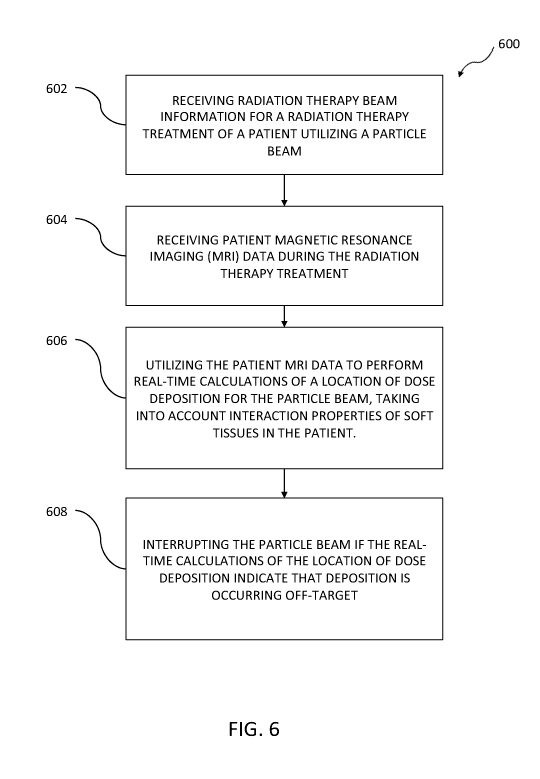
[0083] FIG. 6 is a flowchart for a method 600 of radiation therapy
treatment for particle
radiation therapy, utilizing MRI data, that may be implemented by software,
the method
having one or more features consistent with the present description. The
software can be
implemented using one or more data processors. The software can include
machine-readable
instructions, which, when executed by the one or more data processors, can
cause the one or
more data processors to perform one or more operations. Method 600 is an
example of the
operations that can be performed by controller 424, as discussed herein.
[0084] At 602, radiation therapy beam information for radiation therapy
treatment of a
patient utilizing a particle beam can be received. The radiation therapy beam
information can
include one or more characteristics of a particle beam. The one or more
characteristics can
include an indication of penetrative abilities of the particle beam, the
spread characteristics of
the particle beam, the number of particle beams, or the like.
[0085] At 604, patient magnetic resonance imaging (MRI) data can be
received during the
radiation therapy treatment.
[0086] At 606, the patient MRI data can be utilized to perform real-time
calculations of a
location of dose deposition for the particle beam, taking into account
interaction properties of
23
CA 03016026 2018-08-28
WO 2017/151662 PCT/US2017/020015
soft tissues in the patient through which the particle beam passes, as
discussed herein. The
influence of a magnetic field produced by an MRI system on the particle beam
may also be
accounted for in performing the real-time calculations of location of dose
deposition, as
discussed above. And, a determination of the biological effectiveness of dose
delivered to the
soft tissues by the particle beam, through utilization of the patient magnetic
resonance
imaging data, may also be performed in conjunction with the real-time dose
calculations.
[0087] At 608, the particle beam can be interrupted if real-time
calculations of the location
of dose deposition indicate that deposition is occurring off-target.
[0088] In some variations, the energy of the particle beam can be adjusted
if the real-time
calculations of the location of dose deposition indicate that deposition is
occurring off-target.
In other variations, the patient MRI data can be utilized and the real-time
calculations of the
location of dose deposition to modify a direction of the particle beam in
order to track a
target.
[0089] While components have been described herein in their individual
capacities, it will
be readily appreciated the functionality of individually described components
can be
attributed to one or more other components or can be split into separate
components. This
disclosure is not intended to be limiting to the exact variations described
herein, but is
intended to encompass all implementations of the presently described subject
matter.
[0090] In the descriptions above and in the claims, phrases such as "at
least one of' or
"one or more of' may occur followed by a conjunctive list of elements or
features. The term
"and/or" may also occur in a list of two or more elements or features. Unless
otherwise
implicitly or explicitly contradicted by the context in which it used, such a
phrase is intended
to mean any of the listed elements or features individually or any of the
recited elements or
features in combination with any of the other recited elements or features.
For example, the
24
CA 03016026 2018-08-28
WO 2017/151662 PCT/US2017/020015
phrases "at least one of A and B;" "one or more of A and B;" and "A and/or B"
are each
intended to mean "A alone, B alone, or A and B together." A similar
interpretation is also
intended for lists including three or more items. For example, the phrases "at
least one of A,
B, and C;" "one or more of A, B, and C;" and "A, B, and/or C" are each
intended to mean "A
alone, B alone, C alone, A and B together, A and C together, B and C together,
or A and B
and C together." Use of the term "based on," above and in the claims is
intended to mean,
"based at least in part on," such that an unrecited feature or element is also
permissible.
[0091] The subject matter described herein can be embodied in systems,
apparatus,
methods, and/or articles depending on the desired configuration. The
implementations set
forth in the foregoing description do not represent all implementations
consistent with the
subject matter described herein. Instead, they are merely some examples
consistent with
aspects related to the described subject matter. Although a few variations
have been
described in detail above, other modifications or additions are possible. In
particular, further
features and/or variations can be provided in addition to those set forth
herein. For example,
the implementations described above can be directed to various combinations
and
subcombinations of the disclosed features and/or combinations and
subcombinations of
several further features disclosed above. In addition, the logic flows
depicted in the
accompanying figures and/or described herein do not necessarily require the
particular order
shown, or sequential order, to achieve desirable results. Other
implementations may be
within the scope of the following claims.