Note: Descriptions are shown in the official language in which they were submitted.
CA 03016109 2018-08-29
DESCRIPTION
METHOD FOR ASSESSING PAIN CAUSED BY ADMINISTRATION OF DRUG
SOLUTION, AND METHOD FOR SELECTING DRUG SOLUTION
ADMINISTRATION
Technical Field
[0001]
The present invention relates to a method for assessing
pain caused by administration of a drug solution and a
method for selecting drug solution administration
Background Art
[0002]
An injection is a drug solution administration method
that is used the most widely, but pain caused by the
injection is unpleasant for a patient. Thus, it is desired
to reduce the pain caused by the injection. The pain
caused by the injection includes pain caused by punctuation
of an injection needle and pain caused by a drug solution
(infusion of the drug solution). Therefore, it is
necessary to make it possible to assess (quantify) each of
the pain caused by the punctuation and the pain caused by
the administration of the drug solution in order to reduce
the pain caused by the injection.
[0003]
In relation to this assessment, Literature 1 proposes a
method for assessing pain caused by punctuation of a needle
in a spinal reflex of anesthetized rats.
Literature 1: Okamoto (Okamoto, K), Ami (Ami, N),
Oshima (Oshima, H), "Assessment of needle insertion pain
with flexor reflex responses in anesthetized rats", Pain
Research, The Japanese Association for Study of Pain, 2012,
Vol. 27, No. 24, p.215-225
[0004]
In addition, Literature 2 proposes a method for
assessing vascular pain from an electromyogram (EMG).
Literature 2: Masumi, S, Senba, E, "Nitric oxide
involvement in lipid emulsion-induced vascular pain in
anesthetized rats", Eur J Pharmacol, Elsevier, 2008, No.
594, pp. 64-69
Summary of Invention
[0005]
However, conventionally, there is no proposal on an
effective method for assessing (quantifying) the pain
caused by the administration of a drug solution.
[0006]
The present invention has been made in consideration of
such a problem, and an intended object thereof is to provide
a method for assessing pain caused by administration of a
drug solution and a method for selecting drug solution
2
Date Recue/Date Received 2023-01-16
administration.
[0007]
According to a first broad aspect of the present
invention, there is provided a method for assessing pain
caused by administration of a drug solution, the method
comprising: preparing a mammalian experimental animal
having a plantar subcutaneous tissue, and a semitendinous
muscle bent by a spinal reflex when a stimulus is applied
to the plantar subcutaneous tissue; anesthetizing the
experimental animal by inhalation anesthesia;inserting a
measurement electrode into the semitendinous muscle of the
anesthetized experimental animal;puncturing a plurality of
injection needles into a plurality of administration sites
in the plantar subcutaneous tissue of the anesthetized
experimental animal while measuring a myoelectric potential
of the semitendinous muscle with the measurement electrode
and leaving the plurality of needles in place until after
at least one drug solution has been administered;
administering at least one drug solution to the
anesthetized experimental animal through the plurality of
injection needles after a myoelectric response caused by
the puncture of the injection needles disappears, which
comprises (i) administering a plurality of drug solutions
having different compositions to the plurality of
administration sites under identical administration
conditions, or (ii) administering a single drug solution
to the plurality of administration sites under different
administration conditions; and performing at least one of:
(i) measurement of a duration time of the myoelectric
response caused by the administration of the drug solution,
3
Date Recue/Date Received 2023-01-16
and (ii) measurement of an EMG intensity obtained by
integrating absolute values of myoelectric potentials
during a period from occurrence of the myoelectric response
by the administration of the at least one drug solution to
disappearance of the myoelectric response.
According to a second broad aspect of the present
invention, there is provided a method for selecting drug
solution administration, the method comprising: a preparation
step comprising preparing a mammalian experimental animal
having a plantar subcutaneous tissue, and a semitendinous
muscle bent by a spinal reflex when a stimulus is applied to
the plantar subcutaneous tissue; an anesthesia step
comprising anesthetizing the experimental animal by
inhalation anesthesia;a measurement electrode placement step
comprising placing a measurement electrode in the
semitendinous muscle of the anesthetized experimental animal;
a puncture step comprising puncturing a plurality of
injection needles into a plurality of administration sites in
the plantar subcutaneous tissue of the anesthetized
experimental animal while measuring a myoelectric potential
of the semitendinous muscle by the measurement electrode and
leaving the plurality of needles in place until after at
least one drug solution has been administered; an
administration step comprising administering at least one
drug solution to the anesthetized experimental animal through
the plurality of injection needles after a myoelectric
response caused by the puncture of the injection needles
disappears, which comprises (i) administering a plurality of
drug solutions having different compositions to the plurality
of administration sites under identical administration
conditions, or (ii) administering a single drug solution to
3a
Date Recue/Date Received 2023-01-16
the plurality of administration sites under different
administration conditions; and a measurement step of
performing at least one of (i) measurement of an EMG
intensity obtained by integrating absolute values of the
myoelectric potential from occurrence to disappearance of the
myoelectric response caused by the administration of the drug
solution, and (ii) measurement of a duration time of the
myoelectric response caused by the administration of the drug
solution, wherein the puncture step, the administration step,
and the measurement step are performed for each of a
plurality of drug solutions having different compositions or
for each of a plurality of administration conditions, and
wherein the method further comprises at least one of (i) an
identifying step comprising identifying a drug solution
composition with which the duration time is shortest or the
EMG intensity is smallest, among the plurality of drug
solutions having different compositions, or (ii) identifying
an administration condition with which the duration time is
shortest or the EMG intensity is smallest, among the
plurality of administration conditions.
In order to seek to achieve the above object, a method
for assessing pain by administration of a drug solution
according to embodiments of the present invention includes;
preparing a mammalian experimental animal having a
predetermined part of a body and a skeletal muscle bent by
a spinal reflex when a stimulus is applied to the
predetermined part; anesthetizing the experimental animal
by inhalation anesthesia; inserting a measurement electrode
into the skeletal muscle of the anesthetized experimental
3b
Date Recue/Date Received 2023-01-16
animal; puncturing an injection needle into the
predetermined part of the anesthetized experimental animal
while measuring a myoelectric potential of the skeletal
muscle by the measurement electrode; administering a drug
solution to the anesthetized experimental animal through
the injection needle after a myoelectric response caused by
the punctuation of the injection needle disappears; and
performing at least one of measurement of a duration time
of the myoelectric response caused by the administration of
the drug solution and measurement of an EMG intensity
obtained by integrating absolute values of myoelectric
potentials during the period from the occurrence of the
myoelectric response by the administration of the drug
solution to the disappearance of the myoelectric response.
3c
Date Recue/Date Received 2023-01-16
[0008]
According to the method of embodiments of the present
invention described above, the drug solution is administered to
the experimental animal and the myoelectric response caused by
the administration of the drug solution is measured after the
myoelectric response caused by the punctuation of the injection
needle disappears, and thus, the myoelectric response caused by
the punctuation and the myoelectric response caused by the drug
solution administration do not overlap each other on the
electromyogram (EMG). In this manner, it is possible to assess
(quantify) the pain caused by the administration of the drug
solution separately from the pain caused by the punctuation. In
addition, the pain sensed by a human indicates the same tendency
as a result of the myoelectric response using the experimental
animal, and thus, it is possible to assess the pain caused by
the drug solution administration at the time of the injection
into the human according to the method of embodiments of the
present invention. Therefore, it is possible to contribute to
development of drug solution injection for humans with reduced
pain according to the method of embodiments of the present
invention.
[0009]
In the above-described method for assessing pain caused by
administration of a drug solution, to a plurality of
4
Date Recue/Date Received 2023-01-16
CA 03016109 2018-08-29
administration sites of the predetermined part of the
experimental animal, a plurality of the drug solutions
having different compositions may be administered under an
identical administration condition or the drug solution
having an identical composition may be administered under
different administration conditions.
[0010]
Thus, with the administration to the plurality of sites
of the same experimental animal, it is possible to compare
differences in pain depending on the drug solution
composition or the administration condition, and to select
a drug solution or an administration condition accompanied
by less pain.
[0011]
In the above-described method for assessing pain caused
by administration of a drug solution, the experimental
animal may be a rat.
[0012]
In the above-described method for assessing pain caused
by administration of a drug solution, the predetermined
part may be a plantar subcutaneous, and the skeletal muscle
may be a semitendinosus muscle.
[0013]
In the above-described method for assessing pain caused
by administration of a drug solution, a dose per site at
CA 03016109 2018-08-29
the plurality of administration sites may be a range of 10
to 100 pL.
[0014]
In the above-described method for assessing pain caused
by administration of a drug solution, an interval between
the administration sites, adjacent to each other, may be 2
mm or more.
[0015]
As a result, it is possible to avoid influence by the
adjacent administration site when acquiring the myoelectric
response caused by the drug solution administration and to
perform highly accurate measurement.
[0016]
In the above-described method for assessing pain caused
by administration of a drug solution, a total dose at the
plurality of administration sites may be 200 pL or less.
[0017]
In the above-described method for assessing pain caused
by administration of a drug solution, an administration
rate of the drug solution may be a range of 5 to 100 pL/sec.
[00181
In the above-described method for assessing pain caused
by administration of a drug solution, the administration of
the drug solution may be started after a lapse of one
second or more since the myoelectric response caused by the
6
CA 03016109 2018-089
punctuation has disappeared.
[0019]
As a result, it is possible to more effectively measure
the myoelectric response caused by the drug solution
administration separately from the myoelectric response
caused by the punctuation.
[0020]
In the above-described method for assessing pain caused
by administration of a drug solution, the drug solution may
be administered only when the myoelectric response caused
by the punctuation occurs.
[0021]
As a result, it is possible to avoid wasteful drug
solution administration.
[0022]
In the above-described method for assessing pain caused
by administration of a drug solution, a bipolar electrode
may be used as the measurement electrode and a reference
electrode may be pasted to the thoracic skin surface of the
experimental animal.
[0023]
As a result, it is possible to acquire a waveform of
the myoelectric potential with less noise and to improve
measurement accuracy.
[0024]
7
In addition, a method for selecting drug solution
administration according to embodiments of the present
invention includes: a preparation step of preparing a
mammalian experimental animal having a predetermined part
of a body and a skeletal muscle bent by a spinal reflex
when a stimulus is applied to the predetermined part; an
anesthesia step of anesthetizing the experimental animal by
inhalation anesthesia; a measurement electrode placement
step of placing a measurement electrode in the skeletal
muscle of the anesthetized experimental animal; a
punctuation step of inserting an injection needle into the
predetermined part of the anesthetized experimental animal
while measuring a myoelectric potential of the skeletal
muscle by the measurement electrode; an administration step
of administering a drug solution to the anesthetized
experimental animal through the injection needle after a
myoelectric response caused by the punctuation of the
injection needle disappears; and a measurement step of
performing at least one of measurement of an EMG intensity
obtained by integrating absolute values of the myoelectric
potential from occurrence to disappearance of the
myoelectric response caused by the administration of the
drug solution and measurement of a duration time of the
myoelectric response caused by the administration of the
drug solution. The punctuation step, the administration
8
Date Recue/Date Received 2023-01-16
step, and the measurement step are performed for each of a
plurality of drug solutions having different compositions
or for each of a plurality of administration conditions.
The method further includes a identifying step of
identifying a drug solution composition with which the
duration time is shortest or the EMG intensity is smallest
among the plurality of drug solutions having different
compositions, or identifying an administration condition
with which the duration time is shortest or the EMG
intensity is smallest, among the plurality of
administration conditions.
[0025]
According to this method, it is possible to select the
drug solution composition or the administration condition
accompanied by less pain.
[0026]
According to the method for assessing pain caused by
administration of a drug solution of embodiments of the
present invention, it is possible to assess (quantify) the
pain caused by the administration of the drug solution
separately from the pain caused by the punctuation. Further,
according to the method for selecting drug solution
administration of embodiments of the present invention, it is
possible to select the drug solution composition or the
administration condition accompanied by less pain.
9
Date Recue/Date Received 2023-01-16
Brief Description of Drawings
[0027]
Fig. 1 is a schematic diagram of a measurement system
according to one configuration example used in a method of
embodiments of the present invention.
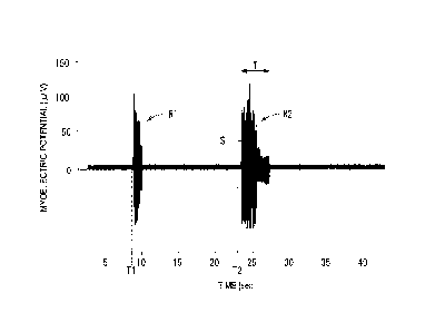
Fig. 2 is an example of a myoelectric potential waveform
obtained by the method of embodiments of the present invention.
Fig. 3A is a graph illustrating a duration time of a
myoelectric response caused by administration of each drug
solution when a plurality of drug solutions having different
compositions was injected into a rat, and Fig. 33 is a graph
illustrating the EMG intensity obtained from a myoelectric
response caused by administration of each drug solution when the
plurality of drug solutions having different compositions was
injected into the rat.
Fig. 4A is a graph illustrating the magnitude of pain (VAS)
when a physiological saline was injected into a human at a
plurality of different injection volume, and Fig. 43 is a graph
illustrating the EMG intensity obtained from a myoelectric
response caused by an injection solution when the physiological
saline was injected into a rat at the plurality of different
doses.
Fig. 5A is a graph illustrating the magnitude of pain (VAS)
at the time of injection into a human with a
Date Recue/Date Received 2023-01-16
CA 03016109 2018-08-29
plurality of different pH values, and Fig. 5B is a graph
illustrating the EMG intensity obtained from a myoelectric
response caused by an injection solution at the time of
injection into a rat with the plurality of different pH
values.
Fig. 6A is a graph illustrating the magnitude of pain
(VAS) over time when 5% NaC1 was injected into a human, and
Fig. 6B is a graph illustrating the EMG intensity obtained
from a myoelectric response caused by an injection solution
when the NaCl was injected into a rat at a plurality of
different concentrations.
Fig. 7A is a graph illustrating the magnitude of pain
(VAS) when glutamic acid was injected into a human at a
plurality of different molar concentrations, and Fig. 7B is
a graph illustrating the EMG intensity obtained from a
myoelectric response caused by an injection solution when
the glutamic acid was injected into a rat at the plurality
of different molar concentrations.
Fig. 8A is a graph illustrating a duration time of a
myoelectric response caused by an injection solution when a
polyethylene glycol/physiological saline mixed solution was
injected into a rat at a plurality of different viscosities,
and Fig. 8B is a graph illustrating the EMG intensity
obtained from a myoelectric response caused by an injection
solution when the polyethylene glycol/physiological saline
11
mixed solution was injected into a rat with the plurality
of different viscosities.
Fig. 9A is a graph illustrating the EMG intensity
obtained from a myoelectric response caused by an injection
solution when a phosphate buffered saline (pH 5) was
injected into a rat at a plurality of different
administration rates, and Fig. 9B is a graph illustrating
the EMG intensity obtained from a myoelectric response
caused by an injection solution when 10% NaC1 was injected
into a rat at a plurality of different administration
rates.
Fig. 10 is a graph illustrating examples of the present
invention, and is the graph illustrating the EMG intensity
obtained from a myoelectric response caused by drug
solution administration when a plurality of drug solutions
having different pHs (an injectable aqueous preparation for
inflammatory autoimmune disease treatment) was injected
into a rat.
Description of Embodiments
[0028]
Hereinafter, illustrative embodiments of a method for
assessing pain caused by administration of a drug solution
and a method for selecting drug solution administration
according to the present invention will be described with
reference to the accompanying drawings.
12
Date Recue/Date Received 2023-01-16
[0029]
Fig. 1 is a schematic diagram of a measurement system 10
according to one configuration example used in the method of
embodiments of the present invention. In the present embodiment
illustrated in Fig. 1, a subject (experimental animal) used for
assessment of pain caused by administration of a drug solution
is a rat 12, located in an enclosure 36. Conditions of the rat
12 that can be used are, for example, a range of 7 to 10 weeks
old, body weight in a range of 200 to 400 g, and a strain is SD
(other strains is also possible).An acclimation and quarantine
period of the rat 12 is illustratively five days or longer. An
anesthesia mask 13 is attached to the rat 12, and inhalation
anesthesia is performed.
[0030]
Incidentally, the experimental animal that can be used may
be any mammal having a predetermined part of a body and a
skeletal muscle to be bent by reflection of a spinal reflex when
a stimulus is applied to the predetermined part. In the case of
the rat 12, a semitendinosus muscle 12b is bent by the spinal
reflex when a stimulus is applied to a plantar subcutaneous 12a.
Examples of the experimental animal of the mammal that can be
used include a mouse, a guinea pig, a gerbil, a hamster, a
ferret, a rabbit, a dog, a minipig, and the like other than the
rat 12.
[0031]
13
Date Recue/Date Received 2023-01-16
CA 03016109 2018-08-29
The drug solution is filled in a syringe 14. The
capacity of the syringe 14 is a range of, for example, 1 to
mL. The syringe 14 is connected to an injection needle
18 through a soft tube 16 made of a resin. A size of the
applicable injection needle 18 is a range of, for example,
34 G to 22 G. The injection needle 18 is punctured under
the plantar subcutaneous 12a of the rat 12.
[0032]
The syringe 14 is attached to a syringe pump 20. The
syringe pump 20 includes a slider 22 that pushes a pusher
14a of the attached syringe 14. In the syringe pump 20,
the speed at which the pusher 14a is pushed by the slider
22 is determined based on a set feeding amount and a type
(capacity) of the syringe 14. As a result, an
administration rate of the drug solution to the rat 12 can
be arbitrarily set. The administration rate of the drug
solution is a range of, for example, 5 to 100 p1/sec.
[0033]
In order to measure the pain caused by the
administration of the drug solution, a myoelectric
potential of the semitendinosus muscle 12b of the thigh of
the rat 12 is recorded. At the time of recording the
myoelectric potential, a needle-shaped measurement
electrode 24 (for example, a bipolar hook electrode) is
punctured under the semitendinosus muscle 12b to be placed,
14
CA 03016109 2018-08-29
and a reference electrode 26 is pasted to a thoracic skin
surface 12c. Potential signals from the measurement
electrode 24 and the reference electrode 26 are amplified
by a high-sensitivity bioelectric amplifier 28 and
transmitted to a data collection device 30.
[0034]
In the data collection device 30, data (potential
signal) is recorded at a predetermined sampling interval
(for example, 0.1 ms) to generate a myoelectric potential
waveform. The myoelectric potential waveform generated by
the data collection device 30 is displayed on a monitor
screen 32a of a personal computer 32.
[0035]
In addition, as pre-preparation for measuring the
myoelectric response caused by injection, electrical
stimulation using a clip-type stimulating electrode 34 is
performed in order to control an anesthetic depth that
enables measurement of the myoelectric response (muscle
contraction) in this measurement system 10. The clip-type
stimulating electrode 34 is connected to an
electrostimulator (not illustrated).
[0036]
The method for assessing pain caused by administration
of a drug solution and the method for selecting drug
solution administration can be performed, for example, as
CA 0301.6109 2018-08-29
follows when using the measurement system 10 configured as
described above.
[0037]
When it is desired to investigate which composition
causes the least pain among a plurality of different drug
solution compositions, a plurality of drug solutions having
different compositions is prepared. A difference in drug
solution composition depends on, for example, a type of a
drug solution component (for example, a chemical structure
of an active component, a buffer, a stabilizer, an
antioxidant, or the like), a pH value, a viscosity, and the
like. Alternatively, when it is desired to investigate
which administration condition causes the least pain among
a plurality of different drug administration conditions, a
plurality of different administration conditions is
prepared for a drug solution having the same composition.
Parameters of the administration condition include an
administration rate (infusion rate) of a drug solution and
a dose.
[0038]
The rat 12 serving as a subject is prepared (a
preparation step), and the rat 12 is subjected to
inhalation anesthesia (an anesthesia step). Examples of an
applicable inhalation anesthetic include isoflurane. An
anesthetic concentration with respect to air is a range of,
16
for example, 3 to 4%/Air at the time of introduction of
anesthesia and a range of, for example, 1 to 2%/Air at the
time of recording. Incidentally, it is illustrative to
heat the rat 12 with a keep-warm mat to keep the body
temperature constant during the anesthesia.
[0039]
Next, the measurement electrode 24 is placed in the
semitendinosus muscle 12b of the anesthetized rat 12 (a
measurement electrode placement step). Specifically, the
clip-type stimulating electrode 34 is used to clamp the
dorsum and sole of the hind paw of the rat 12, and an
electrical stimulus (for example, 40 Hz, 10 mA, 2 ms) is
applied to C fibers of pain sensation via the clip-type
stimulating electrode 34 by the electrostimulator (not
illustrated). At this time, the thigh skin at a position
where contraction has been recognized is incised by about 1
cm to expose the semitendinosus muscle 12b, and then, the
measurement electrode 24 is inserted.
[0040]
When the measurement electrode 24 is placed, an
electrical stimulus (for example, 40 Hz, 5 mA, 2 ms) is
applied again by the electrostimulator (not illustrated)
via the clip-type stimulating electrode 34, and the
anesthetic depth of the rat 12 is finely adjusted while
referring to the myoelectric response intensity.
17
Date Recue/Date Received 2023-01-16
CA 03016109 2018-089
Thereafter, the anesthetic depth is kept constant.
[0041]
In addition, the thorax of the rat 12 is depilated to
expose the thoracic skin 12c, and then, the reference
electrode 26 is pasted to the thoracic skin surface 12c.
Incidentally, the reference electrode 26 may be pasted
before, after, or in parallel with the placement of the
measurement electrode 24. When a bipolar electrode is used
as the measurement electrode 24 and the reference electrode
26 is pasted to the thoracic skin surface 12c of the rat 12,
it is possible to acquire the myoelectric potential
waveform with less noise and to improve the measurement
accuracy. Incidentally, the measurement electrode 24 may
be a monopolar electrode when the reference electrode 26 is
pasted.
[0042]
Once the above preparation has been completed, a
punctuation step, an administration step, and a measurement
step to be described below are performed for each of the
plurality of drug solutions having different compositions
or for each of the plurality of administration conditions
using the same rat 12. That is, the punctuation step, the
administration step, and the measurement step are performed
with a certain drug solution composition or administration
condition, and then, the punctuation step, the
18
CA 03016109 2018-08-29
administration step, and the measurement step are
repeatedly performed with different drug solution
compositions or administration conditions using the same
rat 12.
[0043]
In the punctuation step, the injection needle 18 is
punctured under the plantar subcutaneous 12a of the
anesthetized rat 12. In this case, when the entire blade
surface provided at a distal end of the injection needle 18
pierces the plantar subcutaneous 12a, the punctuation is
completed. When the injection needle 18 is punctured under
the rat 12 in this manner, a myoelectric response caused by
punctuation occurs.
[0044]
In the administration step following the punctuation
step, a drug solution is administered to the anesthetized
rat 12 through the injection needle 18 after the
myoelectric response caused by punctuation of the injection
needle 18 disappears. In this case, the syringe pump 20
pushes the pusher 14a of the syringe 14 based on the preset
administration rate and dose so that the drug solution is
infused into the plantar subcutaneous 12a of the rat 12 at
the set administration rate and dose.
[0045]
Incidentally, in each administration step, it is
19
illustrative to set an interval between adjacent
administration sites (punctuation sites) to be 2 mm or more
when a dose per site is 100 pL or less. As a result, it is
possible to avoid influence by the adjacent administration
site when acquiring the myoelectric response caused by the
drug solution administration and to perform highly accurate
measurement.
[0046]
When a plurality of drug solutions having different
compositions is tested, an administration rate and a dose
of the drug solution at each administration step are set to
be the same. In addition, when a plurality of
administration conditions is tested for the drug solution
having the same composition, the drug solution is
administered with one or both of the administration rate
and the dose of the drug solution changed in each
administration step.
[0047]
The dose per site at the plurality of administration
sites is illustratively a range of 10 to 100 pL in
consideration of a planta pedis size of the rat 12. In
addition, the total dose to the rat 12 at the plurality of
administration sites is illustratively a range of 200 pL or
less. Examples of a combination of the dose and the number
of administration sites may include 20 pL x 8 sites (= 160
Date Recue/Date Received 2023-01-16
CA 03016109 2018-08-29
pL), 50 pL x 4 sites (= 200 pL), 100 pL x 2 sites (200 pL),
and the like. In addition, the administration rate of the
drug solution is a range of, for example, 5 to 100 pL/sec.
[0048]
Incidentally, when the drug solution is administered
only when the myoelectric response caused by the
punctuation occurs in the punctuation step, it is possible
to avoid wasteful drug solution administration. That is,
when the myoelectric response is not observed despite the
punctuation, it is possible to prevent administration of
the drug solution to a site where no myoelectric response
occurs beforehand by piercing another site again.
[0049]
The myoelectric potential generated in the
semitendinosus muscle 12b of the rat 12 accompanying the
injection (punctuation and drug solution administration) is
detected by the measurement electrode 24, and amplified by
the high-sensitivity bioelectric amplifier 28, and then,
sent to the data collection device 30. The myoelectric
potential waveform is generated based on the myoelectric
potential data by the data collection device 30. The
generated myoelectric potential waveform is displayed on
the monitor screen 32a of the personal computer 32.
[0050]
Fig. 2 illustrates an example of the myoelectric
21
potential waveform thus obtained. In Fig. 2, Ti is a time at
which the injection needle 18 is punctured, and a myoelectric
response (R1) caused by the punctuation can be confirmed. In
addition, T2 is a time when the administration of the drug
solution to the rat 12 is started, and a myoelectric response
(R2) caused by the drug solution administration can be
confirmed.
[0051]
As described above, the drug solution is administered to
the anesthetized rat 12 through the injection needle 18 after
the myoelectric response caused by the punctuation of the
injection needle 18 disappears in embodiments of the present
invention, and thus, the myoelectric response caused by the
punctuation and the myoelectric response caused by the drug
solution administration do not overlap each other in time.
That is, the myoelectric response caused by the drug solution
administration can be measured separately from the
myoelectric response caused by the punctuation.
[0052]
Once such a myoelectric potential waveform is obtained,
the measurement step is performed in order to assess
(quantify) the pain caused by the drug solution
administration. In the measurement step, at least one of
measurement of a duration time T of the myoelectric
response caused by the drug solution administration and
22
Date Recue/Date Received 2023-01-16
measurement of an integral value S obtained by integrating
absolute values of myoelectric potentials during a period
from the occurrence of the myoelectric response by the
administration of the drug solution to the disappearance of
the myoelectric response, that is, an EMG intensity (pV.$)
is performed. The integral value S is the area of the
myoelectric potential waveform obtained by rectifying the
myoelectric potentials during the period of the duration
time T of the myoelectric response and integrating the
rectified values. The duration time T and the integral
value S of the myoelectric response caused by the drug
solution administration may be calculated by the personal
computer 32, and a value calculated by the data collection
device 30 may be displayed on the monitor screen 32a.
[0053]
In this manner, according to the method of embodiments of
the present invention, the drug solution is administered to the
rat 12 and the myoelectric response caused by the administration
of the drug solution is measured after the myoelectric response
caused by the punctuation of the injection needle 18 disappears,
and thus, the myoelectric response caused by the punctuation and
the myoelectric response caused by the drug solution
administration do not overlap each other on the electromyogram
(EMG). In this manner, it is possible to assess (quantify) the
pain caused by the administration
23
Date Recue/Date Received 2023-01-16
of the drug solution separately from the pain caused by the
punctuation. In this case, when the administration of the
drug solution is started after a lapse of one second or
more (more illustratively 10 seconds or more) after the
disappearance of the myoelectric response by the puncture,
it is possible to effectively measure the myoelectric
response caused by the drug solution administration
separately from the myoelectric response caused by the
puncture.
[0054]
As described above, the punctuation step, the
administration step, and the measurement step are performed
for each of the plurality of drug solutions having
different compositions or for each of the plurality of
administration conditions, and then, a drug solution
composition with which the duration time T is shortest or
the integral value S is smallest is identified among the
plurality of drug solutions having different compositions,
or an administration condition with which the duration time
T is shortest or the integral value S is smallest is
identified among the plurality of administration conditions
(a identifying step). As a result, it is possible to
select the drug solution composition or the administration
condition accompanied by less pain.
[0055]
24
Date Recue/Date Received 2023-01-16
CA 0301.6109 2018-08-29
Here, Fig. 3A is a graph illustrating a duration time
of a myoelectric response caused by administration of each
drug solution when the plurality of drug solutions having
different compositions was injected into a rat. Fig. 3B is
a graph illustrating the EMG intensity obtained from a
myoelectric response caused by administration of each drug
solution when the plurality of drug solutions having
different compositions was injected into the rat. From
Figs. 3A and 3B, it can be understood that the duration
time of the myoelectric response and the EMG intensity
differ depending on the drug solution composition regarding
the rat, that is, there is a difference in pain caused by
drug solution administration.
f0056]
Fig. 4A is a graph illustrating the magnitude (VAS:
visual analog scale) of pain when a physiological saline
was injected into a human at a plurality of different doses
(the source is illustrated at the lower part). The VAS is
an assessment scale indicating a degree of current pain
with a maximum of 10. Fig. 4B is a graph illustrating the
EMG intensity obtained from a myoelectric response caused
by an injection solution when a physiological saline was
injected into a rat at the plurality of different doses
From Figs. 4A and 4B, it can be understood that both the
human and the rat have the same tendency that the pain
CA 03016109 2018-08-29
increases as the dose increases.
[0057]
Fig. 5A is a graph illustrating the magnitude of pain
(VAS) at the time of injection into a human with a
plurality of different pH values (the source is illustrated
at the lower part). Fig. 5B is a graph illustrating the
EMG intensity obtained from a myoelectric response caused
by an injection solution at the time of injection into a
rat with the plurality of different pH values. From Figs.
5A and 5B, it can be understood that both the human and the
rat have the same tendency that the pain increases as the
pH value decreases.
[0058]
Fig. 6A is a graph illustrating the magnitude of pain
(VAS) over time when 5% NaCl was injected into a human (the
source is illustrated at the lower part). From Fig. 6A, it
can be understood that a great pain is caused when 5% NaC1
is injected into the human. On the other hand, Fig. 613 is
a graph illustrating the EMG intensity obtained from the
myoelectric response caused by an Injection solution when
the NaC1 was injected into a rat at a plurality of
different concentrations. From Fig. 6B, it can be
understood that the pain of the rat significantly increases
when the NaCl concentration is 5% or more. Therefore, it
can be understood from Figs. 6A and 6B that the human and
26
the rat indicate the same tendency with respect to the pain
depending on the NaC1 concentration.
[0059]
Fig. 7A is a graph illustrating the magnitude of pain
(VAS) when glutamic acid was injected into a human at a
plurality of different molar concentrations. Incidentally,
Fig. 7A is the graph created based on the source graph
illustrated at the lower part of the drawing. Fig. 7B is a
graph illustrating the EMG intensity obtained from a
myoelectric response caused by an injection solution when
glutamic acid was injected into a rat at the plurality of
different molar concentrations. From Figs. 7A and 7B, it
can be understood that both the human and the rat have the
same tendency that the pain increases as the molar
concentration of glutamic acid increases.
[0060]
As above, it can be understood that the magnitude of pain
felt by the human indicates the same tendency as the measurement
result of the myoelectric response using the rat. In addition,
in the case of a mammal other than the rat, it can be considered
that the mammal indicates the same tendency as the rat.
Accordingly, the method of the present invention according to
its embodiments can be applied to development of a drug solution
for humans and an administration method.
[0061]
27
Date Recue/Date Received 2023-01-16
CA 03016109 2018-089
Fig. BA is a graph illustrating a duration time of a
myoelectric response caused by an injection solution when a
polyethylene glycol/physiological saline mixed solution was
injected into a rat at a plurality of different viscosities.
Fig. 8B is a graph illustrating the EMG intensity obtained
from a myoelectric response caused by an injection solution
when the polyethylene glycol/physiological saline mixed
solution was injected into a rat with the plurality of
different viscosities. From Figs. 8A and 8B, it can be
understood that the magnitude of pain varies depending on
the viscosity of the injection solution.
[0062]
Fig. 9A is a graph illustrating the EMG intensity
obtained from a myoelectric response caused by an injection
solution when a phosphate buffered saline (pH 5) was
injected into a rat at a plurality of different
administration rate. Fig. 9B is a graph illustrating the
EMC intensity obtained from a myoelectric response caused
by an injection solution when 10% NaCl was injected into a
rat at a plurality of different administration rates. From
Figs. 9A and 9B, it can be understood that the magnitude of
pain varies depending on a difference in the administration
rate of the injection solution depending on the composition
of the injection solution.
[0063]
28
CA 03016109 2018-08-29
Next, specific examples of assessment of the pain
caused by the drug solution administration using the rat 12
will be described. In this example, drug solutions of
Samples 1 to 3 having different compositions (an injectable
aqueous preparation for inflammatory autoimmune disease
treatment) were prepared as follows.
[0064]
(Sample 1)
Disodium hydrogenphosphate (anhydrous) of 0.71 g was
dissolved in water for injection of 50 mL. Sodium
dihydrogen phosphate (anhydrous) of 0.60 g was dissolved in
water for injection of 50 mL. A mixture was obtained with
a volume ratio of the disodium hydrogenphosphate solution :
the sodium dihydrogen phosphate solution = 87 : 13, and the
mixture was diluted to 1/10 in concentration with water for
injection to prepare a 10 mM phosphate buffer solution.
Methotrexate of 125 mg and sodium chloride of 27 mg were
dissolved in the 10 mM phosphate buffer solution of 5 mL,
and the pH thereof was adjusted to around 7.5 with an
appropriate amount of sodium hydroxide, thereby obtaining
an aqueous solution having a methotrexate concentration of
25 mg/mL. This aqueous solution was filtered using a
membrane filter having a pore diameter of 0.2 pm to prepare
the injectable aqueous preparation for inflammatory
autoimmune disease treatment.
29
CA 03016109 2018-08-29
[0065]
(Sample 2)
An injectable aqueous preparation containing
methotrexate at pH 8.0 was prepared in the same manner as
Sample 1, except that an added amount of sodium hydroxide
in Sample I was changed.
[0066]
(Sample 3)
An injectable aqueous preparation containing
methotrexate at pH 8.5 was prepared in the same manner as
Sample 1, except that an added amount of sodium hydroxide
in Sample 1 was changed.
[0067]
The measurement system 10 illustrated in Fig. 1 was
used to measure an electromyogram (EMG) when the injectable
aqueous preparations containing methotrexate of Samples 1
to 3 were administered to the rat 12.
[0068]
Specifically, the rat 12 was anesthetized with
isoflurane at a concentration of 3%/Air and the
semitendinosus muscle 12b was exposed. The anesthesia was
lowered to about 1.5%/Air, and the clip-type stimulating
electrode 34 was attached to distal ends of toes. An
electrical stimulus (40 Hz, 10 mA, 2 ms) was applied, and
the measurement electrode 24 was inserted into a position
CA 03016109 2018-08-29
where contraction was observed. The electrical stimulus
intensity was lowered to 5 mA, and the anesthetic
concentration was lowered (a range of 1 to 1.4%/Air) until
obtaining a response of about 100 pV. The rat 12 is
stabilized for 30 minutes or more after the change of the
anesthetic concentration, and then, the drug solution was
administered to the rat 12.
[0069]
During the administration of the drug solution to the
rat 12, the syringe 14 of 1 mL was loaded in the syringe
pump 20, the injection needle 18 of 29 G was punctured
under the plantar subcutaneous 12a, and the drug solution
of 20 pL was administered at an administration rate of 10
pL/sec. Samples 1 to 3 were sequentially administered to
the rat 12 under the same administration conditions.
Measurement results of the EMG intensity is illustrated in
Fig. 10.
[0070]
From Fig. 10, it was found that the EMG intensity
caused by drug solution administration of Sample 1 was
smallest among Samples 1 to 3 having different compositions.
That is, it was found that the pain caused by the drug
solution administration of Sample 1 was the least.
Therefore, in the present embodiment, it was determined to
select the drug solution composition of Sample 1 as the
31
injectable aqueous preparation for inflammatory autoimmune
disease treatment accompanied by less pain.
[0071]
Although the present invention has been described with
the illustrative embodiments as above, it is obvious that
the present invention is not limited to the above-described
embodiments, and various modifications can be made within a
scope that does not depart from the scope of the present
invention.
32
Date Recue/Date Received 2023-01-16