Note: Descriptions are shown in the official language in which they were submitted.
CA 03016765 2018-08-29
WO 2017/157895
PCT/EP2017/055920
1
ANTI-MICA ANTIBODIES
CROSS-REFERENCE To RELATED APPLICATIONS
This application claims the benefit of U.S. Provisional Application No.
62/308,443
filed March 15, 2016, which is incorporated herein by reference in its
entirety; including any
drawings and sequence listing.
REFERENCE To THE SEQUENCE LISTING
The present application is being filed along with a Sequence Listing in
electronic
format. The Sequence Listing is provided as a file entitled "MI0A2 PCT_5T25
txt", created
March 13, 2017, which is 44 KB in size. The information in the electronic
format of the
Sequence Listing is incorporated herein by reference in its entirety.
FIELD OF THE INVENTION
The present invention provides antigen-binding proteins capable of binding to
MICA
polypeptides. The antigen-binding proteins have increased activity in the
treatment of
disorders characterized by MICA-expressing cells, particularly cancer.
BACKGROUND
The immunoreceptor NKG2D is normally expressed on human T cells (e.g., CD8+ T
cells, y5 T cells) and NK cells. On pre-activated CD8+ cells, NKG2D functions
as a
synergistic co-stimulator of CD28 and TCR signalling via DAP10 association,
whereas in NK
cells it functions as a direct activator. Upon ligand engagement, NKG2D
therefore conveys
directly activating or costimulatory signals via the paired DAP10 adaptor
protein, thereby
promoting cancer and infectious disease immunity.
Various ligands for human NKG2D (hNKG2D) have been identified and
characterized, including the major histocompatibility complex class l-related
chain A and B
polypeptides (MICA and MICB), the UL16-binding protein (ULBP) family, and the
retinoic
acid early transcript-1 (RAET1) family. MICA is frequently associated with
epithelial tumors,
induced by microbial infections, and aberrantly expressed in certain
autoimmune disease
lesions. The structure of MICA is similar to the protein fold of MHC class I,
with an a 1a2
platform domain and a membrane-proximal lg-like a3 domain (Li et al 2001 Nat.
lmmunol.
2:443). MICA and its close relative MICB, which also serves as a ligand for
NKG2D, are both
polymorphic and the polymorphism has been shown to affect the affinity for
NKG2D (Steinle
et al. 2001 lmmunogenetics 53:279).
CA 03016765 2018-08-29
WO 2017/157895
PCT/EP2017/055920
2
In the mouse, which lacks MHC class I chain (MIC) genes, a family of proteins
structurally related to ULBP, the retinoic acid early (RAE-1) molecules
function as ligands for
NKG2D. RAE-1 expression has been shown to be induced by carcinogens and to
stimulate
antitumor activities of T cells. Murine NKG2D, however, recognizes human MICA
polypeptides (Wiemann (2005) J. lmmunol. 175:820-829).
The role of MICA in cancer biology has been complicated by the fact that MICA
is
released as a soluble form from the cell surface of tumor cells (e.g., *019
allele) and on the
surface of exosomes (*08 allele) (Ashiru et al (2010) Cancer Res. 70(2):481-
489)). Soluble
MICA (sMICA) can be detected for example at high levels in sera of patients
with
gastrointestinal malignancies (Salih et al, 2002 J. lmmunol. 169: 4098). The
MMPs ADAM10
and ADAM17, as well as the disulfide isomerase Erp5, have been reported to
have a role in
cleavage and shedding of MICA (Waldhauer (2008) Cancer Research 68 (15) 6368-
76;
Kaiser et al (2007) Nature; and Salih (2002) J. Immunol 169: 4098-4102).
Membrane bound
MICA has been reported to downmodulate the expression of NKG2D on NK and/or T
cells
(Von Lilienfeld-Toal et al. (2010) Cancer lmmunol. lmmunother.). Notably,
Wiemann (2005),
supra, examined MICA Tg mice and concluded that down-regulation of surface
NKG2D on
nontransgenic splenocytes was most pronounced after cocultivation with
splenocytes from
MICA transgenic mice in vitro, and only marginally following treatment with
sera from H2Kb-
MICA mice, whereas incubation with control cells and sera from nontgLM,
respectively, had
no effect and that overall data suggest that reduced surface NKG2D on H2-K-
MICA NK cells
results in NKG2D dysfunction and that NKG2D downregulation is primarily caused
by a
persistent exposure to cellbound MICA in vivo.
Reports have also emerged that NKG2D on NK cells is downregulated by sMICA
(Groh et al. (2002) Nature; Arreygue-Garcia (2008) BMC; Jinushi et al. (2005)
J. Hepatol.),
leading to less reactive NK cells. This rationale may have emerged because
similar systems
have been observed among other protein families such as the lg-like and the
TNF
superfamily have been shown to be released as a soluble form and that release
of the
molecules affects cell-cell interactions by reduction of ligand densities and
modulates NK
cells bearing the respective receptor (Salih 2002). Consequently, attempts to
generate anti-
MICA antibodies have focused on development of antibodies that inhibit MICA
shedding.
It has also been reported that expression of NKG2D ligands MICA and MICB on
healthy cells can break the balance between immune activation and tolerance,
and trigger
autoimmunity. Genetic linkage studies have shown that some MICA alleles are
positively
associated with type 1 diabetes, and development of disease in prediabetic NOD
mice
expressing Rae1 on their islet cells can be completely prevented by treatment
with NKG2D-
blocking mAbs, which reduce expansion and function of autoreactive CD8+ T
cells. MICA
CA 03016765 2018-08-29
WO 2017/157895
PCT/EP2017/055920
3
and MICB molecules are also dramatically upregulated in RA synoviocytes and
activate the
T cells in an NKG2D-dependent manner. Moreover, rheumatoid arthritis patients
have been
reported to have high levels of IL-15 and TNF-a in the sera and inflamed
joints which induce
expression of NKG2D on CD4+0D28- subset of T cells. In Celiac disease, massive
infiltration of intraepithelial NKG2D+ CD8+ T lymphocytes in the gut has been
reported, and
MIC proteins become strongly expressed on the surface of epithelial cells in
patients with
active disease. In inflammatory bowel disorders, increased levels of MIC
expression were
found on intestinal epithelial cells and it the number of intestinal
epithelial CD4+ T cells
expressing NKG2D was found to correlate with intestinal inflammation.
Approaches to date to treat inflammation based on the NKG2D system have
focused
on blockade of NKG2D itself rather than its ligands (Ogasawara et al. (2004)
Immunity
20(6):757-767; Andersson et al (2011) Arthritis. Rheum. 63(9):2617-2629;
Steigerwald et al
(2009) MAbs 1(2):115-127. One possibility is that this focus on NKG2D rather
than its ligand
is due to the perceived difficulty of targeting the NKG2D ligand system which
includes a
variety of ligands and in some cases a large number of alleles.
For MICA and MICB, there are over 97 MICA alleles and at least 31 MICB alleles
recognized. There is only 43% amino acid identity across the MIC polypeptides
in the a1a2
domain (the domain involved in the NKG2D interface), and 80% of the amino acid
substitutions are non-conservative (Steinle et al. (2001) lmmunogenetics 53:
279-287;
Steinle et al. (1998) Proc. Natl. Acad. Sci. U.S.A. 95:12510-12515),
suggesting that it will be
unlikely to obtain antibodies that are effective for a majority of individuals
in a population.
Additionally, the methionine/valine bimorphism at position 129 in MICA
determines
differences in NKG2D binding, and although the side chain of residue 129 is
partially buried
and forms hydrophobic interactions with glutamine 136, alanine 139 and
methionine 140 in
the first a2 helical stretch, it may be associated with a difference in
conformation in this
domain in comparison with valine 129 forms of MICA (Steinle et al (2001)
lmmunogenetics
53: 279-287).
In conclusion, there is a need for new approaches to target MICA with
therapeutic
agents.
Summary of the Invention
In one aspect, the invention results, inter alia, from the discovery of
antibodies with
high affinity across human MICA alleles (as well as on non-human primate
MICA).
In one embodiment, provided is an anti-MICA antigen binding domain, or a
protein
that comprises the antigen binding domain (e.g., a monoclonal antibody, a
multispecific
binding protein, a bispecific antibody, etc.), the antigen binding domain
comprising:
CA 03016765 2018-08-29
WO 2017/157895
PCT/EP2017/055920
4
(a) a heavy chain variable region (VH) comprising an amino acid sequence at
least
80%, 90%, 95% or 98% identical to the amino acid sequence of SEQ ID NO: 6, and
(b) a light chain variable region (VL) comprising an amino acid sequence at
least
80%, 90%, 95% or 98% identical to the amino acid sequence of SEQ ID NO: 7.
In one embodiment, the VL comprises a tyrosine (Y) amino acid residue at Abnum
position 71 (in FR3). In one embodiment, the VL comprises a phenylalanine (F)
at Abnum
position 83.
In one embodiment, the heavy chain variable region (VH) comprises amino acid
residues at Abnum positions 72c (in FR2) and 74 (in FR3) capable of
interacting with one
another by H-bonding between the residue at position 72c and the residue at
position
Abnum 74. In one embodiment, the VH comprises a lysine (K) amino acid residue
at Abnum
position 72c and a glutamine residue at position 74. In one embodiment, the VH
comprises a
threonine (T) at Abnum position 30. In one embodiment, the VH comprises an
isoleucine (I)
at Abnum position 48. In one embodiment the VH comprises a valine (V) at Abnum
position
67. In one embodiment, the VH comprises an arginine (R) at Abnum position 71.
In one embodiment, the VH segment of the VH human acceptor framework is from
IGHV4-b (e.g., IGHV4-b*02) and the J-segment is from IGHJ6 (e.g., IGHJ6*01).
In one
embodiment, the CDR1, 2 and 3 of the VH comprise the amino acid sequences of
SEQ ID
NOS: 30, 31 and 32, respectively. In one embodiment, the VL domain human
acceptor
framework is from IGKV3-11 (e.g., IGKV3-11*01) and the J-segment is from IGKJ2
(e.g.,
IGKJ2*01). In one embodiment, the CDR1, 2 and 3 of the VL comprise the amino
acid
sequences of SEQ ID NOS: 33, 34 and 35, respectively. In one embodiment, the
human
heavy chain and/or light chain acceptor framework comprises one or more back-
mutations in
which an amino acid is substituted by an amino acid present at the particular
position in a
non-human mammal (e.g., murine, rat). In one embodiment, the human heavy chain
acceptor framework 1 (FR1) comprises a threonine (T) at Abnum position 30 and
contains
no other mutations compared to a naturally occurring human VH segment. In one
embodiment, the human heavy chain acceptor framework 2 (FR2) is free of
mutations
compared to a naturally occurring human VH segment. In one embodiment, the
human
heavy chain acceptor framework 3 (FR3) comprises a arginine (R) at Abnum
position 71 and
contains no other mutations compared to a naturally occurring human VH
segment. In one
embodiment, the human heavy chain acceptor framework 4 (FR4) is free of
mutations
compared to a naturally occurring human VH segment. In one embodiment, the
human light
chain acceptor framework 3 (FR3) comprises a tyrosine at Abnum position 71 and
contains
no other mutations compared to a naturally occurring human VH segment. In one
CA 03016765 2018-08-29
WO 2017/157895
PCT/EP2017/055920
embodiment, the human light chain acceptor frameworks 1, 2 and 4 (FR1, FR2 and
FR4) are
free of mutations compared to a naturally occurring human VH segment.
In one embodiment of any aspect herein, the VH comprises the heavy chain CDR1,
CDR2 and CDR3 having the respective amino acid sequences shown in SEQ ID NOS:
30,
5 31 and 32. In one embodiment, the VL comprises the light chain CDR1, CDR2
and CDR3
having the respective amino acid sequences shown in SEQ ID NOS: 33, 34 and 35.
In one embodiment, provided is an anti-MICA antigen binding domain, or a
protein
that comprises the antigen binding domain (e.g., a monoclonal antibody, a
multispecific
binding protein, a bispecific antibody, etc.), comprising:
(a) a heavy chain variable region comprising an amino acid sequence of SEQ ID
NO:
6, optionally further comprising one, two or three amino acid residue
substitutions in a
framework region, and
(b) a light chain variable region comprising an amino acid sequence of SEQ ID
NO:
7, optionally further comprising one, two or three amino acid residue
substitutions in a
framework region.
In one embodiment, provided is an anti-MICA antigen binding domain, or a
protein
that comprises the antigen binding domain (e.g., a monoclonal antibody, a
multispecific
binding protein, a bispecific antibody, etc.), comprising:
(a) a heavy chain variable region comprising an amino acid sequence of SEQ ID
NO:
8, optionally further comprising one, two or three amino acid residue
substitutions in a
framework region, and
(b) a light chain variable region comprising an amino acid sequence of SEQ ID
NO:
9, optionally further comprising one, two or three amino acid residue
substitutions in a
framework region.
In one aspect of any of the embodiments, the light chain variable region
comprises a
tyrosine (Y) residue at position 71 (Abnum numbering).
In another aspect of any of the embodiments, the heavy chain variable region
comprises a lysine (K) residue as position 72c (Abnum numbering).
In one embodiment, provided is an anti-MICA antigen binding domain, or a
protein
that comprises the antigen binding domain (e.g., a monoclonal antibody, a
multispecific
binding protein, a bispecific antibody, etc.), the antigen binding domain
selected from the
group consisting of:
(a) an antibody binding domain comprising a heavy chain variable region
comprising
an amino acid sequence of SEQ ID NO: 6 and a light chain variable region
comprising an
amino acid sequence of SEQ ID NO: 7;
CA 03016765 2018-08-29
WO 2017/157895
PCT/EP2017/055920
6
(b) an antibody binding domain comprising a heavy chain variable region
comprising
an amino acid sequence of SEQ ID NO: 8 and a light chain variable region
comprising an
amino acid sequence of SEQ ID NO: 9; and
(c) an antibody binding domain comprising a heavy chain variable region
comprising
an amino acid sequence of SEQ ID NO: 10 and a light chain variable region
comprising an
amino acid sequence of SEQ ID NO: 11.
In one embodiment, provided is a monoclonal antibody that binds human MICA,
selected from the group consisting of:
(a) an antibody comprising a heavy chain variable region comprising an amino
acid
sequence of SEQ ID NO: 6 and a light chain variable region comprising an amino
acid
sequence of SEQ ID NO: 7;
(b) an antibody comprising a heavy chain variable region comprising an amino
acid
sequence of SEQ ID NO: 8 and a light chain variable region comprising an amino
acid
sequence of SEQ ID NO: 9; and
(c) an antibody comprising a heavy chain variable region comprising an amino
acid
sequence of SEQ ID NO: 10 and a light chain variable region comprising an
amino acid
sequence of SEQ ID NO: 11.
In one aspect, provided are anti-MICA antibodies with human frameworks that
have
modified salt bridges; salt bridges in proteins are H-bonds between oppositely
charged
residues that are sufficiently close to each other to experience electrostatic
attraction. In one
aspect, provided are anti-MICA antibodies with human frameworks that have
amino acid
substitutions in the light chain FR3. In one aspect, an antibody comprises H-
bonding in the
heavy chain FR3 region between residues at positions 72c and 74 (Abnum
numbering).
The antibodies notably bind to the predominant MICA alleles from each of two
major
MICA groups that are determined to represent the main families of MICA: Group
1 alleles
that bind NKG2D strongly (including MICA*001, *002, *007, *012, *017 and *018)
and Group
2 that bind NKG2D weakly (MICA*004, *006, *008, *009 and *019). By binding to
an epitope
present on the subset MICA *001, *004, *007 and *008 or *001, *004, *007, *008
and *019,
the antibodies cover the alleles of both groups that are present in almost all
individuals.
Optionally, the antibodies have an EC50, as determined by flow cytometry, of
no more than 1
pg/ml, optionally no more than 0.5 pg/ml, no more than 0.3 pg/ml, or no more
than 0.2 pg/ml
for binding to cells made to express at their surface *001, to cells made to
express at their
surface *004, to cells made to express at their surface *007 and to cells made
to express at
their surface *008. Optionally, the antibodies have an EC50, as determined by
flow cytometry,
of no more than 0.1 pg/ml, optionally no more than 0.07 pg/ml for binding to
cells made to
express at their surface *004, to cells made to express at their surface *007
and to cells
CA 03016765 2018-08-29
WO 2017/157895
PCT/EP2017/055920
7
made to express at their surface *008. The antibodies optionally further bind
to cells
expressing a human MICB polypeptide.
In one embodiment, an antibody that is capable of binding MICA alleles has an
EC50 for binding to a human MICA*001 that differs by less than 1-log from its
binding affinity
for human MICA*004, *007 and/or *008, as determined by flow cytometry for
binding to cells
expressing at their surface the respective MICA polypeptide cells transfected
with one of the
respective MICA alleles but that do not express the other MICA alleles). In
one embodiment,
the antibody has an EC50 for binding to human MICA*004, *007 and/or *008
polypeptide that
differs from each other by no more than 0.5 log, 0.3 log or 0.2 log, as
determined by flow
cytometry for binding to cells expressing at their surface human MICA*004,
*007 and/or
*008.
Optionally, the EC50 is determined according the methods of the Examples
herein,
or according to Example 3 of PCT publication no W02013/117647, e.g. C1R cells
(ATCC
reference CRL-1993TM) transfected with RSV.5neo vectors (GenBank (NCB!) under
Accession number M83237) containing the MICA nucleic acid of interest, data
acquisition by
flow cytometry and EC50 computation using a 4 parameter model.
High affinity binding is advantageous, inter alia, for an antibody to
effectively mediate
CDC and/or ADCC.
The antibodies of the disclosure are capable of blocking the interaction of
MICA on
the surface of cells (e.g., tumor cells) with NKG2D (e.g., on NK cells and T
cells). Thus, in
addition to induction of ADCC and/or CDC activity when comprising Fc domains
that are
bound by Fcy receptors, these antibodies are useful for their ability to be
able to block
membrane MICA-induced down-modulation of NKG2D, e.g., for the treatment of
cancer
and/or infectious disease. Furthermore, in addition to or alternatively to the
ability to mediate
ADCC and/or CDC activity, these antibodies are useful for their ability to
reduce M2
macrophage-mediated suppression of T cell and/or NK cell activity. In other
embodiments,
antibodies which do not substantially induce ADCC and/or CDC activity (e.g.,
do not
comprise an Fc domain that is bound by Fcyllla receptors) can be useful for
their ability to be
able to block membrane MICA-induced down-modulation of NKG2D and/or to reduce
M2
macrophage-mediated suppression of T cell and/or NK cell activity, for the
treatment of
inflammatory and/or autoimmune disorders. In yet further embodiment, the
antibodies can be
conjugated to a toxic agent (e.g., a cytotoxic moiety) and used to cause the
depletion or
death of MICA-expressing cells (e.g. tumor cells).
In one aspect, provided are methods of treatment using the anti-MICA
antibodies of
the invention. The antibodies can be used as prophylactic or therapeutic
treatment; in any of
the embodiments herein, a therapeutically effective amount of the antibody can
be
CA 03016765 2018-08-29
WO 2017/157895
PCT/EP2017/055920
8
interchanged with a prophylactically effective amount of an antibody. In one
aspect, provided
is a method of treating an individual with a cancer, an autoimmune disorder or
an
inflammatory disorder, the method comprising administering to the individual a
pharmaceutically effective amount of an antigen-binding compound according to
the
disclosure that specifically binds to a MICA polypeptide.
In one aspect, provided is a method of eliminating a MICA-expressing cell
(e.g. a
cancer cell) in an individual, the method comprising administering to the
patient a
pharmaceutically effective amount of an antigen-binding compound according to
the
disclosure that specifically binds to a MICA polypeptide. In one aspect,
provided is a method
of overcoming or reducing myeloid-derived suppression cell (MDSC)-mediated
suppression
of NK cell and/or T cell activity in an individual having a cancer, the method
comprising
administering to the individual a pharmaceutically effective amount of an
antigen-binding
compound according to the disclosure that specifically binds to a MICA
polypeptide. In one
aspect, provided is a method eliminating or inhibiting the immunosuppressive
activity of
myeloid-derived suppression cells (MDSC) and/or M2 macrophages, e.g., tumor
tissue
resident MDSC or M2 cells, in an individual having a cancer, the method
comprising
administering to the individual a pharmaceutically effective amount of an
antigen-binding
compound according to the disclosure that specifically binds to a MICA
polypeptide.
In another aspect, provided is a method (e.g., a method of conducting a
diagnostic
assay, a responder assay, etc.), comprising assessing whether a patient has
disease-related
cells (e.g., tumor cells) expressing a MICA polypeptide, e.g., a MICA
polypeptide (one or
more MICA alleles) bound by an antibody of the disclosure. Said method may
comprise, for
example, obtaining a biological sample from a patient comprising disease-
related cells,
bringing said disease-related cells into contact with such antibody and
assessing whether
the antibody binds to disease-related cells. A finding that MICA is expressed
by disease-
related cells indicates that the patient has a condition characterized by MICA-
expressing
cells and/or is suitable for treatment with an anti-MICA antibody of the
disclosure. The
patient can further be treated with a treatment suitable for the particular
disease
characterized by MICA-expressing cells. Optionally the patient is treated with
the anti-MICA
antibody. In one embodiment, the method is used for selecting subjects having
a cancer,
and the disease-related cells are cancer cells.
These aspects are more fully described in, and additional aspects, features,
and
advantages will be apparent from, the description provided herein.
CA 03016765 2018-08-29
WO 2017/157895
PCT/EP2017/055920
9
BRIEF DESCRIPTION OF THE DRAWINGS
Figure 1 shows that anti-MICA mAb1 induced specific lysis of C1R-MICA*001 and
*008 cells by human KHYG-1 CD16-expressing NK cell compared to negative
controls
(Human IgG1 isotype control antibody) and to its parental (unmodified)
chimeric antibody,
thereby showing that these antibodies induce ADCC toward MICA*001 and *008-
expressing
target cells.
Figure 2 shows that anti-MICA mAb1 caused a strong increase in NK cell
activation
towards the 721.221-MICA*001 tumor cells, with or without M1 or M2
macrophages. In
contrast, in isotype control, not only was NK activation generally far lower,
but incubation of
tumor cells and NK cells with M2 macrophages caused a strong decrease in NK
activation.
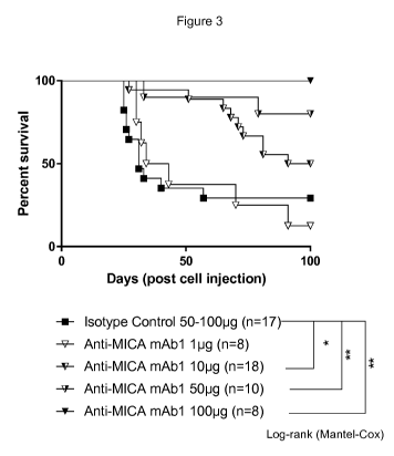
Figure 3 shows that while mice receiving isotype control or 1 pg anti-MICA
antibody
mAb1 did not survive at 100 days post injection, significantly improved
survival was
observed in mice receiving at least 10 pg of anti-MICA antibody. At the 100 pg
dose, anti-
MICA antibody mAb1 achieved survival in all mice at 100 days.
Figure 4 shows, in the left hand panel, mice receiving isotype control, and in
the right
hand panel, mice receiving anti-MICA antibody mAb1. Individual tumor volumes
are shown.
CR=complete response. Treatment with anti-MICA antibody mAb1 caused a decrease
in
tumor volume.
Figure 5 shows that mice treated with anti-MICA antibody mAb1 exhibited a
decreased tumor cell count compared to mice treated with isotype control.
DETAILED DESCRIPTION OF THE INVENTION
The antibodies of the invention are able to directly and specifically target
MICA-
expressing cells as well as MICB-expressing cells, notably tumor cells and
cells involved in
inflammatory or autoimmune processes.
MICA (PERB11.1) refer to MHC class I polypeptide-related sequence A (See,
e.g.,
UniProtKB/Swiss-Prot Q29983), its gene and cDNA and its gene product, or
naturally
occurring variants thereof. Nomenclature of MICA genes and proteins, together
with
reference to accession number of sequence for different alleles are described
in Frigoul A.
and Lefranc, M-P. Recent Res. Devel. Human Genet., 3(2005): 95-145 ISBN: 81-
7736-244-
5, the disclosure of which is incorporated herein by reference. MICA genes and
protein
sequence, including polymorphisms at the protein and DNA level, are also
available from
http://www.ebi.ac.uk/ipd/imgt/h1a/align.html maintained by Cancer Research UK
and the
European Bioinformatics Institute (EBI).
CA 03016765 2018-08-29
WO 2017/157895
PCT/EP2017/055920
The amino acid sequences of MICA were first described in Bahram et al (1994)
Proc.
Nat. Acad. Sci. 91: 6259-6263 and Bahram et al. (1996) Immunogenetics 44:80-
81, the
disclosures of which are incorporated herein by reference. The MICA gene is
polymorphic,
displaying an unusual distribution of a number of variant amino acids in their
extracellular al,
5 a2, and a3 domains. To further define the polymorphism of MICA,
Petersdorf et al. (1999)
examined its alleles among 275 individuals with common and rare HLA genotypes.
The
amino acid sequence of the extracellular al, a2, and a3 domains of human MICA
are shown
in SEQ ID NOS: 1-5. The full MICA sequence further comprises a leader sequence
of 23
amino acids, as well as a transmembrane domain and a cytoplasmic domain. The
amino
10 acid sequence of extracellular al, a2, and a3 domains of selected human
MICA alleles are
shown in SEQ ID NOS: 1-5. The amino acid sequence of MICA*001 is shown in SEQ
ID NO:
1, corresponding to Genbank accession no. AAB41060. The amino acid sequence of
human
MICA allele MICA*004 is shown in SEQ ID NO: 2, corresponding to Genbank
accession no.
AAB41063. The amino acid sequence of human MICA allele MICA*007 is shown in
SEQ ID
NO: 3, corresponding to Genbank accession no. AAB41066. The amino acid
sequence of
human MICA allele MICA*008 is shown in SEQ ID NO: 4, corresponding to Genbank
accession no. AAB41067. The amino acid sequence of human MICA allele MICA*019
is
shown in SEQ ID NO: 5, corresponding to Genbank accession no. AAD27008. The
amino
acid sequence of human MICB is shown Genbank accession no. CAI18747 (SEQ ID
NO:
36).
MICA SEQ ID Amino acid sequence
Allele
MICA*001 1 EPHSLRYNLT VLSWDGSVQS GFLTEVHLDG QPFLRCDRQK CRAKPQGQWA
EDVLGNKTWD RETRDLTGNG KDLRMTLAHI KDQKEGLHSL QEIRVCEIHE
DNSTRSSQHF YYDGELFLSQ NLETKEWTMP QSSRAQTLAM NVRNFLKEDA
MKTKTHYHAM HADCLQELRR YLKSGVVLRR TVPPMVNVTR SEASEGNITV
TCRASGFYPW NITLSWRQDG VSLSHDTQQW GDVLPDGNGT YQTWVATRIC
QGEEQRFTCY MEHSGNHSTH PVPS
M I CA*004 2 EPHSLRYNLT VLSWDGSVQS GFLAEVHLDG QPFLRYDRQK CRAKPQGQWA
EDVLGNKTWD RETRDLTGNG KDLRMTLAHI KDQKEGLHSL QEIRVCEIHE
DNSTRSSQHF YYDGELFLSQ NVETEEWTVP QSSRAQTLAM NVRNFLKEDA
MKTKTHYHAM HADCLQELRR YLESSVVLRR RVPPMVNVTR SEASEGNITV
TCRASSFYPR NITLTWRQDG VSLSHDTQQW GDVLPDGNGT YQTWVATRIC
QGEEQRFTCY MEHSGNHSTH PVPS
M I CA*007 3 EPHSLRYNLT VLSWDGSVQS GFLAEVHLDG QPFLRCDRQK CRAKPQGQWA
EDVLGNKTWD RETRDLTGNG KDLRMTLAHI KDQKEGLHSL QEIRVCEIHE
CA 03016765 2018-08-29
WO 2017/157895
PCT/EP2017/055920
11
DNSTRSSQHF YYDGELFLSQ NLETEEWTMP QSSRAQTLAM NVRNFLKEDA
MKTKTHYHAM HADCLQELRR YLKSGVVLRR TVPPMVNVTR SEASEGNITV
TCRASGFYPW NITLSWRQDG VSLSHDTQQW GDVLPDGNGT YQTWVATRIC
QGEEQRFTCY MEHSGNHSTH PVPS
MICA*008 4 EPHSLRYNLT VLSWDGSVQS GFLAEVHLDG QPFLRYDRQK
CRAKPQGQWA
EDVLGNKTWD RETRDLTGNG KDLRMTLAHI KDQKEGLHSL QEIRVCEIHE
DNSTRSSQHF YYDGELFLSQ NLETEEWTVP QSSRAQTLAM NVRNFLKEDA
MKTKTHYHAM HADCLQELRR YLESGVVLRR TVPPMVNVTR SEASEGNITV
TCRASSFYPR NIILTWRQDG VSLSHDTQQW GDVLPDGNGT YQTWVATRIC
RGEEQRFTCY MEHSGNHSTH PVPS
MICA*019 5 EPHSLRYNLT VLSWDGSVQS GFLAEVHLDG QPFLRYDRQK
CRAKPQGQWA
EDVLGNKTWD RETRDLTGNG KDLRMTLAHI KDQKEGLHSL QEIRVCEIHE
DNSTRSSQHF YYDGELFLSQ NLETEEWTVP QSSRAQTLAM NVRNFLKEDA
MKTKTHYHAM HADCLQELRR YLESSVVLRR TVPPMVNVTR SEASEGNITV
TCRASSFYPR NIILTWRQDG VSLSHDTQQW GDVLPDGNGT YQTWVATRIC
RGEEQRFTCY MEHSGNHSTH PVPS
MICB 36 MGLGRVLLFL AVAFPFAPPA AAAEPHSLRY NLMVLSQDGS
VQSGFLAEGH
LDGQPFLRYD RQKRRAKPQG QWAEDVLGAK TWDTETEDLT ENGQDLRRTL
THIKDQKGGL HSLQEIRVCE IHEDSSTRGS RHFYYDGELF LSQNLETQES
TVPQSSRAQT LAMNVTNFWK EDAMKTKTHY RAMQADCLQK LQRYLKSGVA
IRRTVPPMVN VTCSEVSEGN ITVTCRASSF YPRNITLTWR QDGVSLSHNT
QQWGDVLPDG NGTYQTWVAT RIRQGEEQRF TCYMEHSGNH GTHPVPSGKA
LVLQSQRTDF PYVSAAMPCF VIIIILCVPC CKKKTSAAEG PELVSLQVLD
QHPVGTGDHR DAAQLGFQPL MSATGSTGST EGA
The MICA gene encodes a protein that belongs to the MhcSF and to the IgSF.
This
protein is a transmembrane MHC-I-alpha-like (I-alpha-like) chain, which
comprises three
extracellular domains, two distal G-like domains, G-alphal-like (also referred
to as "Dl" or
"al") and G-a1pha2-like (also referred to as "D2" or "a2"), and a C-like-
domain (also referred
to as "D3" or "a3") proximal to the cell membrane, and three regions, a
connecting-region, a
transmembrane-region and a cytoplasmic-region (labels according to the IMGT
Scientific
Chart of the IMGT (international ImMunoGeneTics information system ),
http://imgt.org and
LeFranc et al. In Silico Biology, 2005; 5:45-60). The MICA mature protein
including leader,
ECD, TM and CY domains, is made up of 360 to 366 amino acids, the difference
arising
from a microsatellite polymorphism in the transmembrane region. The al, a2 and
a3 can be
defined according to any suitable numbering system (e.g., the IMGT numbering
system). In
one embodiment, the al domain comprises residue positions 1 to 88 of the MICA
polypeptide of SEQ ID NO: 1; the a2 domain comprises residue positions 89 to
181 of the
MICA polypeptide of SEQ ID NO: 1; and the a3 domain comprises residue
positions 182 to
CA 03016765 2018-08-29
WO 2017/157895
PCT/EP2017/055920
12
274 of the MICA polypeptide of SEQ ID NO: 1. The al and a2 domains each
comprise A, B,
C and D strands, AB, BC and CD turns, and a helix. The a3 domain comprises A,
B, C, D,
E, F and G strands, a BC loop, a CD strand, a DE-turn and an FG loop. The MICA
protein is
highly glycosylated with eight potential glycosylation sites, two in al, one
in a2 and five in
the a3 domain, including 0-glycans (N-acetyllactosamine linked to serine or
threonine)
and/or N-glycans. While MICA is expressed constitutively in certain cells, low
levels of MICA
expression do not usually give rise to host immune cell attach. However, on
MICA is
upregulated on rapidly proliferating cells such as tumor cells. MICA is the
most highly
expressed of all NKG2D ligands, and it has been found across a wide range of
tumor types
(e.g., carcinomas in general, bladder cancer, melanoma, lung cancer,
hepatocellular cancer,
glioblastoma, prostate cancer, hematological malignancies in general, acute
myeloid
leukemia, acute lymphatic leukemia, chronic myeloid leukemia and chronic
lymphatic
leukemia. Recently, Tsuboi et al. (2011) (EMBO J: 1-13) reported that the 0-
glycan
branching enzyme, core2 13-1,6-N-acetylglucosaminyltransferase (C2GnT) is
active in MICA-
expressing tumor cells and that MICA from tumor cells contains core2 0-glycan
(an 0-
glycan comprising an N-acetylglucosamine branch connected to N-
acetylgalactosamine).
Bauer et al Science 285: 727-729, 1999 provided a role for MICA as a stress-
inducible ligand for NKG2D. As used herein, "MICA" refers to any MICA
polypeptide,
including any variant, derivative, or isoform of the MICA gene or encoded
protein(s) to which
they refer. The MICA gene is polymorphic, displaying an unusual distribution
of a number of
variant amino acids in their extracellular alpha-1, alpha-2, and alpha-3
domains. Various
allelic variants have been reported for MICA polypeptides (e.g., MICA), each
of these are
encompassed by the respective terms, including, e.g., human MICA polypeptides
MICA*001,
MICA*002, MICA*004, MICA*005, MICA*006, MICA*007, MICA*008, MICA*009,
MICA*010,
MICA*011, MICA*012, MICA*013, MICA*014, MICA*015, MICA*016, MICA*017,
MICA*018,
MICA*019, MICA*020, MICA*022, MICA*023, MICA*024, MICA*025, MICA*026,
MICA*027,
MICA*028, MICA*029, MICA*030, MICA*031, MICA*032, MICA*033, MICA*034,
MICA*035,
MICA*036, MICA*037, MICA*038, MICA*039, MICA*040, MICA*041, MICA*042,
MICA*043,
MICA*044, MICA*045, MICA*046, MICA*047, MICA*048, MICA*049, MICA*050,
MICA*051,
MICA*052, MICA*053, MICA*054, MICA*055, MICA*056 and further MICA alleles
MICA*057-MICA*087.
As used herein, "hNKG2D" and, unless otherwise stated or contradicted by
context,
the terms "NKG2D," "NKG2-D," "CD314," "D1252489E," "KLRK1," "killer cell
lectin-like
receptor subfamily K, member 1," or "KLRK1," refer to a human killer cell
activating receptor
gene, its cDNA (e.g., Gen Bank Accession No. NM_007360), and its gene product
(Gen Bank
Accession No. NP 031386), or naturally occurring variants thereof. In NK and T
cells,
CA 03016765 2018-08-29
WO 2017/157895
PCT/EP2017/055920
13
hNKG2D can form heterodimers or higher order complexes with proteins such as
DAP10
(GenBank Accession No. AAG29425, AAD50293). Any activity attributed herein to
hNKG2D, e.g., cell activation, antibody recognition, etc., can also be
attributed to hNKG2D in
the form of a heterodimer such as hNKG2D-DAP10, or higher order complexes with
these
two (and/or other) components.
The 3D structure of MICA in complex with NKG2D has been determined (see, e.g.,
Li
et al., Nat. lmmunol. 2001; 2:443-451; code lhyr, and in IMGT/3Dstructure-DB
(Kaas et al.
Nucl. Acids Res. 2004; 32:D208-D210)). When MICA is in complex with a NKG2D
homodimer, the residues 63 to 73 (IGMT numbering) of MICA a2 are ordered,
adding almost
two turns of helix. The two monomers of NKG2D equally contribute to
interactions with
MICA, and seven positions in each NKG2D monomer interact with one of the MICA
al or a2
helix domains.
The invention provides methods of using the anti-MICA antibodies disclosed
herein;
for example, provided is a method for inhibiting cell proliferation or
activity, for delivering a
molecule to a cell (e.g., a toxic molecule, a detectable marker, etc.), for
targeting, identifying
or purifying a cell, for depleting, killing or eliminating a cell, for
reducing cell proliferation, the
method comprising exposing a cell, such as a tumor cell which expresses a MICA
polypeptide, to an antigen-binding compound of the disclosure that binds a
MICA
polypeptide. It will be appreciated that for the purposes herein, "cell
proliferation" can refer to
any aspect of the growth or proliferation of cells, e.g., cell growth, cell
division, or any aspect
of the cell cycle. The cell may be in cell culture (in vitro) or in a mammal
(in vivo), e.g., a
mammal suffering from a MICA-expressing pathology. Also provided is a method
for
inducing the death of a cell or inhibiting the proliferation or activity of a
cell which expresses
a MICA polypeptide, comprising exposing the cell to an antigen-binding
compound that binds
a MICA polypeptide linked to a toxic agent, in an amount effective to induce
death and/or
inhibit the proliferation of the cell. Thus, also provided is a method for
treating a mammal
suffering from a proliferative disease, and any condition characterized by a
pathogenic
expansion of cells expressing of a MICA polypeptide, the method comprising
administering a
pharmaceutically effective amount of an antibody disclosed herein to the
mammal, e.g., for
the treatment of a cancer.
Definitions
As used in the specification, "a" or "an" may mean one or more. As used in the
claim(s), when used in conjunction with the word "comprising", the words "a"
or "an" may
mean one or more than one. As used herein "another" may mean at least a second
or more.
CA 03016765 2018-08-29
WO 2017/157895
PCT/EP2017/055920
14
Where "comprising" is used, this can optionally be replaced by "consisting
essentially of" or by "consisting of".
Whenever within this whole specification "treatment of cancer" or the like is
mentioned with reference to anti-MICA binding agent (e.g., antibody), there is
meant: (a)
method of treatment of cancer, said method comprising the step of
administering (for at least
one treatment) an anti-MICA binding agent, (for example in a pharmaceutically
acceptable
carrier material) to an individual, a mammal, especially a human, in need of
such treatment,
in a dose that allows for the treatment of cancer, (a therapeutically
effective amount),
optionally in a dose (amount) as specified herein; (b) the use of an anti-MICA
binding agent
for the treatment of cancer, or an anti-MICA binding agent, for use in said
treatment
(especially in a human); (c) the use of an anti-MICA binding agent for the
manufacture of a
pharmaceutical preparation for the treatment of cancer, a method of using an
anti-MICA
binding agent for the manufacture of a pharmaceutical preparation for the
treatment of
cancer, comprising admixing an anti-MICA binding agent with a pharmaceutically
acceptable
carrier, or a pharmaceutical preparation comprising an effective dose of an
anti-MICA
binding agent that is appropriate for the treatment of cancer; or (d) any
combination of a), b),
and c), in accordance with the subject matter allowable for patenting in a
country where this
application is filed.
The term "antibody," as used herein, refers to polyclonal and monoclonal
antibodies. Depending on the type of constant domain in the heavy chains,
antibodies are
assigned to one of five major classes: IgA, IgD, IgE, IgG, and IgM. Several of
these are
further divided into subclasses or isotypes, such as IgG1, IgG2, IgG3, IgG4,
and the like. An
exemplary immunoglobulin (antibody) structural unit comprises a tetramer. Each
tetramer is
composed of two identical pairs of polypeptide chains, each pair having one
"light" (about 25
kDa) and one "heavy" chain (about 50-70 kDa). The N-terminus of each chain
defines a
variable region of about 100 to 110 or more amino acids that is primarily
responsible for
antigen recognition. The terms variable light chain (VL) and variable heavy
chain (VH) refer to
these light and heavy chains respectively. The heavy-chain constant domains
that
correspond to the different classes of immunoglobulins are termed "alpha,"
"delta," "epsilon,"
"gamma" and "mu," respectively. The subunit structures and three-dimensional
configurations of different classes of immunoglobulins are well known. IgG are
the
exemplary classes of antibodies employed herein because they are the most
common
antibodies in the physiological situation and because they are most easily
made in a
laboratory setting. Optionally the antibody is a monoclonal antibody.
Particular examples of
antibodies are humanized, chimeric, human, or otherwise-human-suitable
antibodies.
CA 03016765 2018-08-29
WO 2017/157895
PCT/EP2017/055920
"Antibodies" also includes any fragment or derivative of any of the herein
described
antibodies.
The term "specifically binds to" means that an antibody can bind preferably in
a
competitive binding assay to the binding partner, e.g., MICA and MICB, as
assessed using
5 either recombinant forms of the proteins, epitopes therein, or native
proteins present on the
surface of isolated target cells. Competitive binding assays and other methods
for
determining specific binding are further described below and are well known in
the art.
When an antibody is said to "compete with" a particular monoclonal antibody,
it
means that the antibody competes with the monoclonal antibody in a binding
assay using
10 either recombinant MICA molecules or surface expressed MICA molecules.
For example, if a
test antibody reduces the binding of a reference antibody to a MICA
polypeptide or MICA-
expressing cell in a binding assay, the antibody is said to "compete"
respectively with the
reference antibody.
The term "affinity", as used herein, means the strength of the binding of an
antibody
15 to an epitope. The affinity of an antibody is given by the dissociation
constant Kd, defined as
[AID] x [Ag] / [Ab-Ag], where [Ab-Ag] is the molar concentration of the
antibody-antigen
complex, [AID] is the molar concentration of the unbound antibody and [Ag] is
the molar
concentration of the unbound antigen. The affinity constant Ka is defined by
1/Kd. Methods
for determining the affinity of mAbs can be found in Harlow, et al.,
Antibodies: A Laboratory
Manual, Cold Spring Harbor Laboratory Press, Cold Spring Harbor, N.Y., 1988),
Coligan et
al., eds., Current Protocols in Immunology, Greene Publishing Assoc. and Wiley
lnterscience, N.Y., (1992, 1993), and Muller, Meth. Enzymol. 92:589-601
(1983), which
references are entirely incorporated herein by reference. One standard method
well known
in the art for determining the affinity of mAbs is the use of surface plasmon
resonance (SPR)
screening (such as by analysis with a BlAcoreTM SPR analytical device).
Within the context herein a "determinant" designates a site of interaction or
binding
on a polypeptide.
The term "epitope" refers to an antigenic determinant, and is the area or
region on
an antigen to which an antibody binds. A protein epitope may comprise amino
acid residues
directly involved in the binding as well as amino acid residues which are
effectively blocked
by the specific antigen binding antibody or peptide, i.e., amino acid residues
within the
"footprint" of the antibody. It is the simplest form or smallest structural
area on a complex
antigen molecule that can combine with e.g., an antibody or a receptor.
Epitopes can be
linear or conformational/structural. The term "linear epitope" is defined as
an epitope
composed of amino acid residues that are contiguous on the linear sequence of
amino acids
(primary structure). The term "conformational or structural epitope" is
defined as an epitope
CA 03016765 2018-08-29
WO 2017/157895
PCT/EP2017/055920
16
composed of amino acid residues that are not all contiguous and thus represent
separated
parts of the linear sequence of amino acids that are brought into proximity to
one another by
folding of the molecule (secondary, tertiary and/or quaternary structures). A
conformational
epitope is dependent on the 3-dimensional structure. The term 'conformational'
is therefore
often used interchangeably with 'structural'.
The term "deplete" or "depleting", with respect to MICA-expressing cells,
means a
process, method, or compound that results in killing, elimination, lysis or
induction of such
killing, elimination or lysis, so as to negatively affect the number of such
MICA-expressing
cells present in a sample or in a subject.
The term "antibody-dependent cell-mediated cytotoxicity" or "ADCC" is a term
well
understood in the art, and refers to a cell-mediated reaction in which non-
specific cytotoxic
cells that express Fc receptors (FcRs) recognize bound antibody on a target
cell and
subsequently cause lysis of the target cell. Non-specific cytotoxic cells that
mediate ADCC
include natural killer (NK) cells, macrophages, monocytes, neutrophils, and
eosinophils.
The term "complement-dependent cytotoxicity" or "CDC" is a term well
understood
in the art, and refers to the ability of a molecule to lyse a target in the
presence of
complement. The complement activation pathway is initiated by the binding of
the first
component of the complement system (C1q) to a molecule (e.g., an antibody)
complexed
with a cognate antigen.
The term "agent" is used herein to denote a chemical compound, a mixture of
chemical compounds, a biological macromolecule, or an extract made from
biological
materials. The term "therapeutic agent" refers to an agent that has biological
activity.
For the purposes herein, a "humanized" or "human" antibody refers to an
antibody
in which the constant and variable framework region of one or more human
immunoglobulins
is fused with the binding region, e.g., the CDR, of an animal immunoglobulin.
Such
antibodies are designed to maintain the binding specificity of the non-human
antibody from
which the binding regions are derived, but to avoid an immune reaction against
the non-
human antibody. Such antibodies can be obtained from transgenic mice or other
animals
that have been "engineered" to produce specific human antibodies in response
to antigenic
challenge (see, e.g., Green et al. (1994) Nature Genet 7:13; Lonberg et al.
(1994) Nature
368:856; Taylor et al. (1994) Int lmmun 6:579, the entire teachings of which
are herein
incorporated by reference). A fully human antibody also can be constructed by
genetic or
chromosomal transfection methods, as well as phage display technology, all of
which are
known in the art (see, e.g., McCafferty et al. (1990) Nature 348:552-553).
Human antibodies
may also be generated by in vitro activated B cells (see, e.g., U.S. Pat. Nos.
5,567,610 and
5,229,275, which are incorporated in their entirety by reference).
CA 03016765 2018-08-29
WO 2017/157895
PCT/EP2017/055920
17
As used herein, the term "antigen binding domain" refers to a domain
comprising a
three-dimensional structure capable of immunospecifically binding to an
epitope. Thus, in
one embodiment, said domain can comprise a hypervariable region, optionally a
VH and/or
VL domain of an antibody chain, optionally at least a VH domain. In another
embodiment,
the binding domain may comprise at least one complementarity determining
region (CDR) of
an antibody chain. In another embodiment, the binding domain may comprise a
polypeptide
domain from a non-immunoglobulin scaffold.
The term "hypervariable region" when used herein refers to the amino acid
residues
of an antibody that are responsible for antigen binding. The hypervariable
region generally
comprises amino acid residues from a "complementarity-determining region" or
"CDR" (e.g.,
residues 24-34 (L1), 50-56 (L2) and 89-97 (L3) in the light-chain variable
domain and 31-35
(H1), 50-65 (H2) and 95-102 (H3) in the heavy-chain variable domain;
disclosure (see Kabat
et al. (1991) Sequences of Protein of Immunological Interest, 5th ed., United
States Public
Health Service, National Institute of Health, Bethesda, MD)) and/or those
residues from a
"hypervariable loop" (e.g., residues 26-32 (L1), 50-52 (L2) and 91-96 (L3) in
the light-chain
variable domain and 26-32 (H1), 53-55 (H2) and 96-101 (H3) in the heavy-chain
variable
domain; Chothia and Lesk, J. Mol. Biol 1987;196:901-917), or a similar system
for
determining essential amino acids responsible for antigen binding. Using the
Kabat
numbering system, the actual linear amino acid sequence of a peptide may
contain fewer or
additional amino acids corresponding to a shortening of, or insertion into, a
FR or CDR of the
variable domain. For example, a heavy chain variable domain may include a
single amino
acid insert (residue 52a according to Kabat) after residue 52 of CDR H2 and
inserted
residues (e.g., residues 82a, 82b, and 82c, etc. according to Kabat) after
heavy chain FR
residue 82. The Kabat numbering of residues may be determined for a given
antibody by
alignment at regions of homology of the sequence of the antibody with a
"standard" Kabat
numbered sequence. Another suitable numbering system is the Abnum system.
Unless
otherwise specified, the Abnum amino acid numbering nomenclature for
immunoglobulins is
used to refer to positions in the VH and VL domains (see Abhinandan and
Martin, (2008)
Molecular Immunology 45: 3832-3839, the disclosure of which is incorporated by
reference).
Sequence numbering using the Abnum system can also be automatically generated
at
http://www.bioinfo.org.uk/abs/abnum. However it will be appreciated that the
person of skill
in the art can use an alternative numbering system and identify positions
corresponding to
Abnum numbering. Phrases such as "Abm position", "Abm numbering" and
"according to
Abm" herein refer to this numbering system for heavy chain variable domains or
light chain
variable domains.
CA 03016765 2018-08-29
WO 2017/157895
PCT/EP2017/055920
18
By "framework" or "FR" residues as used herein is meant the region of an
antibody
variable domain exclusive of those regions defined as CDRs. Each antibody
variable domain
framework can be further subdivided into the contiguous regions separated by
the CDRs
(FR1, FR2, FR3 and FR4).
The terms "Fc domain," "Fc portion," and "Fc region" refer to a C-terminal
fragment
of an antibody heavy chain, e.g., from about amino acid (aa) 230 to about aa
450 (Kabat
numbering) of human y (gamma) heavy chain or its counterpart sequence in other
types of
antibody heavy chains (e.g., a, 6, E and p for human antibodies), or a
naturally occurring
allotype thereof.
The terms "isolated", "purified" or "biologically pure" refer to material that
is
substantially or essentially free from components which normally accompany it
as found in
its native state. Purity and homogeneity are typically determined using
analytical chemistry
techniques such as polyacrylamide gel electrophoresis or high performance
liquid
chromatography. A protein that is the predominant species present in a
preparation is
substantially purified.
The terms "polypeptide," "peptide" and "protein" are used interchangeably
herein to
refer to a polymer of amino acid residues. The terms apply to amino acid
polymers in which
one or more amino acid residue is an artificial chemical mimetic of a
corresponding naturally
occurring amino acid, as well as to naturally occurring amino acid polymers
and non-
naturally occurring amino acid polymer.
The term "recombinant" when used with reference, e.g., to a cell, or nucleic
acid,
protein, or vector, indicates that the cell, nucleic acid, protein or vector,
has been modified by
the introduction of a heterologous nucleic acid or protein or the alteration
of a native nucleic
acid or protein, or that the cell is derived from a cell so modified. Thus,
for example,
recombinant cells express genes that are not found within the native
(nonrecombinant) form
of the cell or express native genes that are otherwise abnormally expressed,
under
expressed or not expressed at all.
Within the context herein, the term antibody that "binds" a polypeptide or
epitope
designates an antibody that binds said determinant with specificity and/or
affinity.
The term "identity" or "identical", when used in a relationship between the
sequences of two or more polypeptides, refers to the degree of sequence
relatedness
between polypeptides, as determined by the number of matches between strings
of two or
more amino acid residues. "Identity" measures the percent of identical matches
between the
smaller of two or more sequences with gap alignments (if any) addressed by a
particular
mathematical model or computer program (i.e., "algorithms"). Identity of
related polypeptides
can be readily calculated by known methods. Such methods include, but are not
limited to,
CA 03016765 2018-08-29
WO 2017/157895
PCT/EP2017/055920
19
those described in Computational Molecular Biology, Lesk, A. M., ed., Oxford
University
Press, New York, 1988; Biocomputing: Informatics and Genome Projects, Smith,
D. W., ed.,
Academic Press, New York, 1993; Computer Analysis of Sequence Data, Part 1,
Griffin, A.
M., and Griffin, H. G., eds., Humana Press, New Jersey, 1994; Sequence
Analysis in
Molecular Biology, von Heinje, G., Academic Press, 1987; Sequence Analysis
Primer,
Gribskov, M. and Devereux, J., eds., M. Stockton Press, New York, 1991; and
Carillo et al.,
SIAM J. Applied Math. 48, 1073 (1988).
Methods for determining identity are designed to give the largest match
between
the sequences tested. Methods of determining identity are described in
publicly available
computer programs. Computer program methods for determining identity between
two
sequences include the GCG program package, including GAP (Devereux et al.,
Nucl. Acid.
Res. 12, 387 (1984); Genetics Computer Group, University of Wisconsin,
Madison, Wis.),
BLASTP, BLASTN, and FASTA (Altschul et al., J. Mol. Biol. 215, 403-410
(1990)). The
BLASTX program is publicly available from the National Center for
Biotechnology
Information (NCB!) and other sources (BLAST Manual, Altschul et al.
NCB/NLM/NIH
Bethesda, Md. 20894; Altschul et al., supra). The well-known Smith Waterman
algorithm
may also be used to determine identity.
Production of antibodies
The present invention is based, in part, on the discovery of modified human
acceptor
framework sequences into which antibody CDRs can be incorporated such that the
resulting
anti-MICA variable region has high physicochemical stability and high binding
affinity for the
predominant human MICA alleles. Furthermore, provided are antibodies with high
content of
human amino acid sequences, thereby providing decreased risk of immunogenicity
when
administered to a human individual. Advantageously, the antibodies have low
potential to
elicit human anti-mouse antibodies (HAMA).
Anti-MICA antibody VH and VL sequences are provided below in Table 1, amino
acids differing between respective VH domains and VL domains are underlined:
Table 1
Antibody Amino acid sequence
domain
mAb1 QVQLQESGPGLVKPSETLSLTCTVSGYSITSDYAWNWIRQPPGKGLEWIGFVSYSGTTKY
VH NPSLKSRVTISRDTSKNQFSLKLSSVTAADTAVYYCARGYGFDYWGQGTTVTVSS
_
(SEQ ID NO: 6)
mAb1 EIVLTQSPATLSLSPGERATLSCSATSSISSIYFHWYQQKPGQAPRLLIYRTSNLASGIP
CA 03016765 2018-08-29
WO 2017/157895
PCT/EP2017/055920
VL ARFSGSGSGTDYTLTISSLEPEDFAVYYCQQGTTIPFTFGQGTKLEIK
_
(SEQ ID NO: 7)
mAb2 QVQLQESGPGLVKPSETLSLTCTVSGYSITSDYAWNWIRQPPGKGLEWIGFVSYSGTTKY
VH NPSLKSRVTISRDTSKNQFSLKLSSVTAADTAVYYCARGYGFDYWGQGTTVTVSS
_
(SEQ ID NO: 8)
mAb2 EIVLTQSPATLSLSPGERATLSCSATSSISSIYFHWYQQKPGQAPRLLIYRTSNLASGIP
VL ARFSGSGSGTSYTLTISSLEPEDFAVYYCQQGTTIPFTFGQGTKLEIK
(SEQ ID NO: 9)
mAb3 QVQLQESGPGLVKPSETLSLTCTVSGYSITSDYAWNWIRQPPGKGLEWIGFVSYSGTTKY
VH NPSLKSRVTISRDTSKNQFSLKLSSVTAADTAVYYCARGYGFDYWGQGTTVTVSS
_
(SEQ ID NO: 10)
mAb3 EIVLTQSPATLSLSPGERATLSCSATSSISSIYFHWYQQKPGQAPRLLIYRTSNLASGIP
VL ARFSGSGSGTDYTLTISSLEPEDVAVYYCQQGTTIPFTFGQGTKLEIK
_ _
(SEQ ID NO: 11)
Positions in the VH and VL domains herein are described using the Abnum amino
acid numbering nomenclature for immunoglobulins (see Abhinandan and Martin,
(2008)
Molecular Immunology 45: 3832-3839, the disclosure of which is incorporated by
reference).
5 Sequence numbering using the Abnum system can also be automatically
generated at
http://www.bioinfo.org.uk/abs/abnum. However it will be appreciated that the
person of skill
in the art can use an alternative numbering system and identify positions
corresponding to
Abnum numbering.
In one embodiment, the antibody comprises a heavy chain framework from the
10 human subgroup IGHV4-b (e.g., IGHV4-b*02) and the J-segment is from
IGHJ6 (e.g.,
IGHJ6*01). In one embodiment, the humanized antibody comprises a light chain
framework
from the human subgroup IGKV3-11 (e.g., IGKV3-11*01) and the J-segment is from
IGKJ2
(e.g., IGKJ2*01).
The antibody may further comprise one or more mutations in the human framework
15 sequences, to, e.g., enhance affinity, stability, or other properties of
the antibody.
Examples of VH and VL amino acid sequences of an anti-MICA antibody are shown
in SEQ ID NOS: 6-21, respectively. In one aspect, provided is an isolated
antibody that binds
a human MICA polypeptide, wherein the antibody comprises: a HCDR1 region
comprising
an amino acid sequence SDYAWN as set forth in SEQ ID NO: 30, or a sequence of
at least
20 3 or 4 amino acids thereof; a HCDR2 region comprising an amino acid
sequence
FVSYSGTTKYNPSLKS as set forth in SEQ ID NO: 31, or a sequence of at least 4,
5, 6, 7, 8,
9 or 10 contiguous amino acids thereof; a HCDR3 region comprising an amino
acid
CA 03016765 2018-08-29
WO 2017/157895
PCT/EP2017/055920
21
sequence GYGFDY as set forth in SEQ ID NO: 32, or a sequence of at least 4, 5,
6, 7, 8, 9
or 10 contiguous amino acids thereof; a LCDR1 region comprising an amino acid
sequence
SATSSISSIYFH as set forth in SEQ ID NO: 33, or a sequence of at least 4, 5, 6,
7, 8, 9 or 10
contiguous amino acids thereof; a LCDR2 region comprising an amino acid
sequence
RTSNLA as set forth in SEQ ID NO: 34, or a sequence of at least 3, 4 or 5
contiguous amino
acids thereof; a LCDR3 region comprising an amino acid sequence QQGTTIPFT as
set forth
in SEQ ID NO: 35, or a sequence of at least 5, 6, 7, or 8 contiguous amino
acids thereof.
In one aspect, provided is an antigen binding domain or antibody that binds a
human
MICA polypeptide, comprising:
(a) a CDR-H1 comprising the amino acid sequence of SEQ ID NO: 30;
(b) a CDR-H2 comprising the amino acid sequence of SEQ ID NO: 31;
(c) a CDR-H3 comprising the amino acid sequence of SEQ ID NO: 32;
(d) a CDR-L1 comprising the amino acid sequence of SEQ ID NO: 33;
(e) a CDR-L2 comprising the amino acid sequence of SEQ ID NO: 34;
(f) a CDR-L3 comprising the amino acid sequence of SEQ ID NO: 35; and
(g) human heavy and light chain framework sequences,
wherein the antigen binding domain or antibody comprises a VH comprising an
amino acid sequence at least 80%, 90%, 95% or 98% identical to the amino acid
sequence
of SEQ ID NO: 6 and a VL comprising an amino acid sequence at least 80%, 90%,
95% or
98% identical to the amino acid sequence of SEQ ID NO: 7.
In one embodiment, the light chain variable region (VL) comprises an amino
acid
residue at Abnum position 71 (in FR3) capable of forming a non-covalent bonds
with amino
acids within the CDR1 of the VL. In one embodiment, the VL comprises a
tyrosine (Y) amino
acid residue at Abnum position 71 (in FR3). In one embodiment, the VL
comprises a
phenylalanine (F) at Abnum position 83.
In one embodiment, the heavy chain variable region (VH) comprises amino acid
residues at Abnum positions 72c (in FR2) and 74 (in FR3) capable of
interacting with one
another to form a salt bridge, e.g., H-bonding between the residue at Abnum
position 72c
and the residue at position 74. In one embodiment, the VH comprises a lysine
(K) amino
acid residue at Abnum position 72c and a glutamine residue at position 74. In
one
embodiment, the VH comprises a threonine (T) at Abnum position 30. In one
embodiment,
the VH comprises an isoleucine (I) at Abnum position 48. In one embodiment the
VH
comprises a valine (V) at Abnum position 67. In one embodiment, the VH
comprises an
arginine (R) at Abnum position 71.
In one embodiment, the VH comprises a heavy chain framework from the human
subgroup IGHV4-b (e.g., IGHV4-b*02) and the J-segment is from IGHJ6 (e.g.,
IGHJ6*01). In
CA 03016765 2018-08-29
WO 2017/157895
PCT/EP2017/055920
22
one embodiment, the VL comprises a light chain framework from the human
subgroup
IGKV3-11 (e.g., IGKV3-11*01) and the J-segment is from IGKJ2 (e.g., IGKJ2*01).
Optionally a human VH and/or VL framework (e.g., or a heavy or light chain
FR1,
FR2, FR3 and/or FR4 thereof) may or may not comprises one or more mutations,
e.g., back
mutations to introduce a residue present at the particular position in a non-
human mammal
(e.g., a mouse or a rat). The antibody may or may not further comprise one or
more
additional mutations (e.g., back-mutations) in the human framework sequences,
to, e.g.,
enhance affinity, stability, or other properties of the antibody.
In another aspect, provided is anti-MICA antibodies that comprise a VH domain
having at least about 80% sequence identity (e.g., at least about 85%, 90%,
95%, 97%,
98%, or more identity) to the VH domain of SEQ ID NOS: 6 or 8. In another
aspect, provided
are anti-MICA antibodies that comprise a VL domain having at least about 80%
sequence
identity (e.g., at least about 85%, 90%, 95%, 97%, 98%, or more identity) to
the VH domain
of SEQ ID NOS: 7 or 9.
DNA encoding an antibody can be prepared and placed in an appropriate
expression
vector for transfection into an appropriate host. The host is then used for
the recombinant
production of the antibody, or variants thereof, such as a humanized version
of that
monoclonal antibody, active fragments of the antibody, chimeric antibodies
comprising the
antigen recognition portion of the antibody, or versions comprising a
detectable moiety.
DNA encoding the monoclonal antibodies of the disclosure can be readily
isolated
and sequenced using conventional procedures (e. g., by using oligonucleotide
probes that
are capable of binding specifically to genes encoding the heavy and light
chains of murine
antibodies). In one aspect, provided is a nucleic acid encoding a heavy chain
or a light chain
of an anti-MICA antibody of any embodiment herein. Once isolated, the DNA can
be placed
into expression vectors, which are then transfected into host cells such as E.
coli cells,
simian COS cells, Chinese hamster ovary (CHO) cells, or myeloma cells that do
not
otherwise produce immunoglobulin protein, to obtain the synthesis of
monoclonal antibodies
in the recombinant host cells. As described elsewhere in the present
specification, such DNA
sequences can be modified for any of a large number of purposes, e.g., for
humanizing
antibodies, producing fragments or derivatives, or for modifying the sequence
of the
antibody, e.g., in the antigen binding site in order to optimize the binding
specificity of the
antibody. In one embodiment, provided is an isolated nucleic acid sequence
encoding a light
chain and/or a heavy chain of an antibody, as well as a recombinant host cell
comprising
(e.g., in its genome) such nucleic acid. Recombinant expression in bacteria of
DNA encoding
the antibody is well known in the art (see, for example, Skerra et al., Curr.
Opinion in
Immunol., 5, pp. 256 (1993); and Pluckthun, lmmunol. 130, p. 151 (1992).
CA 03016765 2018-08-29
WO 2017/157895
PCT/EP2017/055920
23
Typically, an anti-MICA antibody provided herein has an affinity for a MICA
polypeptide in the range of about 104 to about 1011 M-1 (e.g., about 108 to
about 1019 M-1).
For example, in a particular aspect the disclosure provides Anti-MICA antibody
that have an
average disassociation constant (KD) of less than 1 x 10⁻⁹ M with respect to
MICA, as
determined by, e.g., surface plasmon resonance (SPR) screening (such as by
analysis with
a BlAcoreTM SPR analytical device). In a more particular exemplary aspect, the
disclosure
provides anti- MICA antibodies that have a KD of about 1 x 10⁻⁸ M to about 1 x
10-19 M, or
about 1 x 10⁻⁹ M to about 1 x 10-11 M, for MICA (e.g., MICA*001, *004, *007
and *008
alleles).
Antibodies can be characterized for example by a mean KD of no more than about
(i.e. better affinity than) 100, 60, 10, 5, or 1 nanomolar, preferably sub-
nanomolar or
optionally no more than about 500, 200, 100 or 10 picomolar. KD can be
determined for
example for example by immobilizing recombinantly produced human MICA proteins
on a
chip surface, followed by application of the antibody to be tested in
solution. In one
embodiment, the method further comprises a step (d), selecting antibodies from
(b) that are
capable of competing for binding to MICA with antibody of the disclosure.
Where the test antibodies have modifications in their VH and/VL, a simple
competition assay may be employed in which the control (the antibody having a
VH and VL
of SEQ ID NOS: 6 and 7, or the antibody having a VH and VL of SEQ ID NOS: 8
and 9, for
example) and test antibodies are admixed (or pre-adsorbed) and applied to a
sample
containing MICA polypeptides. Protocols based upon western blotting and the
use of
Biacore TM analysis are suitable for use in such competition studies.
In certain embodiments, one pre-mixes the control antibodies with varying
amounts
of the test antibodies (e.g., about 1:10 or about 1:100) for a period of time
prior to applying to
the MICA antigen sample. In other embodiments, the control and varying amounts
of test
antibodies can simply be admixed during exposure to the MICA antigen sample.
As long as
one can distinguish bound from free antibodies (e. g., by using separation or
washing
techniques to eliminate unbound antibodies) and control antibody from the test
antibodies (e.
g., by using species-specific or isotype-specific secondary antibodies or by
specifically
labelling control antibody with a detectable label) one can determine if the
test antibodies
reduce the binding of control antibody to the antigens, indicating that the
test antibody
recognizes substantially the same epitope as control antibody. The binding of
the (labelled)
control antibodies in the absence of a completely irrelevant antibody can
serve as the control
high value. The control low value can be obtained by incubating the labelled
control
antibodies with unlabelled antibodies of exactly the same type, where
competition would
occur and reduce binding of the labelled antibodies. In a test assay, a
significant reduction in
CA 03016765 2018-08-29
WO 2017/157895
PCT/EP2017/055920
24
labelled antibody reactivity in the presence of a test antibody is indicative
of a test antibody
that recognizes substantially the same epitope, i.e., one that "cross-reacts"
or competes with
the labelled control antibody. Any test antibody that reduces the binding of
control antibody
to MICA antigens by at least about 50%, such as at least about 60%, or more
preferably at
least about 80% or 90% (e. g., about 65-100%), at any ratio of control
antibody:test antibody
between about 1:10 and about 1:100 is considered to be an antibody that binds
to
substantially the same epitope or determinant as control antibody. In one
embodiment, such
test antibody will reduce the binding of control antibody to the MICA antigen
by at least about
90% (e.g., about 95%).
Competition can also be assessed by, for example, a flow cytometry test. In
such a
test, cells bearing a given MICA polypeptide can be incubated first with
control antibody, for
example, and then with the test antibody labelled with a fluorochrome or
biotin. The antibody
is said to compete with control antibody if the binding obtained upon
preincubation with a
saturating amount of control antibody is about 80%, optionally about 50%,
about 40% or less
(e.g., about 30%, 20% or 10%) of the binding (as measured by mean of
fluorescence)
obtained by the antibody without preincubation with control antibody.
Alternatively, an
antibody is said to compete with control antibody if the binding obtained with
a labelled
control antibody antibody (by a fluorochrome or biotin) on cells preincubated
with a
saturating amount of test antibody is about 80%, optionally about 50%, about
40%, or less
(e.g., about 30%, 20% or 10%) of the binding obtained without preincubation
with the test
antibody.
A simple competition assay in which a test antibody is pre-adsorbed and
applied at
saturating concentration to a surface onto which a MICA antigen is immobilized
may also be
employed. The surface in the simple competition assay is preferably a
BiacoreTM chip (or
other media suitable for surface plasmon resonance analysis). The control
antibody (the
antibody having a VH and VL of SEQ ID NOS: 6 and 7, or the antibody having a
VH and VL
of SEQ ID NOS: 8 and 9, for example) is then brought into contact with the
surface at a
MICA-saturating concentration and the MICA and surface binding of the control
antibody is
measured. This binding of the control antibody is compared with the binding of
the control
antibody to the MICA-containing surface in the absence of test antibody. In a
test assay, a
significant reduction in binding of the MICA-containing surface by the control
antibody in the
presence of a test antibody indicates that the test antibody recognizes
substantially the
same epitope as the control antibody such that the test antibody "cross-
reacts" with the
control antibody. Any test antibody that reduces the binding of control
antibody to a MICA
antigen by at least about 30% or more, preferably about 40%, can be considered
to be an
antibody that binds to substantially the same epitope or determinant as
control antibody.
CA 03016765 2018-08-29
WO 2017/157895
PCT/EP2017/055920
Preferably, such a test antibody will reduce the binding of the control
antibody to the MICA
antigen by at least about 50% (e. g., at least about 60%, at least about 70%,
or more). It will
be appreciated that the order of control and test antibodies can be reversed:
that is, the
control antibody can be first bound to the surface and the test antibody is
brought into
5 contact with the surface thereafter in a competition assay. Preferably,
the antibody having
higher affinity for the MICA antigen is bound to the surface first, as it will
be expected that
the decrease in binding seen for the second antibody (assuming the antibodies
are cross-
reacting) will be of greater magnitude. Further examples of such assays are
provided in,
e.g., Sauna! (1995) J. lmmunol. Methods 183: 33-41, the disclosure of which is
incorporated
10 herein by reference.
Determination of whether an antibody binds within an epitope region can be
carried
out in ways known to the person skilled in the art. As one example of such
mapping/characterization methods, an epitope region for an anti-MICA antibody
may be
determined by epitope "foot-printing" using chemical modification of the
exposed
15 amines/carboxyls in the MICA protein. One specific example of such a
foot-printing
technique is the use of HXMS (hydrogen-deuterium exchange detected by mass
spectrometry) wherein a hydrogen/deuterium exchange of receptor and ligand
protein amide
protons, binding, and back exchange occurs, wherein the backbone amide groups
participating in protein binding are protected from back exchange and
therefore will remain
20 deuterated. Relevant regions can be identified at this point by peptic
proteolysis, fast
microbore high-performance liquid chromatography separation, and/or
electrospray
ionization mass spectrometry. See, e. g., Ehring H, Analytical Biochemistry,
Vol. 267 (2) pp.
252-259 (1999) Engen, J. R. and Smith, D. L. (2001) Anal. Chem. 73, 256A-265A.
Another
example of a suitable epitope identification technique is nuclear magnetic
resonance epitope
25 mapping (NMR), where typically the position of the signals in two-
dimensional NMR spectra
of the free antigen and the antigen complexed with the antigen binding
peptide, such as an
antibody, are compared. The antigen typically is selectively isotopically
labeled with 15N so
that only signals corresponding to the antigen and no signals from the antigen
binding
peptide are seen in the NMR-spectrum. Antigen signals originating from amino
acids
involved in the interaction with the antigen binding peptide typically will
shift position in the
spectrum of the complex compared to the spectrum of the free antigen, and the
amino acids
involved in the binding can be identified that way. See, e. g., Ernst Schering
Res Found
Workshop. 2004; (44): 149-67; Huang et al., Journal of Molecular Biology, Vol.
281 (1) pp.
61-67 (1998); and Saito and Patterson, Methods. 1996 Jun; 9 (3): 516-24.
Epitope mapping/characterization also can be performed using mass spectrometry
methods. See, e.g., Downard, J Mass Spectrom. 2000 Apr; 35 (4): 493-503 and
Kiselar and
CA 03016765 2018-08-29
WO 2017/157895
PCT/EP2017/055920
26
Downard, Anal Chem. 1999 May 1; 71(9): 1792-1801. Protease digestion
techniques also
can be useful in the context of epitope mapping and identification. Antigenic
determinant-
relevant regions/sequences can be determined by protease digestion, e.g., by
using trypsin
in a ratio of about 1:50 to MICA or o/n digestion at and pH 7-8, followed by
mass
spectrometry (MS) analysis for peptide identification. The peptides protected
from trypsin
cleavage by the anti-MICA binder can subsequently be identified by comparison
of samples
subjected to trypsin digestion and samples incubated with antibody and then
subjected to
digestion by e.g., trypsin (thereby revealing a footprint for the binder).
Other enzymes like
chymotrypsin, pepsin, etc., also or alternatively can be used in similar
epitope
characterization methods. Moreover, enzymatic digestion can provide a quick
method for
analyzing whether a potential antigenic determinant sequence is within a
region of the MICA
polypeptide that is not surface exposed and, accordingly, most likely not
relevant in terms of
immunogenicity/antigenicity.
Site-directed mutagenesis is another technique useful for elucidation of a
binding
epitope. For example, in "alanine-scanning", each residue within a protein
segment is re-
placed with an alanine residue, and the consequences for binding affinity
measured. If the
mutation leads to a significant reduction in binding affinity, it is most
likely involved in
binding. Monoclonal antibodies specific for structural epitopes (i.e.,
antibodies which do not
bind the unfolded protein) can be used to verify that the alanine-replacement
does not
influence over-all fold of the protein. See, e.g., Clackson and Wells, Science
1995;
267:383-386; and Wells, Proc Natl Acad Sci USA 1996; 93:1-6.
Electron microscopy can also be used for epitope "foot-printing". For example,
Wang et al., Nature 1992; 355:275-278 used coordinated application of
cryoelectron micros-
copy, three-dimensional image reconstruction, and X-ray crystallography to
determine the
physical footprint of a Fab-fragment on the capsid surface of native cowpea
mosaic virus.
Other forms of "label-free" assay for epitope evaluation include surface
plasmon
resonance (SPR, BiacoreTM) and reflectometric interference spectroscopy
(RifS). See, e.g.,
Fagerstam et al., Journal Of Molecular Recognition 1990;3:208-14; Nice et al.,
J. Chroma-
togr. 1993; 646:159-168; Leipert et al., Angew. Chem. Int. Ed. 1998; 37:3308-
3311; Kroger
et al., Biosensors and Bioelectronics 2002; 17:937-944.
It should also be noted that an antibody binding the same or substantially the
same
epitope as an antibody can be identified in one or more of the exemplary
competition assays
described herein. In one embodiment, a blocking a1a2 domain antibody binds an
epitope
comprising one, two or three residues selected from the group consisting of
E100, D101 and
N102, one, two or three residues selected from the group consisting of S103,
T104 and
R105, one or two residues selected from the group consisting of N121 and E123,
and/or one
CA 03016765 2018-08-29
WO 2017/157895
PCT/EP2017/055920
27
or two residues selected from the group consisting of T124 and E126. In one
embodiment, a
blocking a1a2 domain antibody binds an epitope on a human MICA polypeptide
comprising
1, 2, 3, 4, 5, 6, or more residues selected from the group consisting of
residues (with
reference to SEQ ID NO: 1): E100, D101, N102, S103, T104, R105, N121, E123,
T124 and
E126.
In one embodiment, the anti-MICA antibody has decreased binding to a mutant
human MICA polypeptide having E100A, D101S, N102A substitutions (compared to a
wild-
type human MICA polypeptide of SEQ ID NO: 1. In one embodiment, the anti-MICA
antibody
has decreased binding to a mutant human MICA polypeptide having 5103A, Ti 04S,
R105A
substitutions (compared to a wild-type human MICA polypeptide of SEQ ID NO: 1.
In one
embodiment, the anti-MICA antibody has decreased binding to a mutant human
MICA
polypeptide having N121A, E1235, substitutions (compared to a wild-type human
MICA
polypeptide of SEQ ID NO: 1. In one embodiment, the anti-MICA antibody has
decreased
binding to a mutant human MICA polypeptide having T124A and E126A
substitutions
(compared to a wild-type human MICA polypeptide of SEQ ID NO: 1.
In one embodiment, the anti-MICA antibody binds to a MICA polypeptide at least
partly within the a2 domain of MICA. Optionally, the antibody binds to the a2
domain at the
lateral side of MICA near the NKG2D binding surface, consistent with the
finding that the
antibody block the interaction of cell surface MICA with NKG2D.
In view of the ability of the anti-MICA antibodies to induce ADCC and CDC, the
antibodies can advantageously be made with modifications that increase their
ability to bind
Fc receptors which can affect effector functions such as antibody-dependent
cytotoxicity,
mast cell degranulation, and phagocytosis, as well as immunomodulatory signals
such as
regulation of lymphocyte proliferation and antibody secretion. Typical
modifications include
modified human IgG1 constant regions comprising at least one amino acid
modification (e.g.,
substitution, deletions, insertions), and/or altered types of glycosylation,
e.g.,
hypofucosylation. Such modifications can affect interaction with Fc receptors:
FcyRI (CD64),
FcyRII (CD32), and FcyRIII (CD 16). FcyRI (CD64), FcyRIIA (CD32A) and FcyRIII
(CD 16)
are activating (i.e. , immune system enhancing) receptors while FcyRIIB
(CD32B) is an
inhibiting (i.e., immune system dampening) receptor. A modification may, for
example,
increase binding of the Fc domain to FcyRIlla on effector (e.g., NK) cells.
Anti-MICA antibodies may comprise an Fc domain (or portion thereof) of human
IgG1 or IgG3 isotype, optionally modified. The amino acid sequence of
positions 230 to 447
sequence of a human IgG1 Fc region (Gen Bank accession #: J00228) is shown as
follows:
PAPELLGGPSVFLFPPKPKDTLMISRTPEVTCVVVDVSHEDPEVKFNWYVDGVEVHNAKTK
PREEQYNSTYRVVSVLTVLHQDWLNGKEYKCKVSN KALPAPI EKTISKAKGQPREPQVYTL
CA 03016765 2018-08-29
WO 2017/157895
PCT/EP2017/055920
28
PPSRDELTKNQVSLTCLVKGFYPSDIAVEWESNGQPENNYKTTPPVLDSDGSFFLYSKLTV
DKSRWQQGNVFSCSVMHEALHNHYTQKSLSLSPGK (SEQ ID NO: 37).
Residues 230-341 (Kabat EU) are the Fc CH2 region. Residues 342-447 (Kabat
EU) are the Fc CH3 region. Anti-MICA antibodies may comprise a variant Fc
region having
one or more amino acid modifications (e.g., substitutions, deletions,
insertions) in one or
more portions, which modifications increase the affinity and avidity of the
variant Fc region
for an FcyR (including activating and inhibitory FcyRs). In some embodiments,
said one or
more amino acid modifications increase the affinity of the variant Fc region
for FcyRIIIA
and/or FcyRIIA. In another embodiment, the variant Fc region further
specifically binds
FcyRIIB with a lower affinity than does the Fc region of the comparable parent
antibody (i.e.,
an antibody having the same amino acid sequence as the antibody herein except
for the one
or more amino acid modifications in the Fc region). For example, the one or
both of the
histidine residues at Kabat amino acid positions 310 and 435 may be
substituted, for
example by lysine, alanine, glycine, valine, leucine, isoleucine, proline,
methionine,
tryptophan, phenylalanine, serine or threonine (see, e.g., PCT publication no.
WO
2007/080277); such substituted constant regions provide decreased binding to
the inhibitory
FcyRIIB without decreasing binding to the activatory FcyRIIIA. In some
embodiments, such
modifications increase the affinity of the variant Fc region for FcyRIIIA
and/or FcyRIIA and
also enhance the affinity of the variant Fc region for FcyyRIIB relative to
the parent antibody.
In other embodiments, said one or more amino acid modifications increase the
affinity of the
variant Fc region for FcyRIIIA and/or FcyRIIA but do not alter the affinity of
the variant Fc
regions for FcyRIIB relative to the Fc region of the parent antibody. In
another embodiment,
said one or more amino acid modifications enhance the affinity of the variant
Fc region for
FcyRIIIA and FcyRIIA but reduce the affinity for FcyRIIB relative to the
parent antibody.
Increased affinity and/or avidity results in detectable binding to the FcyR or
FcyR- related
activity in cells that express low levels of the FcyR when binding activity of
the parent
molecule (without the modified Fc region) cannot be detected in the cells.
In one embodiment, said one or more modifications to the amino acids of the Fc
region reduce the affinity and avidity of the antibody for one or more FcyR
receptors. In a
specific embodiment, antibodies comprise a variant Fc region, wherein said
variant Fc region
comprises at least one amino acid modification relative to a wild type Fc
region, which
variant Fc region only binds one FcyR, wherein said FcyR is FcyRIIIA or
FcyRIIA.
Specific mutations in IgG1 which affect (enhance) FcyRIlla or FcRn binding are
also
set forth below.
Effector Effect of
Isotype Species Modification
Function Modification
CA 03016765 2018-08-29
WO 2017/157895
PCT/EP2017/055920
29
, , _____________________________________________________________________
Increased Increased
IgG1 Human T250Q/M428L
binding to FcRn half-life
- ______________________________________________________________________ ¨
1M252Y/S254T/T256E Increased Increased
IgG1 Human
+ H433K/N434F binding to FcRn half-life
Increased Increased
IgG1 Human E333A binding to ADCC and
FcyRIlla CDC
Increased
Increased
IgG1 Human S239D/A330L/1332E binding to
ADCC
FcyRIlla
,
Increased Unchanged
IgG1 Human P257I/Q311
binding to FcRn half-life
Increased Increased
IgG1 Human S239D/I332E/G236A FcyRIla/FcyRIlb macrophage
ratio phagocytosis _
The affinities and binding properties of the antibodies for an FcyR can be
determined using in vitro assays (biochemical or immunological based assays)
known in the
art for determining antibody-antigen or Fc-FcyR interactions, i.e., specific
binding of an
antigen to an antibody or specific binding of an Fc region to an FcyR,
respectively, including
but not limited to ELISA assay, surface plasmon resonance assay,
immunoprecipitation
assays.
In some embodiments, the antibodies comprising a variant Fc region comprise at
least one amino acid modification (for example, possessing 1, 2, 3, 4, 5, 6,
7, 8, 9, or more
amino acid modifications) in the CH3 domain of the Fc region. In other
embodiments, the
antibodies comprise a variant Fc region comprising at least one amino acid
modification (for
example, possessing 1, 2, 3, 4, 5, 6, 7, 8, 9, or more amino acid
modifications) in the CH2
domain of the Fc region. In some embodiments, the antibodies comprise at least
two amino
acid modifications (for example, possessing 2, 3, 4, 5, 6, 7, 8, 9, or more
amino acid
modifications), wherein at least one such modification is in the CH3 region
and at least one
such modification is in the CH2 region. Optionally, an antibody may comprise
an amino acid
modification in the hinge region. In one embodiment, provided are amino acid
modification in
the CH1 domain of the Fc region, optionally within a span of amino acids from
Kabat
positions 216-230 (Kabat EU numbering).
Any combination of Fc modifications can be made, for example any combination
of
different modifications disclosed in United States Patents Nos. US, 7,632,497;
7,521,542;
7,425,619; 7,416,727; 7,371,826; 7,355,008; 7,335,742; 7,332,581; 7,183,387;
7,122,637;
6,821,505 and 6,737,056; in PCT Publications Nos. W02011/109400; WO
2008/105886;
WO 2008/002933; WO 2007/021841; WO 2007/106707; WO 06/088494; WO 05/1 15452;
CA 03016765 2018-08-29
WO 2017/157895
PCT/EP2017/055920
WO 05/110474; WO 04/1032269; WO 00/42072; WO 06/088494; WO 07/024249; WO
05/047327; WO 04/099249 and WO 04/063351; and in Presta, L.G. et al. (2002)
Biochem.
Soc. Trans. 30(4):487-490; Shields, R.L. et al. (2002) J. Biol. Chem. 26;
277(30):26733-
26740 and Shields, R.L. et al. (2001) J. Biol. Chem. 276(9):6591-6604).
5
The disclosure provides anti-MICA antibodies a which comprise a variant Fc
region,
wherein the variant Fc region comprises at least one amino acid modification
(for example,
possessing 1, 2, 3, 4, 5, 6, 7, 8, 9, or more amino acid modifications)
relative to a wild-type
Fc region, such that the molecule has an enhanced effector function relative
to a molecule
comprising a wild-type Fc region, optionally wherein the variant Fc region
comprises a
10
substitution at any one or more of positions 221, 243, 247, 255, 256, 258,
267, 268, 269,
270, 272, 276, 278, 280, 283, 285, 286, 289, 290, 292, 293, 294, 295, 296,
298, 300, 301,
303, 305, 307, 308, 309, 310, 311, 312, 316, 320, 322, 326, 329, 330, 332,
331, 333, 334,
335, 337, 338, 339, 340, 359, 360, 370, 373, 376, 378, 392, 396, 399, 402,
404, 416, 419,
421, 430, 434, 435, 437, 438 and/or 439.
15
The disclosure provides anti-MICA antibodies a which comprise a variant Fc
region,
wherein the variant Fc region comprises at least one amino acid modification
(for example,
possessing 1, 2, 3, 4, 5, 6, 7, 8, 9, or more amino acid substitutions)
relative to a wild-type
Fc region, such that the molecule has an enhanced effector function relative
to a molecule
comprising a wild-type Fc region, optionally wherein the variant Fc region
comprises a
20
substitution at any one or more of Kabat positions 329, 298, 330, 332, 333
and/or 334 (e.g.,
5239D, 5298A, A330L, 1332E, E333A and/or K334A substitutions). In one
embodiment,
antibodies having variant or wild-type Fc regions may have altered
glycosylation patterns
that increase Fc receptor binding ability of antibodies. Such carbohydrate
modifications can
be accomplished by, for example, expressing the antibody in a host cell with
altered
25
glycosylation machinery. Cells with altered glycosylation machinery have been
described in
the art and can be used as host cells in which to express recombinant
antibodies to thereby
produce an antibody with altered glycosylation. See, for example, Shields,
R.L. et al. (2002)
J. Biol. Chem. 277:26733-26740; Umana et al. (1999) Nat. Biotech. 17:176-1, as
well as,
European Patent No: EP 1,176,195; PCT Publications WO 06/133148; WO 03/035835;
WO
30 99/54342, each of which is incorporated herein by reference in its
entirety.
Generally, such antibodies with altered glycosylation are "glyco-optimized"
such
that the antibody has a particular N-glycan structure that produces certain
desireable
properties, including but not limited to, enhanced ADCC and effector cell
receptor binding
activity when compared to non-modified antibodies or antibodies having a
naturally occurring
constant region and produced by murine myeloma NSO and Chinese Hamster Ovary
(CHO)
cells (Chu and Robinson, Current Opinion Biotechnol. 2001, 12: 180-7), HEK293T-
CA 03016765 2018-08-29
WO 2017/157895
PCT/EP2017/055920
31
expressed antibodies as produced herein in the Examples section, or other
mammalian host
cell lines commonly used to produce recombinant therapeutic antibodies.
Monoclonal antibodies produced in mammalian host cells contain an N- linked
glycosylation site at Asn297 of each heavy chain. Glycans on antibodies are
typically
complex biatennary structures with very low or no bisecting N-
acetylglucosamine (bisecting
GIcNAc) and high levels of core fucosylation. Glycan temini contain very low
or no terminal
sialic acid and variable amounts of galactose. For a review of effects of
glycosylation on
antibody function, see, e.g., Wright & Morrison, Trend Biotechno1.15:26-
31(1997).
Considerable work shows that changes to the sugar composition of the antibody
glycan
structure can alter Fc effector functions. The important carbohydrate
structures contributing
to antibody activity are believed to be the fucose residues attached via alpha-
I,6 linkage to
the innermost N-acetylglucosamine (GlacNAc) residues of the Fc region N-linked
oligosaccharides (Shields et al., 2002).
FcyR binding requires the presence of oligosaccharides covalently attached at
the
conserved Asn297 in the Fc region of human IgGI, IgG2 or IgG3 type. Non-
fucosylated
oligosaccharides structures have recently been associated with dramatically
increased in
vitro ADCC activity. "Asn 297" refers to the amino acid asparagine located at
about position
297 in the Fc region; based on minor sequence variations of antibodies, Asn297
can also be
located some amino acids (usually not more than +3 amino acids) upstream or
downstream.
Historically, antibodies produced in CHO cells contain about 2 to 6% of
species that
are non-fucosylated. YB2/0 (rat myeloma) and Lec13 cell line (a lectin mutant
of CHO line
which has a deficient GDP- mannose 4,6-dehydratase leading to the deficiency
of GDP-
fucose or GDP sugar intermediates that are the substrate of a1pha6-
fucosyltransferase have
been reported to produce antibodies with 78 to 98% non-fucosylated species. In
other
examples, RNA interference (RNAi) or knock-out techniques can be employed to
engineer
cells to either decrease the FUT8 mRNA transcript levels or knock out gene
expression
entirely, and such antibodies have been reported to contain up to 70% non-
fucosylated
glycan.
The disclosure provides an antibody binding to MICA being glycosylated with a
sugar chain at Asn297, said antibody showing increased binding affinity via
its Fc portion to
FcyRIII. In one embodiment, an antibody will comprise a constant region
comprising at least
one amino acid alteration in the Fc region that improves antibody binding to
FcyRIlla and/or
ADCC.
In one aspect, the antibodies are hypofucosylated in their constant region.
Such
antibodies may comprise an amino acid alteration or may not comprise an amino
acid
alteration but be produced or treated under conditions so as to yield such
hypofucosylation.
CA 03016765 2018-08-29
WO 2017/157895
PCT/EP2017/055920
32
In one aspect, an antibody composition comprises a chimeric, human or
humanized antibody
described herein, wherein at least 20, 30, 40, 50, 60, 75, 85, 90, 95% or
substantially all of
the antibody species in the composition have a constant region comprising a
core
carbohydrate structure (e.g., complex, hybrid and high mannose structures)
which lacks
fucose. In one embodiment, provided is an antibody composition which is free
of antibodies
comprising a core carbohydrate structure having fucose. The core carbohydrate
will
preferably be a sugar chain at Asn297.
In one embodiment, disclosed is an antibody composition, e.g., a composition
comprising antibodies which bind to MICA, are glycosylated with a sugar chain
at Asn297,
wherein the antibodies are partially fucosylated. Partially fucosylated
antibodies are
characterized in that the proportion of anti-MICA antibodies in the
composition that lack
fucose within the sugar chain at Asn297 is between 20% and 90%, for example
between
20% and 80%, for example between 20% and 50%, 55%, 60%, 70% or 75%, between
35%
and 50%, 55%, 60%, 70% or 75%, or between 45% and 50%, 55%, 60%, 70% or 75%.
Optionally the antibody is of human IgGI or IgG3 type.
The sugar chain show can further show any characteristics (e.g., presence and
proportion of complex, hybrid and high mannose structures), including the
characteristics of
N-linked glycans attached to Asn297 of an antibody from a human cell, or of an
antibody
recombinantly expressed in a rodent cell, murine cell (e.g., CHO cell) or in
an avian cell.
In one embodiment, the antibody is expressed in a cell that is lacking in a
fucosyltransferase enzyme such that the cell line produces proteins lacking
fucose in their
core carbohydrates. For example, the cell lines Ms704, Ms705, and Ms709 lack
the
fucosyltransferase gene, FUT8 (alpha (1,6) fucosyltransferase), such that
antibodies
expressed in the Ms704, Ms705, and Ms709 cell lines lack fucose on their core
carbohydrates. These cell lines were created by the targeted disruption of the
FUT8 gene in
CHO/DG44 cells using two replacement vectors (see U.S. Patent Publication No.
20040110704 by Yamane et al.; and Yamane-Ohnuki et al. (2004) Biotechnol
Bioeng
87:614-22, the disclosures of which are incorporated herein by reference).
Other examples
have included use of antisense suppression, double-stranded RNA (dsRNA)
interference,
hairpin RNA (hpRNA) interference or intron-containing hairpin RNA (ihpRNA)
interference to
functionally disrupt the FUT8 gene. In one embodiment, the antibody is
expressed in a cell
line with a functionally disrupted FUT8 gene, which encodes a fucosyl
transferase, such that
antibodies expressed in such a cell line exhibit hypofucosylation by reducing
or eliminating
the alpha 1,6 bond-related enzyme.
In one embodiment, the antibody is expressed in cell lines engineered to
express
glycoprotein-modifying glycosyl transferases (e.g., beta(1,4)-N-
acetylglucosaminyl-
CA 03016765 2018-08-29
WO 2017/157895
PCT/EP2017/055920
33
transferase III (GnTHI)) such that antibodies expressed in the engineered cell
lines exhibit
increased bisecting GIcNac structures which results in increased ADCC activity
of the
antibodies (PCT Publication WO 99/54342 by Umana et al.; and Umana et al.
(1999) Nat.
Biotech. 17:176-180, the disclosures of which are incorporated herein by
reference).
In another embodiment, the antibody is expressed and the fucosyl residue(s) is
cleaved using a fucosidase enzyme. For example, the fucosidase alpha-L-
fucosidase
removes fucosyl residues from antibodies (Tarentino, et al. (1975) Biochem.
14:5516-5523).
In other examples, a cell line producing an antibody can be treated with a
glycosylation
inhibitor; Zhou et al. Biotech. and Bioengin. 99: 652-665 (2008) described
treatment of CHO
cells with the alpha-mannosidase I inhibitor, kifunensine, resulting in the
production of
antibodies with non-fucosylated oligomannose-type N-glucans.
In one embodiment, the antibody is expressed in a cell line which naturally
has a
low enzyme activity for adding fucosyl to the N-acetylglucosamine that binds
to the Fc region
of the antibody or does not have the enzyme activity, for example the rat
myeloma cell line
YB2/0 (ATCC CRL 1662). Other example of cell lines include a variant CHO cell
line, Led 3
cells, with reduced ability to attach fucosyl to Asn(297)-linked
carbohydrates, also resulting
in hypofucosylation of antibodies expressed in that host cell (WO 03/035835
(Presta et al);
and Shields, RX. et al. (2002) J. Biol. Chem. 277:26733-26740, the disclosures
of which are
incorporated herein by reference). In another embodiment, the antibody is
expressed in an
avian cell, optionally a EBx cell (Vivalis, France) which naturally yields
antibodies with low
fucose content e.g., W02008/142124. Hypofucosylated glycans can also be
produced in cell
lines of plant origin, e.g., WO 07/084926A2 (Biolex Inc.), WO 08/006554
(Greenovation
Biotech GMBH), the disclosures of which are incorporated herein by reference.
In one embodiment, the antibody comprises an Fc domain comprising an amino
acid
substitution that confers decreased sensitivity to cleavage by proteases.
Matrix
metalloproteinases (MMPs) represent the most prominent family of proteinases
associated
with tumorigenesis. While cancer cells can express MMPs, the bulk of the
extracellular MMP
is provided by different types of stromal cells that infiltrate the tumor and
each produce a
specific set of proteinases and proteinase inhibitors, which are released into
the extracellular
space and specifically alter the milieu around the tumor. The MMPs present in
the tumor
microenvironment can cleave antibodies within the hinge region and may thus
lead to the
inactivation of therapeutic antibodies that are designed to function within
the tumor site. In
one embodiment, the Fc domain comprising an amino acid substitution has
decreased
sensitivity to cleavage by any one, two, three or more (or all of) of the
proteases selected
from the group consisting of: GluV8, IdeS, gelatinase A (MMP2), gelatinase B
(MMP-9),
matrix metalloproteinase-7 (MMP-7), stromelysin (MMP-3), and macrophage
elastase
CA 03016765 2018-08-29
WO 2017/157895
PCT/EP2017/055920
34
(MMP-12). In one embodiment, the antibody decreased sensitivity to cleavage
comprises an
Fc domain comprising an amino acid substitution at residues E233-L234 and/or
L235. In one
embodiment, the antibody comprises an Fc domain comprising an amino acid
substitution at
Kabat residues E233, L234, L235 and G236. In one embodiment, the antibody
comprises an
Fc domain comprising an amino acid substitution at one or more residues 233-
238, e.g.,
such that E233-L234-L235-G236 sequence is replaced by P233-V234-A235 (G236 is
deleted). See, e.g., W099/58572 and W02012087746, the disclosures of which are
incorporated herein by reference.
Once an antigen-binding compound is obtained it can be assessed for its
ability to
block an interaction between NKG2D and MICA (e.g., membrane bound MICA), to
inhibit
membrane bound MICA-induced down-modulation of NKG2D on NK or CD8 T cells, to
cause the death of a MICA-expressing cell (e.g., a tumor cell), to induce ADCC
or CDC
towards, and/or to inhibit the proliferation of and/or cause the elimination
of MICA-expressing
target cells.
Assessing the antigen-binding compound's ability to reduce binding or block an
interaction between MICA and NKG2D can be carried out at any suitable stage of
the
method, e.g., as in the examples in PCT publication no. W02013/117647. For
example,
tumor cells expressing MICA on their surface can be brought into contact with
cells (e.g.,
effector cells) expressing NKG2D on their surface, with or without the
addition of a candidate
anti-MICA antibody. Binding between the MICA- and NKG2D-expressing cells can
be
assessed, and an antibody that does not reduce binding is selected. Another
possibility
involves contacting an isolated MICA polypeptide with an isolated NKG2D
polypeptide, or a
cell expressing an NKG2D polypeptide at its surface, and assessing binding
between MICA
and NKG2D polypeptide or cells expressing NKG2D. Another possibility involves
contacting
an isolated NKG2D polypeptide with a cell expressing a MICA polypeptide at its
surface, and
assessing binding between MICA polypeptide or a cell expressing MICA.
For example, to determine whether an agent blocks MICA interactions with
NKG2D,
the following test is performed: The cell line C1R or RMA transfected with
MICA is incubated
with a soluble NKG2D-Fc fusion protein, in the presence or absence of
increasing
concentrations of a test anti-MICA mAb. The cells are washed, and then
incubated with a
secondary antibody that recognizes the Fc part of the NKG2D-Fc fusion protein,
washed
again, and analyzed on a flow cytometer (FACScalibur, Beckton Dickinson), by
standard
methods. In the absence of anti-MICA mAbs, the NKG2D-Fc protein binds well to
C1R or
RMA cells. In the presence of an anti-MICA mAb that blocks MICA binding to
NKG2D, there
is a reduction of binding of NKG2D-Fc to the cells.
CA 03016765 2018-08-29
WO 2017/157895
PCT/EP2017/055920
In one embodiment, assessing the antigen-binding compound's ability to reduce
binding or block an interaction between MICA and NKG2D can also be carried out
by
assessing the effect of the anti-MICA antibody on the function of NKG2D-
expressing cells
(e.g., NK or T cells). Optionally, NK or T cells are used that express NKG2D
but not CD16 so
5 as to avoid any contribution of a CD16-mediated ADCC effect. If an anti-
MICA antibody
reduces or blocks MICA-NKG2D interactions it will be expected to dampen NKG2D-
mediated activation of NK or T cells. An antibody that does not reduce binding
or block an
interaction between MICA and NKG2D will therefore not substantially reduce or
block
NKG2D-mediated activation of NK or T cells. This can be evaluated by a typical
cytotoxicity
10 assay, examples of which are described herein. Any of a number of cell-
based assays can
be used to assess NKG2D activity, including gene expression-based activities,
cytotoxicity-
based assays, and proliferation assays. In one aspect, in vitro assays will
use NK cells or T
cells from human patients, or, e.g., T cell lines transfected with an NKG2D-
encoding
transgene, so long that the expression of the receptor alters the activity of
the cells in a
15 detectable way, e.g., renders them activatable by NKG2D ligand. Any
suitable physiological
change that reflects NKG2D activity can be used to assess the utility of a
test compound or
antibody. For example, one can measure a variety of effects, such as changes
in gene
expression, cytokine production, cell growth, cell proliferation, pH,
intracellular second
messengers, e.g., Ca2+, IP3, cGMP, or cAMP, or activity such as cytotoxic
activity or ability
20 to activate other T cells. In one embodiment, the activity of the
receptor is assessed by
detecting the expression of NKG2D-responsive genes, e.g., CD25, IFN-gamma, or
TNF-
alpha (see, e.g., Groh et al. (2003) PNAS 100: 9452-9457; Andre et al. (2004)
Eur. J.
Immunol 34: 1-11). In one embodiment, NKG2D activity is assessed by incubating
NKG2D+
T or NK cells in the presence of MICA-expressing cells and an anti-MICA
antibody, and
25 assessing the ability of the compound or test antibody to inhibit the
release of TNF-alpha or
IFN-gamma by the T or NK cells.
Exemplary cytotoxicity assays are also described in the examples herein where
NKG2D-mediated killing of target cells is assessed. Here, the ability of anti-
MICA antibodies
to reduce or inhibit primary NK cell-mediated killing of MICA*001 or MICA*008-
transfected
30 C1R by measuring target cell release of 51Cr. The in vitro cytotoxicity
assay is carried out by
standard methods that are well known in the art, as described for example in
Coligan et al.,
eds., Current Protocols in Immunology, Greene Publishing Assoc. and Wiley
lnterscience,
N.Y., (1992, 1993). The MICA-expressing target cells are labelled with 51Cr
prior to addition
of NK cells, and then the killing is estimated as proportional to the release
of 51Cr from the
35 cells to the medium, as a result of killing.
CA 03016765 2018-08-29
WO 2017/157895
PCT/EP2017/055920
36
Assessing the antigen-binding compound's ability to induce ADCC, CDC or
otherwise
(e.g., by delivery of a toxic agent) lead to the elimination or inhibition of
activity of MICA-
expressing target cells, can be carried out at any suitable stage. This
assessment can be
useful at one or more of the various steps involved in modification,
production and/or
development of an antibody (or other compound) destined for therapeutic use.
For example,
activity may be assessed where an antigen-binding compound is modified, where
a cell
expressing the antigen-binding compound (e.g., a host cell expressing a
recombinant
antigen-binding compound) has been obtained and is assessed for its ability to
produce
functional antibodies (or other compounds), and/or where a quantity of antigen-
binding
compound has been produced and is to be assessed for activity (e.g., to test
batches or lots
of product). Generally the antigen-binding compound will be known to
specifically bind to a
MICA polypeptide. The step may involve testing a plurality (e.g., a very large
number using
high throughput screening methods or a smaller number) of antigen-binding
compounds.
Testing CDC and ADCC can be carried out can be determined by various assays
including those described in the experimental examples herein. Testing ADCC
typically
involves assessing cell-mediated cytotoxicity in which a MICA-expressing
target cell (e.g., a
cancer or other MICA-expressing cell) with bound anti-MICA antibody is
recognized by an
effector cell (e.g., a leukocyte bearing Fc receptors), without the
involvement of complement.
A cell which does not express a MICA antigen can optionally be used as a
control. Activation
of NK cell cytotoxicity is assessed by measuring an increase in cytokine
production (e.g.,
IFN-y production) or cytotoxicity markers (e.g., CD107 mobilization).
Optionally the antibody
will induce an increase in cytokine production, expression of cytotoxicity
markers, or target
cell lysis of at least 20%, 50%, 80%, 100%, 200% or 500% in the presence of
target (MICA-
expressing) cells, compared to a control antibody (e.g., an antibody not
binding to MICA, a
MICA antibody having murine constant regions). In another example, lysis of
target cells is
detected, e.g., in a chromium release assay, optionally the antibody will
induce lysis of at
least 10%, 20%, 30%, 40% or 50% of target cells.
Fragments and derivatives of antibodies (which are encompassed by the term
"antibody" or "antibodies" as used in this application, unless otherwise
stated or clearly
contradicted by context) can be produced by techniques that are known in the
art.
"Fragments" comprise a portion of the intact antibody, generally the antigen
binding site or
variable region. Examples of antibody fragments include Fab, Fab', Fab'-SH, F
(ab') 2, and
Fv fragments; diabodies; any antibody fragment that is a polypeptide having a
primary
structure consisting of one uninterrupted sequence of contiguous amino acid
residues
(referred to herein as a "single-chain antibody fragment" or "single chain
polypeptide").
CA 03016765 2018-08-29
WO 2017/157895
PCT/EP2017/055920
37
In one aspect, provided is a multispecific (e.g., bispecific) antibody or
antigen
binding protein comprising a hypervariable region (e.g., a VH and a VL) of an
antibody of
any of the embodiments herein and a hypervariable region (e.g., a VH and a VL)
that binds
to an antigen of interest (other than MICA). In one aspect the antigen of
interest is a receptor
(e.g., an activating receptor) expressed at the surface of an immune effector
cell (e.g., an NK
cell or a T cell). In one aspect, provided is a protein or polypeptide
comprising a
hypervariable region.
Also encompassed are antibodies or antibody fragments of the disclosure
expressed
by a cell, and methods of treatment of cancer that make use of such. For
example, a cell
expressing a chimeric antigen receptor (CAR) can be constructed. CARs are
typically
engineered to comprise an extracellular single chain antibody (scFv) fused to
the
intracellular signaling domain of the T cell antigen receptor complex zeta
chain, and have the
ability, when expressed in effector cells such as T cells or NK cells, to
redirect antigen
recognition based on the monoclonal antibody's specificity. In one aspect,
provided are
genetically engineered immune cells which express and bear on the cell surface
membrane
a MICA-specific chimeric immune receptor comprising an intracellular signaling
domain, a
transmembrane domain (TM) and a MICA-specific extracellular domain (e.g., a
domain
derived from or comprising an antibody or antibody fragment or a variable
heavy and light
chain regions of the a monoclonal antibody that binds specifically to MICA).
In one
embodiment, the VH and VL are a VH and VL or the present disclosure. Also
provided is the
MICA specific chimeric immune receptors, DNA constructs encoding the
receptors, and
plasmid expression vectors containing the constructs in proper orientation for
expression.
An anti-MICA antibody can be incorporated in a pharmaceutical formulation
comprising in a concentration from 1 mg/ml to 500 mg/ml, wherein said
formulation has a pH
from 2.0 to 10Ø The formulation may further comprise a buffer system,
preservative(s),
tonicity agent(s), chelating agent(s), stabilizers and surfactants. In one
embodiment, the
pharmaceutical formulation is an aqueous formulation, i.e., formulation
comprising water.
Such formulation is typically a solution or a suspension. In a further
embodiment, the
pharmaceutical formulation is an aqueous solution. The term "aqueous
formulation" is
defined as a formulation comprising at least 50 (Yow/w water. Likewise, the
term "aqueous
solution" is defined as a solution comprising at least 50 (Yow/w water, and
the term "aqueous
suspension" is defined as a suspension comprising at least 50 %w/w water. In
another
embodiment, the pharmaceutical formulation is a freeze-dried formulation,
whereto the
physician or the patient adds solvents and/or diluents prior to use. In
another embodiment,
the pharmaceutical formulation is a dried formulation (e.g., freeze-dried or
spray-dried) ready
for use without any prior dissolution. In a further aspect, the pharmaceutical
formulation
CA 03016765 2018-08-29
WO 2017/157895
PCT/EP2017/055920
38
comprises an aqueous solution of such an antibody, and a buffer, wherein the
antibody is
present in a concentration from 1 mg/ml or above, and wherein said formulation
has a pH
from about 2.0 to about 10Ø In a another embodiment, the pH of the
formulation is in the
range selected from the list consisting of from about 2.0 to about 10.0, about
3.0 to about
9.0, about 4.0 to about 8.5, about 5.0 to about 8.0, and about 5.5 to about
7.5. In a further
embodiment, the buffer is selected from the group consisting of sodium
acetate, sodium
carbonate, citrate, glycylglycine, histidine, glycine, lysine, arginine,
sodium dihydrogen
phosphate, disodium hydrogen phosphate, sodium phosphate, and
tris(hydroxymethyl)-
aminomethan, bicine, tricine, malic acid, succinate, maleic acid, fumaric
acid, tartaric acid,
aspartic acid or mixtures thereof. Each one of these specific buffers
constitutes an
alternative embodiment. In a further embodiment, the formulation further
comprises a
pharmaceutically acceptable preservative. In a further embodiment, the
formulation further
comprises an isotonic agent. In a further embodiment, the formulation also
comprises a
chelating agent. In a further embodiment the formulation further comprises a
stabilizer. In a
further embodiment, the formulation further comprises a surfactant. For
convenience
reference is made to Remington: The Science and Practice of Pharmacy, 19th
edition, 1995.
It is possible that other ingredients may be present in the peptide
pharmaceutical
formulation. Such additional ingredients may include wetting agents,
emulsifiers,
antioxidants, bulking agents, tonicity modifiers, chelating agents, metal
ions, oleaginous
vehicles, proteins (e.g., human serum albumin, gelatine or proteins) and a
zwitterion (e.g.,
an amino acid such as betaine, taurine, arginine, glycine, lysine and
histidine). Such
additional ingredients, of course, should not adversely affect the overall
stability of the
pharmaceutical formulation.
Pharmaceutical compositions containing an antibody may be administered to a
patient in need of such treatment at several sites, for example, at topical
sites, for example,
skin and mucosal sites, at sites which bypass absorption, for example,
administration in an
artery, in a vein, in the heart, and at sites which involve absorption, for
example,
administration in the skin, under the skin, in a muscle or in the abdomen.
Administration of
pharmaceutical compositions may be through several routes of administration,
for example,
subcutaneous, intramuscular, intraperitoneal, intravenous, lingual,
sublingual, buccal, in the
mouth, oral, in the stomach and intestine, nasal, pulmonary, for example,
through the
bronchioles and alveoli or a combination thereof, epidermal, dermal,
transdermal, vaginal,
rectal, ocular, for examples through the conjunctiva, uretal, and parenteral
to patients in
need of such a treatment.
Diagnosis and treatment of malignancies
CA 03016765 2018-08-29
WO 2017/157895
PCT/EP2017/055920
39
Provided in one aspect are pharmaceutical compositions that comprise an
antigen-
binding agent (e.g., an antibody) according to the disclosure which
specifically binds to MICA
polypeptides on the surface of cells. The antibody in one embodiment inhibits
the growth or
activity (e.g. immunosuppressive activity) of the cells and/or leads to the
elimination of the
MICA positive cells, optionally via induction of CDC and/or ADCC. The
composition further
comprises a pharmaceutically acceptable carrier.
Provided in one aspect is a method of inhibiting the growth or activity of,
and/or
depleting, MICA-positive cells, in a human individual in need thereof,
comprising the step of
administering to said individual a composition according to the disclosure.
Such treatment
methods can be used for a number of disorders, including, but not limited to
the treatment of
cancers.
Provided in one aspect is a method of eliminating or inhibiting the
immunosuppressive activity of MICA-positive immune cells, optionally MDSC or
M2
macrophages, optionally tumor-infiltrating immunosuppressive immune cells, in
a human
individual in need thereof, comprising the step of administering to said
individual a
composition according to the disclosure.
Provided in one aspect is a method of eliminating and/or reducing the
immunosuppressive activity of MICA-positive cancer cells, in a human
individual in need
thereof, comprising the step of administering to said individual a composition
according to
the disclosure.
In one embodiment, the same administration regimen is used to treat
individuals
whose cells express MICA*001, individuals whose cells express MICA*004,
individuals
whose cells express MICA*007 and individuals whose cells express MICA*008. In
one
embodiment, the administration regimen comprises the same mode of
administration, the
same dosage and the same frequency of administration irrespective of the
particular allele of
MICA expressed in an individual (or an individual's tumor).
In one aspect, the methods of treatment comprise administering to an
individual a
composition comprising an antigen-binding compound that binds MICA in a
therapeutically
effective amount. A therapeutically effective amount may be for example an
amount
sufficient to cause the depletion, or an increase in the depletion, of MICA
cells in vivo, or an
increase in the frequency of activated, reactive, cytotoxic and/or IFNy-
production of NKG2D+
effector cells (e.g., NK cells) towards MICA-expressing tumor cells. A
therapeutically
effective amount may be for example an amount sufficient to overcome or reduce
M2
macrophage-mediated suppression of NK cell and/or T cell activity. In another
example, a
therapeutically effective amount may be for example an amount sufficient to
overcome or
reduce myeloid-derived suppression cell (MDSC)-mediated suppression of NK cell
and/or T
CA 03016765 2018-08-29
WO 2017/157895
PCT/EP2017/055920
cell activity, or an amount sufficient to eliminate myeloid-derived
suppression cells (MDSC)
and/or M2 macrophages, e.g., in a tumor tissue.
The methods are utilized advantageously for the treatment of cancers and other
proliferative diseases including, but not limited to, carcinoma, including
that of the bladder,
5 breast, colon, kidney, head and neck (e.g. head and neck squamous cell
carcinoma), liver,
lung, ovary, prostate, pancreas, stomach, cervix, thyroid and skin, including
squamous cell
carcinoma; hematopoietic tumors of lymphoid lineage, including leukemia, acute
lymphocytic
leukemia, acute lymphoblastic leukemia, B-cell lymphoma, T-cell lymphoma,
Hodgkins
lymphoma, non-Hodgkins lymphoma, hairy cell lymphoma and Burketts lymphoma;
10 hematopoietic tumors of myeloid lineage, including acute and chronic
myelogenous
leukemias and promyelocytic leukemia; tumors of mesenchymal origin, including
fibrosarcoma and rhabdomyoscarcoma; other tumors, including neuroblastoma and
glioma;
tumors of the central and peripheral nervous system, including astrocytoma,
neuroblastoma,
glioma, and schwannomas; tumors of mesenchymal origin, including fibrosarcoma,
15 rhabdomyoscaroma, and osteosarcoma; and other tumors, including
melanoma, xeroderma
pigmentosum, keratoacanthoma, seminoma, thyroid follicular cancer and
teratocarcinoma.
Other exemplary disorders that can be treated include hematopoietic tumors of
lymphoid
lineage, for example T-cell and B-cell tumors, including but not limited to T-
cell disorders
such as T-prolymphocytic leukemia (T-PLL), including of the small cell and
cerebriform cell
20 type; large granular lymphocyte leukemia (LGL) optionally of the T-cell
type; Sezary
syndrome (SS); Adult T-cell leukemia lymphoma (ATLL); a/d T-NHL hepatosplenic
lymphoma; peripheral/post-thymic T cell lymphoma (pleomorphic and
immunoblastic
subtypes); angio immunoblastic T-cell lymphoma; angiocentric (nasal) T-cell
lymphoma;
anaplastic (Ki 1+) large cell lymphoma; intestinal T-cell lymphoma; T-
Iymphoblastic; and
25 lymphoma/leukaemia (T-Lbly/T-ALL).
In some embodiments, prior to the administration of the anti-MICA antibody or
composition, the presence of MICA on cells (e.g., tumor cells) of the patient
will be
assessed, e.g., to determine the relative level and activity of MICA-positive
cells in the
patient as well as to confirm the binding efficacy of the antibodies to the
cells of the patient.
30 A patient whose tumor cells express MICA can then be treated with an
anti-MICA antibody
or composition. This can be accomplished by obtaining a sample of sPBLs or
tumor cells
from the site of the disorder, and testing e.g., using immunoassays, to
determine the relative
prominence of MICA and optionally further other markers on the cells. Other
methods can
also be used to detect expression of MICA and other genes, such as RNA-based
methods,
35 e.g., RT-PCR or Northern blotting. Optionally, soluble MICA is used as a
marker for the
presence of tumor cells expressing MICA at their surface. In one embodiment, a
serum
CA 03016765 2018-08-29
WO 2017/157895
PCT/EP2017/055920
41
sample is obtained from an individual and the presence of soluble MICA is
assessed,
wherein a detection of soluble MICA in serum from an individual indicates that
the individual
has tumor comprising tumor cells that express MICA at their surface (membrane
bound
MICA).
In one embodiment, where it is sought to inhibit the activity or growth of, or
deplete, a
patient's MICA-positive cells, the ability of the anti-MICA antibody to
inhibit proliferation of or
deplete a patient's MICA-positive cells is assessed. If the MICA-positive
cells are depleted
by the anti-MICA antibody or composition, the patient is determined to be
responsive to
therapy with an anti-MICA antibody or composition, and optionally the patient
is treated with
an anti-MICA antibody or composition.
The treatment may involve multiple rounds of anti-MICA antibody or compound
administration. For example, following an initial round of administration, the
level and/or
activity of MICA-expressing cells (e.g., by detecting presence and/or levels
of soluble MICA
in serum of an individual), in an individual will generally be re-measured,
and, if still elevated,
an additional round of administration can be performed. In this way, multiple
rounds of MICA
detection and antibody or compound administration can be performed, e.g.,
until the disorder
is brought under control.
In some embodiments, the method may comprise the additional step of
administering
to said patient an appropriate additional (second) therapeutic agent selected
from an
immunomodulatory agent, a hormonal agent, a chemotherapeutic agent, or a
second
antibody (e.g., a depleting antibody) that binds to a polypeptide present on a
MICA-
expressing cell. Such additional agents can be administered to said patient as
a single
dosage form together with said antibody, or as a separate dosage form. The
dosage of the
antibody (or antibody and the dosage of the additional therapeutic agent
collectively) are
sufficient to detectably induce, promote, and/or enhance a therapeutic
response in the
patient. Where administered separately, the antibody, fragment, or derivative
and the
additional therapeutic agent are desirably administered under conditions
(e.g., with respect
to timing, number of doses, etc.) that result in a detectable combined
therapeutic benefit to
the patient.
For tumor (e.g., solid tumor) treatment, for example, the administration of an
anti-
MICA antibody composition of the disclosure may be used in combination with
classical
approaches, such as surgery, radiotherapy, chemotherapy, and the like. The
disclosure
therefore provides combined therapies in which the present antibodies are used
simultaneously with, before, or after surgery or radiation treatment; or are
administered to
patients with, before, or after conventional chemotherapeutic,
radiotherapeutic or anti-
angiogenic agents, or targeted immunotoxins or coaguligands.
CA 03016765 2018-08-29
WO 2017/157895
PCT/EP2017/055920
42
Exemplary anti-cancer anti-angiogenic agents inhibit signaling by a receptor
tyrosine kinase including but not limited to FGFR (fibroblast growth factor
receptor, FGF-
1,2), PDGFR (platelet derived growth factor receptor), angiopoietins receptors
(Ang-1,2),
HGFR (hepatocytary growth factor receptor), ephrines receptor (Eph), VEGFR1,
VEGFR-2,3
PDGFR-a, PDGFR-13, CSF-1R, MET, Flt-3, c-Kit, bcr/abl, p38 alpha and FGFR-1.
Further
anti-angiogenic agents may include agents that inhibit one or more of the
various regulators
of VEGF expression and production, such as EGFR, fit-1, KDR, HER-2, COX-2, or
HIF-la.
Another preferred class of agents includes IMiD (immunomodulatory drugs),
analogs derived
from thalidomide that have a wide range of effects, including both immune and
non-immune
related effects. Representatives of the IMiD class include 00-5013
(lenalidomide,
RevlimidTm), 00-4047 (ActimidTm), and ENMD-0995. Another class of anti-
angiogenic agent
includes cilengitide (EMD 121974, integrin inhibitor), metalloproteinases
(MPP) such as
marinastat (BB-251). Another class of anti-angiogenic agents includes
farnesylation
inhibitors such as lonafarnib (SarasarTM), tipifarnib (ZarnestraTM). Other
anti-angiogenic
agents can also be suitable such as Bevacuzimab (mAb, inhibiting VEGF-A,
Genentech);
IMC-1121B (mAb, inhibiting VEGFR-2, ImClone Systems); CDP-791 (Pegylated
DiFab,
VEGFR-2, Celltech); 203 (mAb, VEGF-A, Peregrine Pharmaceuticals); VEGF-trap
(Soluble
hybrid receptor VEGF-A, PIGF (placenta growth factor) Aventis/Regeneron).
Another
preferred class of agents includes the tyrosine kinase inhibitor (TKI) class,
including, e.g.,
PTK-787 (TKI, VEGFR-1,-2, Vatalanib, Novartis); AEE788 (TKI, VEGFR-2 and EGFR,
Novartis); ZD6474 (TKI, VEGFR-1,-2,-3, EGFR, Zactima, AstraZeneca); AZD2171
(TKI,
VEGFR-1,-2, AstraZeneca); 5U11248 (TKI, VEGFR-1,- 2, PDGFR, Sunitinib,
Pfizer);
AG13925 (TKI, VEGFR-1,-2, Pfizer); AG013736 (TKI, VEGFR-1,-2, Pfizer); CEP-
7055 (TKI,
VEGFR-1,-2,-3, Cephalon); CP-547,632 (TKI, VEGFR-1,-2, Pfizer); GW786024 (TKI,
VEGFR-1,-2,-3, GlaxoSmithKline); GW786034 (TKI, VEGFR-1,-2,-3,
GlaxoSmithKline);
sorafenib (TKI, Bay 43-9006, VEGFR-1,-2, PDGFR Bayer/Onyx); 5U4312 (TKI,
VEGFR,
PDGFR, Pfizer), AMG706 (TKI, VEGFR-1,-2,-3, Amgen), XL647 (TKI, EGFR, HER2,
VEGFR, ErbB4, Exelixis), XL999 (TKI, FGFR, VEGFR, PDGFR, Flt-3, Exelixis),
PK0412
(TKI, KIT, PDGFR, PKC, FLT3, VEGFR-2, Novartis), AEE788 (TKI, EGFR, HER2,
VEGFR,
Novartis), OSI-930 (TKI, c-kit, VEGFR, OSI Pharmaceuticals), OSI-817 (TKI, c-
kit, VEGFR,
OSI Pharmaceuticals), DMPQ (TKI, ERGF, PDGFR, erbB2, p56, pkA, pkC), MLN518
(TKI,
FLT3, PDGFR, c-KIT, 0T53518, Millennium Pharmaceuticals), lestaurinib (TKI,
FLT3, CEP-
701, Cephalon), ZD1839 (TKI, EGFR, gefitinib, Iressa, AstraZeneca), OSI-774
(TKI, EGFR,
Erlotininb, Tarceva, OSI Pharmaceuticals), lapatinib (TKI, ErbB-2, EGFR, GD-
2016, Tykerb,
GlaxoSmithKline). Examples of tyrosine kinase inhibitors that inhibit one or
more receptor
CA 03016765 2018-08-29
WO 2017/157895
PCT/EP2017/055920
43
tyrosine kinases selected from the group consisting of VEGFR-1, VEGFR-2, VEGFR-
3,
PDGFR-a, 13, Flt-3, c-Kit, p38 alpha, MET, c-RAF, b-RAF, bcr/abl and FGFR-1.
In one embodiment, the second agent is a natural ligand of an effector cell
(e.g., NK
cell) activating receptor or an antibody that binds and activates an NK cell
activating receptor
other than NKG2D. In one embodiment the agent is an agent that increases the
presence of
a natural ligand of an NK cell activating receptor other than NKG2D on the
surface of a
target cell (e.g., infected cells, tumor cells, pro-inflammatory cells). NK
cell activating
receptors include, for example, natural cytotoxicity receptors such as NKp30,
NKp46, NKp44
or activating KIR receptors (KIR2DS receptors, KIR2DS2, KIR2DS4). As used
herein, the
term "activating NK receptor" refers to any molecule on the surface of NK
cells that, when
stimulated, causes a measurable increase in any property or activity known in
the art as
associated with NK activity, such as cytokine (for example IFN-y and TNF-a
production,
increases in intracellular free calcium levels, the ability to target cells in
a redirected killing
assay as described, e.g., elsewhere in the present specification, or the
ability to stimulate NK
cell proliferation. The term "activating NK receptor" includes but is not
limited to activating
forms or KIR proteins (for example KIR2DS proteins), NKp30, NKp46, NKp44,
NKG2D, IL-
2R, IL-12R, IL-15R, IL-18R and IL-21R.
In one embodiment, the anti-cancer agent is a chemotherapeutic agent or
radiation
that upregulates expression of NKG2D ligands on the surface of tumor cells.
This includes
well known chemotherapies including ionizing and UV radiation, inhibitors of
DNA
replication, inhibitors of DNA polymerase, chromatin modifying treatments, as
well as
apoptosis inducing agents such as HDAC inhibitors trichostatin A and valproic
acid.
Preferred therapies are those that activate the DNA damage response pathway,
for example
those that activate the ATM (ataxia telangiectasia, mutated) or ATR (ATM- and
Rad3-
related) protein kinases, or CHK1, or yet further CHK2 or p53. Examples of the
latter include
ionizing radiation, inhibitors of DNA replication, DNA polymerase inhibitors
and chromatic
modifying agents or treatment including HDAC inhibitors. Compositions that
upregulate
NKG2D ligands are further described in Gasser et al (2005) Nature
436(7054):1186-90.
NKG2D is an activating receptor that interacts with the MHC class l-related
MICA and MICB
glycoproteins, among other ligands. MICA and MICB (Bauer et al. (1999) Science
285:727-
729, the disclosure of which is incorporated herein by reference) have no role
in antigen
presentation, are generally only found in intestinal epithelium, and can be
stress-induced in
permissive types of cells by viral and bacterial infections, malignant
transformation, and
proliferation. NKG2D is a C-type lectin-like activating receptor that signals
through the
associated DAP10 adaptor protein, which is similar to CD28. It is expressed on
most natural
CA 03016765 2018-08-29
WO 2017/157895
PCT/EP2017/055920
44
killer (NK) cells, NKT cells, y6 T cells CD8 T cells, and T cells, but not, in
general, on CD4 T
cells. Other NKG2D ligands include ULBP proteins, e.g., ULBP-1, -2, -3, -4, -5
and -6,
originally identified as ligands for the human cytomegalovirus glycoprotein
UL16 (Cosman et
al, (2001) Immunity 14: 123-133, and Raulet et al, (2013) Ann Review
Immunology 31:413-
41, the disclosures of which are incorporated herein by reference).
Further anti-cancer agents include alkylating agents, cytotoxic antibiotics
such as
topoisomerase I inhibitors, topoisomerase ll inhibitors, plant derivatives,
RNA/DNA
antimetabolites, and antimitotic agents. Preferred examples may include, for
example,
cisplatin (CDDP), carboplatin, procarbazine, mechlorethamine,
cyclophosphamide,
camptothecin, ifosfamide, melphalan, chlorambucil, busulfan, nitrosurea,
dactinomycin,
daunorubicin, doxorubicin, bleomycin, plicomycin, mitomycin, etoposide (VP16),
tamoxifen,
raloxifene, taxol, gemcitabine, navelbine, transplatinum, 5-fluorouracil,
vincristin, vinblastin
and methotrexate, or any analog or derivative variant of the foregoing.
In the treatment methods, the anti-MICA antibody of the disclosure and the
second
therapeutic agent can be administered separately, together or sequentially, or
in a cocktail.
In some embodiments, the anti-MICA antibody is administered prior to the
administration of
the second therapeutic agent. For example, anti-MICA antibody can be
administered
approximately 0 to 30 days prior to the administration of the second
therapeutic agent. In
some embodiments, an anti-MICA antibody is administered from about 30 minutes
to about
2 weeks, from about 30 minutes to about 1 week, from about 1 hour to about 2
hours, from
about 2 hours to about 4 hours, from about 4 hours to about 6 hours, from
about 6 hours to
about 8 hours, from about 8 hours to 1 day, or from about 1 to 5 days prior to
the
administration of the second therapeutic agent. In some embodiments, an anti-
MICA
antibody is administered concurrently with the administration of the
therapeutic agents. In
some embodiments, an anti-MICA antibody is administered after the
administration of the
second therapeutic agent. For example, an anti-MICA antibody can be
administered
approximately 0 to 30 days after the administration of the second therapeutic
agent. In some
embodiments, an anti-MICA antibody is administered from about 30 minutes to
about 2
weeks, from about 30 minutes to about 1 week, from about 1 hour to about 2
hours, from
about 2 hours to about 4 hours, from about 4 hours to about 6 hours, from
about 6 hours to
about 8 hours, from about 8 hours to 1 day, or from about 1 to 5 days after
the administration
of the second therapeutic agent.
The anti-MICA antibody of the disclosure can be included in kits. The kits may
optionally further contain any number of antibodies and/or other compounds,
e.g., 1, 2, 3, 4,
or any other number of anti-MICA antibodies and/or other compounds. It will be
appreciated
that this description of the contents of the kits is not limiting in any way.
For example, the kit
CA 03016765 2018-08-29
WO 2017/157895
PCT/EP2017/055920
may contain other types of therapeutic or diagnostic agents. The kits may also
include
instructions for using the antibodies and/or agents, e.g., detailing the
herein-described
methods.
5 Examples
Example 1: Production of modified anti-MICA antibodies
The antibodies having the VH and Vk variable regions shown below were produced
as human IgG1 antibodies with human frameworks and murine Kabat CDRs as
described
herein. Briefly, the VH and Vk sequences of each antibody were cloned into
vectors
10 containing the hulgG1-derived constant domains and the huCk constant
domain
respectively. The two obtained vectors were co-transfected into a CHO cell
line. The
established pool of cell was used to produce the antibody in the CHO medium.
The antibody
was then purified using protein A.
3D models based on different human VH gene segments were superimposed and
15 all amino acid differences were scrutinized one by one.
In order to investigate whether introduction of a salt bridge that could
impact
positioning of the CDR1 of the light chain could in turn affect binding to
MICA, residue F71
(Abm numbering) in the light chain was substituted by a tyrosine (Y) within
the tipeptide DFT
4 DYT (the F71Y substitution). Substitution of F by Y at residue 71 just below
the CDR_L1
20 loop might form H-bonds with CDR residues.
Additionally, in a further variant light chain a double substitution (the
570D/F71Y
substitution) was made to replace D70 by S70 (Abnum numbering), yet without
substitution
T72; the resulting tripeptide at Abnum resides 70-72 was thus changed from SYT
to DFT.
Finally, a substitution at residue 83 (Abm numbering) was made in a variant of
the light chain
25 into which was introduced the single F71Y substitution. The V83 radical
is exposed at the
VL/CK interface whereas the F83 radical is buried inside de VL domain
hydrophobic cavity.
In the heavy chain, four variant chains were constructed that all had
substitutions at
Abnum residue 30 (530T substitution, framework 1) and at Abnum residue 71
(V71R
substitution, framework 3), wherein a residue present in murine antibodies was
substituted
30 for the residue in the human sequence. Residue 30 is a CDR flanking
residue which might
face the antigen. Residue 71 takes a critical position just below the top of
the CDR_H2 loop
and form h-bonds with CDR_H2 residues.
In three of these variant chains (chains 2, 3 and 4) a further substitution
was
introduced in framework 2 at Abnum residue 48. lsoleucine was substituted by a
methionine
35 (148M substitution). M48 is a Vernier zone residue which is located just
below the CDR_H2.
CA 03016765 2018-08-29
WO 2017/157895
PCT/EP2017/055920
46
While it does not form any h-bond with adjacent residue in murine antibody,
Vernier zone
residues might be critical for CDR positioning.
Additionally, in two variant heavy chains (chains 3 and 4), a further
framework 3
substitution was made at Abnum residue 72c (K72cE substitution). K72c forms a
H-bond
with Q74 in a murine framework. E72c and K72c adopt divergent conformations
mainly
because of the h-bond formed between K72c and Q74. The possible salt bridge
was
therefore removed at residue 72-74.
Finally, in one variant heavy chain (chain 4), a further framework 3
substitution was
made at Abnum residue 67 (V671 substitution). 167 is a Vernier zone residue
located below
the CDR H2.
In the light chain, Abnum residues 70, 71 and 83 correspond respectively to
residues at positions 71, 72 and 84 of the sequence listing (e.g., SEQ ID NO 7
or 9). In the
heavy chain, Abnum residues 30, 48, 67, 71, 72c and 74 correspond respectively
to residues
at positions 30, 49, 68, 72, 76 and 78 of the sequence listing (e.g., SEQ ID
NO: 6 or 8).
The amino acid sequences of respective heavy and light chain variable regions
are
shown in the Table 2 below.
Table 2
Antibo SEQ Amino acid sequence
dy ID
domain
mAb1 6 QVQLQESGPGLVKPSETLSLTCTVSGYS
ITSDYAWNWIRQPPGKGLEWIGFVSYSGTTKY
VH NPSLKSRVT I SRDTSKNQFSLKLSSVTAADTAVYYCARGYGFDYWGQGTTVTVSS
-
mAb1 7 EIVLTQSPATLSLSPGERATLSCSATSS I SS
IYFHWYQQKPGQAPRLLIYRTSNLASGIP
VL ARFSGSGSGTDYTLTISSLEPEDFAVYYCQQGTTIPFTFGQGTKLEIK
-
mAb2 8 QVQLQESGPGLVKPSETLSLTCTVSGYS
ITSDYAWNWIRQPPGKGLEWIGFVSYSGTTKY
VH NPSLKSRVT I SRDTSKNQFSLKLSSVTAADTAVYYCARGYGFDYWGQGTTVTVSS
-
mAb2 9 EIVLTQSPATLSLSPGERATLSCSATSS I SS
IYFHWYQQKPGQAPRLLIYRTSNLASGIP
VL ARFSGSGSGTSYTLTISSLEPEDFAVYYCQQGTTIPFTFGQGTKLEIK
-
mAb3 10 QVQLQESGPGLVKPSETLSLTCTVSGYS
ITSDYAWNWIRQPPGKGLEWIGFVSYSGTTKY
VH NPSLKSRVT I SRDTSKNQFSLKLSSVTAADTAVYYCARGYGFDYWGQGTTVTVSS
-
mAb3 11 EIVLTQSPATLSLSPGERATLSCSATSS I SS
IYFHWYQQKPGQAPRLLIYRTSNLASGIP
VL ARFSGSGSGTDYTLTISSLEPEDVAVYYCQQGTTIPFTFGQGTKLEIK
- -
mAb4 12 QVQLQESGPGLVKPSETLSLTCTVSGYS
ITSDYAWNWIRQPPGKGLEWMGFVSYSGTTKY
VH NPSLKSRVT I SRDTSKNQFSLKLSSVTAADTAVYYCARGYGFDYWGQGTTVTVSS
-
mAb4 13 EIVLTQSPATLSLSPGERATLSCSATSS I SS
IYFHWYQQKPGQAPRLLIYRTSNLASGIP
CA 03016765 2018-08-29
WO 2017/157895 PCT/EP2017/055920
47
VL ARFSGSGSGTDYTLT I SSLEPEDFAVYYCQQGTT I PFTFGQGTKLEIK
_
mAb5 14 QVQLQESGPGLVKPSETLSLTCTVSGYS I TSDYAWNWIRQPPGKGLEWMGFVSYSGTTKY
VH NPSLKSRVT I SRDTSKNQFSLKLSSVTAADTAVYYCARGYGFDYWGQGTTVTVSS
-
mAb5 15 EIVLTQS PATLSL SPGERATLSC SAT SS I SS I YFHWYQQKPGQAPRLL I YRT
SNLASGI P
VL ARFSGSGSGTSYTLT I SSLEPEDFAVYYCQQGTT I PFTFGQGTKLEIK
-
mAb6 16 QVQLQESGPGLVKPSETLSLTCTVSGYS I TSDYAWNWIRQPPGKGLEWMGFVSYSGTTKY
VH NPSLKSRVT I SRDTSKNQFSLKLSSVTAADTAVYYCARGYGFDYWGQGTTVTVSS
-
mAb6 17 EIVLTQS PATLSLS PGERATLSC SAT SS I SS I YFHWYQQKPGQAPRLL I YRT
SNLASGI P
VL ARFSGSGSGTDYTLT I SSLEPEDVAVYYCQQGTT I PFTFGQGTKLEIK
- -
mAb7 18 QVQLQESGPGLVKPSETLSLTCTVSGYS I TSDYAWNWIRQPPGKGLEWMGFVSYSGTTKY
VH NPSLKSRVT I SRDTSENQFSLKLSSVTAADTAVYYCARGYGFDYWGQGTTVTVS S
- _
mAb7 19 EIVLTQSPATLSLSPGERATLSC SAT SS I SS I YFHWYQQKPGQAPRLL I YRT
SNLASGI P
VL ARFSGSGSGTDYTLT I SSLEPEDFAVYYCQQGTT I PFTFGQGTKLEIK
-
mAb8 20 QVQLQESGPGLVKPSETLSLTCTVSGYS I TSDYAWNWIRQPPGKGLEWMGFVSYSGTTKY
VH NPSLKSRVT I SRDTSENQFSLKLSSVTAADTAVYYCARGYGFDYWGQGTTVTVS S
- _
mAb8 21 EIVLTQS PATLSLS PGERATLSC SAT SS ISS I YFHWYQQKPGQAPRLL I YRT
SNLASGI P
VL ARFSGSGSGTSYTLT I SSLEPEDFAVYYCQQGTT I PFTFGQGTKLEIK
-
mAb9 22 QVQLQESGPGLVKPSETLSLTCTVSGYS I TSDYAWNWIRQPPGKGLEWMGFVSYSGTTKY
VH NPSLKSRVT I SRDTSENQFSLKLSSVTAADTAVYYCARGYGFDYWGQGTTVTVS S
- _
mAb9 23 EIVLTQS PATLSLS PGERATLSC SAT SS I SS I YFHWYQQKPGQAPRLL I YRT
SNLASGI P
VL ARFSGSGSGTDYTLT I SSLEPEDVAVYYCQQGTT I PFTFGQGTKLEIK
- -
mAb10 24 QVQLQESGPGLVKPSETLSLTCTVSGYS I TSDYAWNWIRQPPGKGLEWMGFVSYSGTTKY
VH NPS LKSRI T I SRDTSENQFSLKLSSVTAADTAVYYCARGYGFDYWGQGTTVTVSS
- - _
mAb10 25 EIVLTQS PATLSLS PGERATLSC SAT SS I SS I YFHWYQQKPGQAPRLL I YRT
SNLASGI P
VL ARFSGSGSGTDYTLT I SSLEPEDFAVYYCQQGTT I PFTFGQGTKLEIK
-
mAb11 26 QVQLQESGPGLVKPSETLSLTCTVSGYS I TSDYAWNWIRQPPGKGLEWMGFVSYSGTTKY
VH NPS LKSRI T I SRDTSENQFSLKLSSVTAADTAVYYCARGYGFDYWGQGTTVTVSS
- - ¨
mAb11 27 EIVLTQS PATLSLS PGERATLSC SAT SS I SS I YFHWYQQKPGQAPRLL I YRT
SNLASGI P
VL ARFSGSGSGTSYTLT I SSLEPEDFAVYYCQQGTT I PFTFGQGTKLEIK
-
mAb12 28 QVQLQESGPGLVKPSETLSLTCTVSGYS I TSDYAWNWIRQPPGKGLEWMGFVSYSGTTKY
VH NPS LKSRI T I SRDTSENQFSLKLSSVTAADTAVYYCARGYGFDYWGQGTTVTVSS
- - _
mAb12 29 EIVLTQS PATLSLS PGERATLSC SAT SS I SS I YFHWYQQKPGQAPRLL I YRT
SNLASGI P
VL ARFSGSGSGTDYTLT I SSLEPEDVAVYYCQQGTT I PFTFGQGTKLEIK
- -
Example 2: Binding to MICA alleles
CA 03016765 2018-08-29
WO 2017/157895
PCT/EP2017/055920
48
The binding of the antibodies in Table 2 of Example 1 were tested for binding
to
MICA-expressing C1R transfectant cells (ATCC reference CRL-1993TM) transfected
with
RSV.5neo vectors (GenBank (NCB!) under Accession number M83237), as described
in
Salih et al. (2003) Blood 102(4): 1389-91396, referred to as C1R-MICA*001, C1R-
MICA*004, C1R-MICA*007 and C1R-MICA*008. Binding was analyzed by flow
cytometry.
Flow cytometry. Cells were harvested and stained in PBS 1X / BSA 0,2% / EDTA 2
mM buffer during 30 minutes at 4 C using a dose-range of the anti-MICA mAbs.
After two
washes in staining buffer, cells were stained for 30 min at 4 C with mouse
anti-human IgG1 -
PE monoclonal antibodies (1/11). After two washes, stainings were acquired on
a BD FACS
Canto ll and analyzed using the FlowJo software.
Results. Each of the antibodies in Table 3 showed high affinity binding across
all of
the MICA alleles. Surprisingly, however, mAb1 and mAb2 showed a particularly
strong
improvement in binding affinity for MICA alleles *01, *04 and *07. Affinity
for MICA*01 was
improved by more than 2-fold (2.4-fold for mAb1 and about 3-fold for mAb2)
compared to the
parental antibody having the murine parental VH and VL and sharing the same
human
constant regions as mAbs1-12 and other human framework antibodies. mAb3 also
showed
improved binding affinity compared to the parental chimeric antibody, although
to a lesser
degree than mAbs1 and 2. Results are shown in Table 3 below.
mAbs1, 2 and 3 all share a heavy chain in which a lysine (K) is present at
position
72c (Abnum). A lysine acid at this position can introduce a salt bridge
between residues 72c
and 74. The salt bridge was not introduced in the other heavy chains used in
various other
mAbs which had a glutamic acid (E) at residue 72c in the VH. The heavy chain
of mAbs1, 2
and 3 further has an isoleucine at position 48. mAbs1 and 2 made use of light
chains in
which a tyrosine (Y) replaces a phenylalanine at residue 71 (Abm numbering)
just below the
CDRL1 loop so as to form a possible salt bridge (H-bonds) with CDR residues,
thereby
possibly changing the positioning of the CDR. mAb3 differs from mAbs1 and 2 in
that a
phenylalanine (F) is present at position 83 in the VL in mAbs1 and 2 while
mAb3 has a
valine (V) at position 83 in the light chain (Abnum numbering).
Table 3:
EC50 values in pg/ml of indicated anti-MICA antibodies on C1R transfectant
cells
Antibody C1 R- C1 R- C1 R- C1 R-
MICA*01 M I CA*04 M I
CA*07 M I CA*08
Parental antibody 0.4392 0.0618 0.0698
0.0716
mAb1 0.1841 0.0417 0.0330
0.0612
mAb2 0.1508 0.0381 0.0502
0.0699
CA 03016765 2018-08-29
WO 2017/157895
PCT/EP2017/055920
49
mAb3 0.2645 0.0608 0.0612 0.0822
mAb4 0.3164 0.0630 0.0875 0.0859
mAb5 0.3177 0.0762 0.0493 0.0958
mAb6 0.4071 0.0838 0.0718 0.0953
mAb7 0.2880 0.0616 0.0668 0.0871
mAb8 0.1906 0.0481 0.0462 0.0551
mAb9 0.3178 0.0438 0.0519 0.0515
mAb10 0.3071 0.0631 0.0498 0.1443
mAb11 0.3196 0.0614 0.0513 0.0789
mAb12 0.4534 0.0672 0.0860 0.0642
Example 3 - Antibodies are able to kill MICA expressing targets via ADCC
mAbs were tested for their ability to mediate ADCC towards C1R tumor cells
transfected with MICA*008 (C1R-MICA*008) or MICA*001 (C1R-MICA*001).
Briefly, the cytolytic activity of human NK cell line KHYG-1 transfected with
human
CD16 (F isoform) was assessed in a classical 4-hour 51Cr-release assay in 96
well plates V
from (Greiner). Briefly, C1R-MICA*008 cells were labelled with 51Cr (100 pCi
(3.7 MBq)/1 x
106 cells), then mixed with KHYG- transfected with hCD16F (to bind human IgG1)
at an
effector/target ratio equal to 10, in the presence of antibody at indicated
concentrations. After
brief centrifugation and 4 hours of incubation at 37 C, 50pL supernatant were
removed, and
the 51Cr release was measured with a TopCount NXT beta detector (PerkinElmer
Life
Sciences, Boston, MA). All experimental groups were analyzed in triplicate,
and the
percentage of specific lysis was determined as follows: 100 x (mean cpm
experimental
release - mean cpm spontaneous release)/ (mean cpm total release - mean cpm
spontaneous release). Percentage of total release obtained by lysis of target
cells with 2%
Triton X100 (Sigma).
Results for mAb1 are shown in Figure 1. mAb1 and the chimeric parental
antibody
each induced specific lysis of C1R-MICA*008 and *001 cells by human KHYG-1
hCD16F NK
cell line compared to negative controls (Human IgG1 isotype control antibody),
thereby
showing that these antibodies induce ADCC toward MICA*008- and *001-expressing
target
cells. The extent of target cell lysis is correlated to antibody binding to
the cell (Figure 1);
mAb1 induced somewhat greater specific lysis of *001 cells than the chimeric
parental
antibody.
CA 03016765 2018-08-29
WO 2017/157895
PCT/EP2017/055920
Example 4¨ Anti-MICA antibody overcomes M2 macrophage-mediated suppression of
NK cell activity
NK cells were incubated 24 hours with autologous in vitro monocyte-derived M1
or
M2 macrophages. Then, culture supernatants containing non-adherent NK cells
were
5 incubated with LCL-721.221 cells (EBV-transfected B cell line)
transfected with MICA*001
(LCL-721.221-MICA*001 cells) for an additional 24 hours. The activation marker
0D137 on
NK was measured by flow cytometry. Anti-MICA antibody mAb1 or isotype control
(IC) were
used at 10 pg/mL.
Results are shown in Figure 2. Mean +/- SD, n=4-7 independent healthy donors.
10 Anti-MICA mAb1 caused a strong increase in NK cell activation towards
the 721.221-
MICA*001 tumor cells, including tumor cells with or without M1 or M2
macrophages. The
incubation of tumor cells and NK cells with M2 macrophages did not cause a
substantial
decrease in NK cell activation in the presence of mAb1. In contrast, in
isotype control, not
only was NK activation generally far lower, but incubation of tumor cells and
NK cells with
15 M2 macrophages caused a strong decrease in NK activation.
Example 5¨ In vivo efficacy of anti-MICA antibodies in murine Raji tumor model
Part 1: Intravenous administration, single administration
NOD-SCID mice were engrafted intravenously (i.v.) with Raji human Burkitt's
20 lymphoma cells transfected with MICA*001 (Raji-MICA*001 cells) and
treated the same day
with a single injection of anti-MICA mAb1 at 1 pg, 10 pg, 50 pg or 100 pg or
isotype control
(IC) at indicated doses (pg/mouse, i.v.).
Results are shown in Figure 3. While few mice receiving isotype control or 1
pg anti-
MICA antibody mAb1 did not survive at 100 days post injection, significantly
improved
25 survival was observed in mice receiving at least 10 pg of anti-MICA
antibody. At the 100 pg
dose, anti-MICA antibody mAb1 achieved survival in all mice at 100 days. Log
rank (Mantel-
Cox) test, 10 pg p=0.0303, 50 pg p=0.0081, 100 pg p=0.0024.
Part 2: subcutaneous, repeat administration
NOD-SCID mice (n=12/group) were engrafted s.c. with Raji-MICA*001 cells. Mice
30 were randomized at day 10 (tumor volume ¨120 mm3) and were then treated
with anti-MICA
antibody or isotype control (IC) (250 pg/mouse, i.v., twice a week for 3wk5).
Results are shown in Figure 4. The left hand panel shows mice receiving
isotype
control, and the right hand panel shows mice receiving anti-MICA antibody
mAb1. Individual
tumor volumes are shown. CR=complete response. Treatment with anti-MICA
antibody
35 mAb1 caused a decrease in tumor volume. Furthermore, 17% of mice treated
with mAb1
experienced a complete response compared to 8% of mice receiving isotype
control.
CA 03016765 2018-08-29
WO 2017/157895
PCT/EP2017/055920
51
Example 6¨ In vivo efficacy of anti-MICA antibodies in murine A549 tumor model
NOD-SCID mice (n=7/group) were injected intraperitoneally (i.p.) with A549
cells
(human lung carcinoma; ATCC Ref. CCL-185) and treated with a single injection
of anti-
MICA antibody mAb11 or isotype control (IC) (10 pg/mouse, i.v.). A549 cell
number in
peritoneal cavity lavage (PCL) was assessed 24h after treatment.
Results are shown in Figure 5. Mice treated with anti-MICA antibody mAb1
exhibited
a decreased tumor cell count compared to mice treated with isotype control.
Individual mice
and median are represented. Mann-Whitney comparison p=0.0023.
All references, including publications, patent applications, and patents,
cited herein
are hereby incorporated by reference in their entirety and to the same extent
as if each
reference were individually and specifically indicated to be incorporated by
reference and
were set forth in its entirety herein (to the maximum extent permitted by
law), regardless of
any separately provided incorporation of particular documents made elsewhere
herein.
The use of the terms "a" and "an" and "the" and similar references are to be
construed to cover both the singular and the plural, unless otherwise
indicated herein or
clearly contradicted by context.
Unless otherwise stated, all exact values provided herein are representative
of
corresponding approximate values (e.g., all exact exemplary values provided
with respect to
a particular factor or measurement can be considered to also provide a
corresponding
approximate measurement, modified by "about," where appropriate).
The description herein of any aspect or embodiment herein using terms such as
"comprising", "having," "including," or "containing" with reference to an
element or elements
is intended to provide support for a similar aspect or embodiment herein that
"consists of",
"consists essentially of", or "substantially comprises" that particular
element or elements,
unless otherwise stated or clearly contradicted by context (e.g., a
composition described
herein as comprising a particular element should be understood as also
describing a
composition consisting of that element, unless otherwise stated or clearly
contradicted by
context).
The use of any and all examples, or exemplary language (e.g., "such as")
provided
herein, is intended merely to better illuminate the invention and does not
pose a limitation on
the scope of the invention unless otherwise claimed. No language in the
specification
should be construed as indicating any non-claimed element as essential to the
practice of
the invention.