Note: Descriptions are shown in the official language in which they were submitted.
TRACKED SUCTION TOOL
TECHNICAL FIELD
[0001] The present disclosure relates to image guided medical
procedures using
surgical instrument tracking and more specifically to a tracked suction tool.
BACKGROUND
[0002] Surgical procedures have been greatly assisted by the
implementation of
navigation systems. Navigation systems assist in surgery by providing
previously
acquired imaging information, such as magnetic resonance imaging, during
surgery to
visualize tissue morphology and locate target areas. Navigation systems may
also be
used to track surgical instruments and their location within the tissue during
surgery,
typically incorporating information from previously acquired imaging data.
[0003] As an example, minimally invasive brain surgery may incorporate
navigation
systems to map a target area for surgical resection and access the target area
with
minimal damage to healthy brain tissue. Corridor-based or port-based surgery
is a
minimally invasive neurosurgical procedure allowing a surgeon to perform a
surgical
procedure involving tumor resection in which the residual tumor remaining
after is
minimized, while also minimizing the trauma to the intact white and grey
matter of the
brain. In such procedures, trauma may occur, for example, due to contact with
the
access port, stress to the brain matter, unintentional impact with surgical
devices,
and/or accidental resection of healthy tissue.
[0004] One aspect in minimizing trauma to intact brain matter is to
track the location
1
Date recue/Date received 2023-04-21
of surgical tools within the tissue by providing the surgical tool with a
tracking device. By
tracking a surgical tool, its insertion can be guided within the tissue with
minimal impact
to healthy tissue and the tool can be positioned correctly to serve its
purpose. The tool
may be tracked by overlaying a map of its position over a previously acquired
or real-
time imaging of the tissue. Likewise, other navigated procedures, such as
spine, ENT
(ear nose throat), orthopedic and cardiac procedures benefit from providing
surgical
tools with a tracking device.
[0005] A navigation system typically includes a tracking device or
object marker on
the surgical tool and a detector to detect the position of the tracking
device. In optical
navigation systems, object markers can be light emitting diodes (LEDs),
reflective
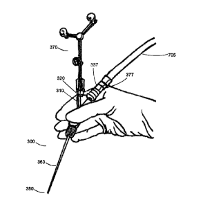
stickers, unique structures and patterns or glass spheres, which utilize
optical detectors.
Alternatively, object markers can utilize electromagnetic (EM) or radio
frequency (RF)
signals, which are detected by antennas. Optical detectors require a line-of-
sight
between the object marker and detector during operation, but are not subject
to noise
and distortion from environmental influences that electrical detection and
emission
systems are subject to.
[0006] In some cases, it can be difficult to incorporate a tracking
device on a
surgical instrument, especially instruments with flexible portions or with
multiple
configurations. For example, if the tracking device is positioned in a handle
or proximal
region of the instrument and the distal tip moves or is moved relative to the
handle, the
distal tip can no longer be accurately tracked. Electromagnetic navigation
systems have
partly overcome the difficulty of tracking flexible tips and multiple
configurations by using
a flexible membrane over the tip to connect the distal tracking device with
the system on
2
Date recue/Date received 2023-04-21
the handle. However, this does not overcome the problem of multiple
configurations in
which the tip is swiveled about the handle or when the tip is exchangeable.
[0007] An important surgical tool is a suction device, which can be
used for tissue
retention, resection and removal of fluids. A suction device typically
includes a handle
portion and tip portion. The tip portion can be any one of multiple
configurations, such
as different lengths, angles and diameters, and may be removable so it can be
swapped
out to provide the most appropriate configuration for the surgical procedure.
The
multiple configurations of the tip present challenges to tracking the distal
end of the tip
through a tracking device on the handle, because the relative positions of the
distal end
of the tip and handle are different for each configuration. The present
disclosure
attempts to solve this problem to provide a suction device that is trackable
over multiple
configurations and exchangeable tips.
SUMMARY
[0008] An object of the present disclosure is to provide methods and
devices for
tracking suction tools using surgical navigation systems or positional
tracking systems.
Thus by one broad aspect of the present disclosure, a tracked suction device
is
provided for use in a medical procedure comprising: an elongated tubular
handle with a
central passage, a main tube having a first proximal end, a distal end, and a
flattened
section with a suction-regulating orifice communicating with the central
passage, and an
entrance tube extending from the main tube having a second proximal end; an
elongated tip, having a hollow tubular body, a tip distal end, and a tip
proximal end
3
Date recue/Date received 2023-04-21
detachably connected to the main tube distal end; and a tracking mechanism
detachably connected to the handle first or second proximal end, for tracking
the tip
distal end, wherein the flattened section of the main tube lies in a plane
defined by the
main tube and the entrance tube, and the handle first and second proximal ends
may be
connected to the tracking mechanism or a suction hose.
[0009] By another broad aspect of the present disclosure, a method is
provided for
tracking the position of a tracked suction device in a medical procedure,
comprising:
attaching a tip to a handle in one of a plurality of fixed positions;
attaching a tracking
mechanism to the handle in one of a plurality of fixed positions; calibrating
the position
of the tip distal end with a positional tracking system using the tracking
mechanism;
positioning the tracking markers of the tracked suction device in view of the
tracking
source (optical camera) of the positional tracking system to be tracked; and
tracking a
position of the distal end of the tip of the suction device.
[0010] A further understanding of the functional and advantageous
aspects of the
disclosure can be realized by reference to the following detailed description
and
drawings.
BRIEF DESCRIPTION OF THE DRAWINGS
[0011] FIG. 1 illustrates systems and equipment of an exemplary
neurosurgical
procedure in accordance with example embodiments of the present disclosure.
[0012] FIG. 2 illustrates exemplary tracked instruments in accordance
with example
embodiments of the present disclosure.
[0013] FIG. 3 illustrates an assembled and exploded view of an
exemplary tracked
4
Date recue/Date received 2023-04-21
suction device in accordance with example embodiments of the present
disclosure.
[0014] FIG. 4 illustrates a perspective and cross-sectional view of an
attachment
mechanism for attaching a tip to a handle in accordance with example
embodiments of
the present disclosure .
[0015] FIG. 5 further illustrates an attachment mechanism for attaching a
tip to a
handle in accordance with example embodiments of the present disclosure .
[0016] FIG. 6 illustrates a perspective and side view of attachment
fittings for
attaching a handle to a suction hose and tracking device in accordance with
example
embodiments of the present disclosure.
[0017] FIG. 7 illustrates a suction hose connection to a handle in
accordance with
example embodiments of the present disclosure.
[0018] FIG. 8 illustrates an attachment mechanism for attaching a
handle to a
tracking device in accordance with example embodiments of the present
disclosure.
[0019] FIG. 9 illustrates use of a tracked suction device in accordance
with example
embodiments of the present disclosure.
[0020] FIG. 10 illustrates a perspective view of a tracked instrument
shown in FIG. 3
inserted into a calibration apparatus in accordance with example embodiments
of the
present disclosure.
DETAILED DESCRIPTION
[0021] Various embodiments and aspects of the disclosure will be
described with
reference to details discussed below. The following description and drawings
are
illustrative of the disclosure and are not to be construed as limiting the
disclosure.
5
Date recue/Date received 2023-04-21
Numerous specific details are described to provide a thorough understanding of
various
embodiments of the present disclosure. However, in certain instances, well-
known or
conventional details are not described in order to provide a concise
discussion of
embodiments of the present disclosure.
[0022] As used herein, the terms "comprises" and "comprising" are to be
construed
as being inclusive and open ended, and not exclusive. Specifically, when used
in the
specification and claims, the terms "comprises" and "comprising" and
variations thereof
mean the specified features, steps or components are included. These terms are
not to
be interpreted to exclude the presence of other features, steps or components.
[0023] As used herein, the term "exemplary" means "serving as an example,
instance, or illustration," and should not be construed as preferred or
advantageous
over other configurations disclosed herein.
[0024] As used herein, the terms "about" and "approximately" are meant
to cover
variations that may exist in the upper and lower limits of the ranges of
values, such as
variations in properties, parameters, and dimensions. Unless otherwise
specified, the
terms "about" and "approximately" mean plus or minus 25 percent or less.
[0025] It is to be understood that unless otherwise specified, any
specified range or
group is as a shorthand way of referring to each and every member of a range
or group
individually, as well as each and every possible sub-range or sub -group
encompassed
therein and similarly with respect to any sub-ranges or sub-groups therein.
Unless
otherwise specified, the present disclosure relates to and explicitly
incorporates each
and every specific member and combination of sub-ranges or sub-groups.
[0026] As used herein, the term "on the order of', when used in
conjunction with a
6
Date recue/Date received 2023-04-21
quantity or parameter, refers to a range spanning approximately one tenth to
ten times
the stated quantity or parameter.
[0027] Unless defined otherwise, all technical and scientific terms
used herein are
intended to have the same meaning as commonly understood to one of ordinary
skill in
the art. Unless otherwise indicated, such as through context, as used herein,
the
following terms are intended to have the following meanings:
[0028] As used herein, the phrase "access corridor" or "access port"
refers to a
cannula, conduit, sheath, port, tube, or other structure that is insertable
into a subject, in
order to provide access to internal tissue, organs, or other biological
substances. In
some embodiments, an access port may directly expose internal tissue, for
example, via
an opening or aperture at a distal end thereof, and/or via an opening or
aperture at an
intermediate location along a length thereof. In other embodiments, an access
port may
provide indirect access, via one or more surfaces that are transparent, or
partially
transparent, to one or more forms of energy or radiation, such as, but not
limited to,
electromagnetic waves and acoustic waves.
[0029] As used herein, the phrase "intraoperative" refers to an action,
process,
method, event or step that occurs or is carried out during at least a portion
of a medical
procedure. Intraoperative, as defined herein, is not limited to surgical
procedures, and
may refer to other types of medical procedures, such as diagnostic and
therapeutic
procedures.
[0030] As used herein, the phrase "navigation system" refers to a
system that
assists in surgery by providing previously acquired imaging information during
surgery
to visualize tissue morphology and locate target areas. Navigation systems may
also be
7
Date recue/Date received 2023-04-21
used to track surgical instruments and their location within the tissue during
surgery,
typically incorporating information from previously acquired imaging data.
[0031] As used herein, the phrase "positional tracking system" refers
to a computer-
implemented system that tracks the position of surgical instruments during
surgery. A
positional tracking system may be incorporated in a navigation system or may
function
independently of a navigation system. Where embodiments of the present
disclosure
refer to a navigation system, an independent positional tracking system may be
alternately used.
[0032] Embodiments of the present disclosure provide suction devices
that are
insertable into a subject or patient for manipulation of internal tissues, and
methods of
use thereof. Some embodiments of the present disclosure relate to minimally
invasive
medical procedures that are performed via an access port, whereby surgery,
diagnostic
imaging, therapy, or other medical procedures are performed based on access to
internal tissue through the access port.
[0033] Several embodiments of the present disclosure seek to address the
aforementionedinadequacies of existing devices and methods to support surgical
procedures utilizing surgical tools.
[0034] Minimally invasive brain surgery using access ports is a method
of
performing surgery on brain tumors previously considered inoperable. One
object of the
present invention is to provide a system and method to assist in minimally
invasive brain
surgery. To address intracranial surgical concerns, navigation systems and
robotic
positioning systems have been developed for port-based surgery. Referring to
FIG. 1
and FIG. 2, port 100 comprises of a cylindrical assembly formed of an outer
sheath.
8
Date recue/Date received 2023-04-21
Port 100 may accommodate an introducer which is an internal cylinder that
slidably
engages the internal surface of port 100. The introducer may have a distal end
in the
form of a conical atraumatic tip to allow for insertion into the sulcal folds
of the brain.
Port 100 has a sufficient diameter to enable bimanual manipulation of surgical
tools
within its annular opening such as suctioning devices, scissors, scalpels, and
cutting
devices as examples.
Surgical Positional Tracking System
[0035] Surgical positional tracking systems are computer-implemented
systems that
track the position of surgical tools, such tools including but not limited to
access
corridors, pointers and suction devices. Positional tracking systems may track
the
location of surgical tools with respect to a patient and may be used in
conjunction with
medical images of the patient and site of surgery. An example of a surgical
positional
tracking system is a navigation system, as described below.
[0036] The description below makes reference to the brain of a patient 102
as an
example of tissue to which the techniques herein may be applied. It will be
understood,
however, that those techniques may also be applied to a wide variety of other
tissues.
Thus, when the brain of patient 102 is mentioned below, it is simply an
example of the
various tissues in connection with which the systems and methods herein may be
implemented. In particular, suction tools are widely used in surgery, thus a
tracked
suction device will be useful in virtually all types of navigated procedures.
Other
examples of navigated procedures wherein a tracked suction device would be
useful
are spine, ENT (ear nose throat), orthopedic and cardiac surgery.
9
Date recue/Date received 2023-04-21
[0037] FIG. 1 illustrates systems and equipment of an exemplary
neurosurgical
procedure. Referring to FIG. 1, an exemplary navigation system 105 which may
be used
in surgery is shown. A surgeon 107 conducts a surgery on a patient 102 in an
operating
room environment. The medical navigation system 105 is illustrated including
an
equipment tower 110, supporting a computing device (not shown) such as a
desktop
computer, as well as one or more displays 111 connected to the computing
device for
displaying images provided by the computing device.
[0038] Equipment tower 110 also supports a tracking system 113.
Tracking system
113 is generally configured to track the positions of one or more tracking
markers 120
mounted on access port 100, or any of the above-mentioned surgical tools, or
any
combination thereof. Such markers may also be mounted on patient 102, for
example at
various points on the head 145 of patient 102. Tracking system 113 may
therefore
include a camera (e.g. a stereo camera) and a computing device (either the
same
device as mentioned above or a separate device) configured to locate the
tracking
markers in the images captured by the camera, and determine the spatial
positions of
those markers within the operating theatre. The spatial positions may be
provided by
tracking system 113 to the computing device in equipment tower 110 for
subsequent
use.
[0039] The nature of the markers and the camera are not particularly
limited. For
example, the camera may be sensitive to infrared (IR) light, and tracking
system 113
may include one or more IR emitters (e.g. IR light emitting diodes (LEDs)) to
shine IR
light on the markers. In other examples, marker recognition in tracking system
113 may
be based on radio frequency (RE) radiation, visible light emitted from devices
such as
Date recue/Date received 2023-04-21
pulsed or un-pulsed LEDs, electromagnetic radiation other than IR or visible
light, and
the like. For RF and electro-magnetic (EM)-based tracking, each object can be
fitted
with markers having signatures unique to that object, and tracking system 113
can
include antennae rather than the above mentioned camera. Combinations of the
above
may also be employed.
[0040] Each tracked object generally includes three or more markers
fixed at
predefined locations on the object. The predefined locations, as well as the
geometry of
each tracked object, are configured within tracking system 113, and thus
tracking
system 113 is configured to image the operating theatre, compare the positions
of any
visible markers to the pre-configured geometry and marker locations, and based
on the
comparison, determine which tracked objects are present in the field of view
of the
camera, as well as what positions those objects are currently in.
[0041] Also shown in FIG. 1 is an automated articulated arm 150, also
referred to as
a robotic arm or a positioning arm, carrying an external scope 160 (i.e.
external to
patient 102). External scope 160 may be positioned over access port 100 by
robotic arm
150, and may capture images of the brain of patient 102 for presentation on
display
111. The movement of robotic arm 150 to place external scope 160 correctly
over
access port 100 may be guided by tracking system 113 and the computing device
in
equipment tr 110. The images from external scope 160 presented on display 111
may
be overlaid with other images, including images obtained prior to the surgical
procedure.
The images presented on display 111 may also display virtual models of
surgical
instruments present in the field of view of tracking system 113 (the positions
and
orientations of the models having been determined by tracking system 113 from
the
11
Date recue/Date received 2023-04-21
positions of the markers mentioned above). Alternatively, a tracking camera
may be
affixed to a monitor or camera cart and connected directly to a positional
tracking
system, which receives the tracking camera information and analyzes it.
Tracking Markers
[0042] FIG. 2 illustrates exemplary tracked instruments with which
aspects of the
present application may be applied. Referring to FIG. 2, active or passive
tracking
markers 220 may be placed on the port 100 and/or any medical instruments 230
to
determine the location of these objects using the tracking system 113 and
navigation
system 105. These markers 220 may be passive reflective spheres configured to
be
seen by the stereo camera of the tracking system 113 to provide identifiable
points for
tracking. A tracked instrument in the tracking system is typically defined by
a grouping
of markers 220, which are used to determine the spatial position and pose of
the
volume of the tracked instrument in three dimensions. Typically, in known
exemplary
.. tracking systems a minimum of three spheres are required on a tracked tool
to define
the instrument.
[0043] In a preferred embodiment, the navigation system 105 or
positional tracking
system may utilize reflective sphere markers in combination with a stereo
camera
system, to determine spatial positioning and pose of the medical instruments
and other
objects within the operating theater. Differentiation of the types of objects
and their
corresponding virtual geometric volumes may be determined by the specific
orientation
of the reflective spheres relative to one another giving each virtual object
an individual
identity within the navigation system 105 or positional tracking system. This
allows the
12
Date recue/Date received 2023-04-21
navigation system 105 or positional tracking system to identify the medical
instrument
230 or other object and its corresponding virtual overlay representation. The
location of
the markers also provides other useful information to the navigation system
105 or
positional tracking system, such as the object's central point, central axis,
orientation,
and other information related to the object.
Trackable Suction Tool
[0044] Referring to FIG. 3, an example embodiment of a suction tool 300
that may
be tracked during surgical procedures is shown. The suction tool 300 is shown
assembled in the left panel and exploded in the right panel. A hollow
substantially
cylindrical handle 310 includes a main tube 315 with a first proximal end 320
and a
distal end 330. The main tube 315 of the handle 310 has an entrance tube 335
extending from the main tube 315 to a second proximal end 337. The main tube
315
and the entrance tube 335 extending from the main tube 315 may form a Y-shaped
handle.
[0045] The handle includes a tapered elongated slot 340, such as a tear-
shaped
orifice in the wall of the handle, which is widest at the proximal end and
narrowest at the
distal end, for controlling the amount of suction provided at the distal end
of the suction
tool tip. In a preferred embodiment, the handle 310 has a flattened portion
345 around
the elongated slot 340, and the flattened portion lies in the plane defined by
the main
tube 315 and the entrance tube 335.
[0046] The handle distal end 330 includes splines and a thread for
connection to a
tip, as described in further detail below. The handle first proximal end 320
and second
13
Date recue/Date received 2023-04-21
proximal end 337 both include ribs for connection to a suction tube and
splines and a
thread for connection to a tracking mechanism.
[0047] The handle distal end 330 is connected to a proximal end 350 of
a tubular
hollow tip 360. The tip proximal end 350 has splines that are complementary
and
interlock with the splines on the handle distal end 330, thus providing
specific rotational
angles of the tip 360 relative to the plane of the handle 310. The connection
is secured
by a semi-captive nut 365.
[0048] A tracking mechanism 370, such as a reference tree, is attached
to the first
proximal end 320 or second proximal end 337 of the handle 310. The tracking
mechanism 370 includes tracking markers, such as reflective sphere markers.
The
tracking mechanism 370 has splines complementary to the splines on the first
and
second proximal ends of the handle, providing fixed rotational positions of
the tree
relative to the plane of the handle 310 defined by the main tube 315 and the
entrance
tube 335. The attachment of the tracking mechanism 370 to the handle 310 is
secured
with a captive nut 375. A suction tube (not shown) may be attached to the
first or
second proximal end 320, 337 of the handle 310 by sliding the suction tube
over the ribs
377.
[0049] The handle 310 can be used to hold and manipulate the suction
tool 300,
such that the tip distal end 380 is directed to the tissue, for example for
holding or
resecting tissue or suctioning fluids. The tip distal end 380 is also blunted
to minimize
trauma to tissue while in use. The tracking mechanism 370 provides an optical
marker
for tracking the position of the suction tool 300 and provides position
information to the
tracking system 113.
14
Date recue/Date received 2023-04-21
[0050] The tip 360 can be removed from the handle 310 by rotating and
unscrewing
the semi-captive nut 365 until it is released from the threads of the distal
end of the
handle 330; tips of different configurations can thereby be exchanged and used
with the
suction tool. The tip 360 may be one of several different lengths, angles and
diameters.
Thus, by removing and replacing the tip 360, the suction tool may have
different
configurations. Information on the parameters for a given tip, such as tip
length,
diameter and angle, can be entered and stored by the computing device of the
navigation system 105, and calibrated using the calibration apparatus (as
described for
FIG. 10), so that for each tip 360 used with the suction tool 300, the
position of the tip
distal end 380 is accurately tracked.
Tip Attachment Mechanism
[0051] Referring to FIG. 4, a perspective view of the tip 360 and semi-
captive nut
365 is shown in the top panel and a cross-sectional view of the nut 365
threaded onto
the tip 360 is shown in the lower panel.
[0052] The semi-captive nut 365 has two internal threads: a left-hand
thread and a
right-hand thread. In the embodiment shown, a left-hand internal thread 405
engages
the nut 365 onto the tip 360 to prevent the nut from slipping off the tip
during assembly
and disassembly of the tip onto the handle. The nut seating position 410
provides free
.. rotation of the nut 365 around the tip 360 without removing the nut from
the tip. A larger
diameter right-hand internal thread 415 is used to secure the tip 360 to the
handle (not
shown).
[0053] Referring to FIG. 5 a perspective view of the attachment
mechanism for the
Date recue/Date received 2023-04-21
tip 360 to the handle 310 is shown in the left panel and the secured tip and
handle are
shown in the right panel. External splines 505 on the tip proximal end 350 are
complementary to internal splines 510 on the handle distal end 330. The tip
external
splines 505 fit into the handle internal splines 510 to prevent rotation and
hold the tip at
.. a fixed position after securing the tip with the nut 365. In an embodiment,
the tip and
handle ends have 18 splines, allowing for 18 rotational positions (200 apart)
of the tip
around the axis of the handle.
[0054] Further referring to FIG. 4 and FIG. 5, to attach the tip to the
handle, the
semi-captive nut 365 is slid onto the tip 360 until the internal left-hand
thread 405 is
engaged with the thread on the tip proximal end 350. The nut 365 is threaded
onto the
tip 360 until the tip threads sit in the seating position 410, so the nut is
attached to the
tip but able to freely rotate. The tip 360 is then inserted into the handle
310 fully to mate
the internal 510 and external splines 505. The nut 365 is then threaded in the
opposite
direction 515 onto the handle distal end 330 until the tip 360 is fully seated
and secured.
Tube and Tracking Mechanism Attachment
[0055] Referring to FIG. 6, a perspective and side view of the fittings
for attaching
the suction tube or hose or the tracking mechanism, such as a reference tree,
to the
handle are shown. The first and second proximal ends 320, 337 of the handle
310 both
include ribs 377 for attachment of a suction tube (not shown in FIG. 6) and
adjacent
threads 610 for a tracking device nut. The tube is attached up to the start of
the threads
for smaller tube diameters and, as shown in FIG. 7, the tube 705 is attached
over the
threads 610 for larger tube diameters.
16
Date recue/Date received 2023-04-21
[0056] Referring to FIG. 8, attachment fittings for a tracking
mechanism, such as a
reference tree, 370 at the first or second proximal end of the handle 310 is
shown. The
top panel of FIG. 8 illustrates an unattached tracking mechanism 370 and
handle 310.
The tracking mechanism end has splines 805 that fit into complementary splines
810 on
.. the handle proximal end, which prevent rotation after the tracking
mechanism is secured
with the captive nut 375. In an embodiment, the tracking mechanism includes 16
external splines and the handle includes 16 internal splines, thereby allowing
16
rotational positions, 22.5 apart, to maximize flexibility with the tool
positioning relative to
the camera of the navigational system. The middle panel of FIG. 8 illustrates
the
tracking mechanism 370 and handle 310 with fully seated splines 805, 810 prior
to
threading the captive nut 375 onto the threads 610, and the lower panel of
FIG. 8
illustrates the tracking mechanism and handle fully secured with the captive
nut 375
screwed onto the threads.
[0057] Referring back to FIG. 3, the attachment mechanisms as described
provide
for multiple positions of the tip 360 relative to the tracking mechanism 370
around the
circumference of the handle 310, allowing easier use for right and left hand
users and
for different positions of an angled tip without obstructing the line of sight
for the tracking
mechanism 370. Multiple positions of the tracking mechanism 370 are also
enabled by
placement on either the first or second proximal end of the handle 320, 337,
and by
rotating the tracking mechanism position relative to the handle by locking
into different
spline positions. The rotatable tracking mechanism 370 affords rotation of the
tracking
mechanism to optimize line of sight and provide a preferred working
configuration, while
maintaining a fixed rotational axis of the tracking mechanism 370 relative to
the plane of
17
Date recue/Date received 2023-04-21
the handle defined by the handle main tube 315 and entrance tube 335. The
attachment
mechanisms also allow different tracking mechanism 370 configurations to be
switched
out for unique identification of one or more suction tools/medical instruments
in the
same surgical space.
[0058] FIG. 9 illustrates a tracked suction tool held in the hand of a
user. Referring
to FIG. 9, a suction tool 300, such as that illustrated in FIG. 3, is held by
user (i.e., a
surgeon), with the tracking markers of the tracking mechanism 370 providing
positional
information of the suction tool 300 to the tracking system 113 (FIG. 1), so
the positional
tracking system or navigation system 105 (FIG. 1) is able to calculate and
display the
position of the suction tool tip 360 to the surgeon. Suction tool 300 is
connected to
suction tube 705 at the handle first or second proximal end 320, 337. The rib
aspect 377
of handle proximal ends 320, 337 ensures for a tight and secure fit with
suction tube
705.
[0059] The suction tool 300 is registered in the positional tracking
system or
navigation system 105 (FIG. 1) and prior to use is calibrated to provide
accurate
registration of the tracking markers 220 (FIG. 2) with the tip distal end 380.
Calibration
ensures that a current configuration of the suction tool 300 is accurately
registered in
the positional tracking system or navigation system 105 (FIG. 1), including
changes
such as different tips, adjustment of the tracking mechanism 370, user's grip
of the
suction tool, and deformations of the tip 360. A vacuum tube or suction tube
705 is
shown connected to the proximal end 320, 337 of the handle 310.
[0060] As seen in FIGS 3 to 9, the suction tool 300 is equipped with an
exchangeable hollow tip 360. The tip may include a bend between the proximal
end and
18
Date recue/Date received 2023-04-21
distal end. The bend angle may range between 60 and 180 degrees. The tip 360
may
also be rigid or malleable. A rigid tip is distinguished from a malleable tip
by an external
marking to enable the surgeon to easily distinguish them. The malleable tip
can be
further bent by the user (surgeon) during the medical procedure.
[0061] The hollow tip may range in length between 50 mm and 250 mm in
length
and may have a diameter between 3 and 34 FR.
[0062] The above embodiments allow a choice of which hand to use to
hold the
suction tool, specific holding angles to be attained, the reference tree to be
manipulated
for the best view, and allow suction tools to be customized and replaced with
accuracy
and minimum inconvenience.
Calibration of Tracked Medical Instrument
[0063] In order to provide the dimensions of the tracked suction tool
300, the
dimensions of the tracked suction tool may be registered and stored in the
navigation
system 105 or positional tracking system, and subsequently calibrated before
use in
surgery using procedures known in the art. An exemplary calibration procedure
is
provided below.
[0064] Referring to FIG. 10, a a tracked suction tool 310 is shown with
a calibration
apparatus 1010. The techniques for calibrating a tracked instrument can be
found in
international application CA2014051004 titled "CALIBRATION APPARATUS FOR A
MEDICAL TOOL" which is incorporated by reference herein in its entirety.
[0065] The suction tool 300 and the calibration apparatus 1010 are
typically used in
conjunction with a positional tracking system, such as the medical navigation
system
19
Date recue/Date received 2023-04-21
105. The calibration apparatus 1010 includes a frame 1020, at least one frame
tracking
marker 1030 attached to the frame 1020, and a reference point 1040 formed on
the
frame 1020. In one example, the reference point 1040 may be a divot that is of
an
appropriate shape for securely receiving the distal end of the suction tool
tip 380. For
the purposes of this example, the reference point 1040 will be referred to
throughout as
a divot 1040. The divot 1040 may provide a known spatial reference point
relative to
the frame tracking markers 1030. For example, the medical navigation system
105 may
have data saved therein so that the medical navigation system knows the
position in
space of a floor of the divot 1040 relative to the tracking markers 1030 to a
high degree
of accuracy. In one example, a high degree of accuracy may refer to a
tolerance of
0.08mm, but any suitable tolerance may be used according to the design
criteria of a
particular application.
[0066] In the example shown, the calibration apparatus 1010 has four
passive
reflective tracking spheres, but any suitable number of tracking markers 1030
may be
used and any suitable type of tracking marker may be used according to the
design
criteria of a particular application, including an active infrared (IR)
marker, an active light
emitting diode (LED), and a graphical pattern. When passive reflective
tracking spheres
are used as the tracking makers 1030, typically at least three tracking
markers will be
attached to a same side of the frame 1020. Likewise, when a suction tool 300
having
passive reflective tracking spheres is used in conjunction with the
calibration apparatus
1010, the suction tool will typically have at least three tracking markers 220
attached
thereto.
Date recue/Date received 2023-04-21
[0067] Referring to FIG. 10, left panel, the distal end 380 of the
suction tool 300 is
inserted into the calibration apparatus 1010 for a reading by the medical
navigation
system 105. When the suction tool 300 is inserted into the calibration
apparatus 1010,
the position of the distal end 380 of the suction tool 300 relative to the
tracking markers
220 that the medical navigation system 105 is seeing (e.g., using the camera
of the
tracking system 113) may be learned and saved by the navigation system 105.
The
distal end 380 of the suction tool 300 is then inserted onto the divot 1040
for verification
of the localization of the suction tool 300. Since the medical navigation
system 105
knows the precise dimensions of the calibration apparatus 1010, the medical
navigation
system 105 learns the dimensions of the suction tool 300. In other words, the
position
of the floor of the divot 1040 relative to the tracking markers 220 that the
medical
navigation system 105 is seeing (e.g., using the camera of the tracking system
113) is
known. Other calibration devices and methods may be used to localize the
distal end
380 of the suction tool 300 relative to the tracking markers 220.
The specific embodiments described above have been shown by way of
example, and it should be understood that these embodiments may be susceptible
to
various modifications and alternative forms. It should be further understood
that the
claims are not intended to be limited to the particular forms disclosed, but
rather to
cover all modifications, equivalents, and alternatives falling within the
spirit and scope of
this disclosure.
21
Date recue/Date received 2023-04-21