Note: Descriptions are shown in the official language in which they were submitted.
CA 03016894 2018-09-06
WO 2017/156058
PCT/US2017/021258
GITR ANTIBODIES, METHODS, AND USES
CROSS-REFERENCE TO RELATED APPLICATIONS
This application claims the benefit of United States Provisional Application
Serial
Number 62/305,270, filed 8 March 2016 and United States Provisional
Application Serial
Number 62/407,106, filed 12 October 2016. The entire content of the
aforementioned
applications is incorporated herein by reference in its entirety.
SEQUENCE LISTING
The instant application contains a Sequence Listing which has been submitted
electronically in ASCII format and is hereby incorporated by reference in its
entirety.
Said ASCII copy, created on February 21, 2017, is named JBI5082USNP SL.txt and
is
92,231 bytes in size.
TECHNICAL FIELD
The disclosure provided herein relates to monoclonal antibodies that
specifically bind glucocorticoid-induced tumor necrosis factor receptor (GITR)
and
methods of producing and using the described antibodies.
BACKGROUND
Glucocorticoid-induced TNFR-related protein (GITR; also AITR, TNFRSF18,
or CD357), a member of the TNFR superfamily, is expressed in many components
of
the innate and adaptive immune system and stimulates both acquired and innate
immunity (Nocentini Get al., (1994) PNAS 94: 6216-6221; Hanabuchi S et al.,
(2006)
Blood 107:3617-3623; Nocentini G & Riccardi C (2005) Eur J Immunol 35: 1016-
1022; Nocentini G et al., (2007) Eur J Immunol 37: 1165-1169). It is expressed
in
several cells and tissues, including T, B, dendritic (DC) and Natural Killer
(NK) cells
and is activated by its ligand, GITR-L, mainly expressed on Antigen Presenting
Cells
(APCs), on endothelial cells, and also in tumor cells.
CA 03016894 2018-09-06
WO 2017/156058
PCT/US2017/021258
The GITR-GITRL system participates in the development of
autoimmune/inflammatory responses and potentiates response to infection and
tumors.
For example, treating animals with GITR-Fc fusion protein ameliorates
autoimmune/inflammatory diseases while GITR triggering is effective in
treating viral,
bacterial, and parasitic infections, as well in boosting immune response
against tumors
(Nocentini G et al., (2012) Br J Pharmacol 165: 2089-99). These effects are
due to
several concurrent mechanisms including: co-activation of effector T-cells,
inhibition
of regulatory T (Treg) cells, modulation of NK and dendritic cell function,
activation of
macrophages, and regulation of the extravasation process. The membrane
expression
of GITR is increased following T cell activation (Hanabuchi S et al, (2006)
supra;
Nocentini G & Riccardi C supra). Its triggering coactivates effector T
lymphocytes
(McHugh RS et al, (2002) Immunity 16: 311-323; Shimizu J et al, (2002) Nat
Immunol
3: 135-142; Roncheti S et al, (2004) Eur J Immunol 34: 613-622; Tone M et al,
(2003)
PNAS 100: 15059-15064). GITR activation increases resistance to tumors and
viral
infections, is involved in autoimmune/inflammatory processes and regulates
leukocyte
extravasation (Nocentini G & Riccardi C (2005) supra; Cuzzocrea S et al,
(2004) J
Leukoc Biol 76: 933-940; Shevach EM & Stephens GL (2006) Nat Rev Immunol 6:
613-618; Cuzzocrea S et al, (2006) J Immunol 177: 631-641; Cuzzocrea S et al,
(2007)
FASEB J 21 :117-129).
Human GITR is expressed at very low levels in peripheral (non-activated) T
cells. After T cell activation, GITR is strongly up-regulated for several days
in both
CD4+ and CD8+ cells (Kwon B et al, (1999) J Biol Chem 274: 6056-6061; Gurney
AL
et al, (1999) Curr Biol 9: 215-218; Ronchetti S et al, (2004) supra; Shimizu J
et al,
(2002) supra; Ji HB et al, (2004) supra; Ronchetti S et al, (2002) Blood 100:
350-352;
Li Z et al, (2003) J Autoimmun 21: 83- 92), with CD4+ cells having a higher
GITR
expression than CD8+ cells (Kober Jet al, (2008) Eur J Immunol 38(10): 2678-
88;
Bianchini R et al, (2011) Eur J Immunol 41(8): 2269-78).
The role of human GITR in modulating immune responses indicates that it may
be a suitable target for antibody-based therapy against diseases such as
cancer.
Antibodies against GITR are described (e.g. in W0200610502, W02011028683,
W02015031667, W020150353637, W02015187835, W02015184099, U59255151
and US9255152), but there is an ongoing need for novel agents and methods for
modulating GITR activity against diseases, such as cancer.
2
CA 03016894 2018-09-06
WO 2017/156058
PCT/US2017/021258
SUMMARY
Provided herein are antibodies that specifically bind to GITR and antigen-
binding fragments thereof Also described are related polynucleotides capable
of
encoding the provided GITR-specific antibodies and antigen-binding fragments,
cells
expressing the provided antibodies and antigen-binding fragments, as well as
associated
vectors and detectably labeled antibodies and antigen-binding fragments. In
addition,
methods of using the provided antibodies and antigen-binding fragments are
described.
For example, given the role GITR plays in modulating an immune response, the
GITR-
specific antibodies have utility in treating a variety of GITR-related
diseases or
disorders in which it is desirable to modulate an immune response. For
example, the
GITR specific antibodies can be used in a variety of immunotherapy
applications, such
as the treatment of a variety of cancers.
GITR-Specific Antibodies
Described herein are isolated antibodies and antigen-binding fragments
specific for
GITR. In some embodiments, the GITR-specific antibodies and antigen-binding
fragments bind human GITR. In some embodiments, the GITR-specific antibodies
and
antigen-binding fragments bind human GITR and cynomolgus monkey GITR. In some
embodiments, the GITR-specific antibodies and antigen-binding fragments bind
to an
epitope including one or more residues from the GITR extracellular domain
(ECD) as
defined in SEQ ID NO:59. This GITR-specific antibody or antigen-binding
fragment
may bind to GITR with a binding affinity of 30 nM or less, may induce an
increase in
luciferase expression in an NF-KB luciferase gene assay and may induce ADCC in
vitro
with an EC50 of 67 ng/mL or less.
Table 1 provides a summary of examples of some GITR-specific antibodies
described
herein:
Table 1. CDR sequences of mAbs generated against human GITR
(SEQ ID NO:)
ID HC-CDR1 HC-CDR2 HC-CDR3 LC-CDR1 LC-CDR2 LC-CDR3
TRGB5 GFTFSGYW ISGSGGST AKDFYWDAFDY (12) QSVSSY (28) DAS (32)
QQRSNWPLT
(1) (5) (35)
3
CA 03016894 2018-09-06
WO 2017/156058
PCT/US2017/021258
ID HC-CDR1 HC-CDR2 HC-CDR3 LC-CDR1 LC-CDR2 LC-CDR3
TRGB14 GFTFSSYA ISGSGGST AKPIRGLDY (13) QSVNNF (29) DAS
(32) QQGFNAPLT
(2) (5) (36)
TRGB20 GFTFSGYW ISSDGGSK AKEVVYDHYAALDY QSVNSF (30) YAS (33) QQYIRWPLT
(1) (6) (14) (37)
TRGB23 GGTFSSYA IIPIFGTA (7) ARHGNWLITFNLDY QSVSSY (28) DAS (32)
QQRSNWPLT
(3) (15) (35)
TRGB25 GGTFSSYA IIPIFGTA (7) ARHRRFWLDY (16) QSVSSY (28) DAS
(32) QQRSNWPLT
(3) (35)
TRGB190
TRGB31 GYSFTSYW IDPSDSDT ARVFPYYGLVLDY QSVSSY (28) DAS
(32) QQRSNWPLT
(4) (8) (17) (35)
TRGB34 GYSFTSYW IYPGDSDT ARDYGWHDFDY (18) QSVSSY (28) DAS (32)
QQRSNWPLT
(4) (9) (35)
TRGB35 GYSFTSYW IDPGDSDT ARHRWSTSLLLDY QSVSSY (28) DAS
(32) QQRSNWPLT
(4) (10) (19) (35)
TRGB120 GGTFSSYA IIPIFGTA (7) ARPRRNTNELDY QSISSY (31) AAS (34)
QQSYSTPLT
(3) (20) (38)
TRGB127 GGTFSSYA IIPIFGNA ARHVYKRGVLNY QSISSY (31) AAS (34)
QQSYSTPLT
(3) (11) (21) (38)
TRGB134 GGTFSSYA IIPIFGTA (7) ARHRWGSGNLDY QSVSSY (28) DAS (32)
QQRSNWPLT
(3) (22) (35)
TRGB144 GGTFSSYA IIPIFGTA (7) ARHGFQRGYLDY QSVSSY (28) DAS (32)
QQRSNWPLT
(3) (23) (35)
TRGB153 GGTFSSYA IIPIFGTA (7) ARHAWLGHLDY (24) QSVSSY (28) DAS (32)
QQRSNWPLT
(3) (35)
TRGB159 GGTFSSYA IIPIFGTA (7) ARHGRNSGRLDY QSVSSY (28) DAS (32)
QQRSNWPLT
(3) (25) (35)
TRGB160 GFTFSNYW ISGSGGST AKDFYWDSFDY (26) QSVSSY (28) DAS (32)
QQRSNWPLT
(27) (5) (35)
TRGB191
TRGB191
CLF
TRGB162 GGTFSSYA IIPIFGNA ARHVYKRGVLNY QSVSSY (28) DAS (32)
QQRSNWPLT
(3) (11) (21) (35)
In some embodiments are provided a GITR-specific antibody, or an antigen-
binding
fragment thereof, comprising a heavy chain comprising a CDR1, a CDR2, and a
CDR3
of any one of the antibodies described in Table 1 and a light chain comprising
a CDR1,
a CDR2, and a CDR3 of any one of the antibodies described in Table 1.
The IgG class is divided in four isotypes: IgGl, IgG2, IgG3 and IgG4 in
humans. They
share more than 95% homology in the amino acid sequences of the Fc regions but
show
major differences in the amino acid composition and structure of the hinge
region. The
Fc region mediates effector functions, such as antibody-dependent cellular
cytotoxicity
(ADCC) and complement-dependent cytotoxicity (CDC). In ADCC and ADCP, the Fc
region of an antibody binds to Fc receptors (FcgRs) on the surface of immune
effector
cells such as natural killers and macrophages, leading to the lysis or
phagocytosis of the
targeted cells. In CDC, the antibodies kill the targeted cells by triggering
the
4
CA 03016894 2018-09-06
WO 2017/156058
PCT/US2017/021258
.. complement cascade at the cell surface. The antibodies described herein
include
antibodies with the described features of the variable domains in combination
with any
of the IgG isotypes, including modified versions in which the Fc sequence has
been
modified to effect different effector functions.
In some embodiments, the antibodies comprise the CDRs of the antibodies
presented in
.. Table 1 above. In some embodiments the described antibodies are capable of
binding
to GITR with a dissociation constant of 30 nM or less as measured by surface
plasmon
resonance (SPR). In some embodiments the described antibodies are capable of
inducing an increase in luciferase expression in an NF--kB luciferase gene
assay. In
some embodiments the described antibodies are capable of inducing ADCC in
vitro
with an EC50 of 67 ng/mL or less.
In addition to the described GITR-specific antibodies and antigen-binding
fragments, also provided are polynucleotide sequences capable of encoding the
described antibodies and antigen-binding fragments. Vectors comprising the
described
polynucleotides are also provided, as are cells expressing the GITR-specific
antibodies
or antigen-binding fragments provided herein. Also described are cells capable
of
expressing the disclosed vectors. These cells may be mammalian cells (such as
293F
cells, CHO cells), insect cells (such as Sf7 cells), yeast cells, plant cells,
or bacteria
cells (such as E. coli). A process for the production of the described
antibodies or
antigen-binding fragments is also provided.
Methods of using GITR-Specific Antibodies
Methods of using the described GITR-specific antibodies or antigen-binding
fragments are also disclosed. Particular antibodies for use in the methods
discussed in
this section include those with the set of CDRs described for antibodies in
Table 1. For
example, the key role that GITR plays in an immune response makes it an
attractive
target for immunotherapy, including inducing or enhancing an immune response
against desired tumor antigens or pathogenic antigens (e.g., viruses and other
pathogenic organisms). As such, the GITR-specific antibodies have utility in
the
treatment of various cancers and infectious disease.
As noted above, GITR activation sends a co-activating signal to CD4+ and
CD8+ T cells and prevents suppression of an immune response by regulatory T
cells.
Thus, in one embodiment, a GITR-specific antibody is administered to inhibit
the
5
CA 03016894 2018-09-06
WO 2017/156058
PCT/US2017/021258
suppression of effector T cell activity by regulatory T cells. Such inhibition
can be
assayed by a variety of methods known in the art, including, for example, by
monitoring T cell proliferation, expression of known markers of activation, or
cytokine
secretion. In another embodiment, a GITR-specific antibody is administered to
a
subject to decrease the level of regulatory T cells, for instance the level of
tumor
regulatory T cells. In yet another embodiment, the activity of effector T
cells is induced
or enhanced by administering a GITR-specific antibody as provided herein.
Specific
assays for each of these methods are provided in the EXAMPLES.
GITR-Specific Antibody Kits
Described herein are kits including the disclosed GITR-specific antibodies or
antigen-
binding fragments thereof The described kits may be used to carry out the
methods of
using the GITR-specific antibodies or antigen-binding fragments provided
herein, or
other methods known to those skilled in the art. In some embodiments the
described
kits may include the antibodies or antigen-binding fragments described herein
and
reagents for use in detecting the presence of GITR in a biological sample.
Accordingly,
the described kits may include one or more of the antibodies, or an antigen-
binding
fragment(s) thereof, described herein and a vessel for containing the antibody
or
fragment when not in use, instructions for use of the antibody or fragment,
the antibody
or fragment affixed to a solid support, and/or detectably labeled forms of the
antibody
or fragment, as described herein.
Brief Description of the Drawings
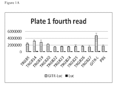
Figure 1A-1C. Agonist activity exhibited by the anti-GITR mAbs from the
traditional
screen panel. Data shown are the Cell-Titer Glo signal from cells that were
treated with
the indicated reagent after being transfected with either the NF-kB luciferase
reporter
gene only (Luc) or with both the GITR expression vector and the NF-kB
luciferase
reporter gene (GITR-Luc). Antibodies that produced a signal greater than that
found
with PBS treatment were preliminarily categorized as agonists.
Figure 2A-2E. Agonist activity exhibited by the anti-GITR mAbs from the next-
generation sequencing panel. Data shown are the Cell-Titer Glo signal from
cells that
6
CA 03016894 2018-09-06
WO 2017/156058
PCT/US2017/021258
were treated with the indicated reagent after being transfected with either
the NF-kB
luciferase reporter gene only or with both the GITR expression vector and the
NF-kB
luciferase reporter gene.
Figure 3A-3D. The effect of anti-GITR antibody ligation on NF--03 activity.
Results
shown are representative of SEAP activity as a measure of NF--kB activation in
HEK-
Blue NF--03 cells stably transfected with GITR. Cells were treated with
varying
concentrations of anti-GITR antibody in the absence (3A, 3B) or presence (3C,
3D) of
25 ng/mL of soluble GITR ligand. Media and CNT03930 were used as no antibody
and isotype antibody controls.
Figure 4A and 4B. The effect of anti-GITR antibodies on memory T cell
responses to
CMV and TT. Sero-reactive PBMCs were pulsed with CMV and TT antigen in the
absence or presence of anti-GITR antibodies, CNT03930 isotype control antibody
or
no antibody. Supernatant was collected and measured for the presence of IFNy.
Figure 5A and 5B. Single agent anti-tumor activity of a surrogate anti-GITR
antibody
(DTA-1) in the syngeneic MC38 colon carcinoma model. DTA-1 or an isotype rat
IgG2b was administered at 200 ug/mouse to animals on days indicated by black
arrowheads (n=10 per group) starting when tumor volumes reached 100mm3. DTA-1
treatment resulted in complete tumor regression in 5/10 animals.
Figure 6A-6C. Combination of surrogate anti-GITR antibody (DTA-1) and anti-PD-
1
antibody (RMP1-14) leads to synergistic anti-tumor activity in the syngeneic
MC38
colon carcinoma model. Isotype rat IgG2b (6A), DTA-1 (6B) or DTA-1 + RMP1-14
(6C) was administered at 100 ug/antibody/mouse to animals on days indicated by
black
arrowheads (n=10 per group) starting when tumor volumes reached 200mm3.
Combination treatment resulted in tumor regressions in 5/10 animals.
Figure 7A-7C. Combination of surrogate anti-GITR antibody (DTA-1) and anti-
CTLA-4 antibody (9D9) leads to synergistic anti-tumor activity in the
syngeneic MC38
colon carcinoma model. Isotype rat IgG2b (7A), DTA-1 (7B) or DTA-1 + 9D9 (7C)
was administered at 100 ug/antibody/mouse to animals on days indicated by
black
7
CA 03016894 2018-09-06
WO 2017/156058
PCT/US2017/021258
.. arrowheads (n=10 per group) starting when tumor volumes reached 200mm3.
Combination treatment resulted in tumor regressions in 3/10 animals.
Figure 8A-8D. Combination of surrogate anti-GITR antibody (DTA-1) and anti-OX-
40
antibody (0X-86) is better than OX-86 alone in the syngeneic MC38 colon
carcinoma
model. Three injections of isotype rat IgG2b (8A), or DTA-1 (8B) was
administered at
100 ug/antibody/mouse to animals on days indicated by black arrowheads (n=10
per
group) starting when tumor volumes reached 200mm3. Sequencing of DTA-1 (dl)
followed by OX-86 (d5, d9, Fig 7D) was better than isotype Ab followed by OX-
86
(7C).
Figure 9A-9F. Combination anti-GITR/anti-PD-1 therapy with vaccination boosts
the
expansion, function and differentiation of Ag-specific CD8" T cells. Naive B6
non-
tumor bearing mice (n = 5/group) were immunized once with an 0VA257-264
peptide
(day 0), along with mono- or combination therapy: 200 lig anti-GITR or control
rat IgG
on days 0, 3 and 6, and 200 lig of anti-PD-1 on days 3, 6, 9 and 12. Desired
immune
responses were monitored at day 7 (d7) and day 14 (d14) in the blood and/or
spleen. A,
ELISpot analysis of IFNy-secreting T cells from spleens of mice stimulated
with
0VA257-264-specific peptide (d7). B, column graphs show polyfunctional
subpopulations of single-, double- and triple-positive CD8" T cells releasing
effector
cytokines IFNy, TNFa, and IL-2 to 0VA257_264 stimulation in the spleen (d7).
C, profile
of the cytolytic phenotype (d7). D, OVA-specific CD8" T cells in peripheral
blood d7.
Dot plots, representative of 5 mice shown in D. E, OVA-specific CD8" T cells
in
peripheral blood at d14. E-F, differentiation of OVA tetramer-specific CD8
memory T
cells in the blood from treated mice at d14 after immunization. Each of the
above
experiments was repeated at least two times with similar results. *P<0.05;
**P<0.01;
***P<0.001. Error bars indicate SEM. EM: effector memory; CM: central memory.
Figure 10A-10D. In vivo combination therapy with vaccination promotes B16-OVA
tumor rejection in mice. A, B16-OVA established tumors (-30-40 mm3) were
treated
with the indicated treatments. B, Individual tumor responses, group tumor
measurements (mean +/- SEM, C) and survival (D) were monitored over time.
Graph
8
CA 03016894 2018-09-06
WO 2017/156058
PCT/US2017/021258
represents mean tumor volume per group of animals studied and chart indicates
number
of tumor-free/total (C). Graphs are representative results of 1 of 3
independent
experiments. *P<0.05; **P<0.01; ***P<0.001.
Figure 11A-11D. Combination Vax/anti-GITR/anti-PD-1 therapy synergized to
enhance the frequency and function of vaccine-induced antigen-specific
responses of
CD8 + TILs. Shown are summary data of the intracellular cytokine staining for
IFNy,
TNFa, INFy/TNF and CD107a/IFNy in CD8 + TILS following 0VA257-264 restricted
(CD8) peptide stimulation (A-B) or with PMA/ION stimulation (D) 12 to 15 days
after
tumor implantation. C, Bar graph shows the percentages of H2-Kb-SIINFEKL-
restricted OVA tetramer-specific CD8" TILs of total CD45+ cells in the tumor.
Experiments were repeated at least two times with similar results. *P<0.05;
**P<0.01;
***P<0.001. Error bars indicate SEM of n = 4-5/group.
Figure 12A-12D. Combination therapy enhances CD8 + T cell infiltration and
reduces
frequency of Tregs in B16-0VA tumors. A, Percentages of Tregs assessed from
the
spleens of non-tumor bearing mice from Figure 9B-D, cohorts of B16-OVA tumor-
bearing mice were treated with Vax, anti-GITR, and/or PD-1 combinations (as in
Figure 10). B, CD8+ TILs as percentage of total CD45+ cells 15 days after
tumor
implantation. C-D. Representative flow dot plots and summary data show the
percentage of Tregs of CD45+ TILs and the ratio of CD8+ effector T cells to
Tregs in
the tumors of treated mice 15 days after tumor implantation. Statistical
analyses are
compared with Vax/anti-GITR/anti-PD-1. Results are representative of 2 to 3
independent experiments with 4 to 5 mice per group. *P<0.05; **P<0.01;
***P<0.001.
Error bars indicate SEM.
Figure 13A-13D. Vax/anti-GITR/anti-PD-1 efficacy depends on CD8+ T cells and
treatment induces long-term memory. A. Dosing schedule for the therapeutic
depletion
study. B6 mice (n = 10/ group) were injected s.c. with 4x10⁵ B16-OVA tumor
cells and
when tumor diameters reached ¨40 mm3 they were depleted of CD8 cells, CD4
cells, or
NK cells by administration of 200 lig each/mouse mAb at days 7, 8,9, 11, 14,
17; day 8
is the day when treatment with Vax/anti-GITR/anti-PD-1 or IgG started. Vaccine
was
9
CA 03016894 2018-09-06
WO 2017/156058
PCT/US2017/021258
.. dosed on day 8; anti-GITR on day 8 and 14; anti-PD-1 on day 10, 13, 16, and
19 post-
tumor implantation. B, Tumor volume was monitored twice a week (mean +/- SEM).
C-D, Tumor-free mice (n = 6-9 per group) after combination treatments were re-
challenged with B16-OVA (2x10⁵; C) or B16.F10 (1.5x10⁵; D) cells on the same
flank
six months after primary tumor rejection. Age-matched mice were used for re-
challenge
controls. Results are representative of 2-3 independent experiments.
Figure 14A-14D. Combination Vax/anti-GITR/anti-PD-1 therapy expands tumor-
specific CD8+ TILs and induces tumor clearance mediated by KLRG1+ effector-
memory CD8+ T cells. A, representative scatter plot graphs show the
percentages of
H2-Kb-SIINFEKL-restricted OVA-specific CD8+ T-cells, (B) percentages of
KLRG1+CD8+ TILs, and (C) the percentages of tetramer-binding KLRG1+CD8+ TILs
15 days after tumor inoculation (4-5 mice/group). D, B6 mice (10 per group)
were
injected s.c. with 4x10⁵ B16-OVA tumor cells and at day 8 when tumor diameters
reached ¨50 mm3, therapy was initiated as in Figure 13. 200 ug of aKLRG1 mAb
was
.. administered on days 7, 8, 9, 11, 14, 17, 20; day 8 being the day when
treatment started.
Tumor volume and survival were monitored twice a week. Overall graphs depict
the
mean+/- SEM of at least two independent experiments. *P<0.05; **P<0.01;
***P<0.001.
Figure 15A and 15B. Dual anti-GITR/anti-PD-1 combination synergizes with a
TRP2-
based peptide vaccine to induce regression of established B16-0VA tumors. B6
mice
(10 per group) were injected s.c. with 4x10⁵ B16-OVA tumor cells; when tumors
reached ¨50 mm3 treatment was initiated. A, Schematic illustration of the
schedule of
TRP2 peptide vaccination alone or in combination with anti-GITR/anti-PD-1
therapy
(or matched isotype IgGs) in B16-0VA tumor-bearing mice. B, Tumor growth was
monitored over time, in groups (mean +/- SEM) and as individuals. Experiments
were
conducted at least two times with similar results. **P<0.01
Figure 16. Depletion of CD25+ cells with combination Vax/anti-GITR/anti-PD-1
therapy does not abrogate or enhance tumor efficacy. Combination treatment and
dosing of 200 ug of anti-CD25 were delivered as illustrated in Fig. 5A. Tumor
volume
was monitored twice a week (mean +/- SEM are plotted). Results are
representative of
CA 03016894 2018-09-06
WO 2017/156058
PCT/US2017/021258
two independent experiments with 10 mice per group.
Figure 17A-17C. Anti-KLRG1 antibody depletes KLRG1+CD8+ target population. A-
B, Naive tumor-free mice were dosed with Vax/anti-GITR/anti-PD-1 combination
and
isotype as in Fig. 9. The anti-KLRG1-treated mice were administered 100 lig of
anti-
KLRG1 mAb on 2, 4, and 6 days post vaccination. Mice were sacrificed on day 7
post
vaccination and lymphocytes from both the blood and spleens were collected to
assess
expression of CD8, KLRG1, and CD44. A, the percentage of CD8+ T cells in the
spleen after treatment with anti-KLRG-1 antibody. B, Representative flow plots
showing percentages of KLRG1+CD8+ in the blood and spleen and (C) compiled
data
of the frequency and/or total cell numbers of KLRG1+CD8+CD44+ and
KLRG1+CD8+Tet+ cells (left and right panels, respectively) in the blood and
spleen.
Results are representative of 2 independent experiments with 5 mice per group.
*P<0.05; **P<0.01; ***P<0.001. Error bars indicate SEM.
Figure 18A and 18B illustrates tumor growth (Fig. 12A) and survival (Fig. 12B)
of
mice (n = 10 per group) implanted with B16-0VA cells (400,000), followed by a
treatment with either anti-CD122 mAb 5H4 (administered 5 times at 2-3 day
intervals),
or with peptide vaccine complex (1 dose), or the combination, as indicated, on
the 7th
day post implantation.
Figure 19A-19D. Illustrates the frequency of TILs harvested at day 16 post B16-
0VA
implantation (400,000 cells), and percentage of tumor infiltrating G-MDSCs
(CD11b+Ly6G+Ly6C-) (Fig. 13A), tumor-infiltrating IFNy+TNFa+ cytokine
secreting
CD8+ T cells upon ex-vivo stimulation with 0VA257-264 peptide (Fig. 13B), H2-
K'-
SIINFEKL OVA-specific CD8+ TILs (Fig. 13C), and tumor-infiltrating Tregs
(CD4+CD44+FOXP3+CD25+) (Fig. 13D). aCD122: anti-CD122 monoclonal antibody.
*P<005; **P<0.01; ***P<0. 001 ; ****P<0. 0001.
Figure 20A-20C. Illustrates that anti-CD122 therapy enhances vaccine-induced
antigen-specific CD8+ T cell responses in the periphery of non-tumor bearing
mice:
percentage of H2-Kb-SIINFEKL-restricted OVA-specific CD8+ T-cells (Fig. 31A);
percentage of CD8+CD122+ T cell population after anti-CD122 treatment in blood
in
11
CA 03016894 2018-09-06
WO 2017/156058
PCT/US2017/021258
non-tumor bearing mice (Fig. 31B), percentage of CD8+CD122+ T cell population
after therapy treatment in the tumors of tumor-bearing mice (Fig. 31C).
aCD122: anti-
CD122 monoclonal antibody; Vax: vaccine. *P<0.05; **P<0.01; ***P<0.001;
****P<0.0001. Errors bars indicate SEM.
Figure 21A and 21B. Illustrates the survival of mice implanted with B16-OVA
cells
(400,000), followed by the treatment with anti-CD122 in combination with
peptide
vaccination (3 doses) (Fig. 32A). Mice treated with Vaccine/anti-CD122 in Fig.
32A
that survived tumor free, were rechallenged with B16-ova cells; graph shows
the
percentage of mice rejecting a second tumor challenge (Fig. 32B).
Figure 22A and 22B. Illustrates tumor growth and survival of naïve mice (n =
10 per
group) which received B16-0VA cells (400,000) implant, followed by a treatment
with
either anti-CD122, or anti-GITR, or a combination of anti-CD122 and anti-GITR,
as
indicated, on day 4 post implantation. *P<0.05; **P<0.01; ***P<0.001;
****P<0.0001.
Figure 23. Difference in deuteration levels for each segment of GITR-CED in
the
presence or absence of TRGB191.CLF. Each block represents the peptides that
could
be mapped and the extent of exchange relative to control at 60, 600, 3,600 and
14,400
sec. Grey: no deuterium protection; Dark grey: strong protection upon mAb
binding;
light grey: moderate protection upon mAb binding.
Figure 24. Space-filling model of GITR ECD monomer with the TRGB191 epitope
highlighted in black.
Figure 25A and 25B. ADCC activity of TRGB191.CLF on primary resting and
activated CD4+ and CD8+ T cells. TRGB191.CLF and CNT03930 (isotype control
antibody) were evaluated for ADCC activity on resting CD4+ (A, left panel) and
resting CD8+ T cells (B, left panel) or activated CD4+ (A, right panel) and
CD8+
T cells (B, right panel). Percentage specific lysis is shown as the mean
standard
.. error of the mean (SEM). E:T ratios are indicated in the legend. N=3
experimental
replicates, n=12 per data point. [Ab] = antibody concentration.
12
CA 03016894 2018-09-06
WO 2017/156058
PCT/US2017/021258
Figure 26. ADCC activity of TRGB191.CLF on the JJN-3 cell line.
TRGB191.CLF and CNT03930 (isotype control antibody) were evaluated for
ADCC activity using JJN-3 target cells and NK-92 158 VN effector cells.
Percentage specific lysis is shown as the mean standard error of the mean
(SEM).
N=6 experimental replicates, n=12 to 24 per data point. [Ab] = antibody
concentration.
Figure 27. TRGB191.CLF and CNT03930 isotype control antibody were tested
for ADCC activity on JJN-3 target cells and in vitro differentiated Tregs as
target
cells, using NK-92 158 VN effector cells. Percentage specific lysis is shown
as the
mean standard error of the mean (SEM). N=3 experimental replicates, n=12 per
data point. [Ab] = antibody concentration, EC50= half maximal effective
concentration, B. = maximal lysis.
Figure 28A and 28B. ADCC activity of TRGB191.CLF using effector cells with
high and low affinity FcyRIIIA polymorphisms. TRGB191.CLF and CNT03930
were tested for ADCC activity on JJN-3 target cells, using NK-92 effector
cells
expressing (A) the 158VN high affinity variant or (B) the 158F/F low affinity
variant. Percentage specific lysis is shown as the mean standard error of
the mean
(SEM). N=3 experimental replicates, n=12 per data point. [Ab] = antibody
concentration, EC50 = half maximal effective concentration, B. = maximal
lysis.
13
CA 03016894 2018-09-06
WO 2017/156058
PCT/US2017/021258
Figure 29. DTA-1 + FGK4.5 Treatment Leads to More Complete Tumor Regressions
in MC38 Model Starting with 100mm3 Tumor Volumes.
Figure 30. DTA-1 + FGK4.5 Treatment Leads to More Complete Tumor Regressions
in MC38 Model Starting with 230mm3 Tumor Volumes.
Figure 31. DTA-1 + 0X86 Treatment Leads to More Complete Tumor Regressions in
MC38 Model Starting with 100mm3 Tumor Volumes.
Figure 32. DTA-1 + RMP1-14 Treatment Leads to More Complete Tumor Regressions
in MC38 Model Starting with 100mm3 Tumor Volumes.
DETAILED DESCRIPTION OF ILLUSTRATIVE EMBODIMENTS
Definitions
Various terms relating to aspects of the description are used throughout the
specification and claims. Such terms are to be given their ordinary meaning in
the art
unless otherwise indicated. Other specifically defined terms are to be
construed in a
manner consistent with the definitions provided herein.
As used in this specification and the appended claims, the singular forms "a,"
"an," and "the" include plural referents unless the content clearly dictates
otherwise.
Thus, for example, reference to "a cell" includes a combination of two or more
cells,
and the like.
The term "about" as used herein when referring to a measurable value such as
an amount, a temporal duration, and the like, is meant to encompass variations
of up to
10% from the specified value, as such variations are appropriate to perform
the
disclosed methods. Unless otherwise indicated, all numbers expressing
quantities of
ingredients, properties such as molecular weight, reaction conditions, and so
forth used
in the specification and claims are to be understood as being modified in all
instances
by the term "about." Accordingly, unless indicated to the contrary, the
numerical
parameters set forth in the following specification and attached claims are
approximations that may vary depending upon the desired properties sought to
be
14
CA 03016894 2018-09-06
WO 2017/156058
PCT/US2017/021258
obtained by the present invention. At the very least, and not as an attempt to
limit the
application of the doctrine of equivalents to the scope of the claims, each
numerical
parameter should at least be construed in light of the number of reported
significant
digits and by applying ordinary rounding techniques.
Notwithstanding that the numerical ranges and parameters setting forth the
.. broad scope of the invention are approximations, the numerical values set
forth in the
specific examples are reported as precisely as possible. Any numerical value,
however,
inherently contains certain errors necessarily resulting from the standard
deviation
found in their respective testing measurements.
"Isolated" means a biological component (such as a nucleic acid, peptide or
.. protein) has been substantially separated, produced apart from, or purified
away from
other biological components of the organism in which the component naturally
occurs,
i.e., other chromosomal and extrachromosomal DNA and RNA, and proteins.
Nucleic
acids, peptides and proteins that have been "isolated" thus include nucleic
acids and
proteins purified by standard purification methods. "Isolated" nucleic acids,
peptides
.. and proteins can be part of a composition and still be isolated if such
composition is not
part of the native environment of the nucleic acid, peptide, or protein. The
term also
embraces nucleic acids, peptides and proteins prepared by recombinant
expression in a
host cell as well as chemically synthesized nucleic acids. An "isolated"
antibody or
antigen-binding fragment, as used herein, is intended to refer to an antibody
or antigen-
binding fragment which is substantially free of other antibodies or antigen-
binding
fragments having different antigenic specificities (for instance, an isolated
antibody that
specifically binds to GITR is substantially free of antibodies that
specifically bind
antigens other than GITR). An isolated antibody that specifically binds to an
epitope,
isoform or variant of GITR may, however, have cross-reactivity to other
related
antigens, for instance from other species (such as GITR species homologs).
As used herein, the terms "glucocorticoid-induced TNFR-related protein" and
"GITR"
specifically include the human GITR protein, for example as described in
GenBank
Accession No. AF241229, NCBI Reference Sequence: NP 004186.1 and
UniProtKB/Swiss-Prot Accession No. Q9Y5U5 (see also Kwon et al. 1999, J. Biol.
.. Chem. 274, 6056-6061). GITR is also known in the scientific literature as
AITR,
CD357, TNFRSF18, and GITR-D.
As used herein, the terms "GITR ligand", "GITRL", and "GITR-L" refer to
CA 03016894 2018-09-06
WO 2017/156058
PCT/US2017/021258
glucocorticoid- induced TNFR-related protein ligand. GITRL is otherwise known
as
activation-induced TNF- related ligand (AITRL) and tumor necrosis factor
ligand
superfamily member 18 (TNFSF18). GenBank accession number AF125303 provides
an exemplary human GITRL nucleic acid sequence. GenBankTM accession number NP
005083 and Swiss-Prot accession number Q9UNG2 provide exemplary human GITRL
amino acid sequences. In a particular embodiment, the GITRL is a human GITRL
of
SEQ ID NO: 65.
"Antibody" refers to all isotypes of immunoglobulins (IgG, IgA, IgE, IgM, IgD,
and IgY) including various monomeric, polymeric and chimeric forms, unless
otherwise specified. Specifically encompassed by the term "antibody" are
polyclonal
antibodies, monoclonal antibodies (mAbs), and antibody-like polypeptides, such
as
chimeric antibodies and humanized antibodies.
"Antigen-binding fragments" are any proteinaceous structure that may exhibit
binding affinity for a particular antigen. Antigen-binding fragments include
those
provided by any known technique, such as enzymatic cleavage, peptide
synthesis, and
recombinant techniques. Some antigen-binding fragments are composed of
portions of
intact antibodies that retain antigen-binding specificity of the parent
antibody molecule.
For example, antigen-binding fragments may comprise at least one variable
region
(either a heavy chain or light chain variable region) or one or more CDRs of
an
antibody known to bind a particular antigen. Examples of suitable antigen-
binding
fragments include, without limitation diabodies and single-chain molecules as
well as
Fab, F(ab')2, Fc, Fabc, and FAT molecules, single chain (Sc) antibodies,
individual
antibody light chains, individual antibody heavy chains, chimeric fusions
between
antibody chains or CDRs and other proteins, protein scaffolds, heavy chain
monomers
or dimers, light chain monomers or dimers, dimers consisting of one heavy and
one
light chain, a monovalent fragment consisting of the VL, VH, CL and CH1
domains, or
a monovalent antibody as described in W02007059782, bivalent fragments
comprising
two Fab fragments linked by a disulfide bridge at the hinge region, a Fd
fragment
consisting essentially of the VH and CH1 domains; a FAT fragment
consisting
essentially of the VL and VH domains of a single arm of an antibody, a dAb
fragment
(Ward et al., Nature 341, 544-546 (1989)), which consists essentially of a VH
domain
and also called domain antibodies (Holt et al; Trends Biotechnol. 2003 Nov.;
21(11):484-90); camelid or nanobodies (Revets et al; Expert Opin Biol Ther.
2005 Jan.;
16
CA 03016894 2018-09-06
WO 2017/156058
PCT/US2017/021258
5(1):111-24); an isolated complementarity determining region (CDR), and the
like. All
antibody isotypes may be used to produce antigen-binding fragments.
Additionally,
antigen-binding fragments may include non-antibody proteinaceous frameworks
that
may successfully incorporate polypeptide segments in an orientation that
confers
affinity for a given antigen of interest, such as protein scaffolds. Antigen-
binding
.. fragments may be recombinantly produced or produced by enzymatic or
chemical
cleavage of intact antibodies. The phrase "an antibody or antigen-binding
fragment
thereof' may be used to denote that a given antigen-binding fragment
incorporates one
or more amino acid segments of the antibody referred to in the phrase.
The terms "CDR", and its plural "CDRs", refer to a complementarity determining
region (CDR) of which three make up the binding character of a light chain
variable
region (CDRL1, CDRL2 and CDRL3) and three make up the binding character of a
heavy chain variable region (CDRH1, CDRH2 and CDRH3). CDRs contribute to the
functional activity of an antibody molecule and are separated by amino acid
sequences
that comprise scaffolding or framework regions. The exact definitional CDR
boundaries and lengths are subject to different classification and numbering
systems.
CDRs may therefore be referred to by Kabat, Chothia, contact or any other
boundary
definitions. Despite differing boundaries, each of these systems has some
degree of
overlap in what constitutes the so called "hypervariable regions" within the
variable
sequences. CDR definitions according to these systems may therefore differ in
length
and boundary areas with respect to the adjacent framework region. See for
example
Kabat et al., Sequences of Proteins of Immunological Interest, 5th ed. NIH
Publication No. 91-3242 (1991); Chothia et al., "Canonical Structures For the
Hypervariable Regions of Immunoglobulins," J. Mol. Biol. 196:901 (1987); and
MacCallum et al., "Antibody-Antigen Interactions: Contact Analysis and Binding
Site
Topography," J. Mol. Biol. 262:732 (1996)), each of which is hereby
incorporated by
reference in its entirety.
Typically, CDRs form a loop structure that can be classified as a canonical
structure. The term "canonical structure" refers to the main chain
conformation that is
adopted by the antigen binding (CDR) loops. From comparative structural
studies, it
has been found that five of the six antigen binding loops have only a limited
repertoire
of available conformations. Each canonical structure can be characterized by
the
torsion angles of the polypeptide backbone. Correspondent loops between
antibodies
17
CA 03016894 2018-09-06
WO 2017/156058
PCT/US2017/021258
may, therefore, have very similar three dimensional structures, despite high
amino acid
sequence variability in most parts of the loops (Chothia et al., "Canonical
Structures
For the Hypervariable Regions of Immunoglobulins," J. Mol. Biol. 196:901
(1987);
Chothia et al., "Conformations of Immunoglobulin Hypervariable Regions," I
342:877
(1989); Martin and Thornton, "Structural Families in Loops of Homologous
Proteins:
Automatic Classification, Modelling and Application to Antibodies," J. Mol.
Biol.
263:800 (1996), each of which is incorporated by reference in its entirety).
Furthermore, there is a relationship between the adopted loop structure and
the amino
acid sequences surrounding it. The conformation of a particular canonical
class is
determined by the length of the loop and the amino acid residues residing at
key
.. positions within the loop, as well as within the conserved framework (i.e.,
outside of
the loop). Assignment to a particular canonical class can therefore be made
based on
the presence of these key amino acid residues.
The term "polypeptide" is used interchangeably with the term "protein" and in
its broadest sense refers to a compound of two or more subunit amino acids,
amino acid
analogs or peptidomimetics. The subunits may be linked by peptide bonds. In
another
embodiment, the subunit may be linked by other bonds, e.g., ester, ether, etc.
As used
herein the term "amino acid" refers to either natural and/or unnatural or
synthetic amino
acids, including glycine and both the D and L optical isomers, amino acid
analogs and
peptidomimetics. A peptide of three or more amino acids is commonly called an
oligopeptide if the peptide chain is short. If the peptide chain is long, the
peptide is
commonly called a polypeptide or a protein. "Specifically binds" or "binds
specifically"
or derivatives thereof when used in the context of antibodies, or antibody
fragments,
represents binding via domains encoded by immunoglobulin genes or fragments of
immunoglobulin genes to one or more epitopes of a protein of interest, without
preferentially binding other molecules in a sample containing a mixed
population of
molecules. Typically, an antibody binds to a cognate antigen with a Kd of less
than
about 1x10⁻⁸ M, as measured by a surface plasmon resonance assay or a cell-
binding
assay. Phrases such as lantigenl-specific" antibody (e.g., GITR-specific
antibody) are
meant to convey that the recited antibody specifically binds the recited
antigen.
"Polynucleotide," synonymously referred to as "nucleic acid molecule,"
"nucleotides"
or "nucleic acids," refers to any polyribonucleotide or
polydeoxyribonucleotide, which
may be unmodified RNA or DNA or modified RNA or DNA. "Polynucleotides"
18
CA 03016894 2018-09-06
WO 2017/156058
PCT/US2017/021258
include, without limitation single- and double-stranded DNA, DNA that is a
mixture of
single- and double-stranded regions, single- and double-stranded RNA, and RNA
that
is mixture of single- and double-stranded regions, hybrid molecules comprising
DNA
and RNA that may be single-stranded or, more typically, double-stranded or a
mixture
of single- and double-stranded regions. In addition, "polynucleotide" refers
to triple-
stranded regions comprising RNA or DNA or both RNA and DNA. The term
polynucleotide also includes DNAs or RNAs containing one or more modified
bases
and DNAs or RNAs with backbones modified for stability or for other reasons.
"Modified" bases include, for example, tritylated bases and unusual bases such
as
inosine. A variety of modifications may be made to DNA and RNA; thus,
"polynucleotide" embraces chemically, enzymatically or metabolically modified
forms
of polynucleotides as typically found in nature, as well as the chemical forms
of DNA
and RNA characteristic of viruses and cells. "Polynucleotide" also embraces
relatively
short nucleic acid chains, often referred to as oligonucleotides.
A "vector" is a replicon, such as plasmid, phage, cosmid, or virus in which
another
nucleic acid segment may be operably inserted so as to bring about the
replication or
expression of the segment.
As used herein, the term "host cell" can be any type of cell, e.g., a primary
cell,
a cell in culture, or a cell from a cell line. In specific embodiments, the
term "host cell"
refers to a cell transfected with a nucleic acid molecule and the progeny or
potential
progeny of such a cell. Progeny of such a cell may not be identical to the
parent cell
transfected with the nucleic acid molecule, e.g., due to mutations or
environmental
influences that may occur in succeeding generations or integration of the
nucleic acid
molecule into the host cell genome. The terms "expression" and "production"
are used
synonymously herein, and refer to the biosynthesis of a gene product. These
terms
encompass the transcription of a gene into RNA. These terms also encompass
translation of RNA into one or more polypeptides, and further encompass all
naturally
occurring post-transcriptional and post-translational modifications. The
expression or
production of an antibody or antigen-binding fragment thereof may be within
the
cytoplasm of the cell, or into the extracellular milieu such as the growth
medium of a
cell culture. The meaning of "substantially the same" can differ depending on
the
context in which the term is used. Because of the natural sequence variation
likely to
exist among heavy and light chains and the genes encoding them, one would
expect to
19
CA 03016894 2018-09-06
WO 2017/156058
PCT/US2017/021258
find some level of variation within the amino acid sequences or the genes
encoding the
antibodies or antigen-binding fragments described herein, with little or no
impact on
their unique binding properties (e.g., specificity and affinity). Such an
expectation is
due in part to the degeneracy of the genetic code, as well as to the
evolutionary success
of conservative amino acid sequence variations, which do not appreciably alter
the
nature of the encoded protein. Accordingly, in the context of nucleic acid
sequences,
"substantially the same" means at least 65% identity between two or more
sequences.
Preferably, the term refers to at least 70% identity between two or more
sequences,
more preferably at least 75% identity, more preferably at least 80% identity,
more
preferably at least 85% identity, more preferably at least 90% identity, more
preferably
at least 91% identity, more preferably at least 92% identity, more preferably
at least
93% identity, more preferably at least 94% identity, more preferably at least
95%
identity, more preferably at least 96% identity, more preferably at least 97%
identity,
more preferably at least 98% identity, and more preferably at least 99% or
greater
identity. The percent identity between two sequences is a function of the
number of
identical positions shared by the sequences (i.e., % homology = # of identical
positions/total # of positions x 100), taking into account the number of gaps,
and the
length of each gap, which need to be introduced for optimal alignment of the
two
sequences. The percent identity between two nucleotide or amino acid sequences
may
e.g. be determined using the algorithm of E. Meyers and W. Miller, Comput.
Appl.
Biosci 4, 11-17 (1988) which has been incorporated into the ALIGN program
(version
2.0), using a PAM120 weight residue table, a gap length penalty of 12 and a
gap
penalty of 4. In addition, the percent identity between two amino acid
sequences may
be determined using the Needleman and Wunsch, J. Mol. Biol. 48, 444-453 (1970)
algorithm.
The degree of variation that may occur within the amino acid sequence of a
protein without having a substantial effect on protein function is much lower
than that
of a nucleic acid sequence, since the same degeneracy principles do not apply
to amino
acid sequences. Accordingly, in the context of an antibody or antigen-binding
fragment, "substantially the same" means antibodies or antigen-binding
fragments
having 90%, 91%, 92%, 93%, 94%, 95%, 96%, 97%, 98%, or 99% identity to the
antibodies or antigen-binding fragments described. Other embodiments include
GITR-
specific antibodies, or antigen-binding fragments, that have framework,
scaffold, or
CA 03016894 2018-09-06
WO 2017/156058
PCT/US2017/021258
other non-binding regions that do not share significant identity with the
antibodies and
antigen-binding fragments described herein, but do incorporate one or more
CDRs or
other sequences needed to confer binding that are 90%, 91%, 92%, 93%, 94%,
95%,
96%, 97%, 98%, or 99% identical to such sequences described herein.
"Binding affinity" generally refers to the strength of the sum total of non-
covalent interactions between a single binding site of a molecule (e.g., an
antibody) and
its binding partner (e.g., an antigen). Unless indicated otherwise, as used
herein,
"binding affinity" refers to intrinsic binding affinity which reflects a 1:1
interaction
between members of a binding pair (e.g., antibody and antigen). The affinity
of a
molecule X for its partner Y can generally be represented by the dissociation
constant
(KD). Affinity can be measured and/or expressed in a number of ways known in
the art,
including, but not limited to, equilibrium dissociation constant (KD), and
equilibrium
association constant (KA). The KD is calculated from the quotient of koff/koo,
whereas
KA is calculated from the quotient of kori/koff. km refers to the association
rate constant
of, e.g., an antibody to an antigen, and koff refers to the dissociation of,
e.g. , an
antibody to an antigen. The koo and koff can be determined by techniques known
to one
of ordinary skill in the art, such as surface plasmon resonance.
The term "subject" refers to human and non-human animals, including all
vertebrates,
e.g., mammals and non-mammals, such as non-human primates, mice, rabbits,
sheep,
dogs, cats, horses, cows, chickens, amphibians, and reptiles. In many
embodiments of
the described methods, the subject is a human.
GITR-Specific Antibodies and Antigen-Binding Fragments
Described herein are isolated monoclonal antibodies or antigen-binding
fragments that specifically bind GITR. The general structure of an antibody
molecule
comprises an antigen binding domain, which includes heavy and light chains,
and the
Fc domain, which serves a variety of functions, including complement fixation
and
binding antibody receptors.
The described GITR-specific antibodies or antigen-binding fragments include
all isotypes, IgA, IgD, IgE, IgG and IgM, and synthetic multimers of the four-
chain
immunoglobulin structure. The described antibodies or antigen-binding
fragments also
include the IgY isotype generally found in hen or turkey serum and hen or
turkey egg
yolk.
21
CA 03016894 2018-09-06
WO 2017/156058
PCT/US2017/021258
The GITR-specific antibodies and antigen-binding fragments may be derived
from any species by recombinant means. For example, the antibodies or antigen-
binding fragments may be mouse, rat, goat, horse, swine, bovine, chicken,
rabbit,
camelid, donkey, human, or chimeric versions thereof For use in administration
to
humans, non-human derived antibodies or antigen-binding fragments may be
genetically or structurally altered to be less antigenic upon administration
to a human
patient.
In some embodiments, the antibodies or antigen-binding fragments are
chimeric. As used herein, the term "chimeric" refers to an antibody, or
antigen-binding
fragment thereof, having at least some portion of at least one variable domain
derived
from the antibody amino acid sequence of a non-human mammal, a rodent, or a
reptile,
while the remaining portions of the antibody, or antigen-binding fragment
thereof, are
derived from a human.
In some embodiments, the antibodies are humanized antibodies. Humanized
antibodies may be chimeric immunoglobulins, immunoglobulin chains or fragments
thereof (such as Fv, Fab, Fab', F(ab')2 or other antigen-binding subsequences
of
antibodies) that contain minimal sequence derived from non-human
immunoglobulin.
For the most part, humanized antibodies are human immunoglobulins (recipient
antibody) in which residues from a complementary-determining region (CDR) of
the
recipient are replaced by residues from a CDR of a non-human species (donor
antibody) such as mouse, rat or rabbit having the desired specificity,
affinity, and
capacity. In general, the humanized antibody will comprise substantially all
of at least
one, and typically two, variable domains, in which all or substantially all of
the CDR
regions correspond to those of a non-human immunoglobulin and all or
substantially all
of the framework regions are those of a human immunoglobulin sequence. The
humanized antibody may include at least a portion of an immunoglobulin
constant
region (Fc), typically that of a human immunoglobulin.
The antibodies or antigen-binding fragments described herein can occur in a
variety of forms, but will include one or more of the antibody CDRs shown in
Table 1.
Described herein are isolated antibodies and antigen-binding fragments that
specifically
bind to GITR. In some embodiments, the GITR-specific antibodies or antigen-
binding
fragments are human IgG, or derivatives thereof While the GITR-specific
antibodies
or antigen-binding fragments exemplified herein are human, the antibodies or
antigen-
22
CA 03016894 2018-09-06
WO 2017/156058
PCT/US2017/021258
binding fragments exemplified may be chimerized.
In some embodiments are provided a GITR-specific antibody, or an antigen-
binding fragment thereof, comprising a heavy chain comprising a CDR1, a CDR2,
and
a CDR3 of any one of the antibodies described in Table 1 and a light chain
comprising
a CDR1, a CDR2, and a CDR3 of any one of the antibodies described in Table 1.
In some embodiments, the GITR-specific antibodies and antigen-binding
fragments comprise a heavy chain CDR1 comprising SEQ ID NO: 1, a heavy chain
CDR2 comprising SEQ ID NO: 5, a heavy chain CDR3 comprising SEQ ID NO: 12, a
light chain CDR1 comprising SEQ ID NO: 28, a light chain CDR2 comprising SEQ
ID
NO: 32, and a light chain CDR3 comprising SEQ ID NO: 35. This GITR-specific
antibody or antigen-binding fragment may comprise human framework sequences.
This GITR-specific antibody or antigen-binding fragment may bind to GITR with
an
affinity of 30 nM or less, may induce an increase in luciferase expression in
an NF-KB
luciferase gene assay and may induce ADCC in vitro with an EC50 of 67 ng/mL or
less.
In some embodiments, the GITR-specific antibodies and antigen-binding
fragments
comprise a heavy chain substantially the same as, or identical to, SEQ ID NO:
39 and a
light chain substantially the same as, or identical to, SEQ ID NO: 55. The
heavy chain
and light chain of antibodies discussed in this paragraph are suitable for
inclusion in
bispecific constructs in which one arm is an anti-GITR arm.
In some embodiments, the GITR-specific antibodies and antigen-binding
fragments comprise a heavy chain CDR1 comprising SEQ ID NO: 2, a heavy chain
CDR2 comprising SEQ ID NO: 5, a heavy chain CDR3 comprising SEQ ID NO: 13, a
light chain CDR1 comprising SEQ ID NO: 29, a light chain CDR2 comprising SEQ
ID
NO: 32, and a light chain CDR3 comprising SEQ ID NO: 36. This GITR-specific
antibody or antigen-binding fragment may comprise human framework sequences.
This GITR-specific antibody or antigen-binding fragment may bind to GITR with
an
affinity of 30 nM or less, may induce an increase in luciferase expression in
an NF-KB
luciferase gene assay and may induce ADCC in vitro with an EC50 of 67 ng/mL or
less.
In some embodiments, the GITR-specific antibodies and antigen-binding
fragments
comprise a heavy chain substantially the same as, or identical to, SEQ ID NO:
40 and a
light chain substantially the same as, or identical to, SEQ ID NO: 56 The
heavy chain
and light chain of antibodies discussed in this paragraph are suitable for
inclusion in
bispecific constructs in which one arm is an anti-GITR arm.
23
CA 03016894 2018-09-06
WO 2017/156058
PCT/US2017/021258
In some embodiments, the GITR-specific antibodies and antigen-binding
fragments comprise a heavy chain CDR1 comprising SEQ ID NO: 1, a heavy chain
CDR2 comprising SEQ ID NO: 6, a heavy chain CDR3 comprising SEQ ID NO: 14, a
light chain CDR1 comprising SEQ ID NO: 30, a light chain CDR2 comprising SEQ
ID
NO: 33, and a light chain CDR3 comprising SEQ ID NO: 37. This GITR-specific
antibody or antigen-binding fragment may comprise human framework sequences.
This GITR-specific antibody or antigen-binding fragment may bind to GITR with
an
affinity of 30 nM or less, may induce an increase in luciferase expression in
an NF-KB
luciferase gene assay and may induce ADCC in vitro with an EC50 of 67 ng/mL or
less.
In some embodiments, the GITR-specific antibodies and antigen-binding
fragments
comprise a heavy chain substantially the same as, or identical to, SEQ ID NO:
41 and a
light chain substantially the same as, or identical to, SEQ ID NO: 57. The
heavy chain
and light chain of antibodies discussed in this paragraph are suitable for
inclusion in
bispecific constructs in which one arm is an anti-GITR arm.
In some embodiments, the GITR-specific antibodies and antigen-binding
fragments comprise a heavy chain CDR1 comprising SEQ ID NO: 3, a heavy chain
CDR2 comprising SEQ ID NO: 7, a heavy chain CDR3 comprising SEQ ID NO: 15, a
light chain CDR1 comprising SEQ ID NO: 28, a light chain CDR2 comprising SEQ
ID
NO: 32, and a light chain CDR3 comprising SEQ ID NO: 35. This GITR-specific
antibody or antigen-binding fragment may comprise human framework sequences.
This GITR-specific antibody or antigen-binding fragment may bind to GITR with
an
affinity of 30 nM or less, may induce an increase in luciferase expression in
an NF-KB
luciferase gene assay and may induce ADCC in vitro with an EC50 of 67 ng/mL or
less.
In some embodiments, the GITR-specific antibodies and antigen-binding
fragments
comprise a heavy chain substantially the same as, or identical to, SEQ ID NO:
42 and a
light chain substantially the same as, or identical to, SEQ ID NO: 55. The
heavy chain
and light chain of antibodies discussed in this paragraph are suitable for
inclusion in
bispecific constructs in which one arm is an anti-GITR arm.
In some embodiments, the GITR-specific antibodies and antigen-binding
fragments comprise a heavy chain CDR1 comprising SEQ ID NO: 3, a heavy chain
CDR2 comprising SEQ ID NO: 7, a heavy chain CDR3 comprising SEQ ID NO: 16, a
light chain CDR1 comprising SEQ ID NO: 28, a light chain CDR2 comprising SEQ
ID
NO: 32, and a light chain CDR3 comprising SEQ ID NO: 35. This GITR-specific
24
CA 03016894 2018-09-06
WO 2017/156058
PCT/US2017/021258
antibody or antigen-binding fragment may comprise human framework sequences.
This GITR-specific antibody or antigen-binding fragment may bind to GITR with
an
affinity of 30 nM or less, may induce an increase in luciferase expression in
an NF-KB
luciferase gene assay and may induce ADCC in vitro with an EC50 of 67 ng/mL or
less.
In some embodiments, the GITR-specific antibodies and antigen-binding
fragments
.. comprise a heavy chain substantially the same as, or identical to, SEQ ID
NO: 43 and a
light chain substantially the same as, or identical to, SEQ ID NO: 55. The
heavy chain
and light chain of antibodies discussed in this paragraph are suitable for
inclusion in
bispecific constructs in which one arm is an anti-GITR arm.
In some embodiments, the GITR-specific antibodies and antigen-binding
fragments comprise a heavy chain CDR1 comprising SEQ ID NO: 4, a heavy chain
CDR2 comprising SEQ ID NO: 8, a heavy chain CDR3 comprising SEQ ID NO: 17, a
light chain CDR1 comprising SEQ ID NO: 28, a light chain CDR2 comprising SEQ
ID
NO: 32, and a light chain CDR3 comprising SEQ ID NO: 35. This GITR-specific
antibody or antigen-binding fragment may comprise human framework sequences.
This GITR-specific antibody or antigen-binding fragment may bind to GITR with
an
affinity of 30 nM or less, may induce an increase in luciferase expression in
an NF-KB
luciferase gene assay and may induce ADCC in vitro with an EC50 of 67 ng/mL or
less.
In some embodiments, the GITR-specific antibodies and antigen-binding
fragments
comprise a heavy chain substantially the same as, or identical to, SEQ ID NO:
44 and a
light chain substantially the same as, or identical to, SEQ ID NO: 55. The
heavy chain
and light chain of antibodies discussed in this paragraph are suitable for
inclusion in
bispecific constructs in which one arm is an anti-GITR arm.
In some embodiments, the GITR-specific antibodies and antigen-binding
fragments
comprise a heavy chain CDR1 comprising SEQ ID NO: 4, a heavy chain CDR2
comprising SEQ ID NO: 9, a heavy chain CDR3 comprising SEQ ID NO: 18, a light
chain CDR1 comprising SEQ ID NO: 28, a light chain CDR2 comprising SEQ ID NO:
32, and a light chain CDR3 comprising SEQ ID NO: 35. This GITR-specific
antibody
or antigen-binding fragment may comprise human framework sequences. This GITR-
specific antibody or antigen-binding fragment may bind to GITR with an
affinity of 30
nM or less, may induce an increase in luciferase expression in an NF-KB
luciferase
gene assay and may induce ADCC in vitro with an EC50 of 67 ng/mL or less. In
some
embodiments, the GITR-specific antibodies and antigen-binding fragments
comprise a
CA 03016894 2018-09-06
WO 2017/156058
PCT/US2017/021258
heavy chain substantially the same as, or identical to, SEQ ID NO: 45 and a
light chain
substantially the same as, or identical to, SEQ ID NO: 55. The heavy chain and
light
chain of antibodies discussed in this paragraph are suitable for inclusion in
bispecific
constructs in which one arm is an anti-GITR arm.
In some embodiments, the GITR-specific antibodies and antigen-binding
fragments comprise a heavy chain CDR1 comprising SEQ ID NO: 4, a heavy chain
CDR2 comprising SEQ ID NO: 10, a heavy chain CDR3 comprising SEQ ID NO: 19, a
light chain CDR1 comprising SEQ ID NO: 28, a light chain CDR2 comprising SEQ
ID
NO: 32, and a light chain CDR3 comprising SEQ ID NO: 35. This GITR-specific
antibody or antigen-binding fragment may comprise human framework sequences.
This GITR-specific antibody or antigen-binding fragment may bind to GITR with
an
affinity of 30 nM or less, may induce an increase in luciferase expression in
an NF--03
luciferase gene assay and may induce ADCC in vitro with an EC50 of 67 ng/mL or
less.
In some embodiments, the GITR-specific antibodies and antigen-binding
fragments
comprise a heavy chain substantially the same as, or identical to, SEQ ID NO:
46 and a
light chain substantially the same as, or identical to, SEQ ID NO: 55. The
heavy chain
and light chain of antibodies discussed in this paragraph are suitable for
inclusion in
bispecific constructs in which one arm is an anti-GITR arm.
In some embodiments, the GITR-specific antibodies and antigen-binding
fragments comprise a heavy chain CDR1 comprising SEQ ID NO: 3, a heavy chain
CDR2 comprising SEQ ID NO: 7, a heavy chain CDR3 comprising SEQ ID NO: 20, a
light chain CDR1 comprising SEQ ID NO: 31, a light chain CDR2 comprising SEQ
ID
NO: 34, and a light chain CDR3 comprising SEQ ID NO: 38. This GITR-specific
antibody or antigen-binding fragment may comprise human framework sequences.
This GITR-specific antibody or antigen-binding fragment may bind to GITR with
an
affinity of 30 nM or less, may induce an increase in luciferase expression in
an NF--03
luciferase gene assay and may induce ADCC in vitro with an EC50 of 67 ng/mL or
less.
In some embodiments, the GITR-specific antibodies and antigen-binding
fragments
comprise a heavy chain substantially the same as, or identical to, SEQ ID NO:
47 and a
light chain substantially the same as, or identical to, SEQ ID NO: 58. The
heavy chain
and light chain of antibodies discussed in this paragraph are suitable for
inclusion in
bispecific constructs in which one arm is an anti-GITR arm.
In some embodiments, the GITR-specific antibodies and antigen-binding
26
CA 03016894 2018-09-06
WO 2017/156058
PCT/US2017/021258
fragments comprise a heavy chain CDR1 comprising SEQ ID NO: 3, a heavy chain
CDR2 comprising SEQ ID NO: 11, a heavy chain CDR3 comprising SEQ ID NO: 21, a
light chain CDR1 comprising SEQ ID NO: 31, a light chain CDR2 comprising SEQ
ID
NO: 34, and a light chain CDR3 comprising SEQ ID NO: 38. This GITR-specific
antibody or antigen-binding fragment may comprise human framework sequences.
This GITR-specific antibody or antigen-binding fragment may bind to GITR with
an
affinity of 30 nM or less, may induce an increase in luciferase expression in
an NF-KB
luciferase gene assay and may induce ADCC in vitro with an EC50 of 67 ng/mL or
less.
In some embodiments, the GITR-specific antibodies and antigen-binding
fragments
comprise a heavy chain substantially the same as, or identical to, SEQ ID NO:
48 and a
light chain substantially the same as, or identical to, SEQ ID NO: 58. The
heavy chain
and light chain of antibodies discussed in this paragraph are suitable for
inclusion in
bispecific constructs in which one arm is an anti-GITR arm.
In some embodiments, the GITR-specific antibodies and antigen-binding
fragments comprise a heavy chain CDR1 comprising SEQ ID NO: 3, a heavy chain
.. CDR2 comprising SEQ ID NO: 7, a heavy chain CDR3 comprising SEQ ID NO: 22,
a
light chain CDR1 comprising SEQ ID NO: 28, a light chain CDR2 comprising SEQ
ID
NO: 32, and a light chain CDR3 comprising SEQ ID NO: 35. This GITR-specific
antibody or antigen-binding fragment may comprise human framework sequences.
This GITR-specific antibody or antigen-binding fragment may bind to GITR with
an
affinity of 30 nM or less, may induce an increase in luciferase expression in
an NF-KB
luciferase gene assay and may induce ADCC in vitro with an EC50 of 67 ng/mL or
less.
In some embodiments, the GITR-specific antibodies and antigen-binding
fragments
comprise a heavy chain substantially the same as, or identical to, SEQ ID NO:
49 and a
light chain substantially the same as, or identical to, SEQ ID NO: 55. The
heavy chain
and light chain of antibodies discussed in this paragraph are suitable for
inclusion in
bispecific constructs in which one arm is an anti-GITR arm.
In some embodiments, the GITR-specific antibodies and antigen-binding
fragments comprise a heavy chain CDR1 comprising SEQ ID NO: 3, a heavy chain
CDR2 comprising SEQ ID NO: 7, a heavy chain CDR3 comprising SEQ ID NO: 23, a
light chain CDR1 comprising SEQ ID NO: 28, a light chain CDR2 comprising SEQ
ID
NO: 32, and a light chain CDR3 comprising SEQ ID NO: 35. This GITR-specific
antibody or antigen-binding fragment may comprise human framework sequences.
27
CA 03016894 2018-09-06
WO 2017/156058
PCT/US2017/021258
This GITR-specific antibody or antigen-binding fragment may bind to GITR with
an
affinity of 30 nM or less, may induce an increase in luciferase expression in
an NF-KB
luciferase gene assay and may induce ADCC in vitro with an EC50 of 67 ng/mL or
less.
In some embodiments, the GITR-specific antibodies and antigen-binding
fragments
comprise a heavy chain substantially the same as, or identical to, SEQ ID NO:
50 and a
light chain substantially the same as, or identical to, SEQ ID NO: 55. The
heavy chain
and light chain of antibodies discussed in this paragraph are suitable for
inclusion in
bispecific constructs in which one arm is an anti-GITR arm.
In some embodiments, the GITR-specific antibodies and antigen-binding
fragments comprise a heavy chain CDR1 comprising SEQ ID NO: 3, a heavy chain
CDR2 comprising SEQ ID NO: 7, a heavy chain CDR3 comprising SEQ ID NO: 24, a
light chain CDR1 comprising SEQ ID NO: 28, a light chain CDR2 comprising SEQ
ID
NO: 32, and a light chain CDR3 comprising SEQ ID NO: 35. This GITR-specific
antibody or antigen-binding fragment may comprise human framework sequences.
This GITR-specific antibody or antigen-binding fragment may bind to GITR with
an
affinity of 30 nM or less, may induce an increase in luciferase expression in
an NF-KB
luciferase gene assay and may induce ADCC in vitro with an EC50 of 67 ng/mL or
less.
In some embodiments, the GITR-specific antibodies and antigen-binding
fragments
comprise a heavy chain substantially the same as, or identical to, SEQ ID NO:
51 and a
light chain substantially the same as, or identical to, SEQ ID NO: 55. The
heavy chain
and light chain of antibodies discussed in this paragraph are suitable for
inclusion in
bispecific constructs in which one arm is an anti-GITR arm.
In some embodiments, the GITR-specific antibodies and antigen-binding
fragments comprise a heavy chain CDR1 comprising SEQ ID NO: 3, a heavy chain
CDR2 comprising SEQ ID NO: 7, a heavy chain CDR3 comprising SEQ ID NO: 25, a
light chain CDR1 comprising SEQ ID NO: 28, a light chain CDR2 comprising SEQ
ID
NO: 32, and a light chain CDR3 comprising SEQ ID NO: 35. This GITR-specific
antibody or antigen-binding fragment may comprise human framework sequences.
This GITR-specific antibody or antigen-binding fragment may bind to GITR with
an
affinity of 30 nM or less, may induce an increase in luciferase expression in
an NF-KB
luciferase gene assay and may induce ADCC in vitro with an EC50 of 67 ng/mL or
less.
In some embodiments, the GITR-specific antibodies and antigen-binding
fragments comprise a heavy chain substantially the same as, or identical to,
SEQ ID
28
CA 03016894 2018-09-06
WO 2017/156058
PCT/US2017/021258
NO: 52 and a light chain substantially the same as, or identical to, SEQ ID
NO: 55. The
heavy chain and light chain of antibodies discussed in this paragraph are
suitable for
inclusion in bispecific constructs in which one arm is an anti-GITR arm.
In some embodiments, the GITR-specific antibodies and antigen-binding
fragments comprise a heavy chain CDR1 comprising SEQ ID NO: 27, a heavy chain
CDR2 comprising SEQ ID NO: 5, a heavy chain CDR3 comprising SEQ ID NO: 26, a
light chain CDR1 comprising SEQ ID NO: 28, a light chain CDR2 comprising SEQ
ID
NO: 32, and a light chain CDR3 comprising SEQ ID NO: 35. This GITR-specific
antibody or antigen-binding fragment may comprise human framework sequences.
This GITR-specific antibody or antigen-binding fragment may bind to GITR with
an
affinity of 30 nM or less, may induce an increase in luciferase expression in
an NF--03
luciferase gene assay and may induce ADCC in vitro with an EC50 of 67 ng/mL or
less.
In some embodiments, the GITR-specific antibodies and antigen-binding
fragments
comprise a heavy chain substantially the same as, or identical to, SEQ ID NO:
53 and a
light chain substantially the same as, or identical to, SEQ ID NO: 55. The
heavy chain
and light chain of antibodies discussed in this paragraph are suitable for
inclusion in
bispecific constructs in which one arm is an anti-GITR arm.
In some embodiments, the GITR-specific antibodies and antigen-binding
fragments comprise a heavy chain CDR1 comprising SEQ ID NO: 3, a heavy chain
CDR2 comprising SEQ ID NO: 11, a heavy chain CDR3 comprising SEQ ID NO: 21, a
light chain CDR1 comprising SEQ ID NO: 28, a light chain CDR2 comprising SEQ
ID
NO: 32, and a light chain CDR3 comprising SEQ ID NO: 35. This GITR-specific
antibody or antigen-binding fragment may comprise human framework sequences.
This GITR-specific antibody or antigen-binding fragment may bind to GITR with
an
affinity of 30 nM or less, may induce an increase in luciferase expression in
an NF--03
luciferase gene assay and may induce ADCC in vitro with an EC50 of 67 ng/mL or
less.
In some embodiments, the GITR-specific antibodies and antigen-binding
fragments
comprise a heavy chain substantially the same as, or identical to, SEQ ID NO:
54 and a
light chain substantially the same as, or identical to, SEQ ID NO: 55. The
heavy chain
and light chain of antibodies discussed in this paragraph are suitable for
inclusion in
bispecific constructs in which one arm is an anti-GITR arm.
In some embodiments, the GITR-specific antibodies and antigen-binding
fragments comprise a heavy chain CDR1 comprising SEQ ID NO: 3, a heavy chain
29
CA 03016894 2018-09-06
WO 2017/156058
PCT/US2017/021258
CDR2 comprising SEQ ID NO: 7, a heavy chain CDR3 comprising SEQ ID NO: 16, a
light chain CDR1 comprising SEQ ID NO: 28, a light chain CDR2 comprising SEQ
ID
NO: 32, and a light chain CDR3 comprising SEQ ID NO: 35. This GITR-specific
antibody or antigen-binding fragment may comprise human framework sequences.
This GITR-specific antibody or antigen-binding fragment may bind to GITR with
an
affinity of 30 nM or less, may induce an increase in luciferase expression in
an NF-KB
luciferase gene assay and may induce ADCC in vitro with an EC50 of 67 ng/mL or
less.
In some embodiments, the GITR-specific antibodies and antigen-binding
fragments
comprise a heavy chain substantially the same as, or identical to, SEQ ID NO:
63 and a
light chain substantially the same as, or identical to, SEQ ID NO: 55. The
heavy chain
and light chain of antibodies discussed in this paragraph are suitable for
inclusion in
bispecific constructs in which one arm is an anti-GITR arm.
In some embodiments, the GITR-specific antibodies and antigen-binding
fragments comprise a heavy chain CDR1 comprising SEQ ID NO: 27, a heavy chain
CDR2 comprising SEQ ID NO: 5, a heavy chain CDR3 comprising SEQ ID NO: 26, a
light chain CDR1 comprising SEQ ID NO: 28, a light chain CDR2 comprising SEQ
ID
NO: 32, and a light chain CDR3 comprising SEQ ID NO: 35. This GITR-specific
antibody or antigen-binding fragment may comprise human framework sequences.
This GITR-specific antibody or antigen-binding fragment may bind to GITR with
an
affinity of 30 nM or less, may induce an increase in luciferase expression in
an NF-KB
luciferase gene assay and may induce ADCC in vitro with an EC50 of 67 ng/mL or
less.
In some embodiments, the GITR-specific antibodies and antigen-binding
fragments
comprise a heavy chain substantially the same as, or identical to, SEQ ID NO:
64 and a
light chain substantially the same as, or identical to, SEQ ID NO: 55. The
heavy chain
and light chain of antibodies discussed in this paragraph are suitable for
inclusion in
bispecific constructs in which one arm is an anti-GITR arm.
Also disclosed are isolated polynucleotides that encode the antibodies or
antigen-binding fragments that specifically bind to GITR. The isolated
polynucleotides
capable of encoding the variable domain segments provided herein may be
included on
the same, or different, vectors to produce antibodies or antigen-binding
fragments.
Polynucleotides encoding recombinant antigen-binding proteins also are within
the
scope of the disclosure. In some embodiments, the polynucleotides described
(and the
peptides they encode) include a leader sequence. Any leader sequence known in
the art
CA 03016894 2018-09-06
WO 2017/156058
PCT/US2017/021258
may be employed. The leader sequence may include, but is not limited to, a
restriction
site or a translation start site.
The GITR-specific antibodies or antigen-binding fragments described herein
include variants having single or multiple amino acid substitutions,
deletions, or
additions that retain the biological properties (e.g., binding affinity or
immune effector
activity) of the described GITR-specific antibodies or antigen-binding
fragments.
These variants may include: (a) variants in which one or more amino acid
residues are
substituted with conservative or nonconservative amino acids, (b) variants in
which one
or more amino acids are added to or deleted from the polypeptide, (c) variants
in which
one or more amino acids include a substituent group, and (d) variants in which
the
polypeptide is fused with another peptide or polypeptide such as a fusion
partner, a
protein tag or other chemical moiety, that may confer useful properties to the
polypeptide, such as, for example, an epitope for an antibody, a polyhistidine
sequence,
a biotin moiety and the like. Antibodies or antigen-binding fragments
described herein
may include variants in which amino acid residues from one species are
substituted for
the corresponding residue in another species, either at the conserved or
nonconserved
positions. In other embodiments, amino acid residues at nonconserved positions
are
substituted with conservative or nonconservative residues. The techniques for
obtaining these variants, including genetic (deletions, mutations, etc.),
chemical, and
enzymatic techniques, are known to persons having ordinary skill in the art.
The GITR-specific antibodies or antigen-binding fragments described herein
may embody several antibody isotypes, such as IgM, IgD, IgG, IgA and IgE. In
some
embodiments the antibody isotype is IgGl, IgG2, IgG3, or IgG4 isotype,
preferably
IgG1 isotype. Antibody or antigen-binding fragment thereof specificity is
largely
determined by the amino acid sequence, and arrangement, of the CDRs.
Therefore, the
CDRs of one isotype may be transferred to another isotype without altering
antigen
specificity. Alternatively, techniques have been established to cause
hybridomas to
switch from producing one antibody isotype to another (isotype switching)
without
altering antigen specificity. Accordingly, such antibody isotypes are within
the scope
of the described antibodies or antigen-binding fragments.
The GITR-specific antibodies or antigen-binding fragments described herein
have binding affinities for GITR that include a dissociation constant (KD) of
less than
about 30 nM. The affinity of the described GITR-specific antibodies, or
antigen-
31
CA 03016894 2018-09-06
WO 2017/156058
PCT/US2017/021258
binding fragments, may be determined by a variety of methods known in the art,
such
as surface plasmon resonance or ELISA-based methods. Assays for measuring
affinity
by SPR include assays performed using a BIAcore 3000 machine, where the assay
is
performed at room temperature (e.g. at or near 25 C), wherein the antibody
capable of
binding to GITR is captured on the BIAcore sensor chip by an anti-Fc antibody
(e.g.
goat anti-human IgG Fc specific antibody Jackson ImmunoResearch laboratories
Prod
# 109-005-098) to a level around 75RUs, followed by the collection of
association and
dissociation data at a flow rate of 40p1/min.
Also provided are vectors comprising the polynucleotides described herein.
The vectors can be expression vectors. Recombinant expression vectors
containing a
.. sequence encoding a polypeptide of interest are thus contemplated as within
the scope
of this disclosure. The expression vector may contain one or more additional
sequences
such as but not limited to regulatory sequences (e.g., promoter, enhancer), a
selection
marker, and a polyadenylation signal. Vectors for transforming a wide variety
of host
cells are well known and include, but are not limited to, plasmids, phagemids,
cosmids,
.. baculoviruses, bacmids, bacterial artificial chromosomes (BACs), yeast
artificial
chromosomes (YACs), as well as other bacterial, yeast and viral vectors.
Recombinant expression vectors within the scope of the description include
synthetic, genomic, or cDNA-derived nucleic acid fragments that encode at
least one
recombinant protein which may be operably linked to suitable regulatory
elements.
.. Such regulatory elements may include a transcriptional promoter, sequences
encoding
suitable mRNA ribosomal binding sites, and sequences that control the
termination of
transcription and translation. Expression vectors, especially mammalian
expression
vectors, may also include one or more nontranscribed elements such as an
origin of
replication, a suitable promoter and enhancer linked to the gene to be
expressed, other
.. 5' or 3' flanking nontranscribed sequences, 5' or 3' nontranslated
sequences (such as
necessary ribosome binding sites), a polyadenylation site, splice donor and
acceptor
sites, or transcriptional termination sequences. An origin of replication that
confers the
ability to replicate in a host may also be incorporated.
The transcriptional and translational control sequences in expression vectors
to
be used in transforming vertebrate cells may be provided by viral sources.
Exemplary
vectors may be constructed as described by Okayama and Berg, 3 Mol. Cell.
Biol. 280
(1983).
32
CA 03016894 2018-09-06
WO 2017/156058
PCT/US2017/021258
In some embodiments, the antibody- or antigen-binding fragment-coding
sequence is placed under control of a powerful constitutive promoter, such as
the
promoters for the following genes: hypoxanthine phosphoribosyl transferase
(HPRT),
adenosine deaminase, pyruvate kinase, beta-actin, human myosin, human
hemoglobin,
human muscle creatine, and others. In addition, many viral promoters function
constitutively in eukaryotic cells and are suitable for use with the described
embodiments. Such viral promoters include without limitation, Cytomegalovirus
(CMV) immediate early promoter, the early and late promoters of 5V40, the
Mouse
Mammary Tumor Virus (MMTV) promoter, the long terminal repeats (LTRs) of
Maloney leukemia virus, Human Immunodeficiency Virus (HIV), Epstein Barr Virus
(EBV), Rous Sarcoma Virus (RSV), and other retroviruses, and the thymidine
kinase
promoter of Herpes Simplex Virus. In one embodiment, the GITR-specific
antibody or
antigen-binding fragment thereof coding sequence is placed under control of an
inducible promoter such as the metallothionein promoter, tetracycline-
inducible
promoter, doxycycline-inducible promoter, promoters that contain one or more
interferon-stimulated response elements (ISRE) such as protein kinase R 2',5'-
oligoadenylate synthetases, Mx genes, ADAR1, and the like.
Vectors described herein may contain one or more Internal Ribosome Entry
Site(s) (IRES). Inclusion of an IRES sequence into fusion vectors may be
beneficial
for enhancing expression of some proteins. In some embodiments the vector
system
will include one or more polyadenylation sites (e.g., 5V40), which may be
upstream or
downstream of any of the aforementioned nucleic acid sequences. Vector
components
may be contiguously linked, or arranged in a manner that provides optimal
spacing for
expressing the gene products (i.e., by the introduction of "spacer"
nucleotides between
the ORFs), or positioned in another way. Regulatory elements, such as the IRES
motif,
may also be arranged to provide optimal spacing for expression.
The vectors may comprise selection markers, which are well known in the art.
Selection markers include positive and negative selection markers, for
example,
antibiotic resistance genes (e.g., neomycin resistance gene, a hygromycin
resistance
gene, a kanamycin resistance gene, a tetracycline resistance gene, a
penicillin resistance
gene), glutamate synthase genes, HSV-TK, HSV-TK derivatives for ganciclovir
selection, or bacterial purine nucleoside phosphorylase gene for 6-
methylpurine
selection (Gadi et al., 7 Gene Ther. 1738-1743 (2000)). A nucleic acid
sequence
33
CA 03016894 2018-09-06
WO 2017/156058
PCT/US2017/021258
encoding a selection marker or the cloning site may be upstream or downstream
of a
nucleic acid sequence encoding a polypeptide of interest or cloning site.
The vectors described herein may be used to transform various cells with the
genes encoding the described antibodies or antigen-binding fragments. For
example,
the vectors may be used to generate GITR-specific antibody or antigen-binding
fragment-producing cells. Thus, another aspect features host cells transformed
with
vectors comprising a nucleic acid sequence encoding an antibody or antigen-
binding
fragment thereof that specifically binds GITR, such as the antibodies or
antigen-binding
fragments described and exemplified herein.
Numerous techniques are known in the art for the introduction of foreign genes
into cells and may be used to construct the recombinant cells for purposes of
carrying
out the described methods, in accordance with the various embodiments
described and
exemplified herein. The technique used should provide for the stable transfer
of the
heterologous gene sequence to the host cell, such that the heterologous gene
sequence
is heritable and expressible by the cell progeny, and so that the necessary
development
and physiological functions of the recipient cells are not disrupted.
Techniques which
may be used include but are not limited to chromosome transfer (e.g., cell
fusion,
chromosome mediated gene transfer, micro cell mediated gene transfer),
physical
methods (e.g., transfection, spheroplast fusion, microinjection,
electroporation,
liposome carrier), viral vector transfer (e.g., recombinant DNA viruses,
recombinant
RNA viruses) and the like (described in Cline, 29 Pharmac. Ther. . 69-92
(1985)).
Calcium phosphate precipitation and polyethylene glycol (PEG)-induced fusion
of
bacterial protoplasts with mammalian cells may also be used to transform
cells.
Cells suitable for use in the expression of the GITR-specific antibodies or
antigen-
binding fragments described herein are preferably eukaryotic cells, more
preferably
cells of plant, rodent, or human origin, for example but not limited to NSO,
CHO,
CHOK1, perC.6, Tk-ts13, BHK, HEK293 cells, COS-7, T98G, CV-1/EBNA, L cells,
C127, 3T3, HeLa, NS1, Sp2/0 myeloma cells, and BHK cell lines, among others.
In
addition, expression of antibodies may be accomplished using hybridoma cells.
Methods for producing hybridomas are well established in the art.
Cells transformed with expression vectors described herein may be selected or
screened for recombinant expression of the antibodies or antigen-binding
fragments
described herein. Recombinant-positive cells are expanded and screened for
subclones
34
CA 03016894 2018-09-06
WO 2017/156058
PCT/US2017/021258
exhibiting a desired phenotype, such as high level expression, enhanced growth
properties, or the ability to yield proteins with desired biochemical
characteristics, for
example, due to protein modification or altered post-translational
modifications. These
phenotypes may be due to inherent properties of a given subclone or to
mutation.
Mutations may be effected through the use of chemicals, UV-wavelength light,
radiation, viruses, insertional mutagens, inhibition of DNA mismatch repair,
or a
combination of such methods.
Methods of using GITR-specific antibodies for treatment
Provided herein are GITR-specific antibodies or antigen-binding fragments
thereof for use in therapy. In particular, these antibodies or antigen-binding
fragments
may be useful in treating cancer. Accordingly, the invention provides a method
of
treating cancer comprising administering an antibody as described herein, such
as
GITR-specific antibodies or antigen-binding fragments. A couple aspects of
GITR
biology make it a potential target for the treatment of a variety of cancers.
The first is
that GITR activation, as described at length above, activates the immune
system.
Additionally, GITR-expressing effector T cells and regulatory T cells
infiltrate multiple
tumor types, yet there is little or no expression of GITR on non-hematopoetic
cells.
This distribution profile means that GITR-expressing cells can become
concentrated at
tumors. This combination of activities and distribution collectively makes
GITR
targeting an attractive approach for treating a variety of cancers. The
antigen binding
proteins can be used to treat both solid tumors, as well as hematological
cancers,
including leukemia.
The antibodies for use in these methods include those described herein above,
for example a GITR-specific antibody or antigen-binding fragment with the
features set
out in Table 1, for example the CDRs or variable domain sequences, and in the
further
discussion of these antibodies.
In some embodiments described herein, immune effector properties of the
GITR-specific antibodies may be modulated through Fc modifications by
techniques
known to those skilled in the art. For example, Fc effector functions such as
Clq
binding, complement dependent cytotoxicity (CDC), antibody-dependent cell-
mediated
cytotoxicity (ADCC), antibody-dependent cell-mediated phagocytosis (ADCP),
down
regulation of cell surface receptors (e.g., B cell receptor; BCR), etc. may be
provided
CA 03016894 2018-09-06
WO 2017/156058
PCT/US2017/021258
and/or controlled by modifying residues in the Fc responsible for these
activities.
"Antibody-dependent cell-mediated cytotoxicity" or "ADCC" refers to a cell-
mediated
reaction in which non-specific cytotoxic cells that express Fc receptors
(FcRs) (e.g.
Natural Killer (NK) cells, neutrophils, and macrophages) recognize bound
antibody on
a target cell and subsequently cause lysis of the target cell.
The ability of monoclonal antibodies to induce ADCC can be enhanced by
engineering their oligosaccharide component. Human IgG1 or IgG3 are N-
glycosylated at Asn297 with the majority of the glycans in the well-known
biantennary
GO, GOF, Gl, G1F, G2 or G2F forms. Antibodies produced by non-engineered CHO
cells typically have a glycan fucose content of about at least 85%. The
removal of the
core fucose from the biantennary complex-type oligosaccharides attached to the
Fc
regions enhances the ADCC of antibodies via improved Fc.gamma.RIlla binding
without altering antigen binding or CDC activity. Such mAbs can be achieved
using
different methods reported to lead to the successful expression of relatively
high
defucosylated antibodies bearing the biantennary complex-type of Fc
oligosaccharides
such as control of culture osmolality (Konno et al., Cytotechnology 64:249-65,
2012),
application of a variant CHO line Lec13 as the host cell line (Shields et al.,
J Biol
Chem 277:26733-26740, 2002), application of a variant CHO line EB66 as the
host cell
line (Olivier et al., MAbs; 2(4), 2010; Epub ahead of print; PMID:20562582),
application of a rat hybridoma cell line YB2/0 as the host cell line (Shinkawa
et al., J
Biol Chem 278:3466-3473, 2003), introduction of small interfering RNA
specifically
against the .alpha. 1,6-fucosyltrasferase (FUT8) gene (Mori et al., Biotechnol
Bioeng
88:901-908, 2004), or coexpression of .beta.-1,4-N-
acetylglucosaminyltransferase III
and Golgi .alpha.-mannosidase II or a potent alpha-mannosidase I inhibitor,
kifunensine
(Ferrara et al., J Biol Chem 281:5032-5036, 2006, Ferrara et al., Biotechnol
Bioeng
93:851-861, 2006; Xhou et al., Biotechnol Bioeng 99:652-65, 2008).
In some embodiments described herein, ADCC elicited by the GITR antibodies may
also be enhanced by certain substitutions in the antibody Fc. Exemplary
substitutions
are for example substitutions at amino acid positions 256, 290, 298, 312, 356,
330, 333,
334, 360, 378 or 430 (residue numbering according to the EU index) as
described in
U.S. Pat. No. 6,737,056.
36
CA 03016894 2018-09-06
WO 2017/156058
PCT/US2017/021258
Pharmaceutical Compositions and Administration
The pharmaceutical compositions provided herein comprise: a) an effective
amount of a GITR-specific antibody or antibody fragment of the present
invention, and
b) a pharmaceutically acceptable carrier, which may be inert or
physiologically active.
In preferred embodiments, the GITR-specific antibody is a GITR-specific
antibody as
described herein, or an antigen-binding fragment thereof As used herein, the
term
"pharmaceutically acceptable carriers" includes any and all solvents,
dispersion media,
coatings, antibacterial and antifungal agents, and the like that are
physiologically
compatible. Examples of suitable carriers, diluents and/or excipients include
one or
more of water, saline, phosphate buffered saline, dextrose, glycerol, ethanol,
and the
like, as well as any combination thereof In many cases, it will be preferable
to include
isotonic agents, such as sugars, polyalcohols, or sodium chloride in the
composition. In
particular, relevant examples of suitable carrier include: (1) Dulbecco's
phosphate
buffered saline, pH.about.7.4, containing or not containing about 1 mg/mL to
25
mg/mL human serum albumin, (2) 0.9% saline (0.9% w/v sodium chloride (NaCl)),
and
(3) 5% (w/v) dextrose; and may also contain an antioxidant such as tryptamine
and a
stabilizing agent such as Tween 20 0.
The compositions herein may also contain a further therapeutic agent, as
necessary for the particular disorder being treated. Preferably, the GITR-
specific
antibody or antibody fragment and the supplementary active compound will have
complementary activities that do not adversely affect each other. In a
preferred
embodiment, the further therapeutic agent is cytarabine, an anthracycline,
histamine
dihydrochloride, or interleukin 2. In a preferred embodiment, the further
therapeutic
agent is a chemotherapeutic agent.
The compositions of the invention may be in a variety of forms. These include
for example liquid, semi-solid, and solid dosage forms, but the preferred form
depends
on the intended mode of administration and therapeutic application. Typical
preferred
compositions are in the form of injectable or infusible solutions. The
preferred mode of
administration is parenteral (e.g. intravenous, intramuscular,
intraperitoneal,
subcutaneous). In a preferred embodiment, the compositions of the invention
are
administered intravenously as a bolus or by continuous infusion over a period
of time.
In another preferred embodiment, they are injected by intramuscular,
subcutaneous,
intra-articular, intrasynovial, intratumoral, peritumoral, intralesional, or
perilesional
37
CA 03016894 2018-09-06
WO 2017/156058
PCT/US2017/021258
.. routes, to exert local as well as systemic therapeutic effects.
Sterile compositions for parenteral administration can be prepared by
incorporating the
antibody, antibody fragment or antibody conjugate of the present invention in
the
required amount in the appropriate solvent, followed by sterilization by
microfiltration.
As solvent or vehicle, there may be used water, saline, phosphate buffered
saline,
.. dextrose, glycerol, ethanol, and the like, as well as combination thereof
In many cases,
it will be preferable to include isotonic agents, such as sugars,
polyalcohols, or sodium
chloride in the composition. These compositions may also contain adjuvants, in
particular wetting, isotonizing, emulsifying, dispersing and stabilizing
agents. Sterile
compositions for parenteral administration may also be prepared in the form of
sterile
solid compositions which may be dissolved at the time of use in sterile water
or any
other injectable sterile medium.
The GITR-specific antibody or antibody fragment may also be orally
administered. As solid compositions for oral administration, tablets, pills,
powders
(gelatine capsules, sachets) or granules may be used. In these compositions,
the active
ingredient according to the invention is mixed with one or more inert
diluents, such as
starch, cellulose, sucrose, lactose or silica, under an argon stream. These
compositions
may also comprise substances other than diluents, for example one or more
lubricants
such as magnesium stearate or talc, a coloring, a coating (sugar-coated
tablet) or a
glaze.
As liquid compositions for oral administration, there may be used
pharmaceutically acceptable solutions, suspensions, emulsions, syrups and
elixirs
containing inert diluents such as water, ethanol, glycerol, vegetable oils or
paraffin oil.
These compositions may comprise substances other than diluents, for example
wetting,
sweetening, thickening, flavoring or stabilizing products.
The doses depend on the desired effect, the duration of the treatment and the
route of administration used; they are generally between 5 mg and 1000 mg per
day
orally for an adult with unit doses ranging from 1 mg to 250 mg of active
substance. In
general, the doctor will determine the appropriate dosage depending on the
age, weight
and any other factors specific to the subject to be treated.
Also provided herein are methods for killing a Treg cell by administering to a
patient in need thereof a GITR specific antibody with ADCC activity and is
able to
recruit immune cells to kill the GITR-expressing Treg cell. Any of the GITR-
specific
38
CA 03016894 2018-09-06
WO 2017/156058
PCT/US2017/021258
antibodies or antibody fragments of the invention may be used therapeutically.
In
preferred embodiments, the GITR-specific antibody is a GITR-specific antibody
as
described herein or an antigen-binding fragment thereof
In a preferred embodiment, GITR-specific antibodies or antibody fragments of
the invention are used for the treatment of a hyperproliferative disorder in a
mammal.
In a more preferred embodiment, one of the pharmaceutical compositions
disclosed
above, and which contains a GITR-specific antibody or antibody fragment of the
invention, is used for the treatment of a hyperproliferative disorder in a
mammal. In
one embodiment, the disorder is a cancer. A variety of different cancerous
tumors have
been demonstrated to contain GITR positive immune cells. Accordingly, these
tumors
are particularly attractive targets. Such tumors include, for instance,
melanoma
(including Stage III and Stage IV malignant melanoma), lung cancer (e.g., non-
small
cell lung cancer¨NSCLC), head and neck cancer, prostate cancer, renal cell
carcinoma
and colorectal cancer. In preferred embodiments, the GITR-specific antibody is
a
GITR-specific antibody as described herein, or an antigen-binding fragment
thereof
Other cancers that can be treated with the antigen binding proteins include,
but
are not limited to, breast, prostate, endometrial, bladder, kidney,
esophageal, testicular,
ovarian, bladder, squamous cell carcinoma (e.g., squamous cell carcinoma of
the head
and neck¨SCCHN), uveal melanoma, follicular lymphoma, cervical, brain,
pancreatic,
liver, lymphoma, Hodgkin's disease, multiple myeloma, gastric cancer, and
astrocyctic
cancer.
In treating any of the foregoing cancers, the treatment methods that are
provided
can be utilized to inhibit further tumor growth, induce tumor regression,
increase
progression-free survival and/or extend overall survival in an individual that
has a
tumor. In some embodiments, the GITR-specific antibody can also delay or
prevent the
onset of metastasis. Progress in treatment can be monitored using various
methods.
For instance, inhibition can result in reduced tumor size and/or a decrease in
metabolic
activity within the tumor. Both of these parameters can be measured by MRI or
PET
scans for example. Inhibition can also be monitored by biopsy to ascertain the
level of
necrosis, tumor cell death and the level of vascularity within the tumor. The
extent of
metastasis can be monitored using known methods. Accordingly, the
pharmaceutical
compositions of the invention are useful in the treatment or prevention of
metastasis of
a variety of cancers, including (but not limited to) the following: melanoma,
lung, head
39
CA 03016894 2018-09-06
WO 2017/156058
PCT/US2017/021258
and neck, renal cell, colorectal, breast, prostate, endometrial, bladder,
kidney,
esophageal, testicular, ovarian, squamous cell carcinoma (e.g., squamous cell
carcinoma of the head and neck¨SCCHN), uveal melanoma, follicular lymphoma,
cervical, brain, pancreatic, liver, lymphoma, Hodgkin's disease, multiple
myeloma,
gastric, and astrocyctic.
Similarly, further provided herein is a method for inhibiting the growth of
selected cell populations comprising contacting GITR-expressing immune cells
with an
effective amount of a GITR-specific antibody or antibody fragment of the
present
invention, either alone or in combination with other therapeutic agents. In
preferred
embodiments, the GITR-specific antibody is a GITR-specific antibody as
described
.. herein, or an antigen-binding fragment thereof In a preferred embodiment,
the further
therapeutic agent is an immunotherapy i.e., an immunostimulatory agent that
induces or
enhances an immune response. Such agents can include, for example: 1)
activators of
dendritic cells, 2) vaccine adjuvants, 3) T cell stimulators, 4) inhibitors of
immune
checkpoints, and 5) inhibitors of suppressive cells, cytokines and/or enzymes.
In one embodiment, the immunostimulatory agent is a cancer vaccine. In
addition to cancer vaccines comprised of cancer-associated antigens, vaccines
useful
in combination with the GITR-specific antibody include, without limitation, GM-
CSF
DNA and cell-based vaccines, dendritic cell vaccines, recombinant viral (e.g.
vaccinia
virus) vaccines, and heat shock protein (HSP) vaccines. Useful vaccines also
include
.. tumor vaccines, such as those formed of melanoma cells; and may be
autologous or
allogeneic. The vaccines may be, e.g., peptide, DNA or cell based. In one
embodiment,
the GITR-specific antibody is administered in combination with a CD8/CD4 Ag-
specific peptide vaccine.
For clinical use, a therapeutically effective amount of the GITR-specific
antibody or antigen-binding fragment is administered to a subject in need
thereof For
example, the GITR-specific antibodies and antigen-binding fragments thereof
may be
useful in the treatment of cancerous tumors that contain GITR positive immune
cells.
In preferred embodiments, the GITR specific antibody is a GITR-specific
antibody as
described herein, or an antigen-binding fragment thereof In some embodiments,
the
subject is a mammal, preferably a human. In some embodiments, the GITR-
specific
antibody or antigen-binding fragment will be administered as a solution that
has been
tested for sterility.
CA 03016894 2018-09-06
WO 2017/156058
PCT/US2017/021258
Dosage regimens in the above methods of treatment and uses are adjusted to
provide the optimum desired response (e.g., a therapeutic response). For
example, a
single bolus may be administered, several divided doses may be administered
over time
or the dose may be proportionally reduced or increased as indicated by the
exigencies
of the therapeutic situation. Parenteral compositions may be formulated in
dosage unit
form for ease of administration and uniformity of dosage.
The efficient dosages and the dosage regimens for the GITR-specific antibodies
and
fragments depend on the disease or condition to be treated and may be
determined by
one skilled in the art. An exemplary, non-limiting range for a therapeutically
effective
amount of a compound of the present invention is about 0.001-10 mg/kg, such as
about
0.001-5 mg/kg, for example about 0.001-2 mg/kg, such as about 0.001-1 mg/kg,
for
instance about 0.001, about 0.01, about 0.1, about 1 or about 10 mg/kg.
A physician or veterinarian having ordinary skill in the art may readily
determine and prescribe the effective amount of the pharmaceutical composition
required. For example, the physician or veterinarian could start doses of the
GITR-
2 0 specific antibody or fragment employed in the pharmaceutical
composition at levels
lower than that required in order to achieve the desired therapeutic effect
and gradually
increase the dosage until the desired effect is achieved. In general, a
suitable daily dose
of a GITR-specific antibody of the present invention will be that amount of
the
compound which is the lowest dose effective to produce a therapeutic effect.
Administration may e.g. be parenteral, such as intravenous, intramuscular or
subcutaneous. In one embodiment, the GITR-specific antibody or fragment may be
administered by infusion in a weekly dosage of calculated by mg/m2. Such
dosages
can, for example, be based on the mg/kg dosages provided above according to
the
following: dose (mg/kg)x70. Such administration may be repeated, e.g., 1 to 8
times,
such as 3 to 5 times. The administration may be performed by continuous
infusion over
a period of from 2 to 24 hr, such as of from 2 to 12 hr. In one embodiment,
the GITR-
specific antibody or fragment may be administered by slow continuous infusion
over a
long period, such as more than 24 hours, in order to reduce toxic side
effects.
In one embodiment, the GITR-specific antibody or fragment may be
administered in a weekly dosage calculated as a fixed dose for up to eight
times, such
as from four to six times when given once a week. Such regimen may be repeated
one
or more times as necessary, for example, after six months or twelve months.
Such
41
CA 03016894 2018-09-06
WO 2017/156058
PCT/US2017/021258
fixed dosages can, for example, be based on the mg/kg dosages provided above,
with a
body weight estimate of 70 kg. The dosage may be determined or adjusted by
measuring the amount of GITR-specific antibody of the present invention in the
blood
upon administration by for instance taking out a biological sample and using
anti-
idiotypic antibodies which target the GITR antigen binding region of the GITR-
specific
antibodies of the present invention.
In one embodiment, the GITR-specific antibody or fragment may be
administered by maintenance therapy, such as, e.g., once a week for a period
of six
months or more.
A GITR-specific antibody or fragment may also be administered
prophylactically in order to reduce the risk of developing cancer, delay the
onset of the
occurrence of an event in cancer progression, and/or reduce the risk of
recurrence when
a cancer is in remission.
The GITR-specific antibodies and fragments thereof as described herein may
also be administered in combination therapy, i.e., combined with other
therapeutic
agents relevant for the disease or condition to be treated. Accordingly, in
one
embodiment, the antibody-containing medicament is for combination with one or
more
further therapeutic agent, such as a chemotherapeutic agent. In some
embodiments, the
other therapeutic agents include, but are not limited to, anti-neoplastic
agents including
alkylating agents including: nitrogen mustards, such as mechlorethamine,
cyclophosphamide, ifosfamide, melphalan and chlorambucil; nitrosoureas, such
as
carmustine (BCNU), lomustine (CCNU), and semustine (methyl-CCNU); TemodalTm
(temozolamide), ethylenimines/methylmelamine such as thriethylenemelamine
(TEM),
triethylene, thiophosphoramide (thiotepa), hexamethylmelamine (HMM,
altretamine);
alkyl sulfonates such as busulfan; triazines such as dacarbazine (DTIC);
antimetabolites
including folic acid analogs such as methotrexate and trimetrexate, pyrimidine
analogs
such as 5-fluorouracil (5FU), fluorodeoxyuridine, gemcitabine, cytosine
arabinoside
(AraC, cytarabine), 5-azacytidine, 2,2'-difluorodeoxycytidine, purine analogs
such as 6-
mercaptopurine, 6-thioguanine, azathioprine, 2'-deoxycoformycin (pentostatin),
erythrohydroxynonyladenine (EHNA), fludarabine phosphate, and 2-
chlorodeoxyadenosine (cladribine, 2-CdA); natural products including
antimitotic drugs
such as paclitaxel, vinca alkaloids including vinblastine (VLB), vincristine,
and
vinorelbine, taxotere, estramustine, and estramustine phosphate;
pipodophylotoxins
42
CA 03016894 2018-09-06
WO 2017/156058
PCT/US2017/021258
such as etoposide and teniposide; antibiotics such as actimomycin D,
daunomycin
(rubidomycin), doxorubicin, mitoxantrone, idarubicin, bleomycins, plicamycin
(mithramycin), mitomycinC, and actinomycin; enzymes such as L-asparaginase;
biological response modifiers such as interferon-alpha, IL-2, G-CSF and GM-
CSF;
miscellaneous agents including platinum coordination complexes such as
cisplatin and
.. carboplatin, anthracenediones such as mitoxantrone, substituted urea such
as
hydroxyurea, methylhydrazine derivatives including N-methylhydrazine (MIH) and
procarbazine, adrenocortical suppressants such as mitotane (o,p-DDD) and
aminoglutethimide; hormones and antagonists including adrenocorticosteroid
antagonists such as prednisone and equivalents, dexamethasone and
.. aminoglutethimide; GemzarTM (gemcitabine), progestin such as
hydroxyprogesterone
caproate, medroxyprogesterone acetate and megestrol acetate; estrogen such as
diethylstilbestrol and ethinyl estradiol equivalents; antiestrogen such as
tamoxifen;
androgens including testosterone propionate and fluoxymesterone/equivalents;
antiandrogens such as flutamide, gonadotropin-releasing hormone analogs and
.. leuprolide; and non-steroidal antiandrogens such as flutamide. Therapies
targeting
epigenetic mechanism including, but not limited to, histone deacetylase
inhibitors,
demethylating agents (e.g., Vidaza) and release of transcriptional repression
(ATRA)
therapies can also be combined with the GITR antibodies.
Additional specific examples of chemotherapeutic agents include, taxol,
taxenes
.. (e.g., docetaxel and Taxotere), modified paclitaxel (e.g., Abraxane and
Opaxio)
doxorubicin, AvastinO, Sutent, Nexavar, and other multikinase inhibitors,
cisplatin and
carboplatin, etoposide, gemcitabine, and vinblastine. Specific inhibitors of
other
kinases can also be used in combination with the GITR antibodies, including
but not
limited to, MAPK pathway inhibitors (e.g., inhibitors of ERK, JNK and p38),
.. PI3kinase/AKT inhibitors and Pim inhibitors. Other inhibitors include Hsp90
inhibitors, proteasome inhibitors (e.g., Velcade) and multiple mechanism of
action
inhibitors such as Trisenox.
Such combined administration may be simultaneous, separate or sequential, in
any order. For simultaneous administration the agents may be administered as
one
composition or as separate compositions, as appropriate.
In one embodiment, a GITR-specific antibody or fragment thereof is
administered
in combination with another immunomodulatory agent, the immunomodulatory agent
43
CA 03016894 2018-09-06
WO 2017/156058
PCT/US2017/021258
can be selected for example from the group consisting of a dendritic cell
activator such
as CD40 ligand and anti-CD40 agonist antibodies, as well as enhancers of
antigen
presentation, enhancers of T-cell tropism, inhibitors of tumor-related
immunosuppressive factors, such as TGF-r3 (transforming growth factor beta),
and IL-
10.
Some methods involve administering a GITR-specific antibody or fragment
thereof with a vaccine adjuvant. Such adjuvants include, for instance, IL-12,
and
various Toll Like Receptor (TLR) agonists, including CpG (a TLR 9 agonist),
monophosphoryl lipid A (MPL¨a TLR4 agonist), PolyI:C or PolyICLC (TLR3
agonist), and resiquimod and 852A (TLR 7/8 agonists).
In other therapeutic approaches, a GITR-specific antibody is administered in
combination with T cell growth factors such as IL-15 and/or IL-17, or
activators of
these molecules. In related methods, a T cell stimulator is combined with a
GITR
antibody. Such stimulators include agonists of 4-1BB, such as agonist anti-4-
1BB
antibodies and 4-1BBL.
In one embodiment, a GITR-specific antibody or fragment thereof is
administered with a T cell checkpoint inhibitor, e.g., molecules that send an
inhibitory
signal to the immune system. Examples of such agents include inhibitors of PD-
1 or
PD-Li (B7-H1), such as anti-PD-1 antibodies, including nivolumab (Bristol-
Myers
Squibb) and pembrolizumab, also known as MK-3475 (Merck), pidilizumab
(Curetech), AMP-224 (Amplimmune), and anti-PD-Li antibodies, including
MPDL3280A (Roche), MDX-1105 (Bristol Myer Squibb), MEDI-4736 (AstraZeneca)
and MSB-0010718C (Merck). Other checkpoint inhibitors include antagonists of
CTLA-4, such as anti-CTLA-4 antibodies. An exemplary anti-CTLA4 antibody is
Yervoy0 (ipilimumab) marketed by Bristol-Myers Squibb. Other exemplary CTLA-4
antibodies include tremelimumab (Pfizer), Ticilimumab (AstraZeneca) and AMGP-
224
(Glaxo Smith Kline).
In yet other methods, a GITR specific antibody or fragment thereof is
administered in combination with an inhibitor of an enzyme that has an
immunosuppressive effect. An example is 1-methyl tryptophan (1MT), which is a
small molecule inhibitor of indoleamine 2,3-dioxygenase.
The GITR specific antibody or fragment thereof can also be used in combination
with
T-VEC (talimogene laherparepvec) by Amgen.
44
CA 03016894 2018-09-06
WO 2017/156058
PCT/US2017/021258
In certain embodiments, the GITR specific antibody or fragment thereof is
administered in combination with a bispecific antibody. The bispecific
antibody can
direct the immune system of a host, in particular the cyotoxic activity of T-
cells, against
cancer cells.
A GITR specific antibody or fragment thereof can also be administered in
combination with a variety of targeted therapies. Examples of targeted
therapies
include, but are not limited to, use of therapeutic antibodies. Exemplary
antibodies
include, but are not limited to, those which bind to cell surface proteins
Her2, CDC20,
CDC33, mucin-like glycoprotein, and epidermal growth factor receptor (EGFR)
present
on tumor cells, 0X40, PD-1, CD122, CD40, CTLA-4, and optionally induce a
cytostatic and/or cytotoxic effect on tumor cells displaying these proteins.
Exemplary
antibodies also include HERCEPTINO (trastuzumab), which may be used to treat
breast cancer and other forms of cancer, and RITUXANO (rituximab), ZEVAL1NTM
(ibritumomab tiuxetan), and LYMPHOCIDETm (epratuzumab), which may be used to
treat non-Hodgkin's lymphoma and other forms of cancer. Certain exemplary
antibodies also include panitumumab (VECTIBIXO), ERBITUXO (IMC-C225);;
BEXXARTm(iodine 131 tositumomab); KDR (kinase domain receptor) inhibitors;
anti
VEGF antibodies and antagonists (e.g., Avastin0 and VEGAF-TRAP); anti VEGF
receptor antibodies and antigen binding regions; anti-Ang-1 and Ang-2
antibodies and
antigen binding regions; antibodies to Tie-2 and other Ang-1 and Ang-2
receptors; Tie-
2 ligands; antibodies against Tie-2 kinase inhibitors; inhibitors of Hif-la,
and
CampathTM (Alemtuzumab). In certain embodiments, cancer therapy agents are
polypeptides which selectively induce apoptosis in tumor cells, including, but
not
limited to, the TNF-related polypeptide TRAIL.
In one embodiment, a GITR-specific antibody or fragment thereof, as
provided herein is used in combination with one or more anti-angiogenic agents
that
decrease angiogenesis. Certain such agents include, but are not limited to, IL-
8
antagonists; Campath, B-FGF; FGF antagonists; Tek antagonists (Cerretti et
al., U.S.
Publication No. 2003/0162712; Cerretti et al., U.S. Pat. No. 6,413,932, and
Cerretti et
al., U.S. Pat. No. 6,521,424); anti-TWEAK agents (which include, but are not
limited
to, antibodies and antigen binding regions); soluble TWEAK receptor
antagonists
(Wiley, U.S. Pat. No. 6,727,225); an ADAM distintegrin domain to antagonize
the
binding of integrin to its ligands (Fanslow et al., U.S. Publication No.
2002/0042368);
CA 03016894 2018-09-06
WO 2017/156058
PCT/US2017/021258
anti-eph receptor and anti-ephrin antibodies; antigen binding regions, or
antagonists
(U.S. Pat. Nos. 5,981,245; 5,728,813; 5,969,110; 6,596,852; 6,232,447;
6,057,124);
anti-VEGF agents (e.g., antibodies or antigen binding regions that
specifically bind
VEGF, or soluble VEGF receptors or a ligand binding regions thereof) such as
Avastin0 or VEGF-TRAPTm, and anti-VEGF receptor agents (e.g., antibodies or
antigen binding regions that specifically bind thereto), EGFR inhibitory
agents (e.g.,
antibodies or antigen binding regions that specifically bind thereto) such as
panitumumab, IRESSATM (gefitinib), TARCEVATm (erlotinib), anti-Ang-1 and anti-
Ang-2 agents (e.g., antibodies or antigen binding regions specifically binding
thereto or
to their receptors, e.g., Tie-2/TEK), and anti-Tie-2 kinase inhibitory agents
(e.g.,
antibodies or antigen binding regions that specifically bind and inhibit the
activity of
growth factors, such as antagonists of hepatocyte growth factor (HGF, also
known as
Scatter Factor), and antibodies or antigen binding regions that specifically
bind its
receptor "c-met" (e.g., rilotumumab and AMG 337, Amgen); anti-PDGF-BB
antagonists; antibodies and antigen binding regions to PDGF-BB ligands; and
PDGFR
.. kinase inhibitors.
Other anti-angiogenic agents that can be used in combination with a GITR-
specific antibody or fragment thereof include agents such as MMP-2 (matrix-
metalloproteinase 2) inhibitors, MMP-9 (matrix-metalloproteinase 9)
inhibitors, and
COX-II (cyclooxygenase II) inhibitors. Examples of useful COX-II inhibitors
include
CELEBREXTM (celecoxib), valdecoxib, and rofecoxib.
A GITR-specific antibody or fragment thereof as provided herein can also be
used in
combination with a growth factor inhibitor. Examples of such agents, include,
but are
not limited to, agents that can inhibit EGF-R (epidermal growth factor
receptor)
responses, such as EGF-R antibodies (e.g., panitumumab (VECTIBIXO)), EGF
antibodies, and molecules that are EGF-R inhibitors; VEGF (vascular
endothelial
growth factor) inhibitors, such as VEGF receptors and molecules that can
inhibit
VEGF; and erbB2 receptor inhibitors, such as organic molecules or antibodies
that bind
to the erbB2 receptor, for example, HERCEPTINO (Genentech, Inc.). EGF-R
inhibitors are described in, for example in U.S. Pat. No. 5,747,498, WO
98/14451, WO
.. 95/19970, and WO 98/02434.
In some treatment applications, particularly when the cancer has metastasized
to
the bone such that the bone is negatively impacted, it can be useful to
administer a
46
CA 03016894 2018-09-06
WO 2017/156058
PCT/US2017/021258
GITR- specific antibody or fragment thereof with a therapeutic agent that
inhibits
further bone loss or aids in restoring bone that has been lost. Accordingly,
the GITR-
specific antibody or fragment thereof can be administered with a
therapeutically
effective amount of a bone growth promoting (anabolic) agent or a bone anti-
resorptive
agent including but not limited to: bone morphogenic factors designated BMP-1
to
BMP-12; transforming growth factor-0 and TGF-0 family members; fibroblast
growth
factors FGF-1 to FGF-10; interleukin-1 inhibitors (including IL-lra,
antibodies to IL-1
and antibodies to IL-1 receptors); TNFa inhibitors (including etanercept,
adalibumab
and infliximab); RANK ligand inhibitors (including soluble RANK,
osteoprotegerin
and antagonistic antibodies that specifically bind RANK or RANK ligand, such
as
denosumab (XGEVAO)), Dkk-1 inhibitors (e.g., anti-Dkk-1 antibodies),
parathyroid
hormone, E series prostaglandins, bisphosphonates and bone-enhancing minerals
such
as fluoride and calcium. Anabolic agents that can be used in combination with
the
GITR antibodies and functional fragments thereof include parathyroid hormone
and
insulin-like growth factor (IGF), wherein the latter agent is preferably
complexed with
an IGF binding protein. An IL-1 receptor antagonist suitable for such
combination
treatment is described in W089/11540 and a suitable soluble TNF receptor-1 is
described in W098/01555. Exemplary RANK ligand antagonists are disclosed, for
example, in WO 03/086289, WO 03/002713, U.S. Pat. Nos. 6,740,511 and
6,479,635.
In one embodiment, a method for treating a cancer includes administration of a
therapeutically effective amount of a GITR-specific antibody as described
herein, along
with radiotherapy to a subject in need thereof Radiotherapy may comprise
radiation or
associated administration of radiopharmaceuticals to a patient. The source of
radiation
may be either external or internal to the patient being treated (radiation
treatment may,
for example, be in the form of external beam radiation therapy (EBRT) or
brachytherapy (BT)). Radioactive elements that may be used in practicing such
methods include, e.g., radium, cesium-137, iridium-192, americium-241, gold-
198,
cobalt-57, copper-67, technetium-99, iodide-123, iodide-131, and indium-111.
Methods of detecting GITR
Provided herein are methods for detecting GITR in a biological sample by
contacting the sample with an antibody, or antigen-binding fragment thereof,
described
herein. As described herein, the sample may be derived from urine, blood,
serum,
47
CA 03016894 2018-09-06
WO 2017/156058
PCT/US2017/021258
plasma, saliva, ascites, circulating cells, circulating tumor cells, cells
that are not tissue
associated (i.e., free cells), tissues (e.g., surgically resected tumor
tissue, biopsies,
including fine needle aspiration), histological preparations, and the like. In
some
embodiments the described methods include detecting GITR in a biological
sample by
contacting the sample with any of the GITR-specific antibodies or antigen-
binding
fragments thereof described herein.
In some embodiments the sample may be contacted with more than one of the
GITR-specific antibodies or antigen-binding fragments described herein. For
example,
a sample may be contacted with a first GITR-specific antibody, or antigen-
binding
fragment thereof, and then contacted with a second GITR-specific antibody, or
antigen-
binding fragment thereof, wherein the first antibody or antigen-binding
fragment and
the second antibody or antigen-binding fragment are not the same antibody or
antigen-
binding fragment. In some embodiments, the first antibody, or antigen-binding
fragment thereof, may be affixed to a surface, such as a multiwell plate,
chip, or similar
substrate prior to contacting the sample. In other embodiments the first
antibody, or
antigen-binding fragment thereof, may not be affixed, or attached, to anything
at all
prior to contacting the sample.
The described GITR-specific antibodies and antigen-binding fragments may be
detectably labeled. In some embodiments labeled antibodies and antigen-binding
fragments may facilitate the detection of GITR via the methods described
herein.
Many such labels are readily known to those skilled in the art. For example,
suitable
labels include, but should not be considered limited to, radiolabels,
fluorescent labels,
epitope tags, biotin, chromophore labels, ECL labels, or enzymes. More
specifically,
the described labels include ruthenium, In-DOTA,
diethylenetriaminepentaacetic acid (DTPA), horseradish peroxidase, alkaline
phosphatase and beta-galactosidase, poly-histidine (HIS tag), acridine dyes,
cyanine
dyes, fluorone dyes, oxazin dyes, phenanthridine dyes, rhodamine dyes,
Alexafluor0
dyes, and the like.
The described GITR-specific antibodies and antigen-binding fragments may be
used in a variety of assays to detect GITR in a biological sample. Some
suitable assays
include, but should not be considered limited to, western blot analysis,
radioimmunoassay, surface plasmon resonance, immunofluorimetry,
immunoprecipitation, equilibrium dialysis, immunodiffusion,
48
CA 03016894 2018-09-06
WO 2017/156058
PCT/US2017/021258
electrochemiluminescence (ECL) immunoassay, immunohistochemistry, fluorescence-
activated cell sorting (FACS) or ELISA assay.
Kits for Detecting GITR
Provided herein are kits for detecting GITR in a biological sample. These kits
include one or more of the GITR-specific antibodies described herein, or an
antigen-
binding fragment thereof, and instructions for use of the kit.
The provided GITR-specific antibody, or antigen-binding fragment, may be in
solution; lyophilized; affixed to a substrate, carrier, or plate; or
detectably labeled.
The described kits may also include additional components useful for
performing the
methods described herein. By way of example, the kits may comprise means for
obtaining a sample from a subject, a control or reference sample, e.g., a
sample from a
subject having slowly progressing cancer and/or a subject not having cancer,
one or
more sample compartments, and/or instructional material which describes
performance
of a method of the invention and tissue specific controls or standards.
The means for determining the level of GITR can further include, for example,
buffers or other reagents for use in an assay for determining the level of
GITR. The
instructions can be, for example, printed instructions for performing the
assay and/or
instructions for evaluating the level of expression of GITR.
The described kits may also include means for isolating a sample from a
subject. These means can comprise one or more items of equipment or reagents
that
can be used to obtain a fluid or tissue from a subject. The means for
obtaining a sample
from a subject may also comprise means for isolating blood components, such as
serum, from a blood sample. Preferably, the kit is designed for use with a
human
subject.
EXEMPLARY EMBODIMENTS OF THE DESCRIBED SUBJECT MATTER
To better and more fully describe the subject matter herein, this section
provides
enumerated exemplary embodiments of the subject matter presented.
.. Enumerated embodiments:
49
CA 03016894 2018-09-06
WO 2017/156058
PCT/US2017/021258
EMBODIMENTS
1. An isolated antibody, or an antigen-binding fragment thereof, that
specifically
binds to human GITR comprising:
a. a heavy chain CDR1 having the amino acid sequence of SEQ ID NO: 1, a
heavy chain CDR2 having the amino acid sequence of SEQ ID NO: 5, and a heavy
chain CDR3 having the amino acid sequence of SEQ ID NO: 12, a light chain CDR1
having the amino acid sequence of SEQ ID NO: 28, a light chain CDR2 having the
amino acid sequence of SEQ ID NO: 32, and a light chain CDR3 having the amino
acid
sequence of SEQ ID NO: 35;
b. a heavy chain CDR1 having the amino acid sequence of SEQ ID NO: 2, a
heavy chain CDR2 having the amino acid sequence of SEQ ID NO: 5, and a heavy
chain CDR3 having the amino acid sequence of SEQ ID NO: 13, a light chain CDR1
having the amino acid sequence of SEQ ID NO: 29, a light chain CDR2 having the
amino acid sequence of SEQ ID NO: 32, and a light chain CDR3 having the amino
acid
sequence of SEQ ID NO: 36;
c. a heavy chain CDR1 having the amino acid sequence of SEQ ID NO: 1, a
heavy chain CDR2 having the amino acid sequence of SEQ ID NO: 6, and a heavy
chain CDR3 having the amino acid sequence of SEQ ID NO: 14, a light chain CDR1
having the amino acid sequence of SEQ ID NO: 30, a light chain CDR2 having the
amino acid sequence of SEQ ID NO: 33, and a light chain CDR3 having the amino
acid
sequence of SEQ ID NO: 37;
d. a heavy chain CDR1 having the amino acid sequence of SEQ ID NO: 3, a
heavy chain CDR2 having the amino acid sequence of SEQ ID NO: 7, and a heavy
chain CDR3 having the amino acid sequence of SEQ ID NO: 15, a light chain CDR1
having the amino acid sequence of SEQ ID NO: 28, a light chain CDR2 having the
amino acid sequence of SEQ ID NO: 32, and a light chain CDR3 having the amino
acid
sequence of SEQ ID NO: 35;
e. a heavy chain CDR1 having the amino acid sequence of SEQ ID NO: 3, a
heavy chain CDR2 having the amino acid sequence of SEQ ID NO: 7, and a heavy
chain CDR3 having the amino acid sequence of SEQ ID NO: 16, a light chain CDR1
having the amino acid sequence of SEQ ID NO: 28, a light chain CDR2 having the
amino acid sequence of SEQ ID NO: 32, and a light chain CDR3 having the amino
acid
CA 03016894 2018-09-06
WO 2017/156058
PCT/US2017/021258
sequence of SEQ ID NO: 35;
f a heavy chain CDR1 having the amino acid sequence of SEQ ID NO: 4, a
heavy chain CDR2 having the amino acid sequence of SEQ ID NO: 8, and a heavy
chain CDR3 having the amino acid sequence of SEQ ID NO: 17, a light chain CDR1
having the amino acid sequence of SEQ ID NO: 28, a light chain CDR2 having the
amino acid sequence of SEQ ID NO: 32, and a light chain CDR3 having the amino
acid
sequence of SEQ ID NO: 35;
g. a heavy chain CDR1 having the amino acid sequence of SEQ ID NO: 4, a
heavy chain CDR2 having the amino acid sequence of SEQ ID NO: 9, and a heavy
chain CDR3 having the amino acid sequence of SEQ ID NO: 18, a light chain CDR1
having the amino acid sequence of SEQ ID NO: 28, a light chain CDR2 having the
amino acid sequence of SEQ ID NO: 32, and a light chain CDR3 having the amino
acid
sequence of SEQ ID NO: 35;
h. a heavy chain CDR1 having the amino acid sequence of SEQ ID NO: 4, a
heavy chain CDR2 having the amino acid sequence of SEQ ID NO: 10, and a heavy
chain CDR3 having the amino acid sequence of SEQ ID NO: 19, a light chain CDR1
having the amino acid sequence of SEQ ID NO: 28, a light chain CDR2 having the
amino acid sequence of SEQ ID NO: 32, and a light chain CDR3 having the amino
acid
sequence of SEQ ID NO: 35;
i. a heavy chain CDR1 having the amino acid sequence of SEQ ID NO: 3, a
heavy chain CDR2 having the amino acid sequence of SEQ ID NO: 7, and a heavy
chain CDR3 having the amino acid sequence of SEQ ID NO: 20, a light chain CDR1
having the amino acid sequence of SEQ ID NO: 31, a light chain CDR2 having the
amino acid sequence of SEQ ID NO: 34, and a light chain CDR3 having the amino
acid
sequence of SEQ ID NO: 38;
j. a heavy chain CDR1 having the amino acid sequence of SEQ ID NO: 3, a
heavy chain CDR2 having the amino acid sequence of SEQ ID NO: 11, and a heavy
chain CDR3 having the amino acid sequence of SEQ ID NO: 21, a light chain CDR1
having the amino acid sequence of SEQ ID NO: 31, a light chain CDR2 having the
amino acid sequence of SEQ ID NO: 34, and a light chain CDR3 having the amino
acid
sequence of SEQ ID NO: 38;
k. a heavy chain CDR1 having the amino acid sequence of SEQ ID NO: 3, a
heavy chain CDR2 having the amino acid sequence of SEQ ID NO: 7, and a heavy
51
CA 03016894 2018-09-06
WO 2017/156058
PCT/US2017/021258
chain CDR3 having the amino acid sequence of SEQ ID NO: 22, a light chain CDR1
having the amino acid sequence of SEQ ID NO: 28, a light chain CDR2 having the
amino acid sequence of SEQ ID NO: 32, and a light chain CDR3 having the amino
acid
sequence of SEQ ID NO: 35;
1. a heavy chain CDR1 having the amino acid sequence of SEQ ID NO: 3, a
heavy chain CDR2 having the amino acid sequence of SEQ ID NO: 7, and a heavy
chain CDR3 having the amino acid sequence of SEQ ID NO: 23, a light chain CDR1
having the amino acid sequence of SEQ ID NO: 28, a light chain CDR2 having the
amino acid sequence of SEQ ID NO: 32, and a light chain CDR3 having the amino
acid
sequence of SEQ ID NO: 35;
m. a heavy chain CDR1 having the amino acid sequence of SEQ ID NO: 3, a
heavy chain CDR2 having the amino acid sequence of SEQ ID NO: 7, and a heavy
chain CDR3 having the amino acid sequence of SEQ ID NO: 24, a light chain CDR1
having the amino acid sequence of SEQ ID NO: 28, a light chain CDR2 having the
amino acid sequence of SEQ ID NO: 32, and a light chain CDR3 having the amino
acid
sequence of SEQ ID NO: 35;
n. a heavy chain CDR1 having the amino acid sequence of SEQ ID NO: 3, a
heavy chain CDR2 having the amino acid sequence of SEQ ID NO: 7, and a heavy
chain CDR3 having the amino acid sequence of SEQ ID NO: 25, a light chain CDR1
having the amino acid sequence of SEQ ID NO: 28, a light chain CDR2 having the
amino acid sequence of SEQ ID NO: 32, and a light chain CDR3 having the amino
acid
sequence of SEQ ID NO: 35;
o. a heavy chain CDR1 having the amino acid sequence of SEQ ID NO: 27, a
heavy chain CDR2 having the amino acid sequence of SEQ ID NO: 5, and a heavy
chain CDR3 having the amino acid sequence of SEQ ID NO: 26, a light chain CDR1
having the amino acid sequence of SEQ ID NO: 28, a light chain CDR2 having the
amino acid sequence of SEQ ID NO: 32, and a light chain CDR3 having the amino
acid
sequence of SEQ ID NO: 35; or
p. a heavy chain CDR1 having the amino acid sequence of SEQ ID NO: 3, a
heavy chain CDR2 having the amino acid sequence of SEQ ID NO: 11, and a heavy
chain CDR3 having the amino acid sequence of SEQ ID NO: 21, a light chain CDR1
having the amino acid sequence of SEQ ID NO: 28, a light chain CDR2 having the
amino acid sequence of SEQ ID NO: 32, and a light chain CDR3 having the amino
acid
52
CA 03016894 2018-09-06
WO 2017/156058
PCT/US2017/021258
sequence of SEQ ID NO: 35.
2. An isolated antibody, or an antigen-binding fragment thereof, that
specifically
binds to human GITR comprising a heavy chain region selected from the group
consisting of SEQ ID NOs: 39-54, 63 and 64.
3. The antibody of embodiment 2, wherein the antibody or antigen binding
fragment thereof comprises a light chain region selected from the group
consisting of
SEQ ID NOs: 55-58.
4. The antibody of embodiment 2, wherein the antibody or antigen binding
fragment thereof comprises a heavy chain region selected from the group
consisting of
SEQ ID NOs: 39-54, 63 and 64 and a light chain region selected from the group
consisting of SEQ ID NOs: 55-58.
5. The antibody of embodiment 4, wherein
a. the heavy chain region comprises SEQ ID NO: 39 paired with a light chain
region comprising SEQ ID NO: 55;
b. the heavy chain region comprises SEQ ID NO: 40 paired with a light chain
region comprising SEQ ID NO: 56;
c. the heavy chain region comprises SEQ ID NO: 41 paired with a light chain
region comprising SEQ ID NO: 57;
d. the heavy chain region comprises SEQ ID NO: 42 paired with a light chain
region comprising SEQ ID NO: 55;
e. the heavy chain region comprises SEQ ID NO: 43 paired with a light chain
region comprising SEQ ID NO: 55;
f the heavy chain region comprises SEQ ID NO: 44 paired with a light chain
region comprising SEQ ID NO: 55;
g. the heavy chain region comprises SEQ ID NO: 45 paired with a light chain
region comprising SEQ ID NO: 55;
h. the heavy chain region comprises SEQ ID NO: 46 paired with a light chain
region comprising SEQ ID NO: 55;
i. the heavy chain region comprises SEQ ID NO: 47 paired with a light chain
region comprising SEQ ID NO: 58;
j. the heavy chain region comprises SEQ ID NO: 48 paired with a light chain
region comprising SEQ ID NO: 58;
k. the heavy chain region comprises SEQ ID NO: 49 paired with a light chain
53
CA 03016894 2018-09-06
WO 2017/156058
PCT/US2017/021258
region comprising SEQ ID NO: 55;
1. the heavy chain region comprises SEQ ID NO: 50 paired with a light chain
region comprising SEQ ID NO: 55;
m. the heavy chain region comprises SEQ ID NO: 51 paired with a light chain
region comprising SEQ ID NO: 55;
n. the heavy chain region comprises SEQ ID NO: 52 paired with a light chain
region comprising SEQ ID NO: 55;
o. the heavy chain region comprises SEQ ID NO: 53 paired with a light chain
region comprising SEQ ID NO: 55;
p. the heavy chain region comprises SEQ ID NO: 54 paired with a light chain
region comprising SEQ ID NO: 55;
q. the heavy chain region comprises SEQ ID NO: 63 paired with a light chain
region comprising SEQ ID NO: 55; or
r. the heavy chain region comprises SEQ ID NO: 64 paired with a light chain
region comprising SEQ ID NO: 55.
6. The antibody or antigen-binding fragment of embodiment 5, wherein the
antibody specifically binds to human GITR by interacting with GITR (SEQ ID NO:
62
amino acid residues:
a. 40-45; and
b. 75-79.
7. The antibody or antigen-binding fragment of embodiment 1 wherein the
antibody or antigen-binding fragment thereof binds to a polypeptide having the
amino
acid sequence of SEQ ID NO: 59.
8. The antibody or antigen-binding fragment of embodiment 1 wherein the
antibody or antigen-binding fragment thereof specifically binds human GITR
with a
binding affinity of at least 30 nM as measured by surface plasmon resonance
using
experimental design described in Example 9.
9. The antibody or antigen-binding fragment of embodiment 1 wherein the
antibody or antigen-binding fragment induces an increase in luciferase
expression in
NF--03 luciferase gene assay.
54
CA 03016894 2018-09-06
WO 2017/156058
PCT/US2017/021258
10. The antibody or antigen-binding fragment of embodiment 1 wherein the
antibody or antigen-binding fragment induces ADCC in vitro with an EC50 of
less than
about 67 ng/mL.
11. The antibody or antigen-binding fragment of embodiment 1 wherein the
antibody or antigen-binding fragment is a human antibody or antigen-binding
fragment.
12. The antigen-binding fragment of embodiment 1 wherein the antigen
binding
fragment is a Fab fragment, a Fab2 fragment, or a single chain antibody.
13. The antibody or antigen-binding fragment of embodiment 1 wherein the
antibody or antigen-binding fragment is recombinant.
14. The antibody or antigen-binding fragment of embodiment 1 wherein the
antibody or antigen-binding fragment thereof are of IgGl, IgG2, IgG3, or IgG4
isotype.
15. The antibody or antigen-binding fragment of embodiment lis IgG1
isotype.
16. The antibody or antigen-binding fragment of any one of embodiments
1wherein
the antibody or antigen-binding fragment thereof specifically binds human GITR
and
cynomolgus monkey GITR.
17. A polynucleotide encoding the antibody or antigen binding fragment of
any one
of embodiment 1.
18. A vector comprising the polynucleotide of embodiment 17.
19. A host cell comprising the vector of embodiment 18.
20. A process for the production of an antibody or antigen-binding
fragment,
.. comprising:
culturing the host cell as defined in embodiment 19 under the conditions
allowing the expression of the antibody or antigen-binding fragment, and
recovering
the antibody or antigen-binding molecule from the culture.
21. A method of alleviating a symptom of a cancer or other neoplastic
condition,
the method comprising administering the antibody, or antigen binding fragment
thereof,
of embodiment 1 to a subject in need thereof in an amount sufficient to
alleviate the
symptom of the cancer or other neoplastic condition in the subject.
22. The method of embodiment 21, wherein the subject is a human.
23. The method of embodiment 21 further comprising one or more of the
following:
a. administering chemotherapy
b. administering radiation therapy; or
c. administering one or more additional therapeutic agents.
CA 03016894 2018-09-06
WO 2017/156058
PCT/US2017/021258
24. The method of embodiment 23 wherein the additional therapeutic agent is
an
immunostimulatory agent.
25. The method of embodiment 24, wherein the immunostimulatory agent is
selected from the group consisting of PD-1 antibody, CTLA-4 antibody, CD122
antibody, CD40 antibody, 0X40 antibody, and a CD8 Ag-specific OVA peptide
vaccine.
26. A pharmaceutical composition comprising the antibody, or antigen
binding
fragment thereof, of embodiment 1 and a pharmaceutically acceptable carrier.
27. A kit comprising the antibody, or antigen binding fragment thereof, of
embodiment 1 and packaging for the same.
EXAMPLES
The following examples are provided to supplement the prior disclosure and to
provide a better understanding of the subject matter described herein. These
examples
should not be considered to limit the described subject matter. It is
understood that the
examples and embodiments described herein are for illustrative purposes only
and that
various modifications or changes in light thereof will be apparent to persons
skilled in
the art and are to be included within, and can be made without departing from,
the true
scope of the invention.
EXAMPLE 1: MATERIALS
GITR ECD MOLECULES:
Recombinant human (h) GITR-Fc fusion protein (R&D Systems catalog
number 689-GR) corresponding to amino acid 26 to 161 of hGITR (SEQ ID NO:59).
The protein was biotinylated for phage panning studies. This protein was also
used for
binding and affinity measurements.
GITR CELL LINES
GITR was expressed in HEK293F cells by transfection or lentiviral transduction
for
anti-GITR antibody reactivity confirmation, to test phage and Next Generation
Sequencing panels, and to check cross-reactivity of GITR mAb hits against cyno-
56
CA 03016894 2018-09-06
WO 2017/156058
PCT/US2017/021258
GITR.
The transfected cells presented the following GITR sequences:
Human GITR (SEQ ID NO:60)
QRPTGGPGCGPGRLLLGTGTDARCCRVHTTRCCRDYPGEECCSEWDCMCVQP
EFHCGDPCCTTCRHHPCPPGQGVQS QGKF SFGFQCIDCAS GTF SGGHEGHCKP
WTDCTQFGFLTVFPGNKTHNAVCVPGSPPAEPLGWLTVVLLAVAACVLLLTS
AQLGLHIWQLRS QCMWPRETQLLLEVPPSTEDARSCQFPEEERGERSAEEKGR
LGDLWV
Cyno GITR (SEQ ID NO:61)
QRPTGGPGCGPGRLLLGTGKDARCCRVHPTRCCRDYQSEECCSEWDCVCVQP
EFHCGNPCCTTCQHHPCP SGQGVQPQGKF SFGFRCVDCALGTF SRGHDGHCKP
WTDCTQFGFLTVFPGNKTHNAVCVPGSPPAEPPGWLTIVLLAVAACVLLLTSA
QL GLHIWQL GS QPTGPRETQLLLEVPPSTEDAS SCQFPEEERGERLAEEKGRLG
DLWV
The lentivirally-transduced cells presented the following GITR sequences:
Human GITR (SEQ ID NO:62)
MAQHGAMGAFRALCGLALLCALSLGQRPTGGPGCGPGRLLLGTGTDARCCRV
HTTRCCRDYPGEECCSEWDCMCVQPEFHCGDPCCTTCRHHPCPPGQGVQSQG
KF SFGF QCIDCAS GTF S GGHEGHCKPWTDCTQFGFLTVFPGNKTHNAVCVPGS
PPAEPLGWLTVVLLAVAACVLLLTSAQLGLHIWQLRSQCMWPRETQLLLEVPP
STEDARSCQFPEEERGERSAEEKGRLGDLWV
Cyno GITR (SEQ ID NO:61)
QRPTGGPGCGPGRLLLGTGKDARCCRVHPTRCCRDYQSEECCSEWDCVCVQP
EFHCGNPCCTTCQHHPCP SGQGVQPQGKF SFGFRCVDCALGTF SRGHDGHCKP
WTDCTQFGFLTVFPGNKTHNAVCVPGSPPAEPPGWLTIVLLAVAACVLLLTSA
QL GLHIWQL GS QPTGPRETQLLLEVPP STEDAS SCQFPEEERGERLAEEKGRLG
DLWV
Transient expression of HEK 293F cells was performed by placing cells in
FreestvleTM
293 media (Gibco #12338) at a density of 1e6 cells/ml to a volume of 30 mls in
a 125
ml vented cap shake flask with shaking at 130 RPM, 24 hours prior to
transfection.
Transfection was carried out using Freestyle max reagent (Invitrogen #16447).
For a
single 30 ml transfection, in one tube 37.5 [il of freestyle max reagent was
diluted in 1
57
CA 03016894 2018-09-06
WO 2017/156058
PCT/US2017/021258
ml of OptiMEM media (Gibco #31985). In a separate tube, 37.5 ug of DNA (300
nanograms target and 37.2 ug of an unrelated carrier plasmid) was mixed into 1
ml
OptiMEM. The two tubes were then mixed together, incubated in the biosafety
cabinet
for 1 minute and then the mixture added directly to the flask of HEK293F
cells. After
48 hours of growth, the cells were ready for use in the indicated assays.
Lentiviral particles presenting full-length GITR generated by Genecopoeia
(Genecopoeia catalog# LPP-U0202-LV105-200-S for human GITR and Genecopoeia
catalog# LPP-U0202-LV105-200-S for cyno GITR) were transduced in cells using
the
manufacturer's protocol. Transduced cells were selected for stable plasmid
integration
and then single cell sorted using the BD Biosciences FACS Jazz cell sorter.
The GITR
surface expression was quantified by flow cytometry staining with R&D Systems
FAB689P anti-huGITR antibody and analyzed on the BD Biosciences Accuri C6.
EXAMPLE 2: DISCOVERY OF GITR ANTIBODIES USING PHAGE DISPLAY
TECHNOLOGY:
The de novo pIX Fab libraries (Shi, L., eta! J Mol Biol, 2010. 397(2):p. 385-
396. WO 2009/085462), consisting of VH1-69, 3-23 and 5-51 heavy chain
libraries
paired with Vic VLK3-20, VLK4-1, VLK3-11, and VLK1-39 light chain libraries,
were
panned against biotinylated human GITR-ECD Fc fusion at a concentration of
100nM
(rounds 1-3) or lOnM (round 4) in a selection process similar to that
described in Rothe
eta!, J Mol Biol 376:1182-1200, 2008 and Steidl eta!, Mol Immunol. 46: 135-
144,
2008.
The pIX gene was excised from phagemid DNA following the fourth round of
panning to generate soluble his-tagged Fab coding regions. Fabs were expressed
in E.
colt and screened for binding to GITR in an ELISA. Briefly, 96-well Nunc
Maxisorp
plates (Nunc #437111) were coated with either streptavidin (Promega) or sheep
anti-
human Fd (The Binding Site #PC075) in PBS at 5 g/mL overnight at 4 C.
Bacterial
cultures containing the Fab expression vector were grown in 1 mL of 2xYT
(Carbenecillin) in deep-well culture plates until turbid (0D600 0.6). Fab
expression
was then induced by the addition of IPTG to a concentration of 1 mM. Cultures
were
grown overnight at 30 C and then clarified by centrifugation the next day.
Streptavidin-coated plates were washed thrice with TBS, 0.5% Tween-20 (Sigma
#79039-10PAK), loaded with biotinylated GITR-Fc at 5 g/mL, and held at room
58
CA 03016894 2018-09-06
WO 2017/156058
PCT/US2017/021258
temperature for 30 minutes. Using the Biomek Liquid Handling Robot (Beckman
Coulter) both anti-Fd coated Maxisorp plates and streptavidin-coated plates
were
washed three times with TBS, 0.5% Tween-20 (Sigma #79039-10PAK) and blocked
with 200 pi PBS-Tween (0.5%) + nonfat dried milk (3%) per well for one hr at
room
temperature. At this step and all subsequent steps plates were washed three
times with
TBS, 0.5% Tween-20 (Sigma #79039-10PAK). Each well received 50 pi of Fab
supernatant followed by one hr incubation at room temperature. After washing,
50 .1_,
of goat anti-human kappa-HRP (Southern Biotech) was added at a 1:5000 dilution
in
TBST with 0.3% milk and plates were incubated for one hour at room
temperature.
Plates were washed and 50uL chemiluminescent substrate, PoD (Roche # 121-
5829500001), was added according to manufacturer's instructions. Plates were
then
read for luminescence on an EnVision (Perkin Elmer) plate reader. Clones that
were
positive in both the fab expression ELISA and the GITR binding ELISA were
selected
for DNA sequencing. A total of 50 unique Fab sequences were discovered via
this
phage panning process. The unique heavy chain V-regions were cloned into human
.. IgG1 Glm(17,1) expression vectors, the unique light chains were cloned into
human
kappa expression vectors, and the resultant antibodies were tested again for
binding
activity in an ELISA.
EXAMPLE 3: INITIAL CHARACTERIZATION OF GITR ANTIBODIES
.. OBTAINED THROUGH PHAGE DISPLAY TECHNOLOGY
HUMAN GITR BINDING ASSAY
Binding of GITR antibodies to engineered cells was assessed using FACS. The
object
of the screening assay was to identify antibodies that bound to cells
expressing hGITR.
Briefly, 300,000 cells per well were plated into a 96-well plate (Greiner bio
one
cat#651261) and the cells were pelleted. The cells were washed with 100 [1.1_,
of FACS
staining buffer (BD Pharmingen Stain Buffer (BSA) cat #554657), incubated at 4
C
for 30 minutes with a mixture of 50 tL FACS staining buffer and 20 pi per well
of the
unpurified antibody supernatants, and washed once with 200 [1.1_, of FACS
staining
.. buffer. For detection, the cells were subjected to a 30 minute incubation
at 4 C with 50
pi per well of Alexa Fluor 488 goat anti-human IgG (H+L) (Molecular Probes,
cat
#A11013) at 2 lig per mL in FACS staining buffer. The cells were washed once
with
59
CA 03016894 2018-09-06
WO 2017/156058
PCT/US2017/021258
200 uL per well of FACS staining buffer, resuspended in 150 uL per well of
FACS
staining buffer, and then transferred to FACS tubes that contained 200 uL per
well of
FACS staining buffer. FACS analysis was then carried out. The assay was
repeated
for data consistency and top binders were selected for further development.
NF-KB- LUCIFERASE GENE ASSAY
To assess agonist activity of the GITR antibodies, the panel was screened
using
an NF-KB- luciferase gene assay. Briefly, HEK293 cells were transiently
transfected
with a reporter plasmid encoding the luciferase gene under control of the NF-
KB
promoter together with human GITR expression plasmid. The cells were allowed
to
recover from the transfection and to express human GITR for four hours at 37
C, at
which point the assay could be performed. To confirm that the assay worked as
intended, recombinant human GITR ligand (R&D Systems 6987-GL/CF) was added to
positive control wells at a final concentration of 2.5 micrograms per mL. Anti-
GITR
antibodies undergoing testing were added to experimental wells at a final
concentration
of 5 micrograms per mL. The plates were then incubated at 37 C for four
hours.
Successful GITR signaling was expected to activate the NF-kB pathway, followed
by
luciferase expression, which could be detected by adding Steady Glo, as
indicated by
the manufacturer (Promega cat #E2550), and measuring the resultant
luminescence in
an Envision plate reader (Perkin Elmer).
Fifteen antibodies induced an increase in luciferase expression compared to
treatment with PBS only (Fig. 1). Antibodies that produced an increase in
luciferase
expression compared to treatment with PBS only were preliminarily categorized
as
agonists, while the remaining antibodies could be antagonists or might simply
bind to
GITR without influencing signaling.
EXAMPLE 4: DISCOVERY OF GITR ANTIBODIES THROUGH NEXT-
GENERATION SEQUENCING
Subsequent to the discovery of the first set of anti-GITR antibodies, DNA
samples from
the output of the final round of the phage selection were provided to Beckman
Coulter
Genomics for next generation sequencing of the heavy chain V-regions using the
Roche
454 sequencing platform. Raw data from Beckman Coulter Genomics was subjected
to
an initial analysis at IMGT and then more closely examined internally using
the
CA 03016894 2018-09-06
WO 2017/156058
PCT/US2017/021258
proprietary Janssen software program 3DX. The idea of using next-generation
sequencing to examine phage selection outputs more extensively has recently
been
developed as a method to increase the number and quality of antibodies
discovered
(Ravn eta!, Methods 60(2013) pg 99-110).
The sequences provided by IMGT were filtered for samples that were poor
quality or that contained stop codons, and then the remaining sequences were
sorted by
heavy chain CDR3. This approach was chosen because CDR3 is expected to drive
the
majority of the binding energy for the antigen, and the majority of the
diversity in the
phage libraries is located in heavy chain CDR3. Eighty-seven V-regions were
chosen
for DNA synthesis and cloning into a human IgG1 Glm(17) vector based on both
.. frequency of occurrence and lack of cysteines, methionines, or highly
hydrophobic
sequences.
After synthesis, the putative anti-GITR heavy chains were tested for binding
to
GITR as previously described. Because the next-generation sequencing dataset
contained no information about the appropriate light chain partners for these
heavy
chain V-regions, the heavy chains were co-transfected with each of the 4 light
chain
germline genes found in the phage libraries: Vk3-20, Vk4-1, Vk3-11, and Vk1-
39.
Unpurified antibody supernatants from these four standard transfections were
tested in
an ELISA for the ability to bind to recombinant human GITR ECD-Fc fusion
protein.
The top binders from this assay were selected for further development.
EXAMPLE 5: INITIAL CHARACTERIZATION OF GITR ANTIBODIES
OBTAINED THROUGH NEXT GENERATION SEQUENCING
After the anti-GITR mAbs from the next-generation sequencing dataset were
shown to bind to a GITR ECD-Fc fusion protein in an ELISA, a subset was tested
for
binding to cell-surface GITR as described in EXAMPLE 3. Positive binders were
tested for agonist activity using an NF-KB- luciferase gene assay as described
in
EXAMPLE 3. At 40 micrograms per mL, antibodies that induced an increase in
luciferase expression equal to at least 20% of the increase over background
observed
upon treatment with the natural ligand were considered to have agonistic
activity (Fig.
2).
61
CA 03016894 2018-09-06
WO 2017/156058
PCT/US2017/021258
EXAMPLE 6: CYNO CROSS REACTIVITY
The purified antibodies from both phage display and next generation sequencing
were tested for binding to the cynomolgus monkey GITR using flow cytomentry.
Transiently transfected cells were incubated at 2-8 C for 30 minutes with 0.1
mg/mL
of test antibodies, washed, and incubated at 2-8 C for 30 minutes with PE-
labeled goat
anti-human IgG. The cells were then washed and analyzed on a MAC SQuant flow
cytometer. Antibodies identified as positive binders exhibited a 1.5 to 2 log
shift in the
mean fluorescent intensity of cells transfected with human or cyno GITR
compared to
the mean fluorescent intensity of cells transfected with the empty vector. The
binding
results are compiled in Table 2.
Table 2. Binding of anti-GITR antibodies to HEK293f cells transiently
transfected
with human or cynomolgus monkey GITR. Note that only antibodies that showed
binding to cyno GITR cells are represented in this table ¨ antibodies tested
but not
shown demonstrated no binding to cyno GITR. Antibodies were assigned a ++ for
a
strongly shifted binding curve and a + for a moderate shift in binding.
Human Cyno
GITR GITR
binding binding
TRGB5 ++ ++
TRGB14 ++
TRGB20 ++ ++
TRGB23 ++ ++
TRGB25 ++
TRGB31 ++
TRGB34 ++
TRGB35 ++
62
CA 03016894 2018-09-06
WO 2017/156058
PCT/US2017/021258
TRGB120 ++
TRGB127 ++
TRGB134
TRGB144 ++
TRGB153 ++ ++
TRGB159 ++ ++
TRGB160 ++ ++
TRGB162 ++ ++
Thus, in total a panel of 16 GITR antibodies- all depicted in Table 3- were
found to
bind to human and cyno GITR.
Table 3. CDR sequences of the 16 GITR mAb candidates that showed binding
against human and cyno GITR
(SEQ ID NO:)
ID HC-CDRI HC-CDR2 HC-CDR3 LC-CDRI LC-CDR2 LC-CDR3
TRGB5 GFTFSGYW ISGSGGST AKDFYWDAFDY (12) QSVSSY (28) DAS (32)
QQRSNWPLT
(1) (5) (35)
TRGB14 GFTFSSYA ISGSGGST AKPIRGLDY (13) QSVNNF (29) DAS (32)
QQGFNAPLT
(2) (5) (36)
TRGB20 GFTFSGYW ISSDGGSK AKEVVYDHYAALDY QSVNSF (30) YAS (33) QQYIRWPLT
(1) (6) (14) (37)
TRGB23 GGTFSSYA IIPIFGTA (7) ARHGNWLITFNLDY QSVSSY (28) DAS (32) QQRSNWPLT
(3) (15) (35)
TRGB25 GGTFSSYA IIPIFGTA (7) ARHRRFWLDY (16) QSVSSY (28) DAS
(32) QQRSNWPLT
(3) (35)
TRGB31 GYSFTSYW IDPSDSDT ARVFPYYGLVLDY QSVSSY (28) DAS (32)
QQRSNWPLT
(4) (8) (17) (35)
TRGB34 GYSFTSYW IYPGDSDT ARDYGWHDFDY (18) QSVSSY (28) DAS (32)
QQRSNWPLT
(4) (9) (35)
TRGB35 GYSFTSYW IDPGDSDT ARHRWSTSLLLDY QSVSSY (28) DAS (32)
QQRSNWPLT
(4) (10) (19) (35)
TRGB120 GGTFSSYA IIPIFGTA (7) ARPRRNTNELDY QSISSY (31) AAS (34)
QQSYSTPLT
(3) (20) (38)
TRGB127 GGTFSSYA IIPIF GNA ARHVYKRGVLNY QSISSY (31) AAS (34)
QQSYSTPLT
(3) (11) (21) (38)
TRGB134 GGTFSSYA IIPIFGTA (7) ARHRWGSGNLDY QSVSSY (28) DAS (32)
QQRSNWPLT
(3) (22) (35)
63
CA 03016894 2018-09-06
WO 2017/156058
PCT/US2017/021258
ID HC-CDR1 HC-CDR2 HC-CDR3 LC-CDR1 LC-CDR2 LC-CDR3
TRGB144 GGTFSSYA IIPIFGTA (7) ARHGFQRGYLDY QSVSSY (28) DAS (32)
QQRSNWPLT
(3) (23) (35)
TRGB153 GGTFSSYA IIPIFGTA (7) ARHAWLGIILDY (24) QSVSSY (28) DAS (32)
QQRSNWPLT
(3) (35)
TRGB159 GGTFSSYA IIPIFGTA (7) ARHGRNSGRLDY QSVSSY (28) DAS (32)
QQRSNWPLT
(3) (25) (35)
TRGB160 GFTFSNYW ISGSGGST AKDFYWDSFDY (26) QSVSSY (28) DAS (32)
QQRSNWPLT
(27) (5) (35)
TRGB162 GGTFSSYA IIPIFGNA ARHVYKRGVLNY QSVSSY (28) DAS (32) QQRSNWPLT
(3) (11) (21) (35)
VH and VL of the 16 GITR mAbs are shown below in Table 4.
Table 4: Heavy chain and light chain sequences of the 16 GITR mAb candidates
that showed binding against human and cyno GITR.
mAb Hea Chain Amino Acid SEQ ID tight Chain Amino Acid SEQ IDr
D Sequence NO: õSequence NO:
TRGB EVQLLESGGGLVQPGG 39 EIVLTQSPATLSLSPGE 55
5 SLRLSCAASGFTFSGY RATLSCRASQSVSSYL
WMSWVRQAPGKGLE AWYQQKPGQAPRLLI
WVSAISGSGGSTYYAD YDASNRATGIPARFSG
SVKGRFTISRDNSKNT SGSGTDFTLTISSLEPE
LYLQMNSLRAEDTAV DFAVYYCQQRSNWPL
YYCAKDFYWDAFDY TFGQGTKVEIKRTVAA
WGQGTLVTVSSASTK PSVFIFPPSDEQLKSGT
GPSVFPLAPSSKSTSGG ASVVCLLNNFYPREAK
TAALGCLVKDYFPEPV VQWKVDNALQSGNSQ
TVSWNSGALTSGVHTF ESVTEQDSKDSTYSLSS
PAVLQSSGLYSLSSVV TLTLSKADYEKHKVY
TVPSSSLGTQTYICNVN ACEVTHQGLSSPVTKS
HKPSNTKVDKKVEPKS FNRGEC
CDKTHTCPPCPAPELL
GGPSVFLFPPKPKDTL
MISRTPEVTCVVVDVS
HEDPEVKFNWYVDGV
EVHNAKTKPREEQYNS
TYRVVSVLTVLHQDW
LNGKEYKCKVSNKAL
PAPIEKTISKAKGQPRE
PQVYTLPPSRDELTKN
64
CA 03016894 2018-09-06
WO 2017/156058
PCT/US2017/021258
QVSLTCLVKGFYPSDI
AVEWESNGQPENNYK
TTPPVLDSDGSFFLYSK
LTVDKSRWQQGNVFS
CSVM1-1EALHNHYTQK
SLSLSPGK
TRGB EVQLLESGGGLVQPGG 40 EIVLTQSPATLSLSPGE 56
14 SLRLSCAASGFTFSSYA RATLSCRASQSVNINFL
MSWVRQAPGKGLEW AWYQQKPGQAPRLLI
VSAISGSGGSTYYADS YDASNRATGIPARF'SG
VKGRFTISRDNSKNTL SGSGTDFTLTISSLEPE
YLQMNSLRAEDTAVY DFAVYYCQQGFNAPL
YCAKPIRGLDYWGQG TFGQGTKVEIKRTVAA
TLVTVSSASTKGPSVFP PSVFIFPPSDEQLKSGT
LAPSSKSTSGGTAALG ASVVCLLNNFYPREAK
CLVKDYFPEPVTVSWN VQWKVDNALQSGNSQ
SGALTSGVHTFPAVLQ ESVTEQDSKDSTYSLSS
SSGLYSLSSVVTVPSSS TLTLSKADYEKF1KVY
LGTQTYICNVNHKPSN ACEVTHQGLSSPVTKS
TKVDKKVEPKSCDKT FNRGEC
HTCPPCPAPELLGGPS
VFLFPPKPKDTLMISRT
PEVTCVVVDVSHEDPE
VKF'NWYVDGVEVHN
AKTKPREEQYNSTYRV
VSVLTVLHQDWLNGK
EYKCKVSNKALPAPIE
KTISKAKGQPREPQVY
TLPPSRDELTKNQVSL
TCLVKGFYPSDIAVEW
ESNGQPENNYKTTPPV
LDSDGSFFLYSKLTVD
KSRWQQGNVFSCSVM
HEALHNHYTQKSLSLS
PGK
TRGB EVQLLESGGGLVQPGG 41 EIVLTQSPATLSLSPGE 57
20 SLRLSCAASGFTFSGY RATLSCRASQSVNSFL
WMNWVRQAPGKGLE AWYQQKPGQAPRLLI
WVSGISSDGGSKYYAD YYASNRATGIPARF'SG
SVKGRF'TISRDNSKNT SGSGTDFTLTISSLEPE
LYLQMNSLRAEDTAV DFAVYYCQQYIRWPLT
YYCAKEVVYDHYAAL FGQGTKVEIKRTVAAP
DYWGQGTLVTVSSAS SVFIFPPSDEQLKSGTA
TKGPSVFPLAPSSKSTS SVVCLLNNFYPREAKV
GGTAALGCLVKDYFPE QWKVDNALQSGNSQE
PVTVSWNSGALTSGV SVTEQDSKDSTYSLSST
HTFPAVLQSSGLYSLSS LTLSKADYEKF1KVYA
VVTVPSSSLGTQTYICN CEVTHQGLSSPVTKSF
CA 03016894 2018-09-06
WO 2017/156058
PCT/US2017/021258
VNHKPSNTKVDKKVE NRGEC
PKSCDKTHTCPPCPAP
ELLGGPSVFLFPPKPKD
TLMISRTPEVTCVVVD
VSHEDPEVKF'NWYVD
GVEVHNAKTKPREEQ
YNSTYRVVSVLTVLHQ
DWLNGKEYKCKVSNK
ALPAPIEKTISKAKGQP
REPQVYTLPPSRDELT
KNQVSLTCLVKGFYPS
DIAVEWESNGQPENNY
KTTPPVLDSDGSFFLYS
KLTVDKSRWQQGNVF
SCSVMI-IEALHNHYTQ
KSLSLSPGK
TRGB QVQLVQSGAEVKKPG 42 EIVLTQSPATLSLSPGE 55
23 SSVKVSCKASGGTFSS RATLSCRASQSVSSYL
YAISWVRQAPGQGLE AWYQQKPGQAPRLLI
WMGGIIPIFGTANYAQ YDASNRATGIPARF'SG
KF'QGRVTITADESTST SGSGTDFTLTISSLEPE
AYMELSSLRSEDTAVY DFAVYYCQQRSNWPL
YCARHGNWLHFNLDY TFGQGTKVEIKRTVAA
WGQGTLVTVSSASTK PSVFIFPPSDEQLKSGT
GPSVFPLAPSSKSTSGG ASVVCLLNNFYPREAK
TAALGCLVKDYFPEPV VQWKVDNALQSGNSQ
TVSWNSGALTSGVHTF ESVTEQDSKDSTYSLSS
PAVLQSSGLYSLSSVV TLTLSKADYEKFIKVY
TVPSSSLGTQTYICNVN ACEVTHQGLSSPVTKS
HKPSNTKVDKKVEPKS FNRGEC
CDKTHTCPPCPAPELL
GGPSVFLFPPKPKDTL
MISRTPEVTCVVVDVS
HEDPEVKF'NWYVDGV
EVHNAKTKPREEQYNS
TYRVVSVLTVLHQDW
LNGKEYKCKVSNKAL
PAPIEKTISKAKGQPRE
PQVYTLPPSRDELTKN
QVSLTCLVKGFYPSDI
AVEWESNGQPENNYK
TTPPVLDSDGSFFLYSK
LTVDKSRWQQGNVFS
CSVMI-IEALHNHYTQK
SLSLSPGK
TRGB QVQLVQSGAEVKKPG 43 EIVLTQSPATLSLSPGE 55
25 SSVKVSCKASGGTFSS RATLSCRASQSVSSYL
YAISWVRQAPGQGLE AWYQQKPGQAPRLLI
66
CA 03016894 2018-09-06
WO 2017/156058
PCT/US2017/021258
WMGGIIPIFGTANYAQ YDASNRATGIPARF'SG
KF'QGRVTITADESTST SGSGTDFTLTISSLEPE
AYMELSSLRSEDTAVY DFAVYYCQQRSNWPL
YCARHRRF'WLDYWG TFGQGTKVEIKRTVAA
QGTLVTVSSASTKGPS PSVFIFPPSDEQLKSGT
VFPLAPSSKSTSGGTA ASVVCLLNNFYPREAK
ALGCLVKDYFPEPVTV VQWKVDNALQSGNSQ
SWNSGALTSGVHTFPA ESVTEQDSKDSTYSLSS
VLQSSGLYSLSSVVTV TLTLSKADYEKF1KVY
PSSSLGTQTYICNVNH ACEVTHQGLSSPVTKS
KPSNTKVDKKVEPKSC FNRGEC
DKTHTCPPCPAPELLG
GPSVFLFPPKPKDTLMI
SRTPEVTCVVVDVSHE
DPEVKF'NWYVDGVEV
HNAKTKPREEQYNSTY
RVVSVLTVLHQDWLN
GKEYKCKVSNKALPA
PIEKTISKAKGQPREPQ
VYTLPPSRDELTKNQV
SLTCLVKGFYPSDIAVE
WESNGQPENNYKTTPP
VLDSDGSFFLYSKLTV
DKSRWQQGNVFSCSV
MHEALHNHYTQKSLS
LSPGK
TRGB EVQLVQSGAEVKKPG 44 EIVLTQSPATLSLSPGE 55
31 ESLKISCKGSGYSFTSY RATLSCRASQSVSSYL
WIGWVRQMPGKGLE AWYQQKPGQAPRLLI
WMGHDPSDSDTRYSPS YDASNRATGIPARF'SG
FQGQVTISADKSISTAY SGSGTDFTLTISSLEPE
LQWSSLKASDTAMYY DFAVYYCQQRSNWPL
CARVFPYYGLVLDYW TFGQGTKVEIKRTVAA
GQGTLVTVSSASTKGP PSVFIFPPSDEQLKSGT
SVFPLAPSSKSTSGGTA ASVVCLLNNFYPREAK
ALGCLVKDYFPEPVTV VQWKVDNALQSGNSQ
SWNSGALTSGVHTFPA ESVTEQDSKDSTYSLSS
VLQSSGLYSLSSVVTV TLTLSKADYEKF1KVY
PSSSLGTQTYICNVNH ACEVTHQGLSSPVTKS
KPSNTKVDKKVEPKSC FNRGEC
DKTHTCPPCPAPELLG
GPSVFLFPPKPKDTLMI
SRTPEVTCVVVDVSHE
DPEVKF'NWYVDGVEV
HNAKTKPREEQYNSTY
RVVSVLTVLHQDWLN
GKEYKCKVSNKALPA
PIEKTISKAKGQPREPQ
67
CA 03016894 2018-09-06
WO 2017/156058
PCT/US2017/021258
VYTLPPSRDELTKNQV
SLTCLVKGFYPSDIAVE
WESNGQPENNYKTTPP
VLDSDGSFFLYSKLTV
DKSRWQQGNVFSCSV
MHEALHNHYTQKSLS
LSPGK
TRGB EVQLVQSGAEVKKPG 45 EIVLTQSPATLSLSPGE 55
34 ESLKISCKGSGYSFTSY RATLSCRASQSVSSYL
WIGWVRQMPGKGLE AWYQQKPGQAPRLLI
WMGITYPGDSDTRYSP YDASNRATGIPARF'SG
SFQGQVTISADKSISTA SGSGTDFTLTISSLEPE
YLQWSSLKASDTAMY DFAVYYCQQRSNWPL
YCARDYGWHDFDYW TFGQGTKVEIKRTVAA
GQGTLVTVSSASTKGP PSVFIFPPSDEQLKSGT
SVFPLAPSSKSTSGGTA ASVVCLLNNFYPREAK
ALGCLVKDYFPEPVTV VQWKVDNALQSGNSQ
SWNSGALTSGVHTFPA ESVTEQDSKDSTYSLSS
VLQSSGLYSLSSVVTV TLTLSKADYEKFIKVY
PSSSLGTQTYICNVNH ACEVTHQGLSSPVTKS
KPSNTKVDKKVEPKSC FNRGEC
DKTHTCPPCPAPELLG
GPSVFLFPPKPKDTLMI
SRTPEVTCVVVDVSHE
DPEVKF'NWYVDGVEV
HNAKTKPREEQYNSTY
RVVSVLTVLHQDWLN
GKEYKCKVSNKALPA
PIEKTISKAKGQPREPQ
VYTLPPSRDELTKNQV
SLTCLVKGFYPSDIAVE
WESNGQPENNYKTTPP
VLDSDGSFFLYSKLTV
DKSRWQQGNVFSCSV
MHEALHNHYTQKSLS
LSPGK
TRGB EVQLVQSGAEVKKPG 46 EIVLTQSPATLSLSPGE 55
35 ESLKISCKGSGYSFTSY RATLSCRASQSVSSYL
WISWVRQMPGKGLEW AWYQQKPGQAPRLLI
MGIIDPGDSDTRYSPSF YDASNRATGIPARF'SG
QGQVTISADKSISTAYL SGSGTDFTLTISSLEPE
QWSSLKASDTAMYYC DFAVYYCQQRSNWPL
ARFIRWSTSLUDYWG TFGQGTKVEIKRTVAA
QGTLVTVSSASTKGPS PSVFIFPPSDEQLKSGT
VFPLAPSSKSTSGGTA ASVVCLLNNFYPREAK
ALGCLVKDYFPEPVTV VQWKVDNALQSGNSQ
SWNSGALTSGVHTFPA ESVTEQDSKDSTYSLSS
VLQSSGLYSLSSVVTV TLTLSKADYEKFIKVY
68
CA 03016894 2018-09-06
WO 2017/156058
PCT/US2017/021258
PSSSLGTQTYICNVNH ACEVTHQGLSSPVTKS
KPSNTKVDKKVEPKSC FNRGEC
DKTHTCPPCPAPELLG
GPSVFLFPPKPKDTLMI
SRTPEVTCVVVDVSHE
DPEVKF'NWYVDGVEV
HNAKTKPREEQYNSTY
RVVSVLTVLHQDWLN
GKEYKCKVSNKALPA
PIEKTISKAKGQPREPQ
VYTLPPSRDELTKNQV
SLTCLVKGFYPSDIAVE
WESNGQPENNYKTTPP
VLDSDGSFFLYSKLTV
DKSRWQQGNVFSCSV
MHEALHNHYTQKSLS
LSPGK
TRGB QVQLVQSGAEVKKPG 47 DIQMTQSPSSLSASVG 58
120 SSVKVSCKASGGTFSS DRVTITCRASQSISSYL
YAISWVRQAPGQGLE NWYQQKPGKAPKLLI
WMGGIIPIFGTANYAQ YAASSLQSGVPSRF'SG
KF'QGRVTITADESTST SGSGTDFTLTISSLQPE
AYMELSSLRSEDTAVY DFATYYCQQSYSTPLT
YCARPRRNTNELDYW FGQGTKVEIKRTVAAP
GQGTLVTVSSASTKGP SVFIFPPSDEQLKSGTA
SVFPLAPSSKSTSGGTA SVVCLLNNFYPREAKV
ALGCLVKDYFPEPVTV QWKVDNALQSGNSQE
SWNSGALTSGVHTFPA SVTEQDSKDSTYSLSST
VLQSSGLYSLSSVVTV LTLSKADYEKF1KVYA
PSSSLGTQTYICNVNH CEVTHQGLSSPVTKSF
KPSNTKVDKKVEPKSC NRGEC
DKTHTCPPCPAPELLG
GPSVFLFPPKPKDTLMI
SRTPEVTCVVVDVSHE
DPEVKF'NWYVDGVEV
HNAKTKPREEQYNSTY
RVVSVLTVLHQDWLN
GKEYKCKVSNKALPA
PIEKTISKAKGQPREPQ
VYTLPPSREEMTKNQV
SLTCLVKGFYPSDIAVE
WESNGQPENNYKTTPP
VLDSDGSFFLYSKLTV
DKSRWQQGNVFSCSV
MHEALHNHYTQKSLS
LSPGK
69
CA 03016894 2018-09-06
WO 2017/156058
PCT/US2017/021258
TRGB QVQLVQSGAEVKKPG 48 DIQMTQSPSSLSASVG 58
127 SSVKVSCKASGGTFSS DRVTITCRASQSISSYL
YAISWVRQAPGQGLE NWYQQKPGKAPKLLI
WMGGIIPIFGNANYAQ YAASSLQSGVPSRF'SG
KF'QGRVTITADESTST SGSGTDFTLTISSLQPE
AYMELSSLRSEDTAVY DFATYYCQQSYSTPLT
YCARHVYKRGVLNY FGQGTKVEIKRTVAAP
WGQGTLVTVSSASTK SVFIFPPSDEQLKSGTA
GPSVFPLAPSSKSTSGG SVVCLLNNFYPREAKV
TAALGCLVKDYFPEPV QWKVDNALQSGNSQE
TVSWNSGALTSGVHTF SVTEQDSKDSTYSLSST
PAVLQSSGLYSLSSVV LTLSKADYEKF1KVYA
TVPSSSLGTQTYICNVN CEVTHQGLSSPVTKSF
HKPSNTKVDKKVEPKS NRGEC
CDKTHTCPPCPAPELL
GGPSVFLFPPKPKDTL
MISRTPEVTCVVVDVS
HEDPEVKF'NWYVDGV
EVHNAKTKPREEQYNS
TYRVVSVLTVLHQDW
LNGKEYKCKVSNKAL
PAPIEKTISKAKGQPRE
PQVYTLPPSREEMTKN
QVSLTCLVKGFYPSDI
AVEWESNGQPENNYK
TTPPVLDSDGSFFLYSK
LTVDKSRWQQGNVFS
CSVM1-1EALHNHYTQK
SLSLSPGK
TRGB QVQLVQSGAEVKKPG 49 EIVLTQSPATLSLSPGE 55
134 SSVKVSCKASGGTFSS RATLSCRASQSVSSYL
YAISWVRQAPGQGLE AWYQQKPGQAPRLLI
WMGGIIPIFGTANYAQ YDASNRATGIPARF'SG
KF'QGRVTITADESTST SGSGTDFTLTISSLEPE
AYMELSSLRSEDTAVY DFAVYYCQQRSNWPL
YCARHRWGSGNLDY TFGQGTKVEIKRTVAA
WGQGTLVTVSSASTK PSVFIFPPSDEQLKSGT
GPSVFPLAPSSKSTSGG ASVVCLLNNFYPREAK
TAALGCLVKDYFPEPV VQWKVDNALQSGNSQ
TVSWNSGALTSGVHTF ESVTEQDSKDSTYSLSS
PAVLQSSGLYSLSSVV TLTLSKADYEKF1KVY
TVPSSSLGTQTYICNVN ACEVTHQGLSSPVTKS
HKPSNTKVDKKVEPKS FNRGEC
CDKTHTCPPCPAPELL
GGPSVFLFPPKPKDTL
MISRTPEVTCVVVDVS
HEDPEVKF'NWYVDGV
EVHNAKTKPREEQYNS
CA 03016894 2018-09-06
WO 2017/156058
PCT/US2017/021258
TYRVVSVLTVLHQDW
LNGKEYKCKVSNKAL
PAPIEKTISKAKGQPRE
PQVYTLPPSREEMTKN
QVSLTCLVKGFYPSDI
AVEWESNGQPENNYK
TTPPVLDSDGSFFLYSK
LTVDKSRWQQGNVFS
CSVMHEALHNHYTQK
SLSLSPGK
TRGB QVQLVQSGAEVKKPG 50 EIVLTQSPATLSLSPGE 55
144 SSVKVSCKASGGTFSS RATLSCRASQSVSSYL
YAISWVRQAPGQGLE AWYQQKPGQAPRLLI
WMGGIIPIFGTANYAQ YDASNRATGIPARF'SG
KF'QGRVTITADESTST SGSGTDFTLTISSLEPE
AYMELSSLRSEDTAVY DFAVYYCQQRSNWPL
YCARHGFQRGYLDYW TFGQGTKVEIKRTVAA
GQGTLVTVSSASTKGP PSVFIFPPSDEQLKSGT
SVFPLAPSSKSTSGGTA ASVVCLLNNFYPREAK
ALGCLVKDYFPEPVTV VQWKVDNALQSGNSQ
SWNSGALTSGVHTFPA ESVTEQDSKDSTYSLSS
VLQSSGLYSLSSVVTV TLTLSKADYEKHKVY
PSSSLGTQTYICNVNH ACEVTHQGLSSPVTKS
KPSNTKVDKKVEPKSC FNRGEC
DKTHTCPPCPAPELLG
GPSVFLFPPKPKDTLMI
SRTPEVTCVVVDVSHE
DPEVKF'NWYVDGVEV
HNAKTKPREEQYNSTY
RVVSVLTVLHQDWLN
GKEYKCKVSNKALPA
PIEKTISKAKGQPREPQ
VYTLPPSREEMTKNQV
SLTCLVKGFYPSDIAVE
WESNGQPENNYKTTPP
VLDSDGSFFLYSKLTV
DKSRWQQGNVFSCSV
MHEALHNHYTQKSLS
LSPGK
TRGB QVQLVQSGAEVKKPG 51 EIVLTQSPATLSLSPGE 55
153 SSVKVSCKASGGTFSS RATLSCRASQSVSSYL
YAISWVRQAPGQGLE AWYQQKPGQAPRLLI
WMGGIIPIFGTANYAQ YDASNRATGIPARF'SG
KF'QGRVTITADESTST SGSGTDFTLTISSLEPE
AYMELSSLRSEDTAVY DFAVYYCQQRSNWPL
YCARHAWLGHLDYW TFGQGTKVEIKRTVAA
GQGTLVTVSSASTKGP PSVFIFPPSDEQLKSGT
SVFPLAPSSKSTSGGTA ASVVCLLNNFYPREAK
71
CA 03016894 2018-09-06
WO 2017/156058
PCT/US2017/021258
ALGCLVKDYFPEPVTV VQWKVDNALQSGNSQ
SWNSGALTSGVHTFPA ESVTEQDSKDSTYSLSS
VLQSSGLYSLSSVVTV TLTLSKADYEKHKVY
PSSSLGTQTYICNVNH ACEVTHQGLSSPVTKS
KPSNTKVDKKVEPKSC FNRGEC
DKTHTCPPCPAPELLG
GPSVFLFPPKPKDTLMI
SRTPEVTCVVVDVSHE
DPEVKFNWYVDGVEV
HNAKTKPREEQYNSTY
RVVSVLTVLHQDWLN
GKEYKCKVSNKALPA
PIEKTISKAKGQPREPQ
VYTLPPSREEMTKNQV
SLTCLVKGFYPSDIAVE
WESNGQPENNYKTTPP
VLDSDGSFFLYSKLTV
DKSRWQQGNVFSCSV
MHEALHNHYTQKSLS
LSPGK
TRGB QVQLVQSGAEVKKPG EIVLTQSPATLSLSPGE
159 SSVKVSCKASGGTFSS RATLSCRASQSVSSYL
YAISWVRQAPGQGLE AWYQQKPGQAPRLLI
WMGGIIPIFGTANYAQ YDASNRATGIPARFSG
KFQGRVTITADESTST SGSGTDFTLTISSLEPE
AYMELSSLRSEDTAVY DFAVYYCQQRSNWPL 55
YCARHGRNSGRLDYW TFGQGTKVEIKRTVAA
GQGTLVTVSSASTKGP PSVFIFPPSDEQLKSGT
SVFPLAPSSKSTSGGTA ASVVCLLNNFYPREAK
ALGCLVKDYFPEPVTV VQWKVDNALQSGNSQ
SWNSGALTSGVHTFPA 52 ESVTEQDSKDSTYSLSS
VLQSSGLYSLSSVVTV TLTLSKADYEKHKVY
PSSSLGTQTYICNVNH ACEVTHQGLSSPVTKS
KPSNTKVDKKVEPKSC FNRGEC
DKTHTCPPCPAPELLG
GPSVFLFPPKPKDTLMI
SRTPEVTCVVVDVSHE
DPEVKFNWYVDGVEV
HNAKTKPREEQYNSTY
RVVSVLTVLHQDWLN
GKEYKCKVSNKALPA
PIEKTISKAKGQPREPQ
VYTLPPSREEMTKNQV
SLTCLVKGFYPSDIAVE
WESNGQPENNYKTTPP
VLDSDGSFFLYSKLTV
DKSRWQQGNVFSCSV
MHEALHNHYTQKSLS
72
CA 03016894 2018-09-06
WO 2017/156058
PCT/US2017/021258
LSPGK
TRGB QVQLLESGGGLVQPG 53 EIVLTQSPATLSLSPGE 55
160 GSLRLSCAASGFTFSN RATLSCRASQSVSSYL
YWMSWVRQAPGKGL AWYQQKPGQAPRLLI
EWVSAISGSGGSTYYA YDASNRATGIPARF'SG
DSVKGRF'TISRDNSKN SGSGTDFTLTISSLEPE
TLYLQMNSLRAEDTA DFAVYYCQQRSNWPL
VYYCAKDFYWDSFDY TFGQGTKVEIKRTVAA
WGQGTLVTVSSASTK PSVFIFPPSDEQLKSGT
GPSVFPLAPSSKSTSGG ASVVCLLNNFYPREAK
TAALGCLVKDYFPEPV VQWKVDNALQSGNSQ
TVSWNSGALTSGVHTF ESVTEQDSKDSTYSLSS
PAVLQSSGLYSLSSVV TLTLSKADYEKF1KVY
TVPSSSLGTQTYICNVN ACEVTHQGLSSPVTKS
HKPSNTKVDKKVEPKS FNRGEC
CDKTHTCPPCPAPELL
GGPSVFLFPPKPKDTL
MISRTPEVTCVVVDVS
HEDPEVKF'NWYVDGV
EVHNAKTKPREEQYNS
TYRVVSVLTVLHQDW
LNGKEYKCKVSNKAL
PAPIEKTISKAKGQPRE
PQVYTLPPSREEMTKN
QVSLTCLVKGFYPSDI
AVEWESNGQPENNYK
TTPPVLDSDGSFFLYSK
LTVDKSRWQQGNVFS
CSVM1-1EALHNHYTQK
SLSLSPGK
TRGB 54 EIVLTQSPATLSLSPGE 55
162 QVQLVQSGAEV RATLSCRASQSVSSYL
KKPGSSVKVSCKASGG AWYQQKPGQAPRLLI
TFSSYAISWVRQAPGQ YDASNRATGIPARF'SG
GLEWMGGIIPIFGNAN SGSGTDFTLTISSLEPE
YAQKF'QGRVTITADES DFAVYYCQQRSNWPL
TSTAYMELSSLRSEDT TFGQGTKVEIKRTVAA
AVYYCARHVYKRGVL PSVFIFPPSDEQLKSGT
NYWGQGTLVTVSSAS ASVVCLLNNFYPREAK
TKGPSVFPLAPSSKSTS VQWKVDNALQSGNSQ
GGTAALGCLVKDYFPE ESVTEQDSKDSTYSLSS
PVTVSWNSGALTSGV TLTLSKADYEKF1KVY
HTFPAVLQSSGLYSLSS ACEVTHQGLSSPVTKS
VVTVPSSSLGTQTYICN FNRGEC
VNHKPSNTKVDKKVE
PKSCDKTHTCPPCPAP
ELLGGPSVFLFPPKPKD
73
CA 03016894 2018-09-06
WO 2017/156058
PCT/US2017/021258
TLMISRTPEVTCVVVD
VSHEDPEVKFNWYVD
GVEVHNAKTKPREEQ
YNSTYRVVSVLTVLHQ
DWLNGKEYKCKVSNK
ALPAPIEKTISKAKGQP
REPQVYTLPPSREEMT
KNQVSLTCLVKGFYPS
DIAVEWESNGQPENNY
KTTPPVLDSDGSFFLYS
KLTVDKSRWQQGNVF
SCSVMHEALHNHYTQ
KSLSLSPGK
EXAMPLE 7: EVALUATION OF GITR ANTIBODIES IN FUNCTIONAL
CELL KILLING ASSAY
Antibodies that bound to both human and cyno GITR were tested for activity in
ADCC and CDC assays. The huIgG1 isotype control antibody was included in these
assays for comparison. ADCC assays were used to look at cell killing carried
out by
NK-92 cells genetically modified to express the high affinity FcyRIIIa 176VN
polymorphism. Three types of target cells were used: HuT102 cells, which
endogenously express human GITR, pooled HT1080-huGITR stable transfectants,
and
HEK293 cells that were transiently transfected with either human GITR or cyno
GITR.
To carry out the ADCC asays, the target cells were labelled with calcein AM,
washed,
resuspended in assay medium, and seeded at 50,000 cells /50 microliters/well
in V
bottom 96 well plates. Anti-hGITR or control antibodies were added to the
wells at
various concentrations (100 microliters/well). NK-92 176V effector cells were
washed,
resuspended in assay medium, and seeded at 50,000 cells/50 microliters/well or
100,000 cells/50 microliters/well along with target cells and antibodies.
Medium alone
(background signal) , target cells alone (spontaneous lysis signal), cells
that would
eventually be treated with Triton X-100 (Max lysis signal) and isotype control
antibody
at a final concentration of 1 microgram/mL were included as controls. After 1
hour
incubation at 37 C, complete cell lysis was induced in the Max signal wells
through the
addition of 20 microliters of 2% Triton X-100 and the plates were centrifuged.
100
microliters of supernatants were removed and added to clear bottom black
plates.
Fluorescence intensity (Fl) units were measured using a Molecular Devices
SpectraMax5. Percent specific lysis was calculated after subtracting the
average FT
74
CA 03016894 2018-09-06
WO 2017/156058
PCT/US2017/021258
observed with medium alone from all wells. The formula to determine percent
specific
lysis was (Sample -Spontaneous lysis)/ (Max lysis -Spontaneous lysis) *100.
Analysis
of the half maximal effective concentration (EC50) was carried out for each
antibody in
Prism.
Table 5 depicts the activity of the GITR antibodies in the different cell
lines
tested.
Table 5: Activity of GITR antibodies in ADCC assays
TRGB23^ TRGB25^ TRGB31 TRGB34 TRGB153 TRGB 159 TRGB 160
ECso ECso ECso ECso ECso ECso ECso
(ng/mL) (ng/mL) (ng/mL) (ng/mL) (ng/mL) (ng/mL)
(ng/mL)
HuT102 1.6 to 1.9 6.8 to 9.4 2.9 to 3.7 1.3 to 1.7 0.6
to 1.8 32.7 to 61.1 11.2 to 21.3
HT1080- 15.0 to
0.9 to 4.5 2.0 to 4.2 2.4 to 9.8 0.01 to 1.2
6.7 4.2 to 26.6
hGITR 67.4
HEK293-
4.4 52.0 7.6 5.8 1.9 2.9 30.2
hGITR
HEK293-
No No
cyno 10.2 465.9 2.5 25.8 56.7
activity activity
GITR
A TRGB23 is TRGB25, TRGB25 is TRGB19
EXAMPLE 8. DOUBLE GENE CONSTRUCTION AND PRODUCTION OF
LOW-FUCOSE MOLECULES
In preparation for cell line development, double gene construction was
initiated
for TRGB25, TRGB153, TRGB159, and TRGB160. During this process it was
discovered that heavy chain of TRGB25 was in the human allotype IgG1 Glm(17,1)
rather than the preferred human allotype IgG1 G1m(17). The heavy chain V-
region
from TRGB25 was switched into a human IgG1 Glm(17) allotype framework during
double gene construction, thereby creating the new protein TRGB190. At this
point it
was also noted that TRGB160 had a framework mutation at the amino terminus of
the
heavy chain. During the construction of the double gene, the amino terminal
residue of
the TRGB160 heavy chain was switched from Q to E, thereby creating the new
protein
TRGB191. In addition, it was decided to produce a low-fucose version of
TRGB191,
le TRGB191.CLF. Table 6 outlines the sequences of this modified anti-GITR
antibodies.
CA 03016894 2018-09-06
WO 2017/156058
PCT/US2017/021258
Table 6: Vii and VL sequences of the 2 GITR mAb candidates that were modified
in the framework region. The CDRs remained identical to the parent.
113n i'IVE,;AiiiiiiifweiwSeqUente¨SEQ Ti)
lD Sequence NO: . NO:
TRGB QVQLVQSGAEVKKPG 63 EIVLTQSPATLSLSPGE 55
190 SSVKVSCKASGGTFSS RATLSCRASQSVSSYL
YAISWVRQAPGQGLE AWYQQKPGQAPRLLI
WMGGIIPIFGTANYAQ YDASNRATGIPARF SG
KFQGRVTITADESTST SGSGTDFTLTISSLEPE
AYMELSSLRSEDTAVY DFAVYYCQQRSNWPL
YCARHRRFWLDYWG TFGQGTKVEIKRTVAA
QGTLVTVSSASTKGPS PSVFIFPPSDEQLKS GT
VFPLAPSSKSTSGGTA ASVVCLLNNFYPREAK
ALGCLVKDYFPEPVTV VQWKVDNALQSGNSQ
SWNSGALTSGVHTFPA ESVTEQDSKDSTYSLSS
VLQSSGLYSLSSVVTV TLTLSKADYEKHKVY
PSSSLGTQTYICNVNH ACEVTHQGLSSPVTKS
KPSNTKVDKKVEPKSC FNRGEC
DKTHTCPPCPAPELLG
GPSVFLEPPKPKDTLMI
SRTPEVTCVVVDV SHE
DPEVKFNWYVDGVEV
HNAKTKPREEQYNSTY
RVVSVLTVLHQDWLN
GKEYKCKVSNKALPA
PIEKTISKAKGQPREPQ
VYTLPPSREEMTKNQV
SLTCLVKGFYPSDIAVE
WESNGQPENNYKTTPP
VLDSDGSFFLYSKLTV
DKSRWQQGNVFSCSV
MHEALHNHYTQKS LS
LSPGK
TRGB EVQLLESGGGLVQPGG 64 EIVLTQSPATLSLSPGE 55
191. SLRLSCAASGFTFSNY RATLSCRASQSVSSYL
CLF WMSWVRQAPGKGLE AWYQQKPGQAPRLLI
WV SAIS GS GGSTYYAD YDASNRATGIPARF SG
SVKGRFTISRDNSKNT SGSGTDFTLTISSLEPE
LYLQMNSLRAEDTAV DFAVYYCQQRSNWPL
YYCAKDFYWDSFDYW TFGQGTKVEIKRTVAA
GQGTLVTVSSASTKGP PSVFIFPPSDEQLKS GT
SVFPLAPSSKSTSGGTA ASVVCLLNNFYPREAK
76
CA 03016894 2018-09-06
WO 2017/156058
PCT/US2017/021258
ALGCLVKDYFPEPVTV VQWKVDNALQSGNSQ
SWNSGALTSGVHTFPA ESVTEQDSKDSTYSLSS
VLQSSGLYSLSSVVTV TLTLSKADYEKHKVY
PSSSLGTQTYICNVNH ACEVTHQGLSSPVTKS
KPSNTKVDKKVEPKSC FNRGEC
DKTHTCPPCPAPELLG
GPSVFLFPPKPKDTLMI
SRTPEVTCVVVDVSHE
DPEVKFNWYVDGVEV
HNAKTKPREEQYNSTY
RVVSVLTVLHQDWLN
GKEYKCKVSNKALPA
PIEKTISKAKGQPREPQ
VYTLPPSREEMTKNQV
SLTCLVKGFYPSDIAVE
WESNGQPENNYKTTPP
VLDSDGSFFLYSKLTV
DKSRWQQGNVFSCSV
MHEALHNHYTQKSLS
LSPGK
EXAMPLE 9: AFFINITY MEASUREMENTS BY SPR
The affinities of the GITR antibodies to recombinant human GITR ECD were
measured by Surface Plasmon Resonance (SPR) using a ProteOn XPR36 protein
interaction array system (BioRad).
The rates of GITR ECD association and dissociation were measured for each
variant. The biosensor surface was prepared by covalently coupling Goat anti-
Human
IgG (Fc) to the surface of a GLC chip (BioRad) using the manufacturer
instructions for
amine-coupling chemistry. Approximately 8800 RU (response units) of Goat anti-
Human IgG (Fc) antibody (Jackson ImmunoResearch laboratories Prod # 109-005-
098)
were immobilized. The RU immobilized also included a goat anti-mouse Fc
antibody
that was added to capture other antibodies not included in the ones reported
here. Since
the mixture was 1:1 about 50% of these RU immobilized are expected to be goat
anti-
human Fc. The binding kinetics experiments were performed at 25 C in running
buffer (PBS pH 7.4, 0.005% P20, 3 mM EDTA). Four-fold (1:3) serial dilutions
of
human GITR ECD or cyno GITR ECD starting at 100 nM were prepared in running
buffer. An average of 300 RU of mAb (174-600) were captured on each channel of
the
sensor chip. The reference spots (Goat anti-Human IgG (Fc)-modified surface)
containing no candidate captured were used as a reference surface. Capture of
mAb
77
CA 03016894 2018-09-06
WO 2017/156058
PCT/US2017/021258
.. was followed by 3 min injection (association phase) of antigen at 40
uL/min, followed
by 10 min of buffer flow (dissociation phase). The chip surface was
regenerated by
injection of 0.85% phosphoric acid at 100 uL/min. Data were processed on the
instrument software. Double reference subtraction of the data was performed by
subtracting the curves generated by buffer injection from the reference-
subtracted
curves for analyte injections. Kinetic analysis of the data was performed
using 1:1
Langmuir binding model with group fit. The result for each mAb was reported in
the
format of kon or on-rate, koff or off-rate, and KD (Equilibrium dissociation
constant)
(Table 7).
Table 7. SPR affinity results for anti-GITR mAbs binding to human and cyno
GITR ECD protein.
r""""""""""""""""""""""""""":=;:;:;:;:;:;:;:;:;:;::""""""""=AVG."1G"""======A
kgfir.============*irl
mAb
(1/Ms) (Vs) (PM)
Human 7.93E+05 4.31E-05 54.3
TRGB25
Cyno 2.37E+05 9.31E-04 3936
Human 1.40E+05 1.50E-04 1077
TRGB160
Cyno 8.80E+04 4.19E-05 476
Human 9.96E+05 2.02E-05 20.2
TRGB153
Cyno 2.99E+05 2.57E-03 8597
Human 5.32E+05 1.62E-03 3045
TRGB159
Cyno 1.85E+05 1.97E-03 10629
negative Human No binding under
control /Cyno tested conditions
The results indicated that few of the antibodies met the objective of binding
to the cyno
GITR ECD with affinity within five-fold of binding to the human GITR ECD. This
outcome seemed to conflict with the cell killing data discussed in EXAMPLE 7,
in
which most of the antibodies killed cells that expressed cyno GITR with a
potency only
slightly less than that shown against cells that expressed the human GITR
protein. It is
possible that the truncated GITR extracellular domains that had been
overexpressed in
78
CA 03016894 2018-09-06
WO 2017/156058
PCT/US2017/021258
.. insect cells may not have been properly folded, which resulted in the
disagreement
between the cell killing and affinity analysis experiments. It is further
possible that
full-length GITR expressed in human cells was more likely to fold properly,
and that
measurements of binding affinity to GITR-expressing cells would be more likely
to
align with the observed cell killing activity. To test these possibilities the
affinities of
these antibodies for cells that express either human or cyno GITR should be
evaluated.
EXAMPLE 10: AFFINITY MEASUREMENTS BY MSD
Cell-based affinity experiments were performed to assess the binding of anti-
GITR antibodies with human and cyno GITR transfected HEK293 cell lines using
MSD-Cell Affinity Technology (MSD-CAT). The MSD-CAT was developed in-house
as a label-free method to determine affinity using intact cells in a high
throughput
format. The parental HEK293 cell line without any GITR expressed was used as a
negative control.
In order to measure the affinity of an interaction by this technique, a series
of
solutions with a fixed concentration of anti-GITR antibody (300, 60, 12, 2.4
pM) and
varying concentrations of human or cyno GITR expressing cells (2.0 x 10⁷ ¨ 1.0
x 103
cells/mL) were prepared and allowed to reach equilibrium by rotating the
plates for 18
hr at 4 C. These samples were prepared in DMEM Glutamax medium (Invitrogen,
Prod# 10569-044) with 0.05% Azide, 1% BSA, 3 mM EDTA. After equilibration the
.. plates were centrifuged for 5 min at 2000 rpm and free anti-GITR mAbs are
detected in
the supernatant. The free anti-GITR mAbs in the mixture were detected by
electrochemiluminescence (ECL) using MSD reader instrument. For detection, MSD-
Streptavidin plates (MesoScale Discovery, Prod# Ll1SA-1) were coated with 0.1
pg/mL of biotinylated human-GITR antigen in assay buffer at 50 pL/well and
equilibrated overnight (-16 hr at 4 C). After equilibration, the plates were
blocked by
adding 150 4/well of assay buffer without removing coating antigen, incubated
for ¨1
hr at ambient temperature and washed 3 times with wash buffer. The
supernatants from
the centrifuged plates were transferred to antigen-coated plates (50 pL/well),
incubated
for 60 min, and then washed three times with wash buffer. After this, 50
pL/well of 0.7
pg/mL ruthenium-conjugated F(ab')2 donkey anti-human IgG (H+L) (Jackson
ImmunoResearch; Prod# 709-006-149) was added and incubated for 1 hr. After 1
hr,
the plates were washed three times with wash buffer and 150 pL of MSD Read
Buffer
79
CA 03016894 2018-09-06
WO 2017/156058
PCT/US2017/021258
(MesoScale Discovery Cat# R92TC-1; prepared by diluting 1:3 of stock into d.
H20)
were added per well. The plates were read immediately on the MSD Sector Imager
6000 Reader for luminescence levels. ECL signal detected by MSD was expressed
in
term of % free antibody in the mixture and the data was analyzed to determine
affinity
using a user defined equation (derived from the law of mass action) introduced
in Prism
software. The free mAb concentration as a function of receptor concentration
is subject
to non-linear least squares analysis with a 1:1 binding model to determine
binding
affinities. Table 8 summarizes the cell binding affinities for all the tested
molecules.
Table 8. MSD-CAT based affinity assessment of anti-GITR mAbs with human
and cyno GITR expressing HEK293 cell lines.
AtO4W
tkp, PM) tRD, PM) 4Cy/Hu)
.=
=
2 41 22 63 40* (91 97) 1.5
TRGB190
3 17 4 27 8 1.6
1 29.2 194 6.6
2 26 11 47 ND 1.8
TRGB25
3 19 4 45 29 2.4
4 23 1 29 7 1.3
2 287 182 424 155 1.5
TRGB191.CLF 3 83 18 262 159 3.2
4 158 86 321 63 2.0
1 112 711 6.3
TRGB160 2 273 192 464 244* (598 481) 1.7
3 83 43 210 78 2.5
TRGB153 1 10** 115 11.4
The affinities for cell-surface expressed GITR were measured by MSD-CAT
CA 03016894 2018-09-06
WO 2017/156058
PCT/US2017/021258
for TRGB25, TRGB190, TRGB160, TRGB191.CLF and TRGB153. Four studies were
performed where the first study was considered preliminary data and consisted
of only
one replicate. Further studies with larger number of repeats were performed
(Studies 2
and 3). Studies 2 and 3 indicate that for TRGB190, the mAb affinity for cyno
GITR is
1.5 - 1.6-fold weaker than for human GITR. Studies 2 and 3 also indicate that
for
TRGB191.CLF, the mAb affinity for cyno GITR is 1.5 - 3.2-fold weaker than for
human GITR. Study 4 was carried out at a later date to confirm the TRGB191.CLF
data from the earlier studies. The data in Study 4 indicates that for
TRGB191.CLF, the
mAb affinity for cyno GITR is 2.0-fold weaker than for human GITR.
EXAMPLE 11: GITR SIGNALS THROUGH NF-kB AND EFFECT OF GITRL
BLOCKADE ON SIGNALING
Antigen primed T lymphocytes need to expand and persist to promote adaptive
immunity. The growth and survival signals required are contributed in large
part by the
NF--03 pathway in activated T cells. Interferon gamma (IFNy), a well-known
target
gene of the NF-kB transcription factor, is a critical cytokine for immunity
against
foreign pathogens, and is produced by Thl CD4 and CD8 cytotoxic T lymphocyte
(CTL) effector T cells once antigen-specific immunity develops (Schoenborn JR,
Wilson CB. Adv Immunol. 2007;96:41-101).
The tumor necrosis factor receptor (TNFR) superfamily members, of which
GITR is a member, can provide a co-stimulatory signal to the T cells. This is
initiated
by binding to their respective ligand and via recruitment of adaptor proteins
known as
TRAFs (TNF receptor associated factor) which can signal through the NF--03
pathway.
In addition, the strength of the co-stimulatory signaling is dependent on
receptor
oligomerization, which can be achieved with trimeric or hexameric soluble
ligands, or
via antibody-mediated cross-linking.
To detect the effect of anti-GITR antibody ligation on NF--03 activity, a
modified version of the HEK-Blue NF--03 system (Invivogen) were utilized.
These
cells express the SEAP (Secreted Embroyonic Alkaline Phosphatase) reporter
gene
under the control of a minimal promoter fused to five NF--03 and AP-1 binding
sites.
They were stably transfected to express human GITR. GITR receptor cros
slinking in
this system drives NF--d3 activity which can be detected by SEAP secretion in
the
supernatant. A trimeric soluble GITR ligand chimeric protein (R&D Systems) was
81
CA 03016894 2018-09-06
WO 2017/156058
PCT/US2017/021258
.. used as a positive control.
For "agonist" testing 25,000 HEK-Blue-NF-KB-GITR cells were treated with
serial 1:2 dilutions of soluble GITRL (starting at 10Ong/mL) or anti-GITR
antibody
(starting at lug/mL) in the presence of 5X excess crosslinker antibody (anti-
HA or anti-
Fc, respectively) for 16-20 hours. Supernatant (40uLs) was removed and mixed
with
160uLs of Quanti-BlueTM reagent. The colorimetric reaction was allowed to
incubate at
37 C for up to lhr before being read in a spectrophotometer at OD650nm.
To test the antibodies in "antagonist" mode, cells were treated with serial
1:2 dilutions
of anti-GITR antibody (starting at 2ug/mL) in the presence of 25ng/mL constant
concentration of soluble GITRL. Antagonists were defined as antibodies that
blocked
binding and NF-KB activity of soluble GITRL by more than 50%. Representative
graphs are provided below and serve to illustrate experiment variability.
In agonist mode, anti-GITR antibodies are capable of cross-linking GITR on the
HEK-Blue-NF-KB-GITR cells, causing a dose-dependent increase in NF-KB
activity,
compared to an isotype control antibody, CNT03930 (Fig. 3). In the presence of
sGITRL, some anti-GITR antibodies are observed to reduce the level of sGITR-
dependent NF-KB activation, whereas other antibodies do not, even when used at
400X
the concentration. TRGB191.CLF, GTRB45 and GTRB49 do not appear to block
GITRL:GITR interaction by greater than 30%, which may be attributed to assay
variability. GTRB45 and GTRB49 are uncharacterized antibodies that also bind
to
GITR.
EXAMPLE 12. GITR ANTIBODIES CAN ENHANCE MEMORY T CELL
RESPONSE TO CMV AND TT ANTIGEN
A hallmark of immunity is the generation of memory T cells against foreign
antigens such that an immune response can be mounted more rapidly upon
subsequent
exposure.
Cytomegalovirus (CMV) is a herpesvirus and is a common infection that is
usually asymptomatic in healthy adults and children. It is estimated that 50-
80% of
adults are infected with CMV by the time they reach 40 years old. Tetanus
toxin (TT)
is produced bacteria called Clostridium tetani. Most adults in the US are
vaccinated for
tetanus 5 times before the age of 6, and receive boosters every 10 years
thereafter.
By exposing sero-positive individuals to their respective antigens, memory T
82
CA 03016894 2018-09-06
WO 2017/156058
PCT/US2017/021258
cells can be reactivated to mount a recall response. GITR expression has been
shown
to be upregulated on T cells and GITRL on the antigen presenting cells. An
agonist
GITR antibody could strengthen the T cell activation by signaling through GITR
and
further enhancing the antigen specific immune response.
Interferon gamma (IFNy) is a critical cytokine for immunity against foreign
pathogens,
and is produced by Thl CD4 and CD8 cytotoxic T lymphocyte (CTL) effector T
cells
once antigen-specific immunity develops (Schoenborn JR, Wilson CB. Adv
Immunol.
2007;96:41-101).
Here, we have developed a CMV and TT recall assay to characterize our anti-
GITR antibodies for their ability to enhance T cell activation, as measured by
IFNy
secretion. Briefly, 150,000 PBMCs obtained from sero-positive donors for CMV
and
TT were incubated in the presence of 0.1 ug/mL of CMV antigen or TT antigen
(CMV
whole antigen, Astarte #1004; TT antigen, Astarte #1002) in wells pre-coated
with a
1:2 dilution series of test antibodies starting at 5 ug/mL to 156 ng/mL (left
to right on
x-axis). Supernatant was harvested 4-6 days afterwards and IFNy levels were
quantified by MSD. Antigen only controls were used to assess reactivity of the
donor
and CNT03930 was an antibody isotype control. Each antibody concentration was
run
in replicates (n=6).
TRGB191.CLF augments CMV-dependent memory T cell activation in a dose
dependent manner, as measured by IFNy secretion, peaking at [Ab1=5 ug/mL (Fig.
4A).
In the TT-recall assay, peak T cell co-activation was observed at [Ab1=625
ng/mL (Fig.
4B).
EXAMPLE 13. ANTI-GITR SINGLE AGENT IMMUNOTHERAPY INDUCES
ROBUST ANTI-TUMOR IMMUNITY
The efficacy of anti-GITR on anti-tumor immunity can only be studied in tumor
models where the host has an intact immune system. For this reason, the GITR
mouse
surrogate antibody, DTA-1, was studied in the established syngeneic colon
carcinoma
models, CT26 and MC38, in Balb/C or C57/BL6 mice, respectively
Mice were implanted subcutaneously (sc) with 5 x 10⁵ CT26 or MC38 tumor cells
on
the right flank. On day 7 post-tumor cell implantation, mice were randomized
into
experimental groups with an average tumor size of approximately 85 mm3 or 120
mm3,
respectively.
83
CA 03016894 2018-09-06
WO 2017/156058
PCT/US2017/021258
Mice were administered DTA-1 (BioXcell #BE0063) or rat IgG2b isotype
control (clone LTF-2, BioXcell #BE0090) intraperitoneally at 200 pg/animal q3d-
q4d
for a total of 3 doses on days 7, 11 and 14 (n=10/ group). Tumor caliper
measurements
were taken twice weekly until the end of the study. Tumor volume was
calculated
using the formula: Tumor Volume (mm3) = (1 x w2/2); where '1' represents the
length,
and 'w' the width of the tumor as determined by caliper measurements, and
monitored
twice weekly throughout the study. Percent tumor growth inhibition (%TGI ) was
defined as the difference between mean tumor volumes of the treated vs.
control group,
calculated as %TGI = [(TVc-TVt)/TVc)*100] where TVc is the mean tumor volume
of
a given control group and TVt is the mean tumor volume of the treatment group.
As
defined by NCI criteria,? 60% TGI is considered biologically significant.
In the MC38 model, statistically significant tumor growth inhibition was
achieved with DTA-1 treatment (80% TGI on day 21 vs. isotype control, p
<0.0001)
with tumor regressions observed as early as day 14 after DTA-1 treatment, and
complete responses (CR) in 5/10 animals achieved by day 28. The CRs appear
durable,
with no re-growth observed up to 35 days post last treatment dose.
In the CT26 model, statistically significant tumor growth inhibition was
achieved with DTA-1 treatment as compared to isotype treated control animals
(>65%
TGI on day 27, p <0.0001), with tumor regressions observed in half the group
(5/10
animals) as early as day 14, and with complete responses (CRs) observed by day
31
(Fig. 5). The CRs appear durable, with no re-growth observed up to 42 days
post last
treatment dose.
EXAMPLE 14. ANTI-GITR COMBINATION THERAPY WITH IMMUNE
CHECKPOINT ANTIBODIES AND T CELL AGONIST ANTIBODY
AUGMENTS ANTI-TUMOR IMMUNITY
The MC38 syngeneic colon carcinoma model was used to evaluate combination
anti-GITR therapy in combination with anti-PD-1, anti-CTLA-4 checkpoint
blockade
or anti OX-40 antibody.
Mice were implanted subcutaneously (sc) with 5 x 10⁵ MC38 tumor cells on the
right flank. On day 14-21 post-tumor cell implantation, mice were randomized
into
experimental groups with an average tumor size of approximately 200 mm3. Mice
were
administered with surrogate anti-GITR (DTA-1, BioXcell #BE0063), anti-PD-1
84
CA 03016894 2018-09-06
WO 2017/156058
PCT/US2017/021258
.. (RMP1-14, BioXcell #BE0146), anti-CTLA-4 (9D9, BioXcell # BP0164), anti-
0X40
(OX-86, BioXcell # BE0031) or rat IgG2b isotype control (LTF-2, BioXcell
#BE0090)
intraperitoneally at 100 [tg/animal q4d for a total of 3 doses on days 1, 5
and 9 post
randomization (n=10/ group). Tumor caliper measurements were taken twice
weekly
until the end of the study. Tumor volume was calculated using the formula:
Tumor
.. Volume (mm3) = (1 x w2/2); where '1' represents the length, and 'w' the
width of the
tumor as determined by caliper measurements, and monitored twice weekly
throughout
the study. Percent tumor growth inhibition (%TGI ) was defined as the
difference
between mean tumor volumes of the treated vs. control group, calculated as
%TGI =
[(TVc-TVt)/TVc)*100] where TVc is the mean tumor volume of a given control
group
.. and TVt is the mean tumor volume of the treatment group. As defined by NCI
criteria,
> 60% TGI is considered biologically significant.
Statistically significant tumor growth inhibition was achieved with anti-GITR
treatment compared to the isotype control cohort, even though treatment was
initiated
when tumors were larger, and the dosage was reduced from 200 [tg/mouse to 100
ug/mouse. In the anti-GITR + anti-PD-1 combination group, tumor regressions in
5/10
animals were observed by day 26 after randomization (Fig. 6). In the anti-GITR
+ anti-
CTLA-4 combination group, tumor regressions were observed in 3/10 animals and
delayed tumor progression was observed in 2/10 animals (Fig. 7). Lastly,
combination
of anti-GITR (d1) + anti-0X40 (d5, d9) was better than anti-GITR therapy alone
(dl,
d5, d9) and anti-0X40 alone (d5, d9) (Fig. 8).
EXAMPLE 15. COMBINED ANTI-GITR AND ANTI-PD! THERAPY WITH
VACCINATION INDUCES ROBUST ANTIGEN-SPECIFIC CD8+ T CELL
EXPANSION, FUNCTION AND DIFFERENTIATION IN NON-TUMOR
BEARING MICE.
The mechanisms by which combination therapy targeting GITR with PD-1
blockade augments Ag-specific CD8+ T cell responses in a vaccine setting were
assessed. To address this, non-tumor bearing mice were immunized once with the
OVA immunodominant CTL epitope 0VA257-264 peptide vaccine (hereafter referred
to
.. as Vax) and treated with 200 pg anti-GITR on days 0, 3, and 6 and 200 pg
and anti-PD-
1 on days 3, 6, 9, and 12. Combination Vax/anti-GITR/anti-PD-1 therapy
augmented
CD8+ effector function over controls, as evidenced by increased levels of
splenic Ag-
CA 03016894 2018-09-06
WO 2017/156058
PCT/US2017/021258
specific IFNy ELISpot responses, polyfunctional CD8+ T cell responses, and
increased
levels of CD107a/IFNy CD8+ T cells demonstrating cytolytic activity (Figs. 9A,
B, and
C, respectively). The triple therapy elicited significantly higher frequencies
of
polyfunctional effector CDS T cells expressing single IFNy, dual IFNy/TNFa,
and
triple IFNy/TNFa/IL-2, as compared with the other treatments and control
groups (Fig.
9B). By direct staining with 0VA267-264 H-2K'-SIINFEKL tetramer, Vax/anti-
GITR/antiPD-1 amplified significantly the frequency of OVA tetramer-specific
CD8+ T
cell responses in the peripheral blood at day 7 and 14 (Figs. 9D and 9E),
suggesting the
trafficking of target-specific CD8- T cells. The high frequencies of effector
cells
secreting Thl inflammatory cytokines are indicative that in vivo combination
of anti-
GITR/anti-PD-1 can enhance vaccine-induced Ag-specific CD8+ T cell responses.
The extent to which combination therapy skewed Ag-specific CD8+ T cell
differentiation toward an effector versus memory phenotype, by surface
expression of
CD44 and CD62L, 14 days after vaccine priming, was determined. The phenotypic
profile for central memory (CM) is typically CD44+ and CD62L+, and effector
memory
(EM) cells are CD44+ and CD62L-. A significant increase was observed in the
tetramer
OVA-specific EM and CM CD8+ T cell population in mice given triple combination
therapy, compared to other groups (Figure 9E). Furthermore, it has been
highlighted
that a predominant population of KLRG1+CD8+ T cells are an optimal effector
subset
for protective immunity (25-27), and likely a vital subset that correlates
with the
efficacy of cancer immunotherapies (23,28-29). Therefore, the phenotype of the
Ag-
specific CD8+ T cell population to express the cell surface expression of
killer cell
lectin-like receptor subfamily G, member 1 (KLRG1) was characterized as a
correlate.
As shown in Figure 9F, the percentages of tetramer-specific KLRG1+ effector
memory
CD8+ T cells were significantly higher in the triple combination group
compared with
control groups. Together, these results demonstrate that anti-GITR/anti-PD-1
combination with vaccination can enhance the expansion and function of potent
Ag-
specific memory CD8+ T cells in vivo.
86
CA 03016894 2018-09-06
WO 2017/156058
PCT/US2017/021258
EXAMPLE 16. COMBINATION THERAPY WITH VACCINATION INDUCED
TUMOR REGRESSION AND ENHANCED SURVIVAL IN TUMOR-BEARING
MICE.
Given the increase of Ag-specific effector CD8+ T cell responses induced by
the
triple combination therapy in the non-tumor bearing setting, the next question
was
.. whether the combination could induce an antitumor response using the poorly
immunogenic B16-0VA melanoma model. B16-0VA tumor cells were implanted into
cohorts of naïve recipient B6 mice (n = 10/group). Seven days after
implantation when
tumors reached an average size of ¨30-40 mm3, mice were randomized, and
treated
with the therapies as outlined in Fig. 10A. The antibody regimens without a
vaccine
slowed tumors modestly, but did not lead to tumor clearance, likely due to
weak
induction of Ag-specific T cells. Similarly, neither Vax alone or in
combination with
anti GITR or anti PD-1 mAb resulted in greater than 10-20% survival. However,
tumors in mice treated with Vax/anti-GITR/anti-PD-1 grew significantly slower
than all
other groups (Figs. 10B-10C). Interestingly, the combination Vax/anti-
GITR/anti-PD-1
therapy significantly enhanced tumor regression and survival in approximately
50% of
mice over other combination therapies or vaccine alone (Figs 10C-10D). Taken
together, the data shows that anti-GITR targeting and anti-PD-1 blockade
combination
can synergize with a vaccine to enhance overall survival.
EXAMPLE 17. COMBINED VAX/ANTI-GITR/ANTI-PD-1
IMMUNOTHERAPY INDUCES AG-SPECIFIC POLYFUNCTIONAL CD8+ T
CELLS AND REDUCES TREG POPULATION IN TUMORS.
To understand the mechanism of action of the combination therapy, the Ag-
specific phenotype and functional response of CD8+ effector and CD4+ Tregs
isolated
from tumors following the various immunotherapies was characterized. Given the
importance of multifunctional effector CD8+ T cell immunity in anti-tumor
immunity
(Villarreal DO, et al. Cancer Res 2014;74:1789-800; Slaney CY, et al. Cancer
Res
2014;74:7168-7174), the Ag-specific CD8+ T cell population and its expression
of IFNy
and TNFa, in response to ex vivo 0VA257-264 SIINFEKL peptide stimulation, was
87
CA 03016894 2018-09-06
WO 2017/156058
PCT/US2017/021258
measured 15 days after tumor implantation (Fig. 11A). The Vax/anti-GITR/anti-
PD-1
combination therapy significantly increased IFNy and T NF production from
effector
CD8+ T cells in tumors compared to all other groups (Figure 11A). Moreover,
the
Vax/anti-GITR/anti-PD-1 therapy showed a synergistic effect, as illustrated by
the
higher frequency of OVA-specific IFNy/TNF dual-positive CD8+ T cells within
the
tumor (Fig. 11A). Given that cytolytic CD8+ CTLs are critical components in
protection against tumors (Villarreal DO, et al. Cancer Res 2014;74:1789-800;
Slaney
CY, et al. Cancer Res 2014;74:7168-7174), the cytolytic potential of the cells
to
undergo degranulation was determined by the expression marker CD107a. The
results
show that CD8- tumor infiltrating lymphocytes (TILs) isolated from tumor-
bearing
mice treated with Vax/anti-GITR/anti-PD-1 had a significantly higher frequency
of
CD8+ T cells with lytic activity against 0VA257-264 compared to controls,
suggesting
these T cells have greater potential to target tumor cells (Figure 11B). The
triple
combination also induced higher frequency of tetramer OVA-specific CD8+ T
cells
trafficking into the tumors (Fig. 11C). Furthermore, a similar trend was seen
with the
frequency of CD8+ T cells secreting IFNy, TNF and/or expressing CD107a when
stimulated with PMA/ION, indicating that the combination Vax/anti-GITR/anti-PD-
1
induced more functional CD8+ T cell responses overall (Fig. 11D). The Vax/anti-
GITR/anti-PD-1 treated TILs stimulated with PMA/ION had higher frequencies of
cytolytic CD8+ T cells coexpressing CD107a+IFNy+. This correlates the
substantial
increase in cytolytic activity with its significant control and/or regression
of established
tumors in the mice.
Given that one mechanism of the anti-GITR mAb is to reduce CD4+ Tregs in
the tumors (Cohen AD, et al. PloS one 2010;5:e10436; Schaer DA, et al. Curr
Opin
Immunol 2012;24:217-224; Schaer DA, et al. Immunother Cancer 2014:15:2-7), the
effects of combined Vax/anti-GITR/anti-PD-1 immunotherapy on these cells in
the
tumors was evaluated. However, prior to assessing the Treg population in the
tumors,
the splenic Treg population at day 14 in non-tumor bearing mice was monitored,
using
the scheme in Fig. 10 but in naive bearing mice. A significant decrease of
Tregs in the
Vax/anti-GITR/anti-PD-1 treated group compared to the other immunotherapeutic
groups was observed (Fig. 12A). Therefore, based on these results, it was
anticipated
88
CA 03016894 2018-09-06
WO 2017/156058
PCT/US2017/021258
that the triple combination therapy would lead to a decrease of Tregs in the
tumors.
When the Treg population at day 15 post-tumor implantation was monitored, both
anti-
GITR/anti-PD-1 and Vax/anti-GITR/anti-PD-1 immunotherapies similarly and
drastically reduced the infiltrating Tregs in the tumors (Fig.s 12C-12D),
indicating that
combination anti-GITR in both settings may reduce tumor infiltrating Tregs.
The triple
combination overall showed better reduction of Tregs in the tumors compared to
all
treated groups. The overall reduction of the Treg population in the majority
of the
vaccinated groups was potentially due to a favorable Thl response in the TME,
shifting
the TME from suppressive to inflammatory (Tatsumi T, et al. J Exp Med
2002;196:619-628; Fridman WH, et al. Nat Rev Cancer 2012;12:298-306). All
immunotherapies, except anti-GITR/anti-PD1, strongly increased CD8+ T cell
infiltration into the tumors (Fig. 12B), likely due to the induction of Ag-
specific CTL
responses induced by the peptide vaccine as demonstrated in Fig. 9 and Fig.
11A. As a
result, the CD8/Treg ratios within the tumor increased markedly, with the
triple
combination therapy being statistically superior to any other Ab combination
therapy
(Figure 12D), a response which has been described as a correlate for
therapeutic
efficacy in the melanoma model (Quezada SA, et al. J Clin Invest 2006;116:1935-
1945). Collectively, the synergistic effects of the combination Vax/anti-
GITR/anti-PD-
1 to enhance tumor-reactive CTL responses, reduce Tregs, and drive higher
ratios of
effector T cells to Tregs in the tumors, may represent a more Ag-specific
inflammatory
microenvironment that is more capable of mediating tumor clearance.
EXAMPLE 18. COMBINATION VAX/ANTI-GITR/ANTI-PD-1 THERAPY
INDUCED B16-0VA TUMOR REJECTION MEDIATED BY CD8+ T CELLS
AND ELICITED LONG-TERM MEMORY.
Tumor-infiltrating CD8+ T cells showed a synergistic enhancement against an
immunizing peptide in the Vax/anti-GITR/anti-PD-1 combination therapy,
indicating
that the superior induction of potent CTL responses was most likely critical
for the
efficacy of the combination therapy. Therefore, the relevance of the effector
populations on tumor rejection induced by the combination therapy was
investigated.
In a therapeutic setting, CD8+ T cells, CD4+ T cells, and NK cells were
depleted in
tumor-bearing mice as illustrated in Fig. 13A. The results show that CD8
depletion
89
CA 03016894 2018-09-06
WO 2017/156058
PCT/US2017/021258
completely abrogated the beneficial effects provided by Vax/anti-GITR/anti-PD-
1, as
no mice survived past 22 days post-implantation (Fig. 13B). In contrast, the
depletion
of CD4 and NK cells did not affect antitumor activity of Vax/anti-GITR/anti-PD-
1
therapy (Fig. 13B), indicating these cells played no role in the efficacy
observed.
Overall, there was no statistical difference in tumors from control mice or
those treated
with anti-CD8 alone and anti-NK1.1 alone. In accordance with a previous study
(Fujiwara S, et al. J Invest Dermatol 2014;134:1884-92), we observed a delay
in tumor
growth and a significant difference in the observed survival (p=0.0037; CD4-
depleted
vs. Isotype) with the group treated with anti-CD4 alone (Fig. 13B). However,
there
was no added benefit of administering aCD4 (Fig. 13B) or aCD25 (Fig. 16) with
the
combination Vax/anti-GITR/anti-PD-1 therapy, suggesting that the combination
can act
independently of helper T cells or depletion of regulatory CD4+ T cells.
Overall, the
results demonstrate that CD8 T cells are the main effector population
responsible for
prolonging survival and eliciting tumor rejection.
The ultimate goal of both vaccination and active immunotherapy against cancer
is the generation of long-lasting memory T cells, which can rapidly respond to
subsequent Ag exposure. To assess memory responses, re-challenge experiments
were
carried out in tumor-free surviving animals, 6 months after completing
treatment. All
the mice that survived the first tumor challenge with Vax/anti-GITR/anti-PD-1
treatment survived a second tumor challenge against the same tumor 6 months
later
.. (Figure 13C), indicating durable antitumor immunity and induction of long-
term
memory responses. In addition, when mice cured after treatment with Vax/anti-
GITR/anti-PD-1 were rechallenged with the parental B16.F10 tumor strain, which
does
not express OVA, ¨80% of the mice remained tumor free, rejecting the tumor on
re-
challenge (Fig. 13D). Overall, these data suggest that the combination
Vax/anti-
GITR/anti-PD-1 therapy can induce long-term memory responses, as well as
epitope
spreading against other antigens expressed by tumor cells.
EXAMPLE 19. COMBINATION VAX/ANTI-GITR/ANTI-PD-1 ELICITS
POTENT AG-SPECIFIC TUMOR INFILTRATING KLRG1+ EFFECTOR CD8+
T CELLS CRITICAL FOR TUMOR CONTROL AND CLEARANCE
Extensive research in the field has demonstrated that CTLs play a major role
in
CA 03016894 2018-09-06
WO 2017/156058
PCT/US2017/021258
tumor rejection, and the numbers of tumor-infiltrating effector CD8+ T cells
are often
correlated with a good prognosis (Blohm. U., et al, Eur. J. Immunoi. 2006:36:
468-477;
Boissonnas, A. et al. Exp.
Med. 2007;204:345-356: Steer; H. J.. et al. Oneogene
2010;29:6301-6313). More recently, several studies have begun to support the
hypothesis that the subset of KLRG1+ effector memory CD8+ T cells may predict
therapeutic efficacy against pathogens and tumors (Villarreal DO, et al.
Molecular
Therapy 2015;10:1653-1662; Olson JA, et al. Immunity 2013;38:1250-60; Cush SS,
Flano E. J Immunol 2011;186:4051-8; Ye F, et al. J Immunol 2012; 189: 5206-11;
van
Duikeren S, et al.. J Immunol 2012;189:3397-403; Villarreal DO, et al. Cancer
Res
2014;74:1789-800; Slaney CY, et al. Cancer Res 2014;74:7168-7174; Brunner SM,
et
al. Hepatology 2015;61:1957-67). The increase of KLRG1+CD8+ T cells in the
peripheral blood of non-tumor bearing mice in Figure 9F suggested these cells
may be
an immune correlate for the complete tumor regression elicited by the triple
combination therapy (Fig. 10). Thus, it was examined whether tumor regression
is
associated with its ability to drive robust tumor infiltrating KLRG1+ effector
memory
Ag-specific CD8+ T cell responses. Twelve days after tumor implantation (5
days after
the start of therapy; Fig. 10A), it was noted that the combination Vax/anti-
GITR/anti-
PD-1 therapy had the highest increase of tetramer-specific CD8+ T cell
responses in the
tumors (Fig. 14A). Then, the effector memory CD8+ T cell subset based on the
expression marker KLRG1 was evaluated. Interestingly, the Vax/anti-GITR/anti-
PD-1
therapy resulted in a ¨2-fold increase in the frequency of tumor-infiltrating
KLRG1+CD8+ effector cells and KLRG1+CD8+Tet+ cells, compared to all other
groups
(Figs. 14B-14C), inferring that Ag-specific KLRG1+CD8+ effector cells can
traffic to
the tumor site to elicit rapid effector function. Overall, we demonstrated
that generating
higher KLRG1+CD8+ effector T cells correlated with the regression of
established
tumors seen in the combination Vax/anti-GITR/anti-PD-1 therapy.
If the expansion of the KLRG1+CD8+ subset population is one mechanism that
helped establish better tumor growth control/regression in the combination
Vax/anti-
GITR/anti-PD-1 therapy, it was important to determine whether depletion of the
KLRG1+CD8+CD44+ effector T cell subpopulation would lead to a loss of tumor
growth control. First, the ability of anti-KLRG1 (aKLRG1) antibody to deplete
the
target population was determined. To examine this, two groups of non-tumor
bearing
mice were vaccinated with the combination Vax/anti-GITR/anti-PD-1 therapy and
one
91
CA 03016894 2018-09-06
WO 2017/156058
PCT/US2017/021258
group was treated with 200 [ig of anti-KLRG1 mAb (200 g) at day 0, 2, 4, and
6 post-
vaccination, and at day 7 after therapy initiation the expression of KLRG1 was
monitored on CD8 T cells from the blood and spleen (Fig. 17). It was observed
that
the anti-KLRG1 mAb reduced the percentage of CD8+ T cells (Fig. 17A) and
depleted
the target KLRG1+CD8+CD44+ population (Figs. 17B-17C). The Vax/anti-GITR/anti-
PD-1 treated anti-KLRG1 mice resulted in a significant decrease in the
frequency
and/or absolute total number of KLRG1+CD8+CD44+ and KLRG1+CD8+Tet+
populations in the blood and spleen, compared with the non-treated KLRG1
control
group (Fig. 17B-17C). Next, the contribution of the KLRG1+CD8+CD44+ population
at facilitating tumor rejection induced by the triple combination therapy was
assessed
by depleting KLRG1+CD8+CD44+ cells in tumor-bearing mice. The results reveal
that
KLRG1 depletion significantly reduced protection as mice depleted with KLRG1
Ab
showed faster tumor growth than combination treated without KLRG1 depletion
(Fig.
14D). More strikingly, the combination therapy with aKLRG1 depletion no longer
established tumor regression and long-term survival over combination therapy
without
aKLRG1 treatment (0% vs 40% tumor rejection). Taken together, these results
suggest
that the increase of Ag-specific KLRG1 + effector CD8+ T cells induced by the
triple
combination was a mechanism by which it facilitated tumor growth control,
regression,
and long-term survival in this melanoma therapeutic model. Thus, the expansion
of
such an effector CD8+ T cell subpopulation could be a major benefit for future
cancer
immunotherapeutic strategies.
EXAMPLE 20. COMBINATION ANTI-GITR/ANTI-PD-1 THERAPY
SYNERGIZES WITH A SELF-ANTIGEN TUMOR ASSOCIATED ANTIGEN
VACCINE TO ENHANCE ANTI-TUMOR EFFICACY.
A major challenge for developing effective cancer immunotherapies is driving
potent antitumor responses against poorly immunogenic tumor associated
antigens
(TAAs), such as self-antigens. The results in Figure 13D suggested that the
combination Vax/anti-GITR/anti-PD-1 treatment likely induced epitope spreading
to
other melanoma TAAs beyond OVA. Thus, this prompted the question about whether
combination anti-GITR/anti-PD-1 therapy could enhance the efficacy of a
vaccine
encoding a self-tumor associated antigen. The melanoma tumor antigen
tyrosinase-
92
CA 03016894 2018-09-06
WO 2017/156058
PCT/US2017/021258
related protein-2 (TRP-2) was chosen because it is one of the most well
studied weakly
immunogenic melanoma tumor antigens. It has been shown that single-agent TRP2
vaccination has potent antitumor effects in a prophylactic setting in the B16
melanoma
mouse model (Avogadri F, et al. Cancer Immuno Res 2014;2:448-458; Pedersen SR,
et
al. J Immunol 2013;191:3955-3967). However, this activity is significantly
reduced
and limited in controlling tumors in a therapeutic setting. Likewise, anti-
GITR/anti-
PD-1 antibody therapy alone has limited efficacy (Fig. 10). Therefore, a
stringent
therapeutic intervention was applied on 8-day tumors (with average tumor
diameter
¨50 mm3) using the combinatorial therapy of anti-GITR/anti-PD-1 and
concomitantly
immunizing the mice three times with the TRP2 peptide vaccine as demonstrated
in
Fig. 15A. The combination treatment of established tumors showed significant
suppression of tumor growth compared to the control groups (Fig. 15B),
suggesting the
treatment is capable of breaking tolerance to a self-antigen. More
importantly, the 3x
TRP2/anti-GITR6/anti-PD-1 combination therapy led to complete and durable
regressions in ¨20% of mice, while the monotherapy elicited no complete
regression
(Fig. 15B). This observation reconfirms that anti-GITR/anti-PD-1 can synergize
with
peptide vaccines to augment antitumor immunity. Overall, these studies support
the
concept that anti-GITR/anti-PD-1 combination can be a useful immunotherapy to
augment both vaccine-induced responses against self- and non-self-tumor
antigens.
EXAMPLE 21. ANTI-CD122 TREATMENT SYNERGIZES WITH A TUMOR
VACCINE AND AN ANTI-GITR MAB TO ACHIEVE OPTIMAL
THERAPEUTIC EFFICACY.
Although anti-CD122 as a monotherapy delayed tumor progression, it was not
curative in a more stringent therapeutic intervention on 7-day tumors (with
average
tumor diameter ¨30mm3) under the conditions tested (Figs. 18A-B). Therefore,
in an
attempt to enhance the magnitude of the tumor-specific immune response, a
peptide-
based cancer vaccine targeting OVA (SIINFEKL) as the neo-tumor antigen was
used in
combination with anti-CD122 therapy. Therapeutic intervention of 7-day
established
tumors using anti-CD122 and a single dose of peptide vaccine showed
significant
suppression of tumor growth, leading to ¨10% long-term survival, whereas
either
monotherapy had little to no effect (Figs. 18A-B). Analysis of TILs showed
that when
anti-CD122 was combined with the vaccine, there was a significant reduction in
the
93
CA 03016894 2018-09-06
WO 2017/156058
PCT/US2017/021258
frequency of G-MDSCs relative to each agent alone (Figs. 19A-D). In addition,
combination therapy also significantly increased the dual IFNy/TNFa production
from
effector CD8+ TILs and synergistically enhanced the OVA-tetramer-specific
CD44+CD8+ memory T cells in the tumors (Fig. 19A-D). The increase of OVA-
tetramer-specific CD8+ T cells was also noted in the periphery of non-tumor
bearing
mice treated with combination vaccine and anti-CD122 therapy (Figs. 20A-C).
The
combination therapy markedly reduced the proportion of CD4+ Tregs relative to
each
agent alone (Fig. 19D), suggesting the overall improved protection observed in
the
combination group was associated with (1) increased Ag-specific CD8+ T cell
responses, (2) decreased G-MDSCs and (3) reduction of CD4+ Treg populations in
the
tumor. These changes could result in a more supportive environment for tumor
rejection.
A prime-boost vaccination strategy was applied on day 7, 10, and 14 for
treating
7-day established tumors, and showed greater long-term survival (30%) than a
single
vaccine dose in this therapeutic setting (Fig. 21A). Given that CD8+CD122+ T
cells
have been described to have memory CD8+ T cells properties (Li S et al., Cell
Mol
Immunol 2014;11:326-31; Liu J et al., Front Immunol 2015;6:494), we examined
if
targeting such a population can likely affect the generation of long-lasting
memory T
cells. A second tumor challenge of the prime-boost survivors at day 80 post-
treatment
showed no tumor growth, indicating that levels of T-cell memory was developed
and
maintained during the combination therapy (Fig. 21B).
Finally, given the improved efficacy by the additive benefit of reducing Tregs
in
the Vax/anti-CD122 combination and anti-CD4/anti-CD122 combination studies, we
determined if anti-CD122 alone can synergize with anti-GITR mAb, an
immunotherapy
capable of reducing the number of CD4+ Tregs in the tumor (Schaer DA et al.,
Curr
Opin Immunol 2012;24:217-224). Therapeutic intervention on 4-day tumors using
combinatorial therapy of anti-CD122 and anti-GITR targeting mAb demonstrated
synergy, showing significant suppression of tumor growth that yielded ¨40%
long-term
survival, compared to anti-CD122 monotherapy (Figs. 22A-22B). These studies
further
set the stage for designing GITR-targeted approaches in combination with
additional
cancer immunotherapies.
94
CA 03016894 2018-09-06
WO 2017/156058
PCT/US2017/021258
EXAMPLE 22. MAPPING OF TRGB191 BINDING EPITOPE
To identify the binding epitopes for TRGB191 on human GITR extracellular
domain, solution hydrogen/deuterium exchange-mass spectrometry (HDX-MS) was
performed.
Pepsin/protease XIII digestion and LC-MS
For pepsin/protease XIII digestion, 3.2 ug of human GITR in 133 uL control
buffer
(50mM phosphate, 100mM sodium chloride at pH 7.4) was denatured by adding 135
uL of 4 M guanidine hydrochloride, 0.85 M TCEP buffer (final pH is 2.5) and
incubating the mixture for 3 min at 25 C. Then, the mixture was subjected to
online
pepsin/protease XIII digestion and the resultant peptides was analyzed using
an UPLC-
MS system comprised of a Waters Acquity UPLC coupled to a Q Exactive Hybrid
Quadrupole-Orbitrap Mass Spectrometer (Thermo). The peptides were separated on
a
50 mm x 1 mm C8 column with a 19 min gradient from 2-28% solvent B (0.2%
formic
acid in acetonitrile) for samples containing human GITR. Solvent A is 0.2%
formic
acid in water. The injection valve and pepsin/protease XIII column and their
related
connecting tubings are inside a cooling box maintained at 11 C. And the
second
switching valve, C8 column and their related connecting stainless steel
tubings are
inside another chilled circulating box maintained at 0 C. Peptide
identification is done
through searching MS/MS data against the human GITR sequence with Mascot. The
mass tolerance for the precursor and product ions is 10 ppm and 0.05 Da,
respectively.
Glycan mass identification
10 ug human GITR was deglycosylated by incubation with 1 uL of PNGase F at
370C
for overnight. The sample was then dried down and glycan was reconsitituted
and
incubated with 5 uL 400mM procainamide (prepared in 3:7 ratio of acetic
acid:DMSO
(v/v) and 1M sodium cyanoborohydride) at 65 0C for 3 h. To remove the excess
labeling reagents, the sample was reconstituted in 90% ACN to a total of 500
uL
solution. After conditioning the HILIC-SPE plate with 200 IA water and 200 IA
90%
ACN, the sample was loaded to the HILIC-SPE plates, washed with 200 uL 90% ACN
and eluted with 50 uL 20% ACN. The 75 tL ACN was added prior to further
analysis.
Glycan masses were measured using a UPLC-MS comprised of Waters ACQUITY
CA 03016894 2018-09-06
WO 2017/156058
PCT/US2017/021258
UPLC and Bruker MicroTOF QII.
HDX
8 pi human GITR (3.2 lig) or 8 pi human GITR & mAb mixture (3.2 pg: 24 pg) was
incubated with 125 1,it deuterium oxide labeling buffer (50mM sodium
phosphate,
100mM sodium chloride at pD 7.4) for 0 sec, 60 sec, 300 sec, 1800 sec, 7200
sec, and
14400 sec at 250C. Hydrogen/deuterium exchange was quenched by adding 135 1,it
of
4 M guanidine hydrochloride, 0.85 M TCEP buffer (final pH is 2.5).
Subsequently, the
quenched samples were subjected to on column pepsin/protease XIII digestion
and LC-
MS analysis as described above. The mass spectra were recorded in MS only
mode.
Raw MS data was processed using HDX WorkBench, software for the analysis of
H/D
exchange MS data (I Am. Soc. Mass Spectrom. 2012, 23 (9), 1512-1521). The
deuterium levels were calculated using the average mass difference between the
deuteriated peptide and its native form (to).
Results
The deuterium levels at the identified peptides were monitored from the mass
shift on
LC-MS. Native human GITR-ECD show significant reduction in deuterium uptakes
upon binding to mAb TRGB191.CLF at residues 28-50 and 70-79 of SEQ ID NO: 62.
These regions with significant reduction in deuterium uptakes upon binding to
mAb are
thus assigned as the epitope peptides, which are highlighted in dark or light
Grey in
Fig. 23.
Modeling the epitope to the GITR structure
HDX-MS measurements of mAb TRGB191 binding to human GITR ECD
indicate that the binding epitope is discontinuous and located within two
peptide
regions of GITR:
region 1 (residues 28-50 of SEQ ID NO: 62)
region 2 (residues 70-79 of SEQ ID NO: 62):
The binding epitope of mAb TRGB191 was further refined by mapping the
HDX data on the 3D model of GITR obtained from the crystal structure of GITR
ECD
in complex with TRGB159 Fab. According to the structure, the large portion of
the
96
CA 03016894 2018-09-06
WO 2017/156058
PCT/US2017/021258
two peptides is inaccessible for solvent. The exposed portions of the peptides
are
proximal in space and include residues 40-45 and 75-79 of SEQ ID NO:62
(highlighted
in Fig. 24).
The structure was determined as follows. The GITR:TRGB159 complex was
prepared by mixing Fab with the 25% molar excess of GITR ECD in 20 mM Tris, pH
8.5, 250 mM NaCl, and incubated at 4 C overnight. Formation of the complex was
monitored on a Superdex 200 column. Crystallization of the complex was carried
out
by the vapor-diffusion method in sitting drops at 20 C. The crystals suitable
for X-ray
analysis were obtained from 14% PEG 3350 and 0.2 M Na formate in 0.1 M HEPES
buffer, pH 7.5. For X-ray data collection, one crystal was soaked for a few
seconds in
a cryo-protectant solution containing mother liquor supplemented with 24%
glycerol
and flash-cooled in liquid nitrogen. X-ray diffraction data were collected at
the
Advanced Photon Source (Argonne, IL) using a Mar225 detector. Diffraction
intensities were detected to 2.8 Angstrom resolution and processed with the
program
XDS (Kabsch, W. (2010). XDS. Acta Cryst. D66, 125-132.). The structure was
solved
by molecular replacement with the program Phaser (McCoy, A.J., Grosse-
Kunstleve,
R.W., Adams, P.D., Winn, M.D., Storoni, L.C. & Read, R.J. (2007). J. Appl.
Cryst.
40, 658-674) using structure 5116 from the Protein Data Bank as a search
model.
When the Fab was positioned in the unit cell, the GITR molecule was manually
built
in the electron density using program Coot (Emsley, P., Lohkamp, B., Scott,
W.G. &
.. Cowtan, K. (2010). Acta Cryst. D66, 486-501).
EXAMPLE 23. ADCC ACTIVITY OF TRGB191.CLF ON PRIMARY
ACTIVATED T CELLS AND THE JJN-3 CELL LINE
A polymorphism in the FcyRIIIA gene (rs396991) results in an amino acid
substitution change from a valine to phenylalanine at position 158 (V158F),
with the
158V allotype displaying a higher affinity for human IgG1 and increased ADCC;
this
polymorphism is occasionally denoted in the literature as V176F (Wu J, Edberg
JC,
Redecha PB, Bansal V, Guyre PM, Coleman K, Salmon JE, Kimberly RP. J Clin
Invest; 1997; 100(5):1059-70) Cartron et al. confirmed the homozygous FcyRIIIA-
3 5 158V genotype to be the single parameter associated with a positive
clinical response to
97
CA 03016894 2018-09-06
WO 2017/156058
PCT/US2017/021258
rituximab, an antibody whose mechanisms of action include ADCC of tumor cells
(Cartron G, Dacheux L, Salles G, Solal-Celigny P, Bardos P, Colombat P, Watier
H.
Blood; 2002; 99(3):754-8).
TRGB191.CLF is manufactured as a low fucose antibody and subsequently has
a roughly 10-fold enhanced affinity to the FcyRIIIA, the Fc receptor present
on NK
cells, compared to a "regular" fucosylated version (RFV) of the same mAb (KD
was
¨37 nM versus 370¨ nM to the high affinity FcyRIIIA-158V variant, and ¨180
nM
versus 1,750 nM to the low affinity FcyRIIIA-158F variant, respectively).
Antibodies with enhanced affinity for FcyRIIIA have been demonstrated to
possess increased ADCC activity (Strohl W, Strohl L. Therapeutic Antibody
Engineering - Current and Future Advances Driving the Strongest Growth Area in
the
Pharmaceutical Industry. 1st ed. Sawston: Woodhead Publishing; 2012). The ADCC
activity of TRGB191.CLF was evaluated against several target cells or cell
lines
expressing varying levels of hGITR. For example, resting peripheral T cells
express
minimal levels of GITR, but GITR expression is upregulated on these cells when
activated in vitro. The JJN-3 cell line is a human plasma cell leukemia line
that
expresses endogenous hGITR at more physiological levels compared to the HuT102
cells, and more similar to those seen on activated T cells and on in vitro
differentiated
Tregs=
The ADCC activity of TRGB191.CLF was characterized across a wide range of
E:T cell ratios on primary resting or activated T cells (see Fig. 25) and JJN-
3 cells (see
Fig. 26), using NK-92 158VN effector cells.
TRGB191.CLF had minimal ADCC activity on resting, unactivated CD4+
T cells (see Fig. 25A, left panel) and unactivated CD8+ T cells (see Fig. 25B,
left
panel). On activated primary T cells expressing GITR, JNJ-64164711 elicited
potent
ADCC activity with EC50 values ranging from 11 ng/mL to 32 ng/mL at the
highest
E:T ratio of 5:1; values varied depending on the E:T ratios (see Table 9). The
Bmax
value also was dependent on the E:T cell ratio and increased as more effector
cells were
present in the system. An isotype control antibody (CNT03930) did not induce
ADCC.
The JJN-3 cells were previously characterized to express lower levels of GITR
than HuT102 cells, and within a range that was more comparable to activated
primary
T cells and in vitro generated Tregs, although levels were several-fold
higher. When
98
CA 03016894 2018-09-06
WO 2017/156058
PCT/US2017/021258
.. JJN-3 cells were tested using the NK-92 158V high affinity effector cells
across a wide
range of E:T ratios (see Fig. 26), TRGB191.CLF induced ADCC activity ranging
from
50 ng/mL to 130 ng/mL, with very similar Bmax values to those obtained in ADCC
assays with activated primary T cells (see Table 9). The isotype control
(CNT03930)
had no effect.
Table 9. Summary of JNJ-64164711-dependent NK-92-mediated ADCC activity as
a function of E:T ratio.
Primary activated Primary activated JJN-3
CD4+ T cells CD8+ T cells
ECso Bmax ECso Bmax ECso Bmax
(ng/mL) (ng/mL) (ng/mL)
E:T (5:1) 11.32 95.48 32.18 93.84 53.78 97.26
E:T (2:1) No data No data No data No data 78.92 87.5
E:T (1:1) 20.31 78.02 49.02 62.71 83.35 76.05
E:T (1:2) No data No data No data No data 105 58.7
E:T (1:5) 29.61 51.11 47.5 37.97 126.8 42.84
No data, conditions not tested. CNT03930 isotype control and non-activated
primary
target cell data are not shown as the software was unable to generate a
reliable curve fit.
E: NK-92 158VN effector cells; T: target cells (CD4+ T cells, CD8+ T cells, or
JJN-3
cells.
EXAMPLE 24. ADCC ACTIVITY OF TRGB191.CLF ON IN VITRO
DIFFERENTIATED TREGS
Recently published and internal data have demonstrated that GITR is expressed
on tumor-infiltrating lymphocytes present within the tumor microenvironment in
both
mice and humans, with the highest level of expression being observed on CD4+
Tõgs in
solid tumors.
Peripheral CD4+ T cells were differentiated and expanded to become
functionally suppressive Tõgs that are defined as CD4+CD25+FOXP3+. These Tõgs
express similar levels of GITR compared to activated primary CD4+ and CD8+ T
cells.
99
CA 03016894 2018-09-06
WO 2017/156058
PCT/US2017/021258
TRGB191.CLF induced antibody-dependent Treg cell killing comparable to the
potency
observed with JJN-3 cells (see Fig. 27). The isotype control (CNT03930) had no
effect.
EXAMPLE 25. ADCC ACTIVITY OF TRGB191.CLF USING EFFECTOR
CELLS WITH HIGH AND LOS AFFINITY FCyRIIA POLYMORPHISMS
TRGB191.CLF has a roughly 10-fold enhanced affinity to both FcyRIIIA-
158VN and FcyRIIIA-158F/F compared to an RFV of the same mAb; its KD value to
the low affinity FcyRIIIA-158F/F variant was equal to ¨180 nM, which is about
2-fold
of the affinity of the RFV to the high affinity variant receptor (370 nM).
The ADCC activity of TRGB191.CLF to JJN-3 cells was comparable when
using NK-92 effector cells expressing either the high affinity FcyRIIIA-158VN
or low
affinity FcyRIIIA-158F/F variant, with the EC50 value varying ¨2-fold (40.01
ng/mL
and 87.44 ng/mL, respectively) (see Fig. 28).
EXAMPLE 26. COMBINATION ANTI-GITR WITH ANTI-CD40, ANTI-0X40
OR ANTI -PDL-1 LEADS TO BETTER TUMOR GROWTH DELAY
Animals treated with isotype control antibodies with smaller starting tumor
volumes (-100mm3) reached median time-to-endpoint (MTE) of roughly 19 days.
DTA-1 dosed at a single of 10 mg/kg on day 1 led to 2 durable complete
regressions
(CR) and a delay of MTE to 29.3 days. FGK4.5, anti-CD40 dosed at 2 mg/kg on
days
1, 5 and 9 resulted in 6 CRs and a measurable delay in MTE of 60 days. The
combination of a single injection of 10 mg/kg of DTA-1 on day 1 along with
FGK4.5
(q4dx3), resulted in 8 CRs up to day 40 at which point there appears to be 1
progression
(Fig. 29).
Animals treated with isotype control antibodies with larger starting tumor
volumes (-230mm3) reached median time-to-endpoint (MTE) of roughly 10.5 days.
FGK4.5 dosed at 10 mg/kg on days 1, 5 and 9 resulted in 1 complete response
(CR),
and a measurable delay in MTE of 33 days. The combination of a single
injection of 10
mg/kg of DTA-1 on day 1 along with FGK4.5 (q4dx3), resulted in 4 durable CRs
(Fig.
30).
Concurrent combination of DTA-1 (10mg/kg, ql, day 1) and 0X86 (anti-0X40,
10mg/kg, q4dx3 starting on day 1) antibodies also resulted in better anti-
tumor growth
100
CA 03016894 2018-09-06
WO 2017/156058
PCT/US2017/021258
responses with an increase from 2 CRs with DTA-1 to 5 CRs with DTA-1 plus 0X86
combination. 0X86 did not inhibit tumor progression as a single agent (Fig
31).
Concurrent combination of DTA-1 (10mg/kg, ql, day 1) and RMP1-14 (anti-
PD-1, 10mg/kg, q4dx3) was also more efficacious than delaying one of the
therapies by
giving it two days after the first agent. Anti-PD-1 single agent led to 3 CRs,
anti-GITR
single agent led to 2 CRs and combination anti-GITR and anti-PD-1 therapies
given
concurrently resulted in 8 CRs. This efficacy was reduced to 4 or 5 CRs if
anti-PD-1 or
anti-GITR is dose sequenced first, respectively (Fig. 32).
Brief Description of the Sequence Listing
SEQ Type Species Description Sequence
ID
NO:
1 PRT human TFGB5 GFTFSGYW
and
TRGB20-
HCDR1
2 PRT human TFGB14- GFTFSSYA
HCDR1
3 PRT human TFGB23, GGTFSSYA
TFGB25,
TFGB120,
TFGB127,
TFGB134,
TFGB144,
TFGB153,
TFGB159,
TRGB162,
and
TRGB190-
HCDR1
4 PRT human TRGB31, GYSF TS YW
TRGB34
and
TRGB35-
HCDR1
5 PRT human TRGB5, ISGSGGST
TRGB14,
TRGB160,
TRGB191,
TRGB191.
101
CA 03016894 2018-09-06
WO 2017/156058
PCT/US2017/021258
CLF-
HCDR2
6 PRT human TRGB20- IS SDGGSK
HCDR2
7 PRT human TRGB23, IIP IF GTA
TRGB25,
TRGB120,
TRGB134,
TRGB144,
TRGB153,
TRGB159,
and
TRGB190-
HCDR2
8 PRT human TRGB31- IDPSDSDT
HCDR2
9 PRT human TRGB34- IYPGDSDT
HCDR2
PRT human TRGB35- IDPGDSDT
HCDR2
11 PRT human TRGB127. IIPIFGNA
TRGB162-
HCDR2
12 PRT human TRGB5- AKDFYWDAFDY
HCDR3
13 PRT human TRGB14- AKPIRGLDY
HCDR3
14 PRT human TRGB20- AKEVVYDHYAALDY
HCDR3
PRT human TRGB23- ARHGNWLHFNLDY
HCDR3
16 PRT human TRGB25- ARHRRFWLDY
HCDR3
17 PRT human TRGB31- ARVFPYYGLVLDY
HCDR3
18 PRT human TRGB34- ARDYGWHDFDY
HCDR3
19 PRT human TRGB35- ARHRWS TS LLLDY
HCDR3
PRT human TRGB120- ARPRRNTNELDY
HCDR3
102
CA 03016894 2018-09-06
WO 2017/156058
PCT/US2017/021258
21 PRT human TRGB127. ARHVYKRGVLNY
and
TRGB162-
HCDR3
22 PRT human TRGB134- ARHRWGSGNLDY
HCDR3
23 PRT human TRGB144- ARHGFQRGYLDY
HCDR3
24 PRT human TRGB153- ARHAWLGHLDY
HCDR3
25 PRT human TRGB159- ARHGRNSGRLDY
HCDR3
26 PRT human TRGB160, AKDFYWDSFDY
TRGB191,
TRGB191.
CLF-
HCDR3
27 PRT human TRGB160, GFTFSNYW
TRGB191,
and
TRGB191.
CLF -
HCDR1
28 PRT human TRGB5, QSVS SY
TRGB23,
TRGB25,
TRGB31,
TRGB34,
TRGB35,
TRGB134,
TRGB144,
TRGB153,
TRGB159,
TRGB160,
TRGB191,
TRGB191.
CLF, and
TRGB162-
LCDR1
29 PRT human TRGB14- QSVNNF
LCDR1
30 PRT human TRGB20- QSVNSF
LCDR1
31 PRT human TRGB120 Q SIS SY
and
103
CA 03016894 2018-09-06
WO 2017/156058
PCT/US2017/021258
TRGB127-
LCDR1
32 PRT human TRGB5, DAS
TRGB14,
TRGB23,
TRGB25,
TRGB31,
TRGB34,
TRGB35,
TRGB134,
TRGB144,
TRGB153,
TRGB159,
TRGB160,
TRGB191,
TRGB191.
CLF, and
TRGB162-
LCDR2
33 PRT human TRGB20- YAS
LCDR2
34 PRT human TRGB120 AAS
and
TRGB127-
LCDR2
35 PRT human TRGB5, QQRSNWPLT
TRGB23,
TRGB25,
TRGB31,
TRGB34,
TRGB35,
TRGB134,
TRGB144,
TRGB153,
TRGB159,
TRGB160,
TRGB191,
TRGB191.
CLF, and
TRGB162-
LCDR3
36 PRT human TRGB14- QQGFNAPLT
LCDR3
37 PRT human TRGB20- Q QYIRWP LT
LCDR3
104
CA 03016894 2018-09-06
WO 2017/156058
PCT/US2017/021258
38 PRT human TRGB120 QQSYSTPLT
and
TRGB127-
LCDR3
39 PRT human TRGB5- EVQLLESGGGLVQPGGSLRLSCAASG
Heavy FTFSGYWMSWVRQAPGKGLEWVSA
Chain ISGSGGSTYYADSVKGRFTISRDNSK
NTLYLQMNSLRAEDTAVYYCAKDF
YWDAFDYWGQGTLVTVSSASTKGPS
VFPLAPSSKSTSGGTAALGCLVKDYF
PEPVTVSWNSGALTSGVHTFPAVLQS
SGLYSLSSVVTVPSSSLGTQTYICNVN
HKPSNTKVDKKVEPKSCDKTHTCPP
CPAPELLGGPSVFLFPPKPKDTLMISR
TPEVTCVVVDVSHEDPEVKFNWYVD
GVEVHNAKTKPREEQYNSTYRVVSV
LTVLHQDWLNGKEYKCKVSNKALP
APIEKTISKAKGQPREPQVYTLPPSRD
ELTKNQVSLTCLVKGFYPSDIAVEWE
SNGQPENNYKTTPPVLDSDGSFFLYS
KLTVDKSRWQQGNVFSCSVMHEAL
HNHYTQKSLSLSPGK
40 PRT human TRGB14- EVQLLESGGGLVQPGGSLRLSCAASG
Heavy FTFSSYAMSWVRQAPGKGLEWVSAI
Chain SGSGGSTYYADSVKGRFTISRDNSKN
TLYLQMNSLRAEDTAVYYCAKPIRG
LDYWGQGTLVTVSSASTKGPSVFPL
APSSKSTSGGTAALGCLVKDYFPEPV
TVSWNSGALTSGVHTFPAVLQSSGL
YSLSSVVTVPSSSLGTQTYICNVNHK
PSNTKVDKKVEPKSCDKTHTCPPCPA
PELLGGPSVFLFPPKPKDTLMISRTPE
VTCVVVDVSHEDPEVKFNWYVDGV
EVHNAKTKPREEQYNSTYRVVSVLT
VLHQDWLNGKEYKCKVSNKALPAPI
EKTISKAKGQPREPQVYTLPPSRDEL
TKNQVSLTCLVKGFYPSDIAVEWESN
GQPENNYKTTPPVLDSDGSFFLYSKL
TVDKSRWQQGNVFSCSVMHEALHN
HYTQKSLSLSPGK
41 PRT human TRGB20- EVQLLESGGGLVQPGGSLRLSCAASG
Heavy FTFSGYWMNWVRQAPGKGLEWVSG
Chain ISSDGGSKYYADSVKGRFTISRDNSK
NTLYLQMNSLRAEDTAVYYCAKEV
VYDHYAALDYWGQGTLVTVSSAST
KGPSVFPLAPSSKSTSGGTAALGCLV
KDYFPEPVTVSWNSGALTSGVHTFP
AVLQSSGLYSLSSVVTVPSSSLGTQT
105
CA 03016894 2018-09-06
WO 2017/156058
PCT/US2017/021258
YICNVNHKPSNTKVDKKVEPKSCDK
THTCPPCPAPELLGGPSVFLFPPKPKD
TLMISRTPEVTCVVVDVSHEDPEVKF
NWYVDGVEVHNAKTKPREEQYNST
YRVVSVLTVLHQDWLNGKEYKCKV
SNKALPAPIEKTISKAKGQPREPQVY
TLPPSRDELTKNQVSLTCLVKGFYPS
DIAVEWESNGQPENNYKTTPPVLDS
DGSFFLYSKLTVDKSRWQQGNVFSC
SVMHEALHNHYTQKSLSLSPGK
42 PRT human TRGB23- QVQLVQSGAEVKKP GS SVKVSCKAS
Heavy GGTFSSYAISWVRQAPGQGLEWMGG
Chain IIPIFGTANYAQKFQGRVTITADESTS
TAYMELSSLRSEDTAVYYCARHGN
WLHFNLDYWGQGTLVTVSSASTKGP
SVFPLAPS SKSTSGGTAALGCLVKDY
FPEPVTVSWNSGALTSGVHTFPAVLQ
SSGLYSLSSVVTVPSSSLGTQTYICNV
NHKPSNTKVDKKVEPKSCDKTHTCP
PCPAPELLGGPSVFLFPPKPKDTLMIS
RTPEVTCVVVDVSHEDPEVKFNWYV
DGVEVHNAKTKPREEQYNSTYRVVS
VLTVLHQDWLNGKEYKCKVSNKAL
PAPIEKTISKAKGQPREPQVYTLPPSR
DELTKNQVSLTCLVKGFYPSDIAVE
WESNGQPENNYKTTPPVLDSDGSFFL
YSKLTVDKSRWQQGNVFSCSVMHE
ALHNHYTQKSLSL SP GK
43 PRT human TRGB25- QVQLVQSGAEVKKP GS SVKVSCKAS
Heavy GGTFSSYAISWVRQAPGQGLEWMGG
Chain IIPIFGTANYAQKFQGRVTITADESTS
TAYMELSSLRSEDTAVYYCARHRRF
WLDYWGQGTLVTVSSASTKGPSVFP
LAPS SKS TS GGTAAL GCLVKDYFPEP
VTVSWNS GALTSGVHTFPAVLQS SG
LYSLSSVVTVPSSSLGTQTYICNVNH
KPSNTKVDKKVEPKSCDKTHTCPPCP
APELLGGPSVFLFPPKPKDTLMISRTP
EVTCVVVDVSHEDPEVKFNWYVDG
VEVHNAKTKPREEQYNSTYRVVSVL
TVLHQDWLNGKEYKCKVSNKALP A
PIEKTISKAKGQPREPQVYTLPPSRDE
LTKNQVSLTCLVKGFYPSDIAVEWES
NGQPENNYKTTPPVLDSDGSFFLYSK
LTVDKSRWQQGNVFSCSVMHEALH
NHYTQKSLSLSPGK
44 PRT human TRGB31- EVQLVQSGAEVKKPGESLKISCKGSG
Heavy YSFTSYWIGWVRQMPGKGLEWMGII
106
CA 03016894 2018-09-06
WO 2017/156058
PCT/US2017/021258
Chain DPSDSDTRYSPSFQGQVTISADKSIST
AYLQWSSLKASDTAMYYCARVFPY
YGLVLDYWGQGTLVTVSSASTKGPS
VFPLAPSSKSTSGGTAALGCLVKDYF
PEPVTVSWNSGALTSGVHTFPAVLQS
SGLYSLSSVVTVPSSSLGTQTYICNVN
HKPSNTKVDKKVEPKSCDKTHTCPP
CPAPELLGGPSVFLFPPKPKDTLMISR
TPEVTCVVVDVSHEDPEVKFNWYVD
GVEVHNAKTKPREEQYNSTYRVVSV
LTVLHQDWLNGKEYKCKVSNKALP
APIEKTISKAKGQPREPQVYTLPPSRD
ELTKNQVSLTCLVKGFYPSDIAVEWE
SNGQPENNYKTTPPVLDSDGSFFLYS
KLTVDKSRWQQGNVFSCSVMHEAL
HNHYTQKSLSLSPGK
45 PRT human TRGB34- EVQLVQSGAEVKKPGESLKISCKGSG
Heavy YSFTSYWIGWVRQMPGKGLEWMGII
Chain YPGDSDTRYSPSFQGQVTISADKSIST
AYLQWSSLKASDTAMYYCARDYGW
HDFDYWGQGTLVTVSSASTKGPSVF
PLAPSSKSTSGGTAALGCLVKDYFPE
PVTVSWNSGALTSGVHTFPAVLQSS
GLYSLSSVVTVPSSSLGTQTYICNVN
HKPSNTKVDKKVEPKSCDKTHTCPP
CPAPELLGGPSVFLFPPKPKDTLMISR
TPEVTCVVVDVSHEDPEVKFNWYVD
GVEVHNAKTKPREEQYNSTYRVVSV
LTVLHQDWLNGKEYKCKVSNKALP
APIEKTISKAKGQPREPQVYTLPPSRD
ELTKNQVSLTCLVKGFYPSDIAVEWE
SNGQPENNYKTTPPVLDSDGSFFLYS
KLTVDKSRWQQGNVFSCSVMHEAL
HNHYTQKSLSLSPGK
46 PRT human TRGB35- EVQLVQSGAEVKKPGESLKISCKGSG
Heavy YSFTSYWISWVRQMPGKGLEWMGII
Chain DPGDSDTRYSPSFQGQVTISADKSIST
AYLQWSSLKASDTAMYYCARHRWS
TSLLLDYWGQGTLVTVSSASTKGPS
VFPLAPSSKSTSGGTAALGCLVKDYF
PEPVTVSWNSGALTSGVHTFPAVLQS
SGLYSLSSVVTVPSSSLGTQTYICNVN
HKPSNTKVDKKVEPKSCDKTHTCPP
CPAPELLGGPSVFLFPPKPKDTLMISR
TPEVTCVVVDVSHEDPEVKFNWYVD
GVEVHNAKTKPREEQYNSTYRVVSV
LTVLHQDWLNGKEYKCKVSNKALP
APIEKTISKAKGQPREPQVYTLPPSRD
107
CA 03016894 2018-09-06
WO 2017/156058
PCT/US2017/021258
ELTKNQVSLTCLVKGFYPSDIAVEWE
SNGQPENNYKTTPPVLDSDGSFFLYS
KLTVDKSRWQQGNVFSCSVMHEAL
HNHYTQKSLSLSPGK
47 PRT human TRGB120- QVQLVQSGAEVKKPGSSVKVSCKAS
Heavy GGTFSSYAISWVRQAPGQGLEWMGG
Chain IIPIFGTANYAQKFQGRVTITADESTS
TAYMELSSLRSEDTAVYYCARPRRN
TNELDYWGQGTLVTVSSASTKGPSV
FPLAPSSKSTSGGTAALGCLVKDYFP
EPVTVSWNSGALTSGVHTFPAVLQSS
GLYSLSSVVTVPSSSLGTQTYICNVN
HKPSNTKVDKKVEPKSCDKTHTCPP
CPAPELLGGPSVFLFPPKPKDTLMISR
TPEVTCVVVDVSHEDPEVKFNWYVD
GVEVHNAKTKPREEQYNSTYRVVSV
LTVLHQDWLNGKEYKCKVSNKALP
APIEKTISKAKGQPREPQVYTLPPSRE
EMTKNQVSLTCLVKGFYPSDIAVEW
ESNGQPENNYKTTPPVLDSDGSFFLY
SKLTVDKSRWQQGNVFSCSVMHEAL
HNHYTQKSLSLSPGK
48 PRT human TRGB127- QVQLVQSGAEVKKPGSSVKVSCKAS
Heavy GGTFSSYAISWVRQAPGQGLEWMGG
Chain IIPIFGNANYAQKFQGRVTITADESTS
TAYMELSSLRSEDTAVYYCARHVYK
RGVLNYWGQGTLVTVSSASTKGPSV
FPLAPSSKSTSGGTAALGCLVKDYFP
EPVTVSWNSGALTSGVHTFPAVLQSS
GLYSLSSVVTVPSSSLGTQTYICNVN
HKPSNTKVDKKVEPKSCDKTHTCPP
CPAPELLGGPSVFLFPPKPKDTLMISR
TPEVTCVVVDVSHEDPEVKFNWYVD
GVEVHNAKTKPREEQYNSTYRVVSV
LTVLHQDWLNGKEYKCKVSNKALP
APIEKTISKAKGQPREPQVYTLPPSRE
EMTKNQVSLTCLVKGFYPSDIAVEW
ESNGQPENNYKTTPPVLDSDGSFFLY
SKLTVDKSRWQQGNVFSCSVMHEAL
HNHYTQKSLSLSPGK
49 PRT human TRGB134- QVQLVQSGAEVKKPGSSVKVSCKAS
Heavy GGTFSSYAISWVRQAPGQGLEWMGG
Chain IIPIFGTANYAQKFQGRVTITADESTS
TAYMELSSLRSEDTAVYYCARHRWG
SGNLDYWGQGTLVTVSSASTKGPSV
FPLAPSSKSTSGGTAALGCLVKDYFP
EPVTVSWNSGALTSGVHTFPAVLQSS
GLYSLSSVVTVPSSSLGTQTYICNVN
108
CA 03016894 2018-09-06
WO 2017/156058
PCT/US2017/021258
HKPSNTKVDKKVEPKSCDKTHTCPP
CPAPELLGGPSVFLFPPKPKDTLMISR
TPEVTCVVVDVSHEDPEVKFNWYVD
GVEVHNAKTKPREEQYNSTYRVVSV
LTVLHQDWLNGKEYKCKVSNKALP
APIEKTISKAKGQPREPQVYTLPPSRE
EMTKNQVSLTCLVKGFYPSDIAVEW
ESNGQPENNYKTTPPVLDSDGSFFLY
SKLTVDKSRWQQGNVFSCSVMHEAL
HNHYTQKSLSLSPGK
50 PRT human TRGB144- QVQLVQSGAEVKKPGSSVKVSCKAS
Heavy GGTFSSYAISWVRQAPGQGLEWMGG
Chain IIPIFGTANYAQKFQGRVTITADESTS
TAYMELSSLRSEDTAVYYCARHGFQ
RGYLDYWGQGTLVTVSSASTKGPSV
FPLAPSSKSTSGGTAALGCLVKDYFP
EPVTVSWNSGALTSGVHTFPAVLQSS
GLYSLSSVVTVPSSSLGTQTYICNVN
HKPSNTKVDKKVEPKSCDKTHTCPP
CPAPELLGGPSVFLFPPKPKDTLMISR
TPEVTCVVVDVSHEDPEVKFNVVYVD
GVEVHNAKTKPREEQYNSTYRVVSV
LTVLHQDWLNGKEYKCKVSNKALP
APIEKTISKAKGQPREPQVYTLPPSRE
EMTKNQVSLTCLVKGFYPSDIAVEW
ESNGQPENNYKTTPPVLDSDGSFFLY
SKLTVDKSRWQQGNVFSCSVMHEAL
HNHYTQKSLSLSPGK
51 PRT human TRGB153- QVQLVQSGAEVKKPGSSVKVSCKAS
Heavy GGTFSSYAISWVRQAPGQGLEWMGG
Chain IIPIFGTANYAQKFQGRVTITADESTS
TAYMELSSLRSEDTAVYYCARHAWL
GHLDYWGQGTLVTVSSASTKGPSVF
PLAPSSKSTSGGTAALGCLVKDYFPE
PVTVSWNSGALTSGVHTFPAVLQSS
GLYSLSSVVTVPSSSLGTQTYICNVN
HKPSNTKVDKKVEPKSCDKTHTCPP
CPAPELLGGPSVFLFPPKPKDTLMISR
TPEVTCVVVDVSHEDPEVKFNVVYVD
GVEVHNAKTKPREEQYNSTYRVVSV
LTVLHQDWLNGKEYKCKVSNKALP
APIEKTISKAKGQPREPQVYTLPPSRE
EMTKNQVSLTCLVKGFYPSDIAVEW
ESNGQPENNYKTTPPVLDSDGSFFLY
SKLTVDKSRWQQGNVFSCSVMHEAL
HNHYTQKSLSLSPGK
52 PRT human TRGB159- QVQLVQSGAEVKKPGSSVKVSCKAS
Heavy GGTFSSYAISWVRQAPGQGLEWMGG
109
CA 03016894 2018-09-06
WO 2017/156058
PCT/US2017/021258
Chain IIPIF
GTANYAQKF QGRVTITADES TS
TAYMELSSLRSEDTAVYYCARHGRN
SGRLDYWGQGTLVTVSSASTKGPSV
FPLAPSSKSTSGGTAALGCLVKDYFP
EPVTVSWNSGALTSGVHTFPAVLQSS
GLYSLSSVVTVPSSSLGTQTYICNVN
HKPSNTKVDKKVEPKSCDKTHTCPP
CPAPELLGGPSVFLFPPKPKDTLMISR
TPEVTCVVVDVSHEDPEVKFNWYVD
GVEVHNAKTKPREEQYNSTYRVVSV
LTVLHQDWLNGKEYKCKVSNKALP
APIEKTISKAKGQPREPQVYTLPPSRE
EMTKNQVSLTCLVKGFYPSDIAVEW
ESNGQPENNYKTTPPVLDSDGSFFLY
SKLTVDKSRWQQGNVFSCSVMHEAL
HNHYTQKSLSLSPGK
53 PRT human
TRGB160- QVQLLESGGGLVQPGGSLRLSCAAS
Heavy
GFTFSNYWMSWVRQAPGKGLEWVS
Chain AI S GS
GGSTYYAD SVKGRFTI SRDNS
KNTLYLQMNSLRAEDTAVYYCAKD
FYWDSFDYWGQGTLVTVS SASTKGP
SVFPLAPS SKSTSGGTAALGCLVKDY
FPEPVTVSWNSGALTSGVHTFPAVLQ
SSGLYSLSSVVTVPSSSLGTQTYICNV
NHKPSNTKVDKKVEPKSCDKTHTCP
PCPAPELLGGPSVFLFPPKPKDTLMIS
RTPEVTCVVVDVSHEDPEVKFNWYV
DGVEVHNAKTKPREEQYNSTYRVVS
VLTVLHQDWLNGKEYKCKVSNKAL
PAPIEKTISKAKGQPREPQVYTLPPSR
EEMTKNQVSLTCLVKGFYPSDIAVE
WESNGQPENNYKTTPPVLDSDGSFFL
YSKLTVDKSRWQQGNVFSCSVMHE
ALHNHYTQKSLSL SP GK
54 PRT human TRGB162-
Heavy
QVQLVQSGAEVKKPGSSVKV
Chain SCKASGGTFSSYAISWVRQAPGQGLE
WMGGIIPIFGNANYAQKFQGRVTITA
DESTSTAYMELSSLRSEDTAVYYCAR
HVYKRGVLNYWGQGTLVTVSSAST
KGPSVFPLAPS SKSTSGGTAALGCLV
KDYFPEPV TV SWNS GALTS GVHTFP
AVLQSSGLYSLSSVVTVPSSSLGTQT
YICNVNHKPSNTKVDKKVEPKSCDK
THTCPPCPAPELLGGPSVFLFPPKPKD
TLMISRTPEVTCVVVDVSHEDPEVKF
NWYVDGVEVHNAKTKPREEQYNST
YRVVSVLTVLHQDWLNGKEYKCKV
110
CA 03016894 2018-09-06
WO 2017/156058
PCT/US2017/021258
SNKALPAPIEKTISKAKGQPREPQVY
TLPPSREEMTKNQVSLTCLVKGFYPS
DIAVEWESNGQPENNYKTTPPVLDS
DGSFFLYSKLTVDKSRWQQGNVF SC
SVMHEALHNHYTQKSLSLSPGK
55 PRT human TRGB5, EIVLTQSPATLSLSPGERATLSCRASQ
TRGB23, SVS SYLAWYQQKPGQAPRLLIYDAS
TRGB25, NRATGIPARF S GS GS GTDFTLTIS SLEP
TRGB31, EDFAVYYCQQRSNWPLTFGQGTKVE
TRGB34, IKRTVAAPSVFIFPPSDEQLKSGTASV
TRGB35, V CLLNNFYPREAKV QWKVDNAL Q S
TRGB134, GNSQESVTEQDSKDSTYSLSSTLTLS
TRGB144, KADYEKHKVYACEVTHQGLSSPVTK
TRGB153, SFNRGEC
TRGB159,
TRGB160,
TRGB162,
TRGB190,
TRGB191
and
TRGB191.
CLF-Light
Chain
56 PRT human TRGB14- EIVLTQSPATLSLSPGERATLSCRASQ
Light SVNNFLAWYQQKPGQAPRLLIYDAS
Chain NRATGIPARF S GS GS GTDFTLTIS SLEP
EDFAVYYCQQGFNAPLTFGQGTKVE
IKRTVAAPSVFIFPPSDEQLKSGTASV
V CLLNNFYPREAKV QWKVDNAL Q S
GNSQESVTEQDSKDSTYSLSSTLTLS
KADYEKHKVYACEVTHQGLSSPVTK
SFNRGEC
57 PRT human TRGB20- EIVLTQSPATLSLSPGERATLSCRASQ
Light SVNSFLAWYQQKPGQAPRLLIYYAS
Chain NRATGIPARF S GS GS GTDFTLTIS SLEP
EDFAVYYCQQYIRWPLTFGQGTKVEI
KRTVAAPSVFIFPP SDEQLKSGTASV
V CLLNNFYPREAKV QWKVDNAL Q S
GNSQESVTEQDSKDSTYSLSSTLTLS
KADYEKHKVYACEVTHQGLSSPVTK
SFNRGEC
58 PRT human TRGB120 DIQMTQSP SSLSASVGDRVTITCRAS
and QSISSYLNWYQQKPGKAPKLLIYAAS
TRGB127- SLQSGVPSRFSGSGSGTDFTLTISSLQ
Light PEDFATYYCQQSYSTPLTFGQGTKVE
Chain IKRTVAAPSVFIFPPSDEQLKSGTASV
V CLLNNFYPREAKV QWKVDNAL Q S
111
CA 03016894 2018-09-06
WO 2017/156058
PCT/US2017/021258
GNSQESVTEQDSKDSTYSLSSTLTLS
KADYEKHKVYACEVTHQGLSSPVTK
SFNRGEC
59 PRT human
GITR 26- QRPTGGPGCGPGRLLLGTGTDARCC
161 RVHTTRCCRDYPGEECCSEWDCMCV
QPEFHCGDPCCTTCRHHPCPPGQGV
QSQGKFSFGFQCIDCASGTFSGGHEG
HCKPWTDCTQFGFLTVFPGNKTHNA
VCVPGSPPAE
60 PRT human
GITR 26- QRPTGGPGCGPGRLLLGTGTDARCC
241 RVHTTRCCRDYPGEECCSEWDCMCV
QPEFHCGDPCCTTCRHHPCPPGQGV
QSQGKFSFGFQCIDCASGTFSGGHEG
HCKPWTDCTQFGFLTVFPGNKTHNA
VCVPGSPPAEPLGWLTVVLLAVAAC
VLLLTSAQLGLHIWQLRSQCMWPRE
TQLLLEVPPSTEDARSCQFPEEERGER
SAEEKGRLGDLWV
61 PRT cyno GITR QRPTGGPGCGPGRLLLGTGKDARCC
RVHPTRCCRDYQSEECCSEWDCVCV
QPEFHCGNPCCTTCQHHPCPSGQGV
QPQGKFSFGFRCVDCALGTFSRGHD
GHCKPWTDCTQFGFLTVFPGNKTHN
AVCVPGSPPAEPPGWLTIVLLAVAAC
VLLLTSAQLGLHIWQLGSQPTGPRET
QLLLEVPPSTEDASSCQFPEEERGERL
AEEKGRLGDLWV
62 PRT human
Full-length MAQHGAMGAFRALCGLALLCALSL
GITR
GQRPTGGPGCGPGRLLLGTGTDARC
CRVHTTRCCRDYPGEECCSEWDCMC
VQPEFHCGDPCCTTCRHHPCPPGQG
VQSQGKFSFGFQCIDCASGTFSGGHE
GHCKPWTDCTQFGFLTVFPGNKTHN
AVCVPGSPPAEPLGWLTVVLLAVAA
CVLLLTSAQLGLHIWQLRSQCMWPR
ETQLLLEVPPSTEDARSCQFPEEERGE
RSAEEKGRLGDLWV
63 PRT human
TRGB190- QVQLVQSGAEVKKPGSSVKVSCKAS
VH GGTFSSYAISWVRQAPGQGLEWMGG
IIPIFGTANYAQKFQGRVTITADESTS
TAYMELSSLRSEDTAVYYCARHRRF
WLDYWGQGTLVTVSSASTKGPSVFP
LAPSSKSTSGGTAALGCLVKDYFPEP
VTVSWNSGALTSGVHTFPAVLQSSG
LYSLSSVVTVPSSSLGTQTYICNVNH
KPSNTKVDKKVEPKSCDKTHTCPPCP
APELLGGPSVFLFPPKPKDTLMISRTP
EVTCVVVDVSHEDPEVKFNWYVDG
112
CA 03016894 2018-09-06
WO 2017/156058
PCT/US2017/021258
VEVHNAKTKPREEQYNSTYRVVSVL
TVLHQDWLNGKEYKCKVSNKALP A
PIEKTIS KAKGQP REP QVYTLPP SREE
MTKNQVSLTCLVKGFYPSDIAVEWE
SNGQPENNYKTTPPVLDSDGSFFLYS
KLTVDKSRWQQGNVF SCSVMHEAL
HNHYTQKSLSLSPGK
64 PRT human TRGB191 EVQLLESGGGLVQPGGSLRLSCAASG
and FTF SNYWMS WVRQAP GKGLEWV S A
TRGB191. I S GS GGS TYYAD SVKGRF TI SRDN SK
CLF-VH NTLYLQMNSLRAEDTAVYYCAKDF
YWDSFDYWGQGTLVTVS S AS TKGP S
VFP LAP S SKS T S GGTAAL GCLVKDYF
PEPVTVSWNSGALTSGVHTFPAVLQS
SGLYSLSSVVTVP SSSLGTQTYICNVN
HKP SNTKVDKKVEP KS CDKTHTCPP
CPAPELLGGPSVFLFPPKPKDTLMISR
TPEVTCVVVDVSHEDPEVKFNWYVD
GVEVHNAKTKP REEQYN S TYRVVS V
LTVLHQDWLNGKEYKCKVSNKALP
APIEKTISKAKGQPREPQVYTLPPSRE
EMTKNQVSLTCLVKGFYPSDIAVEW
ESNGQPENNYKTTPPVLDSDGSFFLY
SKLTVDKSRWQQGNVF SCSVMHEAL
HNHYTQKSLSLSPGK
65 PRT human GITR-L MTLHP SPITCEFLF S TALI SPKMCL SH
LENMPLSHSRTQGAQRSSWKLWLFC
SIVMLLFLC SF SWLIFIFLQLETAKEP C
MAKF GP LP SKWQMAS S EPP CVNKV S
DWKLEILQNGLYLIYGQVAPNANYN
DVAPFEVRLYKNKDMIQTLTNKSKIQ
NV GGTYELHV GDTIDLIFNSEHQV
LKNNTYWGIILLANPQFIS
113