Note: Descriptions are shown in the official language in which they were submitted.
METHODS AND COMPOSITIONS FOR INCREASED DOUBLE STRANDED
RNA PRODUCTION
CROSS REFERENCE TO RELATED APPLICATIONS
[0001] This application claims the benefit of U.S. Provisional
Application No.
62/308,381 filed March 15, 2016.
SEQUENCE LISTING
[0002] A Sequence Listing is provided herewith as a text file
entitled "103827-
5009_sequences ST25" created on March 3, 2017 and having a size of 408 KB.
FIELD OF THE INVENTION
[0003] The present invention relates to methods and compositions for
increasing in
vivo production of double-stranded RNA.
BACKGROUND OF THE INVENTION
[0004] The ability to suppress gene expression with RNA homologous to
targeted
gene sequences has greatly increased demand for large scale production of such
RNA.
However, the chemical fragility of RNA limits commercial development of many
of
these techniques. Large scale production of purified RNA is constrained by the
high
costs associated with in vitro synthesis methods and by the low yields and
complex
processing requirements associated with in vivo methods.
[0005] The susceptibility of RNA to enzymatic and environmental
degradation
varies widely depending on the nature of the RNA molecule. Single-stranded RNA
(ssRNA) is extremely sensitive to degradation and in vivo production of such
molecules requires use of production strains lacking endogenous RNAses and
benefits
by coupling production of the RNA to encapsidation within viral capsid shells
to
produce Virus-Like Particles (VLPs). Encapsiclation reduces degradation of RNA
-1-
Date Recue/Date Received 2021-06-22
CA 03016899 2018-09-06
WO 2017/160600 PCT/US2017/021661
during production and allows more aggressive treatment during purification.
VI,Ps
effectively preserve such fragile RNA from degradation by sequestering the RNA
within a relatively inert protein shell. Double stranded RNA (dsRNA) are
somewhat
less susceptible to degradation by cellular and environmental RNAses, although
the
highest in vivo yields of dsRNA also involve production strains lacking RNAses
and
many dsRNA also benefit from encapsidation. Unfortunately, the semi-rigid
nature of
the double-stranded stem region of dsRNA limits the range of dsRNA that can be
encapsidated since the length of the double-stranded stem structure cannot
exceed the
interior diameter of the capsid.
[0006] In the course of exploring techniques for increasing the range of
dsRNA
stems that can be encapsidated, the inventors discovered that under certain
conditions
a large amount of unencapsidated dsRNA can be recovered directly from cell
lysates,
but only when the host cells co-express capsid protein or specific portions
thereof.
The presence of high quantities of intact unencapsidated dsRNA in crude cell
lysates
represents a significant advance in the ability to generate commercial
quantities of
such RNA for gene suppression and other activities.
[0007] Dimers of bacteriophage capsid proteins such as those of the
leviviruses
MS2 or Qi3 recognize and bind with affinity to cognate pac sequences. Such pac
sequences comprise approximately 19-21 nucleotides comprising an 8 base pair
bulged stem and 4 base loop capable of forming a discrete hairpin structure.
Such
sequences may be referred to herein as pac-sites, pac sequences, pac-site
sequences,
pac-site hairpins, or pac-site hairpin sequences. The interaction of capsid
dimers with
their cognate pac site hairpin is well-characterized and is known to play at
least two
key roles in the bacteriophage life cycle. Binding of capsid dimers to the
cognate pac
sites is required for programmed translational repression of the phage encoded
rephrase, which is only expressed early in infection. In addition, capsid
protein
binding to both to pac-site sequences and multiple dispersed and degenerate
RNA
sites with cognate coat protein affinity (the packaging signals described by
Dykeman
et al., Packaging Signals in Two Single-Stranded RNA Viruses Imply a Conserved
Assembly Mechanism and Geoinetery of the Packaged Genome J. Mol. Biol.
425:3235-3249 (2013)) are required for proper assembly into progeny
bacteriophage.
-2-
CA 03016899 2018-09-06
WO 2017/160600 PCT/US2017/021661
[0008] The interaction of capsid dimers with cognate pac sites is the
subject of a
number of different published in vitro and in vivo methods designed to allow
encapsidation of heterologous RNAs of various kinds by associating the desired
cargo
molecule with pac site sequences, either by direct covalent linkage or by
various
affinity methods. The present invention differs markedly from such approaches
in
that it comprises co-expression of capsid proteins to produce unencapsidated
dsRNA
rather than encapsidated RNA. Further, the present invention allows in vivo
production of dsRNA entirely lacking pac or any recognized dispersed and
degenerate
. RNA sites with cognate protein affinity. In vivo production of such dsRNA
molecules is highly desirable since reducing extraneous sequence reduces the
chance
of off-target RNAi interactions.
SUMMARY OF THE INVENTION
[0009] .. The invention described in the following embodiments provides
methods
and compositions for producing large quantities of unencapsidated dsRNA in
vivo.
The disclosed methods and compositions represent a significant improvement
over
current in vivo methods of producing dsRNA.
[0010] In an embodiment the invention comprises a microbial cell containing
a
gene encoding a self-complementary stretch of sequence separated by non-
complementary sequence such that upon hybridization of the complementary
sequences a stem-loop structure is folined, wherein the stem portion of the
molecule
functions as an RNAi precursor when introduced into the target organism. The
microbial cell also contains a bacteriophage coat protein gene encoding a
capsid
protein. Expression of the dsRNA gene and the coat protein gene results in
increased
accumulation of un-degraded dsRNA and capsid protein. The amount of dsRNA
produced in this way greatly exceeds the amount of dsRNA produced in the
absence
of capsid protein.
[0011] In one embodiment the bacteriophage capsid protein is encoded by the
coat
protein gene of a species of leviviridae. In a preferred embodiment the coat
protein
-3-
CA 03016899 2018-09-06
WO 2017/160600 PCT/1JS2017/021661
gene encodes the capsid protein of bacteriophage MS2. In another preferred
embodiment the coat protein gene encodes the capsid protein of bacteriophage
Qbeta.
100121 In an embodiment the capsid protein comprises the N-terminus of the
MS2
capsid protein. In another embodiment the capsid protein comprises the N-
terminal
41, 35, 25, 21 or 12 amino acids of the MS2 capsid protein. In an embodiment
the
capsid protein comprises the N-terminus of the Qbeta capsid protein. In
another
embodiment the capsid protein comprises the N-terminal 41, 35, 25, 21 or 12
amino
acids of the Qbeta capsid protein.
[0013] In an embodiment the gene encoding the dsRNA may be associated with
and expressed from an inducible transcriptional promoter. The coat protein
gene may
be associated with and expressed from a constitutive or inducible
transcriptional
promoter. The inducible transcriptional promoter associated with expression of
the
dsRNA may be the same inducible transcriptional promoter or a different
transcriptional promoter from a transcriptional promoter associated with
expression of
the coat protein gene. In one embodiment the inducible transcriptional
promoter
associated with expression of the coat protein gene is induced before
induction of the
inducible transcriptional promoter associated with expression of the dsRNA to
allow
accumulation of capsid protein prior to production of dsRNA. In another
embodiment
the transcriptional promoter associated with expression of the coat protein
gene is a
constitutive transcriptional promoter.
[0014] In an embodiment the gene encoding the dsRNA and the coat protein
gene
encoding the capsid protein are present on a plasmid or extrachromosomal
element.
The gene encoding the dsRNA and the coat protein gene may be present on the
same
plasmid or extrachromosomal element or may be present on separate plasmids or
extrachromosomal elements. In another embodiment one or both of the genes
encoding the dsRNA and the coat protein may be present on the microbial host
cell
chromosome or chromosomes.
100151 In related embodiments, the dsRNA may be purified from the microbial
host cell by lysing the cells to produce a lysate and purifying the dsRNA from
the
-4-
CA 03016899 2018-09-06
WO 2017/160600 PCT/US2017/021661
cellular constituents within the lysate prior to processing the purified dsRNA
for
application. Such processing may include, but is not limited to mixing with
excipients, binders or fillers to improve physical handling characteristics,
stabilizers to
reduce degradation, or other active agents such as chemical pesticides,
fungicides,
defoliants or other RNAi molecules to broaden the spectrum of application
targets,
and may include pelletizing, spray drying or dissolving the materials into
liquid
carriers. In another embodiment the dsRNA is not further purified from the
lysate but
is processed directly for application. In still another embodiment the
microbial host
cell is not lysed but is processed directly for application and the dsRNA
remains
unpurified within the processed cells.
-5-
CA 03016899 2018-09-06
WO 2017/160600 PCT/US2017/021661
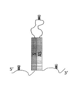
DESCRIPTION OF THE DRAWINGS
[0016] Figure 1 depicts an RNA stem-loop structure with three pac-site
hairpin
sequences, one located 5' of the stem-loop structure, one within the loop of
the stem-
loop structure, and the other 3' of the stem-loop structure.
[0017] Figure 2 depicts a single strand (sense) sequence flanked on each
side by a
pac-site hairpin sequence.
[0018] Figure 3 depicts a single strand (antisense) sequence flanked on
each side
by a pac-site hairpin sequence.
[0019] Figure 4 depicts an RNA stem-loop structure with two pac-site
hairpin
sequences, one located 5' of the stem-loop structure and the other 3' of the
stem-loop
structure.
[0020] Figure 5 depicts an RNA stem-loop structure with a single pac-site
hairpin
sequence located 3' of the stem-loop structure.
[0021] Figure 6 depicts an RNA stem loop structure lacking any pac site
hairpin
sequences.
DETAILED DESCRIPTION OF THE INVENTION
[0022] The present invention comprises compositions and methods for
producing
large quantities of dsRNA in vivo and in some embodiments, recovering such
dsRNA
directly from cell lysates. In its most basic form, the invention involves co-
expressing
a bacteriophage capsid protein, or a portion thereof, in conjunction with the
desired
dsRNA for a period of time sufficient to allow accumulation of the dsRNA in a
host
cell, lysing the host cell, and optionally recovering intact unencapsidated
dsRNA
directly from the cell lysate. In the absence of bacteriophage capsid protein
intact
dsRNA is present in cell lysates in only very small quantities, if at all. In
contrast, in
the presence of bacteriophage capsid protein a relatively large quantity of
unencapsidated dsRNA can be recovered from cell lysates.
-6-
CA 03016899 2018-09-06
WO 2017/160600 PCT/US2017/021661
10023] .. A number of permutations of RNA structure and coat protein were
explored
to determine the essential elements of the invention and to optimize the yield
of
dsRNA produced by the invention. This work is summarized in Table 1 which
outlines the various elements of the invention described in detail and in the
examples
below. The leftmost column of Table 1 refer to individual figures representing
cartoon depiction of the predicted RNA structure produced from each of the
listed
plasmid constructs. In each figure "S" represents the sense strand, "AS"
represents
antisense strand, and the small hairpin structures represent pac site
sequences). The
table also lists the coat protein (if any) and the yields of dsRNA (or ssRNA,
as
indicated) associated with each of the listed plasmid constructs.
Table 1. Production of RNA by E, co/i HT115(DE3) as a function of variation in
RNA structure
and the presence or absence of coat protein and coat protein variants (n.a. =
not applicable; n.d. =
not determined).
RNA Structure Plasmid Loop Stem Stem Coat RNA RNA
as depicted in size size sequence protein en ex
(bases) (bp) capsid capsid
(mg/L) (mg/L)
Figure 1 pAPSE10180 139 180 ErkA MS2 <2 75-90
Figure 1 pAPSE10181 139 180 ErkA none n.a <2.
Figure 2 pAPSE10189 n.a. n.a. beta actin MS2 20
<2
Figure 3 pAPSE10190 n.a. n.a. beta actin MS2 20
<2
Figure 2 pAPSE10274 n.a. n.a. beta actin none
n.a. <2
Figure 3 pAPSE10275 n.a. n.a. beta actin none
n.a. <2
Figure 1 pAPSE10269 166 294 beta actin MS2 2-10 200
Figure 1 pAPSE10306 166 294 beta actin none
n.a. 3
-7-
CA 03016899 2018-09-06
WO 2017/160600 PCT/US2017/021661
Figure 4 pAPSE10216 166 294 beta actin MS2 5-20 50-250
Figure 4 pAPSE10305 166 294 beta actin none n.a. 4
Figure 5 pAPSE10219 166 294 beta actin MS2 5-20 30-60
Figure 5 pAPSE10304 166 294 beta actin none n.a. 3
Figure 6 pAPSE10279 166 294 beta actin MS2 4 65
Figure 6 pAPSE10303 166 294 beta actin none n.a. 4
Figure 4 pAPSE10270 116 294 beta actin MS2 2-10 200
Figure 4 pAPSE10271 136 294 beta actin MS2 2-10 200
Figure 4 pAPSE10272 156 294 beta actin MS2 2-10 200
Figure 4 pAPSE10292 131 294 beta actin MS2 2-10 150
Figure 4 pAPSE10291 142 294 beta actin MS2 2-10 160
Figure 4 pAPSE10276 166 50 beta actin MS2 5-10
80-120
Figure 4 pAPSE10277 166 75 beta actin MS2 20-30
200-
250
Figure 4 pAPSE10366 166 294 beta actin none n.a. <2
(eGFP)
Figure 4 pAPSE10181 139 180 ErkA MS2 n.d. 200
and in trans
pAPSE10149
Figure 1 pAPSE10359 166 294 beta actin Qbeta n.d.
n.d.
-8-
CA 03016899 2018-09-06
WO 2017/160600 PCT/US2017/021661
Figure 4 pAPSE10357 166 294 beta actin none n.d.
n.a.
(U1 A)
Figure 1 pAPSE10372 139 180 ErkA none n.a. 75
(MS2 N-
term
fragment)
A. DEFINITIONS
[0024] As used herein, the term "capsid protein" or "capsid" refers to the
coat
protein of bacteriophage MS2 or Q13, capable of binding the bacteriophage RNA
pac
site with high affinity and assembling into a complex hollow tertiary
structure in
which the bacteriophage RNA is entirely encapsidated within the hollow
tertiary
structure. In a VLP, the capsid protein forms a hollow tertiary structure in
which the
heterologous RNA is entirely encapsidated. The term "capsid" also refers to
the
hollow tertiary structure formed by assembly of individual capsid proteins.
[0025] As used herein "ssRNA" and "dsRNA" refer to "single-stranded RNA and
double stranded RNA, respectively. An ssRNA is comprised of an RNA sequence of
any length that lacks sufficient internal homology to form any significant
secondary
structures such as hairpins or other structures dependent on hybridization of
internal
complementary sequences with one another via Watson-Crick base pairing of
nucleotide bases between the complementary sequences. In contrast, a dsRNA
comprises RNA sequences with sufficient internal homology to form significant
secondary structures such as hairpins due to hybridization of internal
complementary
sequences with one another via Watson-Crick base pairing of nucleotide bases
within
the complementary sequences. Significant secondary structures generally
involve
stretches of homology greater than approximately nine bases, but the exact
length
depends to some extent on context and on whether such secondary structures
impart
any biological function to the molecule.
[0026] As used herein "plasmid" or "extraehromosomal element" refers to any
extrachromosomal episome capable of replication or stable maintenance within
the
host cell. Specifically embraced by this definition are plasmids such as
pBR322,
-9-
pCG1, and pACYC184 which represent the backbones of the described plasmids.
Those of ordinary skill in the art will recognize that other plasmids or
stably
maintained viral episomes can provide the same required functions of
maintenance,
expression and selection and that alternatives to the basic plasmids described
herein
may be generated from such other plasmids or stably maintained viral episomes
without undue experimentation. A key feature of the present invention is the
ability to
express the genes encoding a dsRNA and a capsid protein, not specific modes of
replication, expression or the selective markers found on episomes containing
the
genes encoding the dsRNA and capsid protein.
100271 "Substantially similar sequence" refers to sequence variants
of the claimed
capsid proteins that retain the ability to facilitate accumulation of dsRNA in
a
microbial host cell as described herein. Such substantially similar sequences
include
sequences with at least 26% identity and 47% similarity as shown by the
differences
between MS2 and Qbeta capsid protein sequences (as determined by blastp).
Consequentially, substantially similar sequences encompass conserved and
homologous substitutions allowing sequence variants with as little as 95%,
90%, 80%,
70%, 60%, 50%, 40%, 30% or 25% identity to, and 95%, 90%, 80%, 70%, 60%, 50%
or 40% similarity to, MS2 or Qbeta capsid protein sequences to facilitate
accumulation of dsRNA in a microbial host.
B. COMMON MATERIALS, AND METHODS
100281 Routine microbial and molecular cloning methods and tools,
including
those for generating and purifying DNA, RNA, and proteins, and for
transforming
host organisms and expressing recombinant proteins and nucleic acids as
described
herein, are fully within the capabilities of a person of ordinary skill in the
art and are
well described in the literature. See, e.g., Sambrook, et al., Molecular
Cloning: A
Laboratory Manual, 2nd ed., Cold Spring Harbor Laboratory Press, Cold Spring
Harbor, N.Y. (1989); Davis, et al., Basic Methods in Molecular Biology,
Elsevier
Science Publishing Co., Inc., N.Y. (1986); and Ausubel, et al, Current
Protocols in
Molecular Biology, Greene Publ. Assoc., Wiley-Interscience, NY (1995).
-10-
Date Recue/Date Received 2021-06-22
CA 03016899 2018-09-06
WO 2017/160600 PCT/1JS2017/021661
[0029] Each of the
recombinant DNA constructs described in further detail below
are based on a common plasmid vector series derived from plasmid pBR322. The
first of this plasmid vector series contains a custom synthetic DNA fragment
(produced by PCR GenScript, Piscataway, NJ) comprising a T7 promoter sequence
capable of driving transcription of a single copy of the bacteriophage MS2
capsid
gene followed by a T7 terminator. This synthetic sequence was inserted as a
BamH1-
SphI restriction fragment into the corresponding sites of pBR322 to form
plasmid
pAPSE10118. A second synthetic sequence comprising a T7 promoter sequence
followed by an MS2 pac site sequence, a multi-cloning site containing, in
order (5' to
3') AsiSI-Pmel-AscI-RsrII-NotI-PacI restriction sites, a second high affinity
variant
MS2 pac type sequence (C-pac), a T7 terminator and an Spill restriction site
was
synthesized (PCR Genscript, Piscataway, NJ) and inserted into the EcoRV site
of
pAPSE10118 to form pAPSE10136. The two are oriented such that the T7 promoters
direct transcription of the same strand of pAPSE10136 (clockwise on the
standard
pBR322 map) but are separated from one another by a single T7 terminator.
[0030] A 180 nucleotide
fragment of the ErkA gene of Drosophila melanogaster
(corresponding to the sequence of GenBank Accession NM_001300706 between
nucleotides 156-335) was amplified by PCR incorporating AsiSI and Pmel
restriction
sites on the 5' and 3' sides, respectively. Insertion of this ErkA gene
fragment into the
corresponding sites of pAPSE10136 produced pAPSE10169. A second,
complimentary copy of the ErkA gene fragment sequence was generated by PCR
amplification incorporating a Pmel restriction site on the 5' end, followed by
a
synthetic loop sequence containing an additional MS2 pac sequence, followed by
a
NotI restriction site, followed by the complementary (anti-sense) ErkA gene
fragment
sequence and a PacI restriction site on the 3' end of the PCR fragment. The
synthetic
loop sequence comprises random sequence incapable of hybridizing with the ErkA
gene fragment sequences. This complementary (anti-sense) copy of the ErkA gene
fragment is inserted into the Pmel and Pad l restriction sites of pAPSE10136
to form
pAPSE10180 (SEQ ID NO: 1). A second series of plasmid vectors, lacking the MS2
capsid protein is derived from pAPSE10180 by deleting the MS2 capsid
expression
sequences by SphI restriction digestion and re-ligation to produce pAPSE 10181
(SEQ
ID NO: 2).
-11-
CA 03016899 2018-09-06
WO 2017/160600 PCT/US2017/021661
[0031] Plasmids pAPSE10180 and pAPSE10181 represent the basic platform for
expression of the RNA constructs discussed herein. Transcription of the ErkA
cassette in these plastnids is predicted to produce an RNA transcript capable
of
forming a large stem-loop structure comprising a 180 base pair stem and a 139
base
loop with 3 individual MS2 pac sequences located 5' and 3' of the stem and
within
the loop itself One of ordinary skill in the art will understand that
substitution of the
ErkA gene fragment sequences by other sequences can be easily accomplished by
standard cloning and sub-cloning methods.
[0032] Transformation of plasmids pAPSE10180 or pAPSE10181, or any of their
derivatives, into host cells capable of inducible expression of T7 polymerase
produces
cell lines capable of expressing RNA transcripts. All such strains inducibly
producing
RNA transcripts are referred to generally herein as "expression strains".
Unless
otherwise indicated, each of the plasmids described herein was electroporated
into E.
cull strain HT115(DE3) with genotype F1, rncrA, rnerB, IN (rrnD-rrnE)1,
rnc14::Ta 10 (Lambda DE3 lysogen: /acUV5 promoter-T7 polymerase)) and the
resulting recombinant transformants were selected on LB agar plates containing
12
ug/m1 tetracycline and/or 100 gg/m1 ampicillin. Single colonies were isolated,
the
presence of intact plasmid confirmed by restriction enzyme analysis and the
confirmed
transformed cells archived for future use.
[0033] Standard expression studies comprised inoculating transformed cells
into
100 ml of Super Broth containing 0.1% glucose, 0.4% lactose, 100 ug/m1
ampicillin
and/or 12.5 .1g/m1 tetracycline and incubating the cultures with vigorous
shaking at
37 C. Expression of the T7 polymerase was achieved by auto-induction by
depletion
of the available glucose and the presence of the lactose inducer. This ensures
that all
cultures are induced at the same growth stage. Cells were harvested twelve to
eighteen hours post-induction (late stationary phase) by centrifugation at
3,000 g at
4 C for 30 minutes and stored on ice until lysis.
[00341 RNA was isolated from harvested cells by resuspending a 5 ml
equivalent
of cell culture of harvested cells in sonication buffer comprising Tris-FIC1
pH 7,
mM NaCI and sonicating the suspended cells on ice for 3 minutes. Cell debris
was
-12-
CA 03016899 2018-09-06
WO 2017/160600 PCT/US2017/021661
removed by centrifugation at 16,000 g the supernatant (cleared lysate) was
immediately processed to recover RNA and VLPs as described. RNA was recovered
from half of the cleared lysate using the commercial Purelink RNA Mini Kit
method
(Ambion Cat. No. 12183018A, Thermo Fisher Scientific Inc., Waltham, MA)
according to the manufacturer's instructions.
[0035] VLPs were purified from the remaining half of the cleared lysate
which
were diluted to a total volume of 1 ml and treated with 100 units of
Benzonasee
Nuclease (Sigma Aldrich, St. Louis, MO) at 37 C for two hours. Subsequently,
0.15
mg of Proteinase K was added and the enzymatically treated cleared lysate
incubated
at 37 C for an additional three hours. The VLPS were recovered from the
enzymatically treated cleared lysate by fractional precipitation. A saturated
ammonium sulfate solution was prepared by adding ammonium sulfate to water
until
it reached saturation (approximately 4.1 M). Fifty microliters of the
saturated
ammonium sulfate solution was added to the enzymatically treated cleared
lysate and
the mixture placed on ice and incubated for two hours. Unwanted precipitate
was
removed from the mixture by centrifugation at 16,000 g and the aqueous
solution
transferred to a clean Eppendorf tube. The aqueous solution was then subjected
to a
second precipitation by the addition of 0.171 g of dry ammonium sulfate
directly to
the aqueous solution. The aqueous solution was vortexed and incubated on ice
for
two hours. The precipitate was spun down at 16,000 g the aqueous phase
discarded
and the solid precipitate representing purified VLPs resuspended in 100
microliters of
sonication buffer.
[00361 RNA was recovered from the resuspended purified VLPs by adding 3
volumes of Trizol LS Reagent (Ambion Cat. No. 10296028, Thermo Fisher
Scientific
Inc.), vigorously vortexing the mixture, adding 1 ml of chloroform, further
vortexing
the mixture before pulse centrifugation to separate the aqueous and organic
phases of
the mixture. The aqueous phase was placed in a clean Eppendorf tube and the
RNA
purified with a commercial RNA Clean & ConcentratorTM kit (Cat. No. R1018,
Zymo
Research, lrvine, CA) according to the manufacturer's instructions.
-13-
CA 03016899 2018-09-06
WO 2017/160600 PCT/US2017/021661
[0037] RNA from bacterial and VLP samples were dissolved in 50 111 of
nuclease-
free water. To determine the concentration of dsRNA in a sample, the samples
were
treated with RNAse A (Invitrogen Cat. No. AM2274, Thermo Fisher Scientific
Inc.)
to degrade single stranded RNA under the manufacturers recommended conditions,
the concentration of dsRNA was determined spectrophotmetrically by measuring
0D260 and 1 lig loaded onto Novex 6% TBE-urea gels (Invitrogen, Thermo Fisher
Scientific Inc.). One lane of each gel was loaded with dsDNA size markers of
known
concentration and the samples were electrophoresed, the gel was stained with
ethidium bromide and each band quantitated by densitometry using the dsDNA
markers as a standard curve.
[0038] RNA yields from constructs producing ssRNA were determined by
annealing the sense or anti-sense strand recovered from the induced cells or
VLPs
with an excess of the cognate strand. The annealed mixture was then treated
with
RNAse A and the amount of dsRNA incorporating the ssRNA of interest measured
as
described above.
[0039] Little or no differences in final cell densities were observed
between any of
the cultures from which the samples were harvested and in all cases the
cultures
appear to have reached stationary phase prior to harvest. To allow direct
sample to
sample comparison of RNA yields, all dsRNA and ssRNA concentrations are
reported
as the amount of such RNA present in a 1 L equivalent of culture.
[0040] Northern blot analysis was used to verify the identity of bands
containing
the dsRNA transcripts using a DNA oligonucleotide probe against the random
sequence comprising the loop of each dsRNA construct (5'-GGCCGGCGTCT-
ATTAGTAGATGCC-3', SEQ ID NO 3). RNA from the 6% polyacrylamide
denaturing Urea-TBE gel was transferred to a positively-charged BrightStar ¨
Plus
nylon membrane (Ambion Cat. No. 10102, Thermo Fisher Scientific Inc.) using
the
semi-dry Trans-Blot SD transfer apparatus (BioRad, Hercules, CA) for 1 hour at
constant current of 0.3 A. RNA was fixed on the membrane by the SpectroLinker
XL-
1500 UV crosslinking apparatus (Spectronics Corporation, Westbury, NY) using
the
"optimal crosslink" setting. The membrane was briefly rinsed with water and
-14-
CA 03016899 2018-09-06
WO 2017/160600 PCT/US2017/021661
prehybridized in 50 ml of 5XS SC, 0.1% SDS buffer at 45 C with gentle shaking.
Probe hybridization was carried out overnight at 45 C in 3 ml of
prehybriclization
buffer with gentle shaking. The oligonueleotide probe targeting the hairpin
RNA loop
was conjugated with TAMRA. Three washes (for 2 minutes each) with 100 ml of
water were completed at room temperature and the blot with a ChemiDoc MP
imaging system (BioRad, Hercules, CA), using the rhodamine channel.
C. PREFERRED EMBODIMENTS
100411 The following are among the preferred embodiments of the invention.
[0042] One embodiment of the present invention comprises a bacterial host
cell
containing a plasmid encoding both a gene for the desired dsRNA and a
bacteriophage
capsid protein gene, such that the dsRNA and the capsid protein genes are
transcribed
so that the desired dsRNA is produced and the capsid protein gene translated
to
produce capsid protein and wherein, after a suitable period of time,
unencapsidated
dsRNA accumulates within the cell to a much higher degree than in the absence
of
capsid protein. In other embodiments the dsRNA gene and the capsid protein
gene
may be present on separate compatible plasmids, autonomously maintained phage
or
other epigenetic elements, or one or both genes may be present within the
chromosome of the bacterial host cell.
[0043] In an embodiment the dsRNA gene and the capsid protein gene are each
transcribed from a transcriptional promoter. The transcriptional promoter may
be
inducible. In one embodiment the transcriptional promoters are identical; in
other
embodiments the promoters are different. In still other embodiments the
transcriptional promoters may be differentially induced. In such
differentially
inducible embodiments it may be preferable to induce expression of the capsid
protein
prior to inducing expression of the dsRNA.
[0044] In another embodiment the capsid protein and the dsRNA may be
transcribed as a single transcript from a single promoter. The promoter may be
inducible. In such embodiments the dsRNA is cleaved from the initial RNA
transcript
containing the capsid protein coding sequence by post transcriptional
processing, such
-15-
CA 03016899 2018-09-06
WO 2017/160600 PCT/US2017/021661
post transcriptional processing may depend on bacterial host cell processes or
may be
directed by other RNA processing systems such as ribozymes or specific
ribonucleases.
[0045] In one embodiment one or both of the dsRNA and the capsid protein
genes
are inducibly transcribed from a transcriptional promoter and transcription is
terminated by a transcriptional terminator. In an embodiment the inducible
transcriptional promoter is the bacteriophage T7 gene 1 promoter. In other
embodiments the inducible transcriptional promoter may be the bacteriophage
Lambda PL or PR promoters, the lac operon, trp operon, or synthetic lac
promoter, or
bacteriophage T5 promoter. Other transcription promoters, both constitutive
and
inducible, known to those of ordinary skill in the art, may also be used in
some
embodiments. In an embodiment the transcriptional terminator is the
bacteriophage
T7 late terminator. Other transcription terminators, both rho-dependent and
rho-
independent, known to those of ordinary skill in the art may also be used in
some
embodiments.
[0046] In an embodiment the coat protein gene encodes a leviviral capsid
protein.
The coat protein gene may be the MS2 coat protein gene encoding the MS2 capsid
protein or substantially similar sequences retaining the ability to allow
accumulation
of dsRNA in a microbial host cell. The coat protein gene may encode the Qbeta
coat
protein gene encoding the Qbeta capsid protein or substantially similar
sequences
retaining the ability to allow accumulation of dsRNA in a microbial host cell.
[0047] in an embodiment the dsRNA is recovered from the bacterial host
cells co-
expressing bacteriophage capsid protein by chemical or mechanical methods to
produce a host cell lysate. In an embodiment the dsRNA is further purified
from the
host cell lysate to remove host cell derived proteins, nucleic acids and
membranes
including capsid protein. In another embodiment the host cell lysate is
directly
processed without further purification. In another embodiment bacterial host
cells are
killed, by chemical or heat or other means without lysis and the intact killed
cells
processed without further purification.
-16-
CA 03016899 2018-09-06
WO 2017/160600 PCT/US2017/021661
EXAMPLES
Example 1
Unencapsidated dsRNAs are produced at higher levels in the presence of capsid
protein than in the absence of capsid protein.
[0048] Expression strains containing pAPSE10180 and pAPSE10181 were
constructed and dsRNA production induced by the standard expression procedure
described above. The amount of encapsidated and unencapsidated dsRNA each
strain
produced was measured as described. The initial impetus for this experiment
was to
determine whether an RNA molecule with a 180 base pair double-stranded stem
structure could be packed within a VLP. A 180 bp dsRNA stem is approximately
60
nm in length, whereas the interior diameter of an MS2 capsid is approximately
20 mn.
Based on this geometric limitation, little or no encapsidation was expected
and, due to
host nuclease activity, little or no unencapsidated dsRNA was expected to be
recoverable from the cell lysates. As expected only small amounts of
cncapsidated
dsRNA (en capsid) were recovered (<2 mg/L) from the pAPSE10180 expression
cells.
In contrast, surprisingly large amounts of unencapsidated dsRNA (ex capsid)
were
recovered (75-90 mg/L) from the pAPSE10180 expression cells. Even more
surprisingly, virtually no unencapsidated dsRNA was recovered from the
pAPSE10181 expression cells.
[0049] To determine whether accumulation of RNA is a specific property of
the
ErkA sequence, or is a more general property of expressing dsRNA in the
presence of
capsid protein, a series of expression constructs expressing a 294 base
sequence from
the beta actin gene of the Colorado potato beetle (Leptinotarsa decemlineata
strain
Freeville, GenBank Accession NM_001300706 between nucleotides 156-335) were
produced and tested.
100501 Initially, plasmids expressing the 294 base beta actin sequence from
Colorado potato beetle in the sense and the anti-sense orientation were
constructed
from pAPSE10180 by replacing the ErkA sequences, to produce pAPSE10189 (SEQ
ID NO: 4 and pAPSE10190 (SEQ ID NO: 5) respectively. The beta actin sense and
antisense strand sequences were amplified by PCR (Accuprime Pfx, Invitrogen
Cat.
-17-
CA 03016899 2018-09-06
WO 2017/160600 PCT/US2017/021661
No. 12344040, Thermo Fisher Scientific Inc.) from a gBlock template using
primers
that introduce the AsiSI and Pmel restriction sites at the 5' and 3' ends
respectively
(gBlock template DNA and PCR primers were synthesized by Integrated DNA
Technologies, Coralville IA; all restriction endonucleases were from New
England
BioLabs, Beverly, MA). Restriction digest of pAPSE 10180 and the beta actin
sense
and antisense PCR fragment with AsiSI and PmeI resulted in DNA fragments that
could be ligated together in the desired manner. The pAPSE10180 plasmid
backbone
lacking the ErkA sequence was gel purified and the sense and antisense beta
actin
. sequences were ligated into the gel purified vector to produce pAPSE 10189
and
pAPSE 10190, respectively. When transformed into a suitable expression host,
such
as HT115(DE3) the cells containing pAPSE10189 produces a ssRNA transcript
comprising 294 bases of the sense strand of the beta actin gene flanked by pac
sequences as well as co-express MS2 capsid protein, when cultured and induced
as
described above. Likewise, cells containing pAPSE10190 produces a ssRNA
transcript comprising 294 bases of the anti-sense strand of the same region of
the beta
actin gene flanked by pac sequences as well as co-express MS2 capsid protein
when
transfointed into a suitable expression host, cultured and induced as
described. A
second set of plasmids, lacking the ability to express MS2 capsid protein were
also
produced by replacing the ErkA sequences of pAPSE10181 with the sense and anti-
sense 294 base fragments of the beta actin gene as described above. These
plasmids,
pASPE10274 (SEQ ID NO: 6) and pAPSE10275 (SEQ ID NO: 7) respectively, were
transformed into HT115(DE3) and cultured and induced as described.
[0051] Analysis of un-encapsidated RNA recovered from the cells whether co-
expressed with capsid protein (as with pAPSE10189 and pAPSE10190) or not
(pAPSE10274 and pAPSE10275) showed that virtually no ssRNA can be recovered.
However, VLPs recovered from pAPSE10189 and pAPSE10190 yield at least 20
mg/L of ssRNA of sense or anti-sense sequence respectively. This confirms that
the
plasmid expression systems are capable of producing ssRNA and capsid protein
as
expected.
100521 A dsRNA expression cassette comprising the 294 base Colorado potato
beetle beta actin genes was constructed by a process similar to that described
for the
dsRNA ErkA expression cassette. In this case, the random DNA sequence
comprising
-18-
CA 03016899 2018-09-06
WO 2017/160600 PCT/US2017/021661
the loop between the sense and anti-sense strands of the beta actin sequences
comprised 166 bases, including the same internal pac site sequence as found in
pAPSE10180 and 10181. This beta actin expression cassette was cloned into
pAPSE10180 replacing the ErkA related stem loop sequence to form plasmid
pAPSE10269 (SEQ ID NO: 8), and into pAPSE10181 to form plasmid pAPSE10306
(SEQ ID NO: 9). The plasmids were transformed into HT115(DE3), cultured, and
induced as described. Analysis of the encapsidated dsRNA produced by the cells
containing pAPSE10269 strain showed that 2-10 mg/L dsRNA could be recovered
from VLPs. However, much higher levels of the beta actin dsRNA could be
recovered from the cells containing pAPSE10269 in unencapsidated form (200
mg/L).
Strikingly, analysis of the RNA produced by the pAPSE10306 strain showed that
in
the absence of co-expressed capsid protein only about 3 mg/L of dsRNA could be
recovered.
100533 Thus, the high levels of unencapsidated dsRNA are consistent with a
model
in which such dsRNA are not packaged efficiently, but for some reason appear
to be
present within cells co-expressing capsid protein with the dsRNA at much
higher
levels than in cells which lack capsid protein. One model to account for this
observation is that binding of capsid protein to the pac sites inhibits
degradation by
host cell nucleases.
Example 2
Specific pac site-capsid protein interaction is not required for high level
production of dsRNA.
[0054] To test whether capsid protein bound to pac sites in the dsRNA
results in
the observed increase in dsRNA production in cells co-expressing capsid
protein,
perhaps inhibiting endogenous host nuclease degradation of the bound dsRNA, a
series of constructs comprising the basic beta actin dsRNA described above
were
produced with varying numbers and locations of pac sites. Plasmids pAPSE10216
(SEQ ID NO: 10) and pAPSE10305 (SEQ ID NO: 11), are identical to pAPSE10269
and pAPSE10306 respectively, except they lack the internal loop pac site.
Plasmids
pAPSE10219 (SEQ ID NO: 12) and pAPSE10304 (SEQ ID NO: 13) are identical to
pAPSE10217 and pAPSE10306 respectively, except they have only a single pac
site
-19-
CA 03016899 2018-09-06
WO 2017/160600 PCT/US2017/021661
located on the 3' end of the stem of the dsRNA. Plasmids pAPSE10279 (SEQ ID
NO: 14) and pAPSE10303 (SEQ ID NO: 15) are identical to pAPSE10216 and
pAPSE10306 except they lack all pac site sequences entirely. Each of these
plasmids
was transformed into E. coil HT115(DE3), cultured and induced as described.
Analysis of the encapsidated RNA recovered from VLPs of each of pAPSE10216 and
pAPSE10219 show that 5-20 mg/L of dsRNA is encapsidated. Strikingly, even the
strain containing pAPSE10279 entirely lacking pac sites produced 4 mg/L of
encapsidated dsRNA, indicating that this level of encapsidation may represent
non-
specific entrainment of dsRNA present in the cells at the time the capsids
were
formed. Furthermore, the strain containing pAPSE10216 produced as much as 250
mg/L of unencapsidated dsRNA in the presence of capsid protein. The strains
containing pAPSE10219 and pAPSE10279 produced 30-60 mg/L and 65 mg/L of
unencapsidated dsRNA, respectively in the presence of capsid protein. All of
the
strains containing plasmids comprising the expression cassettes without co-
expression
of capsid protein produced <4 mg/L of dsRNA.
[0055] Together, these results indicate that the ability of capsid protein
to increase
the amount of unencapsidated dsRNA that can be recovered from cell lysates is
not
dependent on the specific binding of capsid protein to its cognate pac site
sequence.
Although the highest levels of unencapsidated dsRNA are recovered from
constructs
containing at least 5' and 3' flanking pac sites (approximately 200 mg/L),
significant
amounts of unencapsidated dsRNA are produced by constructs having only a
single 3'
flanking pac site, or lacking pac sites entirely. Cells containing plasmids
producing
dsRNA lacking pac sites altogether produce significantly higher amounts of
dsRNA
(65 mg/L) when capsid protein is co-expressed with the dsRNA relative to the
cell
lines lacking capsid protein altogether (3-4 ing/L). The approximately I6X
increase
in recoverable dsRNA between cells co-expressing capsid protein and those
lacking
capsid protein (65 mg/L versus 3-4 mg/L) is much more than the approximately
3X-
4X increase due to the presence of pac sites (65 mg/L versus 200-250 mg/L).
The
effect of capsid protein co-expression appears to involve something other than
mere
binding to cognate pac site sequences that may (or may not) be present on the
dsRNA.
-20-
CA 03016899 2018-09-06
WO 2017/160600 PCT/US2017/021661
Example 3
Loop size and structure are irrelevant to high level production of dsRNA.
[0056] To test what effect, if any, differences in loop sequence might
exert on the
production of dsRNA in the presence and absence of co-expressed capsid
protein, a
series of constructs with different lengths of internal non-homologous loop
sequences
were inserted between each of the 294 base sense and anti-sense beta actin
sequences
of pAPSE10269.
[0057] Plasmids pAPSE10270 (SEQ ID NO: 16), pAPSE10271(SEQ ID NO: 17),
pAPSE10272 (SEQ ID NO: 18) and pAPSE10292 (SEQ ID NO: 19) have non-
homologous loop sizes of 116 bases, 136 bases, 156 bases and 166 bases
respectively.
Each of these loop sequences has very little propensity for any secondary
structure as
determined by the m-fold structure prediction program (Zucker and Stiegler
(1981)
Optimal computer folding of large RNA sequences using thermodynamics and
auxiliary information Nucl. Acids. Res. 9(1):133-48). In addition, the 139
base loop
sequence found associated with the ErkA stem sequences in pAPSE10180 and
having
a slightly higher propensity for structural interactions within the loop was
also placed
between the sense and anti-sense beta actin sequences of pAPSE10269, to form
pAPSE10292. Additionally, pAPSE10291 (SEQ ID NO: 20) comprising a 142 base
loop sequence with a high degree of propensity for forming secondary structure
based
on internal homology was synthesized and constructed as described.
[0058] Each of the plasmids described in this Example were transformed
into E.
coli expression strain HT115(DE3), cultured and induced and the amount of
encapsidated and unencapsidated dsRNA determined as described. In each case 2-
10
mg/L of dsRNA was recovered from the VLPs produced by inducing expression of
the plasmid, indicating that loop size or structure had little or no effect on
the ability
of VLPs to encapsiclate the dsRNA. Likewise, expression from each of the
plasmids
produced between 100 and 200 mg/L unencapsidated dsRNA, indicating that loop
size
or structure had little or no effect on overall production of unencapsidated
dsRNA in
the presence of capsid protein.
-21-
CA 03016899 2018-09-06
WO 2017/160600 PCT/US2017/021661
Example 4
Stem size is irrelevant to high level production of dsRNA.
[0059] Differences in stern sequence derived from the Drosophila
melanogaster
ErkA gene sequences expressed from pAPSE10180 and the Colorado potato beetle
beta actin gene sequences expressed from pAPSE10269 do not make a significant
difference in the ability in expression strains to produce large quantities of
unencapsidated dsRNA (75-90 mg/L from pAPSE10180 versus 200 mg/L from
pAPSE10269). Nor does the length of the dsRNA stem (180 base pairs in the
dsRNA
produced from pAPSE10180 and 294 base pairs in dsRNA from pAPSE10269). To
more systematically test what affect, if any, differences stem sequence length
might
exert on the production of dsRNA in the presence and absence of co-expressed
capsid
protein, a series of expression constructs with different lengths of stem
sequences
were substituted for each of the 294 base stem forming sense and anti-sense
beta actin
sequences of pAPSE10269.
[0060] Plasmids pAPSE10276 (SEQ ID NO: 21) and pAPSE10277 (SEQ ID
NO: 22) encode dsRNA with potential double-stranded stems of 50 and 75 base
pairs
respectively. The dsRNA expressed by both plasmids comprise 166 bases of non-
homologous loop sequence. Although these dsRNA structures are significantly
shorter than those in dsRNA from the corresponding ErkA and beta actin
constructs,
they still exceed the interior diameter of the MS2 VLP.
[0061] When transformed into the E. coli expression strain HT115(DE3),
cultured
and induced as described, pAPSE 10276 produces 5-10 mg/L of encapsidated dsRNA
and 80-120 mg/L of unencapsidated dsRNA. Plasmid pAPSE 10277 produces 20-30
mg/L encapsidated dsRNA and 200-250 mg/L unencapsidated dsRNA. These values
are similar to those observed for pAPSE10180 and pAPSE10269 described earlier
in
this Example, indicating that differences in stem length and sequence do not
play a
major role in producing dsRNA in cells co-expressing capsid protein.
-22-
CA 03016899 2018-09-06
WO 2017/160600 PCT/US2017/021661
Example 5
Capsid protein is required for high level production of dsRNA.
[0062] To confirm the requirement for capsid protein, plasmid pAPSE10216,
which produces a dsRNA product at high levels in the presence of capsid
protein, was
altered to replace the MS2 coat protein gene with eGFP. A gBlock template
comprising the T7 promoter to T7 terminator sequences of pAPSE10216 (spanning
the sequences between the unique BamHI and Sall sites of the plasmid) in which
the
coding sequence of MS2 coat protein was replaced with the coding sequence of
eGFP
was designed, produced and amplified with primers encompassing the BamHI site
on
the 5' side and the Sall site on the 3' side. The resulting 1 kb fragment was
digested
with BamHI and Sall and then ligated into BamHI-SalI digested pAPSE10216 to
form
pAPSE10366 (SEQ ID NO: 24). Plasmid pAPSE10366 was confirmed by restriction
digest and transfoimed into the E. coli expression strain HT115(DE3), cultured
and
induced as described, pAPSE10366 produces <2 mg/L of unencapsidated dsRNA, in
contrast to the 200 mg/L produced by pAPSE10216. In addition, the cells
expressed
high amounts of eGFP as evidenced by the intense fluorescence produced on
induction (data not shown) confirming that the basic dual expression plasmid
used
throughout these studies performs as expected. This result further
demonstrates that
capsid protein is necessary for accumulation of unencapsidated dsRNA in cells
expressing the target RNA gene that otherwise accumulate unencapsidated dsRNA
in
the presence of capsid protein.
[0063] To further confirm that the presence of capsid protein is essential
to the
high levels of unencapsidated dsRNA production a plasmid compatible with
pAPSE10181 and capable of inducible expression of the MS2 capsid protein is
constructed. pAPSE10149 (SEQ ID NO: 23) is based on pACYC184. This plasmid
comprises a P1 5A origin of replication that is not excluded by the colE1
based origin
of replication of pAPSE10181 and a chloramphenicol acetyl transferase
antibiotic
marker to allow selection of co-transfonnants containing both
pAPSE10181(encoding
ampicilin resistance) and pAPSE10149 (encoding chloramphenicol resistance).
Plasmid pAPSE10149 also comprises the same T7 promoter sequence capable of
driving transcription of a single copy of the bacteriophage MS2 capsid gene
followed
-23-
CA 03016899 2018-09-06
WO 2017/160600 PCT/US2017/021661
by a T7 terminator as found in pAPSE10118 cloned into the BamHI and SphI sites
of
pACYC184. Plasmid pAPSE10149 is transformed into expression strains already
containing pAPSE10181 to produce ampicilin and chloramphenicol resistant
double
transformants. Expression studies of such double transformants show that co-
expression of the capsid protein from pAPSE10149 in conjunction with
pAPSE10181
produces 200 mg/L of unencapsidated dsRNA whereas cells containing pAPSE10181
alone produce <2 mg/L of unencapsidated dsRNA (see Example 1). This
demonstrates that providing capsid protein in trans is sufficient to
facilitate production
of high levels of unencapsidated dsRNA to host cells containing a plasmid
expressing
the dsRNA target that otherwise fail to accumulate unencapsidated dsRNA in the
absence of capsid protein.
Example 6
Other capsid proteins can induce high level production of dsRNA.
[0064] To test whether the accumulation of unencapsidated dsRNA is a
unique
property of bacteriophage MS2 capsid protein, or whether other capsid proteins
share
this property, a plasmid expression system analogous to pAPSE10216 was
constructed. This plasmid, pAPSE10359 (SEQ ID NO: 25) comprises a Qbeta capsid
protein and Qbeta pac sites at the 5' and 3' ends of the beta actin dsRNA
expression
cassette, but is in all other aspects similar to pAPSE10216.
[0065] Briefly, the Qbeta coat protein gene sequence (Genebank Accession
NC 001890 between nucleotides 1343 and 1744) was synthesized as a gBlock
fragment by Integrated DNA Technologies, Coralville, IA. The synthetic
fragment
was amplified with PCR with primers that introduced a BamHI restriction site
followed by a 17 promoter sequence upstream of the Qbeta coat protein gene
followed by a T7 terminator and a SphI restriction site. The amplified
synthetic
fragment and plasmid pBR322 were digested with BamHI and SphI and ligated
together to form intermediate plasmid pAPSE10358. The beta actin dsRNA
sequence
of pAPSE10269 was amplified by PCR with primers that introduced an EcoRI
restriction site followed by a Qbeta pac sequence followed by the beta actin
dsRNA
sequence followed by a second copy of the Qbeta pac sequence followed by a
BamHI
-24-
CA 03016899 2018-09-06
WO 2017/160600 PCT/US2017/021661
restriction site. This amplified beta actin containing sequence and plasmid
pAPSE10358 were digested with EcoRI and BamHI and ligated together to form
pAPSE10374. Plasmids pAPSE10374 and pAPSE10216 were digested with AsiS1
and NotI. This cleaves pAPSE10374 into two fragments of 4,713 and 113 base
pairs
and pAPSE10216 into two fragments of 5,204 and 786 base pairs. The 4,713 and
786
base pair fragments were isolated and ligated together to produce pAPSE10359.
[0066] When transformed into the E. coli expression strain HT115(DE3),
cultured
and induced as described, pAPSE10359 will produce a large amount of
unencapsidated dsRNA relative to the amount of dsRNA produced from a similar
construct lacking capsid protein (pAPSE10305). This pattern, similar to that
observed
for pAPSE10216 and pAPSE10305 described in Example 1, will confirm that
expression of the Qbeta capsid protein, like the MS2 capsid protein, is
sufficient to
increase the amount of dsRNA produced in vivo.
Example 7
RNA binding proteins other than capsid proteins are not sufficient for high
level
production of dsRNA.
[0067] To test whether the accumulation of unencapsidated dsRNA is a
function of
general RNA binding or is specific to bacteriophage capsid proteins, a plasmid
expression system, pAPSE10357 (SEQ ID NO: 26) was constructed comprising the
RNA binding domain of the human UlA protein and its hairpin cognate binding
site
from human Ul snRNA 5' and 3' of the sense and antisense stem loop structure
of the
beta actin dsRNA. Plasmid pAPSE10357 is similar to pAPSE10216 with the capsid
protein replaced by the human UlA RNA binding protein and UlA binding site
sequences at the 5' and 3' ends of the beta actin dsRNA expression cassette,
but is in
all other aspects similar to pAPSE10216.
[0068] The DNA sequence encoding the N-terminal 102 amino acids comprising
the RNA binding domain of the human 'VIA protein was amplified from a cloned
copy of the UlA protein (Plasmid pAV105, Professor Kathleen Hall, Washington
University, St. Louis, MO) using PCR primers that introduced a BamHI
restriction
site followed by a T7 promoter sequence upstream of the VIA gene fragment
-25-
CA 03016899 2018-09-06
WO 2017/160600 PCT/US2017/021661
followed by a T7 terminator and a SphI restriction site. The amplified
synthetic
fragment and plasmid pBR322 were digested with BamHI and SphI and ligated
together to form intermediate plasmid pAPSE10356. The beta actin dsRNA
sequence
of pAPSE10269 was amplified by PCR with primers that introduced an EcoRI
restriction site followed by the hairpin binding site sequence from human Ul
snRNA
sequence followed by the beta actin dsRNA sequence followed by a second copy
of
the hairpin binding site sequence from human Ul snRNA sequence followed by a
BamHI restriction site. This amplified beta actin containing sequence and
plasmid
pAPSE10356 were digested with EcoRI and BamtH and ligated together to form
pAPSE10373. Plasmids pAPSE10373 and pAPSE10216 were digested with AsiSI
and NotI. This cleaves pAPSE10373 into two fragments of 4,627 and 113 base
pairs
and pAPSE10216 into two fragments of 5,204 and 786 base pairs. The 4,713 and
786
base pair fragments were isolated and ligated together to produce pAPSE10357.
[0069] When transfornied into the E. coil expression strain HT115(DE3),
cultured
and induced as described, pAPSE10357 will not produce a significant amount of
unencapsidated dsRNA relative to the amount of dsRNA produced from a similar
construct lacking capsid protein (pAPSE10305). This will confirm that the mere
presence of an RNA binding site and binding protein in conjunction with the
dsRNA
is not sufficient to increase the amount of dsRNA produced in vivo.
Alternatively,
production of significant amounts of unencapsidated dsRNA will indicate that
the
presence of RNA binding sites at the 5' and 3' end and the cognate RNA binding
protein is sufficient for increasing in vivo production of dsRNA.
Example 8
The N-terminus of capsid protein is sufficient for high level production of
dsRNA.
[0070] To examine whether the increased production of dsRNA from plasmids
containing both the dsRNA gene and the coat protein gene requires the intact
capsid
protein or whether only a portion of the protein is required, a frame-shift
mutation was
introduced into the coat protein gene sequence of pAPSE10180. Double digestion
of
pAPSE10180 with the restriction enzymes StuI and Pm1I produces two restriction
-26-
CA 03016899 2018-09-06
WO 2017/160600 PCT/1JS2017/021661
fragments, a large fragment of 5,485 base pairs and a small thirteen base pair
fragment
comprising about 4 codons of the capsid protein CDS about 40 codons from the
coat
protein start codon of pAPSE10180. The restriction enzymes produce blunt-ended
termini and the larger fragment was re-ligated to produce plasmid pAPSE10372
(SEQ
ID NO: 27), which, in addition to producing an intact inducible dsRNA ErkA-
specific
sequence, also comprises an inducible frame-shifted protein that includes the
N-term
41 codons of the MS2 coat protein followed by 27 codons of frame-shifted
sequence
before terminating at a stop codon (SEQ ID NO: 28). When pAPSE10372 was
transformed into E. coli expression strain HTE115(DE3) and cultured and
induced as
described, 75 mg/L of dsRNA was produced. This indicates that the N terminus
of
the capsid protein alone is sufficient to increase production of dsRNA as well
as the
intact capsid protein (compare yields from pAPSE10180 and pAPSE10372 in
Table 1).
[0071] The N-terminus of the MS2 capsid protein ;Cairns a distinctive
three-
dimensional structure comprised of four separate beta sheets (D. Peabody, The
RNA
binding site of bacteriophage MS2 coat protein, The EMBO journal 12(2) 595-600
(1993)). Each of these sheets, pD from amino acids 31-35, PC from amino acids
22-
25, pB from amino acids 19-21 and PA amino acids 8-11 may play a role in the
ability
of the N-terminus capsid protein fragment to improve dsRNA production. Note
that
the nomenclature is that of Peabody and the numbering includes the N-terminal
methionine omitted by Peabody. Progressive deletion of each of these
structural
motifs can determine the minimum sequence requirement for improving dsRNA
production.
Example 9
Fed batch fermentation produces very high level production of dsRNA.
[0072] To determine whether quantities of dsRNA could be increased by
improving the microbial growth conditions, glucose fed batch fermentations
were
conducted. Briefly, fed-batch fermentations were carried out in an Eppendorf
BioFlo
115 fermenter at 37 C. The pH was controlled by automatic addition of 30%
ammonium hydroxide. The dissolved oxygen probe was calibrated to 0% by
-27-
CA 03016899 2018-09-06
WO 2017/160600 PCT/US2017/021661
unplugging the DO probe and to 100% with air saturation. The vessel was
aerated at
2 vvm and dissolved oxygen maintained at 30% by cascade control of agitation.
An
overnight culture of HT115 (DE3) containing pAPSE10379 was grown in LB
containing 100 ug/ul of ampicillin and 12.5 ug/ul of tetracycline at 37 C to
inoculate
the seed medium. The seed media is a defined media consisting of 5.68 g/L
Na2HPO4, 1.34 g/L KH2PO4, 6.6 g/L (NH4)2SO4, 10 g/L glucose, 1X trace metal
and
1X vitamin solutions maintained at a pH of 7Ø To ensure plasmid stability
antibiotics are added at 100 ug/ul ampicillin and 12.5 ug/ul tetracycline. At
saturation
(0D600 3-5) the seed cultures are used to provide 10% inoculum for the
fermenter.
[0073] During fed batch-cultures a 50% (w/v) solution of glucose was added
according to a carbon limiting DO stat feeding strategy. The basal medium
consists of
6g/L K2HPO4, 3 g/L NaHPO4, 10 g/L (NH4)2SO4, 1 g/L MgSO4, 1X trace metal
solution with antibiotics added at 100 ug/ul of ampicillin and 12.5 ug/ul of
tetracycline. Upon exhaustion of the initial carbon source provided by the
glucose the
feed solution is added automatically in a manner that maintains the DO level
at 30%
of saturation.
[0074] Once the cell culture has reached an 0D600 of 60 the cells are
induced with
1 mM 1PTG or a feed of 20 g/L of lactose by switching the glucose feed to a
lactose
feed. After induction 1 mL samples are taken at different times post
induction. The
samples are lysed by sonication of the cell pellet into 20 mM Tris-HCl at pH
7. Total
RNA from the cell pellet is purified using well-known Trizol extraction
procedures.
Briefly 1 volume of cell lysate is added to 1 volumes of Trizol RNA extraction
reagent. Addition of 1 volume of chloroform results in the RNA partitioning to
the
aqueous layer leaving the protein and DNA contaminants behind.
100751 To analyze the yield of dsRNA the total RNA sample is diluted to 1
ug/ul
and subjected to RNAseA treatment. The reaction is carried out in 20 mM Tris
at pH
7.0 and 37 C for 40 minutes. Once this is done proteinase K is added to the
reaction
to remove the nuclease and is allowed to react at 37 C for 40 minutes. Upon
completion of this step the dsRNA remaining is diluted in half, quarters and
eighths in
order to determine the concentration of the dsRNA using gel densitometry.
-28-
CA 03016899 2018-09-06
WO 2017/160600
PCT/US2017/021661
[0076] Quantification of dsRNA yield by gel densitometry was
performed by
comparing the intensity of dsRNA bands versus dsDNA bands of known mass and
weight on a 1.5% agarose gel containing ethidium bromide. The lambda 100 bp
quantifiable DNA marker was used and a standard curve was generated to
determine
the range in which the dsRNA from the fermentation can be reliably quantified.
The
computer program calculates the amount of dsRNA in the amount of sample loaded
on the gel and a back calculation that considers the dilution steps is
performed.
Yields of dsRNA at levels as high as 3 g/L have been calculated with both IPTG
and
lactose as inducers under these conditions. These results indicate that
further
increases in dsRNA production are possible by improving feimentation
conditions.
Example 10
Compositions and methods for dsRNA production in gram positive bacteria.
= [0077] The ability of gram-positive bacteria to produce increased
levels of dsRNA
by co-expression of capsid proteins can be examined in the following manner.
Corynebacterium glutamicum MB001(DE3) strain DSM 102071, containing an
inducible T7 RNA polymerase gene (described in Kortmann, et al., A
chromosornally
encoded T7 RNA polymerase-dependent gene expression system for Corynebacterium
glutamicum; construction and comparative evaluation at the single cell level.
Microb
Technol. 8(2):253-65. Mar. 2015) is modified to knockout the rnc gene homolog
encoding RNAse III. Briefly, PCR primers capable of amplifying a 1.2 kb
sequence
homologous to the sequence present in C. glutamicum strain MB001(DE3)
immediately upstream of the rnc gene and PCR primers capable of amplifying a
1.5
kb sequence homologous to the sequence immediately downstream of the rnc gene
are
synthesized. A PCR amplification reaction using C. glutamicum strain
MB001(DE3)
genomic DNA and said primers results in a single DNA fragment comprising the
1.2
kb and 1.5 kb target sequences joined together (by standard overlap PCR
methods) to
produce an approximately 2.7 kb SaII-BamHI synthetic DNA fragment. This Sall-
BamIII DNA fragment and plasmid pK18mobsacB (ATCC 87097, described by
Schafer, et al., Small mobilizable multi-purpose cloning vectors derived from
the
Escherichia coli plasmids pK18 and pK19: selection of defined deletions in the
chromosome of Corynebacterium glutamicum. Gene 145:69-73) are digested with
Sall
-29-
CA 03016899 2018-09-06
WO 2017/160600 PCT/US2017/021661
and BamHI and the products ligated together to produce plasmid pAPSE10429 (SEQ
ID NO: 29). Plasmid pAPSE10429 is transformed into C. glutamicum strain MB001
and transfonnants selected on kanamycin containing solid LB medium to identify
chromosomal integrants. Kanamycin resistant clones are transferred to a solid
LB
medium containing 20% sucrose. Conversion of sucrose by the sacB gene product
is
toxic to C. glutamicum strain MB001 so only those chromosomal integrants that
subsequently delete the sacB gene from the chromosome can survive on such
media.
Surviving colonies arc grown up and screened by PCR to confirm concomitant
loss of
the rnc locus from the chromosome. The desired strain is designated C.
glutamicurn
MB001(DE3) rnc. This strain possesses an inducible T7 RNA polymerase and lacks
the rnc gene and is suitable for testing the efficacy of dsRNA production in
the
presence and absence of capsid protein.
[00781 A shuttle vector capable of expression of capsid coat protein and
dsRNA in
both E. coil and C. glutamicum is constructed by synthesizing a DNA comprising
the
origin of replication of the gram-positive plasmid pCG1 (GeneBank Accession
No.
AB027714; described by Trautwetter and Blanco, Structural organization of the
Corynebacterium glutamicum plasmid pCG1 00. J. Gen. Microbiol. 137:2093-101
1991) and the kanamycin resistance gene of pK18mobsac.B. This synthetic DNA
(SEQ ID NO: 30) is ligated into the previously described dsRNA containing
plasmids
at the unique NruI restriction site to allow testing whether the presence of
capsid
protein in gram-positive C. glutamicum MB001(DE3) rue strain produces dsRNA at
high levels as described below.
[0079] Insertion of the synthetic DNA comprising the pCG1 origin of
replication
and the kanamycin resistance gene is accomplished by digesting pAPSE10279 with
NruI and ligating the phosphorylated synthetic DNA into the plasmid to produce
plasmid pAPSE10430 (SEQ ID NO: 31). Plasmid pAPSE10430 contains the
kanamycin resistance gene, the bacteriophage MS2 coat protein, and the dsRNA
construct based on the previously described 294 base sense and antisense
sequences
homologous to the Colorado potato beetle beta actin gene separated by a 166
base
non-homologous loop and entirely lacking any pac sequences. In similar
fashion, the
synthetic DNA comprising the pCG1 origin of replication and the kanamycin
-30-
CA 03016899 2018-09-06
WO 2017/160600 PCT/US2017/021661
resistance gene is also ligated into Nrul digested pAPSE10303 to produce
pAPSE10431 (SEQ ID NO: 32). Plasmid pAPSE10431 contains resistance genes to
ampicillin and kanamycin, as well as the same inducible dsRNA construct as
pAPSE10430. However, pAPSE10431 lacks the inducible MS2 coat protein gene of
pAPSE10430. The relevant features of pAPSE10430 and pAPSE10431 are presented
in Table 2 and the relationship between these two plasmids and their parental
plasmids, pAPSE10279 and pAPSE10303, respectively, can be determined by
comparing Table 2 and Table 1.
[0080] Additional plasmids containing one, two, and three pac sites, with
and
without MS2 coat protein, are constructed using the same procedure. Plasmid
pAPSE10432 (SEQ ID NO: 33) containing a single pac site 3' of the beta actin
stem
loop structure and encoding the MS2 coat protein gene is produced by ligating
the
synthetic DNA fragment into the Nrul site of pAPSE10219. Plasmid pAPSE10433
(SEQ ID NO: 34) is produced by ligating the synthetic DNA fragment into the
Nrul
site of pAPSE10304. Plasmid pAPSE10433 is identical to pAPSE10432 except it
lacks an inducible MS2 coat protein gene. Plasmid pAPSE10434 (SEQ ID NO: 35)
containing two pac site sequences located one on either side of the beta actin
stein
loop and encoding the MS2 coat protein is produced by ligating the synthetic
DNA
fragment into the Nrul site of pAPSE10216. Plasmid pAPSE10435 (SEQ ID NO: 36)
is produced by ligating the synthetic DNA fragment into the Nrul site of
pAPSE10305. Plasmid pAPSE10435 is identical to pAPSE10434 except it lacks an
inducible MS2 coat protein gene. Plasmid pAPSE10436 (SEQ ID NO: 37) containing
three pac site sequences with one each 5' and 3' of the beta actin stem loop
and one
within the loop sequence itself (as depicted in Figure 1) and encoding the MS2
coat
protein is produced by ligating the synthetic DNA fragment into the Nrul site
of
pAPSE10269. Plasmid pAPSE10437 (SEQ ID NO: 38) is produced by ligating the
synthetic DNA fragment into the Nrul site of pAPSE10306. Plasmid pAPSE10437 is
identical to pAPSE104360 except it lacks an inducible MS2 coat protein gene.
[0081] In each case, following ligation of the synthetic DNA fragment into
the
Nrul site of the target plasmid, transfonnants the ligation reactions are
desalted and
transformed in to C. glutamicum MB001(DE3) rue and selected for resistance to
-31-
CA 03016899 2018-09-06
WO 2017/160600 PCT/US2017/021661
kanamycin. The selected clones are subsequently grown at 32 C in 100 ml of LB
media containing kanamycin until the culture reaches 0D600 0.8, at which time
isopropyl f3-D-thiogalactopyranoside is added to a final concentration of 1 mM
to
induce T7 polymerase directed transcription of the MS2 coat protein and the
dsRNA,
or just the dsRNA precursor in the plasmids lacking coat protein. The induced
cultures are allowed to grow for at least 4 hours post-induction to allow
sufficient
time for accumulation of the MS2 coat protein and dsRNA target. Cells are
collected
by centrifugation at 3,000 g at 4 C. Each pellet is stored at 4 C until
processing.
[0082] The dsRNA is purified by re-suspending each pellet in approximately
0.1
volume of 20 mM Tris-HC1, pH 7.0, containing 10 mM NaCl and sonicated to lyse
the cells. Cell debris is removed by centrifugation at 16,000 g. The resulting
lysate is
mixed with 3 volumes of Trizol (Ambion Life Technologies) and the RNA is
extracted by adding 1 volume of chloroform. Addition of NaCl to a final
concentration of 500 mM to the aqueous layer and subsequent ethanol
precipitation
results in a pellet containing the 294 bp siRNA precursor and RNA from the C.
glutamicum host.
[0083] To determine the amount of dsRNA produced by the C. glutamicum
transformed with plasmids containing various pac site configurations, with and
without MS2 coat protein, the ethanol pellets are resuspended and treated with
RNAseA for 1 hour at 37 C followed by Proteinase K digestion for 1 hour at 37
'C.
Quantification of the dsRNA is accomplished by gel densitometry using a BioRad
ChemiDoc MP Imaging System. Several dilutions of the treated dsRNA are run on
a
1.5% agarose gel containing 0.001% ethidium bromide. A 100 bp quantifiable
dsDNA ladder (QuantiBP DNA ladder Lambda) is used as the standard curve and
the
dsRNA is quantified at the concentration that falls within the linear range of
the
standard curve. Software such as Image Lab 4.1 determines the concentration of
the
dsRNA loaded on the gel and a final yield of dsRNA is determined by accounting
for
the dilutions associated with the dsRNA samples present on the gel.
100841 Table 2 summarizes the predicted results of the dsRNA yield
determination
of the Colorado potato beetle beta actin dsRNA produced by C. glutamicum
-32-
CA 03016899 2018-09-06
WO 2017/160600 PCT/1JS2017/021661
MB001(DE3) rnc and the various plasmids described above. Such results confirm
that gram positive hosts such as C. glutamicurn produce large quantities of
dsRNA by
co-expression of the MS2 coat gene and a dsRNA target of interest.
Table 2. Predicted production of dsRNA by C. glutamicum MB001(DE3) rne as a
function of
variation in dsRNA structure and the presence or absence of coat protein.
RNA Structure Plasmid Loop Stem Stem Coat dsRNA
as depicted in size size sequence protein (mg/L)
(bases) (bp)
Figure 6 pAPSE10430 166 294 beta actin MS2 ¨60
Figure 6 pAPSE10431 166 294 beta actin none ¨4
Figure 5 pAPSE10432 166 294 beta actin MS2 ¨120
Figure 5 pAPSE10433 166 294 beta actin none ¨4
Figure 4 pAPSE10434 166 294 beta actin MS2 ¨250
Figure 4 pAPSE10435 166 294 beta actin none ¨4
Figure I pAPSE10436 166 294 beta actin MS2 ¨250
Figure 1 pAPSE10437 166 294 beta actin none 4
Example 11
Compositions and methods for dsRNA production in yeast.
100851 To create a Saccharomyces cerevisiae production host suitable
for dsRNA
accumulation utilizing the MS2 bacteriophage coat protein, the Rntl gene of S
cerevisiae YPH 500 (ATCC 76626) is knocked out according to the procedure of
Gardenr and Jasperson (Gardner, JM and Jaspersen, SL, Manipulating the yeast
-33-
CA 03016899 2018-09-06
WO 2017/160600 PCT/US2017/021661
genome: deletion, mutation and tagging by PCR. Methods Mol Biol. 1205:45-78,
2014). The KanMx4 gene is amplified from pML104-KanMx4 plasmid (Laughery, et
al., New vectors .for simple and streamlined CRISPR-Cas9 genome editing in
Saccharomyces cerevisiae. Yeast 32(12):711-20 Sep. 21, 2015) with PCR primers
including 60 base pair (bp) upstream (forward primer) and 60 bp downstream
(reverse
primer) regions of the S. cerevisiae Rntl gene. The resulting PCR product is
introduced into chemically competent S. cerevisiae cells following the
established S.
cerevisiae transformation protocol. The transfoimed cells are incubated
overnight
without selection marker to allow for homologous recombination to occur, where
in
the kanMx4 gene carrying 60 bp upstream and downstream regions of Rntl
replaced
the Rntl gene. Following overnight incubation, the transfoimed cells are
plated on
YPD plates carrying G418 as selection marker. G418 resistant colonies are
screened
by PCR to confirm presence of kanMx4 gene and deletion of Rntl gene in the YPH
500 genome.
[0086] S. cerevisiae expression vectors pESC-His, pESC-Leu, pESC-Ura and
pESC-Trp are widely used for recombinant protein expression in S. cerevisiae.
Each
of the pESC vectors (Agilent Technologies, Santa Clara CA) contains one of
four
different yeast-selectable markers (HIS3, TRP1, LEU2, or URA3) in the same
vector
backbone, which allows expression of two different genes in a single yeast
cell. The
pESC series vectors are used with S. cerevisiae strain YPH 500 (MATa ura3-52
1ys2-
801 _amber ade2-10I _ochre trp 1 -A63 his3-A200 1eu2-A1). In this example, the
pESC-Trp vector is selected for expression of MS2 coat protein and target
dsRNA
sequence inside S. cerevisiae, although any of the other pESC vectors could be
employed using similar methods since these vectors can replicate in S.
cerevisiae as
well as E.coli, which facilitates molecular manipulations necessary to produce
dsRNA.
[0087] The pESC-Trp vector is modified by cloning a 50-base pair multi-
cloning
site linker containing BamHI, Swal, AsiSI, Not!, Sac!! and Nhel sites,
downstream of
the GAL1 promoter into the existing BamHI and Nhel sites. Following this, the
beta
actin stem loop sequence (dsRNA) of pAPSE10279 is excised as an AsiSl/Notl
fragment and ligated into the AsiS 1/Notl sites of the modified pESC-Trp
vector.
-34-
CA 03016899 2018-09-06
WO 2017/160600 PCT/US2017/021661
Expression of the dsRNA in this plasmid is under the control of galactose
inducible
promoter GALl. The new vector is named pAPSE10439 (SEQ ID NO: 39). Another
plasmid, pAPSE10440 (SEQ ID NO: 40), which is identical to pAPSE10439, but
also
includes the MS2 coat protein. Plasmid pAPSE10440 is constructed by PCR
amplifying the MS2 coat protein expression sequences of pAPSE10279 with a
forward primer carrying an EcoRI restriction site on the 5' end and the
reverse primer
carrying Sad I site on the 3' end. The PCR product is digested with EcoRI and
Sad I and
cloned into the cognate sites of pAPSE10439. Thus, pAPSE10439 inducibly
expresses the dsRNA from the GAL1 promoter, whereas pAPSE10440 inducibly
expresses the dsRNA sequence from the GAL1 promoter and the MS2 coat protein
from the GAL10 promoter,
[0088] Similar plasmid pairs are constructed using this technique.
Plasmids
pAPSE10441 (SEQ ID NO: 41) and pAPSE10442 (SEQ ID NO: 42) are produced by
digesting pAPSE10439 and pAPSE10440 with AsiSI and NotI and isolating the
vector fragment. Plasmid pAPSE10219 is also digested with AsiSI and NotI and
the
dsRNA sequence is isolated. The isolated dsRNA sequence is ligated into the
pAPSE10439 vector to form pAPSE10441 and the isolated dsRNA sequence is
ligated into the pAPSE10440 vector to form pAPSE10442. Plasmids pAPSE10443
(SEQ ID NO: 43) and pAPSE10444 (SEQ ID NO: 44) are produced by digesting
pAPSE10439 and pAPSE10440 with AsiSI and NotI and isolating the vector
fragment. Plasmid pAPSE10216 is also digested with AsiSI and NotI and the
dsRNA
sequence is isolated. The isolated dsRNA sequence is ligated into the
pAPSE10439
vector to form pAPSE10443 and the isolated dsRNA sequence is ligated into the
pAPSE10440 vector to form pAPSE10444. Plasmids pAPSE10445 (SEQ ID NO: 45)
and pAPSE10446 (SEQ ID NO: 46) are produced by digesting pAPSE10439 and
pAPSE10440 with AsiS1 and NotI and isolating the vector fragment. Plasmid
pAPSE10269 is also digested with AsiSI and NotI and the dsRNA sequence is
isolated. The isolated dsRNA sequence is ligated into the pAPSE10439 vector to
form pAPSE10445 and the isolated dsRNA sequence is ligated into the pAPSE10440
vector to form pAPSE10446.
-35-
[0089] Chemically competent YPH 500 DRntl cells are transformed with
each of
the above mentioned plasmids (pAPSE10439-46) separately and individual clones
selected on synthetic dextrose minimal (SD) tryptophan (trp) drop out plates.
After
inoculating the 100 ml SD-Trp drop out broth the cultures are grown for 12 to
16
hours. The cells from the culture are then harvested by centrifugation at 3000
g for 5
minutes, the cell pellet is washed once with sterile water and the cells re-
suspended in
synthetic galactose minimal broth (SG) lacking tryptophan. The cells are grown
in the
SG-trp drop out broth overnight to induce production and accumulation of dsRNA
and MS2 coat protein (where appropriate). Cells are harvested by
centrifugation at
3,000 g at 4 C. Each pellet is stored at -20 C until processing.
[0090] The dsRNA is purified by re-suspending each pellet (10 ml
culture) in
approximately 1.0 ml of yeast cell lysis buffer (Sigma C4482). The resulting
lysate is
mixed with 3 volumes of Trizol (Ambion Life Technologies) and the RNA
extracted
by adding 1 volume of chloroform. Addition of NaC1 to a final concentration of
500
mM to the aqueous layer and subsequent ethanol precipitation results in a
pellet
containing the dsRNA and RNA from the S. cerevisiae host. The resulting RNA
pellet is dissolved in 20 mM Tris HCI pH 7.0 and RNA concentration of the
sample
deteitnined. To determine the amount of dsRNA produced by the S. cerevisiac
strains,
a known amount of RNA (10 ug) from each RNA sample from pAPSE10439-
pAPSE10446) are digested with RNAseA for 1 hour at 37 C followed by
Proteinase
K digestion for 1 hour at 37 'C. The resulting samples contain only the dsRNA
target.
Quantification of the dsRNA is done by gel densitometry using a BioRad
ChemiDoc
MP Imaging System. Several dilutions of the RNAse A reaction are run on a gel
that
contains 1.5% agarose and 0.001% ethidium bromide. A 100 bp quantifiable dsDNA
ladder (QuantiBP DNA ladder Lambda) is used as the standard curve and the
dsRNA
is quantified at the concentration that falls within the linear range of the
standard
curve. Using Image Lab 4.1 software, the concentration of the dsRNA loaded on
the
gel is determine and a final yield of dsRNA calculated by accounting for the
dilutions
of the dsRNA loaded on the gel.
[0091] Table 3 summarizes the predicted results of the dsRNA yield
determination
of the Colorado potato beetle beta actin dsRNA produced by S. cerevisiae YPH-
500
-36-
Date Recue/Date Received 2021-06-22
and the various plasmids described above. Such results confirm that yeasts
such as S.
cereviskte produce large quantities of dsRNA by co-expression of the MS2 coat
gene
and a dsRNA target of interest.
Table 3 Predicted production of dsRNA by S. cerevisiae YPH 500 as a function
of variation
in dsRNA structure and the presence or absence of coat protein.
RNA Structure Plasmid Loop Stem Stem Coat dsRNA
as depicted in size size sequence protein (mg/L)
(bases) (bp)
Figure 6 pAPSE10440 166 294 beta actin M52 ¨60
Figure 6 pAPSE10439 166 294 beta actin none ¨4
Figure 5 pAPSE10442 166 294 beta actin MS2 ¨120
Figure 5 pAPSE10441 166 294 beta actin none ¨4
Figure 4 pAPSE10444 166 294 beta actin MS2 ¨250
Figure 4 pAPSE10443 166 294 beta actin none ¨4
Figure 1 pAPSE10446 166 294 beta actin MS2 ¨250
Figure 1 pAPSE10445 166 294 beta actin none 4
-37-
Date Recue/Date Received 2021-06-22