Note: Descriptions are shown in the official language in which they were submitted.
DRUG DELIVERY DEVICE WITH HOUSING AND SEPARABLE
1VHCRONEEDLES
Background
The present application is generally in the field of microneedles for the
transport of
.. therapeutic, diagnostic, cosmetic, biological or other molecules into, out
of or across
biological tissues, including the skin.
Microneedles are small in size, which allows them to target tissue layers, and
to be
relatively pain free in doing so. However, their small size typically requires
associating the
microneedles with a substrate or other structure to facilitate handling during
production and
application to (i.e., insertion of the microneedles into) biological tissue.
Therefore, after
application, the substrate or other structure (e.g., a patch) may need to
remain on the tissue
surface after microneedle insertion and during the period of release of the
drug or other agent,
which may be disadvantageous.
A substrate or other structure, following penetration of a biological tissue
with
microneedles, can be uncomfortable and/or inconvenient for a patient and/or
subject to
external forces that undesirably change the location or characteristics of the
microneedles.
Moreover, current substrates and other structures associated with microneedles
do not
provide a convenient and/or reliable and quick way to separate the
microneedles from the
substrates or other structures.
Microneedles, due to their size, are capable of targeting specific tissue
layers and
providing controlled release of drug into those tissues. It would be desirable
to provide
additional techniques of managing release kinetics in order to increase the
types and ranges of
release profiles that can be provided. For example, although certain matrix
materials are
known to release drugs at a particular rate, current microneedle
configurations lack the ability
to "turn off' or substantially increase or decrease drug release rate at a
desired time after
deployment. Conventional configurations also may not provide a mechanism for
directing
the direction of diffusion of drug release and/or may not control the region
of the
microneedles from which drug is released.
1
Date Recue/Date Received 2023-07-04
CA 03016984 2018-09-06
WO 2016/168847
PCT/US2016/028164
There remain needs to improve drug delivery device designs for better
insertion and
separation of microneedles, and/or control of drug release rate and location.
Summary
Improved drug delivery devices and methods of drug delivery have been
developed
which address one or more of the above-described needs.
In one aspect, a drug delivery device for delivering a drug with separable
microneedles is provided. In one embodiment, the drug delivery device with
separable
microneedles includes a substrate having a microneedle side and an opposing
back side, an
array of microneedles extending from the microneedle side of the substrate,
wherein the
microneedles comprise a drug, a supporting layer arranged on the opposing back
side of the
substrate, and at least one feature configured to separate the array of
microneedles from the
substrate upon application of a force to the substrate sufficient to at least
partially penetrate a
tissue surface with the array of microneedles.
In another embodiment, the drug delivery device having separable microneedles
includes a housing having a depressible portion, a substrate having a
microneedle side and an
opposing back side, an array of microneedles extending from the microneedle
side of the
substrate, wherein the microneedles comprise a drug, and a supporting layer
arranged on the
opposing back side of the substrate, and movably mounted within the housing,
wherein the
depressible portion is configured to apply or activate upon depression a
shearing force to at
least one of the supporting layer and substrate effective to separate the
array of microneedles
from the substrate. The shearing force, in embodiments, is a rotational or
linear/lateral
shearing force. The drug delivery device may also include an apparatus that
applies a
shearing force upon depression of the depressible portion.
In one aspect, a method of inserting microneedles into a biological tissue for
administering a drug into the biological tissue is provided. In embodiments,
the methods
include positioning a drug delivery device on the biological tissue surface,
the drug delivery
device comprising an array of microneedles, which comprise the drug, extending
from a
substrate, and applying a force to the device effective to (i) penetrate the
tissue surface with
the array of microneedles, and (ii) separate the array of microneedles from
the substrate. The
positioning and applying steps may individually or both be performed manually.
In one
embodiment, penetration of the tissue surface and separation of the array of
microneedles
from the substrate occur substantially simultaneously.
2
In another aspect, a drug delivery device is provided that is capable of
controlling the
rate and/or direction of drug release. In one embodiment, the drug delivery
device includes
an array of microneedles which comprise a drug and which extend from a base,
and a system
for triggering, after the microneedles are inserted at least partially into a
biological tissue, a
change in rate of release of the drug from the microneedles and into the
biological tissue. In
another embodiment, the drug delivery device includes a substrate having a
microneedle side
and an opposing back side, an array of microneedles extending from the
microneedle side of
the substrate, wherein the microneedles comprise a drug, a supporting layer
arranged on the
opposing back side of the substrate, and a barrier configured to permit (i)
discrete periods of
drug release upon or after implantation, (ii) control of the region of the
microneedles from
which the drug is released, or (iii) a combination thereof. In a further
embodiment, the drug
delivery devices include a barrier that is capable of controlling drug release
rate and/or
location of drug release.
Additional aspects will be set forth in part in the description which follows,
and in
part will be obvious from the description, or may be learned by practice of
the aspects
described below. The advantages described below will be realized and attained
by means of
the elements and combinations particularly pointed out in the appended claims.
It is to be
understood that both the foregoing general description and the following
detailed description
are exemplary and explanatory only and are not restrictive.
In another aspect, there is provided a drug delivery device comprising: a
housing
having a depressible portion; a substrate having a microneedle side and an
opposing back
side; an array of microneedles extending from the microneedle side of the
substrate, wherein
the microneedles of the array of microneedles comprise a drug; and a
supporting layer
arranged on the opposing back side of the substrate, and movably mounted
within the
housing; wherein the depressible portion is configured to apply or activate
upon depression
an input force that leads to (i) a first force to the array of microneedles
effective to insert the
array of microneedles into a tissue, and (ii) a second force to at least one
of the supporting
layer and substrate effective to separate the array of microneedles from the
substrate.
In another aspect, there is provided a drug delivery device comprising: a
substrate
having a microneedle side and an opposing back side; an array of microneedles
extending
from the microneedle side of the substrate, wherein the microneedles of the
array of
microneedles comprise a drug; a supporting layer arranged on the opposing back
side of the
substrate; and at least one feature configured to separate the array of
microneedles from the
3
35592328.1
Date Regue/Date Received 2022-11-23
substrate following application of a force to the substrate sufficient to at
least partially
penetrate a tissue surface with the array of microneedles, wherein the at
least one feature
comprises a predefined fracture region.
In another aspect, there is provided a drug delivery device comprising: a
substrate
having a microneedle side and an opposing back side; and an array of
microneedles extending
from the microneedle side of the substrate, wherein the microneedles are
formed by a
molding processing which comprises at least two castings, whereby the
microneedles
comprise (i) a tip portion comprising a first material which comprises a drug,
and (ii) a
proximal end portion comprising a second material, wherein the first and
second materials
have an interface configured to effect separation of the tip portion from the
proximal end
portion following insertion of the array of microneedles into a biological
tissue
Brief Description of the Figures
FIG. 1A depicts, in a cross-sectional view, one embodiment of a drug delivery
device
having an array of microneedles, in which the microneedles include an example
of a
predefined fracture region.
FIG. 1B depicts, in a cross-sectional view, one embodiment of a drug delivery
device
having an array of microneedles, in which a portion of the microneedles has
penetrated a
biological tissue surface_
FIG. 1C depicts, in a cross-sectional view, one embodiment of a drug delivery
device
in which a predefined fracture region has fractured, separating microneedles
from their
substrate after the microneedles have been inserted into a biological tissue.
FIG. 2A depicts, in a cross-sectional view, one embodiment of a drug delivery
device, and the separation of microneedles having one example of a predefined
fracture
region.
3a
35592328.1
Date Regue/Date Received 2022-11-23
CA 03016984 2018-09-06
WO 2016/168847
PCT/US2016/028164
FIG. 2B depicts, in a cross-sectional view, one embodiment of a drug delivery
device,
and the separation of microneedles having another example of a predefined
fracture region.
FIG. 3A depicts, in a cross-sectional view, one embodiment of a drug delivery
device, and separated microneedles beneath a biological tissue surface.
FIG. 3B depicts, in a cross-sectional view, one embodiment of a drug delivery
device,
and separated microneedles partially embedded in a biological tissue.
FIG. 4 depicts, in side and cross-sectional views, one embodiment of a drug
delivery
device having a depressible portion capable of imparting a lateral shearing
force to separate
microneedles that have been inserted into a biological tissue.
FIG. 5 depicts, in side and cross-sectional views, another embodiment of a
drug
delivery device having a depressible portion capable of imparting a lateral
shearing force to
separate microneedles that have been inserted into a biological tissue.
FIG 6 depicts, in side and cross-sectional views, one embodiment of a drug
delivery
device having a depressible portion capable of imparting a rotational shearing
force to
separate microneedles that have been inserted into a biological tissue.
FIG. 7 depicts, in a cross-sectional view, one embodiment of a drug delivery
device
having microneedles coated with a barrier.
FIG. 8A depicts, in a cross-sectional view, one embodiment of a drug delivery
device
including barrier particles in the microneedles.
FIG. 8B depicts, in a cross-sectional view, release of drug from the
embodiment of a
drug delivery device shown in FIG. 8A.
FIG. 9A depicts, in a cross-sectional view, one embodiment of a drug delivery
device
having microneedles coated with a barrier including two different materials.
FIG. 9B depicts, in a cross-sectional view, release of drug from the
embodiment of a
drug delivery device shown in FIG. 9A.
FIG. 10A depicts, in a cross-sectional view, another embodiment of a drug
delivery
device having microneedles coated with a barrier.
FIG. 10B depicts, in a cross-sectional view, release of drug from the
microneedles
separated from the drug delivery device shown in FIG. 10A.
Detailed Description
Improved drug delivery devices and methods of inserting microneedles have been
developed. In embodiments, the drug delivery devices include an array of
microneedles
extending from a substrate, and at least one feature configured to separate
the array of
4
CA 03016984 2018-09-06
WO 2016/168847
PCT/US2016/028164
microneedles from the substrate upon application of a force to the substrate.
The force
applied to the substrate may be effective to at least partially penetrate a
biological tissue with
the array of microneedles. To clarify, an input force leads to two different
forces being
applied to the microneedles. A first force has the effect primarily of
inserting the
microneedles into the tissue, and a second force has the effect primarily of
separating the
microneedles from the substrate.
In embodiments, one or more microneedles of the array of microneedles
advantageously separate from the substrate upon application of a force
effective to at least
partially penetrate a tissue surface with the array of microneedles.
Therefore, in some
embodiments, the application of a force is effective to [1] penetrate a
biological tissue with
the array of microneedles, and then [2] separate one or more microneedles of
the array of
microneedles from the substrate. The separated microneedles may then remain at
least
partially embedded in the biological tissue. The substrate and remainder of
the device
beneficially may be removed from the tissue surface upon separation of the
microneedles.
In a preferred embodiment, the tissue penetration and separation of the
microneedles
occur sequentially but nearly simultaneously. In this way, for example, a user
can manually
apply the device against a person's skin, and simply depress a button or other
portion of the
device, or twist the device, to both insert the microneedles into the skin and
separate the
microneedles from the device, in a simple and quick motion. This
advantageously simplifies
the administration process and avoids the need to have some external device
portion remain
on the skin surface for a prolonged period, e.g., during drug release or while
waiting for a
dissolution-driven separation to occur.
As used herein, the term "user" in reference to use of the devices described
here may
be a person to whom the microneedles are administered (i.e., when self-
administered) or may
be a person who administers the microneedles to another person or animal. For
example, the
user may be a doctor or nurse or other medical professional who applies the
microneedle
device to a patient in need of a drug for treatment or prophylaxis.
In embodiments of the devices and their use described herein, there is a
discontinuity
in a force-displacement curve ¨ i.e., the input force (i.e., the force applied
by the user to the
device) leads to a displacement of the microneedles. In one case, a continuous
input force
leads to a non-continuous microneedle displacement. For example, the output
force (i.e., the
force applied to the microneedles or the substrate) initially moves the
microneedles in the
perpendicular direction (toward/into the target tissue site) and then suddenly
shifts movement
5
CA 03016984 2018-09-06
WO 2016/168847
PCT/US2016/028164
to the lateral direction. In an alternative example, the shift from
perpendicular to lateral
movement happens continuously.
An important aspect of the devices and methods described herein is that the
separation
of the microneedles from the substrate occurs during application of the input
force by a user.
In contrast, a conventional system describes separation to occur based on a
dissolution
process that occurs after microneedle insertion and after no more force is
applied to the
microneedle device. In such conventional cases, at some later time (e.g.,
several minutes or
hours), the microneedles (or a portion of the microneedles) get wet and soft
and may form a
gel and partially dissolve such that the substrate can be removed from the
tissue, and the
microneedles stay behind in the tissue. Again in contrast, with the devices
and methods
described herein separation of the microneedles advantageously is not
facilitated (at all or
substantially) by interaction of the microneedles with water in the tissue or
imbibing water or
dissolving or any other such process.
A further advantage of the presently disclosed devices and methods is that
separation
of the microneedles does not depend on the microneedle having some sort of
barb feature to
resist withdrawal of the microneedle from the biological tissue, unlike some
conventional
systems. The microneedles of present disclosure therefore may have
substantially smooth or
straight sidewalls.
In some embodiments, a physical force, such as a shear force, is applied to
the
microneedles that causes them to break. In other embodiments, there is a
change in
mechanical properties of the microneedles and/or the substrate that causes
their separation,
i.e., the microneedle interface with the substrate is made weaker, which leads
to separation,
due to less shear or even without any shear. For example, a predefined
fracture region may
be formed of or include one or more anisotropic materials or composites. In
some
embodiments, there is a trigger that can change the mechanical properties of
the microneedles
upon insertion into skin or other biological tissue. Examples of these
triggers include (i)
pressure due to the force of insertion causing a phase change (e.g., solid to
liquid phase
change, from one crystal structure to another crystal structure) that
facilitates the microneedle
separation; (ii) liquid contacting the microneedle interface and dissolving it
or otherwise
weakening it, wherein the force of insertion initiates release of a liquid
stored in the device
and that liquid dissolves/weakens the microneedle interface; and (iii)
pressing the device to
insert the microneedles into the biological tissue completes (or disconnects)
an electrical
circuit that triggers a switch to mechanically break the microneedles or that
changes a
6
CA 03016984 2018-09-06
WO 2016/168847
PCT/US2016/028164
material property in the microneedles (e.g., alignment of charged molecules)
due to the
electric field that in turn leads to failure of a fracturable region of the
device.
In some embodiments, the user's application of force downward against the
device
toward the biological tissue applies a force normal to the substrate to cause
the separation of
the microneedles from the substrate. For example, the force may cause the
proximal end
portion of the microneedles to be pushed through the substrate, fracturing it.
In other
embodiments, the user's application of force downward against the device
toward the
biological tissue applies a force parallel to the substrate to cause the
separation of the
microneedles from the substrate. For example, the parallel force may be linear
or rotational.
One embodiment of a drug delivery device is depicted at FIG. 1A. The drug
delivery
device 100 includes a supporting layer 110 and a substrate 120 from which an
array of
microneedles 130 extends. The microneedles 130 of the drug delivery device 100
penetrate a
tissue surface 150 (FIG. 1B), which results in fractured microneedles 160
(FIG. 1C), upon
the application of a force. The microneedles of FIG. 1A include a predefined
fracture region
.. 140, but the presence of the predefined fracture region 140 is not
required.
Improved drug delivery devices capable of controlling drug release and
methods also are provided. In embodiments, the drug delivery devices include
an array of
microneedles which comprise a drug and which extend from a base; and a system
for
triggering, after the microneedles are inserted at least partially into a
biological tissue, a
.. change in rate of release of the drug from the microneedles and into the
biological tissue.
The system for triggering may change a drug release rate in response to a
condition or a
change in a condition, such as temperature, pH, pressure, etc. In one
embodiment, the system
for triggering comprises a barrier material positioned in or on at least part
of the microneedle
to impede release of the drug from the microneedle in at least one direction
and/or for a
predetermined period of time. The barrier material, for example, may
encapsulate,
completely or partially, all or a portion of a drug of the microneedles, coat
at least a portion
of the microneedles, or a combination thereof. The triggering changes may fall
into one of
three categories: (I) the triggering change may be due to an endogenous change
within the
tissue environment that was not the result of human intervention (e.g., an
analyte
concentration changes); (II) the triggering change may be due to human
intervention, such as
providing an electric field or applying pressure, or (HI) the triggering
change maybe due to a
change within the microneedles without human intervention, such as a
dissolution process
working like a fuse that, once sufficient dissolution occurs, drug can be
released that was
previously entrapped.
7
CA 03016984 2018-09-06
WO 2016/168847
PCT/US2016/028164
In embodiments, the drug delivery devices include microneedles that separate
upon
application of a force to the devices, and a system for triggering a change in
rate and/or
location of release of the drug from the microneedles. In a preferred
embodiment, the
microneedle includes a barrier over the microneedle such that release of drug
occurs only
from the end portion/region where the separation occurs. In this way,
delivery/release of
drug occurs preferentially or exclusively to the tissue near to the end
portion/region where the
separation occurs. In the case of skin, the microneedle could separate from
the substrate near
the dermal-epidermal junction. That way, the part of the microneedle in the
dermis would be
coated and not release drug, but the top of the microneedle (where it
separated) would release
drug into the epidermis, which is often the site of skin disease.
Unless otherwise defined herein or below in the remainder of the
specification, all
technical and scientific terms used herein have meanings commonly understood
by those of
ordinary skill in the art to which the present disclosure belongs. It is also
to be understood
that the terminology used herein is for the purpose of describing particular
embodiments
only, and is not intended to be limiting. In describing and claiming the
present embodiments,
the following terminology will be used in accordance with the definitions set
out below.
As used in this specification and the appended claims, the singular forms "a,"
"an,"
and "the" include plural referents unless the content clearly dictates
otherwise. Thus, for
example, reference to "a barrier material" can include a combination of two or
more
components; reference to "a predefined fracture region" can include two
different predefined
fracture regions, and the like. The term "about", as used herein, indicates
the value of a given
quantity can include quantities ranging within 10% of the stated value, or
optionally within
5% of the value, or in some embodiments within 1% of the value.
Array of Microneedles
The microneedle arrays include two or more microneedles which extend from a
surface of a base substrate. The phrase "base substrate" and the term
"substrate" are used
interchangeably herein. Each microneedle has a proximal end attached to the
base substrate
directly, or indirectly via one or more predefined fracture regions, and a
distal tip end which
is sharp and effective to penetrate biological tissue. The microneedle may
have tapered
sidewalls between the proximal and distal ends.
The length of a microneedle (LmN) may be between about 50 pm and 2 mm. In most
cases they are between about 200 pm and 1200 pm, and ideally between about 500
pm and
8
CA 03016984 2018-09-06
WO 2016/168847
PCT/US2016/028164
1000 gm. The volume of a microneedle (VmN) can be between about 1 nl and 100
nl. In
most cases, it is between about 5 nl and 20 nl.
In one embodiment, the array of microneedles includes from 10 to 1000
microneedles.
In a preferred embodiment, the microneedles are solid microneedles that
include a
substance of interest, such as an active pharmaceutical ingredient (API),
which becomes
solubilized in vivo following insertion of the microneedle into a biological
tissue, e.g., into
the skin of a patient. For example, the substance of interest may be mixed
into a water
soluble matrix material forming a solid microneedle. The substance of interest
may be
provided in a formulation which is bioerodible. As used herein, the term
"bioerodible"
means that the structure/material degrades in vivo by dissolution, enzymatic
bond cleavage,
hydrolysis, erosion, resorption, or a combination thereof. In a preferred
embodiment, the
substance of interest and a matrix material in which the substance of interest
is dispersed
form the structure of the microneedle. In a preferred embodiment, the matrix
material of the
bioerodible microneedle is water soluble, such that the entire microneedle
dissolves in vivo.
In another embodiment, the matrix material of the bioerodible microneedle is
biodegradable,
such that the microneedles are not soluble in the form originally inserted
into the biological
tissue, but undergo a chemical change in the body (e.g., break chemical bonds
of a polymer)
that renders the products of the chemical change (e.g., monomers or oligomers
of the
polymer) water soluble or otherwise clearable from the body.
In one embodiment, the microneedles within a given array of microneedles all
contain
the same active and excipients. However, the actives and/or the excipients may
be different
in each microneedle, in different rows of microneedles, or sections/regions of
the
microneedle array. Possible reasons for designing the microneedles with such
segregation
are: i) the different actives are incompatible with one another, ii) the
different actives require
different stabilizing excipients, and iii) different release profiles (e.g.,
combination of rapid
bolus followed by a sustained release) are desired of a single active or of
different actives.
The array of microneedles also includes a drug, active ingredient or agent, or
substance of interest. The terms and phrases "drug," "active ingredient,"
"active agent,"
"active(s)," and "substance of interest" are used interchangeably herein. The
drug may be
inside and/or on the surface of the microneedles, inside and/or on the
substrate, or a
combination thereof. The drug may be dispersed in a particular region of the
microneedles,
disposed in one or more reservoirs within the microneedles, disposed in an
area of high
concentration, or a combination thereof.
9
CA 03016984 2018-09-06
WO 2016/168847
PCT/US2016/028164
Predefined Fracture Region
In embodiments, the drug delivery devices include a predefined fracture
region. The
substrate and/or one or more microneedles may include the predefined fracture
region. In
embodiments, this region may be considered to be a frangible interface between
the
microneedles and the substrate. The predefined fracture region may increase
the likelihood
that the microneedles or the microneedles and a portion of the substrate
separate at or near a
desired location. The predefined fracture region, in some embodiments, ensures
that the
microneedles or the microneedles and a portion of the substrate separate at or
near a desired
location.
In one embodiment, the substrate includes a predefined fracture region about
each of
the one or more microneedles of the array of microneedles. For example, the
substrate may
include predefined fracture regions configured to fracture as a force is
applied to the device.
The predefined fracture regions may fracture as a force is applied, typically
after the
microneedles are at least partially pushed into the substrate. The substrate,
upon breakage of
the predefined fracture region, may be rendered incapable of retaining the
array of
microneedles. In some embodiments, part of the substrate may be associated
with the one or
more microneedles upon separation and/or part of the one or more microneedles
may be
associated with the substrate upon separation.
In one embodiment, one or more microneedles of the array of microneedles
include a
predefined fracture region. The predefined fracture region may be located at a
proximal end
of one or more microneedles of the array of microneedles.
In one embodiment, one or more predefined fracture regions are included in the
substrate and one or more microneedles of the array of microneedles.
In embodiments, the predefined fracture region comprises a structural or
physical
feature (i.e., a geometric feature) that increases the likelihood that the
separation of the one or
more microneedles will occur at a desired location, for example, where the
force required to
separate the microneedle from the substrate is greater in the perpendicular
direction and less
in the lateral direction. For example, the predefined fraction region may
include a
substantially narrowed portion, a scored portion, a notched portion, an
interface of different
materials, or a combination thereof. An interface of different materials may
be provided by
forming at least a portion of the substrate and at least a portion of the one
or more
microneedles from different materials or combinations of materials.
In other embodiments, the predefined fracture region is defined/controlled
based on
material properties (rather than geometric features), such that the material
is stronger under
CA 03016984 2018-09-06
WO 2016/168847
PCT/US2016/028164
compression and weaker under shear. That is, the predefined fracture region
may be made of
one or more materials with anisotropic mechanical properties. This might be
achieved using
a single material and might be achieved using composite materials using
methods known in
the art.
In one embodiment, each microneedle includes a predefined fracture region at
its
proximal end portion where it meets with a funnel portion that connects the
microneedle to
the base.
A single microneedle array may include two or more predefined fracture
regions. For
example, an array could include one row of microneedles having predefined
fracture regions
of a first type and a second row of microneedles having predefined fracture
regions of a
second type. For example, the differences could be beneficially designed for
delivering two
different substances of interest.
One embodiment of a predefined fracture region is depicted at FIG. 1A. The
drug
delivery device 100 of FIG. 1A includes a supporting layer 110 and a substrate
120 from
which an array of microneedles 130 extend. Each of the microneedles 130
includes a notch
140, which facilitates separation of the portion of the microneedles 160 below
the notches
140, as shown at FIG. 1C.
One embodiment of a predefined fracture region is depicted at FIG. 2A. The
device
200 includes a substrate 210 and an array of microneedles 220 extending
therefrom. The
microneedles 220 and substrate 210 are formed of different materials, and the
interface of
these different materials 225 is a predefined fracture region. The
microneedles 220 separate
from the substrate 210 at the interface of the different materials 225 upon or
after application
of a force effective to penetrate the tissue surface 230 with the microneedles
220.
Another embodiment of a predefined fracture region is depicted at FIG. 2B. The
device 240 includes a substrate 250 from which an array of microneedles
extend. The
microneedles include a funnel portion 270 and a substantially narrowed portion
260, which
ensures that narrowed portion 260 of the microneedles separates from the
substrate 250 upon
or after application of a force effective to penetrate the tissue surface 280.
In still another embodiment, the separation of the microneedles from the
substrate
includes a buckling mode of failure. In one case, the interface between the
substrate and
microneedles includes columns connecting them with an open space between the
columns.
Then, application of purely perpendicular force to the columns causes the
columns to buckle,
which breaks them. When a column buckles, there is a lateral force that
buckles the column
materials laterally, such that a translation of perpendicular to lateral force
occurs within the
11
CA 03016984 2018-09-06
WO 2016/168847
PCT/US2016/028164
column. Accordingly, it is to understood that in some embodiments, such as
described in the
embodiments of FIGS. 4 and 5, the translation of a perpendicular to lateral
force happens at a
stage in the force transfer process before the substrate-microneedle
interface, while in other
embodiments, such as with the columns embodiment, the force translation occurs
at the
interface between the substrate and the microneedles.
Biological Tissue
The phrase "biological tissue," as used herein, generally includes any human
or
mammalian tissue. The biological tissue may be the skin or a mucosal tissue of
a human or
other mammal in need of treatment or prophylaxis. It is envisioned that the
present devices
and methods may also be adapted to other biological tissues and other animals.
The phrase "penetrate a tissue surface," as used herein, includes penetrating
a
biological tissue surface with any portion of one or more microneedles. Upon
separation of a
microneedle from a substrate, a proximal end of a microneedle may be above a
tissue surface,
substantially level with a tissue surface, or below a tissue surface.
For example, FIG. 3A depicts one embodiment of a device 300 including a
substrate
310 and microneedles 320 that have penetrated a biological tissue surface 330.
Upon
separation of the microneedles 320 from the substrate 310, the separated
microneedles 340
are located entirely beneath the tissue surface 330. As a further example,
FIG. 3B depicts
another embodiment of a device 350 including a substrate 360 and microneedles
320 that
have penetrated a biological tissue surface 380. Upon separation of the
microneedles 320
from the substrate 360, a distal portion of the separated microneedles 390 is
located beneath
the tissue surface 380 and a proximal portion extends from the tissue surface.
In other words,
the separated microneedles 390 are partially embedded in the biological
tissue.
In an alternative embodiment, the biological tissue is a plant tissue.
Force
In embodiments, the drug delivery devices provided herein are configured to
respond
advantageously to a force applied to the drug delivery devices. The force, in
one
embodiment, is effective to penetrate a biological tissue surface with one or
more
microneedles of an array of microneedles. The force, in another embodiment, is
effective to
penetrate a biological tissue surface with one or more microneedles of an
array of
microneedles, and separate one or more microneedles of the array of
microneedles from the
substrate.
12
CA 03016984 2018-09-06
WO 2016/168847
PCT/US2016/028164
In one embodiment, penetration of a biological tissue surface with the
microneedles
of an array of microneedles upon application of a force precedes the
separation of the
microneedles from the substrate. In another embodiment, penetration of a
biological tissue
surface with the microneedles of an array of microneedles, and separation of
the
microneedles from the substrate occur sequentially but substantially
simultaneously upon
application of a force. As used herein, the phrase "substantially
simultaneously" refers to
events that occur within 5 seconds, 3 seconds, 1 second, or less, of each
other. In a preferred
embodiment, the insertion and separation occur in a continuous motion by the
user. In other
words, a continuous force is applied by the user, during which the
microneedles penetrate
.. into the tissue and then at some point after penetration they break off.
Even though forces
applied to the microneedles may be discontinuous in direction during this
process (e.g.,
perpendicular and then lateral to the tissue surface), the force applied by
the use is
substantially continuous in direction (e.g., perpendicular to the tissue
surface). Often with
this embodiment, the perpendicular movement (i.e., normal toward the surface
of the
.. biological tissue) of the microneedles has substantially stopped by the
time the microneedle
separation occurs, such that insertion and separation are sequential events.
One way to consider these embodiments is that one input from the user leads to
two
outputs from the device. The user presses in a continuous manner for a period
of time. During
this period, the device inserts the microneedles into the tissue and breaks
them off in the
.. tissue. The force application to the device is monophasic. The force output
from the device is
biphasic. It is also possible that the change in force direction is not
biphasic but involves a
continuous switch in direction of the force; for example, the force is
initially perpendicular
and then over time shifts its angle from about 90 degrees progressively to
about 0 degrees and
ends in a substantially lateral direction.
In one preferred embodiment, the force may be manually applied by a user. The
device may transfer the force directly or indirectly to the predefined
fracture region. The
device may redirect the manually applied force, for example converting the
downward force
exerted by a user depressing a portion of the device (which is effective to
cause the
microneedles to penetrate the biological tissue) into a lateral or rotational
force effective to
fracture the predefined fracture region. In another preferred embodiment, the
force may be a
combination of a manual force and a released mechanical force stored in a
spring or other
component in the device.
In another preferred embodiment, the force may be applied manually by
depressing a
portion of the device, which imparts strain energy to the device that is
stored briefly, for
13
CA 03016984 2018-09-06
WO 2016/168847
PCT/US2016/028164
seconds or less, and is then released as rotational or horizontal shear
thereby shearing off the
microneedles. This can be achieved by converting the downward force by a
rotating screw
mechanism that temporarily stores the strain energy in a torsion spring. Once
the desired
force (controlled by loading/cocking the torsion spring) is applied (i.e.,
enough for the
microneedles to either partially or completely insert into tissue), a latch
releases this
rotational energy onto the substrate thereby shearing the microneedles off the
substrate and
leaving them embedded in the tissue.
Generally, the input force may be applied to the device by a user on any
vector or at
any angle effective to achieve penetration, separation, or a combination
thereof. In one
embodiment, the input force is a substantially perpendicular force relative to
the substrate.
The output force applied to the microneedles, i.e., the force causing
separation may be on the
same vector or a different vector from the input force.
In embodiments, the input force, which typically would be applied to the
device
housing, imparts an output shearing force to the microneedles and/or substrate
effective to
separate one or more microneedles from an array of microneedles. The input
force may
impart a shearing force by applying a shearing force to the substrate, by
activating an element
that applies a shearing force to the substrate, or a combination thereof. In
one case, the input
force is substantially mono-directional, and the output force is at least bi-
directional. In one
embodiment, the shearing force is a rotational shearing force. In another
embodiment, the
.. shearing force is a lateral force.
Generally, the force may be applied to any portion of the drug delivery
devices
provided herein. The force, for example, may be applied directly to a
substrate, supporting
layer, or other portions of the devices described herein.
Housing
In embodiments, the drug delivery devices provided herein include a housing.
At
least one of the substrate and supporting layer may be associated with the
housing in any
manner. For example, at least one of the substrate or supporting layer may be
disposed in the
housing. As a further example, at least one of the substrate and supporting
layer may be
fixably or movably mounted in or on the housing by any means known in the art.
For
example, the substrate and/or supporting layer, when movably mounted, may be
mounted on
tracks, a central axis, or a combination thereof.
The housing may include a portion configured to accommodate the application of
a
force. In one embodiment, the portion configured to accommodate the
application of a force
14
CA 03016984 2018-09-06
WO 2016/168847
PCT/US2016/028164
is a depressible portion. The depressible portion generally may be any portion
of the housing
configured to transfer a force applied to the device to the substrate. For
example, the
depressible portion may include a piston-like apparatus movably mounted in the
housing. In
another example, the depressible portion may include an elastic portion of the
housing that is
depressible upon application of a force. The depressible portion may or may
not contact the
supporting layer and/or substrate prior to application of a force.
The depressible portion, in embodiments, imparts a shearing force to the
substrate
upon application of an input force. In some embodiments, the input force could
be applied
directly to the supporting layer which in turn imparts an output force to the
substrate.
The depressible portion, in embodiments, applies a shearing force to the
supporting
layer and/or substrate by directly contacting the supporting layer and/or
substrate. In one
embodiment, at least a portion of the depressible portion that contacts the
supporting layer
and/or substrate is configured to impart motion to the supporting layer and/or
substrate upon
contact. In another embodiment, at least a portion of the depressible portion
that contacts the
supporting layer and/or substrate, and at least a portion of the supporting
layer and/or
substrate that contacts the depressible portion is configured to impart motion
to the
supporting layer and/or substrate. The contacting portions of the depressible
portion,
substrate, supporting layer, or a combination thereof may be angled, non-
linear, etc., and the
contacting surfaces may be lubricated and/or coated or constructed with a
material that
promotes the motion of the supporting layer and/or substrate.
FIG. 4 depicts one embodiment of a drug delivery device 400 that includes a
depressible portion 410 and a housing 420. Within the housing 420, the
supporting layer 430
is movably mounted. The supporting layer 430 supports a substrate 440 from
which an array
of microneedles extends 450. The depressible portion includes a slanted
surface 470 that
corresponds to a slanted surface 460 of the supporting layer 430. Upon
application of a force
to the depressible portion 410, the array of microneedles 450 penetrates the
tissue surface
480, and a shearing force is applied to the supporting layer 430 along with
the substrate 440,
which fractures the microneedles 490 of the array of microneedles 450. The
device of FIG. 4
may be reconfigured to provide a rotational shearing force, for example, by
incorporating two
or more slanted surfaces on the depressible portion and/or supporting layer
(or substrate).
In another embodiment, the supporting layer and the depressible portion have
surfaces
that engage one another at an interface, where the surfaces of that interface
are configured to
provide a high friction force between them (e.g., by surface irregularities,
adhesive-type
coatings, or the like) such that only upon application of a sufficient force
is the frictional
CA 03016984 2018-09-06
WO 2016/168847
PCT/US2016/028164
engagement at the interface overcome to permit displacement of the supporting
layer and
shearing of the microneedles.
The depressible portion, in embodiments, activates a shearing force by
triggering at
least one apparatus that applies the shearing force to the substrate and/or
supporting layer.
In one embodiment, the apparatus that applies the shearing force includes one
or more
devices for storing elastic strain energy configured to apply the shearing
force, such as a
spring or other elastic material. The apparatus may be associated with a
feature that releases
the spring or other elastic material. The device for storing elastic strain
energy may be stored
in the device in an activated state (i.e., compressed or expanded state) or in
a neutral state that
is then either compressed or expanded during the application of the device to
a biological
tissue. The spring may be a resilient device, including, but not limited to, a
helical metal coil
or device having other geometries, that can be pressed or pulled but returns
substantially to its
former shape when released.
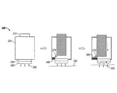
FIG. 5 depicts one embodiment of a drug delivery device 500 having a housing
520, a
depressible portion 510, a supporting layer 530, and a substrate 540 from
which an array of
microneedles 550 extends. The device also has an apparatus that includes a
spring 560 and a
trigger 570. Upon depression of the depressible portion 510, the microneedles
penetrate the
tissue surface 580, and then the trigger 570 is activated, thereby releasing
the spring 560,
which applies a shearing force to the supporting layer 530. The application of
the shearing
force results in separated microneedles 590. The device of FIG. 5 may be
reconfigured to
provide a rotational shearing force, for example, by altering the point of
contact between the
spring and supporting layer, and/or using multiple springs. The trigger may be
configured to
swing out during activation to compress the spring further during insertion of
the
microneedles before releasing the shearing force. In this way, the trigger
force may be
controlled, and it permits the shearing force to occur only after the
microneedles are inserted
into the tissue by a predetermined amount.
In one embodiment, the apparatus that applies the shearing force includes an
electronic element configured to apply the shearing force. The electronic
element may
generate the shearing force by at least one of a magnetic field and electric
field. For example,
the supporting layer and/or substrate may be associated with a magnet that
responds to a
magnetic field generated by the electronic element upon activation. The at
least one
apparatus may be configured to apply a rotational shearing force.
In another embodiment, the device includes magnets which in an initial device
configuration are positioned far away from each other. In use, an input force
pressing the
16
CA 03016984 2018-09-06
WO 2016/168847
PCT/US2016/028164
device to insert the microneedles also causes the magnets to become closer to
one another
such that they interact (attract or repel) to trigger a shearing force. It is
also envisioned that
the device could be configured in a reverse scenario where the magnets
interact before use
and pressing on the device to insert the microneedles separates the magnets
and thereby
releases the shearing force.
In embodiments, at least one of the depressible portion, substrate, and
supporting
layer includes a threaded or spiraled portion that applies a shearing force to
the supporting
layer and/or substrate upon application of a force to the depressible portion.
The shearing
force may be rotational. In one embodiment, the depressible portion includes a
threaded or
spiraled rod that corresponds to a threaded orifice of the supporting layer
and/or substrate. In
another embodiment, the substrate and/or supporting layer includes a threaded
rod that
corresponds to a threaded orifice of the depressible portion. In a further
embodiment, the
depressible portion includes protrusions that correspond with spiraled
tracking of the
substrate and/or supporting layer. In another embodiment, the substrate and/or
supporting
layer includes protrusions that correspond with spiral tracking of the
depressible portion. In
yet another embodiment, the rotational motion is used to load a torsion
spring, which then
imparts rotational shear to the substrate or microneedles. To clarify, in
various embodiments,
a rotational output force may be applied throughout the application of the
input force.
Alternatively, the input force could provide an initial non-rotational output
force, which later
becomes a rotational output force.
FIG. 6 depicts one embodiment of a drug delivery device 600 that includes a
depressible portion 610 and a housing 620 in which a supporting layer 630 and
substrate 640
are rotatably mounted. An array of microneedles 650 extends from the substrate
640. The
depressible portion 610 includes a rod 660 with threading 670 that corresponds
to a threaded
orifice of the supporting layer 630, so that the application of force to the
depressible portion
610 [1] penetrates the biological tissue surface 680 with the microneedles
650, and [2]
applies a shearing force to the substrate 640 and supporting layer 630,
resulting in the
deposition of separated microneedles 690 beneath the surface of the biological
tissue 680.
Systems for Triggering Change of Drug Release Rate
In embodiments, the drug delivery devices include a system for triggering,
after the
microneedles are inserted at least partially into a biological tissue, a
change in rate of release
of the drug from the microneedles and into the biological tissue. The release
of drug from the
17
CA 03016984 2018-09-06
WO 2016/168847
PCT/US2016/028164
drug delivery devices may be achieved by triggering events that take place
gradually or at
specific times upon or after deployment.
In embodiments, the system for triggering a change in rate of release of the
drug allow
for discrete periods of drug release to occur after the drug delivery devices
are deployed. For
example, for discrete periods after deployment the drug delivery devices may
release little or
no drug or one or more desired amounts of drug in any sequence. As a further
example, the
drug delivery devices, upon deployment, may allow little or no drug release
for a first time
period, a significant amount of drug release for a second time period, little
or no release for a
third time period, and a moderate amount of drug release for a fourth time
period. Any
.. sequence of these three drug releasing periods could be employed over at
least two sequential
periods. Also, a drug delivery device may include more than one drug, and each
drug may
have a different release profile.
In embodiments, the drug delivery devices include a drug with different
release
profiles at t1, t2, and t3, as shown in the following table:
_________________________________________________________________________
T=0= 0 <t<ti ti<t <t2 t2<t<t3
microneedle
insertion
Embodiment 1 No Release Significant Little or No Significant
Release Release Release
Embodiment 2 No Release Significant Moderate Release Little
Release
Release
Embodiment 3 No Release Little or No Significant Little or
No
Release Release Release
Embodiment 4 No Release Significant No Release No Release
Release
In one embodiment, the system for triggering release of a drug includes a
first portion of the
microneedles configured to bioerode at a greater rate than a second portion of
the
microneedles upon contact with a biological fluid. For example, the
permeability change
may allow tissue fluid (such as interstitial fluid) to penetrate the
microneedles, and, as a
result, allow the release of drug from the microneedles into the surrounding
tissues via
diffusion. Also, a change in osmotic pressure may cause or drive the release
of drug.
Changes in the surrounding environment or within the microneedles may lead to
a change in
osmotic pressure and result in the release of drug. For example, fluid from
the surrounding
environment, such as biological fluid, may enter the microneedles due to
osmotic forces,
which may help drive drugs out of the microneedles, and, if included in the
drug delivery
devices, a barrier as described herein. Convective flow driven by a pressure
(including
18
CA 03016984 2018-09-06
WO 2016/168847
PCT/US2016/028164
osmotic pressure) difference may also induce flow out of the microneedle,
which can
facilitate drug transport out of the microneedle.
In one embodiment, the system for triggering drug release includes a change in
binding between the drug and another molecule in the microneedles. The other
molecule
may be an excipient. The binding may be covalent or non-covalent. The binding
may retain
the drug within the microneedles until a change in drug binding allows the
release of the drug
from the microneedles. The strength of the binding/affinity between drug and
microneedles
may be tailored to release drug slowly from microneedles into surrounding
tissues. This may
be achieved, for example, by relying on specific intermolecular forces, such
as ionic bonds,
hydrogen bonds, or van der Waal s forces, to achieve a given release profile.
In one embodiment, the system for triggering drug release includes a change in
the
diffusivity of drug. The change in diffusivity may be caused by a change of
the [1] charge
(pH) of a drug and/or another molecule in the microneedles, [2] hydrophilicity
or
hydrophobicity of a drug and/or another molecule in the microneedles, [3]
molecular
size/shape of a drug and/or another molecule in the microneedles, [4]
shape/conformation of
a drug and/or another molecule in the microneedles, or [5] a combination
thereof. A decrease
in the size (mass) of molecules can lead to increased diffusion of drug. The
change in
molecular size can be the result of breaking covalent bonds (e.g.,
degradation) or the result of
breaking weaker bonds (e.g., unbinding/binding, deaggregation/aggregation).
Drugs may be
covalently linked to a component of the microneedles, and such covalent bonds
may be
cleaved enzymatically, chemically, or via a change in pH. The change in
shape/conformation
of drug can influence its rate of release, sometimes in a non-linear manner
because a small
change in shape/conformation can have a large effect on release from the
microneedles
because there may be a pore or other transport pathway that is of similar size
as the drug
molecule, such that a conformational change could determine whether the drug
molecules can
pass through the pathway easily, if at all. A change in conformation also may
affect which
regions of the drug molecule are sequestered in the interior of the molecule
and which are
exposed on the molecule's outer surface. The resulting difference in surface
properties of the
molecule may affect its interaction with the surrounding medium and thereby
have a different
release rate due to changes in attractive and repulsive forces (e.g.,
hydrophobicity, charge).
A change in shape/conformation may be induced by changes in temperature, pH,
ionic
strength, other techniques known in the art, or a combination thereof. The
strength of the
binding/affinity may be varied during deployment of the drug delivery devices
to alter, i.e.,
increase or decrease, the release of drug. This may be the result of an
external stimulus (e.g.,
19
CA 03016984 2018-09-06
WO 2016/168847
PCT/US2016/028164
the user triggers the change in binding strength) or as a result of an
internal stimulus (e.g., a
chemical or biological change within the user triggers the change in
binding/affinity
strength).
In one embodiment, the system for triggering drug release includes a
structural
change of the microneedles. The structural change may include separable
microneedles, such
as those described herein. The structural change, including separation of the
microneedles,
may result in a drug being released from the microneedles upon or after
exposure to
surrounding tissues or fluids following the structural change.
In one embodiment, the system for triggering drug release includes a change in
shape
of the microneedles. For example, a tip or outer layer of the microneedles may
dissolve first,
thereby exposing drugs within the microneedles to biological tissues and
fluids.
In embodiments, the system for triggering drug release includes a change of
one or
more properties of tissues targeted and/or surrounded by the drug delivery
devices upon
deployment. In one embodiment, blood flow/perfusion may be increased or
decreased in
tissues in the proximity of the microneedles' insertion site and, as a result,
affect the release
and uptake of drug. Blood flow/perfusion may be modulated by relying on [1]
temperature
(e.g., applying heat or cooling drug delivery device and/or insertion
site/area), [2] mechanical
forces (e.g., applying pressure, rubbing, vibration, use of a
ring/tourniquet), [3] chemical
methods (e.g., bioactives, irritants, vasodilators, vasoconstrictors), and [4]
a combination
thereof. In another embodiment, tissue permeability and/or convective flow may
be varied to
achieve an increase or decrease in drug release. Changes in tissue
permeability/convective
flow may be achieved [1] chemically/ biochemically (e.g., hyaluronidase may be
used to
degrade the extracellular matrix, or changes in interstitial fluid pressure
also may be achieved
to alter active uptake by surrounding tissues), [2] physically (e.g.,
temperature, pressure,
water content, mechanical (including mechanical damage to tissue, such as by
microneedles),
electroporation, thermal perturbation/damage, ultrasound, cavitation, laser,
radiofrequency
energy are all means to alter tissue permeability), or [3] a combination
thereof. In a further
embodiment, material from biological tissue or extracellular matrix interacts
with or covers,
enters, and/or obstructs microneedles to change the drug release rate. For
example, water
may enter microneedles to dissolve material and/or an analyte from tissue may
displace a
bound drug. In still another embodiment, driving forces that transport drug
from
microneedles and through tissue are modulated. Driving forces that can affect
the rate of
release of actives can be modulated to control the drug release profile.
Driving forces that
can be modulated include [1] electrophoresis, [2] electro-osmosis, [3]
concentration gradient
CA 03016984 2018-09-06
WO 2016/168847
PCT/US2016/028164
(which, when increase, may enhance clearance of drug from tissue by blood
flow, lymphatic
flow, metabolism, other active and passive transport processes (or the
reverse), [4] pressure
gradient (e.g., ultrasound, mechanical perturbation, rubbing/vibration, e.g.,
to cause
convection), or [5] a combination thereof.
In one embodiment, the system for triggering includes a barrier material
positioned in
or on at least part of the microneedle. The barrier provided by the barrier
material, in
embodiments, impedes release of the drug from the microneedle in at least one
direction
and/or for a predetermined period of time.
In embodiments, the system for triggering changes the rate of release in
response to
one or more of the following: analyte concentration, temperature, pH,
pressure, electric field,
magnetic field, electrical charge, electrical current, vibration, ultrasound,
shearing force,
mechanical movement/perturbation, molecule/cell binding, moisture/water
content of the
microneedles, time, diffusion of species from the microneedles, dissolution,
degradation,
chemical reaction, other mechanisms known in the art, or a combination
thereof. Other
mechanisms known in the art include those disclosed at Siepmann, J. et al.,
"Fundamentals
and Applications of Controlled Release Drug Delivery," 1st Edition, 2012,
XIII, p. 592; Li,
X., "Design of Controlled Release Drug Delivery Systems, McGraw-Hill Chemical
Engineering, November 3, 2005; and Wise, Donald L., Handbook of Pharmaceutical
Controlled Release Technology, CRC Press, August 24, 2000.
In embodiments, the system for triggering changes the rate of drug release in
response
to a change of pH. For example, the change of pH may be a lowering of pH in
response to
increased glucose concentration, and the resulting lower of pH may cause the
system for
triggering to release or increase the release of insulin from a drug delivery
device provided
herein.
In embodiments, the system for triggering changes the rate of drug release in
response
to a change of temperature. External or internal (to the body) modulation of
temperature,
therefore, may modulate drug release from the drug delivery devices.
In embodiments, the system for triggering changes the rate of drug release in
response
to mechanical movement/perturbation, vibration, or a combination thereof. The
mechanical
movement/perturbation and/or vibration may be applied to the drug delivery
devices and/or
the surrounding tissues by the drug delivery devices or an external user to
modulate drug
release.
In embodiments, the system for triggering a change of drug release rate
includes
partially inserting one or more microneedles of an array of microneedles,
allowing drug
21
CA 03016984 2018-09-06
WO 2016/168847
PCT/US2016/028164
release from a first part of the microneedles, and then completely or further
inserting the one
or more microneedles, allowing drug release from a second part of the
microneedles. Partial
insertion of microneedles into biological tissue may allow for partial
dissolution of
microneedles, e.g., dissolution of only the part of the microneedles that is
inserted, although
more of the microneedles may dissolve if tissue fluids reach by diffusion or
capillary forces
parts of the microneedles on or above the skin surface that are not yet
inserted. When partial
insertion occurs, the drug associated with the portion of the microneedles may
be released.
These techniques may be used to modulate the quantities and release kinetics
of one or more
drugs within a drug delivery device. For example, initial partial insertion
and dissolution of
microneedles might allow an initial release/burst of drug followed by
additional releases at
specific time points or in a continuous or semi-continuous fashion over a
period of time.
These techniques also may be used to deliver different drugs in sequence when
different parts
of the microneedles contain different drugs. For example, the tip of the
microneedles could
contain drug "A" and the rest of the microneedles' bodies could contain drug
"A" or a
different drug, drug "B." Following insertion of the tips only, drug "A" would
be released,
and further insertion may permit release of an additional amount of drug "A"
or the release of
drug "B." In each of the foregoing scenarios, the amount/degree of
microneedles insertion
and the period over which partial or full microneedle insertion occurs can be
varied.
Each of the foregoing mechanisms may be used alone or in any combination in
the
system for triggering a change of drug release. Each of the foregoing
mechanisms also may
include increasing or decreasing the concentration of the drug and/or
excipients in the
microneedles. Doing so may alter the concentration gradient, which drives
transport by
diffusion, and can also alter the amount of drug moved by other mechanisms,
such as
convection and electrically driven transport. Increasing or decreasing the
concentration of
excipients can alter the rate at which drug moves through the environment
containing the
excipients, such as altering the drug diffusivity/mobility, the medium
viscosity, the medium
porosity and other factors.
Barrier
In embodiments, the system for triggering change of drug release rate is a
barrier that
may be positioned in or on at least part of the microneedle to impede release
of the drug from
the microneedle in at least one direction and/or for a predetermined period of
time. In one
embodiment, the barrier is configured to permit (i) discrete periods of drug
release upon or
after implantation, (ii) control of the region of the microneedles from which
the drug is
22
CA 03016984 2018-09-06
WO 2016/168847
PCT/US2016/028164
released, or (iii) a combination thereof. The term "barrier" and the phrase
"barrier material"
are used interchangeably herein.
In embodiments, the barrier impedes release of a drug from the microneedle
until the
barrier no longer obstructs release of the drug. The obstruction provided by
the barrier may
be permanent, or lessened gradually or substantially instantaneously.
A barrier generally may be positioned in a microneedle, on a microneedle, or a
combination thereof. For example, a barrier may at least partially encapsulate
a drug in a
microneedle, be dispersed within the matrix of one or more microneedles, be
positioned on
and/or at the surface of one or more microneedles, or a combination thereof.
When dispersed
within the matrix of one or more microneedles, the barrier may include
discrete regions
within the matrix. When a barrier encapsulates a drug, a drug delivery device
may include
one drug encapsulated with different amounts/concentrations of one or more
barrier
materials, two or more drugs encapsulated with different
amounts/concentrations of the same
or different barrier materials, or a combination thereof.
FIG. 7 depicts one embodiment of a drug delivery device 700 that includes a
barrier
positioned on the surface of microneedles. The drug delivery device includes a
supporting
layer 710, a substrate 720, and an array of microneedles 740 including a drug
730, which
extend from the substrate 720. Each microneedle 740 has a barrier material 750
positioned
on its surface.
In embodiments, the microneedles themselves act as barriers when drug is
disposed in
the substrate.
In embodiments, at least a portion of the barrier is configured to be
permanent. In
other words, the barrier is configured to remain in place upon and after
deployment, and is
substantially impervious to all mechanism that may remove or lessen the
obstruction
provided by the barrier.
The barrier or barrier material may include one or more different materials.
The
barrier may include two or more different materials, each associated with the
same or
different portions of the drug delivery devices. When associated with the same
portion of a
drug delivery device, the two or more materials may form a multi-layered
barrier material.
Alternatively, the barrier may include two materials, each coating a separate
portion of a
microneedle; or the barrier may include two materials, the first material
being a liquid
disposed in a second material that is a solid.
A single microneedle array may include two or more types of barriers. For
example,
an array could include one row of microneedles having a barrier of a first
type and a second
23
CA 03016984 2018-09-06
WO 2016/168847
PCT/US2016/028164
row of microneedles having a barrier of a second type. For example, the
differences could be
beneficially designed for delivering two different substances of interest.
In one embodiment, the barrier material includes a first coating positioned in
or on at
least a first portion of one or more microneedles of the array of
microneedles. The first
coating may be at least substantially inert in biological fluid (e.g.,
insoluble) and unchanged
upon and after deployment, and prevent drug release from the first portion of
one or more
microneedles of the array of microneedles. Alternatively, barrier material
includes a first
coating having one or more properties, such as permeability or porosity, that
change upon or
after contacting a biological fluid, and therefore permits drug release from
the first portion of
one or more microneedles of the array of microneedles. The first coating, for
example, may
be at least partially soluble in biological fluid. In another embodiment, the
barrier material
also includes a second coating positioned in or on a second portion of one or
more
microneedles of the array of microneedles. The first coating may be inert
(e.g., insoluble)
and unchanged upon and after deployment in biological tissue and fluid, and
the second
coating may have one or more properties, such as porosity and/or permeability,
that change
upon deployment, thereby permitting drug release to occur only from the second
portion of
one or more microneedles of the array of microneedles.
In one embodiment, the barrier material includes a first coating positioned in
or on at
least a first portion of one or more microneedles of the array of
microneedles, and a second
coating positioned in or on a second portion of one or more microneedles of
the array of
microneedles. The first and second coatings may permit drug release at
different times upon
deployment, therefore allowing two drugs to be released simultaneously,
sequentially, or a
combination thereof.
In embodiments, the obstruction provided by at least a portion of the barrier
is
removed, gradually or completely, upon or after the onset of dissolution of a
barrier material,
swelling/expansion of a barrier material, chemical reaction/degradation of a
barrier material,
vaporization of a barrier material, solidification of a barrier material,
melting of a barrier
material, gelling of a barrier material,
deformation/breaking/collapsing/contracting of a
barrier material, change of charge state of a barrier material, or a
combination thereof. The
composition of the barrier material may be selected or formulated so that its
dissolution rate
allows it to achieve a desired drug release profile.
In embodiments, the obstruction provided by at least a portion of the barrier
is
removed, gradually or completely, upon or after a change in binding/affinity
between the
barrier and drug. Binding/affinity between drug and a barrier may be used,
therefore, to
24
CA 03016984 2018-09-06
WO 2016/168847
PCT/US2016/028164
achieve a specific drug release profile, or modulate a drug release profile.
Binding/affinity
between the barrier and drug may be achieved by any techniques known in the
art. For
example, binding/affinity may be charge-mediated, i.e., based on the
respective charge state
of each component. The charge state, as explained herein, may be changed by
modulating
pH. A change in pH can be used to increase or decrease the charge state of
drug and/or
barrier materials. As pH decreases, basic drugs may become more charged, and
acidic drugs
become less charged. Also, a voltage or electric field may be applied to at
least a portion of
the drug delivery devices, resulting in a change in the charge distribution on
the drugs, which
results in a change in binding strength/affinity. As a further example, the
binding of drug to a
barrier material may vary based on the presence or introduction of other
molecules or species
having higher affinity for the barrier material than the drug.
Binding/affinity also may be
modulated by pH, temperature, pressure, ionic strength, competitive binding,
chemical
reactions, or a combination thereof.
In one embodiment, the obstruction provided by at least a portion of the
barrier is
removed, gradually or completely, upon or after the onset of dissolution of a
barrier material.
Therefore, the barrier may be formed with a barrier material, such as a salt,
that dissolves
upon exposure to a biological fluid at the tissue site of insertion, or a
polymer that degrades
via hydrolysis. For example, a salt having low aqueous solubility may delay
release of the
active. In embodiments, upon dissolution of a barrier material, at least a
portion of a
microneedle is exposed to biological tissue and/or fluid, thereby permitting
release of drug
from the portion of the microneedle associated with the barrier material. In
further
embodiments, upon dissolution of a barrier material, the porosity within the
microneedle may
be increased, thereby permitting release of drug.
One embodiment of a drug delivery device including a bioerodible barrier
material is
depicted at FIG. 8A and FIG. 8B. The drug delivery device 800 includes a
supporting layer
810 and a substrate 820 from which microneedles 825 extend. The microneedles
825 include
drug 830 and discrete portions of a bioerodible barrier material 840. Upon
dissolution or
other degradation of the barrier material 840 after the microneedles penetrate
the tissue
surface 845, pores 850 are created in the microneedles, which permits release
of the drug 830.
One embodiment of a drug delivery device included a barrier having a
bioerodible
portion and a (relatively) non-bioerodible portion is depicted at FIG. 9A and
FIG. 9B. FIG.
9A depicts a drug delivery device 900 having a supporting layer 910 and a
substrate 920 from
which microneedles 930 extend. The microneedles include a drug 940, and are
associated
with a barrier material that includes a first coating 950 that is permanent
and insoluble in
CA 03016984 2018-09-06
WO 2016/168847
PCT/US2016/028164
biological fluids, and a second coating 960 that is soluble in biological
fluid. Upon
penetration of skin, the second coating 960 dissolves, as shown at FIG. 9B,
thereby
permitting the drug 940 to be released at the dermis, while the first coating
950 remains
intact, thereby prohibit drug release at the epidermis. In an alternative
embodiment, the
portion of the barrier 950 associated with the epidermis is soluble in
biological fluid, and the
portion of the barrier 960 associated with the dermis is insoluble and
permanent, therefore
resulting in release of the drug only into the epidermis.
In one embodiment, the obstruction provided by at least a portion of the
barrier may
be removed, gradually or completely, upon or after the onset of
swelling/expansion of the
-- barrier material. Therefore, the barrier may be formed with a barrier
material, such as a gel,
that expands upon exposure to a biological fluid, temperature, other stimuli,
or a combination
thereof, thereby increasing the permeability of the biological material, which
permits the
release of drug. For example, the drug delivery devices, including the
microneedles, may
include a channel that is at least partially filled with a gel, so that the
ability of drug to
traverse the channel is increased or decreased as the gel contracts or
expands, respectively.
In one embodiment, the obstruction provided by at least a portion of the
barrier is
removed, gradually or completely, upon or after the onset of a chemical
reaction and/or
degradation of a barrier material. The barrier material, upon or after
exposure to biological
fluids, may undergo changes in its physicochemical properties as a result of
chemical,
physical, mechanical, and/or biological interactions with a biological tissue.
Therefore,
degradation of the barrier material may occur. In embodiments, in which the
barrier material
is a polymeric material, degradation may be initiated, upon or after contact
with biological
fluid, by hydrolytic scission of polymer chains, which may result in bulk
degradation and/or
surface erosion of the polymer. Degradation also may occur by enzymatic
degradation.
In one embodiment, the obstruction provided by at least a portion of the
barrier is
removed, gradually or completely, upon or after vaporization of a liquid
included in a barrier
material. The vaporization of the liquid may change the permeability and/or
porosity of the
barrier material, or create or expose pores within microneedles. The
vaporization may be
triggered by evaporation, boiling, acoustic droplet vaporization, or by other
means known in
the art. The liquid that vaporizes may be formed within the barrier prior to
deployment of the
drug delivery devices, or biological fluid that penetrates the barrier
material upon or after
deployment.
In one embodiment, the obstruction provided by at least a portion of the
barrier is
removed, gradually or completely, upon or after solidification of a barrier
material, such as an
26
CA 03016984 2018-09-06
WO 2016/168847
PCT/US2016/028164
aquamelt, which typically is a naturally hydrated polymeric material capable
of solidifying at
certain temperatures through a controlled stress input, including mechanical
or chemical
input. Aquamelts made from chitin, fibroin, or a combination thereof may be
used. The
solidification may be initiated upon or after exposure to biological fluid, or
upon or after
exposure to the environment prior to deployment. Upon or after solidification,
the
permeability of the barrier material is increased.
In one embodiment, the obstruction provided by at least a portion of the
barrier may
be removed, gradually or completely, upon or after melting of a barrier
material. Melting
may be initiated following insertion of microneedles into biological tissue,
upon exposing the
microneedles to the environment upon or after removal from their packaging, by
application
of heat by an external source, or a combination thereof. For example, a drug
delivery device
stored in freezing conditions may include water as a barrier material, and the
water melts
upon exposure to ambient conditions, biological fluid, or a combination
thereof.
In one embodiment, the obstruction provided by the barrier is removed,
gradually or
completely, upon or after a change of charge state of a barrier material. For
example, a
barrier material may be or become charged prior to or after deployment,
respectively, and
then the charge of the barrier material changes or is lost. The change of
charge state of a
barrier material may be used to control drug release. In embodiments, a
barrier material and
drug have opposite charge states, and the repulsion force is used to retain
the drug in the
microneedles until the charge of the barrier material is changed or lost. In
further
embodiments, a barrier material and drug have the same charge state, and the
attraction
between the barrier material and drug is used to retain the drug in the
microneedles until the
charge state of the barrier material changes or is lost.
Also, a change in charge state of the barrier and/or drug may alter the
release of drug
under the effect of an electric field. The release may also be caused by the
application of a
change in electric field. For example, an electric field may be applied across
the
microneedles and the targeted tissue, and optionally at discrete time points
to affect the
kinetics of the release. The application of the electric field or change in
field strength and/or
direction may trigger the release from the matrix of charged drug. A change of
charge may
be effected by a change of pH or ionic strength (which shields the charge) or
other factors.
Also, electrophoresis also may be used to drive charged particles or drugs
from the
microneedles and/or through a barrier. Electroosmosis may also be used to
drive liquid
containing an active across a charged barrier.
27
CA 03016984 2018-09-06
WO 2016/168847
PCT/US2016/028164
In one embodiment, the obstruction provided by at least a portion of the
barrier is
removed, gradually or completely, upon or after deformation, breaking,
collapsing, and/or
contraction of a barrier material. A barrier material may deform, break,
collapse, and/or
contract upon exposure to compression, tension, shear, torque, vibrations,
ultra-sound, or a
combination thereof. One or more of these forces may be applied to any portion
of the drug
delivery devices. For example, in the device depicted at FIG. 9A, the portion
of the barrier
960 associated with the dermis could have been configured to fail by breaking,
deforming,
collapsing, or contracting, as opposed to dissolving.
In one embodiment, the obstruction provided by at least a portion of the
barrier is
permanent. A permanent barrier, for example, may prevent one or more
microneedles from
releasing drug from the region associated with the permanent barrier.
Therefore, the
pennanent barrier may ensure that the microneedle is capable of releasing drug
only from
areas not obstructed by the permanent barrier, including areas exposed by the
separation of
microneedles.
One embodiment of a drug delivery device having separable microneedles and a
permanent barrier is depicted at FIG. 10A and FIG. 10B. The drug delivery
device 1000 of
FIG. 10A includes a supporting layer 1010 and microneedles 1030 extending from
a
substrate 1020. The microneedles include a drug 1040 and each of the
microneedles 1030 are
coated with a peinianent barrier 1050, so that upon separation of the
microneedles, as
depicted at HG. 10B, the drug 1040 is released primarily to the tissue surface
1060 because
the barrier 1050 remains in place, thereby preventing drug release beneath the
tissue surface
1060.
Supporting Layer
The supporting layer may be adhered to the substrate by any means known in the
art,
including an adhesive. In one embodiment, an adhesive layer is use to adhere
the supporting
layer to the substrate.
The supporting layer may be made out of a variety of materials. In some
embodiments, the supporting layer may be a composite material or multilayer
material
including materials with various properties to provide the desired properties
and functions.
For example, the supporting layer may be flexible, semi-rigid, or rigid,
depending on the
particular application. As another example, the supporting layer may be
substantially
impermeable, protecting the one or more microneedles (or other components)
from moisture,
gases, and contaminants.
28
CA 03016984 2018-09-06
WO 2016/168847
PCT/US2016/028164
Alternatively, the supporting layer may have other degrees of permeability
and/or
porosity based on the desired level of protection that is desired. Non-
limiting examples of
materials that may be used for the supporting layer include various polymers,
elastomers,
foams, paper-based materials, foil-based materials, metallized films, and non-
woven and
woven materials.
An optional mechanical force indicator may be disposed between the supporting
layer
and the substrate, or it may be located within or be an integral part of the
supporting layer.
The mechanical force indicator may be used to indicate to a person the amount
of force
and/or pressure applied to the drug delivery device during its use. For
example, in one
embodiment, the indicator is configured to provide a signal when a force
applied to the drug
delivery device by a person (in the course of applying the drug delivery
device to a patient's
skin to insert the one or more microneedles into the patient's skin) meets or
exceeds a
predetermined threshold. The predetermined threshold may be the minimum force
or some
amount greater than the minimum force that is required for a particular drug
delivery device
to be effectively applied to a patient's skin. In other words, it may be the
force needed to
cause the microneedles to be properly, e.g., partially or fully, inserted into
a patient's skin; or
it may be the force needed to cause the microneedles to be properly, e.g.,
partially or fully,
inserted into a patient's skin, and separate the microneedles from the
substrate.
Methods of Using the Drug Delivery Devices
As used herein, the phrase "penetrate a tissue surface" or the terms
"penetrate" or
"penetration" refers to the insertion of at least 50 %, and typically
substantially all, of the
microneedles of an array of microneedles, including at least the tip or distal
end portion of the
microneedles, into a biological tissue. In a preferred embodiment, the
"penetration" includes
piercing the stratum corneum of the skin of a human patient such that at least
the tip end
portion of the microneedle is within or has passed across the viable
epidermis.
The drug delivery devices provided herein may be self-administered or
administered
by another individual (e.g., a parent, guardian, minimally trained healthcare
worker, expertly
trained healthcare worker, and/or others).
Thus, embodiments provided herein further include a simple and effective
method of
administering a substance of interest with a drug delivery device. The methods
provided
herein may include identifying an application site and, preferably, sanitizing
the area prior to
application of the drug delivery device (e.g., using an alcohol wipe). The
drug delivery
29
CA 03016984 2018-09-06
WO 2016/168847
PCT/US2016/028164
device then is applied to the patient's skin/tissue and manually pressed into
the patient's
skin/tissue (e.g., using the thumb or finger) by applying a force as described
herein.
After administration is complete, the substrate, supporting layer, housing,
and/or
depressible portion may be removed from the patient's skin/tissue in
embodiments having
separable microneedles. In embodiments, the drug delivery devices described
herein are used
to deliver one or more substances of interest (e.g., vaccines, therapeutics,
vitamins) into the
body, tissue, cells, and/or organ. In one embodiment, the drug delivery
devices are used to
deliver the active into skin by inserting the microneedles across the stratum
corneum (outer
to 20 microns of skin that is the barrier to transdermal transport) and into
the viable
10 epidermis and dermis. The small size of the microneedles enables them to
cause little to no
pain and target the intradermal space. The intradermal space is highly
vascularized and rich
in immune cells and provides an attractive path to administer both vaccines
and therapeutics.
The microneedles are preferably dissolvable and once in the intradermal space
they dissolve
within the interstitial fluid and release the active into the skin. In
embodiments that include
separable microneedles, the substrate can be removed and discarded upon or
after separation
of the microneedles, which preferably is nearly immediately upon insertion.
In one embodiment, a method is provided for administering a substance of
interest to
a patient, which includes providing one of the microneedle arrays described
herein; and
applying the microneedles of the array to a tissue surface of the patient,
wherein the insertion
of the microneedles of the array into the skin is done manually without the
use of a separate
or intrinsic applicator device. In this particular context, the term
"applicator device" is a
mechanical device that provides its own force, e.g., via a spring action or
the like, which
serves as the primary force to drive the microneedle array against the tissue
surface, separate
from any force the user may impart in holding the device and/or microneedles
against the
.. tissue surface.
Substance of Interest / Active Pharmaceutical Ingredient
A wide range of substances may be formulated for delivery to biological
tissues with
the present microneedles and methods. As used herein, the term "substance of
interest"
includes active pharmaceutical ingredients, allergens, vitamins, cosmetic
agents,
cosmeceuticals, diagnostic agents, markers (e.g., colored dyes or radiological
dyes or
markers), and other materials that are desirable to introduce into a
biological tissue. The
"substance of interest" is sometimes referred to herein as "the active" or an
"API" or a
"drug".
CA 03016984 2018-09-06
WO 2016/168847
PCT/US2016/028164
In one embodiment, the substance of interest is a prophylactic, therapeutic,
or
diagnostic agent useful in medical or veterinary application. In one
embodiment, the
substance of interest is a prophylactic or therapeutic substance, which may be
referred to
herein as an API. In certain embodiments, the API is selected from suitable
proteins, peptides
and fragments thereof, which can be naturally occurring, synthesized or
recombinantly
produced. Representative examples of types of API for delivery include
antibiotics, antiviral
agents, analgesics, anesthetics, antihistamines, anti-inflammatory agents,
anti-coagulants,
allergens, vitamins, antineoplastic agents.
In one embodiment, the substance of interest comprises a vaccine. Examples of
vaccines include vaccines for infectious diseases, therapeutic vaccines for
cancers,
neurological disorders, allergies, and smoking cessation or other addictions.
Some examples
of current and future vaccines for the prevention of, anthrax, cervical cancer
(human
papillomavirus), dengue fever, diphtheria, Ebola, hepatitis A, hepatitis B,
hepatitis C,
haemophilus influenzae type b (Hib), HIV/AIDS, human papillomavirus (HPV),
influenza
(seasonal and pandemic), Japanese encephalitis (JE), lyme disease, malaria,
measles,
meningococcal, monkeypox, mumps, pertussis, pneumococcal, polio, rabies,
rotavirus,
rubella, shingles (herpes zoster), smallpox, tetanus, typhoid, tuberculosis
(TB), varicella
(chickenpox), West Nile, and yellow fever.
In another embodiment, the substance of interest comprises a therapeutic
agent. The
therapeutic agent may be selected from small molecules and larger
biotechnology produced
or purified molecules (e.g., peptides, proteins, DNA, RNA). Examples of
therapeutics, which
may include their analogues and antagonists, include but are not limited to
insulin, insulin-
like growth factor, insultropin, parathyroid hormone, pramlintide acetate,
growth hormone
release hormone, growth hormone release factor, mecasermin, Factor VIII,
Factor IX,
antithrombin HI, protein C, protein S, P-gluco-cerebrosidase, alglucosidase-a,
laronidase,
idursulphase, galsulphase, agalsidase-P, a-1 proteinase inhibitor, lactase,
pancreatic enzymes,
adenosine deaminase, pooled immunoglobulins, human albumin, erythropoietin,
darbepoetin-
a, filgrastim, pegfilgrastim, sargramostim, oprelvekin, human follicle-
stimulating hormone,
human chorionic gonadotropin, lutropin-a,interferon (alpha, beta, gamma),
aldesleukin,
alteplase, reteplase, tenecteplase, urokinase, factor Vila, drotrecogin-a,
salmon calcitonin,
exenatide, octreotide, dibotermin-a, recombinant human bone morphogenic
protein 7,
histrelin acetate, palifermin, becaplermin, trypsin, nesiritide, botulinum
toxin (types A and
B), collagenase, human deoxyribonuclease I, hyaluronidase, papain, 1-
asparaginase, peg-
asparaginase, rasburicase, lepirudin, bivalirudin, streptokinase,
anistreplase, bevacizumab,
31
CA 03016984 2018-09-06
WO 2016/168847
PCT/US2016/028164
cetuximab, panitumumab, alemtuzumab, rituximab, trastuzumab, abatacept,
anakinra,
adalimumab, etanercept, infliximab, alefacept, efalizuman, natalizumab,
eculizumab,
antithymocyte globulin, basiliximab, daclizumab, muromonab-CD3, omalizumab,
palivizumab, enfuvirtide, abciximab, pegvisomant, crotalidene polyvalent fab
(ovine),
digoxin immune serum fab (ovine), ranibizumab, denileukin diftitox,
ibritumomab tiuxetan,
gemtuzumab ozogamicin, tositumomab, I- tositumomab, anti-rhesus (rh)
immunoglobulin G,
desmopressin, vasopressin, deamino [Va14, D-Arg8] arginine vasopressin,
somatostatin,
somatotropin, bradykinin, bleomycin sulfate, chymopapain, glucagon,
epoprostenol,
cholecystokinin, oxytocin, corticotropin, prostaglandin, pentigetide, thymosin
alpha- 1, alpha-
1 antitrypsin, fentanyl, lidocaine, epinephrine, sumatriptan, benztropine
mesylate, liraglutide,
fondaparinux, heparin, hydromorphone, omacetaxine mepesuccinate, pramlintide
acetate,
thyrotropin-alpha, glycopyrrolate, dihydroergotamine mesylate, Bortezomib,
triptoreline
pamaote, teduglutide, methylnaltrexone bromide, pasireoti de, ondansetron
hydrochloride,
droperidol, triamcinolone (hex)acetonide, aripiprazole, estradiol valerate,
morphine sulfate,
olanzapine, methadone hydrochloride, and methotrexate.
In yet another embodiment, the substance of interest is a vitamin, herb, or
dietary
supplement known in the art. Non- limiting examples include 5-HIP (5-
hydroxytryptophan),
acai berry, acetyl-L-carnitine, activated charcoal, aloe vera, alpha-lipoic
acid, apple cider
vinegar, arginine, ashitaba, ashwagandha, astaxanthin, barley, bee pollen,
beta-alanine, beta-
carotene, beta-glucans, biotin, bitter melon, black cherry, black cohosh,
black currant, black
tea, branched-ahain amino acids, bromelain (bromelin), calcium, camphor,
chamomile,
chasteberry, chitosan, chlorella, chlorophyll, choline, chondroitin, chromium,
cinnamon,
citicoline, coconut water, coenzyme Q10, conjugated linoleic acid, cordyceps,
cranberry,
creatine, D-mannose, damiana, deer velvet, DHEA, DMSO, echinacea, EDTA,
elderberry,
emu Oil, evening primrose oil, fenugreek, feverfew, folic acid, forskolin,
GABA (gamma-
aminobutyric acid), gelatin, ginger, ginkgo biloba, ginseng, glycine,
glucosamine,
glucosamine sulfate, glutathione, gotu kola, grape seed extract, green coffee,
guarana, guggul,
gymnema, hawthorn, hibiscus, holy basil, horny goat weed, inulin, iron, krill
oil, L-carnitine,
L-citrulline, L-trypotophan, lactobacillus, magnesium, magnolia, milk thistle,
MSM
(methylsulfonylmethane), niacin, olive, omega-3 fatty acids, oolong tea,
oregano,
passionflower, pectin, phenylalanine, phosphatidylserine, potassium,
probiotics,
progesterone, quercetin, ribose, red yeast rice, reishi mushroom, resveratrol,
rosehip, saffron,
SAM-e, saw palmetto, schisandra, sea buckthorn, selenium, senna, slippery elm,
St. John's
wort, stinging nettle, tea tree oil, theanine, tribulus terrestris, turmeric
(curcumin), tyrosine,
32
CA 03016984 2018-09-06
WO 2016/168847
PCT/US2016/028164
valerian, vitamin A, vitamin B12, vitamin C, vitamin D, vitamin E, vitamin K,
whey protein,
witch hazel, xanthan gum, xylitol, yohimbe, and zinc.
A microneedle array may include a single substance of interest or it may
include two
or more substances of interest. In the latter case, the different substances
may be provided
together within one of the microneedles, or some microneedles in an array of
microneedles
contain one substance of interest while other microneedles contain another
substance of
interest.
The API desirably is provided in a stable formulation or composition (i.e.,
one in
which the biologically active material therein essentially retains its
physical stability and/or
chemical stability and/or biological activity upon storage). Stability can be
measured at a
selected temperature for a selected period. Trend analysis can be used to
estimate an
expected shelf life before a material has actually been in storage for that
time period.
In embodiments, the substance of interest is provided as a solid that is "dry"
or has
been "dried" to form the one or more microneedles and becomes solubilized in
vivo
following insertion of the microneedle into the patient's biological tissue.
As used herein, the
term "dry" or "dried" refers to a composition from which a substantial portion
of any water
has been removed to produce a solid phase of the composition. The term does
not require the
complete absence of moisture (e.g., the API may have a moisture content from
about 0.1% by
weight and about 25% by weight).
The substance of interest may be included in a formulation with one or more
excipients and other additives, as detailed below.
Matrix Material/Excipients
The matrix material forms the bulk of the microneedle and substrate. It
typically
includes a biocompatible polymeric material, alone or in combination with
other materials. In
embodiments, the matrix material, at least of the microneedles, is water
soluble. In certain
preferred embodiments, the matrix material includes one or a combination of
polyvinyl
alcohol, dextran, carboxymethylcellulose, maltodextrin, sucrose, trehalose,
and other sugars.
As used herein, the telins "matrix material" and "excipient" are used
interchangeably when
referring to any excipients that are not volatilized during drying and
formation of the
microneedles and substrate.
The fluid solution used in the mold filling processes described herein may
include any
of a variety of excipients. The excipients may consist of those that are
widely used in
pharmaceutical formulations or ones that are novel. In a preferred embodiment,
the excipients
33
CA 03016984 2018-09-06
WO 2016/168847
PCT/US2016/028164
are ones in FDA-approved drug products (see the Inactive Ingredient Search for
Approved
Drug Products at http://www.accessdata.fda.gov/scripts/cder/iig/index.Cfm).
None, one, or
more than one excipient from the following categories of excipients may be
used: stabilizers,
buffers, bulking agents or fillers, adjuvants, surfactants, disintegrants,
antioxidants,
solubilizers, lyo-protectants, antimicrobials, antiadherents, colors,
lubricants, viscosity
enhancer, glidants, preservatives, materials for prolonging or controlling
delivery (e.g.,
biodegradable polymers, gels, depot forming materials, and others). Also, a
single excipient
may perform more than one formulation role. For example, a sugar may be used
as a
stabilizer and a bulking agent or a buffer may be used to both buffer pH and
protect the active
from oxidation. Some examples of excipients include, but are not limited to
lactose, sucrose,
glucose, mannitol, sorbitol, trehalose, fructose, galactose, dextrose,
xylitol, maltitol,
raffinose, dextran, cyclodextrin, collagen, glycine, histidine, calcium
carbonate, magnesium
stearate, serum albumin (human and/or animal sources), gelatin, chitosan, DNA,
hylaruronic
acid, polyvinylpyrrolidone, polyvinyl alcohol, polylactic acid (PLA),
polyglycolic acid
(PGA), polylactive co-glycolic acid (PLGA), polyethylene glycol (PEG, PEG 300,
PEG 400,
PEG 600, PEG 3350, PEG 4000), cellulose, methylcellulose, carboxymethyl
cellulose,
sodium carboxymethyl cellulose, hydroxypropyl methylcellulose, acacia,
Lecithin,
Polysorbate 20, Polysorbate 80, Pluronic F-68, Sorbitantrioleate (span 85),
FDTA,
hydroxypropyl cellulose, sodium chloride, sodium phosphate, ammonium acetate,
potassium
phosphate, sodium citrate, sodium hydroxide, sodium carbonate, Tris base-65,
Tris acetate,
Tris HCl-65, citrate buffer, talc, silica, fats, methyl paraben, propyl
paraben, selenium,
vitamins (A, E, C, retinyl palmitate, and selenium), amino acids (methionine,
cysteine,
arginine), citric acid, sodium citrate, benzyl alcohol, chrlorbutanol, cresol,
phenol, thimerosal,
EDTA, acetone sodium bisulfate, ascorbyl palmitate, ascorbate, castor oil,
cottonseed oil,
alum, aluminum hydroxide, aluminum phosphate, calcium phosphate hydroxide,
paraffin oil,
squalene, Quil A, 1L-1, IL-2, IL-12, Freund's complete adjuvant, Freund's
incomplete
adjuvant, killed Bordetella pertussis, Mycobacterium bovis, and toxoids. The
one or more
selected excipients may be selected to improve the stability of the substance
of interest during
drying and storage of the microneedle devices, as well providing bulk and/or
mechanical
properties to the microneedle array and/or serve as an adjuvant to improve the
immune
response to a vaccine.
34
CA 03016984 2018-09-06
WO 2016/168847
PCT/US2016/028164
Manufacture
The arrays of microneedles may be made by any methods known in the art. For
example, the arrays of microneedles may be made using a molding process, which
advantageously is highly scalable. The process may include filling a suitable
mold with
-- suitable fluidized materials; drying the fluidized material to form the
microneedles, the
predefined fracture regions if included, and base substrate; and then removing
the formed part
from the mold. These filling and drying steps may be referred to as "casting"
in the art.
Preferably the methods for making the microneedles are performed under a
minimum ISO 7
(class 10,000) process or an ISO 5 (class 100) process.
In one embodiment, the manufacture of solid, bioerodible microneedles includes
filling a negative mold of the one or more microneedles with an aqueous or non-
aqueous
casting solution of the substance of interest and drying the casting solution
to provide the one
or more solid microneedles. In other embodiments, other solvent or solventless
systems may
be used. Non-limiting examples of methods for filling the negative mold
include deposition,
coating, printing, spraying, and microfilling techniques. The casting solution
may be dried or
cured at ambient temperature, under refrigeration, or at temperatures above
ambient (e.g., 30
to 60 C, or higher) for a period from about 5 seconds to about one week to
form the dry solid
microneedles. In some embodiments, the dry or cure time is from about 10
seconds to about
24 hours, from about 30 minutes to about 12 hours, from about 10 minutes to
about 1 hour, or
from about 1 minute to about 30 minutes. In a preferred embodiment, the dry or
cure time is
from about 10 seconds to about 30 minutes.
Alternatively, the casting solution may be vacuum-filled or filled into the
mold using
a combination of non-vacuum filling and vacuum-filling. For example, in an
embodiment the
negative mold comprises a non-porous but gas-permeable material (e.g., PDMS)
through
which a backside vacuum can be applied. Although the negative mold is solid,
it was
determined that a sufficient vacuum could be applied through the backside when
the molds
are formed of such materials. In some embodiments, the backside vacuum may be
used alone
or in combination with a positive pressure applied on top of the mold for
quicker filling.
Such embodiments could advantageously reduce the time required and improve the
accuracy
and completeness when filling the mold with casting solution. For example, the
casting
solution may be vacuum-filled using a backside vacuum for a period from about
3 minutes to
about 6 hours, from about 3 minutes to about 3 hours, from about 3 minutes to
about 1 hour,
or from about 3 minutes to about 30 minutes.
CA 03016984 2018-09-06
WO 2016/168847
PCT/US2016/028164
Although various temperatures and humidity levels can be employed to dry the
casting solution, the formulations preferably are dried at temperature from
about 1 C to
about 150 C (e.g., from about 5 C to about 99 C, from about 15 C to about 45
C, from
about 25 C to about 45 C, or at about ambient temperature) and about 0 to
about 40%
relative humidity, e.g., about 0% to about 20% relative humidity.
In some embodiments, it may be desirable to use a multi-step casting process
to form
the microneedles and substrate. For example, the tips of the microneedles may
be partially
filled in a first step with a casting solution comprising the substance of
interest followed by
one or more subsequent fill steps with casting solutions of bulking polymers
with or without
.. the same or a different substance of interest. After filling and at least
partially drying the
microneedles in the negative mold, the adhesive layer and backing layer may be
applied to
the base substrate prior to removing the microneedles from the mold. In some
embodiments,
the adhesive layer and/or backing layer are pre-formed prior to application to
the base
substrate, while in other embodiments the adhesive layer and/or backing layer
may be formed
directly in-line.
In one embodiment, the multi-step casting process includes (1) a first cast of
API in
excipient forming the microneedles, (2) a second cast of a frangible material
forming a
fracture region, and (3) a third cast of a matrix material forming the backing
and/or base
substrate.
After at least partially drying the microneedles, the microneedles may be
removed
from the mold. For example, the microneedles may be removed from the mold
before fully
dry (e.g., when still in a rubbery state), but when strong enough to be
peeled, and then dried
further once removed from the mold to further solidify/harden the
microneedles. Such a
technique may be useful when carboxymethylcellulose sodium, polyvinyl alcohol,
sugars,
and other materials are used as a bulking polymer (matrix material) in the
microneedles. In
such embodiments, the microneedles may complete drying prior to or after
packaging.
The devices and methods described above may be further understood with
reference
to the following non-limiting examples.
Example 1
A microneedle array was fabricated as follows: A first solution (of 10 wt%
sucrose, 1
wt% carboxymethyl cellulose in potassium phosphate buffer) was cast under
vacuum into
polydimethylsiloxane (PDMS) microneedle molds and dried under ambient
conditions for 10
minutes to form the microneedles. Then a second solution (of 10 wt% sucrose, 1
wt%
carboxymethyl cellulose in potassium phosphate buffer) was cast under vacuum
in the PDMS
36
CA 03016984 2018-09-06
WO 2016/168847
PCT/US2016/028164
microneedle molds and dried overnight at 35 C to form the substrate/base
layer. Then an
adhesive backing (support layer) (3M 1503 tan single-coated polyethylene
medical tape) was
applied to the base of microneedle array, forming a microneedle patch. The
microneedle
array was then removed from mold and packaged with desiccant in a foil pouch.
Example 2
The microneedle array made in Example 1 was applied to excised porcine skin,
to
insert the microneedles into the skin. Then the patch was pulled laterally
(horizontally,
parallel to the surface of the skin) while maintaining a downward force to
keep the patch
secured against the tissue surface. The patch was then pulled away from the
skin and
imaged. A hydrophilic dye (Gentian violet, 1%) was then applied to the porcine
skin at the
area of the microneedle insertion. The dye was allowed to sit on the surface
of the porcine
skin for 30 seconds, and then it was wiped away with an isopropanol wipe.
Then, the surface
of the porcine skin was imaged and evaluated for staining.
A downward applied force of approximately 10 lbf was applied to the patch
immediately followed by pulling the patch horizontally across the skin. This
resulted in
separation of the microneedles from the substrate, but it also resulted in
surface tears of the
stratum corneum/epidermis as evidenced by gentian staining on the treated
porcine skin.
A smaller downward force (<5 lbf) was then applied and then patch was
horizontally
pulled across the skin. This resulted in less tearing of the stratum
corneum/epidermis, but
also reduced microneedle penetration.
Example 3
Another microneedle array was fabricated as follows: A first solution (of 10
wt%
sucrose, 1 wt% carboxymethyl cellulose, and 0.1% sulforhodamine B (red dye) in
potassium
phosphate buffer) was cast under vacuum into polydimethylsiloxane (PDMS)
microneedle
molds and dried for 30 minutes at 40 C to form the microneedles. Then a
second fluid (a
mixture of a two-part polyurethane high durometer elastomer (60D liquid
urethane, Forsch
Polymer Corp.) was cast under vacuum into the PDMS microneedle molds and let
to cure
overnight to form the tapered substrate/base layer. Then an adhesive backing
(support layer)
(3M 1503 tan single-coated polyethylene medical tape) was applied to the base
of
microneedle array, forming a microneedle patch. The microneedle array was then
removed
from mold and packaged with desiccant in a foil pouch.
Example 4
The microneedle array made in Example 3 was applied to excised porcine skin,
to
insert the microneedles into the skin. Then the patch was pulled laterally
(horizontally,
37
parallel to the surface of the skin) while maintaining a downward force to
keep the patch
secured against the tissue surface. The dye that was incorporated into the
microneedles was
released into the skin, so secondary staining was not performed. Then, the
surface of the
porcine skin was imaged and evaluated for staining. The patch was pulled away
from the
skin and also imaged.
All of the microneedles were separated from the substrate and left embedded in
the
skin, as evidenced by light micrographs of the remains of the microneedle
array and the
porcine skin (which showed delivery of the microneedle dye payload). There was
minimal
tearing of the skin surface.
A comparison of the results of Examples 1-2 and Examples 3-4 showed that the
array
of Examples 3-4 had a clearly-defined point of separation, which was the
interface of the two
separate and distinct materials from the first and second castings. The first
array is made by
two water soluble casting solutions that mix and this did not result in a
clear
separation/interface. The array of Examples 3-4 also had an interface that was
well-defined
by two differently sloping walls that intersect to define a clear angle,
unlike the array of
Examples 1-2, in which the array was parabolic (i.e., radius) at the interface
of the two
materials from the first and second castings. Thus, the geometry of the
interface between the
microneedles and the substrate was important to the separation process in this
example.
The array of Examples 3-4 also had an elastomeric material, albeit with a high
durometer, that allowed for more simple microneedle separation and minimized
tearing of the
skin during the application of the shear force for separating the microneedles
from the
substrate/funnels.
Modifications and variations of the methods and devices described herein will
he
obvious to those skilled in the art from the foregoing detailed description.
Such
modifications and variations are intended to come within the scope of the
appended claims.
38
Date Recue/Date Received 2023-07-04