Note: Descriptions are shown in the official language in which they were submitted.
WO 2012/060968 PCUES2011/054892
-1-
HAND1-1ELD BIOPSY DEVICE WITH NEEDLE FIRING
PRIORITY
10001] This application claims priority to U.S. Provisional Application
Serial No.
61/408,795, entitled "Handheld Biopsy Device with Needle Firing," filed
November 1,
2010,
BACKGROUND
[0002] Biopsy samples have been obtained in a variety of ways in various
medical
procedures using a variety of devices. Biopsy devices may be used under
stereotactic
guidance, ultrasound guidance, MRI guidance, PEM guidance, BSGI guidance, or
otherwise. For instance, some biopsy devices may be fully operable by a user
using a
single hand, and with a single insertion, to capture one or snore biopsy
samples from a
patient. In addition, some biopsy devices may be tethered to a vacuum module
and/or
control module, such as for communication of fluids (e.g., pressurized air,
saline,
atmospheric air, vacuum, etc.), for communication of power, and/or for
communication
of commands and the like. Other biopsy devices may he fay or at feast
partial!),
operable without being tethered or otherwise connected with another device,
[0003] Merely exemplary biopsy devices arc disclosed in U.S. Pat, No.
5,526,S22,
entitled "Method and Apparatus for Automated Biopsy and Collection of Soft
Tissue,"
issued June 18, 1996; U.S. Pat. No. 6,086,544, entitled "Control Apparatus for
an
Automated Surgical Biopsy Device," issued July II, 2000; U.S. Pub, No.
2003/0109803,
entitled "MR1 Compatible Surgical Biopsy Device," published June 12, 2003;
U.S, Pub.
No. 2006/0074345, entitled "Biopsy Apparatus and Method," published April 6,
2006;
U.S. Pub. No. 2007/0118048, entitled "Remote Thumbwheel for a Surgical Biopsy
Device," published May 24, 2007; U.S. Pub. No. 2008/0214955, entitled
"Presentation of
CA 3017312 2018-09-13
WO 2012/060968 PCT/US2011/054892
-2-
Biopsy Sample by Biopsy Device," published September 4, 2008; U.S. Pub. No.
2009/0171242, entitled "Clutch and Valving System for Tetherless Biopsy
Device,"
published July 2, 2009; U.S. Pub. No. 2010/0152610, entitled "Hand Actuated
Tetherless
Biopsy Device with Pistol Grip," published June 17, 2010; U.S. Pub, No.
2010/0160819,
entitled "Biopsy Device with Central Thurnbwheel," published June 24, 20/0;
U.S. Non-
Provisional Pat. App. No. 12/483,305, entitled "Tetherless Biopsy Device with
Reusable
Portion," filed June 12, 2009; and U.S. Non-Provisional Patent App. No.
12/709,624,
entitled "Spring Loaded Biopsy Device," filed February 22, 2010.
[0004] While several systems and methods have been made and used for
obtainnig a
biopsy sample, it is believed that no one prior to the inventors has made or
used the
invention described in the appended claims.
BRIEF DESCRIPTION OF THE DRAWINGS
[0005] While the specification concludes with claims which particularly
point out and
distinctly claim the invention, it is believed the present invention will be
better
understood from the following description of certain examples taken in
conjunction With
the accompanying drawings, in which like reference numerals identify the same
elements. In the drawings some components or portions of components are shown
in
phantom as depicted by broken lines.
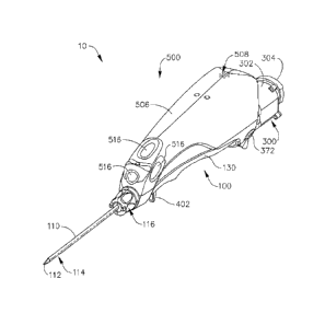
[0006] FIG. 1 depicts a perspective view of an exemplary biopsy device;
[0007] FIG. 2 depicts a perspective view of a probe portion of the
biopsy device of FIG.
1 separated from a holster portion of the biopsy device of FIG. 1;
10081 FIG. 3 depicts a top plan view of the probe portion of the biopsy
device, with a
top chassis removed;
[00091 FIG. 4 depicts an exploded perspective view of cutter actuation
components of the
probe of FIG. 3;
CA 3017312 2018-09-13
WO 2012/060968 PCT/U S2011/054892
-3-
[0010] FIG, 5A depicts a partial cross-sectional side view of the cutter
actuation
components of FIG. 4, as well as a distal portion of the needle and cutter,
with the cutter
in a distal position;
[0011] FIG. 5B depicts a partial cross-sectional side view of the cutter
actuation
components of FIG. 4, as well as a distal portion of the needle and cutter,
with the cutter
in an intermediate position;
[0012] FIG. 5C depicts a partial cross-sectional side view of thc cutter
actuation
components of FIG. 4, as well as a distal portion of the needle and cutter,
with the cutter
in a proximal position;
[0013] FIG. 6 depicts an exploded perspective view of tissue sample
holder components
of the probe of FIG, 3;
[0014] FIG. 7A depicts a partial cross-sectional side view of the tissue
sample bolder of
FIG, 6;
[00151 FIG, 7B depicts a cross-sectional end view of the cup of the
tissue sample holder
of FIG, 6;
100161 FIG, 8 depicts an exploded perspective view of needle firing and
valving
components of the probe of FIG, 3;
[0017] FIG, 9A depicts a partial cross-sectional side view of the needle
valving
components of FIG. 8, with the cutter in a distal position;
[0018] FIG. 9B depicts a partial cross-sectional side view of the needle
valving
components of FIG. 8, with the cutter in an intermediate position;
10019] FIG, 9C depicts a partial cross-sectional side view of the needle
valving
components of FIG, 8, as well as a distal portion of the needle and cutter,
with the cutter
in a proximal position;
[0020] FIG. 10A depicts a partial top plan view of the needle firing
components of FIG.
8, with the needle firing mechanism in a ready to arm configuration;
CA 3017312 2018-09-13
WO 2012/060968
PCT/US2011/054892
-4-
[0021] FIG. 10B depicts a partial top plan view of the needle firing
components of FIG.
8, with the needle firing mechanism in an armed and ready to retract
configuration;
100221 FIG. 10C depicts a partial top plan view of the needle firing
components of FIG.
8, with the needle firing mechanism transitioning to a ready to fire
configuration;
[0023] FIG. 10D depicts a partial top plan view of the needle firing
components of FIG.
8, with the needle firing mechanism in a retracted and ready to fire
configuration;
[0024] FIG. 10E depicts a partial top plan view of the needle firing
components of FIG.
8, with the needle firing mechanism in a fired configuration;
[0025] FIG. 11 depicts a schematic diagram showing components of the
holster portion
of the biopsy device of FIG. 1;
[0026] FIG. 12 depicts a side elcvational view of the holster of FIG. 11,
with housing
components and other components removed, showing a motor and drive components;
[0027] FIG. 13 depicts various views of exemplary alternative versions of
the biopsy
device of FIG. 1;
[0028] FIG. 14 depicts a perspective view of aim exemplary alternative
biopsy probe;
10029] FIG. 15 depicts a top plan view of the probe of FIG. 14, with a
top chassis
removed;
[0030] FIG. 16 depicts an exploded perspective view of valving components
of the probe
of FIG. 15;
[00311 FIG. 17 depicts a side cross-sectional view of a saline manifold
of the valving
components of FIG. 16;
[0032] FIG. 18A depicts a side cross-sectional view of valving components
of the probe
in FIG. 15, with a shuttle valve slider in a proximal position;
[0033] FIG. 1813 depicts a side cross-sectional view of valving
components of the probe
in FIG. 15, with a shuttle valve slider in a distal position;
CA 3017312 2018-09-13
WO 2012/060968 PCT/US2011/054892
-5-
[0034] FIG. 19A depicts a schematic view of exemplaiy communicative
states for a
second lumen of the needle of the probe of FIG. 15, in relation to the
longitudinal
position of the cutter within the needle, during advancement of the cutter
from a proximal
position to a distal position; and
[0035] FIG. 19B depicts a schematic view of exemplary communicative
states for a
second lumen of the needle of the probe of FIG. 15, in relation to the
longitudinal
position of the cutter within the needle, during retraction of the cutter from
a distal
position to a proximal position.
[0036] The drawings are not intended to be limiting in any way, and it is
contemplated
that various embodiments of the invention may be carried out in a variety of
other ways,
including those not necessarily depicted in the drawings. The accompanying
drawings
incorporated in and forming a part of the specification illustrate several
aspects of the
present invention, and together with the description serve to explain the
principles of the
invention; it being understood, however, that this invention is not limited to
the precise
arrangements shown.
DETAILED DESCRIPTION
100371 The following description of ccrtain examples of the invention
should not be used
to limit the scope of the present inventionõ Other examples, features,
aspects,
embodiments, and advantages of the invention will become apparent to those
skilled in
the art from the following description, which is by way of illustration, one
of the best
modes contemplated for carrying out the invention. As will be realized, the
invention is
capable of other different and obvious aspects, all without departing from the
invention.
Accordingly, the drawings and descriptions should be regarded as illustrative
in nature
and not restrictive,
[0038] I. Overview of Exemplary Biopsy Device
[0039] FIGS. 1-2 show an exemplary biopsy device (10). Biopsy device (10)
of this
example comprises a probe (100) and a holster (500). A needle (110) extends
distally
from probe (100), and is inserted into a patient's tissue to obtain tissue
samples as will be
CA 30 1 7312 20 1 8-0 9-13
WO 2012/060968 PCT/US2011/054892
-6-
described in greater detail below. These tissue samples are deposited in a
tissue sample
holder (300) at the proximal end of probe (100), as will also be described in
greater detail
below. It should also be understood that the use of the term "holster" herein
should not
be read as requiring any portion of probe (100) to be inserted into any
portion of holster
(500). Indeed, in the present example, and as best seen in FIG. 2, a finger
(502) extends
distally from holster (500), and is received in a corresponding slot (102) of
probe (100) to
help secure probe (100) and holster (500) together. Other components of probe
(100) and
holster (500) mate when probe (100) and holster (500) are coupled together, as
will be
described in greater detail below. It should be understood that a variety of
types of
structures, components, features, etc. (e.g., bayonet mounts, latches, clamps,
clips, snap
fittings, etc.) may be used to provide removable coupling of probe (100) and
holster
(500), Furthermore, in some biopsy devices (10), probe (100) and holster (500)
may be
of unitary or integral construction, such that the two components cannot be
separated.
By way of example only, in versions where probe (100) and holster (500) are
provided as
separable components, probe (100) may be provided as a disposable component,
while
holster (500) may be provided as a reusable component. Still other suitable
structural
and functional relationships between probe (100) and holster (500) will be
apparent to
those of ordinary skill in the art in view of the teachings herein.
[0040] Some variations of biopsy device (10) may include one or more
sensors (not
shown), in probe (100) and/or in holster (500), that is/are configured to
detect when
probe (100) is coupled with holster (500). Such sensors or other features may
further be
configured to permit only certain types of probes (100) and holsters (500) to
be coupled
together. In addition or in the alternative, such sensors may be configured to
disable one
or more functions of probes (100) and/or holsters (500) until a suitable probe
(100) and
holster (500) are coupled together. Of course, such sensors and features may
be varied or
omitted as desired.
[0041] Biopsy device (10) of the present example is sized and configured
such that
biopsy device (10) may be operated by a single hand of a user. In particular,
a user may
grasp biopsy device (10), insert needle (100) into a patient's breast, and
collect one or a
plurality of tissue samples from within the patient's breast, all with just
using a single
CA 3017312 2018-09-13
WO 2012/060968 PCT/US2011/054892
-7-
hand. Alternatively, a user may grasp biopsy device (10) with more than one
hand and/or
with any desired assistance. It should also be understood that biopsy device
(10) may be
grasped and fully operated by a single hand using a variety of different kinds
of grips,
including but not limited to a pencil grip. In some settings, the user may
capture a
plurality of tissue samples with just a single insertion of needle (110) into
the patient's
breast. Such tissue samples may be pneumatically deposited in tissue sample
holder
(300), and later retrieved from tissue sample holder (300) for analysis. While
examples
described herein often refer to the acquisition of biopsy samples from a
patient's breast, it
should be understood that biopsy device (10) may be used in a variety of other
procedures for a variety of other purposes and in a variety of other parts of
a patient's
anatomy (e.g., prostate, thyroid, etc.). Various exemplary components,
features,
configurations, and operabilities of biopsy device (10) will be described in
greater detail
below; while other suitable components, features, configurations, and
operabilities will
be apparent to those of ordinary skill in the art in view of the teachings
herein.
[00421 II. Exemplary Probe
[00431 FIGS. 3-10 show probe (100) of the present example in greater
detail. As noted
above, probe (100) includes a distally extending needle (110), Probe (100)
also includes
a chassis (120) and a base homing (130), which are fixedly secured together.
Tissue
sample holder (300) is removably coupled with base housing (130) in this
example,
though it should be understood that tissue sample holder (300) may
alternatively be non-
removably secured to base housing (130). A pair of gears (202, 204) are
exposed
through an opening (122) in chassis (120), and arc operable to drive a cutter
actuation
mechanism (200) in probe (100) as will be described in greater detail below.
An arming
finger grip (402) extends downwardly from the bottom of base housing (13(i),
and is
operable to arm a needle firing mechanism (400) in probe (100) as will also be
described
in greater detail below,
[0044] A. Exemplary Needle
[0045] Needle (110) of the present example includes a piercing tip (112),
a lateral
aperture (114) located proximal to tip (112), and a rotation knob (116).
Tissue piercing
CA 3017312 2018-09-13
W02012/060968 PCT/US2011/054892
-8-
tip (112) is configured to pierce and penetrate tissue, without requiring a
high amount of
force, and without requiring an opening to be pre-formed in the tissue prior
to insertion
of tip (112). Alternatively, tip (112) may be blunt (e.g., rounded, flat,
etc.) if desired,
Tip (112) may also be configured to provide greater echogcnicity than other
portions of
needle (110), providing enhanced visibility of tip (112) under ultrasound
imaging. By
way of example only, tip (112) may be configured in accordance with any of the
teachings in U.S. Non-Provisional Pat. App. No. 12/875,200, entitled
"Echogenic Needle
for Biopsy Device," filed September 3, 2010.
Other suitable configurations that may be used for tip (112) will be
apparent to those of ordinary skill in the art in view of the teachings
herein,
100461 Lateral aperture (114) is sized to receive prolapsed tissue
during operation of
device (10). A tubular cutter (150) having a sharp distal edge (152) is
located within
needle (110). As described in greater detail below, cutter (150) is operable
to rotate and
translate relative to needle (110) and past lateral aperture (114) to sever a
tissue sample
from tissue protruding through lateral aperture (114). While lateral aperture
(114) is
shown oriented in a downward position in FIG. 1, it should be understood that
needle
(110) may be rotated to orient lateral aperture (114) at any desired angular
position about
the longitudinal axis of needle (110). Such rotation of needle (110) is
facilitated in the
present example by rotation knob (116), which is secured to needle (110). In
particular,
and now referring to FIG. 8, a needle overmeld (410) is fixedly secured to
needle (110),
and is configured to transfer rotation from rotation knob (116) to needle
(110). By way
of example only, needle (110) may be formed of metal, and needle overmold
(410) may
be formed of a plastic material that is overroolded about needle (110) to
unitarily secure
and form needle evennold (410) to needle (110). Needle ovennold (410) and
needle
(110) may alternatively be formed of any other suitable material(s), and may
be secured
together in any other suitable fashion. Needle overtnold (410) includes a
distal portion
(412) having a pair of flats (414). Distal portion (412) of needle overmold
(410) is
slidahly disposed in a bore (not shown) of a rotation hub (140), 'This bore of
rotation hub
(140) includes flats that complement fiats (114) of needle overrnold (410),
such that
rotation of rotation hub (140) will rotate needle ovennold (140), thereby
rotating needle
CA 3017312 2018-09-13
WO 2012/1)60968 PCT/US21111/1154892
-9-
(110). The relationship between rotation hub (140) and needle overmold (410)
in the
present example will nevertheless permit needle (110) and needle overmold
(410) to
unitarily translate relative to rotation hub (140), as will be described in
greater detail
below.
[00471 Rotation
hub (140) also includes a pair of flats (142) and an annular recess (144).
As shown in FIG. 9, rotation knob (116) of the present example is formed of a
first half
(116a), and a second half (116b), which are configured to snap fit together
about rotation
hub (140). Halves (116a, 116b) have bosses (117) that engage flats (142) of
rotation hub
(140), such that rotation of rotation knob (116) will rotate rotation hub
(140). Halves
(116a, 116b) also include proximal rims (119) that engage annular recess (144)
of =
rotation hub (140), such that rotation knob (116) will translate
longitudinally with
rotation hub (140), Rotation knob (116) of the present example also includes a
pair of
distal latching members (115), which may removably engage other components of
a
biopsy system such as a targeting set for use in an MRI biopsy setting, etc.
[00481 As best
seen in FIGS, 10A-10E, rotation hub (140) also includes a proximal
flange (148) having a plurality of notches (149) formed therein. A coil spring
(146) is
coaxially disposed about rotation hub (140), and is positioned between a
proximally
facing distal inner surface (131) of base housing (130) and the distal face of
proximal
flange (148) of rotation hub (140). Spring (146) is resiliently biased to urge
proximal
flange (148) proximally toward posts (133) of base housing (130). A boss (not
shown)
extends upwardly from the lower surface of base housing (130) and is
configured to
engage a downwardly presented notch (149) of proximal flange (148). Such
engagement
substantially secures the rotational position of rotation hub (140) about the
longitudinal
axis defined by needle (110). The bias of spring (146) further promotes
engagement
between this boss and whichever notch (149) is downwardly presented by urging
proximal flange (148) proximally, Thus, in order to change the rotational
orientation of
needle (110), a user may grasp rotation knob (116) and push or pull rotation
knob (116)
distally against the resilient bias of spring (146) to disengage the boss from
the most
downwardly presented notch (149), rotate rotation knob (116) while holding
rotation
knob (116) in a distal position to rotate needle (110) (thereby re-orienting
lateral aperture
CA 3017312 2018-09-13
WO 2012/060968 PCT/US21111/054892
-10-
(114) about the longitudinal axis of needle (110)), then release rotation knob
(116) to
allow spring (146) to move rotation hub (140) proximally (thereby engaging the
boss
with the notch (149) now downwardly presented). With needle (110) at the
adjusted
angular orientation, the engagement between the boss and the now downwardly
presented notch (149), promoted by the resilient bias of spring (146), will
maintain
needle (110) at the adjusted angular orientation. In some versions, the
underside of
chassis (120) includes a downwardly extending boss that engages an upwardly
presented
notch (149), in addition to or in lieu of an upwardly extending boss of base
housing (130)
engaging a downwardly presented notch (149).
[0049] Various other suitable ways in which manual rotation of needle
(110) may be
provided will be apparent to those of ordinary skill in the art in view of the
teachings
herein. It should also be understood that rotation of needle (110) may be
automated in
various ways, including but not limited to the various forms of automatic
needle rotation
described in various references that are cited herein.
[0050] As best seen in FIGS. 8 and 5A-5C, needle (110) also includes a
longitudinal wall
(160) extending proximally from the proximal portion of tip (112). While wall
(160)
does not extend along the full length of needle (110) in this example, it
should be
understood that wall (160) may extend the full length of needle (110) if
desired. Wall
(160) of the present example proximally terminates at a longitudinal position
that is just
proximal to the longitudinal position of distal cutting edge (152) of cutter
(150) when
cutter (150) is in a proximal position (see FIG. 5C). Thus, wall (160) and
cutter (150)
together define a second lumen (162) that is lateral to and parallel to cutter
(150). Of
course, wall (160) may alternatively proximally terminate at a longitudinal
position that
is just distal to the longitudinal position of distal cutting edge (152) of
cutter (150) when
cutter (150) is in a proximal position; or wall (160) may terminate at any
other suitable
longitudinal position, Wall (160) includes a plurality of openings (164) that
provide fluid
communication between second lumen (162) and the upper portion of needle
(110), as
well as fluid communication between second lumen (162) and the lumen (154) of
cutter
(150). For instance, as will be described in greater detail below, second
lumen (162) may
selectively provide atmospheric air to vent cutter lumen (154) during
operation of biopsy
CA 3017312 2018-09-13
WO 2012/06098 PCT/US2011/054892
=
-11 -
device (1(J) as will be described in greater detail below, Openings (164) are
arranged
such that at least one opening (164) is located at a longitudinal position
that is distal to
the distal edge of lateral aperture (114). Thus, cutter lumen (154) and second
lumen
(162) may remain in fluid communication even when cutter (150) is advanced to
a
position where cutting edge (152) is located at a longitudinal position that
is distal to the
longitudinal position of the distal edge of lateral aperture (114) (se FIG.
5A), Of course,
as with any other component described herein, any other suitable
configurations may be
used.
10051) It should be understood that, as with other components described
herein, needle
(110) may be varied, modified, substituted, or supplemented in a variety of
ways; and
that needle (110) may have a variety of alternative features, components,
configurations,
and funetionalities. A plurality of external openings (not shown) may also be
formed in
needle (110), and may be in fluid communication with second lumen (162). For
instance,
such external openings may be configured in accordance with the teachings of
U.S. Pub.
No. 2007/0032742, entitled "Biopsy Device with Vacuum Assisted Bleeding
Control,"
published February 8, 2007.
Cutter (150) may also include one or more side openings (not shown). Of
course, as with
other components described herein, such external openings in needle (110) and
cutter
(150) are merely optional. As another merely illustrative example, needle
(110) may
simply lack second lumen (162) altogether in some versions. Other suitable
alternative
versions, features, components, configurations, and functionalities of needle
(110) will be
'apparent to those of ordinary skill ha the art in view of the teachings
herein.
[00521 B. Exemplary Cutter Actuation Mechanism
100531 As shown in FIGS. 3-5C, cutter actuation mechanism (200) of the
present
example cotnprises a variety of components that interact to provide
simultaneous rotation
and distal translation of cutter (150) relative to base housing (130) and
noodle (110) in a
firing stroke. Cutter actuation mechanism (200) is also operable to retract
cutter (150)
proximally to ready cutter (150) for firing, Cutter actuation mechanism (200)
of the
present example includes a pair of gears (202, 204), a lead screw (206), a
cutter sleeve or
CA 3017312 2018-09-13
WO 2012/060968 PCT/US21111/054892
-12-
overmold (210), and a plurality of sleeves (230). All of these components
(202, 204, 206,
210, 230) are coaxially aligned with cutter (150). Cutter overmold (210) is
fixedly
secured to cutter (150), such that cutter overmold (210) and cutter (150) will
rotate and
translate unitarily together in the present example. By way of example only,
cutter (150)
may be formed of metal, and cutter overmold (210) may be formed of a plastic
material
that is overmolded about cutter (150) to unitarily secure and form cutter
overmold (210)
to cutter (150). Cutter overmold (210) and cutter (150) may alternatively be
formed of
any other suitable material(s), and may be secured together in any other
suitable fashion.
Cutter overmold (210) includes a proximal portion (212) having external flats
(214), a
distal flange (216), and a proximal flange (218).
[0054] An annular recess (220) divides proximal portion (212) of cutter
overmold (210)
into a distal region (222) and a proximal region (224). Lead screw (206) is
slidably
positioned along distal region (222) of proximal portion (212). A clip (226)
is secured to
annular recess (220), such that lead screw (206) is retained between clip
(226) and
proximal flange (218). Lead screw (206) includes internal flats (207) that
complement
external flats (214) of cutter overmold (210). In particular, engagement
between flats
(207, 214) provides simultaneous rotation of lead screw (206) and cutter
overmold (210)
while also permitting lead screw (206) to translate relative to cutter
overmold (210).
Such translation will be restricted by clip (226) and proximal flange (218).
Furthermore,
a pair of coil springs (227, 229) are configured to resiliently bear against
opposite ends of
lead screw (206). A washer (208) is located between proximal spring (229) and
clip
(226) in this example, though it should be understood that washer (208) may be
omitted if
desired. The spacing between flange (218) and washer (208) permits some
freedom of
movement for lead screw (206) along part of distal region (222) between flange
(218) and
washer (208); while springs (227, 229) bias lead screw (206) to he
substantially centered
between flange (218) and washer (208). It should be understood that any other
suitable
type of resilient member may be used in addition to or in lieu of coil springs
(227, 229).
It should also be understood that the location of lead screw (206) between
flange (218)
and washer (208) may be substantially fixed, if desired.
CA 30 1 7312 20 1 8 -0 9-13
WO 2012/060968 PCT/US2011/054892
-13-
[0055] Gear (202) also includes internal flats (203) that complement
external flats (214)
of cutter overmold (210), In particular, engagement between flats (203, 214)
provides
simultaneous rotation of gear (202) and cutter overmold (210) while also
permitting lead
cutter overmold (210) translate relative to gear (202). While all flats (203,
207, 214) are
octagonal in the present example, it should be understood that other suitable
structures
may be used, including but not limited to hexagonal flats, complementary keys
and
keyways, etc. The longitudinal position of gear (202) remains substantially
constant
relative to base housing (130) during operation of biopsy device (10) of the
present
example. As shown in FIGS. 3 and 5A-5C, gear (202) is supported by a bushing
(232),
which is disposed within an integral support structure (132) of base housing
(130), Gear
(202) is positioned and configured to mesh with a complementary gear (550) of
holster
(500) when probe (100) and holster (500) are coupled together. As will be
described in
greater detail below, components in holster (500) are operable to rotatingly
drive gear
(550), which in turn rotates gear (202). As noted above and as will also be
described in
greater detail below, rotation of gear (202) provides rotation of cutter
overmold (210),
cutter (150), and lead screw (206), which Further provides translation of
cutter (150).
[0056] A threaded sleeve (240) extends distally from gear (204), Threaded
sleeve (240)
and gear (204) rotate unitarily in the present example. For instance, threaded
sleeve
(240) and gear (204) may be molded as a single unitary piece, as two separate
pieces that
are later joined together, etc. As shown in FIG, 5B, cutter actuation
mechanism (200) is
configured such that external threading (242) of lead screw (206) meshes with
internal
threading (244) of threaded sleeve (240). This meshing of threading (242, 244)
provides
translation of lead screw (206), and hence, cutter overmold (210) and cutter
(150), when
lead screw (206) and threaded sleeve (240) are rotated relative to each other.
The
longitudinal position of gear (204) and threaded sleeve (240) remains
substantially
constant relative to base housing (130) during operation of biopsy device (10)
of the
present example. As shown in FIGS. 3 and 5A-5C, threaded sleeve (240) is
supported by
sleeves (230), which are disposed within integral support structures (134) of
base housing
(130) and chassis (120). Gear (204) is positioned and configured to mesh with
a
complementary gear (554) of holster (500) when probe (100) and holster (500)
are
CA 3017312 2018-09-13
WO 2012/060968 ITT/U S2011/054892
-14-
coupled together. As will be described in greater detail below, components in
holster
(500) are operable to rotatingly drive gear (554), which in turn rotates gear
(204). While
sleeves (230) are shown as separate components, it should be understood that a
single
sleeve (230) may be used.
100571 As described in grcatcr detail below, holster (500) may be
activated to rotate gears
(550, 554) simultaneously. As noted above, gears (202, 204) mesh with gears
(550, 554)
when probe (100) and holster (500) are coupled together, such that
simultaneous rotation
of gears (550, 554) provides corresponding simultaneous rotation of gears
(202, 204).
This further provides corresponding simultaneous rotation of cutter overmold
(210),
cutter (150), lead screw (206), and sleeve (240). It should also be understood
that gears
(550, 554) have different pitch diameters in the present example (i.e., the
pitch diameter
of gear (550) is different from the pitch diameter of gear (554)). Gears (202,
204) also
have different pitch diameters (i.e., the pitch diameter of gear (202) is
different from the
pitch diameter of gear (204)). Accordingly, when a motor (528) in holster
(500) that
drives gears (550, 554) rotates at one rotational speed, gear (202) and
threaded sleeve
(240) simultaneously rotate in the same direction as each other yet at
different rotational
speeds relative to each other. Since rotation of lead screw (206) is driven by
rotation of
gear (202), lead screw (206) and threaded sleeve (240) also simultaneously
rotate in the
same direction as each other yet at different rotational speeds relative to
each other.
100581 Even though lead screw (206) and threaded sleeve (240) rotate
simultaneously in
the same direction, the difference between rotational speeds of lead screw
(206) and
threaded sleeve (240) provide a net result of lead screw (206) rotating
relative to threaded
sleeve (240), and such relative rotation provides translation of cutter (150)
as cutter (150)
rotates. By way of example only, with motor (528) in holster (500) providing
an output
speed of approximately 8,000 rpm, the above-described configuration may
provide
rotation of cutter (150) at a speed of approximately 1,000 rpm and rotation of
threaded
sleeve (240) at a speed of approximately 850 rpm, resulting in a net rotation
of cutter
(150) relative to threaded sleeve (240) at approximately 150 rpm. Of course,
any other
suitable differential may be provided. In the present example, the direction
of rotation
provided by motor (528) is simply reversed to reverse the direction of
translation of cutter
CA 3017312 2018-09-13
WO 2012/00968 PCIMS2011/054892
-15-
(150). Alternatively, cutter actuation mechanism (200) may he configured to be
self-
reversing, such that cutter (150) may be translated distally and proximally
without
reversing the direction of motor (528) rotation. By way of example only,
cutter actuation
mechanism (200) may be configured to self-reverse in accordance with the
teachings of
U.S. Pub, No. 2010/0292607, entitled "Tetherless Biopsy Device with Self-
Reversing
Cutter Drive Mechanism," published November 18, 2010_
100591 In one merely illustrative example of operation of cutter
actuation mechanism
(200), cutter (150) may be initially located in a distal-most position, such
that lateral
aperture (14) is "closed" as shown in FIG. 5A; with lead screw (206) being
positioned
distal to threaded sleeve (240), as also shown in FIG. 5A. Spring (227) biases
load screw
(206) proximally to engage threading (242) with threading (244). At this
stage, rotation
of cutter (150) relative to threaded sleeve (240) in a first rotational
direction will not
result in any distal translation of cutter (150) (e.g., lead screw (206) will
essentially
"freewheel"); while rotation of cutter (150) relative to threaded sleeve (240)
in a second
rotational direction will result in proximal translation of cutter (150). As
cutter (150) is
rotated by motor (528) arid cutter actuation mechanism (200) in the second
rotational
direction, cutter actuation mechanism (200) causes cutter (150) to retract
proximally, as
shown in FIG. 513, As noted above, such proximal or rearward translation may
be
effected through engagement of threading (242, 244), and due to lead screw
(206)
rotating at a faster speed than threaded sleeve (240). Lead screw (206)
continues to
traverse threading (244) of threaded sleeve (240) as cutter (150) continues to
retract
proximally.
100601 Cutter (150) then reaches a proximal-most position, such that
lateral aperture
(114) is "opened" as shown in FIG. 5C, At this stage, lead screw (206) is
positioned at a
proximal smooth interior section (245) of threaded sleeve (240) that lacks
threading
(244), us also shown in FIG. 5C, Spring (229) biases lead screw (206) distally
to engage
threading (242) with threads (244). At this stage, continued rotation of
cutter (150)
relative to threaded sleeve (240) in the second rotational direction will not
result in any
tbrtficr proximal translation of cutter (150) (e.g., lead screw (206) will
essentially
CA 3017312 2018-09-13
WO 2012/060968 PCT/US2011/054892
-16 -
"freewheel"); while rotation of cutter (150) relative to threaded sleeve (240)
in the second
rotational direction will result in distal translation of cutter (150). To
that end, motor
(528) may again be activated, with its rotation direction being reversed to
reverse the
rotation direction of cutter (150) and associated components. Such reversed
rotation of
cutter (150) causes cutter (150) to advance distally to reach the distal-most
position again,
as shown in FIG. 5A.
100611 When cutter (150) is retracted to a proximal position, thereby
effectively opening
lateral aperture (114), tissue may prolapse through lateral aperture (114)
under the force
of gravity, due to internal pressure of the tissue (e.g., caused by
displacement of the tissue
upon insertion of needle (110), etc.), caused by manual external palpation of
the patient's
breast by the physician, and/or under the influence of vacuum provided through
cutter
lumen (154) as described elsewhere herein. When cutter (150) is then advanced
distally,
distal edge (152) severs tissue protruding through lateral aperture (114).
This severed
tissue is captured within cutter lumen (154). A vacuum applied through cutter
lumen
(154) (as described herein or otherwise) will be encountered by the proximal
face of a
severed tissue sample within cutter lumen (154). A vent may be applied through
second
lumen (162) of needle (110), which may be communicated to the distal face of
the
severed tissue sample via openings (164), providing a pressure differential
for the severed
tissue sample. This pressure differential may facilitate proximal transport of
the severed
tissue sample through cutter lumen (154), whereby the severed tissue sample
eventually
reaches tissue sample holder (300) as described elsewhere herein.
Alternatively, tissue
samples severed by cutter (150) may be communicated proximally to tissue
sample
holder (300) or be otherwise dealt with in any other suitable fashion.
100621 Of course, any other suitable structures, components,
configurations, or
techniques may be used to provide translation and/or rotation of cutter (150).
It should
therefore be understood that, as with other components described herein,
cutter actuation
mechanism (200) may be varied, modified, substituted, or supplemented in a
variety of
ways; and that cutter actuation mechanism (200) may have a variety of
alternative
features, components, configurations, and functionalities. By way of example
only,
biopsy device (10) may be configured such that cutter (150) does not translate
(e.g., such
CA 3017312 2018-09-13
W0201211)6(1968 PCT/US2011/054892
that cutter (150) merely rotates, etc.); or such that cutter (150) does not
rotate (e.g., such
that cutter (150) merely translates, etc.). As another merely illustrative
example, cutter
(150) may be actuated pneumatically in addition to or in lieu of being
actuated by
mechanical components. Other suitable alternative versions, features,
components,
configurations, and funetionalities of cutter actuation mechanism (200) will
be apparent
to those of ordinary skill in the art in view of the teachings herein,
100631 C. Exemplary Tissue Sample Holder
100641 As shown in FIGS. 6-7, tissue sample holder (300) of the present
example
comprises an outer cup (302) and a cap (304), with a frame (306) interposed
between cap
(304) and cup (302). A seal (308) is interposed between frame (306) and cup
(302).
Tissue sample holder (300) also includes a collection tray (310). Collection
tray (310) is
configured to receive and hold tissue samples that are captured by cutter
(150) and that
are communicated proximally through cutter (150) as will be described in
greater detail
below. A distal port (312) of collection tray (310) aligns with the
longitudinal axis of
cutter (150) such that severed tissue samples communicated proximally through
cutter
lumen (154) will be received on collection tray (310) via distal port (312).
Collection
tray (310) includes a plurality of openings (314) that are sized and
configured to allow
fluids to drain through collection tray (310) while also retaining tissue
samples on
collection tray (310). In some versions, outer cup (302) is transparent and/or
translucent,
allowing a user of biopsy device (10) to see tissue samples residing on
collection tray
(310). Of course, outer cup (302) may alternatively be opaque or any desired
combination of transparent, translucent, and/or opaque.
100651 A protrusion (316) protrudes proximally from collection tray
(310), and is
removably received in an opening (318) formed in cap (304). Cap (304) is
formed of an
elastomerie material, such that friction substantially secures collection tray
(310) to cap
(304). However, collection tray (310) may be decoupled from cap (304) by first
withdrawing cap (304) and collection tray (310) together from cup (302), then
squeezing
side portions (320) of cap (304) inwardly toward each other. For instance,
portions of
cap (304) may bear against ramped surfaces (322) of collection tray (310) when
side
CA 3017312 2018-09-13
W02012/060968 PCFUS2011/054892
-18-
portions (320) of cap (304) are squeezed inwardly toward each other, urging
collection
tray (310) distally away from cap (304). Thus, in some versions, cap (304) and
collection tray (310) may be together removed from cup (302), with tissue
samples
residing on collection tray (310), then collection tray (310) may he ejected
from cap
(304) by squeezing side portions (320) inwardly toward each other and then
releasing to
deposit collection tray and the tissue samples directly into a cup of fonnalin
(not shown),
etc. Thcsc features of tissue sample holder (300) (among other features of
tissue sample
holder (300)) may thus bc configured an operable in accordance with the
teachings of
I.T.S. Pub. No. 2012/0065542 entitled
"Biopsy Device Tissue Sample
Holder with Removable Basket," filed September 10, 2010.
It should also be understood that the elastomerie
properties of cap (304) may provide a substantially fluid tight seal with
frame (306). In
addition, the elastomeric properties of cap (304) provide a substantially
fluid tight seal
against protrusion (316) when protrusion (316) is inserted in opening (318).
Of course,
collection tray (310) and cap (304) may have any other suitable components,
features,
configurations, and relationships.
[00661 The hollow
interior of outer cup (302) is in fluid communication with cutter
lumen (154) and with at least one vacuum source in the present example. In
particular, a
probe port (330) extends distally from outer cup (302) and into base housing
(130), and
receives cutter (150) as shown in FIG. 7A. A dynamic seal (332) is provided at
the
interface of probe port (330) and cutter (150), providing a substantially
fluid tight seal
even as cutter (150) rotates and translates relative to outer cup (302), A
vacuum may be
provided to the interior of outer cup (302) via a primary vacuum port (340),
which
extends upwardly from outer cup (302). Primary vacuum port (340) is positioned
and
configured to couple with a complementary vacuum port (566) in holster (500)
when
probe (100) and holster (500) are coupled together. Complementary vacuum port
(566)
is in fluid communication with a vacuum pump (566) in holster (500), which is
operable
to generate a vacuum as will be described in L,1-eater detail below. A filter
(342) is
positioned between primary vacuum port (340) and outer cup (302), in the fluid
path of a
, vacuum
between primary vacuum port (340) and the interior of outer cup (302). In some
CA 3017312 2018-09-13
WO 20121060968 PC111152011/05489
-19-
versions, filter (342) comprises a hydrophobic filter, In some other versions,
filter (342)
comprises a hydrophilic filter, As yet another variation, a combination of a
hydrophobic
filter and a hydrophilic filter may be used. Alternatively, any other suitable
type of filter
or combination of filters may be used, including no filter (342) at all if
desired. A pair of
o-rings (344) also provide a seal between primary vacuum port (340) and the
housing of
outer cup (302), to substantially prevent leaking at the interface between
primary vacuum
port (340) and the housing of outer cup (302).
[0067] Tissue
sample holder (300) of the present example also includes a secondary
vacuum port (350), which extends proximally from frame (306). Secondary vacuum
port
(350) is configured to be coupled with an external vacuum source (e.g., a
conventional
vacuum pump, etc.) to supplement or substitute the vacuum provided by vacuum
pump
(560). Various examples of how such a secondary vacuum source may be provided
and
used with biopsy device (10) arc described in U.S. Pat. No. 8,376,957
, entitled "Biopsy Device with Auxiliary Vacuum Source," filed February 22,
2010. = As best
seen ill FIGS.
6-7, a tube (352) extends distally from frame (306) and is in fluid
communication with
secondary vacuum port (350). It should he understood that a cap or plug (not
shown)
may be selectively secured to secondary vacuum port (350) to substantially
seal
secondary vacuum port (350), such as when biopsy device (10) is used without a
secondary vacuum source and vacuum pump (560) is the sole source of vacuum.
[00681 As best
seen in FIGS, 7A-7B, a set of baffles (354) ate provided without outer cup
(302), between tube (352) and collection tray (310). In some versions, baffles
(354) are
configured to allow a vacuum to be communicated through tube (352) to the
entire
hollow interior of outer cup (302), yet baffles (354) arc also configured to
"stir" the fluid
flow within outer cup (302) to provide a cyclonic suction action. In addition
or in the
alternative, baffles (354) may provide a tortuous path to reduce the
likelihood of fluid
within tissue sample holder (300) reaching filter (342) when biopsy device
(10) is rotated
about the longitudinal axis of biopsy device (10) during use. For instance, if
a first set of
biopsy samples are collected with port (340) oriented upwardly, fluid may
drain below
baffles (354), and may substantially remain below at least one of baffles
(354) in .the
CA 3017312 2018-09-13
WO 2012/060968 PCT/US2011/054892
-20-
event that biopsy device is rotated in either direction such that port (340)
is oriented
sideways or upwardly during the collection of additional biopsy samples during
the same
use. Of course, as with other components described herein, baffles (354) may
be
configured in any other suitable fashion, and may even be omitted if desired.
It should
also be understood that one or more filters may be provided in or near tube
(352),
including but not limited to particulate filters, hydrophobic filters,
hydrophilic filters, etc.
In some other versions, secondary vacuum port (350) is simply omitted
altogether. In
addition or in the alternative, primary vacuum port (340) and vacuum pump
(560) may be
omitted if desired.
[0069] Tissue sample holder (300) of the present example also includes a
guidance funnel
(360), Guidance funnel (360) includes a central opening (362) that is
configured to align
with the axis of cutter lumen (154) and distal port (312) of collection tray
(310).
Guidance funnel (360) is fixedly secured to a proximal portion of probe port
(330), as
best seen in FIG. 7, When collection tray (310) is positioned within outer cup
(302) and
cap (304) is secured to frame (306), the proximal portion of guidance funnel
(360) abuts
the distal face (364) of collection tray (310). When collection tray (310) and
cap (304)
are removed from tissue sample holder (300), guidance funnel (360) remains
within outer
cup (302), secured to the proximal portion of probe port (330). A plurality of
openings
(366) are formed in the body of guidance funnel (360). Such openings (366) are
configured to prevent guidance funnel (360) from being secured to distal face
(364) of
collection tray (310) like a suction cup, which might otherwise make it more
difficult to
remove collection tray (310) from outer cup (302). In addition or in the
alternative, such
openings (366) may be configured to allow fluid (e.g., blood, saline, air,
etc.) to fill the
space between guidance funnel (360) and collection tray (310), to make greater
use of the
internal volume of outer cup (302). When collection tray (310) and cap (304)
are
removed from tissue sample holder (300), guidance funnel (360) may facilitate
insertion
of a biopsy site marker applier shaft (not shown) into cutter lumen (154) by
helping to
guide the marker applier shaft to be coaxial with cutter lumen (154). It
should therefore
be understood that, after one or more biopsy samples are captured by biopsy
device (10),
and with needle (110) still inserted in tissue, a user may remove collection
tray (310) and
CA 3017312 2018-09-13
WO 2012/060968 PCT/US2011/054892
-21-
cap (304) from tissue sample holder (300) then insert a marker applier shall
into cutter
lumen (154) via guidance funnel (360) to deploy one or more biopsy site
markers to the
biopsy site via lateral aperture (114). In addition or in the alternative,
guidance funnel
(360) may facilitate administration of a pain medication to a biopsy site from
a syringe
having a catheter-like tube coupled with the distal end of the syringe barrel,
by
facilitating insertion of the catheter-like tube from the proximal end of
biopsy device
(10),
[00701 Tissue sample holder (300) of the present example is also
selectively removable
from probe (100). In particular, outer cup (300) includes a pair of latches
(370) that
selectively engage base housing (130), Latches (370) are resiliently biased to
secure
tissue sample holder (300) to base housing (130), yet may be deflected to
disengage
tissue sample holder (300) from base housing (130). Each latch (370) includes
a
respective button portion (372) to provide such disengagement. In particular,
latches
(370) may be disengaged from base housing (130) by pressing button portions
(372)
inwardly toward each other. With button portions (372) depressed inwardly,
latches
(370) deflect to disengage housing (130), such that tissue sample holder (300)
may be
pulled proximally to separate tissue sample holder (300) from probe (100). In
some
versions, vacuum port (340) slides free from outer cup (302), such that vacuum
port
(340) remains coupled with probe (100) and/or holster (500) when tissue sample
holder
(300) is pulled free. Alternatively, vacuum port (340), probe (100), and/or
holster (500)
may be configured to allow vacuum port (340) to be disengaged from probe (100)
and/or
holster (500) with outer cup (302) when tissue sample holder (300) is pulled
free. In
some other versions (e.g,, those that only rely on an external source coupled
with
secondary vacuum port (350) for vacuum), vacuum port (340) is omitted
entirely. It
should also be understood that biopsy device (10) may include one or more
features
configured to substantially seal the proximal end of cutter (150) when tissue
sample
holder (300) is removed from biopsy device (10). For instance, such a seal may
substantially prevent blood and/or other bodily fluids from exiting the
proximal end of
cutter lumen (154) when sample holder (300) is removed from biopsy device (10)
while
needle (110) is still inserted in tissue. Such a seal may also effectively
open when tissue
CA 3017312 2018-09-13
WO 2012/060968 PCT/US2011/054892
-22.-
sample holder (300) is re-coupled with biopsy device (10). Various suitable
ways in
which such a seal may be provided will be apparent to those of ordinary skill
in the art in
view of the teachings herein. Similarly, various other suitable ways in which
tissue
samp:e holder (300) may be selectively engaged with base housing (130) will be
apparent
to those of ordinary skill in the art in view of the teachings herein.
100711 As best seen in FIGS. 2, 6, and 7, tissue sample holder (300) of
the present
example includes a contact (380) that is configured to engage a corresponding
contact
sensor (520) (which is only shown in FIG. 11) of holster (500) when probe
(100) and
holster (500) are coupled together, Thus, as will be described in greater
detail below, a
control module (510) in holster (500) may sense when tissue sample holder
(300) is
coupled with or decoupled from probe (100), and may control or restrict
operation of
biopsy device (10) accordingly. Of course, biopsy device (10) may
alternatively include
a variety of other types of features configured to sense when tissue sample
holder (300) is
coupled with or decoupled from probe (100). Furthermore, some variations of
biopsy
device (10) may include a tissue sample holder (300) that is not removable
from probe
(100).
l00721 Tissue sample holder (300) of the present example is configured
to hold up to at
least ten tissue samples before collection tray (310) must be removed, though
it should be
understood that tissue sample holder (300) may be configured to hold any other
suitable
number of tissue samples. In some alternative versions, in lieu of having a
stationary
collection tray (310), tissue sample holder (300) may have a plurality of
trays that are
removably coupled with a rotatable manifold, such that the manifold is
operable to
successively index each tray relative to cutter lumen (154) to separately
receive tissue
samples obtained in successive cutting strokes of cutter (150). For instance,
tissue
sample holder (300) may be constructed wad operable in accordance with the
teachings of
U.S. Pub. No. 2008/0214955, entitled "Presentation of Biopsy Sample by Biopsy
Device," published September 4, 2008
As another merely illustrative example, tissue sample holder (300) may
be constructed and operable in accordance with the teachings of U.S. Pub. No.
2010/0160824, untitled "Biopsy Device with Discrete Tissue Chambers,"
published June
=
CA 3017312 2018-09-13
WO 2012/060968 PCI1US2011/054892
-23-
24, 2010. Still other
suitable
ways in which tissue sample holder (300) may be constructed and operable will
be
apparent to those of ordinary skill in the art in view of the teachings
herein.
100731 D. Exemplary Needle Valving Mechanism
[0074i As shown in
FIGS. 8-9C, probe (100) further includes components that are
operable to selectively vent or seal second lumen (162) of needle (110)
relative to
atmosphere, These components include a vent sleeve (420) and a shuttle valve
slider
(430), Vent sleeve (420) is secured relative to chassis (120) and base housing
(130), such
that vent sleeve (420) does not move during operation of biopsy device (10);
while
shuttle valve slider (430) translates based on operational movement of cutter
(150). A
distal portion of vent sleeve (420) is slidably disposed within proximal
portion (416) of
needle overmold (410). The outer diameter of vent sleeve (420) and the inner
diameter
of proximal portion (416) of needle ovennold (410) Me secured together
unitarily in the
present example, such that vent sleeve (420) and needle ovennold (410)
translate
unitarily. It should also be understood that, even with cutter disposed
through vent
sleeve (420), the interior of vent sleeve (420) is in fluid communication with
second
lumen (162) of needle (110) via needle ovennold (410). Vent sleeve (420)
.includes a
plurality of transverse openings (422) that are longitudinally co-located with
each other
and that are equidistantly spaced from each other about the outer perimeter of
vent sleeve
(420) at their common longitudinal position. Transverse openings (422) provide
communication of atmospheric air to the interior of vent sleeve (420) as will
be described
in greater detail below. As best seen in FIGS. 9A-9C, the proximal end of vent
sleeve
(420) is sealed by an o-ring (424), which is disposed in an annular recess
(426) formed in
distal portion (211) of cutter overmold (210). Biopsy device (10) of this
example is
configured such that o-ring (424) remains positioned within vent sleeve (420)
at all times
during operation of biopsy device (10), even when cutter (150) is at a
proximal position
as shown in FIG. 10,
100751 Shuttle
valve slider (430) is disposed coaxially about cutter (150), and has an
inner diameter permitting shuttle valve slider (430) to longitudinally slide
freely relative
CA 3017312 2018-09-13
WO 2012/060968 PCT/US2011/054892
-24-
to cutter (150). Shuttle valve slider (430) also translates relative to vent
sleeve (420). A
pair of o-rings (432) are positioned at the ends of shuttle valve slider
(430), and are
configured to seal against the inner surface of vent sleeve (420) yet still
permit shuttle
valve slider (430) to translate relative to vent sleeve (420). Shuttle valve
slider (430) is
longitudinally positioned between the distal end (428) of cutter overmold
(210) and an
annular stop member (434), which is unitarily secured to cutter (150) by a
friction fit.
Shuttle valve slider (430) defines an inner diameter that is greater than the
outer diameter
defined by cutter (150), such that a gap is provided between the outer
diameter of cutter
(150) and the inner diameter of shuttle valve slider (430) along the length of
the interior
of shuttle valve slider (430). Such a gap is sufficient to provide
longitudinal fluid
communication (e.g., atmospheric air, etc.) between the outer diameter of
cutter (150)
and the inner diameter of shuttle valve slider (430). In addition, the distal
and proximal
ends of shuttle valve slider (430) include notches (436) formed therein,
providing an
appearance similar to that of a castellated nut or castle nut.
[00761 The proximal end of shuttle valve slider (430) is also configured
to be engaged by
distal end (428) of cutter overmold (210), such that cutter overmold (210) may
push
shuttle valve slider (430) distally as described below. Notches (436) at the
proximal end
of shuttle valve slider (430) are configured to provide fluid communication to
the interior
of shuttle valve slider (430), even as distal end (428) of cutter overmold
(210) engages
the proximal end of shuttle valve slider (430), Similarly, the distal end of
shuttle valve
slider (430) is configured to be engaged by stop member (434), such that stop
member
(434) may push shuttle valve slider (430) proximally as described below.
Notches (436)
at the distal end of shuttle valve slider (430) are configured to provide
fluid
communication to the interior of shuttle valve slider (430), even as stop
member (434)
engages the distal end of shuttle valve slider (430).
[0077] As described elsewhere herein, cutter (150) is configured to
rotate and translate
relative to base housing (130), while vent sleeve (420) remains substantially
stationary
relative to base housing (130). As noted above, cutter overmold (210) and stop
member
(434) translate unitarily with cutter (150). In addition, stop member (434)
and shuttle
valve slider (430) are configured such that stop member (434) may push shuttle
valve
CA 3017312 2018-09-13
WO 2012/060968 PCT/US2011/054892
-25-
slider (430) proximally when stop member (434) is engaged with shuttle valve
slider
(430) (see, e.g., FIG. 9C); while cutter overmold (210) and shuttle valve
slider (430) are
configured such that cutter overmold (210) may push shuttle valve slider (430)
distally
when cutter overmold (210) is engaged with shuttle valve slider (430) (see,
e.g., FIG.
9A). Shuttle valve slider (430) may thus translate within vent sleeve (420) in
accordance
with translation of cutter (150) relative to base housing (130). However, the
distance
between distal end (428) of cutter overmold (210) and the proximal end of stop
member
(434) is greater than the length of shuttle valve slider (430), such that
there is a degree of
"lost motion" between shuttle valve slider (430) and cutter (150) as cutter
(150) translates
in the present example. In other words, shuttle valve slider (430) remains
substantially
stationary during certain stages of a cutter (150) actuation stroke (see,
e.g,, FIGS. 9A-
9B), such that shuttle valve slider (430) only translates when cutter (150)
starts closely
approaching the distal-most position travelling from the proximal-most
position; and
when cutter (150) starts closely approaching the proximal-most position (see,
e.g., FIG.
9C).
[0078] As noted above, openings (422) of vent sleeve (420) communicate
with ambient
air; and shuttle valve slider (430) is operable to selectively vent second
lumen (162) to
atmosphere. In particular, shuttle valve slider (430) remains distal to
openings (422)
when cutter (150) is at a distal-most position (see, e.g., FIG. 9A); when
cutter (150) is
transitioning between the distal-most position and the proximal-most position
(see, e.g.,
FIG. 9B); and at latter stages of cutter (150) transitioning from the proximal-
most
position to the distal-most position. During these stages of operation, second
lumen
(162) is exposed to ambient air via openings (422) in vent sleeve (422),
notches (436) in
shuttle valve slider (430), the gap between the inner diameter of shuttle
valve slider (430)
and the outer diameter of cutter (150), and the portion of the interior of
vent sleeve (420)
that is distal to shuttle valve slider (430). However, shuttle valve slider
(430) and o-rings
(432) substantially seal second lumen (162) relative to openings (422) when
cutter (150)
is in a proximal position, such as is shown in FIG. 9C. In particular, when
cutter (150)
moves to the proximal position, stop member (434) pushes shuttle valve slider
(430)
proximally such that openings (422) are longitudinally positioned between o-
rings (432).
CA 3017312 2018-09-13
WO 2012/060968 PCT/US2011/054892
-26-
0-rings (432) thus substantially seal off second lumen (162) relative to
openings (422)
when openings (422) are between o-rings (210). When cutter (150) begins moving
again
distally toward the distal-most position, shuttle valve slider (430) remains
at this
proximal position momentarily, continuing to substantially seal second lumen
(162)
relative to openings (422), until distal end (428) of cutter overmold (210)
engages the
proximal end of shuttle valve slider (430) and begins pushing shuttle valve
slider (430)
distally to the point where the proximal-most a-ring (432) is moved distal to
openings
(422). Once the proximal-most o-ring (432) moves distal to openings (422),
second
lumen (162) is again vented to atmosphere as noted above. Thus, the valve
mechanism
of the present example substantially seals off second lumen (162) relative to
atmosphere
when cutter (150) is at a proximal position and when cutter (150) is at
initial stages of
distal advancement; while venting second lumen (162) to atmosphere when cutter
(150)
is at other positions.
[00791 It should be understood that, as with other components described
herein, the
valving components described above may be varied, modified, substituted, or
supplemented in a variety of ways; and that a valve mechanism may have a
variety of
alternative features, components, configurations, and funetionalities.
Suitable alternative
versions, features, components, configurations, and functionalities of a valve
mechanism
will be apparent to those of ordinary skill in the art in view of the
teachings herein. It
should also bc understood that, in some versions of biopsy device (10) that
lack a
vacuum pump (566) (e.g., vacuum only provided by external vacuum pump through
secondary vacuum port (350), etc.), valving functions may be performed by
valve
components located between biopsy device (10) and an external vacuum source,
such
that biopsy device (10) may lack a valve mechanism altogether.
[0080] E. Exemplary Needle Firing Mechanism
[00811 Biopsy device (10) of the present example is operable to
selectively fire needle
(110) distally relative to chassis (120) and relative to base housing (130)
through a needle
firing mechanism (400). A user may wish to employ needle firing mechanism
(400) in
instances where needle (110) is encountering dense tissue or under other
circumstances,
CA 3017312 2018-09-13
WO 2012/060968 PCT/US2011/054892
-27-
Of course, biopsy device (10) may also be operated without ever using needle
firing
mechanism (400). As shown in FIGS. 8 and 10A-10E, needle firing mechanism
(400) of
the present example includes a coil spring (440), a catch (450), and an arming
slider
(460). Coil spring (440) is positioned coaxially about cutter (150) and vent
sleeve (420).
The distal end of coil spring (440) bears against the proximal end (442) of,
needle
overmold (410); while the proximal end of coil spring (440) bears against an
integral
boss (444) of base housing (130), Coil spring (440) is resiliently biased to
urge needle
overmold (410) (and, hence, needle (110)) distally. Distal movement of needle
(110) is
restricted by a bumper washer (446), which abuts a pair of bosses (448) formed
in base
member (130). Bumper washer (446) of the present example is formed of an
elastomerie
material that is configured to absorb at least some of the shock created by
sudden distal
movement of needle overmold (410) when needle (110) is fired distally. Of
course,
bumper washer (446) may be substituted or supplemented with a variety of other
components (e.g., spring, etc.); or may be omitted altogether.
[00821 Catch (450) of needle firing mechanism (400) comprises an elongate
beam (452),
an annular member (454) at the distal end of elongate beam (452), and a
transverse
projection (456) at the proximal end of elongate beam (452). Elongate beam
(452) is
formed of a resilient material such as plastic, and is biased to assume a
bowed
configuration as shown in FIGS. 10A and 10E in some versions. In some other
versions,
elongate beam (452) is resiliently biased to assume a substantially straight
configuration,
but is capable to being bent to the bowed configuration shown in FIGS. 10A and
10E.
Annular member (454) is eoaxially disposed about distal portion (412) of
needle
overmold (410), proximal to bumper washer (446). The inner diameter of annular
member (454) is less than the outer diameter of proximal portion (416) of
needle
overmold (410). Accordingly, when catch (450) is pulled proximally as
described in
greater detail below, annular member (454) pulls needle (110) from a distal
position to a
proximal position, against the distal bias provided by spring (440).
Similarly, as needle
(110) is fired distally from a proximal position to a distal position,
proximal portion (416)
of needle overmold (410) pushes annular member (454) (and, hence, catch (450))
distally. Transverse projection (456) projects inwardly toward other
components of
CA 30 1 7312 20 1 8-0 9-13
WO 2012/060968 PCT/US2011/054892
-28-
needle firing mechanism (400), and is configured to selectively engage distal
flange
(216) of cutter overmold (210) as will be described in greater detail below.
100831 A pin (458) is inserted through the proximal end of elongate beam
(452), near the
position from which transverse projection (456) projects. Pin (458) extends
upwardly
and downwardly from elongate beam (452). A lower portion of pin (458) is
disposed in a
track (470) that is formed in base housing (130), An upper portion of pin is
disposed in a
corresponding track (not shown) that is formed in the underside of chassis
(120) and that
has a shape complementing the shape of track (470). The portion of chassis
(120)
presenting this corresponding track may include reinforcement to provide
additional
strength to bear stresses imposed by pin (458) during operation of needle
firing
mechanism (400). Track (470) in base housing (130) includes an inner portion
(472) and
an outer portion (474). Viewed from the top down and from the bottom up, inner
portion
(472) runs along a path that is substantially parallel to the longitudinal
axis of cutter
(150) and various other components; while outer portion (474) runs along a
path that
includes a curved portion to allow transverse projection (456) to clear distal
flange (216)
of cutter ovennold (210) as will be described in greater detail below. In
other words,
inner portion (472) does not stray transversely away from or toward the
longitudinal axis
of cutter (150) along a horizontal plane passing through inner portion (472);
while outer
portion (474) does stray transversely away from the longitudinal axis of
cutter (150)
along a horizontal plane passing through outer portion (474).
100841 Inner portion (472) and outer portion (474) are generally located
at different
heights in this example. In particular, in some versions, a proximal part of
outer portion
(474) runs at a generally lower (474) height (e.g., in relation to chassis
(120)) than the
proximal part of inner portion (472). In the distal part of track (470), the
height transition
between portions (472, 474) is substantially smooth. In particular, as pin
(458) travels
from outer portion (474) to and along inner portion (472), pin (458) ascends a
generally
gradual incline. However, in the proximal portion of track (470), a step (476)
separates
inner portion (472) from outer portion (474). Thus, as pin (458) transitions
back from
inner portion (472) to outer portion (474), pin (458) jumps down step (476) to
reach outer
portion (474) of track (470). In the present example, step (476) is formed at
an angle that
CA 3017312 2018-09-13
WO 2012/060968 PCT/US2011/054892
-29-
is oblique to the longitudinal axis defined by cutter (150), along a
horizontal plane that
runs through track (470), to further promote pin (458) jumping down step to
reach outer
portion (474) as pin (458) reaches a proximal-most position. In some versions,
outer
portion (474) of track (470) defines an incline ascending upwardly toward
chassis (120)
as outer portion (474) progresses from the proximal end of track (470) to the
distal end of
track (470). It should therefore be understood that pin (458) may ascend
upwardly
toward chassis (120) as it travels proximally from the distal end of inner
portion (472) to
the proximal end of inner portion (472), then jump down step (476) when it
transitions to
outer portion (474), then ascend upwardly again toward chassis (120) as it
travels distally
from the proximal end of outer portion (474) to the distal end of outer
portion (474). Pin
(458) may encounter another step (not shown) at the distal end of outer
portion (474), to
jump down to reach the distal end of inner portion (474). Of course, track
(470) may
alternatively have any other suitable features or configurations.
100851 As noted above, beam (452) is resiliently biased to assume a bent
configuration,
which in turn provides a resilient bias for pin (458) to be disposed in outer
portion (474)
of track (470). Nevertheless, while pin (458) travels proximally from the
distal end of
inner portion (472) toward the proximal end of inner portion (472), track
(470) is
configured to keep pin (458) in inner portion (472) until pin (458) reaches
the proximal
end of inner portion (472). Once pin (458) reaches the proximal end of inner
portion
(472), the resilient urging of beam (452), as well as the angled orientation
of step (476),
causes pin (458) to jump down into outer portion (474) of track (470). It
should also be
understood that the upward travel of pin (458) along inner portion (472) of
track (470)
may further cause vertical deflection in beam (452), which may provide a
downward bias
of beam (452) to further urge pin (458) downward into inner portion (472) of
track (470)
when pin (458) reaches the proximal end of inner portion (472).
[0086] Aiming slider (460) of the present example is operable to deflect
beam (452)
inward, to selectively transition pin (458) from outer portion (474) to inner
portion (472)
at the distal end of track (470). Arming slider (460) is slidable relative to
base housing
(130) and comprises a finger grip (402) protruding downwardly from base
housing (130).
An inner sidewall (462) is configured and positioned to push inwardly against
beam
CA 3017312 2018-09-13
WO 2012/060968 PCT/US2011/054892
-30-
(452) to deflect beam (452) inwardly as slider (460) is slid proximally.
Slider (460) is
also configured to move upwardly toward chassis (120) in the present example.
Slider
(460) includes a set of resilient angled tabs (464) that are configured to
bear against the
underside of chassis (120), biasing slider (460) downwardly away from chassis
(120). In
addition, a coil spring (470) is positioned about a post (466) of slider
(460), and bears
against a boss (473) in base housing (130). Coil spring (470) is resiliently
biased to urge
slider (460) to a distal position. Being movable upwardly toward chassis
(120), slider
(460) is further operable to push upwardly on beam (452), thereby facilitating
transition
of pin (458) from outer portion (474) of track (470) to inner portion (472) of
track (470).
Such capability may be useful in versions where beam (452) is resiliently
biased to
assume a downwardly bent configuration in addition to being resiliently biased
to assume
an outwardly bent configuration; and/or in versions where the transition from
outer
portion (474) of track (470) to inner portion (472) of track (470) includes a
step or is
otherwise not very gradual at the distal end of track (472). In some versions,
base
housing (130) includes a stepped track that substantially prevents an-ning
slider (460)
from being slid proximally without slider (460) also being simultaneously
pushed
upwardly toward chassis (120). Such a stepped track (or other
component/feature/etc.)
may serve as a lockout preventing inadvertent proximal movement of slider
(460)
relative to base housing (130),
[0087] FIGS, 10A-10E show needle firing mechanism (400) at various stages
of
operation, which will be described below, In particular, FIG, 10A shows needle
firing
mechanism (400) in a ready to arm configuration. In this configuration, cutter
(150) is in
a distal position, such that distal flange (216) of cutter overmold (210) is
located at a
longitudinal position that is distal to (yet lateral to) the longitudinal
position of transverse
projection (456) of catch (450). The resilient bias of beam (452) provides
beam (452)
with an outwardly bent configuration, with pin (458) disposed in outer portion
(474) of
track (474) such that transverse projection (456) is positioned away from
distal flange
(216). In some versions, needle (110) is inserted in tissue (e.g,, a human
breast, etc.)
when biopsy device (10) is in this configuration. It should be understood
that, at the
CA 3017312 2018-09-13
WO 2012/060968 PCT/US2011/054892
-31 -
stage of operation shown in FIG. 10A, other components of biopsy device (10)
of the
present example are in the positions and configurations shown in FlGS, 5A and
9A.
100881 FIG. 10B shows needle firing mechanism (400) in an armed and ready
to retract
configuration. In particular, a user has slid alining slider (460) to a
proximal position,
such as by pulling proximally on finger grip (402) or otherwise. In some
versions, the
user has also pushed upwardly to move slider (460) toward chassis (120) in
addition to
pulling proximally on finger grip (402) to slide arming slider (460) to a
proximal
position, Cutter (150) has not moved between the stages shown in FIGS, 10A-
10B, such
that the longitudinal position of distal flange (216) has remained consistent
at this stage.
As seen in FIG. 10B, inner sidewall (462) has pushed inwardly against beam
(452) to
deflect beam (452) inwardly as slider (460) was slid proximally. This inward
deflection
of beam (452) (and upward deflection of beam (452), in some versions) has
moved
transverse projection (456) inwardly to an armed position. With transverse
projection
(456) in this armed position, pin (458) has moved to the inner portion (472)
of track
(470) and transverse projection (456) is now located adjacent to and just
proximal to
distal flange (216) of cutter overrnold (210). In some versions, there is no
change in
height between outer portion (474) of track (470) and inner portion (472) of
track (470)
at this distal end of track (470). In some other versions, while moving from
outer portion
(474) of track (470) to inner portion (472) of track (470), pin (458)
traverses a slight
incline (or a step, in some versions) to move slightly upwardly toward chassis
(130).
100891 FIG. IOC shows needle tiring mechanism (400) transitioning to a
retracted and
loaded or ready to fire configuration. In particular, cutter actuation
mechanism (200) has
been activated to retract cutter (150) proximally, which in turn has retracted
distal flange
(216) proximally. In some versions, cutter (150) is not yet in a filly
proximal position at
this stage. In some other versions, cutter (150) is in a fully proximal
position at this
stage. As can be seen in FIG. 10C, the proximal retraction of cutter (150) and
distal
flange (21.6) has moved catch (450) proximally due to engagement between
distal flange
(216) and transverse projection (456). This proximal movement of catch (450)
has also
moved needle ovennold (410) proximally due to engagement between annular
member
(454) of catch (450) and proximal portion (416) of needle overmold (410). With
needle
CA 30 1 7312 20 1 8 -0 9-13
WO 2012/060968 PCT/US24111/054892
-32-
overmuld (410) moved proximally and being unitarily secured to needle (110),
needle
(110) has also been moved to a proximal position relative to base housing
(130) at this
stage. Spring (440) is now in a more compressed state, resiliently urging
needle
overmold (410) (and, hence, needle (110)) distally. It should be understood
that, at the
stage of operation shown in FIG. 10C, other components of biopsy device (10)
of the
present example are in the positions and configurations shown in FIGS, 5B and
9B. It
should also be understood that needle (110) and cutter (150) have translated
proximally
together during the transition between the stage depicted in FIG, 10B and the
stage
depicted in FIG. 10C,
100901 FIG. 10D shows needle firing mechanism (400) at a stage where
needle firing
mechanism (400) is in a retracted and ready to fire configuration. In
particular, cutter
actuation mechanism (200) has continued to retract cutter (150) proximally,
which in turn
has retracted distal flange (216) further proximally. At this stage, catch
(450) has been
retracted to a point where pin (458) has jumped down step (476) to transition
from inner
portion (472) of track (470) to outer portion (474) of track (470). Also at
this stage,
proximal retraction of cutter (150) is ceased at least temporarily. Since some
versions of
biopsy device (10) permit biopsy device (10) to be used regardless of whether
needle
firing mechanism (400) is also used, it may be beneficial for biopsy device
(10) to have
intelligence permitting control components of biopsy device (10) to discern
whether
needle firing mechanism (400) is being used or not and to operate cutter
actuation
mechanism (200) accordingly, That is, such intelligence may determine whether
retraction of cutter (150) should cease when cutter (150) reaches the position
shown in
FIG. 10D (i.e., in cases where needle firing mechanism (400) is being used by
the user)
or if cutter (150) should continue retracting without cessation when cutter
(150) reaches
the position shown in FIG. 10D (i.e., in eases where needle firing mechanism
(400) is not
being used by the user). For instance, as described in greater detail below,
holster (500)
includes a control module (510) that is in communication with motor (528) and
with an
encoder sensor (526), which is configured to monitor movement produced by
motor
(528). Control module (510) may include a logic configured to monitor the
current
profile (and/or other performance related characteristic) of motor (528) with
respect to
CA 3017312 2018-09-13
WO 2012/060968 PCT/US2011/054892
-33-
the longitudinal position of cutter (150) as discerned by data from encoder
sensor (526),
in relation to a baseline current profile. When needle firing mechanism (400)
is being
used by the user, the work required to compress spring (440) may impose an
additional
load on motor (528) that can be detected based on the amount of current drawn
by motor
(528) with respect to the longitudinal position of cutter (150), as compared
to a baseline
current that might be expected when needle firing mechanism (400) is not being
used by
the user. Control module (510) may thus cease retraction of cutter (150) when
cutter
reaches the position shown in FIG. 10D when that additional load is detected.
[0091] As another merely illustrative example, one or more sensors (e.g.,
hall effect
sensor, proximity sensor, etc.) within probe (100) may be used to detect
whether needle
firing mechanism (400) is being used by the user, and such one or more sensors
may
provide such data to control module (510) to alert control module (510) to
cease
retraction of cutter (150) when cutter reaches the position shown in FIG. 10D,
Various
forms that such sensors may take as well as various ways in which such sensors
may
communicate with control module (510) will be apparent to those of ordinary
skill in the
art in view of the teachings herein. Regardless of the structures and methods
used to
determine whether needle firing mechanism (400) is being used by the user, it
may also
be desirable in versions where cutter (150) retraction is ceased at the stage
shown in FIG.
10D to notify the user of biopsy device (10) that needle firing mechanism
(400) is cocked
and ready to fire. Such notification may be provided through various
components of
holster (500) (e.g., speaker (522), LEDs (524), etc.), through one Or more
mechanical
components that provide a loud audible click or other form of audible
feedback, etc.
Needle firing mechanism (400) may then be fired when the user activates a
button (516)
on holster (500). Such activation may also automatically continue a sampling
cycle by
completing retraction of cutter (150) and then advancing cutter (150) distally
to sever a
tissue sample. Alternatively, control module (510) may be configured to
require a first
activation of button (516) to fire needle firing mechanism (400) after needle
firing
mechanism (400) has reached the stage shown in FIG 10D; and a second
activation of
button (516) to continue/complete a sampling cycle. This may allow needle
(110) to be
CA 3017312 2018-09-13
WO 2012/060968 PCT/US2011/054892
-34-
fired repeatedly during a single insertion of needle (110) in tissue before a
tissue sample
is captured by cutter (150).
[00921 When the user activates button (516) to fire needle firing
mechanism (400),
control module (510) activates cutter actuation mechanism (200) to reverse
motion of
cutter (150) to advance cutter (150) slightly distally in order to facilitate
disengagement
of transverse projection (456) from distal flange (216) once needle firing
mechanism
(400) has reached the configuration shown in FIG. 10D. It should be understood
that, in
some such versions, step (476) keeps pin (458) in outer portion (474) of track
(470) even
if cutter (150) is advanced slightly at this stage. It should also be
understood that, with
pin (458) being positioned in outer portion (474), and with transverse
projection (456)
disengaged from distal flange (216), beam (452) may return to a bent
configuration (e.g.,
in versions where beam (452) is resiliently biased to assume a bent
configuration, etc.) or
otherwise be bent to assume a bent configuration (e.g., in versions where beam
(452) is
resiliently biased to assume a straight configuration, etc.). It should also
be understood
that the oblique orientation of step (476) may encourage beam (452) to
transition to a
bent configuration by providing a transversely located cam surface against pin
(458). In
addition to Of in lieu of advancing cutter slightly distally in order to
facilitate
disengagement of transverse projection (456) from distal flange (216) once
needle firing
mechanism (400) has reached the configuration shown in FIG. 10D, a resilient
outward
bias of beam (452) alone may suffice to disengage transverse projection (456)
from distal
flange (216) once needle firing mechanism (400) has reached the configuration
shown in
FIG. 10D.
100931 As can also be seen in FIG. 10D, the user of biopsy device (10)
has released
finger grip (402) of arming slider (460), allowing arming slider (460) to
return to a distal
position under the resilient distal urging of spring (470). In some versions,
and as
discussed above, biopsy device (10) may provide an audio, visual, and/or
tactile
indication to the user indicating that needle firing mechanism (400) is ready
to fire. This
may alert the user to release arming slider (460) to the extent that the user
has not already
released arming slider (460) at this stage. In some versions, arming slider
(460) includes
a chamfer or similar feature at the proximal end of inner sidewall (462),
which
CA 3017312 2018-09-13
WO 2012/060968 PCT/U S2011/054892
-35 -
substantially prevents re-arming of firing mechanism (400) if arming slider
(460) is not
released between tissue sampling cycles (i.e., cutting strokes/cycles of
cutter (150)).
100941 In the present example, with transverse projection (456)
disengaged from distal
flange (216) (shortly after the moment depicted in FIG. 10D), the resilient
bias of spring
(440) suddenly urges needle ovennold (410) distally relative tu base housing
(130),
thereby firing needle (110) distally. It should be understood that cutter
(150) is still not
yet fully retracted at this stage. It should also be understood that needle
(110) translates
distally relative to cutter (150), in addition to translating distally
relative to base housing
(130), when needle (110) is fired distally by needle firing mechanism (400).
FIG. 10E
shows needle firing mechanism (400) upon firing of needle (110). In
particular, and as
noted above, disengagement between transverse projection (456) and distal
flange (216)
has allowed spring (440) to fire needle (110) to a distal position. During the
transition
from FIG. 10D to FIG. 10E (e.g., during actual firing of needle (110)), pin
(458) has
traversed the full path of outer portion (472) of track (470), returning to
the distal region
of track (470). It should be understood that, after needle (110) is fired by
firing
mechanism (400), cutter actuation mechanism (200) may suspend movement of
cutter
(150) in the present example, in addition to or in lieu of suspending movement
of cutter
(150) when needle firing mechanism (400) is cocked and ready to lire as
described above
with reference to FIG. 10D. In some versions, cutter actuation mechanism (200)
may
continue retracting cutter (150) proximally after suspending movement of
cutter (150) for
a suitable duration (e.g., after predetermined duration, until the user again
actuates a
button (516) of holster (500), etc.). Alternatively, cutter actuation
mechanism (200) may
continue to retract cutter (150) proximally after needle (110) is fired by
filing mechanism
(400), without providing at least temporary suspension of movement of cutter
(150). It
should be understood that, at the stage of operation shown in FIG. 10E, other
components
of biopsy device (10) of the present example are in the positions and
configurations
shown in FIGS. 5C and 9C.
[0095] In the present example, vent sleeve (420) translates when needle
firing
mechanism (400) is cocked and fired. In particular, needle overmold (410)
pushes vent
sleeve (420) proximally as needle firing mechanism (400) pulls needle (110)
proximally
CA 3017312 2018-09-13
WO 2012/060968 PCT/US2011/0M892
-36-
(see FIG. 9B). Needle overmold (410) pulls vent sleeve (420) distally as
needle firing
mechanism (400) pushes needle (110) distally (see FIG. 9C). When biopsy device
(10) is
used without flung needle (110) (e.g., when needle firing mechanism (400) is
present but
not used by the user of biopsy device (10)), vent sleeve (420) simply remains
in the distal
position (see FIG. 9A). It should be understood that the valving components
described
above will operate in the same manner as described above regardless of whether
a user
decides to operate needle firing mechanism (400). In other words, second lumen
(162)
will be vented or scaled relative to atmosphere at the same stages of cutter
(150)
actuation regardless of whether needle firing mechanism (400) is used.
100961 It should be understood that the above described components,
features,
configurations, and operabilities of needle firing mechanism (400) are merely
illustrative.
Any of these components, features, configurations, and operabilities may be
varied,
modified, substituted, supplemented, or even omitted as desired. Various other
suitable
components, features, configurations, and operabilities that may be
incorporated into
needle firing mechanism (400) will be apparent to these of ordinary skill in
the art in
view of the teachings herein. It should also he understood that various other
types of
devices, including but not limited to any of the biopsy devices described in
the references
that are cited herein, may be modified to include a needle firing mechanism
(400).
[0097] 11, Exemplary Holster
100981 A. Exemplary Electrical Components of Holster
[0099] FIG. 11 shows various electrical and electromechanical components
that are
incorporated into holster (500) of biopsy device (10) of the present example.
It should be
understood that each of these components is merely illustrative, and that any
of these
components may be modified, varied, substituted, supplemented, or even
omitted, as
desired, As shown in FIG. 11, holster (500) of the present example includes a
control
module (510), a battery (512), an accelerometer (514), buttons (516), charging
circuitry
(518), a tissue sample holder sensor (520), a speaker (522), LEDs (524), an
encoder
sensor (526), and a motor (528). Control module (510) essentially serves as a
hub for the
other components (512, 514, 516, 518, 520, 522, 524, 526, 528), as all of the
other
CA 3017312 2018-09-13
WO 2012/060968 PCT/US2011/054892
-37-
components (512, 514, 516, 518, 520, 522, 524, 526, 528) are in communication
with
control module (510). As shown in FIGS 1-2 and 12, holster (500) further
comprises a
chassis (504), an upper housing (506), and a vacuum pump (560). Each of these
components will be described in greater detail below, while other suitable
components
for holster (500) will be apparent to those of ordinary skill in the art in
view of the
teachings herein.
1001001 While control module (510) is referred to in the singular, it
should be understood
that control module (510) may comprise a plurality of components and even a
plurality of
separate control modules. For instance, control module (510) may comprise a
plurality
of circuit boards, one or more storage devices configured to store data,
and/or a variety of
microprocessors, etc. In addition, control module (510) may include one or
more
wireless communication technologies (e.g., Bluetooth technology, etc.) that
are operable
to communicate with smart phones, foot pedal actuation means, keypads, etc.
Various
suitable components, features, and configurations that may be employed to form
control
module (510) will be apparent to those of ordinary skill in the art in view of
the teachings
herein.
1001011 Battery (512) of the present example comprises a rechargeable
battery (e.g.,
nickel cadmium, lithium ion, lithium polymer, etc.). Just like control module
(510) and
various other components described herein, while battery (512) is referred to
in the
singular, it should be understood that more than one battery (512) may be
incorporated
into holster (500). Battery (512) is configured to provide power to motor
(528) to
operate cutter actuation mechanism (200). For instance, battery (512) may
provide any
suitable voltage, and may be configured to provide power for at least five
biopsy
procedures or any other suitable number of procedures before requiring a
recharge or
replacement. Charging circuitry (518) in holster (500) is configured to
recharge battery
(512). For instance, holster (500) may be selectively coupled with a docking
station to
enable charging circuitry (518) to recharge battery (512). Such charging may
be
provided through contact between complementary exposed metal contacts (not
shown) of
the docking station and holster (500), through inductive charging components,
and/or in
any other suitable fashion. It should also be understood that charging
circuitry (518) may
CA 3017312 2018-09-13
WO 20121060968 PCPUS2011/054892
-38 -
be configured to monitor the charge level of battery (512). In some such
versions,
charging circuitry (518) may be configured to drive a battery charge indicator
to
constantly show the charge level of battery (512), to simply provide an
indication (e.g.,
through speaker (522) and/or LEDs (524), etc.) when the charge level of
battery (512)
falls below a threshold, and/or provide any other suitable type of
notification. Of course,
battery (512) may be non-rechargeable, if desired. Furthermore, holster (500)
may use
an external source (e.g., conventional AC power source or piece of capital
equipment,
etc.) to power motor (528), in addition to or in lieu of using battery (512).
It should also
be understood that biopsy device (10) may use an external source to drive
cutter
actuation mechanism (200) (e.g., may omit motor (528) and use speedometer
cables from
a remote drive source, use pneumatic components driven by pressurized air,
etc.).
[00102] In some versions (e.g., where battery (512) is charged through
electrical contacts
that contact complementary contacts in a charging station, etc.), charging
circuitry (518)
is omitted and a balun type of transformer is used in its place. Of course, a
balun
transformer may also be used in versions where battery (512) is charged
inductively
instead of being charged through electrical contacts, in versions where
battery (512) is
omitted and biopsy device (10) receives power in some other fashion, etc. In
sonic
versions where a balun transformer is used, the balun may convert electrical
signals from
balanced to unbalanced and/or vice versa. Such a balun may be coupled with
motor
(528), either directly and/or through control module (510). In addition or in
the
alternative, such a balun may be coupled with encoder sensor (526) and/or
other
components of holster (500). Of course, as with other components described
herein, a
balun may be substituted, supplemented, or even omitted, as desired.
1001031 Accelerometer (514) is yet another component that is referred to
in the singular
but may in fact comprise several separate accelerometers. For instance, some
versions of
holster comprise three accelerometers (514), each being configured and
positioned to
sense movement in a respective direction. Movement data from accelerometer
(514) may
be used to provide both automated power down of holster (500) when holster
(500) is not
moved for a certain time period and/or to automatically power on holster (500)
when
holster (500) is moved. For instance, control module (510) may include a logic
CA 3017312 2018-09-13
WO 2012/060968 PCT/US2011/054892
-39-
configured to power down holster (500) and at least substantially cease
consumption of
power from battery (512) if accelerometer (514) fails to indicate movement of
holster
(500) over a period of approximately 10 minutes. Any other suitable inactivity
duration
threshold may be used. This logic may also receive input from charging
circuitry (518)
to ensure that holster (500) is not fully powered down when battery (512) is
being
charged (e.g,, by a docking station, etc.), even if holster (500) is not moved
beyond the
inactivity duration threshold while holster (500) is charging. Accelerometer
(514) may
also be used to detect the orientation of biopsy device (10), and control
module (510)
may include a logic configured to modify operation of biopsy device (10) based
at least
in part on orientation data from accelerometer (514). For instance, control
module (510)
may be configured to stop operation of motor (528) (and, hence, vacuum pump
(560)
when biopsy device (10) is held in an upside-down orientation (e.g., with
holster (500)
positioned vertically below probe (100)) beyond a certain threshold duration.
Such
cessation of vacuum pump (560) operation may reduce the likelihood that filter
(342)
becomes saturated with bodily fluids. Other suitable ways in which
accelerometer (514)
may be used will be apparent to those of ordinary skill in the art in view of
the teachings
herein. It should also be understood that, as with various other components
described
herein, accelerometer (514) may simply be omitted if desired.
1001041 Buttons (516) are operable to selectively activate motor (528) to
drive cutter
actuation mechanism (200). Buttons (516) may comprise thin film switches,
capacitive
switches, spring-loaded mechanical buttons, and/or any other suitable type of
user input
feature. As shown in FIG. 1, a plurality of buttons (516) are provided at
different
positions on holster (500), Having buttons (516) at various positions may
facilitate use
of biopsy device (10) using different grip styles, which may vary depending on
the user's
preference and/or based on the angle at which needle (110) is inserted into
tissue, etc. In
some versions, any one of buttons (516) may be used at any given time, and
pressing any
button (516) will provide the same result as pressing any other button (516).
In some
other versions, once one button (516) is pressed (e.g., pressed once, pressed
and held
down for a certain duration, or pressed twice in rapid succession, etc.), a
logic in control
module (510) identifies that button (516) as the active button (516) and all
other buttons
CA 3017312 2018-09-13
WO 2012/060968 PC1111S2011/054892
-40-
(516) are de-activated. Of course, each button (516) may be assigned to
provide a
different function. For instance, one button (516) may be assigned to initiate
a tissue
sampling cycle when activated, while another button (516) may be assigned to
perform a
clear probe cycle. Examples of
such cycles are described in U.S, Pub. No.
2008/0211955, entitled "Presentation of Biopsy Sample by Biopsy Device,"
published
September 4, 2008. In
addition or in the alternative, one button (516) may be operable to
selectively restrict the
degree to which cutter (150) may be retracted, thereby allowing the user to
selectively
define the effective length of lateral aperture (114). Examples of such
operations are
described in U.S. Pat. No. 7,517,322, entitled "Biopsy Device with Variable
Side
Aperture," issued April 14, 2009. -
In some such versions, needle firing mechanism (400) may be rendered
inoperable if cutter (150) is not allowed to retract fat enough, while in some
other
versions needle firing mechanism (400) will remain fully operable even if
cutter (150) is
only allowed to retract slightly. In some versions where needle firing
mechanism (400)
remains fully operable despite receipt of user input to significantly restrict
the retraction
of cutter (150) (e.g., the user wishes to use a very short effective aperture
(114), etc.),
control module (510) may allow cutter (150) to retract as far as needed to
provide
operation of needle -firing mechanism (400), then provide the user-specified
limit on the
retraction of cutter (150) during a cutting stroke/cycle afler needle (110)
has been fired.
[001051 By way of
example only, buttons (516) may be configured and operable in
accordance with any of the teachings of U.S. Non-Provisional Patent App. No.
12/542,775, entitled "Multi-Button Biopsy Device," filed August 18, 2009.
In versions where one button (516) is
selectively assigned as thc active button (516), one or more of LEDs (524) may
be
activated to provide a visual indication to the user showing which button
(516) is active.
As another merely illustrative variation, a button (516) that is selectively
assigned as the
active button (516) may be illuminated while the other buttons (516) remain
non-
illuminated. Other suitable ways in which buttons (516) may be provided an
operable
will be apparent to those of ordinary skill in the art in view of the
teachings herein,
CA 3017312 2018-09-13
WO 2012/060968 PCT/US2011/054892
-41-
[00106] Tissue sample holder sensor (520) is operable to sense when tissue
sample holder
(300) is coupled with probe (100). Control module (510) may include a logic
that
prevents or restricts activation of motor (528) and/or other components ,of
biopsy device
(10) when tissue sample holder sensor (520) does not sense the presence of
tissue sample
holder (300) coupled with probe (100). In addition or in the alternative,
during operation
of biopsy device (10), when tissue sample holder (300) is removed from probe
(100) in
order to deposit a marker at a biopsy site using an applier fed through cutter
lumen (154),
control module (510) may include a logic that automatically retracts cutter
(150) (or
otherwise allows retraction of cutter (150)) proximally to effectively open
lateral aperture
(114) of needle (110) to allow the marker to be deployed at the biopsy site
through lateral
aperture (114). It should also be understood that control module (510) may be
configured to rely on the presence of contact (380) as sensed by tissue sample
holder
sensor (520) to determine whether probe (100) is coupled with holster (500).
For
instance, holster (500) may remain in a powered-down state when tissue sample
holder
sensor (520) does not sense the presence of tissue sample. holder (300)
through contact
(380). As soon as probe (100) and holster (500) are first coupled together,
tissue sample
holder sensor (520) may detect such coupling by sensing the presence of
contact (380),
and control module (510) may accordingly place holster (500) in a powered-on
and/or
idle state that is ready for full operation of biopsy device (10). Control
module (510)
may further be configured to at least substantially disable functioning of
buttons (516)
before tissue sample holder sensor (520) detects the coupling of probe (100)
with holster
(500). It should also be understood that control module (510) may react
differently in a
period before probe (100) is first coupled with holster (500) than it reacts
in a period
when tissue sample holder (300) is decouplcd from probe (100) after probe
(100) has
been coupled with holster (500). For instance, holster (500) may remain at
least
substantially powered down in the first period while motor (528) may be
activated to
retract cutter (150) in the second period.
[00107] As noted above, tissue sample holder sensor (520) may comprise a
metal contact
that is configured and position to make contact with contact (380) of tissue
sample holder
(300) when holster (500) and probe (100) are coupled together. While sensor
(520) and
CA 3017312 2018-09-13
WO 2012/060968 PCT/US21111/054892
-42-
contact (380) make direct contact in this example, it should be understood
that tissue
sample holder sensor (520) may alternatively sense the presence of tissue
sample holder
(300) in a variety of other ways, including but not limited to using RFID, FF.
proms, or
EAS technology. Furthermore, in some versions, tissue sample holder sensor
(520) may
be used to perform authenticity verification of tissue sample holder (500),
permitting full
operation of biopsy device (10) only when a properly authenticated tissue
sample holder
(300) is coupled with probe (100), and preventing at least some operation of
biopsy
device (10) when a non-authenticated tissue sample holder (300) is coupled
with probe
(100). Still other suitable ways in which a tissue sample holder sensor (520)
may be
configured an operable will be apparent to those of ordinary skill in the art
in view of the
teachings herein.
100108] Speaker (522) and LEDs (524) may be used to provide various forms
of feedback
to a user operating biopsy device (10). As shown in FIG. 1, top housing (506)
of holster
(500) includes a plurality of speaker openings (508) to facilitate
transmission of sound
from speaker (522) to the user of biopsy device (10). While not shown in
l'IGS. 1-2, it
should be understood that LEDs (524) may be positioned at any suitable
locations on
holster (500). In some versions, control module (510) is configured to
communicate
sound through speaker (522) and/or to illuminate/un-illuminate one or more
LEDs (524)
when an error condition is detected (e.g., battery (512) power low, drive
mechanism
jammed, motor etment profile deviating from norm beyond acceptable range,
motor
rotation speed deviating from norm beyond acceptable range, etc.). In addition
or in the
alternative, control module (510) may be configured to communicate sound
through
speaker (522) and/or to illuminate/un-illuminate one or more LEDs (524) to
indicate to
the user which state of operation biopsy device (10) is in (e.g., cutter (150)
at a distal
position, cutter (150) being retracted, cutter (150) at a proximal position,
cutter (150)
being advanced, needle firing mechanism (400) loaded to point where arming
slider
(460) should be released, etc.). Various other suitable ways in which speaker
(522)
and/or LEDs (524) may be used will be apparent to those of ordinary skill in
the art in
view of the teachings herein. It should also be understood that speaker (522)
and/or
LEDs (524) may be substituted or supplemented with other user feedback
features such
CA 3017312 2018-09-13
WO 21112/060968 PCT/CS2011/054892
-43-
as an LED display, etc.; and that speaker (522) and/or LEDs (524) may simply
be
omitted if desired.
1001091 As shown only in FIG. 12, holster (500) of the present example
also includes a
vacuum sensor (572). Vacuum sensor (572) is coupled with a sensor fitting
(570), which
is further coupled with vacuum port (566). Vacuum sensor (572) is thus
configured to
sense the level of vacuum that is provided by vacuum pump (560) and that is
being
communicated to tissue sample holder (300). Vacuum sensor (572) may comprise a
diaphragn, a capacitive coupling, a strain gauge, or any other suitable
device(s),
component(s), or configurations. While not shown in FIG. 11, vacuum sensor
(572) of
the present example is in communication with control module (510), which may
include
a logic configured to process signals from vacuum sensor (572) and affect
operation of
biopsy device (10) accordingly, By way of example only, if vacuum sensor (572)
indicates that the vacuum level within tissue sample holder (300) has not
fallen below a
predefined level (which may indicate that a tissue sample is lodged in
aperture (114)
and/or cutter lumen (154)), a "clear probe" algorithm may be initiated as
described in at
least one of the references cited herein. As another merely illustrative
example, control
logic (510) may be configured to initiate a cutting stroke by cutter (150)
only after a
vacuum level sensed by vacuum sensor (572) has fallen below a threshold. In
addition or
in the alternative, vacuum sensor (572) may be configured and/or used in
accordance
with any of the teachings in U.S. Pub. No. 2009/0171243, entitled "Vacuum
Sensor and
Pressure Pump for Tetherless Biopsy Device," published July 2, 2009.
Still other suitable ways in which vacuum
sensor (572) may be configured and used will he apparent to those of ordinary
skill in the
art in view of the teachings herein. It should also be understood that, as
with other
components described herein, vacuum sensor (572) may be substituted,
supplemented, or
even omitted, as desired.
100110f B. Exemplary Drive Components of Holster
1001111 Motor (528) of the present example comprises a conventional DC
motor, though
it should be understood that any other suitable type of motor may be used. By
way of
CA 3017312 2018-09-13
WO 2012/01)0968 PCT/US2011/054892
-44-
example only, motor (528) may comprise a pneumatic motor (e.g., having an
impeller,
etc.) that is powered by pressurized air, a pneumatic linear actuator, an
electromechanical
linear actuator, a piezoelectric motor (e.g., for use in MRI settings), or a
variety of other
types of movement-inducing devices, As mentioned above, motor (528) receives
power
from battery (512). While motor (528) is located onboard biopsy device (10) in
the
present example, it should be understood that motor (528) may instead be
located some
distance from biopsy device (10) and provide energy to biopsy device (10) via
a drive
shaft or cable, etc.
f001121 As also noted above, motor (528) is operable to drive cutter
actuation mechanism
(20(J). An exemplary drive train that may be coupled with motor (528) to drive
cutter
actuation mechanism (200) is shown in FIG, 12. In this example, the first end
(530) of a
main drive shall extends distally from motor (528) while a second end (532) of
the main
drive shaft extends proximally from motor (528). An encoder wheel (534) is
coupled
with first end (530). Encoder wheel (534) is a conventional encoder wheel in
this
example, and includes a plurality of slots, openings, and/or tabs evenly
spaced
circumferentially at or near the outer periphery of encoder wheel (534).
Encoder sensor
(526) is positioned relative to encoder wheel (534) in a manner allowing
encoder sensor
(526) to track rotation of encoder wheel (534). Encoder sensor (526) is thus
operable to
track operation of motor (528). It should be understood that, with control
module (510)
being in communication with encoder sensor (526), encoder wheel (534), encoder
sensor
(526), and control module (510) may be used to gather data relating to the
rotational
speed and rotational position of first end (530), which may be processed to
reflect the
translation rate of cutter (150), the rotation rate (150) of cutter, the
longitudinal position
of cutter (150), etc. Such information may be used to control operation of
other
components of biopsy device (10), as described elsewhere herein or in other
ways that
will be apparent to those of ordinary skill in the art in view of the
teachings herein.
[001131 First and second ends (530, 532) of the main drive shaft rotate
simultaneously and
in the same direction. A first gear (536) is secured to second end (532) of
the main drive
shaft, such that rotation of the main drive shalt when motor (528) is
activated will also
rotate first gear (536). First gear (536) is engaged with a second gear (538),
which is
CA 30 1 7312 20 1 8-0 9-13
WO 2012/060968 PCT/US2011/054892
-45-
secured to a second drive shaft (540). Accordingly, rotation of the main drive
shaft is
transmitted to second drive shaft (540) via meshing gears (536, 538). Second
drive shaft
(540) is fed into vacuum pump (560). Vacuum pump (560) of the present example
comprises a conventional diaphragm pump. In particular, second drive shaft
(540) is
coupled with an eccentric disk (not shown - e.g., a device for converting
circular motion
into rectilinear motion, comprising a disk fixed off-center to second shaft
(540), etc.),
which is configured to cause a rod (not shown ¨ e.g., the rod may be coupled
with or
otherwise driven by the eccentric disk, etc.) of vacuum pump (560) to
reciprocate as
motor (528) rotates second drive shaft (540). This rod of vacuum pump (560)
drives a
diaphragm (not shown) of vacuum pump (560) as the rod reciprocates, causing
vacuum
pump (560) to induce a vacuum. It should be understood that vacuum pump (560)
of the
present example operates in the same way regardless of which direction motor
(528)
rotates. Of course, any other suitable type of vacuum pump may be used.
1001141 Vacuum pump (560) of the present example includes a port (562)
that is coupled
with a conduit (564), which is further coupled with vacuum port (566). Vacuum
pump
(560) is thus operable to draw a vacuum through vacuum port (566) via port
(562) and
conduit (564) when motor (528) rotates second drive shaft (540). As noted
above,
primary vacuum port (340) is configured to couple with vacuum port (566) when
holster
(500) and probe (100) are coupled together. It should therefore be understood
that
vacuum pump (560) is operable to induce a vacuum in tissue sample holder (300)
when
motor (528) rotates second drive shaft (540) when holster (500) and probe
(100) are
coupled together. The term "vacuum" as used herein should read broadly to
include
suction in general (e.g., any pressure below atmospheric pressure), and should
not be
read as necessarily requiring a pressure level of exactly zero or a negative
pressure level,
etc. As noted above, vacuum pump (560) may be assisted with or replaced by an
external vacuum source that is coupled with secondary vacuum port (350) of
tissue
sample holder (300). Other suitable forms that vacuum pump (560) may take, as
well as
other suitable ways in which a vacuum pump (560) may be operated, will be
apparent to
those of ordinary skill in the art in view of the teachings herein.
Alternatively, biopsy
device (10) may be configured to operate without a vacuum pump.
CA 3017312 2018-09-13
WO 20121060968 PCMJS2011/054892
-46-
[00115] A third
gear (542) is also secured to second drive shaft (540), and rotates unitarily
therewith. Third gear (542) meshes with a fourth gear (544), which is secured
to a third
drive shaft (546). Accordingly, rotation of the main drive shaft is
transmitted to third
drive shaft (546) via meshing gears (536, 538, 542, 544) and second drive
shaft (540), A
fifth gear (548) is also secured to third drive shaft (546), and rotates
unitarily therewith.
Fifth gear (548) meshes with a sixth gear (550), which is secured to a fourth
drive shaft
(552). Sixth gear (554) is also secured to fourth drive shaft (552) and
rotates unitarily
therewith. Accordingly, rotation of the main drive shaft is transmitted to
gears (550, 554)
via meshing gears (536, 538, 542, 544, 548, 554) and drive shafts (540, 546,
552). Drive
shafts (540, 546, 552) are supported by various bearings (556) that are coaxi
ally disposed
about drive shafts (540, 546, 552). Gears (550, 554) are exposed through an
opening
formed through chassis (504) of holster (500), and arc configured to mesh with
gears
(202, 204) exposed through chassis (120) of probe (100) as described above.
Motor
(528) is thus able to rotatingly drive cutter actuation mechanism (200) of
probe (100)
through meshing of gears (550, 554, 202) 204) when probe (100) and holster
(500) are
coupled together. Of course, a variety of other components or .configurations
may be
used to provide a coupling between motor (528) and cutter (150), in addition
to or in lieu
of any or all of those drive components described above.
[001161 In some
versions, holster (500) includes one or more headlights (not shown) that
are operable to illuminate an insertion area for needle (110). Examples of
such use of
headlights are disclosed in U,S, Pub, No. 2008/0214955, entitled "Presentation
of Biopsy
Sample by Biopsy Device," published September 4, 2008.
In addition or in the alternative, bolster (500) may
include a laser light source that is operable to project a laser beam to
assist in targeting
for needle (110). Various examples of such a use of a laser are disclosed in
U.S. Pub,
No. 2010/0106056, untitled "Methods for Medical Device Alignment," published
April
29, 2010. Still other
suitable
components, features, configurations, and operabilites that may be
incorporated into
holster (500), for driving cutter actuation mechanism (200) or otherwise, will
be apparent
to those of ordinary skill in the art in view of the teachings herein.
CA 3017312 2018-09-13
WO 2012/060968 PCT/US2011/0M892
-47-
[00117] V. Exemplary Alternative Versions
1001181 FIG. 13 depicts various exemplary alternative versions of biopsy
device (10). For
instance, FIG. 13 depicts a version of a biopsy device having a laser light
source (600) as
mentioned above. FIG. 13 also depicts a version of a biopsy device having a
fixed
graphical display (602); as well as a version of a biopsy device having a
tilting graphical
display (604). In addition, FIG. 13 depicts a biopsy device disposed in a
docking station
(606), with docking station (606) having a graphical display (608). It should
be
understood that any of these features, among others, may be readily
incorporated into
biopsy device described above, as well as any other type of biopsy device,
[00119] FIGS. 14-15 show another exemplary biopsy probe (700), which may
be coupled
with holster (500) just like probe (100) described above. Unless otherwise
indicated
herein, components of probe (700) in this example are substantially identical
to
components of probe (100) described above, For instance, probe (700) of this
example
includes a tissue sample holder (702) that is substantially identical to
tissue sample
holder (300) described above. Similarly, probe (70(J) of this example includes
a cutter
actuation mechanism (704) that is substantially identical to cutter actuation
mechanism
(200) described above. In probe (700), however, there is no needle firing
mechanism
(400), though it should be understood that probe (700) may alternatively have
a needle
firing mechanism (400) (or variation thereof), if desired.
[00120] Probe (700) of the present example also includes a needle (710)
that is larger than
needle (110) of biopsy device (10). For instance, needle (710) may have a size
of
approximately 8 gauge; with needle (110) having a size of approximately 13
gauge.
Needle (710) of this example is otherwise configured the same as needle (110)
described
above, In some uses of a needle having such a relatively large size, there may
be an
increased risk of excess bleeding when a relatively large sized needle (710)
is inserted
into tissue. In some instances, such excess bleeding may present risks that
the biopsy
device might malfunction, such as due to coagulation of the excess blood on
moving
parts of the device, etc. Accordingly, probe (700) of the present example
includes a port
(720) for communicating saline through needle (710), to facilitate flushing of
any excess
CA 30 1 7312 20 1 8-0 9-13
WO 2012/060968 PCT/US2011/054892
-48-
blood. In some versions, port (720) is coupled with a source of pressurized
saline. In
some other versions, port (720) is coupled with a source of saline that is
positioned
vertically higher than biopsy probe (700), such that saline is fed into port
(720) by
gravity. In addition or in the alternative, saline may simply be drawn through
port (720)
from a saline source by a vacuum induced by vacuum pump (560) through the
inner
lumen (754) of cutter (750).
[00121] As shown in FIG. 15, probe (710) also includes a conduit (722)
that couples port
(720) with a port (732) in a saline manifold (730). As shown in FIG. 17, port
(732)
communicates with an interior region (734) of saline manifold (730). A pair of
exterior
regions (736) are on opposite sides of interior region (734), and are
separated from
interior region (734) by annular walls (738). A respective o-ring (not shown)
is
positioned in each exterior region (736), providing a substantial seal for
annular walls
(738). In other words, saline communicated to interior region (734) will not
leak out into
exterior regions (736) when saline manifold (730) is disposed about a valve
sleeve (740)
as described in greater detail below.
[001221 As shown in FIG. 16, various valving components are coaxially
disposed about
cutter (750) of probe (700). In particular, a needle manifold (752) is located
distal to
other valving components. Needle overmold (752) is coupled with needle (710)
in a
manner similar to the coupling of needle overmold (410) with needle (110)
described
above, Cutter overmold (754) is coupled with cutter (750) in a manner similar
to the
coupling of cutter ovennold (210) with cutter (150) as described above. Cutter
ovennold
(754) is also configured similar to cutter overmold (210), except that cutter
overmold
(754) of this example lacks distal flange (216). Cutter overmold (754)
includes a distal
end (756) and an annular recess (758) just proximal to distal end (756):
Annular recess
(758) is configured to receive an o-ring (not shown).
[00123] Saline manifold (730) is coaxially positioned about valve sleeve
(740). Valve
sleeve (740) is inserted into the proximal end of needle ovennold (752) such
that the
interior of valve sleeve (740) communicates with a second lumen (not shown) of
needle
(710), similar to communication of the interior of vent sleeve (420) with
second lumen
CA 3017312 2018-09-13
WO 2012/060968 PCT/US2011/054892
-49-
(168) of needle (710). Valve sleeve (740) is secured unitarily to needle
overtnold (752).
Unlike vent sleeve (420), valve sleeve (740) does not translate within probe
(710) in this
example (e.g., since needle (710) cannot be fired in this example), Valve
sleeve (740)
includes a distal set of openings (742) and a proximal set of openings (744).
Saline
manifold (730) is positioned along the length of valve sleeve (740) at a
location where
interior region (734) is in fluid communication with proximal openings (744).
This
position and relationship of saline manifold (730) and valve sleeve (740)
stays constant
during operation of probe (710).
[00124] A shuttle valve slider (760) is coaxially positioned within valve
sleeve (740), and
is configured to translate in response to actuation of cutter (750). Shuttle
valve slider
(760) includes first, second, third, fourth, and fifth annular recesses (762,
764, 766, 768,
770). Each annular recess (762, 764, 766, 768, 770) is configured to receive a
respective
o-ring (not shown) to seal against the inner surface of valve sleeve (740),
Shuttle valve
slider (760) is similar in this respect to shuttle valve slider (430)
described above.
Shuttle valve slider (760) also includes a transverse opening (772), which is
configured
to selectively communicate with distal openings (742), communicate with
proximal
openings (744), or be substantially sealed relative to either set of openings
(742, 744),
depending on the longitudinal position of shuttle valve slider (760) in valve
sleeve (740).
Transverse opening (772) is longitudinally positioned between third and fourth
annular
recesses (766, 768).
[00125] An annular stop member (780) is unitarily secured to cutter (150)
by a friction fit,
and is configured to engage the distal end of shuttle valve slider (760) as
cutter (750) is
moved proximally and thereby push shuttle valve slider (760) proximally
relative to
valve sleeve (740). Shuttle valve slider (760) may be pushed distally by
distal end (756)
of cutter overmold (754) when cutter (750) is moved distally,
[001261 Like shuttle valve slider (430), shuttle valve slider (760) of the
present example
defines an inner diameter that is greater than the outer diameter defined by
cutter (750),
such that a longitudinally extending gap (751) is provided between the outer
diameter of
cutter (750) and the inner diameter of shuttle valve slider (760). Such a gap
(751) is
CA 301 7 31 2 2 01 8-0 9-1 3
WO 2012/060968 PCT/US20111054892
-50-
sufficient to provide longitudinal fluid communication (e.g., atmospheric air,
saline, etc.)
between the outer diameter of cutter (750) and the inner diameter of shuttle
valve slider
(760). In addition, the distal end of shuttle valve slider (760) include
notches (774)
formed therein, providing an appearance similar to that of a castellated nut
or castle nut.
[00127] As noted above, translation of cutter (750) provides translation
of shuttle valve
slider (760) due to engagement between either annular stop member (780) and
the distal
end of shuttle valve slider (760) or distal end (756) of cutter ovennold (754)
and the
proximal end of shuttle valve slider (760). The distance separating annular
stop member
(780) and distal end (756) of cutter ovennold (754), even when cutter (750) is
at a distal-
most position, is greater than the length of shuttle valve slider (760). Thus,
like with
shuttle valve slider (430) as described above, there is some degree of "lost
motion"
. between shuttle valve slider (760) and cutter (750) as cutter (750)
translates in either
direction. It should be understood that shuttle valve slider (760) may be
located at
various positions within valve sleeve (740) during various stages of
translation of cutter
(750). In particular, shuttle valve slider (760) may be located at positions
where distal
openings (742) are positioned between one pair of annular recesses (762, 764,
766, 768,
770) and their corresponding o-rings; with proximal openings (744) being
positioned
between another pair of annular recesses (762, 764, 766, 768, 770) and their
corresponding o-rings. In some versions, shuttle valve slider (760) may travel
to a
proximal-most position (FIG. 18A), whereby distal openings (742) are
positioned
proximal to first annular recess (762) and its o-ring and distal to second
annular recess
(764) and its o-ring. In addition or in the alternative, shuttle valve slider
(760) may travel
to a distal-most position (FIG. 18B), whereby proximal openings (744) are
positioned
proximal to fourth annular recess (768) and its o-ring and distal to fifth
annular recess
(770) and its o-ring.
[001281 In configurations where shuttle valve slider (760) is positioned
such that distal
openings (742) are positioned between first and second annular recesses (762,
764) and
their corresponding o-rings (see FIG. 18A), between second and third annular
recesses
(764, 766) and their corresponding a-rings, or between fourth and fifth
annular recesses
(768, 770) and their corresponding o-rings, the interior of valving sleeve
(740) (and,
CA 30 1 7312 20 1 8-0 9-13
WO 2012/060968 PCT/US2011/054892
-51-
hence, the second lumen of needle (710)) is substantially sealed relative to
atmospheric
air. However, in configurations or stages of operation where shuttle valve
slider (760) is
positioned such that distal openings (742) are positioned between third and
fourth
annular recesses (766, 768) and their corresponding o-rings, as shown in FIG.
18B, the
interior of valving sleeve (740) (and, hence, the second lumen of needle
(710)) is in
communication with atmospheric air via distal openings (742) and transverse
opening
(772). In some versions of probe (700), the actual range of travel for shuttle
valve slider
(760) does not include all of these longitudinal positions. FIG. 19A shows the
range of
cutter (750) travel where the second lumen of needle (710) is vented by
shuttle valve
slider (760) and distal openings (742) during distal advancement of cutter
(750) in the
present example; while FIG. 198 shows the range of cutter (750) travel where
the second
lumen of needle (710) is vented by shuttle valve slider (760) and distal
openings (742)
during proximal retraction of cutter (750) in the present example.
[00129] Similarly, in configurations where shuttle valve slider (760) is
positioned such
that proximal openings (744) are positioned between first and second annular
recesses
(762, 764) and their corresponding o-rings, between second and third annular
recesses
(764, 766) and their corresponding o-rings (see FIG. I 8A), or between fourth
and fifth
annular recesses (768, 770) and their corresponding o-rings (see FIG. 18B),
the interior
of valving sleeve (740) (and, hence, the second lumen of needle (710)) is
substantially
sealed relative to saline from saline manifold (730). However, in
configurations where
shuttle valve slider (760) is positioned such that proximal openings (744) are
positioned
between third and fourth annular recesses (766, 768) and their corresponding o-
rings, the
interior of valving sleeve (740) (and, hence, the second lumen of needle
(710)) is in
communication with saline from saline manifold (730) via proximal openings
(744) and
transverse opening (772). Again, in some versions of probe (700), the actual
range of
travel for shuttle valve slider (760) does not include all of these
longitudinal positions.
FIG, 19A shows the range of cutter (750) travel where saline is provided to
the second
lumen of needle (710) by shuttle valve slider (760) and proximal openings
(744) during
distal advancement of cutter (750) in the present example; while FIG. 19B
shows the
range of cutter (750) travel where saline is provided the second lumen of
needle (710) by
CA 3017312 2018-09-13
WO 2012/060968 PCT/US2011/054892
-52-
shuttle valve slider (760) and proximal openings (744) during proximal
retraction of
cutter (750) in the present example.
1001301 It should also be understood that shuttle valve slider (760) may
be located at
longitudinal positions where neither saline nor atmospheric air is
communicated to the
interior of valving sleeve (740) (and, hence, the second lumen of needle
(710)). For
instance, shuttle valve slider may be located at a position where distal
openings (742) are
located between first and second annular recesses (762, 764) and their
corresponding o-
rings; with proximal openings (744) being located between second and third
annular
recesses (764, 766) and their corresponding o-rings. In such a configuration,
as shown in
FIG. 18A, the interior of valving sleeve (740) (and, hence, the second lumen
of needle
(710)) is substantially sealed relative to both atmosphere and saline manifold
(730). FIG.
19A shows the range of cutter (750) travel the second lumen of needle (710) is
substantially sealed by shuttle valve slider (760) during distal advancement
of cutter
(750) in the present example; while FIG. 19B shows the range of cutter (750)
travel the
second lumen of needle (710) is substantially sealed by shuttle valve slider
(760) during
proximal retraction of cutter (750) in the present example.
[00131] Various stages of actuation of cutter (750) at which any of the
above-noted fluid
communication states for the interior of valving sleeve (740) (and, hence, the
second
lumen of needle (710)) may apply will be apparent to those of ordinary skill
in the art in
view of the teachings herein. An exemplary algorithm is shown in FIGS. 19A-
19B,
which depicts the fluid communication state of the second lumen of needle
(710) as
cutter (750) moves in relation to the lateral aperture (714) of needle (710).
The solid line
in the field of the charts shown in FIGS. 19A-19B represents the position of
the distal
end of cutter (750) during translation of cutter (750) within needle (710). In
addition, the
arrowheads in FIGS. 19A-19B simply indicate the direction in which cutter
(750) is
moving (not necessarily the direction in which fluid is flowing).
100132] In the present example, with cutter (750) fully retracted to a
proximal position,
shuttle valve slider (760) is in a proximal position as shown in FIG, 18A. As
shown in
FIG. 19A, the second lumen of needle (710) is thus initially sealed (relative
to
CA 3017312 2018-09-13
WO 2012/0(0968 PCT/US2011/054892
-53-
atmosphere and saline) as cutter (750) begins advancing from the proximal
position to a
distal position. During the initial stages of cutter (750) advancement, the
second lumen
of needle (710) remains sealed by shuttle valve slider (760). As cutter (750)
begins
approaching a distal position, but before cutter (750) effectively closes
lateral aperture
(714), cutter overmold (754) eventually engages shuttle valve slider (760) and
begins to
push shuttle valve slider (760) distally, such that the second lumen of needle
(710)
eventually receives saline through openings (744, 772) and gap (751), With
suction
being applied to the inner lumen (754) of cutter (752), such saline (along
with blood, etc.,
from the biopsy site) may be drawn proximally through inner lumen (754) of
cutter
(752), such that the saline, etc., will ultimately be communicated to tissue
sample holder
(702). As cutter (750) continues to advance distally, cutter overmold (754)
continues to
push shuttle valve slider (760) distally, such that the second lumen of needle
(710) is
eventually vented by receiving atmospheric air through openings (742, 772) and
gap
(751). When cutter (750) reaches a distal-most position, as shown in FIG. 18B,
the
second lumen of needle (710) continues to be vented. It should be understood
that, with
a suction being applied proximally through inner lumen (754) of cutter (750),
and with a
vent being applied to the second lumen of needle (710), the resulting pressure
differential
will provide communication of tissue samples proximally through inner lumen
(754) of
cutter (750), ultimately depositing the severed tissue samples into tissue
sample holder
(702).
[00133] As shown in FIG. 19B, as cutter (750) is initially retracted from
a distal position
(FIG, 18B) to a proximal position (FIG. 18A) in the present example, the
second lumen
of needle (710) is vented to atmosphere via openings (742, 772) and gap (751).
As cutter
(750) continues to retract proximally, stop member (780) eventually engages
shuttle
valve slider (760) and pushes shuttle valve slider (760) proximally, such that
the second
lumen of needle (710) eventually receives saline via openings (742, 744) and
gap (751).
While cutter (750) continues to retract further proximally, shuttle valve
slider (750)
eventually moves to a proximal position shown in FIG, 18A, where shuttle valve
slider
(750) substantially seals the second lumen of needle (710) (relative to
atmosphere and
relative to saline). Of course, the communicative states for the second lumen
of needle
CA 3017312 2018-09-13
WO 2012/060968 PCT/US2911/054892
-54-
(710) and their relation to the movement/position of cutter (750) described
above are
mere examples. Various other suitable communicative states for the second
lumen of
needle (710) and their relation to the movement/position of cutter (750) will
be apparent
to those of ordinary skill in the art in view of the teachings herein.
1001341
[00135] Embodiments of the present invention have application in
conventional
endoscopic and open surgical instrumentation as well as application in robotic-
assisted
surgery.
100136J Embodiments of the devices disclosed herein can be designed to be
disposed of
after a single use, or they can be designed to be used multiple times.
Embodiments may,
in either or both eases, be reconditioned for reuse after at least one use.
Reconditioning
may include any combination of the steps of disassembly of the device,
followed by
cleaning or replacement of particular pieces, and subsequent reassembly. In
particular,
embodiments of the device may be disassembled, and any number of the
particular pieces
or parts of the device may be selectively replaced or removed in any
combination. Upon
cleaning and/or replacement of particular parts, embodiments of the device may
be
reassembled for subsequent usc either at a reconditioning facility, or by a
surgical team
immediately prior to a surgical procedure. Those skilled in the art will
appreciate that
reconditioning of a device may utilizc a variety of techniques for
disassembly,
CA 3017312 2018-09-13
WO 2012/060968 PCT/US2011/054892
-55-
cleaning/replacement, and reassembly. Use of such techniques, and the
resulting
reconditioned device, are all within the scope of the present application.
[00137] By way of example only, embodiments described herein may be
processed before
surgery. First, a new or used instrument may be obtained and if necessary
cleaned, The
instrument may then be sterilized. In one sterilization technique, the
instrument is placed
in a closed and sealed container, such as a plastic or TYVEK bag. The
container and
instrument may then be placed in a field of radiation that can penetrate the
container,
such as gamma radiation, x-rays, or high-energy electrons, The radiation may
kill
bacteria on the instrument and in the container, The sterilized instrument may
then be
stored in the sterile container. The sealed container may keep the instrument
sterile until
it is opened in a medical facility. A device may also be sterilized using any
other
technique known in the art, including but not limited to beta or gamma
radiation,
ethylene oxide, or steam,
[00138] Having shown and described various embodiments of the present
invention,
further adaptations of the methods and systems described herein may be
accomplished by
appropriate modifications by one of ordinary skill in the art without
departing from the
scope of the present invention. Several of such potential modifications have
been
mentioned, and others will be apparent to those skilled in the art. For
instance, the
examples, embodiments, geometries, materials, dimensions, ratios, steps, and
the like
discussed above are illustrative and are not required. Accordingly, the scope
of the
present invention should be considered in terms of the following claims and is
understood not to be limited to the details of structure and operation shown
and described
in the specification and drawings.
CA 3017312 2018-09-13