Note: Descriptions are shown in the official language in which they were submitted.
CA 03017367 2018-09-10
WO 2017/160687
PCT/US2017/022034
METHODS AND COMPOSITIONS FOR THE TREATMENT OF DEMYELINATING
DISORDERS
REFERENCE TO RELATED APPLICATIONS
The present application claims priority to Provisional U.S. Application No.
62/308,814, filed
March 15, 2016, entitled "METHODS AND COMPOSITIONS FOR THE TREATMENT OF
DEMYELINATING DISORDERS," and is incorporated herein in its entirety.
FIELD OF THE INVENTION
The present invention relates to the treatment of demyelinating disorders. In
particular, the
present invention relates to the treatment of demyelinating disorders such as
multiple sclerosis
with therapeutic(s) which promote myelination alone or in combination with
other therapeutics.
BACKGROUND
Myelin is an electrically insulating material which encases the axons of
neurons forming a layer
known as the myelin sheath. The primary purpose of myelin is to increase the
speed at which
nerve impulses propagate down the neural axon. By increasing the electrical
resistance across
the cell membrane, myelin helps prevent the electrical current from leaving
the axon. Neural
demyelination is a condition characterized by a reduction of the myelin sheath
in the nervous
system, and is the basis for many neurodegenerative diseases or injuries,
including but not
limited to multiple sclerosis.
Multiple sclerosis (MS) is the most common disabling neurological disease of
young adults;
once established, it persists for the remainder of a person's life [1]. The
initial triggering events
which lead to MS remain unknown and there is no cure. In MS, central nervous
system (CNS)
lesions form as a result of immune-mediated destruction of myelin sheaths,
resulting in loss of
function and, ultimately, progressive neurodegeneration and permanent
neurological decline.
Current MS therapeutics mainly target the autoimmune response that damages
myelin sheaths.
Although effective in reducing relapses in early disease, or in some cases to
provide
symptomatic relief of pain or muscle spasticity, none of these treatments
prevent long-term
disease progression altogether, and very few have shown signs that they may be
effective in
treating progressive forms of the disease.
CA 03017367 2018-09-10
WO 2017/160687
PCT/US2017/022034
A major unmet medical need in the treatment of MS is the availability of
therapeutics that
directly protect myelin or promote new myelin formation to maintain nerve
function, to prevent
neurodegeneration, and to restore lost function in patients.
This background information is provided to reveal information believed by the
applicant to be of
possible relevance to the present invention. No admission is necessarily
intended, nor should
be construed, that any of the preceding information constitutes prior art
against the present
invention.
SUMMARY OF THE INVENTION
The present invention provides methods and compositions for the treatment of
demyelinating
disorders. In one aspect of the invention, there is provided a method of
repairing and/or
maintaining the myelin sheath of neuronal axons in a subject, the method
comprising
administering an effective amount of one or more TRPV1 agonists exhibiting
promyelinating
activity.
In another aspect of the invention, there is provided a method of promoting
myelination of an
axon of a nerve cell, the method comprising contacting the nerve cell with an
effective amount
of one or more TRPV1 agonists exhibiting promyelinating activity.
In another aspect of the invention, there is provided a method of treating a
demyelinating
disorder in a subject, the method comprising administering an effective amount
of one or more
TRPV1 agonists exhibiting promyelinating activity.
In another aspect of the invention, there is provided a method of
neuroprotection comprising
administering to a subject an effective amount of one or more TRPV1 agonists
exhibiting
promyelinating activity alone or in combination with other therapeutics.
In specific embodiments, the one or more TRPV1 agonists exhibiting
promyelinating activity are
selected from the group consisting of zu-capsaicin, capsaicin, cannabinoids,
such as
cannabidivarin and cannabidiol, anadamide, vanilloids and combinations
thereof.
In specific embodiments, the methods further comprise administration of one or
more other
therapeutics including but not limited to the one or more other therapeutics
are selected from
2
CA 03017367 2018-09-10
WO 2017/160687
PCT/US2017/022034
the group consisting of anti-inflammatory agents, immune modulators, other
agents having
promyelinating activity.
Other embodiments and advantages of the invention are set forth in part in the
description,
which follows, and in part, may be obvious from this description, or may be
learned from the
practice of the invention.
DEFINITIONS AND ABBREVIATIONS:
As used herein, the term "demyelinating disorder" encompasses any neurological
disorder or
disease associated with the destruction or removal of myelin or myelin
deficiency.
As used herein, the terms "treat", "treatment", and the like mean to relieve
or alleviate at least
one symptom associated with such condition, or to slow or reverse the
progression of such
condition. The term "treat" also denotes to arrest, delay the onset (i.e., the
period prior to clinical
manifestation of a disease) of a disease. For example, in relation to
neurological disorders
characterized by myelin loss or myelin deficiency, the term "treat" may mean
to delay
manifestation, arrest the progression, relieve or alleviate at least one
symptom of the
neurological disorder such as, but not limited to, impaired vision or
cognitive function,
numbness, weakness in extremities, tremors or spasticity, heat intolerance,
speech impairment,
incontinence, dizziness, impaired proprioception (e.g., balance, sense of limb
position) or
coordination, pain, memory, depression, and gait disorders.
As used herein, the term "promyelination activity" refers to the generation of
myelin sheaths
and/or promote remyelination. Promyelination activity can be monitored by
methods known in
the art which include direct determination of the state of myelin in a
subject, e.g., one can
measure white matter mass using magnetic resonance imaging (MRI), measure the
thickness of
myelin fibers using a magnetic resonance spectroscopy (MRS) brain scan, or any
other direct
measures known in the art (e.g., Positron-Emission Tomography (PET), Diffusion-
Weighted
Imaging (DW-I, or DW-MRI), Diffusion Tensor Imaging, Myelography,
Magnetization Transfer,
etc.). In vitro myelination assays may also be used to identify therapeutics
having
promyelination activity.
As used herein, the term "effective amount" is an amount of a therapeutic that
is sufficient to
reduce the occurrence of demyelination or increase the occurrence of
remyelination in a
3
CA 03017367 2018-09-10
WO 2017/160687
PCT/US2017/022034
mammalian recipient by at least 10% (e.g., 10%, 15%, 20%, 25%, 30%, 40%, 50%,
60%, 70%,
75%, 80%, 85%, 90%, 95%, 99%, or 100%) and/or is the amount sufficient to
delay the
manifestation, arrest the progression, relieve or alleviate at least one
symptom of the
demyelinating disorder as compared to no treatment.
List of Abbreviations
CNS: central nervous system;
MS: multiple sclerosis;
OL: oligodendrocytes;
OPCs: oligodendrocyte precursor cells;
DIV: days in vitro;
RGC: retinal ganglion cell;
GSI: 7- secretase inhibitor;
MBP: myelin basic protein;
DAPT: NiN-(3,5-Difluorophenacety1)- L-alanyI]-S-phenylglycine t-butyl ester;
DMSO: dimethylsulfoxide;
HIS: high throughput screening;
NCC: NIH clinical collection.
BRIEF DESCRIPTION OF THE FIGURES
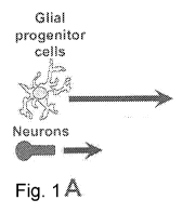
Figure 1 is a flow scheme illustrating the cortical cell myelination assay. A,
Dissociated
cells from the cortex containing neurons and glial progenitor cells were
cultured from E18 rat
embryos onto poly-D- lysine/laminin coated 96-well plates. B, On DIV4, when
axonal projections
(red) are apparent in the neuronal population, the growing co-culture is
changed to MyM media
to induce OL differentiation and initiate myelination. The following day test
compounds are
added and cultures are left undisturbed for an additional eight days. C, Cells
are fixed and
immunostained for MBP, 01ig2 and DAPI on DIV13. Images were acquired using
automated
microscopy and scored phenotypically for myelination as described in the
methods.
Figure 2 illustrates oligodendrocyte processes align with cortical axons and 7-
secretase
inhibitors (GSIs) facilitate myelination. Cortical co-cultures were treated
with the GSI, DAPT or
DMSO as described above with respect to Figure 1. A, On DIV13, cells were
fixed and stained
with antibodies to the axon marker SMI 31/32 neurofilament protein (red) and
MBP (green).
4
CA 03017367 2018-09-10
WO 2017/160687
PCT/US2017/022034
Image at the right is a composite of the SMI 31/32, MBP, and DAPI. Arrowheads
indicate
regions of MBP alignment with axon. Bar = 100 pm. B, Left two panels show
entire image
fields taken from a 96-well plate immunostained for 01ig2 and MBP. Bars = 200
pm. Boxed
regions are enlarged in the middle panel to show morphological detail of MBP-
stained OLs. Bar
= 50 pm. Two images at right depict the digital mask of MBP staining intensity
of the adjacent
image (middle panel) and the far right image are tracings of MBP alignment
used to calculate
fiber length. Bars = 50 pm. C, Raw data from three DAPT dose response
experiments was
quantified from images as in B and compiled from n = 3 experiments, 80 image
fields per
concentration, mean SEM. Asterisk (*) denotes P values versus DMSO of <
0.0001; ANOVA
analysis, followed by Bonferroni correction.
Figure 3 illustrates half maximal effective concentration determination of
four different GSIs for
the promotion of myelination in the cortical culture assay. Dose response data
confirm the
activity of GSIs and enable the calculation of the E050 value for each
compound. Cortical
cultures were treated for eight days with DAPT, LY 411,575, BMS 708,163 or MRK
560 and
immunostained for MBP, 01ig2 and DAPI. Dose-response curve for DAPT is
compiled from n =
3 experiments, 80 image fields per concentration. Representative dose-response
curves for LY
411,575, BMS 708,163 and MRK 560 are 32 image fields per concentration, mean
SEM.
Respective EC50 values are shown in the legend.
Figure 4 illustrates long term cortical cultures and demonstrates persistent
GSI-induced
enhancement of myelination and initiation of axonal node of Ranvier formation.
On DIV5,
cortical cultures were treated with DAPT or DMSO for eight days, media was
changed weekly
thereafter without compound, and cells fixed on DIV28. A, Left panels show
triple
immunostaining of MBP (green), 01ig2 (red), and DAPI (blue). Red overlaid with
blue appears
pink. Right panels show digital masks created from MBP- stained images in the
center panel.
Masks were used for quantification of fiber length. Bars = 100 pm. Arrows
indicate areas with
significant myelination. B, Quantification of myelination showing raw data in
28 DIV cortical
cultures as in A. Representative data shown is averaged from 16 image fields
per
concentration, mean SEM. Asterisk (*) denotes P values versus DMSO of <
0.0001;
ANOVA analysis, followed by Dunnett's correction. C, Cortical co-cultures were
grown for a total
of 21 days, fixed, and immunostained for MBP (green, merged image) and the
paranode-
localized protein Caspr (red, merged image). Note the accumulation of Caspr
protein at the
edges of myelinated axon segments (arrows). Bar, upper panels = 100 pm. Bar,
lower panels =
5
CA 03017367 2018-09-10
WO 2017/160687
PCT/US2017/022034
50 pm.
Figure 5 illustrates analysis of the cortical myelination screen of the NCC
compound library.
A, High-throughput screening data set used to identify promoters of
myelination. The mean
response is indicated by the solid line. The dotted line delineates the value
of three SDs above
the mean. Compounds that significantly reduced 01ig2 expression were excluded.
B, High-
throughput screening data plate control values of myelination. Each point is a
compiled control
value from each screening plate (n = 44, 16 image fields per concentration,
mean SEM *P <
0.0001, t-test). C, Using the Fiber/MBP score as a specific measure of
myelination (See Fig.
S10), the ratio of the DAPT to DMSO controls demonstrates the screening assay
window. The
red line delineates the cutoff value of 1.3. Each point is an averaged value
from each screening
plate (32 image fields per condition, mean SEM). The average DAPT/DMSO-
Fiber/MBP ratio
for the entire NCC library screen = 1.61 (dashed line). D, NCC library hit
selection process in
the cortical culture myelination assay. Fifty three primary hits compounds
were initially identified
from the NCC library with the criteria of >50% DAPT and >1.5 Fiber/MBP ratio.
The primary
hits were further refined with additional criteria of >25% DAPT/01ig2 nuclei
ratio, <40%
DAPI/01ig2 nuclei ratio, and a visual morphology check to yield refined hits
of 33 compounds.
All refined hit compounds were reordered fresh and tested for efficacy in a
dose-
response profile. Ten compounds passed these criteria and were confirmed as
hits.
Figure 6 illustrates determination of embryonic cortical cultures for
screening suitability. A,
Myelination quantification of DMSO and DAPT control values were compared in
two types of
myelination culture preparations. Data shown was compiled from n=6
experiments, 32 image
fields per test condition, mean SEM. P values versus DMSO were determined by
two-tailed t-
test. Coefficient of variation (CV) values are reported below the graphs. CV
values <20% were
considered in the acceptable range. B, Schematic of the cortical co-culture
preparation that
demonstrates that three embryonic brains used for the cortical co-culture
myelination assay will
yield approximately fifty 96-well plates.
Figure 7 illustrates the addition of exogenous OPCs to embryonic cortical
cultures is not
required for quantitative myelination. The promotion of myelination with DAPT
was more robust
(1.76 fold over DMSO) in cultures without exogenously added OPCs. The asterisk
(*) denotes P
values versus DMSO of < 0.0001, t- test. A table of mean, standard deviation
(SO), standard
error of mean (SEM) and coefficient of variation (CV) values are reported
below columns (64
6
CA 03017367 2018-09-10
WO 2017/160687
PCT/US2017/022034
image fields per treatment, mean SEM). CV values of 20% + 5% were considered
in the
acceptable range.
Figure 8 illustrates determination of optimal time courses for myelination in
the cortical cell
myelination assay. E18 cortical cultures were initially differentiated for
either 4 days (green
bars), 5 days (black bars), or 6 days (blue bars) in NB/N21 media, followed by
4, 5, 6, 7, or 8
days in MyM, then fixed for antibody staining and image analysis. Numbers in
bars indicate the
DAPT/DMSO myelination ratio for each condition. The ratio values were compiled
from 64
image fields, mean SEM. The time course with greatest DAPT/DMSO myelination
ratio was 5
days NB/N21 and 8 days MyM plus test compound (ratio = 7.7) and was used in
all subsequent
assay development and screening.
Figure 9 illustrates y¨secretase inhibitors do not promote OL differentiation,
whereas
benztropine and clemastine facilitate OL differentiation in an OL
differentiation assay with
acutely purified OPCs. Acutely prepared OPCs were cultured for 4 days (see
methods) in the
presence of increasing concentrations of test compound. 0.1% DMSO and 40 ng/ml
T3 serve as
negative and positive controls, respectively. Representative data shown are
averaged from
eight image fields per test concentration, mean SEM. * denotes P values
versus DMSO of <
0.0001, ANOVA, followed by Bonferroni correction.
Figure 10 illustrates benztropine and clemastine show little to no activity in
the cortical
myelination assay. Dose response experiments were performed adding test
compound to
cortical cultures on DIV5 and incubated for an additional eight days as
described above.
Representative raw data is averaged from 16 image fields per concentration,
mean SEM.
Figure 11 illustrates neuronal characterization of DIV13 cortical cultures.
Control cortical
cultures were treated with 0.1% DMSO on DIV5, then on DIV13, fixed and stained
with the
antibodies labeled in the left panels. Right images are merged from left and
middle panels with
antibody staining in red and DAPI staining in blue, overlapping staining
appears pink. Counting
NeuN, 01ig2, and GFAP positive cells with overlapping DAPI staining, these
cultures were
calculated to have approximately 22.5% neurons, 22% OPCs/OLs, and 46%
astrocytes. Bar =
200 m.
Figure 12 illustrates neuronal characterization of DIV5 cortical cultures.
Cortical cultures were
7
CA 03017367 2018-09-10
WO 2017/160687
PCT/US2017/022034
established and grown until DIV5, fixed and stained with the antibodies
labeled in the left
panels. Right images are merged from left and middle panels with antibody
staining in red and
DAPI staining in blue, overlapping staining appears pink. Bar = 200 pm.
Figure 13 illustrates A2B5 marker antibodies identify abundant glial
progenitor cells in DIV5
cortical cultures, but are largely absent in DIV13 cultures. Cortical cultures
were grown, fixed
and stained with anti-A2B5 antibodies on either DIV5 (day of test compound
addition) or DIV13
(endpoint of myelination assay). Images at the right show the merged images of
A2B5 (red) and
DAPI (blue). Note the almost complete absence of A2B5 staining in the DIV13
cultures.
.. Bar=200 pm.
Figure 14 illustrates oligodendrocyte characterization of DIV5 and DIV13
cortical cultures,
demonstrate robust OL differentiation during the test compound treatment
window. Cortical
cultures were grown, fixed and stained with the antibodies labeled at the
left. Note the robust
expression of OL markers in the DIV13 cultures. Bar = 200 pm.
Figure 15 illustrates equations for the quantification of myelination.
Schematic figure defining
the image quantification calculations derived from MBP intensity mask and
number of 01ig2
positive cells. OL differentiation is total MBP intensity/01ig2 nuclei and
early myelination is
calculated as the total length of contiguous MBP staining (fiber length)/01ig2
nuclei. The fiber
length/MBP intensity ratio is a score that normalizes the OL differentiation
contribution revealing
morphological changes specific to MBP alignment with axons.
Figure 16 illustrates structures, images, and EC50 curves of cortical
myelination and OL
differentiation hits. A, Chemical structure and name of each hit compound with
the controls,
0.1% DMSO and 1 IJM DAPT. B, Example image of each compound directly from the
library
screening plate at the most efficacious concentration showing MBP (green),
01ig2 (red) and
DAPI (blue) staining. 01ig2 overlapping with DAPI staining appears pink. Bar=
200 IJM. C,
Enlarged monochrome image (stained for MBP) of each hit from the library
screen to highlight
.. OL morphological changes. Bar= 50 M. D, Representative myelination dose-
response curves
of each hit (D).
8
CA 03017367 2018-09-10
WO 2017/160687
PCT/US2017/022034
DETAILED DESCRIPTION
The present invention relates to treatment of demyelinating disorders.
Specifically, the present
invention relates to methods of treatment using one or more therapeutics which
promote
myelination alone or in combination with other therapeutics for the treatment
of demyelinating
disorders.
In certain embodiments of the present invention, therapeutics which promote
myelination were
identified using a high throughput in vitro cortical myelination assay. In
specific embodiments
the cortical cell myelination assay method comprises: (a) culturing
dissociated cells from a
sample cortex containing neurons and glial progenitor cells in a first culture
media to produce a
neuronal media; (b) inducing oligodendrocyte differentiation and initiating
myelination when
axonal projections are apparent in the neuronal cell population by replacing
the first culture
media with a second culture media; (c) introducing a test compound to the
neuronal cell
population in the second culture media and incubate for a period of time; (d)
fixing and staining
cells of incubated neuronal cell population; (e) imaging fixed and stained
cells; and (f) scoring
cells phenotypically for myelination.
The assay can be utilized to screen novel therapeutics or known therapeutics
for promyelination
activity. Libraries of potential therapeutics can be screened using the
myelination assay to
identify therapeutics exhibiting promyelinating activity. The libraries can
include novel and/or
known therapeutics. A non-limiting example of a library comprising known small
molecules is
the NIH Clinical Collection library.
Therapeutics which were identified using the described high throughput in
vitro cortical
myelination assay as exhibiting promyelinating activity include but are not
limited to TRPV1
agonists. TRPV1 is the transient receptor potential cation channel subfamily V
member
1 (TrpV1), also known as the capsaicin receptor and the vanilloid receptor 1.
TRPV1 is found in
both the peripheral nervous system and central nervous system,
Non-limiting examples of TRPV1 agonists include but are not limited to zu-
capsaicin (i.e. cis-
capsaicin, CivanexTm); capsaicin; cannabinoids (see, for example, Costa etal.,
34 and lannotti
etal., 35) including but not limited to cannabidivarin and cannabidiol;
endocannabinoids including
but not limited to anadamide (N-arachidonoyl ethanolamine) and N-Arachidonoyl
dopamine;
9
CA 03017367 2018-09-10
WO 2017/160687
PCT/US2017/022034
vanilloids; resiniferatoxin; AM-404 [N-(4-hydroxyphenyI)-arachidonoyl-
ethanolamine]; N-acyl
ethanolamines (NAEs); N-oleoylethanolamine (OLEA); N-oleoyl dopamine (OLDA); 5-
(S), 8-(S),
12-(S) and 15-(S)-hydroperoxyeicosatetraenoic acids (HPETEs); hepoxilins A3
(HXA3); ATP;
spermine; spermidine; putrescine; 13(S)-hydroxy-9Z,11E-octadecadienoic acid
(13(S)-HODE);
13(R)-hydroxy-9Z,11E-octadecadienoic acid (13(R)-HODE); 9(S)-hydroxy-10(E);
12(Z)-
octadecadienoic acid (9(S)-HODE); 9(R)-hydroxy-10(E); 12(Z)-octadecadienoic
acid (9(R)-
HODE); 13-oxoODE; 9-oxoODE; 20-hydroxy-5Z,8Z,11Z,14Z-eicosatetraenoic acid;
12(S)-
hydroxy-5Z,8Z,10E,12S,14Z-eicosatetraenoic acid (12(S)-HETE, hepoxilin A3
(i.e. 8R/S-
hydroxy-11,12-oxido-5Z,9E,14Z-eicosatrienoic acid) and Hx63 (i.e. 10R/S-
hydroxy-11,12-oxido-
5Z,8Z,14Z-eicosatrienoic acid).
Accordingly, in certain embodiments, there is provided a method of repairing
and/or maintaining
the myelin sheath of neuronal axons in a subject comprising administering an
effective amount
of one or more TRPV1 agonists exhibiting promyelinating activity alone or in
combination with
other therapeutics. In specific embodiments, the one or more TRPV1 agonists
are selected
from the group consisting of zu-capsaicin, capsaicin, cannabinoids, such as
cannabidivarin and
cannabidiol, anadamide, vanilloids and combination thereof. In particular
embodiments, the
method comprises administering an effective amount of zu-capsaicin. Also
provided are
compositions comprising the one or more TRPV1 agonists for repairing and/or
maintaining the
myelin sheath of neuronal axons, including in specific embodiments,
compositions comprising
zu-capsaicin for repairing and/or maintaining.
In certain embodiments, there is provided a method of promoting myelination of
an axon of a
nerve cell comprising contacting the nerve cell with an effective amount of
one or more TRPV1
agonists. Also provided are compositions comprising one or more TRPV1 agonists
for
promoting myelination of an axon of a nerve cell. In specific embodiments, the
one or more
TRPV1 agonists are selected from the group consisting of zu-capsaicin,
capsaicin,
cannabinoids, such as cannabidivarin and cannabidiol, anadamide, vanilloids
and combination
thereof.
In certain embodiments, there is provided a method of treating a demyelinating
disorder in a
subject, said method comprising administering an effective amount of one or
more TRPV1
agonists exhibiting promyelinating activity alone or in combination with other
therapeutics. In
specific embodiments, the one or more TRPV1 agonists are selected from the
group consisting
CA 03017367 2018-09-10
WO 2017/160687
PCT/US2017/022034
of zu-capsaicin, capsaicin, cannabinoids, such as cannabidivarin and
cannabidiol, anadamide,
vanilloids and combination thereof. In particular embodiments, the method
comprises
administering an effective amount of zu-capsaicin. Also provided are
compositions comprising
the one or more TRPV1 agonists for treating a demyelinating disorder in a
subject.
Optionally, the one or more TRPV1 agonists exhibiting promyelinating activity
can be used in
combination with various other treatments which can be useful for the
treatment of
demyelinating disorders. Other therapeutics include but are not limited to
anti-inflammatory
agents, immune modulators, other agents having promyelinating acitivity or
remyelination
agents and known therapies for treatment of the demyelinating disorders. For
example, one or
more TRPV1 agonists can be administered in combination with at least one of
interferon beta
la, interferon beta lb, glatiramer acetate, mitoxantrone, azathiprine,
cyclophosphamide,
cyclosporine, ampyra, dimethyl fumarate, fingolimod, methotrexate, cladribine,
methylprednisone, prednisone, prednisolone, dexamethasone, adreno-
corticotrophic hormone,
Corticotropin, anti-integrin specific antibodies, cytoxan, naltrexone, and the
like. The one or
more TRPV1 agonists can be also administered in combination with anti-LINGO
therapies,
axin/VVnt pathway inhibitors, and/or agonists for RXR transcription factors
such as, e.g., 9-cis-
retinoic acid.
The demyelinating disorders that may be treated by the methods of the
invention include
demyelinating disorders of the central nervous system (CNS) and/or peripheral
nervous system
(PNS), demyelinating injuries that occur as a result of specific or focal
insults such as stroke or
traumatic brain injury, or degradation that may be progressive in nature and
associated with
normal cognitive or physical decline with age. The demyelinating disorders may
include
inflammatory demyelinating disorders and non-inflammatory demyelinating
disorders. Many
demyelinating disorders are classified as either myelinociastic or
leukodystrophic.
Exemplary demyelinating disorders of the central nervous system include but
are not limited to
multiple sclerosis; Devic's disease (neuromyelitis optica); other inflammatory
demyelinating
diseases such as acute-disseminated encephalomyelitis and acute haemorrhagic
leucoencephalitis; demyelinating disease precipitated by tumor necrosis factor
alpha
antagonists or other immunomodulators; viral demyelinating diseases such as
progressive
multifocal leukoencephalopathy and Tabes dorsalis; acquired metabolic
demyelination diseases
such as central pontine myelinolysis and extrapontine myelinolysis; hypoxic-
ischaemic
11
CA 03017367 2018-09-10
WO 2017/160687
PCT/US2017/022034
demyelination, compression-induced demyelination and leukodystrophies
including but not
limited to Adrenomyeloneuropathy, Alexander disease, Cerebrotendineous
xanthomatosis,
Hereditary CNS demyelinating disease, Krabbe disease, Metachromatic
leukodystrophy,
Pelizaeus-Merzbacher disease, Canavan disease, leukoencephalopathy with
vanishing white
matter, Adrenoleukodystrophy and Refsum disease.
Exemplary demyelinating disorders of the peripheral nervous system include but
are not limited
to Guillain¨Barre syndrome; chronic inflammatory demyelinating polyneuropathy;
Anti-MAG
peripheral neuropathy; Charcot¨Marie¨Tooth disease; copper deficiency
associated conditions
and progressive inflammatory neuropathy.
Exemplary demyelinating disorders involving both the central nervous system
and peripheral
nervous system include but are not limited to acute combined central and
peripheral
inflammatory demyelination.
In certain embodiments, the methods of the invention treat demyelinating
disorders of the CNS
in a subject. In specific embodiments, the methods of the invention treat
multiple sclerosis in a
subject. Also provided in certain embodiments are compositions comprising one
or more
TRPV1 agonists exhibiting promyelinating activity alone or in combination with
other
therapeutics for use in the treatment of a demyelinating disorder of the CNS,
including but not
limited to multiple sclerosis. Accordingly, in some embodiments the
compositions of the
invention are specifically formulated for treatment of CNS diseases or for
administration to the
CNS.
In certain embodiments, the methods of the invention treat demyelinating
disorders of the PNS
in a subject. Also provided in certain embodiments are compositions comprising
one or more
TRPV1 agonists exhibiting promyelinating activity alone or in combination with
other
therapeutics for use in the treatment of a demyelinating disorder of the PNS
in a subject.
Accordingly, in some embodiments the compositions of the invention are
specifically formulated
for treatment of PNS diseases or for administration to the PNS.
In certain embodiments, the methods of the invention treat demyelinating
disorders of the CNS
and PNS in a subject. Also provided in certain embodiments are compositions
comprising one
or more TRPV1 agonists exhibiting promyelinating activity alone or in
combination with other
12
CA 03017367 2018-09-10
WO 2017/160687
PCT/US2017/022034
therapeutics for use in the treatment of a demyelinating disorder of the CNS
and PNS in a
subject.
Remyelination of demyelinated axons may be neuroprotective. Accordingly, in
certain
embodiments, there is provided a method of neuroprotection comprising
administering to a
subject an effective amount of one or more TRPV1 agonists exhibiting
promyelinating activity
alone or in combination with other therapeutics.
In certain embodiments, the composition comprising the one or more TRPV1
agonists and
optionally other therapeutics further comprise a pharmaceutically acceptable
carrier.
Pharmaceutically acceptable carriers include, for example, pharmaceutically
acceptable
solvents, suspending agents, or any other pharmacologically inert vehicles.
Pharmaceutically
acceptable carriers can be liquid or solid, and can be selected with the
planned manner of
administration in mind so as to provide for the desired bulk, consistency, and
other pertinent
transport and chemical properties, when combined with one or more therapeutic
compounds
and any other components of a given pharmaceutical composition. Typical
pharmaceutically
acceptable carriers include, without limitation: water; saline solution;
binding agents (e.g.,
polyvinylpyrrolidone or hydroxypropyl methylcellulose); fillers (e.g., lactose
or dextrose and
other sugars, gelatin, or calcium sulfate); lubricants (e.g., starch,
polyethylene glycol, or sodium
acetate); disintegrates (e.g., starch or sodium starch glycolate); and wetting
agents (e.g.,
sodium lauryl sulfate).
Pharmaceutical compositions of the invention can be administered by a number
of methods,
depending upon whether local or systemic treatment is desired. Administration
can be, for
example, parenteral (e.g., by subcutaneous, intrathecal, intraventricular,
intramuscular, or
intraperitoneal injection, or by intravenous (i.v.) drip); oral; topical
(e.g., transdermal, sublingual,
ophthalmic, or intranasal); or pulmonary (e.g., by inhalation or insufflation
of powders or
aerosols), or can occur by a combination of such methods. Administration can
be rapid (e.g., by
injection) or can occur over a period of time (e.g., by slow infusion or
administration of slow
.. release formulations).
In certain embodiments, there is provided a pharmaceutical composition having
promyelinating
activity comprising one or more TRPV1 agonists selected from the group
consisting of zu-
capsaicin, capsaicin, cannabinoids, such as cannabidivarin and cannabidiol,
anadamide,
13
CA 03017367 2018-09-10
WO 2017/160687
PCT/US2017/022034
vanilloids and combination thereof. In specific embodiments, there is provided
a pharmaceutical
composition comprising zu-capsaicin formulated for intranasal or intrathecal
injection.
Compositions and formulations for parenteral, intrathecal or intraventricular
administration may
include sterile aqueous solutions (e.g., sterile physiological saline), which
also can contain
buffers, diluents and other suitable additives (e.g., penetration enhancers,
carrier compounds
and other pharmaceutically acceptable carriers).
Compositions and formulations for oral administration may include, for
example, powders or
granules, suspensions or solutions in water or non-aqueous media, capsules,
sachets, or
tablets. Such compositions also may incorporate thickeners, flavoring agents,
diluents,
emulsifiers, dispersing aids, or binders.
Formulations for topical administration may include, for example, sterile and
non-sterile aqueous
solutions, non-aqueous solutions in common solvents such as alcohols, or
solutions in liquid or
solid oil bases. Such solutions also may contain buffers, diluents and other
suitable additives.
Pharmaceutical compositions and formulations for topical administration can
include
transdermal patches, ointments, lotions, creams, gels, drops, suppositories,
sprays, liquids, and
powders. Conventional pharmaceutical carriers, aqueous, powder or oily bases,
thickeners and
the like may be useful. Methods and compositions for transdermal delivery may
include those
described in the art (e.g., in Wermeling et al. (2008) Proc. Natl. Acad. Sci.
USA 105:2058-2063;
Goebel and Neubert (2008) Skin Pharmacol. Physiol. 21:3-9; Banga (2007) Pharm.
Res.
24:1357-1359; Malik et al. (2007) Curr. Drug Deliv. 4:141-151; and Prausnitz
(2006) Nat.
Biotechnol. 24:416-417).
Nasal preparations may be presented in a liquid form or as a dry product.
Nebulized aqueous
suspensions or solutions can include carriers or excipients to adjust pH
and/or tonicity.
Pharmaceutical compositions include, but are not limited to, solutions,
emulsions, aqueous
suspensions, and liposome-containing formulations. These compositions can be
generated from
a variety of components that include, for example, preformed liquids, self-
emulsifying solids and
self-emulsifying semisolids.
Compositions additionally can contain other adjunct components conventionally
found in
14
CA 03017367 2018-09-10
WO 2017/160687
PCT/US2017/022034
pharmaceutical compositions. Thus, the compositions also can include
compatible,
pharmaceutically active materials such as, for example, antipruritics,
astringents, local
anesthetics or anti-inflammatory agents, or additional materials useful in
physically formulating
various dosage forms of the compositions, such as dyes, flavoring agents,
preservatives,
antioxidants, opacifiers, thickening agents, and stabilizers. Furthermore, the
composition can be
mixed with auxiliary agents, e.g., lubricants, preservatives, stabilizers,
wetting agents,
emulsifiers, salts for influencing osmotic pressure, buffers, colorings,
flavorings, penetration
enhancers, and aromatic substances. When added, however, such materials should
not unduly
interfere with the biological activities of the other components within the
compositions.
In some cases, the one or more TRPV1 agonists and optionally other
therapeutics can be
formulated as a sustained release dosage form, or within pharmaceutical
prodrug formulations
that enable the conversion of the prodrug into the active TRPV1 agonists
within the body upon
administration.
Pharmaceutical formulations as disclosed herein, which can be presented
conveniently in unit
dosage form, can be prepared according to conventional techniques well known
in the
pharmaceutical industry. Such techniques include the step of bringing into
association the active
ingredient(s) (i.e., the one or more TRPV1 agonists and optionally other
therapeutics) with the
desired pharmaceutical carrier(s). Typically, the formulations can be prepared
by uniformly and
intimately bringing the active ingredient(s) into association with liquid
carriers or finely divided
solid carriers or both, and then, if necessary, shaping the product.
Formulations can be
sterilized if desired, provided that the method of sterilization does not
interfere with the
effectiveness of the molecules(s) contained in the formulation.
The compositions of the invention may further comprise agents which facilitate
brain delivery.
Non-limiting examples of such useful agents include, e.g., an implantable
reservoir (Omaya
reservoir), functionalized nanocarriers and liposomes.
The following examples illustrate embodiments of the invention, but should not
be viewed as
limiting the scope of the invention.
CA 03017367 2018-09-10
WO 2017/160687
PCT/US2017/022034
EXAMPLES
Introduction
A high throughput myelination assay was developed and utilized to identify
potential myelin
repair therapeutics. In an effort to create a myelination assay more amenable
to higher
throughput compound screening, embryonic rat cortex was used to develop,
optimize, and
validate an in vitro myelination assay [5, 6] which may be utilized for
chemical library screening.
The culture system was miniaturized into a 96-well plate format enabling high
throughput liquid
handling, automated image acquisition and analysis of myelinating co-cultures.
It has
previously been shown that inhibition of the y-secretase protease activity
promotes
differentiation of OPCs and myelination of retinal ganglion cells (RGC) in RGC-
OPC co-
cultures. [7], [8], [9]. Based on this published work, the y-secretase
inhibitor (GSI), N-EN-
(3,5-Difluorophenacety1)-L-alanylFS- phenylglycine t-butyl ester (DAPT) was
used as a positive
control in the cortical co-cultures [9], and confirmed that the assay allows
for the quantification
of early axonal myelination in a dose-dependent manner. This assay identified
compounds
which are not active in a pure primary OPC differentiation assay [3, 10] but
are capable of
promoting re- myelination in vivo [11]. This myelination assay was used to
screen the NIH
clinical collection library of small molecules.
Development Of A High Throughput Cortical Myelination Assay
Focusing solely on the immunological component of MS only addresses one aspect
of the
disease. Repairing damaged myelin and/or promoting the remyelination of
demyelinated axons
within lesions would, at a minimum, facilitate the preservation and/or
restoration of some
neuronal function. This may also prevent the irreversible neuronal damage
believed to underlie
the progressive disability that eventually affects most MS patients. Thus,
remyelinating
compounds are highly sought after, but have been difficult to identify in part
because of the lack
of high throughput screening (HTS) assays that truly detect myelination.
The goal for developing a co-culture with live axons and oligodendrocytes as a
myelinating in
vitro system was to overcome the challenges of labor intensive OPC/neuron
preparations,
inconsistent performance of classical sources of neurons for modeling
myelination (e.g. retinal
ganglion cells (RGCs), dorsal root ganglion cells), and generating sufficient
quantities of cells
required for a robust HTS assay. In this study, an in vitro myelination assay
that assesses the
functional dynamic interaction between live axons and oligodendrocytes during
myelination and
16
CA 03017367 2018-09-10
WO 2017/160687
PCT/US2017/022034
can be performed with a simple culture technique at a scale and
reproducibility amenable to
HIS drug discovery was developed. This assay is unique in that it evaluates
test compounds in
the presence of the co-developing milieu of native brain cells, including
oligodendrocytes (OLs),
neurons, and astrocytes. It was demonstrated that primary embryonic cortical
tissue is an
.. abundant cell source for both neurons and oligodendrocyte precursor cells
(OPCs) that are
myelination competent [6] [5], easier to culture than RGCs, and widely used in
large-scale HIS
screening within the pharmaceutical industry. This assay was validated using y-
secretase
inhibitors (GSIs), E050 values for four different compounds was established to
allow the
ranking of potency. Using this assay, the NCC library was screened and ten
confirmed hit
compounds from diverse target classes for follow-up characterization were
identified.
In the cortical myelination assay the OLs develop and differentiate alongside
growing axons and
astrocytes, two major sources of signaling molecules known to influence
myelination. The
expression of the axonal protein LRR and Ig domain-containing, Nogo receptor-
interacting
protein (LINGO-1) was demonstrated be a potent inhibitor of differentiation
and myelination [15]
[16, 17]. Indeed, anti-LINGO-1 antibody is being developed as an MS
therapeutic to promote
axon remyelination and is currently in human clinical trials (B116033,
ClinicalTrials.gov
identifiers: NCT01244139, NCT01052506, NCT01864148). Leukemia inhibitory
factor (LIF) has
been shown to be released by astrocytes in response to ATP from action
potential firing axons
.. to promote myelination [16]. Additionally, through the action of TNFR2 on
astrocytes, LIF is
produced to stimulate OL differentiation in a co-culture system [18].
Furthermore, astrocytes
were demonstrated to reduce OL differentiation, but specifically enhance
myelin thickness and
the rate of axon wrapping [9]. TNF impairs OL differentiation [19] attenuating
TNF signaling by
TNFR1 blocking therapy ameliorates MS symptoms in EAE [20]. It was also
demonstrated that
inhibiting glial y-secretase promoted myelination [9], which also showed
efficacy in vivo in the
EAE model of MS [11]. These observations emphasize the importance of having
culture
conditions that more closely mimic the in vivo CNS composition.
The assay could be modified by spiking in test compounds during the course of
the
ensheathment window and/or lengthen the ensheathment window longer than eight
days. T3,
forskolin, and CNTF was included in the MyM medium as factors that facilitate
OL differentiation
and survival [21]. The activity of these factors may mask effects of potential
stimulators of
myelination. In particular, elimination of T3, may lower the threshold for
identifying additional
candidate compounds. This stimulation of differentiation by T3 may account for
the lack of OL
17
CA 03017367 2018-09-10
WO 2017/160687
PCT/US2017/022034
differentiation activity of benztropine and clemastine in the cortical
myelination assay.
Elimination of these factors from the myelination phase of the assay may
reveal additional
compounds with myelination activity.
The Cortical Myelination Assay Identifies Compounds Not Revealed By OL
Differentiation
Assays
The myelination assay described herewith greatly differs from in vitro OL
differentiation assays
used for compound screening which have only assessed differentiation using
purified OPCs (in
isolation from axons and astrocytes) adapted to culture conditions by multiple
passages [3], [2],
or differentiated from induced pluripotent stem cells [22] and carried out in
very short
developmental time frames. Mei etal., 2014 [2] developed an HTS assay
incorporating OL
differentiation in the presence of inert micropillers allowing the
quantification of pillar wrapping
as a surrogate for myelination [2]. Lead compounds identified from these three
studies,
including clemastine, benztropine, miconazole and clobetasol, facilitate OL
differentiation in
cultures of purified OPCs [3], [2], [4] (Fig. 12), but had no effect on
myelination in the live axon
myelination assay described here (Fig. 13, Table 2, compounds 450 - clobetasol
and 588 ¨
miconazole). Notably, the myelination assay did not identify muscarinic
antagonists as
previously identified by independent OL differentiation screens [3],[2], but
revealed entirely new
classes of compounds that promote myelination. No other high throughput assay
has been
developed to date capable of assessing the initiation and facilitation of
myelination in the
presence of axons, arguably the most important features when selecting
candidate compounds
capable of promoting or generating remyelination.
Relation Of Cortical Myelination Assay Hit Compounds To Clinical Applications
And Multiple
Sclerosis
Repositioning approved drugs for the treatment of new indications is an
activity that has grown
in popularity in recent years and is a trend that is predicted to continue.
Eight out of ten
confirmed hit compounds aligned with current MS repositioning efforts. The
confirmed hits
include: Digoxin (LANOXINTm), lmatinib mesylate (GLEEVEC), Artesunate,
Methotrexate
(TREXALLTm), Oxcarbazepine (TRILEPTALC)) and docetaxel (TAXOTEREC)).
18
CA 03017367 2018-09-10
WO 2017/160687
PCT/US2017/022034
The remainder of the confirmed hits, zu-capsaicin (CIVANEXTM) and tegafur
(UFTORALC)) have
not been previously tested in any demyelinating diseases. In particular, the
myelin restorative
effects of these compounds have not been previously evaluated. The data
suggests that these
drugs may have beneficial effects on remyelination in vivo and possibly in MS
patients.
Results
Development Of The Embryonic Cortical Cell Co-Culture Assay.
In an effort to move a low-throughput, well-established myelination cell
culture technique to a
format suitable for higher throughput screening applications, a previously
described RGC-OPC
co-culture technique [9] was miniaturized and automated. Unfortunately, the
low yield of RGCs
and lack of assay robustness from each preparation makes this co-culture
myelination assay
unsuited for high-throughput compound screening (Fig. 6A). Another source of
tissue where
numbers of neurons would not be limiting was sought.
For increased yield of primary neurons, the cortex of embryonic day 18 (E18)
rats was chosen
as an abundant source of relatively homogeneous brain cells with well-
established culture
methods [5, 6] (see methods). From one litter, enough cells can easily be
generated for high
throughput drug screening applications (-30 x 106 cells/cortex; Fig. 6B).
Following the differentiation and growth of neurons and glia for five days,
the differentiation and
early myelination of exogenously added OPCs was determined to proceeded
optimally when the
growth medium was switched from NB/N21 to an OPC- supporting myelination
medium (MyM).
With this growth medium, it was observed that it was not necessary to add
exogenous OPCs as
mature and axon ensheathing OLs that had differentiated from the embryonic
cortical
preparation, presumably from neural precursor cells and/or OPCs could be
readily identified. It
was actually found that the differentiating OPCs already present in the
cultures produced better
myelination (Fig. 7, see below and methods for quantification of myelination).
Finally, it was
determined that the optimal time course for myelination to proceed was eight
days after test
compound addition and 13 DIV total (Fig. 8). Figure 1 depicts the flow scheme
of the embryonic
cortical cell assay. At this early myelination time point, we observed MBP
staining aligning with
SMI 31/32 axon staining, indicating that indeed OLs are contacting and
aligning with axons (Fig.
2A).
19
CA 03017367 2018-09-10
WO 2017/160687
PCT/US2017/022034
The y-secretase inhibitor, DAPT, a known enhancer of myelination [11], [9] was
utilized as a
positive control to test the assay system and establish an automated
morphology analysis. After
compound treatment, cells were stained for the OL lineage marker, 01ig2,
myelin basic protein
(MBP) to stain mature OLs, and the nuclear dye, DAPI, and imaged. Myelination
was scored by
quantifying the characteristic change of morphology of OLs when ensheathing
axons ¨ from
many branched, flattened, and diffusely MBP stained processes to condensed and
aligned
MBP-positive fibers. For each high resolution 10X image, we quantified the
total length of
contiguous, aligned MBP staining (fiber length)/number of 01ig2-positive
(Olig2+) nuclei,
referred to as myelination). Figure 2B demonstrates the digital mask created
by the protocol
used in the fiber length calculation. With these methods, significant dose-
dependent increases
in myelination with DAPT was determined(Fig. 20). Importantly, reproducible
EC50 values of
four GSI compounds, DAPT, LY411,575, BMS 708,163, and MRK560, were determined
allowing the ranking of compounds (Fig. 3A, 3B, 30, and 3D, Table 1). GSI-
mediated facilitation
of myelination was only observed in the presence of live axons and had no
effect on the
differentiation of purified OPCs grown in isolation (Fig. 9). Two other
compounds identified from
published high throughput library screens that promote OL differentiation in
cultures containing
purified OPCs, benztropine and clemastine [2],[3] were tested. As expected,
these compounds
demonstrated significant OL differentiation in the acutely prepared OL
differentiation assay (Fig.
9). However, in the cortical myelination assay, benztropine and clemastine did
not promote
myelination (Fig. 10). This data demonstrates that the cortical myelination
assay identifies novel
compounds with myelination activity distinct from compounds that solely
promote OL
differentiation.
Long-Term Characterization Of Cortical Myelination Cultures
To test whether the enhanced early myelination effects of GSIs had longer
lasting effects with
the single eight day drug treatment course, cortical cultures were treated as
described followed
by two weeks of half medium changes with fresh MyM without GSI compound. These
GSI-
treated cultures demonstrated robust MBP alignment compared to DMSO vehicle
controls (Fig.
4A and 4B). Additionally, these cortical cultures were tested for their
ability to initiate the
formation of axonal nodes of Ranvier, essential to action potential
propagation in functionally
myelinated axons. As an indicator of early node formation, these longer term
cultures were
immunostained with antibodies to the paranode-localized protein, Caspr [12]
along with anti-
CA 03017367 2018-09-10
WO 2017/160687
PCT/US2017/022034
MBP antibodies. Figure 40 demonstrates the accumulation of Caspr protein at
the edges of
myelinated axon segments indicating that the initiation of node formation was
induced by
contact with OL myelin. These data demonstrate the ability of cortical
cultures to form robustly
myelinated axon segments and initiate node formation which is enhanced by an
early, single
dose treatment with GSIs.
Cellular Composition Of Cortical Myelination Cultures
To determine the composition of these cortical cultures, well-established cell
type marker
antibodies were used to identify different cell populations. Cultures were
grown using the culture
conditions described above, and fixed on DIV13. All cultures were stained with
the nuclear
marker DAPI to identify the total population of all the cells in culture. To
identify the neuronal
population, anti-NeuN antibodies were used to identify neuronal nuclei, as
well as anti-MAP2
and anti-SMI 31/32 neurofilament antibodies to assess the health and extent of
dendrite and
axon formation, respectively. Imaging of neurons in these cultures
demonstrated mature cortical
neurons with well-developed dendrites and a dense bed of axons (Fig. 11). In
addition to NeuN
for the identification of neurons, anti-01ig2 antibodies were used to identify
OPC/OLs and anti-
GFAP antibodies to identify astrocytes. The percentage of each of these cell
types in this
cortical co-culture preparation was then quantified as a percentage of the
total cell population
identified with DAPI nuclear staining of all cells. It was found that the cell
composition under
these culture conditions was 23% neurons, 46% astrocytes, 22% OPCs/OLs, and 9%
unidentified cells.
In order to better understand how OLs differentiate and develop in these
cultures, DIV5 cultures
were stained and imaged to assess the cell composition of our cultures on the
day of test
compound addition. At this stage, the cultures contained -50% neurons, having
already
generated an axon network (Fig. 12). Since the cultures were derived from
embryonic cortex,
the bi-potent 02A glial progenitor antibody marker A2B5 [13], [14], was used
to identify glial
progenitors still capable of differentiating. DIV5 cultures contained abundant
A2B5 positive cells
which were not observed at DIV13 (Fig. 13). There were relatively few
astrocytes (positively
staining for GFAP) and differentiated OLs (positively staining for MBP, CNP,
04 or MOG) at this
stage, indicating that a majority of the OL differentiation and myelination
occurred during the test
compound treatment window (Fig. 14). Using the microglial marker lba1, we
detected <1%
microglial cells at DIV5, and undetectable microglia at DIV13.
21
CA 03017367 2018-09-10
WO 2017/160687
PCT/US2017/022034
Screening For Compounds That Promote Myelination
To demonstrate that the assay conditions developed were robust enough to
support drug
discovery screening efforts, a small library of compounds were screended. The
NIH Clinical
Collection (NCC) library was selected for screening which contains Food and
Drug
Administration (FDA)-approved off-patent drugs. Therefore, hits retrieved from
this collection
could lead to potential drug candidates for further development and rapid
repositioning as
therapeutics for MS. The NCC library consists of 727 biologically active
compounds that have
been through phase I-Ill clinical trials. This collection is additionally
attractive because of the
wide variety of cellular targets that are represented. Because this focused
FDA-approved
compound collection is small and the drug structures diverse, two
concentrations (5iaM and 1
iaM) were screened to reduce the possibility of missing hits due to false
negatives. Each plate
contained eight wells treated with DMSO or DAPT controls and each test
compound
concentration was screened in duplicate.
Automated image acquisition was performed from four randomized fields from
each well,
representing a total of eight data points per test concentration. The data was
analyzed to find
active compounds that increase myelin formation above a pre-defined threshold
(>50% of DAPT
pro-myelinating activity). The delineation of three SDs above the mean signal
of DMSO-treated
well was included (Fig. 5A). While not a criterion in the assay for hit
selection, it provided a
statistical assurance that we were well out of a false-positive hit rate
(0.15%) range. Control
DAPT versus DMSO values from the entire myelination screen were highly
statistically
significant (Fig. 5B) indicating an acceptable screen window.
Given the possibility of inter-preparation variability and differences in the
behaviors of
dissociated primary neurons and glia in culture, so we how to evaluate the
consistency of
responses to treatment in addition to assessing the consistency of the plate
controls was
considered. As a measure of assay quality, the Z-factor was considered;
however, since the
assay is based on multiple readouts, an additional parameter was incorporated
for assay
window and robustness, a specific morphological measurement of early
myelination. This
criterion is a quotient of two morphological measurements ¨ fiber length
intensity score and
MBP intensity score which adjusts for the contribution of OL differentiation
(MBP expression)
(Fig. 15). A ratio of one indicates that an observed increase in myelination
may almost be
22
CA 03017367 2018-09-10
WO 2017/160687
PCT/US2017/022034
entirely accounted for by an increase in the extent of OL differentiation,
whereas a value
significantly greater than one indicates that there is an observed increase in
myelination above
and beyond what would be expected by an increase in OL differentiation alone,
i.e. specific
induction of myelination. This was primarily used to measure the quality of
each plate in the
screen. It was typically observed DAPT treated cultures have fiber/MBP scores
>1.5. Fig. 50
depicts the DAPT/DMSO fiber/MBP scores of each plate from the entire library
screen which
generated an average fiber/MBP score of 1.61. For each plate in the screen,
the acceptable
fiber/MBP ratio cutoff was in the range of 1.3 0.2. In addition, the
fiber/MBP score was
incorporated into the criteria for assay hits to potentially distinguish
between active compounds
with distinct mechanisms of action (see below).
7.5 Selection Criteria For Myelination Assay Positive Hit Compounds
To select candidate compounds with substantial activity in our screen,
compounds that had
scores >50% of DAPT were included as secondary hits (Fig. 5D). In addition, to
further
delineate the myelination effect enhanced by compounds, we implemented a
second criterion of
fiber length/MBP (Fig. 15) including any compounds that had scores >1.5. Three
compounds
passed this criterion alone and were included in further hit compound
refinement. We observed
that some compounds displayed unusually large myelination scores but had very
low overall
01ig2+ nuclei expression, which reflects an inhibition of OL proliferation
(e.g. methotrexate, Fig.
1667). Not surprisingly, visual assessment of images from these cultures
revealed a low overall
number of myelinating OLs. We therefore incorporated a third criterion to
eliminate compounds
that primarily act as anti-proliferative compounds and thus greatly reduced
01ig2 expression:
the ratio of total nuclei (DAPI staining)/Olig2+ nuclei. Large DAPI/Olig2+
nuclei numbers (>40)
were a clear indicator that the test compound severely depleted OPCs and OLs,
undesirable in
a screen for compounds that promote myelination. DAPT reduces the number of
Olig2+ cells by
-50%, most likely by promoting OPC differentiation and reducing OPC
proliferation [9].
Therefore, we implemented a criterion of >25% of the DAPT Olig2+ cell count
which also
effectively eliminated compounds that severely reduced the number of Olig2+
cells (Fig. 5D). A
fourth criterion was the qualitative assessment of OL MBP staining, taking
into account the
number of OLs/image field and OL morphology. Compounds that dramatically
changed OL
morphology (e.g. greatly enlarging the cell) while reducing the number of
OLs/field were
eliminated. Active compounds that passed all of these criteria were referred
to as refined hits
(Fig. 5D). A fifth criterion was to confirm activity and potency of refined
hit compounds with full
23
CA 03017367 2018-09-10
WO 2017/160687
PCT/US2017/022034
dose-response curve experiments of at least two replicates using reordered or
resynthesized
material. Actives that met this criterion were referred to as our confirmed
hits (Fig. 5D) and our
hit rate is based on this number. In a screen of 727 FDA-approved drugs, our
screen identified
53 primary hits, 33 refined secondary hits and ten confirmed and reproducible
hits (Table 1).
.. The resulting hit rate for the entire screen was -1.7%. Figure 16 shows the
chemical structure
of each hit compound, screening image of MBP/01ig2/DAPI staining, and the E050
curves for
myelination. Table 1 shows the calculated myelination E050 values for the top
hits from our
cortical myelination screen. Based on the available literature on these
previously characterized
compounds, we grouped the hits based on the known mechanisms of action. These
compounds
fell into many different classes, grouped in Table 1, and are distinct from
compounds previously
identified by other library screens that have used OL differentiation assays
in the absence of
axons [2],[3],[4].
Methods
Reagents: Dulbecco's modified Eagle Medium (DMEM) high glucose, Neurobasal
medium (NB),
Hank's balanced Salt Solution (HBSS), Earle's balanced Salt Solution, L-
glutamine, sodium
pyruvate, penicillin/streptomycin, Diamidino-2-Phenylindole, Dilactate (DAPI)
were purchased
from Life Technologies (Carlsbad, CA, USA), N21-MAX medium supplement from R&D
Systems (Minneapolis, MN, USA), normal goat and fetal bovine serum, forskolin,
triiodothyronine (T3), vitamin B12, hydrocortisone, biotin, boric acid,
apotransferrin, putrescine,
progesterone, sodium selenite, poly-D-lysine, recombinant human insulin,
bovine serum
albumin and DMSO were obtained from Sigma-Aldrich (St. Louis, MO, USA). Trace
elements B
and trypsin 0.05%-EDTA were purchased from Mediatech, Inc. (Manassas, VA,
USA). Human
ceruloplasmin was purchased from EMD Millipore (Billerica, MA, USA).
Recombinant human
BDNF and CNTF were purchased from PeproTech (Rock Hill, NJ, USA). Laminin was
obtained
from Trevigen (Gaithersburg, MD, USA). DNase and papain were purchased from
Worthington
Biochemical Corporation (Lakewood, NJ, USA). Packard Viewplates 96-well were
purchased
from Perkin Elmer (Waltham, MA, USA).
Cell Culture Methods: All animal work was carried out in strict accordance
with the
recommendations in the Guide for the Care and Use of Laboratory Animals of the
National
Institutes of Health. All animal protocols were approved by Institutional
Animal Care and Use
Committee (IACUC) at the Molecular Medicine Research Institute. Animals were
either
24
CA 03017367 2018-09-10
WO 2017/160687
PCT/US2017/022034
euthanized by CO2 asphyxiation or decapitation.
RGC-OPC Culture Methods: RGCs were prepared from P6-P7 Sprague-Dawley rat pups
(Charles River, Wilmington, MA, USA), following the RGC immunopanning
purification protocol
as described in Watkins et al., 2008 [9]. On DIV11 of RGC culture, cortical
OPCs were purified
from P7 Sprague-Dawley rat pups, following the OPC immunopanning purification
protocol (as
described in [30]. Six days following test compound addition (17 DIV), cells
were fixed,
immunostained and imaged as described below.
Embryonic Cortical Culture Methods: The dissection of E18 rat (Charles River,
Wilmington, MA,
USA) cortex is similar to that described previously [31], [32], [33] with some
modifications.
Briefly whole cortices from three embryos were collected in a petri dish
containing HBSS. After
carefully removing the meninges, the tissue was divided into cortical
hemispheres, dissected
and the non-cortical structures were removed. Cortical tissue was then
digested in 7 U/m1
papain dissolved in HBSS with 500 U/mIDNase I, and incubated for 30 minutes at
35 C. The
enzymatic reaction was terminated with DMEM containing 10% FBS. The tissue was
allowed to
settle, supernatant was removed and tissue was triturated with a flame-
polished glass Pasteur
pipette in DMEM/10% FBS, 250 U/mIDNAse I until the tissue was completely
dispersed. The
dissociated cell suspension was centrifuged at 200 x g for 5 minutes and
supernatant replaced
with plating medium (NB medium with 1X N21 supplement and 2 mM L-glutamine and
1%
penicillin-streptomycin). Viable cells were counted using trypan blue
exclusion and typically
exceeded 80%. Isolated cells were seeded onto 96-well plates pre-coated with
poly-D-lysine (10
g/ml) and laminin (2 Aim!) at a density of 20,000 cells/well (2 x 10⁵
cell/cm3). Neurons were
allowed to adhere, recover, mature and extend axons for three days. On the
fourth day, the
plating medium was diluted with an equivolume of myelination medium (MyM), as
described in
Watkins et al., 2008 [9] with minor modifications (see results). The following
day, two-thirds of
the medium was replaced with fresh MyM and test compound. The day after
establishing the
primary culture was defined as day 1 in vitro (DIV1).
Acute Oligodendrocyte Differentiation Assay: OPCs from P7 Sprague-Dawley rat
pups were
purified by immunopanning and cultured as described [30]. OPCs were plated at
5000 cells/well
into pDL-Laminin coated 96-well TO plate wells and centrifuged at 200 x g to
facilitate cell
attachment, survival, and assure even distribution of OPCs. Plated OPCs were
pre-incubated
for 1-2 hours at 37 C in 10% CO2 incubator, followed by addition of test
compounds in
CA 03017367 2018-09-10
WO 2017/160687
PCT/US2017/022034
quadruplicate. Controls were added in eight replicate wells, negative control
= 0.1% DMSO final
concentration; positive control = 40 ng/ml 13. The day of OPC plating was
considered DIVO. On
DIV4, cells were fixed, immunostained, and imaged as described below. Minor
modifications
include blocking cells with 10% normal goat serum (NGS)/0.4 /0 Triton X-100
and staining
overnight at 4 C with rat anti-MBP antibodies diluted in 10% NGS/0.08% Triton
X-100. OL
differentiation was quantified by IN Cell Developer Toolbox image analysis
software which
calculated the MBP staining intensity of two images per well. The extent of OL
differentiation
was defined by the total threshold-selected area of MBP staining x MBP
fluorescence intensity
in this area divided by the total number of OLs (identified by DAPI nuclear
staining).
Immuno fluorescence Staining and Imaging: At the experimental end point,
medium was
removed leaving 50ial/well using an ELX405 microplate washer (BioTek,
Winooski, VT, USA).
Cells were then fixed for 14 min with paraformaldehyde solution to a final
concentration of 4%.
Following fixation, plates were washed with 1 ml PBS leaving 50ial/well using
the microplate
washer. Cells were then blocked in blocking buffer (10% normal goat serum, 0.1
% Triton X-
100, antibody buffer (150 mM NaCI, 50 mM Tris Base, 1% BSA, 100 mM L-lysine,
0.004%
sodium azide, pH 7.4), and stained with mouse anti-rat MBP antibody and anti-
rabbit 01ig2
diluted in blocking buffer overnight at 4 C. The cells were washed and
incubated with secondary
antibodies, and DAPI, 0.3iaM for 1 h at room temperature. After a final wash,
100ial of PBS was
.. added to each well and plates imaged. Images were captured with a Nikon
Eclipse TE-2000-U
microscope, Zyla cMOS megapixel camera (ANDOR Technology, Belfast, UK), fitted
with an
automated stage controlled by NIS Elements AR software 4.0 (Melville, NY,
USA). An air 10X
lens was used to capture four images per well with 16 bit resolution, 2560 x
2160 pixels. Images
for each assay run were captured using identical camera settings. Images were
exported as
TIFF files for analysis and quantification.
Image Quantification: TIFF files were analyzed using a custom algorithm
created with IN Cell
Investigator Developer Toolbox (GE Health Sciences, Piscataway, NJ, USA). For
each well, four
images were analyzed and the data from the duplicate well combined and
averaged (total of
eight images per test condition). The extent of OL differentiation was defined
by the total
threshold-selected area of MBP staining x MBP fluorescence intensity in this
area divided by the
total number of OLs (identified by 01ig2 nuclear staining). We referred to
this as the "MBP
score" or "OL differentiation". Earlier publications have characterized in
vitro myelination as
contiguous segments of MBP staining co- localizing with axons, representing
the contact and
26
CA 03017367 2018-09-10
WO 2017/160687
PCT/US2017/022034
ensheathment of axons with the myelin membrane generated by OLs [9]. Hence,
our assay
defined myelination as the alignment of MBP staining into contiguous segments,
and the length
of those contiguous segments was quantified. This was performed by first
defining and selecting
the area of MBP fluorescence intensity, followed by morphometric analysis of
these areas using
the "fiber length" algorithm (calculates the total pixel length within a
single fibrous shape). This
value was then divided by the total number of OLs to give the value referred
to as "fiber score"
or "myelination". The quotient of the myelination score and the MBP score
equals a value we
referred to as "fiber/MBP ratio", reflecting myelination independent of the
effects of
differentiation. Numerical results from the analyzed images were later
exported for analysis in
Microsoft Excel (Redmond, WA, USA). Data was normalized by fitting parameters
to positive
(liaM DAPT) and negative controls (0.1% DMSO) and expressed as the % of DAPT.
Relative EC50 Analysis: Half maximal effect concentrations (EC50) values were
obtained by
fitting the data to a sigmoidal dose-response curve-fitting function (Prism,
GraphPad, San
Diego, CA, USA). Serial dilutions of eight to ten different concentrations
with four data points per
concentration were used for curve fitting. Experiments were repeated at least
two times.
Compounds: All compounds in the NCC library were supplied in DMSO at 10 mM in
96-well
plates. Hit compounds were purchased as powders and stocks dissolved in DMSO
to 10 mM for
in vitro studies. N-[N-(3,5- Difluorophenacety1)-L-alanyl]-S-phenylglycine t-
butyl ester (DAPT),
LY411,575, and BMS 708163 were from Selleckchem, MRK560 was purchased from
Tocris.
Statistical Methods: For all experiments, assuming normal distribution, two-
tailed t-tests were
used to evaluate comparisons between two groups and ANOVA was used when more
than two
groups were compared. For the quantitative analysis of in vitro myelination
and differentiation,
ANOVA with Bonferroni or Dunnett correction was used. Where possible, data
were
represented as mean standard error of the mean (SEM) or standard deviation
(SD) unless
otherwise indicated in the figure legends.
Other embodiments and uses of the invention will be apparent to those skilled
in the art
from consideration of the specification and practice of the invention
disclosed herein.
All references cited herein, including all publications, and all U.S. and
foreign patents
and patent applications are specifically and entirely incorporated by
reference. The
27
CA 03017367 2018-09-10
WO 2017/160687
PCT/US2017/022034
term comprising, where ever used, is intended to include the terms consisting
and
consisting essentially of. Furthermore, the terms comprising, including, and
containing
are not intended to be limiting. It is intended that the specification and
examples be
considered exemplary only with the true scope and spirit of the invention
indicated by
the claims.
References
1. National Institute of Neurological Disorders and Stroke, Multiple
Sclerosis: Hope
Through Research
[htto://www.ninds.nih.00vidisordersImultiole sclerosis/detail mute
scierosis.htm]
2. Mei F, Fancy SP, Shen YA, Niu J, Zhao C, Presley B, Miao E, Lee S,
Mayoral SR,
Redmond SA et al: Micropillar arrays as a high-throughput screening platform
for
therapeutics in multiple sclerosis. Nat Med 2014, 20(8):954-960.
3. Deshmukh VA, Tardif V, Lyssiotis CA, Green CC, Kerman B, Kim HJ,
Padmanabhan K,
Swoboda JG, Ahmad I, Kondo T eta!: A regenerative approach to the treatment of
multiple sclerosis. Nature 2013, 502(7471):327-332.
4. Najm FJ, Madhavan M, Zaremba A, Shick E, Karl RT, Factor DC, Miller TE,
Nevin ZS,
Kantor C, Sargent A et al: Drug-based modulation of endogenous stem cells
promotes
functional remyelination in vivo. Nature 2015.
5. Demerens C, Stankoff B, Logak M, Anglade P, Allinquant B, Couraud F,
Zalc B,
Lubetzki C: Induction of myelination in the central nervous system by
electrical activity.
Proc Nat! Acad Sci U S A 1996, 93(18):9887-9892.
6. Lubetzki C, Demerens C, Anglade P, Villarroya H, Frankfurter A, Lee VM,
Zalc B: Even
in culture, oligodendrocytes myelinate solely axons. Proc Nat! Acad Sci U S A
1993,
90(14):6820-6824.
7. De Strooper B, Annaert W, Cupers P, Saftig P, Craessaerts K, Mumm JS,
Schroeter
EH, Schrijvers V, Wolfe MS, Ray WJ et al: A presenilin-1-dependent gamma-
secretase-
like protease mediates release of Notch intracellular domain. Nature 1999,
398(6727):518-522.
8. Dovey HF, John V, Anderson JP, Chen LZ, de Saint Andrieu P, Fang LY,
Freedman
SB, Folmer B, Goldbach E, Holsztynska EJ eta!: Functional gamma-secretase
inhibitors
reduce beta-amyloid peptide levels in brain. J Neurochem 2001, 76(1):173-181.
9. Watkins TA, Emery B, Mulinyawe S, Barres BA: Distinct stages of
myelination
28
CA 03017367 2018-09-10
WO 2017/160687
PCT/US2017/022034
regulated by gamma-secretase and astrocytes in a rapidly myelinating CNS
coculture
system. Neuron 2008, 60(4):555-569.
10. Lee S, Leach MK, Redmond SA, Chong SY, Mellon SH, Tuck SJ, Feng ZQ,
Corey JM,
Chan JR: A culture system to study oligodendrocyte myelination processes using
engineered nanofibers. Nat Methods 2012, 9(9):917-922.
11. Jurynczyk M, Jurewicz A, Bielecki B, Raine CS, Selmaj K: Inhibition of
Notch signaling
enhances tissue repair in an animal model of multiple sclerosis. J
Neuroimmunol 2005,
170(1-2):3-10.
12. Buttermore ED, Thaxton CL, Bhat MA: Organization and maintenance of
molecular
domains in myelinated axons. J Neurosci Res 2013, 91(5):603-622.
13. Eisenbarth GS, Walsh FS, Nirenberg M: Monoclonal antibody to a plasma
membrane
antigen of neurons. Proc Nat! Acad Sci US A 1979, 76(10):4913-4917.
14. Hunter SF, Bottenstein JE: Bipotential glial progenitors are targets of
neuronal cell line-
derived growth factors. Brain Res Dev Brain Res 1989, 49(1):33-49.
15. Najm FJ, Zaremba A, Caprariello AV, Nayak S, Freundt EC, Scacheri PC,
Miller RH,
Tesar PJ: Rapid and robust generation of functional oligodendrocyte progenitor
cells
from epiblast stem cells. Nat Methods 2011, 8(11):957-962.
16. Lee X, Yang Z, Shao Z, Rosenberg SS, Levesque M, Pepinsky RB, Qiu M,
Miller RH,
Chan JR, Mi S: NGF regulates the expression of axonal LINGO-1 to inhibit
oligodendrocyte differentiation and myelination. J Neurosci 2007, 27(1):220-
225.
17. lshibashi T, Dakin KA, Stevens B, Lee PR, Kozlov SV, Stewart CL, Fields
RD:
Astrocytes promote myelination in response to electrical impulses. Neuron
2006,
49(6):823-832.
18. Jepson S, Vought B, Gross CH, Gan L, Austen D, Frantz JD, Zwahlen J,
Lowe D,
Markland W, Krauss R: LINGO-1, a transmembrane signaling protein, inhibits
oligodendrocyte differentiation and myelination through intercellular self-
interactions. J
Biol Chem 2012, 287(26):22184-22195.
19. Fischer R, Wajant H, Kontermann R, Pfizenmaier K, Maier 0: Astrocyte-
specific
activation of TNFR2 promotes oligodendrocyte maturation by secretion of
leukemia
inhibitory factor. Glia 2014, 62(2):272-283.
20. Bonora M, De Marchi E, Patergnani S, Suski JM, Celsi F, Bononi A,
Giorgi C, Marchi S,
Rimessi A, Duszynski J et al: Tumor necrosis factor-alpha impairs
oligodendroglial
differentiation through a mitochondria-dependent process. Cell Death Differ
2014,
21(8):1198-1208.
29
CA 03017367 2018-09-10
WO 2017/160687
PCT/US2017/022034
21. Williams SK, Maier 0, Fischer R, Fairless R, Hochmeister S, Stojic A,
Pick L, Haar D,
Musiol S, Starch MK et al: Antibody-mediated inhibition of TNFR1 attenuates
disease in
a mouse model of multiple sclerosis. PLoS One 2014, 9(2):e90117.
22. Barres BA, Lazar MA, Raff MC: A novel role for thyroid hormone,
glucocorticoids and
retinoic acid in timing oligodendrocyte development. Development 1994,
120(5):1097-
1108.
23. Kaji R, Happel L, Sumner AJ: Effect of digitalis on clinical symptoms
and conduction
variables in patients with multiple sclerosis. Ann Neurol 1990, 28(4):582-584.
24. Azizi G, Haidari MR, Khorramizadeh M, Naddafi F, Sadria R, Javanbakht
MH,
Sedaghat R, Tofighi Zavareh F, Mirshafiey A: Effects of imatinib mesylate in
mouse
models of multiple sclerosis and in vitro determinants. Iran J AHergy Asthma
Immunol
2014, 13(3):198-206.
25. Wang Z, Qiu J, Guo TB, Liu A, Wang Y, Li Y, Zhang JZ: Anti-inflammatory
properties
and regulatory mechanism of a novel derivative of artemisinin in experimental
autoimmune encephalomyelitis. J Immunol 2007, 179(9):5958-5965.
26. Mueller AM, Nassery A, Conlon H, Liu X, Jun E, Yoon BH, Cristofanilli
M, Sadiq SA:
Effects of intraventricular methotrexate administration on Cuprizone-induced
demyelination in mice. Front Mol Neurosci 2013, 6:34.
27. Ashtari F, Savoj MR: Effects of low dose methotrexate on relapsing-
remitting multiple
sclerosis in comparison to Interferon beta-1 alpha: A randomized controlled
trial. J Res
Med Sci 2011, 16(4):457-462.
28. Al-lzki S, Pryce G, Hankey DJ, Lidster K, von Kutzleben SM, Browne L,
Clutterbuck L,
Posada C, Edith Chan AW, Amor S et al: Lesional-targeting of neuroprotection
to the
inflammatory penumbra in experimental multiple sclerosis. Brain 2014, 137(Pt
1):92-108.
29. Acar A, Nun i Deniz M, Erhan E, Ugur G: Anesthetic technique in a
patient with multiple
sclerosis scheduled for laparoscopic nephrectomy for a renal tumor: a case
report.
Anesth Pain Med 2013, 2(3):138-140.
30. Dugas JO, Tai YC, Speed TP, Ngai J, Barres BA: Functional genomic
analysis of
oligodendrocyte differentiation. J Neurosci 2006, 26(43):10967-10983.
31. Noh JS, Gwag BJ: Attenuation of oxidative neuronal necrosis by a
dopamine D1
agonist in mouse cortical cell cultures. Exp Neurol 1997, 146(2):604-608.
32. Ratan RR, Ryu H, Lee J, Mwidau A, Neve RL: In vitro model of oxidative
stress in
cortical neurons. Methods Enzymol 2002, 352:183-190.
33. Kraus RL, Pasieczny R, Lariosa-Willingham K, Turner MS, Jiang A,
Trauger JW:
CA 03017367 2018-09-10
WO 2017/160687
PCT/US2017/022034
Antioxidant properties of minocycline: neuroprotection in an oxidative stress
assay and
direct radical-scavenging activity. J Neurochem 2005, 94(3):819-827.
34. Barbara Costa, Gabriella Giagnoni, Chiara Franke, Anna Elisa Trovato
and Mariapia
Colleoni: Vanilloid TRPV1 receptor mediates the antihyperalgesic effect of the
nonpsychoactive cannabinoid, cannabidiol, in a rat model of acute
inflammation. Br J
Pharmacol. 2004, 143(2): 247-250.
35. Lannotti FA, Hill CL, Leo A, Alhusaini A, Soubrane C, Mazzarella E,
Russo E, Whalley
BJ, Di Marzo V, Stephens GJ: Nonpsychotropic plant cannabinoids, can
nabidivarin
(CBDV) and cannabidiol (CBD), activate and desensitize transient receptor
potential
vanilloid 1 (TRPV1) channels in vitro: potential for the treatment of neuronal
hyperexcitability. ACS Chem Neurosci. 2014, 5(11):1131-41.
All cited references are incorporated herein by reference in their entirety.
31
CA 03017367 2018-09-10
WO 2017/160687
PCT/US2017/022034
Table 1
Confirmed Hits from the NCC Library Screen
Known mechanism of action Compound EC50 mM avg t
Kinase inhibitor Imatinib mesylate 1.4
Anti-cholinergic Atracurium Besylate 5.3
Docetaxel 0.1
Methotrexate 0.1
Mitotic inhibitors Tegafur 2.7
Artesunate 3.3
Zu-capsaicin 4.7
Ion channels Amiloride 8.9
Oxcarbazepine 14.7
Na2 /K+ ATPase inhibitor Digoxin 11.3
LY 411, 575* 0.00053
BMS 708, 163* 0.067
y-secretase inhibitors
MRK 560* 0.082
DA PT* 0.55
*Compounds not part of the NCC library screen.
tN = >2
32