Note: Descriptions are shown in the official language in which they were submitted.
CA 03017603 2018-09-12
WO 2017/160806 PCT/US2017/022260
METHODS OF T CELL EXPANSION AND ACTIVATION
CROSS-REFERENCE TO RELATED APPLICATIONS
[0001] This application claims priority to U.S. Provisional Patent
Application No.
62/307,989, filed March 14, 2016, which is incorporated by reference as if
fully set forth in its
entirety.
STATEMENT REGARDING FEDERALLY FUNDED RESEARCH
[0002] This invention was made with government support under
HH5N268201000010C
awarded by the National Institutes of Health. The government has certain
rights in the invention.
BACKGROUND
[0003] T-cell based immunotherapy is a rapidly growing field that has
experienced
impressive clinical anti-cancer successes in the last few years. In
particular, it is now possible to
generate human T cells that display desired specificities and enhanced
functionalities compared
with the natural immune system. Ex vivo expansion and activation of T cells
are pre-requisites
for any form of T cell immunotherapy. Several expansion and activation
methodologies have
been developed including (i) use of IL-2 to expand Tumor Infiltrating
lymphocytes (TILs)
isolated from solid tumors, (ii) use of antigen presenting cells, and (iii)
use of anti-CD3 and anti-
CD28 to activate chimeric antigen receptor (CAR) T cells. However, widespread
utilization of T
cell immunotherapies for treatment of malignancies and infectious diseases has
been hindered by
the lack of rapid, cost-effective, and efficient methods for selecting and
expanding clinical-grade,
therapeutic T cell products that proliferate and persist in vivo. Accordingly,
there remains a need
in the art for more robust methodologies for expanding T cell populations
having clinical
therapeutic potential.
SUMMARY OF THE DISCLOSURE
[0004] In one aspect, provided herein is a method of preparing a population
of T cells, the
method comprising reducing BAFF-R receptor activity in the T cells and
culturing the T cells
- 1 -
CA 03017603 2018-09-12
WO 2017/160806 PCT/US2017/022260
for about 3 to about 14 days in the presence of an anti-CD3 antibody, or a CD3-
binding fragment
thereof, and an anti-CD28 antibody, or a CD28-binding fragment thereof, under
conditions
appropriate for activating cytotoxic T cells, wherein the reducing and
culturing activates and
induces proliferation of activated T cells to yield a population comprising
activated T cells in
sufficient numbers for use in therapy.
[0005] In one embodiment, the T cells are selected from the group
consisting of a leukocyte-
containing cell mixture and a purified T cell population. In one embodiment,
the leukocyte-
containing cell mixture or purified T cell population is obtained from
apheresis of peripheral
blood of a human subject. In another embodiment, the leukocyte-containing cell
mixture or
purified T cell population is obtained from peripheral blood mononuclear cells
of a human
subject.
[0006] In one embodiment, the population of activated T cells comprises at
least one of
activated CD4+ T cells and CD8+ T cells. In another embodiment, cytotoxic CD8+
T cells are
preferentially expanded from the activated T cell population.
[0007] In some embodiments, the method of reducing BAFF-R receptor activity
in the T
cells is selected from the group consisting of culturing T cells in the
presence of a BAFF-R
antagonist and contacting T cells with a BAFF-R specific shRNA. In one
embodiment, the
BAFF-R antagonist is a neutralizing BAFF-R antibody.
[0008] In another aspect, provided herein is a method of preparing a
population of T cells,
the method comprising reducing BAFF-R receptor activity in the T cells,
culturing the T cells
for about 3 to about 14 days in the presence of an anti-CD3 antibody, or a CD3-
binding fragment
thereof, and an anti-CD28 antibody, or a CD28-binding fragment thereof, under
conditions
appropriate for activating cytotoxic T cells, and providing in the T cells a
chimeric antigen
receptor, to generate a population of activated T cells comprising the
chimeric antigen receptor.
[0009] In some embodiments of the present invention, the step of providing
in the T cells a
chimeric antigen receptor is selected from the group consisting of introducing
the chimeric
antigen receptor into the T cells and transfecting a nucleic acid vector
encoding the chimeric
antigen receptor into the T cells whereby the T cells express the chimeric
antigen receptor.
[00010] In another aspect, provided herein is an ex vivo cultured T cell
population comprising
human T cells prepared according to the methods described herein.
- 2 -
CA 03017603 2018-09-12
WO 2017/160806 PCT/US2017/022260
[00011] In another aspect, provided herein is a method of treating a
disease in a subject in
need thereof, the method comprising administering to the subject a
therapeutically effective
amount of the T cell population produced by the methods described herein,
wherein
administering treats the disease in the subject, wherein the disease is
selected from the group
consisting of cancer and infection.
[00012] In some embodiments, the cancer is a blood malignancy. In some
embodiments, the
blood malignancy is leukemia or lymphoma. In some embodiments, the infection
is selected
from the group consisting of bacterial, viral, fungal, and parasitic. In some
embodiments, the T
cells are administered in a pharmaceutical composition. In one embodiment, the
T cells are
administered by intravenous injection.
INCORPORATION BY REFERENCE
[00013] All publications, patents, and patent applications mentioned in this
specification are
herein incorporated by reference to the same extent as if each individual
publication, patent, and
patent application was specifically and individually indicated to be
incorporated by reference.
BRIEF DESCRIPTION OF THE DRAWINGS
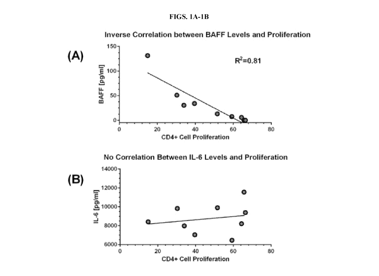
[00014] FIGS. 1A-1B demonstrate down modulation of MSC-BAFF via siRNA gene
silencing. (A) Correlation of BAFF levels and T cell proliferation. Shown is a
representative
graph for a single MSC line that was transfected with seven different BAFF
siRNA constructs.
BAFF levels correlated inversely with T cell proliferation. Correlation
coefficients ranged from
0.72-0.81. (B) MSCs also express IL-6. As a control, IL-6 levels were analyzed
in each of the
supernatants and correlated to T cell proliferation. IL-6 expression failed to
correlate with degree
of T cell proliferation.
[00015] FIGS. 2A-2B demonstrate the effects of BAFF Receptor blockade on
CD4+ T cell
proliferation. Antibodies which block ligand (BAFF and APRIL) binding to BAFF
receptors
BR3, TACI, and BCMA were added to PBL cultures in which T cells were activated
with anti-
CD3E and anti-CD28. Blocking antibodies were added at Day 0 and T cell
proliferation was
analyzed on Day 4 by flow cytometry using an anti-CD4 antigen-presenting cell
(APC) antibody.
- 3 -
CA 03017603 2018-09-12
WO 2017/160806 PCT/US2017/022260
Goat IgG is the control antibody for anti-BR3 and anti-BCMA. Mouse IgG1 is the
control
antibody for anti-TACT. Figures 2A and 2B depict data from two different donor
PBL samples.
[00016] FIGS. 3A-3C demonstrate the effects of BAFF Receptor blockade on
CD8+ T cell
activation and proliferation. Antibodies which block ligand binding to BAFF
receptors BR3,
TACT, and BCMA were added to PBL cultures in which T cells were activated with
anti-CD3E
and anti-CD28. Blocking antibodies were added at Day 0 and T cell
proliferation was analyzed
on Day 4 by flow cytometry using an anti-CD8 APC antibody. Goat IgG is the
control antibody
for anti-BR3 and anti-BCMA. Mouse IgG1 is the control antibody for anti-TACT.
(A)
Proliferation of CD8+ T cells in the presence of BAFF receptor blockade. (B)
ELISA analysis of
IFN-y levels in the supernatants of PBL cultures. Shown are the differential
levels of two
different PBL donors. (C) ELISA analysis of granzyme B levels in the
supernatants of the PBL
(activated T cell) cultures. Experiments were performed using two different
PBL donors.
[00017] FIGS. 4A-4D show flow analysis of BR3 on CD4 and CD8 T cells in
relation to
markers of activation. (A) BR3 percentages on resting and activated human T
cells using anti-
BR3 antibody clone 11C1. T cell subsets were purified and stimulated with
plate-bound anti-
CD3 and anti-CD28 for 21 hours after which they were stained for anti-BR3-PE
for flow
cytometric analysis. (B) Resting and stimulated dot plots of CD25 and BR3. (C)
BR3+ vs. BR3-
CD25 expression on activated T cells. (D) BR3 vs. IFN-gamma dot plot. IFN-
gamma was
detected using intracellular flow cytometry. T cells were stimulated as above
for 18 hours and
incubated with Brefeldin A for 6 hours. Cells were then fixed, permeabilized,
and stained with
anti-BR3 PE and anti-IFN-gamma APC.
[00018] FIGS. 5A-5C demonstrates that anti-BR3 mediated blockade increases
T cell
activation as gauged by CD25 (IL2-R-alpha) expression. (A) Flow cytometry of
increased CD25
expression in CD4+ and CD8+ T cells treated with anti-BR3 and activated for 21
hours. (B)
Semi-quantitative PCR of CD25 mRNA in CD4+ and CD8+ T cells. CD25 expression
was
measured in relation to GAPDH expression in T cell subsets activated for 21
hours. (C) CD25
cell surface expression increases with anti-BR3 in the presence of exogenous
BAFF added at
nanogram/ml concentrations, levels that are found in tumor microenvironments.
[00019] FIGS. 6A-6C shows increased IFN-y expression in purified CD4+ and
CD8+ T cell
subsetswith BR3 blockade. (A) Dot plot of intracellular IFN-y expression with
and without anti-
- 4 -
CA 03017603 2018-09-12
WO 2017/160806 PCT/US2017/022260
BR3. (B) 21 hour ELISA of IFN-y expression in CD4+ and CD8+ T cells. (C) Semi-
quantitative
PCR at 21 hour post-activation. Shown is relative gene expression of IFN-y in
CD4+ and CD8+
T cells treated with the goat IgG control or anti-BR3.
[00020] FIGS. 7A-7C shows the inhibition by shRNA increases CD25 and IFN-y.
(A) The
use of shRNA down modulates BR3 expression in CD4+ T cells. GIPZ BR3 specific
shRNA
constructs from Dharmacon, Inc. were used in addition to the GIPZ control
vector (B) CD25
expression increases on the cell surface of T cells with shRNA down regulation
of BR3. Flow
cytometric analysis was performed after an 18 hour rest of T cells after
nucleoporation with a
subsequent 21 hour activation period. (C) Expression of IFN-y increases in BR-
3 silenced CD4+
T cells. Intracellular flow cytometry was performed after 18 hours of
activation with a
subsequent 6 hour incubation in the presence of Brefeldin A.
[00021] FIGS. 8A-8D demonstrates that anti-BR3 augments T cell
cytotoxicity. (A) FIG. 9A
depicts granzyme B expression, as measured by PCR and ELISA assay, in anti-
CD3/CD28
activated CD4+ and CD8+ T cells with anti-BR3 or the goat IgG control.
Granzyme B
augmentation is specific for BR3 neutralization and is not increased with TACI
or BCMA
neutralization. Perforin is not increased with anti-BR3. (B) CRTAM is
expressed on cytolytic
CD4+ and activated CD8+ cells. Anti-BR3, but not anti-TACI and anti-BCMA
neutralizing
antibodies, increase CD25 expression on CRTAM+ cells as measured by flow
cytometry. The
increase in the median channel fluorescence of CD25 is greater on CRTAM+CD4+ T
cells than
for CRTAM+CD8+ T cells. (C) Anti-BR3 blockade increases killing of myeloma
cell line U266
by CD4+ T cells. Killing was measured by the release of LDH using a
cytotoxicity kit (Bio-Rad,
Inc.) (D) Anti-BR3 increases T cell killing of melanoma cell line A375. Shown
is the depletion
of adherent A375 cells after a 3 day co-culture with T cells stimulated with
anti-CD3/CD28, with
the goat IgG control or anti-BR3.
[00022] FIG. 9 shows IFN-y levels in T cell-A375 co-culture supernatants as
described in
FIG. 8D. T cells were cultured with A375 cells at a 1-0, 1-1, and 1-0.1 ratio
with no antibody,
the goat IgG control, and anti-BR3. A375 cells suppress T cell activation.
Anti-BR3 overcomes
A375 mediated suppression of IFN-gamma production.
DETAILED DESCRIPTION
- 5 -
CA 03017603 2018-09-12
WO 2017/160806 PCT/US2017/022260
[00023] The methods disclosed herein are based at least in part on the
inventors' discovery of
the role of BAFF (B cell activating factor) and APRIL (a proliferation-induced
ligand) in T-cell
suppression. BAFF is a key regulator for B cell differentiation and is
critical in regulating
survival and activation of peripheral B cell populations. BAFF binds to three
TNF receptor
superfamily members: B cell maturation antigen (BCMA/TNFRSF17), transmembrane
activator
and calcium-modulator and cyclophilin ligand interactor (TACl/TNFRSF13B), and
BAFF
receptor (also known as BAFF-R/BR3/TNFRSF13C/BLyS receptor 3 and TNFRSF13C).
These
receptors are type III transmembrane proteins that lack a signal peptide.
Whereas TACT and
BCMA bind both BAFF and another TNF superfamily ligand, APRIL (a proliferation-
inducing
ligand), BAFF-R selectively binds BAFF.
[00024] As described for the first time herein, the addition of either MSC-
derived
BAFF/APRIL or recombinant BAFF/APRIL to interferon gamma-activated MSC
cultures can
increase the expression and activity of the enzyme indoleamine 2,3-dioxygenase
(ID01), which
catalyzes degradation of the essential amino acid L-tryptophan to N-
formylkynurenine. BAFF
and APRIL seem to act as a toggle switch for IDO1 expression. Without being
bound by any
particular theory, it is believed that down-regulating IDO1 by specifically
blocking the
BAFF/APRIL receptor that augments expression will decrease leukocyte functions
and increase
proliferation of effector T cells.
[00025] Accordingly, the present disclosure relates to methods, cells, and
compositions for
preparing cell populations and compositions for adoptive cell therapy. In
particular, provided
herein are efficient and effective methods for robust expansion and activation
of T cell
populations for genetic engineering and adoptive T cell immunotherapies.
Methods of the
present invention provide for the preparation of T cells for use in
therapeutic methods by
selectively activating particular T cell populations. Also provided are cells
and compositions
produced by the methods and methods of their use. The present disclosure also
relates to
methods for the stimulation of T cell activation and expansion in vivo and in
vivo administration
of anti-BAFF-R agents.
[00026] Methods
[00027] In a first aspect, provided herein are robust methods of preparing
a population of T
cell by expanding and activating T cells ex vivo. As used herein, the term "ex
vivo" refers to a
- 6 -
CA 03017603 2018-09-12
WO 2017/160806 PCT/US2017/022260
condition that takes place outside an organism. In the context of this
disclosure, treatment of
immune cells ex vivo means exposing such cells to certain biological molecules
(e.g., agonists,
antagonists) in vitro (i.e., outside of an organism), preferably under sterile
conditions. In some
cases, ex vivo methods additionally include culturing immune cells that have
been isolated from
a human prior to administration back into the same human subject.
[00028] In a first step, the BAFF receptor is down regulated or blocked in
a population of T
cells such that the activity of the BAFF receptor is eliminated or reduced.
The BAFF receptor
may be down regulated or blocked by any suitable method or technique known in
the art. Known
methods for down regulation of gene expression or decreasing the activity of a
receptor include,
but are not limited to, CRISPR, microRNA, shRNA, RNAi, neutralizing
antibodies, small
molecule inhibitors, chemical inhibitors blocking downstream signaling
pathways, and the like.
In some embodiments, BAFF receptor activity or gene expression is reduced by
between 1%-
100% (i.e., 1%, 5%, 10%, 15%, 20%, 25%, 30%, 35%, 40%, 45%, 50%, 55%, 60%,
65%, 70%,
75%, 80%, 85%, 90%, 95%, 98%, 99%, 100%). In one embodiment, T cells are
cultured in the
presence of a BAFF Receptor antagonist. In one embodiment, T cells are
contacted with a
BAFF-R specific shRNA to reduce BAFF-R gene expression.
[00029] In one embodiment, T cells are cultured in the presence of a BAFF
Receptor
antagonist. In exemplary embodiments, the BAFF receptor antagonist is a
neutralizing antibody
that reacts with the B cell activating factor receptor (BAFF-R). Human anti-
BAFF-R antibodies
are available from commercial suppliers such as R&D Systems and Invitrogen.
[00030] Preferably, the T cells are in a leukocyte-containing cell mixture
or purified T cell
population. In some cases, leukocyte-containing cell mixtures or purified T
cell populations are
obtained from, for example, apheresis of peripheral blood of a human subject
or peripheral blood
mononuclear cells of a human subject. As used herein, the term "leukocyte-
containing cell
mixture" refers to a cell population or cell composition comprising leukocyte
cell type including
granulocytes, lymphocytes and monocytes. A leukocyte-containing cell mixture
preferably
comprises one or more specific leukocyte cell types. A preferred cell type is
the lymphocyte,
especially a T-lymphocyte ("T cell"). As used herein, the term "purified T
cell population" refers
to T cells isolated, separated, or otherwise removed from the blood or a
leukocyte milieu (e.g.,
obtained by leukapheresis), whereby isolated/separated T cells exist in a
physical milieu distinct
- 7 -
CA 03017603 2018-09-12
WO 2017/160806 PCT/US2017/022260
from that in which they occur in vivo. The term does not imply any particular
degree of purity,
and the absolute level of purity is not critical. Those skilled in the art can
readily determine
appropriate levels of purity for use according to the methods provided herein.
[00031] In one embodiment, T cells are contacted with a BAFF-R specific
shRNA to reduce
BAFF-R gene expression. A suitable shRNA for the present invention is one that
is able to direct
cleavage and subsequence degradation of complementary BR3 mRNA. Suitable shRNA
constructs are commercially available. For example, shRNA constructs may be
purchased from
Dharmacon Inc.
[00032] In a next step, T cells in which the BAFF receptor has been down
regulated or
blocked are cultured for about 3 to about 14 days (e.g., about 3, 4, 5, 6, 7,
8, 9, 10, 11, 12, 13, 14
days) in the presence of an anti-CD3 antibody, or a CD3-binding fragment
thereof, and an anti-
CD28 antibody, or a CD28-binding fragment thereof, under conditions
appropriate for activating
T cells. In vitro T cell expansion using this about 3 to about 14 day culture
step activates and
induces proliferation of such cultured T cells to yield an expanded population
comprising
activated cytotoxic T cells in sufficient numbers for use in therapy. The
expanded T cell
population can comprise CD4-positive T cells or CD8-positive T cells. In some
cases, cytotoxic
CD8+ T cells are preferentially expanded from the activated T cell population.
[00033] Conditions appropriate for activating T cells include any medium
suitable to maintain
the viability of the T cells and any formulation of the anti-CD3 and anti-CD28
antibodies such
that the antibodies are able to contact the T cells. In some embodiments, the
anti-CD3 and anti-
CD28 antibodies may be affixed to a solid substrate such as a bead or the
surface of a plate. In
some embodiments, the anti-CD3 and anti-CD28 antibodies are soluble. In one
embodiment, an
exemplary culture medium for culturing leukocytes is RPMI 1640 cell culture
medium or a
similar cell culture medium. Optionally, the medium may contain up to about
25% heat-
inactivated human serum albumin. A BAFF-R antagonist is added to the culture
medium, and
incubation with the BAFF-R antagonist can be performed at any appropriate
temperature (e.g.,
4 C, 25 C, or 37 C). Preferably, BAFF-R antagonist incubation occurs at 37 C.
Suitable
treatment duration can be conveniently optimized by one of ordinary skill in
the art. Preferably,
the treatment time is about 1 hour to about 24 hours. Exemplary BAFF-R
antagonist amounts for
contacting to leukocytes include about 0.1 [tg/m1 to about 100 [tg/ml. One of
skill in the art could
- 8 -
CA 03017603 2018-09-12
WO 2017/160806 PCT/US2017/022260
easily determine the useful amounts of BAFF-R antagonist to reduce or
eliminate BAFF-R
activity.
[00034] T cells are broadly divided into cells expressing CD4 on their
surface (also referred to
as CD4-positive cells) and cells expressing CD8 on their surface (also
referred to as CD8-
positive cells). T cells appropriate for use according to the methods provided
herein are
mononuclear lymphocytes (PBLs) derived from bone marrow (BM) or peripheral
blood (PB) of
a human donor. These cells could be collected directly from BM or PB or after
mobilization or
stimulation via administration of growth factors and/or cytokines such as
granulocyte-colony
stimulating factor (G-C SF) or granulocyte-macrophage colony-stimulating
factor (GM-CSF) to
allogeneic or autologous donors. Those skilled in the art would appreciate
that there are many
established protocols for isolating peripheral blood mononuclear cells (PBMC)
from peripheral
blood. Human peripheral blood may be drawn conveniently via venipuncture.
Isolation of PBMC
can be aided by density-gradient separation protocols, usually employing a
density-gradient
centrifugation technique using Ficoll -Hypaque or Histopaque for separating
lymphocytes
from other elements in the blood. Preferably, PBMC isolation is performed
under sterile
conditions. Alternatively, cell elutriation methods may be employed to
separate mononuclear cell
populations. The advantages of the cell elutriation method include sterility
and efficiency.
[00035] In exemplary embodiments, the methods provided herein include
activating BAFF-R-
contacted leukocytes with a stimulus that induces T-cell activation. Exemplary
stimuli include,
but are not limited to, mitogens such as Concanavalin A, IL-2, and anti-CD2-,
anti-CD3-, or anti-
CD28 beads. CD28 (also known as T90/44 antigen or Tp44) is a T-cell surface
expressed antigen
that is a receptor for costimulatory proteins acting on T cells. CD3 is a
complex of at least five
membrane bound polypeptides in mature T-lymphocytes that are non-covalently
associated with
one another and with the T cell receptor. The CD3 complex includes gamma,
delta, epsilon, zeta,
and eta subunits. When antigen binds to the T cell receptor, the CD3 complex
transduces the
activating signals to the cytoplasm of the T cell. For example, cross linking
T cell receptors with
anti-CD3 monoclonal antibody (mAb) leads to T cell activation, proliferation,
cytokine
synthesis, and non-specific cytotoxicity directed at tumor targets. These
activated T cells are
characterized by increased IL-2 production, exhibit non-MHC restricted
cytotoxicity, and
produce IFNy, TNFa, and GM-CSF.
- 9 -
CA 03017603 2018-09-12
WO 2017/160806 PCT/US2017/022260
[00036] In some cases, methods of this disclosure further include
introducing a genetically
engineered or chimeric antigen receptor into activated T cells, wherein the
method thereby
generates an expanded population comprising CD4+ T cells and CD8+ T cells
expressing the
genetically engineered or chimeric antigen receptor. Chimeric antigen
receptors (CARs), also
known as chimeric T cell receptors, artificial T cell receptors and chimeric
immunoreceptors, are
engineered receptors, which graft specificity onto an immune effector cell. In
general, a chimeric
antigen receptor is a transmembrane protein having a target-antigen binding
domain that is fused
via a spacer and a transmembrane domain to a signaling endodomain. When the
CAR binds its
target antigen, an activating signal is transmitted to the T-cell. In one
embodiment, the chimeric
antigen receptor or genetically engineered receptor is introduced into the T
cells. In one
embodiment a nucleic acid vector encoding the chimeric antigen receptor or
genetically
engineered receptor is transfected into the T cells whereby the T cells
express the chimeric
antigen receptor.
[00037] Reagents and other materials used during ex vivo manipulation
procedures, for
example antibodies, cytokines, serum, other chemicals, or solid supports such
as beads and
especially the virus-based gene vectors, should be compatible with aseptic
production of a
therapeutic cell product.
[00038] Expanded T cell populations obtained according to a method provided
herein are
useful for cellular immunotherapies including, without limitation, T cell
therapy, adoptive cell
therapy (ACT), and CAR T cell therapy. As used herein, the term "adoptive cell
therapy" refers
to the transfer of lymphocytes to mediate an effector function. For example,
expanded T-cell
populations obtained as described herein can be used in an ACT method to
reverse in vivo and in
vitro functional T-cell defects in patients having cancer (e.g., lymphoma).
Adoptive T-cell
therapies include administration of T cells that have been engineered to
express chimeric antigen
receptors, administration of tumor-infiltrating lymphocytes without genetic
modifications (TILs),
and administration of T cell receptor (TCR) engineered T cells. T-cell
checkpoint therapies and
TIL therapies exploit the intrinsic tumor recognition capacity of the T-cell
compartment.
Adoptive therapy with gene-modified T cells has the potential to address an
entirely different
need by creating a tumor-specific T cell compartment that is otherwise absent
from patients. As
such, gene-modified ACT has potential for tumor types that may not be
responsive to T cell
- 10 -
CA 03017603 2018-09-12
WO 2017/160806 PCT/US2017/022260
checkpoint or TIL therapies, such as most cancers occurring in children and
many of the
hematological malignancies. In addition, T cell checkpoint therapies and gene-
modified ACT
have the potential to work synergistically. Accordingly, it is contemplated
that adoptive cell
therapy using T cells obtained according to the methods provided herein can be
combined with
additional therapeutic technologies such as checkpoint-blocking antibodies,
vaccines, and
targeted drug therapies.
[00039] Donor lymphocyte infusions (DLIs) induce direct and potent graft
versus tumor
(GVT) effects, which are particularly effective for patients who relapse after
allogeneic stem cell
transplantation (SCT) with a donor graft. It can be advantageous to use ex
vivo-activated DLI
(aDLI) for some leukemia patients. In such cases, activated donor T cells are
produced by co-
stimulation and expansion following exposure to magnetic beads coated with
anti-CD3 (OKT3)
and/or anti-CD28. Generally, co-stimulation of T cells via CD3 and CD28 can
produce activated
T cells that can overcome disease-induced anergy, preserve and augment CD4
function, and
enhance GVT activity. T cells obtained according to a method provided herein
can be infused
into a subject (e.g., a subject having relapsed disease after allogeneic SCT).
[00040] In some cases, it can be advantageous to simultaneously isolate and
activate
(stimulate) T cells from a PBMC product. For example, magnetic beads coated
with anti-CD3
and anti-CD28 (i.e., the CTS Dynabeads CD3/CD28) can be used in combination
with a large
magnet to sort magnetic bead-attached cells from those not bound to magnetic
beads.
[00041] Also contemplated herein are methods for the administration of an
anti-BAFF-R
agent to a subject for the stimulation of T cell activation and expansion in
vivo. As used herein
"anti-BAFF-R agent" refers to an entity that inhibits or reduces the activity
or gene expression of
the BAFF-R receptor. Anti-BAFF-R agents may include, but are not limited to,
inhibitory anti-
BAFF-R monoclonal antibodies, small molecule inhibitors, shRNA, shRNA vectors,
microRNA,
microRNA vectors, and the like. It is envisioned that treatment strategies
utilizing the expanded
populations of T cells obtained according to a method provided herein can be
supplemented or
replaced with an anti-BAFF-R agent for the activation and expansion of T cells
in vivo.
[00042] Expanded populations of T cells obtained according to a method
provided herein are
useful for treating or preventing various disorders such as a cancer (e.g., a
blood malignancy
such as lymphoma or leukemia or a solid tumors such as melanoma or kidney
cancer) or an
- 11 -
CA 03017603 2018-09-12
WO 2017/160806
PCT/US2017/022260
infectious disease such as HIV. As used herein, the terms "treat" and
"treating" refer to both
therapeutic and prophylactic or preventive measures, wherein the object is to
prevent or slow
down (lessen) an undesired physiological change or pathological disorder. For
purposes of this
invention, treating a cancer includes, without limitation, alleviating one or
more clinical
indications, decreasing tumor growth or tumor cell proliferation, reducing the
severity of one or
more clinical indications of a cancer condition, diminishing the extent of the
condition,
stabilizing the subject's disease state (i.e., not worsening), delay or
slowing, halting, or reversing
cancer progression, and bringing about partial or complete remission. Treating
cancer also
includes prolonging survival by days, weeks, months, or years as compared to
prognosis if
treated according to standard medical practice not incorporating T cells
obtained according to a
method provided herein. Subjects in need of treatment can include those
already having or
diagnosed with cancer, as well as those prone to, likely to develop, or
suspected of having cancer
(e.g., lymphoma or multiple myeloma) or an infection. In some cases, the
subject may have an
autoimmune disease. As used herein, the terms "prevent" and "preventing" refer
to prophylactic
or preventive measures intended to inhibit undesirable physiological changes
or the development
of a disorder or condition. In exemplary embodiments, preventing a disease or
condition
comprises initiating the administration of T cells obtained according to a
method provided herein
at a time prior to the appearance or existence of the disease or condition (or
a symptom thereof)
such that the disease or condition, or its symptoms, pathological features,
consequences, or
adverse effects do not occur.
[00043] T
cells obtained according to a method provided herein can be used in an
adoptive
cell therapy method for the treatment of cancer including malignancies
including those of the
hematolymphoid system (leukemias, lymphomas, multiple myeloma). Cancers
appropriate for
treatment as described herein include hematological malignancies such as acute
myelogenous
leukemia (AML), acute lymphocytic leukemia (ALL), chronic myelogenous leukemia
(CML),
myeloma, non-Hodgkin and Hodgkin lymphoma (e.g., relapsed, refractory, or
chemotherapy-
resistant non-Hodgkin lymphoma), and myelodysplastic syndrome (MDS). The terms
"cancer"
and "tumor" are used interchangeably herein. Other cancer appropriate for
treatment include
solid tumors such as melanoma, kidney, colon, lung, brain, and liver cancers.
- 12 -
CA 03017603 2018-09-12
WO 2017/160806 PCT/US2017/022260
[00044] T cells obtained according to a method provided herein can be used
in an adoptive
cell therapy method for the treatment of an infection. As used herein, the
term "infection"
describes a diseased state in which a microorganism or other infectious agent
invades healthy
cells, and includes any conditions or disease states caused by bacterial,
viral, fungal, or parasitic
(e.g., protozoan) infectious agents. For example, the term "viral infection"
describes the
infiltration of healthy cells by a virus (e.g., HIV), wherein the virus uses
the cell's reproductive
machinery to multiply or replicate and ultimately lyse the cell resulting in
cell death, release of
viral particles and the infection of other cells by the newly produced progeny
viruses. With
respect to infections, the term "treatment" further refers to the application
or administration of a
therapeutic where the purpose is to cure, heal, alleviate, relieve, alter,
remedy, ameliorate,
improve, or affect the infection, any associated symptoms of the infection, or
the predisposition
toward the development of the infection.
[00045] As used herein, the terms "subject" or "patient" are used
interchangeably and can
encompass any vertebrate including, without limitation, humans, mammals,
reptiles, amphibians,
and fish. However, advantageously, the subject or patient is a mammal such as
a human, or a
mammal such as a domesticated mammal, e.g., dog, cat, horse, and the like, or
livestock, e.g.,
cow, sheep, pig, and the like. In exemplary embodiments, the subject is a
human. As used herein,
the phrase "in need thereof' indicates the state of the subject, wherein
therapeutic or preventative
measures are desirable. Such a state can include, but is not limited to,
subjects having a disease
or condition such as cancer.
[00046] In some cases, T cells obtained according to a method provided
herein can be
administered as a pharmaceutical composition comprising a therapeutically
effective amount of
T cells as a therapeutic agent (i.e., for therapeutic applications). As used
herein, the term
"pharmaceutical composition" refers to a chemical or biological composition
suitable for
administration to a mammal. Examples of compositions appropriate for such
therapeutic
applications include preparations for parenteral, subcutaneous, transdermal,
intradermal,
intramuscular, intracoronarial, intramyocardial, intracerebral, intratumoral,
intraperitoneal,
intravenous (e.g., injectable), or intratracheal administration, such as
sterile suspensions,
emulsions, and aerosols. Intratracheal administration can involve contacting
or exposing lung
tissue, e.g., pulmonary alveoli, to a pharmaceutical composition comprising a
therapeutically
- 13 -
CA 03017603 2018-09-12
WO 2017/160806 PCT/US2017/022260
effective amount of T cells. In some cases, pharmaceutical compositions
appropriate for
therapeutic applications may be in admixture with one or more pharmaceutically
acceptable
excipients, diluents, or carriers such as sterile water, physiological saline,
glucose or the like. For
example, T cells described herein can be administered to a subject as a
pharmaceutical
composition comprising a saline solution. In exemplary embodiments, a
pharmaceutical
composition comprising T cells expanded according to a method provided herein
is capable of
inducing a desired therapeutic or prophylactic effect upon administration to a
subject.
[00047] Formulations may be designed or intended for oral, rectal, nasal,
topical or
transmucosal (including buccal, sublingual, ocular, vaginal and rectal) and
parenteral (including
subcutaneous, intramuscular, intravenous, intradermal, intraperitoneal,
intrathecal, intraocular
and epidural) administration. In general, aqueous and non-aqueous liquid or
cream formulations
are delivered by a parenteral, oral or topical route. In other embodiments,
the compositions may
be present as an aqueous or a non-aqueous liquid formulation or a solid
formulation suitable for
administration by any route, e.g., oral, topical, buccal, sublingual,
parenteral, aerosol, a depot
such as a subcutaneous depot or an intraperitoneal or intramuscular depot. In
some cases,
pharmaceutical compositions are lyophilized. In other cases, pharmaceutical
compositions as
provided herein contain auxiliary substances such as wetting or emulsifying
agents, pH buffering
agents, gelling or viscosity enhancing additives, preservatives, flavoring
agents, colors, and the
like, depending upon the route of administration and the preparation desired.
The pharmaceutical
compositions may be formulated according to conventional pharmaceutical
practice (see, e.g.,
Remington: The Science and Practice of Pharmacy, 20th edition, 2000, ed. A. R.
Gennaro,
Lippincott Williams & Wilkins, Philadelphia, and Encyclopedia of
Pharmaceutical Technology,
eds. J. Swarbrick and J. C. Boylan, 1988-1999, Marcel Dekker, New York).
[00048] The preferred route may vary with, for example, the subject's
pathological condition
or weight or the subject's response to therapy or that is appropriate to the
circumstances. The
formulations can also be administered by two or more routes, where the
delivery methods are
essentially simultaneous or they may be essentially sequential with little or
no temporal overlap
in the times at which the composition is administered to the subject.
[00049] Suitable regimes for initial administration and further doses or
for sequential
administrations also are variable, may include an initial administration
followed by subsequent
- 14 -
CA 03017603 2018-09-12
WO 2017/160806 PCT/US2017/022260
administrations, but nonetheless, may be ascertained by the skilled artisan
from this disclosure,
the documents cited herein, and the knowledge in the art.
[00050] In some cases, T cells may be optionally administered in
combination with one or
more active agents. Such active agents include anti-inflammatory, anti-
cytokine, analgesic,
antipyretic, antibiotic, and antiviral agents, as well as growth factors and
agonists, antagonists,
and modulators of immunoregulatory agents (e.g., BAFF, APRIL, TNF-a, IL-2, IL-
4, IL-6, IL-
10, IL-12, IL-13, IL-17, IL-18, IL-21, IL-35, IFN-a, IFN-y, CXCL13, IP-10,
VEGF, EPO, EGF,
HRG, Hepatocyte Growth Factor (HGF), Hepcidin, antibodies reactive against any
of the
foregoing, and antibodies reactive against any of their receptors). Any
suitable combination of
such active agents is also contemplated. When administered in combination with
one or more
active agents, T cells can be administered either simultaneously or
sequentially with other active
agents. For example, subjects may simultaneously receive T cells and one or
more of the agents
described herein for a length of time or according to a dosage regimen
sufficient to support
recovery and to treat, alleviate, or lessen the severity of a disease or
condition.
[00051] In some embodiments, T cells are administered to a subject in need
thereof using an
infusion, topical application, surgical transplantation, or implantation. In
exemplary
embodiments, administration is systemic. In such cases, T cells are provided
to a subject in need
thereof in a pharmaceutical composition adapted for intravenous administration
to subjects.
Typically, compositions for intravenous administration are solutions in
sterile isotonic aqueous
buffer. The use of such buffers and diluents is well known in the art. Where
necessary, the
composition may also include a local anesthetic to ameliorate any pain at the
site of the injection.
Generally, the ingredients are supplied either separately or mixed together in
unit dosage form,
for example, as a cryopreserved concentrate in a hermetically sealed container
such as an
ampoule indicating the quantity of active agent. Where the composition is to
be administered by
infusion, it can be dispensed with an infusion bottle containing sterile
pharmaceutical grade
water or saline. Where the composition is administered by injection, an
ampoule of sterile water
for injection or saline can be provided so that the ingredients may be mixed
prior to
administration. In some cases, compositions comprising T cells of the present
invnetion are
cryopreserved prior to administration.
- 15 -
CA 03017603 2018-09-12
WO 2017/160806 PCT/US2017/022260
[00052] Therapeutically effective amounts of T cells as provided herein are
administered to a
subject in need thereof. As used herein, the term "therapeutically effective
dose" refers to any
dose sufficient to prevent advancement, or to cause regression of the disease
or condition at
issue, or which is capable of relieving symptoms caused by the disease or
condition, such as pain
or swelling. The effective dose or amount, which can be administered in one or
more
administrations, is the amount of human T cells sufficient to elicit a
therapeutic effect in a
subject to whom the cells are administered. A therapeutically effective amount
can be an amount
between about 50 x 106 cells and about 700 x 106 cells of an ex vivo expanded
T cell culture. In
some cases, an effective amount is administered as a dosage comprising at
least 1 x 10⁶ cells per
kilogram (kg) of body weight of the recipient. For example, the effective
amount can be
administered to the subject in a dose comprising at least 1 x 10⁶ cells/kg, at
least 10 x 106
cells/kg, at least 30 x 106 cells/kg, at least 100 x 106 cells/kg, or at least
1000 x 106 cells/kg.
[00053] Effective amounts will be affected by various factors which modify
the action of the
cells upon administration and the subject's biological response to the cells,
e.g., the patient's age,
sex, and diet, the severity of inflammation, time of administration, and other
clinical factors. A
therapeutically effective amount of T cells obtained according to a method
provided herein can
be administered to a subject.
[00054] Therapeutically effective amounts for administration to a human
subject can be
determined in animal tests and any art acceptable methods for scaling an
amount determined to
be effective for an animal for human administration. For example, an amount
can be initially
measured to be effective in an animal model (e.g., to achieve a beneficial or
desired clinical
result). The amount obtained from the animal model can be used in formulating
an effective
amount for humans by using conversion factors known in the art. The effective
amount obtained
in one animal model can also be converted for another animal by using suitable
conversion
factors such as, for example, body surface area factors.
[00055] It is to be understood that, for any particular subject, specific
dosage regimes should
be adjusted over time according to the individual need and the professional
judgment of the
person administering or supervising the administration of the T cells. For
example, a T cell
dosage for a particular subject can be increased if the lower dose does not
elicit a detectable or
- 16 -
CA 03017603 2018-09-12
WO 2017/160806 PCT/US2017/022260
sufficient improvement. Conversely, the dosage can be decreased if the disease
or condition is
treated or eliminated.
[00056] In some cases, therapeutically effective amounts of T cells can be
determined by, for
example, measuring the effects of a therapeutic in a subject by incrementally
increasing the
dosage until the desired symptomatic relief level is achieved. A continuing or
repeated dose
regimen can also be used to achieve or maintain the desired result. Any other
techniques known
in the art can be used as well in determining the effective amount range. Of
course, the specific
effective amount will vary with such factors as the particular condition being
treated, the
physical condition of the subject, the type of animal being treated, the
duration of the treatment,
and the nature of any concurrent therapy. Following administration of T cells
to an individual
subject afflicted by, prone to, or likely to develop a disease or condition as
described herein, the
subject is observed and assessed for a positive or negative change in clinical
symptoms or
features of the disease or condition. For example, for methods of treating
cancer in a subject,
positive or negative changes during or following treatment may be determined
by any measure
known to those of skill in the art including, without limitation, measuring
changes in tumor size.
[00057] In any of the methods of the present invention, the donor and the
recipient of the T
cells can be a single individual or different individuals, for example,
autologous, allogeneic or
xenogeneic individuals. As used herein, the term "autologous" refers to cells
or tissues obtained
from an individual and transplanted back into the same individual. As used
herein, the term
"allogeneic" refers to cells or tissues obtained from different individuals of
the same species,
where the donor and recipient are not genetically identical. With regard to
the present disclosure,
an allogeneic cell transplant or tissue graft involves transplantation of
cells or tissues where the
donor and recipient are different individuals of the same species. The term
"xenogeneic" means
that which is derived or obtained from an organism of a different species.
With regard to the
present disclosure, a xenogeneic cell transplant or tissue graft involves
transplantation of cells or
tissues where the donor and recipient are different individuals of different
species.
[00058] Administration to the subject can be by local or systemic injection
or by topical
application. For example, T cells can be administered by intravenous
injections such as drip
infusions, intramuscular injections, intraperitoneal injections, intra-organ
injections, or
- 17 -
CA 03017603 2018-09-12
WO 2017/160806 PCT/US2017/022260
subcutaneous injections. In some cases, the subject is observed or assessed
with regard to tissue
maintenance, tissue repair or function, or overall condition.
[00059] Articles of Manufacture
[00060] In another aspect, the present invention provides articles of
manufacture useful for
adoptive cell therapy. In some cases, a kit of the present invention comprises
one or more vessels
containing human T cells. In particular embodiments, cells expanded and
activated according to
a method provided herein are provided in a kit, and in some cases the cells
can be the sole
component of the kit. The kit may additionally comprise reagents and materials
useful for
expanding and activating T cells as provided herein to obtain the desired cell
product. For
example, a kit can include one or more BAFF-R antagonists.
[00061] Optionally, a kit can further include one or more reagents or other
components
necessary for administering the T cells to a human subject in need thereof
according to a method
of the invention. It may be appropriate in some cases to provide T cells as a
frozen aliquot in a
pharmaceutically acceptable cryopreservant.
[00062] In some cases, the kit, in addition to T cells as provided herein,
also includes a second
therapeutic, such as a chemotherapeutic, a hormone therapeutic, and/or an
immunotherapeutic,
for example. The kit may be tailored to a particular cancer for an individual
and comprise
respective second therapeutics for that individual. In some cases, a kit
further comprises one or
more active agents such as, for example, anti-inflammatory, anti-cytokine,
analgesic, antipyretic,
antibiotic, and antiviral agents, as well as growth factors and agonists,
antagonists, and
modulators of immunoregulatory agents (e.g., TNF-a, interleukin-2 (IL-2), IL-
4, IL-6, IL-10, IL-
12, IL-13, IL-18, IFN-a, IFN-y, BAFF, CXCL13, IP-10, VEGF, EPO, EGF, HRG,
Hepatocyte
Growth Factor (HGF), Hepcidin, including antibodies reactive against any of
the foregoing, and
antibodies reactive against any of their receptors). Classes of pharmaceutical
agents useful for
treating cancer include, without limitation, glucocorticoids (e.g.,
prednisone),
immunosuppressants (e.g., cyclosporine, methotrexate, tacrolimus,
pimecrolimus, sirolimus,
mycophenolate, mofetil, visilizumab, anti-thymocyte globulin (ATG)),
antineoplastics (e.g.,
pentostatin), and antirheumatics (e.g., hydroxychloroquine, infliximab,
entanercept). Also
contemplated are kits comprising suitable combinations of such active agents.
Provided with
such vessels are instructions for human administration and a notice in the
form prescribed by a
- 18 -
CA 03017603 2018-09-12
WO 2017/160806 PCT/US2017/022260
governmental agency regulating the manufacture, use, or sale of biological
products, which
notice reflects approval by the agency of manufacture, use, or sale for human
administration.
[00063] The present invention will be more fully understood upon
consideration of the
following non-limiting Examples. All texts, papers, and patents disclosed
herein are hereby
incorporated by reference as if set forth in their entirety.
EXAMPLES
[00064] Reference is now made to the following examples, which together
with the above
descriptions illustrate the invention in a non-limiting fashion.
[00065] Example 1 - BAFF Interferes with MSC-Mediated T cell Suppression In
Vitro
[00066] We have been investigating mesenchymal stem cells (MSCs) derived
from the bone
marrow of healthy donors to determine whether MSC-derived BAFF interferes with
MSC-
mediated T cell suppression in vitro. We have designed an 'immunopotency
assay' (Bloom et al.,
2015. Cytotherapy 17:140-151) whereby peripheral blood leukocytes (PBLs)
obtained from
healthy donors and labeled with carboxyfluorescein succinimidyl ester (CF SE)
were co-cultured
with titrated numbers of MSCs. T cells within the PBL milieu were stimulated
with soluble anti-
CD3/anti-CD28. Proliferation of T cell subsets was measured by flow cytometry.
MSC-mediated
inhibition was gauged against a positive control of activated T cells without
MSCs. TACI-Fc
(Atacicept) is a soluble TACT receptor that effectively binds BAFF and APRIL.
When added to
PBL:MSC co-cultures, TACI-Fc reversed CD4+ T cell inhibition. The effect of
down-
modulating MSC-BAFF on T cell inhibition was measured.
[00067] Seven different BAFF-specific shRNA plasmids were used in each of
five
experiments (using five different MSC lines) with subsequent PBL co-culture.
Remarkably,
silencing MSC-BAFF reversed T cell suppression. BAFF levels correlated
inversely with CD4+
T cell proliferation (R2= 0.81, FIG. 1A). However, IL-6 expression by MSCs was
not affected by
BAFF down-modulation (FIG. 1B), nor was MSC viability. Decreases in IDO1
(Indoleamine-
pyrrole 2,3-dioxygenase 1) mRNA levels and enzyme activity correlated with
down-modulated
BAFF levels, suggesting that BAFF regulated the expression of this T cell
suppression factor.
[00068] Next, BR3 function was blocked using a BR3 neutralizing antibody
(R&D Systems,
Inc.). In 4-day co-culture assays, we found that anti-BR3 augmented CD4+
proliferation in MSC
- 19 -
CA 03017603 2018-09-12
WO 2017/160806 PCT/US2017/022260
co-culture, but also enhanced CD4+ proliferation in T cell-activated PBLs
without MSCs, to
varying degrees, depending on the PBL donor. These data suggested that BR3 was
controlling
the normal homeostatic suppression of at least a subset of CD4+ cells.
[00069] Upon investigation of CD8+ cells, a consistent increase in
proliferation was observed
with anti-BR3, but not observed with anti-BCMA or anti-TACT blockade (FIG.
3A).
Additionally, expression of IFN-y and granzyme B increased 9-40 fold and 3-7
fold,
respectively, with BR3 blockade (FIGS. 3B-3C). Importantly, the increase in
IFN-y and
granzyme B expression was not due to the anti-BR3 blocking antibody mediating
indiscriminate
T cell stimulation. To the contrary, human cytokine multiplex analysis
demonstrated lowered or
unchanged levels of other T cell cytokines. Preliminary assessments show that
the DO
activity/kynurenine levels in the supernatants are decreased in anti-BR3
treated (24 hour)
cultures. Importantly, anti-TACT had the opposite effect: T cell proliferation
and IFN-y
expression were significantly decreased, which may explain the dual nature of
BAFFs effects on
T cells.
[00070] Together, these data strongly indicate that BAFF augments the
expression of IDO1
and that BR3 is a primary negative regulator of cytotoxic (CD4+ and CD8+) T
cell proliferation,
IFN-y expression, and granzyme B production. We hypothesize that one of the
mechanisms of
BR3-mediated suppression of cytotoxic T cell activation is its ability to
enhance IDO1
expression. Our in vitro data supports the hypothesis that BAFF mediates the
expression of
immune suppression factors. When peripheral blood lymphocytes are stimulated
with T cell
stimulatory antibodies anti-CD3 and anti-CD28, proliferation of both CD4+ and
CD8+ T cells is
increased. Upon addition of antibodies which specifically neutralize the BAFF
receptor BR3,
both CD4+ and CD8+ T cell proliferation increased significantly.
Interestingly, the data also
revealed that blocking BR3 enhances IFN-y production as well as the production
and/or secretion
of the CD8+ T cell toxin granzyme B. These data strongly suggest that BR3 is
one of the
essential suppressors of CD8+ cytotoxic T cell activation and proliferation.
[00071] Methods & Materials
[00072] Preparing Peripheral Blood Leukocytes: PBLs were isolated from blood
taken from
healthy individuals. Apheresis products or whole blood can be used. Cells were
applied to a
Ficoll-Paque gradient to purify the white blood cells away from the red blood
cells and platelet
- 20 -
CA 03017603 2018-09-12
WO 2017/160806 PCT/US2017/022260
contamination as previously described (Corkum et al., 2015. BMC Immunol. Aug
26;16:48). The
PBLs were viably frozen in vials with DMSO and cryopreserved in liquid
nitrogen.
[00073] Antibodies and Reagents: Anti-CD3c is a monoclonal antibody that
stimulates the
epsilon chain of the TCR complex on human T cells. Clone UCHT1 was purchased
from R&D
Systems, Inc. (cat# MAB100). The lyophilized product was resuspended in PBS at
500 pg/ml,
aliquoted, and stored long term at -20 C.
[00074] Anti-CD28 is a monoclonal antibody that co-stimulates human T cells
in conjunction
with anti-CD3c. Clone 37407 was purchased from R&D Systems, Inc. (cat
#MAB342). The
lyophilized product was resuspended in PBS at 500 pg/ml, aliquoted, and stored
long-term at -
20 C.
[00075] Anti-BAFF-R blocking antibody blocks human BR3. It is a goat
polyclonal antibody
purchased from R&D Systems, Inc. (cat#AF1162). The lyophilized product was
resuspended in
PBS at 200 pg/ml, aliquoted, and stored long-term at -20 .
[00076] Goat IgG control was purchased from R&D Systems, Inc. (cat#AB-108-
C). The
lyophilized product was resuspended in PBS at 200 pg/ml, aliquoted, and stored
long-term at -
20 C.
[00077] Complete RPMI Medium: RPMI-1640 with 10% FBS (heat inactivated at
55 for 30',
Atlanta Biologicals), Glutamine, Non-Essential Amino Acids, HEPES, and
NaPyruvate.
Pen/Strep is not typically used but its addition does not alter results.
Medium was filter sterilized
before use.
[00078] CFSE (carboxylfluorescein diaacetate succinimidyl ester): stock
concentration of
1mM in DMSO. The fluorochrome was purchased from Invitrogen.
[00079] Anti-huCD4 APC and anti-huCD8 APC-labeled antibodies for flow
cytometry were
purchased from R&D Systems, Inc.
[00080] Example 2 - BR3 Blockade Protocol
[00081] The study described in Example 2 demonstrates methods used for
achieving a BR3
blockage in non-purified T cells for use in proliferation assays. We rapidly
thawed vials
containing peripheral blood leukocytes (PBLs) in a 37 C water bath (-2
minutes) and sterilely
transfer PBLs to a 15m1 conical tube and resuspend in a complete Roswell Park
Memorial
Institute (RPMI) culture medium. Tubes were centrifuged for 10 minutes at 1200
rpm.
-21 -
CA 03017603 2018-09-12
WO 2017/160806 PCT/US2017/022260
[00082] The supernatant was aspirated from the cell pellet. 10m1 of D-PBS
without Ca2+ or
Mg2+ was added to the cell pellet to wash the cells. The cells were
centrifuged for 10 minutes at
1200 rpm. Supernatant was aspirated from the cell pellet and no more than lml
PBS was added
per 10 x 106 cells. 2u1 of 1mM CFSE per lml of cells was added, mixed and
incubated in the
dark at 37 C for 10 minutes. An equal volume of cold (4 C) FBS was added to
stop the CFSE
labeling. Cells were washed and prepared such that 200 11.1 of cell suspension
corresponding to
approximately 4 x 10' cells was added per well in a 48-well plate.
[00083] Anti-BR3 antibody or control goat anti-IgG antibody was added to
each well of 48-
well plate. Cells and antibodies were incubated for 30 minutes at 37 C in an
incubator containing
5% CO2.
[00084] After incubation with the anti-BR3 or IgG control antibody, anti-
CD3/CD28
stimulatory antibodies (preferably at a ratio of 5:1) were added to the cell
culture the culture was
incubated for 3-4 days for T cell expansion to proceed. To analyze
proliferation, cells were
harvested from each well and placed in centrifuge tubes to separate
supernatants and collect cell
pellets. The supernatants were stored separately at -20 for further analysis.
[00085] Samples introduced to either anti-CD4 APC or anti-CD8 APC flow
antibodies and
were analyzed on a flow cytometer using FL1 (CF SE) and FL4 (APC) channels.
Cells were
analyzed for total proliferation against a non-stimulated control (i.e., the
total percentage of cells
that have undergone cell division). Cell proliferation was analyzed using
standard flow
cytometry data analysis and modeling software, either FlowJoTM or ModFit LTTm.
[00086] Example 3 - Ex vivo co-stimulation and expansion of T cells for
donor leukocyte
infusions (DLI)
[00087] An aliquot of cells from a donor leukocyte product collected on the
first or second
day of leukapheresis is removed prior to DLI for ex vivo expansion. The washed
apheresis
product is enriched for lymphocytes using magnetic bead depletion of monocytes
in a closed
system if monocytes constitute more than 20% of white blood cells (WBCs) as
gated on a
Coulter Multisizer3 (Beckman Coulter, Fullerton, CA). T cells are processed in
a manner
consistent with appropriate FDA guidelines and regulations on Good
Manufacturing Practices.
[00088] The cells are seeded into gas-permeable flasks containing X VIVO 15
(Cambrex,
Walkersville, MD) supplemented with 5% normal human AB serum (Valley
Biomedical,
- 22 -
CA 03017603 2018-09-12
WO 2017/160806 PCT/US2017/022260
Winchester, VA), 2 mM L-glutamine (Cambrex), and 20 mM HEPES (Cambrex).
Magnetic
beads (Dynal, Brown Deer, WI) with conjugated anti-CD3 (OKT3; Ortho Biotech,
Bridgewater,
NJ) and anti-CD28 (clone 9.3) monoclonal antibodies are added at a 3:1
bead/CD3+ cell ratio,
and the cultures are maintained for up to 12 days prior to harvest and
preparation for infusion.
After completion of cell culture, the magnetic beads are removed using a
magnetic cell
separation system, and the cells are washed, concentrated, and resuspended in
100 to 250 mL
PlasmaLyte A (Baxter Oncology)/5% dextrose 0.45% NaCl containing 1% human
serum
albumin (Baxter Oncology). All infused T-cell products are required to meet
release criteria
specified for T-cell phenotype, cell viability, pyrogenicity, sterility, and
freedom from bead
contamination.
[00089] Example 4 - Blockade of BAFF-R on T cells Enhances Their Activation
and
Cytotoxicity
[00090] Materials and Methods
[00091] Cell Culture and Purification: Primary human T cells were obtained
from
leukopheresis products purchased from AllCells, LLC (Alameda, CA) or Key
Biologics, LLC
(Memphis, TN). Upon arrival, PBLs were isolated via ficoll separation and
viably frozen for
future use. For T cell studies, thawed PBLs were used directly in activation
assays or T cell
subsets purified by magnetic bead sorting. For purification, CD4 or CD8 beads
from Miltenyi
Biotec, Inc. (San Diego, CA) were used according to manufacturer protocol. An
AutoMacs
sorter (Miltenyi Biotec) was used for bead sorting. CD4 and CD8 T cell
populations were
typically 90-95% pure. All T cell assays were performed in RPMI containing 10%
FBS,
glutamine, HEPES, Na-Pyruvate, NEAA, and Pen/Strep. Myeloma line U266 was
cultured in
RPMI containing 10% FBS, glutamine, HEPES, Na-Pyruvate, NEAA, and Pen/Strep.
The
adherent melanoma line A375 was expanded in alpha-MEM containing 10% FBS,
glutamine,
NEAA, and Pen/Strep. A375 cells were grown to 70-80% confluence before
passaging.
[00092] Flow Cytometry: All flow cytometry experiments were run on an Accuri
C6 (BD
Biosciences, Inc.) with 2 lasers for 4-color analysis. Antibodies used were as
follows: anti-BR3
PE, clone 11C1, BD Biosciences; anti-CD25 FITC-Violet and APC, clone 3H3,
Miltenyi Biotec;
anti-IFN-g APC, clone B27, BD Biosciences; anti-Granzyme B PE, clone GB11, BD
- 23 -
CA 03017603 2018-09-12
WO 2017/160806 PCT/US2017/022260
Biosciences; anti-CRTAM PE, clone Cr24.1, Biolegend, Inc. Analyses were
performed using
CFlowPlus (BD Biosciences) or FlowJo (TreeStar, Inc.) software.
[00093] Anti-BR3 Neutralization Assays: BAFF receptor blocking antibodies
anti-BR3 (cat#
AF1162), anti-TACT (cat# AF174), and anti-BCMA (cat# AF193), were purchased
from R&D
Systems, Inc (Minneapolis, MN). All three are goat polyclonal antibodies. All
were received as
lyophilized products. All were resuspended in PBS at the recommended
concentrations,
aliquoted, and frozen according to manufacturer's instructions. Care was taken
to use antibody
products that were thawed only once. For blocking assays, CD4 and CD8 T cell
subsets were
bead selected and resuspended in complete RPMI (see above) at lx10e6/ml. Cells
were pre-
incubated with each neutralizing antibody or normal goat IgG control (cat# AB-
108-C) at 10
ug/ml for 30 minutes at 25 C. 2x10e5 cells/well were added to 96-well flat
bottom tissue culture
treated plates; 4x10e5/m1 were added to 48-well plates. Plates were pre-coated
with lug/m1
anti-CD3e (clone UCHT1, R&D Systems, Inc.) and 0.2ug/m1 anti-CD28 (clone
37407, R&D
Systems, Inc.) at 37 C for 8 hours with subsequent PBS washes. Again, care was
taken to use
anti-CD3/CD28 antibodies thawed only once. T cells were activated and
incubated for 21-24
hours in a 37 C incubator at 5% CO2. Cells were gently harvested with wide
bore pipette tips
and supernatants collected and stored at -20 C.
[00094] BR3 shRNA Down-Modulation: Four shRNA plasmid constructs specific for
human
BAFF-R were purchased from Dharmacon, Inc. including the GIPZ shRNA control
plasmid. 1-
2ug of each plasmid was introduced into 2-4x10e6 CD4 or CD8 T cells using
Amaxa-based
nucleofection. Amaxa kits specific for human T cell transfection were
purchased from Lonza,
Inc. Program V24 was used for electroporation according to manufacturer's
instructions after
which cells were rested for 18 hours in 12-well plates containing pre-warmed
complete RPMI.
Cells were then gently harvested, analyzed for live/dead, and added at lx10e6
live cells/well to
12-well plates pre-coated with anti-CD3/CD28 (lug/m1 and 0.2ug/ml,
respectively, as above).
Transfected T cells were activated for 21-24 hours. Cells were then harvested
and analyzed for
BR3, CD25, and IFN-g as described above.
[00095] Cytotoxicity Assay: Cytotoxicity/ target killing was measured using
a
CSFE/propidium iodide assay as previously described in the art. Briefly, CFSE-
labeled A375
melanoma cells were co-cultured with purified CD4+ and CD8+ T cells at Teff:
target ratios
- 24 -
CA 03017603 2018-09-12
WO 2017/160806 PCT/US2017/022260
ranging from 30:1-5:1 for 18 hours. Cells were subsequently harvested and
analyzed by flow
cytometry for percent PI positive in the CFSE positive gate. T cells were
gated out using anti-
CD2 APC.
[00096] Semi-Quantitative PCR: RNA was isolated from activated T cell
subsets treated with
or without anti-BR3 for 21-24 hours. Total mRNA was isolated using RNA Easy
kits (Qiagen,
Inc.). cDNA was generated using a Verso cDNA Synthesis kit (Thermo Fisher
Scientific, Inc.).
Primers used to amplify GAPDH, CD25, CD69, IFN-g, IL-2, granzyme B, granzyme
A, and
perforin were all Quantitect Primers from Qiagen, Inc. A SYBR Green-based PCR
kit (Applied
Biosystems, Inc.) was used to amplify cDNA on a StepOnePlus thermocycler
according to
previously established protocols (Hope C., et al. "TPL2 kinase regulates the
inflammatory milieu
of the myeloma niche," Blood, 2014, 123(21):3305-3315).
[00097] ELISA: Tissue culture supernatants were harvested after 21-24 hours
from assays
noted above and frozen at -20 C. Supernatants were utilized within one month's
time. Human
IFN-g concentrations in culture supernatants were measured using an ELISA kit
(Thermo-Pierce,
Inc.). Human Granzyme B was measured using ELISA kits from eBioscience, Inc,
(Platinum
ELISA) and R&D Systems, Inc. (DuoSet).
[00098] Results
[00099] BR3 is Expressed on Resting and anti-CD3/CD28 Activated T cells: We
began our
analysis by examining the degree to which resting and anti-CD3/anti-CD28
activated T cells
expressed BR3 on their cell surface in our system, using the BR3 specific
antibody clone 1C11
in flow cytometry. CD4+ and CD8+ T cells purified by bead selection were
rested for 24 hours
or activated using plate-bound stimulatory antibodies. Using a series of
different healthy blood
donors, we found that there was significant surface BR3 level variability in
resting CD4+ BR3+
cells (10+/- 8%) whereas resting CD8+BR3+ cell percentages were significantly
less variable
(10+/- 1%) (FIG. 4A). Using anti-CD25 (anti-IL-2Ra) antibody clone 4E3
(Miltenyi Biotec,
Inc.) BR3 expression was detected on a fraction of resting CD4+CD25hi cells,
but was never
more than 1% of the total resting CD4+ population (data not shown). Resting
CD8+CD25hi
cells were not detected. Upon 24 hours of stimulation with plate-bound anti-
CD3/CD28, BR3
expression increased to an average of 25% on CD4+ T cells and 12% on CD8+ T
cells.
- 25 -
CA 03017603 2018-09-12
WO 2017/160806 PCT/US2017/022260
[000100] CD25 and CD69 are established markers of T cell activation. In our
system, using
plate-bound stimulatory antibodies anti-CD3 at lug/ml and anti-CD28 at
0.2ug/ml, 30-50% of T
cells expressed CD25 at 24 hours. Greater than 90% of CD25+ cells co-expressed
CD69.
However, only a minor fraction of BR3+ cells were CD25+/CD69+. On average, 30%
of CD4+
BR3+ T cells co-expressed CD25 whereas only 20% of CD8+ BR3+ cells expressed
CD25
(representative dot plot FIG. 4B, averages FIG. 4C). This suggested that BR3
was not
ubiquitously expressed on activated T cells. Importantly, the majority of
CD4+BR3+ and
CD8+BR3+ populations did not produce IFN-y at 24 hours post-activation as
determined by
intracellular flow cytometry (FIG. 4D). 75-90% of IFN- y + cells were CD25+BR3-
. This
suggested that BR3 was not expressed on most functionally active effector
cells in our system.
[000101] Importantly, we were able to detect BAFF receptors BCMA and TACI on
activated
CD4+ and CD8+ lymphocytes. Flow cytometric analysis demonstrated 10-20% BCMA+
T cells
and 2-5% TACI+ cells 24 hours post-stimulation (data not shown). PCR analysis
verified
relative BCMA and TACI protein levels (data not shown). As such, all three
BAFF receptors
were expressed on activated human T cells in this study.
[000102] Anti-BR3 Neutralization Increases CD25 Expression: We hypothesized
that if BR3
co-stimulated human T lymphocytes, as has been suggested by other studies,
then a BR3
neutralization antibody should decrease CD25 expression and reduce cytokine
expression in
TCR-stimulated T cells. We used the only commercially available BR3 blocking
antibody for
our studies, a goat polyclonal antibody from R&D Systems, Inc. A recombinant
protein which
spans the BAFF binding site on BR3, amino acids 71-121, was used to generate
the blocking
antibody. Goat IgG as well as anti-TACI and anti-BCMA goat polyclonal blocking
antibodies
(also from R&D Systems) served as controls. Importantly, B cells within a PBL
milieu
demonstrated decreased survival in the presence of the anti-BR3 antibody (data
not shown) and
we therefore proceeded with its application toward T cell activation.
[000103] Purified CD4+ and CD8+ T cells were pre-incubated with each
neutralization
antibody for 30 minutes before stimulating with anti-CD3/CD28 for 21-24 hours.
CD25
expression was measured by flow cytometry and semi-quantitative PCR. Flow
analysis of CD25
expression revealed that anti-BR3 blockade increased the percent of
CD4+CD25+CD69+ and
CD8+CD25+CD69+ cells and significantly increased the Median Channel
Fluorescence (MCF)
- 26 -
CA 03017603 2018-09-12
WO 2017/160806 PCT/US2017/022260
of CD25 (FIG. 5A). The goat IgG control, anti-TACI, and anti-BCMA had no
significant effect
on CD25 expression. CD25 mRNA expression increased 2-6 fold for CD4+ cells,
and 2-3 fold
for CD8+ cells (FIG. 5B). The MCF of CD69 as well as CD69 mRNA expression
remained
unchanged with BR3 blockade (data not shown).
[000104] These data suggested that BR3 may be suppressing T cell activation.
However, we
were blocking the binding of endogenous BAFF expressed on and released by
activated T cells
over 24 hours. Therefore, soluble BAFF levels were relatively low, ranging
between 10-30 pg/ml
in culture supernatants (data not shown). We wondered if CD25 expression would
decrease with
BR3 blockade in the presence of high concentrations (2ng/m1) of soluble
recombinant human
BAFF, levels typically detected in autoimmune disease and tumor
microenvironments. T cells
were pre-treated with the anti-BR3 blocking antibody and then stimulated with
anti-CD3/CD28
in the presence of 0.1-2ng/m1 rhBAFF for 24 hours. As shown in Figure 2C, CD25
expression
was significantly increased with BR3 neutralization by both CD4+ and CD8+
cells. These
experiments suggest that BR3 suppresses T cell activation even in the presence
of high soluble
BAFF concentrations.
[000105] BR3 Antibody Blockade Increases Expression of IFN-y: We next examined
whether
anti-BR3 had an effect on IFN-y, a key cytokine expressed at the onset of T
cell activation. As
above, purified T cells were incubated with the series of neutralization
antibodies and then
stimulated with anti-CD3/CD28. IFN-y expression was then analyzed by semi-
quantitative PCR,
intracellular flow cytometry, and ELISA. Increases in IFN-y levels in culture
supernatants after
24 hours of activation were 2-5 fold with anti-BR3, depending on the PBL donor
(FIG. 6A) and
was statistically significant (p<0.05). Anti-BCMA and anti-TACI did not
increase IFN-y
expression. As shown in FIG. 7B, anti-BR3 increased intracellular IFN-y
protein expression 2-3
fold (see the representative dot plot) for CD4+ and CD8+ subsets (FIG. 6B).
PCR analysis
demonstrated that IFN-y mRNA expression also increased in the anti-BR3 treated
group
compared to the Ig control, 2-10 fold for CD4+ cells and 2-6 fold for CD8+
cells (FIG. 6C).
There were several exceptions to this trend since 2 of 6 donors did not show
an increase in IFN-g
mRNA expression. However, all donors showed an increase in IFN-y in tissue
culture
supernatants. These levels were increased further when 2ng/m1BAFF was added to
anti-BR3
cultures.
- 27 -
CA 03017603 2018-09-12
WO 2017/160806 PCT/US2017/022260
[000106] Silencing of BR3 Gene Expression also Increases anti-CD3/CD28
Mediated T cell
Activation: We wanted to examine whether or not T cell activation by the anti-
BR3 blocking
antibody was an epiphenomenon of the antibody itself. To strengthen the
argument that BR3 on
CD3/CD28 activated T cells is inhibitory to T cell activation, we examined
whether shRNA gene
silencing of BR3 on activated T cells would mimic the anti-BR3 antibody in
terms of augmented
T cell activation.
[000107] Three BR3 specific shRNA constructs (Dharmacon, Inc.) as well as the
control
shRNA GIPZ plasmids were tested. Briefly, purified T cells were transfected
via nucleoporation
(Amaxa, Inc.), rested for 20 hours, and then activated by plate-bound anti-CD3
and anti-CD28
for 24 hours (see Materials and Methods). Of the BR3 shRNA constructs, two
effectively
decreased BR3 expression by greater than 70% compared to the control shRNA, as
analyzed by
flow cytometry (FIG. 7A). Of the two that decreased BR3 expression, only
construct 85
increased CD25 expression (FIG. 7B) both for CD4+ and CD8+ cells. Forward
scatter of the
activated cells transfected with BR3-specific shRNA also increased (data not
shown).
[000108] We likewise analyzed the BR3 shRNA constructs for IFN- y expression
using
intracellular flow cytometry. Transfected T cells were activated for 5 hours
in the presence of
Brefeldin A. As shown in FIG. 7C, IFN- y expression increased significantly in
the Br3-silenced
activated T cells compared to the control shRNA. These data further support
the hypothesis that
BR3 can inhibit anti-CD3/CD28 T cell activation.
[000109] Blockade of BR3 Increases Granzyme B Expression and Cytolytic T cell
Activation:
Since IFN-y was increased in CD8+ T cells with BR3 blockade, we considered
whether factors
involved in cytotoxicity were likewise augmented. As such, we looked at the
expression of
granzyme B and perforin, at the mRNA and protein levels, both for CD4 and CD8
subsets.
Perforin expression was not affected by anti-BR3 (FIG. 8A). However, granzyme
B levels
increased 2-10 fold for CD4+ as well as CD8+ T cells as demonstrated by PCR
and ELISA (FIG.
8A).
[000110] We next used the cytolytic T cell marker CRTAM to assess whether
active cytolytic
T cells increased activation with BR3 neutralization. As shown in FIG. 8B,
both
CRTAM+CD4+ and to a lesser extent CRTAM+CD8+ cells significantly increased
CD25
- 28 -
CA 03017603 2018-09-12
WO 2017/160806 PCT/US2017/022260
expression in the presence of anti-BR3. This suggested that BR3 may augment
the cytolytic
function of T cells.
[000111] To assess whether anti-BR3 could increase the killing function of T
cells, we used the
melanoma cell line A375 as a target for activated CD4 and CD8 T effector
cells. A375 cells are
an endothelial derived line that express both HLA Class I and Class II.
Purified T effector cells
from a healthy donor which were pan-T cell activated with anti-CD3/anti-CD28
were able to kill
A375 melanoma tumor cells. Pre-incubation with anti-BR3 with subsequent pan-T
cell
activation enhanced A375 killing 4 fold in a CFSE-Propidium Iodide assay.
After 4 days in
culture, the fibroblast-like A375 cells were significantly diminished (FIG.
8D). Together, these
data suggest that anti-BR3 can enhance T cell cytotoxicity of both CD4+ and
CD8+ cells.
[000112] FIG. 8C depicts the lysis of myeloma line U266 and demonstrates that
cytolytic
CD4+ T cells can kill more effectively in the presence of anti-BR3. This is
important as it
demonstrates that the use of anti-BR3 and the reduction in BR3 activity can
augment the killing
function of CD4+ CTLs and not just the classic CD8+ CTLs. The killing of U266
is a good
example of this because in this assay, CD4+ CTLs are likely targeting the
class II (HLA-DR)
mismatched molecules on U266 B cells. There are likely plenty of tumor
antigens presented by
class II that CD4+ CTLS can target. Anti-BR3 would be a beneficial therapeutic
to significantly
augment the activation and killing function of this T cell subset.
[000113] Discussion
[000114] Data presented herein demonstrates that BAFF-R/BR3 expressed on human
CD4 and
CD8 T cells can limit anti-CD3/CD28 mediated activation and cytotoxicity.
Inhibiting BAFF
binding to BR3 or down-modulating its expression augments IFN-y and granzyme B
expression.
In addition, neutralization with anti-BR3 promotes the killing of tumor cell
line A375 in vitro.
[000115] We are cognizant of previous reports which show that the ligand BAFF
enhances, not
suppresses, CD4+ T cell proliferation. For instance, BAFF co-stimulated TCR
activation of both
murine and human T cells in the absence of CD28 signaling in several studies.
These reports
vary from ours in several distinct ways. First, plate-bound BAFF at microgram
levels was
utilized in activation assays whereas we relied on the membrane-bound and
soluble BAFF
expressed by the activated T cells themselves. As such, levels of soluble BAFF
were at
picogram levels, levels that more mimic those in normal human sera.
Furthermore, activation
- 29 -
CA 03017603 2018-09-12
WO 2017/160806 PCT/US2017/022260
and proliferation was measured at 72 hours post-stimulation, much later than
the optimal
activation period of 24 hours. Finally, these groups did not specifically down-
modulate BR3.
Instead, BR3 was designated as the BAFF receptor mediating the augmentation of
T cell
activation/proliferation based on the inexpression of TACT and BCMA. We were
able to amplify
BCMA-specific mRNA and detect BCMA on the cell surface of activated T cells.
In addition,
low levels of TACT mRNA were also detected in our activated T cells and TACT
was expressed
on both CD4 and CD8 cells. As such, specific blockade of BR3 as well as its
down-modulation
was necessary to determine that BR3 may suppress human T cell activation in
vitro.
[000116] In studies in vivo, BAFF -/- mice demonstrated modest prolongation of
allograft
survival in a heart transplant model and certain TH1 responses are enhanced in
BAFF and
BAFF-R transgenic mice. However, the concept that BAFF only enhances T cell
activation is
far from definitive since BAFF -/- mice also display normal TH1 responses.
Furthermore, in a
murine EAE model, BAFF-R deficiency led to increased disease severity. And in
clinical trials
using anti-BAFF therapeutics for autoimmune diseases such as multiple
sclerosis, there have
been more than several instances of increased disease severity. Thus, it was
important that we
reinvestigated the possible dual nature of BAFF's effects on T cells.
Considering the body of
our data, we propose that BAFF/BR3 suppresses cytotoxic T cell function. To
our knowledge,
this is the first report addressing the function of BR3 on human cytolytic
CD4+ and CD8+ cells.
[000117] From a signaling perspective, what sets BR3 apart from TACT and BCMA?
All three
BAFF receptors signal via the direct and indirect NF-kB pathways but only BR3
has been shown
to suppress PKC-6 signaling. It may be possible that PKC-6, which is
suppressed by BR3, is
increased with anti-BR3 mediated neutralization. It has been demonstrated that
PKC-6 increases
lysosomal activity in CTLs. Thus, this may be one possible mode of action for
this blocking
antibody. Experiments are underway to determine whether PKC-6 signaling is
linked to BR3-
mediated Tc suppression.
[000118] There are many therapeutic implications to this finding. Increased
BAFF levels in
cancer, autoimmunity, and immune deficiencies may be suppressing CTLs. As
such, therapeutic
BAFF ligand competitors may, in some instances of autoimmunity and
transplantation,
neutralize BR3 function to detrimentally increase the activation of auto- and
allo-reactive T cells.
However, in instances of some cancers and immune deficiencies such as AIDS,
blockade of BR3
- 30 -
CA 03017603 2018-09-12
WO 2017/160806 PCT/US2017/022260
with ligand competitors or BR3-targeted antibodies could increase CTL function
and assist in
disease correction.
[000119] One clear application for BR3 neutralization is that of ex-vivo Tc
lymphocyte
activation for CAR-T or TIL based cancer immune therapies. Currently,
activation and
expansion of chimeric and tumor infiltrating T cells is implemented primarily
by stimulating
cells with anti-CD3 and anti-CD28 with subsequent IL-2 based expansion. Given
our data that
demonstrate an increase in expression of the low affinity IL-2 chain CD25, we
hypothesize that
addition of an anti-BR3 neutralization antibody could enhance the expansion of
activated CD4+
and CD8+ CTLs. Experiments to test whether the addition of anti-BR3 to
activated T cells
generates a more effective tumor-killing product are underway.
- 31 -
CA 03017603 2018-09-12
WO 2017/160806 PCT/US2017/022260
[000120] References
1. Callahan MK, Postow MA, Wolchok JD. 2014. CTLA-4 and PD-1 Pathway Blockade:
Combinations in the Clinic. Frontiers in oncology 4:385.
2. Mackay F, Silveira PA, Brink R. 2007. B cells and the BAFF/APRIL axis: fast-
forward
on autoimmunity and signaling. Curr Opin Immunol 19:327-336.
3. Bossen C, Schneider P. 2006. BAFF, APRIL and their receptors: structure,
function and
signaling. Semin Immunol 18:263-275.
4. Lesley R, Xu Y, Kalled SL, Hess DM, Schwab SR, Shu HB, Cyster JG. 2004.
Reduced
competitiveness of autoantigen-engaged B cells due to increased dependence on
BAFF.
Immunity 20:441-453.
5. Mackay F, Schneider P. 2009. Cracking the BAFF code. Nat Rev Immunol 9:491-
502.
6. Bloom D, Chang Z, Pauly K, Kwun J, Fechner J, Hayes C, Samaniego M,
Knechtle S.
2009. BAFF is increased in renal transplant patients following treatment with
alemtuzumab. Am J Transplant 9:1835-1845.
7. Davidson A. 2010. Targeting BAFF in autoimmunity. Curr Opin Immunol 22:732-
739.
8. Ryan MC, Grewal IS. 2009. Targeting of BAFF and APRIL for autoimmunity and
oncology. Adv Exp Med Biol 647:52-63.
9. Vincent FB, Saulep-Easton D, Figgett WA, Fairfax KA, Mackay F. 2013. The
BAFF/APRIL system: emerging functions beyond B cell biology and autoimmunity.
Cytokine Growth Factor Rev 24:203-215.
10. Rickert RC, Jellusova J, Miletic AV. 2011. Signaling by the tumor necrosis
factor
receptor superfamily in B-cell biology and disease. Immunol Rev 244:115-133.
11. Mackay F, Leung H. 2006. The role of the BAFF/APRIL system on T cell
function.
Semin Immunol 18:284-289.
12. Ye Q, Wang L, Wells AD, Tao R, Han R, Davidson A, Scott ML, Hancock WW.
2004.
BAFF binding to T cell-expressed BAFF-R costimulates T cell proliferation and
alloresponses. Eur J Immunol 34:2750-2759.
13. Ng LG, Sutherland AP, Newton R, Qian F, Cachero TG, Scott ML, Thompson JS,
Wheway J, Chtanova T, Groom J, Sutton IJ, Xin C, Tangye SG, Kalled SL, Mackay
F,
Mackay CR. 2004. B cell-activating factor belonging to the TNF family (BAFF)-R
is the
principal BAFF receptor facilitating BAFF costimulation of circulating T and B
cells.
Journal of immunology (Baltimore, Md. : 1950) 173:807-817.
14. Sutherland AP, Ng LG, Fletcher CA, Shum B, Newton RA, Grey ST, Rolph MS,
Mackay
F, Mackay CR. 2005. BAFF augments certain Thl -associated inflammatory
responses.
Journal of immunology (Baltimore, Md. : 1950) 174:5537-5544.
15. Diaz-de-Durana Y, Mantchev GT, Bram RJ, Franco A. 2006. TACI-BLyS
signaling via
B-cell-dendritic cell cooperation is required for naive CD8+ T-cell priming in
vivo.
Blood 107:594-601.
- 32 -
CA 03017603 2018-09-12
WO 2017/160806 PCT/US2017/022260
16. Schiemann B, Gommerman it, Vora K, Cachero TG, Shulga-Morskaya S, Dobles
M,
Frew E, Scott ML. 2001. An essential role for BAFF in the normal development
of B
cells through a BCMA-independent pathway. Science (New York, N.Y.) 293:2111-
2114.
17. Kim SS, Richman DP, Zamvil SS, Agius MA. 2011. Accelerated central nervous
system
autoimmunity in BAFF-receptor-deficient mice. J Neurol Sci 306:9-15.
18. Corkum CP, Ings DP, Burgess C, Karwowska S, Kroll W, Michalak TI. 2015.
Immune
cell subsets and their gene expression profiles from human PBMC isolated by
Vacutainer
Cell Preparation Tube (CPTTm) and standard density gradient. BMC Immunol. Aug
26;16:48.
19. Bloom DD, Centanni JM, Bhatia N, Emler CA, Drier D, Leverson GE, McKenna
DH, Jr.,
Gee AP, Lindblad R, Hei DJ, Hematti P. 2015. A reproducible immunopotency
assay to
measure mesenchymal stromal cell-mediated T-cell suppression. Cytotherapy
17:140-
151.
20. Ward-Kavanagh LK, Lin WW, Sedy JR, Ware CF. 2016. The TNF Receptor
Superfamily
in Co-stimulating and Co-inhibitory Responses. Immunity 44:1005-1019.
21. Figueroa JA, Reidy A, Mirandola L, Trotter K, Suvorava N, Figueroa A,
Konala V,
Aulakh A, Littlefield L, Grizzi F, Rahman RL, Jenkins MR, Musgrove B, Radhi S,
D'Cunha N, D'Cunha LN, Hermonat PL, Cobos E, Chiriva-Internati M. 2015.
Chimeric
antigen receptor engineering: a right step in the evolution of adoptive
cellular
immunotherapy. International reviews of immunology 34:154-187.
22. Faurschou M, Jayne DR. 2014. Anti-B cell antibody therapies for
inflammatory
rheumatic diseases. Annu Rev Med 65:263-278.
23. Patino-Lopez G, Hevezi P, Lee J, Willhite D, Verge GM, Lechner SM, Ortiz-
Navarrete
V, Zlotnik A. 2006. Human class-I restricted T cell associated molecule is
highly
expressed in the cerebellum and is a marker for activated NKT and CD8+ T
lymphocytes.
Journal of neuroimmunology 171:145-155.
24. Takeuchi A, Badr Mel S, Miyauchi K, Ishihara C, Onishi R, Guo Z, Sasaki Y,
Ike H,
Takumi A, Tsuji NM, Murakami Y, Katakai T, Kubo M, Saito T. 2016. CRTAM
determines the CD4+ cytotoxic T lymphocyte lineage. The Journal of
experimental
medicine 213:123-138.
25. Ma JS, Haydar TF, Radoj a S. 2008. Protein kinase C delta localizes to
secretory
lysosomes in CD8+ CTL and directly mediates TCR signals leading to granule
exocytosis-mediated cytotoxicity. Journal of immunology (Baltimore, Md. :
1950)
181:4716-4722.
26. Restifo NP, Dudley ME, Rosenberg SA. 2012. Adoptive immunotherapy for
cancer:
harnessing the T cell response. Nature reviews. Immunology 12:269-281.
27. Khalil DN, Smith EL, Brentj ens RJ, Wolchok JD. 2016. The future of cancer
treatment:
immunomodulation, CARs and combination immunotherapy. Nature reviews. Clinical
oncology 13:273-290.
- 33 -
CA 03017603 2018-09-12
WO 2017/160806 PCT/US2017/022260
28. Fesnak AD, June CH, Levine BL. 2016. Engineered T cells: the promise and
challenges
of cancer immunotherapy. Nature reviews. Cancer 16:566-581.
29. Ninomiya S, Narala N, Huye L, Yagyu S, Savoldo B, Dotti G, Heslop HE,
Brenner MK,
Rooney CM, Ramos CA. 2015. Tumor indoleamine 2,3-dioxygenase (IDO) inhibits
CD19-CAR T cells and is downregulated by lymphodepleting drugs. Blood 125:3905-
3916.
30. Frigault MJ, Lee J, Basil MC, Carpenito C, Motohashi S, Scholler J,
Kawalekar OU,
Guedan S, McGettigan SE, Posey AD, Jr., Ang S, Cooper LJ, Platt JM, Johnson
FB,
Paulos CM, Zhao Y, Kalos M, Milone MC, June CH. 2015. Identification of
chimeric
antigen receptors that mediate constitutive or inducible proliferation of T
cells. Cancer
immunology research 3:356-367.
- 34 -