Note: Descriptions are shown in the official language in which they were submitted.
INTRATHORACIC PACEMAKER
BACKGROUND OF THE INVENTION
Field of the Invention
[002] The present invention relates to the fields of medicine and medical
devices. More
specifically, the present invention relates to pacemakers suitable for use in
patients in need,
including fetuses, and methods of using them to maintain an adequate heart
rate in the patients.
Description of Related Art
[003] Various devices for artificially providing an electrical impulse to
cause or assist in the
regular beating of a heart are known in the art. Such devices, commonly
referred to as "cardiac
pacemakers", "artificial pacemakers", or simply "pacemakers", have evolved
from the relatively
rudimentary electrical devices of the late 1950s and the 1960s to the highly
sophisticated,
programmable devices that are now available. Although there is variation in
design and
implementation of pacemakers, in general, all of them have a common function:
to provide an
electrical stimulus to cardiac muscle tissue to cause controlled, rhythmic
contraction of the
muscle tissue such that blood can be pumped through the heart, thus causing
circulation of the
blood throughout the body.
[004] The design and use of cardiac pacemakers in adults are well
established. In general,
adult cardiac pacemakers consist of at least one conductive connector or
electrode that attaches to
heart tissue on one end and to an electrical lead on the other end. The lead
is a relatively long
conducting material that connects the electrode to a power supply, typically a
lithium battery.
Modern cardiac pacemakers include electronics to control the rate of pacing
and to keep track of
-1-
CA 3017695 2018-09-18
battery power, among other things. Typically, an adult cardiac pacemaker has a
size on the order
of 10 cc and has one or more leads of about 40 - 60 cm in length. There are
several methods of
implanting a pacemaker. These methods include those for epicardial pacing,
which involves
placing the electrodes in contact with the outer wall of the ventricle
(epicardium) to maintain
satisfactory pacing. Epicardial pacing is the pacing method of choice for
babies because their
= veins are considered too small for a transvenous system. In contrast,
transvenous cardiac pacing,
also called endocardial pacing, involves inserting a wire containing an
electrode lead into a vein,
preferably the subclavian vein, and passing it through the vein to either the
right atrium or right
ventricle. The procedure is facilitated by fluoroscopy, which enables the
physician or
cardiologist to view the passage of the electrode lead. Permanent pacing with
an implantable
pacemaker involves connecting the opposite end of the electrode lead and wire
to the pacemaker
generator (battery and control unit). The pacing generator, or control/battery
unit, is then
surgically implanted into the patient's chest (for transvenous systems) or the
abdomen (for
epicardial systems). In transvenous systems, the entire implanted pacemaker
thus includes a
relatively long lead that runs from the heart, through a vein, and to a
battery pack/control unit
implanted in the patient's chest.
[005] While design and use of adult pacemakers is a mature field, the same
cannot be said
for the design and use of fetal pacemakers. Rather, the field of fetal
pacemakers has yet to show
a successful design and implementation. To date, fetal pacemakers have been
designed based on
the same concepts used for adult pacemakers. That is, designs for fetal
pacemakers have
employed an electrode and long lead connected to a battery unit. The electrode
is contacted with
the fetal heart tissue, and the battery unit is placed outside of the uterus.
Such a design has
uniformly met with rapid failure due to movement of the fetus in the uterus,
which causes
dislodgement of the electrode from the fetal heart tissue.
SUMMARY OF THE INVENTION
-2-
CA 3017695 2018-09-18
[006] The present inventors have recognized the need in the art for
improved fetal cardiac
pacemakers, and have developed a solution for that need. In doing so, the
inventors have also
developed an improved cardiac pacemaker that is suitable for use in infants,
children, and adults.
The present invention provides a cardiac pacemaker that can provide artificial
electrical stimuli
for sustained rhythmic beating of a heart. Where designed for use in a fetus,
the pacemaker can
be implanted in utero without significant harm to the developing fetus or the
mother, and can
function for extended periods of time without dislodging or otherwise failing.
Where designed
for infants, children, or adults, the pacemaker can be implanted quickly and
under conditions that
might otherwise preclude successful implantation of a pacemaker. Like the
fetal pacemaker, the
pacemaker for infants, children, and adults can be implanted without
significant harm to the
patient and can function for extended periods of time. In the disclosure that
follows, particular
attention is paid to embodiments relating to fetuses because the design of the
pacemaker is
particularly well suited and advantageous for use in fetuses. However, it is
to be understood that
the concepts and details discussed are equally applicable to design and
implementation of the
invention as it relates to other patients.
[007] In a first aspect, the invention provides a fully implantable cardiac
pacemaker. The
pacemaker of the invention includes one or more relatively short leads (also
referred to herein as
"wires") that connect a source of power to electrodes (also referred to herein
as "coils")
implanted in the heart muscle tissue. The power source can include electronics
for control of the
device, reporting of performance of the device, and other things. Unlike fetal
pacemakers
attempted in the past, the present pacemaker, where designed for use in a
fetus, is fully
implantable in the fetus, and thus avoids problems associated with movement of
the fetus within
the uterus causing dislodgement of the electrodes from the fetal heart.
Providing a pacemaker
that can be fully contained within the body of a fetus overcomes a key
obstacle in fetal
pacemaker art.
[008] The pacemaker device of the invention can be provided as part of a
device assembly
for implantation of the device into a chest cavity, such as a fetal chest
cavity. In general, the
-3-
CA 3017695 2018-09-18
device assembly comprises a pacemaker device releasably attached to means for
deploying the
pacemaker device into a chest cavity. The device and insertion means are
releasably attached to
each other by way of a wire, thread, string, or other similar structure,
referred to herein as a
"holder". Typically, the device assembly comprises a distal region comprising
the pacemaker
device and a proximal region comprising the means for deploying the pacemaker
device into the
body of a patient. The pacemaker device comprises at its distal end at least
one electrode (e.g., a
screw-type electrode) for connecting the device to fetal heart tissue. The
electrode is attached to
a relatively short wire or lead that connects the electrode to a power source
(e.g., a battery pack)
located on the proximal end of the device. The power source also comprises a
controller (e.g., a
computer chip for controlling emission of electrical impulses from the power
source). Located
proximal and releasably attached to the pacemaker device is a structure for
deploying the
pacemaker into the body of a patient. For ease of reference, this structure is
referred to herein as
a "pusher". The pusher is a relatively long rod-like element that has a
diameter or width similar
to or the same as the diameter or width of the pacemaker device, and which is
sized to fit within
the lumen defined by the inner surface of the trocar to be used in conjunction
with the assembly.
The pusher is of sufficient length to allow for inserting of the pacemaker
device into the chest
cavity of a patient, such as into the chest cavity of a fetus from a point
outside of the mother's
body. In embodiments, the device assembly further comprises a housing, such as
a tube (e.g., a
trocar), having an exterior surface defining the outside of the assembly, and
having an interior
surface defining a lumen of the assembly housing in which the pacing device,
pusher, and holder
are located.
[009] The
present invention further provides a method for implanting a cardiac pacemaker
into a patient, such as a fetus. In general, the method comprises inserting a
trocar into the chest
cavity of the patient to the point where the trocar touches heart tissue at a
pre-selected location.
Preferably, the trocar is inserted through the right side of the chest. After
insertion of the trocar,
a device assembly according to the invention is inserted into the interior
space of the trocar
through its proximal end. Using the pusher, the device assembly is passed
longitudinally through
-4-
CA 3017695 2018-09-18
the trocar to the point where the distal end of the pacemaker device (i.e.,
the electrode) touches
heart tissue. The electrode is then implanted in the heart tissue. Upon
implantation, the heart is
monitored for pacing provided by the device. Upon confirmation of proper
pacing, the device
assembly is disassembled to release the pacemaker device from the remaining
components of the
device assembly, and to fully deploy the pacemaker device. In doing so, the
pacemaker device is
released from the pusher by disabling the holder. The holder and pusher are
removed from the
trocar and the trocar is removed from the chest, leaving the pacemaker fully
in the chest. If
necessary or desired, the pacemaker device can be pushed out of the trocar
through physical
movement of the pusher against the pacemaker device prior or during removal of
the trocar from
the chest. The power source/control unit of the device is typically designed
to fit snugly against
the inner wall of the trocar (or device housing, if present), but not so
snugly as to preclude sliding
of the control unit through the trocar during deployment. During retraction of
the trocar and after
deployment of the pacemaker device, the power source/control unit is deployed
in the thoracic
cavity. If necessary or desired, the power source/control unit can be pushed
out of the trocar and
into the thoracic cavity through physical movement of the pusher against the
power
source/control unit. Process steps for implantation of the pacemaker can be
followed using any
known technique, including fiber optic visualization, use of non-invasive
radiation (e.g., real-
time X-ray imaging, etc.), ultrasound, and the like.
[010] Yet further, the present invention provides a method for artificially
pacing a heart,
such as a fetal heart. According to the method, a fully implantable pacemaker
according to the
invention is implanted in the body of a patient and the pacemaker is connected
to heart tissue.
Electrical impulses from the pacemaker cause artificial pacing of the heart.
In embodiments,
artificial pacing is achieved for at least one week. In preferred embodiments,
artificial pacing is
achieved for at least two weeks. In some embodiments, artificial pacing is
achieved for at least
two months. The method is highly suitable for artificial pacing of fetal
hearts for any reason,
including complete heart block or hydrops fetalis. The method is achieved by a
fully implanted,
-5-
CA 3017695 2018-09-18
closed system entirely within the chest and implanted with a minimally
invasive technique.
These attributes make the invention highly advantageous for applications
involving fetuses.
[011] The present invention includes pacemakers, methods of implanting
pacemakers, and
methods of using pacemakers in patients of all stages of development and of
all ages. That is, the
concepts discussed herein are generally applicable to infants, children, and
adults as well as
fetuses. For example, a pacemaker device according to the invention can be
used for pacing in
children and adults who might require urgent pacing or where venous access
issues make
implantation using standard pacing methods difficult. The pacing device (with
pusher and
holder) can be implanted into an infant's, child's, or adult's chest via a
trocar or sheath introduced
through the chest wall. For example, the pleural or pericardial cavities can
be accessed via a
needle through the chest wall using well-known techniques, followed by
placement of chest tubes
or pigtail catheters as required. The tip of the trocar or sheath can then be
pushed up against the
ventricular myocardium and the pacing device implanted by advancing a device
assembly to the
tip of the trocar or sheath (if necessary), then screwing the entire mechanism
(device plus pusher)
into the myocardium. When pacing is confirmed, the trocar or sheath (as well
as pusher and, if
present, device assembly housing) can be removed from the body and allow
pacing of the heart
for days to weeks (or longer if necessary or desired). The invention thus
provides an implantable
pacing system that allows for pacing in patients of all ages for an extended
period of time.
BRIEF DESCRIPTION OF THE DRAWINGS
[012] The accompanying drawings, which are incorporated in and constitute a
part of this
disclosure, illustrate features of embodiments of the invention, and together
with the written
description, serve to explain certain principles of the invention. The
accompanying drawings are
provided as examples of the present invention, and are not to be construed as
limiting the scope
or content of the invention.
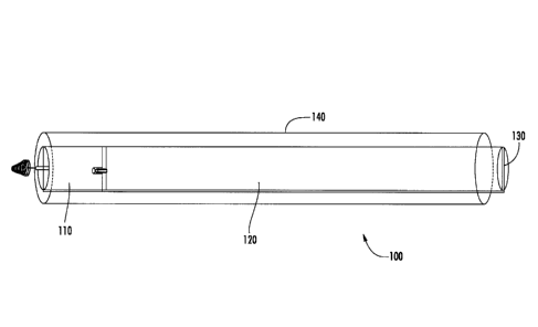
[013] Figure 1 depicts an embodiment of the device assembly according to
one aspect of the
invention, which is disposed within the central lumen of a trocar.
-6-
CA 3017695 2018-09-18
[014] Figure 2 depicts the embodiment of the device assembly according to
Figure 1,
showing additional detail regarding structural features of the pacemaker
device, the pusher, and
the holder.
[015] Figure 3 depicts an embodiment of the pacemaker device after
implantation into
cardiac tissue and release from the device assembly.
DETAILED DESCRIPTION OF VARIOUS
EMBODIMENTS OF THE INVENTION
[016] Reference will now be made in detail to various exemplary embodiments
of the
invention, examples of which are illustrated in the accompanying drawings. It
is to be
understood that the following detailed description is provided to give the
reader a better
understanding of certain details and features of embodiments of the invention,
and that the
following description is not to be understood as a limitation on the scope of
the invention.
[017] One general aspect of the invention is a fully-implantable cardiac
pacemaker. In the
exemplary embodiment discussed now, the pacemaker relates to a fetal
pacemaker, with the
understanding that the concepts, materials, and techniques are equally
applicable to patients after
birth. The pacemaker of this exemplary embodiment of the invention comprises
at least one
electrode for delivering an electric pulse to fetal cardiac tissue. The
electrode can be fabricated
from any suitable material or combination of materials that are electrically
conductive, and is
typically fabricated from one or more metals. Suitable materials are known and
widely used in
the art. The electrode comprises a connector that connects the electrode to
fetal cardiac tissue on
one end and to an electrically conductive lead (also referred to herein as a
wire) on the other end.
While not limited in design, in exemplary embodiments, the connector is
provided in the shape
of a coil or screw, which is capable of being embedded in fetal cardiac tissue
through a twisting
motion. Connectors that are suitable for use in the pacemaker device of the
invention are known
in the art, and any such connector may be used.
-7-
CA 3017695 2018-09-18
[018] The pacemaker further comprises one lead per electrode to connect the
electrode to a
power source. The lead can be fabricated from any suitable material or
combination of materials
that are electrically conductive. Typically, the lead is fabricated from one
or more metals, as
known in the art. Typically, the lead further comprises an insulative material
around the
electrically conductive material(s) along the length of the lead to prevent
transmission of
electricity except through the ends of the lead. Leads that are suitable for
use in the pacemaker
device of the invention are known in the art, and any such lead may be used.
It is to be
recognized that, in contrast to leads used in prior attempts to develop fetal
cardiac pacemakers,
the leads of the present invention are relatively short. That is, in contrast
to prior attempts, rather
than providing a relatively long lead (e.g., about 10 cm - 15 cm in length)
that can span a distance
from the fetal heart to the mother's abdomen, the present device comprises a
lead that is designed
to span only the distance from the fetal heart to the fetal chest cavity
(e.g., about 2 cm - 3 cm).
Likewise, because a fully-intrathoracic pacemaker design for children and
adults has not been
devised before, the leads for pacemakers of those embodiments are likewise
relatively short, such
as on the order of 5-10 mm.
[019] The fetal pacemaker device further comprises a power source connected
to one end of
the lead(s). The power source provides electrical energy to the electrode(s)
and, where present,
to a controller unit. The power source is not limited in design or
composition. However, in
exemplary embodiments, the power source is a battery or set of batteries in
electrical connection
with each other. In embodiments, the power source comprises a non-conducting
covering. The
size of the power source can vary depending on the life stage of the patient
to be treated and the
length of time that pacing is desired. In general, the power source is
designed to fit in the chest
cavity or thorax of a fetus, child, or adult to be treated. While not limited
in size, in general the
power source for fetal applications will be about 4 mm or less in diameter or
width and 1 cm or
less in length. For example, it can be from 1 mm to 4 mm in diameter, such as
2 mm, 3 mm, or 4
mm. Likewise, the power source can have a length of from 5 mm to 1 cm, such as
5 mm, 6 mm,
7 mm, 8 mm, 9 mm, or 1 cm. Further, while not limited in size, in general, the
power source for
-8-
CA 3017695 2018-09-18
child applications will be about 4-10 mm or less in diameter or width and 1-3
cm or less in
length. Where desired, two or more individual batteries can be connected in
series or in parallel
to achieve the desired longevity, voltage, etc. Selection of the appropriate
battery size, shape,
and power will be made by the practitioner after consideration of the amount
of energy needed to
pace the fetal heart, the point in gestation of the fetus, the type of pacing
and number of
electrodes needed, the length of desired time for functionality, and other
parameters of interest to
the practitioner. Many of the same considerations are relevant to child and
adult pacemakers,
with the understanding that, with increasing size of the patient, larger
components may be used.
Use of larger components can provide additional longevity and power to the
pacemaker device.
[0201 In preferred embodiments, the pacemaker device further comprises a
control unit that
controls the frequency and power of impulses sent to the cardiac tissue. In
general, the control
unit comprises electronics. Control units for pacemakers are known in the art,
and any suitable
design can be used. In preferred embodiments, the control unit is programmed
to alter the
frequency of pacing when the power source reaches a pre-defined point of
remaining stored
energy. For example, a controller can pace a fetal heart at 100 beats per
minute. However, when
the controller senses that the power source has only, for example, thirty-six
hours of power life
left, the controller slows the pacing to 90 beats per second. This drop in
heart rate can easily be
detected by the mother's obstetrician, and he can take an appropriate action
(e.g., induce labor,
add an additional pacemaker). The control unit will have a size similar to the
power source. In
embodiments, the two are combined as a single functional unit having a size
according to the
description above for the power source.
[021] Another general aspect of the invention is a device assembly for
implantation of the
pacemaker device into a chest cavity. In general, the device assembly
comprises a pacemaker
device for artificial pacing of a heart, a pusher for implanting the device
into a patient, and a
holder that holds the pacemaker device and pusher together. While not limited
in size, in
general, the assembly is designed to fit within the inner diameter of a trocar
that is suitable for
use in surgery for the patient of interest. Thus, in general, the device
assembly has a diameter or
-9-
CA 3017695 2018-09-18
width of about 4 mm or less, such as from 4 mm to l mm, for example 4 mm, 3
mm, 2 mm, or 1
mm. Likewise, in general, the device assembly has a length of about 10 cm - 15
cm or more for
fetal applications. Thus, in embodiments, the device assembly has a length of
10 cm, 11, cm, 12
cm, 13 cm, 14 cm, or 15 cm. Longer lengths, such as 20 cm or 25 cm are also
contemplated. In
general, the device assembly has a diameter or width of about 4 mm to 10 mm or
less, and a
length of about 1 cm to 3 cm or more for child and adult applications. The
practitioner may
select any particular value falling within these ranges based on various
considerations.
[022] The assembly comprises a distal portion that is defined by a
pacemaker device of the
invention. Located proximal and releasably attached to the pacemaker device is
a pusher. The
pusher is a relatively long rod-like element for inserting the pacemaker
device or the full device
assembly into and through a trocar from its proximal end outside of a
patient's body to its distal
end within the chest cavity of the patient. For fetal applications the length
is sufficient to extend
from the proximal end outside of the mother's body to its distal end within
the fetal chest cavity.
[023] The pusher may be fabricated out of any suitable material, and is
preferably made
from a material that can be easily sterilized. For example, the pusher may be
made from one or
more plastic materials known in the art as suitable for medical devices. The
pusher is preferably,
but need not necessarily be, made from a disposable material, such as known
for single-use in
medical procedures.
[024] The device assembly further comprises a holder. The holder functions
to releasably
connect the pusher to the pacemaker. The holder can be any element that
releasably connects the
pusher to the pacemaker, and can take any shape or size. In exemplary
embodiments, the holder
is a string, thin wire, or elastic band that sits in a two opposing grooves
cut along the walls of the
pusher and power source of the pacemaker. When employed, the holder physically
holds the
pusher and pacemaker together, such that they substantially form a single
unit. When desired
(e.g., when the pacemaker is implanted), the holder is cut, thus releasing the
pacemaker from the
pusher.
-10-
CA 3017695 2018-09-18
[025] The present invention uses the device assembly to provide a method
for implanting a
cardiac pacemaker into a patient. The method permits full implantation of a
pacemaker device
into a patient, and provides a significant improvement in artificial cardiac
pacing, including in
both fetuses and in children or adults, particularly in situations where a
child or adult is in need
of urgent pacing and other pacing device designs are incapable of being
implanted quickly and
effectively. In general, the method comprises inserting a trocar into the
chest cavity of a patient
to the point where the trocar touches heart tissue at a pre-selected location.
A device assembly
according to the invention is then inserted into the interior space of the
trocar to the point where
the electrode touches heart tissue. The electrode is then implanted in the
heart tissue, the holder
is cut, and the holder and pusher are removed. The trocar is retracted from
the chest cavity while
implanting the power source and controller in the chest cavity. That is, after
implantation of the
electrode into the heart, the holder is cut and the trocar is retracted while
deploying the remaining
portion of the pacemaker device into the chest cavity using, if necessary, the
pusher to move the
device out the distal end of the trocar. Where appropriate, the entry points
for the trocar (e.g.,
mother's abdomen) are closed using standard techniques.
[026] While various parameters and method steps may be altered to suit
particular purposes,
the present disclosure provides both general and specific guidance on
practicing the invention.
Those of skill in the art will recognize variations and modifications to the
specifically disclosed
embodiments that fall within the general teachings of the present document.
For example, the
present disclosure discusses batteries as a power source for the pacemaker
device. Those of skill
in the art will recognize that the shape of the battery is not critical, as
long as it does not interfere
with deployment of the device within the patient or with function of the
device. Thus, the battery
may take any cross-sectional geometry, such as round, square, or rectangular.
[027] While embodiments relating to fetal pacing provide significant
improvements in
treatment for such patients, other embodiments of the invention relate to
pacemakers for patients
after birth, and such embodiments provide significant improvements in
treatment of such
patients. In all embodiments, the device is a complete pacing system that can
be implanted
-11-
CA 3017695 2018-09-18
within the thorax without invasive surgery or the need for transvenous access.
For example, in
urgent situations relating to children and adults where ventricular pacing is
necessary and
vascular access cannot be easily achieved, the device can be inserted into
myocardium and allow
for temporary pacing until a permanent pacing system can be implanted.
Likewise, the device of
the invention can be used to treat adults who present with significant
bradycardia and require
urgent pacing.
[028] The device of the invention can also be used in adults who require
extended pacing
and in whom standard transvenous or epicardial systems cannot be implanted. As
is known in
the art, emergency pacing can currently be performed by transcutaneous pacing
(by use of large
pads on the chest), but this make ventilation extremely difficult and is not
always effective ¨ it
can generally only be used for a short period of time. When more prolonged
pacing is required
(e.g., around 24 hours), a temporary pacing lead is placed (through the veins)
and is pushed up
against the ventricular myocardium. This technique, however, requires vascular
access and a
degree of expertise in catheter manipulation to access the ventricle. In
contrast to the current
commonly available technologies, the present invention allows pacing for weeks
to months, if
necessaiy or desired, with a pacemaker that can be implanted without vascular
access and in an
urgent fashion.
[029] Reference will now be made to an exemplary pacemaker device and
device assembly
according to the invention. The exemplary embodiments discussed are depicted
in the figures.
[030] With reference to Figure 1, a device assembly 100 is generally
depicted within the
interior of a trocar 140. Device assembly 100 includes pusher 120, pacemaker
device 110, and
holder 130. It is to be noted that the diameter of the device assembly is
reduced in the figure
solely for the purpose of improved clarity of the drawing. As disclosed above,
the device
assembly will typically have a diameter or width only slightly smaller than
the inside diameter or
width of the trocar to be used.
[031] With reference to Figure 2, a more detailed view of device assembly
200 is shown.
As in Figure 1, Figure 2 depicts device assembly 200 disposed within the inner
lumen of trocar
- I 2-
CA 3017695 2018-09-18
240. Device assembly 200 includes pusher 220, pacemaker 210, and holder 230.
Holder 230 is
disposed within groove 216, which comprises an open space on two diametrically
opposed sides
of pusher 220 and pacemaker 210 that can accommodate holder 230. Pusher 220
further
comprises male connector 221 for rotationally locking pusher 220 and pacemaker
210 such that
neither is free to rotate, with respect to the other, about a longitudinally
central point. The figure
depicts male connector 221 having a hexagonal cross-section. However, it is to
be noted that any
suitable non-circular cross sectional shape can be used, including, but not
limited to, triangular,
square, rectangular, pentagonal, octagonal, star-shaped, and elliptical.
[032] As further shown in Figure 2, device assembly 200 includes pacemaker
device 210
with several structural elements. Pacemaker device 210 comprises female
connector 211 for
rotationally locking pacemaker 210 and pusher 220. It is to be recognized that
the size of female
connector 211 is determined in conjunction with the size of male connector 221
such that male
connector 221 is not free to rotate within the space defined by female
connector 221. In practice
of an exemplary method for implanting a pacing device according to the
invention, rotational
locking of pusher 220 and pacemaker 210 allows transmission of twisting
performed on the
pusher 220 by the practitioner outside the body (e.g., outside a mother's
abdomen) to pacemaker
210. In embodiments where pacemaker 210 comprises a screw or coil-like
connector (as shown
in Figures 1-3), the twisting motion allows the practitioner to embed the
connector into the heart
tissue.
[033] Further with reference to Figure 2, pacemaker 210 comprises
structures for storing
and deploying an electrode. More specifically, pacemaker 210 comprises recess
212 that can
house lead 217, which is connected to coil retention mechanism 213 and the
body of pacemaker
210. Holder 230 is disposed within groove 216 on pusher 220 and pacemaker 210
and through a
conduit (not depicted in Figure 2; depicted as element 318 in Figure 3) in
coil retention
mechanism 213. Placement of holder 230 through the conduit and along groove
216 permits
holder 230 to retain pacemaker device 210 in connection with pusher 220. It is
to be noted that,
in the figures, the pusher and pacemaker device are depicted as not being in
physical contact with
-13-
CA 3017695 2018-09-18
each other. The depiction is for clarity purposes only - the two elements are
in physical contact
with each other when provided as parts of an assembly. Further, while not
shown with
particularity in the figures, recess 212 and coil retention mechanism 213 are
designed to have
cross-sections that permit rotational locking of the two. That is, similar to
male connector 221
and female connector 211, recess 212 and coil retention mechanism 213 have
cross-sections that
interconnect and preclude rotational freedom between the two. In this way, a
twisting motion
imparted on pusher 220 outside a patient's body (e.g., outside a mother's
abdomen) is translated
to a twisting motion through the body of pacemaker 210 and to electrode 214.
Where a coil- or
screw-type connector is used as part of electrode 214, the twisting imparted
outside the body
results in implantation of the electrode in cardiac tissue.
[034] Pacemaker device 210 further comprises seat 215, which blocks coil
retention
mechanism 213 from fully entering recess 212 and provides adequate space for
lead 217 during
storage. In the exemplary embodiment, lead 217, which connects electrode
(which in the
embodiments depicted in the figures is a coil or screw) 214 with the battery
of pacemaker 210,
can be bundled within recess 212. When holder 230 is cut and removed after
implantation of
electrode 214 into cardiac tissue, electrode 214, coil retention mechanism
213, and lead 217
extend away from the body of pacemaker 210.
[035] Turning now to Figure 3, an example of a partially-deployed pacemaker
310 is
depicted. The figure depicts pacemaker 310 after implantation of electrode 314
into cardiac
tissue, but before final placement of pacemaker 310 into the chest cavity. For
the purpose of
clarity, no trocar is depicted. As can be seen in the figure, the holder has
been removed from
groove 316 and conduit 318, and coil retention mechanism 313 is no longer in
contact with seat
315 and has exited recess 312. Lead 317 is partially unbundled as a result of
movement of coil
retention mechanism 313 away from recess 312. In the figure, lead 317 is
depicted as having a
short length; this is for the purpose of clarity of the drawing only. It is to
be noted that the length
of lead 217 can be any suitable length that allows for placement of the body
of pacemaker 310 in
the chest cavity of the patient while attaching electrode 314 to the
appropriate locus in the
- I 4-
CA 3017695 2018-09-18
epicardium. Likewise, for the sake of clarity, coil retention mechanism 313 is
depicted as
relatively long; however, the length can be any length that is appropriate and
useful. Likewise,
while the cross-section of coil retention mechanism 313 is depicted as
hexagonal, any suitable
cross-sectional shape can be used.
[036] It
will be apparent to those skilled in the art that various modifications and
variations
can be made in the practice of the present invention and in construction of
this device without
departing from the scope or spirit of the invention. Other embodiments of the
invention will be
apparent to those skilled in the art from consideration of the specification
and practice of the
invention. For example, the device assembly can include a sheath or other
outer structure that
retains the pacemaker device, power/control unit(s), pusher, and holder. It is
intended that the
specification and examples be considered as exemplary only, with a true scope
and spirit of the
invention being indicated by the following claims.
-15-
CA 3017695 2018-09-18