Note: Descriptions are shown in the official language in which they were submitted.
CA 03017982 2018-09-14
WO 2017/173392 PCT/US2017/025585
NANOPORE DISCRIMINATION OF TARGET POLYNUCLEOTIDES FROM
SAMPLE BACKGROUND BY FRAGMENTATION AND PAYLOAD BINDING
CROSS-REFERENCE TO RELATED APPLICATIONS
[0001] This application claims the benefit under 35 U.S.0 119(e) to U.S.
Provisional
Application No. 62/316,452, filed March 31, 2016, U.S. Provisional Application
No.
62/354,068, filed June 23, 2016, and U.S. Provisional Application No.
62/412,221, filed
October 24, 2016, the contents of each of which are incorporated by reference
in their
entirety.
FIELD OF THE INVENTION
[0002] The invention relates to methods and compositions for target
sequence detection
using a nanopore device.
BACKGROUND
[0003] Detecting nucleic acid specific to an organism is an accurate and
efficient method
for identifying microbes, viruses, and other infection agents. Additionally,
detecting a
specific nucleic acid sequence, or detecting the presence or absence of a
segment of DNA
comprising a specific sequence, can identify disease-causing mutations. Being
able to
accomplish this has applications in biomedical science and technology,
medicine, agriculture
and forensics, as well as in other fields.
[0004] The detection of genes and their modifications, sequence, location,
or number, is
important for the advancement of molecular diagnostics in medicine. DNA
microarrays,
PCR, Southern Blots, and FISH (Fluorescent in situ Hybridization) are all
methods that can
be used to perform or aid nucleic acid detection. These methods can be slow
and labor
intensive, and have limited accuracy and resolution. More recent methods, such
as real-time
PR and next-generation sequencing (NGS) technologies, have improved throughput
and
accuracy, but require complex and costly device infrastructure to perform
quantitation, and
typically incorporate some form of optics for sensing.
[0005] By comparison, a solid state nanopore can provide a nucleic acid
sensor that is
electrical, without the need for optics. Moreover, solid-state nanopore
devices can be made
using scalable fabrication techniques at very low cost, and incorporated into
small form
factors for portable use.
[0006] Solid-state nanopores can detect molecules by applying a voltage
across the pore,
and measuring current impedance changes ("events") as the molecules pass
through the
nanopore. The overall efficacy of any given nanopore device depends on its
ability to
-1-
CA 03017982 2018-09-14
WO 2017/173392 PCT/US2017/025585
accurately and reliably measure impedance events above noise, and to
discriminate events
that are due to molecules of interest from events due to any background
molecules when
present.
[0007] Experiments published in the literature have demonstrated both the
detection of
purified DNA and RNA strands passing through the pores. DNA with synthetic
molecules
bound to specific sequences has been shown to permit target sequence
detection, but only I
the context of a purified DNA sample with defined polynucleotide lengths. In
practical
applications of detecting DNA from a sample, polynucleotides can exceed 1 Mbp
in length,
and can clog or prevent detection in a nanopore.
[0008] Thus, what is needed are methods to facilitate target sequence
detection in a
nanopore that is tolerant to background molecules and can handle samples with
very long
DNA molecules.
SUMMARY
[0009] In some embodiments, provided herein is a method of detecting the
presence or
absence of a target polynucleotide sequence suspected to be present in a
sample, the method
comprising: providing a sample suspected of containing a polynucleotide
comprising a target
sequence; fragmenting said polynucleotide; providing a probe adapted to bind
specifically to
said target sequence of said polynucleotide; contacting said sample with said
probe under
conditions that promote binding of said probe to said target sequence to form
a
polynucleotide-probe complex; loading said sample into a nanopore device
comprising a
nanopore, a first chamber, and a second chamber, wherein said first and second
chamber are
in electrical and fluidic communication through said nanopore via a conducting
fluid, and
wherein said nanopore device further comprises a sensor configured to identify
objects
passing through the nanopore; applying an electrical potential across said
nanopore to induce
translocation of said polynucleotide or polynucleotide-probe complex through
said nanopore;
and detecting an electrical signal associated with the translocation of said
polynucleotide or
polynucleotide-probe complex through the nanopore. In some embodiments, the
method
further comprises analyzing said electrical signal to determine the presence
or absence of said
target polynucleotide sequence in said sample. In some embodiments, the probe
is bound to a
payload molecule.
[0010] In some embodiments, the probe comprises a payload binding moiety.
In some
embodiments, the payload binding moiety comprises a chemical group, a reactive
group, a
small molecule, or a peptide. In some embodiments, the small molecule
comprises biotin. In
-2-
CA 03017982 2018-09-14
WO 2017/173392 PCT/US2017/025585
some embodiments, the reactive group comprises dibenzocyclooctyl (DBCO) or
azide. In
some embodiments, the reactive group comprises a reactive maleimide, a free
thiol (thiolate),
or a sulfur atom.
[0011] In some embodiments, the method further comprises binding a payload
molecule
to said payload binding moiety before applying said electrical potential. In
some
embodiments, the payload molecule is bound to said payload binding moiety
after contacting
said sample with said probe. In some embodiments, the payload molecule is
bound to said
payload binding moiety before contacting said sample with said probe.
[0012] In some embodiments, the payload molecule is selected from the group
consisting
of: a dendrimer, double stranded DNA, single stranded DNA, a DNA aptamer, a
fluorophore,
a protein, an antibody, a polypeptide, a nanobead, a nanorod, a nanotube,
nanoparticle,
fullerene, a PEG molecule, a liposome, or a cholesterol-DNA hybrid.
[0013] In some embodiments, the payload molecule comprises an electrical
charge. In
some embodiments, the charged payload molecule is selected from the group
consisting of: a
peptide, an amino acid, a charged nanoparticle, a synthetic molecule, a
nucleotide, a
polynucleotide, a metal, and an ion. In some embodiments, the sensitivity or
specificity of
detection of the presence of absence of the target polynucleotide is increased
when said target
polynucleotide is bound to said charged payload molecule as compared to
unbound target
polynucleotide.
[0014] In some embodiments, the payload binding moiety and the payload
molecule are
bound via a covalent bond. In some embodiments, the covalent bond is formed by
click
chemistry. In some embodiments, the click chemistry is copper catalyzed. In
some
embodiments, the click chemistry is copper free. In some embodiments, the
covalent bond
comprises a thio-ether bond. In some embodiments, the thio-ether bond is
formed by
maleimido-thiolate chemistry.
[0015] In some embodiments, the payload binding moiety and the payload
molecule are
bound via a non-covalent bond. In some embodiments, the non-covalent bond is
selected
from the group consisting of: a hydrogen bond, an ionic bond, a van der Waals
interaction, a
hydrophobic interaction, a polar bond, a cation-pi interaction, a planar
stacking interaction,
and a metallic bond.
[0016] In some embodiments, the sensitivity or specificity of detection of
the presence or
absence of the target polynucleotide is increased when said target
polynucleotide is bound to
said payload molecule as compared to unbound target polynucleotide.
-3-
CA 03017982 2018-09-14
WO 2017/173392 PCT/US2017/025585
[0017] In some embodiments, two or more payload molecules are bound to the
target
polynucleotide.
[0018] In some embodiments, the specific binding of said probe to said
target sequence of
said polynucleotide occurs via sequence-specific ligation.
[0019] In some embodiments, fragmenting said polynucleotide comprises
exposing said
sample to a fragmentation condition. In some embodiments, the fragmentation
condition is
selected from the group consisting of: chemical shearing, heat and divalent
metal cation,
acoustic shearing, sonication, hydrodynamic shearing, nebulization, needle
shearing, and
French pressing. In some embodiments, the fragmenting said polynucleotide
comprises
contacting said sample with a fragmentation reagent. In some embodiments, the
fragmentation reagent is selected from the group consisting of: a restriction
enzyme, a site-
directed nuclease, endonuclease, non-specific nuclease, transposase, and
catalytic DNA or
RNA.
[0020] In some embodiments, the sample comprises a plurality of target
polynucleotides.
In some embodiments, providing said probe comprises providing a plurality of
unique probes
adapted to specifically bind to target sequence so that each of said plurality
of target
polynucleotide-probe complexes generates a unique and detectable signal upon
translocation
through the nanopore. In some embodiments, contacting said sample with said
probe
comprises contacting said sample with said plurality of unique probes. In some
embodiments,
the method comprises detecting an electrical signal associated with the
translocation of at
least one of said plurality of target polynucleotide-probe complexes.
[0021] In some embodiments, the nanopore device comprises at least two
nanopores, and
wherein said nanopore device is configured to apply an independently-
controlled voltage
across each of said at least two nanopores. In some embodiments, the at least
two nanopores
are in series. In some embodiments, the method further comprises capturing a
polynucleotide
or polynucleotide-probe complex in at least two nanopores in said device
simultaneously.
[0022] In some embodiments, the sample is loaded into said device before
said
fragmentation of said polynucleotide. In some embodiments, the sample is
loaded into said
device after said fragmentation of said polynucleotide. In some embodiments,
the sample is
loaded into said device before said contacting of said sample with said probe.
In some
embodiments, the sample is loaded into said device after said contacting of
said sample with
said probe.
-4-
CA 03017982 2018-09-14
WO 2017/173392 PCT/US2017/025585
[0023] In some embodiments, the sample is not purified. In some
embodiments, the
sample is not purified before said fragmentation, before contacting said
sample with said
probe, or before said detection in said nanopore. In some embodiments, the
sample is loaded
into said nanopore device at a dilution of at least 1:20000, 1:10000, 1:5000,
1:2000, 1:1000,
1:500, 1:200, 1:100, 1:50, 1:20, 1:10, 1:5, 1:2, 1:1.5, 1:1.2, 1:1.1 or
1:1.05. In some
embodiments, the sample is loaded into said nanopore device without dilution.
In some
embodiments, the sample comprises non-target polynucleotides, fragmentation
reaction
reagents, and ligation reaction reagents while in said nanopore device.
[0024] In some embodiments, the nanopore is at least 5 nm, 10 nm, 20 nm, 20
nm, 40
nm, or 50 nm in diameter. In some embodiments, the nanopore is less than 200
nm in
diameter.
[0025] In some embodiments, fragmenting said polynucleotide comprises a
sequence-
specific fragmentation reaction. In some embodiments, the sequence-specific
fragmentation
reaction comprises site-specific restriction enzymes or CRISPR-based cleavage.
In some
embodiments, fragmenting said polynucleotide comprises a non-sequence-specific
fragmentation reaction. In some embodiments, the non-sequence-specific
fragmentation
reaction is achieved by shearing.
[0026] In some embodiments, the probe is contacted with said sample in the
interior
space of the nanopore device.
[0027] In some embodiments, the target polynucleotide comprises double-
stranded
deoxyribonucleic acid (dsDNA), single-stranded DNA (ssDNA), peptide nucleic
acid (PNA),
single-stranded ribonucleic acid (ssRNA), DNA/RNA hybrid, or double-stranded
ribonucleic
acid (dsRNA). In some embodiments, the target polynucleotide is a naturally-
occurring
polynucleotide. In some embodiments, the target polynucleotide is an
artificially-synthesized
polynucleotide. In some embodiments, the target polynucleotide is a
recombinant
polynucleotide.
[0028] In some embodiments, the sensor comprises an electrode pair
configured to
generate said electrical potential across said nanopore and to detect said
electrical signal. In
some embodiments, the electrical signal generated when the payload-bound
target
polynucleotide passes through the nanopore is distinguishable from the
electrical signal of
background molecules. In some embodiments, the electrical signal is a measure
of current
over time, and the electrical signal is distinguishable by its mean depth,
maximum depth,
duration, number of depth levels, area of depth and duration, or noise level.
-5-
CA 03017982 2018-09-14
WO 2017/173392 PCT/US2017/025585
[0029] Also provided herein is a method of quantifying a target
polynucleotide sequence
in a sample, the method comprising: providing a sample suspected of containing
a
polynucleotide comprising a target sequence; fragmenting said polynucleotide;
providing a
probe adapted to bind specifically to said target sequence of said
polynucleotide; contacting
said sample with said probe under conditions that promote binding of said
probe to said target
sequence to form a polynucleotide-probe complex; loading said sample into a
nanopore
device comprising a nanopore, a first chamber, and a second chamber, wherein
said first and
second chamber are in electrical and fluidic communication through said
nanopore via a
conducting fluid, and wherein said nanopore device further comprises a sensor
configured to
identify objects passing through the nanopore; applying an electrical
potential across said
nanopore to induce translocation of said polynucleotide or polynucleotide-
probe complex
through said nanopore; detecting an electrical signal associated with the
translocation of said
polynucleotide or polynucleotide-probe complex through the nanopore; and
analyzing said
electrical signal to determine a measurement of quantity of said target
polynucleotide
sequence in said sample.
[0030] In some embodiments, the probe is bound to a payload molecule. In
some
embodiments, the probe comprises a payload binding moiety. In some
embodiments, the
payload molecule is bound to said payload binding moiety after contacting said
sample with
said probe.
[0031] Also provided herein is a kit, the kit comprising: a device
comprising a nanopore,
wherein said nanopore separates an interior space of the device into two
volumes, wherein
the device comprises a sensor for said nanopore adapted to identify objects
passing through
the nanopore; a probe adapted to bind specifically to a target sequence of a
polynucleotide;
and instructions for use to detect the presence or absence of said target
sequence in a sample.
[0032] In some embodiments, the probe is bound to a payload molecule. In
some
embodiments, the probe comprises a payload binding moiety. In some
embodiments, the kit
comprises a payload molecule adapted to bind to said payload binding moiety.
In some
embodiments, the kit comprises reagents for fragmenting said polynucleotide.
[0033] Provided herein are methods of detecting a polynucleotide comprising
a target
sequence in a sample, comprising: contacting said sample with a probe that
specifically binds
to said polynucleotide comprising said target sequence under conditions that
promote binding
of said probe to said target sequence to form a polynucleotide-probe complex;
loading said
sample into a first chamber of a nanopore device, wherein said nanopore device
comprises at
-6-
CA 03017982 2018-09-14
WO 2017/173392 PCT/US2017/025585
least one nanopore and at least said first chamber and a second chamber,
wherein said first
and second chamber are in electrical and fluidic communication through said at
least one
nanopore, and wherein the nanopore device further comprises an independently-
controlled
voltage across each of said at least one nanopores and a sensor associated
with each of said at
least one nanopores, wherein said sensor is configured to identify objects
passing through the
at least one nanopore, and wherein said polynucleotide-probe complex
translocating through
said at least one nanopore provides a detectable signal associated with said
polynucleotide-
probe complex; and determining the presence or absence of said polynucleotide-
probe
complex in said sample by observing said detectable signal, thereby detecting
said
polynucleotide comprising said target sequence. In an embodiment, the method
further
comprises generating a voltage potential through said at least one nanopore,
wherein said
voltage potential generates a force on said polynucleotide-probe complex to
pull said
polynucleotide-probe complex through said at least one nanopore, causing said
polynucleotide-probe complex to translocate through said at least one nanopore
to generate
said detectable signal.
[0034] In some embodiments, said polynucleotide is DNA or RNA. In an
embodiment,
said detectable signal is an electrical signal. In an embodiment, said
detectable signal is an
optical signal. In an embodiment, said probe comprises a molecule selected
from the group
consisting of: a protein, a peptide, a nucleic acid, a TALEN, a CRISPR, a
peptide nucleic
acid, or a chemical compound. In an embodiment, said probe comprises a
molecule selected
from the group consisting of: a deoxyribonucleic acid (DNA), a ribonucleic
acid (RNA), a
peptide nucleic acid (PNA), a DNA/RNA hybrid, polypeptide, or any chemically
derived
polymer.
[0035] In an embodiment, said probe comprises a PNA molecule bound to a
secondary
molecule configured to facilitate detection of the probe bound to said
polynucleotide during
translocation through said at least one nanopore. In a further embodiment,
said secondary
molecule is a PEG. In a further embodiment, said PEG has a molecular weight of
at least 1
kDa, 2 kDa, 3 kDa, 4 kDa, 5 kDa, 6kDa, 7kDa, 8kDa, 9kDa, or 10kDa.
[0036] In an embodiment, said method of detecting a polynucleotide
comprising a target
sequence in a sample further comprises applying a condition to said sample
suspected to alter
the binding interaction between the probe and the target sequence. In a
further embodiment,
the condition is selected from the group consisting of: removing the probe
from the sample,
-7-
CA 03017982 2018-09-14
WO 2017/173392 PCT/US2017/025585
adding an agent that competes with the probe for binding to the target
sequence, and
changing an initial pH, salt, or temperature condition.
[0037] In an embodiment, said polynucleotide comprises a chemical
modification
configured to modify binding of the polynucleotide to the probe. In a further
embodiment, the
chemical modification is selected from the group consisting of biotinylation,
acetylation,
methylation, summolation, glycosylation, phosphorylation and oxidation.
[0038] In an embodiment, said probe comprises a chemical modification
coupled to the
probe through a cleavable bond. In an embodiment, said probe interacts with
the target
sequence of the polynucleotide via a covalent bond, a hydrogen bond, an ionic
bond, a
metallic bond, van der Waals force, hydrophobic interaction, or planar
stacking interactions.
In an embodiment, said method of detecting a polynucleotide comprising a
target sequence in
a sample further comprises contacting the sample with one or more detectable
labels capable
of binding to the probe or to the polynucleotide-probe complex. In an
embodiment, said
polynucleotide comprises at least two target sequences.
[0039] In an embodiment, said nanopore is about 1 nm to about 100 nm in
diameter, 1 nm
to about 100 nm in length, and wherein each of the chambers comprises an
electrode. In an
embodiment, said nanopore device comprises at least two nanopores configured
to control the
movement of said polynucleotide in both nanopores simultaneously. In an
embodiment, said
method of detecting a polynucleotide comprising a target sequence in a sample
further
comprises reversing said independently-controlled voltage after initial
detection of the
polynucleotide-probe complex by said detectable signal, so that the movement
of said
polynucleotide through the nanopore is reversed after the probe-bound portion
passes through
the nanopore, thereby identifying again the presence or absence of a
polynucleotide-probe
complex.
[0040] In an embodiment, said nanopore device comprises two nanopores, and
wherein
said polynucleotide is simultaneously located within both of said two
nanopores. In a further
embodiment, said method of detecting a polynucleotide comprising a target
sequence in a
sample comprises comprising adjusting the magnitude and or the direction of
the voltage in
each of said two nanopores so that an opposing force is generated by the
nanopores to control
the rate of translocation of the polynucleotide through the nanopores.
[0041] Also provided herein is a method of detecting a polynucleotide or a
polynucleotide sequence in a sample, comprising: contacting said sample with a
first probe
and a second probe, wherein said first probe specifically binds to a first
target sequence of
-8-
CA 03017982 2018-09-14
WO 2017/173392 PCT/US2017/025585
said polynucleotide under conditions that promote binding of said first probe
to said first
target sequence, wherein said second probe specifically binds to a second
target sequence of
said polynucleotide under conditions that promote binding of said second probe
to said
second target sequence; contacting said sample with a third molecule is
configured to bind to
said first and second probe simultaneously when said first and second probe
are within a
sufficient proximity to each other under conditions that promote binding of
said third
molecule to said first probe and said second probe, thereby forming a fusion
complex
comprising said polynucleotide, said first probe, said second probe, and said
third molecule;
loading said sample into a first chamber of a nanopore device, wherein said
nanopore device
comprises at least one nanopore and at least said first chamber and a second
chamber,
wherein said first and second chamber are in electrical and fluidic
communication through
said at least one nanopore, and wherein the nanopore device further comprises
a controlled
voltage potential across each of said at least one nanopores and a sensor
associated with each
of said at least one nanopores, wherein said sensor is configured to identify
objects passing
through the at least one nanopore, and wherein said fusion complex
translocating through
said at least one nanopore provides a detectable signal associated with said
fusion complex;
and determining the presence or absence of said fusion complex in said sample
by observing
said detectable signal.
[0042] In an embodiment, said polynucleotide is DNA or RNA. In an
embodiment, said
detectable signal is an electric signal. In an embodiment, said detectable
signal is an optical
signal. In an embodiment, said sufficient proximity is less than 3, 4, 5, 6,
7, 8, 9, 10, 11, 12,
13, 14, 15, 16, 17, 18, 19, 20, 25, 30, 35, 40, 45, 50, 60, 70, 80, 90, 100,
150, 200, 300, 400,
or 500 nucleotides. In an embodiment, said third molecule comprises a PEG or
an antibody.
[0043] In an embodiment, said third molecule and said first and second
probes are bound
to ssDNA, and wherein said ssDNA linked to said third molecule comprises a
region
complementary to a region of ssDNA linked to said first probe and is
complementary to a
region of ssDNA linked to said second probe. In an embodiment, the method of
detecting a
polynucleotide or a polynucleotide sequence in a sample further comprising
contacting the
sample with one or more detectable labels capable of binding to the third
molecule or to the
fusion complex.
[0044] Also provided herein is a kit comprising a first probe, a second
probe, and a third
molecule, wherein the first probe is configured to bind to a first target
sequence on a target
polynucleotide, wherein the second probe is configured to bind to a second
target sequence
-9-
CA 03017982 2018-09-14
WO 2017/173392 PCT/US2017/025585
on said target polynucleotide, and wherein said third molecule is configured
to bind to the
first probe and the second probe when said first and second probes are bound
to said
polynucleotide at said first and second target sequences, thereby locating the
first and second
probe in sufficient proximity to allow binding of said third molecule to said
first and second
probes simultaneously.
[0045] In an embodiment, said first probe and said second probe are
selected from the
group consisting of: a protein, a peptide, a nucleic acid, a TALEN, a CRISPR,
a peptide
nucleic acid, or a chemical compound. In an embodiment, said third molecule
comprises a
PEG or an antibody. In an embodiment, said third molecule comprises a
modification to
modify binding affinity to said probes.
[0046] Also provided herein is a nanopore device comprising at least two
chambers and a
nanopore, wherein said device comprises a modified PNA probe bound to a
polynucleotide
within said nanopore.
[0047] Also provided herein is a dual-pore, dual-amplifier device for
detecting a charged
polymer through two pores, the device comprising an upper chamber, a middle
chamber and
a lower chamber, a first pore connecting the upper chamber and the middle
chamber, and a
second pore connecting the middle chamber and the lower chamber, wherein said
device
comprises a modified PNA probe bound to a polynucleotide within said first or
second pore.
[0048] In an embodiment, the device is configured to control the movement
of said
charged polymer through both said first pore and said second pore
simultaneously. In an
embodiment, the modified PNA probe is bound to at least one PEG molecule. In
an
embodiment, the device further comprises a power supply configured to provide
a first
voltage between the upper chamber and the middle chamber, and provide a second
voltage
between the middle chamber and the lower chamber, each voltage being
independently
adjustable, wherein the middle chamber is connected to a common ground
relative to the two
voltages, wherein the device provides dual-amplifier electronics configured
for independent
voltage control and current measurement at each pore, wherein the two voltages
may be
different in magnitude, wherein the first and second pores are configured so
that the charged
polymer is capable of simultaneously moving across both pores in either
direction and in a
controlled manner.
BRIEF DESCRIPTION OF THE DRAWINGS
[0049] Provided as embodiments of this disclosure are drawings which
illustrate by
exemplification only, and not limitation.
-10-
CA 03017982 2018-09-14
WO 2017/173392 PCT/US2017/025585
[0050] Figure 1 depicts a polynucleotide comprising a target polynucleotide
sequence
bound to a payload molecule through the probe, and the complex passing through
the
nanopore.
[0051] Figure 2 depicts differences in current signatures when a payload-
bound target
polynucleotide passes through the pore, compared to a non-target background
polynucleotide
and a generic non-polynucleotide background molecule.
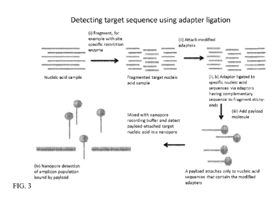
[0052] Figure 3 depicts a method of detecting target sequences from a
sample without
amplification. In particular, Figure 3 shows a method to detect a target
sequence that involves
using site-specific cleavage of the target sequence, and ligating probes that
are competent for
attaching payload molecules that facilitate nanopore detection.
[0053] Figure 4 illustrates probes of differing size or charge or other
configuration to
generate a unique signature upon nanopore translocation that each bind to a
unique target
sequence in the target-bearing molecule.
[0054] Figure 5A shows a PNA ligand that has been modified as to increase
ligand
charge, and therefore facilitate detection by a nanopore. Figure 5B shows an
example in
which a double-stranded DNA is used as the target bearing polymer and multiple
different
DNA binding probes that bind to target sequences that are desired to be
detected.
[0055] Figure 6A shows a PNA-PEG probe bound to its target sequence on a
dsDNA
molecule. Figure 6B shows the results of a gel shift assay with the following
samples: DNA
only (lane 1), DNA/PNA (lane 2), DNA/PNA-PEG (10kDa) (lane 3), and DNA/PNA-PEG
(20kDa) (lane 4). Figure 6C shows the results of a gel shift assay with the
following samples:
DNA marker (lane 1), random DNA sequence incubated with PNA probe (lane 2),
DNA with
single mismatch at target sequence incubated with corresponding PNA probe
(lane 3), and
DNA with target sequence mixed with corresponding PNA probe specific to the
target
sequence (lane 4).
[0056] Figure 7A shows representative current signature events as the
molecule depicted
below each current signature passes through the nanopore under an applied
voltage. Figure
7B shows a scatter plot of events characterized by duration and mean
conductance shift due
to translocation through the nanopore in three populations: DNA/bisPNA
(square),
DNA/bisPNA-PEG 5kDa (circle), and DNA/bisPNA-PEG 10kDa (diamond). Figure 7C
shows a histogram of mean conductance shift probability associated with each
of the three
populations described above. Figure 7D shows a histogram of event duration
probability
associated with each of the three populations described above.
-11-
CA 03017982 2018-09-14
WO 2017/173392 PCT/US2017/025585
[0057] Figure 8A shows representative event signatures correlated with the
translocation
of a PNA-PEG probe bound to a DNA molecule. Figure 8B shows the mean
conductance
shift v. duration plot for each recorded event in the nanopore from a sample
comprising
bacterial DNA and PNA-PEG probe. Figure 8C and Figure 8D show corresponding
histograms to characterize these events detected by mean conductance shift and
duration of
each event respectively. Figure 8E shows the results of a gel shift assay
showing: 100bp
ladder (lane 1), 300 bp DNA with wild type cftr sequence incubated with the
PNA-PEG
probe (lane 2), and 300bp DNA with the cftr AF508 sequence incubated with the
PNA-PEG
probe (lane 3).
[0058] Figure 9A shows the results of the gel shift assay, with lane 1
comprising S. mitis
bacterial DNA without a bisPNA-PEG bound, and lane 2 comprising S. mitis DNA
with a
site-specific bisPNA-PEG bound. Figure 9B shows a scatter plot of mean
conductance shift
(dG) on the vertical axis vs. duration on the horizontal axis for all recorded
events in the two
consecutive experiments. The first sample included bacterial DNA with PEG-
modified PNA
probes (DNA/bisPNA-PEG). The second sample included bacterial DNA alone.
[0059] Figure 10 illustrates a process of fragmentation and binding of a
sequence-specific
probe comprising a payload to a target sequence, according to an embodiment of
the
invention.
[0060] Figure 11 is an agarose gel that shows bacterial plasmid
fractionation.
[0061] Figure 12 illustrates an exemplary bisPNA probe comprising a region
that binds to
a specific 12-mer target oligonucleotide sequence, and a cysteine linker
capable of forming a
covalent bond with a 40 kDa, 3-arm maleimido-PEG payload. Figure 12 also
illustrates an
embodiment of the bisPNA probe covalently attached to the 3-arm maleimido-PEG
payload
and bound to its target DNA sequence.
[0062] Figure 13 shows the results of HPLC purification of bisPNA-PEG
conjugation
reaction.
[0063] Figure 14 shows the results of detection in the nanopore of the
following samples:
i) fragmented DNA only, ii) PNA-PEG probe only, iii) DNA mixed with PNA probe,
and iv)
DNA mixed with DNA probe bound to a payload (4-arm PEG). Panel a) shows the
separation of each population on a plot of event duration and maximum 6G for
each event.
Panel b) and c) show probability histograms for values of maximum 6G (panel
b)) and event
duration (panel c)) for each population detected in a nanopore.
-12-
CA 03017982 2018-09-14
WO 2017/173392 PCT/US2017/025585
[0064] Figure 15 shows an event plot of event duration vs maximum 6G for
two molecule
types (96bp DNA/probe-payload complex and secondary molecule) that were run
sequentially on the same pore.
[0065] Figure 16 illustrates differentiation of the target DNA/probe-
payload complex and
the secondary molecule and methods to quantify relative abundance of the
target to the
known amount of secondary molecule.
[0066] Some or all of the figures are schematic representations for
exemplification;
hence, they do not necessarily depict the actual relative sizes or locations
of the elements
shown. The figures are presented for the purpose of illustrating one or more
embodiments
with the explicit understanding that they do not limit the scope or the
meaning of the claims
that follow below.
DETAILED DESCRIPTION
[0067] Throughout this application, the text refers to various embodiments
of the present
nutrients, compositions, and methods. The various embodiments described are
meant to
provide a variety of illustrative examples and should not be construed as
descriptions of
alternative species. Rather it should be noted that the descriptions of
various embodiments
provided herein may be of overlapping scope. The embodiments discussed herein
are merely
illustrative and are not meant to limit the scope of the present invention.
[0068] Also throughout this disclosure, various publications, patents and
published patent
specifications are referenced by an identifying citation. The disclosures of
these publications,
patents and published patent specifications are hereby incorporated by
reference into the
present disclosure to more fully describe the state of the art to which this
invention pertains.
[0069] As used in the specification and claims, the singular form "a," "an"
and "the"
include plural references unless the context clearly dictates otherwise. For
example, the term
"an electrode" includes a plurality of electrodes, including mixtures thereof.
[0070] As used herein, the term "comprising" is intended to mean that the
devices and
methods include the recited components or steps, but not excluding others.
"Consisting
essentially of' when used to define devices and methods, shall mean excluding
other
components or steps of any essential significance to the combination.
"Consisting of' shall
mean excluding other components or steps. Embodiments defined by each of these
transition
terms are within the scope of this invention.
[0071] All numerical designations, e.g., distance, size, temperature, time,
voltage and
concentration, including ranges, are approximations which are intended to
encompass
-13-
CA 03017982 2018-09-14
WO 2017/173392 PCT/US2017/025585
ordinary experimental variation in measurement of the parameters, and that
variations are
intended to be within the scope of the described embodiment. It is to be
understood, although
not always explicitly stated that all numerical designations are preceded by
the term "about".
It also is to be understood, although not always explicitly stated, that the
components
described herein are merely exemplary and that equivalents of such are known
in the art.
[0072] As used herein, the term "target sequence" refers to a portion of a
polynucleotide
having a sequence of nucleic acids of interest. The target sequence can be
specifically
targeted by reagents for separating (i.e., fragmenting) a polynucleotide into
a plurality of
fragmented segments. The target sequence can also be specifically targeted for
binding by a
probe to facilitate detection of the target sequence in a nanopore sensor, as
described herein.
[0073] As used herein, the term "fragmenting" refers to a physical
separation of a
polynucleotide into at least two polynucleotide fragments. This can be
accomplished by
exposing the polynucleotide to conditions that facilitate separation of the
polynucleotide. This
can also be accomplished by exposing the polynucleotide to an enzyme or other
reagent that
facilitates separation of a polynucleotide into two or more fragments. This
fragmentation can
be designed to occur at specific target sequences on a polynucleotide.
[0074] As used herein, the term "ligation" refers to binding of a probe to
a polynucleotide
comprising a target sequence. In some embodiments, the polynucleotide
comprising the
target sequence has been fragmented. As an example, ligation of the probe to
the
polynucleotide can occur through binding via a complementary sequence, or can
be
facilitated by a ligation enzyme.
[0075] As used herein, the term "specific binding" or "bind specifically"
refers to the
targeted binding of a probe to a polynucleotide comprising a target sequence
or to a fragment
thereof.
[0076] As used herein, the term "probe" refers to a molecule that binds
specifically to a
polynucleotide comprising a target sequence or to a fragment thereof. In some
embodiments,
the probe comprises a payload molecule. In some embodiments, the probe
comprises a
payload molecule binding moiety adapted to bind to a payload molecule.
[0077] As used herein, the term "payload molecule" refers to a molecule
with physical
dimensions that facilitate generation of a unique electrical signal when
captured in a
nanopore within a correlated range of dimensions. A payload molecule may be
attached to a
target molecule to facilitate detection of the target molecule in a nanopore
device. In some
embodiments, the payload molecule may also be charged to act as a driver
molecule. In some
-14-
CA 03017982 2018-09-14
WO 2017/173392 PCT/US2017/025585
embodiments, the payload molecule comprises a probe binding moiety capable of
specifically
binding a probe molecule.
[0078] The term "nanopore" (or, just "pore") as used herein refers to a
single nano-scale
opening in a membrane that separates two volumes. The pore can be a protein
channel
inserted in a lipid bilayer membrane, for example, or can be engineered by
drilling or etching
or using a voltage-pulse method through a thin solid-state substrate, such as
silicon nitride or
silicon dioxide or graphene or layers of combinations of these or other
materials. Geometrically, the pore has dimensions no smaller than 0.1 nm in
diameter and no
bigger than 1 micron in diameter; the length of the pore is governed by the
membrane thickness, which can be sub-nanometer thickness, or up to 1 micron or
more in
thickness. For membranes thicker than a few hundred nanometers, the nanopore
may be
referred to as a "nano channel."
[0079] As used here, the term "nanopore instrument" or "nanopore device"
refers to a
device that combines one or more nanopores (in parallel or in series) with
circuitry for
sensing single molecule events. Specifically, nanopore instruments use a
sensitive voltage-
clamp amplifier to apply a specified voltage across the pore or pores while
measuring the
ionic current through the pore(s). When a single charged molecule such as a
double-stranded
DNA (dsDNA) is captured and driven through the pore by electrophoresis, the
measured
current shifts, indicating a capture event (i.e., the translocation of a
molecule through the
nanopore, or the capture of a molecule in the nanopore), and the shift amount
(in current
amplitude) and duration of the event are used to characterize the molecule
captured in the
nanopore. After recording many events during an experiment, distributions of
the events are
analyzed to characterize the corresponding molecule according to its shift
amount (i.e., its
current signature). In this way, nanopores provide a simple, label-free,
purely electrical
single-molecule method for biomolecular sensing.
[0080] As used herein, the term "electrical signal" encompasses a series of
data collected
on current, impedance / resistance, or voltage over time depending on
configuration of the
electronic circuitry. Conventionally, current is measured in a "voltage clamp"
configuration;
voltage is measured in a "current clamp" configuration, and resistance
measurements can be
derived in either configuration using Ohm's law V = IR. Impedance can also be
generated by
measured from current or voltage data collected from the nanopore device.
Types of electrical
signals referenced herein include current signatures and current impedance
signatures,
although various other electrical signals may be used to detect particles in a
nanopore.
-15-
CA 03017982 2018-09-14
WO 2017/173392 PCT/US2017/025585
[0081] As used herein, the term "event" refers to a translocation of a
detectable molecule
or molecular complex through the nanopore and its associated measurement via
an electrical
signal, e.g., change in current through the nanopore over time. It can be
defined by its current,
duration, and/or other characteristics of detection of the molecule in the
nanopore. A plurality
of events with similar characteristics is indicative of a population of
molecules or complexes
that are identical or have similar characteristics (e.g., bulk, charge).
[0082] As used herein, the term "cleavable linker" or "labile linker"
refers to a substrate
linker sensitive to enzymatic, photolytic, or chemical cleavage by a target
molecule or
condition. In some embodiments, the cleavable linker can be a deoxyribonucleic
acid (DNA),
a polypeptide, a carbon-oxygen bond, a carbon-sulfur bond, a carbon-nitrogen
bond, or a
carbon-carbon bond. In some embodiments, the cleavable linker sensitive to
photolytic
cleavage can be an ortho-nitrobenzyl derivative or phenacyl ester derivative.
In some
embodiments, the cleavable linker sensitive to chemical cleavage can be an azo
compounds,
disulfide bridge, sulfone, ethylene glycolyl disuccinate, hydrazone, acetal,
imine, vinyl ether,
vicinal diol, or picolinate ester.
Molecular Detection
[0083] The present disclosure provides methods and systems for molecular
detection and
quantitation. In addition, the methods and systems can also be configured to
measure the
affinity of a probe binding to a target molecule. Further, such detection,
quantitation, and
measurement can be carried out in a multiplexed manner, greatly increasing its
efficiency.
[0084] Thus, provided herein are compositions and methods for detecting or
quantifying
a polynucleotide that contains a target sequence that is desired to be
detected or quantitated.
[0085] For nucleic acids and polypeptides to which the target sequence
detection method
is applied, a target sequence can be a polynucleotide sequence that is
recognizable by the
probe molecule. Target sequences may be chemically modified (e.g. methylated)
or occupied
by other molecules (e.g. activator or repressors), and depending on the nature
of the probe,
the binding status of the target sequence can be elucidated. In some aspects,
the target
sequence comprises a chemical modification for binding the probe to the
polynucleotide. In
some aspects, the chemical modification is selected from the group consisting
of acetylation,
methylation, summolation, glycosylation, phosphorylation, biotinylation, and
oxidation.
[0086] To facilitate detection of the target sequence using a nanopore
device, the target
DNA can be fragmented. In some embodiments, fragmentation occurs at sequence-
specific
locations on the target DNA. In some embodiments, fragmentation generates a
set of
-16-
CA 03017982 2018-09-14
WO 2017/173392 PCT/US2017/025585
identical length sequences comprising at least a portion of the DNA. In some
embodiments,
the target DNA is fragmented at the target sequence of interest. Fragmentation
can provide
target DNA having uniform lengths to facilitate accurate detection of target
DNA by
generating more consistent and/or more distinguishable current signatures upon
translocation through a nanopore. This fragmentation can be paired with
binding or ligation
of a probe specific for the DNA comprising at least part of the target
sequence to enhance
detection in a nanopore.
[0087] Thus, also provided herein are probes capable of binding to a
specific target
sequence on the polynucleotide. These probes can be ligated to the end of
fragmented DNA,
or can bind to a target sequence on the fragmented DNA The probe can also
comprise or be
bound to a payload molecule to aid detection of the polynucleotide-probe
complex in a
nanopore by altering the dwell time or current.
[0088] In one embodiment, if all are present in a solution, a probe binds
to a target
sequence through the specific recognition of the probe for the target
sequence. Such binding
causes the formation of a complex that includes the probe and the target
sequence.
[0089] The formed polynucleotide-probe complex can be detected by a
nanopore device.
The nanopore device includes electronic components to deliver controlled
voltages across
one or more nanopores (which voltages can, in some embodiments, be
independently
controlled and clamped) along with circuitry for measuring current flow across
the
nanopores. An electrical potential, (e.g., a voltage differential) applied
across each nanopore
facilitates the capture and translocation of a charged polynucleotide through
application of
an electrostatic force on the charged polynucleotide exposed to the voltage
field. Unless
specified below, references to a pore or nanopore or nanopore device are
intended to
encompass single, dual or multi-pore devices within the spirit of the present
invention.
[0090] When a sample that includes the polynucleotide target sequence is in
the
nanopore device, the nanopore can be configured to capture and pass the
polynucleotide
target sequence through the nanopore. For example, as shown in Figure 1 a
polynucleotide
comprising a target sequence is specifically bound by a probe comprising a
payload
molecule. As shown in Figure 1, the probe can be bound to the payload molecule
through an
adapter. In some embodiments, the payload may be bound to the target sequence
through
ligation after, e.g., enzymatic detection. When the target sequence is within
the pore (as
shown in Figure 1) or adjacent to the pore, the binding status of the target
sequence can be
-17-
CA 03017982 2018-09-14
WO 2017/173392 PCT/US2017/025585
detected by the sensor, e.g., due to a unique electrical signature generated
by the complex's
measured effect on current through the pore.
[0091] The "binding status" of a target sequence, as used herein, refers to
whether the
target sequence is bound to a probe. Essentially, the binding status is either
bound or
unbound. Either, (i) the target sequence is free and not bound to a probe (ii)
the target
sequence is bound to a probe. Figure 2 shows representative changes to current
through a
nanopore due to the presence of target sequence bound to a payload, unbound
background
non-target DNA, and other background molecules captured in and translocating
through the
nanopore. Probes of different sizes or having different probe binding sites
can be used to give
additional current profiles to enable more than one target sequence to be
detected in a sample,
either on the same polynucleotide or on different polynucleotides.
[0092] Detection of the binding status of a target sequence can be carried
out by various
methods. In one aspect, by virtue of the different sizes of the target
sequence at each status
(i.e. occupied or unoccupied), when the target sequence passes through the
pore, the different
sizes result in different currents across the pore. In this respect, no
separate sensor is required
for the detection, as the electrodes, which are connected to a power source
and can detect the
current, can serve the sensing function. The two electrodes, therefore, can
serve as a
"sensor."
[0093] In some aspects, a payload molecule can be added to the probe to
facilitate
detection. This payload molecule can be already attached to the probe, or can
be capable of
binding to the probe or polynucleotide /probe complex. In one aspect, the
payload molecule
includes a charge, either negative or positive, to facilitate detection in a
nanopore via an
electrical signal, such as current. In another aspect, the payload molecule
adds size to
facilitate detection via an electrical signal. In another aspect, the payload
molecule includes a
detectable label, such as a fluorophore.
[0094] In some embodiments, the probe comprises a payload binding moiety
adapted to
bind to said payload molecule. The binding interaction between the payload
binding moiety
and the payload molecule can be covalent or non-covalent. In some embodiments,
the non-
covalent binding interaction is characterized as a hydrogen bond, an ionic
bond, a van der
Waals interaction, a hydrophobic interaction, a cation-pi interaction, a
planar stacking
interaction, or a metallic bond.
[0095] In this context, an identification of a bound status (ii) indicates
that the target is
bound to a probe. In other words, the target sequence is detected.
-18-
CA 03017982 2018-09-14
WO 2017/173392 PCT/US2017/025585
[0096] In some embodiments, target sequence-specific detection and/or
quantification in
a nanopore can be performed using the following method (also depicted in
Figure 1):
[0097] A sample suspected of containing a target polynucleotide is
obtained. The sample
is treated to fragment polynucleotides in the sample. This treatment can be
exposure to
shearing conditions, or exposure to enzymatic cleavage, such as restriction
enzymes. The
cleavage can be site-specific to facilitate detection of a target sequence.
After fragmentation,
the sample is contacted with PNA probes (or other suitable probes) capable of
binding to a
specific target sequence on a fragmented polynucleotide. The PNA probes are
bound to a
payload binding moiety, or comprise a payload molecule binding moiety which
will be bound
to the payload binding moiety before detection in a nanopore device. Then, the
sample is
placed in a nanopore device and a voltage applied to induce translocation of
polynucleotide
through the nanopore.
[0098] The flow of current through the nanopore over time is collected
using sensors in
the nanopore device. This data is then analyzed to determine the presence or
absence of
current signatures associated with a polynucleotide target sequence bound to a
probe-payload
complex, i.e., a polynucleotide-probe complex. Quantification of the amount of
target
sequence in the sample can also be performed by comparing the capture rate (or
other method
of event quantification) of the polynucleotide-probe complex in a nanopore
with a reference
linking the capture rate to the concentration under specified conditions.
Fragmentation
[0099] In practical applications of detecting target sequences in a sample
obtained from
an organism or an environment, the sample can contain DNA exceeding a million
base pairs
in length, and also contain a significant number of background molecules.
Detecting a target
sequence among this type of sample poses a significant challenge. For a
commercial
nanopore technology to be viable and simple, the method for target sequence
detection
applications must be tolerant to background molecules in a variety of forms.
This is
particularly true if fragmentation of the sample, and any sequence-specific
labeling, occurs
directly in the chamber adjacent to the nanopore, just prior to or during
nanopore sensing.
[00100] Herein we describe, in some embodiments, a method that permits
detection and/or
quantitation of any target polynucleotide sequence from within the total
population of
fragmented DNA molecules, without requiring a purification step to remove any
background
molecules prior to nanopore measurement. Background molecules can include non-
target
DNA from the fragmentation, and any reagents or molecules utilized with
chemistries to add
-19-
CA 03017982 2018-09-14
WO 2017/173392 PCT/US2017/025585
payload molecules to the target sequence-containing DNA fragments, wherein the
payload
molecule permits selective detection of the target sequence-containing DNA
fragments using
the nanopore sensor.
[00101] In some embodiments, described herein is a method for detecting target
polynucleotide sequences with a nanopore by attaching a probe and/or a payload
molecule to
enable discrimination from background molecules, i.e., all molecules that are
not the target
nucleic acid. The method does not require nucleic acid purification at any
step, which
simplifies the device infrastructure required to implement the method. The
methods described
herein are compatible with a range of nanopore sizes and geometries, and can
be
implemented in an inexpensive and portable form factor. The method also
permits
quantitation (i.e., concentration estimation) of the nucleic acid comprising
the target sequence
in the chamber adjacent to the nanopore sensor.
[00102] In some embodiments, the polynucleotide comprising the target sequence
is
fragmented, either specifically (e.g., via a restriction enzyme) or non-
specifically (e.g., via
e.g., shearing). This is followed by binding a probe to the fragment
comprising the target
sequence. This can be done, for example, via ligation of a probe to the end of
a fragmented
sequence, or through sequence-specific binding to a target sequence of a
polynucleotide. In
some embodiments, the probe comprises a payload molecule. In some embodiments,
the
probe comprises a payload binding moiety for binding a payload molecule to the
probe,
thereby conjugating the payload molecule with the target sequence. In some
embodiments,
sequence-specific shearing is achieved through the use of restriction enzymes,
CRISPR
technology, or another shearing method known in the art.
[00103] In some embodiments, the polynucleotide fragment comprising the target
sequence binds to the probe via a ligation reaction. In some embodiments, the
ligation
reaction binds a terminal end of a polynucleotide fragment to a probe. In some
embodiments,
the ligation reaction binds the probe to the fragment, wherein the probe is
adapted to
specifically bind a payload molecule via a payload molecule binding moiety. In
some
embodiments, probes can be ssDNA, dsDNA, ssRNA, dsRNA, DNA/RNA hybrids, PNA,
or
LNA. In some embodiments, the probe and the payload molecule are connected via
a
covalent bond, or non-covalent bond, e.g. a hydrogen bond, an ionic bond, a
van der Waals
force, a hydrophobic interaction, a cation-pi interaction, a planar stacking
interaction, or a
metallic bond.
-20-
CA 03017982 2018-09-14
WO 2017/173392 PCT/US2017/025585
[00104] In certain embodiments, fragmentation and/or binding of the
polynucleotide
comprising the target sequence to the probe is performed within one or more of
the volumes
within the said device. In some embodiments, background molecules due to
fragmentation
and/or binding steps are present in the volume during detection of target
sequences using the
nanopore device.
[00105] An advantage of our fragmentation and probe binding approach is the
specificity
of the signature generated by the target sequence in a nanopore, allowing
discrimination from
a large population of background molecules. Thus, in some embodiments, the
polynucleotide
comprising the target sequence is detected from a crude sample that has not
been purified
after obtaining the sample from the source (e.g., a source organism or
environment).
[00106] In some embodiments, two or more payload molecules are attached to
each
nucleic acid molecule comprising the target sequence. In some embodiments, a
plurality of
unique target-probe complexes each bound to a different payload molecule can
be detected
with the nanopore sensor, the different payload molecules adapted to allow
discrimination
between target sequences in a nanopore for multiplexing.
[00107] In certain preferred embodiments, an estimate for the concentration of
the
polynucleotide comprising the target sequence can be determined from an
aggregated set of
sensor measurements. In some embodiments, the measurements are compared to a
reference
to determine a concentration or fractional abundance of the polynucleotide
comprising the
target sequence.
Probe Specificity
[00108] In some aspects, the method further comprises using probes that bind
specifically
to a sufficiently long target sequence so that they are capable of binding to
only one unique
sequence in the target population, but also have the ability to not bind to
the target site if only
a single base pair mismatch is present. This discrimination is possible, for
example, when
using probes comprising PNA. A 20 bp gamma-PNA probe is able to efficiently
bind to a
perfectly matched target sequence, but binding is abrogated when the target
sequence and
probe sequence differ by only one base (Strand-Invasion of Extended, Mixed-
Sequence B-
DNA by yPNAs, G. He, D. Ly et al., J Am Chem Soc. 2009 September 2; 131(34):
12088-
12090. doi:10.1021/ja900228j). When considering the human genome that contains
3.1
billion bases, a 20 base pair sequence is likely to randomly occur 0.003
times. Thus, a 20
base pair probe designed to bind to a specific sequence under investigation is
very unlikely to
-21-
CA 03017982 2018-09-14
WO 2017/173392 PCT/US2017/025585
bind to an undesired location and provide a false positive. Therefore, in some
embodiments,
the target sequence is at least 20 base pairs in length.
Cleavable Payload Molecules
[00109] In some aspects, probes comprise payload molecules that allow
detection by a
sensor, but they are attached to the probe using a cleavable linker. Thus a
set of probes that
can be distinguished from each other in the nanopore are bound to a target
bearing
polynucleotide. Once that set of probes is detected in the nanopore, the
features are cleaved
off and a new set of probes are added that also have cleavable detection
feature. The
add/cleave/wash cycle can be continued until all sequence information is
extracted from a
captured target molecule. Example of molecules that aid in probe detection are
discussed
above. Examples of cleavable linkers are reductant cleavable linkers
(disulfide linkers
cleaved by TCEP), acid cleavable linker (hydrazone/hydrazide bonds), amino
acid sequences
that are cleaved by proteases, nucleic acid linkers that are cleaved by
endonucleases (sites
specific restriction enzymes), base cleavable linkers, or light cleavable
linkers [Leriche,
Geoffray, Louise Chisholm, and Alain Wagner. "Cleavable linkers in chemical
biology."
Bioorganic & medicinal chemistry 20, no. 2 (2012): 571-582.]
Probe Molecules
[00110] Probes as used herein are understood to be capable of specifically
binding to a site
on a polynucleotide, wherein the site is characterized by the sequence or
structure. A probe
molecule can be detected or quantitated by virtue of its binding to the target
sequence-bearing
polynucleotide, and capture and translocation of the complex through a
nanopore. Examples
of probe molecules include a PNA (protein nucleic acid), bis-PNA, gamma-PNA, a
PNA-
conjugate that increases size or charge of PNA. Other examples of probe
molecules are from
the group consisting of a natural or recombinant protein, protein fusion, DNA
binding
domain of a protein, peptide, a nucleic acid, oligo nucleotide, TALEN, CRISPR,
a PNA
(protein nucleic acid), bis-PNA, gamma-PNA, a PNA-conjugate that increases
size, charge,
fluorescence, or functionality (e.g. oligo labeled), or any other PNA
derivatized polymer, and
a chemical compound.
[00111] In some aspects, the probe comprises a y-PNA. y-PNA has a simple
modification
in a peptide-like backbone, specifically at the y-position of the N-(2-
aminoethyl)glycine
backbone, thus generating a chiral center (Rapireddy S., et al., 2007. J. Am.
Chem. Soc.,
129:15596-600; He G, et al., 2009, J. Am. Chem. Soc., 131:12088-90; Chema V,
et al., 2008,
Chembiochem 9:2388-91; Dragulescu-Andrasi, A., et al., 2006, J. Am. Chem.
Soc.,
-22-
CA 03017982 2018-09-14
WO 2017/173392 PCT/US2017/025585
128:10258-10267). Unlike bis-PNA, y-PNA can bind to dsDNA without sequence
limitation,
leaving one of the two DNA strands accessible for further hybridization.
[00112] In some aspects, the function of the probe is to hybridize to a
polynucleotide with
a target sequence by complementary base pairing to form a stable complex. The
PNA
molecule may additionally be bound to additional molecules to form a complex
has
sufficiently large cross-section surface area to produce a detectable change
or contrast in
signal amplitude over that of the background, which is the mean or average
signal amplitude
corresponding to sections of non-probe-bound polynucleotide.
[00113] The stability of the binding of the polynucleotide target sequence to
the PNA
molecule is important in order for it to be detected by a nanopore device. The
binding
stability must be maintained throughout the period that the target-bearing
polynucleotide is
being translocated through the nanopore. If the stability is weak, or
unstable, the probe can
separate from the target polynucleotide and will not be detected as the target-
bearing
polynucleotide threads through the nanopores.
[00114] In a particular embodiment, an example of a probe is a PNA bound to a
payload
molecule or to a molecule comprising a binding moiety adapted to bind to a
payload
molecule in which the PNA specifically recognizes a nucleotide sequence and
the payload
molecule increases the sensitivity of detection in a nanopore device.
Different payload
molecules with size/shape/charge differences may also be used to discriminate
between
different PNA-payload complexes bound to their respective target sequences in
a nanopore.
[00115] As illustrated in Figure 4, probes A, B, C and D each specifically
binds to a site on
a DNA molecule, and these probes can be identified and distinguished from each
other by the
width, length, size and/or charge of the bound payload molecule. If their
corresponding sites
are denoted as A, B, C and D, respectively, then identification of the ligands
leads to
revelation of those DNA sequences, A-B-C-D. Note that the probes can each be
bound to
unique polynucleotides, as is more likely with fragmented polynucleotides.
[00116] Different reactive payload binding moieties may be incorporated into
the probes
to provide a chemical handle to which payload molecules may bind. Examples of
reactive
payload binding moieties include, but are not limited to, primary amines,
carboxylic acids,
ketones, amides, aldehydes, boronic acids, hydrazones, thiols, maleimides,
alcohols, and
hydroxyl groups, and biotin.
[00117] Figure 5A shows a PNA probe that has been modified as to increase its
charge,
and therefore facilitate detection by a nanopore. Specifically, this probe,
which binds to the
-23-
CA 03017982 2018-09-14
WO 2017/173392
PCT/US2017/025585
target DNA sequence by complementary base pairing and Hoogsteen base pairing
between
the bases on the PNA molecule and the bases in the target DNA sequence (i.e.,
the target
sequence), has cysteine residues incorporated into the backbone, which provide
a free thiol
payload binding moiety to attach one or more payload molecules. Here, the
cysteine is bound
to a peptide 2-aminoethylmethanethiosulfonate (MTSEA) through a maleimide
linker, which
provides a means to enhance detection in a nanopore device of whether the
probe is bound to
its target sequence since the payload molecule increases the probe's charge.
This greater
charge results in a greater change in current flow through the pore during
translocation as
compared to a PNA probe without the payload molecule bound.
[00118] In some aspects, to increase the specificity and sensitivity of
discrimination
between the polynucleotide-probe complex and other background molecules
present in the
sample, modification can be made to the pseudo-peptide backbone to change the
overall size
of the PNA probe. See, e.g., Figure 5B, which shows a PNA that has cysteine
residues (301)
incorporated that are modified with a succinimidyl 4-(N-
maleimidomethyl)cyclohexane-1-
carboxylate (SMCC) linker (302) to enable conjugation to peptides (303)
through the N-
terminal amine of the peptide. In addition to adding charge directly to the
probe (e.g., as in
Figure 5A), selection of more charged amino acids instead of non-polar amino
acids can
serve to increase the charge of PNA. Payload molecules, such as small
particle, molecules,
protein, peptides, or polymers (e.g. PEG) can be bound to the pseudo-peptide
backbone to
enhance the bulk or cross-sectional surface area of the polynucleotide-probe
complex.
Enhanced bulk serves to improve the signal amplitude contrast so that any
differential signal
resulting from the increased bulk can be easily detected, even in the presence
of a significant
amount of background molecules, e.g., as in a non-purified sample. Examples of
small
particle, molecules, protein, or peptides that can act as payload molecules to
bind to the
pseudo-peptide backbone include but are not limited to alpha-helical forming
peptides,
nanometer-sized gold particles or rods (e.g. 3 nm), quantum dots, polyethylene
glycol (PEG).
Methods of conjugation (i.e., binding) of molecules are well known in the art,
e.g. in U.S.
Patent Nos. 5,180,816, 6,423,685, 6,706,252, 6,884,780, and 7,022,673, which
are hereby
incorporated by reference in their entirety.
[00119] The embodiments above describe binding of payload molecules to a PEG
probe
through cysteine residues, however other residues can also be used. For
example, Lysine
residues are easily interchanged with cysteine residues to enable linkage
chemistry using
NETS-esters and free amines. Also, PEG can easily be interchanged with other
bulk-adding
-24-
CA 03017982 2018-09-14
WO 2017/173392 PCT/US2017/025585
constituents, such as dendrons, beads, or rods. Between the bifunctional
linker and the PNA,
or to directly couple the Dendron. Someone skilled in the art would recognize
the flexibility
of this system in that the amino acid can be changed and linkage chemistry
modified for that
particular amino acid, e.g. serine reactive isocyanates. Some examples of
linkage chemistry
that can be used for this reaction is listed in the table below.
Table 1: Linkage Chemistry
Reactive Group Target Functional Group
aryl azide nonselective or primary amine
carbodiimide amine/carboxyl
hydrazide carbohydrate
hydroxymethyl phosphine amine
imidoester amine
isocyanate hydroxyl
carbonyl hydrazine
maleimide sulfhydryl
NETS-ester amine
PFP-ester amine
psoralen thymine
pyridyl disulfide sulfhydryl
vinyl sulfone sulfhydryl amine, hydroxyl
[00120] Figures 1, 2, 5A, 5B and 6A show PNA probes that have been modified to
increase probe size, or to bind to a payload molecule, an ssDNA oligomer, a
fluorophores, or
a charge. In some embodiments, the payload molecules increase size to
facilitate detection or
to discriminate from other probes during a multiplex target sequence
detection.
[00121] In some embodiments, the binding moiety comprises a chemical handle to
bind
the probe to the payload molecule. A common method for incorporating the
chemical handles
are to include a specific amino acid into the backbone of the probe. Examples
include, but are
not limited to, cysteines (provide thiolates), lysines (provides free amines),
threonine
(provides hydroxyl), glutamate and aspartate (provides carboxylic acids).
[00122] Different types of payload molecules can be added using the binding
moieties.
These include payload molecules that:
1. increase the size of the probe, e.g. biotin/streptavidin, peptide, nucleic
acid;
-25-
CA 03017982 2018-09-14
WO 2017/173392 PCT/US2017/025585
2. change the charge of the probe, e.g. a charged peptide (6xHIS), or protein
(e.g.,
charybdotoxin), or small molecule or peptide (e.g. MTSET);
3. change or add fluorescence to the probe, e.g. common fluorophores, FITC,
Rhodamine, Cy3, Cy5; or
4. provide an epitope or interaction site for binding a third molecule, e.g.
peptides for
binding antibody.
Quantification
[00123] Enhanced detection of the target fragment via the payload probe
detection
mechanism, which allows us to distinguish the event signature of the target-
containing
fragments from all other detected events provides a value for relative
abundance of the target
that is not reflected purely by the capture and detection rate of all
molecules in the nanopore.
Thus, in general, it may not be possible to identify the correct target:non-
target ratio from the
detected event ratio of the payload-bound targets vs. non-targets, since the
ratio of detected
event types is significantly different from the ratio of molecule types in the
chamber of
molecules.
[00124] Thus, in order to determine the concentration of a DNA/PNA-payload
population,
and therefore the target-containing fragment, we provide herein methods to
compensate for
the enhanced detection of the target molecule. In one embodiment, provided
herein is a
method to quantify the target molecule in a sample using a secondary molecule
type that is
detectable in the nanopore with a unique event signature (either alone or
bound to a probe /
probe-payload complex) from the target molecule-payload complex and from non-
target
fragments.
[00125] We have previously derived a method for quantifying the fractional
amount of a
target molecule (e.g., a payload-bound target fragment) relative to a
secondary molecule (US
Provisional Application No. 62/412,221, filed October 24, 2016 incorporated in
its entirety
by reference.
[00126] In some embodiments, a secondary molecule type that has a unique event
profile
distinguished from non-target fragments and from payload-bound targets is
introduced at
known concentrations. The secondary molecule at known concentration can be
mixed with
the prepared sample (containing non-target fragments and the payload-bound
target
fragments). The mixture can then be measured on the nanopore. Prior to or
after measuring
the mixture on the nanopore, a control mixture that contains a known
concentration of
payload-bound target molecules and a known concentration of secondary
molecules can also
-26-
CA 03017982 2018-09-14
WO 2017/173392 PCT/US2017/025585
be measured on the nanopore. The control mixture can use equal concentrations
of payload-
bound target fragments and secondary molecules (i.e., a 1:1 ratio) or any
other ratio.
[00127] In some embodiments, the ratio of the secondary molecule to the
target
molecule in the control concentration is near the anticipated ratio of
secondary molecule to
target in the unknown sample, although this may not be known ahead of time. If
a likely
range of the unknown is identified, the control or secondary molecule
concentrations can be
chosen within the expected range. Separate from the control mixture, isolated
controls may
be run, including secondary molecules alone, and the target-payload molecule
alone. Such
isolated controls can be used instead of the control mixture, or in addition
to the control
mixture, and collectively the controls (isolated and mixtures) can improve the
determination
of fractional abundance or target concentration in a sample.
Multiplexing
[00128] In some embodiments, rather than including probes of the same kind, a
collection
of different probes can be added that each bind to a unique target sequence.
1001291 Within such a setting, multiple different probes can be used to detect
multiple
different target sequences within the same or different target bearing
polynucleotides. By
using probes that each provide a unique current profile (e.g., by differing in
size), the present
technology can detect different target sequences within the same molecule,
providing a
means for multiplexing target sequence detection. Further, by enumerating how
many of each
unique probes are bound, number of each target (or copy number) can be
determined. By
tuning conditions that impact the bindings, the system can obtain more
detailed binding
dynamic information.
[00130] Similarly, multiplexing can be accomplished by having a collection
of probes
with differing attributes and mixed-and-matched in any number of combination,
the only
requirement is that probes that bind to a different sequence are discernable
from each other.
Nanopore Devices
[00131] A nanopore device, as provided, includes a pore that forms an opening
in a
structure separating an interior space of the device into two volumes, and is
configured to
identify objects (for example, by detecting changes in parameters indicative
of objects)
passing through the pore, e.g., with a sensor. Nanopore devices used for the
methods
described herein are also disclosed in PCT Publication WO/2013/012881,
incorporated by
reference in entirety.
-27-
CA 03017982 2018-09-14
WO 2017/173392 PCT/US2017/025585
[00132] The pore(s) in the nanopore device are of a nano scale or micro scale.
In one
aspect, each pore has a size that allows a small or large molecule or
microorganism to pass.
In one aspect, each pore is at least about 1 nm in diameter. Alternatively,
each pore is at least
about 2 nm, 3 nm, 4 nm, 5 nm, 6 nm, 7 nm, 8 nm, 9 nm, 10 nm, 11 nm, 12 nm, 13
nm, 14 nm,
15 nm, 16 nm, 17 nm, 18 nm, 19 nm, 20 nm, 25 nm, 30 nm, 35 nm, 40 nm, 45 nm,
50 nm, 60
nm, 70 nm, 80 nm, 90 nm, or 100 nm in diameter.
[00133] In one aspect, the pore is no more than about 100 nm in diameter.
Alternatively,
the pore is no more than about 95 nm, 90 nm, 85 nm, 80 nm, 75 nm, 70 nm, 65
nm, 60 nm,
55 nm, 50 nm, 45 nm, 40 nm, 35 nm, 30 nm, 25 nm, 20 nm, 15 nm, or 10 nm in
diameter.
[00134] In some aspects, each pore is at least about 100 nm, 200 nm, 500 nm,
1000 nm,
2000 nm, 3000 nm, 5000 nm, 10000 nm, 20000 nm, or 30000 nm in diameter. In one
aspect,
the pore is no more than about 100000 nm in diameter. Alternatively, the pore
is no more
than about 50000 nm, 40000 nm, 30000 nm, 20000 nm, 10000 nm, 9000 nm, 8000 nm,
7000
nm, 6000 nm, 5000 nm, 4000 nm, 3000 nm, 2000 nm, or 1000 nm in diameter.
[00135] In one aspect, the pore has a diameter that is between about 1 nm and
about 100
nm, or alternatively between about 2 nm and about 80 nm, or between about 3 nm
and about
70 nm, or between about 4 nm and about 60 nm, or between about 5 nm and about
50 nm, or
between about 10 nm and about 40 nm, or between about 15 nm and about 30 nm.
[00136] In some aspects, the pore(s) in the nanopore device are of a larger
scale for
detecting large microorganisms or cells. In one aspect, each pore has a size
that allows a large
cell or microorganism to pass. In one aspect, each pore is at least about 100
nm in diameter.
Alternatively, each pore is at least about 200 nm, 300 nm, 400 nm, 500 nm, 600
nm, 700 nm,
800 nm, 900 nm, 1000 nm, 1100 nm, 1200 nm, 1300 nm, 1400 nm, 1500 nm, 1600 nm,
1700
nm, 1800 nm, 1900 nm, 2000 nm, 2500 nm, 3000 nm, 3500 nm, 4000 nm, 4500 nm, or
5000
nm in diameter.
[00137] In one aspect, the pore is no more than about 100,000 nm in diameter.
Alternatively, the pore is no more than about 90,000 nm, 80,000 nm, 70,000 nm,
60,000 nm,
50,000 nm, 40,000 nm, 30,000 nm, 20,000 nm, 10,000 nm, 9000 nm, 8000 nm, 7000
nm,
6000 nm, 5000 nm, 4000 nm, 3000 nm, 2000 nm, or 1000 nm in diameter.
[00138] In one aspect, the pore has a diameter that is between about 100 nm
and about
10000 nm, or alternatively between about 200 nm and about 9000 nm, or between
about 300
nm and about 8000 nm, or between about 400 nm and about 7000 nm, or between
about 500
-28-
CA 03017982 2018-09-14
WO 2017/173392 PCT/US2017/025585
nm and about 6000 nm, or between about 1000 nm and about 5000 nm, or between
about
1500 nm and about 3000 nm.
[00139] In some aspects, the nanopore device further includes means to move a
polynucleotide across the pore and/or means to identify objects that pass
through the pore.
Further details are provided below, described in the context of a two-pore
device.
[00140] Compared to a single-pore nanopore device, a two-pore device can be
more easily
configured to provide good control of speed and direction of the movement of
the
polynucleotide across the pores.
[00141] In certain embodiments, the nanopore device includes a plurality of
chambers,
each chamber in communication with an adjacent chamber through at least one
pore. Among
these pores, two pores, namely a first pore and a second pore, are placed so
as to allow at
least a portion of a polynucleotide to move out of the first pore and into the
second pore.
Further, the device includes a sensor capable of identifying the
polynucleotide during the
movement. In one aspect, the identification entails identifying individual
components of the
polynucleotide. In another aspect, the identification entails identifying
fusion molecules
and/or target analytes bound to the polynucleotide. When a single sensor is
employed, the
single sensor may include two electrodes placed at both ends of a pore to
measure an ionic
current across the pore. In another embodiment, the single sensor comprises a
component
other than electrodes.
[00142] In one aspect, the device includes three chambers connected through
two pores.
Devices with more than three chambers can be readily designed to include one
or more
additional chambers on either side of a three-chamber device, or between any
two of the three
chambers. Likewise, more than two pores can be included in the device to
connect the
chambers.
[00143] In one aspect, there can be two or more pores between two adjacent
chambers, to
allow multiple polynucleotides to move from one chamber to the next
simultaneously. Such a
multi-pore design can enhance throughput of polynucleotide analysis in the
device.
[00144] In some aspects, the device further includes means to move a
polynucleotide from
one chamber to another. In one aspect, the movement results in loading the
polynucleotide
across both the first pore and the second pore at the same time. In another
aspect, the means
further enables the movement of the polynucleotide, through both pores, in the
same
direction.
-29-
CA 03017982 2018-09-14
WO 2017/173392 PCT/US2017/025585
[00145] For instance, in a three-chamber two-pore device (a "two-pore"
device), each of
the chambers can contain an electrode for connecting to a power supply so that
a separate
voltage can be applied across each of the pores between the chambers.
[00146] In accordance with an embodiment of the present disclosure, provided
is a device
comprising an upper chamber, a middle chamber and a lower chamber, wherein the
upper
chamber is in communication with the middle chamber through a first pore, and
the middle
chamber is in communication with the lower chamber through a second pore. Such
a device
may have any of the dimensions or other characteristics previously disclosed
in U.S. Publ.
No. 2013-0233709, entitled Dual- Pore Device, which is herein incorporated by
reference in
its entirety.
[00147] In some embodiments, the device includes an upper chamber, a middle
chamber,
and a lower chamber. The chambers are separated by two separating layers or
membranes
each having a separate pore. Further, each chamber contains an electrode for
connecting to a
power supply. The annotation of upper, middle and lower chamber is in relative
terms and
does not indicate that, for instance, the upper chamber is placed above the
middle or lower
chamber relative to the ground, or vice versa.
[00148] The two pores can be arranged in any position so long as they allow
fluid
communication between the chambers and have the prescribed size and distance
between
them. In one aspect, the pores are placed so that there is no direct blockage
between them.
[00149] In one aspect, the device is connected to one or more power supplies.
In some
aspects, the power supply includes a voltage-clamp or a patch-clamp, which can
supply a
voltage across each pore and measure the current through each pore
independently. In this
respect, the power supply and the electrode configuration can set the middle
chamber to a
common ground for both power supplies. In one aspect, the power supply or
supplies are
configured to apply a first voltage Vi between the upper chamber and the
middle chamber,
and a second voltage V2 between the middle chamber and the lower chamber.
[00150] In some aspects, the first voltage Vi and the second voltage V2 are
independently
adjustable. In one aspect, the middle chamber is adjusted to be a ground
relative to the two
voltages. In one aspect, the middle chamber comprises a medium for providing
conductance
between each of the pores and the electrode in the middle chamber. In one
aspect, the middle
chamber includes a medium for providing a resistance between each of the pores
and the
electrode in the middle chamber. Keeping such a resistance sufficiently small
relative to the
-30-
CA 03017982 2018-09-14
WO 2017/173392 PCT/US2017/025585
nanopore resistances is useful for decoupling the two voltages and currents
across the pores,
which is helpful for the independent adjustment of the voltages.
[00151] Adjustment of the voltages can be used to control the movement of
charged
particles in the chambers. For instance, when both voltages are set in the
same polarity, a
properly charged particle can be moved from the upper chamber to the middle
chamber and
to the lower chamber, or the other way around, sequentially. In some aspects,
when the two
voltages are set to opposite polarity, a charged particle can be moved from
either the upper or
the lower chamber to the middle chamber and kept there.
[00152] The adjustment of the voltages in the device can be particularly
useful for
controlling the movement of a large molecule, such as a charged
polynucleotide, that is long
enough to cross both pores at the same time. In such an aspect, the direction
and the speed of
the movement of the molecule can be controlled by the relative magnitude and
polarity of the
voltages as described below.
[00153] The device can contain materials suitable for holding liquid
samples, in particular,
biological samples, and/or materials suitable for nanofabrication. In one
aspect, such
materials include dielectric materials such as, but not limited to, silicon,
silicon nitride,
silicon dioxide, graphene, carbon nanotubes, TiO2, Hf02, A1203, or other
metallic layers, or
any combination of these materials. In some aspects, for example, a single
sheet of graphene
membrane of about 0.3 nm thick can be used as the pore- bearing membrane.
[00154] Devices that are microfluidic and that house two-pore microfluidic
chip
implementations can be made by a variety of means and methods. For a
microfluidic chip
comprised of two parallel membranes, both membranes can be simultaneously
drilled by a
single beam to form two concentric pores, though using different beams on each
side of the
membranes is also possible in concert with any suitable alignment technique.
[00155] In one aspect, the device includes a microfluidic chip (labeled as
"Dual-core
chip") is comprised of two parallel membranes connected by spacers. Each
membrane
contains a pore drilled by a single beam through the center of the membrane.
Further, the
device preferably has a Teflon housing for the chip. The housing ensures
sealed separation
of Chambers A-C and provides minimal access resistance for the electrode to
ensure that each
voltage is applied principally across each pore.
[00156] By virtue of the voltages present at the pores of the device, charged
molecules can
be moved through the pores between chambers. Speed and direction of the
movement can be
controlled by the magnitude and polarity of the voltages. Further, because
each of the two
-31-
CA 03017982 2018-09-14
WO 2017/173392 PCT/US2017/025585
voltages can be independently adjusted, the direction and speed of the
movement of a
charged molecule can be finely controlled in each chamber.
[00157] One example concerns a charged polynucleotide, such as a DNA, having a
length
that is longer than the combined distance that includes the depth of both
pores plus the
distance between the two pores. For example, a 1000 by dsDNA is about 340 nm
in length,
and would be substantially longer than the 40 nm spanned by two 10 nm-deep
pores
separated by 20 nm. In a first step, the polynucleotide is loaded into either
the upper or the
lower chamber. By virtue of its negative charge under a physiological
condition at a pH of
about 7.4, the polynucleotide can be moved across a pore on which a voltage is
applied.
Therefore, in a second step, two voltages, in the same polarity and at the
same or similar
magnitudes, are applied to the pores to move the polynucleotide across both
pores
sequentially.
[00158] At about the time when the polynucleotide reaches the second pore, one
or both of
the voltages can be changed. Since the distance between the two pores is
selected to be
shorter than the length of the polynucleotide, when the polynucleotide reaches
the second
pore, it is also in the first pore. A prompt change of polarity of the voltage
at the first pore,
therefore, will generate a force that pulls the polynucleotide away from the
second pore.
[00159] Assuming that the two pores have identical voltage-force influence and
1Vi l =1V21
+ 617, the value 617> 0 (or < 0) can be adjusted for tunable motion in the VII
(or V2) direction.
In practice, although the voltage-induced force at each pore will not be
identical with VI = V2,
calibration experiments can identify the appropriate bias voltage that will
result in equal
pulling forces for a given two-pore chip; and variations around that bias
voltage can then be
used for directional control.
[00160] If, at this point, the magnitude of the voltage-induced force at
the first pore is less
than that of the voltage-induced force at the second pore, then the
polynucleotide will
continue crossing both pores towards the second pore, but at a lower speed. In
this respect, it
is readily appreciated that the speed and direction of the movement of the
polynucleotide can
be controlled by the polarities and magnitudes of both voltages. As will be
further described
below, such a fine control of movement has broad applications.
Sensors
[00161] In certain embodiments, the nanopore devices of the present invention
include one
or more sensors to carry out the identification of a target sequence in the
nanopore using the
methods described herein.
-32-
CA 03017982 2018-09-14
WO 2017/173392 PCT/US2017/025585
[00162] The sensors used in the device can be any sensor suitable for
identifying the target
sequence of polynucleotide via translocation of a polynucleotide-probe complex
through the
nanopore. For instance, a sensor can be configured to identify the
polynucleotide-probe
complex by measuring a current, a voltage, pH, an optical feature or residence
time
associated with the polynucleotide-probe complex or one or more individual
components of
the charged polymer. In some embodiments, the sensor includes a pair of
electrodes placed at
opposing sides of a pore to measure an ionic current through the pore when a
molecule or
particle, in particular a polynucleotide-probe complex, moves through the
nanopore.
[00163] In certain embodiments, the sensor measures an optical feature of
the
polynucleotide-probe complex or a component (or unit) of the polymer. One
example of such
measurement includes identification by infrared (or ultraviolet) spectroscopy
of an absorption
band unique to a particular unit.
[00164] When residence time measurements are used, they will correlate the
size of the
unit to the specific unit based on the length of time it takes to pass through
the sensing
device.
[00165] In some embodiments, the sensor is functionalized with reagents that
form distinct
non-covalent bonds with each of the probes. In this respect, the gap can be
larger and still
allow effective measuring. For instance, a 5 nm gap can be used to detect a
probe/target
complex measuring roughly 5 nm. Tunnel sensing with a functionalized sensor is
termed
"recognition tunneling." Using a Scanning Tunneling Microscope (STM) with
recognition
tunneling, a probe bound to a target sequence is easily identified.
[00166] Therefore, the methods of the present technology can provide
polynucleotide-
probe complex delivery rate control for one or more recognition tunneling
sites, each
positioned in one or both of the nanopore channels or between the pores, and
voltage control
can ensure that each probe/target complex resides in each site for a
sufficient duration for
robust identification.
[00167] Sensors in the devices and methods of the present disclosure can
comprise gold,
platinum, graphene, or carbon, or other suitable materials. In a particular
aspect, the sensor
includes parts made of graphene. Graphene can act as a conductor and an
insulator, thus
tunneling currents through the graphene and across the nanopore can accurately
detect the
identity of the polynucleotide-probe complex
[00168] In some embodiments, the tunnel gap has a width that is from about 1
nm to about
20 nm. In one aspect, the width of the gap is at least about 1 nm, or
alternatively at least
-33-
CA 03017982 2018-09-14
WO 2017/173392 PCT/US2017/025585
about 1.5, 2, 2.5, 3, 3.5, 4, 4.5, 5, 6, 7, 8, 9, 10, 12 or 15 nm. In another
aspect, the width of
the gap is not greater than about 20 nm, or alternatively not greater than
about 19, 18, 17, 16,
15, 14, 13, 12, 11, 10, 9, 8, 7, 6, 5, 4, 3, or 2 nm. In some aspects, the
width is between about
1 nm and about 15 nm, between about 1 nm and about 10 nm, between about 2 nm
and about
nm, between about 2.5 nm and about 10 nm, or between about 2.5 nm and about 5
nm.
[00169] In some embodiments, the sensor detects an electrical signal. In some
embodiments, the sensor detects a fluorescent signal emitted by said probe
and/or payload
molecule. A radiation source at the outlet can be used to detect
polynucleotide-probe
complex-specific signal.
[00170] It is to be understood that while the invention has been described in
conjunction
with the above embodiments, that the foregoing description and following
examples are
intended to illustrate and not limit the scope of the invention. Other
aspects, advantages and
modifications within the scope of the invention will be apparent to those
skilled in the art to
which the invention pertains.
EXAMPLES
Example 1 ¨ Sequence Specific Probe Synthesis and Target Sequence Binding
[00171] In this example, we show the generation of a PNA probe for binding to
a target
sequence of interest, with features added to the PNA probe to allow for
increased sensitivity
of detection in a nanopore.
[00172] We generated a bisPNA probe containing 3 cysteine residues. The bisPNA
probe
comprises a sequence of PNA capable of binding to its a DNA sequence
comprising a target
sequence of CTTTCCC at the location of this target sequence on a target DNA
molecule. The
bisPNA probe was also labeled with maleimido-PEG-Me at 3 cysteine residues on
the
bisPNA probe to enhance detection of the probe attached to a target DNA
molecule in a
nanopore. The PNA-PEG probe was generated by incubating a 100 fold excess of
linker
(Methyl-PEG(10kDa)-Maleimide) with bisPNA (Lys-Lys-Cys-PEG3-JTTTM-PEG-Cys-
PEG-CCCTTTC-PEG-Cys-Lys-Lys) under reducing conditions. The maleimide portion
of
the linker reacts with the free thiols in the PNA at pH 7.4, thus creating the
PEGylated-PNA.
The addition of lysines increases the reagent affinity for its specific
cognate DNA sequence
thereby allowing it to remain bound under high salt conditions (1 M LiC1). The
resulting
PNA-PEG probe bound to its target sequence on a dsDNA molecule is shown in
Figure 6A.
[00173] To confirm the binding of the DNA-PEG probe to its target sequence on
a DNA
molecule, we incubated different versions of the PEG probe with DNA and
performed a gel
-34-
CA 03017982 2018-09-14
WO 2017/173392 PCT/US2017/025585
shift assay using the resulting solution. For this assay, we ran 4 samples, as
shown in Figure
6B. Lane 1 is DNA only, lane 2 is DNA + PNA, lane 3 is DNA + PNA-PEG (10kDa),
and
lane 4 is DNA + PNA-PEG (20kDa). The upward shift in lanes 2-4 is consistent
with the
bisPNA species being bound to DNA. The circled species are DNA/PNA-PEG, the
boxed
species are DNA/PNA in lanes 3 and 4 present as residual PNA (sans PEG) in the
labeling
experiment. The results of the gel shift assay show complex formation of a DNA
containing
the target sequence and the PNA probe with a cognate DNA sequence
complementary to the
target sequence regardless of the attachment of a PEG to the PNA. Thus, we
here show
successful complex formation of a sequence-specific probe capable of being
detected in a
nanopore.
[00174] We then performed an assay to show specificity of the PNA probe for
its target
DNA sequence. Here, we incubated a PNA probe (without PEG), with a sample
comprising
DNA without the target sequence (lane 2), DNA with the target sequence
comprising a single
base mismatch with the PNA probe (lane 3), and DNA with the complete target
sequence
(lane 4), and analyzed each sample using a gel shift assay, the results of
which are shown in
Figure 6. Lane 1 is a DNA marker. As shown by our results, DNA with an exact
target
sequence match (lane 4) binds the PNA, while DNA with the target sequence
comprising a
single base mismatch sequence (lane 3), and DNA without the target sequence
(random
sequence in place of the target sequence) (lane 2) show no PNA binding.
Therefore, the gel
shift assay shows that PNA specifically binds to DNA comprising its target
sequence, without
binding to DNA having even a single mismatch in the target sequence.
Example 2 ¨ Target Sequence Detection in a Nanopore Using a Modified
Sequence-Specific Probe
[00175] In this example, we show detection of a DNA molecule comprising the
target
sequence bound to our PEG modified sequence-specific PNA probe.
[00176] Here we provided three different PNA probes to have different
bulkiness based on
PEG attachment and PEG length. Three types of probes were used: 1) PNA with no
PEG, 2)
PNA bound to a 5 kDa PEG, and 3) PNA bound to a 10kDa PEG. Each probe was
mixed
with DNA comprising the target sequence and run in the nanopore to observe
detection of the
DNA bound to the PNA probe. The concentration of each complex in the sample
was 2nM in
1M LiC1 buffer. The sample was run in the nanopore device under an applied
voltage of
100mV. The results are shown in Figures 7A-7D.
-35-
CA 03017982 2018-09-14
WO 2017/173392 PCT/US2017/025585
[00177] Representative individual events observed are shown for DNA bound to
each type
of probe in Figure 7A. The event signature from a DNA/bisPNA event is shown on
the left.
The event signature from a DNA/bisPNA-PEG complex with up to 3 PEGs bound to
each
PNA, and PEG sized 5 kDa is shown in the middle. The event signature from a
DNA/bisPNA-PEG complex with up to 3 PEGs bound to each PNA, and PEG sized 10
kDa
is shown on the right. The event signature for each was measured by current
blockade
through the nanopore during translocation of the identified complex. In Figure
7A, molecule
depictions show linear PEG and DNA sized to scale for visual comparison. As
the probe size
(bulk) is increased, the event signature changes.
[00178] We analyzed the population of events and generated a scatter plot of
mean
conductance shift (dG) vs. duration for all events in each data set. We
generated a scatter plot
of event mean conductance (mean current shift divided by voltage) versus event
duration
(width) from our experiment as shown in Figure 7B. The plot shows that
DNA/PNA,
DNA/PNA-PEG (5kDa), and DNA/PNA-PEG (10kDa) give overlapping populations that
are
distinct based on their event duration and mean conductance. We generated a
histogram to
show the observed difference in mean conductance shift for each event (dG)
between the
different complexes (Figure 7C). We also generated a histogram to show the
observed
difference in event duration between the different complexes (Figure 7D).
Example 3 ¨ Detection of a Mutated cftr Gene Target Sequence in a Nanopore to
Detect Human Cystic Fibrosis
[00179] We have shown the specificity of binding of our modified PNA probe to
a target
sequence and the ability to detect the target sequence using the probe in a
nanopore device.
Here, we look to the use of the modified PNA probe in a nanopore device to
detect a disease
causing mutation in a sample from a patient, specifically cystic fibrosis.
[00180] We generated (according to the methods described in Example 4) a
modified PNA
probe (PNA-PEG probe) which comprises a PNA molecule that binds specifically
to a target
DNA sequence comprising a cftr gene with a mutation therein (AF508) which
causes cystic
fibrosis. The PEG bound to the PNA probe was 5 kDa. DNA containing a Cystic
Fibrosis
disease mutation was incubated with a PEGylated PNA specific for the mutation.
The
samples were then placed in a nanopore device having a 26 nm pore and
translocation events
through the nanopore were recorded and analyzed.
[00181] Translocation event signatures correlated with the translocation of a
PNA-PEG
probe bound to a DNA molecule were observed in the sample with DNA containing
the
-36-
CA 03017982 2018-09-14
WO 2017/173392 PCT/US2017/025585
cystic fibrosis causing mutation (AF508). Representative event signatures are
shown in
Figure 8A. Experiments using sample with DNA only or DNA/PNA only (i.e., no
PEG-PNA)
gave no definitive translocation events above background, showing the ability
of the pore to
accurately identify PNA-PEG probe bound to DNA, and the enhancement of
detection
provided by the modified probes provided herein. For the set of recorded
events from a
sample with the target mutated gene and the PNA-PEG probe, the events were
characterized
by mean conductance shift and duration and analyzed. Figure 8B shows the mean
conductance shift v. duration plot for each recorded event. Figure 8C and
Figure 8D show
corresponding histograms to characterize the events detected by mean
conductance shift and
duration of each event respectively. The analyzed data matched the expected
data for a
DNA/PNA-PEG (5kDa) complex translocation through the nanopore, indicating
successful
binding and identification of the cftr mutation target sequence in the
nanopore device.
[00182] We also ran a gel shift assay on samples comprising our PNA-PEG(5kDa)
probe
specific for the AF508 cftr gene mutation with a sample comprising 300bp DNA
with the
wild-type cftr sequence (lane 2) and with a sample comprising 300bp DNA with
the AF508
cftr gene mutation (lane 3) (Figure 8E). This data shows that our PNA-PEG
probe binds
specifically to only the AF508 target sequence, but does not bind to the wild-
type sequence.
[00183] Therefore, we have successfully detected DNA comprising the single
base cftr
gene mutation (AF508), and have here demonstrated the use of our system to
detect specific
sequences of a polynucleic acid in a sample, including for diagnostic or
treatment indications
in a human patient.
Example 4 ¨ Infectious Bacteria Detection with the PNA-PEG Probe in a
Nanopore
[00184] In this example, we look at the use of our modified probes to detect
the presence
of bacterial DNA in a sample using a nanopore device.
[00185] We synthesized a probe with a PNA molecule capable of specifically
binding to
Staphylococcus mitis (S. mitis) bacterial DNA. The bisPNA contains a sequence
complementary to a sequence that is specific for the S. mitis bacteria
species.
[00186] In this assay, the PNA probe is bound to 10kDa PEG to allow for
detection in a
nanopore when bound to the bacterial DNA. We mixed the PNA probe with the
bacterial
DNA and performed a gel shift assay on the sample to observe binding. Figure
9A shows the
results of the gel shift assay, with lane 1 comprising bacterial DNA without
the PNA probe,
-37-
CA 03017982 2018-09-14
WO 2017/173392 PCT/US2017/025585
and lane 2 comprising bacterial DNA with the PNA probe. Our observed results
show that
our PNA/PEG (10kDa) probe bound to the S. mitis bacterial DNA.
[00187] We next prepared two samples for detection in a nanopore. The first
sample
included bacterial DNA with PEG-modified PNA probes (DNA/bisPNA-PEG). The
second
sample included bacterial DNA alone. We ran these samples through a nanopore
device in
two consecutive experiments, and analyzed the resulting events. Figure 9B
shows a scatter
plot of mean conductance shift (dG) on the vertical axis vs. duration on the
horizontal axis for
all recorded events in the two consecutive experiments. Events from tagged
sample 1
(squares) and untagged sample 2 (circles) are shown.
[00188] The tagged molecules are consistently above a background threshold
(dashed
line), while untagged molecules are below the line and consistent with a
background
population. The population of molecules from a variety of background
experiments
(DNA/PNA without PEG, filtered serum, etc.) are used to establish the
threshold (line) for
flagging tagged events. Background events are not shown here. For accurate
detection of
bacterial DNA in a sample, the DNA must be tagged using a highly site-specific
probe.
[00189] Our results show that the PNA/PEG bound population of S. mitis
bacterial DNA is
discernable from background events while DNA only and DNA/PNA only are not.
Thus, the
modified PNA-PEG sequence specific probe allows confident detection of the
presence or
absence of S. mitis DNA in a sample.
Example 5 ¨ Fragmentation and Ligation for Target Sequence Detection in a
Nanopore Device
[00190] We show successful detection of the target sequence 5'-
TCCCCTCCTTTT-3'
(SEQ ID NO: 1) in the plasmid genome of an E. colt bacteria as follows:
Plasmid DNA from
E. colt was isolated and fragmented. The fragmented DNA was exposed to site-
specific probe
that binds specifically to the target sequence. The probe was bound to a
payload molecule to
facilitate detection. Probe-payload bound polynucleotide fragments were
successfully
detected in a nanopore device (Figure 10).
[00191] To isolate plasmid DNA from E. colt, harvested E. colt bacteria were
lysed and
5.6 kbp plasmid DNA was isolated using standard silica columns and eluted in
water. DNA
was then fractionated to approximately 300 bp using sonication, and then
incubated for 1 hr
at 65 C with the sequence specific (TCCCCTCCTTTT ¨ SEQ ID NO: 1) bisPNA probe-
payload that binds to fractionated DNA molecules that contain the perfectly
matching
cognate sequence. An agarose electrophoresis gel shift assay was performed to
show the
-38-
CA 03017982 2018-09-14
WO 2017/173392 PCT/US2017/025585
portion (11%) of the fractionated DNA that contains the target DNA site is
bound by the
probe-payload (Figure 11).
[00192] The bisPNA probe contains nucleotide bases that perfectly match the
DNA
sequence 5'-AAAAGGAGGGGA-3' (SEQ ID NO: 2), N- and C-terminus Lysines (to aid
in
DNA binding), and a C-terminal Cysteine that allows covalent conjugation to a
40 kDa, 3-
arm maleimido-PEG payload (Figure 12). Post conjugation the bisPNA-PEG was
isolated
from side products and unreacted starting material using HPLC purification
(Figure 13).
[00193] The collection of fragmented DNA (probe-payload bound fragments and
unbound
fragments) were mixed with recording buffer (final buffer composition of 1.5 M
LiC1, 10 mM
NaPhosphate, 1 mM EDTA, pH 8.8), introduced to the chamber above the pore, and
driven
through a ¨35 nanometer nanopore using a voltage of -150 mV.
[00194] Comparing the resulting population of probe-payload exposed DNA to
control
samples of DNA fragments with no probes and DNA fragments with probe only (no
payload), showed a new population emerged. Figure 14, panel a) a shows a 2-D
plot, with
time (event duration, milliseconds) on the X-axis and current blockage (max
deltaG, nano
Siemens) on the Y-axis, for 4 samples. Each DNA molecule that passes through
the pore is
plotted based on these two criteria, the time it took to traverse through the
pore and the
amount of current it blocked while occluding the pore. The red population
shows DNA
fragments only, the black population shows DNA-Probe, the blue population
shows DNA
fragments that were exposed to probe-payloads, and the green are probe-
payloads only (no
DNA). The blue population clearly shows short DNA fragments with probe-payload
attached
are easily differentiated from DNA fragments without probes and DNA fragments
without
payload (but with probe), thus showing the importance of the probe-payload
combination for
detecting DNA fragments containing a target sequence of interest.
[00195] The difference in the 4 different samples is also realized when
viewing the
histograms for each sample. Figure 14, panels b) and c) shows the probability
for any one
event to be of particular depth (dG) or duration (seconds), respectively. It
can be seen that
DNA and DNA/Probe provide a very similar event signature when going through
the pore,
while DNA/PNA-Payload is clearly differentiated. As expected, the blue sample
(fragmented
DNA that was exposed to the PNA-Payload) has two main populations, one that
over laps
with the red sample, representing the DNA fragments without PNA-Payload (of
which is
89% of the population), and one that is different, representing the DNA/PNA-
payload
population, which is ¨11% of the total DNA fragment population.
-39-
CA 03017982 2018-09-14
WO 2017/173392 PCT/US2017/025585
[00196] Identifying the target sequences in this manner is may be used, for
example, to
identify pathogenicity, such as antibiotic resistance mutations or gene
cassettes, along with
other application such as monitoring horizontal gene transfer between species
of bacteria, or
between bacteria and viruses.
Example 6 ¨ Quantification of Target Sequence in a Nanopore
[00197] Although the DNA/PNA-payload population from Example 5 is 11% of the
total
DNA fragment population, the event plots show that the DNA/PNA-payload events
are
significantly more than 11% of the total detected event population. In the
example, from the
Figure 14, panel c) duration histogram, the payload-bound fragments are ¨70%
of the total
event population. This enhanced detection of the target fragment is by design
via the payload
probe detection mechanism, i.e., to distinguish the event signature of the
target-containing
fragments from all other detected events. In order to determine the
concentration of the
DNA/PNA-payload population, and therefore the target-containing fragment,
requires a
method to compensate for the enhanced detection of the target molecule. In
general, it may
not be possible to identify the 11:89 target:non-target ratio from the
detected event ratio of
the payload-bound targets vs. non-targets, since, as shown in Figure 14, the
ratio of detected
event types is significantly different from the ratio of molecule types in the
chamber of
molecules. Thus, we introduce herein a method to quantify the target molecule
in a sample
using a secondary molecule type that is detectable in the nanopore with a
unique event
signature (either alone or bound to a probe / probe-payload complex) from the
target
molecule-payload complex and from non-target fragments.
[00198] As a representative example for the fractional abundance calculation
method,
Figure 15 shows an event plot for two molecule types that were run
sequentially on the same
pore. First, a sample containing a 96 bp DNA/probe-payload complex was
prepared and
measured in a nanopore device. The complex is a model for the target 300 bp
fragment bound
with a probe-payload in Figure 14. The probe-payload was a PNA-PEG with a 4-
arm PEG
structure. Next, a sample containing secondary molecule was placed in to the
nanopore
device and measured. The secondary molecule was designed to generate a unique
event
signature upon translocation through the nanopore with which fractional
abundance
calculations could be achieved. The secondary molecule is a 74bp DNA with PNA-
PEG
bound, where the PEG has an 8-arm structure. The secondary molecule could
instead be
dsDNA, e.g., 5 kb or longer. Alternatively, the secondary molecule could be a
DNA of any
known length with a probe-payload bound, as in this example. The key is that
the secondary
-40-
CA 03017982 2018-09-14
WO 2017/173392 PCT/US2017/025585
molecule generates a unique event subpopulation that is distinct from the
target/probe-
payload molecule or most other background events.
[00199] After generating the unique event signature distributions for the
target
DNA/probe-payload and the secondary molecule (Figure 15), we analyzed the
distributions to
determine fractional abundance according to our method.
[00200] Figures 16A-C presents an example of how we can determine fractional
abundance (FA) of the molecules in Figure 15. Specifically, the event
populations for the
target-payload (or "target" for short) and secondary molecules, run
individually as isolated
controls, were binned into histograms using one or more event signatures such
that the
histogram modes are as distinct as possible. In this example, the event
logioArea (where Area
is defined as the average current depth (nA) of an event signature multiplied
by the duration
(msec) of the event signature) is used (Figure 16A). This measure allows us to
differentiate
between the secondary molecules and the target-payload complexes on a linear
scale to
develop population histograms for further analysis. Here, the secondary
molecule has a
smaller area than the target-payload molecule, owing largely due to the faster
duration of
average event signatures for the secondary molecule (Figure 15). We then
determined Q(q) =
the fraction of events with area exceeding threshold q. This was plotted for
the target and the
secondary molecule in Figure 16B. A mixture of the two molecule types was also
added to
the nanopore device and event signatures were detected from the nanopore. From
the
collected data, we generated a mixed mode histogram (Figure 16A) and
corresponding Q(q)
curve (Figure 16B). In the example, the known mixture ratio we used was 3:10
target:secondary molecule, corresponding to a fractional amount of 30% for the
target
molecule. Note that fractional amount is defined as the fraction of
concentration of the target
to the secondary molecule, not as the ratio of target to total. Figure 16C
illustrates how the
information provided in Figure 16B was used to compute PFA(q), defined as the
predicted
fractional amount of the target in the mixture. The PFA(q) was computed over
the q range
from the 25% quintile of the secondary molecule to the 75% quintile of the
target molecule,
and averaged. In this example, the mean PFA(q) was determined to be 0.322,
which
corresponds well with the true FA of 0.30.
[00201] There are other design elements to the framework that can improve
performance
in the context of this application. For example, we can run the isolated
controls prior to and
after the unknown mixture, and averaging the Q(q) values, to improve
performance in some
cases.
-41-
CA 03017982 2018-09-14
WO 2017/173392 PCT/US2017/025585
[00202] The capture rate constant (e.g., 100-200 (min*nM)-1 for the two types
here) and
the need for 100-200 events to achieve desired confidence suggests that total
nanopore
sensing time (controls, mixtures) can be done in a few minutes at nM
concentrations. Also,
when capture probability / rate of the two molecule types (target and
secondary) are
sufficiently different, bias in the estimation can be introduced. This can be
compensated for
by using information in the different capture rate constants for the two
molecule types, as
established when running the controls. Running a control mixture (e.g., 50:50)
can also
identify the amount of bias present and can be used to cancel it out
(subtraction / inversion).
This compensation can take the form of a parameter that multiplies the Qmix(q)
term in the
PrA(q) equation.
[00203] Using the framework described, the fractional amount (defined as the
ratio of
target/payload molecule concentration-to-secondary molecule concentration)
from Figures 15
and 16A-C was predicted. Table 2 shows the results. Each row in the table is a
separate
nanopore experiments, in which one or more mixtures were treated as unknowns
and the
framework was applied. Individually, prediction errors are 10% or better. By
aggregating
information from more than one nanopore, prediction errors can be reduced
further (6% or
better in the example here).
Table 2 - Results of Fractional Abundance Determination
True FA 10% 15% 20% 25% 30% 40%
(%)
FA (%) 12 2.7% 16.6 3.1% 19.4 3.4% 18.5 3.1% -- 30.6
3.6% -- 40.7 5.4%
Pred.(per
run)
17.6 2.3% 17.0 2.2% 20.8 2.2% 26.1 2.5% 24.8
2.3% 50.8 4.6%
12.1 3.2% 24.8 4.2%
29.6 3.3% 33.0 3.5%
Combined 14.8 1.8% 16.8 1.9% 20.5 1.5% 22.3 2.0%
28.3 1.7% 45.8 3.5%
FA (%)
Prediction
Error 5% 2% <1% 3% 3% 6%
[00204] The workflow demonstrated in this example for quantitating the
abundance of a
target sequence in a population did not require any amplification,
purification, concentration
-42-
CA 03017982 2018-09-14
WO 2017/173392
PCT/US2017/025585
or buffer exchange steps. The results were obtained by mere sequential
addition of reagents,
such as PNA-probe-payloads and salt. This workflow is compatible with
inexpensive,
disposable sample prep cartridges, to allow a sample-in answer-out workflow in
a
miniaturized (handheld or desk top) unit.
OTHER EMBODIMENTS
[00205] It is to be understood that the words which have been used are words
of
description rather than limitation, and that changes may be made within the
purview of the
appended claims without departing from the true scope and spirit of the
invention in its
broader aspects.
[00206] While the present invention has been described at some length and with
some
particularity with respect to the several described embodiments, it is not
intended that it
should be limited to any such particulars or embodiments or any particular
embodiment, but
it is to be construed with references to the appended claims so as to provide
the broadest
possible interpretation of such claims in view of the prior art and,
therefore, to effectively
encompass the intended scope of the invention.
[00207] All publications, patent applications, patents, and other
references mentioned
herein are incorporated by reference in their entirety. In case of conflict,
the present
specification, including definitions, will control. In addition, section
headings, the materials,
methods, and examples are illustrative only and not intended to be limiting.
-43-