Note: Descriptions are shown in the official language in which they were submitted.
CA 03018536 2018-09-20
WO 2017/168411 PCT/IL2017/050363
IMAGE PROCESSING DEVICE FOR IDENTIFYING BLOOD PARASITES
CROSS-REFERENCE TO RELATED APPLICATIONS
The present application claims priority from U.S. Provisional Patent
Application No.
62/315,223 to Eshel, filed March 30, 2016, entitled "Distinguishing between
blood sample
components."
The above-referenced application is incorporated herein by reference.
FIELD OF EMBODIMENTS OF THE INVENTION
The present invention relates to methods and systems for analyzing bodily
fluids, and
particularly to methods and systems for analyzing blood samples.
BACKGROUND
Plasmodium is a genus of eukaryotic parasites (protozoa) known to cause
malaria.
The life cycle of Plasmodium includes a stage during which Plasmodium
parasites
principally inhabit erythrocytes.
A primary method of detection of such infections is the microscopic
examination of
blood samples, and visual confirmation of the presence and concentration of
the parasite.
Staining the blood sample with a stain or dye prior to microscopic examination
is often used
to visually highlight the parasites. Microscopic examination of blood samples
may include
preparing a monolayer of the cells in the sample, thereby allowing examination
of the
majority of cells in any given field of vision.
Babesiosis is an emerging disease caused by the pathogen Babesia. Similarly to
Plasmodium, Babesia's life cycle also includes an intra-erythrocytic stage.
Babesiosis is
endemic to the US, particularly New England. The transmitting vector is a tick
(that also
transmits Lyme disease). Though Babesiosis infection is mostly asymptomatic in
healthy
adults, if it is transmitted through transfusion of an infected blood unit, it
may be fatal in
immunocompromised, splenectomized or elderly recipients.
SUMMARY OF EMBODIMENTS
In accordance with some applications of the present invention, first and
second
images of a blood sample are acquired at respective times. A computer
processor determines
1
CA 03018536 2018-09-20
WO 2017/168411 PCT/IL2017/050363
whether, between acquisitions of the first and second images, there was
relative motion
between at least one erythrocyte within the sample and at least one entity
within the sample,
by comparing the first and second images to one another. At least partially in
response
thereto, the computer processor determines whether the entity is an extra-
erythrocytic or an
intra-erythrocytic entity.
For example, based upon its dimensions and or other characteristics, the
entity may
be a platelet candidate (i.e., an entity that could potentially be a
platelet), and/or an intra-
erythrocytic-parasite candidate (i.e., an entity that could potentially be an
intra-erythrocytic
parasite, such as Plasmodium and/or Babesia). At times, the entity is an
entity the
dimensions or other characteristics of which (e.g., the location of which with
respect to an
erythrocyte), are such that the entity appears to be either a platelet or an
intra-erythrocytic
parasite, and it is unclear which of the two it is. In response to determining
that (a) in the
first image the entity is disposed in the vicinity of an erythrocyte, and that
(b) there was
relative motion between the erythrocyte and the entity between acquisitions of
the first and
second images, the computer processor may confirm that the entity is a
platelet.
Alternatively, in response to determining that (a) in the first image the
entity is disposed in
the vicinity of an erythrocyte, and that (b) there was little or no relative
motion between the
erythrocyte and the entity between acquisitions of the first and second
images, the computer
processor may determine that the entity is an intra-erythrocytic entity. Based
at least in part
upon determining that the entity is an intra-erythrocytic entity, the computer
processor may
determine that the entity is an intra-erythrocytic parasite, such as
Plasmodium and/or
B abesia.
Alternatively or additionally, the determination of whether the entity is an
intra-
erythrocytic entity or an extra-erythrocytic entity, is used as data in blood
sample analysis.
For example, the computer processor may perform a complete blood count or part
of a blood
count, which includes a count of platelets, using, as data, the determination
of whether the
entity is an extra-erythrocytic entity (and therefore a platelet) or an intra-
erythrocytic entity.
In general, in the context of the specification and the claims of the present
application, an entity being disposed in the vicinity of an erythrocyte should
be interpreted
as including an entity that appears to be completely or overlapping with an
erythrocyte,
partially overlapping with an erythrocyte, abutting an erythrocyte, or an
entity disposed
within a given physical distance, or within a given number of pixels of an
erythrocyte.
2
CA 03018536 2018-09-20
WO 2017/168411 PCT/IL2017/050363
For some applications, the computer processor does not necessarily determine
whether or not the entity is an intra-erythrocytic entity or an extra-
erythrocytic entity, but
rather determines a likelihood of the entity being one or the other of these,
and performs
analysis of the blood sample based upon the determined likelihood.
Plasmodium parasites and Babesia parasites found within erythrocytes sometimes
have similar dimensions to platelets and may be stained by the same staining
substances (e.g.
staining substances that stain nucleic acids). Therefore, platelets located in
the vicinity of
an erythrocyte may be confused with Plasmodium parasites and/or Babesia
parasites, leading
to false positive detection of Plasmodium and/or Babesia. In addition, in
blood sample
analysis (for example, in a complete or partial blood count), it may be useful
to distinguish
between platelets and intra-erythrocytic entities. For example, such a
distinction may be
used in order to increase the accuracy of a platelet count, in order to reduce
the likelihood of
confusing between platelets and intra-erythrocytic entities, such as parasitic
entities, Howell
Jolly bodies, reticular networks of ribosomal DNA within reticulocytes, Heinz
bodies,
Pappenheimer bodies, and/or nuclei within nucleated erythrocytes, etc., and/or
in order to
increase the accuracy of a count of such intra-erythrocytic entities. It is
noted that some of
the aforementioned intra-erythrocytic entities are typically found in immature
erythrocytes
(e.g., inside reticulocytes or nucleated erythrocytes).
It is noted that, under some circumstances, platelets that are disposed in the
vicinity
of erythrocytes may be differentiated from parasites (or other intra-
erythrocytic bodies)
based on properties such as staining intensity, which may be significantly
higher for
parasites, for example, than for platelets. In such cases, the number of
platelets that might
be falsely identified as being inside an erythrocyte may be very small.
However, in some
cases blood samples include a substantial amount of platelets that have the
appearance of an
intra-erythrocytic entity. In some cases, there are 5-30 of such platelets per
500,000
erythrocytes. For some applications, the apparatus and methods described
herein are used
to distinguish between such platelets and intra-erythrocytic entities, such as
parasites (e.g.,
Plasmodium, and/or Babesia).
Typically, images are acquired while the blood sample is in a preparation
within
which erythrocytes and other entities within the sample are not maintained in
fixed positions.
For example, the blood sample may be prepared within a monolayer, as
described, for
example, in PCT Application Publication WO 15/001553 to Pollack, which is
incorporated
3
CA 03018536 2018-09-20
WO 2017/168411 PCT/IL2017/050363
herein by reference. The aforementioned reference describes introducing a cell
suspension
comprising red blood cells onto a base surface of a carrier having a vertical
height that is
greater than or equal to a vertical depth of the cell suspension when on the
base carrier. The
cells in the cell suspension are allowed to settle (without applying any force
thereon) on the
base surface of the carrier to form a monolayer of cells on the base surface
of the carrier,
without fixing the cells in position. Optionally, the solution has a vertical
height of between
20 micrometers and 1,000 micrometers. Preparing the sample in this manner
allows motion
of bodies within the sample with respect to one another, even after the cells
have settled and
analysis thereof has begun. For some applications, between acquisitions of the
first and
second images, the sample is moved, vibrated, and/or agitated, thereby causing
increased
movement of bodies within the sample with respect to one another.
For some applications, rather than automatically comparing the first image to
the
second image, a first set of one or more images of the blood sample is
acquired. A computer
processor analyzes the first set of one or more images of the blood sample, in
order to
determine whether there are any entities within the images for which it would
be desirable
to determine whether the entity is an extra-erythrocytic entity or an intra-
erythrocytic entity.
In response to the analysis, the computer processor may automatically acquire
a second set
of one or more images of the blood sample, and/or may generate an output
indicative of a
recommendation to acquire a second set one or more images of the blood sample.
For
example, the computer processor may acquire the second set of images, and/or
generate the
output, in response to determining that there are one or more entities that
overlap with an
erythrocyte and that may be either a platelet or an intra-erythrocytic entity
(e.g., an intra-
erythrocytic parasite, such as Plasmodium, and/or Babesia).
For some applications, in response to the analysis of the first set of images,
the
computer processor selects to compare the first set of one or more images of
the blood sample
to a second set of one or more images of the blood sample that were acquired
after acquisition
of the first set of one or more images of the blood sample. For some such
applications, the
second set of one or more images is acquired, regardless of the results of the
analysis of the
first set of one or more images, but the first set of images is compared to
the second set of
images, only if the analysis of the first set of images indicates that there
is a reason for doing
so. For some applications, the second set of one or more images of the blood
sample is only
acquired, based upon the computer processor selecting to compare the first set
of one or
4
CA 03018536 2018-09-20
WO 2017/168411 PCT/IL2017/050363
more images of the blood sample to a second set of one or more images of the
blood sample.
For example, as described hereinabove, the second set of one or more images of
the blood
sample may be acquired automatically, or an output may be generated by the
computer
processor that is indicative of a recommendation to acquire a second set one
or more images
of the blood sample. The computer processor determines a characteristic of the
blood sample
by comparing the first set of one or more images to the second set of one or
more images,
and generates an output in response to the determined characteristic.
Typically, the computer
processor determines whether the entity is an extra-erythrocytic or an intra-
erythrocytic
entity, by comparing the first set of one or more images to the second set of
one or more
images, as described hereinabove.
There is therefore provided, in accordance with some applications of the
present
invention, a method for use with a blood sample that was drawn from a subject,
the method
including:
acquiring first and second images of the blood sample at respective times,
using a
microscope system; and
using a computer processor:
determining whether between acquisitions of the first and second images
there was relative motion between at least one erythrocyte within the sample
and at
least one entity within the sample, by comparing the first and second images
to one
another;
at least partially in response thereto, determining whether the entity is an
extra-erythrocytic or an intra-erythrocytic entity; and
generating an output, at least partially in response thereto.
In some applications, the microscope system includes a microscope system that
is
disposed in a blood diagnosis machine that is accessible to the subject, and
the method
includes receiving the blood sample into the blood diagnosis machine by the
subject placing
the blood sample into a sample receiving unit of the blood diagnosis machine.
In some applications, acquiring first and second images of the blood sample
includes
acquiring first and second at least partially overlapping images of a portion
of the blood
sample.
5
CA 03018536 2018-09-20
WO 2017/168411 PCT/IL2017/050363
In some applications, determining whether the entity is an extra-erythrocytic
or an
intra-erythrocytic entity includes determining whether the entity is an extra-
erythrocytic or
an intra-erythrocytic entity, at least partially based upon an amount of
motion between the
erythrocyte and the entity, and a time interval between acquisitions of the
first and second
.. images.
In some applications, determining whether the entity is an extra-erythrocytic
or an
intra-erythrocytic entity includes determining whether the entity is an extra-
erythrocytic or
an intra-erythrocytic entity, at least partially based upon an amount of
motion between the
erythrocyte and the entity, and an amount of agitation applied to the blood
sample between
acquisitions of the first and second images.
In some applications, determining whether the entity is an extra-erythrocytic
or an
intra-erythrocytic entity includes determining whether the entity is an extra-
erythrocytic or
an intra-erythrocytic entity, at least partially based upon an amount of
motion between the
erythrocyte and the entity, a time interval between acquisitions of the first
and second
images, and an amount of agitation applied to the blood sample between
acquisitions of the
first and second images.
In some applications, acquiring the first and second images of the blood
sample at
respective times includes acquiring the first image of the blood sample during
a first scan of
the blood sample in which a plurality of images of the blood sample are
acquired from
respective fields of view, and acquiring the second image of the blood sample
during a
second scan of the blood sample in which a plurality of images of the blood
sample are
acquired from respective fields of view.
In some applications, the method further includes preparing the blood sample
in a
monolayer, and acquiring the first and second images of the blood sample
includes acquiring
first and second images of the blood sample, while the blood sample is
disposed in the
monolayer.
In some applications, the method further includes, using the computer
processor, at
least partially based upon determining whether the entity is an extra-
erythrocytic or an intra-
erythrocytic entity, performing a blood count of the subject, and generating
the output
includes generating an indication of the blood count.
6
CA 03018536 2018-09-20
WO 2017/168411
PCT/IL2017/050363
In some applications, the method further includes, using the computer
processor, at
least partially based upon determining whether the entity is an extra-
erythrocytic or an intra-
erythrocytic entity, diagnosing the subject as suffering from an intra-
erythrocytic infection,
and generating the output includes generating an indication of the diagnosis.
In some applications, the method further includes, using the computer
processor, at
least partially based upon determining whether the entity is an extra-
erythrocytic or an intra-
erythrocytic entity, diagnosing the subject as suffering from a medical
condition, and
generating the output includes generating an indication of the diagnosis.
In some applications, the method further includes staining the blood sample
with a
staining substance, and acquiring the first and second images includes
acquiring the first and
second images of the blood sample, while the blood sample is in a stained
state.
In some applications, determining whether the entity is an extra-erythrocytic
or an
intra-erythrocytic entity includes determining whether the entity is a
platelet.
In some applications, the method further includes, using the computer
processor:
analyzing the first image;
based upon the analysis, identifying one or more entities within the first
image that
are disposed in a vicinity of the erythrocyte, and which have dimensions that
indicate that
the entities could be platelets; and
in response thereto, selecting to perform the comparing of the first image and
the
.. second image to one another.
In some applications, determining whether the entity is an extra-erythrocytic
or an
intra-erythrocytic entity includes determining that the entity is an intra-
erythrocytic entity
selected from the group consisting of: a Howell Jolly body, a reticular
network of ribosomal
DNA, a Heinz body, a Pappenheimer body, and a nucleus of a nucleated
erythrocyte.
In some applications, determining whether the entity is an extra-erythrocytic
or an
intra-erythrocytic entity includes determining that the entity is an intra-
erythrocytic parasite.
In some applications, determining that the entity is an intra-erythrocytic
parasite
includes determining that the entity is an intra-erythrocytic parasite
selected from the group
consisting of a Plasmodium parasite, and a Babesia parasite.
In some applications:
7
CA 03018536 2018-09-20
WO 2017/168411 PCT/IL2017/050363
acquiring the first image of the blood sample includes acquiring a first set
of images
of the blood sample that includes a plurality of images;
acquiring the second image of the blood sample includes acquiring a second set
of
images of the blood sample that includes one or more images; and
comparing the first and second images to one another includes comparing one or
more of the images belonging to the first set of images to respective images
belonging to the
second set of images.
In some applications, comparing one or more of the images belonging to the
first set
of images to respective images belonging to the second set of images includes
comparing
only some of the first set of images to respective images belonging to the
second set of
images, the method further including determining a characteristic of all of
the blood sample
based on the comparison.
In some applications, acquiring the second set of images includes imaging a
portion
of the blood sample that is smaller than a portion of the blood sample that
was imaged by
acquiring the first set of images.
In some applications, the method further includes:
analyzing the first set of images; and
based upon the analysis, selecting the portion of the blood sample to image in
the
second set of images.
In some applications, acquiring the first and second images of the blood
sample at
respective times includes acquiring the first and second images of the blood
sample, a time
interval between acquisitions of the first and second images being less than
ten minutes.
In some applications, acquiring the first and second images of the blood
sample at
respective times includes acquiring the first and second images of the blood
sample, the time
.. interval between acquisitions of the first and second images being less
than one minute.
In some applications, acquiring the first and second images of the blood
sample at
respective times includes acquiring the first and second images of the blood
sample, the time
interval between acquisitions of the first and second images being less than
one second.
In some applications, the method further includes agitating the blood sample
between
acquisitions of the first and second images.
8
CA 03018536 2018-09-20
WO 2017/168411
PCT/IL2017/050363
In some applications, agitating the blood sample includes placing magnetic
beads
inside the sample and moving the magnetic beads using an external magnetic
field.
In some applications, agitating the blood sample includes moving a microscope
stage
upon which the blood sample is disposed.
There is further provided, in accordance with some applications of the present
invention, a method for use with a blood sample that was drawn from a subject,
the method
including:
acquiring a first image of the blood sample, using a microscope system;
acquiring a second image of the blood sample, using the microscope system,
there
being a time interval between acquisitions of the first and second images; and
using a computer processor:
analyzing the first image of the blood sample;
at least partially in response thereto:
selecting to compare the first and second images of the blood sample
to one another;
comparing the first and second images of the blood sample to one
another; and
determining a characteristic of the blood sample, at least partially
based upon comparing the first and second images of the blood sample to one
another; and
generating an output in response to the determined characteristic.
In some applications, the microscope system includes a microscope system that
is
disposed in a blood diagnosis machine that is accessible to the subject, and
the method
includes receiving the blood sample into the blood diagnosis machine by the
subject placing
the blood sample into a sample receiving unit of the blood diagnosis machine.
In some applications, selecting to compare the first and second images of the
blood
sample to one another includes selecting to acquire the second image of the
blood sample,
and acquiring the second image of the blood sample includes automatically
acquiring the
second image in response thereto.
In some applications, acquiring the first and second images of the blood
sample
includes acquiring the first image of the blood sample during a first scan of
the blood sample
9
CA 03018536 2018-09-20
WO 2017/168411 PCT/IL2017/050363
in which a plurality of images of the blood sample are acquired from
respective fields of
view, and acquiring the second image of the blood sample during a second scan
of the blood
sample in which a plurality of images of the blood sample are acquired from
respective fields
of view.
In some applications, the method further includes preparing the blood sample
in a
monolayer, and acquiring the first and second images of the blood sample
includes acquiring
the first and second images of the blood sample, while the blood sample is
disposed in the
monolayer.
In some applications, the method further includes staining the blood sample
with a
staining substance, and acquiring the first and second images of the blood
sample includes
acquiring the first and second images of the blood sample while the blood
sample is in a
stained state.
In some applications:
analyzing the first image includes identifying one or more entities within the
first
image that are disposed in a vicinity of an erythrocyte, and which have
dimensions that
indicate that the entities could be platelets, and
selecting to compare the first and second images of the blood sample to one
another
is performed at least partially in response thereto.
In some applications:
acquiring the first image of the blood sample includes acquiring a first set
of images
of the blood sample that includes a plurality of images;
acquiring the second image of the blood sample includes acquiring a second set
of
images of the blood sample that includes one or more images; and
selecting to compare the first and second images of the blood sample to one
another
includes selecting to compare at least a portion of the images belonging to
the plurality of
first images to respective images belonging to the plurality of second images.
In some applications, selecting to compare at least a portion of the images
belonging
to the plurality of first images to respective images belonging to the
plurality of second
images includes selecting to compare only some of the plurality of first
images to respective
images belonging to the plurality of second images, the method further
including
CA 03018536 2018-09-20
WO 2017/168411 PCT/IL2017/050363
determining a characteristic of all of the blood sample based on comparing
only some of the
plurality of first images to respective images belonging to the plurality of
second images.
In some applications, selecting to compare the first and second images of the
blood
sample to one another includes selecting to acquire the second set of images
of the blood
sample, the second set of images imaging a portion of the blood sample that is
smaller than
a portion of the blood sample that was imaged by acquiring the first set of
images.
In some applications, determining a characteristic of the blood sample, at
least
partially based upon comparing the first and second images to one another
includes:
determining whether between acquisitions of the first and second images,
there was relative motion between at least one erythrocyte within the sample
and at
least one entity within the sample; and
at least partially in response thereto, determining whether the entity is an
extra-erythrocytic or an intra-erythrocytic entity.
In some applications, determining whether the entity is an extra-erythrocytic
or an
intra-erythrocytic entity includes determining whether the entity is an extra-
erythrocytic or
an intra-erythrocytic entity, at least partially based upon an amount of
motion between the
erythrocyte and the entity, and the time interval between acquisitions of the
first and second
images.
In some applications, determining whether the entity is an extra-erythrocytic
or an
intra-erythrocytic entity includes determining whether the entity is an extra-
erythrocytic or
an intra-erythrocytic entity, at least partially based upon an amount of
motion between the
erythrocyte and the entity, and an amount of agitation applied to the blood
sample between
acquisitions of the first and second images.
In some applications, determining whether the entity is an extra-erythrocytic
or an
intra-erythrocytic entity includes determining whether the entity is an extra-
erythrocytic or
an intra-erythrocytic entity, at least partially based upon an amount of
motion between the
erythrocyte and the entity, the time interval between acquisitions of the
first and second
images, and an amount of agitation applied to the blood sample between
acquisitions of the
first and second images.
In some applications, the method further includes, using the computer
processor, at
least partially based upon determining whether the entity is an extra-
erythrocytic or an intra-
11
CA 03018536 2018-09-20
WO 2017/168411
PCT/IL2017/050363
erythrocytic entity, performing a blood count of the subject, and generating
the output
includes generating an indication of the blood count.
In some applications, the method further includes, using the computer
processor, at
least partially based upon determining whether the entity is an extra-
erythrocytic or an intra-
erythrocytic entity, diagnosing the subject as suffering from an intra-
erythrocytic infection,
and generating the output includes generating an indication of the diagnosis.
In some applications, the method further includes, using the computer
processor, at
least partially based upon determining whether the entity is an extra-
erythrocytic or an intra-
erythrocytic entity, diagnosing the subject as suffering from a medical
condition, and
generating the output includes generating an indication of the diagnosis.
In some applications, determining whether the entity is an extra-erythrocytic
or an
intra-erythrocytic entity includes determining that the entity is a platelet.
In some applications, determining whether the entity is an extra-erythrocytic
or an
intra-erythrocytic entity includes determining that the entity is an intra-
erythrocytic entity
selected from the group consisting of: a Howell Jolly body, a reticular
network of ribosomal
DNA, a Heinz body, a Pappenheimer body, and a nucleus of a nucleated
erythrocyte.
In some applications, determining whether the entity is an extra-erythrocytic
or an
intra-erythrocytic entity includes determining that the entity is an intra-
erythrocytic parasite.
In some applications, determining that the entity is an intra-erythrocytic
parasite
includes determining that the entity is an intra-erythrocytic parasite
selected from the group
consisting of a Plasmodium parasite, and a Babesia parasite.
In some applications, acquiring the first and second images of the blood
sample
includes acquiring the first and second images of the blood sample, the time
interval between
acquisitions of the first and second images being less than ten minutes.
In some applications, acquiring the first and second images of the blood
sample
includes acquiring the first and second images of the blood sample, the time
interval between
acquisitions of the first and second images being less than one minute.
In some applications, acquiring the first and second images of the blood
sample
includes acquiring the first and second images of the blood sample, the time
interval between
acquisitions of the first and second images being less than one second.
12
CA 03018536 2018-09-20
WO 2017/168411
PCT/IL2017/050363
In some applications, the method further includes agitating the blood sample
between
acquisitions of the first and second images.
In some applications, agitating the blood sample includes placing magnetic
beads
inside the sample and moving the magnetic beads using an external magnetic
field.
In some applications, agitating the blood sample includes moving a microscope
stage
upon which the blood sample is disposed.
There is further provided, in accordance with some applications of the present
invention, apparatus for use with an output device, and a blood sample that
was drawn from
a subject, the apparatus including:
a microscope system configured to acquire first and second images of the blood
sample at respective times; and
a computer processor configured to:
determine whether, between acquisitions of the first and second images, there
was relative motion between at least one erythrocyte within the sample and at
least
one entity within the sample, by comparing the first and second images to one
another,
at least partially in response thereto, determine whether the entity is an
extra-
erythrocytic or an intra-erythrocytic entity, and
generate an output on the output device, at least partially in response
thereto.
In some applications, the microscope system includes a microscope system that
is
disposed in a blood diagnosis machine, the apparatus further including a
sample receiving
unit configured to receive the blood sample into the blood diagnosis machine
by the subject
placing the blood sample into the sample receiving unit.
In some applications, the microscope system is configured to acquire the first
and
second images of the blood sample by acquiring first and second at least
partially
overlapping images of a portion of the blood sample.
In some applications, the computer processor is configured to determine
whether the
entity is an extra-erythrocytic or an intra-erythrocytic entity, at least
partially based upon an
amount of motion between the erythrocyte and the entity, and a time interval
between
acquisitions of the first and second images.
13
CA 03018536 2018-09-20
WO 2017/168411 PCT/IL2017/050363
In some applications, the computer processor is configured to determine
whether the
entity is an extra-erythrocytic or an intra-erythrocytic entity, at least
partially based upon an
amount of motion between the erythrocyte and the entity, and an amount of
agitation applied
to the blood sample between acquisitions of the first and second images.
In some applications, the computer processor is configured to determine
whether the
entity is an extra-erythrocytic or an intra-erythrocytic entity, at least
partially based upon an
amount of motion between the erythrocyte and the entity, a time interval
between
acquisitions of the first and second images, and an amount of agitation
applied to the blood
sample between acquisitions of the first and second images.
In some applications, the microscope system is configured to acquire the first
and
second images of the blood sample at respective times by acquiring the first
image of the
blood sample during a first scan of the blood sample in which a plurality of
images of the
blood sample are acquired from respective fields of view, and acquiring the
second image
of the blood sample during a second scan of the blood sample in which a
plurality of images
of the blood sample are acquired from respective fields of view.
In some applications, the computer processor is configured to perform a blood
count
of the subject, at least partially based upon determining whether the entity
is an extra-
erythrocytic or an intra-erythrocytic entity, and the computer processor is
configured to
generate the output by generating an indication of the blood count.
In some applications, the computer processor is configured to diagnose the
subject
as suffering from an intra-erythrocytic infection, at least partially based
upon determining
whether the entity is an extra-erythrocytic or an intra-erythrocytic entity,
and the computer
processor is configured to generate the output by generating an indication of
the diagnosis.
In some applications, the computer processor is configured to diagnose the
subject
as suffering from a medical condition, at least partially based upon
determining whether the
entity is an extra-erythrocytic or an intra-erythrocytic entity, and the
computer processor is
configured to generate the output by generating an indication of the
diagnosis.
In some applications, the apparatus further includes a staining substance
configured
to stain the blood sample, and the microscope system is configured to acquire
the first and
second images by acquiring the first and second images of the blood sample,
while the blood
sample is in a stained state.
14
CA 03018536 2018-09-20
WO 2017/168411 PCT/IL2017/050363
In some applications, the computer processor is configured to determine
whether the
entity is a platelet, at least partially based upon determining whether the
entity is an extra-
erythrocytic or an intra-erythrocytic entity.
In some applications, the computer processor is configured to:
analyze the first image;
based upon the analysis, identify one or more entities within the first image
that are
disposed in a vicinity of the erythrocyte, and which have dimensions that
indicate that the
entities could be platelets; and
in response thereto, select to perform the comparing of the first image and
the second
image to one another.
In some applications, the computer processor is configured to determine
whether the
entity is an intra-erythrocytic entity selected from the group consisting of:
a Howell Jolly
body, a reticular network of ribosomal DNA, a Heinz body, a Pappenheimer body,
and a
nucleus of a nucleated erythrocyte, at least partially based upon determining
whether the
entity is an extra-erythrocytic or an intra-erythrocytic entity.
In some applications, the computer processor is configured to determine that
the
entity is an intra-erythrocytic parasite, at least partially based upon
determining whether the
entity is an extra-erythrocytic or an intra-erythrocytic entity.
In some applications, the computer processor is configured to determine that
the
entity is an intra-erythrocytic parasite selected from the group consisting of
a Plasmodium
parasite, and a B abesia parasite.
In some applications:
the microscope system is configured to acquire the first image of the blood
sample
by acquiring a first set of images of the blood sample that includes a
plurality of images;
the microscope system is configured to acquire the first image of the blood
sample
by acquiring a second set of images of the blood sample that includes one or
more images;
and
the computer processor is configured to compare the first and second images to
one
another by comparing one or more of the images belonging to the first set of
images to
respective images belonging to the second set of images.
CA 03018536 2018-09-20
WO 2017/168411 PCT/IL2017/050363
In some applications, the computer processor is configured to compare only
some of
the first set of images to respective images belonging to the second set of
images, and to
determine a characteristic of all of the blood sample based on the comparison.
In some applications, the microscope system is configured to acquire the
second set
of images by imaging a portion of the blood sample that is smaller than a
portion of the blood
sample that was imaged by acquiring the first set of images.
In some applications, the computer processor is configured to:
analyze the first set of images; and
based upon the analysis, select the portion of the blood sample to image in
the second
set of images.
In some applications, the microscope system is configured to acquire the first
and
second images of the blood sample, a time interval between acquisitions of the
first and
second images being less than ten minutes.
In some applications, the microscope system is configured to acquire the first
and
second images of the blood sample, the time interval between acquisitions of
the first and
second images being less than one minute.
In some applications, the microscope system is configured to acquire the first
and
second images of the blood sample, the time interval between acquisitions of
the first and
second images being less than one second.
In some applications, the computer processor is configured to generate
agitation of
the blood sample between acquisitions of the first and second images.
In some applications, the apparatus further includes magnetic beads configured
to be
placed inside the sample, and the computer processor is configured to move the
magnetic
beads by controlling an external magnetic field.
In some applications, the computer processor is configured to generate
agitation of
the sample by moving a microscope stage upon which the blood sample is
disposed.
There is further provided, in accordance with some applications of the present
invention, apparatus for use with a blood sample that was drawn from a subject
and an output
device, the apparatus including:
a microscope system configured to acquire:
16
CA 03018536 2018-09-20
WO 2017/168411
PCT/IL2017/050363
a first image of the blood sample, using a microscope system, and
a second image of the blood sample, there being a time interval between
acquisitions of the first and second images; and
a computer processor configured to:
analyze the first image of the blood sample,
at least partially in response thereto:
select to compare the first and second images of the blood sample to
one another,
compare the first and second images of the blood sample to one
another, and
determine a characteristic of the blood sample, at least partially based
upon comparing the first and second images of the blood sample to one
another, and
generate an output in response to the determined characteristic.
In some applications, the microscope system includes a microscope system that
is
disposed in a blood diagnosis machine, the apparatus further including a
sample receiving
unit configured to receive the blood sample into the blood diagnosis machine
by the subject
placing the blood sample into the sample receiving unit.
In some applications, the computer processor, in selecting to compare the
first and
second images of the blood sample to one another, is configured to select to
acquire the
second image of the blood sample, and is configured to automatically drive the
microscope
system to acquire the second image, in response thereto.
In some applications, the microscope system is configured to acquire the first
and
second images of the blood sample by acquiring the first image of the blood
sample during
a first scan of the blood sample in which a plurality of images of the blood
sample are
acquired from respective fields of view, and acquiring the second image of the
blood sample
during a second scan of the blood sample in which a plurality of images of the
blood sample
are acquired from respective fields of view.
In some applications, the apparatus further includes a staining substance
configured
to stain the blood sample, and the microscope system is configured to acquire
the first and
17
CA 03018536 2018-09-20
WO 2017/168411
PCT/IL2017/050363
second images by acquiring the first and second images of the blood sample,
while the blood
sample is in a stained state.
In some applications, the computer processor is configured:
to identify one or more entities within the first image that are disposed in a
vicinity
of an erythrocyte, and which have dimensions that indicate that the entities
could be platelets,
by analyzing the first image, and
to select to compare the first and second images of the blood sample to one
another
at least partially in response thereto.
In some applications:
the microscope system is configured to acquire the first image of the blood
sample
by acquiring a first set of images of the blood sample that includes a
plurality of images;
the microscope system is configured to acquire the second image of the blood
sample
by acquiring a second set of images of the blood sample that includes one or
more images;
and
the computer processor is configured to select to compare at least a portion
of the
images belonging to the plurality of first images to respective images
belonging to the
plurality of second images.
In some applications, the computer processor is configured to select to
compare only
some of the plurality of first images to respective images belonging to the
plurality of second
images, and is configured to determine a characteristic of all of the blood
sample based on
comparing only some of the plurality of first images to respective images
belonging to the
plurality of second images.
In some applications, the computer processor, in selecting to compare the
first and
second images of the blood sample to one another, is configured to select to
acquire the
second set of images of the blood sample, the second set of images imaging a
portion of the
blood sample that is smaller than a portion of the blood sample that was
imaged by acquiring
the first set of images.
In some applications, the computer processor is configured to determine a
characteristic of the blood sample, at least partially based upon comparing
the first and
second images to one another by:
18
CA 03018536 2018-09-20
WO 2017/168411 PCT/IL2017/050363
determining whether between acquisitions of the first and second images, there
was
relative motion between at least one erythrocyte within the sample and at
least one entity
within the sample; and
at least partially in response thereto, determining whether the entity is an
extra-
erythrocytic or an intra-erythrocytic entity.
In some applications, the computer processor is configured to determine
whether the
entity is an extra-erythrocytic or an intra-erythrocytic entity, at least
partially based upon an
amount of motion between the erythrocyte and the entity, and the time interval
between
acquisitions of the first and second images.
In some applications, the computer processor is configured to determine
whether the
entity is an extra-erythrocytic or an intra-erythrocytic entity, at least
partially based upon an
amount of motion between the erythrocyte and the entity, and an amount of
agitation applied
to the blood sample between acquisitions of the first and second images.
In some applications, the computer processor is configured to determine
whether the
entity is an extra-erythrocytic or an intra-erythrocytic entity, at least
partially based upon an
amount of motion between the erythrocyte and the entity, the time interval
between
acquisitions of the first and second images, and an amount of agitation
applied to the blood
sample between acquisitions of the first and second images.
In some applications, the computer processor is configured to perform a blood
count
of the subject, at least partially based upon determining whether the entity
is an extra-
erythrocytic or an intra-erythrocytic entity, and the computer processor is
configured to
generate the output by generating an indication of the blood count.
In some applications, the computer processor is configured to diagnose the
subject
as suffering from an intra-erythrocytic infection, at least partially based
upon determining
whether the entity is an extra-erythrocytic or an intra-erythrocytic entity,
and the computer
processor is configured to generate the output by generating an indication of
the diagnosis.
In some applications, the computer processor is configured to diagnose the
subject
as suffering from a medical condition, at least partially based upon
determining whether the
entity is an extra-erythrocytic or an intra-erythrocytic entity, and the
computer processor is
configured to generate the output by generating an indication of the
diagnosis.
19
CA 03018536 2018-09-20
WO 2017/168411 PCT/IL2017/050363
In some applications, the computer processor is configured to determine
whether the
entity is a platelet, at least partially based upon determining whether the
entity is an extra-
erythrocytic or an intra-erythrocytic entity.
In some applications, the computer processor is configured to determine
whether the
entity is an intra-erythrocytic entity selected from the group consisting of:
a Howell Jolly
body, a reticular network of ribosomal DNA, a Heinz body, a Pappenheimer body,
and a
nucleus of a nucleated erythrocyte, at least partially based upon determining
whether the
entity is an extra-erythrocytic or an intra-erythrocytic entity.
In some applications, the computer processor is configured to determine
whether the
entity is an intra-erythrocytic parasite, at least partially based upon
determining whether the
entity is an extra-erythrocytic or an intra-erythrocytic entity.
In some applications, the computer processor is configured to determine that
the
entity is an intra-erythrocytic parasite selected from the group consisting of
a Plasmodium
parasite, and a Babesia parasite, at least partially based upon determining
whether the entity
is an extra-erythrocytic or an intra-erythrocytic entity.
In some applications, the microscope system is configured to acquire the first
and
second images of the blood sample, the time interval between acquisitions of
the first and
second images being less than ten minutes.
In some applications, the microscope system is configured to acquire the first
and
second images of the blood sample, the time interval between acquisitions of
the first and
second images being less than one minute.
In some applications, the microscope system is configured to acquire the first
and
second images of the blood sample, the time interval between acquisitions of
the first and
second images being less than one second.
In some applications, the computer processor is configured to generate
agitation of
the blood sample between acquisitions of the first and second images.
In some applications, the apparatus further includes magnetic beads configured
to be
placed inside the sample, and the computer processor is configured to move the
magnetic
beads by controlling an external magnetic field.
CA 03018536 2018-09-20
WO 2017/168411 PCT/IL2017/050363
In some applications, the computer processor is configured to generate
agitation of
the sample by moving a microscope stage upon which the blood sample is
disposed.
There is further provided, in accordance with some applications of the present
invention, a computer software product, for use with a blood sample that was
drawn from a
subject, and a microscope system configured to acquire first and second images
of the blood
sample at respective times, the computer software product including a non-
transitory
computer-readable medium in which program instructions are stored, which
instructions,
when read by a computer cause the computer to perform the steps of:
determining whether
between acquisitions of the first and second images there was relative motion
between at
least one erythrocyte within the sample and at least one entity within the
sample, by
comparing the first and second images to one another; at least partially in
response thereto,
determining whether the entity is an extra-erythrocytic or an intra-
erythrocytic entity; and
generating an output, at least partially in response thereto.
There is further provided, in accordance with some applications of the present
invention, a computer software product, for use with a blood sample that was
drawn from a
subject, and a microscope system configured to acquire a first and image of
the blood sample
and a second image of the blood sample, there being a time interval between
acquisitions of
the first and second images, the computer software product including a non-
transitory
computer-readable medium in which program instructions are stored, which
instructions,
when read by a computer cause the computer to perform the steps of: analyzing
the first
image of the blood sample; at least partially in response thereto: selecting
to compare the
first and second images of the blood sample to one another; comparing the
first and second
images of the blood sample to one another; and determining a characteristic of
the blood
sample, at least partially based upon comparing the first and second images of
the blood
sample to one another; and generating an output in response to the determined
characteristic.
The present invention will be more fully understood from the following
detailed
description of applications thereof, taken together with the drawings, in
which:
BRIEF DESCRIPTION OF THE DRAWINGS
Fig. 1 is a schematic illustration of a microscope system that is used for
analyzing a
cell sample, in accordance with some applications of the present invention;
21
CA 03018536 2018-09-20
WO 2017/168411 PCT/IL2017/050363
Figs. 2A-B are first and second images of a Plasmodium parasite within an
erythrocyte, a time interval having passed between acquisitions of the first
and second
images, the images having been acquired in accordance with some applications
of the present
invention;
Figs. 3A-B are first and second images of a platelet in the vicinity of an
erythrocyte,
a time interval having passed between acquisitions of the first and second
images, the images
having been acquired in accordance with some applications of the present
invention;
Fig. 4 is a flowchart showing steps of a procedure for analyzing a blood
sample, in
accordance with some applications of the present invention;
Fig. 5 is a flowchart showing steps of a procedure for analyzing a blood
sample, in
accordance with some applications of the present invention;
Fig. 6 is a flowchart showing steps of a procedure for analyzing a blood
sample, in
accordance with some applications of the present invention; and
Fig. 7 is a schematic illustration of a blood diagnosis machine, in accordance
with
some applications of the present invention.
DETAILED DESCRIPTION OF EMBODIMENTS
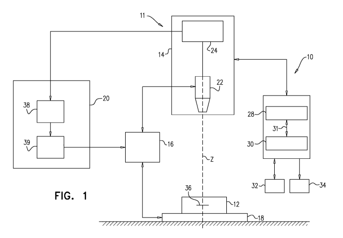
Reference is now made to Fig. 1, which is a schematic illustration of a
microscope
system 10 that is used for analyzing a cell sample (e.g., a blood sample) 12,
in accordance
with some applications of the present invention. Typically, microscope system
10 includes
an imaging module 14, a focus variation module 16, a sample carrier 18 and an
autofocus
system 20. For some applications, the microscope system is generally similar
to the
microscope system described in US 2014/0347459 to Greenfield, which is
incorporated
herein by reference. Cell sample 12 is typically a blood sample that is
prepared such as to
form a monolayer within which cells are not fixed in position, for example,
using techniques
as described in PCT Application Publication WO 15/001553 to Pollack, which is
incorporated herein by reference.
Imaging module 14 acts as an imaging device. Typically, imaging module 14,
which
acts as an imaging device, includes an optical unit 22 and an image sensor
unit 24. Optical
unit 22 is configured to form a magnified image of a sample (for example, cell
sample 12)
by conjugating a focus plane 36 and an image plane. The image sensor unit 24
typically
22
CA 03018536 2018-09-20
WO 2017/168411 PCT/IL2017/050363
includes an image sensor, for example a charge-coupled-device (CCD),
complementary
metal-oxide-semiconductor (CMOS) sensor, and/or a matrix sensor, positioned in
the image
plane of the optical unit 22 so as to sense the magnified image.
A computer processor 28 typically receives and processes images. The computer
processor communicates with a memory 30. Via a user interface 32, a user
(e.g., a laboratory
technician) sends instructions to the computer processor. For some
applications, the user
interface includes a keyboard, a mouse, a joystick, a touchscreen device (such
as a
smartphone or a tablet computer), a touchpad, a trackball, a voice-command
interface, and/or
other types of user interfaces that are known in the art. Typically, the
computer processor
generates an output via an output device 34. Further typically, the output
device includes a
display, such as a monitor, and the output includes an output that is
displayed on the display.
For some applications, the processor generates an output on a different type
of visual, text,
graphics, tactile, audio, and/or video output device, e.g., speakers,
headphones, a
smartphone, or a tablet computer. For some applications, user interface 32
acts as both an
input device and an output device. For some applications, the processor
generates an output
on a computer-readable medium (e.g., a non-transitory computer-readable
medium), such as
a disk, or a portable USB drive, and/or generates an output on a printer.
Image sensor unit 24 may output acquired digital images to output device 34
(which
may include a display) and/or to the autofocus system 20. Focus variation
module 16 may
be configured to vary a distance between the focus plane 36 of the optical
unit 22 and the
sample carrier 18. Focus variation module 16 may be operated manually or
automatically
via a mechanical interface which may, for example, modify the position of the
sample carrier
18 along an optical axis Z of the optical unit 22. Alternatively or
additionally, focus variation
module 16 may be commanded by autofocus system 20. For example, the focus
variation
module 16 may vary the distance between the sample carrier 18 and the focus
plane by (1)
modifying the position of optical unit 22 along the optical axis Z, (2)
modifying the position
of the sample carrier 18 along the position of the optical axis Z (e.g., by
moving a stage upon
which the sample carrier is placed), (3) modifying the position of the focus
plane by, for
example, changing a focal length of the optical unit 22, or a combination
thereof.
The sample carrier 18 may comprise a plate, which is typically placed on a
stage of
the microscope system. Sample carrier 18 may be configured to accommodate cell
sample
12. The carrier may be any carrier known in the art for holding a biological
sample.
23
CA 03018536 2018-09-20
WO 2017/168411 PCT/IL2017/050363
Optionally, the bottom surface of the carrier is essentially flat, to allow
cells in contact
therewith to be at about the same distance from the focal plane of the
microscope. Examples
include carrier slides, laboratory receptacles, dishes, plates, multi-well
plates, test tubes (e.g.
with a flat bottom), microfluidic cells and cartridges and the like.
Typically, the sample
carrier is similar to that described in PCT Application Publication WO
15/001553 to Pollack,
which is incorporated herein by reference. For some applications, a cell
suspension
comprising red blood cells is introduced onto a base surface of a carrier
having a vertical
height being greater than or equal to a vertical depth of said cell suspension
when on the
base carrier. The cells in the cell suspension are allowed to settle (without
applying any
force thereon) on the base surface of the carrier to form a monolayer of cells
on the base
surface of the carrier, without fixing the cells in position. Optionally, the
solution has a
vertical height of between 20 micrometers and 1,000 micrometers.
The blood sample that is imaged is typically raw blood, or a portion of raw
blood that
includes at least red blood cells, in diluted or undiluted form. Optionally,
the blood sample
is a cell sample derived from the human body, the sample including at least
red blood cells,
and is optionally modified by addition and/or removal of cells and/or other
components.
Typically, images are acquired of a portion of a blood sample that has been
drawn from a
subject's body. For example, the sample that is drawn from the subject's body
may be
divided between a plurality of sample carriers, within each of which
monolayers are allowed
to form (e.g., using techniques as described in PCT Application Publication WO
15/001553
to Pollack, which is incorporated herein by reference). Images may be acquired
of sample
carrier 18 or a portion thereof. For example, each of the sample carriers may
be scanned,
such that a plurality of images of the carrier are acquired, from respective
fields of vision at
respective locations along the bottom surface of sample carrier 18.
For some applications, one or more staining substances are used to stain the
sample
before the sample is imaged. For example, the staining substance may be
configured to stain
DNA with preference over staining of other cellular components. Alternatively,
the staining
substance may be configured to stain all cellular nucleic acids with
preference over staining
of other cellular components. For example, the sample may be stained with
acridine orange
reagent, Hoechst reagent, and/or any other staining substance that is
configured to
preferentially stain DNA and/or RNA within the blood sample. Optionally, the
staining
substance is configured to stain all cellular nucleic acids but the staining
of DNA and RNA
24
CA 03018536 2018-09-20
WO 2017/168411 PCT/IL2017/050363
are each more prominently visible under some lighting and filter conditions,
as is known, for
example, for acridine orange. Images of the sample may be acquired using
imaging
conditions that allow detection of cells (e.g., bright-field) and/or imaging
conditions that
allow visualization of stained bodies (e.g. appropriate fluorescent
illumination).
For some applications, the methods described herein are performed without
staining
the blood sample. For example, when the methods described herein are performed
in order
to determine a platelet count, the blood sample may be imaged without staining
the blood
sample.
Autofocus system 20 may comprise an autofocus computation module 38 and an
autofocus adaption module 39. The autofocus computation module may be
connected to the
image sensor unit 24 so as to receive images acquired by the imaging module
14. The
autofocus adaptation module may be connected to the focus variation module 16
and may
be configured to command the focus variation module 16, e.g., as described
above.
In accordance with some applications, a blood sample is scanned by the
microscope
system, such that a plurality of portions of the blood sample are imaged. For
some
applications, a plurality of images are acquired of one or more portions of
the blood sample,
with each of the plurality of images being acquired under respective imaging
conditions. For
example, two images of a portion of the sample may be acquired using,
respectively, imaging
conditions that allow detection of cells (e.g., bright-field) and imaging
conditions that allow
.. visualization of stained bodies (e.g. appropriate fluorescent
illumination).
Reference is now made to Figs. 2A-B, which are first and second images of a
Plasmodium parasite 40 (which appears as a bright speck) within an erythrocyte
42, a time
interval of approximately 5 minutes having passed between acquisitions of the
first and
second images, the images having been acquired in accordance with some
applications of
the present invention. Reference is also made to Figs. 3A-B, which are first
and second
images of a platelet 44 (which also appears as a bright speck) in the vicinity
of an erythrocyte
46, a time interval of approximately 5 minutes having passed between
acquisitions of the
first and second images, the images having been acquired in accordance with
some
applications of the present invention.
The images shown in Figs. 2A-B and 3A-B are of monolayers of diluted blood
samples that were stained with fluorescent nucleic acid stains and were imaged
at 20 times
CA 03018536 2018-09-20
WO 2017/168411 PCT/IL2017/050363
magnification. The samples were placed in sample carriers, which were scanned
such that
180 fields of vision of each sample carrier were imaged. The samples were
scanned twice,
such that each field was re-imaged after a time interval of approximately 5
minutes had
passed since the previous image of that field. During scanning, the samples
were gently
moved together with the microscope stage so that each field of vision was
disposed, in turn,
under the microscope objective lens for imaging. Images were acquired using
bright-field
imaging, as well as fluorescent imaging. Each of the images shown in Figs. 2A-
B and 3A-
B shows the fluorescent intensity overlaid on a bright-field image.
As may be observed by comparing the transition from Fig. 2A to 2B, to the
transition
from Fig. 3A to Fig. 3B, it was found that there is relative motion between
platelets and
erythrocytes within the sample. In general, it was found that, while both
platelets and
erythrocytes typically moved in the order of tens of microns or less,
platelets underwent
greater movement than the erythrocytes. Thus, when two images, the
acquisitions of which
were separated by a time interval (e.g., as was the case for the images shown
in Figs. 3A and
3B) were compared to one another, platelets moved relative to a nearby or
overlapping
erythrocyte. By contrast, as shown in Figs. 2A and 2B, Plasmodium parasites
within infected
erythrocytes did not move substantially with respect to the erythrocytes. Only
in very rare
events does Plasmodium separate from an essentially intact erythrocyte in
which the
Plasmodium is disposed.
As stated above, the images shown in Figs. 2A-B and 3A-B were generated when
the
sample carrier was gently moved together with a microscope stage, in order to
image a
plurality of fields of vision along the sample. However, relative motion of
platelets with
respect to erythrocytes was evident, even when the sample was not moved
between the
acquisitions of respective images. Movement of the platelets relative to
erythrocytes may
be enhanced by moving the sample carrier, agitating the sample carrier, and/or
vibrating the
sample carrier. Therefore, for some applications of the present invention, a
sample carrier
is moved, agitated, or vibrated between acquisitions of respective images of
the sample.
Alternatively or additionally, the sample is stirred using magnetic beads
disposed within the
sample, and an external magnetic field that drives the magnetic beads to move.
Motion of platelets with respect to erythrocytes relative that of
intracellular parasites
is detectable within a short time period, such as less than 10 minutes, 7
minutes, or 5 minutes.
In some cases, motion of platelets with respect to erythrocytes relative that
of intracellular
26
CA 03018536 2018-09-20
WO 2017/168411 PCT/IL2017/050363
parasites is detectable within less than 1 minute, less than 10 seconds, or
less than 1 second,
the extent of the motion depending on the conditions that are used. Therefore,
for some
applications of the present invention, first and second images that are
separated by a time
interval of less than 10 minutes, less than 7 minutes, less than 5 minutes,
less than 1 minute,
less than 10 seconds, or less than 1 second are compared to one another.
Typically, the
difference between the motion of platelets with respect to erythrocytes
relative that of
intracellular parasites is dependent upon the time interval between image
acquisitions, and/or
the extent to which the sample carrier is agitated between image acquisitions.
It was found that the above-described effect is evident even if there is a
time interval
of several hours between when the sample is prepared, and when the first image
is acquired.
Within this time period drying effects of the blood are not detrimental to the
above-described
effect. Therefore, for some applications, techniques as described herein are
performed on a
blood sample, even several hours (e.g., up to five hours) from when the blood
sample is
prepared.
Reference is now made to Fig. 4, which is flowchart showing steps of a
procedure
that is performed, in accordance with some applications of the present
invention.
In a first step 50, the blood sample is prepared, for example, in sample
carrier 18
(schematically shown in Fig. 1). The blood sample is typically raw blood, or a
portion of
raw blood that includes at least red blood cells, optionally in diluted form.
Optionally, the
blood sample is a cell sample derived from the human body, the sample
including at least
red blood cells, and optionally modified by addition and/or removal of cells
and/or other
components. Typically, the blood sample is in a preparation within which
erythrocytes and
other entities within the sample are not maintained in fixed positions. For
example, the blood
sample may be prepared by allowing the sample to form a monolayer, as
described, for
example, in PCT Application Publication WO 15/001553 to Pollack, which is
incorporated
herein by reference. Preparing the sample in this manner facilitates motion of
bodies within
the sample with respect to one another. For some applications, a sample that
is drawn from
the subject's body is divided between a plurality of sample carriers, within
each of which
monolayers are allowed to form.
In step 52, a first image of the sample is acquired, typically using
microscope system
10. A time interval from the acquisition of the first image is allowed to pass
(step 54). For
27
CA 03018536 2018-09-20
WO 2017/168411 PCT/IL2017/050363
some applications, the time interval is less than 10 minutes, less than 7
minutes, less than 5
minutes, less than 1 minute, less than 10 seconds, and/or less than 1 second.
Optionally,
during this period the sample is agitated (step 56, which is in a dashed box
to indicate that
this step is optional). For example, the sample carrier may be moved, or
vibrated, and/or
magnetic beads may be used to stir the sample, as described hereinabove. After
the time
interval has passed, a second image of the sample is acquired (step 58),
typically using the
microscope system.
It is noted that, typically, first and second sets of images are acquired,
with images
from the second set of images typically at least partially overlapping with
corresponding
images from the first set of images. As such, steps of the procedure that are
described as
being performed with respect to first and second images are typically
performed with respect
to a plurality of first images, and a plurality of second images. For example,
the sample
carrier may be scanned twice in sequence, such that first and second images of
the sample
are acquired from a plurality of fields of view. The first and second scans
may be performed,
for example, in the same direction as one another (i.e., such that the order
in which the fields
of view are imaged in the first and second scans is the same), or in reverse
from one another
(i.e., such that, in the second scan, the fields of view are imaged in the
reverse order from
the first scan). Optionally, one or both of the first and second scans is
performed in a random
order, and/or the order in which the fields of view are imaged (at least in
the second scan) is
such as to minimize the time needed to acquire all needed images. For some
applications,
the time interval between acquisitions of first and second images of a field
of view is
determined by the scanning speed of the microscope system (i.e., the time that
it takes the
system to arrive back at the field of view in order to image the field of view
for a second
time). For some applications, first and second images of a field of view are
acquired without
the microscope system acquiring images of any additional fields of view
between the
acquisitions of the first and second images.
For some applications, the image or the second set of images is acquired only
after
analysis of the first image, or first set of images, or a portion thereof,
indicates that it is
desirable to acquire a second image or second set of images (e.g., as
described herein). For
some such applications, only a portion of fields of view are re-imaged (for
example, a
plurality of first images and only one second image may be acquired), and/or
at least some
28
CA 03018536 2018-09-20
WO 2017/168411 PCT/IL2017/050363
of the images that are acquired during a second scan may be acquired at a
different
magnification from that of images acquired during the first scan.
In step 60, the first and second images are compared to one another.
Typically,
computer processor identifies one or more entities having dimensions and/or
other
characteristics that are such that the entity is a platelet candidate (i.e.,
an entity that could
potentially be a platelet), and/or an intra-erythrocytic-parasite candidate
(i.e., an entity that
could potentially be an intra-erythrocytic-parasite, such as Plasmodium,
and/or Babesia).
Typically, the entity is an entity the dimensions or other characteristics of
which (e.g., the
location of which with respect to an erythrocyte), are such that the entity
appears to be either
a platelet or an intra-erythrocytic parasite, and it is unclear which of the
two it is.
In step 62, the computer processor determines a characteristic of the blood
sample
based at least in part upon the comparison of the first and second images to
one another. For
example, in response to determining that (a) in the first image the entity is
disposed in the
vicinity of an erythrocyte, and that (b) there was relative motion between the
erythrocyte and
the entity between acquisitions of the first and second images (e.g., relative
motion of at least
one micron), the computer processor may confirm that the entity is a platelet.
Alternatively,
in response to determining that (a) in the first image the entity is disposed
in the vicinity of
an erythrocyte, and that (b) there was little or no relative motion between
the erythrocyte
and the entity between acquisitions of the first and second images, the
computer processor
may determine that the entity is an intra-erythrocytic entity. Based at least
in part upon
determining that the entity is an intra-erythrocytic entity, the computer
processor may
determine that the entity is an intra-erythrocytic parasite, such as
Plasmodium, and/or
B abesia.
Alternatively or additionally, based at least in part upon determining whether
the
entity is an intra-erythrocytic entity or an extra-erythrocytic entity, the
computer processor
may perform a blood sample analysis. For example, the computer processor may
perform a
complete blood count, which includes a count of platelets that takes into
account whether
the entity is an extra-erythrocytic entity (and therefore a platelet) or an
intra-erythrocytic
entity.
For some applications, the computer processor does not necessarily determine
whether or not the entity is an intra-erythrocytic entity or an extra-
erythrocytic entity, but
29
CA 03018536 2018-09-20
WO 2017/168411 PCT/IL2017/050363
rather determines the likelihood of the entity being one or the other of
these, and performs
analysis of the blood sample based upon the determined likelihood.
For some applications, in performing steps 60 and 62, the computer processor
runs
an algorithm that accounts for the time interval between acquisitions of the
first and second
images, and/or data indicative of agitation of the sample (e.g., the extent of
its motion with
the microscope stage).
Typically, if the computer processor identifies that an entity that (based
upon the first
image) was an intra-erythrocytic candidate, is no longer associated with the
same erythrocyte
(in the second image), or if there is no intra-erythrocytic candidate in the
vicinity of the
original location of the candidate, then the computer processor determines
that the candidate
is not an intra-erythrocytic entity, and/or that the candidate is not a
parasite. For some
applications, movement of an intra-erythrocytic candidate relative to the
movement of an
erythrocyte is used to filter out non-parasites from a parasite count, and/or
to enhance
confidence in a count of malaria parasites (e.g., utilizing a machine learning
statistical
algorithm).
For some applications, a blood sample contains a plurality of entities which
may be
intra-erythrocytic entities, or may be platelets. Some fields of view are re-
imaged using the
techniques described herein, in order to re-image some or all of the entities
which may be
intra-erythrocytic entities, or may be platelets. Based upon the number of
such entities that
are determined to be either platelets or intra-erythrocytic parasites, the
computer processor
estimates the number of such entities within the whole sample that are
platelets and the
number of such entities that are intra-erythrocytic parasites.
For some applications, the computer processor uses the determination of
whether the
entity is an intra-erythrocytic entity or an extra-erythrocytic entity as data
in blood sample
analysis e.g., in order to perform a complete blood count, or a portion
thereof. For such
applications, the computer processor may utilize the techniques described
herein to correct
the platelet count, by accounting for platelets that may not otherwise have
been identified as
platelets, due to platelets being disposed in the vicinity of erythrocytes. In
addition, the
computer processor may use the techniques described herein to identify certain
intra-
erythrocytic entities, which might otherwise not have been identified as such,
due to being
confused with platelets. For example, the computer processor may utilize the
techniques
CA 03018536 2018-09-20
WO 2017/168411 PCT/IL2017/050363
described herein to identify Howell Jolly bodies, reticular networks of
ribosomal DNA of
reticulocytes, Heinz bodies, Pappenheimer bodies, and/or nuclei of nucleated
erythrocytes,
inter alia, by distinguishing between such entities and platelets.
Reticulocytes are immature
erythrocytes having reticular networks of ribosomal DNA, while nucleated
erythrocytes are
immature erythrocytes having a nucleus. These intracellular organelles, which
do not exist
in mature erythrocytes, may sometimes appear similar to platelets.
Reference is now made to Fig. 5, which is flowchart showing steps of a
procedure
that is performed, in accordance with some applications of the present
invention. In a first
step 70, the blood sample is prepared, for example, in sample carrier 18
(shown
schematically in Fig. 1). Step 70 is typically generally similar to step 50
described with
reference to Fig. 4. In a second step 72, a first set of one or more images of
the sample is
acquired. Step 72 is typically generally similar to step 52 described with
reference to Fig. 4.
As described hereinabove, an image from a single field of view may be
acquired, or the
sample carrier may be scanned such that a plurality of images are acquired
from respective
fields of view.
In step 74, the first set of image(s) is analyzed. In step 76, based upon the
analysis
of the first set of images, the computer processor selects whether it is
desirable to compare
images belonging to the first set of images to images belonging to a second
set of images.
For example, the computer processor may determine that within one or more of
the first set
of images there are one or more platelet candidates and/or one or more
Plasmodium
candidates and/or one or more Babesia candidates that overlap with an
erythrocyte. In
response to the analysis, the computer processor may automatically acquire a
second set of
one or more images of the blood sample (step 78), and/or may generate an
output (e.g., on
output device 34, shown in Fig. 1) indicative of a recommendation to acquire a
second set
one or more images of the blood sample.
It is noted that Fig. 5 indicates that the computer processor acquires a
second set of
images (step 78), based upon analyzing the first set of images. However, for
some
applications, even without having analyzed the first set of images, the
computer processor
automatically drives the microscope system to acquire a second set of images.
For example,
the computer processor may drive the microscope system to scan a sample
carrier twice. For
such applications, in step 76, subsequent to having acquired both sets of
images, the
computer processor selects whether or not it is desirable to compare the
images belonging
31
CA 03018536 2018-09-20
WO 2017/168411 PCT/IL2017/050363
to the first set to images belonging to the second set, and then proceeds
directly to comparing
the images, in step 80.
It is noted that, in general, the steps of the flowchart shown in the figures
(e.g., Figs.
4 and 5) are not necessarily performed in the sequence in which they are
shown. For
example, with reference to Fig. 5, for some applications, step 78 is performed
before step 72
is terminated, such that images belonging to both the first and second sets
are acquired
simultaneously and/or alternately with respect to one another. For example,
once at least
one image of the first set is acquired (step 72), it may be analyzed (step 74)
and a selection
may be made to compare images (step 76). This may be performed while step 72
continues
and additional images of the first set are acquired. At this stage, acquiring
of the second set
of images (step 78) may commence, while one or more of steps 72, 74 and 76 are
continued
(or resumed). Alternatively, images belonging to the first and second sets may
be acquired
simultaneously and/or alternately with respect to one another (steps 72 and
78), prior to the
analysis of the first set of images (step 74) commencing, or at the same time
as the analysis
of the first set of images is performed.
Once a second set of at least one image(s) has been acquired, in step 80,
images
belonging to the first and second sets of images are compared to one another,
and in step 82,
the computer processor determines a characteristic of the blood sample based
at least in part
upon the comparison of the first and second images to one another. Steps 80
and 82 are
typically generally similar to steps 60 and 62 described with reference to
Fig. 4. Optionally,
comparing images of the first and second sets (step 80) commences, before
steps 72, 74, 76
and/or 78 are completed.
Typically, in step 72, a set of two or more first images are acquired from
respective
fields of view. For example, as described hereinabove, the microscope system
may scan a
sample carrier and acquired a plurality of images of the sample carrier form
respective fields
of view. For some applications, in step 74 the computer processor analyzes one
or more of
the first set of images of the sample. In response thereto, the computer
processor may acquire
at least one second image (or recommend that a second image be acquired) from
all of the
fields of view in which there are entities that are platelet and/or intra-
erythrocytic-entity
(e.g., Plasmodium, and/or Babesia) candidates, or from only a portion of the
fields of view
in which there are entities that are platelet and/or Plasmodium, and/or
Babesia candidates.
If only a portion of the fields of view are re-imaged, then typically the
results of the analysis
32
CA 03018536 2018-09-20
WO 2017/168411 PCT/IL2017/050363
of those fields of view are extrapolated and applied to fields of view in
which there were
entities that were platelet and/or Plasmodium, and/or Babesia candidates, but
which were
nor re-imaged. Typically, if the computer processor determines that a first
image of a given
portion of the sample should be re-imaged (e.g., because it contains entities
as described
herein), then a second image is acquired that at least partially overlaps with
the first image.
Typically, the area of overlap will include at least one erythrocyte, and at
least one entity
which is disposed in the vicinity of the erythrocyte, as described herein.
For some applications, in step 74, the computer processor runs an algorithm in
order
to determine whether some or all of the fields of view should be re-imaged,
based upon an
overall analysis of the first set of images. For example, the computer
processor may take
one or more of the following factors into account when determining whether to
re-image (or
recommend to re-image) all or a portion of a sample: the number of candidate
Plasmodium
parasites, and/or Babesia parasites, their associated parasitic phase, and/or
the likelihood of
false positive diagnosis of malaria due to platelet adhesion, and/or the
likelihood of presence
of specific types of blood cells or inclusion bodies found in such cells (e.g.
reticulocytes,
Howell Jolly Bodies, Heinz bodies, Pappenheimer bodies, and/or nucleated
erythrocytes).
For some applications, the likelihood of false positive diagnosis of malaria
due to platelet
adhesion is determined based upon the general platelet count in the sample,
and/or the
platelet morphology, and/or fluorescence of platelets within the sample that
do not overlap
with erythrocytes. For some applications, only images that contain platelet
candidates that
have morphology and/or fluorescence that are similar to that of platelets
within the sample
that do not overlap with erythrocytes are re-imaged (or recommended to be re-
imaged).
For some applications, the computer processor determines that some or all of
the
fields of view should be re-imaged, in order to improve a parasitemia count,
even though the
computer processor has diagnosed the subject as having malaria.
Reference is now made to Fig. 6, which is a flowchart showing steps of a
procedure
for analyzing a blood sample, in accordance with some applications of the
present invention.
In step 90 of the procedure, a blood sample is imaged, typically, from a
plurality of fields of
view, in accordance with the techniques described hereinabove. In step 92,
which is optional
(as indicated by the dashed lines), a platelet count is determined or
estimated by analyzing
the acquired images. Optionally, this count is restricted to free platelets
that are not intra-
erythrocytic candidates. In step 94, a count of intra-erythrocytic candidates
is determined
33
CA 03018536 2018-09-20
WO 2017/168411 PCT/IL2017/050363
by analyzing the acquired images. For example, in this step, the computer
processor may
determine a count of entities that have characteristics that are indicative of
the entities being
intra-erythrocytic parasites, such as Plasmodium, and/or Babesia.
In step 96, the computer processor determines whether the count of intra-
erythrocytic
candidates is below a first threshold. In response to determining that the
count is below the
first threshold, the computer processor determines that the sample is negative
with respect
to the intra-erythrocytic entity that is being detected. In step 98, the
computer processor
determines whether the count of intra-erythrocytic candidates is above a
second threshold.
This second threshold may be predetermined or may be adjusted at least
partially according
to a platelet count performed in step 92. Typically, a high platelet count
would increase the
likelihood that an intra-erythrocytic candidate is in fact a platelet, and
thus the threshold may
increase. In response to determining that the count is above the second
threshold, the
computer processor determines that the sample is positive with respect to the
intra-
erythrocytic entity that is being detected. For some applications, steps 96
and 98 are
performed in reverse order, or at the same time as one another.
If the processor determines that the count of intra-erythrocytic candidates is
above a
first threshold, but below the second threshold, this may indicate that it is
desirable to image
at least some of the sample a second time, in order to determine whether there
are any entities
such as those described hereinabove (such as platelets), which may otherwise
have been
mistakenly identified as intra-erythrocytic entities. Therefore, in response
to such a
determination, the computer processor proceeds to step 100 and re-images at
least a portion
of the sample. As described hereinabove, for some applications, only a portion
of the sample
(e.g., portions which include entities regarding which it is unclear whether
they are intra-
erythrocytic or extra-erythrocytic) is re-imaged.
For some applications, the platelet count that was determined in step 92 is
used as an
input in determining whether to re-image a portion of the sample. For example,
a platelet
count that is greater than a threshold amount may be indicative of a greater
likelihood that
platelets may otherwise have been mistakenly identified as intra-erythrocytic
entities. For
some applications, the platelet count relates only to free platelets, i.e.,
platelets that are not
intra-erythrocytic candidates.
34
CA 03018536 2018-09-20
WO 2017/168411 PCT/IL2017/050363
For some applications, the apparatus and methods described herein are used for
the
detection of an infection by a DNA-carrying pathogen. As such, at least a
first dye stains at
least the DNA, if present in the sample to thereby provide a first stained
area indicative of
the presence of the DNA carrying pathogen in the sample. The pathogen may be
any
infectious microorganism. In some embodiments, the pathogen is a eukaryotic
pathogen.
When referring to eukaryotic pathogen, in the context of the present
disclosure, it is to be
understood as encompassing one cell pathogens and multicellular pathogens but
also fungi,
such as yeast (e.g. Candida) and Aspergillus.
For some applications, the pathogen is a eukaryotic pathogen. In accordance
with
such applications, the pathogen may be a one cell pathogen, such as protozoa.
This includes
genital protozoa, e.g. Trichomonas vaginalis, nervous system protozoa, e.g.
Naegleria
fowleri fecal protozoa, e.g. Giardia lamblia, blood protozoa. For some
applications, the
pathogen may be a multicellular pathogen, such as Wuchereria bancrofti, Brugia
malayi,
Brugia timori, Mansonella streptocerca, or Onchocerca volvulus.
For some applications, the pathogen is a blood protozoa selected from the
genuses
consisting of Trypanosoma (causing Chagas disease and African sleeping
sickness);
Plasmodium (causing Malaria); Toxoplasma (causing Toxoplasmosis); Babesia
(causing
Babesiosis).
References to Plasmodium are to be understood as encompassing at least any
.. member of the group consisting of Plasmodium falciparum (P. falciparum),
Plasmodium
vivax (P. vivax), Plasmodium ovale (P. ovale), Plasmodium malariae (P.
malariae), and
Plasmodium knowlesi (P. knowlesi).
References to Babesia are to be understood as encompassing at least any member
of
the group consisting of Babesia duncani (B. duncani) or Babesia microti (B.
microti) and
Babesia divergens (B. divergens).
It is noted that the terms "parasite" and "pathogen" are used interchangeably
in the
context of the present application. For some applications, the terms
"pathogen" and
"parasite" refer to a particular stage of the life cycle of a particular
pathogen or group thereof.
For example, the invention disclosed herein can be applied specifically to the
detection of
trophozoites, schizonts and/or gametocytes of Plasmodium species or P.
falciparum in
particular.
CA 03018536 2018-09-20
WO 2017/168411 PCT/IL2017/050363
The apparatus and methods described herein may be applicable for the detection
of
multiple pathogens using the same conditions and/or in the same sample, e.g.,
the same
combination of dyes, same test conditions, etc., as well as for the detection
of a pathogen at
multiple stages of its life cycle. For some applications, the apparatus and
methods described
herein may determine which one or more of the multiple pathogens (or life
stages) is
suspected.
Applications of the invention described herein can take the form of a computer
program product accessible from a computer-usable or computer-readable medium
(e.g., a
non-transitory computer-readable medium) providing program code for use by or
in
connection with a computer or any instruction execution system, such as
computer processor
28. For the purpose of this description, a computer-usable or computer
readable medium
can be any apparatus that can comprise, store, communicate, propagate, or
transport the
program for use by or in connection with the instruction execution system,
apparatus, or
device. The medium can be an electronic, magnetic, optical, electromagnetic,
infrared, or
semiconductor system (or apparatus or device) or a propagation medium.
Typically, the
computer-usable or computer readable medium is a non-transitory computer-
usable or
computer readable medium.
Examples of a computer-readable medium include a semiconductor or solid state
memory, magnetic tape, a removable computer diskette, a random access memory
(RAM),
a read-only memory (ROM), a rigid magnetic disk and an optical disk. Current
examples of
optical disks include compact disk-read only memory (CD-ROM), compact disk-
read/write
(CD-RAY) and DVD.
A data processing system suitable for storing and/or executing program code
will
include at least one processor (e.g., computer processor 28) coupled directly
or indirectly to
memory elements (e.g., memory 29) through a system bus. The memory elements
can
include local memory employed during actual execution of the program code,
bulk storage,
and cache memories which provide temporary storage of at least some program
code in order
to reduce the number of times code must be retrieved from bulk storage during
execution.
The system can read the inventive instructions on the program storage devices
and follow
these instructions to execute the methodology of the embodiments of the
invention.
36
CA 03018536 2018-09-20
WO 2017/168411 PCT/IL2017/050363
Network adapters may be coupled to the processor to enable the processor to
become
coupled to other processors or remote printers or storage devices through
intervening private
or public networks. Modems, cable modem and Ethernet cards are just a few of
the currently
available types of network adapters.
Computer program code for carrying out operations of the present invention may
be
written in any combination of one or more programming languages, including an
object
oriented programming language such as Java, Smalltalk, C++ or the like and
conventional
procedural programming languages, such as the C programming language or
similar
programming languages.
It will be understood that blocks of the flowcharts shown in Figs. 4-6 and
combinations of blocks in the flowchart, can be implemented by computer
program
instructions. These computer program instructions may be provided to a
processor of a
general purpose computer, special purpose computer, or other programmable data
processing apparatus to produce a machine, such that the instructions, which
execute via the
processor of the computer (e.g., computer processor 28) or other programmable
data
processing apparatus, create means for implementing the functions/acts
specified in the
flowcharts and/or algorithms described in the present application. These
computer program
instructions may also be stored in a computer-readable medium (e.g., a non-
transitory
computer-readable medium) that can direct a computer or other programmable
data
processing apparatus to function in a particular manner, such that the
instructions stored in
the computer-readable medium produce an article of manufacture including
instruction
means which implement the function/act specified in the flowchart blocks and
algorithms.
The computer program instructions may also be loaded onto a computer or other
programmable data processing apparatus to cause a series of operational steps
to be
performed on the computer or other programmable apparatus to produce a
computer
implemented process such that the instructions which execute on the computer
or other
programmable apparatus provide processes for implementing the functions/acts
specified in
the flowcharts and/or algorithms described in the present application.
Computer processor 28 is typically a hardware device programmed with computer
program instructions to produce a special purpose computer. For example, when
programmed to perform the algorithms described with reference to Figs. 4-6,
computer
processor 28 typically acts as a special purpose blood-sample-analysis
computer processor.
37
CA 03018536 2018-09-20
WO 2017/168411 PCT/IL2017/050363
Typically, the operations described herein that are performed by computer
processor 28
transform the physical state of memory 29, which is a real physical article,
to have a different
magnetic polarity, electrical charge, or the like depending on the technology
of the memory
that is used. For some applications, operations that are described as being
performed by a
computer processor are performed by a plurality of computer processors in
combination with
each other.
Typically, computer processor generates an output on output device 34. The
output
may be provided in any acceptable form, including a graph, graphic or text
displayed on a
monitor of a control unit, a printout, as a voice message, or on a user's
smartphone display,
for accepting processed data from the processing utility and displaying
information relating
to the structural features obtained and/or associated values determining the
presence and
optionally the identity of a pathogenic infection, using lists, tables, graphs
etc. The output
device may include a monitor that is connected to a printer for printing the
output.
User interface 32 may be used to control the operation of system 10 and/or
computer
processor 28, including, inter alia, inputting data with respect to the
examined bodily fluid
source, date, place, etc.), controlling conditions of operating the system,
types of dyes used,
number of images to be taken, time interval between images, etc.
At times, image analysis by the computer processor may involve adjustment or
normalization of image brightness on the basis of degree of staining of the
sample. These
may be based on, for example, identifying one or more of brightest and/or
dimmest pixel
values in the image or set of image (for example, corresponding to a
particular sample),
average brightness of brightest and/or dimmest area, and/or image histogram.
Such features
may be extracted from a representative image (not necessarily the one being
normalized) or
from statistical analysis of multiple images. The features used for
normalization may be
based on a single or multiple images, which may be captured using different
excitation
wavelengths (e.g., acridine orange providing different colors under different
illumination
wavelengths).
Image brightness may also be adjusted using other control means, such as image
capturing component exposure time and/or brightness of illumination.
The conditions of microscope system 10 may be such as to control the timing of
the
image acquisition, e.g., to allow sufficient incubation time with the one or
more dyes as well
38
CA 03018536 2018-09-20
WO 2017/168411 PCT/IL2017/050363
as the operation with different optical configurations of excitation and/or
emission
wavelengths, in order to image the stained sample at various colors or
fluorescence spectra.
In order to image the stained sample at various colors or fluorescence
spectra,
changes in excitation may be achieved by switching the color of illumination.
This can be
done, for example, by providing two or more light sources (e.g. for acridine
orange, UV LED
light at 365nm and blue LED light at 475nm) and combining them optically (for
example,
using a dichroic mirror, or a grating).
In another example, a single illumination source (e.g., UV LED light at 365nm)
may
be used to excite two dyes simultaneously, and one or more optical filters are
moved in or
out of the optical path to select the relevant emission wavelengths. Other dye
sets can be
simultaneously excited using the same incident illumination as described here,
even if one
or more of the dye is excited non-optimally. As an example, acridine orange
can be similarly
co-excited together with a Hoechst stain, DAPI and DRAQ stains.
In yet another example, a single illumination source (e.g. UV LED light at
365nm)
may be used to excite two or more dyes simultaneously, and the emission
optical path is split
such that the two or more emissions are captured on two or more image
capturing
components.
In yet another example, a color imaging sensor is used to simultaneously
capture two
or more fluorescence signals. Use of a color imaging sensor can, for example,
obviate the
need for one or more optical filters that are moved in or out of the optical
path to select the
relevant wavelength.
In the context of the present disclosure, various illumination sources may be
used.
These include, without being limited thereto, those providing white light (as
in bright light
microscopy), UV light, blue light, green light, yellow light, red light, a
combination thereof,
or any light applicable for exciting one or more of the dyes used for
staining.
The components of the system, namely, imaging module 14, computer processor
28,
output device 34, etc., may be directly connected to each other (e.g. directly
by a wire) or
one or more of the components may be remote from one or more other components.
For
example, the imaging module may send data to computer processor 28 over an
intranet or
over the internet, to allow processing at a remote location.
39
CA 03018536 2018-09-20
WO 2017/168411 PCT/IL2017/050363
Examples of systems which may be used for performing the method of the present
disclosure are described in WO 2012/090198 to Bachelet and in US 2014/0347459
to
Greenfield, both of which applications are incorporated herein by reference.
It is noted that,
although with respect to some applications of the present invention, images of
a sample are
described as being acquired using a microscope system, the scope of the
present invention
includes using any imaging system for acquiring images of a sample, mutatis
mutandis.
Reference is now made to Fig. 7, which is a schematic illustration of a blood
diagnosis machine 110, in accordance with some applications of the present
invention. For
some applications, microscope system 10 is configured for use with blood
diagnosis machine
110, the machine being an operator-free fully automated blood diagnosis
machine which
allows patients to undergo blood tests without the assistance of trained
personnel in the
extraction of the blood sample or operation of the device. The machine may be
configured
to perform any type of test which requires only a limited amount of blood. For
example,
these may include complete blood count, CD4 count, and/or or malaria tests.
The machine
may be placed in medical facilities or non-medical facilities (e.g.,
pharmacies). When placed
at the facility, upon receiving a formal prescription or any other authorized
form, and/or at
will, the general public may come and undergo a blood test at his/her
convenience without
needing to set up an appointment.
For such applications, typically, before a test is performed, the user will
identify
himself/herself to machine 110, via user interface 32, which is typically as
described
hereinabove. The identification may be performed using a state issued
identification (ID)
card or number, a system username or card, a patient ID, insurance ID,
biometric
identification or any other accepted means of identification. The test to be
performed is
typically determined either by formal prescription or any other authorized
form presented to
the machine via user interface 32, or through a network connection to one or
more medical
service providers.
Once the user identification and tests are verified, a small amount of blood
(typically,
20 microliters), is extracted from the user and placed into the machine. For
example, the
machine may provide the user with the required apparatus (e.g. a disposable
lancet and
capillary device), which are stored in an unused disposables storage section
111, and the user
may then extract blood and place the blood into the machine, e.g., into a
sample receiving
unit 112 of the machine. Alternatively, the device itself may perform the
sample extraction.
CA 03018536 2018-09-20
WO 2017/168411 PCT/IL2017/050363
For example, the user may place a finger into the device and the device
automatically lancets
and extracts the required amount of blood. The machine typically proceeds to
automatically
perform any necessary sample preparation, e.g., blood staining and injection
into a cartridge,
using disposables which are typically be stored in unused disposables storage
section 111.
Used disposables are typically transferred to a used disposables storage
section 114. The
sample is then automatically analyzed by an analysis unit 116, which typically
includes
microscope system 10, as well as computer processor 28, both of which are
typically
generally as described hereinabove. For example, fluorescent and/or bright-
field microscope
images may be acquired by microscope system 10. Based upon the analysis, the
analysis
unit evaluates relevant measurands. The analysis of the blood sample is
typically completed
within a short time, e.g. within 10 minutes.
Typically, once a test is completed, machine 110 notifies the user whether the
test
was performed successfully or not, via user interface 32, e.g., via an on-
machine notification
screen or through a phone or text message (e.g., via e-mail), or any other
relevant means.
Further typically, the device sends the results through secured means either
directly to the
prescribing doctor or to one or more central lab information systems, or any
other authorized
servers.
To facilitate servicing and maintenance, the machine typically communicates
with
an online server. Using this connection, data on machine status, such as usage
statistics,
failures, or internal inventory are accessed and software updates are
performed.
Furthermore, online support for users of the machine may also be provided
through this or
similar servers.
A blood sample as described herein may be from any living creature, and is
typically
from warm blooded animals. For some applications, the blood sample is a sample
from a
mammal, e.g., from a human body. For some applications, the sample is taken
from any
domestic animal, zoo animals and farm animals, including but not limited to
dogs, cats,
horses, cows and sheep. Alternatively or additionally, the blood sample is
taken from
animals that act as disease vectors including deer or rats.
There is provided, in accordance with some applications of the present
invention, the
following inventive concepts:
41
CA 03018536 2018-09-20
WO 2017/168411
PCT/IL2017/050363
Inventive concept 1. A method for use with a blood sample that was drawn from
a subject,
the method comprising:
acquiring first and second images of the blood sample at respective times,
using a
microscope system; and
using a computer processor:
determining whether between acquisitions of the first and second images
there was relative motion between at least one erythrocyte within the sample
and at
least one entity within the sample, by comparing the first and second images
to one
another;
at least partially in response thereto, determining whether the entity is an
extra-erythrocytic or an intra-erythrocytic entity; and
generating an output, at least partially in response thereto.
Inventive concept 2. The method according to inventive concept 1, wherein the
microscope
system includes a microscope system that is disposed in a blood diagnosis
machine that is
accessible to the subject, and wherein the method comprises receiving the
blood sample into
the blood diagnosis machine by the subject placing the blood sample into a
sample receiving
unit of the blood diagnosis machine.
Inventive concept 3. The method according to inventive concept 1 or inventive
concept 2,
wherein acquiring first and second images of the blood sample comprises
acquiring first and
second at least partially overlapping images of a portion of the blood sample.
Inventive concept 4. The method according to any one of inventive concepts 1-
3, wherein
determining whether the entity is an extra-erythrocytic or an intra-
erythrocytic entity
comprises determining whether the entity is an extra-erythrocytic or an intra-
erythrocytic
entity, at least partially based upon an amount of motion between the
erythrocyte and the
entity, and a time interval between acquisitions of the first and second
images.
Inventive concept 5. The method according to any one of inventive concepts 1-
3, wherein
determining whether the entity is an extra-erythrocytic or an intra-
erythrocytic entity
comprises determining whether the entity is an extra-erythrocytic or an intra-
erythrocytic
entity, at least partially based upon an amount of motion between the
erythrocyte and the
entity, and an amount of agitation applied to the blood sample between
acquisitions of the
first and second images.
42
CA 03018536 2018-09-20
WO 2017/168411 PCT/IL2017/050363
Inventive concept 6. The method according to any one of inventive concepts 1-
3, wherein
determining whether the entity is an extra-erythrocytic or an intra-
erythrocytic entity
comprises determining whether the entity is an extra-erythrocytic or an intra-
erythrocytic
entity, at least partially based upon an amount of motion between the
erythrocyte and the
entity, a time interval between acquisitions of the first and second images,
and an amount of
agitation applied to the blood sample between acquisitions of the first and
second images.
Inventive concept 7. The method according to any one of inventive concepts 1-
6, wherein
acquiring the first and second images of the blood sample at respective times
comprises
acquiring the first image of the blood sample during a first scan of the blood
sample in which
a plurality of images of the blood sample are acquired from respective fields
of view, and
acquiring the second image of the blood sample during a second scan of the
blood sample in
which a plurality of images of the blood sample are acquired from respective
fields of view.
Inventive concept 8. The method according to any one of inventive concepts 1-
7, further
comprising preparing the blood sample in a monolayer, wherein acquiring the
first and
second images of the blood sample comprises acquiring first and second images
of the blood
sample, while the blood sample is disposed in the monolayer.
Inventive concept 9. The method according to any one of inventive concepts 1-
8, further
comprising, using the computer processor, at least partially based upon
determining whether
the entity is an extra-erythrocytic or an intra-erythrocytic entity,
performing a blood count
of the subject, wherein generating the output comprises generating an
indication of the blood
count.
Inventive concept 10. The method according to any one of inventive concepts 1-
9, further
comprising, using the computer processor, at least partially based upon
determining whether
the entity is an extra-erythrocytic or an intra-erythrocytic entity,
diagnosing the subject as
suffering from an intra-erythrocytic infection, wherein generating the output
comprises
generating an indication of the diagnosis.
Inventive concept 11. The method according to any one of inventive concepts 1-
10, further
comprising, using the computer processor, at least partially based upon
determining whether
the entity is an extra-erythrocytic or an intra-erythrocytic entity,
diagnosing the subject as
suffering from a medical condition, wherein generating the output comprises
generating an
indication of the diagnosis.
43
CA 03018536 2018-09-20
WO 2017/168411 PCT/IL2017/050363
Inventive concept 12. The method according to any one of inventive concepts 1-
11, further
comprising staining the blood sample with a staining substance, wherein
acquiring the first
and second images comprises acquiring the first and second images of the blood
sample,
while the blood sample is in a stained state.
Inventive concept 13. The method according to any one of inventive concepts 1-
12, further
comprising, using the computer processor:
analyzing the first image;
based upon the analysis, identifying one or more entities within the first
image that
are disposed in a vicinity of the erythrocyte, and which have dimensions that
indicate that
the entities could be platelets; and
in response thereto, selecting to perform the comparing of the first image and
the
second image to one another.
Inventive concept 14. The method according to any one of inventive concepts 1-
13, wherein
determining whether the entity is an extra-erythrocytic or an intra-
erythrocytic entity
comprises determining whether the entity is a platelet.
Inventive concept 15. The method according to any one of inventive concepts 1-
13, wherein
determining whether the entity is an extra-erythrocytic or an intra-
erythrocytic entity
comprises determining that the entity is an intra-erythrocytic entity selected
from the group
consisting of: a Howell Jolly body, a reticular network of ribosomal DNA, a
Heinz body, a
Pappenheimer body, and a nucleus of a nucleated erythrocyte.
Inventive concept 16. The method according to any one of inventive concepts 1-
13, wherein
determining whether the entity is an extra-erythrocytic or an intra-
erythrocytic entity
comprises determining that the entity is an intra-erythrocytic parasite.
Inventive concept 17. The method according to inventive concept 16, wherein
determining
that the entity is an intra-erythrocytic parasite comprises determining that
the entity is an
intra-erythrocytic parasite selected from the group consisting of a Plasmodium
parasite, and
a Babesia parasite.
Inventive concept 18. The method according to any one of inventive concepts 1-
15,
wherein:
acquiring the first image of the blood sample comprises acquiring a first set
of images
of the blood sample that includes a plurality of images;
44
CA 03018536 2018-09-20
WO 2017/168411 PCT/IL2017/050363
acquiring the second image of the blood sample comprises acquiring a second
set of
images of the blood sample that includes one or more images; and
comparing the first and second images to one another comprises comparing one
or
more of the images belonging to the first set of images to respective images
belonging to the
second set of images.
Inventive concept 19. The method according to inventive concept 18, wherein
comparing
one or more of the images belonging to the first set of images to respective
images belonging
to the second set of images comprises comparing only some of the first set of
images to
respective images belonging to the second set of images, the method further
comprising
determining a characteristic of all of the blood sample based on the
comparison.
Inventive concept 20. The method according to inventive concept 18, wherein
acquiring the
second set of images comprises imaging a portion of the blood sample that is
smaller than a
portion of the blood sample that was imaged by acquiring the first set of
images.
Inventive concept 21. The method according to inventive concept 20, further
comprising:
analyzing the first set of images; and
based upon the analysis, selecting the portion of the blood sample to image in
the
second set of images.
Inventive concept 22. The method according to any one of inventive concepts 1-
15, wherein
acquiring the first and second images of the blood sample at respective times
comprises
acquiring the first and second images of the blood sample, a time interval
between
acquisitions of the first and second images being less than ten minutes.
Inventive concept 23. The method according to inventive concept 22, wherein
acquiring the
first and second images of the blood sample at respective times comprises
acquiring the first
and second images of the blood sample, the time interval between acquisitions
of the first
and second images being less than one minute.
Inventive concept 24. The method according to inventive concept 23, wherein
acquiring the
first and second images of the blood sample at respective times comprises
acquiring the first
and second images of the blood sample, the time interval between acquisitions
of the first
and second images being less than one second.
Inventive concept 25. The method according to any one of inventive concepts 1-
15, further
comprising agitating the blood sample between acquisitions of the first and
second images.
CA 03018536 2018-09-20
WO 2017/168411 PCT/IL2017/050363
Inventive concept 26. The method according to inventive concept 25, wherein
agitating the
blood sample comprises placing magnetic beads inside the sample and moving the
magnetic
beads using an external magnetic field.
Inventive concept 27. The method according to inventive concept 25, wherein
agitating the
blood sample comprises moving a microscope stage upon which the blood sample
is
disposed.
Inventive concept 28. A method for use with a blood sample that was drawn from
a subject,
the method comprising:
acquiring a first image of the blood sample, using a microscope system;
acquiring a second image of the blood sample, using the microscope system,
there
being a time interval between acquisitions of the first and second images; and
using a computer processor:
analyzing the first image of the blood sample;
at least partially in response thereto:
selecting to compare the first and second images of the blood sample
to one another;
comparing the first and second images of the blood sample to one
another; and
determining a characteristic of the blood sample, at least partially
based upon comparing the first and second images of the blood sample to one
another; and
generating an output in response to the determined characteristic.
Inventive concept 29. The method according to inventive concept 28, wherein
the
microscope system includes a microscope system that is disposed in a blood
diagnosis
machine that is accessible to the subject, and wherein the method comprises
receiving the
blood sample into the blood diagnosis machine by the subject placing the blood
sample into
a sample receiving unit of the blood diagnosis machine.
Inventive concept 30. The method according to inventive concept 28 or
inventive concept
29, wherein selecting to compare the first and second images of the blood
sample to one
another comprises selecting to acquire the second image of the blood sample,
and wherein
46
CA 03018536 2018-09-20
WO 2017/168411 PCT/IL2017/050363
acquiring the second image of the blood sample comprises automatically
acquiring the
second image in response thereto.
Inventive concept 31. The method according to any one of inventive concepts 28-
30,
wherein acquiring the first and second images of the blood sample comprises
acquiring the
first image of the blood sample during a first scan of the blood sample in
which a plurality
of images of the blood sample are acquired from respective fields of view, and
acquiring the
second image of the blood sample during a second scan of the blood sample in
which a
plurality of images of the blood sample are acquired from respective fields of
view.
Inventive concept 32. The method according to any one of inventive concepts 28-
31, further
comprising preparing the blood sample in a monolayer, wherein acquiring the
first and
second images of the blood sample comprises acquiring the first and second
images of the
blood sample, while the blood sample is disposed in the monolayer.
Inventive concept 33. The method according to any one of inventive concepts 28-
32, further
comprising staining the blood sample with a staining substance, wherein
acquiring the first
and second images of the blood sample comprises acquiring the first and second
images of
the blood sample while the blood sample is in a stained state.
Inventive concept 34. The method according to any one of inventive concepts 28-
33,
wherein:
analyzing the first image comprises identifying one or more entities within
the first
image that are disposed in a vicinity of an erythrocyte, and which have
dimensions that
indicate that the entities could be platelets, and
selecting to compare the first and second images of the blood sample to one
another
is performed at least partially in response thereto.
Inventive concept 35. The method according to any one of inventive concepts 28-
34,
wherein:
acquiring the first image of the blood sample comprises acquiring a first set
of images
of the blood sample that includes a plurality of images;
acquiring the second image of the blood sample comprises acquiring a second
set of
images of the blood sample that includes one or more images; and
47
CA 03018536 2018-09-20
WO 2017/168411 PCT/IL2017/050363
selecting to compare the first and second images of the blood sample to one
another
comprises selecting to compare at least a portion of the images belonging to
the plurality of
first images to respective images belonging to the plurality of second images.
Inventive concept 36. The method according to inventive concept 35, wherein
selecting to
compare at least a portion of the images belonging to the plurality of first
images to
respective images belonging to the plurality of second images comprises
selecting to
compare only some of the plurality of first images to respective images
belonging to the
plurality of second images, the method further comprising determining a
characteristic of all
of the blood sample based on comparing only some of the plurality of first
images to
respective images belonging to the plurality of second images.
Inventive concept 37. The method according to inventive concept 35, wherein
selecting to
compare the first and second images of the blood sample to one another
comprises selecting
to acquire the second set of images of the blood sample, the second set of
images imaging a
portion of the blood sample that is smaller than a portion of the blood sample
that was imaged
by acquiring the first set of images.
Inventive concept 38. The method according to any one of inventive concepts 28-
34,
wherein determining a characteristic of the blood sample, at least partially
based upon
comparing the first and second images to one another comprises:
determining whether between acquisitions of the first and second images,
there was relative motion between at least one erythrocyte within the sample
and at
least one entity within the sample; and
at least partially in response thereto, determining whether the entity is an
extra-erythrocytic or an intra-erythrocytic entity.
Inventive concept 39. The method according to inventive concept 38, wherein
determining
whether the entity is an extra-erythrocytic or an intra-erythrocytic entity
comprises
determining whether the entity is an extra-erythrocytic or an intra-
erythrocytic entity, at least
partially based upon an amount of motion between the erythrocyte and the
entity, and the
time interval between acquisitions of the first and second images.
Inventive concept 40. The method according to inventive concept 38, wherein
determining
whether the entity is an extra-erythrocytic or an intra-erythrocytic entity
comprises
determining whether the entity is an extra-erythrocytic or an intra-
erythrocytic entity, at least
48
CA 03018536 2018-09-20
WO 2017/168411 PCT/IL2017/050363
partially based upon an amount of motion between the erythrocyte and the
entity, and an
amount of agitation applied to the blood sample between acquisitions of the
first and second
images.
Inventive concept 41. The method according to inventive concept 38, wherein
determining
whether the entity is an extra-erythrocytic or an intra-erythrocytic entity
comprises
determining whether the entity is an extra-erythrocytic or an intra-
erythrocytic entity, at least
partially based upon an amount of motion between the erythrocyte and the
entity, the time
interval between acquisitions of the first and second images, and an amount of
agitation
applied to the blood sample between acquisitions of the first and second
images.
Inventive concept 42. The method according to inventive concept 38, further
comprising,
using the computer processor, at least partially based upon determining
whether the entity is
an extra-erythrocytic or an intra-erythrocytic entity, performing a blood
count of the subject,
wherein generating the output comprises generating an indication of the blood
count.
Inventive concept 43. The method according to inventive concept 38, further
comprising,
using the computer processor, at least partially based upon determining
whether the entity is
an extra-erythrocytic or an intra-erythrocytic entity, diagnosing the subject
as suffering from
an intra-erythrocytic infection, wherein generating the output comprises
generating an
indication of the diagnosis.
Inventive concept 44. The method according to inventive concept 38, further
comprising,
using the computer processor, at least partially based upon determining
whether the entity is
an extra-erythrocytic or an intra-erythrocytic entity, diagnosing the subject
as suffering from
a medical condition, wherein generating the output comprises generating an
indication of the
diagnosis.
Inventive concept 45. The method according to inventive concept 38, wherein
determining
whether the entity is an extra-erythrocytic or an intra-erythrocytic entity
comprises
determining that the entity is a platelet.
Inventive concept 46. The method according to inventive concept 38, wherein
determining
whether the entity is an extra-erythrocytic or an intra-erythrocytic entity
comprises
determining that the entity is an intra-erythrocytic entity selected from the
group consisting
of: a Howell Jolly body, a reticular network of ribosomal DNA, a Heinz body, a
Pappenheimer body, and a nucleus of a nucleated erythrocyte.
49
CA 03018536 2018-09-20
WO 2017/168411 PCT/IL2017/050363
Inventive concept 47. The method according to inventive concept 38, wherein
determining
whether the entity is an extra-erythrocytic or an intra-erythrocytic entity
comprises
determining that the entity is an intra-erythrocytic parasite.
Inventive concept 48. The method according to inventive concept 47, wherein
determining
that the entity is an intra-erythrocytic parasite comprises determining that
the entity is an
intra-erythrocytic parasite selected from the group consisting of a Plasmodium
parasite, and
a Babesia parasite.
Inventive concept 49. The method according to any one of inventive concepts 28-
34,
wherein acquiring the first and second images of the blood sample comprises
acquiring the
first and second images of the blood sample, the time interval between
acquisitions of the
first and second images being less than ten minutes.
Inventive concept 50. The method according to inventive concept 49, wherein
acquiring the
first and second images of the blood sample comprises acquiring the first and
second images
of the blood sample, the time interval between acquisitions of the first and
second images
being less than one minute.
Inventive concept 51. The method according to inventive concept 50, wherein
acquiring the
first and second images of the blood sample comprises acquiring the first and
second images
of the blood sample, the time interval between acquisitions of the first and
second images
being less than one second.
Inventive concept 52. The method according to any one of inventive concepts 28-
34, further
comprising agitating the blood sample between acquisitions of the first and
second images.
Inventive concept 53. The method according to inventive concept 52, wherein
agitating the
blood sample comprises placing magnetic beads inside the sample and moving the
magnetic
beads using an external magnetic field.
Inventive concept 54. The method according to inventive concept 52, wherein
agitating the
blood sample comprises moving a microscope stage upon which the blood sample
is
disposed.
Inventive concept 55. A computer software product, for use with a blood sample
that was
drawn from a subject, and a microscope system configured to acquire first and
second images
of the blood sample at respective times, the computer software product
comprising a non-
CA 03018536 2018-09-20
WO 2017/168411 PCT/IL2017/050363
transitory computer-readable medium in which program instructions are stored,
which
instructions, when read by a computer cause the computer to perform the steps
of:
determining whether between acquisitions of the first and second images there
was relative
motion between at least one erythrocyte within the sample and at least one
entity within the
sample, by comparing the first and second images to one another; at least
partially in
response thereto, determining whether the entity is an extra-erythrocytic or
an intra-
erythrocytic entity; and generating an output, at least partially in response
thereto.
Inventive concept 56. A computer software product, for use with a blood sample
that was
drawn from a subject, and a microscope system configured to acquire a first
and image of
the blood sample and a second image of the blood sample, there being a time
interval
between acquisitions of the first and second images, the computer software
product
comprising a non-transitory computer-readable medium in which program
instructions are
stored, which instructions, when read by a computer cause the computer to
perform the steps
of: analyzing the first image of the blood sample; at least partially in
response thereto:
selecting to compare the first and second images of the blood sample to one
another;
comparing the first and second images of the blood sample to one another; and
determining
a characteristic of the blood sample, at least partially based upon comparing
the first and
second images of the blood sample to one another; and generating an output in
response to
the determined characteristic.
It will be appreciated by persons skilled in the art that the present
invention is not
limited to what has been particularly shown and described hereinabove. Rather,
the scope
of the present invention includes both combinations and subcombinations of the
various
features described hereinabove, as well as variations and modifications
thereof that are not
in the prior art, which would occur to persons skilled in the art upon reading
the foregoing
.. description.
51