Note: Descriptions are shown in the official language in which they were submitted.
CA 03018546 2018-09-20
WO 2017/171706 PCT/US2016/024468
AMNIOTIC OR PLACENTAL PREPARATION AND DEVICE FOR OPHTHALMIC
USE AS A DRESSING TO ENHANCE HEALING
BACKGROUND OF THE INVENTION
[0001] Treatment of ocular surface disorders requires medical and surgical
intervention, both
acutely and in the long term. Regardless of the underlying causes involved,
the common goals of
management include controlling inflammation and promoting ocular surface
healing with
maximal visual rehabilitation. Various medical therapies have been used to
achieve these
objectives.
[0002] Amniotic membrane (AM) graft has been used in ophthalmology for several
indications
because of its beneficial effects. Amniotic compositions, such as amniotic
membrane and
extracts from amniotic membrane obtained from amniotic tissue derived from
mammals, such as
humans, pigs, or horses, include biological growth factors. Amniotic membrane
is a biological
membrane that lines the inner surface of the amniotic cavity and comprises a
simple, cuboidal
epithelium, a thick basement membrane, and an avascular mesenchymal layer
containing
hyaluronic acid. Amniotic compositions are known to reduce inflammation,
fibrovascular
ingrowth, and to facilitate epithelialization in animal models. Amniotic
membrane is believed to
play a role in the scarless wound healing process in a fetus.'
[0003] Previous studies revealed that early intervention with amniotic
membrane transplantation
(AMT) results in marked reduction of inflammation, rapid restoration of the
ocular surface, and
improved visual acuities while preventing cicatricial complications.2 However,
surgically
performed AMT renders a relatively high cost and potentially unnecessary
surgical trauma in
such compromised eyes. Furthermore, the membrane patch usually dissolves
within several days
so that multiple sessions of AMT may be required. Previously, a self-retaining
AM mounted on a
double ring system has been effectively used to promote healing and reduce
corneal scarring in a
U.S. Patent No. 7,494,8002; Tseng, S.-C.-G., et al., "Suppression of
Transforming Growth Factor-Beta Isoforms,
TGF-I3 Receptor Type H, and Myofibroblast Differentiation in Cultured Human
Corneal and Limbal Fibroblasts by
Amniotic Membrane Matrix," J. Cell. Physiol., 179: 325-335 (1999).
2 Dua HS, Gomes JA, King AJ, et al. The amniotic membrane in ophthalmology.
SUIT OphihalM01. 2004;49:51-77.
1
CA 03018546 2018-09-20
WO 2017/171706 PCT/US2016/024468
variety of ocular surface disorders,3 however, patients experienced ocular
discomfort from the
ring and incomplete healing.4
100041 Previous studies have also shown that topical amniotic membrane extract
(AME) has
comparable effect to AMT in promoting epithelialization, decreasing
inflammation, and
suppressing corneal neovascularization.5 However amniotic membrane extract
lacks the physical
characteristics of a bandage and as such it cannot be used as a patch graft.6
Another previous
approach to the use of amniotic proteins to treat ocular disease and injury is
the application of
amniotic fluid topically delivered to the eye.7
[0005] However, surgically performed AMT renders a relatively high cost and
potentially
unnecessary surgical trauma and complications. Amniotic membrane retained by a
ring does not
require surgery, but it is obtrusive, not well tolerated and as a result it
suffers from sub-optimal
therapeutic outcomes. Amniotic membrane extract ¨ though it shares the healing
qualities of
intact amniotic membrane ¨ does not have the physical characteristics of a
patch. There is,
therefore, an unmet need for improved delivery of amniotic proteins for ocular
indications.
SUMMARY OF THE INVENTION
[0006] The invention described herein is a method and medical device for the
treatment of
diseases and injuries to the cornea of an eye, that may include a self-
retaining patch that
incorporates amniotic membrane preparations that is applied using non-surgical
means, does not
require external mechanical support, is well tolerated by patients, and lasts
for a controlled
period of time to achieve the ultimate goal of using amniotic membrane
preparation in treating
ocular surface disorders.
3 Pachigolla G, Prasher P, Di Pascuale MA, McCulley JP, McHenry JO, Mootha VV.
Evaluation of the role of
ProKera in the management of ocular surface and orbital disorders. Eye Contact
Lens. 2009;35:172-175.
4 Sun i K, Kosker M, Raber 1M, et al. Sutureless Amniotic Membrane ProKera for
Ocular Surface Disorders: Short-
Term Results. Eye Contact Lens. 2013;39:341-347
Liang L, Li W, Ling S, Sheha H, Qiu W, Li C, Liu Z. Amniotic membrane
extraction solution for ocular chemical
burns. Clin Experiment Ophthalmol. 2009 Dec;37(9):855-63.
6 Sheha H, Liang L, Hashem 11, Ramzy M, ZaKi A, Amniotic Membrane Extract for
Acute Ocular Chemical Burns.
Techniques in Ophthalmology2010 Dec; 8(4) pp 146-150
7 US 2008/0286378 Al.
2
CA 03018546 2018-09-20
WO 2017/171706 PCT/US2016/024468
[0007] In an embodiment, the invention disclosed herein may be a multi-purpose
ophthalmic
patch incorporating amniotic extracts and/or placental extracts, mixed with or
carried on a
biodegradable material. The amniotic patch is formed in the general shape of a
contact lens that
can easily be placed on the surface of the eye to act as a bandage and protect
and enhance the
healing of the ocular surface. The inventive patch may achieve the known
therapeutic benefits of
the amniotic membrane without the need for surgery or a retaining ring to
eliminate surgery
related complication as well as ring related discomfort. Moreover, the patch
is designed with
controlled rate of degradation, such that the time it is used for different
ocular surface disorders
can vary according to the desired treatment period. For instance, if long term
treatment is
desired, then a lens with a slow degradation rate may be utilized, whereas, if
more short term
treatment is desired a lens having a faster degradation rate may be utilized.
100081 In an embodiment, a method is provided of treating an ocular disease or
injury in a
patient having an ocular disease or injury, by providing a lens-shaped patch
formed of
biodegradable carrier material and an active ingredient comprising an amniotic
extract and/or
placental extract, the lens-shaped patch having a substantially convex
exterior surface and a
substantially concave interior surface; applying the lens-shaped patch to the
patient's cornea,
whereby the interior surface of the lens-shaped patch contacts the cornea; and
allowing the lens-
shaped patch to dissolve while on the patient's cornea.
[0009] In an embodiment, a method is provided of treating an ocular disease or
injury in a
patient having an ocular disease or injury, by providing a lens-shaped patch
formed of
biodegradable carrier material and an active ingredient comprising an amniotic
extract and/or
placental extract, the lens-shaped patch having a substantially convex
exterior surface and a
substantially concave interior surface, wherein the lens-shaped patch has a
controlled
degradation time; determining a treatment time, according the nature of the
ocular disease or
injury, during which the lens-shaped patch should be applied to the patient's
cornea; selecting
the lens-shaped patch that has a degradation rate such that the patch applies
active ingredient to
the cornea for a time that is equal to or greater than treatment time; and
applying the lens-shaped
patch to the patient's cornea, whereby the interior surface of the lens-shaped
patch contacts the
cornea.
3
CA 03018546 2018-09-20
WO 2017/171706 PCT/US2016/024468
[0010] In an embodiment, a lens-shaped patch is provided for the treatment of
diseases and
injuries to the cornea of an eye, where the patch is has a biodegradable
carrier material and an
active ingredient comprising an amniotic extract and/or placental extract, and
the biodegradable
material and amniotic and/or placental extract are combined and formed into
the shape of a
contact lens, such that the active ingredient in the lens-shaped patch is in
physical contact with
the outer surface of the cornea.
BRIEF DESCRIPTION OF THE DRAWINGS
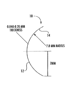
100111 Fig. 1 is a side cross-sectional schematic view of a contact lens-
shaped patch according to
an embodiment of the invention.
[0012] Fig. 2 is a prospective side view of a contact lens-shaped patch
according to an
embodiment of the invention,
[00131 Fig, 3 is a side perspective view of a contact lens-shaped patch
positioned for insertion on
an ocular surface according to an embodiment of the invention.
[0014] Fig. 4 is a front schematic view of a lens-shaped patch where the
biological curative
active ingredient is confined to a peripheral ring.
[00151 Fig. 5 is a graph of mass changes over time for ¨0.1 mm thick lens-
patches containing
15% and 30% placental extract, respectively, and 50:50 PLGA.
DETAILED DESCRIPTION OF THE INVENTION
[00161 The term "amniotic extract" herein means any of various preparations
derived from
amniotic membrane materials, including preparations derived from amniotic
membrane,
amniotic stroma and amniotic jelly (e.g. membrane particles obtained or
purified via a suitable
extraction/purification process such as pulverization or homogenization).
Placental extracts of
any of various sources may be used in different embodiments of this invention.
Placenta extract
or placenta membrane may be derived from any of various mammalian sources,
including
human, sheep, or bovine. Sheep placental extract is available commercially.
For many uses,
human placental extract may be less antigenic for use in humans. The term
"placental extract"
4
CA 03018546 2018-09-20
WO 2017/171706 PCT/US2016/024468
herein means any of various preparations derived from mammalian, including
human, placenta
(e.g. placental particles obtained or purified via a suitable
extraction/purification process such as
pulverization or homogenization). In this invention, amniotic extracts or
placental extracts may
be used alone or they may be blended. Any of amniotic extract or placental
extract, or blend
thereof, may be alternatively referred to as the "active ingredient" herein.
[0017] In an embodiment of this invention, the active ingredients are
supported by or associated
with a biodegradable carrier, filler or matrix. The biodegradable carrier
active ingredient is
formable into various shapes and configurations according to different
embodiments of the
invention. In an embodiment of the invention, the biodegradable carrier is
formed into
substantially the shape and size of a conventional contact lens. A patch
formed by combining
active ingredients with a synthetic or natural biodegradable and casting the
same into the general
shape of a conventional contact lens may be referred to as a "lens-shaped
patch" or "patch" or
"lens patch" herein.
[0018] In an embodiment, a lens-shaped patch for treating diseases and
injuries to the cornea is
provided, including a biodegradable carrier material and an active ingredient
comprising an
amniotic extract and/or placental extract, wherein the biodegradable material
and amniotic and/or
placental extract are combined and formed into the general shape of a contact
lens.
[0019] In an embodiment, the base curvature of the lens-shaped patch is
similar to or slightly
greater than the curvature of the cornea. As such, the inner surface of the
patch substantially
conforms to the external surface of the cornea and it adheres thereto allowing
the patch to remain
centered on the cornea.
[0020] In an embodiment of the invention, the inventive lens-shaped patch has
a variable
thickness and provides no magnification power or vision corrective properties.
For example, the
patch may have a thickness ranging between .040 mm and .20 mm and more
preferably
between .04 mm and .08 mm. In an embodiment, the lens is approximately 0.0 5mm
thick. The
lens-shaped patch may be made in any of various diameters, preferably in the
range of 14-24mm.
In order to conform to a medium-size cornea, the lens patch diameter is most
preferably around
CA 03018546 2018-09-20
WO 2017/171706 PCT/US2016/024468
14mm in diameter. The lens patch radius of curvature is within the range of
7.5-9 nun and most
preferably a 7.8 mm radius of curvature. For example, Fig. 1 shows a side
cross-sectional view
of a lens-shaped patch having a diameter of 14 mm and a 7.8 mm radius of
curvature. Lens-
shaped patch 10 has a substantially convex outer surface 12 and a
substantially concave interior
surface 14. Fig. 2 shows a perspective view of the lens-shaped patch embodied
in Fig. 1. Fig. 3
shows a lens-shaped patch 10 positioned to be inserted over the iris 16 of an
eye 18, so that the
lens-shaped patch is in physical contact with cornea 17 on the eye. Lens patch
10 is applied with
its inner concave surface 14 contacting the cornea. As shown, the lens patch
is appropriately
sized and shaped to substantially cover the entire surface of the iris 16.
[0021] It will be understood by those of ordinary skill in the art that lens
patches of other sizes
may be molded to fit other eye geometries. In addition, where a numerical
range is provided
herein for any parameter, it is understood that all numerical subsets of that
numerical range, and
all the individual integer values contained therein, are provided as part of
the invention.
[0022] For example, in an embodiment of this invention, a lens-shaped patch
may be formed
with a hole cut in the center of the lens to increase wearer visibility in
that region. Alternatively,
a small circular region of PLGA with no placental extract may be placed in the
center of the lens
to increase wearer visibility. Fig. 4 shows a lens-shaped patch 20 having a
two regions, an outer
peripheral ring region 22, and an inner central region 24. Outer peripheral
ring 22 may contain
active ingredients, whereas inner central region 24 may not. In one
embodiment, inner central
region 24 is a cutout. In another embodiment, inner central region 24 is
carrier material (e.g.
PLGA) without active ingredients. In either embodiment, the active ingredient
is confined to the
peripheral band 22.
[0023] In an embodiment, the diameter of lens patch 10 is around 14 mm, The
diameter of inner
central region may be anywhere between 4 mm to 10 mm that allows sufficient
light to pass
through for reasonable vision.
[0024] In use, the lens-shaped patch is applied to a corneal surface like a
contact lens and it need
not be implanted or supported by external structures like a supporting ring.
6
CA 03018546 2018-09-20
WO 2017/171706 PCT/US2016/024468
[0025] In an embodiment, the inventive lens-shaped patch need not be removed.
Rather, it may
be designed to dissolve in place on a cornea into small bio-acceptable
components that may
either by absorbed by the body or washed out with the eye's natural processes,
[0026] The lens-shaped patch may be translucent or transparent and may be
composed of two
primary ingredients to create a matrix or similar carrier for carrying
curative biological extracts,
and provide curative ingredients to an ocular surface. The material used as a
carrier or matrix
may be any of various natural or synthetic biodegradable materials or blends
thereof. For
instance, in one embodiment of the invention, a lens-shaped patch is formed of
placental extract
powder that is embedded in or carried on a poly (DL-lactide co-glycolide)
copolymer carrier.
The poly (DL-lactide co-glycolide) copolymer provides acts as a biodegradable
carrier for the
active ingredients. The glycolide carriers provide structural support for
shaping the lens-shaped
patch.
[0027] In an embodiment of the invention, a poly(DL-lactide co-glycolide)
copolymer is used as
a biodegradable structural carrier material for the lens-shaped patch. Poly(DL-
lactide co-
glycolide) copolymer is an ester-terminated copolymer of lactide and glycolic
acid (PLGA). In
an embodiment, the ratio of lactide to glycolic acid is 50:50. Poly(DL-lactide
co-glycolide)
copolymers (PLGA) with different lactide toglycolide ratios may be used in
different
embodiments of the invention. Moreover, any of various biodegradable polymers
may be used as
an alternative to PLGA. For example, poly(DL-lactide-co-caprolactone), methoxy
(polyethylene
glycol)-b-poly(L-lactide), polylactide, polyglycolide, and other biodegradable
polymers known
in the art may be used in embodiments of the invention.
[0028] PLGA is commercially available, for example, via LACTEL Absorbable
Polymers,
Birmingham, AL, PLGA has been used in medical devices, primarily as sutures or
cell growth
scaffolds. It should be noted that 50:50 PLGA described above provides an
excellent balance of
the following material qualities: mechanical modulus at 22 C, mechanical
modulus at 37 C, <1
month degradation rate at 37 C in phosphate buffer, optical transparency,
high solubility in
common organic solvents, and overall material quality.
7
CA 03018546 2018-09-20
WO 2017/171706 PCT/US2016/024468
[0029] In addition to, or as an alternative to, synthetic carriers, natural
biodegradable carrier
materials may be used in different embodiments of the invention. For example,
collagen obtained
from any of various mammalian sources may be utilized in embodiments of the
invention as a
biodegradable structural material carrier.
[0030] The active ingredients may be bleached and finely ground to render them
transparent or
translucent. For example, placental extract may be bleached with a suitable
solution, such as a
solution of approximately 10% aqueous hydrogen peroxide for a period of about
24 hours.
Thereafter, the water is removed (e.g. via freeze drying, vacuum drying, air
drying), and the
resultant placental extract is ground to a powder preferably with a particle
size of less than 5
microns, or more preferably a particle size of less than 1.0 microns, or less
than 0.1 microns. The
degree of transparency or translucency may depend on several factors,
including the degree of
bleaching of extract powder and the particle size,
[0031] Plasticizers and other additives and/or active ingredients may be added
in different
embodiments of the invention. For example, plasticizers such as triethyl
citrate, tributyl
acetylacetate, glycerol, or the like may be added.
[0032] In an embodiment of the invention, a lens-shaped patch is formed of
approximately 90%
PLGA by weight and approximately 10% active ingredients by weight. In another
preferred
embodiment of the invention, the patch is formed of approximately 80% PLGA by
weight and
approximately 20% active ingredients by weight. Any of various plasticizers
may be added in the
range of 1-20% by weight.
[00331 The following are non-exhaustive possible ranges of respective
biodegradable carrier,
active ingredient and plasticizer:
= 10-30% bleached, dried active ingredient, micronized to a particle size
of about 5
microns to less than 1.0 microns;
= 50-90% 50:50 poly(DL-lactide co-glycolide) block copolymer (PLGA) or
other
biodegradable polymer(s);
8
CA 03018546 2018-09-20
WO 2017/171706 PCT/US2016/024468
= 0-20% other additives such as plasticizers, other active ingredients,
etc.
[0034] In other embodiments of the invention, active ingredients may comprise
as much as 50%
of the lens-shaped patch by weight. In another embodiment, the biodegradable
carrier may
comprise more than 90% of the lens-shaped patch by weight. The following are
non-limiting
examples of such ranges:
. 1-50% bleached, dried active ingredients, micronized to a particle size
of about 5 microns
to less than 1.0 microns;
= 20-99% 50:50 poly(DL-lactide co-glycolide) copolymer (PLGA) or other
biodegradable
polymer(s);
= 0-30% other additives such as plasticizers, other active ingredients,
etc.
[0035] In one embodiment of the invention, the constituents of the lens-shaped
patch (e.g.
biodegradable polymer, active ingredient and any additives) are associated in
a matrix or mixture
with no covalent bonding or cross-linking of molecules. In this embodiment,
the active
ingredient is not chemically bound to the carrier material, active ingredient
is not chemically
bound to active ingredient, and carrier material is not chemically bound to
carrier material,
CONTROLLED BIODEGRADABILITY
[0036] Depending on the nature of the ocular condition being treated, the
treatment time will
vary from a few days to several weeks. These treatment durations are
determined by experienced
clinicians.
[0037] Accordingly, the lens-shaped patches provided herein may be designed to
degrade over a
specified timeframe, That is, different lens patches may be specifically
calibrated to degrade at
pre-determined times, ranging from days to several weeks, A specific
degradation time may be
selected, for example, by varying any of the following; the dissolvable
polymer, the ratio of
polymer to active ingredient, the plasticizer concentration and type, the
thickness of lens patches
or a combination of the foregoing. Alternatively or additionally, chemical
curing processes may
9
CA 03018546 2018-09-20
WO 2017/171706 PCT/US2016/024468
be performed, such as, crosslinking by gamma or e-beam sterilization, or a
adding a chemical
crosslinker.
[0038) The term degradation (or biodegradation) as used herein means the loss
of mechanical
integrity of the lens-shape patch, followed by the degraded material breaking
into small portions
that can either be absorbed by the body or slough off the eye in the form of
tears.
[00391 It has been demonstrated that in 37 C phosphate buffer, a PLGA film
containing 0-30%
sheep placental extract (PE) and having a thickness of ¨0.1 nun exhibited
significant softening
after approximately 18 days. By 25 clays, these same films had broken into
fragments and with
some mechanical action were easily decomposed. Thus, lens patches of ¨0.1min
in thickness
should degrade in the eye at ¨18-25 days. For example, Fig. 5 shows a graph
illustrating mass
changes over time for ¨0.1 mm thick lens-patches containing 15% and 30%
placental extract,
respectively, and 50:50 PLGA. As shown, the mass decreases slightly over the
first week, as the
PE is leached out of the film and into the buffer solution. The mass then
increases with time as
the ALGA matrix swells with water as part of the degradation process. Note
that this behavior
may be modified for other mixtures of biodegradable polymer with the extract.
10040] The 50:50 PLGA copolymer is known to degrade faster than other
copolymers or
homopolymers of lactide and glycolide. Water can diffuse through the matrix,
and the PLGA
degrades by the action of hydrolysis on the esters in the lactide/glycolide
chains. This hydrolysis
occurs much faster at 37 C than at 22 C.
i. Thickness:
[00411 With thinner layers of the PLGA-based films, the overall degradation
rate should be
slightly faster since there is more surface area exposed to the 37 degrees C
water and because
mechanical failure (tearing through the thickness) will occur more quickly.
However,
degradation does not necessarily scale 1:1 with thickness because water can
saturate into the
PLGA within hours of exposure. As stated, the biodegradation rate of a 0.1 mm
PLGA patch
lens is approximately 18-25 days. A lens patch of approximately 0.05 mm
thickness will degrade
CA 03018546 2018-09-20
WO 2017/171706 PCT/US2016/024468
in approximately 14-21 days. Thus, extending or reducing the degradation
period may be
achieved by respectively increasing or decreasing the patch lens thickness.
ii. Active ingredient Content:
[0042] Control of degradation rates may also be effected by altering the
concentration of active
ingredients. For example, in a lens-shaped patch where amniotic and/or
placental extract contents
are below ¨40% by weight (-40% by volume), most active ingredient particles
will be dispersed
and isolated within the PLGA matrix. In this situation, the degradation rate
will be slightly
decreased as the active ingredient level goes from 0-40%, since the
dissolution of active
ingredients into the surrounding water-based region will lead to cavities in
the PLGA matrix
which will slightly increase degradation rates. However, this acceleration in
degradation rate
will be small, since the PLGA matrix will remain continuous.
[9043] For active ingredient contents above ¨50% by weight (-50% by volume),
the active
ingredient particles will start to bridge the PLGA matrix, and dissolution of
the PE particles will
lead to the PLGA matrix to rapidly break down. It may be expected that if 50%
or greater PE
particles are added, degradation rates of the PE-PLGA film will be very fast,
on the order of <1
week.
iii. Plasticizer Content:
[0044] Yet another method mechanism of altering degradation rates is through
the use of
plasticizers. That is, when plasticizers such as triethyl citrate, tributyl
acetylacetate, glycerol, etc.
are added to the PLGA/active ingredient matrix, the degradation could be
significantly
accelerated. For example, the 50:50 PLGA has a glass transition temperature of-
45-50 C
[LACTEL product literature], which is still above the 37 C use temperature.
Addition of a
plasticizer will suppress the glass transition temperature to close to or
below 37 C, leading to
the PLGA being significantly softened and a much faster degradation time. With
plasticizer at 1-
20% by weight, a 0.1mm thick PLGA film will degrade at 37 C in phosphate
buffer in 1-7 days.
Thus, adding varying concentrations of plasticizers will accordingly allow for
producing lens
patches of varying degradation rates.
11
CA 03018546 2018-09-20
WO 2017/171706
PCT/US2016/024468
iv. Crosslinking
[0045] Many biomedical polymers are sterilized with gamma radiation or e-beam
sterilization
techniques. In addition to preventing future microbial or viral growth, this
sterilization has the
effect of partially crosslinking the polymer. This method may be used on the
PLGA/active
ingredient matrix described herein in order to decrease the rate of
degradation, That is,
crosslinking by gamma or e-beam will make the PLGA material more resistant to
degradation
and, as such, it may be extend the degradation time by up to 2-3 times
depending on the total
radiation level imposed by the gamma radiation or e-beam exposure. However,
the dosage of
gamma radiation or e-beam used for this purpose must be adjusted to maintain
the biological
activity of the active ingredients.
[00461 Additionally or alternatively, a chemical crosslinker could be used in
a similar fashion.
There are many biologically compatible, bi- and tri-functional chemical
crosslinkers that are
commercially available. These may include organic molecules terminated with
azides, amines,
bromides, maleimides, isocyanates, sulfides, and esters.
[0047] It will be understood by those of ordinary skill in the art that other
additives and active
ingredients may be used in different embodiments of the invention. For
example, bioactive
ingredients (pharmaceuticals or natural ingredients) that are known in the art
to assist with
healing and limit irritation may be included in addition to the placental or
amniotic active
ingredients. Biocompatible dyes may also be added to increase the visibility
of the lens when
being handled, prior to insertion in the eye. Finally, other biocompatible
polymers that have a
different biodegradation rate than the primary biocompatible polymer may be
added.
[0048] In one embodiment of the invention, placental extract is encapsulated
in biodegradable
polymer microcapsules (made of similar biodegradable polymer as discussed
above), and then
incorporated into an alternate polymer matrix. For example, these
microcapsules could be
placed in a traditional contact lens hydrogel and the release of placental
extract would be
dependent on biodegradation of the microcapsule walls, rather than
biodegradation of the full
bandage lens.
12
CA 03018546 2018-09-20
WO 2017/171706 PCT/US2016/024468
[0049] It will be understood that the patch lens may be formed of different
thicknesses and/or
geometries in different embodiments of the invention. For example, lenses may
be between
0.02mm and 0.2inm thick.
[0050] In some embodiments of the invention, the lens may incorporate varying
thickness to
provide a magnification power. Additionally, the lens radius of curvature may
be changed to
provide more or less centering force on the underlying cornea. The lens
diameter may also be
varied to fit different corneal sizes or to cover more of the sclera.
[0051] The inventive lens-shaped patches may be used to maintain the ocular
surface health and
to promote healing after injury due to inflammation, infection, trauma or
surgery. Because the
patch contains naturally active ingredients, and it degrades into bio-
absorbable molecules, it is an
ideal alternative to bandage contact lenses for protecting the eye.
100521 Inventive lens-shaped patches also may be designed as a drug delivery
system. For
example, the lens patch may be impregnated or otherwise provided with drugs
such as
antibiotics, vitamins or other medicines that are to be applied to the eye.
The drugs, as such, are
delivered directly to a site where they are needed, As mentioned above, the
timing of drug
delivery may be manipulated by choosing a patch having a desired degradation
rate,
THERAPEUTIC UTILITY
[0053] The amniotic extract and placental extracts employed in this invention
reduce
inflammation, reduce fibrovaseular ingrowth, and facilitate epithelialization.
These activities will
have a healing effect for diseases and injuries to the cornea. This invention
provides a method of
treating an ocular condition, including providing a lens-shaped patch formed
of biodegradable
carrier material and an active ingredient comprising an amniotic extract
and/or placental extract,
where the lens-shaped patch has a substantially convex exterior surface and a
substantially
concave interior surface. The lens-shaped patch is applied to a patient's
cornea, so that the
interior surface of the lens-shaped patch contacts the cornea. The lens-shaped
patch may be
allowed to dissolve while on the patient's cornea.
13
CA 03018546 2018-09-20
WO 2017/171706 PCT/US2016/024468
[0054] The inventive lens-shaped patch may be used to therapeutically treat
damaged or diseased
cornea, limbus and surrounding conjunctiva, in addition to other wounds and
conditions. For
example, the patch may be used to promote healing after injury due to
inflammation, infection,
trauma or surgery and/or to treat dry eye or other ocular conditions. Any
treatment and/or benefit
imparted to a patient's cornea, limbus and/or surrounding conjunctiva is
alternatively termed
"treatment of an ocular condition" herein.
[0055] The following are non-limiting examples of methods of ocular treatment
using the
inventive lens-shaped patch:
1) Treatment of Dry Eye Syndrome
[0056] The inventive lens-shaped patch may be inserted into a patient's eye to
treat ocular
surface disorders associated with dry eye condition. With the patch so fitted
in the eye, it will
provide protection and enhance healing of ocular surface. Depending on the
desired treatment
duration, the patch will dissolve over time and eventually liquefy to provide
additional
lubrication to the healed ocular surface. The use of the lens-shaped patch may
be repeated as
needed to protect and maintain ocular surface health. This will provide a
sustained level of
treatment and may positively impact the quality of life.
2) Treatment of ocular surface trauma caused by chemical or thermal injury
(burns)
[0057] The inventive lens-shaped patch has the potential to enhance healing of
the damaged
ocular surface. After copious irrigation of the injured surface and removing
of any residual
chemical particles, the patch will be applied to the ocular surface as a
bandage contact lens.
Other conventional treatment can be applied while the patch in place. And the
size of the patch
can be changed depends on the affected surface area. Multiple applications of
a rapidly degraded
lens can be applied during the acute phase. Slower degrading lens-shaped
patches can be used
thereafter. As the acute phase of this condition is extremely short, early
intervention usually
decreases the risk of blindness.
3) Treatment of non-healing corneal ulceration:
14
CA 03018546 2018-09-20
WO 2017/171706 PCT/US2016/024468
[0058] The lens-shaped patch may be inserted into a patient's eye to as an
adjunct in treating
superficial corneal ulcers secondary to trauma, infection, disease of after
surgery. Treatment of
the underlying cause is recommended.
4) Post-refractive treatment:
[0059] Postoperative complications after refractive surgery include pain,
epithelial defect, and/or
haze. The inventive lens patch may present an effective solution to solve
these critical problems
when inserted post photorefractive keratectomy (PRK),
[0060] The inventive method further includes the steps of determining a
timeframe required for
proper healing and/or treatment of an ocular condition ("heal rate") and
selecting a lens-shaped
patch that has an expected degradation timeframe (degradation rate) that is
the same as or greater
than the expected heal rate. Once the determination of a heal rate is made, a
clinician inserts a
lens patch having a matching degradation rate or a patch having a degradation
rate that is greater
than the heal rate into the eye of a patient. The patient is then free to go
home wearing the patch,
which will dissolve on its own, potential averting the need to return to the
doctor for removal,
and also dispensing with the need for maintaining rings required in prior art
systems.
[0061] The inventive device and method is also of utility for treating corneal
injuries to non-
human mammals, including pets and livestock. For example, the method can be
used to treat
ocular injuries in dogs, cats, horses, cattle, sheep, pigs, etc.
EXAMPLE
[0062] In embodiments of the invention, the inventive lens patches are formed
as described
below.
i. Sheep Placental Extract Lens-Shaped Patch
[0063] PLGA is dissolved in a suitable solvent (e.g. a blend of 95% acetone
and 5% N,N-
dimethylacetamide), and bleached sheep PE (20% by weight compared to the PLGA)
is added to
this mixture and it is wet ball milled until the mixture is transparent. This
mixture is then cast
onto a smooth, low energy surface (e.g., Teflon-coated glass) to produce a
film of PLGA+PE
with a dry film thickness of 0.040-0.050 rum The film is then carefully dried,
first under room
CA 03018546 2018-09-20
WO 2017/171706 PCT/US2016/024468
temperature air, and then under vacuum, until it is stiff and durable. The
film is then
thermoformed at 70 C into the bandage lens-shaped patch and demolded at 0 C, A
concave or
convex thermoforming mold may be used. After thermoforming, the bandage lens
is trimmed or
punched to the appropriate diameter. This solvent casting and molding process
leads to a highly
uniform, high optical transparency product.
[0064] In another embodiment, patch lenses may be produced by compounding the
PE powder
and PLGA at elevated temperatures, followed by hot pressing sheets, and
finally, followed by
thermoforming to lens shapes. Alternatively, extract powder and PLGA are
compounded at
elevated temperatures, followed by compression molding to lens shapes. In
another alternative,
extract powder and PLGA are injection molded at elevated temperatures into a
lens cavity.
[0065] It should be understood that the preferred embodiment was described to
provide the best
illustration of the principles of the invention and its practical application
to thereby enable one of
ordinary skill in the art to utilize the invention in various embodiments and
with various
modifications as are suited to the particular use contemplated. All such
modifications and
variations are within the scope of the invention as determined by the appended
claims when
interpreted in accordance with the breadth to which they are fairly legally
and equitably entitled.
16