Note: Descriptions are shown in the official language in which they were submitted.
CA 03018621 2018-09-20
WO 2017/184630 PCT/US2017/028178
SYSTEMS AND METHODS FOR CALIBRATING AND CORRECTING A
SPECKLE CONTRAST FLOWMETER
INCORPORATION BY REFERENCE
[0001] This application claims priority benefit of U.S. Provisional
Patent
Application No. 62/324,903, filed April 20, 2016, which is incorporated herein
by reference
in its entirety for all purposes. Any and all applications related thereto by
way of priority
thereto or therefrom are hereby incorporated by reference in their entirety.
BACKGROUND
[0002] This disclosure relates to devices, systems and methods for
calibrating and
correcting flowmetry measurements made using laser speckle imaging (LSI). LSI
is an
optical technique for determining the rate of motion within a sample using
interferometric
information. LSI is typically performed with a coherent illumination source
and image
sensor, where light interrogates a sample and randomly interferes on the image
sensor,
producing a signature "speckle" pattern. The pattern is then analyzed, in
space and/or in
time, to determine particle motion within the sample.
[0003] Dynamic Light Scattering (DLS) is a technique for determining
particle
size and fluid flow rate that utilizes coherent illumination and interference.
The technology
has been used in medical applications for some time to measure blood perfusion
[1]. In
recent years, DLS technologies have seen major innovation and are now
performed in a
variety of ways [2]. One DLS method, called laser speckle imaging (LSI), uses
a coherent
laser source to illuminate a sample of light scattering particles, and images
the scattered light
using a multi-pixel detector. Early iterations used multiple photodetectors
[3, 4], but many
instruments now use a silicon-based camera sensor [5]. The sensor records the
so-called
"speckle" pattern, produced by light interference, as the scattered coherent
light recombines
onto the detection element. If the scattering particles are in motion, the
interference pattern
will fluctuate over time. The detection element has a finite exposure time,
and if the
interference pattern fluctuates during the exposure, the speckles will "blur,"
or their light
intensity will be averaged within the detection element pixels. Researchers
have previously
-1-
CA 03018621 2018-09-20
WO 2017/184630 PCT/US2017/028178
developed a methodology to quantify the amount of "blurring" during the
exposure by
analyzing the amount of contrast between pixel intensity values in time and/or
space. One
common way to quantify contrast is to calculate the standard deviation of a
local
neighborhood of pixel intensities, often normalizing to the mean [6]. This
parameter is
typically referred to as the "speckle contrast." A reduction in speckle
contrast indicates an
increase in flow and vice versa. The speckle contrast may alternatively be
calculated for
multiple frames in time.
[0004] LSI is a useful technology in biomedical research to study blood
flow
within vascularized tissue [7]. Cells and other structures within the blood
scatter the
coherent light as they flow through the vasculature, and LSI can quantify this
flow. Further
developments have seen the inclusion of Monte Carlo simulation results and
static scattering
components in the LSI model [8]. However, a major disadvantage to LSI is that
it is highly
susceptible to numerous sources of noise. Because LSI relies on the standard
deviation
between pixels, noise from random and/or system sources, such as shot noise or
dark sensor
noise, can affect the speckle contrast and hence impact the quantification of
flow. A myriad
of other factors may affect the formulation of the speckle pattern onto the
sensor including:
coherence length of the laser (which varies between lasers and manufacturers),
numerical
aperture of the optical system, pixel size, wavelength, and ambient light,
among others [9].
SUMMARY
[0005] LSI is an optical technique for determining the rate of motion
within a
sample using interferometric information. LSI is typically performed with a
coherent
illumination source and image sensor, where light interrogates a sample and
randomly
interferes on the image sensor, producing a signature "speckle" pattern. The
pattern is then
analyzed, in space and/or in time, to determine particle motion within the
sample.
Particularly, this disclosure relates to ways to correct errors in the output
given by laser
speckle contrast analysis. Errors may arise when undesired signals affect the
speckle
contrast. Generally these signals are unrelated to particle motion
characteristics of the light
scattering particles in the interrogated sample. The effect of the undesired
signals on the
speckle contrast value may be determined through calibration steps involving
measurements
of known samples (samples with known particle characteristics), or the effect
may be
-2-
CA 03018621 2018-09-20
WO 2017/184630 PCT/US2017/028178
estimated from known characteristics of the sensor, source, or other
conditions. The
correction may account for and reduce or eliminate the errors caused by non-
flow elements
that affect speckle contrast such as, but not limited to: sensor noise, source
coherence (due to
fluctuations in laser power supply voltage), statistical variance (natural
variation in speckle
pattern statistics), and ambient light. More specifically, in a particular non-
limiting case, this
method may be used in a clinical setting to determine a more accurate flow
rate of blood cells
within vascularized tissue by eliminating the effect of camera noise on the
speckle image. In
a second non-limiting case, this method may be used to increase the amplitude
of the pulse
waveform caused by the cardiac cycle, by removing the components of the signal
that arise
from non-pulsatile elements.
[0006] In some embodiments, a system for determining unknown particle
motion
characteristics in a sample of interest using a calibrated contrast
measurement from a laser
speckle imaging device is disclosed. The system includes a laser speckle
imaging device
configured for contrast analysis, a computer-readable memory storing
calibration data, and a
processor operably coupled to the detector and to the computer-readable
memory. The laser
speckle imaging device includes a light source configured to emit light such
that the light
scatters within a sample and a photo-sensitive detector having one or more
light-sensitive
pixel elements configured to receive at least some of the scattered light. The
stored
calibration data comprises one or more measurements of light scattered from a
calibration
sample comprising light scattering particles with particle characteristics
known a priori and
data related to the known particle characteristics of the calibration sample
and/or data
derived from the combined analysis of the measurements and data. The processor
is
programmed to derive a contrast measurement by comparing light detected by the
one or
more pixels in time and/or space that has scattered from the sample of
interest comprising
light scattering particles with unknown particle motion characteristics. The
processor is
further programmed to read the stored calibration data from the computer-
readable memory
and calibrate the contrast measurement from the sample of interest by
correlating the contrast
measurement to the calibration data so as to determine the unknown particle
motion
characteristics of the sample of interest.
[0007] Correlating the contrast measurement to the calibration data may
comprise
evaluating a calibration function estimated from the one or more measurements
from the
-3-
CA 03018621 2018-09-20
WO 2017/184630 PCT/US2017/028178
calibration sample. Correlating the contrast measurement to the calibration
data may
comprise interpolation or extrapolation of the one or more measurements from
the calibration
sample. Correlating the contrast measurement to the calibration data may
comprise at least
partially correcting the contrast measurement to account for a measure of
noise arising from
undesired signals, the measure of noise being derived from the one or more
measurements
from the calibration sample. At least partially correcting the contrast
measurement can
comprise subtracting from the contrast measurement the measure of noise or
dividing the
contrast measurement by the measure of noise. The measure of noise may account
for one or
more of detector noise, light source coherence, statistical variance, and
ambient or
background light. The processor may be further programmed to store to the
computer-
readable memory a calibration result made from determining the unknown
particle motion
characteristics, to read the stored calibration result, and to calibrate
subsequent
measurements based on the stored calibration result.
[0008] The light scattering particles of the sample of interest may be
blood cells
and the unknown particle characteristics may be a measure of the flow rate of
the blood cells.
The one or more measurements from the calibration sample may be acquired from
the same
laser speckle imaging device used to detect the light scattered from the
sample of interest in
deriving the contrast measurement. The one or more measurements from the
calibration
sample may be acquired from a laser speckle imaging device distinct from that
used to detect
the light scattered from the sample of interest in deriving the contrast
measurement. The one
or more measurements from the calibration sample may include a measurement
taken using
incoherent light. Correlating the contrast measurement to the calibration data
may comprise
correcting the contrast measurement to be approximately zero for the
measurement taken
using incoherent light. The calibration data may include a look-up table
comprising pairs of
contrast measurements from the calibration sample and known flow rates of the
light
scattering particles of the calibration sample.
[0009] The laser speckle imaging device, the computer-readable memory,
and the
processor may be housed within a single device. The single device may be
configured to be
worn by a user to measure a sample of interest within the user. The laser
speckle imaging
device may be configured to measure pulsatile blood flow deriving from the
cardiac cycle.
The system may include the calibration sample. The calibration sample may be a
fluid
-4-
CA 03018621 2018-09-20
WO 2017/184630 PCT/US2017/028178
comprising light scattering particles, wherein the fluid is configured to be
pumped at known
volumetric flow rates.
[0010] In some embodiments, a system for determining unknown particle
motion
characteristics in a sample of interest using a calibrated contrast
measurement from a laser
speckle imaging device is disclosed. The system includes a laser speckle
imaging device
configured for contrast analysis, a computer-readable memory storing
calibration data, and a
processor operably coupled to the detector and to the computer-readable
memory. The laser
speckle imaging device includes a light source configured to emit light such
that the light
scatters within a sample and a photo-sensitive detector having one or more
light-sensitive
pixel elements configured to receive at least some of the light. The
calibration data includes
an a priori estimate of the effect on contrast arising from signals unrelated
to particle motion
characteristics of the light scattering particles in the sample of interest.
The processor is
programmed to derive an empirical measure of the total contrast in light
detected by the one
or more pixel elements in time and/or space that has scattered from the sample
of interest
comprising light scattering particles with unknown particle motion
characteristics. The
processor is further programmed to calibrate the empirical measure of total
contrast by using
the a priori estimate to correct for contrast elements that are unrelated to
particle motion
characteristics of the light scattering particles of the sample of interest
and determine the
unknown particle motion characteristics of the sample of interest from the
calibrated
empirical measure of total contrast.
[0011] The a priori estimate may be based on at least one previously
recorded
measurement. The at least one previously recorded measurement may have been
taken using
incoherent light. The at least one previously recorded measurement may have
been recorded
using the same laser speckle imaging device used to detect the light scattered
from the
sample of interest in deriving the empirical measure of the total contrast.
The a priori
estimate may be based at least in part on the noise characteristics of the
detector. The a priori
estimate may be based at least in part on ambient or background light. The a
priori estimate
may be based at least in part on light intensity variation not due to
interference. The light
scattering particles of the sample of interest may be blood cells and the
unknown particle
characteristics may include a measure of the flow rate of the blood cells. The
empirical
measure of total contrast may be a measure of pixel variance and the a priori
estimate may be
-5-
CA 03018621 2018-09-20
WO 2017/184630 PCT/US2017/028178
a measure of pixel variance. Correcting the empirical measure of total
contrast may comprise
subtracting or ratioing the a priori estimate of variance from the empirical
measure of
variance.
[0012] The laser speckle imaging device, the computer-readable memory,
and the
processor may be housed within a single device. The single device may be
configured to be
worn by a user to measure a sample of interest within the user. The laser
speckle imaging
device may be configured to measure pulsatile blood flow deriving from the
cardiac cycle.
[0013] In some embodiments, a method for determining unknown particle
motion
characteristics in a sample of interest using a calibrated contrast
measurement from a laser
speckle imaging device is disclosed. The method comprises employing a laser
speckle
imaging device configured for contrast analysis to obtain a measurement of
light scattered
from a sample of interest comprising light scattering particles with unknown
particle motion
characteristics. The laser speckle imaging device includes a light source
configured to emit
light such that the light scatters within a sample and a photo-sensitive
detector having one or
more light-sensitive pixel elements configured to receive at least some of the
scattered light.
The method further comprises accessing calibration data from a computer-
readable memory.
The calibration data includes one or more measurements of light scattered from
a calibration
sample comprising light scattering particles with particle characteristics
known a priori and
data related to the known particle characteristics of the calibration sample
and/or data
derived from the combined analysis of the one or more measurements and the
data. The
method further comprises deriving a contrast measurement by comparing light
detected by
the one or more pixels in time and/or space from the measurement of light and
calibrating the
contrast measurement from the sample of interest by correlating the contrast
measurement to
the calibration data so as to determine the unknown particle motion
characteristics of the
sample of interest.
[0014] The method may further comprise employing the laser speckle
imaging
device to obtain the one or more measurements from the calibration sample. The
calibration
sample may be a fluid comprising light scattering particles with particle
characteristics
known a priori and the method may further comprising pumping the fluid at a
known flow
rate. Pumping the fluid at a known flow rate may comprise pumping the fluid at
two or more
different known flow rates.
-6-
CA 03018621 2018-09-20
WO 2017/184630 PCT/US2017/028178
[0015] The calibration sample may be a living subject, and the method
may
further comprise occluding blood flow within an extremity of the subject to
reduce or cause a
cessation of blood flow. Occluding blood flow may comprise applying a blood-
pressure cuff
to the ankle, legs, or arms of the subject.
[0016] The method may further comprise illuminating the calibration
sample with
incoherent light to obtain the one or more measurements from the calibration
sample. The
method may further comprise storing a result from the calibration to the
computer readable
memory; employing the laser speckle imaging device to obtain a subsequent
measurement of
light scattered from the same or a different sample of interest comprising
light scattering
particles with unknown particle motion characteristics; accessing the
calibration result from
the computer-readable memory; deriving a subsequent contrast measurement by
comparing
light detected by the one or more pixels in time and/or space from the
subsequent
measurement of light; and calibrating the subsequent contrast measurement by
correlating
the subsequent contrast measurement to the calibration result so as to
determine the unknown
particle motion characteristics. The light scattering particles of the sample
of interest may be
blood cells and determining the unknown particle characteristics may comprise
determining
the flow rate of the blood cells.
[0017] In some embodiments, a method for determining unknown particle
motion
characteristics in a sample of interest using a calibrated contrast
measurement from a laser
speckle imaging device is disclosed. The method may comprise employing a laser
speckle
imaging device configured for contrast analysis comprising to obtain a
measurement of light
scattered from a sample of interest comprising light scattering particles with
unknown
particle motion characteristics. The laser speckle imaging device includes a
light source
configured to emit light such that the light scatters within a sample and a
photo-sensitive
detector having one or more light-sensitive pixel elements configured to
receive at least some
of the scattered light. The method further comprises accessing from computer-
readable
memory an a priori estimate of the effect on contrast arising from signals
unrelated to particle
motion characteristics of the light scattering particles of the sample of
interest. The method
further comprises deriving an empirical measure of the total contrast in light
detected by the
one or more pixel elements in time and/or space from the measurement of light.
The method
further comprises calibrating the empirical measure of total contrast by using
the a priori
-7-
CA 03018621 2018-09-20
WO 2017/184630 PCT/US2017/028178
estimate to correct for contrast elements that are unrelated to particle
motion characteristics
of the light scattering particles of the sample of interest and determining
the unknown
particle motion characteristics of the sample of interest from the calibrated
empirical measure
of total contrast.
[0018] The method may further comprise employing the laser speckle
imaging
device to obtain the a priori estimate. Employing the laser speckle imaging
device to obtain
the a priori estimate may comprise pumping fluid comprising light scattering
particles with
particle characteristics known a priori at a known flow rate and measuring
light scattered
from the light scattering particles with particle characteristics known a
priori. Pumping the
fluid at a known flow rate may comprise pumping the fluid at two or more
different known
flow rates. Employing the laser speckle imaging device to obtain the a priori
estimate may
comprise occluding blood flow within an extremity of a living subject to
reduce or cause a
cessation of blood flow and measuring light scattered from the occluded
extremity of the
subject. Occluding blood flow may comprise applying a blood-pressure cuff to
the ankle,
legs, or arms of the subject.
[0019] The method may further comprise illuminating a calibration
sample with
incoherent light to obtain the a priori estimate. The method may further
comprise employing
the laser speckle imaging device to obtain a subsequent measurement of light
scattered from
the same or a different sample of interest comprising light scattering
particles with unknown
particle motion characteristics; accessing the a priori estimate from the
computer-readable
memory; deriving a subsequent empirical measure of the total contrast in light
detected by
the one or more pixel elements in time and/or space from the subsequent
measurement of
light; calibrating the subsequent empirical measure of total contrast by using
the a priori
estimate to correct for contrast elements that are unrelated to particle
motion characteristics
of the light scattering particles; and determining the unknown particle motion
characteristics
from the calibrated subsequent empirical measure of total contrast. The light
scattering
particles of the sample of interest maybe blood cells and determining the
unknown particle
characteristics may comprise determining the flow rate of the blood cells.
BRIEF DESCRIPTION OF THE DRAWINGS
-8-
CA 03018621 2018-09-20
WO 2017/184630 PCT/US2017/028178
[0020] These and other features, aspects, and advantages of the present
disclosure
will now be described with reference to the drawings of embodiments, which
embodiments
are intended to illustrate and not to limit the disclosure. One of ordinary
skill in the art would
readily appreciate that the features depicted in the illustrative embodiments
are capable of
combination in manners that are not explicitly depicted, but are both
envisioned and
disclosed herein.
[0021] FIGS. 1A-1D schematically illustrate various system
configurations.
Figure 1A shows the system in a reflectance, non-contact configuration. Figure
1B shows
the system in a transmission, non-contact configuration. Figure 1C shows the
system in a
reflectance, contact configuration. Figure 1D shows the system in a
transmission, contact
configuration.
[0022] FIGS. 2A-2B illustrates an example of an interrogation device.
Figure 2A
schematically illustrates use of the interrogation device to transilluminate a
subject's digit.
Figure 2B illustrates the interrogation device coupled to an external
processor with display.
[0023] FIG. 3 schematically illustrates the components of an example
system
including an interrogation device coupled to a computer comprising a processor
and memory.
[0024] FIG. 4 illustrates an example of expected contrast error
measured for a
photodetector illuminated by different intensities of incoherent light.
[0025] FIG. 5 illustrates an example of flow indices measured by LSI
plotted
against known values of the true flow rate for a calibration sample.
[0026] FIG. 6 illustrates a before-and-after example of using the
disclosed
calibration to improve the photodetector output of an LSI system used to
measure the
pulsatile blood flow of a subject.
DETAILED DESCRIPTION
[0027] The systems, devices, and methods disclosed herein may
incorporate
component devices, including a light source 100, a photodetector 200 (i.e. a
photosensitive
detector, such as an image sensor), memory, and one or more processors, which
may be
operatively connected to one another to interrogate a sample 300. In many
embodiments, the
sample may be a physiological sample, such as a region of tissue on a subject,
about which
physiological information is to be ascertained. The subject may be a living
animal, such as a
-9-
CA 03018621 2018-09-20
WO 2017/184630 PCT/US2017/028178
human. The component devices may be standard devices employed in new
configurations,
methodologies, and/or systems or they may be devices specifically designed or
adapted to
perform in the systems and methods disclosed herein. The light source 100 may
be
configured to emit at least partially coherent light. The light source 100 may
be a laser, such
as a diode laser. In some embodiments, the light source 100 is a VCSEL laser.
The
photodetector 200 may comprise one or more light-sensitive elements (e.g.,
pixels) for
detecting light recovered from the light source 100 after interaction with a
sample. The
photodetector 200 may, for example, be a silicon-based camera sensor. The
camera sensor
may be of any suitable type, including but not limited to CMOS or CCD image
sensors. The
photodetector 200 may be configured to generate one or more signals related to
the detected
light and to transmit these signals to the processor. The signals may comprise
quantifiable
information about the intensity of light detected at one or more pixels at a
point in time or
over a course of time. In some embodiments, the signals may comprise
information about
the wavelength(s) of the detected light. The signals may be analog or digital.
If the signals
are analog they may be subsequently converted into digital signals either
before or after
being transmitted from the photodetector 200.
[0028] The light source 100 and photodetector 200 may be positionable
in any
number of configurations relative to the sample 300 including but not limited
to being placed
in contact or noncontact geometries, or in reflectance or transmission
geometries, as seen in
Figures 1A-1D. The devices are positionable in that they can each be
maintained in a
relatively constant spatial orientation relative to the sample 300 during the
measurement so
that changes in the detected signal resulting from movement of the light
source 100,
photodetector 200, and/or sample 300 relative to one another are negligible
relative to the
informational content attained from the sample 300. The positionable devices
may be
affixed to each other, part of an integral device, or distinct structures. One
or both of the
devices may be removably attached to the sample, such as affixed to a surface
of the sample,
or they may be free-standing or affixed to a structure independent of the
sample 300. At
least a portion of the light emitted from a positionable light source 100 is
able to reach a
surface of the sample 300 and at least a portion of the light detected by a
positionable
photodetector 200 has contacted the sample 300. Figure 1A shows a non-contact
reflectance
geometry wherein the light source 100 and photodetector 200 are both
positioned on the
-10-
CA 03018621 2018-09-20
WO 2017/184630 PCT/US2017/028178
same side of the sample 300, neither of which is in direct physical contact
with a surface of
the sample 300. The photodetector 200 is configured to receive light reflected
from the
surface of the sample 300 as well as light scattered internally within the
sample. Figure 1B
shows a non-contact transmission geometry wherein the light source 100 and the
photodetector 200 are positioned on opposite sides of the sample 300 through
which the light
emitted from the light source 100 passes through and in which neither the
light source 100
nor the photodetector 200 are in direct physical contact with a surface of the
sample 300.
The light source 100 and photodetector 200 may or may not be positioned
directly across
from each other in a transmission geometry. Figure 1C shows a contact
reflectance geometry
wherein the light source 100 and the photodetector 200 are both positioned on
the same side
of the sample 300, both of which are in direct physical contact with a surface
of the sample
300. Figure 1D shows a contact transmission geometry wherein the light source
100 and
photodetector 200 are positioned on opposite sides of the sample 300 through
which the light
emitted from the light source 100 passes through and in which both the light
source 100 and
the photodetector 200 are in direct physical contact with a surface of the
sample 300.
Variations are also possible for each geometry wherein one of the light source
100 and the
photodetector 200 is in direct physical contact with a surface of the sample
300 and the other
is not. These geometries as described and illustrated in Figures 1A-1D are non-
limiting
examples and the systems and methods disclosed herein may be practiced with
any suitable
configuration of the system components. For example, the photodetector 200 may
be
positioned in a configuration that neither receives surface-reflected light
nor transmitted
light.
[0029] In many embodiments, coherent light or at least partially
coherent light is
emitted by the light source 100 and directed toward the sample 300. The
photodetector 200
is positioned to recover at least some of the light emitted by the light
source 100 after it has
interacted with the sample 300. In various embodiments, the device, system, or
method may
be configured to maximize collection of light scattered from light scattering
particles within
the sample 300, particularly light scattered from light scattering particles
undergoing flow
(e.g., blood cells) or other types of motion (e.g., diffusion). The light
emitted by the light
source 100 may be emitted at a constant intensity over a time sufficient for
detection. In
other embodiments, the light may be emitted according to dynamic patterns. In
many
-11-
CA 03018621 2018-09-20
WO 2017/184630 PCT/US2017/028178
embodiments, the light may be emitted and detected over a period of time
sufficient to detect
changes which occur in the sample 300 and which alter the path of the emitted
light and/or
properties of the detected light. For example, by recording over sufficient
time frames,
dynamic properties of light scattering particles, such as a rate of motion
(e.g., flow rate) can
be observed. The processor may be used to record the signal(s) detected by the
photodetector 200 over time to memory and/or analyze the signals and/or the
temporal
changes in the signals over time to determine information about the sample
300, such as
unknown particle motion characteristics of light scattering particles in the
sample 300.
[0030] Figures 2A and 2B illustrate examples of an interrogation device
400,
which is configured as a finger clip to interrogate blood flow within
vascularized tissue of a
digit (e.g., finger). Figure 2A schematically illustrates the
transillumination of a portion of
the finger coupled to the interrogation device 400. Figure 2B illustrates the
interrogation
device operatively coupled to an external processor 500. The interrogation
device 400 can
include the light source 100 and photodetector 200 in an integrated or
joinable housing, as
shown in Figures 2A and 2B. The finger clip 400 may be configured to operate
in any
configuration (e.g., transmission or reflectance as well as contact or non-
contact). Some
embodiments of the interrogation device 400 may be configured to be wearable
or attachable
to a subject. These may include, but are not limited to, belts, wrist-bands,
skin patches, ear-
clips, etc. The interrogation device 400 may be operatively coupled to the
processor 500 by
a data cable 402, which may transfer data and/or power between the
interrogation device 400
and the processor 500. The data cable 402 may be a USB cable or any other
suitable cable.
In some embodiments, the interrogation device 400 may include wireless
functionality for
operatively coupling to the processor 500. The processor 500 can include a
display 502 for
displaying data, such as a detected waveform, an image of a spectral pattern,
a histogram of
data, etc.
[0031] Figure 3 schematically illustrates the interaction of the
components of an
example interrogation device 400 and a computer. The processor 500 can be part
of a
computer, a tablet, or any other suitable device. The computer may further
include a
memory, a display, audio devices, and/or other components. The computer may
comprise a
PC USB hub for operatively coupling to the interrogation device 400. In some
embodiments,
a display 502 may be separate from the processor 500. In some embodiments, the
-12-
CA 03018621 2018-09-20
WO 2017/184630 PCT/US2017/028178
interrogation device 400 can include a display. The interrogation device 400
can include the
light source 100 (e.g., a laser diode) and/or the photodetector 200. In the
example shown in
Figure 3, the light source 100 and the photodetector 200 are configured in a
transmission
geometry around a sample 300 of physiological tissue. The processor 500 may
receive
information from the photodetector 200, such as receive generated signals, and
from the light
source 100, and send instructions for controlling operation of the light
source 100 and the
photodetector 200. In some embodiments, the systems may incorporate feedback
for
modulating the emission of light from the light source 100 and/or the
detection of light by the
photodetector 200 according to an analysis of the detected light and/or
generated signals by
the processor 500.
[0032] The processor 500 may be operatively coupled to memory, which
may be
comprised of one or more memory components. The memory may be integral with
the
processor (e.g., part of an integrated chip) and/or may be external to the
processor 500. The
processor 500 may be configured to read and/or write to memory. For example,
the
processor 500 may be configured to store raw input from the photodetector 200
to the
memory (e.g., raw measurements of light intensity, time points, pixel
identifications) and/or
may store processed or partially processed input to the memory (e.g.,
calculations of contrast
or a metric derived therefrom, waveforms formed by the light intensity
measurements, etc.).
The processor 500 may be configured to read from the memory. For example, the
processor
500 may read raw input from the photodetector 200 stored in the memory or
partially
processed input and perform further operations on the data (e.g., calculation
of a volumetric
flow rate from a metric of contrast, calibration of a measurement, etc.). Data
stored in the
memory may be stored short-term or long-term. For example, the processor 500
may send
and retrieve input data to and from the memory while simultaneously performing
operations
on input from the photodetector 200 as the input is being generated and/or
transmitted to the
processor 500. The processor 500 may store data, measurements, calculations,
and the like,
from previous measurements/uses of the interrogation device 400 or store data
from another
interrogation device to be used in the processing of subsequent input from the
interrogation
device 400 (e.g., for calibration, as described elsewhere herein). The data
stored in the
memory may be written to the memory by the processor 500 or another processor
operatively
coupled to the memory. The stored data may be generated from the interrogation
device 400,
-13-
CA 03018621 2018-09-20
WO 2017/184630 PCT/US2017/028178
generated by another interrogation device 400, or generated by other means
(e.g., input by a
user into a computer or input into an interrogation device 400).
[0033] In some embodiments, the processor(s) and/or memory used in
determining particle characteristics, including for example calibrating
measurements derived
from photodetector 200 input, may be integrated into the interrogation device
400. In some
implementations, the interrogation device 400 may store measurements from
previous
interrogations. For example, measurements made on one or more calibration
samples
including light scattering particles with known particle characteristics, as
described
elsewhere herein, may be stored locally within the interrogation device 400
and used by the
processor(s) for calibrating subsequent measurements of samples with unknown
particle
characteristics. Similarly, data relating to components of the interrogation
device 400, such
as estimates of sensor noise or light source coherence length, may be stored
locally on the
interrogation device 400 and used by the processor for calibrating
measurements. In some
embodiments, the system may comprise a pre-calibrated device without the need
to interface
with an external processor or memory. The calibration may be performed as part
of the
manufacturing process or may be subsequently calibrated. The systems and
methods
disclosed herein can be practiced according to any combination of processor(s)
and memory.
The processor and memory may be both integrated into the interrogation device
400 (i.e.
internal) or both external to the interrogation device 400. The memory may be
internal and
the processor external or vice-versa. In some implementations, the
calibrations disclosed
herein may be performed using both internal and external processors and/or
using internal
and external memory.
[0034] The disclosed devices, systems, and methods employ an innovative
concept of reducing the susceptibility of speckle images to noise and
deleterious speckle
pattern formulation effects by calibrating the sensor output. In some
embodiments, the
output may be calibrated by performing measurements of samples comprising
light scattering
particles with particle characteristics known a priori (e.g., known motion
and/or light
intensity variance). Correlating future measurements from samples with unknown
particle
motion characteristics to these measurements from calibration samples in
combination with
data related to the a priori known particle characteristics, or to data
derived from a combined
analysis of the measurements and known particle characteristics (e.g. a
calibration function
-14-
CA 03018621 2018-09-20
WO 2017/184630 PCT/US2017/028178
or model), may be used to correct for unwanted signals in the measurements and
inform the
samples of interest. The correction in subsequent contrast measurements can be
used to more
accurately determine particle motion characteristics.
[0035]
Particle motion characteristics may generally be derived from measuring
contrast in the disclosed systems. Particle motion characteristics may include
volumetric
flow rates of particles, diffusion coefficients (from which particle size and
viscosity may be
derived), degrees of laminarity/turbulence, hematocrit, blood perfusion in
biological samples,
etc. Other particle characteristics may include, for example, optical particle
characteristics,
such as absorption spectrum, absorption coefficients, scattering coefficients,
reduced
scattering coefficients, scattering anisotropy, etc. A priori knowledge of
particle motion
characteristics (e.g., flow rate) in a calibration sample may be used to
correct measurements
so that the determined particle motion characteristics of the calibration
sample would match
the true values known a priori. Non-motion particle characteristics (e.g.,
optical particle
characteristics) may affect the measurement of contrast and ultimate
determination of particle
motion characteristics. For instance, high levels of absorption by light
scattering particles
within a sample may affect (e.g., increase) contrast. A priori knowledge of
these
characteristics may also be used to correct contrast measurements. For
example,
measurements of calibration samples with unknown flow rates but known optical
particle
characteristics, such as absorption coefficient, may be used to adjust for
that optical particle
characteristic in future samples of interest. For instance, a calibration
sample with
substantially 0% absorption could be measured at an unknown flow rate and the
absorption
coefficient increased a known amount, such as by adding an absorbing dye to
the sample
while at the same flow rate. The measured change in contrast as a result of
absorption
coefficient could be stored to memory and used to correct future empirical
measurements of
contrast for a sample of interest with unknown particle motion characteristics
but a known
(e.g., measurable) absorption coefficient.
[0036] In
some embodiments, the output may be calibrated by determining an a
priori estimate for the amount of unwanted signal affecting total measured
contrast, which
may or may not be based on prior LSI measurements, and correcting empirical
measurements
in samples of interest by accounting for the estimated unwanted effect on
contrast. In some
implementations, the correction may comprise a simple mathematical operation,
such as a
-15-
CA 03018621 2018-09-20
WO 2017/184630 PCT/US2017/028178
subtraction/addition or multiplication/division. The correction may, in a non-
limiting
example, take the form of simple division or subtraction of the signal derived
from a
relatively motionless element or object. In one embodiment, the amount of dark
current in
the sensor pixels could be estimated a priori based on a manufacturer
specification. The
estimate of the dark current could then be referenced to predict the undesired
effect on pixel
intensity variance and/or mean intensity, and finally subtracted from the
empirical contrast
calculation to estimate the noise-free contrast. In some embodiments, contrast
measured
from a static calibration sample arising from undesired signals may be
subtracted from future
measurements. For example, the contrast for a static object exhibiting no
flow, such as a
piece of paper, may be assumed to have a theoretical contrast of 1 when the
actual measured
contrast is less than 1. Any deviation from the expected result may be assumed
to arise from
imperfections in the system components (e.g., finite laser coherence or
polarization, pixel
size, sensor non-linearity, system optics, etc.). All future measurements
could be divided by
the calibration value (e.g., 0.8) to correct for the error. The correction
may, in another non-
limiting example, take the form of creating a corrective lookup-table or
analytical calibration
function.
[0037] Disclosed herein are novel methods, systems, and devices for the
calibration of speckle contrast flowmetry measurements using previously
recorded data from
samples at a known volumetric flow or known or expected contrast. Broadly, the
disclosure
relates to an innovative method to calibrate a dynamic light scattering
measurement, and in
particular the speckle contrast analysis method. The LSI devices disclosed
herein are
configured to measure the optical contrast detected by the one or more pixels
of the
photodetector and may be referred to as laser speckle contrast analysis
devices.
Advantageously, the images detected by the photodetector 200 of the present
disclosure can
be unfocused. The rate of motion (e.g., flow rate) can be determined from a
global average
of the detected speckle contrast rather than by mapping the detected speckle
pattern to
focused light scattering particles. Configuring the photodetector 200 to
obtain focused
images can be expensive and spatially constraining. Photodetectors 200
configured to accept
unfocused light may advantageously be smaller and may be more suitable to be
worn by a
user. As such, the photodetector 200 may be configured to accept unfocused
(i.e. non-
convergent) light rays. For example, the photodetector 200 may be configured
to accept raw
-16-
CA 03018621 2018-09-20
WO 2017/184630 PCT/US2017/028178
unaltered light paths that have not been altered by optical elements, such as
a lens, which
modify the path or direction of the impinging light.
[0038] The
method of speckle contrast imaging is commonly used to image
vessels and vascularized tissues within the field of biomedical engineering
and medicine [7].
The method takes advantage of the interference pattern formed when coherent
laser light
scatters randomly in a sample media. The so-called speckle pattern is formed
onto an image
sensor. If the scattering objects are in motion, the speckle pattern will
fluctuate during the
exposure time of the image sensor, which will cause a blurring of the pattern.
For a given
camera exposure, faster fluctuations induce more blurring. One measure of the
"blur" in a
speckle image is commonly referred to as the speckle contrast, and is
conventionally defined
as:
K=G/(I) [1]
where a is the standard deviation and <I> is the mean of N pixel intensities
(for a silicon-
based image sensor, the pixel intensity is proportional to the voltage output
from the detector
element). Other measures of contrast can be used as well, with contrast being
defined
generally as any measure of disparity, difference, or distinction between
values of multiple
pixel elements of the photodetector 200, and/or the evolution of a single
pixel element over
time. Non-limiting examples include statistical properties of the spatial or
temporal contrast,
such as the speckle flow index (defined as k0/K2 where K is the speckle
contrast as described
herein and ko is a constant), standard deviation from mean or median,
difference metrics such
as mean percent difference (e.g., between pixels of the photodetector 200),
potential-well fill
time difference, gradient between pixels, metrics of comparisons between
subregions such as
subtraction, the magnitude of fluctuation in the pixel intensities over time,
reduction of the
pixels to local binary patterns or local ternary patterns, etc. An
autocorrelation performed on
the signal generated by a single pixel over a period of time may quantify the
temporal
decorrelation in detected light intensity as a result of the motion of the
moving light
scattering particles.
[0039] As a
non-limiting example of relating a metric of contrast to the flow
rate of moving particles, the spatial speckle contrast can be related to the
autocorrelation time
of the speckle image, which can then be related to the mean square
displacement (e.g. flow
speed or diffusion) of the moving scattering objects [6]. In general, a
relatively high contrast
-17-
CA 03018621 2018-09-20
WO 2017/184630 PCT/US2017/028178
speckle pattern will produce higher values of K and a more blurry pattern will
produce lower
values of K. The rate of movement (e.g., flow) within a sample can then be
related to the
contrast, which can be computed either through analytic or empirical means. It
should also be
noted that temporal calculations of K, where contrast is derived from a single
optical
detection element over time can be used interchangeably with spatial
computations of
contrast. Temporal calculations of K depend on the arithmetic comparison of
different
intensity values within a single optical element over a period of time. In
this case, multiple
values for a single optical element collected over a sequence of time are
compared to one
another, as opposed to the comparison of values of an optical detection
element to that of its
surrounding neighbors at the same moment in time. While temporal calculations
of K
involve the comparison of a single optical element to itself, by comparing
different values
detected over time, the ultimate calculation of K can and often involves
multiple optical
detection elements. Additionally, combinations of spatial and temporal
calculations of
contrast may also be used without a loss of generality. In some embodiments,
the rate of
movement may be determined as the speed, or average speed (e.g., m/s), of the
moving light
scatterers within a sample. The flow rate may be a measure of the volume of
fluid (e.g.,
blood) transported per unit of time (i.e. volumetric flow) and may be
represented in any
suitable units (e.g., cm3/s). In some embodiments, the flow rate may be
determined as a
measure of volumetric flux (e.g., m3.5-i=m-2) through, for example, a blood
vessel or blood
vessels.
[0040] The present disclosure relates to novel devices, systems, and
methods for
calibrating and/or correcting the speckle contrast, in a manner that accounts
for detector
noise and/or other non-flow factors that may cause undesired errors in a
measurement. In
some embodiments, the calibration step involves measurement of known or
expected
contrast. The measurement of known or expected contrast may be used to correct
subsequent
measurements of unknown contrast, prior to determining unknown particle
characteristics in
a sample of interest. For example, the expected contrast during illumination
under
incoherent light is 0. If the contrast measurement of a speckle flowmetry
system is not 0 in
these conditions, the contrast may be corrected to achieve the expected result
of 0. Figure 4
illustrates an example of a measured speckle flow metric, the speckle contrast
K as defined
by eq. 1, averaged across the photodetector 200, illuminated by incoherent
light. The
-18-
CA 03018621 2018-09-20
WO 2017/184630 PCT/US2017/028178
measured contrast is expected to be 0 at all intensities of light, yet is non-
zero at all measured
intensities. The measured contrast drastically increases as the intensity of
light is reduced.
The nonzero speckle contrast can be due to camera noise effects, which vary as
a function of
intensity. To correct for this error in the measure of contrast, the values
plotted here for each
intensity can be subtracted from future contrast measurements as a process of
calibration to
account for camera noise.
[0041] In a non-limiting case, the detector noise and other non-flow
elements
may be assumed to be an additive term to the variance, G2, between pixels, and
is described
as:
2 2 2
G measured = G true G noise. [2]
Under illumination of incoherent light, as described in the example above,
Gime is presumed
to be 0. Thus, Gnoise can be solved for algebraically. The term Gnoise may be
assumed to be
constant noise from factors such as camera current, shot noise, ambient light,
etc. Thus, Gnoise
may be subtracted from future measurements to eliminate effects from noise
parameters and
determine ate. The value of atm, can then be used further to determine a
metric of calibrated
contrast by using, for example, the conventional contrast in eq. 1. Some
embodiments can
comprise other ways to determine or estimate Gnoise. These may be, but are not
limited to, a
priori estimates from the specifications of the sensor pixels or coherent
source, through
expected stochastic process statistics, through measurement of background
light by a
different sensor, or by assuming equivalent performance as other systems
(e.g., interrogation
devices) already measured.
[0042] In some embodiments, the calibration step involves the
measurement of
contrast for a sample with a known particle characteristic (e.g., flow rate).
In one non-
limiting example, a sample of light scattering fluid (a fluid comprising light
scattering
particles) may be pumped at a known volumetric flow rate through a tube,
channel, or other
container. A portion or the entirety of the tube, channel, or container may be
transparent to
optimize interrogation of the fluid with light. The sample may then be
illuminated by
coherent light, and the contrast values of the detection system recorded for
varying rates of
flow. Unknown particle motion characteristics from new samples of interest may
then be
determined by comparing the measured contrast to that measured for the sample
with known
flow.
-19-
CA 03018621 2018-09-20
WO 2017/184630 PCT/US2017/028178
[0043] A
calibration function can be determined using contrast measurements
derived from known volumetric flow rates. One may assume the contrast can be
related to
the volumetric flow through an unknown function:
Flow= f(K). [3]
The speckle contrast, K, or any other suitable measure of contrast may be
employed by the
function. The term f(K) may be assumed to be an unknown function, which may be
approximated through simulation, analytic modeling work, or left unknown.
Through
measurements of known flow, f(K) may be determined empirically. In a non-
limiting case,
the function may be assumed to be continuous, and a table of Flow vs. K pairs
may be
created, wherein future measurements of K may be interpolated or extrapolated
between
known pairs. For example, a data-set may be stored to memory comprising a look-
up table.
The look-up table can include pairs of measurements and known particle
characteristics. For
instance, each pair may include a measure of contrast generated by
interrogating a sample
with known particle characteristics and the associated known particle
characteristic (e.g.,
value of the flow rate or absorption coefficient). The look-up table may, for
example,
include, a range of known flow rates of fluid comprising light scattering
particles pumped
through a calibration sample, the flow rates being selected across a
continuous range of flow,
and the respective measures of contrast derived from the photodetector 200
input as
measured from the calibration sample for each known flow rate. The processor
may then use
the look-up table to interpolate the unknown particle characteristic (e.g.,
flow rate) of a
measured sample of interest by comparing the empirically measured contrast to
the stored
measures of contrast in the look-up table. The processor may assume the true
particle
characteristic of the sample of interest lies between values of the stored
particle
characteristics corresponding to the measures of contrast immediately greater
than and
immediately less than the measured contrast of the unknown sample. In
some
implementations, the processor may assume a linear relationship between the
measure of
contrast and the particle characteristic between immediately adjacent stored
data pairs.
[0044] In a
second non-limiting case, f(K) may be determined through neural
networks, where future measurements of K may be fed into the forward network,
which then
outputs a best approximation of the Flow metric. Calibration measurements of K
for known
-20-
CA 03018621 2018-09-20
WO 2017/184630 PCT/US2017/028178
flow rates may be used to train the neural network. Using larger numbers of
calibration
measurements may result in a more accurate neural network.
[0045] In a third non-limiting case, f(K) may be approximated through
simulation
or analytic modeling work, and any unknown parameters within the model may be
estimated
or solved for by comparing the Flow vs. K pairs. Simulations may rely on
random number
generators and assumed probability distributions to approximate the contrast
for particles of
known flow rates. For example, a Monte Carlo simulation can be used simulate
the path of
many photons, including scattering angles and length between scattering
events, to
statistically calculate the measure of contrast across multiple flow rates.
Interpolation may
be used to accurately determine unknown flow rates from measured values of
contrast.
Analytical approximations may use some approximation of scattered photon
properties (e.g.,
a diffusion equations) to determine a continuous closed form solution which
can be evaluated
for any measure of contrast. For example, an assumed particle velocity
distribution (e.g.,
Lorenztian) may be used to estimate an autocorrelation function of the
remitted light, which
could be integrated over time to approximate contrast as a continuous function
of velocity.
The analytical model can include a variable term (e.g., a scalar multiple,
exponent, additive
term, etc.) to account for deviation from the predicted solution. The variable
term could be
determined for a given system by using an optimizer to fit the analytical
model, keeping the
variable term as a free term, to a set of empirically determined data from a
calibration
sample. The resolved variable term could be used to more accurately determine
particle
motion characteristics from future contrast measurements using the analytical
model.
[0046] In a fourth non-limiting case, the function f(K) may be
estimated by
common functions, such as polynomial series, exponential function, geometric
function (e.g.,
sine, cosine, tangent), Fourier series, Taylor series, statistical
distribution (e.g., Gaussian), or
other function, wherein future values of K may be inserted into the expression
to yield a
value of Flow. The scope of the present disclosure includes all other means
for determining
a relationship between f(K) and flow using previously determined measurements
at known
flow values.
[0047] Figure 5 illustrates an example of a calculated speckle flow
metric, the
flow index (defined elsewhere herein), averaged across the photodetector 200,
as measured
for a calibration sample subjected to known flow rates of light scattering
particles. The flow
-21-
CA 03018621 2018-09-20
WO 2017/184630 PCT/US2017/028178
index is approximately linear with flow. The values plotted can be used to
generate a look-
up table of values, where future values of flow index or K in a sample of
unknown conditions
can be interpolated into, as described elsewhere herein. Alternatively, the
equation of the
fitted line may be solved and future values of flow index or K converted to
true measures of
volumetric flow using the linear approximation, as described elsewhere herein.
[0048] A calibration can also be performed using physiological
measurements
under known or expected conditions. For example, during an occlusion of the
extremities,
there is a cessation or significant reduction of blood flow to the hands
and/or feet. An
occlusion can be carried out using a device such as, but not limited to, a
blood-pressure cuff
often placed over the ankle, to produce cessation of blood flow to the feet,
and over the bicep
to produce cessation of blood flow to the hands. After blood flow is stopped
to the hands and
or feet, the measured value is expected to represent a state of no flow and
can be offset as
such. This form of calibration can allow for customization due to subject-to-
subject
variability, and can be carried out independently or used in conjunction with
the other
aforementioned calibration methods. A physiological method of calibration can
also aid in
calibrating for differences between a subject's own hands and feet, for
instance.
Furthermore, while the examples presented above are illustrated with hands and
feet, this
methodology can be applied to any measurement of blood flow within the
vascularized
tissue.
[0049] The disclosed systems and methods may produce a more reliable
device
with applications in healthcare and wearable technology. For example, the
system could
provide more accurate measurements of flow, or provide a larger pulsatile
amplitude for
detecting the cardiac waveform. A system could be integrated into a wearable
wrist monitor,
to perform blood flow monitoring or heart rate monitoring. The blood flow and
heart rate
monitoring could be improved using the calibration technique described above.
In a second
non-limiting example, a system could miniaturized and placed on a medical
device intended
to monitor vascular health, where the vascular flow can be made more accurate
through
calibration. In this example, the medical device could be affixed to tissue of
interest to
clinicians and the disclosed system and method could be used to measure the
flow of red
blood cells within this tissue. Specifically, the medical device could, for
example, be affixed
to a patient's foot so that blood flow could be quantified in this tissue
using the disclosed
-22-
CA 03018621 2018-09-20
WO 2017/184630 PCT/US2017/028178
system and method. In such tissue (and others), blood circulation is required
to deliver
oxygen and remove cellular waste products. As such, a minimal amount of blood
flow is
required to sustain continued tissue viability such that nutrient delivery is
adequate to meet
metabolic tissue demands. The disclosed system and method could thus be used
to measure
blood flow (circulation) in the tissue for the purpose of determining whether
the measured
quantity is consistent with continued tissue viability, and as such, be used
to assess the
degree of blood circulation adequacy. The processor may be programmed to
compare the
measured blood flow (circulation) to a predetermined value and determine
whether the blood
circulation is adequate.
WORKING EXAMPLE
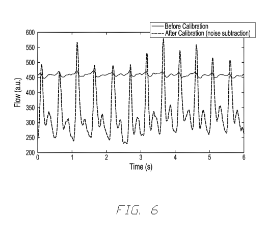
[0050] Figure 6 illustrates an example of data output from an
interrogation
device, such as illustrated in Figures 2A and 2B, and operated according to
the methods and
systems described herein. The measured waveforms correlate to the pulsatile
blood flow
originating from the cardiac cycle. The pulsatility reflects the changes in
the volumetric flow
rate as the subject's heart pumps blood through the interrogated vasculature.
The periodicity
of the flow arises from the cardiac cycle and can be used to determine heart
rate by
determining the period between successive waveform features (such as systolic
contraction
peaks). The output from the photodetector 200 is shown before accounting for
noise and
non-flow elements and after non-flow elements are accounted for through
calibration. The
calibration in this example was performed utilizing previously recorded
contrast data on a
calibration sample subject to static flow and interrogated under incoherent
light conditions.
The noise measured during calibration was subtracted from the present
photodetector
measurements recorded over time. As shown in Figure 6, the calibration
effectively reduced
the measured non-pulsatile contrast elements, essentially amplifying the true
flow signal.
[0051] While the present invention has been described in terms of
particular
embodiments and applications, in both summarized and detailed forms, it is not
intended that
these descriptions in any way limit its scope to any such embodiments and
applications, and
it will be understood that many substitutions, changes and variations in the
described
embodiments, applications and details of the method and system illustrated
herein and of
-23-
CA 03018621 2018-09-20
WO 2017/184630 PCT/US2017/028178
their operation can be made by those skilled in the art without departing from
the spirit of
this invention.
References (incorporated herein by reference thereto)
/. M. D. Stern and D. L. Lappe, "Method of and apparatus for measurement of
blood
flow using coherent light," U54109647A (1978).
2. R. Pecora, Dynamic light scattering: applications of photon correlation
spectroscopy
(Springer Science & Business Media, 2013).
3. A. Taniji and M. lshikawa, "Apparatus for measuring blood flow,"
U55291886 (1994).
4. G. E. Nilsson and J. T. Tenland, "Method and apparatus for measuring
flow motions
in a fluid," U54476875A (1984).
5. J. D. Briers, "Laser Doppler, speckle and related techniques for blood
perfusion
mapping and imaging," Physiological measurement 22, R35 (2001).
6. J. D. Briers and S. Webster, "Laser speckle contrast analysis (LASCA): a
nonscanning,
full-field technique for monitoring capillary blood flow," BIOMEDO 1, 174-179
(1996).
7. D. A. Boas and A. K. Dunn, "Laser speckle contrast imaging in biomedical
optics,"
BIOMEDO 15, 011109-011109-011112 (2010).
8. P. Zakharov, A. Volker, A. Buck, B. Weber, and F. Scheffold,
"Quantitative modeling
of laser speckle imaging," Opt. Lett. 31, 3465-3467(2006).
9. S. E. Skipetrov, J. Peuser, R. Cerbino, P. Zakharov, B. Weber, and F.
Scheffold, "Noise
in laser speckle correlation and imaging techniques," Opt. Express 18, 14519-
14534
(2010).
-24-