Note: Descriptions are shown in the official language in which they were submitted.
METHODS OF TREATING AUTOIMMUNE DISEASE
TECHNICAL FIELD
This disclosure relates to the use of methyldopa in the prevention or
treatment of
autoimmune diseases, such as autoimmune diabetes.
BACKGROUND
Autoimmune disorders are diseases caused by the body producing an
inappropriate
immune response against its' own tissues, in which the immune system creates T
lymphocytes and autoantibodies that attack one's own cells, tissues, and/or
organs.
Researchers have identified 80-100 different autoimmune diseases and suspect
at least 40
additional diseases have an autoimmune basis.
Autoimmune disorders are classified into two types, organ-specific (directed
mainly at one organ) and non-organ-specific (widely spread throughout the
body).
Examples of organ-specific autoimmune disorders are insulin-dependent Type 1
diabetes,
which affects the pancreas, Celiac disease, which affects the lining of the
small intestine,
Hashimoto's thyroiditis and Graves' disease, which affect the thyroid gland,
pernicious
anemia, which affects the stomach, Addison's disease, which affects the
adrenal glands,
chronic active hepatitis, which affects the liver, and myasthenia gravis,
which affects the
muscles. Examples of non-organ-specific autoimmune disorders are rheumatoid
arthritis,
multiple sclerosis, and lupus.
One of the most prevalent organ-specific autoimmune diseases, Type 1 diabetes,
is
characterized by the production of autoantibodies that target the insulin-
secreting
pancreatic beta cells. The disease pathogenesis involves T cell infiltration
into the islets of
the pancreas, which subsequently destroys insulin producing beta cells, and
results in overt
symptoms of disease. In most cases, T cells can respond to an antigen only
when the
antigen is properly presented by an antigen-presenting cell expressing the
appropriate
major histocompatibility complex (MHC) molecule. Thus, T cell immune response
to an
antigen requires recognition by the T cell receptor of an antigen coupled to a
MHC
molecule, and this recognition requires the assembly of a tri-molecular
complex between
an antigen, a MHC molecule, and a T cell receptor.
1
Date Recue/Date Received 2023-07-07
CA 03018727 2018-09-21
WO 2017/165508
PCT/US2017/023571
Evidence strongly indicates that insulin/proinsulin is a key or primary auto-
antigen
in the development of type 1 diabetes in the NOD (non-obese diabetic) mouse
model.
Initial cloning of T cells from islets of NOD mice led to the discovery that
the native
insulin B chain amino acids 9-23 (B:9-23 insulin peptide) is the dominant
antigenic
peptide epitope presented by the class II MHC molecule I-A. Mice lacking the
native B:9-
23 sequence fail to develop diabetes and development of insulin autoantibodies
and
insulitis are markedly decreased. Restoring the native B:9-23 sequence with an
islet
transplant (but not bone marrow transplant) or peptide immunization, or a
native
proinsulin transgene, restores anti-insulin autoimmunity and generates CD4 T
cells that
cause diabetes.
The major genetic determinant of islet autoimmunity and diabetes in human and
animal models are genes within the major histocompatibility complex, and in
particular,
class II MHC alleles. The NOD mouse's unique sequence of IA (homologous to
human
DQ) and lack of expression of I-E (shared with many standard mouse strains)
are essential
for the development of diabetes.
Celiac disease is an autoimmune disorder of the small intestine that affects
between 1 in 100 and 1 in 300 people depending on the region of the world. The
disease
occurs in people of all ages and causes pain and discomfort in the digestive
tract, chronic
constipation and diarrhea, failure to thrive in children, anemia, and fatigue.
The disease is
caused by a reaction to gliadin, a prolamin (gluten protein) found in wheat,
and other
common grains such as barley and rye, in which the immune system cross-reacts
with the
small-bowel tissue, causing an inflammatory autoimmune reaction. The only
known
effective treatment is a lifelong gluten-free diet.
There exists a need in the art for safer and more effective methods for
treating or
slowing the progression or development of autoimmune disorders, such as
autoimmune
diabetes (type 1 diabetes, T1D) and Celiac disease (gluten sensitivity). This
disclosure
addresses these needs by providing molecules and formulations useful in the
treatment and
prevention of autoimmune diseases while achieving other advantages discussed
more fully
below.
SUMMARY
The present disclosure provides new uses of D-a-methyldopa to prevent or
reduce
the binding of T cell receptors to peptides presented by class II MHC
molecules, as well as
therapeutic uses of D-a-methyldopa and pharmaceutical compositions comprising
D-o-
2
CA 03018727 2018-09-21
WO 2017/165508
PCT/US2017/023571
methyldopa to prevent or slow the formation of autoimmune diabetes (type 1
diabetes;
T1D) and Celiac disease in an individual.
Many autoimmune disorders have strong associations with specific HLA alleles,
including T1D, which is the immune-mediated form of diabetes resulting from
the chronic
autoimmune destruction of pancreatic beta cells. Approximately 90% of all
individuals
with T1D have DQ8 and/or DQ2 alleles, with a predominance of DQ8 (DQA*0301,
DQB*0302) in 50-60% of all T1D patients. DQ8 and DQ2 alleles confer
significant
disease risk while another DQ allele, DQ6 (DQB*0602), provides dominant
protection
from diabetes development. DQ8 and DQ2 are also the predominant 1-ILA alleles
in Celiac
disease, present in about 99% of all Celiac disease patients. Thus,
individuals with DQ8
and/or DQ2 alleles who have not yet manifested clinical disease may be at risk
of
developing T1D and/or Celiac disease, and/or may be suspected of suffering
from T1D or
Celiac disease.
T1D is now a predictable disease with the measurement of islet autoantibodies
(insulin, glutamic acid decarboxylase, insulinoma associated antigen 2, and
zinc
transporter 8), but it cannot yet be prevented. Furthermore, T1D incidence is
increasing 3-
5% every year in industrialized countries with children less than five years
of age being
the most affected. Additionally, there is currently no known cure for T1D, and
treatment
for this disease consists of lifelong administration of insulin. Despite
treatment with
insulin therapy, long-term complications, including nephropathy, retinopathy,
neuropathy,
and cardiovascular disease can result. While the progress to complete insulin
dependence
occurs quickly after clinical onset, initially after diagnosis the pancreas is
still able to
produce a significant amount of insulin. The Diabetes Control and
Complications Trial
(DCCT) found that 20% of patients studied who were within 5 years of
diagnosis, had
remaining insulin production (0.2-0.5 pmol/ml). Thus, immunologic intervention
during
the window following diagnosis could save beta cell function, delay the onset
of T1D, and
reduce reliance on insulin administration.
Class II major histocompatability molecules are the primary susceptibility
locus for
many autoimmune diseases, including type 1 diabetes. "Diabetogenic" alleles
HLA-DQ8
in humans and I-A in non-obese diabetic (NOD) mice confer disease risk, and
both
molecules share structural similarities. The present inventors have evaluated
a novel
pathway to identify safe and specific therapies to treat the underlying T cell
autoimmunity
in T1D. This pathway involves blocking allele-specific MEC class II antigen
presentation
as a treatment to inhibit DQ8-mediated T cell responses. DQ8 confers
significant disease
3
CA 03018727 2018-09-21
WO 2017/165508
PCT/US2017/023571
risk by presenting epitopes of insulin and other beta cell antigens to
effector CD4 T cells.
The present inventors have surprisingly found that D-a-methyldopa blocks or
reduces
insulin and gliadin peptide presentation to T cells. Without intending to be
bound by
theory, it is believed that D-a-methyldopa occupies a pocket along the DQ8
(DQA*03:01,
DQB*03:02) peptide binding groove, thereby blocking DQ8 restricted T cell
responses in
vitro, and inhibiting DQ8 antigen presentation in vivo. Blocking HLA-DQ8
antigen
presentation in this way may help preserve beta cell mass (and endogenous
insulin
production) in new onset T1D and may also prevent or delay T1D onset in
multiple islet
autoantibody-positive individuals (i.e., 2 or more islet autoantibodies), 70-
90% of whom
develop diabetes within 10 years.
Thus, the present disclosure provides methods of reducing the binding of T
cell
receptors to insulin/proinsulin peptides presented by class II MHC molecules,
to treat or
slow the progression or development of T1D or Celiac disease in an individual
suffering
from, or at risk of developing, T1D or Celiac disease, comprising
administering D-a-
methyldopa to such individuals. The present disclosure also provides
pharmaceutical
compositions containing D-a-methyldopa that are particularly useful in such
methods of
treating or slowing the progression or development of T1D.
One aspect of this disclosure is a method of inhibiting an autoimmune disease
by
administering to an individual in need of such treatment, a therapeutically
effective
amount of D-a-methyldopa that inhibits the T cell response to the targeted
antigenic
peptide of the autoimmune disease. The D-a-methyldopa inhibits the binding of
a DQ8
peptide to an MHC class II molecule for presentation to CD4+ T cells, thereby
slowing the
development or progression of T1D or Celiac disease. The inhibition of the
binding of a
DQ8 peptide to an MEC class II molecule may result from a distortion of the
spatial
orientation of the complex so that the DQ8 antigen is not properly presented
to T cells.
A related aspect provides a method of selectively treating T1D in an
individual,
including selecting an individual for treatment with D-a-methyldopa on the
basis of the
individual having at least two islet autoantibodies detectable in a blood
sample from the
individual, and selectively administering D-a-methyldopa to that individual.
In any of the methods of this disclosure, the individual may have been tested
for
the presence of autoantibodies to insulin or proteins within beta cells,
wherein the
presence of such antibodies in the individual is indicative of the presence or
likely
development of T1D. Thus, a related aspect of this disclosure provides methods
of treating
4
CA 03018727 2018-09-21
WO 2017/165508
PCT/US2017/023571
an individual found to have such autoantibodies by administering D-a-
methyldopa to the
individual.
Another aspect of this disclosure provides methods of monitoring and adjusting
the
dosage of D-a-methyldopa administered to an individual suffering from, or at
risk of
developing, an autoimmune disorder (such as T1D or Celiac disease) including
receiving a
blood sample from an individual suffering from, or at risk of developing, the
autoimmune
disorder who has been administered a-methyldopa and determining the DQ8-
stimulated
response of IL-2 T cells in the blood sample. The DQ8-stimulated response of
1L-2 T cells
in the blood sample is compared to a control level of DQ8-stimulated response
of IL-2 T
cells in blood samples from at least one of a patient suffering from the
autoimmune
disease and a control or 'wild type' individual known to be free of the
autoimmune
disease. The dosage and/or the frequency of the D-a-methyldopa administered to
the
individual is increased if the DQ8-stimulated response of IL-2 T cells in the
blood sample
from the individual is statistically similar to the DQ8-stimulated response of
IL-2 T cells
from the baseline level in the T1D patient.
Other aspects of the invention will be set forth in the accompanying
description of
embodiments, which follows and will be apparent from the description. However,
it
should be understood that the following description of embodiments is given by
way of
illustration only, as various changes and modifications within the spirit and
scope of the
invention will become apparent to those skilled in the art and are encompassed
within the
scope of this disclosure.
BRIEF DESCRIPTION OF DRAWINGS
Fig. 1A shows the DQ8-, and Fig. 1B shows the DR4-T cell responses, of T1D
patients treated with varying doses of methyldopa, shown side-by-side for
comparison.
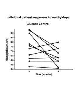
Fig. 2 shows the glucose control achieved in human T1D patients treated with
methyldopa over 3 months.
Fig. 3 shows the beta-cell function in human T1D patients treated with
methyldopa
over 3 months.
Fig. 4A shows dose-dependent inhibition of a gliadin-responsive CD4 T cell
receptor (TCR) transductant by enantiomers of a-methyldopa. Fig. 4B shows a
similar
inhibition of an insulin responsive CD4 T cell receptor (TCR) transductant by
the a-
methyldopa enantiomers.
5
CA 03018727 2018-09-21
WO 2017/165508
PCT/US2017/023571
Fig. 5A shows an in vivo pharmacokinetic assessment of oral L- and D-a-
methyldopa doses in HLA-DQ8 transgenic mice, and Fig. 5B shows a
phamiacokinetic
comparison of an oral L- and D-a-methyldopa dose in HLA-DQ8 transgenic mice.
Fig. 6 shows an in vivo pharmacodynamic assessment of identical once-daily
dosing of L- and D-a-methyldopa in HLA-DQ8 transgenic mice.
DESCRIPTION OF EMBODIMENTS
The present disclosure is drawn to methods of treating or slowing the
progression
or development of an autoimmune disease by reducing the binding, or altering
the
presentation of, antigenic peptides, or fragments of antigenic peptides,
presented by an
MEC class II molecule (DQ8) by the administration of D-a-methyldopa to an
individual
suffering from, or at risk of developing, an autoimmune disease.
The term "insulin peptide" is used to denote a peptide fragment of an insulin
protein. Although the fragment is typically a subset of the amino acid
sequence of the
insulin protein, an insulin peptide may contain the entire amino acid sequence
of a
naturally-occurring insulin protein.
The terms "individual" or "subject" are used interchangeably herein. The terms
"individual" and "individuals" refer to an animal, such as a mammal, including
a non-
primate (e.g., a cow, pig, horse, cat, dog, rat, and mouse) and a primate
(e.g., a monkey
such as a cynomolgous monkey, a chimpanzee and a human), and a human. In
certain
embodiments, the subject is refractory or non-responsive to current treatments
for an
autoimmune disease.
"Tissue" means any biological sample taken from any individual, preferably a
human. Tissues include blood, saliva, urine, biopsy samples, skin or buccal
scrapings, and
hair.
Persons of skill in the art will appreciate that blood plasma drug
concentrations
obtained in individual subjects will vary due to inter-patient variability in
the many
parameters affecting drug absorption, distribution, metabolism and excretion.
For this
reason, unless otherwise indicated, when a drug plasma concentration is
listed, the value
listed is the calculated mean value based on values obtained from a group of
subjects
tested.
The term "bioavailability" refers to the extent to which, and sometimes the
rate at
which, the active moiety (drug or metabolite) enters systemic circulation,
thereby gaining
access to the site of action.
6
CA 03018727 2018-09-21
WO 2017/165508
PCT/US2017/023571
"AUC" is the area under the plasma concentration-time curve and is considered
to
be the most reliable measure of bioavailability. It is directly proportional
to the total
amount of unchanged drug that reaches the systemic circulation.
The phrase "pharmaceutically acceptable" is employed herein to refer to those
compounds, materials, compositions, and/or dosage forms which are, within the
scope of
sound medical judgment, suitable for use in contact with the tissues of human
beings and
animals without excessive toxicity, irritation, allergic response, or other
problem or
complication commensurate with a reasonable benefit/risk ratio.
"Pharmaceutically-acceptable salts" refer to derivatives of the disclosed
compounds wherein the parent compound is modified by making acid or base salts
thereof. Examples of pharmaceutically acceptable salts include, but are not
limited to,
mineral or organic acid salts of basic residues such as amines, or alkali or
organic salts of
acidic residues such as carboxylic acids. Pharmaceutically-acceptable salts
include the
conventional non-toxic salts or the quaternary ammonium salts of the parent
compound
formed, for example, from non-toxic inorganic or organic acids. Such
conventional
nontoxic salts include those derived from inorganic acids such as
hydrochloric,
hydrobromic, sulfuric, sulfamic, phosphoric, nitric and the like; and the
salts prepared
from organic acids such as acetic, propionic, succinic, glycolic, stearic,
lactic, malic,
tartaric, citric, ascorbic, pamoic, maleic, hydroxymaleic, phenylacetic,
glutamic, benzoic,
salicylic, sulfanilic, 2-acetoxybenzoic, fumaric, toluenesulfonic,
methanesulfonic, ethane
disulfonic, oxalic, isethionic, and the like. Phallnaceutically acceptable
salts are those
forms of compounds, suitable for use in contact with the tissues of human
beings and
animals without excessive toxicity, irritation, allergic response, or other
problem or
complication, commensurate with a reasonable benefit/risk ratio.
The term "therapeutically-effective amount" of D-a-methyldopa means an amount
effective to modulate the formation or progression of autoimmune diseases
(including
T1D and Celiac disease) in an individual.
Most amino acids are chiral (designated as 'L' or 13' wherein the `L'
enantiomer
is the naturally occurring configuration) and can exist as separate
enantiomers. The USP
standard methyldopa for antihypertensive therapy is the `1_,' enantiomer of a-
methyldopa:
L-a-Methyl-3,4-dihydroxyphenylalanine (hereinafter "L-a-methyldopa" available
commercially under the tradenames ALDOMETTm, ALDORILTM, DOPAMETTm,
DOPEGYTTm), and has the chemical structure:
7
CA 03018727 2018-09-21
WO 2017/165508
PCT/US2017/023571
0
H3c
HO
OH
HO NH2
L-a-methyldopa is an alpha-adrenergic agonist (selective for a2-adrenergic
receptors) that
was developed as a psychoactive drug and has been used extensively as a
sympatholytic or
antihypertensive.
The 'Cc enantiomer of a-methyldopa (D-a-Methyl-3,4-dihydroxyphenylalanine;
hereinafter "D-a-methyldopa" is also referred to in the literature as "3-
Hydroxy-a-methyl-
D-tyrosine"; "D-3-(3,4-Dihydroxypheny1)-2-methylalanine"; "( )-a-Methyldopa";
"D-
(3,4-Dihydroxypheny1)-2-methylalanine"; "D-Methyldopa"), has the chemical
structure:
HO CO2H
si I-12K/ Me
HO
Prior to the present disclosure, D-a-methyldopa was considered to have no
pharmacologic
activity (see, for example, U.S. Patent Publication No. 2011/0245334, filed
Dec 10, 2009,
to Du et al.). Gillespie, etal. (1962, Clinical and Chemical Studies with a-
Methyl-Dopa in
Patients with Hypertension, Circulation 25:281-291) were the first to describe
the lack of
phaunacological activity in the `130' isomer and suggest using only the `L'
enantiomeric
form for hypertension. Similarly, Sjoerdsma, et al., (1963, Studies on the
Metabolism and
Mechanism of Action of Methyldopa, Circulation, 28:492-502) showed that
patients treated
with the D enantiomer did not decarboxylate the D enantiomer to a-methyl-
dopamine,
whereas a-methyl-dopamine did appear in the urine of the same patients treated
with the L
enantiomer, indicating metabolism of this L enantiomer. Au et al. (1972, The
Metabolism
of 14C-Labelled a-Methyldopa in Normal and Hypertensive Human Subjects,
Biochem. J.,
129:1-10) more extensively described the marked differences in metabolism of
the two
enantiomers in humans, and found that the D isomer was "much less readily
absorbed than
the active L isomer." These authors also cite other supporting literature
references,
including a suggestion that there is an optically specific, active transport
mechanism that
may be responsible for this difference in adsorption. Additionally, Renwick et
al. (1983,
The Absorption and Conjugation of Methyldopa in Patients with Coeliac and
('rohn's
Diseases During Treatment, Br. J. Clin. Pharmac., 16:77-83) showed that
absorption rates
8
CA 03018727 2018-09-21
WO 2017/165508
PCT/US2017/023571
of methyldopa are different between normal individuals and those with certain
GI diseases
such as Celiac and Crohn's disease.
D-a-methyldopa may be purchased commercially (see, for example, Toronto
Research Chemicals, catalogue M303790). D-a-methyldopa may also be prepared in
ways
well known to one skilled in the art of organic synthesis, including, for
example, by
resolution of the racemic form by recrystallization techniques, by synthesis
from optically-
active starting materials, by chiral synthesis, or by chromatographic
separation using a
chiral stationary phase. The resolution of methyldopa, may be carried out by
known
procedures, e.g., as described in the four volume compendium Optical
Resolution
Procedures for Chemical Compounds: Optical Resolution Information Center,
Manhattan
College, Riverdale, N.Y., and in Enantiomers, Racemates and Resolutions, Jean
Jacques,
Andre Collet and Samuel H. Wilen; John Wiley & Sons, Inc., New York, 1981.
Additionally, several methods of effectively and efficiently testing the
enantiomeric purity
of a-methyldopa are known, as described in Martens, J., et al., Resolution of
Optical
Isomers by Thin-Layer Chromatography: Enantiomeric Purity qfMethyldopa, Arch.
Pharm. (Weinheim) 319:572-74 (1986); and Gelber, L.R., Neumeyer, J.L.,
Determination
of the Enantiomeric Purity of Levodopa, Methyldopa, Carbidopa and Tryptophan
by the
Use of Chiral Mobile Phase High-Performance Liquid Chromatography, J.
Chromatography, 257:317-26 (1983).
The invention is based on the inventors' surprising discovery that, contrary
to the
accepted understanding that L-a-methyldopa comprises all of the pharmacologic
activity
of a-methyldopa, D-a-methyldopa actually possesses nearly equivalent activity
in
reducing the presentation of antigenic peptides, or fragments of antigenic
peptides, by
MEC class II molecules in autoimmune diseases, such as T1D or Celiac disease.
This
unexpected discovery is particularly fortuitous as L-a-methyldopa, which is a
competitive
inhibitor of the enzyme DOPA decarboxylase (also known as aromatic L-amino
acid
decarboxylase), is associated with many adverse side effects, including
depression and
even suicidal ideation, as well as nightmares, anhedonia, dysphoria, anxiety,
decreased
desire, drive, and motivation, lethargy, malaise, sedation or drowsiness,
cognitive and
memory impairment, sexual dysfunction including impaired libido, dizziness,
lightheadedness, or vertigo, xerostomia, headache, migraine, myalgia,
arthralgia,
paresthesia, parkinsonian symptoms such as muscle tremors, rigidity,
hypokinesia, and/or
balance or postural instability, hypotension, hepatotoxi city, pancreatitis,
haemolytic
anaemia, and myelotoxicity. These effects are believed, at least in part, to
be due to
9
CA 03018727 2018-09-21
WO 2017/165508
PCT/US2017/023571
metabolites of L-a-methyldopa. As D-a-methyldopa presumably is not
metabolized, D-a-
methyldopa is less likely to cause such adverse effects. This distinction also
improves the
window of therapeutic dosing of D-a-methyldopa compared to L-a-methyldopa.
D-a-methyldopa preparations, for use in the methods and compositions of this
disclosure, are substantially free of the L-enantiomer of a-methyldopa. In the
methods and
compositions of this disclosure, the D-a-methyldopa compounds are at least
85%, 90%,
95%, 98%, 99% to 100% by weight of the D-enantiomer, the remainder comprising
other
chemical species or enantiomers. Thus, these D-a-methyldopa compositions
contain less
than 10%, less than 1%, or preferably less than 0.1%, by weight, of the L-
enantiomer of a-
methyldopa.
D-a-methyldopa can be formulated into pharmaceutical compositions using
methods available in the art and provided in the appropriate pharmaceutical
composition
and administered by a suitable route of administration. The methods provided
herein
encompass administering pharmaceutical compositions containing D-a-methyldopa,
if
appropriate in the salt form, either used alone or in the form of a
combination with one or
more compatible and pharmaceutically acceptable carriers, such as diluents or
adjuvants,
or with another therapeutic agent. The second therapeutic agent can be
formulated or
packaged with the D-a-methyldopa. Of course, the second agent will only be
formulated
with the D-a-methyldopa, according to the judgment of those of skill in the
art, as such co-
formulation should not interfere with the activity of either agent or the
method of
administration. The D-a-methyldopa and the second agent may be foiniulated
separately.
They may also be packaged together, or packaged separately, for the
convenience of the
medical practitioner. These additional agent(s) may include an anti-diabetic
compound
selected from at least one of an alpha-glucosidase inhibitor, a biguanide, a
Dpp-4 inhibitor,
a meglitinide, a sulfonylurea, a thiazolidinedione, or combinations of these
agents.
In clinical practice the D-a-methyldopa may be administered by any
conventional
route, in particular orally, or parenterally. In preferred embodiments, the D-
a-methyldopa
is administered orally, as solid or liquid compositions for oral
administration, for example,
as tablets, pills, hard gelatin capsules, powders or granules.
A composition provided herein is a pharmaceutical composition or a single unit
dosage form. Pharmaceutical compositions and single unit dosage forms provided
herein
comprise a prophylactically or therapeutically effective amount of D-a-
methyldopa and
typically one or more pharmaceutically acceptable carriers or excipients. In
this context,
the term "pharmaceutically acceptable" means approved by a regulatory agency
of the
CA 03018727 2018-09-21
WO 2017/165508
PCT/US2017/023571
Federal or a state government or listed in the U.S. Pharmacopeia or other
generally
recognized pharmacopeia for use in animals, and more particularly in humans.
The term
"carrier" includes a diluent, adjuvant (e.g., Freund's adjuvant (complete and
incomplete)),
excipient, or vehicle with which the therapeutic is administered. Examples of
suitable
pharmaceutical carriers are described in "Remington's Pharmaceutical Sciences"
by E. W.
Martin.
A preferred formulation of this disclosure is a mono-phasic pharmaceutical
composition suitable for oral administration for the treatment, prophylaxis,
or slowing the
progression of autoimmune diabetes, consisting essentially of a
therapeutically-effective
amount of D-a-methyldopa, and a pharmaceutically acceptable carrier.
Methods of Use
Provided herein are methods for the treatment and/or prophylaxis of an
autoimmune disease in an individual. These methods include the treatment of an
individual suffering from an autoimmune disease, such as T1D or Celiac
disease, by the
administration of an effective amount of D-a-methyldopa. These methods may
encompass
the step of administering to the individual in need of such treatment an
amount of D-a-
methyldopa effective for treating or delaying the development of an autoimmune
disease,
such as T1D or Celiac disease. The D-a-methyldopa may be in the form of a
pharmaceutical composition or single unit dosage form, as described above.
This disclosure includes methods of treating or slowing the progression or
development of autoimmune diabetes or Celiac disease by reducing the binding
of MHC
class II molecules to antigenic peptides or fragments of antigenic peptides of
the
autoimmune disease by the administration of D-ct-methyldopa to individuals
suffering
from, or at risk of developing, autoimmune diabetes (T1D) or Celiac disease.
A specific method includes treating T1D in an individual comprising
administering
an effective amount of D-a-methyldopa to an individual in need of such
treatment.
Another method includes treating Celiac disease in an individual comprising
administering
an effective amount of D-a-methyldopa to an individual in need of such
treatment.
Another method includes treating an individual at risk of developing T1D
comprising administering an effective amount of D-a-methyldopa to the
individual.
Another method includes treating an individual at risk of developing Celiac
disease
comprising administering an effective amount of D-a-methyldopa to the
individual. Thus,
this disclosure provides for the use of D-a-methyldopa in the manufacture of a
11
CA 03018727 2018-09-21
WO 2017/165508
PCT/US2017/023571
medicament for the treatment of T1D and the use of D-a-methyldopa in the
manufacture
of a medicament for the treatment of Celiac disease. This disclosure also
provides D-a-
methyldopa for use in the treatment of T1D, and D-a-methyldopa for use in the
treatment
of Celiac disease.
In these methods, the D-a-methyldopa may be administered orally to the
individual. The D-a-methyldopa may be administered to an individual once
daily, or more
frequently. The D-a-methyldopa may be administered to an individual as a
pharmaceutically acceptable salt, solvate or hydrate thereof. The D-a-
methyldopa, or a
pharmaceutically acceptable salt thereof, may be administered to an individual
as a
pharmaceutical composition described above.
In the methods of treating or slowing the progression or development of T1D of
this disclosure, D-a-methyldopa or a pharmaceutically acceptable salt thereof,
is
administered to an individual suspected of suffering from, or at risk of
developing T1D.
Preferably, the administration to an individual diagnosed with T1D commences
within 5
years of the initial diagnosis of T1D in the individual, or more preferably,
within 1 year of
the initial diagnosis of T1D in the individual, or more preferably, within 6
months of the
initial diagnosis of T1D in the individual, or more preferably, within 1 month
of the initial
diagnosis of T1D in the individual.
In any of these methods, the individual may be administered a dosage of D-a-
methyldopa, or a pharmaceutically acceptable salt, solvate or hydrate thereof,
between
about 50 mg and about 3000 mg of D-a-methyldopa per day. Preferably, the
individual is
administered a dosage of D-a-methyldopa between about 50mg and about 1000 mg
of D-
a-methyldopa per day. The individual may be administered a dosage of D-a-
methyldopa
between about 250 mg and about 500 mg in an oral, immediate release dosage
formulation
at least twice daily. Typically, the D-a-methyldopa is administered in dosages
ranging
between 50 mg and 3000 mg per day. In most instances, the D-a-methyldopa may
be
initially administered at a dosage of 50 mg to 500 mg once, twice, or three
times daily.
These doses may be increased in patients with Crohn's or Celiac disease.
In any of these methods, in addition to D-a-methyldopa, the individual may
also be
administered an anti-diabetic compound, including at least one of an alpha-
glucosidase
inhibitor, a biguanide, a Dpp-4 inhibitor, a meglitinide, a sulfonylurea, a
thiazolidinedione,
or combinations of these agents.
12
CA 03018727 2018-09-21
WO 2017/165508
PCT/US2017/023571
In any of these methods, the individual may be between 1 year and 15 years of
age.
Alternatively, in these methods, the individual may be between 15 years and 30
years of
age. Alternatively, in these methods, the individual may be older than 30
years of age.
Another aspect of these methods includes an initial determination of which
patients
may benefit from the administration of D-a-methyldopa (and, optionally, an
anti-diabetic
compound, as described above), prior to administration of D-a-methyldopa to
individual
determined to be in need of such treatment. As described above, DQ8 and DQ2
alleles
confer significant risk of T1D and Celiac disease, while DQ6 allele provides
dominant
protection from diabetes development. Thus, individuals with DQ8 and/or DQ2
alleles are
individuals at risk of developing T1D and/or Celiac disease. Additionally,
individuals with
islet autoantibodies (i.e., antibodies that specifically recognize insulin,
glutamic acid
decarboxylase, insulinoma associated antigen 2, or zinc transporter 8), or
autoantibodies
that recognize a MHC class II molecule bound to an insulin protein or to a
gliadin peptide,
or to insulin or gliadin peptide fragment(s), wherein the presence of such
antibodies in the
individual is indicative of the presence or likely development of T1D and/or
Celiac disease
in that individual. Such individual therefore may benefit from the methods of
administering D-a-methyldopa, as described above.
The determination of which DQ8 or DQ2 alleles are present in an individual may
enable a clinician to establish the subject's risk of developing an autoimmune
disease.
For example, if the genotyping methods of this disclosure reveal a homozygous
DQA*0301 or DQB*0302 genotype (two copies of one or both of these alleles) in
the
nucleic acid sample obtained from the individual, this finding indicates that
the individual
is more likely to develop T1D or Celiac disease. The clinician may then
consider it
beneficial to administer D-ct-methyldopa in accordance with the methods of
this
disclosure. Additionally, an individual found to be heterozygous DQA*0301 or
DQB*0302 genotype may still benefit from the administration of D-a-methyldopa.
In this
instance, the finding of a heterozygote genotype in the individual may be used
to modify
the dosage and/or dosing regimen of D-a-methyldopa to the individual, which
may include
reducing the dose or frequency of administration of D-a-methyldopa to the
individual.
Alternatively, if the genotyping methods of this disclosure reveal homozygous
wild type HLA alleles or protective alleles, such as DQB*0602, in the nucleic
acid sample
obtained from the individual, then the clinician may rule out an elevated risk
of developing
T1D or Celiac disease in the individual and consider different treatments or
diagnoses for
that individual.
13
A number of methods are available for analyzing and determining the DQ8 and/or
DQ2 genotype in a subject, which can be applied to a nucleic acid sample
obtained from a
subject. Assays for detection of polymorphisms or mutations fall into several
categories,
including but not limited to, direct sequencing assays, fragment polymorphism
assays,
hybridization assays, and computer based data analysis. Protocols and kits or
services for
performing these general methods are commercially available and well known to
those of
skill in the art. In some embodiments, assays are performed in combination or
in hybrid
(e.g., different reagents or technologies from several assays are combined to
yield one
assay). Thus, the presence or absence of DQ8 and/or DQ2 alleles may be
determined using
direct sequencing. Alternatively or additionally, the DQ8 and/or DQ2 alleles
may be
determined using a PCR-based assay using oligonucleotide primers to amplify a
DNA
fragment containing the DQ8 and/or DQ2 polymorphism of interest. Alternatively
or
additionally, the DQ8 and/or DQ2 alleles may be determined using a fragment
length
polymorphism assay, such as a restriction fragment length polymorphism assay
(RPLP), to
detect a unique DNA banding pattern indicative of an DQ8 and/or DQ2 genotype
based on
cleaving the DNA at a series of positions is generated using an enzyme (e.g.,
a restriction
endonuclease). Alternatively or additionally, the DQ8 and/or DQ2 alleles may
be
determined using a hybridization assay, wherein the genotype is determined
based on the
ability of the DNA from the sample to hybridize to a complementary DNA
molecule (e.g.,
.. an oligonucleotide probe). The DQ8 and/or DQ2 polymorphisms of interest may
be
detected using a DNA chip hybridization assay, in which a series of
oligonucleotide
probes, designed to be unique to a given single nucleotide polymorphism, are
affixed to a
solid support, and the nucleic acid sample from the subject is contacted with
the DNA
"chip" and hybridization is detected. Alternatively or additionally, the DQ8
and/or DQ2
alleles may be determined using a "bead array" (such as those described in PCT
Publications W099/67641 and W000/39587 ).
Alternatively or additionally, the DQ8 and/or DQ2 alleles may be determined
using an assay that detects hybridization by enzymatic cleavage of specific
structures
(such as those assays described in U.S. Pat, No. 6,001,567, and Olivier, M.,
The Invader
assay for SNP Genotyping, 2005 Mutat. Res. June 3; 573(1-2):103-110).
Genomic DNA samples are usually, but need not be, amplified before being
analyzed. Genomic DNA can be obtained from any biological sample.
Amplification of
genomic DNA potentially containing an DQ8 and/or DQ2 polymorphism generates a
14
Date Recue/Date Received 2023-07-07
single species of nucleic acid if the individual from whom the sample was
obtained is
homozygous at the polymorphic site, or two species of nucleic acid if the
individual is
heterozygous. RNA samples may also be subjected to amplification. In this
case,
amplification is typically, but not necessarily, proceeded by reverse
transcription.
Amplification of all expressed mRNA may also be performed (such as described
in Innis
M A et al., 1990, Academic Press, PCR Protocols: A Guide to Methods and
Applications
and Bustin SA 2000. Journal of Molecular Endocrinology, 25 Absolute
quantification of
mRNA using real-time reverse transcription polymerase chain reaction assays.
pp. 169-
193).
Any known method of analyzing a sample for an analyte can be used to practice
the present invention, so long as the method detects the presence, absence, or
amount of
anti-islet antibodies. Examples of such methods include, but are not limited
to,
immunological detection assays and non-immunological methods (e.g., enzymatic
detection assays). Additionally or alternatively, an binding compound is
immobilized on a
.. substrate, such as a microtiter dish well, a dipstick, an immunodot strip,
or a lateral flow
apparatus. A sample collected from a subject is applied to the substrate and
incubated
under conditions suitable (i.e., sufficient) to allow the formation of a
complex between the
binding compound and any anti-islet antibody present in the sample. Once
formed, the
complex is then detected. As used herein, the term "detecting complex
formation" refers to
identifying the presence of a binding compound complexed to an anti-islet
antibody. If
complexes are formed, the amount of complexes formed can, but need not be,
quantified.
Complex formation, or selective binding, between an anti-islet antibody and a
binding
compound can be measured (i.e., detected, determined) using a variety of
methods
standard in the art including, but not limited to, an enzyme-linked
immunoassay, a
competitive enzyme-linked immunoassay, a radioimmunoassay, a fluorescence
immunoassay, a chemiluminescent assay, a lateral flow assay, a flow-through
assay, an
agglutination assay, a particulate-based assay (e.g., using particulates such
as, but not
limited to, magnetic particles or plastic polymers, such as latex or
polystyrene beads), an
immunoprecipitation assay, a BIACORETm assay (e.g., using colloidal gold), an
.. immunodot assay (e.g., CMG's Immunodot System, Fribourg, Switzerland), and
an
immunoblot assay (e.g., a western blot), an phosphorescence assay, a flow-
through assay,
a chromatography assay, a PAGE-based assay, a surface plasmon resonance assay,
a
spectrophotometric assay, a particulate-based assay, and an electronic sensory
assay. The
assays may be used to give qualitative or quantitative results. The assay
results can be
Date Recue/Date Received 2023-07-07
CA 03018727 2018-09-21
WO 2017/165508
PCT/US2017/023571
based on detecting the entire antibody or fragments, or degradation products.
Some assays,
such as agglutination, particulate separation, and immunoprecipitation, can be
observed
visually (e.g., either by eye or by a machine, such as a densitometer or
spectrophotometer)
without the need for a detectable marker. A detectable marker can be
conjugated to the
compound or reagent at a site that does not interfere with the ability of the
compound to
bind anti-islet antibodies. Methods of conjugation are known to those of skill
in the art.
Examples of detectable markers include, but are not limited to, a radioactive
label, a
fluorescent label, a chemiluminescent label, a chromophoric label, an enzyme
label, a
phosphorescent label, an electronic label, a metal sol label, a colored bead,
a physical
label, or a ligand. A ligand refers to a molecule that binds selectively to
another molecule.
Preferred detectable markers include, but are not limited to, fluorescein, a
radioisotope, a
phosphatase (e.g., alkaline phosphatase), biotin, avidin, a peroxidase (e.g.,
horseradish
peroxidase), beta-galactosidase, and biotin-related compounds or avidin-
related
compounds (e.g., streptavidin or IMMUNOPURETm NeutrAvidin). Means of detecting
such markers are well known to those of skill in the art.
A tri-molecular complex between an insulin or gliadin antigen, a MHC molecule,
and T cell receptor can be detected by contacting a biological sample from an
individual
with an antibody specific for the complex, wherein the antibody is conjugated
to a
detectable marker. A detectable marker can also be conjugated to a tri-
molecular complex
between an insulin or gliadin antigen, a MHC molecule, and T cell receptor
such that
contact of the labeled complex with a biological sample from an individual can
detect the
presence of antibodies to the complex present in the individual tested.
Detectable markers
include, but are not limited to, fluorescein, a radioisotope, a phosphatase
(e.g., alkaline
phosphatase), biotin, avidin, a peroxidase (e.g., horseradish peroxidase),
beta-
galactosidase, and biotin-related compounds or avidin-related compounds (e.g.,
streptavidin or IMMUNOPURETm NeutrAvidin).
A tri-molecular complex may be detected by contacting the complex with an
indicator molecule. Suitable indicator molecules include molecules that can
bind to the tri-
molecular binding molecule complex. As such, an indicator molecule can
comprise, for
example, an antibody. Preferred indicator molecules that are antibodies
include, for
example, antibodies reactive with the antibodies from animals in which the
anti-islet
antibodies are produced. An indicator molecule itself can be attached to a
detectable
marker of the present invention. For example, an antibody can be conjugated to
biotin,
horseradish peroxidase, alkaline phosphatase or fluorescein. One or more
layers and/or
16
types of secondary molecules or other binding molecules capable of detecting
the presence
of an indicator molecule may be used. For example, an untagged (i.e., not
conjugated to a
detectable marker) secondary antibody that selectively binds to an indicator
molecule can
be bound to a tagged (i.e., conjugated to a detectable marker) tertiary
antibody that
selectively binds to the secondary antibody. Suitable secondary antibodies,
tertiary
antibodies and other secondary or tertiary molecules can be readily selected
by those
skilled in the art. Preferred tertiary molecules can also be selected by those
skilled in the
art based upon the characteristics of the secondary molecule. The same
strategy can be
applied for subsequent layers.
A lateral flow assay may be used for detection, examples of which are
described in
U.S. Patent No. 5,424,193; U.S. Pat. No. 5,415,994; WO 94/29696; and WO
94/01775.
Once a biological sample from an individual has been analyzed to determine
which
allele of an DQ8 and/or DQ2 polymorphism is present, the individual can be
selected, or
identified, as likely or unlikely to develop, or at higher or lower risk of
developing, T1D or
Celiac disease. Such a selection is made using the results from the analysis
step of the
disclosed method.
EXAMPLES
Example 1 Human T1D treatment study
Human leukocyte antigen (HLA) alleles confer significant genetic risk for type
1
diabetes (T1D) with recent studies implicating DQ8 in its pathogenesis, and
DQ8 antigen
presentation can be inhibited with methyldopa in animal models. In this pilot
study, the
inventors evaluated methyldopa treatment in 10 DQ8 positive human individuals
with
T1D, ages 18-46 years (mean 27) with less than 2 years of diabetes duration
(mean 3
months). This was an open label phase lb dose escalation study. All
individuals tolerated
low (500mg BID) and moderate (500mg l'Ill) dosages of methyldopa, while 9/10
tolerated
the high dose (2-3g/day).
There was a dose-dependent reduction in DQ8-stimulated IL-2 T cell response at
1
(-32%) and 3 weeks (-39%), which returned to normal 6 weeks after stopping
therapy
(Fig. 1A). This response was specific for the DQ8-stimulated T cell response
because DR4
17
Date Recue/Date Received 2023-07-07
CA 03018727 2018-09-21
WO 2017/165508
PCT/US2017/023571
T cell responses were minimally effected (Fig. 1B). The treated T1D patients
had good
glycemic control (Fig. 2). Additionally, the 2 hour AUC for C-peptide
following a mixed
meal tolerance test at 12 weeks was similar to baseline levels (Fig. 3). No
serious adverse
events (hypotension, DKA, or hypoglycemia) were reported throughout the study.
This
example demonstrates that methyldopa inhibits DQ8 antigen presentation in T1D.
Example 2 Enantiomers of a-methyldopa similarly inhibit D08 restricted T
responses in
vitro
Inhibition of DQ8 antigen presentation by the D- and L-enantiomers of a-
methyldopa was tested and compared in vitro. HLA-DQ8 antigen presenting cells
were
cultured with a fixed concentration of peptide and a concentration of D- or L-
a-
methyldopa for 4 hours. TCR transductants were then added to each culture
condition and
cultured overnight. Secreted IL-2 from the TCR transductant was measured by
ELISA for
each condition.
As shown in Fig. 4A, a gliadin-responsive CD4 T cell receptor (TCR)
transductant
was blocked in a dose-dependent manner with statistically similar IC50 values
between the
a-methyldopa enantiomers. Similarly, as shown in Fig. 4B an insulin responsive
CD4 T
cell receptor (TCR) transductant was blocked in a dose-dependent manner with
statistically similar IC50 values between the a-methyldopa enantiomers. In
these figures, 0
represents the TCR transductant response to peptide without the addition of a-
methyldopa.
No antigen IL-2 responses (negative control) were very low at < 2 pg/ml.
Results are
presented as mean +/- SEM. These data demonstrate that the dose-dependent
inhibitory
effects of the D- and L-enantiomers of a-methyldopa on DQ8 antigen
presentation in in
vitro models of Celiac disease and T1D, are nearly identical.
Example 3 Pharmacokinetic and pharmacodynamic assessment of a-methyldopa
enantiomers in mice
Separate cohorts of HLA-DQ8 transgenic mice (n=2) were gavaged with a specific
dose of L- and D-a-methyldopa, followed by the collection of plasma 90 minutes
later.
Fig. 5A shows the plasma concentrations of a-methyldopa as determined using
HPLC-MS
detection. Similarly, separate cohorts of mice (n=3) were gavaged with a
specific
enantiomer (D or L enantiomer) and dose (4mg by gavage) of a-methyldopa
followed by
the collection of plasma at baseline, 1, 2, 4, 6, and 18 hours later. Fig. 5B
shows the
plasma concentrations of each enantiomer at these time points. In these PK
studies the
Lower Limit of Quantification (LLOQ) was < 2.5ng/ml. Results are presented as
mean +/-
SEM. These data demonstrate that the bioavailability of both the D- and L-
enantiomer of
18
CA 03018727 2018-09-21
WO 2017/165508
PCT/US2017/023571
ct-methyldopa is nearly identical following oral administration in mammals,
while the L-
enantiomer has a slightly slower time to maximum plasma concentration (Tmax).
DQ8 transgenic mice (n=3 per group) were treated with 4mg methyldopa by oral
gavage daily for 5 days. Ex vivo splenocytes were used to stimulate a DQ8-
restricted T
cell to a deamidated gliadin peptide. As shown in Fig. 6, a positive control
stimulus (anti-
CD3) was not different between the groups, but inhibition of the T cell
response is
observed only for the D enantiomer. These data demonstrate that, despite
similar
bioavailability, the D-a-methyldopa enantiomer comprises T cell inhibitory
activity in
mammals at once daily dosing.
The foregoing description of the invention has been presented for purposes of
illustration and description and is not intended to limit the invention to the
form disclosed
herein. Variations and modifications commensurate with the above teachings,
and the skill
or knowledge of the relevant art, are within the scope of this invention. It
is intended that
the appended claims be construed to include alternative embodiments to the
extent
permitted by the prior art.
19