Note: Descriptions are shown in the official language in which they were submitted.
CA 03018729 2018-09-21
WO 2017/165167
PCT/US2017/022447
METHODS OF TREATING MITOCHONDRIAL DISORDERS
CROSS REFERENCE TO RELATED APPLICATION(S)
[0001] This application claims the benefit of priority under 35 U.S.C.
119(e) of U.S.
Serial No. 62/312,105, filed March 23, 2016, the entire content of which is
incorporated
herein by reference.
GRANT INFORMATION
[0002] This invention was made with government support under Grant
No. R21N5090066 awarded by the National Institutes of Health. The United
States
government has certain rights in the invention.
SEQUENCE LISTING
[0003] The instant application contains a Sequence Listing which has been
submitted
electronically in ASCII format and is hereby incorporated by reference in its
entirety. Said
ASCII copy, created on March 13, 2017, is named 20378-201301 SL.txt and is
23,442
bytes in size.
BACKGROUND OF THE INVENTION
FIELD OF THE INVENTION
[0004] The invention relates generally to mitochondrial disease and more
specifically to
methods of treating mitochondrial diseases with hematopoietic stem and
progenitor cell
(HSPC) gene therapy.
BACKGROUND INFORMATION
[0005] Mitochondrial disease is a group of disorders caused by
dysfunctional
mitochondria, the organelles that are the powerhouse of the cell. Mitochondria
are found in
every cell of the human body except red blood cells, and convert the energy of
food
molecules into the ATP that powers most cell functions. Mitochondrial diseases
are
sometimes caused by mutations in the mitochondrial DNA that affect
mitochondrial
function. Other causes of mitochondrial disease are mutations in genes of the
nuclear DNA,
whose gene products are imported into the mitochondria (mitochondrial
proteins) as well as
acquired mitochondrial conditions. Mitochondrial diseases take on unique
characteristics
both because of the way the diseases are often inherited and because
mitochondria are so
critical to cell function. The subclass of these diseases that have
neuromuscular disease
1
CA 03018729 2018-09-21
WO 2017/165167
PCT/US2017/022447
symptoms are often called mitochondrial myopathies. Symptoms associated with
mitochondrial disease typically include poor growth, loss of muscle
coordination, muscle
weakness, visual problems, hearing problems, learning disabilities, heart
disease, liver
disease, kidney disease, gastrointestinal disorders, respiratory disorders,
neurological
problems, autonomic dysfunction and dementia.
[0006] Mitochondrial diseases/disorders may be caused by mutations,
acquired or
inherited, in mitochondrial DNA (mtDNA) or in nuclear genes that code for
mitochondrial
components. They may also be the result of acquired mitochondrial dysfunction
due to
adverse effects of drugs, infections, or other environmental causes.
[0007] One of the most common inherited autosomal recessive diseases
associated with
reduced expression of the nuclear-encoded mitochondrial protein, frataxin, is
Friedreich's
ataxia (FRDA) which affects people at an early age. Point mutations have also
been
described resulting in truncated or dysfunctional frataxin. FRDA is
characterized by ataxia,
areflexia, sensory loss, muscle weakness, and cardiomyopathy. Symptoms
typically begin
between 5 to 15 years of age and patients will be in a wheelchair within 10-15
years of
onset.
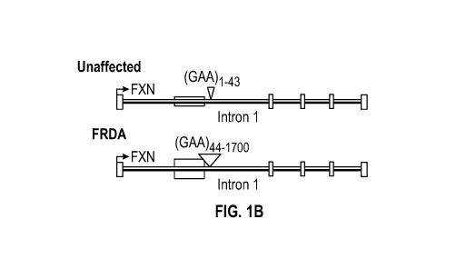
[0008] FRDA is caused, in 98% of all cases, by a genetic mutation resulting
in
expansion of GAA repeats in the first intron of the frataxin gene (FXN). In
healthy
individuals the alleles may contain up to about 40 GAA repeats, whereas
expanded alleles
in FRDA patients can consist of 90 to 1700 repeats (SEQ ID NO: 12) (see Fig.
1B). The
GAA repeat expansion leads to reduced expression of frataxin, a highly
conserved
mitochondrial protein mainly expressed in mitochondria-rich tissues including
the nervous
system, muscle, and heart. Also, carriers (heterozygous for the expanded
allele) show ¨50%
reduction of frataxin mRNA and protein levels compared to normal expression,
although
they do not show any symptoms. While its function is not fully elucidated,
frataxin is an
iron binding protein participating in Fe-S cluster assembly and in its
absence, iron
accumulates within mitochondria leading to defective iron-mediated
biosynthetic processes
and increased oxidative stress.
[0009] Expanded GAA repeats form an intramolecular triple-helix (triplex),
so-called H-
DNA, in supercoiled plasmids isolated from E. coil. Several models
representing the triplex
structures formed at expanded GAA repeats are proposed, and direct evidence
for a
2
CA 03018729 2018-09-21
WO 2017/165167
PCT/US2017/022447
pyrimidine motif H-DNA structure at pathological GAA expansions in vitro has
recently
been provided. Also, formation of a higher order structure named "sticky DNA"
has been
observed in frataxin GAA repeats-containing plasmids using gel electrophoresis
and atomic
force microscopy. The molecular structure of sticky DNA is not resolved;
however, current
evidence demonstrates that sticky DNA forms as one long intramolecular triplex
structure
or by the association of two triplexes.
[0010] The observed effects on DNA replication and transcription are
dependent on the
length and orientation of the GAA repeats in plasmids, which correlate with
formation of
the specific DNA structure (H-DNA). Finally, the GAA repeats are associated
with a
pattern of DNA methylation and histone acetylation in the adjacent regions and
the
formation of silenced chromatin. The presence of H-DNA and higher order
structures
within the GAA repeats is believed to recruit chromatin-remodeling protein
complexes that
maintain a close chromatin structure leading to down-regulation of frataxin
gene
transcription.
[0011] Numerous data have demonstrated that analysis of GAA repeats
constitute an
essential part in the diagnosis of FRDA along with clinical diagnosis.
Molecular genetic
tests are also performed to identify carriers and in prenatal testing. Current
FA diagnostic
methods involve polymerase chain reaction (PCR) analysis and Southern blotting
technique.
The PCR test is performed by amplification of the GAA repeat-containing DNA
region in
the frataxin gene. The different PCR reactions that have been employed to map
GAA
repeat expansions are classical PCR, long-range PCR or triplet-primed PCR (TP-
PCR). In
all cases, the size of the PCR fragment is analyzed using agarose-gel
electrophoresis and
DNA sequencing. In most cases, both PCR and Southern blot are combined to
complement
the results. Problems encountered during amplification of medium- and long-
sized GAA
repeats (i.e., number of repeats >200) using PCR have been reported. The
repetitive nature
of the expanded sequence and its ability to adopt H-DNA and higher order DNA
structures
are the two main factors causing polymerase pausing leading to false results.
[0012] To date, there are no known cures or preventative measures for such
mitochondrial diseases, with current therapies being directed to treating the
associated
symptoms. Thus, there is a need in the art for alternative or improved methods
for treating
mitochondrial diseases/disorders.
3
CA 03018729 2018-09-21
WO 2017/165167
PCT/US2017/022447
SUMMARY OF THE INVENTION
[0013] Accordingly, in one aspect, the invention provides a method of
treating a
mitochondrial disease or disorder in a subject. The method includes
introducing ex vivo a
functional human frataxin (hFXN) into hematopoietic stem and progenitor cells
(HSPCs) of
the subject, and transplanting the HSPCs into the subject, thereby treating
the mitochondrial
disease or disorder. The step of introducing may include contacting a vector
comprising a
polynucleotide encoding hFXN and a FXN promoter (or other regulatory sequence
that is
operable with the polynucleotide and in the cell) with the HSPCs and allowing
expression
of hFXN. In various embodiments, the mitochondrial disease or disorder is
selected from
the group consisting of Friedreich's ataxia (FRDA), diabetes, Leigh syndrome,
Leber's
hereditary optic neuropathy, myoneurogenic gastrointestinal encephalopathy,
and cancer.
The subject may be a mammal, such as a human. In various embodiments, the
vector is a
self-inactivating (SIN)-lentivirus vector, such as pCCL-FRDAp-FXN. In various
embodiments, expression of hFXN corrects neurologic, cardiac and muscular
complications
within about 6-12 months post-transplantation. In another aspect, the hFXN
polynucleotide
is introduced into HSPCs in vivo in a subject.
[0014] In another aspect, the present invention provides a method of
treating a
mitochondrial disease or disorder in a subject comprising contacting cells
expressing hFXN
from the subject with a vector encoding a gene editing system that when
transfected into the
cells removes a trinucleotide extension mutation of endogenous hFXN, thereby
treating the
mitochondrial disease or disorder. In various embodiments, the gene editing
system is
selected from the group consisting of CRISPR/Cas, zinc finger nucleases, and
transcription
activator-life effector nucleases. The step of contacting may include
obtaining a sample of
cells from the subject, transfecting or transducing the gene editing system
into the sample of
cells to create gene-corrected cells, and thereafter, transplanting the gene-
corrected cells
into the subject. The sample of cells may be any cells expressing hFXN, such
as blood cells
and HSPCs from the subject.
[0015] In another aspect, the present invention provides an expression
cassette
comprising a promoter or regulatory sequence functionally linked to a
polynucleotide
encoding hFXN. Also provided are a vector, such as a self-inactivating (SIN)-
lentivirus
vector, that includes a regulatory sequence such as a promoter functionally
linked to a
polynucleotide encoding hFXN.
4
CA 03018729 2018-09-21
WO 2017/165167
PCT/US2017/022447
BRIEF DESCRIPTION OF THE DRAWINGS
[0016] Figures 1A-1D are graphical and pictorial diagrams showing that
systemic
transplantation of WT HSPCs prevents sensory neuron degeneration and
neurobehavioral
deficits in YG8R mice. Fig. 1A shows the results of WT (n=16), YG8R control
(n=4),
YG8R/YG8R HSPCs (n=5) and YG8R/WT HSPCs (n=13) mice at both 5 and 9 months of
age. Locomotor activity was tested using an open field, coordination using a
rotarod, gait
using an automated gait analysis system and muscle strength using forelimb
grip strength.
Data are expressed as means sem; *P<0.05, "P<0.005, ***P<0.0005; NS
statistically
non-significant. For statistical comparison of three experimental groups, a
mixed analysis of
variance (ANOVA) with age of testing as a within-subjects variable was used
followed by
independent sample t-test. Fig. 1B is a representation showing intron 1 of an
unaffected
(top) frataxin gene (FXN) and intron 1 of FRDA (bottom) FXN, and discloses SEQ
ID NOs:
15-16, respectively, in order of appearance. Fig. 1C shows Nissl-stained
sections of lumbar
DRG (L5) from representative 9-month-old WT (n=15), YG8R control (n=4),
YG8R/YG8R
HSPCs (n=4) and YG8R/WT HSPCs (n=11) mice. DRGs of YG8R controls exhibit large
vacuoles (arrows). Scale bars, 100 p.m. Graph on the right depicts total
vacuole area per
DRG area; data are expressed as means sem; "P<0.005; ***P<0.0005. NS,
statistically
non-significant. Fig. 1D shows representative confocal images from a WT GFP+
HSPC-
transplanted YG8R mouse 7 months post-transplantation stained with anti-GFP
and anti-
NeuN. Left: Image of a lumbar (L5) DRG illustrates engraftment of GFP+ HSPC-
derived
cells throughout DRG. Scale bar, 100 p.m. Magnified image (below) demonstrates
frequent
close association of HSPC-derived cells with DRG neurons. Scale bar, 20 p.m.
Right:
Images of cervical, thoracic and lumbar spinal cord show abundant HSPC
engraftment
throughout spinal cord gray and white matter at all levels. Scale bars, 250
p.m. Fig. 1E
shows confocal images of DRG and spinal cord sections of a GFP+ HSPC-treated
YG8R
mouse. Engrafted cells (GFP) are closely associated with neurons (NeuN), and
co-
localization with Ibal marker; Scale bars: 30 p.m.
[0017] Figures 2A-2E are graphical and pictorial diagrams showing that
transplanted
HSPCs engraft throughout the brain and prevent frataxin-deficiency toxicity.
Fig. 2A
shows representative transverse sections of the brain of a WT GFP+ HSPC-
transplanted
YG8R mouse 7 months post-transplantation labeled with anti-GFP and anti-NeuN.
Scale
bar, lmm. Magnified picture #1 of the brain shows that GFP+ HSPC-derived cells
are
CA 03018729 2018-09-21
WO 2017/165167
PCT/US2017/022447
observed in periventricular regions including the corpus callosum (cc),
lateral septal nuclei
(LS), caudate putamen (CP), anterior cingulate area (ACA), and the
somatosensory cortex
(M1, S2). VL, lateral ventricle. Scale bar, 150 p.m. Magnified picture #2 of
ventral
striatum of the brain shows that the engrafted GFP+ HSPCs are present in
regions of the
ventral striatum including the anterior commissure (aco), nucleus accumbens
(ACB), and
lateral septal nuclei (LS). CP, caudate putamen. Scale bar, 150 p.m. Magnified
picture #3
shows that GFP+ HSPC-derived cells are observed in the ventral pallidum (PAL)
and the
ventral striatum, including the islands of Calleja (isl) and the olfactory
tubercle (OT). Scale
bar, 150 p.m. GFP+ HSPCs were also detected through gray and white matter of
the
brainstem and cerebellum. Scale bar, 500 p.m. Insets depict engraftment within
the dentate
nucleus (DN) of the cerebellum and the spinal trigeminal nucleus (Sp) of the
brainstem.
Scale bar, 50 p.m. Fig. 2B shows confocal image of brain labeled with anti-
GFP, anti-Ibal
and anti-NeuN. Most of the bone marrow-derived GFP+ cells co-localize with the
microglial marker Ibal. Scale bar, 30 p.m. Fig. 2C shows quantification of
murine frataxin
mRNA expression in cerebellum from WT (n=14), YG8R (n=8) and YG8R/HSPCs (n=13)
mice. Data are represented as fold change relative to WT normalized to GAPDH.
Data are
expressed as means sem; "P<0.005, ***P<0.0005. Fig. 2D shows the results of
a
representative Western blot showing the level of oxidation in cerebrum of one
WT, one
YG8R, one YG8R/YG8R HSPCs and one YG8R/WT HSPCs mouse with (+) or without (-)
derivatization reagent. Oxyblot analysis detected significantly higher level
of oxidized
proteins in cerebrum of 9-month-old YG8R (n=4) and YG8R/YG8R HSPCs (n=4)
compared to WT (n=6) and YG8R/HSPCs (n=6) mice. Data are expressed as means
sem;
*P<0.05, NS statistically non-significant. Figure 2E shows scatter plots of
mitochondrial
gene changes in cerebrum from WT animals (n=3) compared to YG8R (n=3) (left
scatter
plot) or YG8R/WT HSPCs mice (n=3) (right scatter plot). The center line
represents the
cipher, and upregulated and downregulated genes are noted by dots,
respectively. mRNA
changes that are significantly different between groups are represented on a
separate bar
graph. Data are expressed as means sem; *P<0.05, "P<0.005, ***P<0.0005, NS
statistically non significant as compared to WT.
[0018] Figures 3A-3H are pictorial and graphical diagrams showing
transplanted HSPCs
engraft abundantly in heart and muscle. Fig. 3A shows the results of a
representative
Western blot showing level of oxidation in skeletal muscle of one WT, one YG8R
and one
6
CA 03018729 2018-09-21
WO 2017/165167
PCT/US2017/022447
YG8R/HSPCs mouse with (+) or without (-) derivatization reagent. Oxyblot
analysis
detects high level of protein oxidation only in skeletal muscle of 9-month-old
YG8R
controls (YG8R, n=4 and YG8R/YG8R HSPCs, n=5) compared to WT (n=16) and
YG8R/HSPCs (n=13) mice. Error bars indicate SEM. *p<0.05, NS statistically non-
significant. Fig. 3B shows quantification of lactate and pyruvate by mass-
spectrometry in
muscle tissues from WT (n=6), YG8R (n=3) and YG8R/WT HSPCs (n=5) mice. The
lactate/pyruvate ratio is significantly increased in the YG8R mice compared to
WT while
comparable in YG8R/WT HSPCs animals. Error bars indicate sem; *P<0.05,
***P<0.0005,
NS statistically non-significant. Fig. 3C shows representative Perl's staining
of heart
sections from 18 month old WT, YG8R control and YG8R/WT HSPCs. Characteristic
staining indicates iron deposition. Scale bars, 50 p.m and 15 p.m (zoom). The
associated bar
graph shows iron quantification in heart sections from WT (n=4), YG8R controls
(YG8R
(n=2), YG8R/YG8R HSPCs (n=2)), and YG8R/WT HSPCs (n=3). Error bars indicate
sem;
*P<0.05, NS statistically non-significant. Figs. 3D-3E show quantification of
murine
frataxin mRNA expression in heart (Fig. 3D) and skeletal muscle (Fig. 3E) from
WT
(n=12), YG8R (n=7) and YG8R/HSPCs (n=11) mice. Data are represented as fold
change
relative to WT normalized to GAPDH, error bars indicate sem; *P<0.05,
**13<0.005,
***P<0.0005, NS statistically non-significant. Fig. 3F shows an image of a
heart section
from WT HSPCs transplanted YG8R mouse 7 months post-transplantation stained
with
anti-GFP, the cardiomyocyte marker anti-a-actinin and DAPI. GFP+ cells are
found in all
the cardiac tissue with a highest expression in the valve suggesting that
HSPCs derived cells
are entering the heart by the blood flow. Scale bar, 150 p.m. Magnified
pictures of the heart
show high level of engraftment in the left ventricle (bottom) and in the base
of the aorta
(top). Scale bars, 50 p.m. Fig. 3G shows skeletal muscle section from WT HSPCs
transplanted YG8R mouse 7 months post-transplantation stained with anti-GFP,
filamentous
actin dye Phalloidin and DAPI. GFP+ cells are engrafted homogenously in the
tissue. Scale
bar, 150 p.m. Magnified picture of the skeletal muscle (on the left) shows
that GFP+ cells
are localized interstitially between muscle fibers. Scale bar, 50 p.m. Fig. 3H
shows
quantification of murine MuRF-1, Atrogin-1 and myostatin mRNA expression in
skeletal
muscle from WT (n=5), YG8R (n=5) and YG8R/HSPCs (n=5) mice. Data are
represented
as fold change relative to WT normalized to GAPDH, error bars indicate sem;
*P<0.05, NS
statistically non-significant.
7
CA 03018729 2018-09-21
WO 2017/165167
PCT/US2017/022447
[0019] Figures 4A-4F are pictorial and graphical diagrams showing that HSPC-
derived
cells deliver frataxin-bearing mitochondria to the diseased cells in vitro and
in vivo. Figs.
4A and 4B show representative frames from confocal imaging movies of YG8R-
derived
fibroblasts (F) co-cultured with primary macrophages (M) isolated from a DsRed
Cox8-
GFP transgenic mouse (Fig. 4A) or with IC21 macrophages transduced with a LV-
hFXN-
GFP and stained with a red MitoTracker (Fig. 4B). Scale bar, 10 p.m. Fig. 4C
shows a
representative confocal image of brain sections from an YG8R mouse
transplanted with
DsRed+ HSPCs (control) and brain and spinal cord sections from an YG8R mouse
transplanted with DsRed+/Cox8-GFP+ HSPCs at 7 months post-transplantation
labelled with
an anti-NeuN antibody. In addition to the DsRed-derived bone marrow cells,
cox8-GFP are
observed in host neurons in brain and spinal cord (arrows). For DRG, heart and
muscle, see
Figs. 7A and 7B. Scale bars, 10 pm. Fig. 4D shows representative confocal
images of
spinal cord section from an YG8R mouse transplanted with DsRed+/Cox8-GFP+
HSPCs at 7
months post-transplantation labelled with an anti-NeuN antibody showing cox8-
GFP within
the branch extension of the DsRed+ microglial cell (arrows). Scale bar, 5 pm.
Fig. 4E
shows quantification of neurons containing cox8-GFP in the cervical spinal
cord gray
matter of YG8R mice transplanted with DsRed+/Cox8-GFP+ HSPCs at 7 months post-
transplantation (for description of the automatic unbiased quantification
method see Fig. 8).
Fig. 4F shows representative confocal images of brain and spinal cord sections
from an
YG8R mouse transplanted with DsRed+ HSPCs transduced with LV-hFXN-GFP at 7
months post-transplantation and stained with anti-mcherry and anti-NeuN
antibodies. In
addition to the DsRed-derived bone marrow cells, frataxin-GFP are observed in
host
neurons. Scale bar, 10 pm.
[0020] Figure 5 is a pictorial diagram showing that HSPCs engraft in the
peripheral
nerve in YG8R mice. Confocal images of sciatic nerve from WT GFP+ HSPC-
transplanted
YG8R mice labeled with anti-GFP, and with a neurofilament marker, anti-NF200,
and a
myelin basic protein marker, anti-MBP. Scale bars: 100 p.m (left), 10 p.m
(inset).
[0021] Figures 6A-6F are pictorial diagrams showing that HSPCs
differentiate into
macrophages in DRG and microglia in the spinal cord and brain. Figs. 6A and 6B
show
confocal images of DRG, spinal cord and brain sections from WT GFP+ HSPC-
transplanted
YG8R mice labeled with anti-GFP, anti-CD68 (Fig. 6A), anti-MHCII (Fig. 6B),
anti-NeuN
(Fig. 6A), and DAPI. Scale bars, 30 pm. Figs. 6C and 6D show transverse spinal
cord (Fig.
8
CA 03018729 2018-09-21
WO 2017/165167
PCT/US2017/022447
6C) and brain (Fig. 6D) section from WT GFP + HSPC-transplanted YG8R mouse
labeled
with anti-MHCII. Scale bars, 100 p.m (Fig. 6C) and 300 p.m (Fig. 6D). Fig. 6E
shows a
confocal image of brain section from WT GFP + HSPC-transplanted YG8R mouse
labeled
with anti-vwf. Scale bar, 50 p.m. Fig. 6F shows a confocal image of choroid
plexus from
WT DsRed+ HSPC-transplanted YG8R mouse labeled with anti-RFP and anti-Ibal.
Scale
bar, 100 p.m.
[0022] Figures 7A and 7B are pictorial diagrams showing that HSPCs
differentiate into
macrophages in heart and muscle. Confocal images of heart and skeletal muscle
section
from YG8R transplanted with WT GFP + HSPCs after labeling with anti-GFP, anti-
CD68
(Fig. 7A) anti-MHCII (Fig. 7B), Phalloidin and DAPI. Scale bar, 30 p.m.
[0023] Figure 8 is a pictorial diagram showing that HSPC-derived
macrophages deliver
mitochondria to neurons in DRG and to myocytes in heart and skeletal muscle.
Representative confocal images of DRG, heart and skeletal muscle from an YG8R
mouse
transplanted with DsRed+/Cox8-GFP+ HSPCs at 7 months post-transplantation
stained with
anti-NeuN (DRG), anti-a-Actinin (heart) or Palloidin (muscle), and DAPI (heart
and
muscle). Scale bars, 10 p.m.
[0024] Figures 9A-9D are pictorial and graphical diagrams showing
quantification of
Cox8-GFP transfer from HSPC-derived microglia to neurons. Fig. 9A shows a
representative transverse image of cervical spinal cord gray matter from a
YG8R mouse at 7
months following transplantation with Cox8-GFP DsRed HSPCs, stained with anti-
NeuN.
Scale bar, 500 p.m. Fig. 9B shows automatic outline and quantification of
neurons by
ImagePro software. Fig. 9C shows that GFP signal is only counted within the
delineated
neurons (arrow) and not outside (star). Fig. 9D shows the percentage of
neurons within the
gray matter of the spinal cord that contain GFP for three different animals
(transplanted)
and for one control. The entire gray matter from three experimental animals
and one
control (three sections per animal) were quantified.
DETAILED DESCRIPTION OF THE INVENTION
[0025] The present invention is based on the finding of complete phenotypic
correction
of mitochondrial disorders occurs after a single transplantation of wildtype
hematopoietic
stem and progenitor cells, which differentiated into phagocytic cells in the
nervous system,
muscle and heart leading to the neuronal and myocyte cross-correction. There
is a pressing
9
CA 03018729 2018-09-21
WO 2017/165167
PCT/US2017/022447
need to identify effective therapies for mitochondrial disorders such as FRDA
for which
there remains no treatment. To date, preclinical studies using stem cells or
gene therapy
have had limited success, or have been restricted to assessment of specific
tissues.
[0026] The present disclosure demonstrates that a self-inactivating (SIN)-
lentivirus
vector containing the human frataxin (hFXN) cDNA as well as the optimal
promoter can be
used to ex vivo gene-corrected patients' autologous hematopoietic stem and
progenitor cells
(HSPCs), which can then be re-transplant in the patients to repopulate their
bone marrow,
which will be a reservoir of "healthy" cells for the rest of the life of the
patients. These
cells mobilize and integrate into the diseased tissues (brain, muscle, heart),
and will lead to
their rescue. While autologous HSPCs are used in the illustrative examples
herein, one of
skill in the art would recognize that other HSPCs would be useful as well
(e.g., allogeneic).
[0027] Before the present compositions and methods are described, it is to
be understood
that this invention is not limited to particular compositions, methods, and
experimental
conditions described, as such compositions, methods, and conditions may vary.
It is also to
be understood that the terminology used herein is for purposes of describing
particular
embodiments only, and is not intended to be limiting, since the scope of the
present
invention will be limited only in the appended claims.
[0028] As used in this specification and the appended claims, the singular
forms "a",
"an", and "the" include plural references unless the context clearly dictates
otherwise.
Thus, for example, references to "the method" includes one or more methods,
and/or steps
of the type described herein which will become apparent to those persons
skilled in the art
upon reading this disclosure and so forth.
[0029] The term "comprising," which is used interchangeably with
"including,"
"containing," or "characterized by," is inclusive or open-ended language and
does not
exclude additional, unrecited elements or method steps. The phrase "consisting
of"
excludes any element, step, or ingredient not specified in the claim. The
phrase "consisting
essentially of" limits the scope of a claim to the specified materials or
steps and those that
do not materially affect the basic and novel characteristics of the claimed
invention. The
present disclosure contemplates embodiments of the invention compositions and
methods
corresponding to the scope of each of these phrases. Thus, a composition or
method
CA 03018729 2018-09-21
WO 2017/165167
PCT/US2017/022447
comprising recited elements or steps contemplates particular embodiments in
which the
composition or method consists essentially of or consists of those elements or
steps.
[0030] Unless defined otherwise, all technical and scientific terms used
herein have the
same meaning as commonly understood by one of ordinary skill in the art to
which this
invention belongs. Although any methods and materials similar or equivalent to
those
described herein can be used in the practice or testing of the invention, the
preferred
methods and materials are now described.
[0031] The term "subject" or "host organism," as used herein, refers to any
individual or
patient to which the subject methods are performed. Generally the subject is
human,
although as will be appreciated by those in the art, the subject may be an
animal. Thus
other animals, including mammals such as rodents (including mice, rats,
hamsters and
guinea pigs), cats, dogs, rabbits, farm animals including cows, horses, goats,
sheep, pigs,
etc., and primates (including monkeys, chimpanzees, orangutans and gorillas)
are included
within the definition of subject.
[0032] The term "therapeutically effective amount" or "effective amount"
means the
amount of a compound or pharmaceutical composition that will elicit the
biological or
medical response of a tissue, system, animal or human that is being sought by
the
researcher, veterinarian, medical doctor or other clinician. Thus, the term
"therapeutically
effective amount" is used herein to denote any amount of a formulation that
causes a
substantial improvement in a disease condition when applied to the affected
areas
repeatedly over a period of time. The amount will vary with the condition
being treated, the
stage of advancement of the condition, and the type and concentration of
formulation
applied. Appropriate amounts in any given instance will be readily apparent to
those skilled
in the art or capable of determination by routine experimentation.
[0033] A "therapeutic effect," as used herein, encompasses a therapeutic
benefit and/or a
prophylactic benefit as described herein.
[0034] The terms "administration" or "administering" are defined to include
an act of
providing a compound or pharmaceutical composition of the invention to a
subject in need
of treatment. The phrases "parenteral administration" and "administered
parenterally" as
used herein means modes of administration other than enteral and topical
administration,
11
CA 03018729 2018-09-21
WO 2017/165167
PCT/US2017/022447
usually orally or by injection, and includes, without limitation, intravenous,
intramuscular,
intraarterial, intrathecal, intracapsular, intraorbital, intracardiac,
intradermal, intraperitoneal,
transtracheal, subcutaneous, subcuticular, intraarticulare, subcapsular,
subarachnoid,
intraspinal and infrasternal injection and infusion. The phrases "systemic
administration,"
"administered systemically," "peripheral administration" and "administered
peripherally" as
used herein mean the administration of a compound, drug or other material
other than
directly into the central nervous system, such that it enters the subject's
system and, thus, is
subject to metabolism and other like processes, for example, subcutaneous
administration.
[0035] If a viral vector specific for the cell type is not available, the
vector can be
modified to express a receptor (or ligand) specific for a ligand (or receptor)
expressed on the
target cell, or can be encapsulated within a liposome, which also can be
modified to include
such a ligand (or receptor). A peptide agent can be introduced into a cell by
various methods,
including, for example, by engineering the peptide to contain a protein
transduction domain
such as the human immunodeficiency virus TAT protein transduction domain,
which can
facilitate translocation of the peptide into the cell. In addition, there are
a variety of
biomaterial-based technologies such as nano-cages and pharmacological delivery
wafers
(such as used in brain cancer chemotherapeutics) which may also be modified to
accommodate this technology.
[0036] The viral vectors most commonly assessed for gene transfer are based
on DNA-
based adenoviruses (Ads) and adeno-associated viruses (AAVs) and RNA-based
retroviruses and lentiviruses. Lentivirus vectors have been most commonly used
to achieve
chromosomal integration.
[0037] As used herein, the terms "reduce" and "inhibit" are used together
because it is
recognized that, in some cases, a decrease can be reduced below the level of
detection of a
particular assay. As such, it may not always be clear whether the expression
level or activity
is "reduced" below a level of detection of an assay, or is completely
"inhibited."
Nevertheless, it will be clearly determinable, following a treatment according
to the present
methods.
[0038] As used herein, "treatment" or "treating" means to administer a
composition to a
subject or a system with an undesired condition. The condition can include a
disease or
disorder. "Prevention" or "preventing" means to administer a composition to a
subject or a
12
CA 03018729 2018-09-21
WO 2017/165167
PCT/US2017/022447
system at risk for the condition. The condition can include a predisposition
to a disease or
disorder. The effect of the administration of the composition to the subject
(either treating
and/or preventing) can be, but is not limited to, the cessation of one or more
symptoms of
the condition, a reduction or prevention of one or more symptoms of the
condition, a
reduction in the severity of the condition, the complete ablation of the
condition, a
stabilization or delay of the development or progression of a particular event
or
characteristic, or minimization of the chances that a particular event or
characteristic will
OMIT.
[0039] As used herein, the term "genetic modification" is used to refer to
any
manipulation of an organism's genetic material in a way that does not occur
under natural
conditions. Methods of performing such manipulations are known to those of
ordinary skill
in the art and include, but are not limited to, techniques that make use of
vectors for
transforming cells with a nucleic acid sequence of interest. Included in the
definition are
various forms of gene editing in which DNA is inserted, deleted or replaced in
the genome
of a living organism using engineered nucleases, or "molecular scissors."
These nucleases
create site-specific double-strand breaks (DSBs) at desired locations in the
genome. The
induced double-strand breaks are repaired through nonhomologous end-joining
(NHEJ) or
homologous recombination (HR), resulting in targeted mutations (i.e., edits).
[0040] There are several families of engineered nucleases used in gene
editing, for
example, but not limited to, meganucleases, zinc finger nucleases (ZFNs),
transcription
activator-like effector-based nucleases (TALEN), and the CRISPR-Cas system.
[0041] CRISPR (Clustered Regularly Interspaced Short Palindromic Repeats)
is an
acronym for DNA loci that contain multiple, short, direct repetitions of base
sequences.
The prokaryotic CRISPR/Cas system has been adapted for use as gene editing
(silencing,
enhancing or changing specific genes) for use in eukaryotes (see, for example,
Cong,
Science, 15:339(6121):819-823 (2013) and Jinek, et al., Science, 337(6096):816-
21 (2012)).
By transfecting a cell with elements including a Cas gene and specifically
designed
CRISPRs, nucleic acid sequences can be cut and modified at any desired
location. Methods
of preparing compositions for use in genome editing using the CRISPR/Cas
systems are
described in detail in US Pub. No. 2016/0340661, US Pub. No. 20160340662, US
Pub. No.
13
CA 03018729 2018-09-21
WO 2017/165167
PCT/US2017/022447
2016/0354487, US Pub. No. 2016/0355796, US Pub. No. 20160355797, and WO
2014/018423, which are specifically incorporated by reference herein in their
entireties.
[0042] Thus, as used herein, "CRISPR system" refers collectively to
transcripts and
other elements involved in the expression of or directing the activity of
CRISPR-associated
("Cas") genes, including sequences encoding a Cas gene, a tracr (trans-
activating CRISPR)
sequence (e.g., tracrRNA or an active partial tracrRNA), a tracr-mate sequence
(encompassing a "direct repeat" and a tracrRNA-processed partial direct repeat
in the
context of an endogenous CRISPR system), a guide sequence (also referred to as
a "spacer",
"guide RNA" or "gRNA" in the context of an endogenous CRISPR system), or other
sequences and transcripts from a CRISPR locus. One or more tracr mate
sequences
operably linked to a guide sequence (e.g., direct repeat-spacer-direct repeat)
can also be
referred to as "pre-crRNA" (pre-CRISPR RNA) before processing or crRNA after
processing by a nuclease.
[0043] In some embodiments, a tracrRNA and crRNA are linked and form a
chimeric
crRNA-tracrRNA hybrid where a mature crRNA is fused to a partial tracrRNA via
a
synthetic stem loop to mimic the natural crRNA:tracrRNA duplex as described in
Cong,
Science, 15:339(6121):819-823 (2013) and Jinek, et al., Science, 337(6096):816-
21 (2012)).
A single fused crRNA-tracrRNA construct can also be referred to as a guide RNA
or gRNA
(or single-guide RNA (sgRNA)). Within an sgRNA, the crRNA portion can be
identified as
the 'target sequence' and the tracrRNA is often referred to as the 'scaffold'.
[0044] There are many resources available for helping practitioners
determine suitable
target sites once a desired DNA target sequence is identified. For example,
numerous
public resources, including a bioinformatically generated list of about
190,000 potential
sgRNAs, targeting more than 40% of human exons, are available to aid
practitioners in
selecting target sites and designing the associate sgRNA to affect a nick or
double strand
break at the site. See also, crispr.u-psud.fr, a tool designed to help
scientists find CRISPR
targeting sites in a wide range of species and generate the appropriate crRNA
sequences.
[0045] In some embodiments, one or more vectors driving expression of one
or more
elements of a CRISPR system are introduced into a target cell such that
expression of the
elements of the CRISPR system direct formation of a CRISPR complex at one or
more
target sites. While the specifics can be varied in different engineered CRISPR
systems, the
14
CA 03018729 2018-09-21
WO 2017/165167
PCT/US2017/022447
overall methodology is similar. A practitioner interested in using CRISPR
technology to
target a DNA sequence can insert a short DNA fragment containing the target
sequence into
a guide RNA expression plasmid. The sgRNA expression plasmid contains the
target
sequence (about 20 nucleotides), a form of the tracrRNA sequence (the
scaffold) as well as
a suitable promoter and necessary elements for proper processing in eukaryotic
cells. Such
vectors are commercially available (see, for example, Addgene). Many of the
systems rely
on custom, complementary oligos that are annealed to form a double stranded
DNA and
then cloned into the sgRNA expression plasmid. Co-expression of the sgRNA and
the
appropriate Cas enzyme from the same or separate plasmids in transfected cells
results in a
single or double strand break (depending of the activity of the Cos enzyme) at
the desired
target site.
[0046] Zinc-finger nucleases (ZFNs) are artificial restriction enzymes
generated by
fusing a zinc finger DNA-binding domain to a DNA-cleavage domain. Zinc finger
domains
can be engineered to target specific desired DNA sequences and this enables
zinc-finger
nucleases to target unique sequences within complex genomes. By taking
advantage of
endogenous DNA repair machinery, these reagents can be used to precisely alter
the
genomes of higher organisms. The most common cleavage domain is the Type ITS
enzyme
Fokl. Fokl catalyzes double-stranded cleavage of DNA, at 9 nucleotides from
its
recognition site on one strand and 13 nucleotides from its recognition site on
the other. See,
for example, U.S. Pat. Nos. 5,356,802; 5,436,150 and 5,487,994; as well as Li
etal. Proc.,
Natl. Acad. Sci. USA 89 (1992):4275-4279; Li etal. Proc. Natl. Acad. Sci. USA,
90:2764-
2768 (1993); Kim etal. Proc. Natl. Acad. Sci. USA. 91:883-887 (1994a); Kim
etal. I Biol.
Chem. 269:31,978-31,982 (1994b), all of which are incorporated herein by
reference. One
or more of these enzymes (or enzymatically functional fragments thereof) can
be used as a
source of cleavage domains.
[0047] Transcription activator-like effector nucleases (TALENs) have an
overall
architecture similar to that of ZFNs, with the main difference being that the
DNA-binding
domain comes from TAL effector proteins, transcription factors from plant
pathogenic
bacteria. The DNA-binding domain of a TALEN is a tandem array of amino acid
repeats,
each about 34 residues long. The repeats are very similar to each other;
typically they differ
principally at two positions (amino acids 12 and 13, called the repeat
variable diresidue, or
RVD). Each RVD specifies preferential binding to one of the four possible
nucleotides,
CA 03018729 2018-09-21
WO 2017/165167
PCT/US2017/022447
meaning that each TALEN repeat binds to a single base pair, though the NN RVD
is known
to bind adenines in addition to guanine. TAL effector DNA binding is
mechanistically less
well understood than that of zinc-finger proteins, but their seemingly simpler
code could
prove very beneficial for engineered-nuclease design. TALENs also cleave as
dimers, have
relatively long target sequences (the shortest reported so far binds 13
nucleotides per
monomer) and appear to have less stringent requirements than ZFNs for the
length of the
spacer between binding sites. Monomeric and dimeric TALENs can include more
than 10,
more than 14, more than 20, or more than 24 repeats. Methods of engineering
TAL to bind
to specific nucleic acids are described in Cermak, eta!, Nucl. Acids Res. 1-11
(2011); US
Published Application No. 2011/0145940, which discloses TAL effectors and
methods of
using them to modify DNA; Miller etal. Nature Biotechnol 29: 143 (2011)
reported making
TALENs for site-specific nuclease architecture by linking TAL truncation
variants to the
catalytic domain of Fokl nuclease. The resulting TALENs were shown to induce
gene
modification in immortalized human cells. General design principles for TALE
binding
domains can be found in, for example, WO 2011/072246. Each of the foregoing
references
are incorporated herein by reference in their entireties.
[0048] The nuclease activity of the genome editing systems described herein
cleave
target DNA to produce single or double strand breaks in the target DNA. Double
strand
breaks can be repaired by the cell in one of two ways: non-homologous end
joining, and
homology-directed repair. In non-homologous end joining (NHEJ), the double-
strand
breaks are repaired by direct ligation of the break ends to one another. As
such, no new
nucleic acid material is inserted into the site, although some nucleic acid
material may be
lost, resulting in a deletion. In homology-directed repair, a donor
polynucleotide with
homology to the cleaved target DNA sequence is used as a template for repair
of the
cleaved target DNA sequence, resulting in the transfer of genetic information
from a donor
polynucleotide to the target DNA. As such, new nucleic acid material can be
inserted/copied
into the site. Therefore, in some embodiments, the genome editing vector or
composition
optionally includes a donor polynucleotide. The modifications of the target
DNA due to
NHEJ and/or homology-directed repair can be used to induce gene correction,
gene
replacement, gene tagging, transgene insertion, nucleotide deletion, gene
disruption, gene
mutation, etc.
16
CA 03018729 2018-09-21
WO 2017/165167
PCT/US2017/022447
[0049] Accordingly, cleavage of DNA by the genome editing vector or
composition can
be used to delete nucleic acid material from a target DNA sequence by cleaving
the target
DNA sequence and allowing the cell to repair the sequence in the absence of an
exogenously provided donor polynucleotide. Alternatively, if the genome
editing
composition includes a donor polynucleotide sequence that includes at least a
segment with
homology to the target DNA sequence, the methods can be used to add, i.e.,
insert or
replace, nucleic acid material to a target DNA sequence (e.g., to "knock in" a
nucleic acid
that encodes for a protein, an siRNA, an miRNA, etc.), to add a tag (e.g.,
6xHis (SEQ ID
NO: 13), a fluorescent protein (e.g., a green fluorescent protein; a yellow
fluorescent
protein, etc.), hemagglutinin (HA), FLAG, etc.), to add a regulatory sequence
to a gene
(e.g., promoter, polyadenylation signal, internal ribosome entry sequence
(IRES), 2A
peptide, start codon, stop codon, splice signal, localization signal, etc.),
to modify a nucleic
acid sequence (e.g., introduce a mutation), and the like. As such, the
compositions can be
used to modify DNA in a site-specific, i.e., "targeted" way, for example gene
knock-out,
gene knock-in, gene editing, gene tagging, etc., as used in, for example, gene
therapy.
[0050] The terms "polypeptide," "peptide," and "protein" are used
interchangeably
herein to refer to a polymer of amino acid residues. The terms apply to amino
acid
polymers in which one or more amino acid residue is an artificial chemical
mimetic of a
corresponding naturally occurring amino acid, as well as to naturally
occurring amino acid
polymers and non-naturally occurring amino acid polymer.
[0051] The term "amino acid" refers to naturally occurring and synthetic
amino acids, as
well as amino acid analogs and amino acid mimetics that function in a manner
similar to the
naturally occurring amino acids. Naturally occurring amino acids are those
encoded by the
genetic code, as well as those amino acids that are later modified, e.g.,
hydroxyproline, a-
carboxyglutamate, and 0-phosphoserine. Amino acid analogs refers to compounds
that
have the same basic chemical structure as a naturally occurring amino acid,
i.e., an a carbon
that is bound to a hydrogen, a carboxyl group, an amino group, and an R group,
e.g.,
homoserine, norleucine, methionine sulfoxide, methionine methyl sulfonium.
Such analogs
have modified R groups (e.g., norleucine) or modified peptide backbones, but
retain the
same basic chemical structure as a naturally occurring amino acid. Amino acid
mimetics
refers to chemical compounds that have a structure that is different from the
general
17
CA 03018729 2018-09-21
WO 2017/165167
PCT/US2017/022447
chemical structure of an amino acid, but that functions in a manner similar to
a naturally
occurring amino acid.
[0052] Amino acids may be referred to herein by either their commonly known
three
letter symbols or by the one-letter symbols recommended by the IUPAC-TUB
Biochemical
Nomenclature Commission. Nucleotides, likewise, may be referred to by their
commonly
accepted single-letter codes.
[0053] As used herein, a "regulatory gene" or "regulatory sequence" is a
nucleic acid
sequence that encodes products (e.g., transcription factors) that control the
expression of
other genes.
[0054] As used herein, a "protein coding sequence" or a sequence that
encodes a
particular protein or polypeptide, is a nucleic acid sequence that is
transcribed into mRNA
(in the case of DNA) and is translated (in the case of mRNA) into a
polypeptide in vitro or
in vivo when placed under the control of appropriate regulatory sequences. The
boundaries
of the coding sequence are determined by a start codon at the 5' terminus (N-
terminus) and a
translation stop nonsense codon at the 3' terminus (C-terminus). A coding
sequence can
include, but is not limited to, cDNA from eukaryotic mRNA, genomic DNA
sequences
from eukaryotic DNA, and synthetic nucleic acids. A transcription termination
sequence
will usually be located 3' to the coding sequence.
[0055] As used herein, a "promoter" is defined as a regulatory DNA sequence
generally
located upstream of a gene that mediates the initiation of transcription by
directing RNA
polymerase to bind to DNA and initiating RNA synthesis. A promoter can be a
constitutively active promoter (i.e., a promoter that is constitutively in an
active/"ON"
state), it may be an inducible promoter (i.e., a promoter whose state,
active/"ON" or
inactive/"OFF", is controlled by an external stimulus, e.g., the presence of a
particular
compound or protein), it may be a spatially restricted promoter (i.e.,
transcriptional control
element, enhancer, etc.)(e.g., tissue specific promoter, cell type specific
promoter, etc.), and
it may be a temporally restricted promoter (i.e., the promoter is in the "ON"
state or "OFF"
state during specific stages of embryonic development or during specific
stages of a
biological process.
18
CA 03018729 2018-09-21
WO 2017/165167
PCT/US2017/022447
[0056] As used herein, the term "gene" means the deoxyribonucleotide
sequences
comprising the coding region of a structural gene. A "gene" may also include
non-
translated sequences located adjacent to the coding region on both the 5' and
3' ends such
that the gene corresponds to the length of the full-length mRNA. The sequences
which are
located 5' of the coding region and which are present on the mRNA are referred
to as 5' non-
translated sequences. The sequences which are located 3' or downstream of the
coding
region and which are present on the mRNA are referred to as 3' non-translated
sequences.
The term "gene" encompasses both cDNA and genomic forms of a gene. A genomic
form
or clone of a gene contains the coding region interrupted with non-coding
sequences termed
"introns" or "intervening regions" or "intervening sequences." Introns are
segments of a
gene which are transcribed into heterogenous nuclear RNA (hnRNA); introns may
contain
regulatory elements such as enhancers. Introns are removed or "spliced out"
from the
nuclear or primary transcript; introns therefore are absent in the messenger
RNA (mRNA)
transcript. The mRNA functions during translation to specify the sequence or
order of
amino acids in a nascent polypeptide.
[0057] As used herein, the terms "functionally linked" and "operably
linked" are used
interchangeably and refer to a functional relationship between two or more DNA
segments,
in particular gene sequences to be expressed and those sequences controlling
their
expression. For example, a promoter/enhancer sequence, including any
combination of cis-
acting transcriptional control elements is operably linked to a coding
sequence if it
stimulates or modulates the transcription of the coding sequence in an
appropriate host cell
or other expression system. Promoter regulatory sequences that are operably
linked to the
transcribed gene sequence are physically contiguous to the transcribed
sequence.
[0058] "Conservatively modified variants" applies to both amino acid and
nucleic acid
sequences. With respect to particular nucleic acid sequences, conservatively
modified
variants refers to those nucleic acids which encode identical or essentially
identical amino
acid sequences, or where the nucleic acid does not encode an amino acid
sequence, to
essentially identical sequences. Because of the degeneracy of the genetic
code, a large
number of functionally identical nucleic acids encode any given protein. For
instance, the
codons GCA, GCC, GCG and GCU all encode the amino acid alanine. Thus, at every
position where an alanine is specified by a codon, the codon can be altered to
any of the
corresponding codons described without altering the encoded polypeptide. Such
nucleic
19
CA 03018729 2018-09-21
WO 2017/165167
PCT/US2017/022447
acid variations are "silent variations," which are one species of
conservatively modified
variations. Every nucleic acid sequence herein which encodes a polypeptide
also describes
every possible silent variation of the nucleic acid. One of skill will
recognize that each
codon in a nucleic acid (except AUG, which is ordinarily the only codon for
methionine,
and TGG, which is ordinarily the only codon for tryptophan) can be modified to
yield a
functionally identical molecule. Accordingly, each silent variation of a
nucleic acid which
encodes a polypeptide is implicit in each described sequence.
[0059] As to amino acid sequences, one of skill will recognize that
individual
substitutions, deletions or additions to a nucleic acid, peptide, polypeptide,
or protein
sequence which alters, adds or deletes a single amino acid or a small
percentage of amino
acids in the encoded sequence is a "conservatively modified variant" where the
alteration
results in the substitution of an amino acid with a chemically similar amino
acid.
Conservative substitution tables providing functionally similar amino acids
are well known
in the art. Such conservatively modified variants are in addition to and do
not exclude
polymorphic variants, interspecies homologs, and alleles of the invention.
[0060] The term "antibody" as used herein refers to polyclonal and
monoclonal
antibodies and fragments thereof, and immunologic binding equivalents thereof
The term
"antibody" refers to a homogeneous molecular entity, or a mixture such as a
polyclonal
serum product made up of a plurality of different molecular entities, and
broadly
encompasses naturally-occurring forms of antibodies (for example, IgG, IgA,
IgM, IgE) and
recombinant antibodies such as single-chain antibodies, chimeric and humanized
antibodies
and multi-specific antibodies. The term "antibody" also refers to fragments
and derivatives
of all of the foregoing, and may further comprise any modified or derivatised
variants
thereof that retains the ability to specifically bind an epitope. Antibody
derivatives may
comprise a protein or chemical moiety conjugated to an antibody. A monoclonal
antibody
is capable of selectively binding to a target antigen or epitope. Antibodies
may include, but
are not limited to polyclonal antibodies, monoclonal antibodies (mAbs),
humanized or
chimeric antibodies, camelized antibodies, single chain antibodies (scFvs),
Fab fragments,
F(ab1)2 fragments, disulfide-linked Fvs (sdFv) fragments, for example, as
produced by a Fab
expression library, anti-idiotypic (anti-Id) antibodies, intrabodies,
nanobodies, synthetic
antibodies, and epitope-binding fragments of any of the above.
CA 03018729 2018-09-21
WO 2017/165167
PCT/US2017/022447
[0061] As used herein, the term "humanized mouse" (Hu-mouse) is a mouse
developed
to carry functioning human genes, cells, tissues, and/or organs. Humanized
mice are
commonly used as small animal models in biological and medical research for
human
therapeutics. Immunodeficient mice are often used as recipients for human
cells or tissues,
because they can relatively easily accept heterologous cells due to lack of
host immunity.
[0062] HSCs possess the ability of multipotency (i.e., one HSC can
differentiate into all
functional blood cells) and self-renewal (i.e., HSCs can divide and give rise
to an identical
daughter cell, without differentiation). Through a series of lineage
commitment steps,
HSCs give rise to progeny that progressively lose self-renewal potential and
successively
become more and more restricted in their differentiation capacity, generating
multi-potential
and lineage-committed progenitor cells, and ultimately mature functional
circulating blood
cells.
[0063] The ability of hematopoietic stem and progenitor cells (HSPCs) to
self-renew and
differentiate is fundamental for the formation and maintenance of life-long
hematopoiesis
and deregulation of these processes may lead to severe clinical consequences.
HSPCs are
also highly valuable for their ability to reconstitute the hematopoietic
system when
transplanted and this has enabled their use in the clinic to treat a variety
of disorders
including bone marrow failure, myeloproliferative disorders and other acquired
or genetic
disorders that affect blood cells.
[0064] As used herein, a "pluripotent cell" refers to a cell derived from
an embryo
produced by activation of a cell containing DNA of all female or male origin
that can be
maintained in vitro for prolonged, theoretically indefinite period of time in
an
undifferentiated state that can give rise to different differentiated tissue
types, i.e., ectoderm,
mesoderm, and endoderm. "Embryonic stem cells" (ES cells) are pluripotent stem
cells
derived from the inner cell mass of a blastocyst, an early-stage
preimplantation embryo.
[0065] As used herein "pharmaceutically acceptable carrier" encompasses any
of the
standard pharmaceutical carriers, such as a phosphate buffered saline
solution, water and
emulsions such as an oil/water or water/oil emulsion, and various types of
wetting agents.
[0066] This work shows that one-time hematopoietic stem and progenitor cell
(HSPC)
transplantation holds the potential to become a life-long curative therapy for
a disease or
21
CA 03018729 2018-09-21
WO 2017/165167
PCT/US2017/022447
disorder associated with mitochondrial dysfunction. Given the risks associated
with
allogeneic stem cell transplantation, the objective was to develop an
autologous HSPC gene
therapy for mitochondrial diseases.
[0067] As discussed above, mitochondrial diseases/disorders may be caused
by
mutations, acquired or inherited, in mitochondrial DNA (mtDNA) or in nuclear
genes that
code for mitochondrial components. They may also be the result of acquired
mitochondrial
dysfunction due to adverse effects of drugs, infections, or other
environmental causes.
[0068] Examples of mitochondrial diseases include, but are not limited to,
mitochondrial
myopathy, diabetes mellitus and deafness (DAD), Leber's hereditary optic
neuropathy
(LHON), Leigh syndrome, subacute sclerosing encephalopathy, Neuropathy,
ataxia, retinitis
pigmentosa, and ptosis (NARP), myoneurogenic gastrointestinal encephalopathy
(MNGIE),
Myoclonic Epilepsy with Ragged Red Fibers (MERRF), Mitochondrial myopathy,
encephalomyopathy, lactic acidosis, stroke-like symptoms (MELAS),
mitochondrial
neurogastrointestinal encephalomyopathy (MNGIE) and diseases due to
mitochondrial
complex deficiency, such as Friedreich's ataxia (FRDA).
[0069] FRDA is a progressively lethal multi-systemic disease. Although the
exact
function of FXN is still under debate, it is predicted to assist in the
biogenesis of
mitochrondrial iron-sulfur clusters. Thus, frataxin deficiency results in
altered cellular iron
metabolism, increased mitochondrial iron load, decreased mitochondrial energy
production
and biogenesis as well as increased oxidative stress. Clinical features
include gait and limb
ataxia, muscle weakness, dysarthria and also vision and hearing anomalies,
diabetes and
cardiomyopathy. Frataxin deficiency impacts neuronal functions particularly
and this
affects mainly the peripheral and central nervous systems (CNS), leading to
the progressive
destruction of the Dorsal Root Ganglia (DRG). This progressive
neurodegeneration leads to
loss of motor skills and progressive muscle degeneration, and ultimately
inability to walk
within 10 to 15 years of onset. Heart abnormalities cause premature death in
60% to 80%
of the affected individuals; the average age of death is in the mid-thirties.
The different
clinical trials of pharmacological compounds against oxidative stress (idebone
and
Coenzyme Q10) or mitochondrial iron accumulation (deferipone) failed to prove
efficacy.
An epigenetic approach using an histone deacetylase inhibitor is currently
being testing in
phase I clinical trial.
22
CA 03018729 2018-09-21
WO 2017/165167
PCT/US2017/022447
[0070] Hematopoietic stem and progenitor cells (HSPCs) are ideal candidates
for use in
regenerative medicine and cell replacement therapies because of their ease of
isolation, self-
renewal capacity, and safety. As such, the present disclosure evaluates the
impact of
hematopoietic stem and progenitor cell (HSPC) transplantation in a mouse model
of FRDA.
The rationale for using HSPC to treat FRDA came from previous work on
cystinosis, a
multi-systemic lysosomal storage disorder. Briefly, HSPC transplantation using
a self-
inactivating (SIN)-lentivirus vector containing human CTNS cDNA under the
control of the
strong ubiquitous short intron-less human Elongation Factor 1 alpha (EFS)
promoter in
lethally irradiated Ctns-/- mice (mouse model of cystinosis) led to the
abundant engraftment
of HSPC-derived cells in all organs, which correlated with the dramatic
reduction in tissue
cystine levels (up to 94% decrease). This treatment also led to long-term
preservation of the
kidney structure and function, rescue of the eye defects and thyroid
dysfunction. These data
showed that a single HSPC transplant could prevent the multi-organ failure for
the lifespan
of the mice. However, these results were particularly surprising as cystinosin
is a
ubiquitous, lysosomal transmembrane protein. Addressing the cellular
mechanism, it was
demonstrated that transplanted HSPCs led to the transfer of cystinosin-bearing
lysosomes
via tunneling nanotubes (TNTs) after differentiating into macrophages. In
vivo,
macrophage-derived tubular extensions penetrated the dense tubular basement
membrane
and delivered cystinosin-containing lysosomes into the epithelia in Ctns-/-
mice, so as to
prevent proximal tubule degeneration. The same mechanism has been demonstrated
in the
eye and thyroid of HSPC-transplanted Ctns-/- mice.
[0071] However, in contrast to the CTNS gene, overexpression of frataxin is
toxic.
Thus, one strategy is to generate a new lentiviral construct in which FXN will
be expressed
under the control of its own promoter and test the efficacy and safety of this
strategy in vitro
and in vivo. Alternatively, or in addition thereto, removing the trinucleotide
extension
mutation using gene editing techniques is contemplated to correct the defect
in FRDA
HSPC.
[0072] Accordingly, in one aspect, the invention provides a method of
treating a
mitochondrial disease or disorder in a subject. The method includes
introducing ex vivo a
functional human frataxin (hFXN) into hematopoietic stem and progenitor cells
(HSPCs) of
the subject, and thereafter transplanting the HSPCs into the subject, thereby
treating the
mitochondrial disease or disorder. The step of introducing may include
contacting a vector
23
CA 03018729 2018-09-21
WO 2017/165167
PCT/US2017/022447
comprising a polynucleotide encoding hFXN and an ubiquitous or endogenous FXN
promoter with the HSPCs and allowing expression of hFXN. In various
embodiments, the
vector is a self-inactivating (SIN)-lentivirus vector, such as pCCL-EFS-FXN or
pCCL-
FRDAp-FXN. In various embodiments, expression of hFXN corrects neurologic,
cardiac
and muscular complications within about 6-12 months post-transplantation.
[0073] Nucleic acid sequences for human and mouse frataxin (FRDA) are known in
the
art. See, for example, GenBank Accession No.: U43747.1, human frataxin mRNA,
complete cds, which provides the nucleic acid sequence (SEQ ID NO: 1):
TTTACAGGGCATAACTCATTTTATCCTTACCACAATCCTATGAAGTAGGAACTTTT
ATAAAACGCATTTTATATNCAAGGGCACAGAGAGGNTAATTAACTTGCCCTCTGGT
CACACAGCTAGGAAGTGGGCAGAGTACAGATTTACACTAGGCATCCGTCTCCTGNC
CCCACATANCCAGCTGCTGTAAACCCATACCGGCGGCCAAGCAGCCTCAATTTGTG
CATGCACCCACTTCCCAGCAAGACAGCAGCTCCCAAGTTCCTCCTGTTTAGAATTT
TAGAAGCGGCGGGCCACCAGGCTGCAGTCTCCCTTGGGTCAGGGGTCCTGGTTGCA
CTCCGTGCTTTGCACAAAGCAGGCTCTCCATTTTTGTTAAATGCACGAATAGTGCT
AAGCTGGGAAGTTCTTCCTGAGGTCTAACCTCTAGCTGCTCCCCCACAGAAGAGTG
CCTGCGGCCAGTGGCCACCAGGGGTCGCCGCAGCACCCAGCGCTGGAGGGCGGAGC
GGGCGGCAGACCCGGAGCAGCATGTGACTCTCGGGCGCCGCGCAGTAGCCGGCCTC
CTGGCGTCACCCAGCCCGGCCCAGGCCCAGACCCTCACCCGGGTCCCGCGGCCGGC
AGAGTTGGCCCCACTCTGCGGCCGCCGTGGCCTGCGCACCGACATCGATGCGACCT
GCACGCCCCGCCGCGCAAGTTCGAACCAACGTGGCCTCAACCAGATTTGGAATGTC
AAAAAGCAGAGTGTCTATTTGATGAATTTGAGGAAATCTGGAACTTTGGGCCACCC
AGGCTCTCTAGATGAGACCACCTATGAAAGACTAGCAGAGGAAACGCTGGACTCTT
TAGCAGAGTTTTTTGAAGACCTTGCAGACAAGCCATACACGTTTGAGGACTATGAT
GTCTCCTTTGGGAGTGGTGTCTTAACTGTCAAACTGGGTGGAGATCTAGGAACCTA
TGTGATCAACAAGCAGACGCCAAACAAGCAAATCTGGCTATCTTCTCCATCCAGTG
GACCTAAGCGTTATGACTGGACTGGGAAAAACTGGGTGTTCTCCCACGACGGCGTG
TCCCTCCATGAGCTGCTGGCCGCAGAGCTCACTAAAGCCTTAAAAACCAAACTGGA
CTTGTCTTGGTTGGCCTATTCCGGAAAAGATGCTTGATGCCCAGCCCCGTTTTAAG
GACATTAAAAGCTATCAGGCCAAGACCCCAGCTTCATTATGCAGCTGAGGTGTGTT
TTTTGTTGTTGTTGTTGTTTATTTTTTTTATTCCTGCTTTTGAGGACACTTGGGCT
ATGTGTCACAGCTCTGTACAAACAATGTGTTGCCTCCTACCTTGCCCCCAAGTTCT
GATTTTTAATTTCTATGGAAGATTTTTTGGATTGTCGGATTTCCTCCCTCACATGA
TACCCCTTATCTTTTATAATGTCTTATGCCTATACCTGAATATAACAACCTTTAAA
AAAGCAAAATAATAAGAAGGAAAAATTCCAGGAGGG
GenBank Accession No.: U43747.1:526-1158, human frataxin mRNA, complete cds,
which
provides the nucleic acid sequence (SEQ ID NO: 2):
ATGTGGACTCTCGGGCGCCGCGCAGTAGCCGGCCTCCTGGCGTCACCCAGCCCGGC
CCAGGCCCAGACCCTCACCCGGGTCCCGCGGCCGGCAGAGTTGGCCCCACTCTGCG
GCCGCCGTGGCCTGCGCACCGACATCGATGCGACCTGCACGCCCCGCCGCGCAAGT
TCGAACCAACGTGGCCTCAACCAGATTTGGAATGTCAAAAAGCAGAGTGTCTATTT
GATGAATTTGAGGAAATCTGGAACTTTGGGCCACCCAGGCTCTCTAGATGAGACCA
24
CA 03018729 2018-09-21
WO 2017/165167
PCT/US2017/022447
CCTATGAAAGACTAGCAGAGGAAACGCTGGACTCTTTAGCAGAGTTTTTTGAAGAC
CTTGCAGACAAGCCATACACGTTTGAGGACTATGATGTCTCCTTTGGGAGTGGTGT
CTTAACTGTCAAACTGGGTGGAGATCTAGGAACCTATGTGATCAACAAGCAGACGC
CAAACAAGCAAATCTGGCTATCTTCTCCATCCAGTGGACCTAAGCGTTATGACTGG
ACTGGGAAAAACTGGGTGTTCTCCCACGACGGCGTGTCCCTCCATGAGCTGCTGGC
CGCAGAGCTCACTAAAGCCTTAAAAACCAAACTGGACTTGTCTTGGTTGGCCTATT
CCGGAAAAGATGCTTGA,
GenBank Accession No.: U95736.1, Mus musculus frataxin mRNA, complete cds,
which
provides the nucleic acid sequence (SEQ ID NO: 3):
CGGCCGCGGAGCTGGAGTAGCATGTGGGCGTTCGGAGGTCGCGCAGCCGTGGGCTT
GCTGCCCCGGACGGCGTCCCGGGCCTCCGCCTGGGTCGGGAACCCGCGCTGGAGGG
AACCGATCGTAACCTGCGGCCGCCGAGGCCTACATGTCACAGTCAACGCCGGCGCC
ACCCGCCACGCCCATTTGAACCTCCACTACCTCCAGATTCTGAACATCAAAAAGCA
GAGCGTCTGCGTGGTGCATTTGAGGAACTTGGGGACATTGGACAACCCAAGCTCTC
TAGACGAGACAGCGTATGAAAGACTGGCGGAAGAGACCCTGGACTCCCTGGCCGAG
TTCTTTGAAGACCTCGCAGACAAGCCCTATACCCTGGAGGACTACGATGTCTCTTT
TGGGGATGGCGTGCTCACCATTAAGCTGGGCGGGGATCTAGGGACCTACGTGATCA
ACAAGCAGACCCCAAACAAGCAAATCTGGCTGTCTTCTCCTTCCAGCGGCCCCAAG
CGCTATGACTGGACCGGGAAGAACTGGGTGTACTCTCATGACGGCGTGTCTCTGCA
TGAGCTGCTGGCCAGGGAGCTGACTAAAGCTTTAAACACCAAACTGGACTTGTCTT
CATTGGCCTATTCTGGAAAAGGCACTTGACTGCCAGCCAGATTCCAAGACATTAAA
CACTGTCAGGTGAAGACCCCCAGCCTCCTCCTGTAGCTGAATGTCTGCCTTCCCAT
ACCTGCTCCTGAAGATAGTCACACCGTGTGTGACAGCTCTGTGAAAAAAGTGTGTT
CCCTCCCACCCTGTCCCCGGACCTGGCTCTTCATTTCTACAGACATTTGTTAGGAT
TATGTCATTTGCTCCCCAACCTGAGACCTCTGGTCTCTTAGAAAGTCTTATATGCT
GGGCAGTGGTGGCGCACGCCTTTAATCCCAGCACTCGGGAGGCAGAGGCAGGCGGA
TTTCTGAGTTGGAGGCCAGCCTGGTTTACAGAGTGAGTTCCAGGACAGCCAGGACT
ACACAGAGAPJC C CTGTGTCGAPAAGAAAGAAAGAAAGTCT
TACACCACAAGTGTGTCCATGATATAACAGCC ,
and GenBank Accession No.: U95736.1:22-645 Mus musculus frataxin mRNA,
complete
cds, which provides the nucleic acid sequence (SEQ ID NO: 4):
ATGTGGGCGTTCGGAGGTCGCGCAGCCGTGGGCTTGCTGCCCCGGACGGCGTCCCG
GGCCTCCGCCTGGGTCGGGAACCCGCGCTGGAGGGAACCGATCGTAACCTGCGGCC
GCCGAGGCCTACATGTCACAGTCAACGCCGGCGCCACCCGCCACGCCCATTTGAAC
CTCCACTACCTCCAGATTCTGAACATCAAAAAGCAGAGCGTCTGCGTGGTGCATTT
GAGGAACTTGGGGACATTGGACAACCCAAGCTCTCTAGACGAGACAGCGTATGAAA
GACTGGCGGAAGAGACCCTGGACTCCCTGGCCGAGTTCTTTGAAGACCTCGCAGAC
AAGCCCTATACCCTGGAGGACTACGATGTCTCTTTTGGGGATGGCGTGCTCACCAT
TAAGCTGGGCGGGGATCTAGGGACCTACGTGATCAACAAGCAGACCCCAAACAAGC
AAATCTGGCTGTCTTCTCCTTCCAGCGGCCCCAAGCGCTATGACTGGACCGGGAAG
AACTGGGTGTACTCTCATGACGGCGTGTCTCTGCATGAGCTGCTGGCCAGGGAGCT
GACTAAAGCTTTAAACACCAAACTGGACTTGTCTTCATTGGCCTATTCTGGAAAAG
GCACTTGA .
CA 03018729 2018-09-21
WO 2017/165167
PCT/US2017/022447
[0074] In another aspect, the method of treating a mitochondrial disease or
disorder in a
subject includes contacting cells expressing hFXN from the subject with a
vector encoding a
gene editing system that when transfected into the cells removes a
trinucleotide extension
mutation of endogenous hFXN, thereby treating the mitochondrial disease or
disorder. In
various embodiments, the gene editing system is selected from the group
consisting of
CRISPR/Cas, zinc finger nucleases, and transcription activator-life effector
nucleases. The
step of contacting may be performed ex vivo by first obtaining a sample of
cells from the
subject, transfecting the gene editing system into the sample of cells, and
thereafter
transplanting the transfected cells into the subject, thereby treating the
mitochondrial
disease or disorder. The sample of cells may be any cells expressing hFXN,
such as, for
example, blood cells or HSPCs of the subject.
[0075] In addition to lysosomes, mitochondria can readily be transferred
via tunneling
nanotubes (TNTs). Using the YG8R mouse model, it was therefore tested if HSPC
transplantation could rescue FRDA. The premise is that mitochondrial cross-
correction
would occur in all injured tissues via TNTs generated by HSPC-derived
macrophages.
YG8R mice are currently considered the best animal model of FRDA as they
express only
the human mutated frataxin containing 280 GAA repeats (SEQ ID NO: 14), without
endogenous murine frataxin, fxn-/- FXN+. This mouse model exhibits a decrease
of 57%
frataxin expression resulting in a mild progressive phenotype including
ataxia, and
coordination and locomotor anomalies similar to the clinical manifestations in
FRDA
patients. The mice display a degeneration of the large sensory neurons of DRG,
and
decrease in aconitase activity and increase of oxidized proteins in the brain,
heart and
skeletal muscle. Thus, the advantages of this mouse model, compared to tissue-
specific
conditional FXN knockout models for FRDA, are that the genetic defect is
similar to that of
humans and that the impact of stem cell therapy is tested in the CNS, heart
and skeletal
muscle in the same animal model. The impact of HSPC transplantation in YG8R
mice has
been impressive as the neurological complications and muscle weakness were
fully rescued
in the treated mice, with functional, histological and biochemical properties
comparable to
wild-type (WT) mice.
[0076] The present disclosure also demonstrates that HSPCs differentiated
into
phagocytic cells in the brain, spinal cord, DRG, muscle and heart and
transferred frataxin to
the adjacent disease cells. These data represent the first proof of concept
that FRDA can be
26
CA 03018729 2018-09-21
WO 2017/165167
PCT/US2017/022447
treated by HSPC transplantation and the first treatment strategy resulting in
physiologic
rescue of the complications associated with FRDA in a mouse model.
[0077] Given the high risk of morbidity and mortality associated with
allogeneic HSPC
transplantation, it remains an uncertain therapeutic choice for many diseases
after
consideration of the risk/benefit ratio. The major complication is graft-
versus-host disease
(GVHD), acute GVHD grade II-IV occurred in 20% to 32% of patients and chronic
GVHD
in 16% to 59%, both significantly impacting survival of the recipients.
Moreover, high
risks of infection related to the myeloablative regimen and immunosuppressive
medications
account for 16% to 19% of deaths. Since it avoids the risks of immune
rejection and
GVHD, autologous HSPC transplantation is a safer approach. Thus, in the case
of
cystinosis, an autologous HSPC transplantation was developed using a self-
inactivated
(SIN)-lentivirus vector (LV) containing human CTNS cDNA and tested this
strategy in the
Ctns-/- mice. It was therefore shown that transduced cells were capable of
decreasing
cystine content in all tissues and led to kidney function improvement. In
vitro studies using
human CD34+ HSPCs isolated from peripheral blood of healthy donors and
cystinosis
patients have now completed, and the serial transplantation in the Ctns-/-
mice has been
significantly advanced.
[0078] Accordingly, the present disclosure provides a method for autologous
transplantation of ex vivo gene-modified HSPCs to introduce a functional
frataxin. In
various embodiments, the method involves use of a pCCL SIN-LV vector or gene
editing to
remove a trinucleotide extension mutation of endogenous hFXN in the HSPCs. As
demonstrated herein, this approach has proven effective in the YG8R mouse
model. This
represents a unique treatment approach for FRDA that should lead to a clinical
trial for this
disease after completing the pharmacology/toxicology studies. Gene therapy
approaches for
FRDA have already been tested in vitro and in vivo with successful outcomes.
Infection of
human fibroblasts derived from FRDA patients with different viral vectors,
adeno-
associated virus (AAV), LV or herpes simplex virus type 1 (HSV-1), containing
human
FXN (hFXN) cDNA or full genomic DNA resulted in the partial or complete
restoration of
the WT cellular phenotype in response to oxidative stress. Human FXN cDNA
delivery in
the nervous system of conditional neuronal fxn-knockout mice using HSV-1
vector led to
the complete recovery in motor coordination. Intraperitoneal injection of AAV-
9 vector
containing hFXN cDNA in the cardiac and skeletal muscle conditional frataxin-
knockout
27
CA 03018729 2018-09-21
WO 2017/165167
PCT/US2017/022447
mouse model (MCK mice), doubled the life span of the mice and improved their
cardiac
function. It has been recently reported that complete prevention and reversal
of severe
cardiomyopathy in MCK mice by has been achieved by intravenous injection of
AAV9-
hFXN cDNA.
[0079] In contrast to the gene therapy approaches tested so far for FRDA,
the present
disclosure provides use of a SIN-LV or gene editing to correct HSPCs for a
systemic
therapeutic strategy. Vectors derived from lentiviruses have supplanted y-
retroviral vector
for gene therapy due to their superior gene transfer efficiency and better
biosafety profile.
Indeed, all cases of leukemogenic complications observed to date in clinical
trials or animal
models involved the use of retroviral vectors with LTR containing strong
enhancer/promoters that can trigger distant enhancer activation. In contrast,
the third-
generation of lentivirus vectors, SIN-LV, with the deletions in their LTR,
contains only one
internal enhancer/promoter, which reduces the incidence of interactions with
nearby cellular
genes, and thus, decreases the risk of oncogenic integration. SIN-LV are also
designed to
prevent the possibility of developing replication competent lentivirus (RCL)
during
production of viral supernatants with three packaging plasmids necessary for
production.
Lentivirus vectors efficiently transduce HSPCs and do not alter their
repopulation
properties, which make this type of vector an attractive vehicle for stem cell
gene therapy.
[0080] Clinical trials using SIN-LV to gene-correct human HSPCs are being
undertaken
in the U.S. and Europe for several conditions including HIV-I, 0-thalassemia,
immune
deficiencies, metabolic diseases and cancers. For immune deficiency disorders,
35 patients
have been transplanted with SIN-LV-modified HSPCs so far. A clinical trial in
patients
with Adrenoleukodystrophy (ALD) has achieved stable gene correction in ¨20% of
hematopoietic cells in two patients. Cerebral demyelination was arrested
without further
progression over three years of follow-up, which represents a clinical outcome
comparable
to that observed after allogeneic transplantation; there was no evidence of
clonal
dominance. Recently, a clinical trial for Wilskott-Aldrich syndrome was
reported in three
patients 32 months post-transplantation. Stable and long-term engraftment of
the gene-
modified HSPCs (25-50%) resulted in improved platelet counts, protection from
bleeding
and infections, and resolution of eczema. Another clinical success was
recently reported in
three pre-symptomatic patients with Metachromatic Leukodystrophy. Transduced
cell-
derived blood cell engraftment achieved 45 to 80%, and up to 24 months later,
protein
28
CA 03018729 2018-09-21
WO 2017/165167
PCT/US2017/022447
activity was reconstituted to above normal values in cerebrospinal fluid
associated with a
clear therapeutic benefit.
[0081] Because Friedreich's ataxia is a monogenic disease caused by a
shortage of the
frataxin protein, gene therapy appears to be a promising alternative
treatment. The recent
gene therapy successes using AAV vectors in the MCK mice not only prevented
heart
failure when given to presymptomatic animals, but also reversed the
cardiomyopathy when
given after the onset. While encouraging, this approach presents potential
safety and
logistic concerns: i) localized delivery by direct viral injection to affected
sites poses certain
challenges in accessing sites such as heart and brain and leads only to tissue-
specific rescue,
ii) systemic AAV delivery remains difficult in humans due to the high levels
of vector
necessary, leading to vector synthesis and safety concerns. In contrast, HSPC
gene therapy
approach has the key advantages: i) it treats all the complications by a
single infusion of
stem cells, ii) gene-correction will occur ex vivo in a controlled environment
allowing cell
characterization prior to transplantation, iii) gene-corrected HSPCs will
reside in the bone
marrow niche after transplantation where they will self-renew and become a
reservoir of
healthy cells for the lifespan of the patients, iv) it avoids immune reaction
as compared to
allogeneic transplantation. Thus, autologous HSPC gene therapy could provide a
cure for
the lethal disease FRDA for which no treatment currently exists.
[0082] Another innovative aspect provided herein is the use of HSPCs as
delivery
vehicles for functional mitochondrial genes. Many diseases such as metabolic,
cancer,
cardiovascular and neurodegenerative disorders are associated with
mitochondrial
dysfunction. Inherited mitochondrial diseases are relatively frequent and
affect 1 in every
5,000 children, often causing fatal illnesses. While many attempts have been
made to
deliver healthy mitochondria to diseased cells and tissues, the efficacy of
such approaches
has been limited and usually short-term.
[0083] The present disclosure demonstrates that one single systemic
transplantation of
WT HSPCs in young adult YG8R mice fully prevents the development of FRDA
pathology
including neurobehavioral deficits, muscle weakness and degeneration of DRG
sensory
neurons. One advantage of exogenous HSPC transplantation is the capacity of
these cells to
permanently replace/repopulate the marrow and migrate from their niche to
differentiate
into phagocytic cell types within multiple diseased tissues. HSPCs can even
transmigrate
29
CA 03018729 2018-09-21
WO 2017/165167
PCT/US2017/022447
across the blood brain barrier and engraft within the CNS as differentiated
microglia. This
phenomena is enhanced by tissue injury and even by the use of busulfan-
mediated
myeloablation, as opposed to total body irradiation, which enhances the
clinical relevance
of this work for the treatment of FRDA. Consistently, it has been shown that
transplanted
HSPCs differentiate into microglial cells within the CNS of the YG8R mice but
also
macrophages in DRG, peripheral nerves, skeletal muscle and heart, the primary
sites of
FRDA pathological complications.
[0084] Restoration of mitochondrial function in WT HSPC-treated mice as
compared to
YG8R controls was evidenced by biochemical, molecular and histological
studies. First,
significant reduction in oxidative stress was observed in WT HSPC-treated YG8R
tissues as
compared to control littermates. Oxidative stress is a major component in FRDA
pathogenesis and likely to account for neuronal preservation. Oxidative stress
has also
recently been shown to induce DNA damage and elevation of Poly (ADP-Ribose)
Polymerase-1 (PARP-1) expression in frataxin-deficient microglial cells, which
increased
microglial activation. Because PARP1 activation leads to increased
inflammatory cytokine
expression in microglial cells, these findings suggest that oxidative stress
may induce
neuroinflammatory-mediated neurodegeneration in FRDA. Hence, the robust
neurological
phenotype rescue demonstrated herein in HSPC-treated YG8R may partially be due
to the
replacement of the frataxin-deficient microglial cells by wild-type microglia,
another
potential advantage of this therapeutic strategy. Mitochondrial function was
also assessed
by mitochondrial PCR array profiling in the cerebrum of the mice. The findings
provided
herein show largely upregulated genes >2 fold change in YG8R mice compared to
WT (13
genes out of 84 total) while very few changes were identified between WT and
YG8R/WT
HSPCs mice (4 genes) and for none the difference was significant. The
significantly
upregulated genes in YG8R vs WT include three solute mitochondrial carrier
family 25
genes, Mipep, an important component of the human mitochondrial import
machinery
implicated in developmental delay and the fatty acid transporter Cpt lb, which
is
upregulated in stress and Post-Traumatic Stress Disorder. Finally, cellular
iron metabolism
dysregulation is evidenced in FRDA by the presence of iron deposits in
cardiomyocystes of
patients (Lamarche, etal. Lemieux, The cardiomyopathy of Friedreich's ataxia
morphological observations in 3 cases. The Canadian journal of neurological
sciences. Le
journal canadien des sciences neurologiques 7, 389-396 (1980)). Similarly, the
present
CA 03018729 2018-09-21
WO 2017/165167
PCT/US2017/022447
disclosure demonstrates the presence of abundant iron deposition in heart
sections from
YG8R controls while very few were observed in WT and YG8R/WT HSPCs mice,
suggesting normal iron metabolism in the treated YG8R mice. In contrast,
preclinical and
clinical data using an iron chelator are sometimes opposite in function of the
dosage
(Pandolfo, etal., Deferiprone for the treatment of Friedreich's ataxia. J
Neurochem 126
Suppl 1, 142-146 (2013)). These data demonstrate correction of mitochondrial
function in
the different affected tissues in FRDA, brain, skeletal muscle and heart,
after one single
systemic transplantation of WT HSPCs.
[0085] The data provided herein strongly suggest that frataxin cross-
correction
mechanism is involved in FRDA phenotype rescue after WT HSPC transplantation.
Indeed,
the evidence demonstrates abundant transfer of the mitochondrial frataxin from
the HSPC-
derived microglia/macrophages to neurons in brain, spinal cord, and DRGs, and
myocytes
in skeletal muscle and heart. The data also demonstrates the transfer of the
non-related
mitochondrial protein Cox8, showing non-selective transfer of mitochondrial
proteins occur.
[0086] As discussed above, it has previously been reported that HSPC-
derived
macrophages engrafted in kidney could deliver cystinosin-containing lysosomes
to proximal
tubular cells via TNTs in the mouse model of cystinosis. In this context, TNTs
crossing the
basement membrane was the only route possible across the continuous, thick,
dense tubular
basement membrane to access the tubular cells. Transfer of mitochondria via
TNTs has
previously been shown in vitro in response to cellular stress, and this
prompted the testing
of HSPC transplantation in FRDA. Here, it has been shown in culture that
frataxin-bearing
mitochondria could be transferred via TNT intercellular connections from
macrophages to
frataxin-deficient cells. In vivo, it has been observed that the mitochondrial
proteins
frataxin and Cox8 conjugated with GFP within host neurons, demonstrating
neuronal cross-
correction from microglial cells, which is efficient as about 50% of neurons
contained
Cox8-GFP in the spinal cord. Several routes have to be considered for this
transfer: i)
Vesicular exchange of genetic material, messenger RNAs were shown to be
transferred
from graft-derived microglia to neurons via extracellular vesicles/exosome
shedding; ii)
Release of mitochondria-containing vesicles, this was previously shown from
mesenchymal
stem cells to pulmonary alveoli in acute lung injury model, or more recently
from astrocytes
to neurons in a cerebral ischemia model; iii) Microglia-to-neuron transfer of
mitochondria
via the microglial branch extensions directly in contact with neurons. While
this route has
31
CA 03018729 2018-09-21
WO 2017/165167
PCT/US2017/022447
not yet been considered, the data presented herein suggest that this is a
possible mode of
transfer. Indeed, it has been shown that the mitochondrial proteins Cox8-GFP
and FXN-
GFP were transferred to neurons and that GFP punctae were also present within
the DsRed+
microglial branch extensions. Moreover, it has been shown that most of the
neurons
containing GFP+ mitochondria were in contact with the DsRed+ microglial branch
extensions. Microglial processes are dynamic, actively retracting and
expanding, and
capable of making direct contact with neurons, especially in context of
injury, during which
the duration of the contact is prolonged, supporting this hypothesis.
[0087] Thus, this strategy turns HSPCs into intelligent and widespread
delivery vehicles
to obtain stable and sustained cross-correction after their differentiation
into
microglia/macrophages in the brain, spinal cord, DRG, skeletal muscle and
heart. This
work also demonstrates the transfer of frataxin from LV-hFXN-GFP-transduced
HSPCs to
diseased neurons and represents the first proof of concept for the development
of a HSPC
gene therapy strategy for mitochondrial disorders such as FRDA.
[0088] The following examples are intended to illustrate but not limit the
invention.
EXAMPLE 1
Treatment of FRDA Mouse Model Using HPSC Transplantation
[0089] Systemic transplantation of wild-type HSPCs prevents onset of locomotor
deficits
in YG8R mice. To assess the effects of HSPC transplantation on FRDA, the YG8R
mouse
model expressing the mutant human FXN gene containing 280 GAA repeats (SEQ ID
NO:
14), and lacking endogenous murine frataxin, man-/- hFXN+ was used. Lethally
irradiated
2 month-old YG8R mice were transplanted with wild-type (WT) GFP-expressing
HSPCs
(n=13) and donor-derived blood cell engraftment ranged from 35 to 96% as
determined by
flow cytometry. Mice are sacrificed for analysis at 7 months post-
transplantation, i.e., at 9
months of age. As controls, WT littermates (n=17), untreated YG8R (n=4) or
lethally
irradiated YG8R mice transplanted with man-/- hFXN+ HSPCs (n=5) were analyzed.
All
the mice were assessed by behavioral testing at 5 months old (3 months post-
transplant),
and 8 WT, 4 YG8R (3 untreated and 1 transplanted with mfxn-/- hFXN+ HSPCs) and
3
YG8R mice transplanted with WT HSPCs were analyzed at 9 months old.
[0090] Progressive neurodegeneration in FRDA patients leads to loss of
motor skills and
progressive muscle degeneration. The YG8R mouse replicates human FRDA
neurological
32
CA 03018729 2018-09-21
WO 2017/165167
PCT/US2017/022447
symptoms such as coordination deficits from three months of age with a
progressive
decrease in locomotor activity. Thus, the effect of HSPC transplantation on
performance of
motor- and sensory-dependent functional tasks and on muscle strength at both 5
and 9
months of age was assessed (3 and 7 months post-transplantation,
respectively). No
difference was observed in performance in any of the behavioral tests at
either time point
between untreated YG8R mice and those transplanted with man-/- hFXN+ HSPCs,
indicating that neither irradiation nor transplantation with mfxn-/- hFXN+
HSPCs ameliorate
the disease phenotype. Compared to WT mice, YG8R mice (controls) and YG8R mice
transplanted with man-/- hFXN+ HSPCs displayed significantly reduced open
field
locomotor activity, impaired coordination on rotarod, and alterations in gait
as well as
significantly decreased forelimb grip strength at both time points (Fig. 1A).
In contrast,
YG8R mice transplanted with WT HSPCs exhibited normal locomotor activity and
muscle
strength at both 3 and 7 months post-transplantation (Fig. 1A). Interestingly,
and in contrast
to previous findings in the cystinosis model, the YG8R mouse exhibiting the
lowest level of
donor-derived blood cell engraftment still exhibited physiological rescue of
the
neurobehavioral deficits. Together, these data demonstrate that HSPC
transplantation in 2-
month-old YG8R mice completely rescued the progressive neurobehavioral and
muscular
deficits characteristic of this FRDA animal model.
[0091] Neurodegeneration in FRDA involves primarily the sensory components
of the
central nervous system (CNS) and peripheral nervous system (PNS), beginning
with loss of
large sensory neurons in the dorsal root ganglia (DRG). Loss of sensory
neurons in DRGs
also occurs in YG8R mice and is characterized by the presence of large
vacuoles. In 9-
month-old control YG8R mice, vacuolar accumulation in L5 DRG neurons was
detected
with no significant difference in vacuole area between non-treated and mfxn-/-
hFXN+
HSPC-transplanted YG8R mice (Fig. 1C). In contrast, YG8R mice treated with WT
HSPCs
exhibited a significantly reduced vacuolar area that was comparable to WT mice
(Fig. 1C).
These data demonstrate that early transplantation of HSPCs prevents the
degeneration of
sensory neural cell bodies of the DRG in YG8R mice.
[0092] HSPCs differentiate into phagocytic cells after engraftment in the
nervous
system. Because FRDA affects the central nervous system (CNS) in addition to
peripheral
sensory neurons, the engraftment and differentiation of HSPCs was investigated
in different
regions of the nervous system. It was found that substantial engraftment of
GFP+ HSPC-
33
CA 03018729 2018-09-21
WO 2017/165167
PCT/US2017/022447
derived cells within the DRGs, spinal cord and peripheral nerves (Figs. 1C and
5). Within
DRGs at all levels, donor cells were found in close proximity to neurons and
were
immunoreactive for the macrophage markers CD68 and MHCII, as well as Ibal,
characterizing these cells as DRG resident macrophages (Fig. 1D, 1E, 6A and
6B). In the
spinal cord, HSPC-derived cells were abundant in the ascending sensory axon
tracts, within
the dorsal and ventral roots, motor pools and dorsal spinal cord gray matter
(Figs. 1C and
1D). These cells were >99% Ibal+ and CD68, while fewer cells expressed MHCII (-
30 %;
Figs. 6A-6C) indicating their microglial identity. 3D-visualization of
engrafted spinal cord
subjected to tissue clearing showed that a high concentration of engrafted
HSPC-derived
cells was found in close proximity to perivascular regions, suggesting that
these cells
infiltrate the CNS via the vasculature.
[0093] Graft-derived cells were also detected throughout gray and white
matter in the
brain, brainstem and cerebellum in treated YG8R mice (Fig. 2A). The vast
majority
(>99%) of HSPC-derived cells within all regions of the brain displayed the
typical ramified
morphology of microglia and expressed CD68 and Ibal, but were not
immunoreactive for
MHCII, demonstrating that these cells were microglial cells (Figs. 2B, 6A, 6B
and 6D).
Perivascular infiltration in the brain was further demonstrated by the
presence of GFP+
HSPC-derived cells in close proximity of blood vessels (Fig. 6E) especially in
the highly
vascularized choroid plexus (Fig. 6F).
[0094] WT HSPC transplantation restores frataxin expression and
mitochondrial
function in the brain of YG8R mice. Murine frataxin (mFxn) expression analysis
in the
brain confirmed that tissue engraftment of the HSPC-derived cells correlated
with partial
restoration of mfxn expression in treated mice as compared to YG8R controls,
although not
up to WT expression levels; a residual expression was also detected in YG8R
mice likely
due to cross-reactivity with human FXN (Fig. 2C). Mitochondrial dysfunction in
FRDA is
associated with the presence of increased levels of oxidized proteins within
tissues.
Compared to WT controls, levels of oxidized proteins were significantly higher
in the
cerebrum of YG8R mice and YG8R mice transplanted with mfxn-/- hFX1\1+ HSPCs
(Fig.
2D). WT HSPC transplantation resulted in significant attenuation of oxidized
protein levels
in YG8R mice to a level comparable to WT, suggesting restoration of
mitochondrial
function in treated mice (Fig. 2D).
34
CA 03018729 2018-09-21
WO 2017/165167
PCT/US2017/022447
[0095] Additionally, mitochondrial function was assessed using
mitochondrial PCR
array profiling in the cerebrum of WT, YG8R, and YG8R/WT HSPCs. Expression of
numerous mitochondrial genes crucial to a wide variety of processes ranging
from control
of apoptosis to oxidative phosphorylation were altered in the YG8R animals;
out of 89
genes tested, 15.7% had at an increase of at least two-fold over WT, while
only 4.4% were
upregulated in treated animals (Fig. 2E). Of these genes, five were
significantly
upregulated genes were found in YG8R mice compared to WT, including several
members
of the SLC family of inner mitochondrial membrane transporters as well as
other proteins
involved in mitochondrial lipid metabolism (Fig. 2E). No significant
difference was
evidenced between YG8R/WT HSPCs and WT mice (Fig. 2E). The PCR array data
findings reflect significant mitochondrial dysfunction in YG8R mice that is
corrected in the
WT HSPC-treated YG8R mice.
[0096] HSPCs engraft abundantly in heart and muscle of YG8R mice, restore
mitochondrial function and improve skeletal muscle atrophy. Increased oxidized
proteins
was also demonstrated in skeletal muscle of YG8R controls (YG8R and YG8R/YG8R
HSPCs; p=0.0798) relative to WT mice, although not significant, and normal
level was
found in the treated YG8R mice (Fig. 3A). Furthermore, lactate and pyruvate
levels were
measured by mass spectrometry analysis of skeletal muscle biopsies, a common
assay for
measuring impairment in oxidative metabolism, which was shown to be elevated
in some
mitochondrial diseases. A significant increase of lactate and lactate-to-
pyruvate ratio in
skeletal muscle of YG8R mice was demonstrated compared to WT mice, which was
corrected in the transplanted WT HSPC-transplanted YG8R mice (Fig. 3B). These
data
represent further evidence of mitochondrial dysfunction in the YG8R mice,
which is
normalized in the treated mice.
[0097] In addition to neurological deficits, FRDA patients also develop a
progressive
hypertrophic cardiomyopathy. Thus, the potential impact of HSPC
transplantation on heart
pathology in YG8R mice was investigated. However, as cardiomyopathy is very
mild in
this mouse model, no significant phenotype was found in the YG8R mice compared
to WT
at 9 months of age. A significant indicator of cellular iron metabolism
dysregulation is the
presence of iron deposits. Iron deposits in cardiomyocytes were observed in
FRDA patients
and in old (14 - 18 months) YG22 mice. Perl's staining of heart sections did
not reveal any
iron deposit in 9-month old YG8R mice as expected. Thus, the test was
performed in older
CA 03018729 2018-09-21
WO 2017/165167
PCT/US2017/022447
mice (18 month old), and iron deposition in cardiomyocytes were present in the
non-treated
YG8R or transplanted with YG8R HSPCs mice, while significantly decreased in
YG8R/WT
HSPCs mice (Fig. 3C). These data show the capacity of WT HSPC transplantation
to
correct mitochondrial iron metabolism in YG8R mice.
[0098] In both heart and skeletal muscle tissues, levels of mFxn expression
were
increased in the WT HSPC-treated mice compared to YG8R controls (Figs. 3D and
3E) and
confocal microscopy analysis revealed a high level of GFP+ cells engrafted in
these tissues
in HSPC-transplanted YG8R animals (Figs. 3F and 3G). The engrafted GFP+ cells
expressed CD68 and MHCII (Figs. 7A and 7B), indicating that these cells are
macrophages.
Taken together, these data indicate that HSPC-derived cells integrate into the
heart and
skeletal muscle and differentiate into macrophages in YG8R mice.
[0099] Muscle strength was also observed to be significantly impaired in
YG8R mice
and normal in the WT HSPC-transplanted YG8R mice. To investigate potential
muscular
atrophy in YG8R mice, the expression levels were measured of two muscle-
specific E3
ibiquitin lagases, Muscle RING finger 1 (MuRF-1) and F-box (MAFbx)/atrogin-1,
and a
member of the transforming growth factor-0 superfamily, myostatin, which are
increased in
each type of skeletal muscle atrophy. MuRF-1, atrogin-1 and myostatin
expression was
increased in skeletal muscle from YG8R mice compared to WT (although not
significant for
Atrogin 1), whereas the levels were normal in the treated YG8R mice (Fig. 3H),
demonstrating the rescue of this defect by HSPC transplantation.
[0100] Macrophages deliver frataxin-bearing mitochondria to diseased cells via
tunneling nanotubes in vitro. It has been previously reported in the context
of the lysosomal
storage disorder cystinosis, that HSPC-derived macrophages promote functional
rescue of
diseased cells through a lysosomal cross-corrective mechanism via TNTs. Hence,
it was
investigated whether phagocytic cells could also mediate the transfer of
frataxin-bearing
mitochondria into man-/- hF)(1\1+ cells via similar route. Fibroblasts
harvested from YG8R
neonate skin were co-cultured with macrophages isolated from the bone marrow
of Cox8-
GFP DsRed mice, ubiquitously expressing the mitochondrial Cox8 protein fused
to GFP
alongside the cytosolic DsRed reporter gene. Using live imaging, it was
observed that
GFP+ mitochondria were transferred from the DsRed-expressing macrophages to
the mfxn4-
hFXN+ fibroblasts via long tubular protusions (Fig. 4A). In parallel,
macrophages stably
36
CA 03018729 2018-09-21
WO 2017/165167
PCT/US2017/022447
transduced with a lentiviral vector containing the human mitochondrial
frataxin tagged with
GFP (LV-hFXN-GFP) were used. Mitochondria were then labeled with red
MitoTracker in
the co-culture assay. Transfer of hFXN-GFP-bearing mitochondria via TNTs was
observed
from the macrophages to the diseased fibroblasts (Fig. 4B). Together, these
results
demonstrate the ability of macrophages to transfer frataxin-bearing
mitochondria to FRDA
cells via TNTs, suggesting a potential mechanism of rescue by HSPC-derived
cells in the
YG8R model.
[0101] HSPC-derived microglial cells/macrophages enable neuronal and muscular
cross-correction in vivo. To assess whether transfer of mitochondrial proteins
occurs in
vivo, YG8R mice were transplanted with HPSCs isolated from DsRed Cox8-GFP
mice.
Cox8-GFP punctae were detected within the DsRed-expressing microglial cells
but also
within neurons in brain, spinal cord and DRGs (Figs. 4C and 8). It was
observed that
neurons containing Cox8-GFP were in contact with one or more DsRed+ microglial
branch
extensions (Fig. 4C) and GFP+ punctae were also observed within these
microglial
processes (Fig. 4D). These data suggest the involvement of the microglial
membrane
projections in the transfer of Cox8-GFP proteins from HSPC-derived microglia
to host
neurons. Quantification in spinal cord tissue revealed that about 50% of
neurons contained
Cox8-GFP (Figs. 4E and 9A-9D). Cross-correction of frataxin from microglia to
neurons
was also demonstrated by transplanting YG8R mice with HSPCs isolated from
DsRed-
transgenic mice and stably transduced with LV-hFXN-GFP (Fig. 4F). In addition,
evidence
of transfer was apparent in heart and skeletal muscle, in which Cox8-GFP was
detected in
host cardiac/muscular myocytes in apposition to graft-derived macrophages
(Fig. 8).
Together, these results represent the first demonstration of mitochondrial
protein transfer
from microglia to neuronal cells and provide strong indication that cross-
correction is
involved in HSPC-mediated rescue of FRDA phenotype in this animal model.
[0102] pCCL-FXN Constructs and In Vitro Testing. For developing a HSC gene
therapy
approach for FRDA, pCCL-EFS-X-WPRE (pCCL) LV were used. This vector backbone
is
the one used for the future clinical trial for cystinosis. A central
polypurine tract (cPPT)
fragment that increases the nuclear import of viral DNA was added to the CCL
vector
backbone. A Woodchuck hepatitis virus Posttranslational Regulatory Element
(WPRE) is
present to boost titer and gene expression. However, its open-reading frame
was eliminated
because it overlapped with the woodchuck hepatitis virus X protein, a
transcriptional
37
CA 03018729 2018-09-21
WO 2017/165167
PCT/US2017/022447
activator involved in the development of liver tumors. Transgene expression is
driven by
the ubiquitously expressed short intron-less human Elongation Factor 1 alpha
promoter
(EFS, 242 bp). The human FXN cDNA (633bp), corresponding to the canonical
frataxin
(isoform I, FXN I) found in mitochondria, was amplified by PCR and inserted
into pCCL
generating pCCL-EFS-hFXN (Fig. 5A), and upstream eGFP generating pCCL-EFS-
hFXNeGFP. Additionally, a lentviral construct that carries Cas9 enzyme and
guide RNA
was generated to remove the expansion of GAA repeats in the first intron of
frataxin gene.
The integrity of the constructs was verified by sequencing and restriction
enzyme digestion.
LV virus particles were produced and titered as previously described.
[0103] YG8R fibroblasts were transduced with pCCL-EFShFXNeGFP, resulting in
¨100% GFP+ cells, which were tested for their functional rescue. It was
reported that
frataxin deficiency results in increased cell susceptibility to H202 toxicity.
Compared to
WT fibroblasts, significant reduction in cell survival after exposure to H202
was observed in
YG8R fibroblasts. Improved survival was demonstrated in the FXN-GFP-transduced
fibroblasts compared to YG8R controls but did not reach the WT level (Fig.
5B).
[0104] The data provided herein demonstrates that neurological and muscular
pathology
can be fully prevented in the YG8R mice transplanted with WT HSPCs at 2 months
of age.
Finally, the data suggests that the mechanism involved in this rescue is the
transfer of
frataxin-bearing mitochondria from the HSPC-derived phagocytic cells to the
diseased cells
via TNTs.
EXAMPLE 2
Materials and Methods
[0105] Animals. YG8R mice with a deletion of murine Fxn gene (mFxn) and
expressing
mutant human FXN gene (hFXN) containing 190+90 GAA repeat expansion were
generated
in a C57BL/6J background as previously described (Al-Mandawi, et al., GAA
repeat
instability in Friedreich ataxia YAC transgenic mice. Genomics 84, 301-310
(2004); Al-
Mandawi, etal., GAA repeat expansion mutation mouse models of Friedreich
ataxia exhibit
oxidative stress leading to progressive neuronal and cardiac pathology.
Genomics 88, 580-
590 (2006), both of which are incorporated herein by reference). Breeding
pairs consisted
of females heterozygous for Fxn and males heterozygous for Fxn and hemizygous
for FXN
(B6.Cg-Fxntm1Mkn Tg(FXN)YG8Pook/J), and were purchased from Jackson Laboratory
38
CA 03018729 2018-09-21
WO 2017/165167
PCT/US2017/022447
(Bar Harbor, ME). YG8R mice and wild-type (WT) mice used as controls for these
studies
were obtained from these breeders. Genotyping was performed using the
following
primers:
mfxn-F: 5'-CTTCCCTCTACCCTGCCTTC-3' (SEQ ID NO: 5)
mfxn-R: 5'-GGAGAACAGTGGACACAGTAACA-3' (SEQ ID NO: 6)
PGK-NEO: 5'-CATCGCCTTCTATCGCCTTCT-3' (SEQ ID NO: 7)
FXN-F: 5'-GGGCAGATAAAGGAAGGAGATAC-3' (SEQ ID NO: 8)
FXN-R: 5'-ACGATAGGGCAACACCAATAA-3' (SEQ ID NO: 9).
[0106] Transgenic mice constitutively expressing GFP (C57BL/6-Tg(ACTB-
EGFP)10sb/J) or DsRed (B6.Cg-Tg(CAG-DsRed*MST)1Nagy/J) were also purchased
from Jackson Laboratory. The mtGFP-Tg transgenic mice (C57BL/6J-Tg(CAG-
Cox8/EGFP)49Rin) expressing the Cox8-GFP mitochondrial fusion protein were
purchased
from the RIKEN BioResource Center through the National Bio-Resource Project of
the
MEXT (Wako, Saitama, Japan). mtGFP-Tg mice were backcrossed with Dsred-Tg mice
to
produce DsRed-mtGFP-tg mice. Genotyping for mt-GFP was done by PCR as
previously
described (Shitara, etal., Non-invasive visualization of sperm mitochondria
behavior in
transgenic mice with introduced green fluorescent protein (GFP). FEBS Lett
500, 7-11
(2001)). Mice were maintained in a temperature- and humidity-controlled animal
facility,
with a 12-h light-dark cycle and free access to water and food. Both male and
female mice
were used in all experiments.
[0107] Frataxin-GFP lentivirus construction, production and titer. The Self
Inactivated
(SIN)-lentivirus vector (LV), pCCL-EFS-X-WPRE-GFP (pCCL-GFP) was used for
stable
gene transfer in HSPCs and macrophages. The vector backbone contains the
intron-less
human elongation factor 1a promoter to drive transgene expression. The human
FXN
cDNA (Clone ID 5300379, GE Healthcare; 633bp) corresponding to the canonical
frataxin
(isoform I, FXN I) found in mitochondria (Perez-Luz, etal., Delivery of the
135 kb human
frataxin genomic DNA locus gives rise to different frataxin isoforms. Genomics
106, 76-82
(2015), incorporated herein by reference) was amplified by PCR using the
following
primers: F: 5'-TTAGGATCCATGTGGACTCTCG-3' (SEQ ID NO: 10) and R: 5'-
AGAGGATCCAGCATCTTTTCCG-3' (SEQ ID NO: 11); and inserted into pCCL at the
BamH1 restriction site in phase with the GFP cDNA. LV were produced and
titered as
39
CA 03018729 2018-09-21
WO 2017/165167
PCT/US2017/022447
previously described (Harrison, etal., Hematopoietic stem cell gene therapy
for the
multisystemic lysosomal storage disorder cystinosis. Mol Ther 21, 433-444
(2013),
incorporated herein by reference).
[0108] Bone marrow cell isolation, transduction transplantation and
engraftment
determination. Bone marrow cells were flushed from the femurs of 6-8 week old
YG8R
mice, GFP transgenic mice, DsRed transgenic mice or DsRed mt-GFP transgenic
mice.
Hematopoietic stem and progenitor cells (HSPCs) were isolated by
immunomagnetic
separation using anti-Scal antibody conjugated to magnetic beads (Miltenyl
Biotec,
Auburn, CA). Scal+ cells were directly transplanted by tail vein injection of
1x10⁶ cells re-
suspended in 100 p1 of PBS into lethally irradiated (7Gy; X-Rad 320, PXi) YG8R
mice.
Prior to transplantation, Scal+ cells from the DsRed transgenic mice were
first transduced
with LV-hFXN-GFP using a multiplicity of infection (MOT) of 10 in presence of
polybrene
(4mg/mL) in retronectin-coated (20g/mL) 24-well plates at a density of 2x10⁶
cells per well
for 16 hours in StemSpan medium (StemCell Technologies) supplemented with SCF,
TPO,
FLT3 ligand (10Ong/mL each), and IL6 (20ng/mL) cytokines (PeproTech). Bone
marrow
cell engraftment of the transplanted cells was measured in peripheral blood 2
months post-
transplantation; blood samples freshly harvested from the tails were treated
with red blood
cell lysis buffer (eBioscience, San Diego, CA) and subsequently analyzed by
flow
cytometry (BD Accuri C6, BD Biosciences) to determine the proportion of GFP-
or DsRed-
expressing cells.
[0109] Behavioral tests. WT mice, YG8R mice, YG8R mice transplanted with man-/-
hFXN+ HSPCs, and YG8R mice transplanted with either WT GFP or DsRed/mt-GFP
HSPCs were tested at both 5 and 9 months of age before being sacrificed for
tissue analysis.
Rotarod analysis was performed using a Roto-rod Series 8 apparatus (Ugo
Basille, Comerio,
Italy). The rod was a knurled plastic dowel (6.0 cm diameter) set at a height
of 30 cm.
During training the mice were placed on the stationary rotarod for 30 sec
before the trial
was initiated. Then each mouse was given 4 trials per day, with a 60 sec inter-
trial interval
on the accelerating rotarod (4-40 rpm over 5 min). The latency to fall was
recorded for each
trial. Locomotor activity was measured using an automated monitoring system
(Kinder
Associates, San Diego, CA). Polycarbonate cages (42 x 22 x 20 cm) containing a
thin layer
of bedding material were placed into frames (25.5 x 47 cm) mounted with
photocell beams.
Each mouse was placed into the open field and all movements were recorded over
a 60-
CA 03018729 2018-09-21
WO 2017/165167
PCT/US2017/022447
second testing period. Grip strength was measured using a device consisting of
a 10 cm
long T-shaped bar connected to a digital dynamometer (Ugo Basile, Comerio,
Italy).
Animals were held by the tail and placed before the bar, allowed to grip the
bar with their
forelimbs, and then gently pulled backwards until the bar was released. Ten
consecutive
measurements were made for each animal and both the average and maximal
readouts were
recorded. Gait measure (stride length) was collected using an automated gait
analysis
system (CatWalk (Noldus Instruments)). Animals were placed at one end of the
walkway
and allowed to run down the length of the walkway, as two light sources
illuminated the
surface contact of paws with the glass floor, producing an image of a paw
print. During
locomotion, the glass walkway was filmed from below by a video camera. The
CatWalk
software program was used to analyze recorded footage, define individual paw
prints (e.g.,
left forepaw, right hindpaw), and give readouts of multiple parameters of
gait. Testing was
administered daily for 5 days. Only unbroken bouts of locomotion, during which
animals
ran down the walkway at a consistent speed, were used for analysis.
[0110] Primary fibroblast and macrophage isolation, and transduction.
Fibroblasts
were generated from skin biopsies of neonate of YG8R mice. Cultures were
maintained
using high-glucose DMEM (Dulbecco's modified Eagle's medium; Life
Technologies,
Carlsbad, CA) supplemented with 10% fetal bovine serum (FBS; Gibco, Life
Technologies)
and 1% penicillin/streptomycin (PenStrep; Gibco) at 37 C under 5% CO2. Primary
macrophages from DsRed mt-GFP mice were derived from bone marrow cells. Bone
marrow cells were flushed from the femurs of 6-8 week old mice and kept in
culture in
RPMI medium with 10% FBS, 1% PenStrep and 10% L929 conditioned medium 29 at 37
C
under 5% CO2. For macrophage transduction with pCCL-FXN-eGFP, the IC-21
macrophage cell line was used (American Type Culture Collection, catalog #TIB-
186) and
cultured in RPMI 1640 medium (Gibco). Six-well plates were coated with
retronectin (20
p,l/m1; Takara Bio) following the manufacturer's instructions. IC-21
macrophages were
plated at 250,000 cells in 2 ml per well and transduced with pCCLFXN-eGFP
using a MOI
of 15. Media was changed 24 hours after transduction.
[0111] Live imaging. YG8R fibroblasts were co-cultured with DsRed Cox8-GFP or
macrophages stably transduced with a lentivirus expressing hFXN-GFP as
previously
described (Naphade, etal., Brief reports: lysosomal cross-correction by
hematopoietic stem
cell-derived macrophages via tunneling nanotubes. Stem Cells 33, 301-309
(2015),
41
CA 03018729 2018-09-21
WO 2017/165167
PCT/US2017/022447
incorporated herein by reference). Briefly, 75,000 fibroblasts were co-
cultured with equal
number of macrophages in glass-bottomed culture dishes (MatTek Corp, Ashland,
MA).
hFXN-GFP co-cultures were stained with 50 nM MitoTracker (Invitrogen) for 45
minutes
prior to imaging. Confocal live imaging was performed 1 and 2 days later using
Perkin
Elmer UltraView Vox Spinning Disk Confocal with X40 (Numerical aperture
(NA)=1.30)
and X60 (NA=1.42) oil objective at 37 C under 5% CO2. Images were captured,
processed,
and analyzed using Volocity Software (Perkin Elmer, Waltham, MA).
[0112] Mouse frataxin quantitative RT-PCR. Total RNA was prepared from snap-
frozen
skeletal muscle, brain and heart biopsies using the RNeasy Lipid and Fibrous
Tissue kits
(Qiagen) according to manufacturer's instructions. cDNA was then prepared
using iScript
cDNA Synthesis kit (Bio-Rad). Commercial TaqMan probes specific to mouse
frataxin
were employed to quantitate expression (Applied Biosystems).
[0113] Oxidative stress detection. Protein lysates from tissues directly
snap-frozen in
liquid nitrogen after dissection were prepared using RIPA buffer (Sigma)
containing
proteases inhibitors (Roche) as previously described (Campuzano, et al.,
Frataxin is reduced
in Friedreich ataxia patients and is associated with mitochondrial membranes.
Human
molecular genetics 6, 1771-1780 (1997), incorporated herein by reference). For
each assay,
20 lig of protein was used after total protein concentration was determined
using the BCA
assay. Proteins were then derivatized by adding lx 2,4-Dinitrophenylhydrazine
(DNPH)
solution contained in the OxyBlot Protein Oxidation Detection kit (Chemicon
International)
according to manufacturer's instructions. Samples were applied to
electrophoresis and
transferred to a PVDF membrane. After blocking with 1% BSA/PBS-T, membrane was
incubated with Rabbit anti-Dinitrophenyl (DNP) antibody followed by a Goat
anti-rabbit
HRP conjugate, and visualized using ECL kit (Pierce). Protein levels were
normalized
using an anti-Tubulin (ab6161, Abcam) antibody and band intensity was
quantified using
ImagePro software (Media Cybernetics).
[0114] Mouse Mitochondria RT2 Profiler PCR Array. RNA was isolated from the
cerebrum using the RNeasy Lipid Tissue Mini Kit (Qiagen) and 0.5 lig was then
reverse
transcribed with the iScript cDNA Synthesis Kit (Bio-rad). Samples were mixed
with
SYBR green and equally loaded into all wells of the Mouse Mitochondria RT2
Profiler PCR
Array (Qiagen, Cat. no. PAMM-087Z) and amplified per manufacture's
recommendation on
42
CA 03018729 2018-09-21
WO 2017/165167
PCT/US2017/022447
the CFX96 Thermocycler (Bio-rad). Ct data was exported and fold change
calculated using
the delta Ct method between sample genes and a panel of housekeeping controls.
[0115] Lactate/Pyruvate analysis. Muscle biopsies (10 mg) were homogenized
in ice in
1 ml of ice cold 40% acetonitrile (containing 0.1 %formic acid)/40%
methanol/20% H20)
using a tissue grinder (dounce), followed by centrifugation for 10 minutes at
13,000 x g.
The extraction solution contained stable isotope of lactate (13C3 sodium-
lactate, Cambridge
Isotope Laboratories, Inc.). Supernatants were removed, dried in a speed
vac/lyophilizer
system, and re-suspended in 150 pl 0.1% formic acid. Pellets were re-dissolved
in 0.1N
NAOH and protein content measured using a bicinchoninic acid (BCA assay). 5 pl
of each
resuspended supernatant was injected on a C18-pfp HPLC column (Mac-Mode
Analytical,
Chadds Ford, PA), as previously described (Gertsman, et al., Validation of a
dual LC-
HRMS platform for clinical metabolic diagnosis in serum, bridging quantitative
analysis
and untargeted metabolomics. Metabolomics 10, 312-323 (2014), incorporated
herein by
reference), and coupled to an API-4000 triple quadrupole mass spectrometer (AB
Sciex).
MRM (molecular reaction monitoring) for lactate (89>43), 13C3-lactate (92>45),
and
pyruvate (87>43 and 87>87) were used during the acquisition. Lactate and
pyruvate peaks
were both normalized to 13C3 lactate. Both lactate and pyruvate were further
normalized to
protein content (mg) prior to calculation of the final lactate/pyruvate (L/P)
peak area ratios
used in Fig. 3B. Since the ratio is expressed in terms of normalized peak
areas, the ratio
values should not be confused with those determined from absolute
concentration
measurements as performed in previous studies measuring L/P, but still
effective for
examining relative differences between cohorts.
[0116] Vacuole imaging and quantification. Dorsal root ganglia (DRG) from
lumbar
level 5 (L5) were collected, sectioned at 30 p.m intervals using a cryostat,
and mounted on
gelatin-coated slides. DRG sections were stained with thionin (Nissl stain)
for visualization
of neuronal cell bodies. Three DRGs per subject were acquired at 60x
magnification using
a BZ-X700 fluorescent microscope (Keyence). The presence of vacuoles in each
DRG was
traced and measured by a blinded experimenter in duplicate using ImageJ;
vacuoles were
defined as extremely circular white (Nissl negative) areas with smooth edges
within DRG
neurons. Number of vacuoles and area of vacuolar space relative to entire area
of each DRG
section was compared across genotypes.
43
CA 03018729 2018-09-21
WO 2017/165167
PCT/US2017/022447
[0117] Heart histology and iron quantification. For histological
preparations, terminally
anesthetized mice were fixed by intracardial perfusion with 10% formalin.
Fixed tissues
were dissected, embedded in paraffin wax, and sectioned by standard methods.
Sections
were deparaffinized and stained using Perl's technique to detect ferric iron
as previously
described (Al-Mandawi, et al., GAA repeat expansion mutation mouse models of
Friedreich
ataxia exhibit oxidative stress leading to progressive neuronal and cardiac
pathology.
Genomics 88, 580-590 (2006)). Whole heart sections were imaged on the Keyence
Fluorescence Microscope and a single wide-field image stitched together. Using
ImagePro
Preimier Software (MediaCybernetics), levels of iron staining were assessed by
isolating the
blue channel, measuring the area of signal and then dividing from total area
of the section.
Values were reported normalized to wild-type levels.
[0118] Immunofluorescence and image acquisition. Heart and muscle tissues
were fixed
in 5% paraformaldehyde, equilibrated in 20% sucrose overnight and frozen in
Tissue-Tek
Optimal Cutting Temperature (OCT) medium at ¨80 C (Sakura Finetek USA,
Torrance,
CA); 10 p.m sections were cut. DRG, brain, and spinal cord tissue were fixed
in
paraformaldehyde, cryopreserved in 30% sucrose, and frozen in OCT medium. For
DRGs,
tissue was cut into 20 p.m sections and directly mounted to gelatin-coated
slides. For brain
and spinal cord, tissue was sectioned to 30 p.m and collected as free-floating
sections. For
immunofluorescence, tissues were incubated with the following antibodies: rat
anti-CD68
(1:100; BioLegend 137001), Biotin rat anti-MHCII (1:100; BD Pharmigen 553622),
rabbit
anti-GFP (1:500; Abcam ab290), chicken anti-GFP (1:1500, Abcam ab13970),
rabbit anti-
Ibal (1:1500; Wako #019-19741), goat anti-mCherry (1:1000, Sicgen AB0040),
mouse
anti-NeuN (1:500; Millipore MAB377), rabbit anti-MBP (1:200, Millipore AB980),
mouse
anti-NF200 (1:500, Millipore MAB5262), mouse anti-a-Actinin (1:400; Sigma),
Rabbit
anti-von Willibrand factor (1:300; Chemicon), DAPI (1:500; Molecular Probes),
Bodipy-
Phalloidin (1:100; Molecular Probes). The appropriate AlexaFluor-conjugated
secondary
antibodies (Invitrogen) were used for visualization of antigens. Images were
acquired using
the LSM 880 with Airyscan confocal microscope (Zeiss), a Keyence BZ-X710
digital
microscope system for high resolution stitching images of tissue sections, or
an Olympus
FV1000 confocal microscope for live imaging. Confocal image stacks were
analyzed with
IMARIS Software (Bitplane, Oxford Instruments).
44
CA 03018729 2018-09-21
WO 2017/165167
PCT/US2017/022447
[0119] Quantification of neuronal cross-correction. The entire gray matter
region of
lumbar spinal cord sections from three YG8R mice transplanted with Cox8-GFP
HSPCs
and an untransplanted control were stained with NeuN and imaged at 20x on the
LSM 880
confocal microscope (Zeiss). NeuN+ neuronal cells were outlined and counted
using
ImagePro Plus software (Media Cybernetics) and then assessed for GFP
positivity which
was reported as a percentage of total NeuN cells (Fig 8). All acquisition,
filtration and
processing steps were performed identically on the GFP channel between all
samples.
[0120] Clearing of mouse spinal cord. A 6-mm segment of cervical spinal
cord from a
mouse at 3 months post-transplantation with DsRed+ HSPCs was processed for
optical
clearing as previously described (Chung, et al., Structural and molecular
interrogation of
intact biological systems. Nature 497, 332-337 (2013), incorporated herein by
reference).
Briefly, PFA-fixed tissue was infused with hydrogel monomer solution (4% PFA,
4%
acrylamide, 0.05% bis-acrylamide) and thermally polymerized. Lipids were then
passively
extracted in SDS-containing borate buffer at 37 C for 4 weeks, until tissue
was cleared.
Clarified tissue was incubated in Rapidclear CS for 1 day and mounted using a
Wilco dish.
Tissues were then imaged using an Olympus FV1200 system equipped with a 10x
water-
immersion objective (numerical aperture: 0.6; working distance: 3mm; stack
size: 1.65mm;
step size, 5 p.m).
[0121] Statistics. No animals were excluded from the experiments.
Experimenters were
blinded to the genotype of the specific sample to every extent possible. Power
calculation
analysis was not performed. All data displayed normal variance except DRG
vacuole
measurements. For normal data and mitochondrial PCR array data, one-way
analysis of
variance (ANOVA) was performed, followed by post-hoc Student's t-test to
determine
statistical significance using GraphPad Prism 7.01 (GraphPad Software, La
Jolla, CA).
Oxidative stress measurements employed one-tailed t-tests with the assumption
that YG8R
oxidation levels would be higher. For vacuole measurements, the Mann-Whitney
nonparametric test corrected for multiple testing by the Bonferroni correction
was used. In
vitro experiments were performed in biological triplicates. Error bars denote
s.e.m. The
level of significance is indicated as follows: *P <0.05, **P <0.01, ***P
<0.005.
CA 03018729 2018-09-21
WO 2017/165167
PCT/US2017/022447
[0122] Although the invention has been described with reference to the
above examples,
it will be understood that modifications and variations are encompassed within
the spirit and
scope of the invention. Accordingly, the invention is limited only by the
following claims.
46