Note: Descriptions are shown in the official language in which they were submitted.
- 1 -
Method and apparatus for inspecting the abdominal cavity of gutted fish
The present invention relates to a method for optical inspection of gutted
fish with opened
abdominal cavity. The invention further relates to a method for gutting fish
which are conveyed
along a processing line in the longitudinal direction by means of a conveying
device.
In addition, the invention relates to an apparatus for optical inspection of
the opened abdominal
cavity of gutted fish as well as an apparatus for gutting fish which are
conveyed along a
processing line in the longitudinal direction by means of a conveying device.
Such apparatuses and methods are used in industrial fish processing. The fish
are usually
automatically slaughtered by opening the abdominal cavity of the fish and
automatically removing
the guts thereof using appropriate tools. The gutting result is usually
checked optically. For this,
the abdominal cavity of the fish in inspected with a colour camera subsequent
to the gutting step
and the image taken of the abdominal cavity is examined for any possible
remaining residues of
blood and/or gut using known image processing algorithms.
These known apparatuses and methods have a number of disadvantages. Due to the
use of a
colour camera, which splits the incident light into each of the primary
colours by means of a Bayer
filter, the effective resolution is significantly reduced compared to a
monochrome camera. On the
other hand, correspondingly long exposure times are required. This is contrary
to the requirement
for the highest possible conveying speed of the fish because the maximum
conveying speed is
severely limited due to the exposure time to be observed. An increase in the
conveying speed
results in blurred images of the abdominal cavity and thus overall to lower
reliability of the image
evaluation. A further disadvantage is that evaluation of the colour images is
computing- and time-
intensive.
It is therefore the object of the present invention to propose an apparatus
and a method which
guarantee a reliable optical inspection of the abdominal cavity with
simultaneously high conveying
speeds of the fish. The object is additionally to propose an apparatus and a
method for gutting
fish which enable the most reliable gutting result possible at high conveying
speed.
CA 3019085 2020-01-29
- 2 -
The object is achieved by the apparatus referred to hereinbefore, the
apparatus comprising a
conveying device configured for conveying the fish along a processing line in
the longitudinal
direction, pivotable ventral flaps holding elements configured for holding
open the abdominal
cavity of the fish, at least one optical imaging system arranged along the
processing line, said
optical imaging system being designed and configured to take at least one
sequence of images of
abdominal cavity regions of one of the fish in each case, said sequence
comprising a plurality of
single images, at least one illumination device configured for illuminating
the abdominal cavity
regions, in each case with light of a predetermined spectral composition, in
each case at the
times the single images are taken, an evaluation unit configured to evaluate
the image sequence
according to prescribed quality parameters, said evaluation unit being
designed to activate an
ejection device to remove the relevant fish from the processing line by
ejecting it, should the
evaluation reveal any deviation from the prescribed quality parameters. This
offers the advantage
that the abdominal cavity regions are illuminated by means of the illumination
devices with a
prescribed colour of light or a light wavelength range and the single images
are each taken by
one of the optical imaging systems. By illuminating with coloured light, the
respective wavelength
range of interest is absorbed by means of the optical imaging system. A colour
filter, for example
a Bayer filter or similar, is no longer necessary in the optical imaging
system. On the one hand
this benefits the overall resolution of each of the optical imaging systems,
on the other hand the
intensity of the incident light is not attenuated by any filters. This favours
short exposure times so
that the fish can be transported at correspondingly high conveying speeds. A
further advantage is
that, due to active illumination by means of the illumination device, the fish
or its abdominal cavity
can be lit up with high-intensity light. The illumination devices are
preferably made up of LED
arrays. The illumination devices are further preferably each operated only for
the duration of the
required exposure time so that the regions to be lit up are illuminated in a
flash. The present
invention is therefore particularly suited to the optical inspection of fast
moving objects.
Also, a plurality of the optical imaging systems are each arranged and
configured separately
along the processing line in order to take each of the single images with one
of said optical
imaging systems.
An advantageous development of the invention is characterised in that the at
least one
illumination device is configured to illuminate the abdominal cavity region,
in each case with light
of different spectral ranges, at the times the images are taken. In this way,
it
CA 3019085 2020-01-29
CA 03019085 2018-09-26
- 3 -
is possible to inspect the abdominal cavity of the fish according to any
quality
characteristics with optimum adjustment. The light spectral ranges used are
matched in
each case to the structures to be identified. Actual evaluation of the image
sequence is
carried out using customary image evaluation algorithms that are based either
on
statistical methods or on the use of neural networks.
According to a preferred embodiment, the at least one illumination device
comprises a
light source which is configured to emit light of a narrow band spectrum. In
this way,
the selectivity for identifying specific structures is further increased.
According to a further advantageous embodiment of the invention, the apparatus
comprises further control means which are designed and configured to control
the at
least one optical imaging system in such a way that the time interval between
taking
two consecutive single images of one of the image sequences is chosen such
that the
two consecutive single images at least substantially illustrate the same
abdominal
cavity region. This has a particularly advantageous effect on the complexity
of image
evaluation. Due to the short time interval between the images which is now
possible,
the continuous onward movement of each fish does not result in local
displacement of
the single images. It is therefore possible to dispense with otherwise
necessary
correction or 'mapping' of the image position in order to align the single
images. This
has a positive effect on the computing time required for evaluating the image
sequence.
A preferred embodiment of the invention is characterised in that a plurality
of the optical
imaging systems are each arranged and configured separately along the
processing
line to take each of the single images with one of said optical imaging
systems. In this
way, reliable inspection is always guaranteed even at high conveying speeds.
According to an advantageous embodiment of the invention, each of the optical
imaging systems is assigned a separate illumination device which is configured
to
illuminate the abdominal cavity regions, in each case at the times the images
are
taken. As already mentioned, it is thus possible to examine the relevant
regions and
structures with a high level of detection accuracy.
A further advantageous development is characterised in that the at least one
optical
imaging system is designed as a black and white camera. The use of black and
white
17321-8 - translabon
CA 03019085 2018-09-26
- 4 -
cameras enables high-resolution images and thus correspondingly high accuracy
of the
abdominal cavity inspection. In addition, they are correspondingly light-
sensitive with
the result that it is possible to implement the short exposure times or high
conveying
speeds referred to.
According to a further preferred embodiment, the at least one optical imaging
system is
arranged in such a manner that at least one of the single images is taken with
a view-
ing direction onto the abdominal cavity that is vertical or at least
substantially vertical.
In this manner, the entire region of the abdominal cavity can be lit up
optimally and any
possible remaining residues can be reliably detected.
A further embodiment of the invention provides for at least one further
optical imaging
system being arranged in such a manner that at least one other of the single
images is
taken with a viewing direction that is inclined in relation to the vertical
viewing direction.
On the one hand, this enables the detection of areas that are in shadow during
vertical
illumination so that overall the detection quality and therefore the
reliability of the
abdominal cavity inspection is increased.
Advantageously, the at least one further optical imaging system is inclined in
such a
manner that in the inclined viewing direction the images are taken towards the
head of
the fish. In other words, an oblique image is taken with the direction of view
from the
tail towards the head. Added to the advantage mentioned above regarding
inspection
of the regions in shadow, it also offers the additional benefit of being
explicitly able to
inspect the head-end region of the abdominal cavity.
According to a preferred embodiment of the invention, the evaluation unit is
configured
to overlay the single images of the image sequence to create one overall
image. The
overall image is used, for example, as a control image to check the inspection
process
on a random basis. Alternatively, the overall image is used as a starting
point for the
optical inspection.
The object is also achieved by the apparatus, referred to hereinbefore, for
gutting fish
which are conveyed along a processing line in the longitudinal direction by
means of a
conveying device, comprising opening tools configured for opening the
abdominal
cavity of the fish, gutting tools designed for cleaning out the abdominal
cavity and an
apparatus for subsequent optical inspection of the abdominal cavity of the
gutted fish
17321 = 8 - translation
- 5 -
as described above. The combination of the method according to the invention
for optical
inspection of the abdominal cavity and the upstream gutting process ensures a
high-quality
production workflow during which it is ensured that, after the gutting
process, the fish meet the
imposed quality requirements in respect of a cleaned abdominal cavity.
In addition, the object is achieved by the method referred to hereinbefore,
comprising conveying
the fish along a processing line in the longitudinal direction by means of a
conveying device,
pivoting of ventral flaps holding elements configured for holding open the
abdominal cavity of the
fish, taking at least one sequence of images of abdominal cavity regions of
one of the fish, each,
by means of an optical imaging system arranged along the processing line, said
image sequence
comprising a plurality of single images, illuminating the abdominal cavity
regions with light of in
each case a predetermined spectral composition by means of at least one
illumination device, in
each case at the times the single images are taken, evaluating the image
sequence according to
prescribed quality parameters and removal of the relevant fish from the
processing line by
ejecting it using an ejection device, should the evaluation reveal any
deviation from the prescribed
quality parameters. Also, each of the single images is taken with a separate
optical imaging
system.
The advantages associated with the method according to the invention have
already been
explained in detail above in connection with the apparatus according to the
invention. To avoid
repetition, reference is made to the relevant passages. This applies equally
to the preferred
embodiments of the method according to the invention referred to below.
A preferred embodiment of the invention is characterised in that the
illumination takes place in
each case with light of different spectral ranges at the times the images are
taken. Illumination
preferably takes place with light of a narrow band spectrum.
According to an advantageous development of the invention, the time interval
between taking two
consecutive single images of one of the image sequences is chosen such that
the two
consecutive single images at least substantially illustrate the same abdominal
cavity region.
According to an advantageous development, it is provided that each of the
single images is taken
with a separate optical imaging system.
CA 3019085 2020-01-29
CA 03019085 2018-09-26
- 6 -
Advantageously, each of the optical imaging systems is assigned a separate
illumina-
tion device by means of which the illumination takes place in each case at the
times the
images are taken.
A preferred embodiment of the invention is characterised in that the single
images are
taken in black and white.
According to an advantageous embodiment of the invention, at least one of the
single
images is taken with one of the optical imaging systems with a viewing
direction onto
the abdominal cavity that is vertical or at least substantially vertical.
It is further preferably provided that at least one other of the single images
is taken with
one other of the optical imaging systems with a viewing direction that is
inclined in
relation to the vertical viewing direction. In particular, it is advantageous
for the images
to be taken in the inclined viewing direction towards the head of the fish.
A preferred embodiment of the invention provides for single images of the
image
sequence to be overlaid to create one overall image.
The object is further achieved by the method, referred to hereinbefore, for
gutting fish
which are conveyed along a processing line in the longitudinal direction by
means of a
conveying device, the method comprising opening the abdominal cavity of the
fish,
cleaning out the abdominal cavity using gutting tools and subsequent optical
inspection
of the abdominal cavity of the gutted fish using the method described above.
Further preferred and/or expedient features and embodiments of the invention
emerge
from the dependent claims and the description. Especially preferred
embodiments are
explained in greater detail with reference to the attached drawing. The
drawing shows:
Fig. 1 a schematic diagram of the apparatus according to the invention in
lateral view,
Fig. 2 a schematic diagram of a first process step according to the invention
in detail,
Fig. 3 a schematic diagram of a second process step according to the invention
in
detail,
17321 -8 - translation
CA 03019085 2018-09-26
- 7 -
Fig. 4 a schematic detail view with a ventral flaps holding element in a
waiting position
of the apparatus according to the invention in lateral view,
Fig. 5 a schematic detail view with a ventral flaps holding element in a
working
position of the apparatus according to the invention in lateral view,
Fig. 6 a perspective view of the detail view illustrated in Fig. 4
and
Fig. 7 a perspective view of the detail view illustrated in Fig. 5.
The apparatus according to the invention and the method according to the
invention
will be described in greater detail in the following based on the drawing. To
avoid
repetition, the method according to the invention will also be explained based
on the
apparatus according to the invention. The statements made regarding the
apparatus
therefore also apply analogously to the method according to the invention.
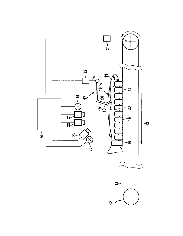
Figure 1 shows a schematic diagram of the apparatus according to the invention
in
lateral view. The apparatus is designed and configured for optical inspection
of the
opened abdominal cavity 10 of gutted fish 11. The apparatus comprises a
conveying
device 12 configured for conveying the fish 11 along a processing line in the
longi-
tudinal direction 13. The conveying device 12 is designed, for example, as a
rotating
conveyor belt 14 with holding and centring means 15. The holding and centring
means
15 are configured to hold the fish 11 in the dorsal position and to align them
so that
they are conveyed in the longitudinal direction 13 with the opened abdominal
cavity 10
pointing upwards. The holding and centring means 15 are preferably designed in
a V
shape. One each of the holding and centring means is designed as a tail clamp
16
which secures the relevant fish 11 against slipping in the longitudinal
direction 13.
The apparatus according to the invention further comprises pivotable ventral
flaps
holding elements 17 for holding open the abdominal cavity 10 of the fish. The
ventral
flaps holding elements 17 preferably comprise a substantially V-shaped
spreading bow
18 which is moved via a pivot lever 19 into the abdominal cavity 10 and back
out of it
17321 -8 translation
CA 03019085 2018-09-26
- 8 -
again. The ventral flaps holding elements 17 are designed and configured to
spread
apart the ventral flaps of the opened abdominal cavity 10 of the fish 11.
At least one optical imaging system is arranged on the processing line, for
example ¨
as shown in Figure 1 ¨ a first imaging system 21, a second imaging system 22
and a
third imaging system 23. The present invention, however, is not limited to the
number
of imaging systems 21, 22, 23 shown but alternatively may also comprise fewer
or
more of the imaging systems 21, 22, 23. The imaging systems 21, 22, 23 are
designed
and configured to take at least one sequence of images of abdominal cavity
regions of
one of the fish 11 in each case, said image sequence comprising a plurality of
single
images.
Using a first illumination device 24 and a second illumination device 25, the
abdominal
cavity regions are illuminated, in each case with light of a predetermined
spectral
composition, in each case at the times the single images are taken. As shown
in Figure
1, the number of illumination devices is not limited to only two.
Alternatively, it is also
possible to employ only a single one of the illumination devices 24, 25 or to
use more
than two of the illumination devices 24, 25.
An evaluation unit 26 is designed and configured to evaluate the image
sequence in
respect of prescribed quality parameters and to activate an ejection device ¨
not shown
in the drawing ¨ to remove the relevant fish 11 from the processing line by
ejecting it,
should the evaluation reveal any deviation from the prescribed quality
parameters. The
prescribed quality parameters relate, for example, to optical comparative
values that
.. provide information about the extent or degree to which the guts have
already been
removed. This also includes comparative values pertaining to blood residues or
connective tissue residues. In addition, the quality parameters include
variables for
checking the correct position of the ventral incision.
The evaluation unit 26 is connected to the optical imaging systems 21, 22, 23,
the first
and second illumination devices 24, 25, an actuator 27 for moving the ventral
flaps
holding elements 17 and preferably to a drive 31 for driving the conveying
device 12
such that the evaluation unit 26 can control or regulate the aforementioned
com-
ponents. The connections are preferably designed as electrical connections.
17321 - 8 - translabon
CA 03019085 2018-09-26
- 9 -
The first illumination device 24 and the second illumination device 25 are
each con-
figured to illuminate the abdominal cavity region, in each case with light of
different
spectral ranges at the times the images are taken. The spectral ranges each
comprise
a section of the visible or infra-red wavelength spectrum.
Preferably, the first illumination device 24 and the second illumination
device 25 each
comprise a light source which is configured to emit light of a narrow band
spectrum.
Narrow band means that the light covers a spectral range having a wavelength
smaller
than 100 nm. Especially preferably, the light is monochromatic in each case.
The evaluation unit 26 further comprises control means which are designed and
con-
figured to control the imaging systems 21, 22, 23 in such a way that the time
interval
between taking two consecutive single images of one of the image sequences is
chosen such that the two consecutive single images at least substantially
illustrate the
same abdominal cavity region. In other words, the control means are
configured, in
coordination with the set conveying speed of the conveying device 12, to take
the
single images at such a time interval that each of the single images
substantially shows
the same region of the abdominal cavity 10.
As can be seen from Figures 2 and 3, a plurality of optical imaging systems
21, 22, 23
are arranged along the processing line, in each case only the first imaging
system 21
and the third imaging system 23 being illustrated in the detail views of
Figures 2 and 3.
The optical imaging systems 21, 22, 23 are preferably each arranged and
configured
separately to take each of the single images with one of the optical imaging
systems
21, 22, 23. For the sake of clarity, the ventral flaps holding elements 17
which hold
open the abdominal cavity 10 of each fish 11 for optical inspection are not
shown in
Figures 2 and 3.
Especially preferably, each of the optical imaging systems 21, 22, 23 is
assigned a
separate illumination means which is configured to illuminate the abdominal
cavity
regions in each case at the times the images are taken. As shown in Figures 2
and 3,
the first illumination device 24 is assigned to the first imaging system 21
and the
second illumination device 25 is assigned to the third imaging system 23.
According to an advantageous embodiment of the invention, the imaging systems
21,
22, 23 are each designed as a black and white or as a monochrome camera.
17321 - 8 - transLation
CA 03019085 2018-09-26
-
Alternatively, one or more of the imaging systems 21, 22, 23 in each case is
designed
as a colour camera.
At least one of the optical imaging systems 21, 22, 23 is preferably arranged
in such a
5 manner that at least one of the single images is taken with a viewing
direction onto the
abdominal cavity 10 that is vertical or at least substantially vertical. For
this, for
example, the first imaging system 21 and the first illumination device 24 are
arranged ¨
as shown in Figure 2 ¨ above the conveying device 12.
10 As shown in Figure 3, at least one further optical imaging system, for
example the third
imaging system 23, is arranged in such a manner that at least one other of the
single
images is taken with a viewing direction that is inclined in relation to the
vertical viewing
direction. In particular, the third imaging system 23 is inclined in such a
manner that in
the inclined viewing direction the images are taken towards the head of the
fish.
The evaluation unit 26 is configured in particular to overlay the single
images of the
image sequence to create an overall image. The overall image may optionally be
displayed for control purposes. The overall image is further used for quality
control. The
evaluation unit 26 is preferably configured to assemble the image sequence
into a false
colour image by weighting the single frames differently.
In particular, the imaging systems 21, 22, 23 and the illumination devices 24,
25 are
preferably arranged encapsulated in a housing 28 with at least one viewing
window 30.
Figure 5 shows a schematic detail view with the ventral flaps holding elements
17 in a
working position of the apparatus according to the invention in lateral view.
The ventral
flaps holding elements 17 are guided into the abdominal cavity and hold the
ventral
flaps 20 open laterally so that the abdominal cavity can be inspected. Figure
4 shows
how the ventral flaps holding elements 17 are in a waiting position outside
the
abdominal cavity.
Figures 6 and 7 respectively show a perspective view of the detail views of
Figures 4
and 5. Figures 4 to 7 each show a centring aid 29, by means of which the fish
11 are
guided laterally and are thus centred in the middle.
17321 - 8 - translation
CA 03019085 2018-09-26
¨ 11 -
The present invention further comprises an apparatus for gutting fish 11 which
are
conveyed along a processing line in the longitudinal direction 13 by means of
the
conveying device 12. The apparatus comprises opening tools configured for
opening
the abdominal cavity 10 of the fish 11, gutting tools designed for cleaning
out the
abdominal cavity 10 and an apparatus for subsequent optical inspection of the
abdominal cavity 10 of the gutted fish 11 as described above. Such apparatuses
for
opening the abdominal cavity and the gutting tools are known, for example,
from
documents EP 1003378 Al and EP 1411775 and are therefore not further described
in
detail.
17321-8 translation