Note: Descriptions are shown in the official language in which they were submitted.
CA 03019311 2018-09-27
WO 2016/168658 PCT/US2016/027840
COMPOSITIONS AND METHODS FOR XI CHROMOSOME REACTIVATION
RELATED APPLICATIONS
This Application claims the benefit under 35 U.S.C. 119(e) of U.S. provisional
application USSN 62/148,106, filed April 15, 2015, entitled "COMPOSITIONS AND
METHODS FOR XI CHROMOSOME REACTIVATION", the entire contents of which are
incorporated by reference herein.
FEDERALLY SPONSORED RESEARCH
This invention was made with government support under Grant No. R01-GM033977
awarded by the National Institutes of Health. The government has certain
rights in the
invention.
FIELD OF THE DISCLOSURE
The invention relates to methods for reactivating mammalian inactive X
chromosomes through genetic and pharmacological means.
BACKGROUND OF THE DISCLOSURE
X chromosome inactivation (XCI), the random transcriptional silencing of one X
chromosome in somatic cells of female mammals, is a mechanism that ensures
equal
expression of X-linked genes in both sexes. XCI is initiated by Xist, a 17-kb
non-coding
RNA whose expression during early embryogenesis is both necessary and
sufficient for
silencing. Xist represses transcription in cis by coating only the X
chromosome from which it
is produced. Once Xist has been upregulated during early development or
differentiation, it
continues to be expressed from the inactive X (Xi) even in fully
differentiated somatic cells.
Prior to the initiation of XCI, Tsix, an antisense repressor of Xist, blocks
Xist upregulation on
the future active X chromosome (Xa).
An understanding of the factors and mechanisms involved in XCI is directly
relevant
to certain human diseases (e.g., dominant X-linked diseases). For example,
loss-of-function
mutations in the X-linked methyl-CpG binding protein 2 (MECP2) gene lead to
the
neurodevelopmental disorder Rett syndrome (RTT). Most RTT patients are females
who are
heterozygous for MECP2 deficiency due to random XCI. Therapeutic options for
the
treatment of dominant X-linked diseases, such as Rett syndrome, remain
limited.
Accordingly, there is a need for new compositions and methods of treatment for
dominant X-
linked diseases.
1
CA 03019311 2018-09-27
WO 2016/168658 PCT/US2016/027840
SUMMARY OF THE DISCLOSURE
The instant disclosure relates to methods and compositions for the
reactivation of
inactive X (Xi) chromosomes. In some aspects, the methods and compositions
described
herein may be useful for the treatment of dominant X-linked diseases, such as
Rett syndrome.
The disclosure is based, in part, on the discovery that inhibition of X
chromosome
inactivating factors (XCIFs) can mediate reactivation of inactive X
chromosomes, re-
expression of Xi-linked genes and/or reduce expression or activity of the
Xist.
Accordingly, in some aspects, the disclosure provides a method of inducing
expression of an X-linked gene in a cell having an inactive X chromosome, the
method
comprising delivering to the cell an X chromosome inactivation factor (XCIF)
inhibitor in an
amount effective for inducing expression of the X-linked gene.
In some aspects, the disclosure provides a method of treating a subject having
a
dominant X-linked disease, the method comprising administering to the subject
an X
chromosome inactivation factor (XCIF) inhibitor in an amount effective for
inducing
expression a target X-linked gene. In some embodiments, the dominant X-linked
disease
results from a mutated allele of the X-linked gene, and wherein the inhibitor
is administered
in an amount effective for inducing expression of a wild-type allele of the X-
linked gene.
In some embodiments, the cell is of a subject having a dominant X-linked
disease
resulting from a mutated allele of the X-linked gene. In some embodiments, the
X-linked
gene is MECP2. In some embodiments, the X-linked gene is MECP2 and the X-
linked
disease is Rett Syndrome.
In some embodiments, the dominant X-linked disease is selected from the group
consisting of: X-linked hypophosphatemia, incontinentia pigmenti type 2,
Aicardi syndrome,
CDK5L syndrome, focal dermal hypoplasia, CHILD syndrome, Lujan-Fryns syndrome,
orofaciodigital syndrome 1, hereditary nephritis (Alport syndrome), Giuffre-
Tsukahara
syndrome, Goltz syndrome, Fragile X syndrome, Bazex-Dupre-Christol syndrome,
Charcot-
Marie-Tooth disease, chondrodysplasia punctate, erythropoietic protoporphyria,
scapuloperoneal myopathy, and craniofrontonasal dysplasia.
In some embodiments, the XCIF inhibitor selectively inhibits activity of an X
chromosome inactivation factor selected from the group consisting of: ACVR1,
AURKA,
DNMT1, FBX08, LAYN, NF1, PDPK1, PYG01, RNF165, SGK1/2, 50X5, STC1, ZNF426
and C17orf98. In some embodiments, the X chromosome inactivation factor is
PI3K and the
2
CA 03019311 2018-09-27
WO 2016/168658 PCT/US2016/027840
XCIF inhibitor is GNE-317 or LY29400. In some embodiments, the X chromosome
inactivation factor is PDPK1 and the XCIF inhibitor is OSU-03012 or BX912. In
some
embodiments, the X chromosome inactivation factor is AURKA and the XCIF
inhibitor is
VX680, CD532, or MLN8237. In some embodiments, the X chromosome inactivation
factor
is SGK1/2 and the XCIF inhibitor is GSK650394. In some embodiments, the X
chromosome
inactivation factor is ACVR1 and the XCIF inhibitor is dorsomorphin, K02288 or
LDN193189.
In some embodiments, the XCIF inhibitor selectively inhibits activity of
mammalian
target of rapamycin (mTOR). In some embodiments, the XCIF inhibitor is
rapamycin, KU-
0063794, or everolimus.
In some embodiments, the XCIF inhibitor is an inhibitory oligonucleotide
having a
region of complementarity that is complementary with at least 8 nucleotides of
an mRNA
encoded by an XCIF gene. In some embodiments, the inhibitory oligonucleotide
is selected
from the group consisting of: antisense oligonucleotide, siRNA, shRNA and
miRNA. In
some embodiments, the inhibitory oligonucleotide is a modified inhibitory
oligonucleotide.
In some embodiments, the modified inhibitory oligonucleotide comprises a
bridged
nucleotide (e.g., a locked nucleic acid (LNA)), phosphorothioate backbone,
and/or a 2'-0Me
modification.
In some embodiments, the method further comprises determining that cell has a
mutant allele of the X-linked gene. In some embodiments, the method further
comprises
determining that delivery of the XCIF inhibitor to the cell results in induced
expression of the
X-linked gene. In some embodiments, the method further comprises determining
that
delivery of the inhibitor to the cell results in induced expression of a wild-
type allele of the
X-linked gene. In some embodiments, the method further comprises determining
that
delivery of the XCIF inhibitor to the cell results in reactivation of an X
chromosome. In
some embodiments, the method further comprises determining that delivery of
the XCIF
inhibitor to the cell results in decreased expression or activity of XIST. In
some
embodiments, the cell is in vitro. In some embodiments, the cell is in a
subject.
BRIEF DESCRIPTION OF THE DRAWINGS
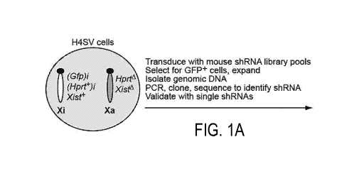
FIGS. 1A-1C show identification of factors involved in mammalian XCI. FIG. lA
shows a schematic summary of the shRNA screen. The Xi is designated as such
due to
deletion of Xist on the Xa. FIG. 1B shows H4SV cells expressing an shRNA
against one of
3
CA 03019311 2018-09-27
WO 2016/168658 PCT/US2016/027840
the 13 candidates or, as a control, a non-silencing (NS) shRNA were FACS
sorted and GFP-
positive cells isolated. For each KD cell line, the percent GFP-positive cells
was expressed
as the fold increase relative to that obtained with the NS shRNA, which was
set to 1. FIG. 1C
shows two-color RNA FISH monitoring expression of G6pdx and Lamp2 (left) and
Pgkl and
Mecp2 (right) in each of the 13 XCIF KD BMSL2 cell lines. DAPI staining is
shown in blue.
The experiment was performed at least twice, and representative images are
shown (top) and
the results quantified (bottom) from one experiment.
FIGS. 2A-2D show XCIFs are involved in initiation of XCI in mouse embryonic
stem
cells. FIG. 2A shows two-color RNA FISH monitoring expression of G6pdx and
Lamp2 (left)
and Pgkl and Mecp2 (right) in the 13 XCIF KD ES cell lines following
differentiation. DAPI
staining is also shown. Representative images are shown (top) and the results
quantified
(bottom). FIG. 2B shows percentage of alkaline phosphatase-negative single
cells in the 13
XCIF KD ES cell lines before (top, undifferentiated) and after (bottom,
differentiated)
treatment with RA. FIG. 2C shows qRT-PCR analysis monitoring expression of
0ct4 in the
13 XCIF KD ES cell lines following treatment with RA. As a control, expression
of 0ct4 in
undifferentiated ES cells is shown and was set to 1. Error bars indicate SD.
FIG. 2D shows
qRT-PCR analysis of XCIFs in undifferentiated and differentiated mouse ES
cells.
Expression in differentiated ES cells was normalized to that observed in
undifferentiated
cells, which was set to 1. Error bars indicate SD.
FIGS. 3A-3I show XCIFs function by promoting Xist expression and/or
localization,
and DNMT1 is a transcriptional activator of Xist on the Xi. FIG. 3A shows qRT-
PCR
analysis monitoring Xist expression in the 13 XCIF KD ES cell lines following
differentiation. Expression in differentiated ES cells was normalized to that
obtained with the
NS shRNA, which was set to 1. Error bars indicate SE. FIG. 3B shows RNA FISH
monitoring localization of Xist in the 13 XCIF KD ES cell lines following
differentiation.
Cells were categorized as having either a typical Xist cloud or "other"
pattern, which includes
either the lack of a detectable Xist signal or presence of two small Xist
signals, as in
undifferentiated ES cells. FIG. 3C shows RNA FISH monitoring expression of
Xist (top) and
Mecp2 (bottom) in BMSL2 cells treated with an Xist locked nucleic acid
antisense
oligonucleotide (LNA ASO) or a control LNA ASO. FIG. 3D shows ChIP analysis
monitoring binding of DNMT1 and POL2 to the Xist promoter and exon 2 in BMSL2
cells
expressing a NS or Dnmtl shRNA. Error bars indicate SD. FIG. 3E shows nuclear
run-on
4
CA 03019311 2018-09-27
WO 2016/168658 PCT/US2016/027840
assay monitoring transcription of Xist, Hprt and Tbp in BMSL2 cells expressing
a NS or
DNMT1 shRNA. FIG. 3F shows qRT-PCR analysis monitoring Xist levels in BMSL2
cells
expressing a NS or Dnmtl shRNA following treatment with actinomycin D. Actin
mRNA
was used as a normalization control. Error bars indicate SD. FIG. 3G shows qRT-
PCR
analysis monitoring Xist expression in MEFs isolated from female Dnmtl +/+ and
Dnmtl -/-
embryos. Four different litters were analyzed (n=4 mice total per genotype),
and the results
were averaged. Expression was normalized to that observed in Dnmtl +/+ MEFs,
which was
set to 1. Error bars indicate SD.*P<0.001 (Student's t-test). FIG. 3H shows
qRT-PCR
monitoring levels of Xist and Tsix in H4SV cells expressing a NS or DNMT1
shRNA.
Expression was normalized to that obtained with the NS shRNA, which was set to
1. Error
bars indicate SD. FIG 31 shows qRT-PCR analysis monitoring Hprt and Xist
expression in
BMSL2 cells treated in the absence or presence of 5-AZA. Expression was
normalized to that
observed in the absence of 5-AZA, which was set to 1. Error bars indicate SD.
FIGS. 4A-4I show reactivation of the Xi-linked Mecp2 gene by small molecule
XCIF
inhibitors. FIGs. 4A-4B show two-color RNA FISH monitoring expression of Xist
and Mecp2
in differentiated mouse ES cells treated with DMSO (control or ¨), OSU-03012
or LY294002
(FIG. 4A), and in BMSL2 cells treated with DMSO or GNE-317 (FIG. 4B).
Representative
images are shown (top) using the higher concentrations of the inhibitors, and
the results
quantified (bottom). Yellow arrowheads indicate co-localizing Xist and Mecp2
signals; white
arrowheads indicate Mecp2 signals not co-localizing with Xist. FIG. 4C shows
two-color
RNA FISH monitoring Xist and Mecp2 expression in mouse cortical neurons
treated with
DMSO (control or ¨), OSU-03012, BX912 or LY294002. Representative images are
shown
(top) and the results quantified (bottom). Arrowheads indicate Mecp2 signals.
FIG. 4D
shows two-color RNA FISH monitoring expression of Xist and Mecp2 in mouse
BMSL2
fibroblasts treated with DMSO (control or ¨) or G5K650394. Representative
images are
shown (top) and the results quantified (bottom). Arrowheads indicate Mecp2
signals. FIG. 4E
shows qRT-PCR monitoring expression of Xist (left) and Mecp2 (right) in BMSL2
cells
treated with DMSO or increasing concentrations of G5K650394 (2.5, 5 or 10
t.M). FIG. 4F
shows two-color RNA FISH monitoring expression of Xist and Mecp2 in BMSL2
cells
treated with DMSO or K02288. Representative images are shown (top) and the
results
quantified (bottom). Arrowheads indicate Mecp2 signals. FIG. 4G shows qRT-PCR
monitoring expression of Xist (left) and Mecp2 (right) in BMSL2 cells treated
with DMSO,
1(02288 (0.5 t.M) or LDN193189 (0.5 t.M). FIG. 4H shows Two-color RNA FISH
CA 03019311 2018-09-27
WO 2016/168658 PCT/US2016/027840
monitoring Xist and Mecp2 expression in BMSL2 cells treated with DMSO (control
or ¨),
LY294002 or OSU-03012, and at least 6 days following removal of the inhibitor.
Representative images are shown (top) and the results quantified (bottom).
Arrowheads
indicate Mecp2 signals. FIG. 41 shows qRT-PCR monitoring Xi-linked wild-type
MECP2
expression in human RTT fibroblasts treated with DMSO (¨), 5-azacytidine (5-
AZA),
BX912, OSU-03012 or VX680. As a control, Xa-linked wild-type MECP2 expression
was
monitored in another clonal fibroblast cell line derived from the same RTT
patient (lane 1).
The arrowhead indicates the wild-type MECP2 qRT-PCR product. GAPDH was
monitored as
a loading control. Bottom, schematic of the MECP2 wild-type (wt) and mutant
(mut) alleles.
FIGS. 5A-5B show defective XCI in female Stc/-/- MEFs. FIG. 5A shows two-color
RNA FISH monitoring expression of G6pdx and Lamp2 (top) and Pgkl and Mecp2
(bottom)
in female Stc/ +1+ and Stc/-/- MEFs, and as a control male Stc/-/- MEFs.
Representative
images are shown (top) and the results quantified (bottom). FIG. 5B shows qRT-
PCR
analysis monitoring Xist expression in MEFs isolated from female Stc/ +/+ and
Stc/-/-
embryos. Four different litters were analyzed (n=4 mice total per genotype),
and the results
were averaged. Expression was normalized to that of the ribosomal gene RPM,
and Xist
expression in Stc/ +/+ MEFs was set to 1. Error bars indicate SD. *13<0.001
(Student's t-
test).
FIGS. 6A-6G show defective XCI in female Stc/-/- mice is not accompanied by
increased X-linked gene expression. FIG. 6A shows a schematic of the RNA-Seq
analysis
pipeline. FIG. 6B shows 6istribution of 1og2 transformed ratio of X-linked
gene expression
in MEFs from female Stc/-/- (KO) and Stc/ +1+ (WT) embryos (n=3 per genotype).
FIG. 6C
shows a box plot of X-linked gene expression (10g2 transformed FPKM) in MEFs
from
female Stc/-/- and Stc/ +1+ embryos (n=3 per genotype). Boxed areas span the
first to the
third quartile. Whiskers represent 15th and 85th percentiles. FIG. 6D shows
qRT-PCR analysis
monitoring expression of Mecp2 and Hprt in MEFs from 2 different litters of
female Stc/-/-
and Stc/ +1+ embryos (n=2 mice total per genotype). The results were
normalized to those
obtained in Stc/ +1+ MEFs, which was set to 1. Error bars indicate SE. FIG. 6E
shows an
immunoblot showing MECP2 and STC1 levels in female Stc/ +1+ and Stc/-/- MEFs
(left) or
brain tissue female Stc/ +/+ and Stc/-/- P1 mice (right) (n=3 per genotype). a-
tubulin
(TUBA) was monitored as a loading control. FIG. 6F shows qRT-PCR analysis of
Stc/, Xist,
6
CA 03019311 2018-09-27
WO 2016/168658 PCT/US2016/027840
Mecp2 and Hprt expression in BMSL2 cells expressing a NS or STC1 shRNA. The
results
were normalized to those obtained with the NS shRNA, which was set to 1. Error
bars
indicate SE. FIG. 6G shows an immunoblot showing MECP2 and STC1 levels in
BMSL2
cells expressing a NS or Stcl shRNA.
FIGS. 7A-7D show shRNAs targeting an XCIF reactivate the Xi-linked Hprt gene
and decrease mRNA levels of the targeted gene. FIG. 7A shows bright field
images showing
growth of the 13 XCIF KD H4SV cell lines following selection in HAT medium.
FIG. 7B
shows qRT-PCR analysis monitoring target gene expression in the 13 XCIF KD
H4SV cell
lines expressing the shRNA identified in the primary screen. For each gene,
knockdown
efficiency was determined relative to the level of target gene expression in
the control cell
line expressing a non-silencing (NS) shRNA, which was set to 1. Error bars
indicate SD.
FIG. 7C shows bright field images showing growth of the 13 XCIF KD H4SV cell
lines,
expressing a second, unrelated shRNA to that shown in FIG. 7A, following
selection in HAT
medium. FIG. 7D shows qRT-PCR analysis monitoring target gene expression in
the 13
XCIF KD H4SV cell lines expressing a second, unrelated shRNA to that shown in
FIG. 7B.
Error bars indicate SD.
FIGS. 8A-8D show additional RNA FISH images and control experiments related to
FIGS. 1A-1C. FIG. 8A shows representative two-color RNA FISH images showing
expression of G6pdx and Lamp2 (top) and Pgkl and Mecp2 (bottom) in each of the
13 XCIF
KD BMSL2 cell lines. DAPI staining is also shown. FIG. 8B shows that in BMSL2
cells the
Xi and Xa encode two distinguishable Pgkl alleles, Pgkla and Pgklb,
respectively, which
differ by a single nucleotide polymorphism within the mRNA. Allele-specific
expression of
the Xi- and Xa-linked Pgkl genes in each of the 13 XCIF KD BMSL2 cell lines
was
analyzed using a single nucleotide primer-extension (SNuPE) assay. The ratio
of
Pgkla:Pgklb expression was calculated and normalized to that obtained with the
NS shRNA,
which was set to 1. The results show that in each of the 13 XCIF KD BMSL2 cell
lines the
ratio of Pgkla to Pgklb was increased, indicating that knockdown of each of
the 13 XCIFs
reactivated the Xi-linked Pgk-la gene. FIG. 8C shows that in BMSL2 cells the
Xi and Xa
encode two distinguishable Pgkl alleles, Pgkla and Pgklb, respectively, which
differ by a
single nucleotide polymorphism within the mRNA. Allele-specific expression of
the Xi- and
Xa-linked Pgkl genes in six representative XCIF KD BMSL2 cell lines was
analyzed using a
single nucleotide primer extension (SNuPE) assay. The data are plotted as the
function of
7
CA 03019311 2018-09-27
WO 2016/168658 PCT/US2016/027840
ARn for each sample, which represents the reporter fluorescence for each
allele (VIC/FAM)
normalized to the passive dye. The results show that in each of the six XCIF
KD BMSL2
cell lines the Xi-linked Pgkl a gene was reactivated. FIG. 7D shows X
chromosome painting
experiments in the 13 XCIF KD BMSL2 cell lines. The results show that the X
chromosome
content of the XCIF KD BMSL2 cell lines was similar to that of the control
BMSL2 cell line
expressing a NS shRNA. Thus, the substantially increased bi-allelic expression
of X-linked
genes observed by RNA FISH in the XCIF KD cell lines cannot be explained by
differences
in X chromosome number.
FIGS. 9A-9C show additional RNA FISH images and control experiments related to
FIGS. 2A-2D. FIG. 9A shoes representative two-color RNA FISH images monitoring
expression of G6pdx and Lamp2 (top) and Pgkl and Mecp2 (bottom) in the 13 XCIF
KD ES
cell lines following differentiation. DAPI staining is also shown. FIG. 9B
shows X
chromosome painting experiments in the 13 XCIF KD ES cell lines following
differentiation.
FIG.9C shows qRT-PCR analysis monitoring expression of Eames, Tcf712 and Cdx2
in the 13
XCIF KD ES cell lines following treatment with RA. As a control, expression of
each gene
in undifferentiated ES cells is shown and was set to 1. Error bars indicate
SD.
FIGS. 10A-10C show RNA FISH images and control experiments related to FIGS.
3A-3I. FIG. 10A shows RNA FISH images. In each of the 13 XCIF KD ES cell lines
following differentiation, the majority of cells that lost the typical Xist
localization pattern
lacked a detectable Xist signal (see FIG. 3B). However, some cells that had
lost the typical
Xist localization pattern contained two small Xist signals, reminiscent of
undifferentiated ES
cells. Examples of this latter localization pattern are shown here. Nuclear
signals are
indicated in red and denoted by arrowheads; DAPI staining is also shown. FIG.
10B shows
qRT-PCR analysis monitoring expression of Xist (left), Tsix (middle) and Dnmtl
(right) in
H4SV cells expressing a NS or one of two Dnmtl shRNAs (Dnmtl-1 or Dnmtl-2).
For Xist
and Tsix expression, a second, unrelated Dnmtl shRNA to that used in FIG. 3H.
Expression
was normalized to that obtained with the control NS shRNA, which was set to 1.
Error bars
indicate SD. FIG. 10C shows qRT-PCR analysis monitoring expression of Xist
(left), Tsix
(middle) and Dnmtl (right) in differentiated ES cells expressing a NS shRNA or
one of two
Dnmtl shRNAs (Dnmtl-1 or Dnmtl-2). Expression was normalized to that obtained
with the
control NS shRNA, which was set to 1. Error bars indicate SD.
8
CA 03019311 2018-09-27
WO 2016/168658 PCT/US2016/027840
FIGS. 11A-11C show additional RNA FISH images related to FIGS. 4A-4E. FIG.
11A and FIG. 11B show two-color RNA FISH monitoring expression of Xist and
Mecp2 in
differentiated ES cells treated with DMSO (control), OSU-03012 (4 t.M) or
LY294002 (10
i.t.M) (FIG. 11A), and in BMSL2 cells treated with DMSO or GNE-317 (5 t.M)
(FIG. 11B).
Yellow boxes indicate cells with co-localizing Xist and Mecp2 signals; white
boxes indicate
cells with biallelic expression of Mecp2 and complete loss of the Xist signal.
FIG. 11C shows
two-color RNA FISH monitoring Xist and Mecp2 expression in BMSL2 cells treated
with
DMSO (control), OSU-03012 (2.5 t.M) or LY294002 (8 t.M), and at least 6 days
following
removal of the inhibitor. White boxes indicate cells with biallelic expression
of Mecp2.
FIGS. 12A-12B show control experiment and RNA FISH images related to FIG. 5.
FIG. 12A shows X chromosome painting experiments in female Stc/ +/+ and Stc/-/-
MEFs.
The results show that the X chromosome content of Stc/-/- MEFs was similar to
that of
Stc/ +/+ MEFs. Thus, the substantially increased bi-allelic expression of X-
linked genes
observed by RNA FISH in the Stc/-/- MEFs cannot be explained by differences in
X
chromosome number. FIG. 12B shows defective XCI in cortical neurons from brain
sections
of female Stc/-/- mice. Two-color RNA FISH monitoring expression of Xist and
Mecp2 or
G6pdx in cortical neurons from adjacent 5-1.tm brain sections of female Stc/-/-
and Stc/ +/+
mice (n=3 per genotype, stage P1). Boxed regions denote cells with two Mecp2
or G6pdx
signals; yellow boxes indicate cells with co-localizing Xist and Mecp2/G6pdx
signals. All
cells in the regions shown represent neurons that, based on anatomical
landmarks, are present
in post-hybridized sections.
FIGS. 13A-13E show additional experiments and data analyses related to FIGS.
6A-
6G. FIG. 13A shows a volcano plot showing distribution of 1og2 transformed
ratio of X-
linked gene expression in MEFs isolated from Stc/-/- (KO) and Stc/ +1+ (WT)
embryos (n=3
per genotype). The genes are plotted against negative transformed log of P
value. Red
circles represent genes with a >2-fold change in expression and P <0.01. The
results show
that the similarity of X-linked gene expression between female Stc/ +/+ and
Stc/-/- MEFs
was statistically significant. FIG. 13B shows box plots displaying changes in
autosomal gene
expression (10g2 transformed FPKM) in Stc/-/- and Stc/ +/+ MEFs. Boxed areas
span the
first to the third quartile. Whiskers represent 15th and 85th percentiles;
samples falling outside
these percentiles are shown as circles. FIG. 13C shows XCIFs are not generally
required for
repression of imprinted genes. Primary female mouse embryonic fibroblasts from
the strain
C57BL/6 (CAST7), which contains chromosome 7 from Mus castaneus (Cast), were
9
CA 03019311 2018-09-27
WO 2016/168658 PCT/US2016/027840
transduced with shRNAs against each of the XCIFs and analyzed for allele-
specific
expression of four genes located on chromosome 7 that are either paternally
expressed,
(Kcnqlotl and Peg3) or maternally expressed (Ascl2 and Zim/). Expression of
the two
alleles can be distinguished by allele-specific restriction enzyme digestion
following gene-
specific RT-PCR. The sizes of the undigested and digested bands are indicated,
and the sizes
of the predicted digested fragments are shown in the table (bottom). If
knockdown of an
XCIF results in reactivation of the normally silenced allele, a mixture of the
maternal and
paternal allele-specific digestion patterns would be observed. The results
show that in all 13
XCIF KD cell lines, all four genes displayed only the expected allele-specific
expression
pattern, indicating that the XCIFs are not generally required for repression
of the imprinted
genes. FIG. 13D shows involvement of Polycomb subunits EZH2 and BMI1 for
repression
of the X-linked Hprt gene. (Left) qRT-PCR analysis monitoring Hprt expression
in BMSL2
cells expressing an Ezh2 or Bmil shRNA or, as a control, a NS shRNA. (Right)
qRT-PCR
analysis confirming target gene knockdown in mouse ES cells expressing an Ezh2
(left) or
Bmil (right) shRNA. Error bars indicate SD. FIG. 13E shows analysis of
available datasets
from Yildirim et al. 2013 showing the distribution of 1og2 transformed ratio
of X-linked gene
expression in hematopoietic cells from female heterozygous (HET) Xist mutant
mice and
wild-type (WT) mice. The data were downloaded from Gene Expression Omnibus
(G5E43961), normalized by RMA and filtered by detection above background (DAB
G)
(cutoff P-value <0.0001) using Bioconductor package xps. The percentage of X-
linked genes
upregulated >1.5-fold is shown.
FIG. 14 shows a schematic diagram of downstream targets of 3-phosphoinositide
dependent protein kinase-1 (PDPK1).
FIG. 15 shows treatment of mouse fibroblasts with an mTOR inhibitor
reactivates the
Xi-linked Mecp2 gene. Relative expression of Xist and Mecp2 in mouse
fibroblasts was
measured after treatment with rapamycin, KU-0063794, or everolimus (left).
Mecp2 RNA
was measured by fluorescence in situ hybridization (FISH) and percentage of
nuclei stained
was quantified (right).
FIG. 16 shows treatment of mouse fibroblasts with an mTOR inhibitor
(rapamycin,
KU-0063794, everolimus) reactivates the Xi-linked Hprt gene, as measured by a
hypoxanthine-aminopterin-thymidine (HAT) selection assay.
CA 03019311 2018-09-27
WO 2016/168658 PCT/US2016/027840
FIG. 17 shows inhibition of Aurora kinase A (AURKA) reactivates Xi-linked
genes.
Relative expression of Xist and Mecp2 in mouse fibroblasts was measured after
treatment
with CD532 and MLN8237. Mecp2 RNA was measured by fluorescence in situ
hybridization (FISH) and percentage of nuclei stained was quantified (right).
Results were
confirmed using a HAT selection assay.
FIG. 18 shows treatment of mouse fibroblasts with Activin Receptor Type 1
(ACVR1) inhibitor reactivates Xi-linked genes. Relative expression of Xist and
Mecp2 in
mouse fibroblasts was measured after treatment with K02288, dorsomorphin, or
LDN193189.
Mecp2 RNA was measured by fluorescence in situ hybridization (FISH) and
percentage of
nuclei stained was quantified (right). Results were confirmed using a HAT
selection assay.
DETAILED DESCRIPTION
Aspects of the disclosure relate to the biological and pharmacological
inhibition or
reversal of X chromosome inactivation. The disclosure is based, in part, on
the discovery that
inhibition of X chromosome inactivating factors (XCIFs) can mediate
reactivation of inactive
X chromosomes, re-expression of X-linked genes and/or reduce expression or
activity of Xist.
In some aspects, the disclosure relates to a method of inducing expression of
an X-
linked gene in a cell having an inactive X chromosome, the method comprising
delivering to
the cell an X chromosome inactivation factor (XCIF) inhibitor in an amount
effective for
inducing expression of the X-linked gene. As used herein, the term "X
chromosome
inactivation factor" refers to a gene or gene product (e.g., a protein) that
are required for or
involved in maintenance or establishment of X chromosome inactivation. In some
embodiments, inhibition of XCIF expression and/or activity leads to
reactivation of an
inactivated X chromosome or one or more genes residing thereon (Xi-linked
genes). Thirteen
X chromosome inactivation factors (XCIFs) have been identified herein (Table
1), and are
indicated as being involved in diverse processes including cell signaling
(ACVR1, AURKA,
NF1, LAYN and PDPK1), cell metabolism (STC1), ubiquitin-dependent regulation
(FBX08
and RNF165) and transcription (PYG01, 50X5 and ZNF426), for example, as
disclosed in
Bhatnagar et al., 2014, Proc Natl Acad Sci USA 111:12591-12598.
XCIF Inhibitors
The disclosure relates in part to a discovery of inhibitors of XCIFs that can
reactivate
expression of the Xi-linked genes. Inhibitors of XCIFs can be peptides,
proteins, antibodies,
11
CA 03019311 2018-09-27
WO 2016/168658 PCT/US2016/027840
small molecules, or nucleic acids. In some embodiments, an XCIF inhibitor
selectively
inhibits activity of an X chromosome inactivation factor selected from the
group consisting
of: ACVR1, AURKA, DNMT1, FBX08, LAYN, NF1, PIK3, PDPK1, PYG01, RNF165,
SOX5, STC1, ZNF426 and Cl7orf98.
Aspects of the disclosure relate to inhibition of Activin Receptor Type 1
(ACVR1), an
XCIF that encodes a receptor serine-threonine kinase (also known as ALK2) that
mediates
signaling by bone morphogenic proteins (BMPs). Gain-of-function mutations in
ACVR1
result in the autosomal dominant disease fibrodysplasia ossificans progressiva
(FOP) and
have been found in the childhood malignancy diffuse intrinsic pontine glioma
(DIPG).
Several small molecule ACVR1 inhibitors are available, including K02288 and
LDN193189.
K02288 is a potent and selective inhibitor of BMP type 1 receptor signaling;
strongly
inhibiting ACVR1/ALK2, ALK1, and ALK6, and weakly inhibiting the other ALKs
and
ActRIIA. LDN 193189 is a selective BMP signally inhibitor that inhibits the
transcriptional
activity of the BMP type I receptors ACVR1/ALK2 and ALK3; it also exhibits 200-
fold
selectivity for BMP versus TGF-f3. Further examples of ACVR1 inhibitors
include
LDN19318, DMH-1, ML-347, BML-275, dorsomorphin, and LDN-212854.
Aspects of the disclosure relate to inhibition of Aurora Kinase A (AURKA). In
some
embodiments, AURKA inhibitors are small molecules. Examples of AURKA
inhibitors
include but are not limited to VX-680, MLN8237, TAS-119, MLN8054, PF-03814735,
SNS-
314, BI 811283, AMG 900, AZD1152, A5703569, R763, PHA-739358, CD532, and MK-
0457. In some embodiments, the X chromosome inactivation factor is AURKA and
the
XCIF inhibitor is VX680. In some embodiments, the X chromosome inactivation
factor is
AURKA and the XCIF inhibitor is CD532 or MLN8237.
Aspects of the disclosure relate to inhibition of DNA (cytosine-5)-
methyltransferase 1
(DNMT1). In some embodiments, DNMT1 inhibitors are small molecules. Examples
of
DNMT1 inhibitors include but are not limited to azacitadine, fazarabine,
decitabine,
sinefungin, psammaplin A, disulfiram, zebularine, and SGI-1027.
Aspects of the disclosure relate to the inhibition of PI3K/Akt signaling to
reactivate
Xi-linked genes. In some embodiments, PI3K inhibitors are small molecules.
Examples of
PI3K inhibitors include but are not limited to GNE317, LY294002, Wortmannin,
demethoxyviridin, BEZ235, BGT226, BKM120, BYL719, XL765, XL147, GDC-0941,
SF1126, G5K1059615, PX-866, CAL-101, BAY80-6946, GDC-0032, IPI-145, VS-5584,
Z5TK474, 5AR245409, and RP6530. In some embodiments, the XCIF is PI3K and the
XCIF inhibitor is GNE-317 or LY29400.
12
CA 03019311 2018-09-27
WO 2016/168658 PCT/US2016/027840
Aspects of the disclosure relate to inhibition of 3-phosphoinositide-dependent
protein
kinase 1 (PDPK1). In some embodiments, PDPK1 inhibitors are small molecules.
Examples
of PDPK1 inhibitors include but are not limited to OSU-03012 , BAG-956, BX-
795, GSK-
2334470, BX-912, and PHT-427. In some embodiments, the XCIF is PDPK1 and the
XCIF
inhibitor is OSU-03012 or BX912.
The serum and glucocorticoid kinase (SGK) family of serine/threonine kinases
includes three distinct but highly homologous isoforms (SGK1, SGK2, and SGK3)
that share
a similar domain structure. All three are activated by PDPK1 and have been
implicated in a
wide variety of cellular processes and small molecule inhibitors with
selectivity for SGKs
over AKTs have been developed. Examples of SGK1/2 inhibitors include GSK-
650394 and
EMD638683.
In some embodiments, an XCIF inhibitor targets a downstream substrate of
PDPK1.
Examples of downstream substrates to PDPK1 include but are not limited to AKT
(also
known as protein kinase B), ribosomal protein S6 kinase beta-1 (S6K1), protein
kinase C
(PKC), ribosomal s6 kinase (e.g. , p70rsk, S6 Kinase), rho-associated, coiled-
coil-containing
protein kinase 1 (ROCK1), and mammalian target of rapamycin (mTOR). In some
embodiments, an XCIF inhibitor targets mTOR. In some embodiments, an mTOR
inhibitor
is a small molecule. Examples of mTOR inhibitors include but are not limited
to rapamycin,
everolimus, sirolimus, temsirolimus, deforolimus, and KU-0063794.
In some embodiments, the term "small molecule" refers to a synthetic or
naturally
occurring chemical compound, for instance a peptide or oligonucleotide that
may optionally
be derivatized, natural product or any other low molecular weight (often less
than about 5
kDalton) organic, bioinorganic or inorganic compound, of either natural or
synthetic origin.
Such small molecules may be a therapeutically deliverable substance or may be
further
derivatized to facilitate delivery.
As used herein the term "inhibitor" or "repressor" refers to any agent that
inhibits,
suppresses, represses, or decreases a specific activity, such as the activity
of an X
chromosome inactivation factors.
In some embodiments, an XCIF inhibitor when delivered to a cell reactivates an
inactive X chromosome or one or more genes residing thereon. In some
embodiments,
delivery of an XCIF inhibitor to a cell results in an increase in the level of
expression of an
Xi-linked gene (a gene residing on the inactive X-chromosome) of at least 10%,
20%, 30%,
40%, 50%, 60%, 70%, 80%, 90%, 100%, 200% or 500% compared with the level of
expression of the gene in a control cell that has not been delivered an XCIF
inhibitor. In
13
CA 03019311 2018-09-27
WO 2016/168658 PCT/US2016/027840
some embodiments, delivery of an XCIF inhibitor to a cell results in an
increase in the level
of expression of an Xi-linked gene (a gene residing in the inactive X-
chromosome) in a range
of 10% to 50%, 10% to 100%, 10% to 200%, 50% to 500% or more compared with the
level
of expression of the gene in a control cell that has not been delivered an
XCIF inhibitor.
Inhibitory Oligonucleotides
In some embodiments, the XCIF inhibitor is an inhibitory oligonucleotide.
Inhibitory
oligonucleotides may interfere with gene expression, transcription and/or
translation.
Generally, inhibitory oligonucleotides bind to a target polynucleotide via a
region of
complementarity. For example, binding of inhibitory oligonucleotide to a
target
polynucleotide can trigger RNAi pathway-mediated degradation of the target
polynucleotide
(in the case of dsRNA, siRNA, shRNA, etc.), or can block the translational
machinery (e.g.,
antisense oligonucleotides). In some embodiments, inhibitory oligonucleotides
have a region
of complementarity that is complementary with at least 8 nucleotides of an
mRNA encoded
by an XCIF gene. Inhibitory oligonucleotides can be single-stranded or double-
stranded. In
some embodiments, inhibitory oligonucleotides are DNA or RNA. In some
embodiments,
the inhibitory oligonucleotide is selected from the group consisting of:
antisense
oligonucleotide, siRNA, shRNA and miRNA. In some embodiments, inhibitory
oligonucleotides are modified nucleic acids.
The term "nucleotide analog" or "altered nucleotide" or "modified nucleotide"
refers
to a non-standard nucleotide, including non-naturally occurring
ribonucleotides or
deoxyribonucleotides. In some embodiments, nucleotide analogs are modified at
any
position so as to alter certain chemical properties of the nucleotide yet
retain the ability of the
nucleotide analog to perform its intended function. Examples of positions of
the nucleotide
which may be derivitized include the 5 position, e.g., 5-(2-amino)propyl
uridine, 5-bromo
uridine, 5-propyne uridine, 5-propenyl uridine, etc.; the 6 position, e.g., 6-
(2-amino)propyl
uridine; the 8-position for adenosine and/or guanosines, e.g., 8-bromo
guanosine, 8-chloro
guanosine, 8-fluoroguanosine, etc. Nucleotide analogs also include deaza
nucleotides, e.g., 7-
deaza-adenosine; 0- and N-modified (e.g., alkylated, e.g., N6-methyl
adenosine, or as
otherwise known in the art) nucleotides; and other heterocyclically modified
nucleotide
analogs such as those described in Herdewijn, Antisense Nucleic Acid Drug
Dev., 2000 Aug.
10(4):297-310.
14
CA 03019311 2018-09-27
WO 2016/168658 PCT/US2016/027840
Nucleotide analogs may also comprise modifications to the sugar portion of the
nucleotides. For example the 2' OH-group may be replaced by a group selected
from H, OR,
R, F, Cl, Br, I, SH, SR, NH2, NHR, NR2, COOR, or OR, wherein R is substituted
or
unsubstituted C1-C6 alkyl, alkenyl, alkynyl, aryl, etc. Other
possible modifications
include those described in U.S. Pat. Nos. 5,858,988, and 6,291,438. A locked
nucleic acid
(LNA), often referred to as inaccessible RNA, is a modified RNA nucleotide.
The ribose
moiety of an LNA nucleotide is modified with an extra bridge connecting the 2'
oxygen and
4' carbon.
The phosphate group of the nucleotide may also be modified, e.g., by
substituting one
or more of the oxygens of the phosphate group with sulfur (e.g.,
phosphorothioates), or by
making other substitutions which allow the nucleotide to perform its intended
function such
as described in, for example, Eckstein, Antisense Nucleic Acid Drug Dev. 2000
Apr.
10(2):117-21, Rusckowski et al. Antisense Nucleic Acid Drug Dev. 2000 Oct.
10(5):333-45,
Stein, Antisense Nucleic Acid Drug Dev. 2001 Oct. 11(5): 317-25, Vorobjev et
al. Antisense
Nucleic Acid Drug Dev. 2001 Apr. 11(2):77-85, and U.S. Pat. No. 5,684,143.
Certain of the
above-referenced modifications (e.g., phosphate group modifications)
preferably decrease the
rate of hydrolysis of, for example, polynucleotides comprising said analogs in
vivo or in vitro.
In some embodiments, the inhibitory oligonucleotide is a modified inhibitory
oligonucleotide.
In some embodiments, the modified inhibitory oligonucleotide comprises a
locked nucleic
acid (LNA), phosphorothioate backbone, and/or a 2'-0Me modification.
Methods of Treatment
The disclosure relates, in some aspects, to methods useful for the treatment
of certain
diseases, such as dominant X-linked diseases. For example, loss-of-function
mutations in the
X-linked methyl-CpG binding protein 2 (MECP2) gene lead to the
neurodevelopmental
disorder Rett syndrome (RTT).
Accordingly, in some aspects, the disclosure provides a method of treating a
subject
having a dominant X-linked disease, the method comprising administering to the
subject an X
chromosome inactivation factor (XCIF) inhibitor in an amount effective for
inducing
expression a target X-linked gene.
Dominant X-linked diseases typically result from a mutated allele of the X-
linked
gene. The disclosure relates, in part, to XCIF inhibitors that are effective
for inducing
expression of a wild-type allele of the X-linked gene. Examples of X-linked
diseases and
CA 03019311 2018-09-27
WO 2016/168658 PCT/US2016/027840
their associated X-linked genes include Rett syndrome (MECP2), X-linked
hypophosphatemia (PHEX), incontinentia pigmenti type 2 (IKB KG), Aicardi
syndrome (de
novo mutation of an X-linked gene), CDK5L syndrome (CDKL5), focal dermal
hypoplasia
(PORCN), CHILD syndrome (NSDHL), Lujan-Fryns syndrome (MED12), orofaciodigital
syndrome 1 (OFD1), hereditary nephritis or Alport syndrome (COL4A3, COL4A4,
COL4A5), Giuffre-Tsukahara syndrome (Xp22.13-q21.33), Goltz syndrome (PORCN),
Fragile X syndrome (FMR1), Bazex-Dupre-Christol syndrome (Xq24-q27), Charcot-
Marie-
Tooth disease (GJB1), chondrodysplasia punctata (EBP), erythropoietic
protoporphyria
(ALAS2), scapuloperoneal myopathy (FLH1), and craniofrontonasal dysplasia
(EFNB1).
As used herein, a "subject" is interchangeable with a "subject in need
thereof", both of
which may refer to a subject having a dominant X-linked disease, or a subject
having an
increased risk of developing such a disorder relative to the population at
large. A subject in
need thereof may be a subject having an inactive X chromosome. A subject can
be a human,
non-human primate, rat, mouse, cat, dog, or other mammal.
In some aspects, the disclosure provides a method of inducing expression of an
X-
linked gene in a cell having an inactive X chromosome, the method comprising
delivering to
the cell an X chromosome inactivation factor (XCIF) inhibitor in an amount
effective for
inducing expression of the X-linked gene. In some embodiments, the cell is in
vitro. In some
embodiments, the cell is in a subject.
As used herein, the terms "treatment", "treating", and "therapy" refer to
therapeutic
treatment and prophylactic or preventative manipulations. The terms further
include
ameliorating existing symptoms, preventing additional symptoms, ameliorating
or preventing
the underlying causes of symptoms, preventing or reversing causes of symptoms,
for
example, symptoms associated with a dominant X-linked disease. Thus, the terms
denote
that a beneficial result has been conferred on a subject with a disorder
(e.g., a dominant X-
linked disease), or with the potential to develop such a disorder.
Furthermore, the term
"treatment" is defined as the application or administration of an agent (e.g.,
therapeutic agent
or a therapeutic composition) to a subject, or an isolated tissue or cell line
from a subject,
who may have a disease, a symptom of disease or a predisposition toward a
disease, with the
purpose to cure, heal, alleviate, relieve, alter, remedy, ameliorate, improve
or affect the
disease, the symptoms of disease or the predisposition toward disease.
Therapeutic agents or therapeutic compositions may include a compound in a
pharmaceutically acceptable form that prevents and/or reduces the symptoms of
a particular
disease (e.g., a dominant X-linked disease). For example a therapeutic
composition may be a
16
CA 03019311 2018-09-27
WO 2016/168658 PCT/US2016/027840
pharmaceutical composition that prevents and/or reduces the symptoms of a
dominant X-
linked disease. It is contemplated that the therapeutic composition of the
present invention
will be provided in any suitable form. The form of the therapeutic composition
will depend
on a number of factors, including the mode of administration as described
herein. The
therapeutic composition may contain diluents, adjuvants and excipients, among
other
ingredients as described herein.
Pharmaceutical Compositions
In some aspects, the disclosure relates to pharmaceutical compositions
comprising an
XCIF inhibitor. In some embodiments, the composition comprises an XCIF
inhibitor and a
pharmaceutically acceptable carrier. As used herein the term "pharmaceutically
acceptable
carrier" is intended to include any and all solvents, dispersion media.,
coatings, antibacterial
and antifungal agents, isotonic and absorption delaying agents, and the like,
compatible with
pharmaceutical administration. The use of such media and agents for
pharmaceutically active
substances is well known in the art. Except insofar as any conventional media
or agent is
incompatible with the active compound, use thereof in the compositions is
contemplated.
Supplementary active compounds can also be incorporated into the compositions.
Pharmaceutical compositions can be prepared as described below. The active
ingredients may
be admixed or compounded with any conventional, pharmaceutically acceptable
carrier or
excipient. The compositions may be sterile.
Typically, pharmaceutical compositions are formulated for delivering an
effective
amount of an agent (e.g., an XCIF inhibitor). In general, an "effective
amount" of an active
agent refers to an amount sufficient to elicit the desired biological response
(e.g., reactivation
of the inactive X chromosome or one or more genes residing thereon. An
effective amount of
an agent may vary depending on such factors as the desired biological
endpoint, the
pharmacokinetics of the compound, the disease being treated (e.g., a dominant
X-linked
disease), the mode of administration, and the patient.
A composition is said to be a "pharmaceutically acceptable carrier" if its
administration can be tolerated by a recipient patient. Sterile phosphate-
buffered saline is one
example of a pharmaceutically acceptable carrier. Other suitable carriers are
well-known in
the art. See, for example, REMINGTON'S PHARMACEUTICAL SCIENCES, 18th Ed.
(1990).
17
CA 03019311 2018-09-27
WO 2016/168658 PCT/US2016/027840
It will be understood by those skilled in the art that any mode of
administration,
vehicle or carrier conventionally employed and which is inert with respect to
the active agent
may be utilized for preparing and administering the pharmaceutical
compositions of the
present disclosure. Illustrative of such methods, vehicles and carriers are
those described, for
example, in Remington's Pharmaceutical Sciences, 4th ed. (1970), the
disclosure of which is
incorporated herein by reference. Those skilled in the art, having been
exposed to the
principles of the disclosure, will experience no difficulty in determining
suitable and
appropriate vehicles, excipients and carriers or in compounding the active
ingredients
therewith to form the pharmaceutical compositions of the disclosure.
An effective amount, also referred to as a therapeutically effective amount,
of a
compound (for example, an antisense_nucleic acid (e.g., oligonueleotide) or
small molecule
capable of inhibiting an XCIF) is an amount sufficient to ameliorate at least
one adverse
effect associated with expression, or reduced expression, of the gene in a
cell or in an
individual in need of such modulation. The therapeutically effective amount to
be included
in pharmaceutical compositions depends, in each case, upon several factors,
e.g., the type,
size and condition of the patient to be treated, the intended mode of
administration, the
capacity of the patient to incorporate the intended dosage form, etc.
Generally, an amount of
active agent is included in each dosage form to provide from about 0.1 to
about 250 mg/kg,
and preferably from about 0.1 to about 100 mg/kg. One of ordinary skill in the
art would be
able to determine empirically an appropriate therapeutically effective amount.
Combined with the teachings provided herein, by choosing among the various
active
compounds and weighing factors such as potency, relative bioavailability,
patient body
weight, severity of adverse side-effects and selected mode of administration,
an effective
prophylactic or therapeutic treatment regimen can be planned which does not
cause
substantial toxicity and yet is entirely effective to treat the particular
subject. The effective
amount for any particular application can vary depending on such factors as
the disease or
condition being treated, the particular therapeutic agent being administered,
the size of the
subject, or the severity of the disease or condition. One of ordinary skill in
the art can
empirically determine the effective amount of a particular nucleic acid and/or
other
therapeutic agent without necessitating undue experimentation.
In some cases, compounds of the disclosure are prepared in a colloidal
dispersion
system. Colloidal dispersion systems include lipid-based systems including oil-
in-water
emulsions, micelles, mixed micelles, and liposomes. In some embodiments, a
colloidal
system of the disclosure is a liposome. Liposomes are artificial membrane
vessels which are
18
CA 03019311 2018-09-27
WO 2016/168658 PCT/US2016/027840
useful as a delivery vector in vivo or in vitro. It has been shown that large
unilamellar
vesicles (LUVs), which range in size from 0.2 - 4.0 [tm can encapsulate large
macromolecules. RNA, DNA and intact virions can be encapsulated within the
aqueous
interior and be delivered to cells in a biologically active form. Fraley et
al. (1981) Trends
Biochem Sci 6:77.
Liposomes may be targeted to a particular tissue by coupling the liposome to a
specific ligand such as a monoclonal antibody, sugar, glycolipid, or protein.
Ligands which
may be useful for targeting a liposome to, for example, an smooth muscle cell
include, but
are not limited to: intact or fragments of molecules which interact with
smooth muscle cell
specific receptors and molecules, such as antibodies, which interact with the
cell surface
markers of cancer cells. Such ligands may easily be identified by binding
assays well known
to those of skill in the art. In still other embodiments, the liposome may be
targeted to a
tissue by coupling it to an antibody known in the art.
Lipid formulations for transfection are commercially available from QIAGEN,
for
example, as EFFECTENETm (a non-liposomal lipid with a special DNA condensing
enhancer) and SUPERFECTTm (a novel acting dendrimeric technology).
Liposomes are commercially available from Gibco BRL, for example, as
LIPOFECTINTm and LIPOFECTACETm, which are formed of cationic lipids such as
N41-(2,
3 dioleyloxy)-propyll-N, N, N-trimethylammonium chloride (DOTMA) and dimethyl
dioctadecylammonium bromide (DDAB). Methods for making liposomes are well
known in
the art and have been described in many publications. Liposomes also have been
reviewed
by Gregoriadis G (1985) Trends Biotechnol 3:235-241.
Certain cationic lipids, including in particular N-[1-(2, 3 dioleoyloxy)-
propyl]-N,N,N-
trimethylammonium methyl-sulfate (DOTAP), may be advantageous when combined
with
the XCIF inhibitors of the disclosure.
In some aspects of the disclosure, the use of compaction agents may also be
desirable.
Compaction agents also can be used alone, or in combination with, a biological
or
chemical/physical vector. A "compaction agent", as used herein, refers to an
agent, such as a
histone, that neutralizes the negative charges on the nucleic acid and thereby
permits
compaction of the nucleic acid into a fine granule. Compaction of the nucleic
acid facilitates
the uptake of the nucleic acid by the target cell. The compaction agents can
be used alone,
e.g., to deliver an XCIF inhibitor in a form that is more efficiently taken up
by the cell or, in
combination with one or more of the above-described carriers.
19
CA 03019311 2018-09-27
WO 2016/168658 PCT/US2016/027840
Other exemplary compositions that can be used to facilitate uptake of an XCIF
inhibitor include calcium phosphate and other chemical mediators of
intracellular transport,
microinjection compositions, electroporation and homologous recombination
compositions
(e.g., for integrating a nucleic acid into a preselected location within the
target cell
chromosome).
The compounds may be administered alone (e.g., in saline or buffer) or using
any
delivery vehicle known in the art. For instance the following delivery
vehicles have been
described: cochleates; Emulsomes; ISCOMs; liposomes; live bacterial vectors
(e.g.,
Salmonella, Escherichia coli, Bacillus Calmette-Guerin, Shigella,
Lactobacillus); live viral
vectors (e.g., Vaccinia, adenovirus, Herpes Simplex); microspheres; nucleic
acid vaccines;
polymers (e.g., carboxymethylcellulose, chitosan); polymer rings; proteosomes;
sodium
fluoride; transgenic plants; virosomes; and, virus-like particles.
The formulations of the disclosure are administered in pharmaceutically
acceptable
solutions, which may routinely contain pharmaceutically acceptable
concentrations of salt,
buffering agents, preservatives, compatible carriers, adjuvants, and
optionally other
therapeutic ingredients.
The term pharmaceutically-acceptable carrier means one or more compatible
solid or
liquid filler, diluents or encapsulating substances which are suitable for
administration to a
human or other vertebrate animal. The term carrier denotes an organic or
inorganic
ingredient, natural or synthetic, with which the active ingredient is combined
to facilitate the
application. The components of the pharmaceutical compositions also are
capable of being
commingled with the compounds of the present disclosure, and with each other,
in a manner
such that there is no interaction which would substantially impair the desired
pharmaceutical
efficiency.
Dragee cores are provided with suitable coatings. For this purpose,
concentrated
sugar solutions may be used, which may optionally contain gum arabic, talc,
polyvinyl
pyrrolidone, carbopol gel, polyethylene glycol, and/or titanium dioxide,
lacquer solutions,
and suitable organic solvents or solvent mixtures. Dyestuffs or pigments may
be added to the
tablets or dragee coatings for identification or to characterize different
combinations of active
compound doses.
In addition to the formulations described herein, the compounds may also be
formulated as a depot preparation. Such long-acting formulations may be
formulated with
suitable polymeric or hydrophobic materials (for example as an emulsion in an
acceptable
CA 03019311 2018-09-27
WO 2016/168658 PCT/US2016/027840
oil) or ion exchange resins, or as sparingly soluble derivatives, for example,
as a sparingly
soluble salt.
The pharmaceutical compositions also may comprise suitable solid or gel phase
carriers or excipients. Examples of such carriers or excipients include but
are not limited to
calcium carbonate, calcium phosphate, various sugars, starches, cellulose
derivatives, gelatin,
and polymers such as polyethylene glycols.
Suitable liquid or solid pharmaceutical preparation forms are, for example,
aqueous or
saline solutions for inhalation, microencapsulated, encochleated, coated onto
microscopic
gold particles, contained in liposomes, nebulized, aerosols, pellets for
implantation into the
skin, or dried onto a sharp object to be scratched into the skin. The
pharmaceutical
compositions also include granules, powders, tablets, coated tablets,
(micro)capsules,
suppositories, syrups, emulsions, suspensions, creams, drops or preparations
with protracted
release of active compounds, in whose preparation excipients and additives
and/or auxiliaries
such as disintegrants, binders, coating agents, swelling agents, lubricants,
flavorings,
sweeteners or solubilizers are customarily used as described above. The
pharmaceutical
compositions are suitable for use in a variety of drug delivery systems. For a
brief review of
methods for drug delivery, see Langer R (1990) Science 249:1527-1533, which is
incorporated herein by reference.
The compounds may be administered per se (neat) or in the form of a
pharmaceutically acceptable salt. When used in medicine the salts should be
pharmaceutically acceptable, but non-pharmaceutically acceptable salts may
conveniently be
used to prepare pharmaceutically acceptable salts thereof. Such salts include,
but are not
limited to, those prepared from the following acids: hydrochloric,
hydrobromic, sulphuric,
nitric, phosphoric, maleic, acetic, salicylic, p-toluene sulphonic, tartaric,
citric, methane
sulphonic, formic, malonic, succinic, naphthalene-2-sulphonic, and benzene
sulphonic. Also,
such salts can be prepared as alkaline metal or alkaline earth salts, such as
sodium, potassium
or calcium salts of the carboxylic acid group.
Suitable buffering agents include: acetic acid and a salt (1-2% w/v); citric
acid and a
salt (1-3% w/v); boric acid and a salt (0.5-2.5% w/v); and phosphoric acid and
a salt (0.8-2%
w/v). Suitable preservatives include benzalkonium chloride (0.003-0.03% w/v);
chlorobutanol (0.3-0.9% w/v); parabens (0.01-0.25% w/v) and thimerosal (0.004-
0.02% w/v).
The compositions may conveniently be presented in unit dosage form and may be
prepared by any of the methods well known in the art of pharmacy. All methods
include the
step of bringing the compounds into association with a carrier which
constitutes one or more
21
CA 03019311 2018-09-27
WO 2016/168658 PCT/US2016/027840
accessory ingredients. In general, the compositions are prepared by uniformly
and intimately
bringing the compounds into association with a liquid carrier, a finely
divided solid carrier, or
both, and then, if necessary, shaping the product. Liquid dose units are vials
or ampoules.
Solid dose units are tablets, capsules and suppositories.
Modes of Administration
The pharmaceutical compositions of the present disclosure preferably contain a
pharmaceutically acceptable carrier or excipient suitable for rendering the
compound or
mixture administrable orally as a tablet, capsule or pill, or parenterally,
intravenously,
intradermally, intramuscularly or subcutaneously, or transdermally.
The pharmaceutical compositions containing an XCIF inhibitor and/or other
compounds can be administered by any suitable route for administering
medications. A
variety of administration routes are available. The particular mode selected
will depend, of
course, upon the particular agent or agents selected, the particular condition
being treated,
and the dosage required for therapeutic efficacy. The methods of this
disclosure, generally
speaking, may be practiced using any mode of administration that is medically
acceptable,
meaning any mode that produces therapeutic effect without causing clinically
unacceptable
adverse effects. Various modes of administration are discussed herein. For use
in therapy, an
effective amount of the XCIF inhibitor and/or other therapeutic agent can be
administered to
a subject by any mode that delivers the agent to the desired surface, e.g.,
mucosal, systemic.
Administering the pharmaceutical composition of the present disclosure may be
accomplished by any means known to the skilled artisan. Routes of
administration include
but are not limited to oral, parenteral, intravenous, intramuscular,
intraperitoneal, intranasal,
sublingual, intratracheal, inhalation, subcutaneous, ocular, vaginal, and
rectal. Systemic
routes include oral and parenteral. Several types of devices are regularly
used for
administration by inhalation. These types of devices include metered dose
inhalers (MDI),
breath-actuated MDI, dry powder inhaler (DPI), spacer/holding chambers in
combination
with MDI, and nebulizers.
For oral administration, the compounds can be formulated readily by combining
the
active compound(s) with pharmaceutically acceptable carriers well known in the
art. Such
carriers enable the compounds of the disclosure to be formulated as tablets,
pills, dragees,
capsules, liquids, gels, syrups, slurries, suspensions and the like, for oral
ingestion by a
subject to be treated. Pharmaceutical preparations for oral use can be
obtained as solid
excipient, optionally grinding a resulting mixture, and processing the mixture
of granules,
22
CA 03019311 2018-09-27
WO 2016/168658 PCT/US2016/027840
after adding suitable auxiliaries, if desired, to obtain tablets or dragee
cores. Suitable
excipients are, in particular, fillers such as sugars, including lactose,
sucrose, mannitol, or
sorbitol; cellulose preparations such as, for example, maize starch, wheat
starch, rice starch,
potato starch, gelatin, gum tragacanth, methyl cellulose, hydroxypropylmethyl-
cellulose,
sodium carboxymethylcellulose, and/or polyvinylpyrrolidone (PVP). If desired,
disintegrating agents may be added, such as the cross-linked polyvinyl
pyrrolidone, agar, or
alginic acid or a salt thereof such as sodium alginate. Optionally the oral
formulations may
also be formulated in saline or buffers for neutralizing internal acid
conditions or may be
administered without any carriers.
Pharmaceutical preparations which can be used orally include push-fit capsules
made
of gelatin, as well as soft, sealed capsules made of gelatin and a
plasticizer, such as glycerol
or sorbitol. The push-fit capsules can contain the active ingredients in
admixture with filler
such as lactose, binders such as starches, and/or lubricants such as talc or
magnesium stearate
and, optionally, stabilizers. In soft capsules, the active compounds may be
dissolved or
suspended in suitable liquids, such as fatty oils, liquid paraffin, or liquid
polyethylene
glycols. In addition, stabilizers may be added. Microspheres formulated for
oral
administration may also be used. Such microspheres have been well defined in
the art. All
formulations for oral administration should be in dosages suitable for such
administration.
For buccal administration, the compositions may take the form of tablets or
lozenges
formulated in conventional manner.
For administration by inhalation, the compounds for use according to the
present
disclosure may be conveniently delivered in the form of an aerosol spray
presentation from
pressurized packs or a nebulizer, with the use of a suitable propellant, e.g.,
dichlorodifluoromethane, trichlorofluoromethane, dichlorotetrafluoroethane,
carbon dioxide
or other suitable gas. In the case of a pressurized aerosol the dosage unit
may be determined
by providing a valve to deliver a metered amount. Capsules and cartridges of
e.g., gelatin for
use in an inhaler or insufflator may be formulated containing a powder mix of
the compound
and a suitable powder base such as lactose or starch.
The compounds, when it is desirable to deliver them systemically, may be
formulated
for parenteral administration by injection, e.g., by bolus injection or
continuous infusion.
Formulations for injection may be presented in unit dosage form, e.g., in
ampoules or in
multi-dose containers, with an added preservative. The compositions may take
such forms as
suspensions, solutions or emulsions in oily or aqueous vehicles, and may
contain formulatory
agents such as suspending, stabilizing and/or dispersing agents.
23
CA 03019311 2018-09-27
WO 2016/168658 PCT/US2016/027840
Pharmaceutical formulations for parenteral administration include aqueous
solutions
of the active compounds in water-soluble form. Additionally, suspensions of
the active
compounds may be prepared as appropriate oily injection suspensions. Suitable
lipophilic
solvents or vehicles include fatty oils such as sesame oil, or synthetic fatty
acid esters, such as
ethyl oleate or triglycerides, or liposomes. Aqueous injection suspensions may
contain
substances which increase the viscosity of the suspension, such as sodium
carboxymethyl
cellulose, sorbitol, or dextran. Optionally, the suspension may also contain
suitable
stabilizers or agents which increase the solubility of the compounds to allow
for the
preparation of highly concentrated solutions.
Alternatively, the active compounds may be in powder form for constitution
with a
suitable vehicle, e.g., sterile pyrogen-free water, before use.
The compounds may also be formulated in rectal or vaginal compositions such as
suppositories or retention enemas, e.g., containing conventional suppository
bases such as
cocoa butter or other glycerides.
Other delivery systems can include time-release, delayed release or sustained
release
delivery systems. Such systems can avoid repeated administrations of the
compounds,
increasing convenience to the subject and the physician. Many types of release
delivery
systems are available and known to those of ordinary skill in the art. They
include polymer
base systems such as poly(lactide-glycolide), copolyoxalates,
polycaprolactones,
polyesteramides, polyorthoesters, polyhydroxybutyric acid, and polyanhydrides.
Microcapsules of the foregoing polymers containing drugs are described in, for
example, U.S.
Pat. No. 5,075,109. Delivery systems also include non-polymer systems that
are: lipids
including sterols such as cholesterol, cholesterol esters and fatty acids or
neutral fats such as
mono-, di-, and tri-glycerides; hydrogel release systems; silastic systems;
peptide-based
systems; wax coatings; compressed tablets using conventional binders and
excipients;
partially fused implants; and the like. Specific examples include, but are not
limited to: (a)
erosional systems in which an agent of the disclosure is contained in a form
within a matrix
such as those described in U.S. Pat. Nos. 4,452,775, 4,675,189, and 5,736,152,
and (b)
diffusional systems in which an active component permeates at a controlled
rate from a
polymer such as described in U.S. Pat. Nos. 3,854,480, 5,133,974 and
5,407,686. In addition,
pump-based hardware delivery systems can be used, some of which are adapted
for
implantation.
In some embodiments, an inhibitory oligonucleotide can be delivered to the
cells via
an expression vector engineered to express the inhibitor oligonucleotide. An
expression
24
CA 03019311 2018-09-27
WO 2016/168658 PCT/US2016/027840
vector is one into which a desired sequence may be inserted, e.g., by
restriction and ligation,
such that it is operably joined to regulatory sequences and may be expressed
as an RNA
transcript. An expression vector typically contains an insert that is a coding
sequence for a
protein or for a inhibitory oligonucleotide such as an shRNA, a miRNA, or an
miRNA.
Vectors may further contain one or more marker sequences suitable for use in
the
identification of cells that have or have not been transformed or transfected
with the vector.
Markers include, for example, genes encoding proteins that increase or
decrease either
resistance or sensitivity to antibiotics or other compounds, genes that encode
enzymes whose
activities are detectable by standard assays or fluorescent proteins, etc.
As used herein, a coding sequence (e.g., protein coding sequence, miRNA
sequence,
shRNA sequence) and regulatory sequences are said to be "operably" joined when
they are
covalently linked in such a way as to place the expression or transcription of
the coding
sequence under the influence or control of the regulatory sequences. If it is
desired that the
coding sequences be translated into a functional protein, two DNA sequences
are said to be
operably joined if induction of a promoter in the 5' regulatory sequences
results in the
transcription of the coding sequence and if the nature of the linkage between
the two DNA
sequences does not (1) result in the introduction of a frame-shift mutation,
(2) interfere with
the ability of the promoter region to direct the transcription of the coding
sequences, or (3)
interfere with the ability of the corresponding RNA transcript to be
translated into a protein.
Thus, a promoter region would be operably joined to a coding sequence if the
promoter
region were capable of effecting transcription of that DNA sequence such that
the resulting
transcript might be translated into the desired protein or polypeptide. It
will be appreciated
that a coding sequence may encode an miRNA, shRNA or miRNA.
The precise nature of the regulatory sequences needed for gene expression may
vary
between species or cell types, but shall in general include, as necessary, 5'
non-transcribed
and 5' non-translated sequences involved with the initiation of transcription
and translation
respectively, such as a TATA box, capping sequence, CAAT sequence, and the
like. Such 5'
non-transcribed regulatory sequences will include a promoter region that
includes a promoter
sequence for transcriptional control of the operably joined gene. Regulatory
sequences may
also include enhancer sequences or upstream activator sequences as desired.
The vectors of
the disclosure may optionally include 5' leader or signal sequences.
In some embodiments, a virus vector for delivering a nucleic acid molecule is
selected
from the group consisting of adenoviruses, adeno-associated viruses,
poxviruses including
CA 03019311 2018-09-27
WO 2016/168658 PCT/US2016/027840
vaccinia viruses and attenuated poxviruses, Semliki Forest virus, Venezuelan
equine
encephalitis virus, retroviruses, Sindbis virus, and Ty virus-like particle.
Examples of viruses
and virus-like particles which have been used to deliver exogenous nucleic
acids include:
replication-defective adenoviruses, a modified retrovirus, a nonreplicating
retrovirus, a
replication defective Semliki Forest virus, canarypox virus and highly
attenuated vaccinia
virus derivative, non-replicative vaccinia virus, replicative vaccinia virus,
Venzuelan equine
encephalitis virus, Sindbis virus, lentiviral vectors and Ty virus-like
particle. Another virus
useful for certain applications is the adeno-associated virus. The adeno-
associated virus is
capable of infecting a wide range of cell types and species and can be
engineered to be
replication-deficient. It further has advantages, such as heat and lipid
solvent stability, high
transduction frequencies in cells of diverse lineages, including hematopoietic
cells, and lack
of superinfection inhibition thus allowing multiple series of transductions.
The adeno-
associated virus can integrate into human cellular DNA in a site-specific
manner, thereby
minimizing the possibility of insertional mutagenesis and variability of
inserted gene
expression. In addition, wild-type adeno-associated virus infections have been
followed in
tissue culture for greater than 100 passages in the absence of selective
pressure, implying that
the adeno-associated virus genomic integration is a relatively stable event.
The adeno-
associated virus can also function in an extrachromosomal fashion.
In general, other useful viral vectors are based on non-cytopathic eukaryotic
viruses in
which non-essential genes have been replaced with the gene of interest. Non-
cytopathic
viruses include certain retroviruses, the life cycle of which involves reverse
transcription of
genomic viral RNA into DNA with subsequent proviral integration into host
cellular DNA.
In general, the retroviruses are replication-deficient (e.g., capable of
directing synthesis of the
desired transcripts, but incapable of manufacturing an infectious particle).
Such genetically
altered retroviral expression vectors have general utility for the high-
efficiency transduction
of genes in vivo. Standard protocols for producing replication-deficient
retroviruses
(including the steps of incorporation of exogenous genetic material into a
plasmid,
transfection of a packaging cell lined with plasmid, production of recombinant
retroviruses
by the packaging cell line, collection of viral particles from tissue culture
media, and
infection of the target cells with viral particles) are provided in Kriegler,
M., "Gene Transfer
and Expression, A Laboratory Manual," W.H. Freeman Co., New York (1990) and
Murry,
E.J. Ed. "Methods in Molecular Biology," vol. 7, Humana Press, Inc., Clifton,
New Jersey
(1991).
26
CA 03019311 2018-09-27
WO 2016/168658 PCT/US2016/027840
Various techniques may be employed for introducing nucleic acid molecules of
the
disclosure into cells, depending on whether the nucleic acid molecules are
introduced in vitro
or in vivo in a host. Such techniques include transfection of nucleic acid
molecule-calcium
phosphate precipitates, transfection of nucleic acid molecules associated with
DEAE,
transfection or infection with the foregoing viruses including the nucleic
acid molecule of
interest, liposome-mediated transfection, and the like. Other examples
include: N-TERTm
Nanoparticle Transfection System by Sigma-Aldrich, FECTOFLYTm transfection
reagents for
insect cells by Polyplus Transfection, Polyethylenimine "Max" by Polysciences,
Inc.,
Unique, Non-Viral Transfection Tool by Cosmo Bio Co., Ltd., LIPOFECTAMINETm
LTX
Transfection Reagent by Invitrogen, SATISFECTIONTm Transfection Reagent by
Stratagene,
LIPOFECTAMINETm Transfection Reagent by Invitrogen, FUGENEO HD Transfection
Reagent by Roche Applied Science, GMP compliant IN VIVO-JETPEITm transfection
reagent by Polyplus Transfection, and Insect GENEJUICEO Transfection Reagent
by
Novagen.
EXAMPLES
The following examples are intended to illustrate the disclosure. They are not
meant
to limit the disclosure in any way.
Aspects of the present disclosure relate to the reactivation of X chromosomes.
As
described herein, small molecule inhibitors of XCIFs can, like RNAi knockdown,
reactivate
the expression of the Xi-linked genes, which has implications for treatment of
Rett syndrome
and other dominant X-linked diseases. Thirteen X chromosome inactivation
factors (XCIFs)
have been identified (Table 1), and are involved in the transcriptional
repression of X-linked
genes.
Table 1: Summary of X Chromosome Inactivation Factors
Mouse gene Human Gene name Chromoso Biological process
symbol gene me
symbol
Mouse
(human)
Acvrl ACVR1 activin A receptor, 2 (2) Signal transduction
27
CA 03019311 2018-09-27
WO 2016/168658 PCT/US2016/027840
type 1
Aurka AURKA aurora kinase A 2 (20) Cell cycle
regulation
Dnmt 1 DNMT 1 DNA 9 (19) Chromatin
modification
methyltransferase
(cytosine-5) 1
Fbxo8 FBX08 F-box protein 8 8 (4) Unknown/Ubiquitin-
dependent protein
catabolic process
Layn LAYN Layilin 9 (11) Unknown/Receptor for
hyaluronic acid
Nfl NF 1 neurofibromatosis 1 11(17) Signal transduction
Pdpkl PDPK1 3-phosphoinositide 17 (16) Signal transduction
dependent protein
kinase-1
Pygo 1 PY GO 1 pygopus 1 9 (15) Transcriptional
regulation
Rnfl 65 RNF 165 ring finger protein 18 (18) Unknown
165
Sox5 SOX5 SRY-box containing 6 (12) Transcriptional
gene 5 regulation
Stc 1 STC1 stanniocalcin 1 14 (8) Cell metabolism
Zfp426 ZNF426 zinc finger protein 9 (19) Transcriptional
426 regulation
1700001 PO1Rik C 1 7orf98 RIKEN cDNA 11(17) Unknown
1700001P01 gene
Example 1: Identification of Factors Involved in Mammalian XCI
28
CA 03019311 2018-09-27
WO 2016/168658 PCT/US2016/027840
A previously derived female mouse embryonic fibroblast cell line (H4SV) in
which
genes encoding green fluorescent protein (GFP) and hypoxanthine guanine
phosphoribosyltransferase (HPRT) are present only on the Xi was used.
Knockdown of a
factor involved in XCI is expected to reactivate expression of the Gfp and
Hprt reporter genes
(FIG. 1A).
A genome-wide mouse shRNA library comprising 62,400 shRNAs was divided into
pools, which were packaged into retrovirus particles and used to transduce
H4SV cells.
GFP-positive cells were selected by fluorescence-activated cell sorting
(FACS), expanded,
and the shRNAs were identified by sequence analysis. To validate the
candidates, single
shRNAs directed against each candidate gene were transduced into H4SV cells
and the
number of GFP-positive cells measured by FACS analysis. The results of these
experiments
identified 13 candidate genes whose knockdown resulted in an increased
percentage of GFP-
positive cells relative to that obtained with a control, non-silencing (NS)
shRNA (FIG. 1B).
The cell viability assay of FIG. 7A shows that knockdown of each candidate
enabled growth
in HAT medium, indicating that the Xi-linked Hprt gene was reactivated. As
expected, the
mRNA levels of the 13 candidate genes were decreased in the corresponding KD
H4SV cell
line (FIG. 7B). To rule out off-target effects, for all 13 candidates it was
shown that a second,
unrelated shRNA also reactivated the Xi-linked Hprt gene (FIG. 7C) and
decreased mRNA
levels of the targeted gene in the corresponding KD H4SV cell line (FIG. 7D).
The 13 X
chromosome inactivation factors (XCIFs) are listed in Table 1 and include
proteins that are
known, or predicted, to be involved in diverse processes including cell
signaling (PDPK1,
AURKA, LAYN, ACVR1 and NF1), transcription (DNMT1, PYG01, SOX5 and ZFP426)
and ubiquitin-dependent regulation (RNF165 and FBX08). Significantly, DNMT1
has been
previously shown to be involved in XCI, validating the screening strategy.
To confirm these results, the expression of four X-linked genes, G6pdx, Lamp2,
Pgkl
and Mecp2 was analyzed, using two-color RNA fluorescence in situ hybridization
(FISH) in
BMSL2 cells, an unrelated female mouse fibroblast cell line. In BMSL2 cells
expressing a
control NS shRNA, RNA FISH revealed, as expected, a single nuclear signal for
G6pdx,
Lamp2, Pgkl and Mecp2, indicative of monoallelic expression (FIG. 1C and FIG.
8A).
Knockdown of each of the 13 XCIFs substantially increased the fraction of
cells containing
two nuclear G6pdx, Lamp2, Pgkl and Mecp2 signals, indicative of biallelic
expression.
Reactivation of G6pdx, Pgkl, Mecp2 and Hprt in the 13 XCIF KD BMSL2 cell lines
was
also demonstrated by a ¨1.5-2-fold increase in mRNA levels as monitored by qRT-
PCR
(FIG. 8B). Reactivation of the Xi-linked Pgkl gene in representative XCIF KD
BMSL2 cell
29
CA 03019311 2018-09-27
WO 2016/168658 PCT/US2016/027840
lines was also demonstrated using a single nucleotide primer extension (SNuPE)
assay (FIG.
8C), which could distinguish expression of the Xi- and Xa-linked Pgkl alleles
by virtue of a
single nucleotide polymorphism. DNA FISH experiments using an X chromosome-
specific
paint probe indicated that the X chromosome content of the XCIF KD BMSL2 cell
lines was
similar to that of the control BMSL2 cell line expressing a NS shRNA (FIG.
8D).
Example 2: The XCIFs are Involved in Initiation of XCI in Mouse Embryonic Stem
Cells
Undifferentiated female mouse PGK12.1 ES cells were transduced with a
retrovirus
expressing an XCIF shRNA. Cells were then treated with retinoic acid (RA),
which induces
predominantly, but not exclusively, neuronal differentiation. X-linked gene
expression was
monitored by two-color RNA FISH. Figure 2A and FIG. 9A show that biallelic
expression of
the X-linked G6pdx, Lamp2, Pgkl and Mecp2 genes was substantially increased
following
knockdown of each XCIF. As above, the X chromosome content of the XCIF KD ES
cells
was similar to that of the control ES cell line expressing a NS shRNA (FIG.
9B).
A possible explanation for the failure of one or more of the 13 XCIF KD ES
cell lines
to undergo XCI is that the XCIF is involved in differentiation. Following RA
treatment,
differentiation of the 13 XCIF KD ES cell lines was normal, as evidenced by
monitoring two
well-established markers of undifferentiated ES cells, alkaline phosphatase
activity (FIG. 2B)
and 0ct4 expression (FIG. 2C). Likewise, several markers of differentiated
cells that increase
after RA treatment (Eomes [neuronal], Tcf712 [mesoderm] and Cdx2 [epithelial])
were
unaffected by XCIF knockdown (FIG. 9C). Finally, the quantitative real-time RT-
PCR (qRT-
PCR) results of FIG. 2D show that expression of all 13 XCIFs was upregulated
following
differentiation, explaining, at least in part, the selective onset of XCI
following
differentiation.
Example 3: XCIFs Function by Promoting Xist Expression and/or Localization to
the Xi
Following knockdown of the 13 XCIFs in mouse ES cells, RA was added to induce
differentiation and XCI, and Xist expression was analyzed by qRT-PCR. The
results of FIG.
3A show that Xist levels were reduced to varying extents in all XCIF KD ES
cell lines. In
differentiated female ES cells, Xist is detected by RNA FISH as a large,
diffuse nuclear signal
referred to as a "cloud" that co-localizes with the Xi. Figure 3B shows that
knockdown of
each of the XCIFs reduced to varying extents the percentage of cells with the
Xist localization
CA 03019311 2018-09-27
WO 2016/168658 PCT/US2016/027840
pattern characteristic of XCI (see also FIG. 10A). Taken together, these
results indicate that
XCIFs promote Xist expression and/or localization of Xist to the Xi.
Several previous studies have suggested that Xist is required for the
initiation but not
maintenance of XCI. However, the results of FIG. 3A and B implied that Xist
was also
necessary for maintenance of XCI. To provide independent evidence for this
model, the Xist
function in mouse BMSL2 fibroblasts was abrogated using an Xist antisense
locked nucleic
acid (LNA) oligonucleotide. The results of FIG. 3C show, consistent with
previous results,
that the Xist antisense LNA oligonucleotide perturbed the normal pattern of
Xist
expression/localization. Most importantly, the Xist antisense LNA
oligonucleotide
substantially increased biallelic expression of X-linked Mecp2. Thus, Xist is
involved in both
the initiation and maintenance of XCI.
Example 4: DNMT1 is a Transcriptional Activator of Xist on the Xi
DNMT1, which typically functions as a transcriptional repressor, was found to
be
involved in Xist expression and/or localization to the Xi. To further
investigate this finding,
chromatin immunoprecipitation (CUP) experiments were performed in BMSL2 cells
in
which the Xa harbors a deletion encompassing the Xist promoter and several
genes including
Hprt. Figure 3D shows that DNMT1 and, as expected, RNA polymerase II (POL2)
were
bound near the Xist transcription start-site on the Xi. The fact that DNMT1
was involved in
Xist transcription and bound to the Xist promoter suggested that DNMT1 might
function as a
direct transcriptional activator of Xist. Consistent with this idea, following
knockdown of
DNMT1 the level of POL2 bound to the Xist promoter substantially decreased
(FIG. 3D).
Moreover, in a nuclear run-on assay DNMT1 knockdown reduced Xist transcription
but
increased Xi-linked Hprt transcription, as expected (FIG. 3E). As a control,
transcription of
the TATA-box-binding protein (Tbp) gene, which is not X-linked and expressed
constitutively, was unaffected by DNMT1 knockdown. In addition, knockdown of
DNMT1
did not affect the half-life of Xist RNA (FIG. 3F) indicating the decreased
levels of Xist RNA
following DNMT1 depletion were predominantly transcriptional. Finally, the
level of Xist
transcripts was significantly lower in Dnmt1-1- compared to Dnmt1+I+ mouse
embryonic
fibroblasts (MEFs) (FIG. 3G). Collectively, these results indicate that DNMT1
is a
transcriptional activator of Xist on the Xi.
The possibility that DNMT1 indirectly activated Xist transcription by
repressing
expression of Tsix, which negatively regulates Xist was considered. However,
knockdown of
31
CA 03019311 2018-09-27
WO 2016/168658 PCT/US2016/027840
DNMT1 in fibroblasts (FIG. 3H and FIG. 10B) or murine ES (FIG. 10C) cells
substantially
decreased Xist expression but did not affect Tsix levels. DNMT1-mediated
methylation at the
Xist promoter could block the binding of a transcriptional repressor.
Consistent with this
possibility, following addition of 5-azacytidine, which inhibits DNMT1
enzymatic activity
resulting in DNA demethylation, Xist levels were markedly reduced whereas
expression of
the Xi-linked Hprt gene increased, as expected (FIG. 31). Collectively, these
results suggest
that DNMT1 promotes Xist transcription by antagonizing a repressor.
Example 5: Reactivation of the Xi-linked Mecp2 Gene by Small Molecule XCIF
Inhibitors
One of the XCIFs is PDPK1, a serine-threonine kinase that regulates
phosphatidylinosito1-3-kinase (PI3K)/AKT signaling. FIG. 4A and FIG. 11A show
that
following treatment of differentiated female mouse ES cells with a chemical
inhibitor of
either PDPK1 (OSU-03012) or PI3K (LY294002), there was a dose-dependent loss
of the
Xist cloud and increased biallelic expression of Mecp2. Similar results were
obtained in
BMSL2 cells using GNE-317 (FIG. 4B and FIG. 11B), a PI3K inhibitor
specifically designed
to cross the blood-brain barrier. As expected, with all three inhibitors the
majority of cells
contained two Mecp2 RNA FISH signals and lacked a detectable Xist cloud.
Notably,
however, in some cells one of the two Mecp2 RNA FISH signals colocalized with
a Xist
cloud, which marked the Xi. Similar results were obtained with post-mitotic
mouse cortical
neurons using the PDPK1 inhibitors OSU-03012 and BX912 or the PI3K inhibitor
LY294002
(FIG. 4C).
PDPK1 has a number of known substrates, which are themselves protein kinases,
such
as the family of serum- and glucocorticoid-inducible kinases (SGKs). FIG. 4D
shows that
treatment of BMLS2 cells with the SGK1/2 inhibitor G5K650394 resulted in loss
of the Xist
cloud and increased biallelic expression of Mecp2. Consistent with these
results, qRT-PCR
analysis shows that treatment of BMSL2 cells with G5K650394 resulted in a dose-
dependent
decrease in Xist expression and increase in Mecp2 expression (FIG. 4E).
Similar results were
obtained for two chemical inhibitors of another XCIF, ACVR1: K02288 and
LDN193189
(FIG. 4F and 4G).
BMSL2 cells were treated with PDPK1 inhibitor OSU-03012 or PI3K inhibitor
LY294002 resulting in biallelic expression of the Xi-linked Mecp2 gene (FIG.
4H and FIG.
32
CA 03019311 2018-09-27
WO 2016/168658 PCT/US2016/027840
11C). Following removal of the drug for at least six days, normal Xist
expression and
localization, and monoallelic expression of Mecp2, was largely restored,
indicating that small
molecule-mediated reactivation of Xi-linked genes is reversible.
In a clonal fibroblast cell line from an RTT patient, the Xa-linked mutant
MECP2
allele contains a 32 bp deletion, enabling selective detection of Xi-linked
wild-type MECP2
mRNA in an RT-PCR assay using a primer within the deleted region. Another
clonal
fibroblast cell line derived from the same RTT patient in which the wild-type
MECP2 allele
is on the Xa provided a control for the correct RT-PCR product (FIG. 41, lane
1). The results
show, as expected, that the Xi-linked wild-type MECP2 allele was not expressed
(lane 2) but
could be reactivated by addition of the DNA methyltransferase inhibitor 5-
azacytidine (lane
3). Significantly, addition of the PDPK1 inhibitors BX912 and OSU-03012 (lanes
4,5), or
VX680 (lane 6), an inhibitor of AURKA, another XCIF (Table 1), reactivated the
Xi-linked
wild-type MECP2 allele. Thus, XCIF chemical inhibitors can reactivate the Xi-
linked
Mecp2/MECP2 gene in murine fibroblasts, ES cells and cortical neurons, as well
as human
RTT fibroblasts.
Example 6: Defective XCI in Female Stc/-/- Mice
One of the XCIFs isolated in the screen, STC1, is a glycoprotein found in both
the
cytoplasm and nucleus. Stc/-/- mice have no obvious phenotype and litters have
the expected
Mendelian and male:female ratios. To determine whether STC1 is involved in XCI
in the
mouse, Stc/+/- mice were intercrossed and the MEFs from the resultant progeny
were
analyzed by two-color RNA FISH for expression of G6pdx, Lamp2, Pgkl and Mecp2.
As
expected, female Stc/ +/+ MEFs, and as a control male Stc/-/- MEFs, displayed
monoallelic
expression of G6pdx, Lamp2, Pgkl and Mecp2 (FIG. 5A). By contrast, female Stc/-
/- MEFs
predominantly displayed biallelic expression of the four genes, indicative of
an XCI defect.
qRT-PCR analysis revealed reduced Xist levels in female Stc/-/- MEFs compared
to female
Stc/ +1+ MEFs (FIG. 5B). Notably, the X chromosome content of female Stc/-/-
and Stc/ +1+
MEFs was comparable (FIG. 12A).
To further validate these findings, Xist and Mecp2, or Xist and G6pdx were
analyzed
in cortical neurons from brain sections of Stc/-/- and Stc/ +1+ post-natal
female mice. In
female Stc/-/- mice, biallelic expression of Mecp2 and G6pdx was clearly
evident in some
cortical neurons (FIG. 12B). Again, in some cells the colocalization of Mecp2
and Xist, or
33
CA 03019311 2018-09-27
WO 2016/168658 PCT/US2016/027840
G6pdx and Xist signals were observed, indicative of reactivation of the Xi-
linked Mecp2 and
G6pdx genes.
Example 7: Defective XCI in Female Stc/-/- Mice is Not Accompanied by
Increased X-
Linked Gene Expression
Transcriptome profiling (RNA-Seq) experiments were performed to determine
whether the expression levels of X-encoded genes were elevated in female Stc/-
/- MEFs. In
these experiments, RNA was prepared from three independent cultures of female
Stc/ +1+ or
Stc/-/- MEFs. RNA samples were processed and amplified followed by high-
throughput
sequencing (IIlumina Hiseq 2000) (FIG. 6A). Sequences were aligned to the
reference
genome and bioinformatic analysis of relative X-linked gene expression was
performed. The
results of FIG. 6B shows that total expression levels of the vast majority
(98%) of X-linked
genes were indistinguishable in Stc/ +1+ and Stc/-/- MEFs. The similarity of X-
linked gene
expression between Stc/ +1+ and Stc/-/- MEFs was statistically significant
(FIG. 6C and FIG.
13A). Moreover, the vast majority (99%) of autosomal genes were also expressed
at
statistically comparable levels in female Stc/ +/+ and Stc/-/- MEFs (FIG.
13B).
To support these RNA-seq-based results, the levels of X-linked genes Mecp2 and
Hprt were analyzed by qRT-PCR. Figure 6D shows that Mecp2 and Hprt mRNA levels
were
equivalent in female Stc/ +1+ and Stc/-/- MEFs, despite deficient XCI.
Furthermore, the
immunoblot results of FIG. 6E show that the level of MECP2 protein in Stc/ +1+
female
MEFs (left) and brain lysates (right) was comparable to that in Stc/-/-
females.
The experiments described above were performed in Stc/-/- mice in which there
was
a long-term, stable impairment of XCI. Long-term conditional depletion of Xist
in mouse
hematopoietic cells was shown to not be accompanied by a general increase in
the
expression of X-linked genes. To determine whether X-linked gene expression
was increased
immediately following abrogation of XCI, the expression of Mecp2 and Hprt was
analyzed in
mouse BMSL2 fibroblasts following shRNA-mediated knockdown of STC1. In STC1 KD
BMSL2 cells there was an approximate two-fold increase in Mecp2 and Hprt
expression,
which was evident at both the mRNA (FIG. 6F and see FIG. 8B) and protein (FIG.
6G) level.
Collectively, these results suggest the existence of a mechanism(s) that can
compensate for a
persistent XCI deficiency to regulate X-linked gene expression.
Example 8: Reactivation of the Xi-linked Mecp2 Gene by Small Molecule
Inhibition of
Downstream Targets of PDPK1.
34
CA 03019311 2018-09-27
WO 2016/168658 PCT/US2016/027840
One of the XCIFs is PDPK1, a serine-threonine kinase that regulates
phosphatidylinosito1-3-kinase (PI3K)/AKT signaling. PDPK1 has a number of
known
substrates, which are themselves protein kinases, such as mammalian target of
rapamycin
(mTOR), Aurora kinase A (AURKA), and Activin receptor type 1 (ACVR1), as shown
in
FIG. 14. This example describes treatment with inhibitors of downstream
substrates of
PDPK1 results in reactivation of Xi-linked genes (e.g., Mecp2).
mTOR is a serine-threonine protein kinase that is a downstream component in
PI3K
signaling pathways. Mouse fibroblasts were treated with three mTOR inhibitors
(rapamycin,
KU-0063794, or everolimus) and relative expression levels of Xist and Mecp2
were
measured. Treatment with each mTOR inhibitor resulted in a decrease in the
relative
expression of Xist and an increase in relative expression of Mecp2, indicating
reactivation of
the Xi-linked Mecp2 gene (FIG. 15). The IC50 of rapamycin, KU-0063794, or
everolimus,
were measured at 0.1 nm, 10 nm, and 2.4 nm, respectively. Expression of Mecp2
was also
analyzed by FISH in BMSL2 cells. Two Mcep2 signals were observed in cells
treated with
the mTOR inhibitor, indicating biallelic expression of Mcep2. Thus, treatment
with each of
the mTOR inhibitors reactivates Xi-linked Mecp2 (FIG. 15).
To confirm these results, a hypoxanthine-aminopterin-thymidine (HAT) selection
assay was performed. The HAT assay is a dual selection assay that requires
activation of the
Xi-linked Hprt gene by an inhibitor with sufficiently low cytotoxicity to
allow cellular
proliferation and survival. Cells containing Xi-linked Hprt were treated with
either DMSO
(negative control), rapamycin, KU-0063794, or everolimus, and cellular growth
was
measured. Treatment with each mTOR inhibitor but not DMSO resulted in cellular
growth,
indicating that mTOR inhibitors reactivate Xi-linked Hprt gene (FIG. 16).
Aurora kinase A (AURKA) is a serine-threonine kinase that is associated with
regulation of cell division in the G2-M phases and is a downstream substrate
of PDPK1. The
human Aurora kinase family comprises three members, Aurora kinase A (AURKA), B
(AURKB), and C (AURKC). Here, the reactivation of Xi-linked genes using AURKA
inhibitors (e.g., VX680, CD532, and MLN 8237) is described.
Mouse fibroblasts were treated with CD532 or MLN 8237 (which have greater
selectivity for AURKA than VX680) and relative expression levels of Xist and
Mecp2 were
measured. Treatment with each AURKA inhibitor resulted in a decrease in the
relative
expression of Xist and an increase in relative expression of Mecp2, indicating
reactivation of
the Xi-linked Mecp2 gene (FIG. 17). The IC50 of CD532 and MLN 8237 were 45 nm
and
1.2 nm, respectively. Expression of Mecp2 was also analyzed by FISH in BMSL2
cells.
CA 03019311 2018-09-27
WO 2016/168658 PCT/US2016/027840
Two Mcep2 signals were observed in cells treated with the AURKA inhibitors,
indicating
biallelic expression of Mcep2. Results were confirmed using HAT selection
assay. Thus,
treatment with each of the AURKA inhibitors reactivates Xi-linked Mecp2 (FIG.
17).
Activin receptor type 1 (ACVR1, also known as ALK2) is a receptor serine-
threonine
kinase that mediates signaling by bone morphogenic proteins. ACVR1 is a
downstream
substrate of PDPK1. Here, reactivation of Xi-linked genes using ACVR1
inhibitors (e.g.,
K02288, dorsomorphin, and LDN193189) is described.
Mouse fibroblasts were treated with K02288, dorsomorphin, or LDN193189 and
relative expression levels of Xist and Mecp2 were measured. Treatment with
each ACVR1
inhibitor resulted in a decrease in the relative expression of Xist and an
increase in relative
expression of Mecp2, indicating reactivation of the Xi-linked Mecp2 gene (FIG.
18). The
IC50 of K02288, dorsomorphin, and LDN193189 were 1 nm, 200 nm, and 5 nm,
respectively. Expression of Mecp2 was also analyzed by FISH in BMSL2 cells.
Two Mcep2
signals were observed in cells treated with the ACVR1 inhibitors, indicating
biallelic
expression of Mcep2. Results were confirmed using HAT selection assay. Thus,
treatment
with each of the ACVR1 inhibitors reactivates Xi-linked Mecp2 (FIG. 18).
Example 9: CRISPR/Cas9-Based Screen to Identify New XCIFs
A CRISPR/Cas9-based screen has been conducted to identify new XCIFs. First,
BMSL2 cells, female mouse fibroblasts stably expressing Cas9 and selected for
blasticidin
resistance, were infected with a mouse GeCK0 v2 CRISPR library (including
100,000 guide
RNAs) and then selecting for puromycin resistance. Next, the clones were
subjected to HAT
selection for one week. Reactivation of X chromosomes is caused by CRISPR-
mediated
inactivation of an XCIF. Growth in HAT medium results from expression of
functional
HPRT from a reactivated X chromosome. Guide RNAs were identified and validated
from
positive clones.
Example 10: Materials and Methods
Cell Culture
H4SV cells, BMSL2 (HOBMSL2) cells and human RTT fibroblasts were cultured as
recommended by the supplier. PGK12.1 cells were cultured as previously
described and
differentiated by replating, on gelatinized plastic dishes, in the presence of
100 nM alpha-
retinoic acid (Sigma) and absence of leukemia inhibitory factor for at least
one week.
36
CA 03019311 2018-09-27
WO 2016/168658 PCT/US2016/027840
Isolation of MEFs, Brain Tissue and Cortical Neurons
MEFs were isolated from E8.5 (Dnmtl mice; Jackson Laboratories) or E14.5 (Stc/
mice, provided by D. Sheikh-Hamad) embryos, and were PCR genotyped using gene-
specific
and SRY primers (Table 2). Stc/ +/+ and Stc/-/- P1 pup heads were embedded in
O.C.T.
compound (Tissue-Tek) and frozen in liquid nitrogen. Brain tissue cryo-
sections (5 p.m thick)
were mounted, fixed and hybridized with FISH probes as described. Neurons were
isolated
from the cerebral cortexes of E19.5 C57BL/6 embryos and cultured as described.
Large-scale shRNA Screen and Validation
The mouse shRNA' library (release 2.16; Open Biosystems/Thermo Scientific) was
obtained. H4SV cells (1.1x10⁶) were transduced at a multiplicity of infection
of 0.2 with the
retroviral pools, generated as previously described, and selected for
resistance to puromycin
for 7 days. Cells were FACS sorted and GFP-positive cells were selected.
Candidate shRNAs
were identified as described previously. To validate the candidates, 3x10⁵
H4SV or BMSL2
cells were transduced with single shRNAs and puromycin selected for 4 days.
For HAT
selection, 3x10⁵ cells were plated in 6-well plates and selected in medium
containing 1X
HAT (GIBCO) for 1 week, followed by live cell imaging using a Zeiss Axiovert
200
microscope.
RNA FISH
RNA FISH experiments were performed (see Table 2 for cDNA template sources for
probes). Cells were visualized on a Leica DM IRE2 confocal microscope. For
quantification,
100-500 cells total from at least 10 different fields were counted and scored;
only cells with a
detectable RNA FISH signal were included in the analysis, with the exception
of the
experiment in FIG. 3A. Images were adjusted consistently for contrast and
brightness using
AxioVision Software (Zeiss). All RNA-FISH experiments were performed at least
twice, and
representative images and quantification are shown from one experiment.
Alkaline Phosphatase Assay
ES cells were treated in the presence or absence of retinoic acid (see above)
and
analyzed using an Alkaline Phosphatase Staining Kit (Stemgent).
Quantitative real-time RT-PCR (qRT-PCR)
Total RNA was isolated and reverse transcribed using Superscript II Reverse
Transcriptase (Invitrogen). qRT-PCR was performed as described previously
using primers
37
CA 03019311 2018-09-27
WO 2016/168658 PCT/US2016/027840
listed in Table 2. For the experiments shown in FIG. 3F and 3H and FIG. 10B
and C, strand
specific cDNA synthesis of Xist and/or Tsix RNAs was performed as described
previously,
and expression of Xist and Tsix were normalized to that of Gapdh.
Locked Nucleic Acid (LNA) Nucleofection
Cy3-labeled Xist and control (scrambled) LNAs were added to 104 BMSL2 cells at
a
final concentration of 1 i.t.M in OptiMem using Lipofectamine (Invitrogen)
every 6-8 hr for
48 hr.
ChIP Assay
ChIP assays were performed as described previously using extracts prepared 7
days
post-retroviral transduction and puromycin selection, and antibodies against
DNMT1 or
POL2 (Abcam). Primer sequences used for amplifying ChIP products are listed in
Table 2.
Nuclear run-on assay
Assays were performed in the presence of [P32]UTP, and radioactive RNA was
isolated using TRIzol reagent. Samples were hybridized to a nylon membrane
immobilized
with cDNA probes to Xist (prepared from a plasmid containing Xist exons 1 and
6; (51)),
Hprt (prepared from a plasmid containing the Hprt coding region PCR-amplified
using
forward 5'-TCCGCCTCCTCCTCTGCT-3' (SEQ ID NO: 114) and reverse 5'-
GGGAATTTATTGATTTGCAT-3' (SEQ ID NO: 115) (primers) and Tbp (prepared from a
cloned Tbp cDNA; Open Biosystems). After washing the membranes, filters were
exposed to
a PhosphorImager screen and the signal was quantified on a Fujifilm FLA-7000
imaging
system using Image Gauge V4.22 Software.
Xist RNA Stability Assay
After treatment with DNase (Ambion), strand-specific Xist RNA levels, and as a
control Actin, were quantified by qRT-PCR (see Table 2 for primer sequences).
Chemical Inhibitor Treatment
Differentiated mouse ES or BMSL2 cells were treated with dimethyl sulfoxide
(DMSO), LY294002 (Cayman Chemicals; 4 or 10 t.M), OSU-03012 (Selleck
Chemicals; 2.5
or 4 t.M), GNE-317 (Genentech Inc., 1.25, 2.5 or 5 t.M), G5K650394 (Tocris
Bioscience, 5
i.t.M), K02288 (Cayman Chemical, 0.5 t.M), or LDN192189 (Cayman Chemical, 0.5
t.M) for
3 days prior to RNA FISH analysis. For XCI reversibility experiments, BMSL2
cells were
treated with 8 i.t.M LY294002 or 2.5 i.t.M OSU-03012 for 3 days, washed twice
with PBS, and
38
CA 03019311 2018-09-27
WO 2016/168658 PCT/US2016/027840
then the media was replaced with fresh media every day for at least 5 days
prior to RNA
FISH analysis.
Mouse cortical neurons, isolated as described above, were treated with DMSO, 5
i.t.M
BX912 (Axon Medchem), 0.4 i.t.M LY294002 or 2.5 i.t.M OSU-03012 for 4 days
prior to RNA
FISH analysis.
RTT fibroblasts were treated with either DMSO, 5-azacytidine (Calbiochem; 10
i.t.M
for 3 days), BX912 (10 i.t.M for 3 days), OSU-03012 (10 i.t.M for 2 days
followed by 5 i.t.M for
1 day) or VX680 (ChemieTek; 10 i.t.M for 2 days followed by 3 i.t.M for 1
day). The wild-type
MECP2 levels were analyzed as using primers listed in Table 2.
RNA Sequencing and Data Analysis
Total RNA was isolated from MEFs from Stc/ +1+ and Stc/-/- embryos (n=3 for
each
genotype) using the RNeasy Plus Mini Kit (Qiagen) and treated with RNase-free
DNase I
(Qiagen). mRNA libraries were generated as described in the TruSeq RNA sample
preparation guide (IIlumina).
Libraries were sequenced as 50-bp paired ends using an Illumina HiSeq 2000.
Raw
reads (ranging from 47-92 million reads per sample) were trimmed by removing
adaptor
sequences and demultiplexed with barcodes. Reads with ambiguous nucleotides
and Phred
quality scores <46 were removed before assembly. Paired-end sequencing reads
were aligned
using TopHat (v2Ø6) against mouse genome assembly NCBI38/mm10 (downloaded
from
pre-built indexes at bowtie-bio.sourceforge.net/) by default parameters, with
the exception of
expecting an inner distance between mate pairs of 75 bp instead of the default
value of 50 bp.
The reads aligned by TopHat were processed by Cufflinks (v2Ø1) to assemble
transcripts
and to measure their relative abundances in FPKM units (fragments per kilobase
of exon per
million fragments mapped). Assembled transcripts from control and knockout
samples were
compared with the transcriptome downloaded from Ensembl.org and tested for
differential
expression using the Cuffcompare and Cuffdiff utilities in the Cufflinks
package. Cuffdiff
was run with classic-FPKM normalization and a false discovery rate (FDR)
threshold of 0.05.
Genes with a >2-fold change in expression between Stc/ +1+ and Stc/-/- samples
and P <0.05
(calculated using Cufflinks) were considered significant.
The gene expression results measured by Cufflinks were annotated based on a
GTF
file downloaded from Ensembl.org using Bioconductor package ChIPpeakAnno (55).
All
figures were plotted using R/Bioconductor (v2.15.2) software. The RNA-Seq data
have been
deposited in NCBI' s Gene Expression Omnibus (56) and are accessible to
reviewers through
39
CA 03019311 2018-09-27
WO 2016/168658 PCT/US2016/027840
GEO Series accession number GSE47395
(ncbi.nlm.nih.gov/geo/query/acc.cgi?token=jtslncmggoemsro&acc=GSE47395).
Immunoblotting
Cell extracts were prepared and immunoblots proved using antibodies against
HPRT
(Abcam), MECP2 (Abcam), STC1 (Santa Cruz Biotechnology) and a-tubulin.
Single Nucleotide Primer Extension (SNuPE) Assay
A SNuPE assay for Pgkl was carried out using a Taqman SNP genotyping assay
(Applied Biosystems) according to the manufacturer's specifications. The
following primers
and reporters were used for the assay: 5'-CCGGCCAAAATTGATGCTTTCC-3' (SEQ ID
NO: 116) , 5'-CAGTCCCAAAAGCATCATTGACAT-3' (SEQ ID NO: 117), 5'-
CACTGTCCAAACTAGG-3' (SEQ ID NO: 118) and 5'-CACTGTCCACACTAGG-3' (SEQ
ID NO: 119). The data are plotted as the function of ARn for each sample,
which represents
the reporter fluorescence for each allele (VIC/FAM) normalized to the passive
reference dye.
Imprinted Gene Analysis
Mouse embryonic fibroblasts from strain C57BL6 (CAST 7), provided by M.
Bartolomei, were cultured in DMEM supplemented with 10% fetal calf serum and
10%
NEAA. Analysis of imprinted genes was performed using mouse embryonic
fibroblasts
isolated from the C57BL/6 (CAST7) strain, which contains chromosome 7 from the
Mus
castaneus (Cast) strain in a C57BL/6 background. Briefly, total RNA was
extracted and
cDNA synthesis was carried out as described above. For PCR amplification, the
cDNA was
added to Ready-To-Go PCR Beads (GE Life Sciences) together with 0.3 i.t.M gene-
specific
primers (Table 2). Expression of the imprinted gene was analyzed by allele-
specific
restriction enzyme digestion (StcI for Ascl2, StuI for Kcnql otl , MnlI for
Peg3 , and FauI for
Zim/) and digested PCR products were resolved by polyacrylamide gel
electrophoresis.
Table 2. List of primers used for qRT-PCR and RT-PCR analysis, cDNA synthesis,
ChIP
assays, and mouse genotyping; oligo ID numbers for shRNAs; and cDNAs used to
prepare
RNA FISH probes.
Primers ,
qRT-PCR Forward primer (5' 4 3') Reverse primer(s) (5' 4 3')
Actin TTGCCGACAGGATGCAGAA GCCGATCCACACGGAGTACT
(SEQ ID NO: 1) T (SEQ ID NO: 43)
CA 03019311 2018-09-27
WO 2016/168658
PCT/US2016/027840
Am.] (mouse) GGCCAGCAGTGTTTTTCTTC TTCCCCTGCTCATAAACCTG
(SEQ ID NO: 2) (SEQ ID NO: 44)
ACVR1 (human) TCAGGAAGTGGCTCTGGTCT CGTTTCCCTGAACCATGACT
(SEQ ID NO: 3) (SEQ ID NO: 45)
Aurka (mouse) TAGGATACTGCTTGTTACTT CCTCCAACTGGAGCTGTA
(SEQ ID NO: 4) (SEQ ID NO: 46)
AURKA (human) TGGAATATGCACCACTTGGA ACTGACCACCCAAAATCTGC
SEQ ID NO: 5 (SEQ ID NO: 47)
Bmil AAATCAGGGGGTTGAAAAAT GCTAACCACCAATCTTCCTTT
CT (SEQ ID NO: 6) G (SEQ ID NO: 48)
Cdx2 GCCAAGTGAAAACCAGGACA GCTGCTGTTGCTGCTGCTGCT
AAAGAC (SEQ ID NO: 7) TC (SEQ ID NO: 49)
Dnmtl (mouse) GGAAGGCTACCTGGCTAAAG ACTGAAAGGGTGTCACTGTC
TCAAG (SEQ ID NO: 8) CGAC (SEQ ID NO: 50)
DNMT1 (human) GTGGGGGACTGTGTCTCTGT TGAAAGCTGCATGTCCTCAC
(SEQ ID NO: 9) (SEQ ID NO: 51)
Eomes CCTGGTGGTGTTTTGTTGTG TTTAATAGCACCGGGCACTC
(SEQ ID NO: 10) (SEQ ID NO: 52)
Ezh2 CTAATTGGTACTTACTACGAT ACTCTAAACTCATACACCTG
AACTTT (SEQ ID NO: 11) TCTACAT (SEQ ID NO: 53)
Fbxo8 (mouse) GCTGAGCCATTTTCTTCTCG ATGATGGTTTCTGGCCACTC
(SEQ ID NO: 12) (SEQ ID NO: 54)
FBX08 (human) CAAGGGTTGTGGAGAGTGGT ATGTCAATGCCTCCTTGGAC
(SEQ ID NO: 13) (SEQ ID NO: 55)
Gapdh ATGGCCTTCCGTGTTCCTAC ATAGGGCCTCTCTTGCTCAG
(SEQ ID NO: 14) (SEQ ID NO: 56)
G6pdx TCAAAGCACACGCCCTCTT TAGCGCACAGCCAGTTTCC
(SEQ ID NO: 15) (SEQ ID NO: 57)
Hprt AAGCTTGCTGGTGAAAAGGA TTGCGCTCATCTTAGGCTTT
(SEQ ID NO: 16) (SEQ ID NO: 58)
Layn (mouse) GCAAGGAGAGTGGATGGGTA ACTTGTGATGCTGTGCTTGC
(SEQ ID NO: 17) (SEQ ID NO: 59)
LAYN (human) CTACAGGCCGTGCTGCTG CTGACTAGCTGGCCTCCATC
(SEQ ID NO: 18) (SEQ ID NO: 60)
Mecp2 CATGGTAGCTGGGATGTTAG GCAATCAATTCTACTTTAGA
G (SEQ ID NO: 19) GCG (SEQ ID NO: 61)
Nfl (mouse) GTAGCCACAGGTCCCTTGTC CTGAGAACAAGTACACAGAG
(SEQ ID NO: 20) AGTGA (SEQ ID NO: 62)
NF1 (human) AATTCTGCCTCTGGGGTTTT GCTGTTTCCTTCAGGAGTCG
(SEQ ID NO: 21) (SEQ ID NO: 63)
0ct4 CTCACCCTGGGCGTTCTCT AGGCCTCGAAGCGACAGA
(SEQ ID NO: 22) (SEQ ID NO: 64)
Pdpkl (mouse) GGTCCAGTGGATAAGCGAAA TTTCTGCACCACTTGTGAGC
(SEQ ID NO: 23) (SEQ ID NO: 65)
PDPK1 (human) GACTCTTCCGTGCGTTCTTC GAGGAGAAAGGTGACCCAC
(SEQ ID NO: 24) A (SEQ ID NO: 66)
Pgkl ATGTCGCTTTCCAACAAGCT GCTCCATTGTCCAAGCAGAA
G (SEQ ID NO: 25) T (SEQ ID NO: 67)
Pygol (mouse) TAATGTCAGCGGAACAGGAC TTATCTGGGCTTCCGAGTTG
41
CA 03019311 2018-09-27
WO 2016/168658
PCT/US2016/027840
(SEQ ID NO: 26) (SEQ ID NO: 68)
PYGO 1 (human) ATCCTGGCTTTGGAGGCTAT GTGGCCCAAAGTTAAAAGCA
(SEQ ID NO: 27) (SEQ ID NO: 69)
Rnf165 (mouse) ATGCCTCCAGCTACAGCCTA GCCCAATGCTAACTGAGAGC
(SEQ ID NO: 28) (SEQ ID NO: 70)
RNF 165 AGGGAGAGCTGGAAAAGGA AGCCCTCCCTGGTTTAGTGT
(human) G (SEQ ID NO: 29) (SEQ ID NO: 71)
Sox5 (mouse) GTGGAAGAGGAGGAGAGTG AAATTCCTCAGAGTGAGGCT
AGA (SEQ ID NO: 30) TG (SEQ ID NO: 72)
SOX5 (human) AGGGACTCCCGAGAGCTTAG TTGTTCTTGTTGCTGCTTGG
(SEQ ID NO: 31) (SEQ ID NO: 73)
Stc/ (mouse) AAGTCATACAGCAGCCCAAT CCAGAAGGCTTCGGACAAGT
CA (SEQ ID NO: 32) C (SEQ ID NO: 74)
STC 1 (human) TGATCAGTGCTTCTGCAACC TCACAGGTGGAGTTTTCCAG
(SEQ ID NO: 33) (SEQ ID NO: 75)
Tcf712 AAAACAGCTCCTCCGATTCC TAAAGAGCCCTCCATCTTGC
(SEQ ID NO: 34) (SEQ ID NO: 76)
Tsix CAATCTCGCAAGATCCGGTG TCAAGATGCGTGGATATCTC
A (TSIX2F) (SEQ ID NO: 35) GG (P422R) (SEQ ID NO: 77)
Xist CCCTGCTAGTTTCCCAATGA GGAATTGAGAAAGGGCACA
(non-strand (SEQ ID NO: 36) A (SEQ ID NO: 78)
specific)
Xist GATGCCAACGACACGTCTGA AAGGACTCCAAAGTAACAAT
(strand specific) (XI5T2281F) (SEQ ID NO: 37) TCA (XI5T2424R) (SEQ ID NO:
79)
XIST (human) ACGCTGCATGTGTCCTTAGT ATTTGGAGCCTCTTATAGCTG
AGTC (SEQ ID NO: 38) TTTG (SEQ ID NO: 80)
Zfp426 (mouse) ATGACCTTTCGCTCATGGAC GGCAAGCTTTGCTTTAGTGC
(SEQ ID NO: 39) (SEQ ID NO: 81)
ZNF426 (human) CTGAGGTGGGTGGATCACTT CTCTGCTTCCTGGGTTCAAG
(SEQ ID NO: 40) (SEQ ID NO: 82)
1700001P01Rik GCTGATGTCAACTGTTTCC CGCAGAATCTTCCACCCT
(mouse) (SEQ ID NO: 41) (SEQ ID NO: 83)
Cl Oorf98 TCGGGCAAGGACAAAGATAC CGATGGCTATGAAGGGAAAA
(human) (SEQ ID NO: 42) (SEQ ID NO: 84)
RT-PCR Forward primer (5' 4 3') Reverse primer(s) (5' 4 3')
Mecp2 (Fr CCGATCTGTGCAGGAGACCG TGGGGTCCTCGGAGCTCTCG
round) (SEQ ID NO: 85) GGCT (SEQ ID NO: 91)
Mecp2 (2nd GACCCGGGAGACGGTCAGCA AGCTCTCGGGCTCAGGTGGA
round) (SEQ ID NO: 86) GGT (SEQ ID NO: 92)
Asc12 TGAGCATCCCACCCCCCTA CCAAACATCAGCGTCAGTAT
(SEQ ID NO: 87) AG (SEQ ID NO: 93)
Kncql ot 1 ATTGGGAACTTGGGGTGGAG GGCACACGGTATGAGAAAAG
GC (SEQ ID NO: 88) ATTG (SEQ ID NO: 94)
Peg3 ATGCCCACTCCGTCAGCG GCTCATCCTTGTGAACTTTG
(SEQ ID NO: 89) (SEQ ID NO: 95)
Zim/ CTTCAAGCAGAGCACAAAGC GTGGCACACGAAAGGTTTCT
(SEQ ID NO: 90) C (SEQ ID NO: 96)
cDNA synthesis
42
CA 03019311 2018-09-27
WO 2016/168658
PCT/US2016/027840
Xist AGAGCATTACAATTCAAGGCTC (XIST2688R) (SEQ ID NO: 97)
Tsix GATGCCAACGACACGTCTGA (TSIX2R) (SEQ ID NO: 98)
Gapdh TGTGAGGGAGATGCTCAGTG (GAPDR) (SEQ ID NO: 99)
ChIP Forward primer (5' 4 3') Reverse primer(s) (5' 4 3')
Xist (promoter) TAAAGGTCCAATAAGATGTC GGAGAGAAACCACGGAAGA
AGAA (SEQ ID NO: 100) A (SEQ ID NO: 102)
Xist (exon 2) GTGCTCCTGCCTCAAGAAGA GCACTCTTCACTCCTCTAAAT
A (SEQ ID NO: 101) CCAG (SEQ ID NO: 103)
Mouse Forward primer (5' 4 3') Reverse primer(s) (5' 4 3')
genotyping
Dnmtl +/+ CTTGGGCCTGGATCTTGGGG GGG CCAGTTGTGTGACTTGG
ATC (SEQ ID NO: 104) (SEQ ID NO: 109)
Dnmtl -/- GGGAACTTCCTGACTAGGGG GGGCCAGTTGTGTGACTTGG
(SEQ ID NO: 105) (SEQ ID NO: 110)
Stc/+/+ AGCGCACGAGGCGGAACAA AGAGAGCCGCTGTGAGGCGT
A (SEQ ID NO: 106) (SEQ ID NO: 111)
Stc/-/- AAAAGCCAGAGGTGCAAGA TATGATCGGAATTCCTCGAC
A (SEQ ID NO: 107) (SEQ ID NO: 112)
SRY TTGTCTAGAGAGCATGGAGG CCACTCCTCTGTGACACTTTA
GCCATGTCAA (SEQ ID NO: GCCCTCCGA (SEQ ID NO: 113)
108)
shRNAs
Gene Oligo ID
Acyr1 V2MM 75565
V2MM 76215
Aurka V2MM 188005
V2MM 71909
Bmil V2MM 10594
V2MM 2034
Dnmtl V2MM 46797
V2LMM 43170
Ezh2 V2MM 35988
V2MM 30422
Fbxo8 V2MM 36526
V3LMM 494067
Layn V2MM 130482
V2MM 214085
Nfl V2MM 194180
V2HS 76027
Pdpkl V2MM 75859
V2MM 72465
Pygol V2MM 110610
V2MM 110609
Rnf165 V2MM 172866
TRCN0000135474
Sox5 V2MM 6385
V2HS 94936
Stc/ V2MM 22454
V2MM 26886
43
CA 03019311 2018-09-27
WO 2016/168658 PCT/US2016/027840
TRCNO000109921
Zfp426 V2MM 31994
TRCNO000085016
1700001P01Rik V2MM 100177
V2MM 205788
(DNA s
Gene Clone number*
G6pdx BAC clone RP23-13D21
Lamp2 BAC clone RP24-173A8
fosmid clone WI1-894A5 or WI1-
Mecp2
1269o10
Pgkl BAC RP23-404E5
Xist
* obtained from the BACPAC Resources Center
Other Embodiments
The description of the specific embodiments of the disclosure is presented for
the
purposes of illustration. It is not intended to be exhaustive or to limit the
scope of the
disclosure to the specific forms described herein. Although the disclosure
includes reference
to several embodiments, it will be understood by one of ordinary skill in the
art that various
modifications can be made without departing from the spirit and the scope of
the disclosure.
All patents, patent applications, and publications referenced herein are
hereby
incorporated by reference. Other embodiments are in the claims.
44