Note: Descriptions are shown in the official language in which they were submitted.
METHOD TO PREPARE SPERM
Priority
This application claims the benefit of priority from U.S. Provisional Patent
Application Ser. No. 62/316,990, filed on April 1, 2016.
Government Grant Support
This invention was made with government support under HD038082 and HD044044
awarded by the National Institutes of Health. The government has certain
rights in the invention.
Background
Assisted reproductive technology (ART) includes such techniques as in vitro
fertilization
(IVF), artificial insemination (Al), intracytoplasmic sperm injection (ICSI)
(other techniques
using enucleated cells) multiple ovulation and embryo transfer (MOET) and ART
(as well as
other embryo transfer techniques), is used across the animal kingdom,
including humans and
other animals. ART methods are usually expensive, time consuming and
marginally successful
given the inherent fragility of gametes and embryos when outside of their
natural environments.
Furthermore, the use of ART within the animal breeding industry in a
commercially feasible
manner is additionally challenging due to the limited availability of
genetically desirable gametes
and zygotes. One way to lower the cost of ART and to improve its commercial
feasibility is to
increase the efficiency of the involved processes by improving the viability
and overall quality of
gametes, zygotes and embryos.
For example, in conventional Al, one problem limiting its commercial
application in certain
species is the need to use extremely high number of sperm cells per Al dose to
ensure successful
fertilization. Similarly, in IVF, the percentage of zygotes that develop into
embryos remains
frustratingly low; this high rate of loss significantly increases the cost of
embryos and related
services to end-users.
Summary of the Invention
The invention is directed to a novel method of treating sperm for artificial
reproductive techniques including in vitro fertilization, ICSI, and artificial
insemination such as
intrauterine insemination (R7I) and intravaginal insemination (IVI). Each
species can benefit
from this technology, for example, improvement of IVF, ICSI and artificial
1
CA 3019523 2020-03-30
CA 03019523 2018-09-28
WO 2017/173391 PCT/US2017/025583
insemination for humans, IVF for horses; maintenance of live sperm in
extenders for pigs;
improvement of ART for mice genetic models, and for all species, improvement
of
embryonic development after fertilization. For example, benefits include
significantly
improved percentage of success fertilization and/or embryonic development in
all species.
Or, for example, such as horse IVF, the method is unique as IVF in this
species has not been
achieved.
The present invention is based on the surprising finding that reducing
intracellular
energy molecules including, but not limited to ATP, using a nutrient
starvation protocol
carried out on isolated sperm can increase sperm functionality and fertility
rates, as well as
embryo development to blastocysts rates and that those blastocysts when
transferred to a
female increased pregnancy rates. Also, we have the surprising finding that
treatment with
calcium ionophore, such as A23187, for a short time period, in addition to
increasing sperm
motility and fertilization rates, A23187 significantly increased embryo
development rates to
blastocysts (Scientific Reports 6, Article number: 33589 (2016)). Accordingly,
one
embodiment of the present invention comprises a method of treating sperm cells
by exposing
sperm cells to conditions of temporary starvation obtained by removing energy
substrates
(which include, but are not limited to, glycolytic substrates and Krebs cycle
substrates such as
glucose, fructose, pyruvate, lactate, citrate or a combination thereof from
sperm surrounding
media), exposing the sperm cells to an ionophore and/or combining these
procedures in
different (any) order.
One embodiment provides a method to increase sperm functionality comprising a)
isolating sperm; b) removing, or not, some or all endogenous energy nutrients
including, but
not limited to, glycolytic substrates and Krebs cycle substrates such as
glucose, fructose,
pyruvate, lactate, citrate or a combination thereof; c) placing said sperm in
a media with
reduced or no added energy nutrients (as defined in b)) for a period of time
dependent on the
species under consideration; and d) after (b and c), adding an energy nutrient
(which is any
energy substrate including but not limited to glycolytic substrates and Krebs
cycle substrates
such as glucose, fructose, pyruvate, lactate, citrate or a combination
thereof) to said media
and sperm, so as to increase sperm functionality as compared to sperm cells
that had not
undergone energy nutrient starvation.
One embodiment provides a method to increase Artificial Insemination pregnancy
rates comprising a) isolating sperm; b) removing, or not, some or all
endogenous energy
nutrients including but not limited to glycolytic substrates and Krebs cycle
substrates such as
glucose, fructose, pyruvate, lactate, citrate or a combination thereoff, c)
placing said sperm in
2
CA 03019523 2018-09-28
WO 2017/173391
PCT/US2017/025583
a media without an energy nutrient (as defined in b)) for a period of time; d
optionally adding
an energy nutrient (which is any energy substrate including but not limited to
glycolytic
substrates and Krebs cycle substrates such as glucose, fructose, pyruvate,
lactate, citrate or a
combination thereof) to said media and sperm of b) and/or c), and e) using
said sperm from
b), c) and/or d) for intrauterine (IUI) or vaginal insemination (WI).
Another embodiment provides a method to increase fertility in vitro comprising
a)
isolating sperm, b) removing, or not, some or all endogenous energy nutrients
including, but
not limited to, glycolytic substrates and Krebs cycle substrates such as
glucose, fructose,
pyruvate, lactate, citrate or a combination thereof; c) placing said sperm in
a media without
an energy nutrient (which is any energy substrate including, but not limited
to, glycolytic
substrates and Krebs cycle substrates such as glucose, fructose, pyruvate,
lactate, citrate or a
combination thereof) (for a period of time dependent on the species under
consideration); d)
adding an energy nutrient (as defined in b and c) to said media and sperm of b
and/or c); and
e) contacting said sperm with an ovum of the same species as the sperm, so as
to increase
fertility as compared to a method where sperm cells have not undergone energy
nutrient
starvation.
Another embodiment provides a method to increase fertility using intracellular
sperm
injection (ICSI) comprising a) isolating sperm; b) removing, or not, some or
all endogenous
energy nutrients including, but not limited to, glycolytic substrates and
Krebs cycle substrates
such as glucose, fructose, pyruvate, lactate, citrate or a combination
thereof; c) placing said
sperm in a media without an energy nutrient (which is any energy substrate
including, but not
limited to, glycolytic substrates and Krebs cycle substrates, such as glucose,
fructose,
pyruvate, lactate, citrate or a combination thereof) (for a period of time
dependent on the
species under consideration); d) optionally adding an energy nutrient (defined
in b and c) to
said media and sperm of b); and e) injecting the sperm of b), c) or d) inside
an ovum of the
same species as the sperm, so as to increase fertility as compared to a method
where sperm
cells have not undergone energy nutrient starvation.
Another embodiment provides a method to increase embryo quality comprising a)
isolating sperm; b) removing, or not, some or all endogenous energy nutrients
including, but
not limited to, glycolytic substrates and Krebs cycle substrates such as
glucose, fructose,
pyruvate, lactate, citrate or a combination thereof; c) placing said sperm in
a media without
an energy nutrient (which is any energy substrate including, but not limited
to, glycolytic
substrates and Krebs cycle substrates such as glucose, fructose, pyruvate,
lactate, citrate or a
combination thereof); d) adding an energy nutrient (defined as in b) and c))
to said media and
3
CA 03019523 2018-09-28
WO 2017/173391 PCT/US2017/025583
sperm; e) contacting said sperm with an ovum of the same species as the sperm;
and f)
allowing said sperm and ovum to develop into a blastocyst, so as to increase
embryo quality
as compared to a method where sperm cells have not undergone energy nutrient
starvation
Another embodiment provides a method to increase embryo quality comprising a)
isolating sperm, b) removing, or not, some or all endogenous energy nutrients
including, but
not limited to, glycolytic substrates and Krebs cycle substrates such as
glucose, fructose,
pyruvate, lactate, citrate or a combination thereof, c) placing said sperm in
a media without
an energy nutrient (e.g., metabolic nutrient as defined in b)), d) optionally
adding an energy
nutrient (as defined in b)) to said media and sperm of b); e) injecting the
speim of b), c) or d)
inside an ovum of the same species as the spelm; and e) allowing said sperm
and ovum to
develop into a blastocyst, so as to increase embryo quality as compared to a
method where
sperm cells have not undergone energy nutrient starvation.
In one embodiment, removal of energy nutrients from biological fluids will be
done
by washing the sperm using centrifugation techniques with media lacking
metabolic nutrients
(including, but not limited to, glycolytic substrates and Krebs cycle
substrates such as
glucose, fructose, pyruvate, lactate, citrate or a combination thereof).
Depending on the
species, the centrifugation procedure includes one, two or more washes.
In one embodiment, removal of energy nutrients from biological fluids
including
epididymal and seminal fluid will be done by passing the sperm through
materials such as gel
filtration resins (e.g Sephadexg) or ion-exchange resins (e.g. DOWEX, DEAF).
These resins
will be used with the goal of removing metabolic nutrients including, but not
limited to,
glycolytic substrates and Krebs cycle substrates such as glucose, fructose,
pyruvate, lactate,
citrate or a combination thereof from the said biological fluids.
In one embodiment, removal of energy nutrients will be done using density
gradients
lacking energy nutrients including but not limited to Percoll gradients.
In one embodiment, the sperm are in an energy nutrient (as defined herein)
absent
environment (step b and/or c) for any period of time (such as from about 1
minute to several
hours, including about 5 minutes, about 10 minutes, about 15 minutes, about 20
minutes,
about 25 minutes, about 30 minutes, about 35 minutes, about 40 minutes, about
45 minutes,
about 50 minutes, about 1 hour, about 1.5 hours, about 2 hours, about 3 hours,
about 4 hours,
about 5 hours and so on, including about 18-24 hours).
In one embodiment, the energy nutrient added to said media in step d) is any
energy
substrate including, but not limited to, glycolytic substrates and Krebs cycle
substrates such
as glucose, fructose, pyruvate, lactate, citrate or a combination thereof.
4
CA 03019523 2018-09-28
WO 2017/173391 PCT/US2017/025583
In one embodiment, no energy nutrient will be added back in d). Sperm will be
used
for any assisted reproductive technique while in starving media lacking
metabolic nutrients.
In one embodiment, decrease of intracellular energy pools in the form of ATP
or other
energy molecules will be obtained using inhibitors of any of the enzymes of
glycolysis, Krebs
cycle or mitochondria oxidative phosphorylation. In this embodiment, said
reagents can be
used alone or in combination with the starving protocols described above.
In one embodiment, decrease of intracellular energy pools (e.g. ATP) will be
induced
by incubation of sperm in the absence of divalent cations including, but not
limited, to
calcium and magnesium. In the absence of these cations, there is an influx of
sodium ions
towards the intracellular spelin compartments. To eliminate this excess of
sodium, the sperm
use high levels of ATP and reduce the total amount of ATP. Elimination of
divalent cations
can be done by eliminating them from biological fluids such as seminal fluid,
by not adding
the divalent cations to the incubation media, and/or by adding divalent cation
chelators
including, but not limited to, EDTA and EGTA. Elimination of divalent cations
for assisted
reproductive techniques including, but not limited to, IVF, ICSI, IUI and IVI,
can be done
alone or in combination with the starving protocol.
In one embodiment, the sperm is vertebrate, including mammalian, including,
but not
limited to human, murine, avian (poultry), bovine, porcine, ovine, camelids
(e.g. alpaca) or
equine.
In one embodiment, the sperm cells are exposed to an ionophore, such as a
calcium
ionophore. This embodiment will be used alone or in combination to starving
protocols.
One embodiment provides the use of sperm, prepared according to the methods
described herein, with the purpose of producing genetically modified species
(including, but
not limited, to mouse) using techniques such as gene editing (e.g. TALEN,
CRISPR/CAS) or
any other transgenic, knock-out/in technology in eggs, zygotes and other
embryonic stages,
including early embryonic stages such as morula and blastocyst as well as post-
implantation.
One embodiment provides the use of sperm, prepared according to the methods
described herein, as a vector to introduce DNA and/or RNA material in the egg
by artificial
insemination, in vitro fertilization or ICSI, with the purpose of producing
genetically
modified species (in some embodiments with the aid of techniques such as gene
editing (e.g.
TALEN, CRISPR/CAS) or any other transgenic, knock-out/in technology in eggs,
zygotes
and other embryonic stages, including early embryonic stages such as morula
and blastocyst
as well as post-implantation).
CA 03019523 2018-09-28
WO 2017/173391 PCT/US2017/025583
Thus, the invention provides a method for improving the functionality and/or
fertilizing capability of sperm cells by subjecting them to reduced levels of
intracellular
energy in the form of ATP or other energy substrates. This decrease in ATP
will be produced
by a period of starvation, use of inhibitors of glycolytic, Krebs cycle, or
oxidative
phosphorylation, by incubation of sperm in media without divalent cations
(achieved by
elimination of divalent cations from incubation media, by addition of divalent
cation
chelators (including EDTA or EGTA), or by combination of these procedures), or
by a
combination of the said methodologies. The invention further comprises
treating sperm cells
with or without an ionophore, such as a calcium ionophore, optionally in
combination with
any of the methods described herein with the purpose of improving embryo
development and
pregnancy rates.
One embodiment provides a new Sperm Conditioning Medical Device, which can be
assembled as a commercially available kit to improve Assisted Reproductive
Technology
(ART). The general translational objective of the invention is to generate a
new ART
technology to be applied in IVF, ICSI and AT in humans, as well as in the
biomedical
research industry of animal models for human diseases, and in the breeding
industry. In
particular, disclosed herein are sperm media conditions, particularly for the
use in human
sperm, as well as a sperm conditioning device that will allow for sperm
treatment, and for
changes in the sperm-containing suspension without the use of centrifugation.
This new
method/device has the potential of replacing current standard media and of
revolutionizing
ART practices worldwide. Specifically, a sperm-compatible, plastic column of
approximately
2-5"x0.5" (LxW), and 10-ml total capacity is packaged with a gel filtration
slurry such as
Sephadex G-15 or Sephadex G-25 which will allow for separation of the sperm
cell fraction
(larger size) from the low molecular weight components present in seminal
fluid (or a sperm
sample from other sources). The base of the column can be provided with a
porous lining of
either glass wool or a filtering membrane; this will be optional and/or
depending on sperm
species. As an alternative, a dialysis-based device from proper material and
of appropriate
pore size can be used. As another alternative, ion-exchange resins including,
but not limited
to, DOWEX, can be used instead of gel filtration. These known sperm medium
components,
of a much smaller MW, play a role metabolically in sperm motility and
fertilizing capacity.
In a first step, the sperm sample will be passed through the device, in which
the slurry of
Sephadex G-15 or Sephadex G-25 is free of those components, labeled as
Solution A. After
a 45-60 min incubation, the sperm will be recovered in Solution B, which does
contain those
metabolically components. This metabolic switch allows for a highly competent
sperm
6
sample, with an increased motility and fertilizing capacity, and significantly
improved
pregnancy rates and potential for healthier embryo development.
In one embodiment, a kit is adapted to the needs of each species. Such kits
can include
generation of kits for better sperm conservation in extenders; kits for
artificial insemination in
all animal species including humans; kits for in vitro fertilization; kits for
ICSI; and kits for
treating sperm produced in vitro from stem cells.
According to an aspect of the invention is a method to increase sperm
functionality
comprising:
a) isolating biological fluid comprising sperm;
b) optionally removing energy nutrients from said biological fluid in a);
c) placing said sperm of a) and/or b) in a media absent energy nutrients for a
period of
time sufficient for the sperm to lose progressive motility; and
d) adding an energy nutrient to said media and sperm of c), so as to increase
sperm
functionality as compared to sperm cells that had not undergone energy
nutrient starvation.
According to a further aspect, is a method to increase Artificial Insemination
pregnancy rates comprising:
a) isolating biological fluid comprising sperm;
b) optionally removing energy nutrients from said biological fluid in a);
c) placing said sperm of a) and/or b) in a media absent energy nutrients for a
period of
time sufficient for the sperm to lose progressive motility;
d) optionally adding an energy nutrient to said media and sperm of c); and
e) using said sperm from c) or d) for intrauterine or vaginal insemination so
as to
increase Artificial Insemination pregnancy rates as compared to a method where
sperm cells
have not undergone energy nutrient starvation.
According to a further aspect of the invention is method to increase fertility
comprising:
a) isolating biological fluid comprising sperm;
b) optionally removing energy nutrients from said biological fluid in a);
c) placing said sperm of a) and/or b) in a media absent energy nutrients for a
period of
time sufficient for the sperm to lose progressive motility; and
d) adding an energy nutrient to said media and sperm of c); and
e) contacting or injecting said sperm from d) with an ovum of the same species
as the sperm or using the sperm from d) for intrauterine or vaginal
insemination so as to
Date Recue/Date Received 2021-04-14 7
increase fertility as compared to a method where sperm cells have not
undergone
energy nutrient starvation.
According to a further aspect, is a method to increase embryo quality
comprising:
a) isolating biological fluid comprising sperm;
b) optionally removing energy nutrients from said biological fluid in a);
c) placing said sperm of a) or b) in a media absent energy nutrients for a
period of time sufficient for the sperm to lose progressive motility;
d) optionally adding an energy nutrient to said media and sperm of c);
e) contacting said sperm from c) or d) with an ovum of the same species as the
spei _______ and
f) allowing said sperm and ovum to develop into a blastocyst so as to increase
embryo quality as compared to a method where sperm cells have not undergone
energy
nutrient starvation.
Brief Description of the Drawings
Figure 1 depicts a motility assay.
Figure 2 depicts a prepared tissue culture dish for the IVF experiment.
Figure 3 depict images of embryos from two cells to blastocyst stage
Figure 4 depicts a mouse proestrus and with vaginal plug.
Figures 5A-5F show that A23187 improves hyperactivation and fertilizing
capacity of
sperm from B57BL6 (black 6) genetic background. Mouse sperm were incubated in
Hepes-
TYH (western blotting) or TYH standard (Motility and IVF assay) and KSOM for
embryo
culture. (A) CD-I and C57BL6 mice sperm hyperactivation in 60 mm with or
without A23187
pre-treatment. (B) CD-I and C57BL6 mice in-vitro fertilization rate with or
without A23187
pre-treatment after 4 hours of insemination. (C) Developmental stage from eggs
fertilized by
C57BL6 sperm with A23187 pre-treatment. (D) A23187 pre-treatment over comes
the
fertilization inhibition by H-89. (E) PKA activation in spermatozoa with a
concentration of
A23187 (20 uM) pre-treatment. (F) The addition of H-89 (50 uM) inhibited PKA
activation in
spermatozoa with or without A23187 pre-treatment.
Figures 6A-6D demonstrate that A23187 treatment induced hyperactivation and
fertilizing capacity of CatSperl KO sperm. Mouse sperm were incubated in Hepes-
TYH
Date Recue/Date Received 2021-04-14 7a
(western blotting) or TYH standard (Motility and IVF assay) and KSOM for
embryo culture.
(A) Catsper KO mouse sperm hyperactivate in 60 minutes with A23187 pre-
treatment. (B)
Catsper WT and KO mice in-vitro fertilization rate with or without A23187 pre-
treatment. (C)
Developmental stage from eggs fertilized by Catsper KO sperni rate with A23187
pre-
treatment and pups obtained from Catsper KO sperm treated with A23187. (E)
Genotyping of
F2 Pups from Catsper heterozygous obtained from Catsper K.0 rescued with
A23187.
Figures 7A-7E show that A23187 treatment also induced fertilizing capacity in
sperm
from sAC and SLO3 sterile KO genetic models, but not in sperm from PMCA4 KO.
Mouse
sperm were incubated in Hepes-TY1-1 (western blotting) or TYH standard
(Motility and IVF
assay) and KSOM for embryo culture. (A) Sperm from C57BL6, 5L03 KO, and SAC 1-
2 KO
were pre-treated with or without A23187 and the percentage of motility was
obtained
Date Recue/Date Received 2021-04-14 7b
CA 03019523 2018-09-28
WO 2017/173391 PCT/US2017/025583
after 60 min of capacitation. (B) C57BL6, SLO3 KO, and SAC 1-2 KO sperm
increase
hyperactivation after 60 min upon A23187 pre-treatment. (C) Also SLO3 KO, and
SAC 1-2
KO fertility rates are rescued when sperm are pre-treated with A23187. (D-E)
Plasma
membrane Calcium ATPase pump 4 efflux pump KO (PMC4) was used as a control
A23187
could not rescue hyperactivation and fertility rates.
Figures 8A-8D depict starving conditions induced loss of phosphorylation
pathways
and motility. After incubation in the absence of nutrients, addition of
nutrients rescued all
parameters and improved motility and hyperactivation over controls. In these
experiments,
sperm were obtained from C57B16/j male mice. A and B. Measurement of PKA
activation
using anti phosphoPKA substrate antibodies (A) and the increase in tyrosine
phosphorylation
(B). Sperm were incubated in the absence of HCO3- and BSA (non capacitating
conditions),
or in the presence of these compounds (capacitating conditions) for 1 hour and
in the
presence or in the absence of glucose and pyruvate as indicated. After 1 hour,
aliquots of
sperm incubated in the absence of glucose and pyruvate (starving conditions),
were
supplemented with glucose (5 mM), pyruvate (0.5 mM) or both. C and D. Aliquots
of sperm
treated using the same protocol as described in A and B were evaluated for
motility (C) and
hyperactivated motility (D) using CASA.
Figures 9A-9F depict starving plus rescue sperm incubation increased
fertilization
rates and embryo development rates in mouse sperm. Sperm obtained from C57BL6
mouse
strain from different age mice (as shown in figure) were incubated in
capacitating TYH
media in the presence (control) or in the absence of glucose and pyruvate
(starving + rescue).
After 40 min, sperm in starving conditions are rescued by addition of glucose
(5 mM) and
pyruvate (0.5 m114). Sperm in both conditions are left for additional 20 min
and then added to
the insemination droplet containing cumulus enclosed CD1 oocytes (A, B and C)
or C57BL6
(D, E and F). Number of repetitions (independent mice) is given below each
treatment.
Percentage of fertilization considers the number of oocytes that achieved 2-
cell stage (A and
D). 2 ¨cell embryos are then transferred to KSOM media and further incubated
for additional
days. Percentage of blastocyst is calculated either by considering the number
of 2-cell
embryos (B and E) or by considering the initial number of oocytes (C and F).
Figures 10A-10D depict starving plus rescue method improves blastocyst cell
number, outgrowth and number of pups per embryo transferred. A. Blastocyst
cell number.
Sperm were incubated in control or starved plus rescue (S+R) conditions and
used for in vitro
fertilization. Two-cell embryos were then transferred to KSOM media and
further incubated
for a total of 3.5 days. Blastocysts were then stained with Hoecsht and the
number of cells in
8
CA 03019523 2018-09-28
WO 2017/173391 PCT/US2017/025583
each blastocyst counted. Numbers represent the average + SEM (n=10). B.
Blastocyst in
vitro outgrowth. Blastocysts obtained with control or starved plus rescue
sperm were assayed
for outgrowth in vitro (n=10) C. Litter size obtained with the different
treatments analyzed
by age group. Blastocysts obtained from sperm incubated in control or starved
plus recue
conditions were transferred to pseudo-pregnant females. The analysis was done
separating the
results into two groups (sperm form mice 2-12 month old (n=15) and speim from
mice 12-24
month old (n=8). Each data point is presented in the graph. D. Percentage of
pups per
number of embryos transferred. The same data were analyzed considering the
number of pups
that were born considering the respective number of blastocysts transferred in
each case.
Each data point is presented in the graph.
Figures 11A-11C. JUT is improved using starved sperm. Sperm from C57BL6 mice
were incubated in control media or in starving media (starved). Once sperm are
not moving
(about 40 min), sperm are transferred non-surgically to pseudo-pregnant
females. A.
percentage of females that become pregnant after IUI with sperm incubated in
either control
or starved media. B. Average litter size + SEM (n=10). C. Example of pups
obtained by IUI
using starved method.
Figures 12A-12C. Starved plus rescue treatment improves fertilization rates
and
embryo development from sub-fertile strains. A. Fertilization rate of
FerTDR/DR sperm
incubated under control, transient exposure to A23187 CA2+ ionophore, or
starved plus
rescued protocols. Data represents average + SEM (n = 6). B. Embryo
development rates.
Percentage of blastocysts obtained from two-cell embryos under the same
conditions
described in A. C. Fertilization and embryo development rates of Akita and
SJL/J mice
strains. Sperm were treated in control or starved plus rescued condition. The
table indicates
the number of oocytes used in 4 independent experiments, together with the
number of cells
that reach two-cell stage with the respective percentage. Two-cell embryos
were transferred
to KSOM media and further incubated for 3.5 days. The number of blastocysts
obtained with
the respective percentage of blastocysts from two-cell embryo is given.
Finally, the last
column represents the effectiveness of each treatment given by the percentage
of blastocysts
from the initial number of oocytes used in the assays.
Figures 13A-13B. Combination of starved plus rescued protocols with the
transient
exposure to CA2+ ionophore A23187 rescued the completely sterile phenotype of
CatSper
KO mice. A. Fertilization Rate. Sperm from CatSperl KO mice were incubated in
four
different conditions: 1) control; 2) A23187 transient treatment; 3) starved
plus recue
treatment; and 4) starved plus rescue treatment followed by A23187 transient
treatment. B.
9
CA 03019523 2018-09-28
WO 2017/173391
PCT/US2017/025583
Blastocyst development. Two-cell embryos obtained in A were transferred to
KSOM media
and further incubated for 3.5 days. The percentage of blastocyst with respect
to the two-cell
embryos are presented. In both A and B, the results presents the average + SEM
form 4
independent CatSper KO mice (n=4).
Figures 14A-14D. Bovine IVF is improved when sperm are treated with
metabolically-enhanced media. Frozen bovine sperm were thawed and incubated in
control
IVF media or in metabolically-enhanced IVF media (MEM). In vitro fertilization
was
conducted with eggs from ovaries obtained from slaughter houses and matured in
vitro.
Notice that different to mouse eggs, the quality of these eggs is not
homogeneous and may
influence in vitro fertilization from the egg side. A. IVF was assessed by
counting the
percentage of oocytes that reach the two-cell embryo stage. B. Development was
assessed
by evaluating the percentage of 2-cell embryos that reach blastocyst stage. C.
2 blastocysts
were obtained with control IVF. D. 4 blastocysts were obtained using MEM-
treated sperm.
Figures 15A-15B. Calcium oscillations elicited by intracellular-sperm
injection
(IC SI) are enhanced when sperm are treated with metabolically-enhanced media.
Frozen
bovine sperm were thawed and incubated in control IVF media or in
metabolically-enhanced
IVF media (starved plus rescue). ICSI was conducted using eggs from ovaries
obtained from
slaughter houses and matured in vitro. Oocytes were previously loaded with the
calcium dye
Fura 2. Oscillations were measured for six hours after sperm injection. A.
Calcium
oscillations after injections of starved plus rescue-treated bovine sperm. B.
Calcium
oscillations after injection of control bovine sperm.
Figure 16. Starved and rescue protocol improves two-cell and blastocyst
development
when bovine sperm are used in ICSI. Frozen bovine sperm were thawed and
incubated in
control IVF media or following the starved and rescue protocol. ICSI was
conducted using
eggs from ovaries obtained from slaughter houses and matured in vitro. A. IVF
was assessed
by counting the percentage of oocytes that reach two-cell embryo stages. B.
Development
was assessed by evaluating the percentage of 2-cell embryos that reach
blastocyst stage.
Detailed Description of the Invention
Definitions:
In describing and claiming the invention, the following terminology will be
used in
accordance with the definitions set forth below. Unless defined otherwise, all
technical and
scientific terms used herein have the same meaning as commonly understood by
one of
ordinary skill in the art to which this invention belongs. Any methods and
materials similar
or equivalent to those described herein can be used in the practice or testing
of the present
CA 03019523 2018-09-28
WO 2017/173391 PCT/US2017/025583
invention. Specific and preferred values listed below for radicals,
substituents, and ranges are
for illustration only; they do not exclude other defined values or other
values within defined
ranges for the radicals and sub stituents.
As used herein, the articles "a" and "an" refer to one or to more than one,
i.e., to at
least one, of the grammatical object of the article. By way of example, "an
element" means
one element or more than one element.
The term "about," as used herein, means approximately, in the region of,
roughly, or
around. When the term "about" is used in conjunction with a numerical range,
it modifies
that range by extending the boundaries above and below the numerical values
set forth. In
general, the term "about" is used herein to modify a numerical value above and
below the
stated value by a variance of 20%.
The term "isolated" refers to a factor(s), cell or cells which are not
associated with
one or more factors, cells or one or more cellular components that are
associated with the
factor(s), cell or cells in vivo.
In relation to sperm, it should be understood that the terms "activity" and/or
"function" encompass physiological processes such as, for example, sperm
motility, sperm
tropism (namely, the tendency of sperm to move towards or away from certain
stimuli),
capacitation (understood as the gaining of the ability to fertilize) and
fertilizing ability. The
terms "activity" and/or "function" may further include processes which occur
prior to and
during fertilization and/or interaction with the egg (or membranes/layers
thereof)¨such
processes may include, for example sperm capacitation and acrosomal activity.
With regard to sperm motility, one of skill will appreciate that the term
"motility" not
only relates to general movement, but may be applied to other aspects of
motility such as, for
example, the speed of movement of a sperm cell and/or any increase or decrease
in the
proportion of moving sperm cells in any given population. It also applies to a
specialized type
of motility known as "Hyperactive motility or hyperactivation" which encompass
changes in
the symmetry of the sperm flagellum movement as well as in the force generated
by such
movement. As such, the PDEIs described herein may be used not only to increase
sperm
motility, but also to increase the speed of movement of a sperm cell, the
changes in symmetry
of the flagella, the changes in the force generated by movement and/or the
proportion of
moving and hyperactive cells in any given population of sperm.
The terms "comprises," "comprising," and the like can have the meaning
ascribed to
them in U.S. Patent Law and can mean "includes," "including" and the like. As
used herein,
"including" or "includes" or the like means including, without limitation.
11
CA 03019523 2018-09-28
WO 2017/173391
PCT/US2017/025583
Sperm
Sperm cell quality may refer to any one or a combination of the various
attributes of
sperm cells previously mentioned or further mentioned herein, such as, for
example, viability,
motility, functionality, stimulation, and preservation of the sperm, or
fertility rates,
insemination rates, or fertilization rates corresponding to the speim (such as
in the fertility of
the sperm). Sperm cell characteristic may refer to any one or a combination of
various
biological, chemical, physical, physiological, or functional attributes of one
or more sperm
cells, such as chromosome bearing attributes of the cell, or in some
embodiments may refer
to sperm cell quality as previously described.
Sperm Sample Collection
The sperm sample may be a freshly collected sample from a source animal, such
as
bovine, equine, porcine, murine, human, or other vertebrate source including
mammals, or a
thawed, previously cryopreserved sample. Moreover, the sample may be a single
ejaculate,
multiple pooled ejaculates from the same mammal, or multiple pooled ejaculates
from two or
more animals. It can also be directly collected from any section of the male
reproductive tract
including testicular sperm, and sperm obtained from caput, corpus or cauda
epididymis.
Various collection methods are known and include the gloved-hand method, use
of
an artificial vagina, and electro-ejaculation. The sperm are preferably
collected or quickly
transferred into an insulated container to avoid a rapid temperature change
from physiological
temperatures (typically about 35 C to about 39 C). The ejaculate typically
contains about
0.5 to 15 billion sperm per milliliter, depending upon the species and
particular animal.
However, the number of sperm could be reduced because of subfertile or
infertile
phenotypes. In some cases, the sperm are directly taken from testicular or
epididymal tissue
using different methodologies such as puncture of the testis or epididymis
using surgical
procedures or removing the testis or epididymis and collecting the sperm in
surrounding
media.
Regardless of the method of collection, an aliquot may be drawn from the speim
sample and evaluated for various characteristics, such as for example, sperm
concentration,
sperm motility, sperm progressive motility, sample pH, sperm membrane
integrity, and sperm
morphology. This data may be obtained by examination of the sperm using, for
example, the
Hamilton-Thorn Motility Analyzer (IVOS), according to standard and well known
procedures (see, for example, Farrell et al. Theriogenology (1998) 49(4): 871-
9; and U.S. Pat.
Nos. 4,896,966 and 4,896,967).
Dilution/Media
12
CA 03019523 2018-09-28
WO 2017/173391 PCT/US2017/025583
The sperm sample may be combined with a buffer (in the form of a solid or
solution)
to form a sperm suspension. Among other things, the buffer may enhance sperm
viability by
buffering the suspension against significant changes in pH or osmotic
pressure. Generally, a
buffer is non-toxic to the cells and is compatible with the dye used to stain
the cells.
Exemplary buffers include phosphates, diphosphates, citrates, acetates,
lactates, and
combinations thereof. Examples of such buffers include TRIS, TCA, TEST,
bicarbonate/CO2,
sodium citrate, HEPES, TL, TES, citric acid monohydrate, HEPEST (Gradipore,
St. Louis,
Mo.), PBS (Johnson et al., Gamete Research, 17:203-212 (1987)), and Dulbecco's
PBS
(Invitrogen Corp., Carlsbad, Calif.).
One or more buffers may be combined together or with additives to form a
buffered
solution, and the buffered solution combined with the speini sample to form a
sperm
suspension.
In addition to a buffer, the sperm suspension may also contain a range of
additives to
enhance sperm viability or motility. Exemplary additives include energy
sources, protein
sources, antibiotics, and compositions which regulate oxidation/reduction
reactions
intracellularly and/or extracellularly. One or more of these additives may be
introduced into
the buffer or buffered solution before the formation of the sperm suspension
or, alternatively,
may be separately introduced into the sperm suspension.
To minimize dilution shock, provide support to the cells, or disperse the
cells
throughout the suspension, a protein source may also be included in the
buffer, buffered
solution, or sperm suspension. Exemplary protein sources include egg yolk, egg
yolk extract,
milk (including heat homogenized and skim), milk extract, soy protein, soy
protein extract,
serum albumin, bovine serum albumin, human serum substitute supplement, and
combinations thereof.
An antibiotic may be added to the sperm suspension in order to inhibit
bacterial
growth. Exemplary antibiotics include, for example, tylosin, gentamicin,
lincomycin,
spectinomycin, Linco-Spectin® (lincomycin hydrochloride-spectinomycin),
penicillin,
streptomycin, ticarcillin, or any combination thereof The Certified Semen
Services (C SS)
and National Association of Animal Breeders (NAAB) have promulgated guidelines
regarding the use of antibiotics with respect to sperm collection and use.
A composition which regulates oxidation/reduction reactions intracellularly
and/or
extracellularly may also be included in the sperm suspension. Such a
composition may
provide a protective effect to the sperm cells, such as for example by
maintaining sperm
viability or progressive motility. Examples of such a composition include, for
example,
13
CA 03019523 2018-09-28
WO 2017/173391
PCT/US2017/025583
pyruvate, vitamin K, lipoic acid, glutathione, flavins, quinones, superoxide
dismutase (SOD),
and SOD mimics. If included in the sperm suspension, such a composition may be
present in
a concentration sufficient to affect the protective effect without
detrimentally affecting sperm
health.
Nutrient Starvation Method
In the method disclosed herein, isolated sperm cells are placed in conditions
absent
energetic nutrient compounds. For example, most media that sperm cells are
placed in
contain glucose, lactate and/or pyruvate, which are energetic compounds. If
such compounds
are removed, the sperm cells are essentially starved because they lack energy
sources. When
each one is added back in singly, their individual role can be determined. It
was deteimined
that the sperm cells were not dead after being placed in a media free of
energetic compounds.
Rather, they just stopped swimming and appeared completely immotile. It was
determined
that glucose is more important than pyruvate as an energy source for mouse
sperm. However,
in other species, such as bovine, mitochondrial Krebs cycle and oxidative
phosphorylation are
more relevant.
Nutrient(-) Nutrient (+)
removing any type of adding any type of
carbohydrate/sugar/energy nutrient carbohydrate/sugar/energy nutrient
yields
increased motility after starvation/removal of
sugar/energy nutrient
Surprisingly, the sperm not only survive the starving process, but are very
active.
Even more surprisingly, they actually increase in activity - hyperactivated
motility/hyperactivation. This is very good for fertilization They also
changed their motility
pattern; in that they move very fast and the movement is more asymmetric. This
led to
increased IVF rates as compared to control (IVF without starvation of sperm
cells prior to
IVF) when sperm from a suboptimal strain of mice (CBL57, black six) were used.
For example, CDI mice have a good fertilization rate to begin with, however,
with the
starvation method, the rate of zygotes going to blastocyst improved. In
addition, the overall
success of embryo development already good in CD1 mice improved; thereby
showing an
increase in embryo health Although sperm of these mice are already good for
IVF and
embryo development, other mice strains have suboptimal fertilization and
embryo
development rates. Two cases assayed were C57BL/6, black six and Balb6. These
mice
14
CA 03019523 2018-09-28
WO 2017/173391 PCT/US2017/025583
naturally show poor rate for reproduction in vivo and in vitro and only 35 %
arrive to
blastocyst, with approximately a 50% fertilization rate. However, with
starvation method,
both strains of mice show 90% and up to 100% go to blastocyst This is a vast
improvement
and very surprising. It is believed that a sperm issue is the cause of ba1b6
and C57BL/6 mice
not being good reproducers. With the sperm starvation protocol described
herein, fertilization
and embryo formation are greatly improved.
In the starvation protocol, isolated sperm are placed in an energy nutrient
absent
environment for a period (for example, until the sperm loose progressive
motility) that could
last from the starting point of the incubation in starving media to several
hours depending on
the species, including immediate contact up-to many seconds, minutes, hours or
days. For
example in mouse sperm the time to stop motility is between 30 min and 1 hour.
In bull and
human ejaculated sperm is between 3 and 5 hours. The time frame of incubation
in starving
media will depend on the species. The method can also be used to extend the
life of sperm in
extenders with limited amount of energy sources. In those cases, the
embodiment
contemplates suspending sperm treated or not with the starving procedure in
media that
contain zero or low concentrations of energy substrates.
The energy nutrient can be any agent/molecule that can provide energy or be
used as
energy by the sperm cells; this includes, but is not limited to, carbohydrates
or sugar,
including monosaccharides (such as fructose, glucose, galactose and mannose)
and
disaccharides (sucrose, lactose, maltose, and trehalose), as well as
polysaccharides, galactose,
oligosaccharides, polymers of sugar, glucose, pyruvate and combinations
thereof. The
energy nutrient can also be sodium lactate and lactic acid. Also, any other
metabolizable
molecule (e.g., any metabolite that has the potential to be converted in a
source of energy
including ATP, ADP, AMP, analogues of these compounds or compounds that could
be
converted in ATP, ADP or AMP) such as lipids, amino acids, nucleotides, etc.
Assisted reproductive technology (ART)
ART is the technology used to achieve pregnancy in procedures such as
fertility
medication, artificial insemination, in vitro fertilization and surrogacy. It
is reproductive
technology used primarily for infertility treatments, and is also known as
fertility treatment. It
mainly belongs to the field of reproductive endocrinology and infertility, and
may also
include intracytoplasmic sperm injection (IC SI) and cryopreservation. Some
folms of ART
are also used with regard to fertile couples for genetic reasons (pre-
implantation genetic
diagnosis).
CA 03019523 2018-09-28
WO 2017/173391
PCT/US2017/025583
The cost for fertility investigation and treatments can be great and many
times
insurance does not cover such procedures
A) Artificial Insemination, IVF and ICSI
Artificial insemination in mice carried out with the starvation protocol
described
herein in which sperm were starved prior to use led to 55 % of female
pregnant, whereas
control Al without starvation, led to only 10 % of pregnancy. Moreover, litter
size from
pregnant females using starving sperm was on average 6 pups while pregnant
females
obtained with control sperm only deliver an average of 2 pups. Therefore, the
protocol not
only led to increased motility, but also increased fertility rates/ability to
fertilize. Thus, the
use of the speim starvation protocol in humans can lead to the use of more
artificial
insemination procedures rather than IVF or IC SI.
IVF in humans is costly, easily about $15,000-$17,000 USD per try. In IVF,
after
fertilization, the cells are grown to the blastocyst stage and then implanted.
Thus, not only
fertilization and fertilization rates are important, but also rates of cells
that continue on to
blastocyst are important (improve embryo quality). The sperm cell starvation
protocol
described herein leads to an increase in both.
For Intracellular sperm injection (ICSI), it does not matter if the sperm are
not motile.
Thus, one would believe that a starvation protocol which leads to increased
motility would
not be needed. Surprisingly, in addition to fertility rates, embryo quality
increased with the
starvation protocol after conducting ICSI in bovine eggs. This improvement in
bovine is very
relevant because this species is known to be resilient to ICSI treatment.
Maximum blastocyst
formation using ICSI in bovines has been reported by many laboratories to be
not more than
%. Using the starving protocol, sperm injected using ICSI technology achieved
50 /0 of
cleavage (two cells).
In conclusion, the sperm cell starvation protocol is a method that improves in
vitro
fertilization, embryo quality, and artificial insemination.
B) Uses in vitro in Infertility Clinics
Procedures used in infertility clinics to prepare human sperm samples for
either in
vitro fertilization, ICSI or intrauterine insemination can involve the
starvation protocol
described herein to prepare sperm samples prior to their use.
C) Agricultural Applications
The present invention is applicable to stimulating fertilizing ability of
sperm in
domestic animals. In many agriculturally important species (e.g., cattle,
pigs, sheep) artificial
insemination using either fresh or frozen/thawed semen samples is used to
establish
16
CA 03019523 2018-09-28
WO 2017/173391
PCT/US2017/025583
pregnancies. This is particularly important in controlled breeding programs
where it is
commercially advantageous for farmers to have specific genetically-determined
traits
introduced into their stock. Use of the methods described herein will result
in improved
pregnancy rates. Mammalian sperm are frequently damaged by freezing and
thawing and
results in lower fertility. By improving the performance of the viable sperm,
the starvation
protocol for sperm preparation used for insemination may promote a higher
pregnancy rate
per estrus cycle, reducing the number of cycles required to ensure conception
and hence
reducing the overall cost of artificial insemination. At the same time, semen
from animals
with highly desirable traits could be used to inseminate more females because
fewer cycles
would be needed to ensure conception in any one female.
D) Exotic Animals.
In zoos all over the world, reproduction of exotic species in captivity or in
the wild is
a relevant goal. The methods described herein including starving can be used
to improve
artificial insemination, IVF or ICSI in exotic species. In addition to those
animals maintained
captive in a zoo, conservation programs aim to improve reproduction in animals
that are close
to extinction in the wild. The methods described herein can be used for this
purpose.
The following examples are intended to further illustrate embodiments of the
invention and are not intended to limit the scope of the invention in any way.
EXAMPLES
Example I: Starvation Protocol
Materials
Males CD1 male 3-8 months old (or retired breeder) or C57BL6 mice; Females CD1
or C57BL6 6-8 weeks old; Hormones PMSG (G4877) y hCG (C1063), Filter (Sterivex
0.2
um Millipore); Syringe (10 ml to filter media and 1 ml to inject hormones),
BSA (Sharlip et
al.), TL-Hepes Medium; TYH Standard; TYH Standard Free (Glucose and Pyruvate
free);
BSA (Sigma); 50 ml Falcon tubes; 15 ml Falcon tubes; 2 ml Falcon Tubes; 2 ml
dishes;
Tissue Culture dish 35 X lOmm (Falcon ref 353001); Glass microcapilar
(pipette); Aspirator
tube; light mineral oil Fetus Bovine Serum (Atlanta Biologicals cat# S11150H);
KSOM
((cat// MR-106-D))
Methods
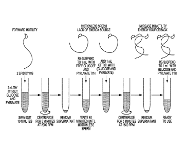
Motility Assay (Figure 1)
1. Sacrifice male mouse via dislocation or CO2 chamber.
2. Open the abdomen with fine scissors. Begin from the pelvic area and make a
V shape
to see all the organs
17
CA 03019523 2018-09-28
WO 2017/173391 PCMJS2017/025583
3. Look for the testis (white pale balls) and follow the seminiferous tubules
until you
find the cauda of the epididymis (looks like a small brain).
4. Take the cauda epididymis and make three or four incisions until you see
white fluid
coming out.
5. Put cut 1 epididymis in 2 ml modified TYH-Hepes media (Free of glucose and
Pyruvate) pH 7.2 to 7.4.
6. Leave the sperm to swim out of the tissue for 10 to 15 minutes.
7. Then take the 2 ml swim out and centrifuge for 5 minutes at 2000 RPM or
subject the
sperm to the device disclosed herein (with or without centrifugation).
8. Take the supernatant up to 300 ul or 500 ul.
9. Re-suspend up to lml, including 2m1, with modified TYH-Hepes (Glucose and
Pyruvate Free)
10. Wait about 30- 40 minutes until sperm stop moving
11. Add 1 ml of TYH supplemented with glucose 5mM and pyruvate 800 uM.
12. Centrifuge for 5 minutes at 1500 RPM.
13. Take the supernatant up to 500 ul.
14. Re-suspend up to lml of TYH supplemented with glucose 5mM and pyruvate 800
uM.
15. Take 100 ul of the swim out and add it to capacitation media (TYH
supplemented
with 15 mM HCO3" and 5mg/ ml serum albumin) with a final volume of 400 ul.
16. Wait about 60 minutes until sperm is fully capacitated (time can adjusted
for species).
17. Check motility with CASA system.
Results/Discussion
Proof of principle has been conducted using mouse sperm. This can be
extrapolated to
other species including farm animals and humans.
Example II ¨ In Vitro Fertilization/Starving Protocol
Methods
Day 1:
=
Inject females with 5 IU (100 DIP qinIP.Mhanabas were prepared and
diluted in sterilized PBS and keep to -20 C).
Day 3:
= Inject females with 5 IU (100 OV_Tott 9-
10 p.m. (48 h after PMSG).
DAY BEFORE IVF
18
CA 03019523 2018-09-28
WO 2017/173391 PCMJS2017/025583
Media:
= 5 ml TYH- Standard (4mg/m1 BSA) IVF at 37 C, 5% CO2.
= 8 ml TYH ¨ free glucose and pyruvate (4mg/m1 BSA) for sperm swim out at
37 C, 5%
CO2.
= TL-HEPES supplemented with 5% Fetus Bovine Serum prepare the same of the
IVF
For oocytes:
= Prepare Tissue Culture dish 35 X lOmm with 90 Ill of media TYH- Standard
(4mg/m1
BSA) IVF at 37 C, 5% CO2. See Figure 2 for further details.
= Put different plates into incubator at 37 C, 5% CO2.
For oviducts:
Prepare Tissue Culture dish 35 X lOmm with 90 il of media TYH- Standard
(4mg/m1
BSA) INT at 37 C, 5% CO2.
= Put different plates into incubator at 37 C, 5% CO2.
For speiiii:
= Prepare 2 ml tube of TYH (Free of glucose and pyruvate for sperm swimming
out)
= Put tube into incubator at 37 C, 5% CO2.
Day 4:
9:30 Prepare TL Hepes
= Prepare TL-HEPES supplemented with 5% Fetus Bovine Serum
For oviducts:
= Prepare dish plate with 2 ml of TL-HEPES supplemented with 5% Fetus
Bovine Serum (one
to wash, and other to get the cumulus-oocyte complex).
a.m. Sperm collection
= Sacrifice male. Sperm cell from the cauda epididymes are spilt and
allowed to swim
out in 2 ml TYH¨free glucose and pyruvate and standard TYH control medium for
10
min in 2 ml tube. Place the tube in at 37 C, 5% CO2 incubator for 10 min.
= After 10 min take the 2 ml swim out and centrifuge for 5 minutes at 2000
RPM.
= Take the supernatant up to about 300u1 or 500 ul.
= Re-suspend up to 2m1 with TYH (Glucose and Pyruvate Free) or standard TYH
control at 37 C, 5% CO2
= Centrifuge for 5 minutes at 1500 RPM
19
CA 03019523 2018-09-28
WO 2017/173391 PCMJS2017/025583
= Take the supernatant up to 300 ul or 500 ul.
= Re-suspend up to 1 ml with TYH (Glucose and Pyruvate Free) or standard
TYH
control at 37 C, 5% CO2
= Wait until sperm stop moving around 1 hour
= Add 1 ml of TYH- Standard with glucose and pyruvate at 37 C, 5% CO2.
= Centrifuge for 5 minutes at 1500 RPM
= Take the supernatant up to 300 ul or 500 ul.
= Re-suspend up to 500 ul or 1 ml with TYH standard with glucose and
pyruvate at
37 C, 5% CO2
= Ready for insemination
10:30 -11 a.m. Egg collection while sperm stop moving
= Sacrifice females super ovulated 13 to 14 hours after hCG administration.
= Remove oviducts and place in 1 ml TL-HEPES (5% FBS) medium in dish plate
to rinse of
blood and loose tissue.
= Open the oviducts with thin tweezers, and release the cumulus
= Using a fine-bore pipette transfer cumulus to a clean 2 ml TL-HEPES (5%
FBS) dish.
= Transfer cumulus to a clean dish with 3 ml TYH standard at 37 C, 5% CO2.
Hepes inhibits
IVF so make sure wash off the hepes very well before you place the cumulus in
the IVF drop.
= Transfer cumulus to IVF drop leave at 37 C, 5% CO?.
= Ready to be inseminated
11 a.m. Fertilization
= Inseminate using 20 pi of sperm capacitated. Co-incubate oocytes and
sperm at 37 C, 5%
CO2 for 4 h, and then wash sperm of oocytes by transferring two times the
oocytes into drop
1 and 2 with TYH- Standard with glucose and pyruvate (4mg/m1 BSA) using a fine-
bore
pipette.
= After washing, place oocytes in post-fertilization drop 3 and incubate up
to 24 h at 37 C, 5%
CO2.
Day 5:
= 12:30a.m to 2:00 pm Putative zygote evaluation:
= Check for two pronuclei or two embryo
= Follow embryo culture protocol
Embryo Culture
CA 03019523 2018-09-28
WO 2017/173391
PCT/US2017/025583
= Following day after IVF Prepare dishes with KSOM medium drops (50u1)
covered
with light mineral oil and put it in CO2 incubator at 37 C for 1 hour before
transferring 2 cell stage embryos.
= Transfer 2 cell embryos to KSOM medium (wash 2 times) make the same dish
as
the IVF, instead add 25 ul of KSOM medium.
= Transfer only 35 2-cell embryos per drop of KSOM culture drop
= This day follow the pseudo-pregnant female preparation chart
= Wait 2.5 days until blastocyst formation; see Figure 3.
= At blastocyst stage ready to transfer
Embryo Transfer and Pseudo-pregnant females
DAY-0 DAY-1 DAY 2 DAY 3.5
IVF start 11 AM Transfer 2 cell Do embryo transfer
Embryos to KSOM before noon in the
-Finish 4 pm (1 pm to 4 pm)
Mate the females with Check plugs at 9 am, 12 pm 2.5
day
vasectomized Males at and separate females
5pm. Mice mate at with plug
midnight (day 0)
- 12 pm (dayl)
Thursday Friday Day-1 Saturday Day-2 Sunday-Day3 Monday 3.5
Day-0
IVF start 11 Transfer 2 cell Embryos Check for Do embryo
AM to KSOM Morulas transfer at 6 to
(1 pm to 4 pm) 12 am am in
-Finish 4 pm the morning
Recipients Mate the females with Check plugs at 9
Females- vasectomized Males at am, and separate
5pm. Mice mate at females with
midnight 12 pm (day 0) plug
12 pm (day 2) 12 pm 2.5 day
- 12 pm (dayl)
Embryo transfer Procedures
1. Place a 151.11 drop of culture medium (KSOM already equilibrated at 37C 5%
CO2) onto the
lid of a 100 mm petri dish (Falcon 1029, or similar).
2. Load 12-20 blastocysts into the medium using a standard embryo-handling
pipette. (Note:
optimal number of embryos to transfer will vary depending upon mouse strain
and
manipulations embryos have received.)
21
CA 03019523 2018-09-28
WO 2017/173391 PCT/US2017/025583
3. Place the NSET device onto a P2 pipette that has been set to 1.81al.
Recommended pipettes
are the Pipette Rainin Classic PR2, 0 1-41 or Gilson Pipetman P2, 0 2-21al.
4. Press pipette plunger to first stop, lower tip of the NSET device into
medium and slowly
pull embryos into the tip. Remove NSET device tip from medium.
5. Carefully set pipette to 2.0 1 to create a small air bubble at NSET tip to
help ensure
embryos stay inside device tip during insertion into the mouse. Gently lay
pipette with loaded
tip aside (near cage) for use in step #9
6. Place the un-anesthetized recipient female on top of a cage with a wire
rack, allowing the
mouse to "grab" the cage bar surface with its forefeet. Grasp the midpoint of
the tail using
thumb and forefinger, and angle the tail upward while lightly pressing the
base of the tail
with the opposite edge of the hand.
7. Gently place smaller speculum into mouse's vagina, and then remove. This
will help open
the vagina.
8. Place larger speculum into vagina. Using an adequate light source, shine
the light into the
speculum to visualize the cervix.
9. While holding the female mouse with one hand as described in step #6,
carefully pick up
the pipette and gently insert the NSET device tip into the large speculum and
through the
cervix. Once NSET device hub contacts speculum, expel embryos by pressing
plunger
completely.
10. Gently remove NSET device without releasing pipette plunger and remove
speculum.
Return mouse to cage. No post-procedure monitoring is required.
Artificial Insemination
Animals: Female mice (at least 8 weeks old); Male mice as spettli donors Sperm
(C57BL6/J);
Male vasectomized mice (VASEX= vasectomized male)
Equipment: NSET device with specula; P-20 Rainin/Gilson pipette; lcc syringes,
26 gauge
needles; Scissors, forceps; IVF Tissue culture dishes (Falcon Cat# 353653);
Microscope (s);
Wire-topped cage
Monday Day-0 Tuesday Day-1 Wed Day-2 Thursday-Day3 Friday 4
PMSG Injection NONE hCG Al at 9:00 am
IU Injection Add 40 ul of
5:30 pm 511J sperm/female
5:00 pm
Recipients NONE Put super- Check
plugs
Females- ovulated females at 9 am, and
with the separate
vasectomized females
with
22
CA 03019523 2018-09-28
WO 2017/173391 PCT/US2017/025583
males plug
Spelm Preparation
1. Take one male Mice, sacrifice via dislocation or CO2 chamber.
2. Open the abdomen with fine scissors. Begin from the pelvic area and make
a V shape
so you can see all the organs
3. Look for the testis (white pale balls) and follow the seminiferous
tubules until you
find the cauda of the epididymis (looks like a small brain).
4. Take the cauda epididymis and make three or four incisions until you see
white fluid
coming out.
5. Take one epididymis for treatment and one for the control
6. Place epididymis in 2 ml modified TYH-Hepes media with 5% BSA (Free of
glucose
and Pyruvate) pH 7.2 to 7.4. Notice the control must have glucose and
pyruvate.
7. Leave the sperm to swim out of the tissue for 10 to 15 minutes.
8. Then take the 2 ml swim out and centrifuge for 5 minutes at 2000 RPM.
9. Take the supernatant up to 300 ul.
10. Re-suspend up to 2m1 with modified TYH-Hepes media with 5% BSA (Glucose
and
Pyruvate Free)
11. Then take the 2 ml swim out and centrifuge for 5 minutes at 1500 RPM
12. Take the supernatant up to 300 ul.
13. Wait around 40 minutes until sperm stop moving
14. Ready to inseminate the female: At 9:00 am: Deliver sperm to the
uterine horn using
the NSET procedure.
= Place the NSET device onto a P-20 pipette that has been set to 20
= Press pipette plunger to first stop, lower tip into media at the edge of
the sperm
sample and slowly load sperm into the NSET device. Avoid clumps. Set aside
pipette. Sperm
at the edge of the sperm sample are
= Place the un-anesthetized recipient female on the top of a cage, allowing
the mouse to
"grab" the cage bar surface with its forefeet. Grasp the midpoint of the tail
using thumb and
forefinger, and angle the tail upward while lightly pressing the base of the
tail.
= Place small speculum into vagina.
23
CA 03019523 2018-09-28
WO 2017/173391
PCT/US2017/025583
= While holding the female mouse with one hand as described above,
carefully pick up
the pipette and insert the NSET tip into the speculum, through the cervix and
into the uterus.
Once NSET hub contacts speculum, expel sperm by pressing plunger to the first
stop.
= Repeat procedure to deliver a total sperm volume of 40 [11, but wash NSET
device
with a TYH-Hepes media with 5% BSA (Free of glucose and Pyruvate) every time
device
goes into the uterus. This prevents contamination of Starved sperm with
metabolic substrates
present in the uterus
= Remove NSET device and speculum. No post-procedure monitoring is
required.
15. Immediately pair the female with a VASEX male overnight. Copulatory
activity seems
to be required to obtain pups from this procedure but not for embryo
fertilization.
2. Dissolve Mating Pairs Day 5:
a. Remove the female from the VASEX male cage.
b. Visually check for a copulation plug. The female is removed from the mating
cage and
transferred to the top of a wire-topped cage. Visual inspection and/or a blunt-
end probe may
be used to determine the presence of a vaginal plug (Figure 4).
Results
Example III ¨ method for treating sperm with Ca' ionophores alone and in
combination with
starvation to improve embryo development.
Abstract
Mammalian sperm acquire fertilizing capacity in the female tract in a process
called
capacitation. As part of capacitation, sperm undergo changes in their motility
pattern (i.e.,
hyperactivate) and become prepared for an exocytotic acrosome reaction that is
necessary for
fertilization. At the molecular level, capacitation requires a fast activation
of protein kinase A
(PKA) which is followed by hyperpolarization of the sperm plasma membrane and
an
increase in intracellular Ca2+. Genetic or pharmacological inhibition of these
pathways results
in loss of fertilizing ability both in vivo and in vitro. Recently, it was
demonstrated that
transient incubation of mouse sperm with the Ca2+ ionophore A23187 accelerated
capacitation and rescued fertilizing capacity in sperm with inactivated PKA
function (1).
Based upon these results, it was believed that A23187 could be used to
overcome defects in
signaling pathways upstream of the increase in intracellular Ca' required for
capacitation. It
is herein shown that a pulse of ionophore induces fertilizing capacity in
sperm from infertile
CatSper 1 (sperm specific Ca' channel), Adcy10 (soluble adenylyl cyclase sAC)
and SLO3
(sperm-specific K+ channel) KO mice. In contrast, sperm from infertile mice
lacking the Ca24-
efflux pump PMACA4 (Plasma membrane Ca2+-ATPase) were not rescued by
ionophore.
24
CA 03019523 2018-09-28
WO 2017/173391 PCT/US2017/025583
These results indicate that a transient increase in intracellular Ca' can be
used to overcome
genetic infertility in mice and suggest this approach may prove adaptable to
rescue male
infertility of other species in which in vitro fertilization protocols are
currently unsuccessful.
Introduction
While treating sperm with Ca' ionophores is known (Ca' ionophore A23187 can
make mouse spermatozoa capable of fertilizing in vitro without activation of
cAMP-
dependent phosphorylation pathways). It was not appreciated that this
treatment impacted
embryo development. Further, Ca' ionophore treatment (such as Ca' ionophore
A23187)
has not been used in conjunction with starvation to improve fertility
procedures.
In 1978, Steptoe and Edwards reported the birth of Louise Joy Brown, the first
successful "Test-Tube" baby (2). A major step toward this achievement (3)
occurred in the
early 1950's, when Chang (4) and Austin (5) demonstrated independently that
sperm have to
be in the female reproductive tract for a period of time before acquiring
fertilizing capacity, a
phenomenon now known as sperm capacitation. Capacitation includes all post-
ejaculation
biochemical and physiological changes that render mammalian sperm able to
fertilize (4, 5).
As part of capacitation, sperm become prepared to undergo acrosomal exocytosis
(6, 7) and
undergo changes in their motility pattern (e.g. hyperactivation). Although the
molecular basis
of these physiological processes is not well understood, capacitation is
associated with: 1)
activation of a cAMP/protein kinase A (PKA) pathway (8, 9); 2) loss of
cholesterol (10, 11)
and other lipid modifications (12); 3) increase in intracellular pH (pHi)
(13); 4)
hyperpolarization of the sperm plasma membrane potential (14, 15, 16); 5)
increase in
intracellular Ca" concentration [Ca2]i (17); and 6) increase in protein
tyrosine
phosphorylation (9, 18). These pathways were first identified as playing a
role in capacitation
using compounds that stimulate or block the respective signaling processes.
More recently,
the essential role of cAMP, Ca' and plasma membrane hyperpolarization was
confirmed
using KO genetic approaches (19, 20).
The role of cAMP in capacitation and fertilization was asserted using reagents
such as
cAMP agonists (dibutyryl cAMP, 8-BrcAMP) and antagonists of PKA-dependent
pathways
(e.g. H89, PKI, rpScAMP), as well as other conditions in which soluble
adenylyl cyclase
Adcy (10 21), the major source of cAMP in sperm, cannot be activated (e.g.
HCO3"-free
incubation media; addition of KH7, a specific sAC inhibitor) (for review see
7). The roles of
cAMP were confirmed using KO genetic mouse models lacking either the PKA sperm-
specific catalytic splicing variant Ca2, or sAC; these mice are sterile and
their sperm cannot
fertilize in vitro (22). It was demonstrated that hyperpolarizing changes in
membrane
CA 03019523 2018-09-28
WO 2017/173391 PCT/US2017/025583
potential are necessary and sufficient to prepare the sperm for a
physiological acrosome
reaction (23) Accordingly, sperm missing the sperm-specific K+ channel SLO3
cannot
hyperpolarize and are infertile (24, 25). Finally, Ca' was shown to be
essential for
hyperactivation and the acrosome reaction by using Ca'-free incubation media
with or
without addition of chelating agents such as EGTA to decrease this ion
concentration or using
Ca' ionophores such as A23187 to elevate it (1). Consistent with this, sperm-
specific Ca'
channel complex CatSper KO mice are infertile, and their sperm are unable to
hyperactivate.
Recently, it was found that addition of Ca' ionophore A23187 produced a fast
increase in intracellular Ca" that was accompanied by complete loss of sperm
motility (1).
After A23187 removal, intracellular Ca' levels dropped and sperm gain
hyperactive motility
(1). In addition to inducing hyperactivated motility, this Ca' ionophore
A23187 pulse
enhanced fertilizing capacity. Interestingly, this Ca' ionophore pulse
supported capacitation
in sperm incubated under non-capacitating conditions, and it induced
hyperactivation and the
capacity to fertilize in vitro even under conditions where cAMP-dependent
pathways are
blocked (1). These results suggested that A23187 could overcome defects in the
signaling
pathways upstream of the increase in intracellular Ca' required for
capacitation. This was
tested using infertile genetic mouse models. Consistent with the hypothesis, a
short A23187
pulse overcomes the infertile phenotypes of CatSper (19), sAC (22) and SLO3 KO
sperm
(25). The previous results suggested that subsequent washout of A23187, sperm
intrinsic
mechanisms involved in extruding Ca' are necessary to induce hyperactivation
and
fertilizing capacity (1). Consistent with this hypothesis, sperm lacking the
Ca' efflux pump
PMCA4, which mediates Ca' extrusion (26), were not rescued by the ionophore
treatment,
suggesting that this ATPase is required downstream to remove excess
intracellular Ca'.
Materials and Methods
Materials
Different materials and chemicals were purchased from different companies
(codes
between parenthesis represent the catalog number of the respective compound):
Calcium
Ionophore A23187 (C7522; dissolved in DMSO 2 mM stock), Bovine serum albumin
(BSA,
fatty acid-free) (A0281), Tween-20 (P7949), fish skin gelatin (G7765),
Pregnant mare serum
gonadotropin (G4877) and human chorionic gonadotropin (CGS), were purchased
from
Sigma (St. Louis, MO). Non-Surgical Embryo Transfer (NSET) Device was acquired
from
Paratechs (Billerica, MA). N-[24[3-(4-bromopheny1)-2-propen-l-yl]amino]ethyl]5-
isoquinolinesulfonamide,and dihydrochloride H-89 (130964-39-5) were purchased
from
Cayman chemical (Ann Arbor, Michigan). Anti-phosphotyrosine (anti-PY)
monoclonal
26
CA 03019523 2018-09-28
WO 2017/173391 PCT/US2017/025583
antibody (clone4G10), embryo transfer light mineral oil (ES-005-C) and
EmbryoMax
KSOM Medium (1X) w/ 1/2 Amino Acids (MR-106-D) were obtained from Millipore
(Billerica, MA). Rabbit monoclonal anti-phosphoPKA substrates (anti-pPKAS)
(clone100G7E), was purchased from Cell Signaling (Danvers, MA). Horseradish
peroxidase-
conj ugated anti-mouse and anti-rabbit IgGs were purchased from Jackson
ImmunoResearch
Laboratories (West Grove, PA) and GE Life Sciences. Triton X-100 (161-0407),
30%
Acrylamide and f3 -Mercaptoethanol was obtained from Biorad.
Animals
CD1 (Charles River Laboratories, Wilmington, MA) and C57BL/6 background mice,
7-18 wk of age, were used. Infertile KO mice genetic models (CatSper-/-, sAC-/-
and 5L03-
were in C57BL/6 background; PMCA4-/- mice were in FVBN background. For CatSper
embryo recipients, surrogate mothers were CD1 females, 8-12 wk of age. Animals
were
sacrificed in accordance with the Animal Care and Use Committee guidelines of
UMass,
Amherst. In experiments in which phosphorylation by PKA and tyrosine
phosphorylation was
investigated, CD1 and C57BL/6 male mice were used as indicated in the
respective figure
legend.
Media
Medium used for speini capacitation and fertilization assays was
Toyoda¨Yokoyama¨
Hosi (standard TYH) medium. Containing 119.37 mM NaCl, 4.7 mM KCl, 1.71 mM
CaC17.2H20, 1.2 mM KH2PO4, 1.2 mM MgSO4. 7H20, 25.1 mM NaHCO3, 0.51 mM Na-
pyruvate, 5.56 mM glucose, and 4 mg/mL bovine serum albumin (BSA), 10 [tg/mL
Gentamicin and phenol red 0.0006% at pH 7.4 when equilibrated with 5% CO?. To
analyze
the role of capacitation in phosphorylation pathways an HEPES-modified TYH
media was
used. Media that does not support capacitation (Non-Cap) contained 20 mM HEPES
instead
of HCO3- and does not contain BSA. For capacitating conditions HEPES-modified
TYH was
supplemented with 15 mM HCO3- and 4 mg/ml of BSA. Ca2+ ionophore A23187 (Sigma
Aldrich, location) was used at 20 [iM in TYH or H-TYH. Non-capacitating H-TYH
was
prepared by replacing 25 mM NaHCO3 with 20 mM Na-Hepes. Day before IVF add
4mg/ ml
BSA.
Mouse sperm preparation for Western blots
Cauda epididymal mouse sperm were placed in 1 ml Hepes-TYH media as stated in
figure legend for each experiment. After 10 min incubation at 37 C,
epididymides were
removed, and the suspension adjusted with non-cap medium to a final
concentration of 1-
2x10⁷ cells/ml. After dilution 1:4 (total 400 ul), sperm were incubated at 37
C for 1 hour in
27
CA 03019523 2018-09-28
WO 2017/173391
PCT/US2017/025583
conditions that support or not capacitation. For capacitation, media was
supplemented with
15 mM NaHCO3 and 4 mg/ml BSA. For A23187 treatment, 500 uL of the sperm
suspension
were taken from the initial swim out and supplemented with 20 litM A23187 for
10 minutes.
Then, A23187 was washed off with 2 rounds of centrifugationsl-) 2000 RPMS and
the 2-)
1500 RPMS for 5 min each. Sperm were re-suspended in free A23187 media. To
evaluate
the behavior of A23187 when PKA is inactivated, H89 was used at a
concentration of 50 uM
for all incubation periods including those used for washing A23187. Sperm
proteins were
then extracted for western blotting.
SDS-PAGE and immunoblotting
Sperm were centrifuged, and washed in 1 ml of phosphate buffer solution (PBS),
re-
suspended in Laemmli sample buffer (63), and boiled for 4 min. Before Loading,
5%13-
mercaptoethanol was added to the protein extracts and boiled for 3 min.
Protein extracts
equivalent to lx106 sperm were loaded per line and subjected to SDS-PAGE an
electro-
transfer to PVDF membranes (Bio-Rad) at 250 mA for 60 min on ice. For anti
pPKA
substrates Western blots, membranes were blocked with 5% fat-free milk in TBS
containing
0.1% Tween 20 (T-TBS). For anti-pY, membranes were blocked with 20% fish skin
gelatin
(54) in T-TBS. Antibodies were diluted in TBS containing 0.1% Tween-20 as
follows:
1/10,000 for anti-PY (clone4G10), and 1/1000 for anti-pPKA (clone100G7E).
Secondary
antibodies were diluted 1/10,000 in T-TBS and developed using an enhanced
chemiluminescence detection kit (ECLplus, Amersham, GE Healthcare) according
to the
manufacturer's instructions. When necessary, PVDF membranes were stripped at
65 C for 15
min in 2% SDS, 0.74% f3 -mercaptoethanol, 62.5 mM Tris, pH 6.5, and washed six
times for
mM each in T-TBS.
Motility and IVF Sperm Ionophore Pre-treatment
Sperm from CD-1, C57BL6 (K.0 control), CatSper KO 19, SLO3 KO (56), Soluble
Adenylyl Cyclase KO (20), and PMC4 KO (35) cauda epididymides were allowed to
swim
out in 1 mL of standard TYH for 10 minutes. Each swim out tube was split in
two halves
(500 ul each) and one half was incubated with 20uM A23187 for 10 minutes.
Then, the
A23187 was washed by centrifugation as described above (2000 and 1500 RPMS x 5
min),
the remaining sperm were re-suspended in free A23187 TYH standard medium and
capacitated for an addition 1 hour and 20 minutes before adding the sperm to
the fertilization
drop or for CASA analysis.
Sperm motility analysis
28
CA 03019523 2018-09-28
WO 2017/173391 PCT/US2017/025583
Sperm suspensions (25 pl) were loaded into one pre-warmed chamber slide
(depth,
100 u.m) (Leja slide, Spectrum Technologies) and placed on a microscope stage
at 37 C.
Sperm movements were examined using the CEROS computer-assisted semen analysis
(CASA) system (Hamilton Thome Research, Beverly, MA). The default settings
include the
following: frames acquired: 90; frame rate: 60 Hz, minimum cell size. 4
pixels; static head
size: 0.13-2.43; static head intensity: 0.10-1.52; static head elongation: 5-
100. Sperm with
hyper activated motility, defined as motility with high amplitude thrashing
patterns and short
distance of travel, were sorted using the criteria established by (64). The
data was analyzed
using the CASA nova software (64). At least 20 microscopy fields corresponding
to a
minimum of 200 sperm were analyzed in each experiment.
Video Recordings
Sperm suspensions (25 pl) were loaded into one pre-warmed chamber slide
(depth,
100 pm) (Leja slide, Spectrum Technologies). Videos were recorded for 15
seconds using an
Andor Zyla microscope camera (Belfast, Northern Ireland) mounted on Nikon
TE300
inverted microscope (Chiyoda, Tokyo, Japan) fitted with 10 and 20 times
objective lenses.
Sample temperatures were maintained at 37 C using a Warm Stage (Frank E.
Fryer scientific
instruments, Carpentersville, Illinois).
Mouse eggs collection and IVF assays
Metaphase II-arrested eggs were collected from 6-8 week-old super ovulated CD-
1
female mice (Charles River Laboratories). Females were each injected with 5-10
IU equine
chorionic gonadotropin and 5-10 IU human chorionic gonadotropin 48 h apart.
The cumulus-
oocyte complexes (COC's) were placed into a well with 500 [A of media (TYH
standard
medium) previously equilibrated in an incubator with 5% CO2 at 37 C.
Fertilization wells
containing 20-30 eggs were inseminated with sperm (final concentration of 2.5
x 106
cells/ml) that had been incubated for 1 h and 20 min (in a medium supporting
capacitation
with or without calcium ionophore treatment A23187). After 4 h of
insemination, eggs were
washed and put in a fresh media. The eggs were evaluated 24 h post-
insemination. To assess
fertilization the three following criteria were considered: 1) the formation
of the male and
female pronuclei, 2) the emission of the second polar body, and 3) two cells
stages.
Embryo Culture, Embryo transfer and Mice Genotyping
Fertilized 2 cell embryos were cultured in KSOM media to blastocyst stage
between
3.5 days wt and K.0 between 3.8 to 4.1 days. Then they were transferred to 2.5
days post
coitum (dpc) pseudo-pregnant CD-1 recipient females using the non-surgical
uterine embryo
transfer device (65). Pseudo-pregnant CD-1 recipient females were obtained by
mating with
29
CA 03019523 2018-09-28
WO 2017/173391
PCT/US2017/025583
vasectomized males one day after in vitro fertilization. Only females with a
clear plug were
chosen as embryo recipients; late morula and early stage blastocysts were
chosen to be
transferred. Routine genotyping was performed with total DNA from tail biopsy
samples
from weaning age pups as templates for PCR using genotyping primers for
Catsper gene
forward [5'-TAAGGACAGTGACCCCAAGG-3'] and reverse [5'-
TAAGGACAGTGACCCCAAGG-3'] and for the reporter gene Lacz forward
[5'TGATTAGCGCCGTGGCCTGATTCATTC-3'] and reverse [5'-
AGCATCATCCTCTGCATGGTCAGGTC-3'] (19).
Results
A23187 improves hyperactivation and fertilizing capacity of sperm from C57BL6
mice.
The relevance of genetic background for sperm physiology and for their ability
to
fertilize in vitro has been well-established (27, 28, 29, 30). Over the years,
C57BL6 has been
a common genetic background for studying KO genetic mouse models.
Unfortunately,
relative to sperm from mice of other genetic backgrounds, specifically CD1
mice, sperm from
C57BL6 exhibit significantly lower hyperactivation rates when capacitated
(Fig. 5 A) and are
less efficient for in vitro fertilization (31) (Fig. 5 B). To test the effect
of a short pulse of Ca'
ionophore, sperm from CD1 was compared with those from C57BL6 mice. A23187
treatment
increased the percentage of hyperactive C57BL6 spetut to similar levels as
those obtained
using CD1 sperm (Fig. 5 A). Moreover, this increase was followed by a
significant increase
in C57BL6 sperm fertilization rate (Fig. 5 B). Two-cell derived from the use
of control
sperm developed to blastocysts in about 50 %. This number is expected for
sperm derived
from this mouse strain. Surprisingly, after A23187 treatment, over 80 % of
fertilized eggs
continued to the blastocyst stage (Fig. 5 C and D), and when non-surgically
transferred to
pseudo pregnant mice females, became live pups. Capacitation requires PKA
activation (32),
and, as expected, in the presence of the PKA inhibitor H89, C57BL6 sperm are
unable to
fertilize in vitro and do not show the prototypical increase in PKA substrate
phosphorylation
(Fig. 5 E and F). Remarkably, as seen previously with CD1 sperm, incubating
H89-treated
C57BL6 sperm for 10 min in A23187 was sufficient to induce fertilizing
capacity (Fig. 5 E),
despite the fact that PKA remains inactive (Fig 5 F). These data show that
transient exposure
to A23187 can improve IVF success for C57BL6 mice strains and suggest this
treatment has
the potential to facilitate distribution of C57BL6 mouse lines.
A23187 treatment induced hyperactivation and fertilizing capacity of CatSperl
KO sperm.
In the absence of the CatSper channel complex, sperm fail to undergo
hyperactivated
motility and are unable to fertilize (19). To test the extent by which Ca2+
ionophore treatment
CA 03019523 2018-09-28
WO 2017/173391 PCT/US2017/025583
can overcome the CatSper infertile phenotype, sperm from CatSperl KO mice were
incubated in conditions that support capacitation in the absence or in the
presence of 20 uM
A23187. After 10 min the sperm were washed twice by centrifugation in A23187-
free media
and the percentage of hyperactive sperm was measured using CASA. As expected,
in the
absence of A23187, CatSper KO sperm did not undergo hyperactivation (Fig. 6
A). However,
once exposed to Ca' ionophore, a significant number of CatSper KO speiiii
exhibited
hyperactivated motility (Fig. 6 A). In addition, A23187-treated CatSper KO
sperm were
competent to fertilize metaphase II-arrested eggs in vitro (Fig. 6 B). A
fraction of the
fertilized eggs that reached blastocyst stage were non-surgically transferred
to
pseudopregnant female mice (33) , and five CatSper (+/-) mouse pups were born
from two
different females (Fig. 6 C). These heterozygous Fl mice were fertile, as
mating a male and
female from this heterozygous population yielded a normal litter with 1 wild
type, 4
heterozygous and 3 CatSper KO F2 progeny (Fig. 6 D).
A23187 treatment also rescued fertilizing capacity in sperm of sAC KO and SLO3
KO mice.
Among the earliest molecular events during capacitation are up-regulation of
cAMP-
dependent pathways (32) and hyperpolarization of the sperm plasma membrane
(23). These
events precede the essential increase in intracellular Ca', and it was tested
whether defects in
each of these cascades can be rescued by Ca' ionophore pulse. As shown above,
a short
pulse of Ca' ionophore bypassed the need for the capacitation-induced PKA
activation (1)
(Fig. 5E and F). Under normal capacitation conditions, sAC KO sperm are almost
immotile
(Fig. 7A), while SLO3 KO sperm are able to move. However, neither sAC KO nor
SLO3 KO
sperm have the ability to undergo hyperactivation (Fig. 7 B). Despite this
phenotype, once
treated with A23187 for 10 min, a significant fraction of sAC KO sperm became
motile and
both sAC KO and SLO3 KO sperm underwent hyperactivation (Fig. 7 B). Moreover,
A23187
treatment induced fertilizing capacity in sperm from both KO models (Fig. 7
C).
A23187 treatment does not rescue Pmca4 KO sperm.
It was previously shown that the increase in intracellular Ca" caused by
A23187 has
to be followed by a reduction of intracellular calcium after removal of the
ionophore (1). In
sperm, two molecules are thought to mediate Ca2+ extrusion, namely the Na/Ca'
exchanger
and the more efficient sperm-specific Ca" ATPase PMCA4 (34) Male PMCA4 KO mice
are
infertile (35), suggesting this molecule is involved in regulation of Ca'
homeostasis in
sperm. It was hypothesized that sperm lacking PMCA4 would not respond to
A23187 rescue.
PMCA4 KO sperm display poor motility and do not hyperactivate (Fig. 7 A and
B). Addition
of A23187 rendered all the sperm motionless and their motility was not
recovered after
31
CA 03019523 2018-09-28
WO 2017/173391
PCT/US2017/025583
ionophore removal (Fig. 7D). Consequently, neither their hyperactivated
motility nor their
fertilizing capacity was rescued (Fig. 7E)
Discussion
Capacitation encompasses a series of sequential and concomitant biochemical
changes required for sperm to gain full fertilization competency. Despite the
relevance of
capacitation, the molecular mechanisms intrinsic to this process are not well
understood. A
very early event in sperm capacitation is the activation of motility by a cAMP
pathway (36).
The activation of cAMP synthesis occurs immediately after sperm are released
from the
epididymis and come into contact with high HCO3" and Ca' present in the
seminal fluid (37,
38). Plasma membrane transport of these ions regulates sperm cAMP metabolism
through
stimulation of Adcy10 (aka sAC) (21 20, 39). sAC activation, elevates
intracellular cAMP
and stimulates PKA. Then, PKA phosphorylates target proteins which initiate
several
signaling pathways. These pathways include a sperm plasma membrane
hyperpolarization, an
increase in pHi, and an increase in intracellular Ca' ions. Consistent with
the influence of
these events, KO mice genetic models in which any of these pathways is
interrupted are
infertile.
Physiologically, sperm capacitation is associated with changes in their
motility pattern
collectively known as hyperactivation and with the preparation for a
physiological acrosome
reaction. Originally observed in hamster sperm moving in the oviduct,
hyperactivated
motility (40) was later described in other mammalian species including humans
(41).
Hyperactivation is associated with a strong high-amplitude asymmetrical
flagellar beating
that appears to be essential for the sperm to loosen their attachment to the
oviductal
epithelium and to penetrate the zona pellucida (42). Consistent with an
essential role of
hyperactivation for fertilization competency, one of the most common
phenotypes observed
in sperm from many different infertile knock-out models including those used
in the present
work (e.g. CatSper, sAC, SLO3 and PMCA4) is low motility and/or defects in
hyperactivation (22, 32, 38, 43, 44, 45).
Although very little is known about the molecular pathways regulating
hyperactivation, Ca' ions have been shown to play roles in the initiation and
maintenance of
this type of movement (46, 47, 48, 49). Most of the information regarding the
role of Ca" in
hyperactivation has been obtained using loss of function approaches analyzing
sperm motility
in media devoid of Ca' ions. Gain of function experiments using Ca' ionophores
(e.g.
A23187, ionomycin) to increase [Ca2]i have yielded unexpected results because,
instead of
enhancing hyperactivation, these compounds stopped sperm movement (50, 51,
52). Despite
32
CA 03019523 2018-09-28
WO 2017/173391 PCT/US2017/025583
being motionless, ionophore-treated sperm are alive as they recover motility
after the
compound is quenched with lipophilic agents (50) or washed by centrifugation
(52). The
reversibility of the A23187 effect suggests that the sperm is able to return
to physiological
[Ca after a drop in free ionophore concentration. In previous work, it was
shown that a
short incubation period with A23187, in addition to initiating
hyperactivation, accelerated the
acquisition of fertilizing capacity. Unexpectantly, the data indicated that 10
min incubation
with A23187 followed by wash out induced fertilization competence without
activation of
cAMP-dependent signaling pathways that are needed for capacitation (1).
Considering these results, it was hypothesized that a temporary elevation of
intracellular Ca' primes the speim for hyperactivation and bypasses the need
for other
signaling pathways required to up-regulate Ca' influx in sperm. To test this
hypothesis, in
the present work, four KO models affecting independent signaling pathways were
selected.
Three of these signaling molecules are believed to act upstream of the
increase in Ca'
required for hyperactivation: CatSper, sAC and SL03. Sperm from each of these
mouse
models are unable to undergo hyperactivation and are incapable of fertilizing
metaphase II
arrested eggs in vitro. In addition, PMCA4b KO sperm were used, which would
not allow
intracellular Ca2+ lowering after flooding with this ion. Sperm from PMCA4b KO
mice are
deficient in both progressive and hyperactivated motility resulting in
sterility (53, 26).
PMCA4 has been shown to be the principal source of Ca' clearance in sperm and
it is
essential to achieve a low resting [Ca2]i (34). Consistent with the
hypotheses, a short
incubation of sperm with A23187 induced hyperactivation of CatSper, sAC and
SLO3 KO
but not of PMCA4 KO sperm.
Male factors contribute to approximately half of all cases of infertility (54,
55).
However, in over 75 % of these cases it is unusual to have a clear diagnosis
of the
abnormalities found in semen parameters. Currently, assisted reproductive
technologies
(ART) remain the main therapy available. Recent studies using KO mouse models,
including
those used in the present work, revealed that loss of function of a variety of
genes results in
infertility. Interestingly, several of these models present normal sperm
counts and their main
deficiency is found in capacitation-associated processes such as impediments
to hyperactivate
(19), to undergo the acrosome reaction (56), or to go through the utero-tubal
junction in vivo
(57, 58). It was hypothesize that strategies designed to elevate [Ca2]i such
as the use of
A23187 pulse denoted above should overcome the need of upstream signaling
pathways
including but not limited to PKA activation. In addition, although IVF has
been successfully
employed in multiple species (6), requirements of sperm for capacitation vary
greatly among
33
CA 03019523 2018-09-28
WO 2017/173391 PCT/US2017/025583
species and have been developed for each sperm type essentially by trial and
error. In some
species, such as the horse, effective methods for IVF have still not been
established despite
decades of work (59). Failure of equine IVF does not appear to be associated
with oocyte
characteristics (60), but is associated with the inability of horse sperm to
hyperactivate and to
penetrate the egg zona pellucida (ZP) (61), two landmarks of capacitation. A
better
understanding of capacitation signaling processes have the potential to
generate "universal"
IVF technology that can be used in endangered/exotic species for which ART is
not currently
available.
Improving IVF conditions would be of great value; however, at the clinical
level,
ICSI has replaced IVF when confronted with cases of unknown male factor
infertility. IC SI is
reliable and from the patient's point of view more economical because of
higher probability
of success. Despite these advantages, ICSI bypasses certain aspects of normal
fertilization
and may bear effects that are not easily observed (e.g. epigenetic
alterations). Taking this into
consideration, a method to improve IVF can be a desirable option in some male
factor cases.
It is worth noting that A23187 has already been used in the clinic for
patients with repeated
IC SI failure (62). In these cases, eggs are transiently incubated with
ionophore after IC SI,
which exposes the zygote to high Ca'. On the contrary, when sperm are treated
with
A23187, the ionophore is washed and does not come in contact with the embryo.
More
interestingly, overcoming infertility problems related to motility and
hyperactivation could
have other potential uses in the clinic. For example, this methodology could
be used to
improve the success rate of intrauterine insemination which is a significantly
less invasive
and less costly procedure than either IVF or IC SI.
Example IV - Mouse Sperm
It is well known that out-breed and in-breed mice sperm differ in the ability
to
capacitate and fertilize the egg. According to the National Institute of
Health (NIH-US)
almost 90% of research is done in in-breed mice and the most common breed used
is
C57BL6. Wild type C57BL6 mice have shown low fertility in vivo compared to out
¨breed
strains. In addition, when it comes to sperm in vitro hyperactivation and
fertilization, fertility
is also reduced. Therefore sperm from C57BL6 mice are a good model to show if
a particular
treatment can improve fertilization parameters (Navarrete et al., Sci. Rep.
2016,
demonstrating that a transient exposure to calcium ionophore A23187 improves
hyperactivation and fertilizing capacity of sperm from C57BL6/J mice in vitro.
In the
experiments presented below, C57BL6 in-breed mice strain was used.
Sperm Cell Signaling cascades and protein phosphorylation:
34
CA 03019523 2018-09-28
WO 2017/173391
PCT/US2017/025583
After ejaculation, mammalian sperm are not able to fertilize, they require
being in the
female tract for a certain period of time which is species-specific. The
physiological changes
that occur to the sperm during this time period are collectively known as
capacitation. Sperm
capacitation can be mimicked in vitro in defined media containing: 1) ions
such as Na+, Cl-,
HCO3-, Ca2+ and Mg2+; 2) energy sources such as glucose, pyruvate, lactate or
others; 3) a
cholesterol acceptor such as bovine serum albumin (BSA) or beta-cyclodextrin.
The in vitro
capacitation media is used to incubate mammalian sperm before combining them
with eggs
during in vitro fertilization. Capacitation is associated with changes in the
motility pattern.
After incubation in capacitation media, sperm undergo changes in their
movement known as
hyperactivation. Hyperactivation motility has been associated with the sperm
ability to
fertilize.
At the molecular level, capacitation is associated with a fast increase in
cAMP,
mediated by the atypical adenylyl cyclase Adcy10, followed by the activation
of cAMP-
dependent kinase, PKA. This increase in PKA is dependent on the presence of
HCO3- and
BSA in the capacitation media and can be measured using anti phospho
antibodies against a
PKA-consensus phosphorylation sequence RXXS (where X is any type of aminoacid)
(Fig. 8
A). Downstream of the activation of PKA, there is an increase in tyrosine
phosphorylation
which can be measured using anti phosphotyrosine antibodies (Fig. 8 B). As
shown in the
figures, neither PKA activation nor the increase in tyrosine phosphorylation
occur in the
absence of HCO3- and BSA (lane 1 in both panels A and B). After adding HCO3-
and BSA,
phosphorylation patterns are activated (lane 2 in both panels A and B). In
both cases, sperm
are incubated in the presence of the energy nutrients glucose (5 m114) and
pyruvate (0.5 mM).
The experiments depicted in Fig. 8 A and B were performed to evaluate how the
different substrates affect phosphorylation pathways. While glucose produces
energy by
glycolysis and might be also coupled to oxidative phosphorylation through the
use of
pyruvate and lactate at the end of glycolysis. Pyruvate can only be used by
the mitochondria.
It is shown that PKA activation occurs with both type of substrates (Lane 3
and 4 in Fig. 8
A). On the other hand, the increase in tyrosine phosphorylation occurs
normally with glucose
as substrate (lane 3 in Fig. 8 B), but it is reduced when only pyruvate is
present in the
incubation media (lane 4 in Fig. 8 B). When sperm are incubated for 1 hour in
the absence of
energy nutrients, PKA activity and the increase in tyrosine phosphorylation
are not observed
(lane 5 in both Fig. 8 A and B). Addition of glucose, pyruvate or both after 1
hour incubation
in the absence of nutrients rescued activation of PKA (lanes 6, 7 and 8 in
Fig. 8 A and B); on
CA 03019523 2018-09-28
WO 2017/173391
PCT/US2017/025583
the other hand only when glucose was present the increase in tyrosine
phosphorylation was
observed (lanes 6, 7 and 8 in Fig 8 A and B).
Aliquots of sperm treated as described for the phosphorylation assays in Fig.
8 A and
B, were evaluated for motility using Computer Assisted Sperm Analysis (CASA).
It was
observed that in the absence of nutrients for 1 hour, the percentage of motile
sperm was zero.
However, the motility was rescued by the addition of glucose, pyruvate or both
(Fig. 8 C). As
mentioned above, hyperactivated sperm motility increased when sperm are
incubated in
capacitation conditions (presence of HCO3- and BSA), compare Bar 1 with Bar 2
in Fig. 8 D.
In the absence of nutrients (Bar 5 in Fig. 8 C and D), no motility is
observed, and therefore,
the percentage of hyperactivated sperm is also zero. When nutrients are added,
hyperactivation is rescued (Bars 6, 7 and 8 in Fig. 8 D). Remarkably, glucose
induced
significantly higher values of sperm hyperactivation after starving. This
experiment suggests
that upon starving, sperm can move better than when they are incubated with
energy nutrients
the whole time.
In vitro fertilization and embryo development is enhanced using Starving and
Rescue method
in young and old mice.
The significant increase in hyperactivated motility observed after rescuing
sperm
incubated previously in starving media (starving plus rescue) suggested that
this treatment
can improve fertilization rates. To evaluate this hypothesis, the fertilizing
capacity of sperm
incubated in control TYH capacitation media (CONTROL (C)) with sperm incubated
in TYH
media devoid of glucose and pyruvate and then rescued with the addition of
glucose and
pyruvate (STARVING + RESCUE (S+R)) were compared. In mice, like in humans,
decreased fertility has been observed in aged individuals, and it is caused by
different factors
such as diet, exercise and genetic outcomes. A recent study has shown the
effects of
advanced paternal age on mice reproduction. Interestingly, the authors
concluded that
fertilization capacity (natural conception) is reduced when mice reach 12
months of age and it
declines after that age. Remarkably, they also discover that IVF, in vitro
embryo
development, and embryo quality were also affected with age. Taking this into
consideration,
our experiments were conducted using male mice of three different age groups:
3-6 month
old, 6 to 12 month old, and 12-24 months old. For oocyte donors, young females
were used
(2 months old).
Initially these experiments were done using CD-1 oocytes (Fig. 9 A, B and C)
and
then repeated with oocytes obtained from C57BL6 mouse strain (Fig. 9 D, E and
F). The
rationale of trying both types of oocytes is that C57BL6 oocytes are not as
good in
36
CA 03019523 2018-09-28
WO 2017/173391
PCT/US2017/025583
fertilization protocols as the CD-1 oocytes. It was observed that the
percentage of fertilized
oocytes (measured by the percentage of oocytes arriving to 2-cell embryos) was
increased
when sperm are treated in S+R conditions (Fig. 9 A). The higher fertilization
rates were
maintained regardless the age of the mice (3-6; 6-12, and 12-24 months old).
Two-cell
embryos obtained in each condition were then transferred to KSOM media for
further embryo
development. Surprisingly, the percentage of blastocysts achieved from two-
cell embryos
obtained using S+R sperm was higher than the ones fertilized with C sperm
(Fig. 9 B). Notice
that in Fig. 9 B, the percentage of blastocyst development is calculated from
the total amount
of two-cell embryos in each condition. If the percentage of blastocyst
development was
plotted considering the initial number of oocytes, it is possible to observe
that the S+R
treatment is highly efficient when compared with standard media (Fig. 9 C).
Improvement in
the percentage of fertilization and embryo development was also observed when
C57BL6
oocytes were used (Fig. 9 D, E and F).
Experiments were then conducted to evaluate the speed by which sperm treated
in
standard conditions or starved and then rescue can fertilize CD1 oocytes.
Table 1A, first
column, shows the incubation time of sperm with eggs for each treatment
(control or starving
+ rescue) (second column). After this time period, eggs were removed, washed
and continue
the incubation in fertilization media. The number of two-cell embryos was
evaluated the
following day. The data presented in each of the columns indicate the number
of oocytes
(Oocytes #), the number of oocytes reaching two-cell embryo stage (cleavage)
with the
respective percentage between parenthesis, the number of two-cell embryos
reaching
blastocyst stage (Blastocyst) with the respective percentage compare with the
initial number
of two-cell embryos, and, finally, the percentage of blastocysts taken into
consideration the
initial number of oocytes in the assay (Total % blastocyst). In this table, it
is possible to
observe that the starving plus rescue conditions increased the velocity of
fertilization.
Table 1 A and B. Comparison of standard and Starving plus Rescue sperm
treatment
regarding the speed of fertilization and the minimum amount of sperm needed
for
insemination in vitro.
37
CA 03019523 2018-09-28
WO 2017/173391 PCT/US2017/025583
TABLE 1 A
VAAL. ..... ..
t240 5 (6 5)
278. (.35) 154 (70.! 23.
=
2.41.0 154 100 (05 rit (t.41 )
14 Q9) Z (tit.:1)
30 999 ) 1M 5.3$) 5Z
1.= õõõ -*====
0,4) 291t Z5i) (8,4 0,10) 5,"t3
240
TABLE 1 El
9J.7
11*xitArtOrg Oicaoyte rAii) 13t4S44.07y.t .CS40:
IZSI4otoolikst
Z.35: 54 (25) 25.) (54) 12
A900: 200 et' (2 4=3.1 17.
. C=0.arrirol
M000 241 :W 0E4 13
At <601:
1011t:/ 1.vo : 1:60')= 10-7 (74 ) 40
-"---- -
:1.0009 .26.S= 331 f71> (81)
0000* 105921.5
A similar experiment was conducted to evaluate the minimum number of sperm
needed for fertilization. This experiment is presented in Table 1B For this
experiment, sperm
incubated in control (Control) or in starving plus rescue (Rescue) media were
counted and
then a series of dilution of the original sperm suspension were done with the
purpose of
adding different number of sperm to the insemination drop as detailed in
column 1 The
columns present the same information described for Table 1A: number of
oocytes, number of
two-cell (percentage), number of blastocysts (percentage with respect to the
number of two
cells), and total % blastocyst with respect to the initial number of eggs.
Data in Table 1B
indicates that fertilization can be achieved with lower sperm number.
Since the first successful IVF in mammals, it has become clear that there is a
direct
relationship between embryo quality and gestational success post embryo
transfer. Different
studies have shown that IVF have an impact on embryo development in vitro.
Considering
that the starving methodology improved fertilization rates and also increased
the number of
blastocysts, it was decided to compare quality of blastocysts obtained with
the different
methodologies by counting the number of cells present in 3.5 blastocysts. For
each condition,
35 blastocysts obtained from 10 individual experiments were evaluated. The
total number of
cells was assessed by counterstaining of nuclei and served as an indicator for
division rates.
While control sperm produced blastocysts with an average of 46 cells, starved
and rescued
38
CA 03019523 2018-09-28
WO 2017/173391
PCT/US2017/025583
sperm produced blastocysts with an average of 55 cells (Fig. 10 A).
Blastocysts were further
assessed by three-day outgrowth (OG) assay to test hatching, attachment and
formation of the
inner cell mass (ICM) with surrounding trophoblast cells in an in vitro
system. An n=10
outgrowth assays were done from different 10 WF experiments, and a total of
200 blastocyst
were recorded for each of the sperm treatments. Control blastocyst outgrowth
showed a
significantly lower embryo attachment to the petri dish (55%) than the
Starving plus rescue
treatment (75%) (Fig. 10 B). The next step to evaluate embryo quality from 3.5
blastocyst
embryos obtained with different sperm was to perfoim embryo transfer to pseudo-
pregnant
mice females. 25 independent experiments were evaluated. For each experiment,
the same
number of 3.5 day blastocysts obtained with sperm treated either with control
media or with
starving plus rescue protocol were transferred non-surgically. Because, more
blastocysts were
obtained routinely using the starving plus rescue protocol, for these
experiments, the number
of blastocysts transferred was always limited by the amount of blastocysts
obtained with
control-treated sperm which range between 8 and 16 blastocysts. After
transfer, the females
become pregnant and the litter size for each condition was recorded (Fig. 10
C). Results were
analyzed depending on the mice age (2-3 months old vs 12-24 months old). As
shown in
Figure 10 C, the average litter size for embryos obtained using Starving plus
Rescue sperm
treatment was significantly higher for both age groups. The data were also
analyzed as
percentage of pups per number of blastocysts transferred (Fig. 10 D).
Altogether these data
indicate that blastocysts obtained using starved plus rescued sperm have
better quality than
those obtained using standard control conditions.
Intrauterine insemination (IUI) is an assisted reproductive technique that
delivers
sperm into the female tract bypassing the cervix. TUT is implemented in humans
as a fertility
treatment, and widely performed in commercial breeding of livestock. However,
in the
literature there is few studies about IUI in mouse compared with other species
such as
humans, bovine, horses and many others. Therefore, it was decided to evaluate
the success of
sperm using control media vs the success of sperm treated with the starving
protocol. In IUI
protocol, the rescue of sperm was not done; the sperm were used directly after
incubation in
media without nutrients (starving). Because IUI in mice has been shown to work
better when
sperm are incubated in non-capacitating conditions, the media used for these
experiments for
both control and starving conditions was done without addition of HCO3- and
BSA. Success
of JUT in mice depends on two main variables. The first one is the number of
females that
becomes pregnant with respect to the total of females inseminated (Fig. 11 A).
The second
one is the litter size in those females that become pregnant (Fig. 11 B). As
an example pups
39
CA 03019523 2018-09-28
WO 2017/173391
PCT/US2017/025583
obtained in one of the IUI experiments using starved plus rescue protocol are
shown (Fig. 11
C).
As mentioned above, in-breed genetic backgrounds have lower fertility in vivo
and in
vitro. These mice strains such as C57BL6 are relevant for research and high
priority on the
mouse phenome database. In addition genetic manipulation of these valuable
mice leads in
many cases to acute sub-fertility. It was decided to use two knock out (KO)
models
(FerTDR/DR and Akita) and one in-breed mouse (SJL/J) with proven sub-fertility
in vitro. As
it was shown previously calcium ionophore A23187 overcome mice infertility in
different
KO models; therefore, for FerT KO, A23187 treatment was also used to compare
the success
rate of different treatments. FerT sperm fertilization rate was improved with
A23187 transient
incubation. However, starving treatment improves fertilization of FerTDR/DR
sperm to
higher levels (Fig. 12 A). In both treatments, two-cell embryos were
transferred to KSOM
media and further incubated for 3.5 days. Results indicate that once two-cell
are obtained,
both ionophore treatment and starving plus rescue protocols are equally
successful for
embryo development (Fig. 12 B). Similarly, starving plus rescue improved
fertilization rates
of sperm from Akita and SJL/J mice (Fig. 12 C). In one of the experiments
using FerTDR/DR
and one using Akita mice, blastocysts obtained using starved plus rescue
protocol were
transferred and pups were obtained (10 for FerTDR/DR and 5 for Akita) (data
not shown).
Many genes have been shown to play a role in fertilization. A group of genes
code for
the sperm-specific calcium channel complex CatSper. Knock-out mice lacking any
of the
four main CatSper subunits (CatSperl, CatSper2, CatSper3 or CatSper4) are
infertile in vivo
and in vitro. A short treatment with calcium ionophore A23187 induces
fertilization capacity
in sperm from these mice. Using this technique combined with embryo
development and
embryo transfer pups were obtained (Navarrete et al., Sci. Rep. 2016). To
investigate if the
starving plus rescue technology was also able to rescue the infertility
phenotype, sperm were
incubated in four different conditions: 1) Control; 2) A23187 transient
treatment; 3) starved
plus rescue treatment; and 4) starved plus rescue treatment followed by A23187
transient
treatment. After these treatments, sperm were combined with eggs and the
number of two-
cell embryos developed counted (Fig. 13 A). The use of A23187 rescued the
infertile
phenotype. On the other hand, starved plus rescued sperm incubation protocol
did not rescue
infertility. However, when both treatments were combined the CatSper
fertilization rate was
close to 90 %. In each of these experiments, two-cell embryos were transferred
to KSOM
media and further incubated for 3.5 days. Blastocyst were then counted and
plotted as
percentage of blastocyst in relation to the number of two-cell embryos (Fig. 6
B). In this case,
CA 03019523 2018-09-28
WO 2017/173391 PCT/US2017/025583
the A23187 transient treatment achieved 20 % development and the combined
starved plus
rescue with A23187 transient treatment was close to 90 %. From these
experiments, a total of
15 blastocysts from each treatment were transferred into 2.5 days pseudo-
pregnant females.
A23187 alone gave birth to three heterozygous pups and the starved plus rescue
protocol
gave birth to six heterozygous pups. Altogether, these results suggest that
combination of
treatments is very effective in producing healthy embryos and that it can be
used as treatment
in some infertility cases.
Example V - Bovine Sperm
Media requirements for sperm in ART approaches vary greatly among species and
have been developed for each sperm type essentially by trial and error.
However, in all cases
sperm need to efficiently synthesize ATP. To investigate the extent by which
metabolic
regulation can be used in other animal models, bovine sperm was used to
conduct IVF, ICSI
and embryo development experiments. These experiments were obtained using
frozen sperm
and in vitro matured embryo. IVF in bovines is well-established and the
efficiency of the
method is around 60 %. Control and metabolically modified sperm performed
similarly in
IVF (Fig. 14 A); however, an increased percentage of blastocysts were obtained
with the
metabolically treated sperm (Fig. 14 B). Blastocysts from one of the
experiments are shown
(Fig. 14 C and 14 D).
Although as said, IVF methods are well-established in bovine, intracellular
sperm
injection (ICSI) is not effective using bovine sperm. This is due to
deficiencies in the ability
of sperm to induce Ca2+ oscillations. Therefore, the only method available to
do ICSI with
bovine sperm is by treating embryos with pharmacological reagents such as Ca2+
ionophores
which activates the egg. Notice that one main difference on the Ca2+ ionophore
treatment
herein is that in the instant case, only the sperm are transiently exposed to
this
pharmacological reagent and that the reagent is washed out before the sperm
become in
contact with the egg. In an unexpected result, bovine sperm incubated in
starved media and
then exposed to nutrients acquire the ability to induce calcium oscillations
in bovine eggs
after injection by ICSI (Fig. 15 A) in significantly more number of injected
eggs than controls
(Fig. 15 B). Moreover, this method also increased significantly the percentage
of two cell
embryos (Fig. 16 A) and 2 blastocysts were obtained using this treatment (Fig.
16 B).
Although in comparison with mouse sperm data the percentage of blastocysts
appear low, it
is important to highlight that this is the first time that blastocyst are
obtained using ICSI
without exposing the embryo to pharmacological reagents.
41
CA 03019523 2018-09-28
WO 2017/173391 PCT/US2017/025583
The methods described herein find use in WF, ICSI and artificial insemination
in
humans and other mammals animals. However, each species has specific
difficulties. For
example, in humans, artificial insemination is not used frequently despite
being the less
invasive and costly method because the success rate is limited. The methods
described herein
improve the outcome of intrauterine insemination as well as for vaginal
insemination.
Bibliography
1. Tateno H, et al. Ca2+ ionophore A23187 can make mouse spermatozoa
capable of
fertilizing in vitro without activation of cAMP-dependent phosphorylation
pathways. Proc
Natl Acad Sci US A 110, 18543-18548 (2013).
2. Steptoe PC, Edwards RG. Birth after the reimplantation of a human
embryo. Lancet 2,
366 (1978).
3. Chang MC. Fertilization of rabbit ova in vitro. Nature 184(Suppl 7), 466-
467 (1959).
4. Chang MC. Fertilizing capacity of spermatozoa deposited into the
fallopian tubes.
Nature 168, 697-698 (1951).
5. Austin CR. Observations on the penetration of the sperm in the mammalian
egg. Aust
J Sci Res (B) 4, 581-596 (1951).
6. Yanagimachi R. Mammalian fertilization. In: The Physiology of
Reproduction
(ed^(eds Knobil E, Neill JD). Raven Press, Ltd. (1994).
7. Visconti PE, Krapf D, de la Vega-Beltran JL, Acevedo JJ, Darszon A. Ion
channels,
phosphorylation and mammalian sperm capacitation. Asian J Androl 13, 395-405
(2011).
8. Harrison RA. Rapid PKA-catalysed phosphorylation of boar sperm proteins
induced
by the capacitating agent bicarbonate. Mol Reprod Dev 67, 337-352 (2004).
9. Krapf D, et al. Inhibition of Ser/Thr phosphatases induces capacitation-
associated
signaling in the presence of Src kinase inhibitors. J Biol Chem 285, 7977-7985
(2010).
10. Davis BK, Byrne R, Bedigian K. Studies on the mechanism of
capacitation: albumin-
mediated changes in plasma membrane lipids during in vitro incubation of rat
sperm cells.
Proc Natl Acad Sci U S A 77, 1546-1550 (1980).
11. Cross NIL. Effect of cholesterol and other sterols on human sperm
acrosomal
responsiveness. Mol Reprod Dev 45, 212-217 (1996).
12. Gadella BM, Harrison RA. The capacitating agent bicarbonate induces
protein kinase
A-dependent changes in phospholipid transbilayer behavior in the sperm plasma
membrane.
Development 127, 2407-2420 (2000).
42
CA 03019523 2018-09-28
WO 2017/173391
PCT/US2017/025583
13. Zeng Y, Oberdorf JA, Florman HM. pH regulation in mouse sperm:
identification of
Na(+)-, Cl(-)-, and HCO3(-)-dependent and arylaminobenzoate-dependent
regulatory
mechanisms and characterization of their roles in sperm capacitation. Dev Biol
173, 510-520
(1996).
14. Zeng Y, Clark EN, Florman HM. Sperni membrane potential:
hyperpolarization
during capacitation regulates zona pellucida-dependent acrosomal secretion.
Dev Biol 171,
554-563 (1995).
15. Escoffier J, Krapf D, Navarrete F, Darszon A, Visconti PE. Flow
cytometry analysis
reveals a decrease in intracellular sodium during sperm capacitation. J Cell
Sci 125, 473-485
(2012).
16. de La Vega-Beltran JL, et al. Mouse sperm membrane potential
hyperpolarization is
necessary and sufficient to prepare speint for the acrosome reaction. . J Biol
Chem In Press,
(2012).
17. Ruknudin A, Silver IA. Ca2+ uptake during capacitation of mouse
spermatozoa and
the effect of an anion transport inhibitor on Ca2+ uptake. Mol Reprod Dev 26,
63-68 (1990).
18. Visconti PE, Bailey JL, Moore GD, Pan D, Olds-Clarke P, Kopf GS.
Capacitation of
mouse spermatozoa. I. Correlation between the capacitation state and protein
tyrosine
phosphorylation. Development 121, 1129-1137 (1995).
19. Ren D, et al. A sperm ion channel required for sperm motility and male
fertility.
Nature 413, 603-609 (2001).
20. Xie F, et al. Soluble adenylyl cyclase (sAC) is indispensable for sperm
function and
fertilization. Dev Biol 296, 353-362 (2006).
21. Sachan DS, Hoppel CL. Carnitine biosynthesis. Hydroxylation of N6-
trimethyl-lysine
to 3-hydroxy-N6-trimethyl-lysine. Biochem J 188, 529-534 (1980).
22. Esposito G, et al. Mice deficient for soluble adenylyl cyclase are
infertile because of a
severe sperm-motility defect. Proc Natl Acad Sci USA 101, 2993-2998 (2004).
23. Escoffier J, Navarrete F, Haddad D, Santi CM, Darszon A, Visconti PE.
Flow
Cytometry Analysis Reveals That Only a Subpopulation of Mouse Sperm Undergoes
Hyperpolarization During Capacitation. Biol Reprod, (2015).
24. Zeng XH, Yang C, Kim ST, Lingle CJ, Xia XM. Deletion of the Slo3 gene
abolishes
alkalization-activated K+ current in mouse spermatozoa. Proc Natl Acad Sci U S
A 108,
5879-5884 (2011).
43
CA 03019523 2018-09-28
WO 2017/173391
PCT/US2017/025583
25. Zeng XH, Yang C, Xia XM, Liu M, Lingle CJ. SLO3 auxiliary subunit
LRRC52
controls gating of sperm KSPER currents and is critical for normal fertility.
Proc Natl Acad
Sci US A 112, 2599-2604 (2015).
26. Okunade GW, et al. Targeted ablation of plasma membrane Ca2+-ATPase
(PMCA) 1
and 4 indicates a major housekeeping function for PMCA1 and a critical role in
hyperactivated sperm motility and male fertility for PMCA4. J Biol Chem 279,
33742-33750
(2004).
27. Kawai Y, Hata T, Suzuki 0, Matsuda J. The relationship between sperm
morphology
and in vitro fertilization ability in mice. J Reprod Dev 52, 561-568 (2006).
28. Nishizono H, Shioda M, Takeo T, Inc T, Nakagata N. Decrease of
fertilizing ability
of mouse spermatozoa after freezing and thawing is related to cellular injury.
Biol Reprod 71,
973-978 (2004).
29. Songsasen N, Leibo SP. Cryopreservation of mouse spermatozoa. II.
Relationship
between survival after cryopreservation and osmotic tolerance of spermatozoa
from three
strains of mice. Cryobiology 35, 255-269 (1997).
30. Sztein JM, Farley JS, Mobraaten LE. In vitro fertilization with
cryopreserved inbred
mouse sperm. Biol Reprod 63, 1774-1780 (2000).
31. Liu L, Nutter LM, Law N, McKerlie C. Sperm freezing and in vitro
fertilization in
three substrains of C57BL/6 mice. J Am Assoc Lab Anim Sci 48, 39-43 (2009).
32. Nolan MA, Babcock DF, Wennemuth G, Brown W, Burton KA, McKnight GS.
Sperm-specific protein kinase A catalytic subunit Calpha2 orchestrates cAMP
signaling for
male fertility. Proc Nati Acad Sci USA 101, 13483-13488 (2004).
33. Bin Ali R, et al. Improved pregnancy and birth rates with routine
application of
nonsurgical embryo transfer. Transgenic Res 23, 691-695 (2014).
34. Wennemuth G, Babcock DF, Hille B. Calcium clearance mechanisms of mouse
sperm. J Gen Physiol 122, 115-128 (2003).
35. Prasad V, Olcunade GW, Miller ML, Shull GE. Phenotypes of SERCA and
PMCA
knockout mice. Biochem Biophys Res Commun 322, 1192-1203 (2004).
36. Visconti PE. Understanding the molecular basis of sperm capacitation
through kinase
design. Proc Natl Acad Sci U S A 106, 667-668 (2009).
37. Wennemuth G, Carlson AE, Harper AJ, Babcock DF. Bicarbonate actions on
flagellar
and Ca2+ -channel responses: initial events in sperm activation. Development
130, 1317-
1326 (2003).
44
CA 03019523 2018-09-28
WO 2017/173391
PCT/US2017/025583
38. Carlson AE, Quill TA, Westenbroek RE, Schuh SM, Hille B, Babcock DF.
Identical
phenotypes of CatSperl and CatSper2 null sperm. J Biol Chem 280, 32238-32244
(2005)
39. Hess KC, et al. The "soluble" adenylyl cyclase in sperm mediates
multiple signaling
events required for fertilization. Dev Cell 9, 249-259 (2005)
40. Yanagimachi R. The movement of golden hamster spermatozoa before and
after
capacitation. J Reprod Feral 23, 193-196 (1970).
41. Burkman U. Characterization of Hyperactivated Motility by Human-
Spermatozoa
during Capacitation - Comparison of Fertile and Oligozoospermic Spelin
Populations. Arch
Andrology 13, 153-165 (1984).
42. Suarez SS, Osman RA. Initiation of hyperactivated flagellar bending in
mouse sperm
within the female reproductive tract. Biol Reprod 36, 1191-1198 (1987).
43. Carlson AE, et al. CatSperl required for evoked Ca2+ entry and control
of flagellar
function in speitii. Proc Natl Acad Sci U S A 100, 14864-14868 (2003).
44. Danshina PV, et al. Phosphoglycerate kinase 2 (PGK2) is essential for
sperm function
and male fertility in mice. Biol Reprod 82, 136-145 (2010).
45. Miki K, et al. Glyceraldehyde 3-phosphate dehydrogenase-S, a sperm-
specific
glycolytic enzyme, is required for sperm motility and male fertility. Proc
Natl Acad Sci U S
A 101, 16501-16506 (2004).
46. Yanagimachi R. Requirement of Extracellular Calcium-Ions for Various
Stages of
Fertilization and Fertilization-Related Phenomena in the Hamster. Gamete
Research 5, 323-
344 (1982).
47. Cooper TG. The Onset and Maintenance of Hyperactivated Motility of
Spermatozoa
from the Mouse. Gamete Research 9, 55-74 (1984).
48. Yanagimachi R, Usui N. Calcium dependence of the acrosome reaction and
activation
of guinea pig spermatozoa. Exp Cell Res 89, 161-174 (1974).
49. Ahmad K, Bracho GE, Wolf DP, Tash JS. Regulation of human sperm
motility and
hyperactivation components by calcium, calmodulin, and protein phosphatases.
Arch Androl
35, 187-208. (1995).
50. Suarez SS, Vincenti L, Ceglia MW. Hyperactivated motility induced in
mouse sperm
by calcium ionophore A23187 is reversible. J Exp Zool 244, 331-336 (1987).
51. Visconti PE, et al. Roles of bicarbonate, cAMP, and protein tyrosine
phosphorylation
on capacitation and the spontaneous acrosome reaction of hamster sperm. Biol
Reprod 61,
76-84 (1999).
CA 03019523 2018-09-28
WO 2017/173391
PCT/US2017/025583
52. Tateno H, Mikamo K. A Chromosomal Method to Distinguish between X-
Bearing
and Y-Bearing Spermatozoa of the Bull in Zona-Free Hamster Ova. Journal of
Reproduction
and Fertility 81, 119-125 (1987).
53. Schuh K, et al. Plasma membrane Ca2+ ATPase 4 is required for sperm
motility and
male fertility. J Biol Chem 279, 28220-28226 (2004).
54. Sharlip ID, et al. Best practice policies for male infertility. Feria
Steril 77, 873-882
(2002).
55. Martinez G, Daniels K, Chandra A. Fertility of men and women aged 15-44
years in
the United States. National Survey of Family Growth, 2006-2010. Natl Health
Stat Report, 1-
28 (2012).
56. Santi CM, et al. The SLO3 sperm-specific potassium channel plays a
vital role in
male fertility. FEBS Lett 584, 1041-1046 (2010).
57. Coy P, Garcia-Vazquez FA, Visconti PE, Aviles M. Roles of the oviduct
in
mammalian fertilization. Reproduction 144, 649-660 (2012).
58. Tokuhiro K, Ikawa M, Benham AM, Okabe M. Protein disulfide isomerase
homolog
PDILT is required for quality control of sperm membrane protein ADAM3 and male
fertility
[corrected]. Proc Natl Acad Sci U S A 109, 3850-3855 (2012).
59. Hinrichs K. Assisted reproduction techniques in the horse.
Reproduction, Fertility and
Development 25, 80-93 (2013).
60. Hinrichs K, Love CC, Brinsko SP, Choi YH, Varner DD. In vitro
fertilization of in
vitro-matured equine oocytes: effect of maturation medium, duration of
maturation, and
sperm calcium ionophore treatment, and comparison with rates of fertilization
in vivo after
oviductal transfer. Biol Reprod 67, 256-262 (2002).
61. Choi YH, Okada Y, Hochi S, Braun J, Sato K, Oguri N. In vitro
fertilization rate of
horse oocytes with partially removed zonae. Theriogenology 42, 795-802 (1994).
62. Thompson P. HIFEA response to 'A plea for caution and more research in
the
"experimental" use of ionophores in IC SI'. Reprod Biomed Online 31, 829-830
(2015).
63. Laemmli UK. Cleavage of structural proteins during the assembly of the
head of
bacteriophage T4. Nature 227, 680-685 (1970).
64. Goodson SG, Zhang Z, Tsuruta JK, Wang W, O'Brien DA. Classification of
mouse
sperm motility patterns using an automated multiclass support vector machines
model. Biol
Reprod 84, 1207-1215 (2011).
46
65. Green M, Bass S, Spear B. A device for the simple and rapid
transcervical transfer of
mouse embryos eliminates the need for surgery and potential post-operative
complications.
Biotechniques 47, 919-924 (2009).
Bin Ali R, van der Ahe F, Braumuller TM, Pritchard C, Krimpenfort P, Berns A,
Huijbers H. 2014. Improved pregnancy and birth rates with routine application
of nonsurgical
embryo transfer. Transgenic Res 23 (4): 691-695.
Sharlip ID, Jarow JP, Belker AM, Lipshultz LI, Sigman M, Thomas AJ, Schlegel
PN,
Howards SS, Nehra A, Damewood MD, Overstreet JW, Sadovsky R. 2002. Best
practice policies
for male infertility. Fertil Steril 77(5):873-882.
The invention is described with reference to various specific and preferred
embodiments and techniques. However, it should be understood that many
variations and
modifications may be made while remaining within its scope.
47
CA 3019523 2020-03-30