Note: Descriptions are shown in the official language in which they were submitted.
CA 03020521 2018-10-09
WO 2017/178081
PCT/EP2016/076572
1
THERMOCOUPLES COMPRISING A POLYMER COATING FOR
DETECTING ANALYTES AND RELATED METHODS
TECHNICAL FIELD
Embodiments of the present disclosure relate generally to devices and methods
of detecting analytes using a thermocouple having a polymer material thereon.
BACKGROUND
Molecularly imprinted polymers (MIPs) can be used for detecting chemical
substances in complex mixtures. In modern research, these polymers are of
increasing interest for bioanalytical applications. Advantages of using these
MIPs
include easy and cheap production; mechanical, chemical, and thermal
stability;
reusability; and long shelf life. In recent years, the concept of molecular
imprinting has
been extended to surface imprinting of thin polymer films with micrometer
sized cells to
create so-called "surface imprinted polymers" (SIPs) for the detection of
proteins,
glycoproteins, plant viruses, human viruses, bacteria, pollen, yeast cells,
and even
mammalian red blood cells. SIPs are polymeric materials with indentations at
the
surface, with a form and function matching part of a desired target. SIPs are
suitable for
bonding with larger objects (e.g., cells, bacteria, etc.) which do not diffuse
quickly through
pores of an MIP. Imprinting may occur after polymerization by softening the
polymer.
The detection of cells using biosensors described in literature is typically
done by
gravimetric detection, electronic read-out platforms or micro-fluidic
techniques.
However, these techniques are often time-consuming, provide difficulties for
analysis,
or require expensive equipment.
For example, temperature resistance of substrates having MIPs attached
thereto based on the concentration of analytes is described in U.S. Patent
Application
Publication 2014/0011198 Al, "Heat-Transfer Resistance Based Analysis
Bioparticles,"
published January 9, 2014, the entire disclosure of which is hereby
incorporated herein
by reference.
A low-cost sensor platform able to differentiate between cells with slight
differences in shape, size, and functionalities in functional groups on their
surface
would be a valuable tool for modern research and industry.
CA 03020521 2018-10-09
WO 2017/178081
PCT/EP2016/076572
2
DISCLOSURE
In some embodiments, a device for detecting an analyte includes a
thermocouple coated with an assay polymer. The assay polymer is formulated to
bind
to the analyte, and a heat transfer property of the assay polymer varies
responsive to
an amount of the analyte bound thereto.
A method of forming a sensor includes coating a thermocouple with an assay
polymer. The assay polymer is formulated to bind to an analyte, and the assay
polymer is formulated such that a heat transfer property of the assay polymer
varies
responsive to an amount of the analyte bound thereto.
In certain embodiments, a method for detecting an analyte includes passing a
liquid containing an analyte adjacent to and in contact with a thermocouple
coated with
an assay polymer, binding an analyte to the assay polymer, detecting a
temperature of
the thermocouple, and calculating a concentration of an analyte in the liquid
based at
least in part on the heat transfer property of the assay polymer. A heat
transfer
property of the assay polymer is formulated to vary responsive to an amount of
the
analyte bound thereto.
BRIEF DESCRIPTION OF THE DRAWINGS
FIG. 1 is a simplified schematic diagram showing a device for detecting an
analyte;
FIG. 2A through 2C are simplified schematic diagrams showing how the device
of FIG. 1 may be used to detect an analyte;
FIGS. 3A through 3C are simplified schematic diagrams comparatively showing
how other devices would react when exposed to an analyte under the conditions
shown
.. in FIGS. 2A through 2C;
FIG. 4 is a simplified expanded view showing how a thermal wave may travel in
the device of FIG. 1 through 2C;
FIG. 5 is a graph showing binding isotherms for dopamine as measured
according to an embodiment of the disclosure;
FIG. 6A is a graph showing power requirements for different concentrations of
dopamine passing over devices as shown in FIGS. 1 through 3C;
FIG. 6B is a graph showing temperatures for different concentrations of
dopamine passing over devices as shown in FIGS. 1 through 3C; and
FIG. 7 is a graph showing dose-response curves comparing the response of
devices according to an embodiment of the disclosure.
CA 03020521 2018-10-09
WO 2017/178081
PCT/EP2016/076572
3
MODE(S) FOR CARRYING OUT THE INVENTION
The illustrations presented herein are not actual views of any particular
device
or method, but are merely idealized representations employed to describe
example
embodiments of the present disclosure. Elements common between figures may
retain
the same numerical designation.
As used herein, the term "template molecule" refers to a molecule used to form
a molecularly imprinted polymer (MIP) or surface imprinted polymer (SIP). Such
MIPs
or SIPs can then detect "target molecules" or "binding partners," which have a
geometry at least partially corresponding to the template molecules used to
form the
MIP or SIP.
As used herein, the term "may" encompasses the word "can," and the term "may
be" encompasses the words "is" or "are," depending on context. Furthermore,
presence
of the word "may" is intended to explain options for practicing or
implementing the
disclosure, without limitation.
FIG. 1 is a simplified schematic diagram showing a device 200 for detecting an
analyte. In some embodiments, the device 200 may be configured to detect a
target
molecule, a nucleic acid such as DNA and/or RNA, single-nucleotide
polymorphisms
(SNPs) in DNA and/or RNA, small molecules, proteins, etc.
The device 200 may include a thermocouple 210 coated with an optional base
material 212 and with an assay polymer 214 over a surface thereof (e.g.,
formed
directly on a surface of the thermocouple 210 or on another material on a
surface of the
thermocouple 210). For example, the base material 212 may be formed over a
generally cylindrical surface of the thermocouple 210, such that an entire end
of the
thermocouple 210 is enclosed. In some embodiments, the exterior surface of the
thermocouple 210 may have any appropriate cross-sectional shape, such as a
circle,
square, rectangle, etc. That is, the thermocouple 210 need not be cylindrical,
but may
have a "ribbon" shape, etc. The thermocouple 210 may include a junction
between two
materials formulated to provide a temperature-dependent voltage between
electrical
contacts 216, 218. In some embodiments, the thermocouple 210 may include one
or
more of a metal (e.g., platinum, gold, iridium, palladium, etc.) or an alloy
(e.g., a nickel
alloy, a copper alloy, a rhodium alloy, a rhenium alloy, an iron alloy, a
molybdenum
alloy, etc.). The thermocouple 210 may be, for example, any commercially
available
standard thermocouple, such as a Type E thermocouple (i.e., chromel and
constantan);
a Type J thermocouple (i.e., iron and constantan); a Type K thermocouple
(i.e.,
CA 03020521 2018-10-09
WO 2017/178081
PCT/EP2016/076572
4
chromel and aluminum); a Type M thermocouple (i.e., nicrosil and nisil); a
Type T
thermocouple (i.e., copper and constantan); a Type B, R, or S thermocouple
(i.e.,
platinum-rhodium alloys); a Type C, D, or G thermocouple (i.e., tungsten-
rhenium
alloys); a Type P thermocouple (i.e., palladium-gold-platinum alloys); etc.
The base material 212 may be a polymer material such as polylactic-(L)-acid,
which may be referred to in the art as PLLA. PLLA is transparent, inexpensive
to
produce from environmentally renewable sources (e.g., starch or sugar-
containing
agricultural products), biodegradable, and biocompatible. Furthermore, PLLA
can be
solubilized in chloroform to enable application to the thermocouple 210. The
base
material 212 may be selected to be another material instead of PLLA, based on
desired
properties. In some embodiments, the base material 212 may include
polyurethane,
polylactic acid, polycaprolactone, poly(lactic-co-glycolic acid), poly(D,L-
lactide-co-
glycolide), or another selected polymer. The base material 212 may be in the
form of a
thin, smooth, and homogeneous coating over the exterior of the thermocouple
210.
Uniformity of the base material 212 may enable to the device 200 to yield
reproducible
results. The thickness of the base material 212 may vary proportionally with
the
thermal resistance of the 212 to heat flow toward or away from the
thermocouple 210.
Thus, a thinner base material 212 may be beneficial for applications in which
a fast
response is desired or temperature differentials are small.
The base material 212 may be selected to be elastic, such that the device 200
may be flexible to allow bending of the thermocouple 210 without breaking the
base
material 212. This may enable the device 200 to be used for applications
requiring
tight clearance or bends (e.g., in vivo use in catheters).
The assay polymer 214 may be on a surface of the base material 212. In some
embodiments, the assay polymer 214 may be directly bonded to the surface of
the
thermocouple 210, and the base material 212 may be omitted. That is, the assay
polymer 214 may be over and in contact with the thermocouple 210. Typically,
the
assay polymer 214 may surround the thermocouple 210. The assay polymer 214 may
include a material for which a heat transfer property varies responsive to an
amount of
the analyte bound thereto. For example, the thermal conductivity, thermal
diffusivity,
heat capacity, or another property of the assay polymer 214 may vary with
concentration of the analyte on the surface thereof.
In some embodiments, the assay polymer 214 may include an imprinted
polymer, such as a molecularly imprinted polymer (MIP) or surface imprinted
polymer
.. (SIP). MIPs and SIPs may also be referred to in the art as "plastic"
antibodies. MIPs
CA 03020521 2018-10-09
WO 2017/178081
PCT/EP2016/076572
typically possess a high affinity for a specific binding partner, so that when
such binding
partners are contacted with the MIP, the molecules bind with the MIP. MIPs are
synthetic receptors that contain nanocavities with high affinity for their
respective target
molecules. Imprinting (i.e., formation of the nanocavities) is often part of
the
5 polymerization process. MIPs are able to specifically bind targets
varying from small
ions to large cells in complex matrices. Binding of the molecules to the MIP
may alter
some properties of the MIP, such as thermal properties, mechanical properties,
electrical properties, etc. MIPs may therefore be used to detect such
molecules at
relatively low concentrations. MIPs are described in, for example, U.S. Patent
Application Publication 2009/0281272 Al, "Monodisperse Molecularly Imprinted
Polymer Beads," published November 12, 2009, the entire disclosure of which is
hereby incorporated herein by reference.
Similarly, SIPs typically possess a high affinity for a specific binding
partner, but
may typically bind to relatively larger objects (e.g., cells, bacteria, etc.)
that do not diffuse
quickly through pores of an MIP. SIPs may be polymer materials formed over a
surface,
then imprinted after polymerization by softening the polymer.
When the device 200 is in contact with a liquid carrying the analyte, a
portion of
the analyte may bind to the assay polymer 214, changing the heat transfer
property
thereof.
In certain embodiments, the assay polymer 214 may include DNA, RNA,
proteins, or portions or analogs thereof (e.g., antibodies). For example, the
device 200
may include a base material 212 (e.g., a diamond surface) functionalized with
a assay
polymer 214 such as DNA, RNA, a protein, a polypeptide, a nucleic acid
polymer, a
probe, or a portion or analog thereof (e.g., complementary DNA). The assay
polymer
214 may be configured to possess a high affinity for a specific binding
partner, so that
when such binding partners are contacted with the surface of the thermocouple
210,
the molecules bind with the assay polymer 214. In some embodiments, the assay
polymer 214 may include at least about seven (7) repeating units, such as ten
(10)
repeating units or more.
In some embodiments, the device 200 may include a processor 223 in electrical
contact with the thermocouple 210 and programmed to calculate an amount of the
analyte
bound to the assay polymer 214. The processor 223 may calculate a
concentration of
the analyte in a liquid in contact with the device 200 based at least in part
on the amount
of the analyte bound to the assay polymer 214. For example, the processor 223
may
calculate the amount of the analyte by a method as disclosed in U.S. Patent
CA 03020521 2018-10-09
WO 2017/178081
PCT/EP2016/076572
6
Application Publication 2014/0011198 Al, "Heat-Transfer Resistance Based
Analysis
Bioparticles," published January 9, 2014; or U.S. Patent Application
Publication
2014/0242605 Al, "Heat-Transfer Resistance Based Analysis of Bioparticles,"
published August 28, 2014, the entire disclosures of each of which are hereby
.. incorporated herein by reference. In certain embodiments, the processor 223
may be
used to detect a phase shift between a thermal wave at or emanating from a
heat sink
and an attenuated thermal wave at the thermocouple 210. The processor 223 may
then
calculate the concentration of the analyte in the liquid based at least in
part on a
difference in amplitude between the thermal wave at the heat sink and the
attenuated
thermal wave at the thermocouple 210.
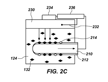
FIGS. 2A through 20 illustrate how the device 200 shown in FIG. 1 may be
used to detect an analyte 132 in a liquid 124. The liquid 124 may be passed
adjacent
to the thermocouple 210. The liquid 124 may include an analyte 132 that
specifically
binds to the assay polymer 214 and changes thermal properties thereof, as
described
above. A heat sink 230 may provide heat to the liquid 124. Though referred to
as a
heat "sink" for the sake of simplicity, the heat sink 230 may be configured to
provide
heat to or remove heat from the liquid 124 and, so, may also be characterized
as a
heat transfer element 230. The heat sink or heat transfer element 230 may be a
material having a high thermal conductivity, such as a transition metal (e.g.,
copper,
silver, etc.) or an alloy or mixture thereof. The heat sink 230 may be
thermally coupled
to a temperature sensor 232 (e.g., a thermocouple or another device)
configured to
detect a temperature of the heat sink 230, and to a temperature modification
device
234 configured to maintain the temperature of the heat sink 230. If the
properties of
the heat sink 230 are known (e.g., if a relationship between a control signal
to the
modification device 234 and the temperature of the heat sink 230 is well
characterized),
the temperature sensor 232 may be omitted. In some embodiments, the
temperature
sensor 232 may be integral to the temperature modification device 234. For
example,
the resistance of the temperature modification device 234 itself may be
measured to
determine its temperature. The temperature modification device 234 may
include, for
example, a thermoelectric device, a heat exchanger, a fan, a resistance
heater, etc.
The temperature sensor 232 may be a resistor having a resistance that varies
with
temperature. The temperature of the liquid 124 may be different from the
temperature
of the heat sink 230, and may vary based at least in part on the presence or
absence of
the analyte 132 and its concentration in the liquid 124.
CA 03020521 2018-10-09
WO 2017/178081
PCT/EP2016/076572
7
The temperature sensor 232 and the temperature modification device 234 may
be connected to a processor 236 programmed to control the temperature
modification
device 234 to cause the heat sink 230 to produce a thermal wave emanating from
the
heat sink 230 and through the liquid 124 to the thermocouple 210. For example,
the
.. processor 236 may be a computer having an input-output card configured to
receive
and provide electrical signals, or any other suitable controller. The
processor 236 may
be a proportional-integral-derivative (PID) controller capable of changing the
temperature of the heat sink 230 by a small amount on a relatively short time
scale.
For example, the processor 236 may be programmed to change the temperature of
the
heat sink 230 by about 0.5 C or less, about 0.2 C or less, or even about 0.05
C or less.
Thus, the thermal wave may have an amplitude of about 1.0 C or less, about 0.4
C or
less, or even about 0.10 C or less. The processor 236 may be capable of
changing the
temperature of the heat sink 230 via the temperature modification device 234
from one
set point to another and back to form a thermal wave having a frequency from
about
0.001 to about 0.5 Hz, such as from about 0.005 to about 0.1 Hz, or from about
0.01 to
about 0.05 Hz. In some embodiments, the processor 236, the temperature
modification device 234, and the heat sink 230 may together produce a thermal
wave
having a variable frequency. Based on a measurement from the temperature
sensor
232 (if present), a known input to the temperature modification device 234, or
other
means, properties of the thermal wave may be known (e.g., a phase, amplitude,
frequency at a specific time, rate of frequency change, etc.).
As shown in FIG. 2A, the liquid 124 may be substantially free of the analyte
132
of interest at one point in time, and the assay polymer 214 may also be
substantially
free of the analyte 132 at that time. Thus, heat (indicated by arrows in FIG.
2A) may be
transferred from the heat sink 230 through the liquid 124 to the thermocouple
210 and
along the thermocouple 210 at a rate related to the heat transfer properties
of the
assay polymer 214 unbound with the analyte 132 (because the thermocouple 210
itself
may provide minimal thermal resistance).
At another time, shown in FIG. 2B, the liquid 124 may have a nonzero
concentration of the analyte 132, and some of the analyte 132 may bind to the
assay
polymer 214. Therefore, heat may be transferred from the heat sink 230 through
the
liquid 124 to the thermocouple 210 at a different rate than shown in FIG. 2A
(as
indicated by the arrows in FIG. 2B). At yet another time, shown in FIG. 2C,
the liquid
124 may have a higher concentration of the analyte 132 than shown in FIG. 2B,
and
more of the analyte 132 may bind to the assay polymer 214. Therefore, heat may
be
CA 03020521 2018-10-09
WO 2017/178081
PCT/EP2016/076572
8
transferred from the heat sink 230 through the liquid 124 to the thermocouple
210 at a
different rate than shown in FIGS. 2A and 2B. For example, an increase in the
amount
of the analyte 132 bound to the assay polymer 214 may decrease the heat
transfer rate
through the assay polymer 214 to the thermocouple 210.
The concentration of the analyte 132 in the liquid 124 may be calculated based
at least in part on a heat transfer property of the assay polymer 214 (which
may be
inferred based on, e.g., the amount of heat transferred to the thermocouple
210 as a
function of time).
For comparison purposes, FIGS. 3A through 30 illustrate how a thermocouple
210' having a polymer 214' that does not have an affinity for the analyte 132
(e.g., a non-
imprinted polymer) may perform under similar conditions to those shown in
FIGS. 2A
through 20. In FIG. 3A, the liquid 124 may be substantially free of the
analyte 132 of
interest at one point in time, and the polymer 214' may also be substantially
free of the
analyte 132 at that time. At another time, shown in FIG. 3B, the liquid 124
may have a
nonzero concentration of the analyte 132, yet the analyte 132 may not bind to
the
polymer 214' in an appreciable amount. Therefore, heat (indicated by arrows in
FIG. 3A)
may be transferred from the heat sink 230 through the liquid 124 to the
thermocouple
210 at the same rate shown in FIG. 3A. At yet another time, shown in FIG. 30,
the
liquid 124 may have a higher concentration of the analyte 132 than shown in
FIG. 3B,
yet the analyte 132 may still not bind to the polymer 214' to an appreciable
amount.
Some portion of the analyte 132 may bind to the polymer 214', particularly for
high
concentrations of the analyte 132 in the liquid 124, but the amount of analyte
132 bound
may be much smaller than the amount bound to the assay polymer 214 (FIG. 20)
at
similar concentrations. Heat may be transferred from the heat sink 230 through
the
liquid 124 to the thermocouple 210' at substantially similar rates no matter
the
concentration of the analyte 132 in the liquid 124.
Referring again to FIGS. 2A through 20, a processor 236 (which may be or
include the processor 223 shown in FIG. 1, or which may be in electronic
communication with the processor 223) may be programmed to calculate a
concentration of the analyte 132 in the liquid 124 based at least in part on a
thermal
wave emanating through the liquid 124. For example, the heat sink 230 may
cause a
change in a temperature of the liquid 124, and may form a thermal wave through
the
liquid 124. The processor 236 may determine a difference in amplitude and/or
phase
between the thermal wave as provided by the heat sink 230 and an attenuated
thermal
wave at the thermocouple 210. The difference in amplitude and/or phase may be
used
CA 03020521 2018-10-09
WO 2017/178081
PCT/EP2016/076572
9
to determine the amount of the analyte 132 bound to the assay polymer 214,
which may
in turn be used to determine the concentration of the analyte 132 in the
liquid 124.
In some embodiments, the processor 236 may implement a frequency change
of the thermal wave produced by the heat sink 230. The processor 236 may then
detect a phase shift between the thermal wave produced by the heat sink 230
and an
attenuated thermal wave in the liquid 124 after the thermal wave passes
through the
assay polymer 214 and the base material 212 to the thermocouple 210.
FIG. 4 is a simplified schematic representation showing how the thermal wave
may travel into and within the device 200 of FIG. 1. FIG. 4 includes some of
the
components shown in FIGS. 1 and 2A through 20, but shows them separated to
allow
representation of thermal waves traveling through and between the components.
In
particular, FIG. 4 shows the heat sink 230 thermally coupled to the
temperature
modification device 234 and the temperature sensor 232, which are connected to
the
processor 236.
The heat sink 230 may produce a thermal wave 202 and transfer the thermal
wave 202 to the liquid 124 toward the assay polymer 214 on the thermocouple
210. For
example, if the heat sink 230 is initially maintained at a constant
temperature of 37 C,
the thermal wave 202 may be produced by heating the heat sink 230 to a
temperature
of 37.1 C and then cooling the heat sink 230 to a temperature of 36.9 C. The
heating
and cooling of the heat sink 230, driven by the temperature modification
device 234,
may cause the assay polymer 214 and the thermocouple 210 to heat and cool in a
corresponding manner. The thermal wave 202 may have an amplitude al and a
frequency (pi. The amplitude al and/or the frequency (Pi may vary with time.
For
example, the thermal wave 202 may have a continuously varying frequency (pi.
As discussed above, the presence or absence of the analyte 132 on the assay
polymer 214 may change the thermal conductivity, thermal diffusivity, heat
capacity, or
another property of the assay polymer 214. The assay polymer 214 may define
cavities
therein adapted to interact with at least a portion of the analyte 132.
Without being
bound to any particular theory, the cavities may be configured to act to
specifically bind
the analyte 132. Thus, the assay polymer 214 may receive particles or
molecules of the
analyte 132 from the liquid 124 in some of the cavities, based on the
concentration of
the analyte 132 in the liquid 124. The liquid 124 and the assay polymer 214
may reach
equilibrium at a given temperature, such that the analyte 132 binds to and
separates
from the assay polymer 214 at equal rates. The thermal properties of the assay
polymer
CA 03020521 2018-10-09
WO 2017/178081
PCT/EP2016/076572
214 may depend in part on the fraction of the cavities bound to particles or
molecules
of the analyte 132.
The assay polymer 214 and/or the analyte 132 thereon may alter the thermal
wave 202 passing therethrough to form an attenuated thermal wave 204. The
5 attenuated thermal wave 204 may be detected by the thermocouple 210, and
recorded
by the processor 236. The attenuated thermal wave 204 may have an amplitude a2
and a frequency cp2 , which may be different from the amplitude ai and a
frequency (pi
of the thermal wave 202. The differences in the amplitudes al, a2 and/or the
frequencies (pi, cp2 may be correlated to the amount of the analyte 132 bound
to the
10 .. assay polymer 214, and thus, to the concentration of the analyte 132 in
the liquid 124.
Measurement of the differences in the amplitudes al, a2 and/or the frequencies
(pi, cp2
may allow the device 200 to detect relatively lower amounts of the analyte 132
bound
to the assay polymer 214 (corresponding to lower concentrations of the analyte
132 in
the liquid 124) as compared with methods of measuring the temperature of the
thermocouple 210 at steady state.
Referring again to FIG. 1, to form the device 200, the thermocouple 210 may be
coated with the base material 212. For example, the thermocouple 210 may be
dip-
coated with the base material 212 by immersing a portion of the thermocouple
210 into
a liquid containing the base material 212 or a precursor thereof. Dip-coating
may be
performed efficiently and scaled to produce mass quantities, with relatively
high
uniformity in comparison with other methods. Dip-coating of wire is described
in, for
example, U.S. Patent 4,924,037, "Electrical Cable," granted May 8, 1990, the
entire
disclosure of which is hereby incorporated herein by reference. Dip-coating
may form
the base material 212 to be relatively thin, such that the base material 212
has a
relatively low intrinsic thermal resistivity than thicker layers of polymer.
For example,
the base material 212 may have a thickness from about 0.01 mm to about 1 mm,
such
as from about 0.05 mm to about 0.5 mm.
The thermocouple may be coated with the assay polymer 214, either over and
secured to the base material 212 (e.g., directly on a surface of the base
material 212 or
on another material on a surface of the base material 212) or directly onto
the surface
of the thermocouple 210. In some embodiments, a thermocouple 210 with a base
material 212 thereon may be heated such that the base material 212 softens.
For
example, the base material 212 may be heated to a temperature above its glass
transition temperature (Tg). The thermocouple 210 and base material 212
coating may
then be rolled in a powder of the assay polymer 214 to attach the assay
polymer 214 to
CA 03020521 2018-10-09
WO 2017/178081
PCT/EP2016/076572
11
the base material 212. The base material 212 may then be cooled to retain the
particles of the assay polymer 214, such as by cooling the base material 212
to a
temperature below Tg. The time between attaching particles of the assay
polymer 214
and cooling the base material 212 may be kept relatively short, such that the
particles of
the assay polymer 214 can become securely embedded into the base material 212
without becoming covered by the polymer 212. For example, the thermocouple 210
may be placed in a refrigerator to cool the base material 212 within about a
time period
from about 1 second to about 60 seconds after coating with the assay polymer
214.
The thermocouple 210 may remain in the refrigerator until the base material
212 is in a
solid phase (e.g., crystalline). For example, the thermocouple 210 may remain
in the
refrigerator for a time period from about 1 minute to about 20 minutes. After
cooling,
the thermocouple 210 may be washed in a liquid (e.g., water, alcohol, etc.) to
remove
loosely bound or unbound particles of the assay polymer 214 from the surface
of the
base material 212.
Without being bound to any particular theory, it appears that above Tg,
polymers soften and plasticize, meaning that the time the thermocouple 210
spends
above Tg may allow particles of the assay polymer 214 to sink into or
otherwise bond to
the base material 212.
The processor 236 (e.g., a PID controller) may be electrically connected to
the
temperature modification device 234 to provide power sufficient to drive the
temperature of the heat sink 230, and to cause the temperature modification
device
234 to change the temperature of the heat sink 230 to produce the thermal wave
202
(FIG. 4).
The thermocouple 210 may be disposed within a flow of the liquid 124 to be
measured. The heat sink 230 may be secured to a conduit through which the
liquid
124 passes, or may be disposed within the flow of the liquid 124. The
processor 236
may be configured to continuously detect the temperature at the thermocouple
210 and
the temperature sensor 232, and to calculate the concentration of the analyte
132 in
the liquid 124 based at least in part on a phase shift between the thermal
wave 202
(FIG. 4) produced by the heat sink 230 and the attenuated thermal wave 204
(FIG. 4)
at the thermocouple 210.
The device 200 shown and described may be configured to detect any of a wide
range of selected analytes 132. For example, the device 200 may be used for
detecting, sensing, or quantifying biological analytes or other chemicals in
the liquid
.. 124. The analyte 132 may be a gas, liquid, or solid dissolved or otherwise
mixed with
CA 03020521 2018-10-09
WO 2017/178081
PCT/EP2016/076572
12
the liquid 124. For example, the device 200 may be used for detecting,
sensing,
quantifying analytes, antibodies, antigens, nucleic acids, (e.g., DNA, RNA,
etc.),
including nucleic acids with particular sequences (e.g., SNPs), proteins,
small
molecules (e.g., dopamine, histamine, etc.) or other substances. In some
embodiments, the device 200 may be used for detecting histamine, dopamine,
serotonin, adrenalin, methylphenidate, etc.
One of the many attractive features of molecular imprinting methods as
disclosed herein is that methods can be applied to a diverse range of
analytes. The
imprinting of small, organic molecules (e.g., pharmaceuticals, pesticides,
amino acids
and peptides, nucleotide bases, steroids, sugars, etc.) is described in, for
example, K.
Haupt and K. Mosbach, "Molecularly Imprinted Polymers and Their Use in
Biomimetic
Sensors," Chem. Rev. 100, 2495-2504 (2000); and G. Mustafa and P. Lieberzeit,
"MIP
Sensors on the Way to Real-World Applications," in Springer Series on Chemical
Sensors and Biosensors, vol. 12, pp. 167-187 (Springer, 2012). Somewhat larger
organic compounds (e.g., peptides) can also be imprinted via similar
approaches.
Protocols for imprinting larger structures, such as proteins, cells, and
mineral crystals
have been proposed in, for example, M. Kempe, M. Glad, and K. Mosbach, "An
Approach Towards Surface Imprinting Using the Enzyme Ribonuclease A," J.
Molecular Recognition, 8,35-39 (1995); S. Hjerten etal., "Gels Mimicking
Antibodies in
.. Their Selective Recognition of Proteins," Chromatographia 44, 227-234
(1997); H. Shi
et al., "Template-Imprinted Nanostructu red Surfaces for Protein Recognition,"
Nature
398, 593-597 (1999); A. Aherne et al. "Bacteria-Mediated Lithography of
Polymer
Surfaces," J. Am. Chem. Soc. 118, 8771-8772 (1996); and S. M. D'Souza, etal.,
"Directed Nucleation of Calcite at a Crystal-Imprinted Polymer Surface,"
Nature 398,
312-316 (1999). Molecular imprinting as a bridge to drug advanced drug
delivery is
described in B. Sellergren and C. Allender, "Molecularly Imprinted Polymers: A
Bridge
to Advanced Drug Delivery," Advanced Drug Delivery Reviews 57, 1733-1741
(2005).
The entire disclosures of each of the documents cited in this paragraph are
hereby
incorporated herein by reference.
To detect the analyte 132, the liquid 124 containing the analyte 132 may be
passed through a conduit adjacent the assay polymer 214 over the thermocouple
210.
Particles or molecules of the analyte 132 bind to the assay polymer 214,
changing one
or more thermal properties of the assay polymer 214. The liquid 124 may flow
continuously adjacent the assay polymer 214 during detection, or the flow may
terminate before detection begins. The thermal wave 202 (FIG. 4) and the
attenuated
CA 03020521 2018-10-09
WO 2017/178081
PCT/EP2016/076572
13
thermal wave 204 may travel through the liquid 124 whether the liquid 124 is
flowing or
stagnant. The thermal properties of liquid 124 may differ for flowing and
stagnant
liquids 124, but can be determined based on flow properties. In some
embodiments,
the liquid 124 may be brought to a test temperature before detection of the
analyte 132.
As discussed above, the assay polymer 214 may be a molecularly imprinted
polymer
formulated to bind a particular analyte 132 of interest.
The thermal wave 202 (FIG. 4) is provided from the adjustable heat sink 230 to
the thermocouple 210 through the assay polymer 214. The processor 236 (e.g., a
PID
controller) may change the temperature of the heat sink 230 via the
temperature
modification device 234, such as by raising the temperature and lowering the
temperature of the heat sink 230 by a preselected amount and at a preselected
frequency. The change in the temperature of the heat sink 230 may be small
enough
that the change does not interfere significantly with other measurements that
may
occur simultaneously. For example, the average temperature of the liquid 124
may be
measured even though the temperature of the heat sink 230 is varying, so long
as the
time scale of the average temperature measurement is longer than the frequency
of the
variation and/or the amount of the temperature variation is small in
comparison with the
temperature change induced by the interaction of the analyte 132 with the
assay
polymer 214. In some embodiments, the heat sink 230 may provide a thermal wave
202 having a frequency from about 0.001 to about 0.5 Hz, such as from about
0.005 to
about 0.1 Hz, or from about 0.01 to about 0.05 Hz. Furthermore, the frequency
of the
thermal wave 202 may vary during testing (e.g., the frequency may be
continuously
varied from a low frequency to a high frequency or vice versa). The thermal
wave 202
may have an amplitude of about 1.0 C or less, about 0.4 C or less, or even
about
0.10 C or less.
The temperature of the thermocouple 210 may be tested, and the result may be
compared with the temperature of the heat sink 230 (as measured at the
thermocouple
232).
The concentration of the analyte 132 in the liquid 124 may be calculated at
least
in part on a phase shift between the thermal wave 202 produced by the heat
sink 230
and the attenuated thermal wave 204 wave at the thermocouple 210. A comparison
of
the thermal wave 202 and the attenuated thermal wave 204 may be performed by
the
processor 236 based on responses of liquids of known concentration. In some
embodiments, the comparison of the thermal wave 202 with the attenuated
thermal
CA 03020521 2018-10-09
WO 2017/178081
PCT/EP2016/076572
14
wave 204 may be based at least in part on the amplitudes, the phase shift, or
another
property.
Measurement of the thermal wave enables measurement of thermal resistance
without significantly changing the overall temperature of the assay polymer
214.
Without being bound to any particular theory, such a measurement appears to be
a
thermal analog to the measurement of capacitance or inductance in the field of
electronics. For example, measuring resistance reveals some information about
an
electronic device or material, but measuring capacitance or impedance reveals
additional information, such as how the device or material responds to a load.
Similarly, measuring thermal resistance by the methods disclosed herein can
reveal
additional information that measuring a steady-state temperature difference
cannot.
For example, when applying a thermal wave, different types of information are
available in the form of a change in amplitude, frequency and/or phase of the
attenuated thermal wave upon binding of a target to the receptor. The phase
shift may
.. vary based on the frequency of the input. The amount of information
provided by a
thermal wave is greater than steady-state analysis, and the information may
enable
detection or differentiation of a wider variety of materials.
Furthermore, and again without being bound to any particular theory, an
increase in thermal mass of the assay polymer 214 may occur upon binding of
the
analyte 132 onto its receptor (i.e., the cavities therein). Before binding of
the analyte
132, the cavities may be filled with liquid. Upon binding of the analyte 132
into its
receptor, the liquid may be replaced by the analyte 132, thus increasing the
thermal
mass of the entire coated thermocouple 210.
EXAMPLES
Example 1: Preparation of MIP having a template for detecting dopamine.
Ethylene glycol dimethacrylate (EGDM), methacrylic acid (MAA), dopamine
hydrochloride salt (99%), and methanol were purchased from Acros Organics
(Loughborough, United Kingdom). Prior to polymerization, the stabilizers in
the MAA
and EGDM were removed by filtration over alumina. 4,4'-azobis(4-cyanovaleric
acid)
and serotonin creatinine sulfate monohydrate (98%) were purchased from Sigma-
Aldrich (Gillingham, United Kingdom).
A mixture of MAA (0.54 g, 6.6 mmol), EGDM (2.96 g, 14.9 mmol), and
4,4'-azobis(4-cyanovaleric acid) (65 mg) was dissolved in methanol (3.67 ml)
and water
(0.57 ml) together with dopamine (0.063 g, 0.33 mmol), the template molecule.
This
CA 03020521 2018-10-09
WO 2017/178081
PCT/EP2016/076572
mixture was degassed with N2 and heated to initiate polymerization. To allow
full
completion of the reaction, the mixture was kept at 65 C for 12 hours. After
polymerization, the bulk polymer was ground and sieved to obtain
microparticles
having diameters smaller than 10 pm. Dopamine was removed from the MIP powders
5 by continuous extraction with a 50/50 mixture of methanol and water.
After 6 hours,
the MIP was substantially free of dopamine, as verified by AT-IR spectroscopy
with a
NICOLETTm 380 FT-IR device from Thermo Scientific (Loughborough, United
Kingdom). Subsequently, the MIP powder was dried in an oven for 12 hours at
100 C.
A non-imprinted polymer (NIP) was synthesized as a control according to the
same
10 method, but without the presence of the dopamine.
Example 2: Testing of MIP for detecting dopamine
Specificity and binding isotherms of the MIP and NIP particles were determined
by optical batch rebinding experiments with an Agilent 8453 UV-visible
15 spectrophotometer (Santa Clara, California). For the rebinding
experiments, 20 mg of
MIP or NIP powder was added to 5 ml of aqueous dopamine solutions in
concentrations between 0.3 to 1.0 mM. The resulting suspensions were shaken
for 12
hours on a rocking table at room temperature. Subsequently, the suspensions
were
filtered and the free concentration of dopamine (Cf) was determined by UV-vis
spectroscopy. The bound concentrations (Sb) of dopamine were calculated per
gram of
MIP and NIP and binding isotherms, and are shown in FIG. 5. By fitting the
binding
isotherms, the specificity of the MIP toward the template dopamine was
determined.
To test the selectivity, the competitor molecule serotonin was used, since its
structure
is very similar to dopamine. For these experiments, 20 mg of MIP powder was
added
to 5 ml of aqueous serotonin solutions and binding isotherms were determined
after
filtration of the suspensions.
FIG. 5 shows that there is a significant difference in binding between the MIP
and its reference, the NIP. To determine the specificity, the imprint factor
(IF) was
used, which is the amount bound to the MIP divided by the amount bound to the
reference NIP at a selected concentration. The binding isotherms were fitted
with a
two-parameter fit of the following type to analyze the imprint factor at a
specific
concentration (Equation 1):
Equation 1: Sb = A = Cfv
CA 03020521 2018-10-09
WO 2017/178081
PCT/EP2016/076572
16
Equation 1 corresponds to the Freundlich isotherm and may be used for fitting
of MIP binding isotherms if the distribution of the binding sites and affinity
constants are
assumed to be heterogeneous. At Cf = 0.3 mM, the IF was 3.1 0.1, whereas
higher
concentrations yielded slightly lower IF values (-2.5) due to saturation of
the binding
sites. The results were comparable to other dopamine MIPs in literature. The
response of the MIP to the competitor serotonin was not significantly
different than the
reference, demonstrating the selectivity of the system.
Example 3: Preparation of MIP-coated thermocouples
PLLA was mixed with chloroform at 60 C under a water reflux for 120 minutes
to ensure dissolution of the PLLA and a negligible loss of chloroform. The
concentration of the resulting solution was 200 pg/mL.
Mineral-insulated Type K thermocouples having diameters of 0.5 mm and
lengths of 30 cm were obtained from TO Direct, of Nederweert, the Netherlands.
The
thermocouples were dipped into the PLLA-chloroform solution for 10 seconds and
withdrawn at the rate of 0.39 cm/min. The chloroform was evaporated to leave a
coating of PLLA having a thickness of approximately 0.09 mm (90 pm) on the
thermocouples.
The PLLA coated thermocouples were heated to the glass transition
temperature of the PLLA polymer, between 65 C and 75 C. The thermocouples were
manually roll coated with the MIP or NIP powder formed in Example 1.
Subsequently,
the coated thermocouples were placed in a refrigerator at 4 C for
approximately 5
minutes to allow the PLLA polymer to be brought back to its crystalline state.
The
thermocouples were then washed with isopropanol in order to wash away loosely
associated or excessive MIP or NIP particles to leave securely attached MIP or
NIP
particles.
Example 4: Heat-transfer method for detection of dopamine with MIP-coated
thermocouples
A lx phosphate buffered saline (PBS) solution was prepared with Dulbecco
tablets obtained from Oxoid Limited (Basingstoke, United Kingdom). MIP-coated
and
NIP-coated thermocouples were sequentially exposed in a flow cell to fixed
concentrations of dopamine in a phosphate-buffered saline (PBS) buffer
solution.
Solutions of dopamine in concentrations of 0.5 pM, 1 pM, 2 pM, 5 pM, 10 pM, 15
pM,
20 pM, 25 pM, and 50 pM were pumped in ascending succession. At first, the
flow cell
CA 03020521 2018-10-09
WO 2017/178081
PCT/EP2016/076572
17
was flushed with PBS and left for at least 45 minutes to allow temperature
stabilization
to occur. Afterwards, 3 ml of the lowest concentration (0.5 pM) was added over
12
minutes, at a constant rate of 0.25 ml/min. Before the addition of the next
concentration, the flow cell was left to equilibrate for 30 minutes. Thus, a
pattern of 12
minutes addition by constant pumping and 30 minutes stabilization was kept
throughout each measurement. A copper heating element in the flow cell in
contact
with the solution was maintained at 37 C by controlling a voltage across a
resistor in
thermal contact with the copper heating element. The change in voltage
required to
maintain the temperature was recorded, and is shown in FIG. 6A.
As seen in FIG. 6B, increasing concentrations of dopamine correspond to an
increase in temperature of the MIP-coated thermocouple (T2 MIP). Furthermore,
the
voltage required to maintain the copper element at 37 C (V MIP) decreases
concurrently with increased dopamine concentration. When the copper heating
element is maintained at a temperature of 37 C, buffer solution in the flow
cell is at a
temperature of about 32.5 C, and receptor cites on the MIP coating are
unoccupied.
Also shown in FIG. 6B, the temperature of the NIP-coated thermocouple (T2 NIP)
was
maintained constant throughout the experiment. Furthermore, the voltage
required to
maintain the copper element at 37 C (V NIP) did not decrease when the
concentration
of dopamine increased. The MIP experiment was repeated two more times to
.. investigate the reproducibility of the attachment method used, with similar
results.
FIGS. 6A and 6B show that there is no significant response by the NIP-coated
thermocouple to any dopamine-PBS additions. This indicates that the MIP
coating is
the source of the temperature variations observed between the MIP-coated
thermocouple and the NIP-coated thermocouple.
The temperature for MIP-coated thermocouples (T2 MIP) was constant at about
32.5 C for the first four additions of dopamine-PBS. Upon addition of 10pM
dopamine,
there was a significant increase in T2, which continues for the subsequent
higher
concentrations. This can be explained by a change in thermal resistance of
receptors
of the MIP. Heat loss occurs through unoccupied receptors, which appear to be
present for the first four dopamine-PBS additions to the MIP-coated
thermocouples as
well as for all additions to the NIP-coated thermocouples. Upon occupation of
the MIPs
by dopamine, an insulation layer appears to be created, decreasing heat loss
through
the thermocouple; hence the overall temperature of the flow cell increases to
a
maximum of 33.5 C for concentrations above 20pM DA. The increase in the
CA 03020521 2018-10-09
WO 2017/178081
PCT/EP2016/076572
18
temperature measured by the MIP-coated thermocouple may be explained by the
retention of heat energy by the insulating layer.
The increase in the temperature measured (T2) is accompanied by a decline in
voltage over power resistor (V MIP), meaning that less power is needed to keep
the
copper element at a constant 37 C, since heat loss from the flow cell has
decreased.
Meanwhile, V NIP stays constant throughout the experiment. The ambient
temperature
was found to be stable at room temperature, meaning that a change in T2 could
not be
attributed to a change in ambient temperature. Thus, the increase in T2
appears to
correspond to the occupation of MIPs by dopamine.
Furthermore, because V MIP is associated with a change in T2, the
concentration of dopamine in the liquid may be inferred based on a measurement
of V
MIP alone, T2 alone, of both V MIP and T2 in combination.
FIG. 7 compares dose-response curves for thermocouples coated with MIPs
and NIPs when exposed to various concentrations of dopamine. For MIPs, there
is an
increase in response with increasing dopamine concentrations. The data suggest
that
the limit of detection for dopamine by the MIP-coated thermocouples as
prepared in
Example 3 may be approximately 5 pM dopamine and that MIP saturation may be
reached between about 20 pM and 25 pM dopamine. NIP-coated thermocouples did
not show any significant response to the increase in dopamine concentrations.
Thermocouples and sensors as described herein may offer benefits as
compared to conventional sensors. For example, thermocouples may be relatively
easier to prepare, because methods such as dip-coating may be scaled to
production
quantities while retaining uniform (reproducible) characteristics.
Furthermore, though a
thermocouple may have a much smaller surface area than a flat substrate, the
detection sensitivity of a thermocouple per unit of surface are may be higher
than the
detection sensitivity of flat substrates. Thus, the thermocouple may be both
smaller
and more sensitive. Thus, thermocouples and sensors may be used for
microfluidics
and in vivo testing, applications for which flat substrates may not be
practical. Coating
of thermocouples may be relatively easier than coating of flat substrates
because a
smaller volume of liquid may be needed.
While the present disclosure has been described herein with respect to certain
illustrated embodiments, those of ordinary skill in the art will recognize and
appreciate
that it is not so limited. Rather, many additions, deletions, and
modifications to the
illustrated embodiments may be made without departing from the scope of the
disclosure as hereinafter claimed, including legal equivalents thereof. In
addition,
CA 03020521 2018-10-09
WO 2017/178081
PCT/EP2016/076572
19
features from one embodiment may be combined with features of another
embodiment
while still being encompassed within the scope of the disclosure as
contemplated by
the inventors. Further, embodiments of the disclosure have utility with
different and
various detection devices and methods.