Note: Descriptions are shown in the official language in which they were submitted.
CA 03020567 2018-10-10
WO 2017/180708 PCT/US2017/027146
Systems, Methods and Products for Minimizing Tissue Reactions and Tissue
Injury at an
Infusion Site
Background
Despite their demonstrated clinical benefits, currently insulin infusion sets
are only approved
for in vivo usage for only 3 days. Even with this limited approved lifespan, a
substantial portion
of sets fail to meet this recommended lifespan during practical use.
Nevertheless Continuous
Subcutaneous Insulin Infusion (CSII) therapy represents the most advanced form
of insulin
delivery technology currently available and administers more precise amounts
of insulin in a
programmable format as compared to traditional injection methods, which
provides increased
flexibility and enhanced quality of life for the user. To achieve effective
glucostasis using an
artificial pancreas, a combination of a highly accurate continuous glucose
monitor (CGM) and
reliable continuous subcutaneous insulin infusion (CSII) is required. Although
CGM
performance and lifespan has significantly improved over the last decade, CSII
with a current
lifespan of 3 days or less has not. As such the current approved usage
lifespans for commercial
CGM and CSII devices is highly mismatched with in vivo durations of 10+ days
vs. 3d,
respectively.
The high occurance of inflammation and scarring at insulin infusion sites in
patients with
diabetes is well known (i.e. 25-42 %) particularly in pediatric populations
and whereas infection
at insulin infusion sites is also frequently seen. It would be useful to
develop products and
methods to reduce this inflammation and scarring.
Summary
One embodiment described herein is a method of lowering the concentrations of
at least
one of preservatives and fibrils in a liquid insulin formulation, comprising
replacing at least one
of phenol and m-cresol with at least one of cyclodextrins, cyclodextrin
polymers and
cyclodextrin beads. Another embodiment is a method for removing at least one
of preservatives
at fibrils from a liquid insulin composition comprising incorporating at least
one of an ion
exchange resin and cyclodextrin polymers or beads into the infusion set. A
liquid insulin
1
CA 03020567 2018-10-10
WO 2017/180708 PCT/US2017/027146
formulation comprising cyclodextrins and/or cyclodextrin polymers as
preservatives also is
described.
Another embodiment is a method of lowering the concentration of phenol and/or
m-
cresol, and/or fibrils in a liquid insulin composition by replacing at least a
portion of the phenol
and/or m-cresol with at least one of cyclodextrins and cyclodextrin polymers.
The disclosure
also describes a method for removing insulin solubilizers from a liquid
insulin composition
comprising incorporating at least one of an ion exchange resin and
cyclodextrin into the infusion
set. A liquid insulin formulation comprising cyclodextrins and/or cyclodextrin
polymers as
solubilizers also is disclosed.
A further embodiment is a method for removing anti-microbial agents from a
liquid
insulin composition comprising incorporating at least one of an ion exchange
resin and
cyclodextrin polymers and/or beads into the infusion set. A liquid insulin
formulation
comprising cyclodextrins and/or cyclodextrin polymers as anti-microbial agents
also is
described.
An insulin delivery apparatus is disclosed comprising an ion exchange resin
configured
to remove insulin preservatives. An insulin delivery system comprising
cyclodextrin
preservatives is described herein.
Another embodiment is a method of preventing insulin degradation at an
infusion site
comprising incorporating an anti-protease into the insulin formulation. A
system comprising the
filter used to remove fibrils from a liquid insulin formulation is described.
A method is disclosed
for suppressing inflammation resulting from the injection or infusion of
insulin, comprising
delivering an anti-inflammatory drug, factor or agent to (a) the insulin
formulation or (b) during
an insulin infusion. Additionally, a method of supressing fibrosis induced
insulin delivery,
comprising incorporating growth factor inhibitors into the liquid insulin
formulation is disclosed.
A further embodiment is a method of inducing blood vessel and/or lymphatic
vessel
growth at an insulin infusion site, comprising introducing vascular
endothilial growth factor to
the infusion site. A method of introducing an anti-protease, anti-
inflammatory, anti-fibrotic or
lymphatic drug, factor or agent to a liquid insulin composition using a dual
lumen cannula also is
disclosed.
2
CA 03020567 2018-10-10
WO 2017/180708 PCT/US2017/027146
A further embodiment is a method of introducing an anti-protease, anti-
inflammatory,
anti-fibrotic or lymphatic drug, factor or agent to a liquid insulin
composition using a dual lumen
cannula. Yet another embodiment is a method for coating an insulin infusion
set cannula and/or
a biocompatible collar for the cannula with a composition that contains at
least one of a basement
membrane or another extracellular matrix, an oil, such as a high molecular
weight silicone oil,
and a lubricant, such as proteoglycan 4 (PRG4), Libricin and/or hyaluronan.
Another
embodiment is a method for local delivery of agents from these coatings to
suppress
inflammation, fibrosis and /or insulin degradation, and optionally also
promoting blood and
lymphatic vessel ingrowth into the infusion site.
Brief Description of the Drawings
Fig. 1A schematically illustrates the effects of insulin fibrils and
preservatives on tissue.
Fig. 1B is a chart showing the effect of insulin, fibrils and preservatives on
cells and tissues.
Fig. 2 schematically shows a mammalian CSII air pouch model used in testing
described herein.
Fig. 3 shows an example of a murine transdermal insulin pump.
Fig. 4A shows equipment used in the mammalian air pouch open loop model.
Fig. 4B shows a mouse being studied using the equipment shown in Fig. 4A.
Fig. 5 is a graph showing the in vitro toxicity of insulin and insulin
formulation excipients on
human PBMC.
Fig. 6 shows PBMC morphology after exposure to insulin or fibrils
Fig. 7A is a table showing the impact of excipients/diluents (preservatives)
on PBMC expression
in vitro.
Fig. 7B is a graph showing IL-8 chemokine induction in human PBMC's by insulin
and/or
excipients.
Fig. 7C is a graph showing IL-8 expression by human PBMC in vitro.
Fig. 7D is a graph showing INF-g expression by human PBMC in vitro.
Fig. 8A shows a test mouse.
Fig. 8B is another view of the test mouse.
Fig. 8C shows a third view of the test mouse.
Fig. 8D is a graph showing continuous glucose monitoring (CGM) blood glucose
levels and
external BG control test monitored in a diabetic NOD mouse.
3
CA 03020567 2018-10-10
WO 2017/180708 PCT/US2017/027146
Fig. 9A shows H & E stained mouse skin and SC tissue sections in a control
mouse that has been
administered saline.
Fig. 9B shows H & E stained mouse skin and SC tissue sections in a normal
mouse that has
received insulin.
Fig 9C shows H & E stained mouse skin and SC tissue sections in a diabetic
mouse that has
received insulin.
Fig. 9D is a bar graph showing leukocyte influx into a murine air pouch model
for a normal
mouse.
Fig. 9E is a bar graph showing leukocyte influx into a murine air pouch model
for a diabetic
mouse.
Fig. 10 is a graph showing leukocyte influx into a murine air pouch model
after an infusion of
insulin excipient or saline.
Figs. 11A-11F show the effect of saline and insulin infusion excipients on
inflammation over a
three day period.
Figs. 12A-12C show the uptake of FITC-insulin by PBMCs in vitro.
Figs. 13A-13C show the impact of leukocyte protease on insulin.
Fig. 14 is a table showing the impact of anti-proteases on FITC insulin
degradation.
Fig. 15 is a table showing an overview of research described herein.
Fig. 16 is a table showing types of mice used in tests described herein.
Fig. 17 is a table describing the insulin and excipients used in studies
described herein.
Fig. 18 is a table describing the initial evaluation of tissue reactions
detected in tests described
herein.
Fig. 19 shows photos of tissue exposed to saline vs. diluent.
Figs. 20A-20C are photomicrographs showing LCM capture of "Giant Cell" from in
vitro
culture.
Fig. 21 is a graph of qPCR RNA analysis of macrophages (MQ) and giant cells
obtained by
LCM.
Fig. 22 is a table showing inhibitors and inducers.
Fig. 23 is a general flow diagram for gene expression in AP/OL.
Fig. 24 is a chart showing in vitro TIE induced gene expression.
Figs. 25A-25D show dual insulin pumps.
4
CA 03020567 2018-10-10
WO 2017/180708 PCT/US2017/027146
Fig. 26 shows a CSII inline filter.
Fig. 27 shows an in-line filter/dispenser device for CSII.
Fig. 28 shows the effect of filters on fibrils, insulin and diluent.
Fig. 29 is a graph showing uses of a filtration device (0.2 micron pore size)
to remove insulin-
fibrils but not insulin in diluent solutions.
Fig. 30 is a table describing test mice.
Fig. 31 is a table describing IFP used in studies described herein.
Fig. 32 is a table describing evaluation of tissue reactions.
Fig. 33 is a graph showing insulin-induced degranulation of HMC-1 in human
mast cells.
Fig. 34 is a table showing MQ/DC depletion models.
Fig. 35 is a chart showing the impact of local drug infusion on IFP tissue
reactions and blood
glucose regulation.
Fig. 36 is a table of observed tissue and cellular effects after exposure to
IFP components.
Fig. 37 is a table of agents used to target certain biological conditions
and/or components.
Fig. 38 is a table showing observed tissue and cellular effects after exposure
to IFP components.
Fig. 39 is a table schematic drawing showing the effects of insulin, fibrils
and preservatives on
tissue surrounding a cannula.
Fig. 40 is a set of photomicrographs showing the impact of an insulin pump on
tissues 3 days
post implantation.
Fig. 41 is a set of photomicrographs showing the impact of an insulin pump on
tissues 7 days
post implantation.
Fig. 42 illustrates tissue inflammation caused by a cannula.
Fig. 43 is a graph showing total cell number for various cell types in vivo in
mice based on
exposure to various diluents or saline.
Fig. 44 is a set of photomicrographs showing PBMCs + Insulin ¨ morphology.
Fig. 45 is a set of photomicrographs showing PBMCs + fibrils ¨ morphology.
Fig. 46 is a set of photomicrographs showing mast cells + Insulin ¨
morphology.
Fig. 47 is another set of photomicrographs showing PBMCs + insulin ¨
morphology.
Fig. 48 is a bar graph showing insulin and mast cell viability at various
concentrations.
Fig. 49 is a bar graph showing insulin induced degranulation of HMC-1 human
mast cells.
CA 03020567 2018-10-10
WO 2017/180708 PCT/US2017/027146
Figs. 50A and 50B are bright light (50A) and fluorescence (50B) photos for
mouse MQs plus
GFP insulin study.
Figs. 51A and 51B are bright light (%1A) and fluorescence (51B) photos for
mouse MQs plus
GFP fibril study.
Fig. 52 is a bar graph showing the effect of insulin and its preservatives on
human neutrophils.
Fig. 53 shows the effect of leukocytes and leukocyte proteases on insulin.
Fig. 54 shows the effect of inhibitors on insulin degradation.
Fig. 55A shows devices and methods for removing preservative and antimicrobial
agents from
insulin.
Fig. 55B shows devices and methods for removing fibrils during SCII.
Fig. 55C shows devices and methods for delivering drugs, factor, and/or agents
for CSII.
Fig. 55D shows method for dual lumen or cannula drug, factor and/or agent
delivery during
CSII.
Fig. 55E shows methods and devices to make cannulas more biocompatible and/or
prevent
infections.
Fig. 55F shows additional methods and devices to make cannulas more
biocompatible and/or
prevent infections.
Fig. 55G shows methods and devices to make cannulas and collars more
biocompatible and/or
prevent infections.
Fig. 55H shows further methods and devices to make cannulas more biocompatible
and/or
prevent infections.
Fig. 551 shows more methods and devices to make cannulas more biocompatible
and/or prevent
infections.
Fig. 551 shows additional methods and devices to make cannulas more
biocompatible and/or
prevent infections.
Fig. 56A shows a conventional cannula.
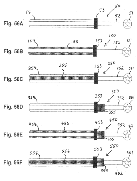
Figs. 56B-56F show cannulas incorporating filters and/or absorbing materials.
Fig. 57A shows a conventional cannula.
Figs. 57B-57F show cannulas incorporating drugs, factors and/or agents.
Fig. 58A shows a conventional syringe used to deliver insulin.
Figs. 58B-58F show syringes incorporating filters and/or absorbing materials.
6
CA 03020567 2018-10-10
WO 2017/180708 PCT/US2017/027146
Fig. 59 is a flow chart for in vivo evaluation of cannula, insulin diluent or
fibril biocompatibility
in a mouse SQ model.
Fig. 60 is another flow chart for in vivo evaluation of cannula, insulin
diluent or fibril
biocompatibility in a mouse SQ model.
Fig. 61 is a flow chart for in vitro evaluation of cell toxicity and
activation.
Fig. 62 is a flow chart for in vitro evaluation of insulin degradation.
Fig. 63 is a flow chart for in vitro evaluation of inhibition of insulin
degradation.
Fig. 64 is a flow chart for in vitro evaluation of inhibition of insulin
degradation by proteases and
cell extracts.
Detailed Description
Insulin infusion remains one of the least studied, but most critical elements
of an
integrated artificial pancreas (AP) system. Successful AP system requirements
include the need
to maintain precise and accurate in vivo delivery of very minute and
continuously variable
amounts of insulin in response to changing blood glucose (BG). Additionally,
the physical
absorption and BG response to infused insulin should remain constant
permitting stable AP
algorithm performance. Interestingly, little was known in the past about the
impact of insulin
excipients/diluents and continuous subcutaneous insulin infusion (CSII)
failures including loss of
blood glucose regulation
Embodiments disclosed herein solve problems associated with insulin/excipient
induced
tissue reactions during CSII and syringe delivery of insulin. We have found
that insulin infusion
triggers tissue injury and local inflammatory responses at insulin infusion
sites, which ultimately
results in limited infusion site longevity, premature infusion failure and PK
absorption
variability. We also have found that IFP trigger tissue injury and local
inflammatory reactions
(inflammation and fibrosis) both during infusion and afterwards (i.e. after
cannula withdrawal),
that ultimately limit infusion site longevity, infusion failure and PK
absorption (Figures 1A and
1B). Furthermore, based on the data described herein, we understand that
insulin formulations
containing phenol and/or m-creosol (excipients/diluents) trigger infusion site
tissue injury and
local tissue reactions (inflammation and fibrosis), occurring during both
infusion and afterwards
(i.e. after cannula withdrawal). The consequences of these diluent induced
tissue reactions
include limiting Embodiments infusion site longevity (short and long term),
premature infusion
7
CA 03020567 2018-10-10
WO 2017/180708 PCT/US2017/027146
failure and pharmacokinetics (PK) absorption variability, based on our present
data we believe
that the influx of chemokine-recruited leukocytes into the infusion site,
results in the release of
leukocyte-derived proteases that degrade insulin. Insulin degradation will
further limit the
effectiveness of insulin mediated BG regulation in vivo (Figure 1). We further
understand that
inhibitors of cytokine, chemokine and leukocyte proteases will decrease
infusion site
inflammation, tissue injury and thereby improve both short-term (decrease
inflammation) and
long-term (decrease fibrosis) CSII performance and BG regulation in vivo.
One embodiment described herein uses an adsorption technology such as ion
exchange
resin to reduce the concentration of at least one of fibrils, and insulin
preservatives from an
insulin solution before the insulin is administered to a patient. Non-limiting
examples of suitable
adsorption resins include ion exchange resins include nonfunctionalized hyper-
cross-linked
polymer Macronet MN200 and two ion exchange resins, Dowex XZ (strong anion
exchange
resin) and AuRIX 100 (weak anion exchange for removal of phenols in water
treatment).
Another embodiment described herein uses a cyclodextrin-containing component
(or another
absorbing component) to remove at least one of insulin fibril and insulin
preservatives from an
insulin solution before the insulin is administered to a patient.
Example 1 ¨ In vivo model for measuring tissue reactions
Currently commercial insulin formulations contain phenol, m-cresol or a
mixture of both, to
stabilize insulin in vitro. We have demonstrated that phenol / m-cresol are
not only cell and
tissue toxic, resulting in tissue injury and inflammation, but are also able
to induce expression of
1) pro-inflammatory cytokines 2) chemokines (directly and indirectly via
cytokine mediated
induction of chemokines), as well as 3) insulin degrading proteases (see
preliminary data
section). Translation of these observations into clinically meaningful
strategies and
treatments requires the development of quantitative in vivo models. Developing
and validating
these in vivo models is critical to developing effective strategies and
therapies to overcome
failure of CSII to sustain insulin based BG regulation in vivo. To this end we
have modified the
classic murine "air pouch" model for evaluation of inflammatory agents and
inhibitors to
evaluate diluent induced tissue reactions and BG regulation. For this model,
sterile air was
injected subcutaneously into mouse skin creating a sustained compartment
(pouch) for injection
of test agents (Figure 2). At various times post-air / post-agent injection,
the "air pouch" can be
lavaged, and the cell and fluid content removed and characterized using
standard technology
8
CA 03020567 2018-10-10
WO 2017/180708 PCT/US2017/027146
(Figure 2). After lavage, tissue reactions in the air pouch walls can also be
determined using
standard histopathology/immunohistochemistry (Figure 2). Using this model we
have
demonstrated that injection or infusion of diluent into the "air pouch", using
human insulin
pumps, induces significant inflammation when compared to saline infusion. We
have extended
this model by replacing the traditional insulin infusion pump system with a
wireless totally
implantable pump (Iprecio totally implantable pumps, Alzet inc) by converting
it into a
transdermal pump for uses in murine CSII (Figure 3). This conversion was
achieved by
mounting the pump on the back of the mouse using a bio-jacket or running a
line from the
Ipericio pump into the mouse skin (see preliminary data below (Figure 4). As
such, the pump is
not implanted under the skin. Thus limiting an excessive trauma or
inflammation. We have also
added our murine CGM system to create a murine "open loop" system (i.e. "air
pouch"
CGM/CSII model).
Example 2 - In vitro cell studies
For these in vitro studies, generally either human or mouse leukocyte cells or
cell lines were
cultured in vitro in the presence or absence of insulin preservatives or
fibrils at various
concentrations. at selected times cell viability and or cell activation has
(cytokine expression)
was determined. The results of these studies are presented below the general
work flow for these
studies are located in figure 61.
Figure 5 demonstrates decreasing cell viability of human peripheral blood
mononuclear cells
(PMBC) after 3 days of exposure to increasing concentrations of insulin or
insulin
diluent/preservatives as measured by Alamar Blue assay. Insulin diluent
contains the formulation
components of commercial insulin solutions but without the insulin protein
itself.
Figure 6. Toxicity of insulin and fibrils in vitro: Preliminary in vitro
morphology of human
peripheral blood mononuclear cells (PBMC) after exposure to control media,
high (1.0 mg/mL),
and low (0.1 mg/mL) concentrations of insulin or insulin fibrils (i.e. insulin
degradation
byproducts) Figure 6). Healthy control cells [Left column] show a rounded
morphology. Cells
exposed to high concentrations of insulin fibril preservatives (IFP) factors
[Middle column] show
tight contracted morphology indicative of cell death/dying. Cells exposed to
low concentrations
of Insulin or insulin fibrils factors [Right column] show a spread, expanded
morphology
indicative of cell injury/activation.
9
CA 03020567 2018-10-10
WO 2017/180708 PCT/US2017/027146
Figure 7. Our in vitro studies Insulin and excipients induced expression of
pro-inflammatory
cytokines in PBMC in vitro. Specifically these in vitro studies demonstrated
that insulin (+1-
preservatives) and preservatives alone, as well as fibrils (data not shown),
induce expression of
pro-inflammatory cytokines including IL-6, IL-8 and TNFa from PBMC in vitro
(IL-8 data
presented in Figures 7B-D) Summary results for PBMCs is presented in Figure
7A. Similar data
has been obtained with human cell lines (THP-1) and mouse macrophages (MQ).
This supports
our belief that FE can induce inflammation in vivo, and that chronic infusion
of IFP can cause
chronic inflammation and fibrosis.
For these studies we utilized the work flow described in figure 61. Fig. 7B
shows the impact
of insulin and fibrils on cytokine expression by human PBMCs in vitro. At
physiologic
concentrations, both insulin formulation and insulin fibrils are toxic for
human cells in vitro.
Figure 7C shows insulin induced activation of Human PBMCs in vitro: IL-8. The
Protocol was:
Hu-PBMC + insulin (72hr) => Assay Interleukin 8 (IL-8). Fig. 7D shows insulin
induced
activation of human PBMCs in vitro: IFN-g. The Protocol was Hu-PBMC + insulin
(7 days) =>
Assay Interferon gamma. This data clearly demonstrates that insulin, diluents
and fibril cause
pro-inflammatory activation of these cells in vitro. The in vivo activation of
these cells would
cause inflammation and tissue destruction resulting in loss of effective CSII
function.
Example 3A - "Open Loop" Mouse Model ¨ In vivo
The "airpouch" model was prepared and evaluated as presented in the workflow
diagram
in Figure 60. Figures 8A-8D. Combined CSII and CGM mouse model (A B, C). Non-
obese
diabetic (NOD) mouse implanted with Abbott Navigator glucose sensor (GS) and a
short
polymer infusion set for CSII. The set and sensor were placed sufficiently far
apart to avoid
interference. Open Loop CGM and CSII insulin infusion system cage and
assembly, that allows
animal ambulation and mobility during simultaneous continuous glucose sensing
and insulin
infusion. Figure 8D is an example of CGM blood glucose levels (blue line) and
external BG
control tests (red diamonds), monitored in a diabetic NOD mouse that received
periodic insulin
infusion (bars present at the top of figure 8D) These studies demonstrate the
successful open
loop BG control in our murine model of CSII and CGM.
Example 3B ¨ In vivo data ¨ Tissue reactions to CSII insulin and diluents
Tissue toxicity of insulin in vivo: Injection: Saline control tissues manifest
minimal
infiltration of inflammatory cells. Diluent treated tissues demonstrate
substantially higher levels
CA 03020567 2018-10-10
WO 2017/180708 PCT/US2017/027146
of inflammatory cells, potentially due top inflammatory activation and
recruitment in the
injection site, as is shown in Fig. 19, which compares a saline control to a
diluent. Fig. 19 shows
H & E stained sections of a mouse skin and subcutaneous tissue after 3
consecutive 2X daily
injections of saline or insulin diluent.
Figure 9. Tissue Toxicity of Insulin in vivo: Infusion: The sectioned polymer
catheter wall is
visible and marked with a black asterisk (*) in all tissues. Darker colored
zones (marked with a
black I) in the diluent and insulin infusion sample indicate the presence of
extensive cell damage,
inflammation and cellular infiltrate at the infusion site immediately
surrounding the infusion
catheter. Infusion was accomplished using the open loop delivery system
described in Figure 5.
It is important to note that the insulin infusion cannula alone (i.e. saline
infusion did not induce
tissue reactions thus indicating the reactions seen with insulin infusion are
not related to the
cannula.
Example 3C ¨tissue toxicity of multi-diluent injections; tiisue reactions to
Cs!! cannulas
For these studies he workflow diagram described in figure 59 was utilized.
Figures 40-
42. (saline vs. diluent) These figure shows that multiple injections of
diluent, but not saline,
cause major inflammatory reactions at injection sites on mouse skin. As is
shown in the figure,
saline control tissues manifest minimal infiltration of inflammatory cells
(dark dots). Diluent
treated tissues demonstrate substantially higher levels of inflammatory cells
potentially due to
inflammatory activation of recruitment to the injection site.
The purpose of these studies was to demonstrate that the cannula alone (not
infusion of
fluids) would induced tissue reactions that would compromise SCII. Thus we
need to coat the
cannulas with more biocompatible substances (like claims) so the cannula alone
would not
damage the tissue.
Figure 40 shows low to high power magnification of the implantation site for
the cannula
showing that cannulas induce inflammation on the entire length of the cannula
(labeled tip
middle and skin entry point) after 3 days implantation in in 2 mice. This
demonstrates that the
cannula along triggers inflammation the entire length of the cannula as well
as the entry point of
the cannula thru the skin thus to prevent this you would coat the cannula with
materials that
would enhance biocompatibility alone or with the incorporation of agents in
those coating
agents. It also demonstrates that there is tissue injury and inflammation at
the entry point of the
cannula thus having infusion set collars (with or without impregnated agents
to reduce
11
CA 03020567 2018-10-10
WO 2017/180708 PCT/US2017/027146
inflammation and tissue reactions as well as infections would be extremely
important to
extending the lifespan of the infusion sets in vivo. 7 day data is shown in
Fig. 41. Fig. 42 shows
inflammation next to the catheter.
Example 4 - "Air Pouch/Open Loop" Mouse Model
Leukocyte influx into Air pouch model to evaluate tissue response to saline or
insulin
excipient
The air pouch model (figure 60) has been used for the evaluation of tissue
responses to tissue
irritants and/or for the evaluation of tissue reaction inhibitors. We adapted
this model for the
evaluation of tissue responses to infusion of insulin, excipients, factors,
drugs and control
solutions (e.g. saline). An example of air pouch model response to infusion of
saline or insulin
excipient is present in Figure 2. Initially, CD-1 mice received air injections
(3 mL) into the
dorsal site of the low back to induce the air pouch in each animal. On the
following day
individual mice received infusions of either saline or insulin diluent. The
infusion rate for both
fluids was infused at 6 units/hr. (equivalent to insulin infusion volumes) for
3 consecutive days
into the air pouch. At the end of that time period mice were anesthetized and
the air pouch
cavity was washed three times with 2 mL pyrogen-free PBS. Exudate was then
centrifuged and a
TC10 automated cell counter determined the total viable leukocyte cells. The
potential
inflammatory effect of insulin diluent was evaluated through the analysis on
the mouse leukocyte
count and exudate concentrations in the inflamed air pouch cavity. Diluent
infusion over a 3-day
period caused an average of 7-10 fold increase in the total leukocyte count
when compared to
saline treated air pouch (Figure 10). Cytocentrifuge / H&E staining of these
cell populations
indicated that at 3 days post infusion, approximately 60% of the leukocyte
present were PMN
and 40% mononuclear cells, predominately macrophages. Additional studies of
saline vs.
excipient infusions indicated that at 4 days post infusion the predominate
leukocyte in the lavage
were mononuclear and again predominately macrophages (i.e. > 80% macrophages).
This data
clearly demonstrates that diluent causes a significant increase of
inflammatory cells and that
these reactions evolve from a PMN dominated inflammation to a macrophage
dominated tissue
reaction. Similar results were obtained in other non-diabetic mice (e.g. B6.V-
Lepoba and
C57BL/6).
Example 5¨ mouse Air pouch model
12
CA 03020567 2018-10-10
WO 2017/180708 PCT/US2017/027146
Using cells obtained from "air pouch" models (figure 60) by lavage, we
demonstrated
that diluents and insulin which contains diluents, but not saline, cause
tissue reactions
characterized by the influx of PMNs, monocytes and macrophages.
Fig. 43 shows total cell number for treatments with various diluents and
saline,
demonstrating insulin preservative/diluent induced inflammation in a normal CD-
1 mouse using
a modified "air pouch" model. Air was injected subcutaneously into the mouse
skin creating a
sustained compartment (pouch) for injection of diluent or saline (control).
The diluent and the
control agent (saline) were infused continuously for 7 days at a rate of 5
units equivalents/hour
one day post air pouch creation. After 7 days of infusion, the air pouch was
lavaged. The
resulting fluid was characterized for cell number (auto-hemo-cytometer), and
cell type using
fluorescence activated Cell sorting (FACS). Consistently, diluent treated
preservative mice
demonstrated a dramatically higher cell count when compared to saline infused
mice.
Additionally Neutrophil, Monocyte/Macrophage and Lymphocyte counts were
significantly
higher in the diluent/preservative infused mice when compared to the saline
treated mice. This
data demonstrates that diluents cause major inflammatory reactions when
infused into the air
pouch model, but saline do not cause inflammatory reactions. Since the major
components in
diluent (preservatives) is phenol and met-cresol removing these preservatives
would prevent
inflammation seen when they are infused into the air pouch of SQ tissue.
Example 6 - Histological Evaluation of Tissue Reactions Induced at Air Pouch
Infusion
Sites by saline vs. excipients
In addition to leukocyte counts in the lavage, we also evaluated the effect of
saline and
insulin infusion excipient on inflammation over a 3-day period (see workflow
diagram figure
60). Initial histological analysis of the infusion sites demonstrated that
leukocyte accumulation
was only prevalent in the excipient infused tissue site (Figure 11 B, D and
F). The predominant
leukocytes were PMN and monocyte/macrophages. The saline treated infusion site
experienced
minimal to no tissue reaction (Figure 11 A, C and E). These results confirm
the observation that
insulin excipient causes significant tissue reaction at site of infusion. The
black star (*) in
Figure 11 indicates the location of the air pouch. The individual
magnification is listed on each
figure (lower left corner). This data directly demonstrates that diluents
present in commercial
preparations of insulin trigger inflammation in the air pouch model in vivo
and thus likely induce
the same inflammatory reactions in the subcutaneous tissue when infused during
CSII in vivo.
13
CA 03020567 2018-10-10
WO 2017/180708 PCT/US2017/027146
Example 7 ¨ Effect of insulin and its preservatives on human peripheral blood
leukocytes
For theses in vitro studies we utilized the general worflow diagram presented
in figure
61.
Fig. 52 shows the number of PMN's surviving after 3 days in buffer +/- a
serial dilution of
insulin or its preservatives (phenol and m-cresol). Even at a 1:48 dilution,
there were
significantly fewer cells surviving than in buffer alone. As the concentration
increased, the
number of cells surviving decreased for both insulin and insulin diluent. As
the highest
concentration texted (a 1:3 dilution of standard insulin formulations), fewer
than 1,000 cells
survived, compared to over 100,000 in the buffer solution (estimated from
optical density). this
data demonstrates that complete insulin (insulin plus preservatives) and
preservatives only are
toxic to human leukocytes when the leukocytes are co cultured with the insulin
or preservatives
Example 8- fluorescent uptake of insulin into leukocytes in vitro
Studies in our laboratories demonstrated that insulin uptake and degradation
by
inflammatory and tissue cells lowers effective insulin levels, ultimately
requiring higher insulin
dosages to achieve blood glucose regulation. This added insulin infusion also
results in
increased tissue inflammation at the infusion site. The aim of this example
was to determine
whether leukocytes can degrade insulin in vitro. We utilized fluorescent
insulin (FITC-insulin;
Sigma, St. Louis, MO), Humalog insulin, and human peripheral blood leukocytes
isolated from
diabetic and non-diabetic patients. We cultured leukocyte subpopulations
(PMN's,
macrophages, and lymphocytes) in vitro +1- f-Met-Leu-Phe (a chemotactic and
leukocyte
activating factor). We then added FITC-insulin and monitored leukocyte uptake
of FITC-insulin
using fluorescent microscopy (figure 12A, 12B, 12C). inverted microscope and
cyto-chemical
staining was used to confirm subpopulations that took up FITC-insulin and to
assess cell viability
with trypan blue and intact nuclei by DAPI staining.
Example 9 - In vitro insulin degradation
For these studies we utilized the general workflow diagrams presented in
figure 62, 63
and 64.
Role of leukocytes and leukocyte proteases in limiting insulin regulation of
blood glucose
levels during CS!!
To characterize the ability of purified proteases or leukocyte extracts to
degrade FITC-
insulin in vitro we analyzed the impact of cell culture supernatants and cell
lysates on insulin
14
CA 03020567 2018-10-10
WO 2017/180708 PCT/US2017/027146
degradation using 10-20% SDS-PAGE gels. This was performed +/- anti-protease
cocktails to
characterize proteases responsible for insulin degradation. The functional
activity of individual
leukocyte proteases was analyzed using protease PAGE gels +/- protease
inhibitors. This study
clarifies the role of leukocytes in insulin therapy. (figure 13a, 13b, 13c and
figure 14). Our
results show that (1) leukocytes take up and degrade FITC-insulin in vitro,
(2) activating
leukocytes with f-Met-Leu-Phe increases this degradation, (3) Humalog insulin
and insulin-FITC
are degraded by leukocyte proteases including neutrophil elastase, trypsin,
and insulin-degrading
enzyme, and (4) the activity of these proteases can be reduced by natural
inhibitors including
alpha-1 antitrypsin and aprotinin.
Fig. 53 shows degradation of insulin by leukocyte proteases. Analysis by lane:
(1)
Insulin appears as a bright band ( MW=5.8kDa), (2) Molecular marker to 2kDa,
25kDa and
75kDa in red, insulin in yellow (what is relative position of yellow ¨ final
drawing will be in
black and white.. (3)Trypsin degrades insulin so that the original band is no
longer visible,
replaced by two primary degradation products. (4) Elastase cleaves insulin at
many sites, leaving
a streak of products at a wide range of molecular weights. (5) Insulin
degrading enzyme cleaves
insulin into several smaller peptides, including a bright band at a low
molecular weight. (6)
PMNs taken from a Type I diabetic patient and lysed with Triton X100
completely degrade
insulin into a wide range of products.
Lymphocytes and monocytes taken from human peripheral blood also degraded
insulin, although
not to the same extent (data not shown). PMNs from non-diabetic patients
degraded insulin as
well (data not shown).
In Figure 53, I=insulin, T=Trypsin, E=Elastase, IDE=Insulin Degrading Enzyme,
PMN=Triton
X100 extract of human PMN, mwn=molecular weight marker.
Figure 12 shows human and mouse leukocytes uptake of FITC-insulin. Our initial
in vitro
studies demonstrated that FITC-insulin (green) is taken up by human peripheral
blood leukocytes
such as PMNs (Fig. 12A) monocytes (Fig. 12B). Figure 12 is a combined bright
field and
fluorescence photomicrograph with FITC-insulin appearing green once phagocyte
by the
individual leukocyte subpopulations (primarily PMN & MQ, but not Lymphocytes
(Fig. 12C)).
Mouse MQ also uptake FITC-insulin in vitro and degrade FITC-insulin in vitro
(SDS PAGE
analysis data not shown).
CA 03020567 2018-10-10
WO 2017/180708 PCT/US2017/027146
Figures 50A and 50B show mouse MQs plus GFP insulin ¨Figs. 51A (bright light)
and 51B
(Fluroescence) show mouse MQ plus GFP fibril study.
Figure 13 shows insulin degradation by cells. Degradation of FITC-insulin by
elastase (E)
and human leukocyte extracts (LE): To determine whether triton X100 extracts
of total human
leukocytes isolated from blood could degrade insulin in vitro we incubated
FITC-Insulin with LE
or +/- HALT (protease inhibitor cocktail (Fig 13A), E or +/- aprotinin
(elastase inhibitor) (Fig
13B) E +/- protease inhibitor AAT (Fig 13C) and analyzed the results by PAGE.
LE or E
degraded FITC insulin, and this degradation was blocked by the serine protease
inhibitor
aprotinin (A), HALT and AAT. We have analyzed the ability of various proteases
and anti-
proteases to degrade insulin and or block degradation of FITC- insulin (Figure
14). We obtained
similar data using Humalog and SDS-PAGE with Coomassie staining (protein
staining). These
studies demonstrate that leukocytes induced at IFP infusion sites decreases
functional insulin
levels in vivo, thereby decreasing the effective control of blood glucose
levels in vivo. The
addition of protease to insulin formulation not only increases insulin
effectiveness but also
suppresses tissue injury and inflammation.
Example 10 ¨Effect of anti-proteases in inhibiting insulin degradation
For these studies we utilized the workflow diagram presented in figure 64. In
Figure 54,
I=insulin, PMN=Triton X100 extract of human PMN, mwn=molecular weight marker,
AAT=alpha-1-antitrypsin, SP16=synthetic short peptide from AAT), H=Halt=AEBSF-
HCL+Aprotinin+Bestatin++e-64+Leupeptin+Pepstatin A.
Fig. 54 shows PMN degradation of insulin =+/- inhibitors. Analysis by lane:
(1) Insulin
appears as a bright band ( MW=5.8kDa), (2) Molecular marker to 2kDa, 25kDa and
75kDa in
red, insulin in yellow (what is relative position of yellow ¨ final drawing
will be in black and
white.) (3) PMN extracts completely degrade insulin (as in Lane 6 above). (4)
AAT completely
inhibits the degradation of insulin; insulin is visible at 5.8 kDa. (5) SP16
fails to inhibit
degradation; at higher concentrations, it is able to do so. (6) HALT (anti-
protease cocktail)
inhibits the degradation of insulin.
In this example, we tested the ability of various anti-proteases to inhibit
insulin
degradation by leukocyte extracts and proteases. The table in Fig. 14
summarizes the results of
these studies. HALT anti-protease cocktail was the only inhibitor to block
degradation of IDE.
Aprotinin, AAT, SP16, and HALT all blocked insulin degradation by Elastase,
Trypsin, and
16
CA 03020567 2018-10-10
WO 2017/180708 PCT/US2017/027146
Trion X100 extracts of human PMNs. This shows that addition of anti-proteases
to insulin
formulations inhibits degradation of insulin in vivo.
Example 11 - Impact of Insulin on Mast Cell Morphology ¨ in vitro
Figures 46-47 show the impact of insulin on mast cell morphology. At
physiologic
concentrations, both insulin formulations fibrils (not shown) are toxic to
mouse mast cells in
vitro. Fig. 47 shows results based on concentation.
Example 12 ¨ Impact of Insulin on Mast Cell viability and degranulation - in
vitro
For these studies we utilized the general workflow diagram presented in figure
61. Figs.
48-49 show the impact of insulin on mast cell viability and degranulation.
Mast cells (MC) are
key skin "sentinel" cells and are generally the first tissue cell population
activated by tissue
trauma triggering acute inflammatory or allergic reactions and serve a central
role in chronic
inflammation and wound healing. Recent results from our laboratory indicate
that skin mast cells
affect glucose sensor induced tissue reactions and CGM function (13).figure 48
demonstrates
that increasing concentration of insulin causes increasing increasing cell
death (alamar Assay)
Our data demonstrate that insulin can be MC toxic and activate MCs in vitro
(Figure 49). We
believe that IFP also trigger MC toxicity and activation in vivo thus
triggering acute sustained
inflammation during continuous IFP infusion, which could be significantly
decreased by MC
deficiency or depletion. At physiologic concentrations, both insulin
formulations fibrils (not
shown) are toxic to human mast cells and also cause mast cell degrandulation
in vitro
Example 13 ¨ Insulin, fibrils and preservative induce tissue injury and
inflammation
Using general flow diagrams 59-64 we have shown that 1) insulin, fibrils and
preservatives (IFP) induced tissue injury and inflammation when infused in
vivo, 2) IFP induced
toxicity and immune-dysfunction (e.g. cytokine expression) in exposed
leukocytes and tissue
cells in vitro, and 3) using our new open loop system in diabetic mice glucose
control requires an
increased insulin infusion with CSII post infusion time, and 4) leukocytes
take-up insulin and
degrade it using serine proteases e.g. elastase and 5) blockage of insulin
degradation using anti-
proteases. All these issues decrease the local and systemic levels of insulin.
The increased
requirement of insulin infusion with time on CSII is also seen in patients
with diabetes. These
data show that IFP trigger SQ tissue reactions that compromise infused insulin
regulation of
blood glucose (BG).
17
CA 03020567 2018-10-10
WO 2017/180708 PCT/US2017/027146
The data shown demonstrated that 1) IFP trigger inflammation at SQ infusion
sites, and
2) leukocytes (PMN and MQ) take up and degrade insulin in vitro.
Example 14 ¨ Observed tissue and cellular effects after exposure to IFP
components
Fig. 36 shows the outcome of in vivo and in vitro exposure testing using
various cell
types and various IFP factors. Concentrations represent the levels of various
IFP factors at
which effects can be observed. These data show the impact of IFP factors on
tissue and cells.
Acute and long-term failure of CSII blood glucose (BG) regulation in T1D is
the result of
insulin/excipient (FE) induced tissue reactions (i.e. inflammation, loss of
vasculature and
fibrosis). Specifically FE induced tissue reactions limit insulin access /
transport to the
vasculature (blood and lymphatic vessels) due to inflammation (acute phase)
and fibrosis
(chronic phase), as well as inflammation induced degradation of insulin at the
infusion site (see
Figure 1). The solutions described below overcome CSII induced tissue
reactions and thereby
extend the lifespan and effectiveness of CSII. This result is demonstrated
using our in vivo
murine "air pouch/open loop" model). A brief summary of the specific
approaches and
methodology are detailed below.
Prophetic Examples
Prophetic Example 15 - Murine "air pouch/open loop" model of blood glucose
regulation
utilizing CGM and CSII
Distribution of infused fluids, such as insulin or excipients, into the tissue
occurs in highly
variable patterns due to tissue structure and gravity. This variability makes
tissue reaction
evaluation often extremely difficult. In order to be able to consistently
evaluate
insulin/excipients/saline (I/E/S) induced tissue sites, a predictable infusion
site for histologic
analysis is required. The ability to retrieve viable cell population from that
site in a simple
fashion is an additional requirement for quantitative evaluation of tissue
reactions and cell
expression profiles. To achieve this goal we utilize a classic model to
evaluate inflammation and
agents that induce or suppress inflammation: known as the "air pouch model".
Additionally, for
these studies our focus is on using rapid acting analog (Humalog) insulin.
Humalog is currently
routinely used for CSII pump infusion reducing the rationale for testing
longer acting insulin
proteins and their formulation excipients. In addition, most insulin
excipients are conserved
across regular and rapid acting insulin analog preparations with minor
exceptions in preservative
type and concentration.
18
CA 03020567 2018-10-10
WO 2017/180708 PCT/US2017/027146
For these studies we utilized the general work flow diagram described in
figure 60. We use
the murine "AP/OL" model described in the preliminary data section of this
application (see
workflow diagram in figure 60). I/E/S infusion is done using Iperico wireless
pumps (Figure 3)
as well as traditional patient insulin infusion pumps. Fig. 16 lists mouse
models for evaluation.
Fig. 17 lists working ranges of I/E/S and Figure 18 lists tissue reactions for
evaluation. We
evaluate I/E/S induced tissue reactions daily during 3-days of infusion as
well as the cumulative
effects at the end of 3 days. Subsequent studies focus on tissue reactions for
up to 7 to 14 days
of infusion. Infusion sites are marked for location using tattoo ink. Insulin
+ excipient and
excipient only induced tissue reactions and lavage samples are be evaluated
systematically in
control (non-diabetic) and diabetic mice using standard immunohistopathology
and
immunocytocemistry (ICC) (Figure 18).
Qualitative or quantitative differences in tissue reactions between I/E/S
components &
concentrations as well as between animal models are determined. For example,
due to wound
healing defects associated with diabetes, I/E/S induced tissue reactions may
substantially differ
in the diabetic state. As such, spontaneous NOD (autoimmune) and
streptozotocin (STZ) models
of type 1-diabetes mouse models and the db/db mouse model of type 2-diabetes
will also be
considered. Tissue reactions and cell influx will be correlated with insulin
regulation of BG
levels and CGM in control and I/E/S treated and compared between diabetic and
non-diabetic
mice on the C57BL/6 background. These studies elucidate the baseline IFP
induced tissue
reactions and their relative component potencies.
It is expected that I/E/S induces significant and increased tissue reactions
(histology and cell
influx) over the first 3 days of infusion. Due to I/E/S induced tissue injury
we anticipate a
potential for sustained tissue reactions after infusion removal.
Prophetic Example 16 - Evaluations of cell and gene expression obtained
through lavage
following insulin, excipient or saline exposure using the murine "air pouch"
model
The focus of this study is primarily on characterization lavage and blood
associated cells
and factors involved in the E/I induced tissue reactions. Specifically the
"air pouch" model
allows lavage of leukocytes that have been recruited into the air pouch. The
recruited leukocytes
can be sorted into significant subpopulations using standard FACS sorting and
analysis (Figure
16). Using FACS analysis allows greater speed analysis of large and diverse
numbers of samples
and leukocyte subpopulations. For example, evaluation of the impact on various
treatment
19
CA 03020567 2018-10-10
WO 2017/180708 PCT/US2017/027146
protocols and therapies, as well as new infusion pump technology. As such, the
analysis of
lavaged cells represents an important tool dissecting the mechanisms as well
as effectiveness of
approaches to better control CSII induced tissue reactions. Additionally one
of the byproducts of
lavage are unique microvesicles that can be used for both biomarkers, as well
as for mechanistic
insights into the E/I induced tissue injury. Microvesicles are small membrane
extrusions
(packets) that are released from activated and injured cells and bind to
target cells (Figure 17).
Once bound to target cells microvesicles unload their "cargos" of RNA and
proteins and as such
take control of the target cells. Since these microvesicles are also released
into the blood stream,
they have been used as biomarkers for disease progression in cancer and
vascular disease. The
evaluation of cells and microvesicles obtained from the air pouch lavages is
an extremely
important tool in obtaining insights in order to control CSII induced tissue
reactions.
Leukocytes and microvesicles derived from lavaged fluid are obtained from
various animal
population and treatment regiments (described above including Tables 3-5).
Blood samples
from these same animals are utilized for analysis of peripheral blood
leukocyte gene expression,
as well as isolated blood-derived microvesicles for RNA and protein analysis
(Figure 16).
Lavage or blood derived cells are separated from the fluid phase by low speed
centrifugation.
The resulting cell populations are fixed and analyzed by FACS analysis and
sorted for leukocyte
subpopulations (haps:/lwww.bdbiosciences.com/documents/cd marker
handbook.polf). The
sorted cells are then extracted for RNA and processed (cDNA libraries) for
NexGen RNA
Sequencing and analysed by SBI (https://www.systembio.com/services/exo-
miseq/overview).
The microvesicles are isolated for lavage fluid or blood plasma using Exoquick
(SBI) and
processed for NexGen RNA sequencing and analysis
(https://www.systembio.com/services/exo-
miseq/overview), as well as MS/MS analysis by SBI
(https://www.systembio.com/services/exosomes/mass-spec). Unique biomarkers for
E/I induced
tissue reactions are processed using qPCR/RNA arrays as well as ELISA assays
to aid in the
development of simple rapid assays to determine the impact of therapies and
new devices on
I/E/S induced tissue injury and CSII blood glucose regulation.
We expect that RNA analysis of the lavaged leukocytes subpopulation will
demonstrate
significant increases in pro-inflammatory proteins versus anti-inflammatory
proteins. The
specific nature of these RNA/proteins and their levels could provide useful
and important
prognostic tools for evaluating the success or failure of E/I infusion in our
animal models. I/E/S
CA 03020567 2018-10-10
WO 2017/180708 PCT/US2017/027146
infusion in normal and diabetic mice will determine diabetes wound-healing
defects on I/E/S
induced tissue reactions and blood glucose regulation. Currently there is no
literature on the
existence of microvesicles in murine models or human models regarding insulin
and excipient
infusion. As such, it is important to determine whether the RNA/protein
profiles seen in the
microvesicles are associated with any of the leukocyte populations seen in the
lavage, tissue or in
the blood of the infused diabetic and non-diabetic animal populations. Results
of these data
provide important insights into potential mediators and mechanism related to
I/E/S induced CSII
failure. The discovery of E/I specific biomarkers or biomarker panels would
provide useful tools
for rapid evaluation of various therapeutics or new devices that may prevent
I/E/S induced tissue
injury and subsequent failure of blood glucose regulation in vivo.
Prophetic Example 17 Evaluation of gene expression in tissue derived from the
murine
"air pouch" model
One of the cornerstones of the present studies is to characterize reactions
that occur at I/E/S
infusion site within the open loop murine air pouch model. Although we have
developed
significant preliminary data indicating that the insulin/excipients cause
substantial tissue
reactions including tissue injury and influx of inflammatory cells, these
observations need to be
confirmed and expanded. It is important to emphasize that these studies
provide important
insights into leukocyte gene expression in vivo. These studies also allow
insights into the gene
expression of tissue cells such as mast cells, dendritic cell, endothelial
cells and fibroblasts all of
which are critical in inflammation and wound healing. This data provide the
foundation for
developing useful assays (RNA arrays and ELISA) that aid in the evaluation of
I/E/S induced
injury markers, as well as lead to the effectiveness of therapeutic approaches
to prevent I/E/S
induced tissue reaction.
Initially tissue obtained from sites of I/E/S infusions in our "air pouch open
loop model" will
be removed enbloc, fixed and processed using standard technology Fig. 18.. We
will identify
cells, proteins as well as RNA present at the infusion site. In addition to
these traditional
methods of "staining" tissue we will also utilize new cutting edge
technologies including
RNAScope for RNA presence and distribution of RNA probes for detection of all
classes of
RNA including mRNA, miRNA, siRNA (http://www.acdbio.com/products). These
probes have
the advantage detecting all forms of RNA present in cells including RNA for
proteins that are
unknown or not transcribed, as well as proteins that currently no antibodies
exist. We will also
21
CA 03020567 2018-10-10
WO 2017/180708 PCT/US2017/027146
use LaserCapture Microscopy coupled with Next-Gen RNA and MS/MS sequencing to
determine all RNA and proteins present in injured and non-injured cells. These
studies utilized
LCM and RNA arrays to characterize gene expression in various inflammatory
giant cell
subpopulations. For the AP/OL studies we will isolate specific cell
populations located at the
I/E/S infusion sites including: macrophages, mast cells, lymphocytes,
fibroblasts and endothelial
cells. In vivo RNA expression in these various cell population over time and
various conditions
enables better understanding of the cells, mediators and mechanisms that
affect CSII function.
Comparing RNA and protein present in both injured and non-injured cell after
various treatments
(i.e. I/E/S or saline infusion) will allow us to determine unique signatures
of RNA, proteins and
pathways that are affected by TIE infusion into the murine air pouch.
These studies provide important insights into leukocyte gene expression
including gene
expression of tissue cells such as mast cells, dendritic cell, endothelial
cells and fibroblasts. The
combination of traditional histopathology, IHC and LCM coupled with RNAScope
and NexGen
RNA
Prophetic Example 18- Impact of post-CSII on tissue reactions in the "air
pouch" model
Although the clinical dictum for CSII failure is "when in doubt, pull it out".
Changing the
infusion location (arm, belly or butt) may address blood glucose regulation in
the short-term, it
does not address the long term consequence of the induced tissue reaction at
the original infusion
site. Our belief predicts that even with the secession of insulin infusion and
removal of the
cannula at the infusion site, tissue reactions set in motion continue.
Subsequent tissue repair
leads to chronic inflammation characterized by increased recruitment of pro-
inflammatory
macrophages and lymphocytes ending with scarring (fibrosis) of the original
infusion site, which
compromises that site for future CSII infusion. Due to well-established
defects in wound healing
seen in diabetic populations the outcome is most likely more pronounced. To a
large degree this
deficiency in wound healing is believed to be a lack of transitioning
macrophages from pro-
inflammatory M1 macrophages into pro-wound healing M2 macrophages. This
transition failure
from M1 to M2 induces chronic inflammation, which causes prolonged tissue
injury and
ultimately results in more severe fibrosis associated with the disappearance
of vasculatures
networks (blood and lymphatic vessels) at the tissue site. The lack of
vasculature networks
delays tissue repair and as such leads to limiting the effectiveness of CSII
at that site in the
22
CA 03020567 2018-10-10
WO 2017/180708 PCT/US2017/027146
future. Understanding and preventing the prolonged tissue reactions seen at
CSII sites is critical
to maintaining viable tissue infusion sites.
For these studies we use the same general protocol, approaches and metrics as
described
above. As described above we will also initially tattoo the perimeters of the
"air pouch" prior to
infusion in an effort to assure identification of the infusion site used
during the initial I/E/S
infusion segment of the experiment. Post 3 day E/I/S infusion the cannula is
removed and the
tissue site is evaluated for tissue reactions for 7, 14 and 21 days post
termination of infusion.
Tissue reaction is evaluated utilizing standard histopathologic (H&E and
trichrome),
immunohistochemical analysis for cell populations and biomarkers including
RNAScope
analysis. In a second set of studies we will sustain the "air pouch" after
cession of infusion and
removal of the cannula. This is accomplished by infusion of sterile air into
the "air pouch" once
every third day. Lavage and "air pouch" tissue analysis can be done as
described in figure 60.
We expect that despite secession of FE infusion and cannula removal, tissue
injury leads to
chronic inflammation with significant fibrosis and associated loss of
vasculature networks.
There is nothing known about potential cells and mediators that drive these
post CSII tissue
reactions including how to overcome them. The studies outlined above will lead
the way to
therapies and new devices that will limit this insulin induced tissue
destruction. Initial studies in
our lab suggest that the evolution from acute inflammation, with PMN hallmark
cells, will
progress to a more chronic inflammation characterized by the presence of
macrophages and
lymphocytes. The exact nature and products of these PMN and macrophages and
their influence
on controlling tissue reaction at the insulin infusion site remains unknown.
Considering that
wound-healing defects are more pronounced as a result of diabetes, the insulin
infusion induced
tissue reactions are most likely more prolonged. Understanding the mechanisms
and mediators
that drive these tissue reactions will aid in the development of new
therapeutic strategies and
devices which will limit the chronic inflammation and fibrosis at sites of
CSII infusion.
Prophetic Example 19 - Impact of extended infusion and "same site"
Insulin/Excipient re-
infusions on tissue reactions and blood glucose regulation
We believe that sustained or repeated I/E/S infusion within the same tissue
area (e.g. repeated
infusion in the lower abdomen) induces chronic tissue injury, inflammation and
fibrosis
ultimately resulting in loss of viable tissue sites for CSII and CGM. This
study examines the
23
CA 03020567 2018-10-10
WO 2017/180708 PCT/US2017/027146
impact of extended and repeated "same site" I/E/S infusion on tissue reactions
and CSII blood
glucose regulation in normal and diabetic mice.
To investigate the impact of extended CSII infusion we will extend I/E/S
infusion into
normal and diabetic mice beyond the normal 3 days to 7 and 14 days and
evaluate tissue
reactions, blood glucose regulation and gene expression, In the case of same
site infusion
studies, we intermittently-infuse I/E/S at the same site using the "air pouch
open loop" model.
For these studies we use at least three complete cycles of continuous 3-day
IFP infusion
separated by catheter removal, and a 7-day rest period prior to reinitiate the
I/E/S reinfusion in
the same "air pouch". Tissue dye (i.e. tattoo a 4 corner box around the
original infusion site) will
ensure a consistent infusion location. Diabetic mice receive bolus insulin
injections in the
peritoneum during the 7-day rest period to control BG levels in also see
Figure 14. The ability
of the infused insulin to maintain blood glucose regulation in our open loop
murine model is also
considered.
Based on the clinical observations of site fibrosis in T1D patients, we
anticipate increased
chronic inflammation and tissue scarring/fibrosis at repetitive infusion
sites. The most potent
fibrosis inducing I/E/S component or combination thereof could provide a key
target for either
insulin reformulation or mechanical removal prior to delivery. Systematic
characterization of
I/E/S induced tissue reactions are critical steps in determining the primary
causative factors and
mechanisms as well as determining concentrations & timing of tissue injury &
site viability for
studies described below.
Prophetic Example 20 Insulin degradation in vivo
We believe that FE induced tissue reactions can induce loss of blood glucose
regulation as a
result of degradation of insulin by proteases at the infusion site. This
belief is supported by our in
vitro preliminary data, which demonstrates that leukocyte protease can degrade
insulin in vitro.
This degradation can be inhibited by the addition of clinically relevant anti-
proteases (see Figure
22 for list of anti-protease to be used). These studies provide the foundation
for in vivo anti-
protease studies and determine whether protease inhibitors can block the
degradation of insulin
in vivo and thereby extend CSII.
The occurrence and degree of insulin degradation is studied utilizing the
"AP/OL" model
followed by analyzing the lavage fluid. Using both traditional as well as
fluorescent insulin (see
preliminary data) coupled with traditional analysis (SDS peptide PAGE, western
blot and/or gel
24
CA 03020567 2018-10-10
WO 2017/180708 PCT/US2017/027146
filtration) we will determine the extent of protease-based degradation of
insulin. We will
consider 2 approaches 1) the addition of florescent insulin to the existing
insulin formulation in
infusion pumps and/or 2) the analysis of insulin fragmentation using standard
Western blot
technology using the same PAGE conditions as used for our in vitro studies
(see preliminary data
above). Standard gel filtration/ion exchange studies may also be undertaken to
isolate individual
insulin fragments of the degraded insulin. Intact & degraded florescent-
insulin are detected in the
PAGE gels using black light (see prelim data). Proteases present in the lavage
fluids will also be
characterized using BioRad protease PAGE gels (BioRad Zymogram gels) and
protease
inhibitors (Figure 22).
Based on our preliminary data we expect that insulin present in the lavage
fluids will be
degraded. Proteases (particularly leukocyte derived proteases) will also be
detected in the lavage
(e.g. insulin degrading enzyme (IDE), elastase, trypsin). Once we have
confirmed the
degradation of insulin in the lavage fluids, we will determine the ability of
specific protease
inhibitors to block insulin degradation in vivo). If the studies find that
specific protease
inhibitors will block insulin degradation in vivo, and that this blockage of
insulin degradation
enhances CSII effectiveness in regulating blood glucose levels in diabetic
mice, we will use this
information as the foundation for future studies in swine and eventually
humans.
Prophetic Example 21 -- In vitro evaluation of the impact of insulin and
components on the
activation/gene expression in blood (leukocytes) and tissue cells from normal
and diabetic
mice
It is important to develop in vitro screening tools that will mimic these in
vivo results (see
Figure 23 for flow diagram). This will allow high throughput evaluation of
various inhibitors
and introducers of FE specific gene expression, which is critical in saving
time and cost when
compared to in vivo assays. We propose to utilize NexGen RNA sequencing and in
vitro cell
cultures to establish a screening panel for various inhibitor/enhancers of FE
induced reactions.
The most likely therapeutic agents and concentrations are then tested in our
murine model.
For this screening tool we will utilize representative murine cell populations
as indicator
cells, i.e. leukocytes, adipose cells and fibroblasts. Cells are cultured in
vitro with varying
concentrations of I/E/S for 24 hrs (Table 4). Following RNA harvest, cDNA
libraries are
prepared and NexGen RNA sequencing undertaken (Figure 21). Since only 100-1000
ng of
RNA is required for deep sequencing, only 8,000-10,000 cells are required for
each assay.
CA 03020567 2018-10-10
WO 2017/180708 PCT/US2017/027146
NexGen RNA sequencing results are used to develop RNA arrays for subsequent
selection of
the most effective agents for in vivo studies. We will consider the use of
standard ELISA assays
to also screen agents for consideration in the in vivo "air pouch" model. We
will also consider
usage of this same approach to compare responsiveness of leukocytes from
diabetic versus non-
diabetic mice to see if there is any difference in the responsiveness of these
2 cell populations in
vitro. It should be noted that all cell culture supernatants are collected and
frozen at -80 C for
potential microvesicle analysis in the event that studies in Section 1 (above)
suggest that
microvesicles are useful biomarkers for FE induced tissue reactions. See
Figure 24 for the
general approach for analysis of the exosomes.
We already established the utility of screening leukocyte populations exposed
to FE in
vitro as a useful tool for modeling FE tissue reactions, i.e. cytokine express
studies in
preliminary data section above. We believe that coupling NexGen RNA sequencing
with high
throughput RNA arrays will give us the most comprehensive view of FE induced
cell activation
since it will represent the entire expression profile in cells in response to
specific inducers (FE)
or agents (inducers or inhibitors). Comparison of these in vitro data with the
in vivo data will
validate the in vitro data and help understand the underlying pathophysiology
involved in FE
induced tissue reactions. With the establishment of this in vitro assay system
we anticipate that
we can undertake rapid analysis of the various inhibitor described above,
which will allow rapid
selection of candidate agents, which can prevent TIE induced tissue reactions
and extend CSII
lifespan and function in vivo. It should be noted that if time and money is
available we will
undertake selected studies using leukocyte populations from normal and
diabetic patients to
establish a human FE profile panel, which is useful in future human CSII
studies.
Our current preliminary data supports our belief that FE induced tissue
reactions at
infusion sites compromises CSII function and lifespan both in the short term
(inflammation and
loss of vasculature networks) and long term (fibrosis at the infusion site).
We have selected a
representative group of candidate inhibitors to deliver locally to site of FE
infusion (see Figure
221). This group was selected based on our current understanding of major
inhibitors of
inflammation, fibrosis and proteolysis as well as vascularization. We plan to
use the same insulin
infusion pumps and co-deliver inhibitors individually or in combination. We
will determine the
impact of co-delivery on the FE induced tissue reactions and CSII infusion
effectiveness and
lifespan. We will utilize 2 approaches for this delivery 1) add the inhibitors
in the FE
26
CA 03020567 2018-10-10
WO 2017/180708 PCT/US2017/027146
formulations in a traditional pump system or 2) use the dual pump delivery
system from Ipercio
dual pump. The choice of 2 approaches for inhibitor delivery is that 1) the
potential of the
inhibitors affecting the insulin while in the same pump container and 2)
possible FDA concerns
regarding changes to currently approved insulin formulation when combining
inhibitors.
Prophetic Example 22 - Impact of infusion of inhibitors/inducers of tissue
response on
tissue reactions and CSII blood glucose regulation using the "AP/OL" model in
normal
and diabetic mice
For the candidate inhibitors and inducers present in Figure 22, we have
selected the most
likely candidates based on our current preliminary data. Nevertheless as
knowledge is gained
from Goal 1, this list will be modified to select the most likely tissue
modifiers that will
successfully control FE induced tissue reactions in vivo.
We use representative general anti-inflammatory drugs (Figure 22), followed by
more
targeted inhibitors on inducer as presented in Figure 22. Each drug is
injected into the air pouch
twice daily to determine FE inhibitory impact. Once the optimal dose of drug
is obtained from
these injection studies, we will determine the stability of the individual
drug in the FE solutions.
For that selected inhibitors are incubated with I/E/S individually at 37C for
3 days to mimic the
typical on-patient exposure time and temperature. The resulting (individual or
combination) of
drug FE of saline treated samples will be infused into air pouch model for 3
days and tissue
reactions evaluated If combining of the drugs with I or E results in loss of
insulin functionality or
drug function we will utilize the dual pump system It should be noted that in
the case of the anti-
protease studies we plan on incorporating protease inhibitors that show
effective blockade of
insulin degradation. Possible examples are: alpha 2 macroglobulin, IDE
inhibitors (neutralizing
antibodies) as well as protease inhibitors including aprotinin, alpha-l-
antitrypsin (AAT), 5P16,
pepstatin, and or HALT alone or in combinations, into the various insulin
formulations
(including FITC-insulin, +/- preservatives) used for infusion in our diabetic
mouse model (see
Preliminary data section). We will also consider additional protease targets
such as plasmin
plasminogen activator and cathepsin D. It has recently been demonstrated that
cyclodextrins are
able to protect insulin from protease degradation in vitro. Since our studies
have shown that
leukocyte proteases can degrade insulin (see preliminary data sections), the
usage of
cyclodextrins would provide added protection to insulin degradation .Further
determine whether
27
CA 03020567 2018-10-10
WO 2017/180708 PCT/US2017/027146
local infusion of individual protease inhibitors (or combination of
inhibitors) can block insulin
(FITC-insulin +/- insulin) degradation, inhibit tissue reactions, and maintain
BG regulation in
our diabetic mouse models.
If any of these inhibitors or inducers demonstrate the ability to inhibit TIE
induced tissue
reactions and enhancing CSII performance, we will extend the studies from 3 to
7 days of
infusion and beyond depending on the results. Depending on the result we will
also consider
using drug combinations to maximize control of the tissue reactions at the
infusion sites.
We anticipative that the general anti-inflammatory drug (Group 1 in Figure 22)
will
block the TIE induced tissue reactions and promote CSII in the mouse model. As
such, these will
represent positive controls for evaluation of other drugs. We anticipate that
cytokine and
chemokine inhibitors will also inhibit leukocyte recruitment and activation
and as such enhance
CSII performance directly as well as decrease the amount of protease present
at the infusion
sites. The ability of Borezomib to inhibit fibrosis will be particularly
important in preventing
long-term loss of infusion sites. Of particular interest will be if increasing
vascular networks at
the infusion sites will enhance CSII performance and lifespan. Based on our
studies and others
that demonstrate that VEGF induced vascular networks at glucose sensor
implantation sites
increases sensor performance in vivo, we anticipate that increased vascular
networks will benefit
CSII function.
Our preliminary data supports our belief that current insulin excipients
(phenol/m-cresol)
are tissue toxic. Although it is important in finding solutions to control
excipient induced tissue
reaction, it is equally important to consider alternatives. As such, it is our
goal to consider
solvents to replace existing excipient with solutions already FDA approved and
which provide
insulin stability. Cyclodextrins, a family of cyclic compounds made up of
sugar molecules from
starch by enzymatic conversion, have been demonstrated to provide insulin
stability for extended
period of time. Cyclodextrins are designated as GRAS by the FDA (i.e.
Generally Regarded As
Safe) and are utilized as a solvent in drug delivery and in a wide variety of
food.
Cyclodextrins are composed of glucose monomers ranging from six to eight units
in a
ring, creating a cone shape. The original cyclodextrins contained 6-8 sugar
rings: a (alpha)-
cyclodextrin: 6-membered sugar ring molecule 0 (beta)-cyclodextrin: 7-membered
sugar ring
molecule y (gamma)-cyclodextrin: 8-membered sugar ring molecule. Cyclodextrins
have a
"donut" shape with the polar hydrophobic hole and a hydropilic outer ring. Due
to this
28
CA 03020567 2018-10-10
WO 2017/180708 PCT/US2017/027146
configuration cyclodextrins can solubilize and stabilize both small and large
molecules including
proteins in aqueous solutions . In the case of large molecules, like proteins,
cyclodextrin make
"caps" over hydrophobic regions of the protein thus allowing the hydrophilic
outer ring to be
exposed to the water molecules which increases its solubility. Cyclodextrins
have been shown to
be more effective than current phenol based excipients in stabilizing insulin
in vitro. It has also
been demonstrated that long-term insulin (insulin-Glargine) solubilized by
Cyclodextrins are
functionally active in diabetic animals. Unfortunately there is not data on
the functionality of
fast acting insulin utilized in CSII. As such, we propose to determine the
effectiveness of 1) total
replacement of current phenol based excipients and 2) significantly decrease
current CSII
excipients while replenishing them with the addition of Cyclodextrins.
Prophetic Example 23 - Evaluation of cyclodextrins as excipients for infusible
Insulin
The central goal of this investigation is to determine whether cyclodextrins
can be used to
replace traditional phenol based excipients. For that, we will focus our study
on Dexolve
(http://cyclolab.hu/index.php/dexolve), also referred to as Dexolve. Due to
its high solvent
efficacy and FDA approval (http://cyclolab.hu/index.php/dexolve), Dexolve is
an ideal candidate
for stabilizing insulin in aqueous solutions. As such, our goal for this
section of the application is
to demonstrate that cyclodextrins, such as Dexolve, when replaced with phenol
can serve as an
insulin stabilizer and that cyclodextrins do not cause tissue reaction. It has
recently been
demonstrated that cyclodextrins are able to protect insulin from protease
degradation in vitro.
Since our studies have shown that leukocyte proteases can degrade insulin (see
preliminary data
sections), the usage of cyclodextrins would provide added protection to
insulin degradation.
We will first investigate whether Dexolve can function as replacement
recipients for
current CSII fast acting insulin (Humalog) preparations. In order to remove
phenol, Humalog
insulin is dialyzed according to protocols described by Kitagawa. Humalog
insulin is then
replaced with Dexolve at a concentration of 10-50% as recommended by CycloLab.
Alternatively insulin formulations are dialyzed against varying concentrations
of Dexolve (10-
50% solutions). Following dialyses, the kinetics of amyloid fibril formation
of Humalog is
investigated according to protocols of Kitagawa, i.e., fibrillation of Humalog
is monitored as a
function of time by measuring Thioflavin T fluorescence intensity and by the
usage of
transmission electron microscopy. Dexolve exchanged insulin will be compared
for
functionality and biocompatibility by injection into the diabetic air pouch
mouse model. If the
29
CA 03020567 2018-10-10
WO 2017/180708 PCT/US2017/027146
Dexolve exchange insulin shown functionality similar to the original insulin
we will incubate
both forms of insulin at 37C for 1 month and determine the functionality and
biocompatibility of
these 2 insulin preparations again using the diabetic mice. If the dialysis
exchange studies are
successful we will under take studies to investigate the direct solubilization
of insulin with
Dexolve or other related cyclodextrins.
Based on the data already developed by CycloLab, as well as the literature
related to
cyclodextrins and long lasting insulin we believe that the Dexolve will
successfully replace
phenols in the insulin formulation, and that the new Dexolve based insulin
will be more stable
and more biocompatible when compared to current insulin formulations. If the
Dexolve is not
effective, CycloLab has a large number of other forms of cyclodextrins that
will be investigated
for both insulin stability and biocompatibility. If successful we will than
investigate the ability
of Dexolve to solubilize other factors with the insulin including the various
factors described in
Goal 2 above and incorporate them into the Dexolve insulin formulations.
We are investigating new pump devices that will enhance the effectiveness of
both existing
as well as new insulin formulation in the future. The 2 approaches we are
considering are the
usage of dual drug pumping devices (Iprecia Dual Pump; Figure 27) and secondly
the removal
of phenol form insulin formulations immediately before infusion into the
tissue, i.e. phenol
removing resins / beads.
Prophetic Example 24 ¨ Effectivenesss of Iprecin wireless dual insulin pumps
on CSII
function and lifespan as well as blood glucose regulation
Central to our belief is the concept of lowering phenol concentrations within
insulin
formulations currently available for CSII. The approach focuses on long-term
removal of the
phenol has part of the original formulation of the insulin. An alternative
approach is to dilute
insulin immediately prior to infusion thereby lowering the effective dose of
phenol infused into
the site during CSII. To achieve this we propose to use an Iperci dual pump to
allow dilution of
the standard insulin formulation with buffer or other solvents such as Dexolve
just before it is
infused into the tissue. Additionally using this dual pump approach it will be
possible to
combine various agents drugs described above with the insulin immediately
prior to infusion
thus limiting effect of insulin or factor on each other during normal shelve
life of any new drug-
insulin formulation (e.g. inhibitor-insulin formulation). This could also
simplify issues with
approval of new configuration of insulin for uses in CSII.
CA 03020567 2018-10-10
WO 2017/180708 PCT/US2017/027146
For these studies will begin by determining the impact of diluting the current
formulation
of insulin 1/10 using the dual pump in real time during CSII into the "AP/OL"
mouse model and
evaluate the blood glucose regulation and tissue reactions as described above.
In this case full
strength insulin will be pumped through channel 1 and buffer will be pumped at
10x higher
speed through channel 2. The effective result will be a 10 fold dilution of
the insulin and phenol
immediately before infusion. The pump rates will be adjusted to account for
the lower insulin
concentration. As controls we will also pump combinations of insulin and
standard excipants in
channel 2 to account for any pump variations. The metric will be to see if
diluting the excipiants
will decrease tissue reactions and increase CSII function and lifespan in
vivo. Additionally we
will determine if pumping insulin via one channel and various drugs and
factors that control
tissue reactions will be more effective than mixing the drugs/factors into the
original insulin
formulation.
Prophetic Example 25- In-line removal of phenols during CSII
An alternative to utilizing dual pump technology to lower the concentration of
phenols in
the current formulations of insulin in real time, would be to have inline
filters immediately in
front of the infusion site to remove the phenol (see Figure 27). Ion exchange
resins have been
shown in vitro to remove phenols from solution of insulin and we have also
demonstrated that
ion exchange resins can remove phenols form current insulin formulations.
Additionally,
Cyclodextrin also bind to phenol in vitro and as such are a candidate for ion
exchange resins to
remove phenols in insulin formulations. Although there is currently no
published data on
Cyclodextrin binding to phenols, personnel communication with our consultants
at Cyclolab
highlights that phenol is bound by cyclodextrin polymer (cyclodextrin
immobilized into a
network by crosslinking with epichlorohydrin) strongly most probably with
higher affinity than
to the aromatic amino acids of the protein. The moieties in the para position
strengthen the
interaction with the cyclodextrin but those at the ortho or meta (especially
meta) position weaken
it. For preliminary experiments we recommend to use our beta-cyclodextrin bead
polymer"."
For these studies wel utilize a simple in-line filter that is placed
immediately before the
infusion needle on a standard CSII infusion set and at various amounts of
cation resin or beta-
cyclodextrin bead polymer will be added and standard insulin formulation will
be pumped thru a
established flow rates. We will first monitor the rate and capacity of phenol
and insulin removal
be these beads in vitro and once optimized in vitro we will begin in vivo
studies using our air
31
CA 03020567 2018-10-10
WO 2017/180708 PCT/US2017/027146
pouch/ open loop model in mice. We will use the same metric for evaluating
CSII effectiveness
and blood regulation as described in Goal 1.
We believe that these inline filters are effective in removing phenol
immediately prior to
infusion into tissue in our mouse model. If it is successful this may be a
simple and effective
approach since it will not require the reformulation of the current FDA
insulin promulgations.
Prophetic Example 28 - Impact of IFP on SQ tissue reactions in normal &
diabetic mice
Tissue reactions induced by individual IFP components (i.e. insulin, fibrils &
preservatives,),
alone and in combination, are evaluated systematically in control (non-
diabetic) and diabetic
mice using standard histopathology and immunohistochemistry (IHC) (Fig. 323).
The focus is
on Humalog insulin since only rapid acting analog insulin is currently
routinely used for CSII
pump infusion. Most insulin excipients are conserved across regular and rapid
acting insulin
analog preparations with minor exceptions in preservative type and
concentration. Because of
wound healing defects associated with diabetes, IFP induced tissue reactions
may substantially
differ in the diabetic state, so spontaneous NOD (autoimmune) and
streptozotocin (STZ) models
of type 1-diabetes mouse models and the ob/ob mouse model of type 2-diabetes
are examined.
These studies show the baseline IFP induced tissue reactions and their
relative component
potencies.
Fig. 30 lists mouse models for evaluation. Fig. 31 lists working ranges of IFP
components,
and Fig. 32 lists tissue reactions for evaluation. We evaluate IFP induced
tissue reactions daily
during 3-days of infusion, and days 4,5,6 and 7 post infusion. Extended
duration infusion of 7-d
will also be examined. Infusion sites will be marked for location using tattoo
ink. Qualitative or
quantitative differences in tissue reactions between IFP components &
concentrations and
between animal models will be determined. Tissue reactions are correlated with
insulin
regulation of BG levels and CGM in control and IFP treated mice.
Based on our preliminary data, we expect that all IFP will induce significant
and increased
tissue reactions over the first 1-3 days of infusion with a potential for
sustained tissue reactions
after infusion removal. Insoluble fibril may have the most sustained effects,
while preservatives
are expected to be immediately tissue toxic. Insulin alone may have cellular
activation potential.
Prophetic Example 29 - Impact of "same site" IFP re-infusions on tissue
reactions
We believe that sustained or repeated IFP infusion induces repeated tissue
injury,
inflammation and fibrosis, ultimately resulting in loss of viable tissue sites
for CSII and CGM.
32
CA 03020567 2018-10-10
WO 2017/180708 PCT/US2017/027146
This study examines the impact of repeated "same site" IFP infusion in normal
and diabetic
mice.
At least three complete cycles of continuous 3-day IFP infusion separated by
catheter
removal and a 7-day rest period simulate catheter site rotation. Tissue dye
(i.e. tattooing a 4
corner box) ensures a consistent infusion location. Pathology and IHC tissue
reactions (Fig. 35)
provides quantitative endpoints for IFP components as above. In diabetic mice
both cumulative
tissue reactions and the ability to maintain blood glucose regulation in our
open loop murine
model are considered.
Based on the clinical observations of site fibrosis in diabetic patients, we
expect increased
chronic inflammation and tissue scarring/fibrosis at repetitive infusion sites
along with additional
adverse effects in diabetic animals due to impaired wound healing.
Prophetic Example 30 - Impact of inflammation on insulin regulation of blood
glucose
levels in diabetic mice
To evaluate the impact of inflammation of insulin regulation of BG in diabetic
mice,
inflammation is induced prior to or during insulin infusion (Fig. 33) using
direct injection of
inflammatory agents (LPS (endotoxin) [12], leukocyte chemotactic factors:f-MLP
or KC (rodent
version of IL-8) or leukocytes (insulin uptake and degradation) at insulin
infusion sites. Agents
will be injected or added to insulin formulations used for SCII. Saline
infusions will serve as
control for insulin infusions. Fluorescent insulin (Sigma) will be used in
selected experiments to
track the distribution and uptake of insulin by tissue and inflammatory cells
at the infusion sites.
Tissue reactions and BG regulations (Fig. 35) will be determined in diabetic
mice with inflamed
infusion sites. Treatment protocols include:
1. Inject/infuse insulin subcutaneous in pre-existing diabetic mice with LPS/f-
MLP induced
inflammation present
2. Co-infusion of insulin subcutaneous plus LPS/f-MLP in diabetic mouse
3. Local injection of LPS (0.1-10 ug) /f-MLP (10^-5M)/KC (0.5 ug) at infusion
sites during
ongoing SCII
4. Local injection of 105-106 leukocytes (PMN, macrophages or lymphocytes) +/-
IFP or
LPS/f-MLP pretreatment at infusion sites
BG levels and CGM and tissue reactions will be monitored and correlated with
the above
treatments
33
CA 03020567 2018-10-10
WO 2017/180708 PCT/US2017/027146
We expect that LPS/f-MLP induced inflammation or direct injection of
leukocytes
compromises insulin therapy in diabetic mice, as reflected by decreased
responsiveness of BG to
insulin infusion/injection. We expect that the decrease in BG responsiveness
to insulin will
correlate with the increased inflammation and number of leukocytes injected at
infusion site.
Prophetic Example 31 - Impact of corticosteroid therapy on insulin control of
blood glucose
in diabetic mice
Given that IFP induces inflammation and that inflammation decreases BG insulin
regulation, it is believed inflammation is responsible for these regulatory
effects. As such, we
will correlate IFP and LPS/f-MLP induced tissue reactions with insulin control
of blood glucose
in diabetic mice with and without systemic or local (corticosteroid added to
insulin formulation)
corticosteroid (hydrocortisone [15] /dexamethasone ) treatment for 3-7 days of
SCII. The impact
of corticosteroid on IFP and LPS/f-MLP induced tissue reactions is determined
by standard
histopathology and BG using our murine model of CGM and insulin infusion (see
preliminary
data section).
It is expected that anti-inflammatory treatment suppresses IFP and LPS/f-MLP
induced
tissue reactions and enhances insulin BG regulation.
Prophetic Example 32 ¨ Demonstrate insulin/fibril binding and uptake by
leukocytes
(PMN, MQ and lymphocyte cells) in vitro and in vivo using fluorescent insulin
We utilize FITC-insulin (Sigma) alone or "spiked" into Humalog insulin.
Insulin fibril
will be obtained by standard protocol using the FITC-insulin or the spiked
insulin. For in vitro
studies mouse peritoneal PMN or MQ, and spleen lymphocytes as well as human
peripheral
blood leukocytes +/- LPS orf-MLP are used. Individual leukocyte populations
are isolated and
cultured in vitro. Once the cultures are established FITC insulin or FITC
fibrils are added and
florescence uptake by cells is followed microscopically for up to 3 days. Cell
viability is
determined with trypan blue and presence of intact nuclei by DAPI staining.
For in vivo studies
FITC-insulin or fibrils will be injected or infused SQ in diabetic mice over a
3-day period. At
days 1, 2, and 3 mice are sacrificed and the resulting fixed tissue is
processed for standard
histopathology. Fluorescence distribution is determined by microscopy.
Additional in vivo
studies include secession of SCII and removal of the cannula and evaluation of
FITC-insulin and
fibril at the infusion site for up to 1 week. Standard immunohistochemistry
(IHC) for leukocyte
populations is used for the analysis as needed.
34
CA 03020567 2018-10-10
WO 2017/180708 PCT/US2017/027146
Based on our preliminary data shown in Figure 12 above, we expect that FITC-
insulin
and fibrils will be taken up by leukocytes in vitro and in vivo. We also
anticipate that normal and
injured tissue cells at the injection/infusion sites are likely taken up the
FITC-insulin and fibrils.
We anticipate fluorescence insulin is seen in the plasma & leukocytes.
Prophetic Example 33 - Demonstrate cell-based degradation of insulin and
fibril in vitro
and in vivo
To characterize the ability of leukocyte derived protease (total leukocytes,
PMN, MQ,
Lymphocytes or mast cells) to degrade FITC-insulin or fibrils in vitro or in
vivo. For in vitro
studies we analyze in vitro leukocyte culture supernatants and leukocyte
lysates or IFP injected
tissue extracts (non-glutaraldhyde fixed) obtained from Prophetic Example 24
above. Standard
10-20% SDS PAGE gels (Bio-Rad) are utilized to show insulin degradation.
Standard anti-
proteases (see Figure 14 above), and anti-protease cocktails (e. g. HALT) are
added to the
extract to characterize proteolysis in leukocyte and tissue extract involved
in the degradation of
the FITC insulin. Intact & degraded insulin or fibrils are detected in the
PAGE gels using black
light (see prelim data). We also measure the levels of human insulin, IDE and
elastase in the IFP
infusion sites tissue extracts using commercial ELISA. Proteases are
characterized using BioRad
protease PAGE gels (BioRad Zymogram gels) and protease inhibitors (Figure 14).
Based on our data we anticipate that total leukocyte extracts (Figure 14 and
Figure 13
above), as well as their related proteases (e.g. insulin degrading enzyme
(IDE), elastase, trypsin),
will degrade FITC-Insulin and that this degradation can be blocked with
various protease
inhibitors (see preliminary data). Once we have confirmed the degradation of
the FITC-insulin,
we will determine the ability of specific protease inhibitors to block FITC-
insulin degradation
(Figure 14) in vitro.
Prophetic Example 34 ¨ Role of resident skin leukocytes, such as mast cells
(MC) and their
products to IFP induced tissue reactions
Mast cells (MC) are key skin "sentinel" cells and are generally the first
tissue cell
population activated by tissue trauma. Recent results from our laboratory
indicate that skin
mast cells affect glucose sensor induced tissue reactions and CGM function.
Figure 35
demonstrates insulin is MC toxic and activates MC in vitro. We expect that in
vivo IFP trigger
MC toxicity and activation and as such trigger inflammation during continuous
IFP infusion. We
CA 03020567 2018-10-10
WO 2017/180708 PCT/US2017/027146
expect that IFP induced tissue reactions are decreased in mouse model of MC
deficiency or
depletion.
For these MC studies we utilize 2 established murine models of MC deficiency
and
depletion. Bone marrow of MC (i.e. MC reconstitution) are used to confirm the
MC
involvement in the IFP induced tissue reactions. A third model of MC
involvement in IFP
reactions is to directly inject isolated MC into the IFP infusion sites. We
also evaluate chemical
based mast cell inactivation as possible future therapeutic interventions. To
deplete MC of the
granules we utilize Compound 48/80 (1.2 mg/kg body wt./24hr) prior to
implantation using the
procedure by Kolaczkowska et al. We compare streptozotocin as well as NOD and
ob/ob
diabetic mice as described above. Tissue MC numbers and distribution are
determined
histologically, and histamine (granule marker) levels are monitored by
ELISA/RIA using tissue
homogenates obtained from the sites of IFP infusion/ injection. Blood glucose
and leukocyte
levels also are monitored. The impact of granule depletion on the tissue
reactions and CSII is
determined as described above. The impact of 48/80 on all tissue, blood
factors and cells is done
as described in the Cromolyn studies above.
These models were recently used in our laboratories to evaluate MC induced
reactions to
CGM glucose sensors. We expect that mast cell deficiency and depletions
dramatically suppress
IFP induced tissue reactions. Conversely, we expect that MC injections at IFP
delivery sites will
increase tissue reactions & decrease glucose control in diabetic mouse models.
Prophetic Example 35A - Contributions of circulating leukocytes, such as
Polymorphonuclear leukocytes (PMN), to IFP induced tissue reactions
MC and dendritic cell (DC) activation can trigger inflammation by releasing
leukocyte
chemotactic factors. Generally, PMN (neutrophils, granulocytes) are the "first
wave" of
peripheral inflammatory blood leukocytes recruited to tissue injury sites. Our
preliminary data
has clearly demonstrated IFP induced PMN recruitment to insulin infusion
sites. Nevertheless
the contribution of these PMN to IFP induced tissue injury and MQ recruitment
is not known.
We believe that PMN depletion will decrease tissue damage and MQ recruitment
in IFP induced
reactions.
Systemic depletion of mouse PMN/granulocytes using anti-GR-1 antibodies is
routinely used
to evaluate the role of PMN/granulocytes in tissue reactions. Here, we will
pre-deplete and
maintain depletion of circulating PMN/granulocytes in normal and diabetic mice
prior to IFP
36
CA 03020567 2018-10-10
WO 2017/180708 PCT/US2017/027146
infusion for up to 7 day exposure. Non-immune IgG injections will be used as
negative
procedural controls. A 3-day and 7-day infusion timeline will be used for
these studies. Tissue
samples at 1, 2, 3, 4, 5 and 7-day time points post IFP infusion from both
normal and diabetic
mice will be evaluated using our histopathology panel. In the case of the
diabetic mice, we will
also evaluate the impact of the PMN depletion on CSII control of blood glucose
levels. A
second approach is to determine the impact of direct injections of PMN at
insulin infusion sites
& determine the impact of PMN injections on tissue reactions & BG levels in
diabetic mice.
We expect that systemic PMN depletion will decrease tissue injury/inflammation
by limiting
the availability of tissue toxic PMN as well as their products, i.e. MQ
chemotactic factors (MCF)
at insulin infusion sites and thereby decreasing MQ recruitment to these
sites. Alternatively,
peripheral macrophages alone may be key contributors to IFP reactions and MQ
deficient and
depletion studies will be examined as described below. We also anticipate that
the direct
injection of PMN at insulin infusion sites will decrease insulin BG regulation
in diabetic mice.
Prophetic Example 35B - Contributions of circulating monocyte/macrophages to
IFP induced
tissue reactions
To address MQ impact on IFP local tissue reactions, we will utilize a classic
"addition/ deletion" approach, enhance or deplete M/MQ populations.
Specifically we will
utilize:
1) Direct Injection of Monocyte/lVIacrophages at Sites of IFP Injection or
Infusion
Our previous data demonstrated that direct MQ injection at CGM sensor sites
induces rapid
loss of CGM sensor function. Based on our in vitro data, which showed IFP
toxicity, we believe
that IFP induce MQ activation and thereby amplify IFP induced inflammatory
reactions by
releasing MQ mediators. We also believe that MQ decreases CSII effectiveness
by insulin
uptake & degradation by MQ derived proteases (see also below).
We will utilize our published approach by first isolating thioglycolate
induced peritoneal MQ
from C57BL/6. These cells will then directly injected at the IFP infusion site
in normal and
diabetic mice (105-107MQ per site). CSII and CGM sensor function, blood
glucose levels
and histology at the implantation site will be determined up to 7 days post MQ
injection.
Injection of equivalent numbers of strain matched spleen-derived lymphocytes
are used as a
negative cellular control. If discernible effects are observed, we may utilize
direct injection of
37
CA 03020567 2018-10-10
WO 2017/180708 PCT/US2017/027146
MQ using strain-matched cells in other pre-diabetic and diabetic mouse models
including ob/ob
mice.
Thioglycolate induced MQ are generally referred to as M1 and are inflammation
promoters.
Alternatively, M2/angiogenesis-repair MQ could have a positive impact on CSII.
We believe
the direct injection of thioglycolate derived pro-inflammatory 1µ11 (both
normal and diabetic
derived) will negatively affect CSII function and trigger increased
inflammation at the insulin
infusion sites. This model should simulate MQ recruitment to CSII sites.
2) Transgenic Macrophage Depletion (CD11b-Diptheria Toxin (DT) Receptor (DTR)
Mice)
Similar to the above studies on local DT DC depletion, this study will examine
the effects of
depletion of peripheral M/MQ using a DTR/DT transgenic depletion mouse model.
This model
has been used successfully to demonstrate the importance of recruited MQ in
CGM sensor
induced tissue reactions.
Similar to the transgenic DC model in 2.2.2 above, mice over-expressing the
diphtheria toxin
(DT) receptor on CD11b positive monocytes/macrophages (Jackson Lab, stock
number 005515)
provide a method to deplete peripheral MQ selectively by low dose IV DT
injection (lOng DT/g
body weight). DT will be injected in a priming dose 1-week before testing, and
weekly
thereafter to maintain M/MQ depletion. Cellular and histological markers
including blood
leukocyte levels, including PMN, lymphocytes and monocytes, as well as blood
glucose levels
will be monitored. DT injection into non-transgenic animals and development of
chimeric
animals after M/MQ replenishment from normal mouse bone marrow donors will be
used as
negative and positive controls respectively.
We anticipate that DT mediated M/MQ depletion will result in decreased IFP
induced tissue
reactions in both normal and diabetic mice, similar to the depletion models
described above,
while M/MQ replenished chimeric mice should display similar outcomes as normal
controls.
3) Genetically Macrophage Deficient Mice (op/op mice)
As an alternative to M/MQ addition or depletion models, op/op mice, are
genetically M/MQ
deficient due to a gene mutation that eliminates colony-stimulating factor-1
(CSF-1) production
resulting in severe monocytopenia and diminished granulomatious responses. We
have used
this model to successfully demonstrate the role of MQ in CGM in vivo response
studies.
38
CA 03020567 2018-10-10
WO 2017/180708 PCT/US2017/027146
The op/op mice are commercially available (Jackson Lab). IFP induced tissue
reactions in
the op/op and control mice will be evaluated from 0-7 days post IFP infusion.
Cs!! and CGM
sensor function, blood glucose levels and histology will be evaluated in this
model. If
decreased inflammation or decreased CSII function is observed in the op/op
strain, M/MQ
reconstitution from matched control bone marrow donors will be examined. STZ-
induced
diabetic version of this strain may also be examined. Blood monocyte levels
will be correlated
to the degree of tissue reactions and CSII function.
We anticipate that op/op (MQ deficient) mice will have decreased IFP induced
tissue
reactions and increased CSII lifespan. However these differences will be
abolished after M/MQ
reconstitution. This would support the role of MQ in IFP induced tissue
reactions, and also
suggest that CSF-1 dependent MQ may be specifically involved. Lack of
difference in IFP
induced tissue reactions and CSII function would suggest that CSF-1
independent MQ may be
central to tissue reactions. If this is the case clodronate/etoposide
depletion (Aim 4) of
monocyte/macrophages in op/op and control mice would likely decrease IFP
induced tissue
reactions and increase CSII function in both mouse strains. Finally, op/op
mice with STZ
induced diabetes may have decreased tissue reactions and enhanced sensor
function due to
defective wound healing including decreased collagen production.
Prophetic Example 36 - Cell specific gene expression in vivo - Laser Capture
Microsurgery
(LCM)
To better correlate the above in vivo results and in vitro results from our
experiments, we use
LCM to dissect individual cell populations at the IFP infusion tissue-device
cannula interface
and characterize cytokine expression and tissue reaction pathways using qRT-
PCR and standard
RT-PCR arrays (Fig. 21).
These studies will isolate specific cell populations located at the device
implantation site
including: giant cells, macrophages, mast cells, lymphocytes, fibroblast and
endothelial cells. In
vivo RNA expression in these various cell population over time and various
conditions enables
better understanding of the cells, mediators and mechanisms that affect CSII
function.
Prophetic Example 37 - High throughput in vitro cellular toxicity, cell and
cytokine
expression screening assays
While there are numerous commercially available PCR, cytokine and cytotoxicity
assay kits
available, assays must be identified and/or customized that are appropriate
for the cell lines of
39
CA 03020567 2018-10-10
WO 2017/180708 PCT/US2017/027146
interest, capable of performing within the anticipated dynamic range of
cellular expression or
viability, and that are compatible with a high throughput format.
Screening assays will be developed utilizing real time polymerase chain
reaction (RT-PCR)
as a high throughput-screening assay followed by multi-marker ELISA assays in
order to
confirm important expression patterns revealed in the RT-PCR screening panels,
with both
negative and positive controls for mediator expression. Cytotoxicity assays
will also be
validated with relevant cell types such that marker expression can be
correlated with the viable
cell population. These studies will utilize human immortalized or primary
inflammatory and
tissue cells to maximize the data's translational reliability.
Assay development will result in reproducible, accurate, high throughput
screening
methodologies for use in Prophetic Examples 38-39.
Prophetic Example 38- Screen and quantify in vitro impact of IFP components on
gene and
cytokine expression across the range of expected concentrations found during
normal CSII
use, using primary leukocytes, tissue cells and related cell lines
The contribution of individual IFP components to pro-injury and pro-
inflammatory
mediator(s) expression is unexplored. IFP component effect on gene and cell
marker expression
will be investigated using leukocyte and tissue cell populations described in
Fig. 38. These
studies will establish endpoints and baselines for gene and cytokine
expression including the
chief mediators and pathways of local cell injury and inflammation (Fig. 38).
Preliminary data
indicate that a wide concentration range of IFP can induce expression of pro-
inflammatory
cytokines in human PBMC including IL-1B, IL-6 and IL-8 in vitro. Additionally,
LCM
technology plus RT-PCR has been used to characterize gene expression in
multinucleated giant
cells generated in vitro. These studies demonstrate important insights into
causes of
inflammation, loss of viable tissue, and the failure of CSII and CGM in vivo.
IFP components, single and in combination, will be incubated in select non-
diabetic and
diabetic (if available) cell populations at concentration ranges expected
during normal CSII and
over 3-5 days to simulate normal and extended duration wear. PCR and cytokine
assays (ELISA)
developed in Prophetic Example 29 will be used to evaluate cytokine and
receptor gene marker
up-regulation of cellular inflammation pathways at relevant time points to
assess possible
inflammation mechanisms. Specific cells of interest include various leukocytes
and
representative subcutaneous tissue cells, from immortalized lines or primary
human blood
CA 03020567 2018-10-10
WO 2017/180708 PCT/US2017/027146
isolates: THP-1 Monocyte/Macrophage, HMC-1 mast cells, PMN, PBMC, adipose
cells, and
fibroblasts. Baseline data can be established in non-diabetic cell populations
followed by
comparison in diabetic cells.
In vitro endpoints will be examined in order to determine the mediators and
pathways that
drive IFP-induced tissue injury, inflammation, and fibrosis, as well as to
compare the effects in
non-diabetic vs. diabetic cell populations. These data will be compared to in
vivo data previously
collected, as well as aid in designing or refining additional in vitro and in
vivo studies. In future
studies it will be important to quantify the in vitro impact of IFP components
on various cell
function (e.g. chemotaxis, phagocytosis, proliferation) across the range of
expected
concentrations found during normal CSII use, using primary leukocytes, tissue
cells & related
cell lines.
The focus of Prophetic Examples 39-43 is to demonstrate the mediators,
mechanisms and
cells involved in IFP induced tissue reactions and interference with CSII, and
identify targets that
can overcome the negative impact of IFP on CSII. Approaches to deliver various
inhibitors at
the insulin infusion site must also be developed. We believe the insulin
infusion
pump/formulations may already enable this, by co-mixing various anti-
inflammatory and anti-
protease drugs and factors ) with the existing insulin formulations or through
use of dual
pump/lumen infusion cannulas. The below drug examples provide initial
translation of the above
"proof of concept" studies into practical solutions. For these studies we
utilize our open loop
mouse model (see Figures 8A-8D).
Prophetic Example 39 - Impact of local delivery of dexamethasone / Prednisone
on IFP
induced tissue reactions and CSII function
Previously we have demonstrated that dexamethasone or prednisone can
dramatically
suppress tissue reactions induced by CGM sensors as well as significantly
increase sensor
function in our mouse model. Based on the results of the systemic
dexamethasone studies in
Aim 1, we will next determine whether local dexamethasone / prednisone
infusions or injections,
in combination with insulin formulations or other IFP agents, can suppress IFP
induced tissue
reaction as well as CSII function in diabetic mice.
We determine the impact of systemic dexamethasone on CSII considering the
published
mouse protocol and the results from Aim 1 in order to establish dexamethasone
levels. We will
evaluate the impact of dexamethasone on various IFP at all concentrations that
induce significant
41
CA 03020567 2018-10-10
WO 2017/180708 PCT/US2017/027146
tissue reactions or interfere with CSII in our initial studies from above. If
systemic
dexamethasone treatment significantly decreases IFP induced tissue reactions
and
increases/maintains CSII in our diabetic mouse model, we will subsequently
incorporate various
concentrations of dexamethasone into the insulin solutions for injection and/o
infuse in the
mouse models. The impact of the local infusion/injection of the insulin,
preservatives or fibrils
+/- dexamethasome or prednisone combinations will be included to evaluate
tissue reactions and
blood glucose levels in diabetic mice.
Based on our previous studies we anticipate that local dexamethasone /
prednisone will
suppress IFP induced tissue reactions and enhance CSII in our diabetic mouse
models. The
salient issue is whether co-infusion of dexamethasone will be able to suppress
IFP induced tissue
reactions. If we are able to demonstrate that an insulin injection or pump
infusion can locally co-
deliver dexamethasone at effective levels for short periods of time (1-3 and
up to 7 days), it will
be an ideal system to test other anti-inflammatory, anti-fibrotic or other
tissue engineering drugs
and agents directly at the infusion site such as investigations described
below.
Prophetic Example 40 - Impact of local drug depletions of mast cells (MC)/1VIC
products on
IFP induced tissue reactions and CSII function
Many of the agents used to test the role of mast cell function in these
studies have been used
to therapeutically control mast cell effects in allergic diseases. For
example, existing drugs, such
as oral Cromolyn (Gastrocrom) or Ketotifen (Apo-ketotifen, Zaditen) commonly
used to treat
patients with allergic disorders, could be used in the near future to extend
CSII. These same
agents, likely in a topical form, could also control mast cell function at
CSII sites.
Previous studies have reported that blocking mast cell degranulation and the
associated
release of pro-inflammatory factors with granule stabilizing agents prevents
mast cell induced
inflammation and disease. Current data from our lab supports a role for mast
cell degranulation
in the loss of CGM sensor function: blocking MC degranulation with the
stabilizing agents
Cromolyn or Doxantrozole extends sensor lifespan in vivo, and MC granule
contents can directly
inhibit sensor function in vivo. Thus, we propose to evaluate MC granule
stabilization on IFP
induced tissue reactions in wild type and diabetic mice as well as CSII in
diabetic mice.
A classic approach to determine the role of mast cell in tissue reactions and
disease is to
use Cromolyn or Doxantrozole to stabilize MC membranes and block MC
degranulation. We
utilize co-infusion or co-injection of Cromolyn (100-400 mg/kg body wt.) or
Doxantrozole (20-
42
CA 03020567 2018-10-10
WO 2017/180708 PCT/US2017/027146
100 mg/kg body wt) with insulin at sites of CSII infusion in C57BL/6 control
and streptozotocin
(diabetic) C57BL/6 mice as previously described. Upon completion of the
studies, we will
evaluate these treatments on both NOD and ob/ob mice, and their corresponding
controls. Since
diabetes in the NOD and ob/ob mice is progressive, (i.e. they progress from
non-diabetic to pre-
diabetic and finally full diabetic states), we will evaluate the impact of
drug treatment at each
diabetic stage to provide data regarding the role of disease progression on
CSII function. Tissue
MC numbers will be determined histologically, and blood leukocyte counts and
differentials will
be performed in order to confirm the absence of these drugs' side effects. No
drug side effects
are anticipated as these drugs have been extensively used in mast cell
research, but we must
consider any potential impact of diabetes on the various cells of interest.
Based on our sensor data, we expect that Cromolyn or Doxantrozole based
stabilization of
mast cell will decrease IFP induced tissue site reactions and will
significantly enhance CSII in
diabetic mice. It will be particularly interesting to assess for any
significant differences between
diabetic mouse strains as well as any subtle differences in tissue responses
and as a function of
disease progression.
Prophetic Example 41 - Impact of local drug blockade of PMN accumulation &
edema on
IFP induced tissue reactions and CSII function
Aspirin is a safe and effective non-steroidal anti-inflammatory agent that
blocks acute
inflammation including PMN recruitment and edema. As such, we will consider
its effectiveness
in controlling IFP induced tissue reactions regulation in our open loop mouse
model
For these aspirin studies we will use the same general approach described
above for local
delivery of dexamethasone / Prednisone
Due to the effectiveness of aspirin as an anti-inflammatory agent it will
suppress IFP
induced tissue reactions as well as promote more effective and long lasting
blood glucose
regulation in diabetic mice.
Prophetic Example 42 - Impact of local macrophage depletion (Etoposide and
Clodronate
Liposome Depletion) on IFP induced tissue reactions and CSII function
Chemical/pharmacologic depletion of monocytes/MQ in mice has been employed to
determine the role of macrophages in a variety of diseases and tissue
reactions. We propose to
independently use two different pharmacologic agents, clodronate liposomes and
etoposide to
deplete mice of circulating monocytes (i.e. systemic depletion). Comparing two
agents
43
CA 03020567 2018-10-10
WO 2017/180708 PCT/US2017/027146
minimizes possible artifacts due to drug side effects. We will initially use
these agents to deplete
normal and diabetic mice (streptozotocin treated (type I), NOD (type I) and
db/db (type II)
diabetic mice). For the initial studies C57BL/6 are selected since they are a
common control for
a number of the proposed mouse deficiency models. NON-mice will be used as
controls for the
db/db mice. Other mice backgrounds will be utilized for the various deficient
and transgenic
mice described in Aim 3.
CSII is evaluated in MQ depleted and non-MQ depleted mice. Initially, we will
deplete
non-diabetic as well as diabetic mice (streptozotocin treated, NOD and db/db
mice) etoposide.
Mice will be depleted of monocytes/MQ (M/MQ) using i.v. injections of
clodronate liposomes
(200u1) that are available commercially from
http://www.clodronateliposomes.org/. Liposomes
lacking clodronate will be injected as a control. Blood leukocyte levels,
including PMN,
lymphocytes and monocytes, as well as blood glucose levels will be monitored
in all mice. We
will also monitor continuous sensor function in all mice using our recently
described CGS
Model. At selected time points mice (1,2,3 and 7) will be sacrificed for
histological evaluation
of tissue reactions at sites of sensor implantation.
In the case of the two control mice strains (C57BL/6 and NON), we expect that
systemic
depletion of M/MQs will decrease inflammation and fibrosis at the site of
CSII. If we see that
M/MQ depletion enhances CSII of one of the control strain mice to a different
degree, this may
suggest that there are some strain variations. Since all our mutant and
knockout animals share
the same background as the matched control/normal mouse, any strain variation
can be
automatically incorporated into the evaluations. Additionally, we anticipate
that depletion of
M/MQ in the diabetic mice (NOD and db/db) will markedly decrease inflammation
as well as
increase CSII, since it is known that diabetic mice have impaired wound
healing, similar to what
is observed in diabetic patients. It will also be interesting to determine the
effect of MQ
depletion on CSII in the spontaneous diabetic mice as they progress from the
normal to pre-
diabetic to diabetic states. Demonstrating that systemic depletion of M/MQ
decreases tissue
reactions and enhances CSII will provide key support for their role in the
tissue reactions & loss
of CSII.
Prophetic Example 43 Impact of local drug blockade of fibrosis on IFP induced
tissue
reactions and CSII function
44
CA 03020567 2018-10-10
WO 2017/180708 PCT/US2017/027146
Bortezomib is a known inhibitor of fibrosis by blocking TGFb signaling
pathways in both
mouse skin and lungs. As such, we will determine whether Bortezomib will
suppress fibrosis at
IFP infusion sites in our mouse models of open loop blood glucose regulation.
Controlling
fibrosis is important in preventing IFP loss of healthy tissue. As CSII is
extended beyond 7 days,
preventing fibrosis will become even more important.
For these Bortezomib studies we will use the same general approach described
above for
local delivery of dexamethasone / Prednisone
We expect that Bortezomib will be an effective inhibitor of IFP induced tissue
reactions and
will significantly extend CSII in our open model of blood glucose regulation.
This should
provide the proof of concept for CSII exceeding 7 days or longer.
Prophetic Example 44 - Impact of local drug induced blood/lymphatic vessel on
IFP
induced tissue reactions and CSII
We have previously demonstrated that increasing blood and lymphatic vessel
density at
glucose sensor implantation increases sensor performance and lifespan. Based
on these findings
we believe that increasing both blood and lymphatic vessels at sites of SCII
will increase the
effectiveness and lifespan (>7 days) of this device.
For these studies we use the angiogenic factor VEGFa and lymphogenic factors
VEGFc and
VEGFd to induce blood and or lymphatic vessels at sites of SCII. With that we
will determine
the impact of vessel formation on tissue reactions and blood glucose
regulation in our open loop
mouse model of blood regulation.
Based on our experience on inducing blood and lymphatic vessels at glucose
sensor
implantation sites we anticipate that this family of angiogenic and lymphatic
agents will enhance
blood regulation at sites of CSII. The increased vessel network at CSII sites
will more
effectively transport insulin into the systemic circulation as well as
decrease inflammation at the
insulin infusion sites.
Prophetic Example 45 - Local suppression of inflammation, insulin degradation
and
enhanced blood glucose regulation by introducing anti-protease drugs and
agents in insulin
formulations in vivo
Our previously generated data support the concept that leukocyte derived
proteases can
degrade insulin in vitro and in vivo. In vivo this would lower the effective
insulin levels at the
infusion site and thereby impede BG regulation. Therefore we expect that
utilizing addition of
CA 03020567 2018-10-10
WO 2017/180708 PCT/US2017/027146
protease inhibitors to insulin formulations would decrease insulin degradation
and thereby
prevent variation in the quantities of insulin required to achieve effective
BG regulation. Since
these anti-proteases are known to have anti-inflammatory effects we believe
that they will also
decrease inflammation at sites of insulin infusion of injection.
Initially we incorporate protease inhibitor that show effective blockade of
insulin
degradation. Examples include IDE inhibitors (neutralizing antibodies) as well
as protease
inhibitors including aprotinin, alpha-l-antitrypsin (AAT), 5P16, pepstatin,
and or HALT alone or
in combinations, into the various insulin formulations (including FITC-
insulin, +/- preservatives)
used for infusion in our diabetic mouse model. We will believe that additional
protease targets
such as plasmin plasminogen activator and cathepsin D will be effective.
Determine whether
local infusion these individual protease inhibitors (or combination of
inhibitors) can block insulin
(FITC-insulin +/- insulin) degradation, inhibit tissue reactions, and maintain
BG regulation in
our diabetic mouse models (see Preliminary Data, Figure 7A and 8 as well as
Fig. 7B).
We anticipate that incorporating protease inhibitors into insulin formulations
will prevent
insulin degradation, which will result in sustained insulin functional levels
at infusion sites and
assure effective regulation of BG levels in the murine mouse model. It is
likely that there will be
a need for multiple inhibitors to have a significant impact on insulin levels
at the infusion sites
and BG regulation. We also feel that although systemic uses of protease
inhibitor will likely
parallel the impact of local co-infusion with insulin formulations that in the
long run
incorporating the inhibitors into the insulin formulation will be the most
function on cost
effective approach to preventing insulin degradation and enhancing BG
regulation in vivo.
Prophetic Example 46 - Swine Model Qualification: Evaluate and qualify the
relative
response of normal and diabetic swine models for IFP infusion site
inflammation effects
compared to mice
Although swine are more physiologically similar to humans, their inflammatory
response
may be different from that predicted from murine models. These studies are
designed to
differentiate the similarities or differences between murine and porcine
models.
Both non-diabetic (Yorkshire) and diabetic (alloxan induced Yucatan mini-pigs;
Sinclair)
with appropriate physiological SC dimensions based on historical studies, are
exposed to IFP
components at concentrations and time points identified to cause inflammatory
cellular responses
in mouse models from Aim 1. Excised tissue samples are examined by IHC and
histopathology
46
CA 03020567 2018-10-10
WO 2017/180708 PCT/US2017/027146
in order to compare the cellular response, for activated cell types of
interest, time course, and
response severity and local tissue toxicity. Method development or IFP
exposure without active
insulin protein may utilize primarily non-diabetic animals, although the
salient responses will be
confirmed in diabetic swine. Acute IFP effect will be evaluated over 5
consecutive wear days,
while repetitive same site exposure will use 3 cycles of the 3 day on, 7 day
rest, in order to
simulate both extended acute wear and chronic repetitive site exposure.
Pathology assessment
will be as above but will also include assessment of fibrin capsule formation
at the delivery site.
Prior to biopsy, tissue sites are evaluated in vivo at 3d intervals, using
high-resolution ultrasound
and photo-acoustic microvascular imaging in order to examine the local tissue
density and
capillary network density in order to determine if repetitive inflammatory
challenge causes
physiological changes that could affect local insulin uptake. Infusion sites
will be followed
longitudinally after device removal and will also evaluate healing/scarring
processes.
The porcine cellular response is expected to be similar in nature, scope and
causation to
murine models. However the increased SC tissue density and increased dermal
vascularity may
result in some differences, especially for peripherally recruited PMN or M/MQ,
or local tissue
toxicity on the more organized SC adipose cells.
Prophetic Example 47 - Correlate IFP inflammatory effects to insulin PK/PD
variability in
diabetic swine
The above examples should yield sufficient knowledge to produce a controlled
inflammatory
response in the swine model, which will be utilized to examine the
inflammatory effects on
insulin uptake and BG control. Similar parallel studies can be performed in
the mouse model but
are limited by delivery volume, insulin concentration, sample numbers and
volume, and
repetitive studies in a given individual.
IFP component, concentration, and timing from Aim 5A will be dosed in order to
establish a
"standardized" inflammatory potential that will be confirmed by pathological
examination acute
inflammatory effects on insulin PK will be evaluated as follows: insulin PK
absorption from
standardized single bolus injections (3 IU) will be evaluated longitudinally
over 5 consecutive
days in naive and intentionally pre-inflamed tissue sites. Concomitant blood
glucose will be
obtained via lab analyzer and/or a contralaterally implanted CGM sensor. PK
outcomes will be
compared for speed of uptake ( µtmax, t50%max rising and falling) and relative
bioavailability (Cmax, Insulin
47
CA 03020567 2018-10-10
WO 2017/180708 PCT/US2017/027146
AUC) as a function of time and degree of inflammation. Effects on BG response
will also be
examined based on the delta BG, and calculated insulin sensitivity/insulin
responsiveness. Sites
exposed to repetitive inflammation injury with subsequent wound healing and
scarring will also
be examined for changes in PK/PD outcomes using methods similar to those
previously
described. Delivery at scarred sites will be measured using X-ray fluoroscopy
studies of a radio-
opaque dye in order to quantify deposition area, patterning, and tissue
diffusion rate.
There is no consensus in the literature to predict the expected study
outcomes. We believe
that acute inflammation will reduce insulin availability and glucose
regulation as a function of
inflammatory severity, possibly due to local insulin degradation from
inflammatory cells. Sites
of repetitive injury showing increased collagen density, and reduced tissue
diffusion should
exhibit decreased insulin absorption and increased dose variability.
Historically swine provide
an excellent predictive PK response model.
Prophetic Example 48 - Determine if systemic or local administration of anti-
inflammatory
compounds mitigate the inflammatory response and PK variability in diabetic
swine
models.
Using methods developed in lower order mouse models, diabetic swine are
exposed
systemically or locally to anti-inflammatory agents developed in our murine
models above.
Reduced local tissue site reactions from IFP infusion will be confirmed
histopathologically.
Comparative PK/PD studies using the methods developed as described above are
used to
evaluate the effect of inflammation reduction on PK/PD outcomes and
variability. Ideally, local
or systemic intervention to minimize inflammation will result in PK/PD
responses equivalent to
naive tissue sites. Later device prototypes with integrated anti-inflammatory
agents are
examined for direct effect on inflammation reduction via pathology and
ultrasonagraphy and
effects on PK/PD uptake (from Invest. 2b).
Based on the anticipated responses from above, it is expected that the swine
model should
exhibit similar effects although dose scaling or optimization may be required.
Once optimized,
these results should be a reliable predictor of human responses in
translational clinical studies
based on previous device testing experience in swine.
Prophetic Example 49 - Impact of anti-inflammatory, anti-fibrosis & anti-
protease strategies
on porcine models of CS!!
Protocols for the porcine studies will be developed based on the porcine
models in
48
CA 03020567 2018-10-10
WO 2017/180708 PCT/US2017/027146
described above. We anticipate that that the anti-inflammatory and anti-
protease studies
developed will translate into the porcine models.
Solutions to Problems Resulting From the Use of Insulin Formulations
The Examples provided above tissue infection and injury resulting from insulin
injection
and continuously infused insulin can cause inflammation, which leads to the
loss of viable tissue
for continuous subcutaneous insulin infusion and fibrosis.
Artificial pancreas system requirements include the need to maintain precise
and accurate
in vivo delivery of very minute and continuously variable amounts of insulin
in response to
changing blood glucose. Additionally, the physical absorption and BG response
to infused
insulin should remain constant, permitting stable AP algorithm performance.
Based upon our
recent work, we understand that insulin infusion triggers tissue injury and
local inflammatory
responses at insulin infusion sites, which ultimately results in limited
infusion site longevity,
premature infusion failure and PK absorption variability. We also understand
the IFP trigger
tissue injury and local inflammatory reactions (inflammation and fibrosis)
both during infusion
and afterwards (i.e. after cannula withdrawal), that ultimately limit infusion
site longevity,
infusion failure and PK absorption.
Problem 1. Insulin, insulin additives and their products are cell and tissue
toxic, as well as
immunomodulatory, and induce inflammation and scarring at sites of insulin
injection and
infusion.
Solution for Problem 1. Employ "In-line" device for the removal of insulin
preservatives
from insulin formulations immediately prior to injection or infusion. Using
commercial
preparations of insulin, we have made an in vitro device that demonstrates
that insulin
preservatives can be removed "in-line" from insulin formulations without
reduction of
insulin levels. These data demonstrate that a (small void volume) device can
be placed in-
line in an infusion set (or may be fabricated as an element of an infusion
set) to remove toxic
preservatives just prior to the insulin formulation entering the patient.
Using this system will
extend tissue Integrity at sites of insulin injections and infusion.
Problem 2. Insulin, insulin additives and their products are cell and tissue
toxic, as well as
immunomodulatory, and thereby decrease local host defenses at sites of insulin
injections and
infusion and thereby increases site infections. This increase in site
infections lead to increased
inflammation, and scarring which compromises short and long term insulin
therapy for diabetes
49
CA 03020567 2018-10-10
WO 2017/180708 PCT/US2017/027146
Solution for Problem 2. Employ collar-like barriers with added-microbial
agents in order to
alleviate CSII associated infection(s). We have developed a (tacky) silicone-
based collar that
contains an added antimicrobial agent and we have demonstrated that this
device attribute
extends the functional lifespan of commercial glucose sensors in vivo'. We
believe these
same silicone collars can be used with current insulin infusion sets to extend
tissue Integrity
at sites of insulin injections and infusion.
Problem 3. Movement of infusion set cannula causes tissue injury to both
insertion site
(cannula entry site) as well as underlying tissue. This movement 1) damages
skin epithelial
layers, thereby increasing risk of infection, and 2) induces dermal and
subcutaneous tissue injury,
inflammation and scarring which compromises short and long term insulin
therapy for diabetes.
Additionally extended CSII infusion can cause compression on tissue beneath
the infusion set,
thereby inducing tissue injury, inflammation, and scarring which compromises
short and long-
term insulin therapy for diabetes.
Solution A for Problem 3. Employ non-drug/agent supplemented silicone collars
as device
"shock absorbers" to minimize tissue damage and "barriers" to infections
associated with
cannula movement that would compromise both short term and long term CSII
tissue site
integrity and to minimize the migration of bacteria into the open wound at the
implantation
site. Studies in our lab on the use of collar-like tacky silicone barriers
with transcutaneous
glucose sensors supports the concept that barrier-like collars without the
addition of an
antimicrobial agent can enhance transcutaneous device biocompatibility. We
believe this
technology can be very effective in enhancing CSII technology, particularly in
efforts to
extend the effective usage beyond 3 days.
Problem 4. Because CSII requires insertion of the insulin cannula across the
skin into the
subcutaneous tissue layer, the insertion site remains an open wound for the
period of infusion
that exposes the underlying tissue to the risk of infiltrating pathogens and
subsequent infection
and the associated inflammation, scarring and loss of tissue integrity.
Solution A for Problem 4. Employ collar-like barriers with added-antimicrobial
agents in
order to alleviate CSII-associated infection(s) and resulting inflammation
that can
compromise both short-term and long-term CSII tissue site integrity. We have
developed a
(tacky) silicone-based collar, that contain(s) antimicrobial or other
clinically accepted agents,
which extend the functional lifespan of commercial glucose sensors in vivo. We
believe
CA 03020567 2018-10-10
WO 2017/180708 PCT/US2017/027146
these same silicone collars can be used with current insulin infusion sets to
decrease infusion
site infections, inflammation and tissue scarring at sites of device
implantation.
Solution B for Problem 4. Employ collar-like barriers modified to include
epithelial growth
factor (EGF) in order to promote wound closure by re-epithelialization of the
cannula
insertion site. We are currently developing epithelial cell growth factor
(ECGF) collar-like
barriers for implantable glucose sensors as part of our SBIR grant from the
NIH. We believe
these ECGF-containing collars will be extremely useful in extending CSII
functional life
spans in vivo.
Solution C for Problem 4. Employ collar-like barriers modified to include
epithelial growth
factor (EGF) and an antimicrobial agent. The development of growth factor
based silicone
collars we believe we can quickly integrate this growth factor technology into
our existing
anti-microbial collar technology.
Problem 5. Extended CSII causes increased adhesive damage to skin epithelium,
thereby
increasing the risk of infections, inflammation and scarring, all of which
compromises short and
long term insulin therapy for diabetes.
Solution for Problem 5. Employ "extended" collar-like barriers containing
epithelial cell
growth factors to promote wound closure at cannula insertion site and prevent
infection
associated inflammation that would compromise both short term and long term
CSII Tissue
site integrity.
Problem 6. CSII Cannula's induced tissue reactions and associated infections.
Solution A for Problem 6A. Employ a local drug delivery coated cannula to help
minimize
infections and inflammation and promote new blood vessel formation at sites of
CSII. We
have developed data that both CSII cannulas and Insulin formations can induce
inflammation
at implantation sites. As such developing local anti-inflammatory and anti-
fibrosis as wells
as angiogenesis therapy would likely significantly extend tissue viability and
thereby CSII.
As part of our SBIR Grant we are currently developing drug delivery "sleeves"
for
implantable glucose sensors, and believe that these drug delivery "sleeves"
can easily be
translated into CSII cannula format.
Solution B for Problem 6B. Develop pump based drug delivery (single or dual
lumen
cannulas) to decrease infection, inflammation and fibrosis and induce new
blood vessels at
CSII infusion sites. An alternative of "coating" based drug delivery is to
utilize the insulin
51
CA 03020567 2018-10-10
WO 2017/180708 PCT/US2017/027146
pump system as part of an integrated insulin + drug delivery system. This
could be done
using a single or dual lumen system that could deliver insulin and drugs such
as an anti-
inflammatory and anti-fibrotic agent like dexamethasone and angiogenesis
factors such as
VEGF. We have developed a murine model of continuous glucose monitoring (CGM)
with
CSII (open loop) that we plan to utilize for these and other related studies.
Problem 7. CSII induced tissue reactions and infection risk continue after
insulin infusion
and removal of the CSII cannula
Solution for Problem 7. We believe that it is critical to preserve infusion
site tissue integrity
by controlling inflammation and infection both during and after insulin
infusion. We use
post-infusion topical agents and delivery systems that control post-infusion
tissue reactions
and infections.
Methods to make cannulas and cannulas chronic insertion wounds more
biocompatible and
or prevent cannula infections/biofilms using liquid coating such as silicone,
SLIPS and or
Liquiglide with and without local drug delivery systems. Since poor cannula
biocompatibility
causes inflammation which insulin and its preservative can even further
enhance, thereby
decreasing CSII effectiveness, increasing cannula biocompatibility using
liquid coating such as
silicone, SLIPS and or Liquiglide with and without local drug delivery
systems. Additionally
incorporating anti-microbial agents into the liquid coating such as silicone,
SLIPS and or
Liquiglide will also prevent cannula related biofilms, infections and
inflammation.
Removal of preservative and/or fibrils, from CSII systems; using drugs, factor
and other
agent to improve cannula compatibility.
Embodiments that incorporate collars at the point of insertion into the skin
Fig. 55A shows a Diagram of pump and infusion set with indicating sites where
preservatives are removed by insertion of a removal or filtration system
(designated as A with a
white box in the diagram). Non-limiting examples of removal systems are ion
exchange resins
or cyclodextrin beads/polymers. Examples of filtration by size are porous
membrane with
specific sizing pores (e.g. membranes that retain complexes > 50,000 kD).
Fig. 55B shows a Diagram of pump and infusion set with indicating sites were
fibrils are
removed by insertion of a removal or filtration system (designated as B with a
white box in the
diagram). Non-limiting examples of the removal systems are ion exchange resins
or
52
CA 03020567 2018-10-10
WO 2017/180708 PCT/US2017/027146
cyclodextrin beads/polymers. Examples of filtration by size are porous
membrane with specific
sizing pours (e.g. membranes that retain complexes > 50,000 kD.
Fig. 55C shows Sites for addition of drugs, factors and or agents (e.g
cyclodextrins)
before or during CSII (designated C in white box) including addition of drug
delivery systems.
For example, adding drugs, factors and or agents to insulin formulations
before or after
introducing the insulin into the pump; introducing drugs, factors and or
agents as the insulin
leaves the pump or inline release in the tubing; release of drugs, factors and
or agents in the
infusion housing or "cap" releasing drugs, factors and or agents from the
cannula or cannulas in
the tissue
Fig. 55D shows Dual lumen cannulas for separate delivery channels for insulin
and other
drugs, factors and or agents simultaneously at CSII infusion sites. This
configuration prevents
negative interactions between the insulin and drugs, factors and or agents use
to control tissue
reactions such as inflammation, fibrosis neovascularizations during storage of
the insulin or
drugs, factors and or agents prior to infusion. This system can utilize a
single pump or 2 separate
pumps.
Fig. 55E depicts Methods to make cannulas more biocompatible and or prevent
cannula
infections/biofilms using hydro-gels such as Basement membrane (BM) cross-
linked or
combinations of cross-linked and non-cross-linked BM with and without local
drug delivery
systems. Since poor cannula biocompatibility causes inflammation which insulin
and its
preservative can even further enhance, thereby decreasing CSII effectiveness,
increasing cannula
biocompatibility using bio-hydrogels such as basement membrane coatings with
or without drugs
incorporated into the hydrogels will decrease inflammation. Additionally
incorporating anti-
microbial agents into the hydrogels will also prevent cannula related
biofilms, infections and
inflammation.
53
CA 03020567 2018-10-10
WO 2017/180708 PCT/US2017/027146
Fig. 55F shows Methods to make cannula chronic insertion wounds more
biocompatible
and or prevent infections using collars of hydrogels such as Basement membrane
(BM) cross-
linked or combinations of cross-linked and non-cross-linked BM with and
without local drug
delivery systems. Since chronic wounds result from extended cannula insertion,
which in turn
causes inflammation which insulin and its preservative can even further
enhance, thereby
decreasing CSII effectiveness, increasing wound healing and biocompatibility
using biohydrol
gels such as basement membrane coatings with or without drugs incorporated
into the hydrogels
will decrease inflammation. Additionally incorporating anti-microbial agents
into the hydrogels
will also prevent cannula related biofilms, infections and inflammation.
Fig. 55G Methods to make cannulas and collars more biocompatible and or
prevent
infections by combining the cannula coatings and cannula collars described in
figure 64E and
64F above will significantly prevent inflammation and infections when used in
conjunction with
each other as well as with as without drugs factors and or agents.
Fig. 55H shows methods to make cannulas more biocompatible and or prevent
cannula
infections/biofilms using liquid coating such as silicone, SLIPS and or
Liquiglide with and
without local drug delivery systems. Since poor cannula biocompatibility
causes inflammation
which insulin and its preservative can even further enhance, thereby
decreasing CSII
effectiveness, increasing cannula biocompatibility using liquid coating such
as silicone, SLIPS
and or Liquiglide with and without local drug delivery systems. Additionally
incorporating anti-
microbial agents into the liquid coating such as silicone, SLIPS and or
Liquiglide will also
prevent cannula related biofilms, infections and inflammation.
Fig. 551 shows Methods to make cannulas chronic insertion wounds more
biocompatible
and or prevent cannula infections/biofilms using liquid coating such as
silicone, SLIPS and or
Liquiglide coating collars with and without local drug delivery systems. Since
poor cannula
biocompatibility causes inflammation which insulin and its preservative can
even further
enhance, thereby decreasing CSII effectiveness, increasing cannula
biocompatibility using liquid
coating such as silicone, SLIPS and or Liquiglide with and without local drug
delivery systems.
Additionally incorporating anti-microbial agents into the liquid coating such
as silicone, SLIPS
and or Liquiglide will also prevent cannula related biofilms, infections and
inflammation.
Use of filters and/or absorbing materials at other locations in the csii
system to remove
preservatives and/or fibrils
54
CA 03020567 2018-10-10
WO 2017/180708 PCT/US2017/027146
Control - Figure 56A shows a conventional insulin delivery system. Figs. 56B-
56F
schematically show filtration cannula and /or cannula prefilter systems to
remove preservatives
and/or insulin fibrils from insulin. In Fig. 65A the overall CSII system is
generally designated as
50. The system 50 includes an insulin pump 51, which pumps insulin through an
insulin
delivery line 52. The insulin then enters an infusion housing 53 positioned
between the delivery
line 52 and a cannula 54. The cannula 54 is in direct contact with
subcutaneous tissue in the
body of a patient.
Coated Cannula - Figure 56B shows an insulin delivery system in which the
cannula is
coated with, or made from, materials that can remove preservatives and/or
fibrils from insulin
formulations. In Fig. 56B the overall CSII system is generally designated as
150. The system
150 includes an insulin pump 151, which pumps insulin through an insulin
delivery line 152.
The insulin then enters an infusion housing 153 positioned between the
delivery line 152 and a
cannula 154. In the coated embodiment, the cannula is coated with a coating
layer 155 of a
filtration or absorbing material. In other cases, the cannula walls themselves
are made from a
filtration or absorbing material, which is a filtration system that removes
preservatives and/or
fibrils from the insulin before the insulin enters the patient's body. The use
of the filtration
system or absorbing material prevents or reduces tissue inflammation,
infection and loss of
effective insulin delivery using a CSII system.
Filled Cannula - Figure 56C shows an insulin delivery system in which the
cannula is
filled with a material that can remove preservatives and/or fibrils from
insulin formulations. In
Fig. 56A the overall CSII system is generally designated as 250. The system
250 includes an
insulin pump 251, which pumps insulin through an insulin delivery line 252.
The insulin then
enters an infusion housing 253 positioned between the delivery line 252 and a
cannula 254. The
cannula 254 is filled with a material 255, which absorbs preservative and/or
fibrils from the
insulin before the insulin enters the patient's body. The use of the absorbing
material prevents
or reduces tissue inflammation, infection and loss of effective insulin
delivery using a CSII
system.
Modified Cannula Housing - Figure 56D shows a system in which the cannula
housing
is filled with a material that can remove preservatives and/or fibrils from
insulin, or is made from
a material that can remove preservatives and/or fibrils from insulin. In Fig.
56D the overall
CSII system is generally designated as 350. The system 350 includes an insulin
pump 351,
CA 03020567 2018-10-10
WO 2017/180708 PCT/US2017/027146
which pumps insulin through an insulin delivery line 352. The insulin then
enters an infusion
housing 353 positioned between the delivery line 352 and a cannula 354. The
infusion housing
353 contains a filtration or absorbing materia 355, or is made from a
filtration or absorbing
material, which removes preservatives and/or fibrils from the insulin.
Coated Cannula and Modified Cannula Housing - Figure 56E shows a system which
is a combination of the systems of Figs. 56B and 56D. In Fig. 56E the overall
CSII system is
generally designated as 450. The system 450 includes an insulin pump 451,
which pumps
insulin through an insulin delivery line 452. The insulin then enters an
infusion housing 453
positioned between the delivery line 452 and a cannula 454. In this
embodiment, the infusion
housing 453 contains a filtration or absorbing component 455, which removes
one or both of
preservatives and fibrils. The walls of the cannula 454 are made of _a
filtration or absorbing
material, which removes at least one of preservatives in fibrils. In some
cases, component 455
removes preservatives and the cannula 454 wall material removes fibrils. In
other cases, the
component 455 removes fibrils and the cannula wall removes preservatives.
Further
embodiments, component 455 removes both preservatives and fibrils, while the
cannula wall 454
removes either one of both of preservatives and fibrils. In other embodiments,
the cannula wall
removes both preservatives and fibrils while component 455 removes either
preservatives or
fibrils.
Filled Cannula and Modified Cannula Housing - Fig. 56F shows a system that is
a
combination of the systems of Figs. 56C and 56D. In Fig. 56F the overall CSII
system is
generally designated as 550. The system 550 includes an insulin pump 551,
which pumps
insulin through an insulin delivery line 552. The insulin then enters an
infusion housing 553
positioned between the delivery line 552 and a cannula 554. In this
embodiment, the infusion
housing 553 contains a filtration or absorbing component 555, which removes
one or both of
preservatives and fibrils. The cannula 554 is filled with a filtration or
absorbing material 556,
which absorbs preservative and/or fibrils from the insulin before the insulin
enters the patient's
body. In some cases, component 555 removes preservatives and the material 556
inside the
cannula 454 removes fibrils. In other cases, the component 555 removes fibrils
and material 556
removes preservatives. In further embodiments, component 555 removes both
preservatives and
fibrils, while material 556 removes either one of both of preservatives and
fibrils. In other
embodiments, material 556 removes both preservatives and fibrils while
component 555 removes
56
CA 03020567 2018-10-10
WO 2017/180708 PCT/US2017/027146
either preservatives or fibrils.
Use of drugs, factors and/or other agents to improve biocompatibility of
cannulas in CSII
Control - Figure 57A shows a conventional insulin delivery system. Figs. 66B-
66F
schematically show systems which incorporate drugs, factors and/or other
agents to improve
biocompatibility of cannulas. In Fig. 57A the overall CSII system is generally
designated as 70.
The system 70 includes an insulin pump 71, which pumps insulin through an
insulin delivery
line 72. The insulin then enters an infusion housing 73 positioned between the
delivery line 72
and a cannula 74. The cannula 74 is in direct contact with the body of a
patient.
Coated Cannula - Figure 57B shows an insulin delivery system in which the
cannula is
coated with materials that can deliver drugs, factors and/or other agents to
improve
insulin/preservative/cannula biocompatibility. In Fig. 57B the overall CSII
system is generally
designated as 170. The system 170 includes an insulin pump 171, which pumps
insulin through
an insulin delivery line 172. The insulin then enters an infusion housing 173
positioned between
the delivery line 172 and a cannula 174. In the coated embodiment, the cannula
is coated with a
coating layer 155, which can deliver drugs, factors or agents that reduce
inflammation in the
tissue that is in contact with, and surrounding, the cannula 174.
Filled Cannula - Figure 57C shows an insulin delivery system in which the
cannula is
filled with a material that can deliver drugs, factors and/or other agents to
improve
insulin/preservative/cannula biocompatibility. In Fig. 57C the overall CSII
system is generally
designated as 270. The system 270 includes an insulin pump 271, which pumps
insulin through
an insulin delivery line 272. The insulin then enters an infusion housing 273
positioned between
the delivery line 272 and a cannula 274. The cannula 274 is filled with a
component 275, which
delivers drugs, factors or agents that reduce inflammation in the tissue that
is in contact with, and
surrounding, the cannula 274. The use of the drugs, factors or agents prevents
or reduces tissue
inflammation, infection and loss of effective insulin delivery using a CSII
system.
Modified Cannula Housing - Figure 57D shows a system in which the cannula
housing
is filled with a material that can deliver drugs, factors and/or other agents
to improve
insulin/preservative/cannula biocompatibility. In Fig. 57D the overall CSII
system is generally
designated as 370. The system 370 includes an insulin pump 371, which pumps
insulin through
an insulin delivery line 372. The insulin then enters an infusion housing 373
positioned between
the delivery line 372 and a cannula 374. The infusion housing 373 contains a
material 375 that
57
CA 03020567 2018-10-10
WO 2017/180708 PCT/US2017/027146
can deliver drugs, factors and/or other agents, or is made from a material
that can deliver drugs,
factors or other agents,.
Coated Cannula and Modified Cannula Housing - Figure 57E shows a system which
is a combination of the systems of Figs. 57B and 57D. In Fig. 57E the overall
CSII system is
generally designated as 470. The system 470 includes an insulin pump 471,
which pumps
insulin through an insulin delivery line 472. The insulin then enters an
infusion housing 473
positioned between the delivery line 472 and a cannula 474. In this
embodiment, the infusion
housing 473 contains a component 475, which delivers drugs, factor or other
agents that promote
biocompatibility. The wall 476 of the cannula 474 has an outer coating 478 of
this type of
material. In some cases, component 475 delivers one type of substance and the
coating 478
delivers another type of substance. In other cases, both component 475 and the
coating 478 of
the cannula 474 deliver the same substances.
Filled Cannula and Modified Cannula Housing - Fig. 57F shows a system that is
a
combination of the systems of Figs. 57C and 57D. In Fig. 57F the overall CSII
system is
generally designated as 570. The system 570 includes an insulin pump 571,
which pumps
insulin through an insulin delivery line 572. The insulin then enters an
infusion housing 573
positioned between the delivery line 572 and a cannula 574. In this
embodiment, the infusion
housing 573 contains a component 575, which delivers drugs, factor or other
agents that promote
biocompatibility. The cannula 574 is filled with a material 576, which
delivers drugs, factor or
other agents that promote biocompatibility. In some cases, component 575
delivers one type of
substance and the material 576 delivers another type of substance. In other
cases, both
component 575 and the material 576 deliver the same substances.
Removal of fibrils and/or preservatives from insulin delivered by a syringe
Control - Figure 58A shows a conventional syringe-type insulin delivery
system. Figs.
58B-58F schematically show syringe chamber and/or plunger sleeve systems to
remove
preservatives and/or insulin fibrils from insulin. In Fig. 58A the overall
insulin delivery system is
generally designated as 50. The system 50 includes a plunger cap 51, a plunger
sleeve 52, a
syringe housing 53, a syringe chamber 54 and a needle 56. The insulin enters
the patient through
the outer end of the needle 56. At least a portion of the needle 56 is in
direct contact with
subcutaneous tissue in the body of a patient during insulin delivery.
Coated Syringe Chamber - Figure 58B shows an insulin delivery system in which
the
58
CA 03020567 2018-10-10
WO 2017/180708 PCT/US2017/027146
inner wall of the syringe housing is coated with, or made from, materials that
can remove
preservatives and/or fibrils from insulin formulations. In Fig. 67B the
overall syringe-type
insulin deliver system is generally designated as 150. The system 150 includes
a plunger cap
151, a plunger sleeve 152, a syringe housing 153, a syringe chamber 154 and a
needle 156. The
insulin in the syringe chamber 154 enters the patient's body through the
needle 156. In the
coated embodiment, the inner wall 157 of the syringe housing, that is, the
tubular wall defining
the syringe chamber 154, is coated with a coating layer 155 which can remove
preservatives
and/or fibrils. In other cases, the syringe inner chamber wall 157 itself is
made from a system
that removes preservatives and/or fibrils from the insulin before the insulin
enters the patient's
body. The use of the filtration system or absorbing material prevents or
reduces tissue
inflammation, infection and loss of effective insulin delivery using a syringe-
type insulin
delivery system.
Filled Syringe Chamber - Figure 58C shows an insulin delivery system in which
the
syringe chamber contains a porous material that can remove preservatives
and/or fibrils from
insulin formulations. In Fig. 58C the overall syringe-type insulin deliver
system is generally
designated as 250. The system 250 includes a plunger cap 251, a plunger sleeve
252, a syringe
housing 253, a syringe chamber 254 and a needle 256. The insulin in the
syringe chamber 254
enters the patient's body through the needle 256. The component 255 is formed
from a material
that removes preservatives and/or fibrils from the insulin is contained within
the syringe chamber
254.
End-Modified Syringe Chamber - Figure 58D shows an insulin delivery system in
which the downstream end of the syringe housing is coated with, or made from,
materials that
can remove preservatives and/or fibrils from insulin formulations. In Fig. 58D
the overall
syringe-type insulin deliver system is generally designated as 350. The system
350 includes a
plunger cap 351, a plunger sleeve 352, a syringe housing 353, a syringe
chamber 354 and a
needle 356. The insulin in the syringe chamber 354 enters the patient's body
through the needle
356. At the downstream end of the syringe chamber, a filter, absorbing
material, or other
component 355 is incorporated in order to remove preservatives and/or fibrils
before the insulin
enters a patient's body. The use of the filtration system or absorbing
material prevents or
reduces tissue inflammation, infection and loss of effective insulin delivery
using a syringe-type
insulin delivery system.
59
CA 03020567 2018-10-10
WO 2017/180708 PCT/US2017/027146
Coated and End-Modified Syringe Chamber - Figure 58EH shows an insulin
delivery
system that contains a combination of the elements shown in Figs. 58B and
587G. In Fig. 58H
the overall syringe-type insulin deliver system is generally designated as
450. The system 450
includes a plunger cap 451, a plunger sleeve 452, a syringe housing 453, a
syringe chamber 454
and a needle 456. The component that removes preservatives and/or fibrils is
designated as 455.
Filled and End-Modified Syringe Chamber - Figure 67F shows an insulin delivery
system that contains a combination of the elements shown in Figs. 67C and 67G.
In Fig. 671 the
overall syringe-type insulin deliver system is generally designated as 550.
The system 550
includes a plunger cap 551, a plunger sleeve 552, a syringe housing 553, a
syringe chamber 554
and a needle 556. The components that remove preservatives and/or fibrils is
designated as 555.
The embodiments shown in Figs. 58A-58FL can be revised to incorporate drugs,
factors,
and/or agents in place of, or in addition to, the components that remove
preservatives and/or
fibrils.