Note: Descriptions are shown in the official language in which they were submitted.
CA 03020772 2018-10-12
WO 2017/177342 PCT/CA2017/050472
ANTI-INFECTIVE COMPOSITIONS COMPRISING PHYTOGLYCOGEN
NANOPARTICLES
[001] This application claims priority from United States Application No.
62/322,478, which is
incorporated herein by reference.
TECHNICAL FIELD
[002] This invention relates to anti-infective compositions.
BACKGROUND OF THE ART
[003] Glycogen is a short-term energy storage material in animals. In
mammals, glycogen occurs
in muscle and liver tissues. It is comprised of 1,4-glucan chains, highly
branched via at 6-
glucosidic linkages with a molecular weight of 106-108 Daltons. Glycogen is
present in
animal tissues and is also found to accumulate in microorganisms, e.g., in
bacteria and
yeasts.
[004] Phytoglycogen is a polysaccharide that is very similar to glycogen,
both in terms of its
structure and physical properties. It is distinguished from glycogen based on
its plant-based
sources of origin. The most prominent sources of phytoglycogen are kernels of
sweet corn,
as well as specific varieties of rice, barley, and sorghum.
[005] Applications of glycogen, phytoglycogen and related glycogen-like
material have been
suggested.
BRIEF SUMMARY
[006] The present disclosure relates to anti-infective compositions
comprising glycogen or
phytoglycogen nanoparticles, including modified glycogen or phytoglycogen such
as
cationized phytoglycogen functionalized with quaternary ammonium compounds
(herein
referred to as a "phytoglycogen nanoparticle(s)"). Further, the present
disclosure relates to
compositions comprising phytoglycogen nanoparticles for use as anti-
infectives.
[007] In one embodiment, the phytoglycogen nanoparticles are functionalized
with a primary,
secondary, tertiary or quaternary ammonium compound. In a preferred
embodiment, the
phytoglycogen nanoparticles are functionalized with a quaternary ammonium
compound. In
1
CA 03020772 2018-10-12
WO 2017/177342 PCT/CA2017/050472
one embodiment, the phytoglycogen nanoparticles are functionalized with a
quaternary
ammonium compound having the general structure:
PIG ¨ (linker) ¨ N(Ri R2R3)
with R1/2/3 being C1-C32 alkyl chains. In some embodiments, R1/2/3 are C1-C30
alkyl chains,
preferably C1-C24 alkyl chains. In other embodiments, the linker is optional
and the
quaternary ammonium compound is directly attached to the nanoparticle. The
linker may
comprise a C1-C32 alkyl chain with or without further functional groups, or an
oligomer or
polymer such as polyethylene oxide or polyethylene imine.
[008] In one embodiment, the anti-infective composition comprises glycogen
or phytoglycogen
nanoparticles, with an anti-infective component, wherein the anti-infective
component
comprises one or more molecules that impart anti-infective activity to the
composition, and
a carrier.
[009] In one embodiment, the anti-infective composition comprises a
composition of
monodisperse phytoglycogen nanoparticles having a polydispersity index (PDI)
of less than
about 0.3 as measured by dynamic light scattering. In one embodiment, the anti-
infective
composition comprises a composition of monodisperse phytoglycogen
nanoparticles having
an average particle diameter of between about 30 nm and about 150 nm. In one
embodiment, the anti-infective composition comprises a composition of
monodisperse
phytoglycogen nanoparticles having an average particle diameter of about 60 nm
to about
110 nm.
[0010] In one embodiment, the anti-infective component comprises an
antibiotic, an antifungal, an
anti-parasitic or an anti-protozoal compound.
[0011] In various embodiments, the phytoglycogen nanoparticles are conjugated
to one or more of
an antibiotic, an antifungal, an anti-parasite and/or anti-protozoal compound.
In other
embodiments, the phytoglycogen nanoparticles are administered concurrently
with one or
more of an antibiotic, an antifungal, an anti-parasite and/or anti-protozoal
compound.
[0012] In one embodiment, the anti-infectives are used as a biofilm
inhibitor. In one embodiment,
the composition decreases or inhibits biofilm formation, maintenance or
growth. In another
embodiment, the composition interferes with quorum sensing processes and the
production
of virulence factors.
2
CA 03020772 2018-10-12
WO 2017/177342 PCT/CA2017/050472
[0013] In some embodiments, the anti-infective composition can be used as
skin sanitizer or
surface sanitizer, wherein the sanitizer is in the form of a gel, lotion, wash
or spray. In other
embodiments, the anti-infective composition is used to treat an intracellular
infection.
BRIEF DESCRIPTION OF THE DRAWINGS
[0014] Figure 1 shows phytoglycogen/glycogen nanoparticle derivatization
via cyanylation.
[0015] Figure 2 is a schematic drawing of a phytoglycogen/glycogen
nanoparticle.
[0016] Figure 3 shows the cytotoxicity as measured by dead cells due to
monodisperse glycogen
nanoparticles on Hep2 (cancer liver cells) as compared to poly(lactic-co-
glycolic acid)
(PLGA).
[0017] Figure 4 shows the cytotoxicity as measured by release of lactate
dehydrogenase (LDH)
by monodisperse glycogen nanoparticles (nps) on Hep2 (cancer liver cells) as
compared to
poly(lactic-co-glycolic acid) (PLGA).
[0018] Figure 5 shows fluorescence microscopy of normal murine endothelial
cells incubated with
monodisperse phytoglycogen nanoparticles conjugated to Rhodamine B.
[0019] Figure 6 shows fluorescence microscopy of white blood cells incubated
with monodisperse
phytoglycogen nanoparticles conjugated with Rhodamine B.
[0020] Figure 7 shows pyocyanin production by P. aeruginosa during growth in
the presence or
absence of native phytoglycogen (dark grey bars) and its cationized form
(hollow bars).
Data are the average of three independent experiments, with internal
triplicate replicate (n =
9 SEM).
[0021] Figure 8 shows that (a) swimming (b) twitching and, (c) swarming
motility of P. aeruginosa
PA01 is negatively affected by cationized phytoglycogen (III) but not native
phytoglycogen
(M). Data are normalized relative to the average value obtained under non-
supplemented
conditions. Results were confirmed by independent triplicate experiments with
in-assay
triplicate replicate and three measurements recorded per plate (n = 27 SEM).
[0022] Figure 9 shows representative images of biofilms formed by P.
aeruginosa in modified M9
medium supplemented with native or cationized phytoglycogen.
3
CA 03020772 2018-10-12
WO 2017/177342 PCT/CA2017/050472
[0023] Figure 10 shows quantification of biofilm accretion by P. aeruginosa in
modified M9
medium (M) or King's A medium (0) supplemented with native phytoglycogen.
Ratio data
are the average of n = 9 SEM; absorbance values were normalized to the
average
absorbance value of biofilm grown in medium only.
[0024] Figure 11 shows the quantification of biofilm formation by P.
aeruginosa in modified M9
medium (LI) or King's A medium (0) supplemented with cationized phytoglycogen.
Ratio
data are the average of n = 9 SEM; absorbance values were normalized to the
average
absorbance value of biofilm grown in medium only.
[0025] Figure 12 shows representative images of biofilm accretion by P.
aeruginosa following
treatment of pre-formed biofilms with cationized phytoglycogen.
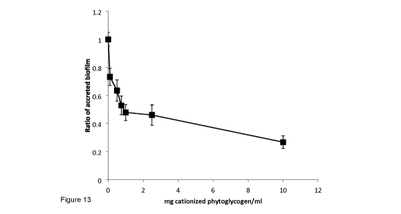
[0026] Figure 13 shows the removal of pre-formed biofilms by P. aeruginosa
following treatment
with cationized phytoglycogen. Experiments were performed as quadruplicate in-
assay
replicates and were repeated three times. Data are normalized relative to the
average A570
obtained for the 20 hT biofilm subset (n = 12 SEM).
[0027] Figure 14 shows that short-term exposure of 20 h P. aeruginosa biofilms
to cationized
phytoglycogen causes a reduction in biofilm. 20 h biofilms were exposed to
medium only
(dark bars), and medium with 1 mg native phytoglycogen.m1-1 (grey bars) or
with 1 mg
cationized phytoglycogen.m1-1 (hollow bars). Values are the average of n = 12
SEM.
[0028] Figure 15 shows cationized phytoglycogen prevents the enhanced biofilm
formation which
is an undesirable feature of sub-MIC of select antibiotics. Absorbance data
were
normalized to the corresponding medium condition without antibiotic. Assays
were done in
Mueller-Hinton medium (0) or medium supplemented with 1 ( ) or 10 mg (0)
cationized
phytoglycogen.m1-1. Values are the average of n = 12 SEM.
[0029] Figure 16 shows that a combination of cationized phytoglycogen and
the antibiotic
tobramycin enhances biofilm eradication. Absorbance data were normalized to
the
corresponding medium condition without antibiotic. Assays were done in Mueller-
Hinton
medium (0) or medium supplemented with 1 ( ) or 10 mg (0) cationized
phytoglycogen.m1-1. Values are the average of n = 12 SEM.
[0030] Figure 17 shows that a combination of cationized phytoglycogen and
the antibiotic
ciprofloxacin enhances biofilm eradication. Absorbance data were normalized to
the
4
CA 03020772 2018-10-12
WO 2017/177342 PCT/CA2017/050472
corresponding medium condition without antibiotic. Assays were done in Mueller-
Hinton
medium (0) or medium supplemented with 1 ( ) or 10 mg (0) cationized
phytoglycogen.m1-1. Values are the average of n = 12 SEM.
[0031] Figure 18 shows that cationized but not native phytoglycogen causes the
sedimentation of
cells from suspension. Representative images are presented of microfuge tubes
containing
suspensions of cells incubated in medium supplemented with native or
cationized
phytoglycogen. Note the formation of material (cells) at the bottom of the
tube containing
cationized phytoglycogen, which was accompanied by a concomitant clarification
of the
upper liquid phase.
[0032] Figure 19 shows representative transmission electron micrographs of P.
aeruginosa cells
incubated with native or cationized phytoglycogen. The dark arrows indicate
phytoglycogen.
Note the localization of cationized phytoglycogen at the cell surface; white
arrows indicate
regions of cell surface perturbation. The scale bar represents 1 pm.
[0033] Figure 20 shows the internalization of Cy5.5-labelled PHX particles
by THP-1 monocytes:
Fluorescence confocal images of THP-1 cells incubated with Cy5.5-Phytoglycogen
nanoparticles (1 mg/mL) at 4 C for 24 hrs (A), at 37 C for 6 hrs (B) and at
37 C for 24 hrs.
Cy5.5-Phytoglycogen nanoparticles, Nucleus stained with DAPI, and cell
membrane
stained with AF488.
[0034] Figure 21 shows the pharmacokinetic profile of Cy5.5-phytoglycogen
taken from repeated
blood sampling of nude CD-1 mice.
[0035] Figure 22 shows the quantification of fluorescent signals in organs
imaged ex vivo at 30
min and 24 hrs after i.v. injection in naïve nude CD-1 mice. The average
fluorescence
concentration data, suggests that in addition to the liver and kidney, high
signal can also be
detected in lung and heart. The fluorescence concentrations at 30 mins are
higher than at
24 hrs. Pre-scan data indicates the fluorescence concentration data for a
mouse not
injected with Cy5.5-Phytoglycogen (i.e. background autofluorescence). Data are
presented
as mean +/- SD.
[0036] Figure 23 shows the quantification of fluorescent signals in brain
imaged ex vivo at 30 min
and 24 hrs after i.v. injection of Cy5.5-Phytoglycogen in naïve nude CD-1
mice. The data
indicate that compared to pre-scan (autofluorescence level), there are
measureable signals
CA 03020772 2018-10-12
WO 2017/177342 PCT/CA2017/050472
in the brain from Cy5.5-Phytoglycogen. The signal is highest at 30 mins and
goes down
slowly over time at 24 hrs.
DETAILED DESCRIPTION
[0037] As used herein, the term "anti-infective" refers to an agent that
limits the progression or
spread of infection. Anti-infectives include antimicrobials such as
antibacterials, antifungals
and antiparasitics, which act by limiting cell growth or causing cell death.
Anti-infectives
also include those agents which limit the progression or spread of infection
though
mechanisms other than growth inhibition and cell death. Anti-infectives may
act by altering
the physiological responses of both infectious agent and the target host.
Quorum sensing
inhibitors are an example of the former; vaccines of the latter. The term
"anti-infective" as
used herein may act through both antimicrobial activity, and also through the
attenuation or
modification of the production of virulence factors. The term "virulence
factors" as used
herein are those factors produced by a cell which contribute to that
organism's capabilities
to cause infection. Virulence factors may be excreted, secreted or shed from
the cell (e.g.
enzymes, toxins), may be part of the cell (e.g. membrane modifications), or a
behaviour of
the cell (e.g. motility, biofilm formation)
[0038] The terms "antibiotic" and "antibacterial" are used interchangeably to
refer to agents used in
the treatment or prevention of bacterial infection or the spread of bacteria,
and include both
agents that kill bacteria or inhibit the growth of bacteria. The term
"antifungal" is used to
refer to agents used in the treatment or prevention of fungal infection or the
spread of fungi,
and includes both agents that kill fungi or inhibit the growth of fungi.
[0039] As used herein, the term "biofilm" refers to an aggregate of
microorganisms, including
bacteria, archaea, viruses, protozoa, fungi or algae, in which cells are
frequently embedded
within a self-produced matrix of extracellular polymeric substance (EPS) and
adhere to
each other and/or to a surface.
[0040] As used herein, the term "cationized phytoglycogen" refers to
phytoglycogen modified to
include a positively charged functional group such as those containing a short
chain
quaternary ammonium compound. The short-chain quaternary ammonium compound
includes at least one alkyl moiety having from 1 to 32 carbon atoms,
preferably 1 to 30
carbon atoms, and more preferably 1 to 24 carbon atoms, unsubstituted or
substituted with
one or more N, 0, S, or halogen atoms. In a preferred embodiment, the short-
chain
6
CA 03020772 2018-10-12
WO 2017/177342 PCT/CA2017/050472
quaternary ammonium compound includes at least one alkyl moiety having from 1
to 16
carbon atoms. In one embodiment, the modifier is 3-(trimethylammonio)2-
hydroxypropy-1-y1
with a degree of substitution of 0.05 to 2.0, preferably 0.3 to 1.2.
[0041] As used herein, the term "extracellular polymeric substance" (EPS)
refers to self-produced
matrix by a microorganism, and any incorporated extraneous materials,
generally
composed of extracellular biopolymers in various structural forms including,
for example,
extracellular DNA, proteins, lipids and polysaccharides.
[0042] As used herein, "therapeutically effective amount" refers to an amount
effective, at dosages
and for a particular period of time necessary, to achieve the desired
therapeutic result. A
therapeutically effective amount of the pharmacological agent may vary
according to factors
such as the disease state, age, sex, and weight of the individual, and the
ability of the
pharmacological agent to elicit a desired response in the individual. A
therapeutically
effective amount is also one in which any toxic or detrimental effects of a
pharmacological
agent are outweighed by the therapeutically beneficial effects.
[0043] As used herein "patient" refers to an animal being treated for an
infection, which in one
embodiment may be a vertebrate, in one embodiment a mammal, in one embodiment,
a
human patient. As used herein, the term "treatment" refers to administering a
composition
of the invention to effect an alteration or improvement of the disease or
condition, which
may include alleviating one or more symptom thereof. The use may be
prophylactic.
Prevention, amelioration, and/or treatment may require administration of
multiple doses at
regular intervals, or prior to onset of the disease or condition to alter the
course of the
disease or condition.
[0044] The present disclosure relates to anti-infective compositions
comprising glycogen or
phytoglycogen nanoparticles, including modified glycogen or phytoglycogen such
as
cationized phytoglycogen functionalized with short chain quaternary ammonium
compounds ("phytoglycogen nanoparticle(s)"). Further, the present disclosure
relates to
compositions comprising phytoglycogen nanoparticles for use as anti-
infectives. In one
embodiment, the anti-infectives are used as a biofilm inhibitor.
[0045] In one embodiment, the nanoparticles may be used as a component of an
antibiotic
treatment to reduce the amount of antibiotic required to achieve the desired
therapeutic
result.
7
CA 03020772 2018-10-12
WO 2017/177342 PCT/CA2017/050472
[0046] Phytoglycogen is composed of molecules of a-D glucose chains having an
average chain
length of 11-12, with 1¨>4 linkage and branching point occurring at 1¨>6 and
with a
branching degree of about 6 % to about 13 %. In one embodiment, phytoglycogen
includes
both phytoglycogen derived from natural sources and synthetic phytoglycogen.
As used
herein the term "synthetic phytoglycogen" includes glycogen-like products
prepared using
enzymatic processes on substrates that include plant-derived material e.g.
starch.
[0047] The yields of most known methods for obtaining glycogen and
phytoglycogen and most
commercial sources of glycogen and phytoglycogen are highly polydisperse
products that
include both glycogen or phytoglycogen particles, as well as other products
and
degradation products of glycogen or phytoglycogen, which will render them less
effective in
the compositions and methods described herein. Accordingly, suitably
substantially
monodisperse glycogen or phytoglycogen is used. These substantially
monodisperse
glycogen or phytoglycogen nanoparticles have a low polydispersity index. In a
preferred
embodiment, monodisperse phytoglycogen nanoparticles are used. In one
embodiment, the
monodisperse phytoglycogen nanoparticles are PhytoSpherixTM by Mirexus
Biotechnologies, Inc.
[0048] In one embodiment, phytoglycogen refers to monodisperse phytoglycogen
nanoparticles
manufactured according to methods described herein. The described methods
enable
production of substantially spherical nanoparticles, which are a single
phytoglycogen
molecule.
[0049] In a preferred embodiment, monodisperse cationized phytoglycogen
nanoparticles are
used.
[0050] Detailed below are monodisperse compositions of phytoglycogen
nanoparticles. The
monodisperse and particulate nature of the compositions described herein are
associated
with properties that render them highly suitable for use in anti-infective
applications.
Further, these phytoglycogen nanoparticles suitably have a size of between
about 30 and
150 nm, in one embodiment, between 60 and 110 nm.
[0051] Accordingly, in a preferred embodiment, anti-infective compositions of
monodisperse
phytoglycogen nanoparticles are used.
8
CA 03020772 2018-10-12
WO 2017/177342 PCT/CA2017/050472
[0052] Phytoglycogen nanoparticles as taught herein have a number of
properties that make them
particularly suitable for use in anti-infective compositions. Many existing
drugs are rapidly
eliminated from the body leading to a need for increased dosages. The compact
spherical
nature of phytoglycogen nanoparticles is associated with efficient cell
uptake, while the
highly-branched nature and high molecular weight of phytoglycogen is believed
to be
associated with slow enzymatic degradation and increased intravascular
retention time,
respectively.
[0053] As shown in Figure 2, each phytoglycogen particle is a single molecule,
made of highly
branched glucose homopolymer characterized by very high molecular weight (up
to 107
Da). This homopolymer consists of a-D-glucose chains with 1-4 linkage and
branching
points occurring at 1¨>6 and with branching degree about 10 %. These particles
are
spherical and can be manufactured with different sizes, in the range of 30 to
150 nm in
diameter by varying the starting material and filtering steps. The high
density of surface
groups on the phytoglycogen nanoparticles results in a variety of unique
properties of
phytoglycogen nanoparticles, such as fast dissolution in water, low viscosity
and shear
thinning effects for aqueous solutions at high concentrations of phytoglycogen
nanoparticles. This is in contrast to high viscosity and poor solubility of
linear and low-
branched polysaccharides of comparable molecular weight. Furthermore, it
allows
formulation of highly concentrated (up to 30 %) stable dispersions in water or
DMSO.
[0054] As demonstrated by the Examples, the present inventors have found that
phytoglycogen
nanoparticles can be accumulated intracellularly by different types of cells.
[0055] When phytoglycogen nanoparticles are internalized, the nanoparticles
are digested by
cellular hydrolases. The rate of breakdown can be controlled by the degree of
phytoglycogen derivatization by small molecules, e.g., methylation,
hydroxypropylation,
(which affect the affinity of hydrolases to polysaccharide chain and,
therefore, the rate of
hydrolysis).
[0056] The phytoglycogen nanoparticles can be further modified with specific
tissue targeting
molecules.
[0057] The phytoglycogen nanoparticles are non-toxic, have no known
allergenicity, and can be
degraded by glycogenolytic enzymes (e.g. amylases and phosphorylases) of the
human
body. The products of enzymatic degradation are non-toxic molecules of
glucose.
9
CA 03020772 2018-10-12
WO 2017/177342 PCT/CA2017/050472
[0058] Phytoglycogen nanoparticles are generally photostable and stable over a
wide range of pH,
electrolytes, e.g. salt concentrations.
[0059] United States patent application publication no. United States
2010/0272639 Al, assigned
to the owner of the present invention and the disclosure of which is
incorporated by
reference in its entirety, provides a process for the production of glycogen
nanoparticles
from bacterial and shell fish biomass. The processes disclosed generally
include the steps
of mechanical cell disintegration, or by chemical treatment; separation of
insoluble cell
components by centrifugation; elimination of proteins and nucleic acids from
cell lysate by
enzymatic treatment followed by dialysis which produces an extract containing
crude
polysaccharides, lipids, and lipopolysaccharides (LPS) or, alternatively,
phenol-water
extraction; elimination of LPS by weak acid hydrolysis, or by treatment with
salts of
multivalent cations, which results in the precipitation of insoluble LPS
products; and
purification of the glycogen enriched fraction by ultrafiltration and/or size
exclusion
chromatography; and precipitation of glycogen with a suitable organic solvent
or a
concentrated glycogen solution can be obtained by ultrafiltration or by
ultracentrifugation;
and freeze drying to produce a powder of glycogen. Glycogen nanoparticles
produced from
bacterial biomass were characterized by Mwt 5.3-12.7 x 10⁶ Da, had particle
size 35-40 nm
in diameter and were monodisperse.
[0060] Methods of manufacturing monodisperse compositions of phytoglycogen are
disclosed in
the International patent application entitled "Phytoglycogen Nanoparticles and
Methods of
Manufacture Thereof", published under the international application
publication no
W02014/172786 and the disclosure of which is incorporated by reference in its
entirety. In
one embodiment, the described methods of producing monodisperse phytoglycogen
nanoparticles include: a. immersing disintegrated phytoglycogen-containing
plant material
in water at a temperature between about 0 and about 50 C; b. subjecting the
product of
step (a.) to a solid-liquid separation to obtain an aqueous extract; c.
passing the aqueous
extract of step (b.) through a microfiltration material having a maximum
average pore size
of between about 0.05 pm and about 0.15 pm; and d. subjecting the filtrate
from step c. to
ultrafiltration to remove impurities having a molecular weight of less than
about 300 kDa, in
one embodiment, less than about 500 kDa, to obtain an aqueous composition
comprising
monodisperse phytoglycogen nanoparticles. In one embodiment of the method, the
phytoglycogen-containing plant material is a cereal selected from corn, rice,
barley,
sorghum or a mixture thereof. In one embodiment, step c. comprises passing the
aqueous
CA 03020772 2018-10-12
WO 2017/177342 PCT/CA2017/050472
extract of step (b.) through (c.1) a first microfiltration material having a
maximum average
pore size between about 10 pm and about 40 pm; (c.2) a second microfiltration
material
having a maximum average pore size between about 0.5 pm and about 2.0 pm, and
(c.3) a
third microfiltration material having a maximum average pore size between
about 0.05 and
0.15 pm. The method can further include a step (e.) of subjecting the aqueous
composition
comprising monodisperse phytoglycogen nanoparticles to enzymatic treatment
using
amylosucrose, glycosyltransferase, branching enzymes or any combination
thereof. The
method avoids the use of chemical, enzymatic or thermal treatments that
degrade the
phytoglycogen material. The aqueous composition can further be dried.
[0061] In one embodiment, the nanoparticles are produced from sweet corn
starting material (Zea
mays var. saccharata and Zea mays var. rugosa). In one embodiment, the sweet
corn is of
standard (su) type or sugary enhanced (se) type. In one embodiment, the
composition is
produced from dent stage or milk stage kernels of sweet corn. Unlike glycogen
from animal
or bacterial sources, use of phytoglycogen reduces the risk of contamination
with prions or
endotoxins, which may be associated with these other sources.
[0062] The polydispersity index (PDI) of a composition of nanoparticles can be
determined by the
dynamic light scattering (DLS) technique and, in this embodiment, PDI is
determined as the
square of the ratio of standard deviation to mean diameter (PDI = (aid)2. PDI
can also be
expressed through the distribution of the molecular weight of polymer and, in
this
embodiment, is defined as the ratio of Mw to Mn, where Mw is the weight-
average molar
mass and Mn is the number-average molar mass (hereafter this PDI measurement
is
referred to as PDI*). In the first case, a monodisperse material would have a
PDI of zero
(0.0) and in the second case the PDI* would be 1Ø
[0063] In one embodiment, there is provided an anti-infective composition
that comprises, consists
essentially of, or consists of a composition of monodisperse phytoglycogen
nanoparticles.
Suitably, these nanoparticles are modified as described further below. In one
embodiment,
the anti-infective composition comprises, consists essentially of, or consists
of a
composition of monodisperse phytoglycogen nanoparticles having a PDI of less
than about
0.3, less than about 0.2, less than about 0.15, less than about 0.10, or less
than 0.05 as
measured by dynamic light scattering. In one embodiment, the anti-infective
composition
comprises, consists essentially of, or consists of a composition of
monodisperse
11
CA 03020772 2018-10-12
WO 2017/177342 PCT/CA2017/050472
phytoglycogen nanoparticles having a PDI* of less than about 1.3, less than
about 1.2, less
than about 1.15, less than about 1.10, or less than 1.05 as measured by SEC
MALS.
[0064] In one embodiment, the anti-infective composition comprises,
consists essentially of, or
consists of a composition of monodisperse phytoglycogen nanoparticles having
an average
particle diameter of between about 30 nm and about 150 nm. In one embodiment,
the anti-
infective composition comprises, consists essentially of, or consists of a
composition of
monodisperse phytoglycogen nanoparticles having an average particle diameter
of about
60 nm to about 110 nm. In other embodiments, there is provided compositions
comprising,
consisting essentially of, or consisting of, nanoparticles having an average
particle diameter
of about 40 to about 140 nm, about 50 nm to about 130 nm, about 60 nm to about
120 nm,
about 70 nm to about 110 nm, about 80 nm to about 100 nm. These nanoparticles
may be
modified as described further below.
[0065] The methods of producing phytoglycogen nanoparticles as detailed in
Example 1 and in the
international patent application no. PCT/CA2014/000380, published under the
international
application publication no WO/2014/172786, entitled "Phytoglycogen
Nanoparticles and
Methods of Manufacture Thereof", are amenable to preparation under
pharmaceutical
grade conditions.
Chemical Modification of Phytoglycogen Nanoparticles
[0066] To impart specific properties to phytoglycogen nanoparticles, they can
be chemically
modified via numerous methods common for carbohydrate chemistry.
[0067] Accordingly, in a preferred embodiment, the phytoglycogen nanoparticles
are modified. The
resulting products are referred to herein interchangeably as functionalized
nanoparticles or
derivatives. Functionalization can be carried out on the surface of the
nanoparticle, or on
both the surface and the interior of the particle, but the structure of the
glycogen or
phytoglycogen molecule as a single branched homopolymer is maintained. In one
embodiment, the functionalization is carried out on the surface of the
nanoparticle. As will
be understood by those of skill in the art, chemical modifications should be
non-toxic and
generally safe for human consumption. The chemical character of phytoglycogen
nanoparticles produced according to methods described above may be changed
from their
hydrophilic, slightly negatively charged native state to be positively and/or
negatively
charged, or to be partially or highly hydrophobic. Chemical processing of
polysaccharides is
12
CA 03020772 2018-10-12
WO 2017/177342 PCT/CA2017/050472
well known in the art. See for example J.F Robyt, Essentials of Carbohydrate
Chemistry,
Springer, 1998; and M. Smith, and J. March, March's Advanced Organic
Chemistry:
Reactions, Mechanisms, and Structure Advanced Organic Chemistry, Wiley, 2007.
[0068] As will be described further below, nanoparticles modified to have a
positive charge
demonstrate anti-infective activity, including antimicrobial activity.
[0069] The nanoparticles can be functionalized either directly or
indirectly, where one or more
intermediate linkers or spacers can be used. The nanoparticles can be
subjected to one or
more than one functionalization steps including two or more, three or more, or
four or more
functionalization steps.
[0070] Various derivatives can be produced by chemical functionalization of
hydroxyl groups of
phytoglycogen, either by etherification with a suitably functionalized alkyl
group, by
interconversion of the hydroxyl group into another functional group, or by
oxidation. Such
functional groups include, but are not limited to, nucleophilic and
electrophilic groups, and
acidic and basic groups, e.g., carbonyl groups, amine groups, thiol groups,
carboxyl groups
and their derivatives such as amide or esters, azide, nitrile, halogenide and
pseudo-
halogenide such as tosyl, mesyl or triflate, and hydrocarbyl groups such as
alkyl, vinyl,
phenyl, benzyl, propargyl and ally! groups. Amino groups can be primary,
secondary,
tertiary, or quaternary amino groups, preferably quaternary amino groups.
[0071] Functionalized nanoparticles can be further conjugated with various
desired molecules,
which are of interest for a variety of applications, such as biomolecules,
small molecules,
therapeutic agents, micro- and nanoparticles, pharmaceutically active
moieties,
macromolecules, diagnostic labels, chelating agents, dispersants, charge
modifying agents,
viscosity modifying agents, surfactants, coagulation agents and flocculants,
as well as
various combinations of these chemical compounds. In certain embodiments, two
or more
different chemical compounds are used to produce multifunctional derivatives.
[0072] In one embodiment, the functionalized nanoparticles are modified
with a quaternary
ammonium compound.
[0073] The reactivity of hydroxyl groups on glucose subunits is low. Even so,
reactions are
possible at high pH with epoxides, alkyl halides or anhydrides, forming the
corresponding
ether or ester linkages. Water-soluble chemicals with epoxide or anhydride
functionalities
react at basic pH (8-13) with phytoglycogen nanoparticles (in the presence of
an
13
CA 03020772 2018-10-12
WO 2017/177342 PCT/CA2017/050472
appropriate catalyst). Although derivatization in aqueous environment is often
preferable,
some reactions (e.g. with alkyl halides) are best conducted in organic
solvents such as
dimethyl sulfoxide (DMSO), dimethyl formamide (DMF), dimethyl acetamide or
pyridine, or
mixtures of the aforementioned with salts such as lithium chloride or
tetrabutylammonium
fluoride. As will be apparent to one of skill in the art, water-soluble
compounds with low
toxicity and reactive at relatively mild conditions are particularly suitable.
[0074] A simple approach to increasing the reactivity of hydroxyl groups is
the selective oxidation
of glucose hydroxyl groups at positions of C-2, C-3, C-4 and/or C-6, yielding
carbonyl or
carboxyl groups or carboxyl. There is a wide spectrum of redox initiators
which can be
employed, such as persulfate, periodate (e.g. potassium periodate, bromine,
sodium
chlorite (2,2,6,6-tetramethylpiperidin-1y1)oxidanyl, commonly known as TEMPO,
and Dess-
Martin periodinane.
[0075] Phytoglycogen nanoparticles functionalized with carbonyl groups are
readily reactive
towards compounds bearing primary or secondary amine groups. This results in
imine
formation (eq. 1) which can be further reduced to amines with a reducing agent
e.g.,
sodium borohydride (eq. 2). This reduction step provides an amino-product
which is more
stable than the imine intermediate, and also converts unreacted carbonyls in
hydroxyl
groups. The elimination of carbonyls significantly reduces the possibility of
non-specific
interactions of derivatized nanoparticles with non-targeting molecules (e.g.
plasma
proteins).
(eq. 1) P/G NANO ¨CH=0 + H2N¨R P/G NANO¨CH=NH¨R + H20
reducing agent
(eq. 2) P/G NANO ¨CH=NH¨R > P/G NANO ¨CH2¨NH¨R
[0076] Carboxyl groups can be activated using coupling reagents such as N,N'-
Dicyclohexylcarbodiimide (DCC), 1-Ethyl-3-(3-dimethylaminopropyl)carbodiimide
(EDC), or
1,1'-Carbonyldiimidazole (CDI), with or without the addition of auxiliary
reagents such as 1-
Hydroxybenzotriazol (HOBt) or N-Hydroxysuccinimide (NHS). The activated
carboxylate
then reacts under very mild conditions with nucleophiles such as amino or
hydroxy groups
(examples 9, 10). This type of activation can either be used to activate
carboxyl groups on
a small molecule, and react it with hydroxy groups of native phytoglycogen or
amino groups
14
CA 03020772 2018-10-12
WO 2017/177342 PCT/CA2017/050472
of aminated phytoglycogen; or it can be used to activate carboxyl groups on
oxidized
phytoglycogen and attach an amino-containing small molecule to it (example
12).
[0077] In certain embodiments, the nanoparticles described are
functionalized via a process of
cyanylation. This process results in the formation of cyanate esters and
imidocarbonates on
polysaccharide hydroxyls. These groups react readily with primary amines under
very mild
conditions, forming covalent linkages (Figure 1). Cyanylation agents such as
cyanogen
bromide and 1-cyano-4-diethylamino-pyridinium (CDAP) can be used for
functionalization
of the nanoparticles.
[0078] A chemical compound bearing a functional group capable of binding to
the functional
groups present on phytoglycogen or modified phytoglycogen can be directly
attached to the
nanoparticle. However, for some applications chemical compounds may be
attached via a
polymer spacer or a "linker". These can be homo- or hetero-bifunctional
linkers bearing
functional groups such as amino, carbonyl, carboxyl, sulfhydryl, succimidyl,
maleimidyl,
isocyanate, (e.g. diaminohexane,
ethylene glycobis(sulfosuccimidylsuccinate),
disulfosuccimidyl tartarate, dithiobis(sulfosuccimidylpropionate),
aminoethanethiol, etc.)
[0079] The antimicrobial activity of modified phytoglycogen nanoparticles
functionalized with
quaternary ammonium compounds may be further enhanced by modifying its
hydrophobicity Therefore, in a preferred embodiment, the glycogen or
phytoglycogen
nanoparticle is double-modified with both quaternary ammonium and hydrophobic
groups.
The hydrophobic interactions can be fine-tuned by choosing an appropriate
degree of
substitution and hydrophobic functional group. Example functional groups
include, but are
not limited to aliphatic alkyl, alkenyl, alkynyl or benzyl ethers and esters
or trialkylsilyl
ethers of chain lengths between 1 and 24 (Examples 5-7).
[0080] In one embodiment, there is provided a method of treating a subject
suffering from a
microbial infection comprising administering to the subject a therapeutically
effective
amount of a composition as described herein. In one embodiment, the
composition
comprises functionalized phytoglycogen nanoparticles having a positive surface
charge. In
one embodiment, the phytoglycogen nanoparticles are functionalized with a
secondary,
tertiary or quaternary ammonium group. In one embodiment, the composition
comprises
phytoglycogen nanoparticles functionalized with an amphiphilic group. In one
embodiment,
the composition comprises glycogen or phytoglycogen nanoparticles
functionalized with
CA 03020772 2018-10-12
WO 2017/177342 PCT/CA2017/050472
quaternary ammonium compounds. In certain embodiments, phytoglycogen
nanoparticles
as described above may be functionalized and used without further conjugation.
[0081] In other embodiments, the nanoparticles may further be conjugated to
other chemical
compounds that can include biomolecules, small molecules, therapeutic agents,
pharmaceutically active moieties, macromolecules, diagnostic labels, chelating
agents,
dispersants, surfactants, charge modifying agents, viscosity modifying agents,
coagulation
agents and flocculants, to name a few, as well as various combinations of the
above.
[0082] Biomolecules which can be conjugated include peptides, enzymes,
receptors,
neurotransmitters, hormones, cytokines, cell response chemical compounds such
as
growth factors and chemotactic factors, antibodies, vaccines, haptens, toxins,
interferons,
ribozymes, anti-sense agents, and nucleic acids.
[0083] Anti-infective compositions according to one embodiment include
functionalized
monodisperse phytoglycogen nanoparticles conjugated to other molecule(s). In
various
embodiments, the phytoglycogen nanoparticles are further conjugated to a
pharmaceutical.
In various embodiments, the nanoparticles are conjugated to one or more of an
antibiotic,
an antifungal, an anti-parasite and/or anti-protozoal compound.
Pharmaceutically useful
moieties used as modifiers include hydrophobicity modifiers, pharmacokinetic
modifiers,
and biologically active modifiers.
[0084] Chemical compounds which are conjugated to phytoglycogen nanoparticles
may have light
absorbing, light emitting, fluorescent, luminescent, Raman scattering,
fluorescence
resonant energy transfer, and electroluminescence properties.
[0085] Two or more different chemical compounds can be used to produce
multifunctional
derivatives. For example, one chemical compound can be selected from the list
of specific
binding biomolecules, such as antibody and aptamers, while the second compound
would
be selected from the list of anti-infectives. For example, one chemical
compound may be a
cationic species, while the second compound may be an antibiotic.
[0086] Loading efficiency depends on the molecular weight and properties
(charge,
hydrophobicity, etc.) of the molecules to be conjugated. Degree of
substitution is expressed
as % of anhydroglucose units derivatized with the drug. E.g. if the drug has a
molecular
weight of 100 Da, and the degree of substitution is 50 %, then 1 g of
phytoglycogen
nanoparticles would carry 0.31 g of the drug. For small molecules (<100 Da) a
degree of
16
CA 03020772 2018-10-12
WO 2017/177342 PCT/CA2017/050472
substitution >30 % was generally achieved, going as high as 100 % for methyl
groups.
Larger molecules (which cannot penetrate the pore structure of the particles)
can be
conjugated only at the surface of the phytoglycogen nanoparticles, and the
degree of
substitution is lower, generally 0.1- 2.0 %.
Anti-infective Activity
[0087] As detailed in the Examples, the present inventors have developed
compositions of
phytoglycogen nanoparticles including functionalized forms thereof with
properties that
render them highly suitable for use in anti-infective applications.
[0088] In one embodiment, there is provided an anti-infective composition
comprising, consisting
of or consisting essentially of positively charged phytoglycogen
nanoparticles. The surface
of phytoglycogen nanoparticles can be made cationic through a number of
techniques, as
described above.
[0089] In one embodiment, there is described an anti-infective composition
comprising
phytoglycogen, preferably positively charged nanoparticles of phytoglycogen.
In one
embodiment, the composition further comprises a carrier, which in one
embodiment is a
pharmaceutically acceptable carrier.
[0090] In one embodiment, the nanoparticles are modified with an
amphiphilic compound.
[0091] Cationic modifications to phytoglycogen nanoparticles, which can render
them useful as
anti-infectives may include secondary, tertiary or quaternary amino groups
and, in
particular, modifications with quaternary-ammonium derivatives. In one
embodiment, the
quaternary ammonium derivatives can be selected from hydroxypropyl-
trimethylammonium
and hydroxypropyl-alkyl-dimethylammonium, wherein alkyl is a aliphatic C2 to
C32 aliphatic
hydrocarbon, such as, but not limited to lauryl-, myristyl- or stearyl-. In
another
embodiment, the alkyl is a C2 to C32 hydrocarbon, preferably C2 to C30, more
preferably C2
to C24 =
[0092] The surfaces of bacteria are typically anionic, and without wishing to
be bound by a theory,
the inventors hypothesize that the creation of localized high densities of
cationized groups
on the surface of a phytoglycogen nanoparticle create a cumulative charge-
based effect
capable of affecting bacterial growth and physiology.
17
CA 03020772 2018-10-12
WO 2017/177342 PCT/CA2017/050472
[0093] As demonstrated by the Examples, anti-infective activity against
Escherichia coil, Bacillus
subtilis, Pseudomonas aeruginosa and Candida utilis is shown in the presence
of a
composition of phytoglycogen nanoparticles.
[0094] In a preferred embodiment, the phytoglycogen nanoparticles are
modified to a cationized
form functionalized with short chain quaternary ammonium compounds.
[0095] As shown in the Examples, increased anti-infective susceptibility to a
variety of classes of
antibiotics is shown against P. aeruginosa, E. coli, B. subtilis and C. uti/is
following
incubation in the presence of cationized phytoglycogen.
[0096] In one embodiment, phytoglycogen nanoparticles are co-administered
with an antibiotic,
which may be selected from but is not limited to Penicillins,
Carboxypenicillins,
Aminopenicillins, Glycopeptides, Quinolones,
Cephalosporins, Macrolides,
Fluoroquinolones, Phenicols, Sulfonamides, Tetracyclines, Aminocoumarins,
Lipopeptides
or Aminoglycosides.
[0097] In one embodiment, phytoglycogen nanoparticles are co-administered
with an antifungal,
which may be selected from but is not limited to a Polyene, Imidazole,
Triazole, Thiazole,
Ally!amine, or Echinocandin antifungal.
[0098] In one embodiment, there is provided an anti-infective composition
comprising both
phytoglycogen nanoparticles and an antibiotic, which may be selected from but
is not
limited to Penicillins, Carboxypenicillins, Aminopenicillins, Glycopeptides,
Quinolones,
Cephalosporins, Macrolides, Fluoroquinolones, Phenicols, Sulfonamides,
Tetracyclines,
Aminocoumarins, Lipopeptides, cationic antimicrobial peptides, or
Aminoglycosides.
[0099] In one embodiment, there is provided an anti-infective composition
comprising both
phytoglycogen nanoparticles and an antifungal, which may be selected from but
is not
limited to a Polyene, Imidazole, Triazole, Thiazole, Allylamine, or
Echinocandin antifungal.
[00100] In one embodiment, the nanoparticles are conjugated to an antibiotic,
which may be
selected from but is not limited to Penicillins, Carboxypenicillins,
Aminopenicillins,
Glycopeptides, Quinolones, Cephalosporins, Macrolides, Fluoroquinolones,
Phenicols,
Sulfonamides, Tetracyclines, Aminocoumarins, Lipopeptides, Cationic
antimicrobial
peptides, or Aminoglycosides.
18
CA 03020772 2018-10-12
WO 2017/177342 PCT/CA2017/050472
[00101] In one embodiment, the nanoparticles are conjugated to an antifungal,
which may be
selected from but is not limited to a Polyene, Imidazole, Triazole, Thiazole,
Ally!amine, or
Echinocandin antifungal.
[00102] In one embodiment, the anti-infective composition further comprises a
pharmaceutically
acceptable carrier or excipient.
[00103] Anti-infective compositions as described herein may be used to treat
bacterial, fungal or
parasitic infections and may also be used prophylactically.
[00104] Also provided is a method of treating a microbial infection comprising
administering a
therapeutically effective amount of an anti-infective composition as described
herein to a
subject in need thereof. In one embodiment, the microbial infection is a
fungal infection. In
one embodiment, the microbial infection is a bacterial infection.
[00105] In one embodiment, phytoglycogen nanoparticles are used as a co-
therapeutic not as an
antibiotic, but as an anti-infective to regulate virulence and pathogenicity
of
microorganisms.
[00106] The infection may be an intracellular infection.
[00107] The anti-infective activity of phytoglycogen nanoparticles may operate
in whole or in part by
decreasing or inhibiting biofilm formation, maintenance or growth as discussed
more
particularly below.
[00108] In some embodiment, the anti-infective activity of phytoglycogen
nanoparticles may operate
through the attenuation or modification of the production of virulence factors
by an infective
agent such as a bacterium, yeast, fungus, or parasite, resulting in a
diminished ability to
cause infection.
[00109] In various embodiments, the infection is in the liver, upper and lower
respiratory tracts (e.g.
sinusitis, whooping cough, pneumonia), eyes, ears, gum and/or mouth (e.g.
periodontitis
and gingivitis), kidney, intestinal tract, genito-urinary tract and bladder,
blood (e.g.,
bacteraemia), brain, meninges, spinal cord, bone, gut and/or cardiac system.
In another
embodiment, the infection is a wound or skin infection.
[00110] In one embodiment, there is provided a method of treating an
intracellular infection
comprising administering a therapeutically effective amount of a composition
as described
19
CA 03020772 2018-10-12
WO 2017/177342 PCT/CA2017/050472
herein to a subject in need thereof. In various embodiments, the intracellular
infection may
be caused by microorganisms, including, but not limited to, Legionella
pneumophila,
Candida spp., Salmonella spp., invasive E. coil spp. Listeria monocyto genes,
Rickettsia
rickettsii, Chlamydia, Shigella spp., Francisella tularensis, Yersinia pestis,
Neisseria,
Brucefla spp., Bartonella spp., Staphylococcus aureus, Coxiella bumettii,
Ctyptococcus
neoformans, Histoplasmata capsulatum, and/or Pneuomcystis jiroveciricarinii.
[00111] Infections of the upper and/or lower respiratory tract and/or airways
may be bacterial or
fungal in nature. Common causes of bacterial lung infections include
Streptococcus
pneumoniae, Haemophilus species, Klebsiella pneumoniae, Staphylococcus aureus,
Mycobacterium tuberculosis, and Pseudomonas aeruginosa. Common pathogens
causing
fungal lung infections include Histoplasma capsulatum, Coccidioides immitis,
Blastomyces
dermatitidis, Paracoccidioides brasiliensis, Pneumocytis jirovechicarinii,
Candida spp.,
Aspergillus spp., Mucor spp. and Cryptococcus neoformans.
[00112] In one embodiment, there is provide a method of treating an
intracellular infection within the
lungs comprising administering a therapeutically effective amount of a
composition as
described herein to a subject in need thereof.
[00113] In one embodiment, the anti-infective may act to decrease or inhibit
biofilm formation,
maintenance or growth.
[00114] Administration to the lung may be by, although is not limited to,
inhalation.
[00115] In one embodiment, phytoglycogen nanoparticles are used as a co-
therapeutic or as part of
a conjugated anti-infective for the treatment of a pulmonary infection.
[00116] In one embodiment, phytoglycogen nanoparticles can be used as a co-
therapeutic for the
treatment of chronic pulmonary infections of P. aeruginosa, which are typical
of individuals
with cystic fibrosis.
[00117] Gastroenteritis may be caused by a number of microorganisms,
including, but not limited to,
Yersinia enterocolitica, Clostridium perfringens, Clostridium difficile,
Helicobacter pylori,
Staphylococcus aureus, Shigefla spp., Pseudomonas aeruginosa, Salmonella spp.,
Campylobacter jejuni, Escherichia coli, Candida spp. In one embodiment,
compositions as
described herein can be used to treat gastroenteritis.
CA 03020772 2018-10-12
WO 2017/177342 PCT/CA2017/050472
[00118] In one embodiment, the anti-infective may act to decrease or inhibit
biofilm formation,
maintenance or growth in the intestines.
[00119] Administration to the intestines may be by, although is not limited
to, orally or by
suppository.
[00120] In one embodiment, the infection is a skin infection and the
composition is topically applied.
Bacterial skin infections include, but are not limited to acne, impetigo,
cellulitis and
streptococcal infections. Fungal skin infections include but are not limited
to Tinea pedis
(athlete's foot), Tinea cruris (jock itch), Tinea corporis (ringworm) and
yeast infections.
[00121] In various embodiments, the infection may be associated with a cut,
blister, burn, insect
bite, surgical wound, injection site or catheter insertion site.
[00122] In one embodiment, the infection is of the hair or nails. In one
embodiment, there is
provided an anti-infective shampoo comprising compositions as described
herein.
[00123] As described further below, the compositions as described herein can
be suitably
formulated as powders, lotions, gels, foams, sprays or ointments.
[00124] Uses also include antibacterial skin sanitizers, and surface
sanitizers. In one embodiment
phytoglycogen nanoparticles may be conjugated with an active compound of an
antiseptic
or sanitizer. The use of phytoglycogen nanoparticles as a surface sanitizer
may operate
through inhibition of cell growth or cell death, inhibition of biofilm
formation, biofilm
dissolution or disruption of quorum sensing as discussed more particularly
below.
[00125] In another embodiment, anti-infective compositions as described herein
may be used as
anti-infective coatings for medical devices, such as diagnostic devices,
implanted devices
such as pacemakers, artificial joints, stents, and catheters. In other
embodiments, the
compositions may be impregnated into or coated onto bandages, surgical suture
thread,
wound dressings, wipes, towelettes, patches or sponges, or incorporated into
bone cement.
In one embodiment, the infections are associated with implanted devices such
as indwelling
catheters, pacemakers, artificial joints, auditory implants, and stents.
[00126] In one embodiment, anti-infective compositions of the present
invention are used in the
treatment of intracellular infections. Many pathogenic bacteria can infect and
survive within
host cells, including cells of the immune system (monocytes/phagocytes) that
are supposed
21
CA 03020772 2018-10-12
WO 2017/177342 PCT/CA2017/050472
to kill them. It is more challenging to treat such infections since once
within the cell interior
the pathogens are somewhat protected from antibiotics. Many antibiotics show a
lack of
accumulation whether in phagocytic or non-phagocytic cells and tissues in
general due to
low cell membrane permeability, fast efflux etc. Often, higher antibiotics
doses are needed
to effectively kill bacteria in the cell interior. The phytoglycogen
nanoparticles as described
herein provide a solution to this problem by providing targeted delivery of
antibiotics to host
cells, e.g., macrophages, and to reach effective concentration to kill
intracellular bacteria.
As demonstrated by the Examples, phytoglycogen nanoparticles can carry
compounds
across the cell membrane and were shown to accumulate within the cytoplasm.
[00127] Phytoglycogen nanoparticles as described herein can stabilize peptides
e.g. antimicrobial
peptides. Protein and peptides stored in solution or frozen or formulated in
dry formulations
(e.g. spray dried or freeze-dried) tend to lose their efficacy over time due
to aggregation,
decomposition, denaturation, oxidation and deamidation. While the stabilizing
activity can
help improve shelf life, it may also allow for less onerous storage
requirements e.g. limiting
the requirement for refrigeration. Phytoglycogen nanoparticles can stabilize
organic
compounds. As mentioned above, the highly-branched nature of glycogen
and
phytoglycogen is associated with slow enzymatic degradation. Without wishing
to be bound
by a theory, the monodisperse phytoglycogen nanoparticles as described herein
can
provide both structural stabilization to protein and peptide solutions and
inhibit degradation
through steric hindrance of enzymatic degradation.
[00128] As demonstrated by the Examples, the conjugated antibiotic-
phytoglycogen may act
without being cleaved; equally, it may act as a cleaved product.
[00129] A biofilm is a sessile community of microorganisms in which the cells
are adhered to one
another and also often to a surface. These adherent cells are physiologically
distinct from
planktonic microbial cells which are single cells that are suspended in a
liquid medium. The
adherent cells found in a biofilm are embedded within a self-produced matrix
of
extracellular polymeric substance (EPS); the EPS may also comprise
incorporated
extraneous materials. This EPS is a conglomeration generally composed of
extracellular
biopolymers in various structural forms. The EPS allows the microorganisms
living in this
type of environment to be less susceptible to anti-infectives in some cases.
The EPS
confers benefits to microorganisms including, but not limited to, enabling 3-D
architecture,
cellular organization, creation of micro-environments, and the generation of a
plethora of
22
CA 03020772 2018-10-12
WO 2017/177342 PCT/CA2017/050472
phenotypes. Collectively these enable key features of biofilm communities,
including
decreased susceptibility to anti-infectives and other inimical agents, reduced
predation and
invasion, evasion of components of the immune response and the consequent
difficulty to
eradicate infections.
[00130] Biofilms are present in the natural environment, and are common in
hospitals and industrial
settings. Biofilms can form on living and non-living surfaces, including
native tissues and
medical devices. In cases where microorganisms succeed in forming a biofilm on
or within
a host, including human hosts, chronic and untreatable infection can result.
[00131] As detailed in the Examples, the present inventors have developed
compositions of
phytoglycogen nanoparticles including functionalized forms thereof with
properties that
render them highly suitable for use to decrease or inhibit biofilm formation,
maintenance
and growth.
[00132] Without wishing to be bound by a theory, the present inventors
hypothesize that treatment
of biofilms with functionalized phytoglycogen nanoparticles may decrease or
inhibit biofilm
formation, maintenance and growth through charge-based mechanisms which result
in
disruption of and/or reduction in biofilm formation and/or enhanced biofilm
dissolution.
[00133] Further, as discussed below, charge-based interactions may interfere
with quorum sensing-
related processes, leading to the attenuation of the production of virulence
factors.
[00134] The modification or attenuation of the production of virulence factors
may alter a cellular
phenotype that modulates cell-extracellular interactions, or that decreases or
inhibits the
production of toxins, biofilms or enzymes.
[00135] Quorum sensing is a density dependent cell-to-cell signalling system
that regulates a range
of bacterial processes. It is a two-step process that involves the production
and release of
signals by the bacteria into the environment and signal detection by a
receptor (sensing).
When a threshold concentration is reached, indicating a quorum, this directs
up- or down-
regulation of genes thereby enabling co-ordinated responses of single cells
and concerted
population responses.
[00136] Quorum sensing is pivotal for a number of bacterial processes
including infection,
production of virulence factors, colonisation of surfaces and biofilm
formation. Since
quorum sensing is established as a central factor in the progression of
infectious disease
23
CA 03020772 2018-10-12
WO 2017/177342 PCT/CA2017/050472
by microorganisms, there has been a drive to develop strategies which
interfere with
quorum sensing, thereby attenuating virulence.
[00137] In addition to the role of quorum sensing in regulating production of
virulence factors and
phenotypes consistent with virulence and pathogenesis, quorum sensing signals
may also
interface with the host. Certain quorum sensing signals produced have
immunomodulatory
properties which alter the response of the host immune system and coordinate
subversion
of host defences.
[00138] Without wishing to be bound by a theory, the present inventors
hypothesize that
phytoglycogen nanoparticles may interfere with quorum sensing processes to
regulate the
production of virulence factors and interface with the host to alter the
response of the host
immune system.
[00139] As demonstrated in the Examples, down-regulation and disruption of
quorum sensing
regulated processes in P. aeruginosa occurs in the presence of cationized
phytoglycogen.
Further, as demonstrated in the Examples, reduced pyocyanin production,
decreased
biofilm formation, enhanced biofilm dissolution and decreased biofilm
accretion occurs in
the presence of cationized phytoglycogen.
[00140] In one embodiment, phytoglycogen nanoparticles are used as a skin or
surface sanitizer as
described above. In one embodiment, the phytoglycogen nanoparticles can be
used as a
gel or in a semi-solid state as described above.
[00141] In one embodiment, phytoglycogen nanoparticles can be used in a spray,
optionally an
aerosol form. In one embodiment, the composition is a spray on product that
can be used
topically on a human or on a non-living surface. In another embodiment,
phytoglycogen
nanoparticles can be inhaled in an aerosolized form.
[00142] In another embodiment, phytoglycogen nanoparticles can be used
internally to decrease or
inhibit biofilm formation, maintenance or growth.
[00143] As shown in the Examples, decreased motility and pyocyanin production
of P. aeruginosa
is observed in the presence of cationized phytoglycogen. Swarming motility and
pyocyanin
production are two virulence factors regulated by quorum sensing processes in
P.
aeruginosa. Cationized phytoglycogen may act as a co-therapeutic in the
management of
chronic P. aeruginosa infections typical within the respiratory tracts of
patients with cystic
24
CA 03020772 2018-10-12
WO 2017/177342 PCT/CA2017/050472
fibrosis, not as an antibiotic per se but as an anti-infective to regulate
virulence and
pathogenicity.
[00144] In one embodiment, phytoglycogen nanoparticles are used in conjunction
with an antibiotic,
an antifungal, or an antiparasitic as described above. In another embodiment,
phytoglycogen nanoparticles are conjugated to one or more of an antibiotic, an
antifungal
agent, an anti-parasite and/or anti-protozoal compound, an anti-adhesion
molecule, an
analgesic, an anticoagulant, a local anesthetic, and an imaging agent as
described above.
[00145] In one embodiment, phytoglycogen nanoparticles are used as a co-
therapeutic not as an
antibiotic but as an anti-infective to regulate virulence and pathogenicity of
microorganisms.
In one embodiment, the microorganisms are in a planktonic population. In
another
embodiment, the microorganisms are in a biofilm community.
Formulation and Administration
[00146] The nanoparticles of the invention may also be admixed, encapsulated,
or otherwise
associated with other molecules, molecule structures or mixtures of compounds
and may
be combined with any pharmaceutically acceptable carrier or excipient. As used
herein, a
"pharmaceutically carrier" or "excipient" can be a pharmaceutically acceptable
solvent,
suspending agent or any other pharmacologically inert vehicle for delivering
functionalized
phytoglycogen nanoparticles, whether alone or conjugated to a biologically
active or
diagnostically useful molecule, to an animal. The excipient may be liquid or
solid and is
selected, with the planned manner of administration in mind, so as to provide
for the
desired bulk, consistency, etc., when combined with phytoglycogen
nanoparticles and the
other components of a given pharmaceutical composition. Examples of
pharmaceutically
acceptable carriers include one or more of water, saline, phosphate buffered
saline,
glycerol, ethanol, propylene glycol, 1,3-butylene glycol, dimethyl sulfoxide,
N,N-
dimethylacetamide and the like, as well as combinations thereof.
Pharmaceutically
acceptable carriers may further comprise minor amounts of auxiliary substances
such as
wetting or emulsifying agents, preservatives or buffers, which enhance the
shelf life or
effectiveness of the pharmacological agent.
[00147] The pharmaceutical formulations of the present invention, which may
conveniently be
presented in unit dosage form, may be prepared according to conventional
techniques well
known in the pharmaceutical industry. Such techniques include the step of
bringing into
CA 03020772 2018-10-12
WO 2017/177342 PCT/CA2017/050472
association the active ingredients with the pharmaceutical carrier(s) or
excipient(s). In
general, the formulations are prepared by uniformly and intimately bringing
into association
the active ingredients with liquid carriers, finely divided solid carriers, or
both, and then, if
necessary, shaping the product (e.g., into a specific particle size for
delivery).
[00148] For the purposes of formulating pharmaceutical compositions,
monodisperse
phytoglycogen nanoparticles prepared as taught herein, may be provided in a
dried
particulate/powder form or may be dissolved e.g. in an aqueous solution.
[00149] In various embodiments, where a low viscosity is desired, the
phytoglycogen nanoparticle
component as described herein may suitably be used in the anti-infective
compositions in a
concentration of up to about 25 % w/w, about 20 % w/w, about 15 % w/w, about
10 % w/w,
about 5 % w/w, about 1 % w/w and between about 0.05 and 0.5 %.
[00150] In applications where a high viscosity is desirable, the phytoglycogen
nanoparticle
component may be used in formulations in concentrations above about 25 % w/w.
In
applications where a gel or semi-solid is desirable, concentrations up to
about 35 % w/w
can be used, or the phytoglycogen nanoparticle component can be used in a
mixture with
viscosity builders or gelling agents.
[00151] The composition may be a water-based formulation or an alcohol-based
formulation.
Suitable alcohols include ethyl alcohol, propyl alcohol, isopropyl alcohol,
ethylene glycol,
propylene glycol, butylene glycol, dipropylene glycol, ethoxydiglycol, or
glycerol or a
combination thereof.
[00152] The anti-infective compositions as described herein may be
administered in a number of
ways depending upon whether local or systemic treatment is desired and upon
the area to
be treated. Without limiting the generality of the foregoing, the route of
administration may
be topical, e.g. administration to the skin or by inhalation or in the form of
ophthalmic or
optic compositions; enteral, such as orally (including, although not limited
to in the form of
tablets, capsules or drops) or in the form of a suppository; or parenteral,
including e.g.
subcutaneous, intravenous, intra-arterial or intra-muscular; or in an inhaled
form for delivery
to the airways and/or to the lungs.
[00153] In one embodiment, the anti-infective composition is a topical
formulation for application to
the skin, for transdermal delivery. The monodisperse nanoparticles disclosed
herein are
particularly useful as film-forming agents. Because the nanoparticles are
monodisperse,
26
CA 03020772 2018-10-12
WO 2017/177342 PCT/CA2017/050472
uniform close-packed films are possible. The compositions form stable films
with low water
activity. Accordingly, when chemically modified, they may be used to attach
and carry bio-
actives across the skin. In various embodiments, the topical formulation may
be in the form
of a gel, cream, foam, lotion, spray or ointment.
[00154] In another embodiment, the anti-infective compositions of the present
invention are in the
form of an implant. In one embodiment, the biomedical compositions as
described herein
are used to form biomedical articles. Suitably, these implants and biomedical
articles may
be biocompatible, meaning that they will have no significant adverse effects
on cells, tissue
or in vivo function. Suitably, these implants and biomedical articles may be
bioresorbable or
biodegradable (in whole or in part). Examples of biomedical articles that can
be formed in
whole or in part using compositions as described herein include, without being
limited to:
tissue engineering scaffolds and related devices, wound dressings and
bandages, suture
threads, coating for implantable wires, implanted devices such as catheters,
stents,
angioplasty balloons and other devices.
[00155] In one embodiment, the anti-infective compositions of the present
invention are in the form
of a coating or film. These coatings and films can be used e.g. for coating
dosage forms,
including pills. They can also suitably be used in topical application,
including as protective
films or in wound healing film dressing formulations. The phytoglycogen
nanoparticles can
be used in water dispersions or can be mixed with other film-forming polymers,
plasticizers
such as polyols, glycerol, sorbitol, propylene glycol, and polyethylene
glycol, together with
hydrophobic modifiers (e.g., lipids, stearopten and beeswax), binders e.g.,
polyvinylpyrrolidone, active pharmaceutical ingredients (APIs), and anti-
infectives. In this
regard, modified glycogen and phytoglycogen nanoparticles with ionizable
groups e.g.,
carboxyl, amino or hydrophobic groups can provide better moisturization,
adhesion to
surfaces, API dispersion and anti-infective properties.
[00156] In the case of coatings for catheters, stents etc., phytoglycogen
nanoparticle compositions
as described herein can also provide lubrication.
[00157] In various embodiments, modified phytoglycogen nanoparticles can be
used to encapsulate
important materials (e.g. another API) to provide enhanced thermal, oxidative
and UV
stability, e.g., an API can be dispersed in a glycogen or phytoglycogen
solution and spray
dried (the encapsulation providing protection from thermal and/or oxidative
degradation).
27
CA 03020772 2018-10-12
WO 2017/177342 PCT/CA2017/050472
[00158] In one embodiment, a further API can be first encapsulated in
phytoglycogen nanoparticles
and then introduced to the formulation.
EXAMPLES
EXAMPLE 1. Extraction of phytoglycogen from sweet corn kernels
[00159] I kg of frozen sweet corn kernels (75 % moisture content) was mixed
with 2 L of deionized
water at 20 C and was pulverized in a blender at 3000 rpm for 3 min. Mush was
centrifuged at 12,000 x g for 15 min at 4 C. The combined supernatant
fraction was
subjected to cross flow filtration (CFF) using a membrane filter with 0.1 pm
pore size. The
filtrate was further purified by a batch diafiltration using membrane with
MWCO of 500 kDa
and at RT and diavolume of 6, where the diavolume is the ratio of total milliQ
water volume
introduced to the operation during diafiltration to retentate volume.
[00160] The retentate fraction was mixed with 2.5 volumes of 95 % ethanol and
centrifuged at 8,000
x g for 10 min at 4 C. The retentate was mixed with 2.5 volumes of 95 %
ethanol and
centrifuged at 8,000 x g for 10 min at 4 C. The pellet containing
phytoglycogen was dried in
an oven at 50 C for 24 hrs and then milled to 45 mesh. The weight of the
dried
phytoglycogen was 97 g.
[00161] According to dynamic light scattering (DLS) measurements, the
phytoglycogen
nanoparticles produced had particle size diameter of 83.0 nm and a
polydispersity index of
0.081.
EXAMPLE 2. Synthesis of 3-(trimethylammonio)-2-hydroxyprop-1-ylderivatized
phytoglycogen
[00162] 225 g of phytoglycogen was dispersed in 1500 ml of 0.5 M NaOH solution
in water. Then
346 ml of a 69 % solution of 2,3-epoxypropyltrimethylammonium chloride in
water was
added to the mixture in the course of 5 h. The mixture was stirred for 24 h at
room
temperature before adjusting the pH to 7.0 with 6.2 M HCI. The product is
precipitated by
addition of 2 I of ethanol and stored over night at -20 C. The precipitate is
collected,
washed three times with ethanol, and oven-dried at 80 C to dryness. The
degree of
substitution (DS) of the product was assessed using NMR spectroscopy and was
found to
be 0.73.
28
CA 03020772 2018-10-12
WO 2017/177342 PCT/CA2017/050472
[00163] Preparations of sterile phytoglycogen and modified phytoglycogen were
obtained using one
of two methods.
[00164] Method 1 - Filter sterilization of a solution (not exceeding 2 %
wt/vol)
[00165] Solutions were sterilised by syringe-driven filtration through a
sterile 0.2 pm pore size filter
and the filtrate collected in a sterile container. Dry weights of filtrates
were then determined
to account for any reduction in concentration.
[00166] Method 2- Gamma irradiation of dry material
[00167] Pre-weighed samples of phytoglycogen and its derivatives were
aliquoted into glass vials
and irradiated to a dose of 6 kGy. Gamma-irradiated materials (20 mg/mL of
trypticase soy
broth) were incubated at either 25 C or 37 C. No growth was shown after for
48 hrs at
which point the materials were deemed sterile.
EXAMPLE 3. Synthesis of 3-(trimethylammonio)-2-hydroxyprop-1-y1 derivatized
phytoglycogen,
alternative procedure.
[00168] 1 g of phytoglycogen is mixed with aqueous sodium hydroxide solution
(different examples,
between 0.125 - 2.5 mmol of NaOH dissolved in 1 -5 ml of water) and heated to
45 C. In
the course of 30 min, 3.07 ml of a 69 % solution of 2,3-
epoxypropyltrimethylammonium
chloride in water are added, and the mixture is stirred for another 2 - 6 h at
45 C. At the
end of the reaction time, 5 - 9 ml of water are added, the mixture is cooled
down to room
temperature and neutralized with 1 M HCI. Then, the product is isolated by
precipitation in
80 ml of ethanol. The solids are washed with ethanol, re-dissolved in 20 ml of
water and
further purified by dialysis. Freeze-drying affords the product as a white
solid.
EXAMPLE 4. Synthesis of long-chain 3-(N-alkyl-N,N-dimethylammonio)-2-
hydroxyprop-1-y1
derivatized phytoglycogen
[00169] 8.23 mmol of (3-chloro-2-hydroxy-prop-1-yl)dimethylalkyl ammonium
chloride (alkyl= lauryl,
cocoalkyl, stearyl) are mixed with 0.82 ml of 50% NaOH at 45 C. After
stirring for 5 min,
3.33 ml of 20 % solution of phytoglycogen in water is added and the mixture is
stirred for 6
h at 45 C. The reaction mixture is cooled down to 25 C and neutralized with
1 M HCI.
Then, the product is isolated by precipitation in 50 ml of hexanes. The solids
are washed
29
CA 03020772 2018-10-12
WO 2017/177342 PCT/CA2017/050472
with hexanes, re-dissolved in 20 ml of water and 10 ml saturated NaCI, and
further purified
by dialysis. Freeze-drying affords the product as a white solid.
EXAMPLE 5. Alkylation of cationized phytoglycogen
[00170] 0.5 g of cationized phytoglycogen with DS=0.88 from Example 4 is
dissolved in 10 ml of dry
dimethylsulfoxide at 80 C. 0.5 ml of water and 50% NaOH (different amounts,
a. 0.021,
b. 0.083, c. 0.206, d. 0.495, e. 1.235) are added and stirred vigorously for
10 min. Then,
different amounts of alkyl halides (ethyl iodide, benzyl bromide, dodecyl
iodide, octadecyl
iodide; amounts: a. 0.255 mmol, b. 1.02 mmol, c. 2.55 mmol, d. 6.12 mmol, e.
15.3 mmol)
are added. The mixture is stirred for 2 h at 60 C, then it is cooled to room
temperature and
neutralized with glacial acetic acid. Then, the reaction mixture is extracted
several times
with diethyl ether and/or hexanes, and re-suspended in saturated aqueous NaCI.
After
dialysis and freeze-drying, the product is obtained as a white powder. In the
case of the
dodecyl and octadecyl modifications, the dry product was washed with diethyl
ether to
remove residual long-chain alcohols.
EXAMPLE 6. Acylation of cationized phytoglycogen
[00171] 0.5 g of cationized phytoglycogen with DS=0.88 from Example 4 are
weighed into a glass
vial with septum. Through a syringe, 30.5 mmol of neat acid chloride (butyryl
chloride,
lauroyl chloride) are added drop-wise while stirring. The forming suspension
is stirred for
another 1 h, and then precipitated by addition of 30 ml of hexanes. The
precipitate is
washed three times with hexanes, dissolved in 10 ml saturated NaHCO3 and
stirred for 1 h.
Excess NaHCO3 is neutralized with 1M HCI, then 20 ml saturated NaCI are added
and the
mixture is dialyzed. Oven-drying at 60 C affords the product as a white
powder.
EXAMPLE 7. Silylation of cationized phytoglycogen
[00172] 0.5 g of cationized phytoglycogen with DS=0.88 from Example 4 are oven-
dried at 105 C
for 16 h, and dissolved in 10 ml dimethylsulfoxide by heating at 80 C for 1
h. The reaction
vessel is capped with a rubber septum and cooled to 0 C. Triethylamine is
added (different
amounts: a. 0.166 ml, b. 0.662 ml, c. 1.65 ml, d. 3.97 ml, e. 9.53 ml)
followed by dropwise
addition of silyl chloride (trimethylsilyl chloride, triethylsilyl chloride;
amounts: a. 0.36 mmol,
b. 1.46 mmol, c. 3.64 mmol, d. 8.74 mmol, e. 20.74 mmol). The reaction mixture
is stirred
CA 03020772 2018-10-12
WO 2017/177342 PCT/CA2017/050472
overnight, then precipitated into acetonitrile. The solids are washed several
times with hot
acetonitrile, and then dried overnight at 60 C and 100 mbar.
TABLE 1. Degree of substitution, hydrodynamic radius and zeta potential for
Examples 4-7
substituent reagent DSNmR Hydrodynamic -potential
amount(mol- diameter (DLS) in
eq. of AGU) (DLS) in D20 mV
Ethyl 0.15 0.006 61.95 53.3
Ethyl 0.6 0.083 64.81 51.4
Ethyl 1.5 0.66 64.38 51.3
Ethyl 3.6 2.91* 101.2 45.3
Ethyl 9 3.3* 94.37 55.4
Dodecyl 0.15 0.004 76.91 57.1
Dodecyl 0.6 0.17 166 33.3
Dodecyl 1.5 0.58*
Benzyl 0.15 0.052 64.57 57.9
Benzyl 0.6 0.528 70.19 57.5
Benzyl 1.5 1.2* 78.64 62.5
Benzyl 3.6 1.58 65.61 51.6
Benzyl 9 3.6* 60.45 56.5
Butyryl 6 0.02 101.7 54
Lauroyl 6 0.01 120.8 52
Triethylsilyl 0.28 0.19*
Triethylsilyl 0.7 0.45*
QUAB 342 2 0.88 62.74 67.7
QUAB 360 2 1.02* 76.58 52.6
QUAB 426 2 0.136 165.7 57.7
* broad NMR peaks, imprecise DS determination.
31
CA 03020772 2018-10-12
WO 2017/177342 PCT/CA2017/050472
EXAMPLE 8. Amination of phytoglycogen with 2-bromoethylamine
[00173] 200 mg of phytoglycogen were dissolved in 2 mL dimethylsulfoxide, and
250 mg powdered
NaOH slowly added and stirred for 15 min. A solution of 324.9 mg 2-
bromoethylamine=HBr
in 1.5 mL dimethylsulfoxide was added. After 10 minutes, an additional 0.5 mL
dimethylsulfoxide were added and the reaction was stirred for another 4 h at
25 C. After 4
h, 10 mL of water were added to the reaction mixture. The product was
precipitated into 28
ml of ethanol, cooled to 0 C, and washed three times with cold ethanol.
Drying at room
temperature afforded the product as a white solid.
EXAMPLE 9. Conjugation of aminated phytoglycogen with Cy5.5-N-
hydroxysuccinimide Ester
[00174] 100 mg of aminated phytoglycogen (prepared according to Example 8)
were suspended in
19.8 mL 0.1 M Sodium bicarbonate buffer, pH 8.4. A solution of 1 mg Cy5.5-NHS
ester in
2.2 mL dimethylsulfoxide was added and vortexed. The reaction was left to stir
at room
temperature overnight in the dark. The sample was precipitated by addition of
45 ml cold
ethanol. The solids were washed with cold ethanol until the supernatant is
colorless, and
air-dried to obtain the product as a blue solid.
EXAMPLE 10. Conjugation of Polysaccharide Nanoparticles with Cy5.5-N-
hydroxysuccinimide Ester
[00175] 100 mg of polysaccharide nanoparticles, produced according to Example
1, were
suspended in 20 mL of 0.1 M Sodium bicarbonate buffer, pH 8.4. With a
temperature
probe in a control vial (containing 0.1 M Sodium bicarbonate buffer), the
reaction vessel
containing the solution was wrapped in aluminum foil and placed on a hot plate
at 35 C. 1
mg Cy5.5-NHS ester (Lumiprobe Corp.) was suspended in 4 mL DMF. During a 1 hr
period, Cy5.5-NHS ester was added in 1-mL aliquots. The pH of solution was
constantly
checked before and after addition, adjusting to 8.4 with the addition of a 2 M
HCI solution.
After the final aliquot of Cy5.5NHS ester in DMF was added, the pH was
monitored and
adjusted as needed. The reaction was allowed to proceed for 2 hrs further,
after which the
pH was adjusted to 4.0 with a 2 M HCI solution as aforementioned.
[00176] To the acidified solution containing the resulting polysaccharide
nanoparticle-Cy5.5
conjugate was added 2 volumes of ethanol. This solution was cooled to 4 C and
centrifuged at 6000 rpm for 15 minutes. After centrifugation, the supernatant
was poured
off and the pellet was resuspended in 15 mL deionized water. 2 volumes of
ethanol were
added to the resuspended pellet and it was cooled and centrifuged as before.
This was
32
CA 03020772 2018-10-12
WO 2017/177342 PCT/CA2017/050472
repeated one time further until the supernatant that was poured off was clear
and
colourless. The pellet was resuspended a final time in 10 mL anhydrous diethyl
ether via
use of a homogenizer. The resulting conjugate was rendered by evaporating to
dryness
with trace heat.
EXAMPLE 11. TEMPO Oxidation of Polysaccharide Nanoparticles
[00177] 50 mg of polysaccharide nanoparticles, produced according to Example
1, was suspended
in 15 mL 0.05 M glycine buffer, pH 10Ø The solution was placed in an ice
bath to cool to 4
C for 30 minutes. 0.3 mg (2,2,6,6-Tetramethylpiperidin-1-yl)oxidanyl (TEMPO)
and 3.5 mg
Sodium bromide were suspended in 250 pL 0.05 M glycine buffer (pH 10). After
30
minutes, both were added dropwise to the polysaccharide nanoparticle solution.
0.08 mL
Sodium hypochlorite was subsequently added to the polysaccharide nanoparticle
solution
and it was subsequently sealed for reaction. Oxidation was permitted to
continue for 26
hrs. Oxidation was terminated by the addition of 40 pL of ethanol. The
oxidation was
stirred for a further 30 min at room temperature.
[00178] The solution containing oxidized polysaccharide nanoparticles was
removed to dialysis
bagging (Spectrum Laboratories, Inc.; MWCO 12-14,000) to exchange against
deionized
water for 2 days. Afterwards, the residual solution from the dialysis bagging
was lyophilized
to dryness to render the conjugate as a dry compound.
EXAMPLE 12. Conjugation of TEMPO-Oxidized Polysaccharide Nanoparticles to
Amphotericin B
[00179] Prior to reaction, a solution containing 1-ethyl-3-(3-
dimethylaminopropyl)carbodiimide
(EDC) and 2-(N-morpholino)ethanesulfonic acid (MES), pH 5.5, was created for
use (13.5
mL 0.5 M MES buffer, pH 5.5; 270 pL EDC). 27 mg of oxidized polysaccharide
nanoparticles, produced by TEMPO oxidation as previously outlined, was
dissolved in 10
mL of the aforementioned EDC/MES solution. The dissolved polysaccharide
nanoparticles
were permitted to react with 9 mg amphotericin B (in 3.5 mL EDC/MES) at room
temperature for 2 hrs. Thereafter, the reaction mixture was brought to 37 C
to react for a
further 24 hrs.
[00180] After 24 hrs, the conjugation reaction solution was transferred to
dialysis bagging and
dialysed against deionized water for 2 days. The resulting solution contained
in the dialysis
bagging was lyophilized to dryness to render the conjugate.
33
CA 03020772 2018-10-12
WO 2017/177342 PCT/CA2017/050472
[00181] The product was analyzed using UV-Vis spectroscopy and it was found
that 1 mg of
conjugate contained 73 pg of amphotericin B. This corresponds to a DS of
0.015.
EXAMPLE 13. Cytotoxicity of glycogen/phytoglycogen in cell cultures
[00182] The effects of the glycogen/phytoglycogen nanoparticles on cell
viability was analyzed to
assess cytotoxicity of the particles. Glycogen/Phytoglycogen nanoparticles
were extracted
from rabbit liver, mussels, and sweet corn using cold-water and extracted as
described in
Example 1.
[00183] Cell lines used: rainbow trout gill epithelium (RTG-2).
[00184] To measure changes in cell viability two fluorescence indicator dyes
were used, alamar
blue (ThermoFisher) and CFDA-AM (Thermofisher); these dyes measure cell
metabolism
and membrane integrity respectively. For these dyes, more fluorescence
indicates more
viable cells.
[00185] The results are presented in Figures 3 and 4. None of the assays
detected any of
cytotoxicity effects in cells after 48 hrs incubation in the presence of
phytoglycogen at
concentrations of 0.1-10 mg/mL.
EXAMPLE 14. Cellular uptake of glycogen/phytoglycogen nanoparticle
compositions
[00186] Fluorescence microscopy was performed of normal murine endothelial
cells exposed to
phytoglycogen nanoparticles conjugated to Rhodamine B (orange fluorescence).
As shown
in Figure 5, the nanoparticles accumulated only in the cytoplasm.
[00187] Fluorescence microscopy was performed on white blood cells exposed to
phytoglycogen
nanoparticles conjugated to Rhodamine B.
[00188] Two milliliters of blood from the wing vein was collected by a 5-mL
syringe containing 50
pg/mL of heparin to prevent clotting. Peripheral blood mononuclear cells were
isolated by
density-gradient. Briefly, 2 mL of heparinized blood mixed with an equal
volume of PBS
was added carefully onto the surface of 2 mL of Histopaque 1083 (Sigma-
Aldrich) in a 10-
mL conical tube and then was subject to centrifugation at 500 x g for 20 min
at room
temperature. After centrifugation, mononuclear-containing cells in the
interface between the
first layer and Histopaque 1083 medium were collected. After being washed with
5 mL of 38
C heated PBS 3 times by centrifugation at 500 x g for 5 min, the pellet was
resuspended
34
CA 03020772 2018-10-12
WO 2017/177342 PCT/CA2017/050472
with the complete medium containing 90 % RPM! 1640 (Invitrogen) and 10 % fetal
bovine
serum.
[00189] P. aeruginosa PA01 was cultured in Tryptic Soy Broth (TSB) medium at
32 C on a shaker
at 180 rpm. Bacterial cells were harvested from overnight culture by
centrifugation at 3000
x g for 15 min. and then resuspended in TSB to a density of ca. 109 cells/ml.
[00190] Monocyte cell suspensions were mixed with nanoPG-RhodamineB (final
conc. ca. 0.1 %)
and/or bacteria (to a final bacterial cell concentrations ca. 108 cells/ml)
and the mixtures
were incubated at 38 C for 2 hrs prior to CLSM investigation.
[00191] As shown in Figure 6 (the arrows P indicate phagosomes with bacterial
cells and
PGRhodamineB) the phytoglycogen nanoparticles conjugated to Rhodamine B were
taken
up by the monocytes and localized in phagosomes. In this experiment, the
monocytes were
activated by the bacteria and then internalized both the bacteria and nanoPG-
RhodamineB.
As a result, RhodamineB was co-localized with bacteria in the phagosomes. In
contrast,
when monocytes were not stimulated by exposure to bacteria, there was no
internalization
and accumulation of nanoPG-RhodamineB by monocytes. This indicates that nanoPG-
RhodamineB did not activate monocyte phagocytosis and, therefore, will not be
cleared
from the blood stream by monocytes.
EXAMPLE 15. Anti-infective properties of cationized phytoglycogen
[00192] Anti-infectives limit or prevent the spread of infection. Agents
inhibiting cell growth or
causing cell death have potential as anti-infectives. Minimum inhibitory
concentration (MIC)
assays were done to evaluate antibacterial properties of cationized
phytoglycogen. The
MIC is defined as "the lowest concentration of an anti-infective agent that
prevents visible
growth of a micro-organism in an agar or broth dilution susceptibility test".
[00193] MIC assessments against bacteria were performed following broth micro-
dilution in
accordance with the Clinical and Laboratory Standards Institute document M07-
A9:
Methods for Dilution Antimicrobial Susceptibility Tests for Bacteria that Grow
Aerobically;
Approved Standard. A Gram-positive organism, Bacillus subtilis 168, and two
Gram-
negative organisms, Escherichia coil AB264 (K-12) and Pseudomonas aeruginosa
PA01
were assessed. Long-term stock cultures of organisms were maintained at -80 C
in
glycerol (15 % vol/vol). Stocks were revived by sub-culturing onto trypticase
soy agar
CA 03020772 2018-10-12
WO 2017/177342 PCT/CA2017/050472
plates, grown for 18 hrs at 37 C and then stored at 4 C for up to one week.
Overnight
cultures were grown by inoculating Mueller-Hinton broth with 3-4 colonies from
stock plates
and incubating for 20-24 hrs at 37 C and 150 rpm. Overnight cultures were
used to
inoculate fresh Mueller-Hinton broth (2 % vol/vol). Cultures were then grown
to the mid-
exponential phase, approx. 2-4 x 10⁷ CFU.m1-1, at which point cultures were
used to
prepare inocula for MIC plates. Sterile solutions of gamma-irradiated native
phytoglycogen
and cationized phytoglycogen were prepared by reconstitution and dilution as
required in
sterile Mueller-Hinton broth. A side-by-side comparison was performed using
matched filter-
sterilized materials MIC was assessed at final in-assay concentrations of 100-
1000 pg
native or cationized phytoglycogen.m1-1, increasing in increments of 100 pg.m1-
1. Negative
growth controls comprised sterile medium. Positive growth controls contained
inoculated
medium. The inoculum was prepared immediately prior to use by diluting in
sterile Mueller-
Hinton broth. Further dilution in the assay yielded a final in-assay cell
density of 5 x 10⁵
CFU.m1-1. Plates were incubated at 37 C for 20 h, at which points wells were
scored for
growth. MIC was recorded as the lowest concentration of an agent which
resulted in no
growth (optically clear). MIC assays were performed in triplicate within each
experiment
and were repeated twice to confirm data (n = 6). Cationized phytoglycogen,
prepared
according to the alternative procedure detailed in Example 3, was also
assessed in double-
dilution increments from 19 to 10 000 pg.m1-1. MIC assays were performed in
single
replicate and were repeated thrice to confirm data (n = 3).
[00194] MIC assay was done to evaluate the antifungal activity of cationized
phytoglycogen against
the yeast Candida utilis ATCC9950. Broth micro-dilution assay was performed in
accordance with the Clinical and Laboratory Standards Institute document M27-
A2
Reference Method for Broth Dilution Antifungal Susceptibility Testing of
Yeasts; Approved
Standard¨Second Edition. Long-term stock cultures of C. uti/is were maintained
at -80 C
in glycerol (15 % vol/vol). RPM! 1640 medium (Sigma-Aldrich; Canada),
containing no
sodium bicarbonate, supplemented with 0.165 M MOPS and adjusted to a pH of
7.0, was
used for the assay. Sterile solutions of gamma-irradiated native phytoglycogen
and
cationized phytoglycogen (Example 2) were prepared by reconstitution and
dilution as
required in sterile broth. A side-by-side comparison was performed using
matched filter-
sterilized materials. MIC was assessed at concentrations of final in-assay
concentrations
from 30 to 100 pg cationized phytoglycogen.m1-1 RPM! 1640, increasing in 10
pg.m1-1
increments. Negative growth controls comprised sterile medium. Positive growth
controls
36
CA 03020772 2018-10-12
WO 2017/177342 PCT/CA2017/050472
contained inoculated medium. The inoculum was prepared immediately prior to
use. An
overnight culture of C. uti/is ATCC9950, grown in tryptic soy broth (35 C,
150 RPM), was
diluted to yield a final in-well concentration of 0.5 x 10³ CFU.m11 in all
wells except the
negative growth control wells. Upon inoculation, plates were incubated
statically for 48 h at
35 C. At the termination of the assay wells were scored for growth. The MIC
was recorded
as the lowest concentration of an agent which resulted in no growth (optically
clear).
Experiments were performed as in-assay triplicates and repeated three times (n
= 9).
[00195] Both B. subtilis 168 and E. coil K-12 were susceptible to cationized
phytoglycogen, with
Inhibition of cell growth at concentrations of 200 and 300 pg.m11
respectively. At all tested
concentrations, native phytoglycogen did not cause growth inhibition. Growth
of P.
aeruginosa PA01 was not affected by the cationized phytoglycogen at the
concentration
tested. This may be due to the robust cell wall architecture and adaptability
of this
organism, and does not preclude the potential inhibitory effects at higher
concentrations.
[00196] Similarly, concentrations of 60 pg cationized phytoglycogen.m1-1
resulted in growth
inhibition of the yeast C. uti/is ATCC9950. At all tested concentrations,
native
phytoglycogen did not cause growth inhibition.
[00197] The alternate synthesis protocol (described in Example 3) was also
used to generate
cationized phytoglycogen, substituted to varying degrees of substitution (DS)
and which
was found to be important for growth inhibition. The MIC of this cationized
phytoglycogen,
matched to that obtained using the Example 3 synthesis protocol and having a
similar DS
of 0.7, remained relatively similar against B. subtilis 168 with a value of
312.5 pg.m1-1. The
MIC against the two Gram-negative bacteria, E. coil K-12 and P. aeruginosa
PA01 were
substantively altered, both having values of 10 000 pg.m11. Increasing the DS
to a value of
1.34 lowered these MIC values to 2500 and 312.5 pg.m11 respectively. The
increased
sensitivity shown by P. aeruginosa PA01 is likely due to increased interaction
between the
core region of lipopolysaccharide in the outer surface of the bacterial cells,
and which
carries a greater net negative charge in P. aeruginosa PA01 than in E. coil,
with cationized
phytoglycogen bearing a greater net positive charge.
37
CA 03020772 2018-10-12
WO 2017/177342 PCT/CA2017/050472
EXAMPLE 16. Double modification of phytoglycogen improves the growth
inhibition of properties of
cationized phytoglycogen
[00198] Phytoglycogen was modified according to Examples 5-7 to bear both
cationic groups and
other substituents. MIC values were established according to the protocol
previously
described in Example 15. Experiments were performed as single in-assay
replicates and
repeated three times (n = 3). MIC values with a four-fold or greater change
relative to the
MIC of the single-modification cationized) phytoglycogen represented a
statistically-
significant change in MIC value. Only those double-modifications to cationized
phytoglycogen combinations which caused a four-fold change relative to the MIC
of the
single-modification cationized phytoglycogen were indicative of resulting in
statistically
significant improvement (MIC value was statistically significantly less).
[00199] The two modifications from the assessed panel that resulted in
statistically significant
enhancement of the MIC of cationized phytoglycogen against B. subtilis 168
were the
butyryl or QUAB426 substituents. In both instances, the MIC was reduced by 8-
fold
concentration when the reagent was used at molar equivalent ratios of 6 and 2
respectively
during the synthesis protocol.
[00200] No significant enhancement was found for the Gram-negative E. coil K-
12, whereas a
number of the modifications resulted in reductions in MIC when tested against
P.
aeruginosa PA01. Notably, the benzyl modification resulted in 8-fold
reductions when
employed at molar equivalent ratios of 0.15 and 9, and 32-fold reduction when
employed at
molar equivalent ratios of 0.6, 1.5 and 3.6. The ethyl modification resulted
in 8-fold
reductions when employed during synthesis at molar equivalent ratios of 0.15,
0.6 and 9.
The butyryl and lauryl modification, at molar equivalent ratios of 6, both
resulted in 8-fold
reductions in MIC as well. Generation of a trimethylsilyl double modification
reduced MIC 8-
fold (reagent used at 0.07, 0.28 molar equivalent ratio), and four-fold (4.3).
Double-
modification of cationized phytoglycogen to bear triethylsilyl groups reduced
MIC by 8-fold
(molar equivalent ratio of 0.07).
EXAMPLE 17. Cationized phytoglycogen enhances the susceptibility of planktonic
cells to antibiotics.
[00201] MIC assays as defined in Example 15 were performed in sterile and
untreated 96 well
microtitre plates according to the broth micro-dilution technique described in
either the
Clinical and Laboratory Standards Institute document M07-A9: Methods for
Dilution
38
CA 03020772 2018-10-12
WO 2017/177342 PCT/CA2017/050472
Antimicrobial Susceptibility Tests for Bacteria that Grow Aerobically;
Approved Standard
and document M27-A2 Reference Method for Broth Dilution Antifungal
Susceptibility
Testing of Yeasts; Approved Standard¨Second Edition, with the exception that,
where
indicated, medium was additionally supplemented with native or cationized
phytoglycogen.
Assessments done using C. uti/is ATCC9950, B. subtilis 168 and E. coil AB264
employed
native or cationized phytoglycogen at concentrations 1/8, 1/4, 1/2 their
respective MIC values
for cationized phytoglycogen. Assessments of the opportunistic pathogen P.
aeruginosa
PA01 were made at final in-assay concentrations of 0.5, 1 or 10 mg native or
cationized
phytoglycogen.m1-1. Experiments were performed as in-assay duplicates, and
were
repeated a minimum of three times (n = 6).
[00202] Antibiotics screened were representative of a number of different
classes and are
summarised in Table 2. MIC assays were performed in duplicate within each
experiment
and were repeated a minimum three times to confirm the data (n = 6). MIC
values with a
four-fold or greater change relative to the non-supplemented medium MIC
represent a
statistically-significant change in MIC value. Only those antibiotic-
cationized phytoglycogen
combinations which caused a four-fold change relative to the non-supplemented
medium
MIC were deemed as indicative of resulting in potentiation (MIC value was
less), or
tolerance (MIC value increased).
[00203] For all tested organisms, supplementation with the native
phytoglycogen resulted in MIC
values which remained the same as or showed a two-fold change relative to the
MIC values
obtained in non-supplemented medium. That is, native phytoglycogen did not
affect the
MIC of representative examples from diverse classes of antibiotics. This
highlights the
potential of native phytoglycogen as a neutral platform for the development of
tailored
nanotechnology targeted against micro-organisms e.g. to deliver high and
localised doses
of conjugated antibiotics.
39
CA 03020772 2018-10-12
WO 2017/177342 PCT/CA2017/050472
Table 2. Summary of antibiotics employed in sensitization to antibiotics assay
antibiotic antibiotic abbvn. range of in-assay concentration
(iag.m1-1)
class
B. subtilis E. coil P. aeruginosa C. uti/is
iminoglycoside gentamicin GEN 3.9 x10⁻³-2 1.95x10 -3-1
0.0625-32 -
tobramycin TOB - - 0.0625-32 -
p - I a c t a m ceftazidime CAZ - - 2x10-
3 -32 -
penicillin G PNG 4.8828 x 10⁻⁴ -0.25 0.5-256 48-25000 -
carbenicillin CAR 7.8125 x 10⁻³ -4 0.0625-32 2-
1024 -
ampicillin AMP 2.4414 x 10⁻⁴ -
0.125 0.015625-8 2-1024 -
glycopeptide vancomycin VAN
3.9 x10⁻³-2 1-512 2-1024 -
luoroquinolone ciprofloxacin CIP 2.44 x 10 -4-1.95 x
1.95 x 10-3-1 10 -3 0.00195-1 -
quinolone nalidixic acid NAL - - 2-1024 -
macrolide erythromycin ERY 1.2207 x 10⁻⁴ -
0.0625 0.03125-16 2-1024 -
amphenicol chloramphenicol CHL 0.03125-16 0.25-128 2-1024 -
tetracycline tetracycline TET - - 2-
1024 -
iminocoumarin novobiocin NOV - - 4-2048 -
lipopeptide polymyxin B PMB - - 0.0625-32
-
polyene amphotericin B AMB
0.03125-
- - - 16
[00204] In contrast, supplementation with cationized phytoglycogen resulted in
several-fold changes
to MIC values of select antibiotics for all assayed organisms (Tables 3, 4).
In all instances,
CA 03020772 2018-10-12
WO 2017/177342
PCT/CA2017/050472
these value changes were fold reductions i.e. cells were more susceptible to
antibiotics.
Sensitization to an antibiotic was also concentration-dependent, and at low
concentrations
this was lost (Table 3) or reduced (Table 4). As expected, when cationized
phytoglycogen
was employed at sub-MIC concentrations (Table 3), the potentiation effects
were less than
when employed at higher concentrations (Table 4). In addition, not all
antibiotic-cationized
phytoglycogen combinations showed sensitization; this was both concentration-
dependent
and also related to the target of the antibiotic and the organism used in the
screen. That is,
there exists optimal combinations of antibiotic and cationized phytoglycogen
which need to
be established empirically. Overall, assessment across all organisms and
cationized
phytoglycogen-antibiotic combinations tested, indicated 13 out of 14 different
classes of
antibiotics showed statistically significant fold-reductions.
Table 3 Sub-MIC cationized phytoglycogen sensitizes micro-organisms to
antibiotics.
Antibiotic Candida utilis Bacillus subtilis Escherichia coil
*CP0.5 CP 0.25 C1'0.125 CP0.5 C1"0.25 C1'0.125 CI:$0.5
C1:$0.25 C1'0.125
amphotericin B 14 1 1-2 - - - - - -
gentamicin - - - 14-8 1 2-4 1-2 14-8 2
1-2
ciprofloxacin - - - 12-4 2 1 1-2 1-2 1-2
penicillin G - - - 116-32 1 8-16 1-2 1 8 11-8
1-2
carbenicillin - - - 1 4-8 2 1 18-16 14-8
1-2
ampicillin - - - 2 1 1 1-2 1-2 1-2
vancomycin - - - 1-2 1 1 14-16 12-4 1-2
erythromycin - - - 1 2-4 1 2-4 1-2 1-2 1-
2 1-2
chloramphenicol - - - 1 4 1 2-4 1 1-2 1-2 1-2
*CP denotes supplementation with cationized phytoglycogen and the number
indicates the sub-MIC
strength at which supplementation was done e.g. CP05 indicates half-strength
of MIC. Numbers in
the table indicate fold-change reductions in MIC relative to the MIC (non-
supplemented medium).
41
CA 03020772 2018-10-12
WO 2017/177342 PCT/CA2017/050472
Table 4 Supplementation with cationized phytoglycogen enhances sensitivity of
P. aeruginosa
to multiple and diverse antibiotics
Antibiotic MIC 1Fold-change in MIC (cationized phytoglycogen)
(pg.mil
MIC.5 mg/ml MIC1 mg/ml MIC10 mg/ml
gentamicin 0.5 2 2 1
tobramycin 0.25 2 2 1
ceftazidime 2 1 8 1 32-64 1 4-8
ciprofloxacin 0.125 1 2-4 1 8 2
penicillin G 12500 1 16-32 164-128 1 2-4
carbenicillin 64 1 16-32 1 32 1 8-16
ampicillin 1024 1 8 1 8-16 2
vancomycin 1024 1 2-4 1 16 1 4
nalidixic acid 1024 1 4-32 1 16-32 1 8-16
erythromycin 128 1 4 1 8 1-2
chloramphenicol 64 1 4 1 8 2
tetracycline 16-32 1 8 1 8 2
novobiocin 2048 1 4-16 1 16-32 1 4
polymixin B 0.125-0.25 1-2 1 2-4 2
* MIC 05, MIC 1, and MIC 10 refer to the fold-change reduction in MIC values
obtained when
supplemented with 0.5, 1 or 10 mg cationized phytoglycogen.m1-1 Mueller-Hinton
Broth. Values are
reported as fold change with respect to the MIC values obtained in non-
supplemented Mueller-Hinton
Broth.
[00205] Cationized phytoglycogen sensitized bacteria and yeast to the action
of diverse classes of
antibiotics and lower concentrations of antibiotics were required to achieve
growth inhibition
in the assay. Such changes to MIC values are also indicative of potential
underlying
mechanisms of action and include perturbation of the cell permeability
barrier, and also
responsive changes resulting in a more hydrophobic cell surface. In addition,
changes to
cell properties such as permeability may also render the cells more sensitive
to
42
CA 03020772 2018-10-12
WO 2017/177342 PCT/CA2017/050472
components of the immune system. Treatment of cells with permeabilizers such
as EDTA
has demonstrated enhanced action of lysozyme, a component of the innate immune
system which attacks the bacterial cell wall, resulting in cell lysis.
Accordingly, cationized
phytoglycogen nanoparticles may be used as co-therapeutic whereby infectious
agents
such as bacteria and yeast are rendered more tractable to chemotherapeutic
regimens
such as antibiotics and also the host immune system.
EXAMPLE 18. Synthesis of antibiotic-phytoglycogen conjugates with
antimicrobial activity
[00206] The method for conjugation of amphotericin B to phytoglycogen
nanoparticles is detailed in
Example 12.
[00207] The activity of the amphotericin B-phytoglycogen conjugate (PHG-AMB)
was assayed
against a yeast, Candida utilis ATCC9950, using a modified broth microdilution
assay (CLSI
M27-A2; Example 15). RPM! 1640 medium (Sigma-Aldrich; Canada), containing no
sodium
bicarbonate, supplemented with 0.165 M MOPS and adjusted to a pH of 7.0, was
used for
the assay. Stocks of 1600 pg AMB/ml DMSO were stored at ¨ 80 C and diluted
prior to
use. 500 pg PHG-AMB.m1-1 RPM! 1640 was prepared fresh on the day of assay.
Briefly,
doubling dilutions of 16 pg AMB.m11 RPM! 1640 and 500 pg PHG-AMB.m1-1 RPM!
1640
were prepared by serial two-fold dilution in RPM! 1640. Controls comprised AMB
supplemented with phytoglycogen adjusted to mimic the changing concentration
of PHG-
AMB in the wells. Negative growth control wells contained sterile medium.
Positive growth
control wells contained medium or medium supplemented with phytoglycogen. The
MIC
was recorded as the lowest concentration of an agent which resulted in no
growth (optically
clear) in a well. Assays were performed as in duplicate replicate with a
minimum of three
independent replicates.
[00208] MIC values of 0.5 pg AMB.m1-1 in RPM! 1640 and RPM! 1640 supplemented
with
phytoglycogen were obtained following growth of C. uti/is ATCC9950 for 48 h at
35 C. That
is, consistent with the previous observations on antibacterial antibiotics,
phytoglycogen had
no impact on the MIC of an antifungal.
[00209] The conjugate PHX-AMB displayed growth inhibitory properties with a
MIC of 31.25 pg
PHG-AMB.m1-1. UV-Vis spectrophotometry was used to determine that the
conjugate
contained 73 pg of amphotericin B per mg conjugate, this yielded an absolute
MIC value of
2.28 pg amphotericin B.m1-1. That is, amphotericin B could be conjugated to
phytoglycogen
43
CA 03020772 2018-10-12
WO 2017/177342 PCT/CA2017/050472
and remain active, with some reduction in MIC, and highlighted that
phytoglycogen may
function as a nanoparticle delivery system for antibiotics.
EXAMPLE 19. Cationized phytoglycogen negatively affects production of
pyocyanin, a virulence
factor which is tightly regulated via quorum sensing pathways
[00210] Pyocyanin is a pigment produced by many strains of P. aeruginosa and
serves diverse
roles as a virulence factor, in redox processes and as a terminal signal in
quorum sensing
pathways. Importantly, the production of pyocyanin is tightly regulated via a
hierarchy of
quorum sensing signalling systems. These systems, and others, form an
intricate regulatory
network which together govern other key phenotypes critical for the virulence
of P.
aeruginosa and its ability to cause acute and chronic infection. The readily
discernible
signature blue-green colour of pyocyanin in culture has made this a frequent
choice in initial
screens to assess for interference with quorum sensing systems and consequent
alterations in virulence factor production. While antibacterial activity is
desirable, the ability
to attenuate virulence is also an asset. Macrolide antibiotics such as
azithromycin have
been utilised as a co-therapeutic in the treatment of P. aeruginosa infections
within the
respiratory tracts of patients with cystic fibrosis, not as an antibiotic but
as a means to
regulate virulence and pathogenicity.
[00211] Pyocyanin production was assayed and quantified according to
previously known methods
with minor modifications. P. aeruginosa cultures were grown in Mueller-Hinton
broth at 37
C and 150 rpm in the presence of varying concentrations of native
phytoglycogen or
cationized phytoglycogen. At 20 h, 2 ml of culture were extracted by vigorous
mixing with
1.2 ml of chloroform. After phase separation, the chloroform phase was
transferred to a
fresh tube, 0.4 ml of 0.2 N HCI was added and the phases were mixed
vigorously. Once the
phases separated, 200 pl aliquots of the HCI phase were transferred to the
wells of a 96
well plate and the A550 measured with a BioTek EL800 plate reader; values were
converted
to ug pyocyanin.m1-1 using a calibration plot. Experiments were performed in
triplicate and
repeated in independent triplicate experiment (n = 9).
[00212] Quantitation of pyocyanin production following 20 h incubation in the
presence of native or
cationized phytoglycogen revealed that only cationized phytoglycogen affected
pyocyanin
production (Figure 7). At concentrations of 0.5 to 2.5 mg.m11 of cationized
phytoglycogen,
reductions of 50-90 % in pyocyanin production were measured relative to non-
supplemented medium. At 10 mg.m1-1 there was a ca. 75 % restoration of
pyocyanin
44
CA 03020772 2018-10-12
WO 2017/177342 PCT/CA2017/050472
production. Curiously, when the upper limit was exceeded pyocyanin production
did not
exceed that in non-supplemented conditions.
[00213] That is, Incubation of P. aeruginosa with cationized phytoglycogen
affected production of
the virulence factor, pyocyanin. Moreover, this resulted in a U-shaped
concentration
dependent response, indicating lower and upper thresholds of efficacy, and a
concentration
range of optimal efficacy with respect to the ability of the modified
nanoparticles to impair
production of the virulence factor pyocyanin.
EXAMPLE 20. Cationized phytoglycogen negatively impacts bacterial motility, a
process which is
important for bacterial migration, colonization and infection
[00214] Pseudomonas aeruginosa displays three forms of motility ¨ swimming,
swarming and
twitching ¨ all of which contribute to the organism's ability to cause
infectious disease and
are important for migration, attachment and colonization, as well as biofilm
formation and
maturation, and dispersal from a biofilm population or nidus of infection.
[00215] A modified swimming motility assay was used to assess for inhibition
of swimming motility
of P. aeruginosa by cationized phytoglycogen. Stocks were revived by sub-
culturing into
modified M9 and grown for 20-24 h (150 rpm, 37 C). Modified M9 medium was
prepared
as follows - 20 mM NH4CI, 12 mM Na2HPO4, 22 mM KH2PO4, 8.6 mM NaCI, 0.5 %
wt/vol
casamino acids. The medium was sterilized by autoclaving at 121 C for 30 min
and, after
cooling to ca. 45 C, supplemented with final concentrations of 11 mM glucose,
1 mM
MgSO4, 1 mM CaC12.2H20. Motility plates were prepared on the day using mM9
medium
solidified with 0.3 % (wt/vol) Bacto agar (Becton-Dickson; Fisher Scientific,
Canada). After
autoclaving, cooled agar was supplemented with the above indicated supplements
and also
0.1, 0.5, 0.75, 1.0, 2.5 or 10.0 mg sterile native or cationized
phytoglycogen.m1-1 medium.
The control comprised swim agar with no added phytoglycogen. 25 ml aliquots
were poured
into sterile petri dishes, while working in a biological safety cabinet under
laminar flow
conditions, and allowed to set with lids open (1 h). After 1 h the plates were
stab-inoculated
with the overnight culture to a few mm below the surface of the agar and then
incubated
without inversion (37 C, 24 h). At the termination of the growth period,
three diameter
measurements were recorded per swim zone and used calculate the spread of the
surface
area of the swim zones. Experiments were performed as in-assay triplicates and
repeated
three times (n = 27).
CA 03020772 2018-10-12
WO 2017/177342 PCT/CA2017/050472
[00216] Swarming motility of P. aeruginosa was measured using swarm plate agar
made with
modified M9 medium containing 0.5 % wt/vol agar. Sterile native or cationized
phytoglycogen was added to achieve final concentrations of 0.1, 0.5, 0.75,
1.0, 2.5 or 10.0
mg.m1-1 medium. The control comprised swarm agar without supplementation.
Working in
a biosafety cabinet, 20 ml aliquots were poured into sterile petri dishes and
allowed to set
with the lids open. After 15 min, the agar was allowed to set for a total of
60 min. An
inoculum was prepared by harvesting cells from a culture of P. aeruginosa PA01
grown for
24 h in modified M9 medium at 37 C and 150 rpm. Cells were pelleted and the
pellet was
resuspended and washed twice in sterile 0.9 % wt/vol NaCI. The washed cell
pellet was
resuspended to a final 0D600 of 3.0 units. Plates were inoculated by pipetting
3 iii of the
inoculum onto the centre of the plate. Plates were left at room temperature
for 2 h, prior to
incubating at 37 C for 24 h. After 24 h, observations were made and the
plates were
incubated for a further 24 h at room temperature. At the termination of the
experiment,
observations and images were recorded. Three measurements of diameter spread
(mm)
were made on each plate (n = 27). Experiments were performed in triplicate and
repeated
in independent triplicate experiment (n = 27).
[00217] Twitching motility was assayed in Luria-Bertani medium (Becton-
Dickson; Fisher Scientific,
Canada) supplemented with 1 % (wt/vol) bacteriological agar (Becton-Dickson:
Fisher
Scientific, Canada). Where appropriate, the twitching motility agar was cooled
to ca. 45 C
and sterile native or cationized phytoglycogen was added to achieve final
concentrations of
0.1, 0.5, 0.75, 1.0, 2.5 or 10.0 mg phytoglycogen.m1 -1. The control comprised
twitch agar
without supplementation. Working in a biosafety cabinet, 10 mL aliquots of
twitching motility
agar were dispensed into sterile petri dishes and allowed to set, with lids
closed, for 1 h. P.
aeruginosa PA01 stocks were revived by sub-culturing onto tryptic soy agar
plates
(Becton-Dickson; Fisher Scientific, Canada) and grown for 20 h at 37 C. An
inoculum was
made using a sterile pipette tip to scrape several colonies from the surface
of the tryptic soy
agar plates and stirring these into a thick slurry. The slurry was used to
stab inoculate the
centre of each motility assay plate all the way through to the base of the
plate. Plates were
incubated at 37 C (48 h). Experiments were performed in triplicate replicate,
three
measurements made on each assessment, and repeated independently three times
(n =
27). Due to opacity at high concentrations of phytoglycogen, the twitch zone
at the
agar:plate interface was re-measured after excision of the overlying agar.
Values were
within 1 mm.
46
CA 03020772 2018-10-12
WO 2017/177342 PCT/CA2017/050472
[00218] For all three forms of motility, cationized phytoglycogen but not the
native non-derivatized
phytoglycogen (Figure 8). Moreover, this was shown to be concentration-
dependent, and
increasing concentrations of cationized phytoglycogen had greater effect on
impeding
motility.
[00219] Supplementation with phytoglycogen or low concentrations of cationized
phytoglycogen
resulted in moderate (<10 % increase) to high (>20 %) increased motility
(Figure 8),
signifying potential application limits since swarming and twitching motility
have been
positively correlated with biofilm formation and growth. However, this was not
supported
when biofilm formation was assayed (refer to Example 22 for further detail).
It is probable
that the limitation of efficacy is related to the assay method and that
motility will be impeded
at lower concentrations than found here. The underlying cause is not
understood, yet the
incredible water-retaining ability of phytoglycogen is likely of consequence
to cells on air-
exposed agar plates.
[00220] At concentrations of 0.75 mg.m1-1 cationized phytoglycogen clearly
impedes all forms of
bacterial motility. Since bacterial motility is important for a number of
processes which
contribute to the spread and progression of infectious disease we also
hypothesize that
cationized phytoglycogen will be able to act as an anti-infective which
affects motility and
motility-based processes.
EXAMPLE 21. Native phytoglycogen limits the formation and accretion of
biofilms
[00221] Biofilms are sessile communities of microorganisms in which cells
adhere to one another
and also, often, to a surface. Members of the community may be drawn from
viruses,
bacteria, yeast, fungi, algae, protozoa, nematodes. The biofilm is typically
encased within a
matrix, comprised of extracellular polymeric substances. This is typically
produced by the
biofilm, but may also incorporate materials from an exogenous source.
[00222] Biofilms are present in the natural environment, and are common in
hospitals and industrial
settings. Biofilms can form on living and non-living surfaces, including
native tissues and
medical devices. Biofilm communities are more persistent and recalcitrant than
free-
swimming planktonic cells. Observed differences include decreased
susceptibility to anti-
infectives and other inimical agents, reduced predation and invasion, and
evasion of
components of the immune response. That is, once an infectious agent adopts a
biofilm
mode of growth, clearance or treatment of the infectious agent is more
difficult. In cases
47
CA 03020772 2018-10-12
WO 2017/177342 PCT/CA2017/050472
where microorganisms succeed in forming a biofilm on or within a host,
including human
hosts, chronic and untreatable infection can result. It is therefore desirable
to be able to
both limit and control the formation and growth of biofilms.
[00223] P. aeruginosa PA01 stocks were revived by sub-culturing P. aeruginosa
PA01 in modified
M9 or King's A medium (22-24 h, 37 C, 150 rpm). Modified M9 medium was
prepared as
described in Example 19 with the exception that no agar was added; King's A
medium
contained 2 % proteose peptone, 1 % K2SO4, 0.164 % MgCl2, 1 % glycerol. Clean,
sterile
glass tubes (18 x 150 mm) were used for the assay. Stock media solutions of
medium or
medium supplemented with sterile native phytoglycogen were prepared. The stock
solutions were inoculated with the pre-grown culture (1:2000 dilution), mixed
well and 2 ml
aliquots transferred to sterile tubes. The tubes were incubated for 20 h (37
C, 150 rpm).
Accumulated biofilm was visualized by staining with Hucker's crystal violet
(final in-assay
concentration of 0.1 % wt/vol), for 15 min, at room temperature. Excess stain
was removed
by decanting and then profuse rinsing with water to remove unretained dye. The
tubes were
inverted and air dried. The accreted biomass retained stain and was seen as a
purple-
coloured mass on the walls of the tube (Figure 9). Images were recorded and
archived.
Retained stain was solubilised with 33 % acetic acid (vol/vol) and quantified
spectrophotometrically (A = 570 nm) using a Perkin Elmer Lambda 25 UV/Ms
spectrophotometer. Experiments were performed in triplicate assay and repeated
as
independent triplicate experiments (n = 9).
[00224] The ability of native phytoglycogen to alter biofilm formation and
accretion was found to be
dependent on growth conditions (Figure 10). Whereas little change in biofilm
was noted
when modified M9 was used, supplementation of King's A medium with
phytoglycogen
reduced final biomass values by up to 53 %.
[00225] As observed, concentrations of up to 10 mg phytoglycogen.m1 -1
modified M9 medium
resulted in only small reductions in biofilm formed (Figure 10). However, when
the
concentration of phytoglycogen was increased to 100 mg phytoglycogen.m1-1,
there was a
substantial reduction in biofilm formation (modified M9 medium; Figure 9).
This was
supported by quantitative measurements which demonstrated that supplementation
with
100 mg phytoglycogen.m1 -1 medium resulted in biofilms with accreted biomass
values
which were 17.7% 3.3 (modified M9 medium) and 59.0% 8.0 (King's A medium)
of the
values for the respective biofilm grown in non-supplemented medium (n = 14
SEM). The
48
CA 03020772 2018-10-12
WO 2017/177342 PCT/CA2017/050472
% reduction for King's A medium was similar at both 10 and 100 mg
phytoglycogen.m11,
indicating that this may be the extent of efficacy in this medium.
[00226] That is, native phytoglycogen can limit both initial formation
processes and subsequent
biofilm growth. Furthermore, this ability is dictated in part by the local
environmental
conditions, as shown here by changing growth conditions, and is also
concentration
dependent with higher concentrations resulting in greater reductions. Similar
to reports on
the ability of other polysaccharides such as dextran to limit surface
colonization and biofilm
formation, native unmodified phytoglycogen impairs the ability of P.
aeruginosa to form
biofilms. This may occur at any, or all, of the steps of biofilm formation and
growth,
including cell attachment and adhesion, cell division and microcolony
formation,
microcolony expansion and maturation of a biofilm, and biofilm dispersal. P.
aeruginosa is
considered the model organism for biofilm studies and it is expected that the
data obtained
will have application to other organisms.
EXAMPLE 22. Cationized phytoglycogen is more effective than phytoglycogen in
inhibiting biofilm
formation and development
[00227] Example 21 relayed knowledge on the use of native and unmodified
phytoglycogen to
impede the ability of the model organism for biofilm studies, P. aeruginosa,
to form biofilms.
In this example, we utilise a cationized form of phytoglycogen.
[00228] P. aeruginosa PA01 stocks were revived by sub-culturing P. aeruginosa
PA01 in modified
M9 or King's A medium (22-24 h, 37 C, 150 rpm); recipes and preparation are
described in
Example 21. The experiment was essentially performed as described in Example
21, with
the exception that media were supplemented with sterile cationized
phytoglycogen.
Experiments were performed in triplicate assay and repeated as independent
triplicate
experiments (n = 9).
[00229] Growth in the presence of cationized phytoglycogen resulted in readily
apparent visual and
quantitative differences in the amount of biofilm formed. Figure 9 shows an
image of
representative stained biofilms formed by P. aeruginosa grown with varying
concentrations
of cationized phytoglycogen. Figure 11 shows that supplementation with
increasing
amounts of cationized phytoglycogen caused a steady reduction in accreted
biofilm. Two
different media were assessed with similar outcomes (open squares represent
modified M9
medium and open diamonds represent King's A Medium).
49
CA 03020772 2018-10-12
WO 2017/177342 PCT/CA2017/050472
[00230] While native phytoglycogen reduced biofilm growth (Example 21), lower
concentrations of
cationized phytoglycogen were required to bring about comparable biofilm
prevention.
Visual observations were supported by quantitative measurements as shown in
Figures 10
and 11.
[00231] Cationized phytoglycogen possesses superior ability to native
phytoglycogen in limiting
biofilm accretion. Approximately ten-fold more native phytoglycogen is
required to cause
reductions similar to cationized phytoglycogen. In this respect, native and
modified forms of
phytoglycogen may serve as anti-biofilm and anti-fouling agents, or as part of
a formulation.
EXAMPLE 23. Cationized phytoglycogen limits cell attachment to surfaces
[00232] The defining step of biofilm formation begins with the attachment of a
cell to a surface. This
is a multifactorial process, the outcome of which is determined by parameters
such as cell,
cell phenotype, cell surface properties and appendages, the properties of the
surface and
local environmental conditions. The ability to interfere with cell attachment
to a surface is
desirable since it will limit downstream biofilm growth.
[00233] P. aeruginosa PA01 stocks were revived by sub-culturing P. aeruginosa
PA01 in Mueller-
Hinton broth (Oxoid; Fisher Scientific, Canada) and grown for 20-22 h (37 C,
150 rpm).
Cell attachment assay was done in sterile 96 well polystyrene plates. Briefly,
stocks of
broth, non-supplemented or supplemented with 1 or 10 mg cationized
phytoglycogen.m1 -1,
were inoculated to a final cell density of 5 x 10⁵ CFU.m1-1.100 pl aliquots
were pipetted into
8 wells/condition. Negative growth controls contained uninoculated media.
Plates were
incubated statically for 5 h at 37 C and were then transferred to a
biological safety cabinet.
Well contents were aspirated using a pipette, discarded, and the wells rinsed
with 0.9 %
(wt/vol) NaCI. Adhered cells were stained with 0.1 % Hucker's crystal violet
(100 p1/well) for
15 min at room temperature. Well contents were aspirated and the wells were
washed
excessively with water to remove any unbound stain. Plates were allowed to air
dry.
Retained stained was solubilised with 33 % acetic acid (vol/vol; 100 p1/well)
and then
quantitated at A = 600 nm using a BioTek EL800 plate reader. Experiments were
done in
octuplicate replicate and repeated three times (n = 24 sem).
[00234] After a 5 h incubation period, P. aeruginosa cells incubated in the
presence of non-
supplemented growth medium yielded attachment values, as indicated by stain
retention
absorbance value, of 1.023 0.049, whereas incubation in the presence of 1 or
10 mg
CA 03020772 2018-10-12
WO 2017/177342 PCT/CA2017/050472
cationized phytoglycogen.m1 -1 caused reductions and had values of 0.345
0.012 and
0.536 0.041, respectively.
[00235] A greater reduction for attachment of P. aeruginosa to polystyrene
plates was seen at 1 mg
cationized phytoglycogen.m1 -1 than for the higher concentration of 10 mg.m1 -
1. The most
probable explanation relates to a balance of the interactions arising between
nanoparticles
and cells, and also the nature of the surface material, with polystyrene being
relatively more
hydrophobic. In this instance, cationized phytoglycogen, bearing a net
positive charge, will
be expected to bind to both the hydrophobic polystyrene surface and to the
negatively
charged cells.
[00236] There likely is some optimum which results in maximum repulsion
between cells and
surface, and will be influenced by other parameters such as the composition
and
concentration of nanoparticles. It is recommended that for a given application
the
formulation be empirically established and will take into account any
surfaces,
environmental milieu, cells and composition and concentration of
nanoparticles.
EXAMPLE 24. Pre-treatment of surfaces with cationized phytoglycogen limits
cell attachment
[00237] While initial cell attachment is a defining moment in the biofilm
formation, the properties of
the surface are also of consequence. The naïve surface properties influence
the formation
of the so-called conditioning film, which simplistically consists of moieties
adsorbed onto the
surface from the local environment, and may include lipids and proteins. The
conditioning
film is thus critical since it contributes to the overall surface properties;
one approach to
limiting biofilms is through deliberate alteration of surface properties via a
conditioning film
which is repellent to attachment and adhesion.
[00238] P. aeruginosa PA01 stocks were revived by sub-culturing P. aeruginosa
PA01 in Mueller-
Hinton broth (Oxoid; Fisher Scientific, Canada) and grown for 20-22 h (37 C,
150 rpm).
Cell attachment assay was done in sterile 96 well polystyrene plates. Briefly,
pre-treatment
of wells was done by pipetting into the corresponding wells 100 pl of broth,
or broth
supplemented with 1 or 10 mg cationized phytoglycogen.m1-1. The plate was
sealed using
ParaFilmTM, and incubated for 20 h at 4 C. After 20 h the plates were
transferred to a
biological safety cabinet, well contents were aspirated, discarded and the
wells washed
with 0.9 % NaCI (wt/vol). At this point, cell attachment to pre-conditioned
wells was done as
51
CA 03020772 2018-10-12
WO 2017/177342 PCT/CA2017/050472
described in Example 23. Experiments were done in octuplicate replicate and
repeated
three times (n = 24 sem).
[00239] Bacterial attachment in wells which were pre-treated with non-
supplemented medium or 1
mg cationized phytoglycogen.m1-1, had similar absorbance values of 1.023
0.049 and
1.036 0.045 units respectively (Table 5). Pre-treatment of wells with 10 mg
cationized
phytoglycogen.m1-1 however reduced bacterial attachment by approximately 75 %
(measured absorbance value of 0.251 0.016).
Table 5. Pre-treatment of substratum and addition of cationized phytoglycogen
to the
attachment medium both reduce cell adhesion to a surface
Attachment broth Pre-treatment of surfaces (mg cationized phytoglycogen.mll
(mg cationized
phytoglycogen.m1-1) 0 mg.mil 1 mg.mil 10 mg.mil
0 1.023 0.049 1.036 0.045 0.251
0.016
1 0.345 0.012 0.299 0.010 0.266
0.009
0.536 0.041 0.489 0.034 0.396 0.025
n = 24 sem; values are absorbance units (A = 600 nm).
[00240] It was further found that combining surface pre-treatment and
incubation with a solution
containing cationized phytoglycogen limited cell attachment (Table 5). There
was a trend in
efficacy where maximum reduction was found to occur with surface pre-treatment
of 10 mg
cationized phytoglycogen.m1-1 and followed a trend of 0>1>10 mg cationized
phytoglycogen.m1-1 of attachment broth. In contrast to the null efficacy of
the 1 mg
cationized phytoglycogen.m1-1 surface pre-treatment, supplementation of the
attachment
broth restored the ability to prevent attachment and was slightly more
effective than
incubation with attachment broth alone.
EXAMPLE 25. Cationized phytoglycogen disrupts biofilm maturation
[00241] P. aeruginosa PA01 stocks were revived by sub-culturing P. aeruginosa
PA01 in modified
M9 medium and grown for 22-24 h (37 C, 150 rpm). Modified M9 medium was
prepared as
described in Example 21. Sterile glass tubes (18 x 150 mm) were used for the
biofilm
formation assay. A stock solution was inoculated with the stationary phase
culture (1:2000
dilution), mixed well and 2 mL aliquots were transferred to sterile tubes. The
tubes were
52
CA 03020772 2018-10-12
WO 2017/177342 PCT/CA2017/050472
incubated for 6 h at 37 C and 150 rpm. Prior to the 6 h transfer time point,
stock sterile
solutions of media or media supplemented with modified phytoglycogen were
prepared. At
6 h, the culture was removed by aspiration with a pipette and immediately
replaced with the
appropriate pre-warmed solution. The tubes were returned to the incubator for
a further 14
h for a total incubation time of 20 h at 37 C and 150 rpm. Negative controls
comprised non-
transferred and transferred non-supplemented medium sample tubes and were used
to
assay for discrepancies arising from the transfer step. At 20 h, the
accumulated biofilm was
quantified as described in Example 14. Experiments were performed in
triplicate assay and
repeated as independent quadruplicate experiments (n = 12).
[00242] The treatment of 6 h-old biofilms with cationized phytoglycogen
resulted in an alteration in
the accretion pattern at 20 h. Figure 12 shows recorded images of
representative biofilms
formed by P. aeruginosa following treatment with varying concentrations of
cationized
phytoglycogen; Figure 13 describes quantification of accretion of pre-formed
biofilms from
P. aeruginosa following treatment with varying concentrations of cationized
phytoglycogen.
At 6 h considerable biofilm was formed, albeit less than at 20 h. The 20 h and
20hT biofilms
represent samples where biofilm was allowed to accumulate for 20 h without
exchanging
medium for 20 h (not transferred), and where the associated culture medium was
exchanged at 6 hrs and then allowed to incubate for a total of 20 h (20hT ¨
transferred). In
both instances, similar amounts of biofilm were present and indicated that the
transfer step
had not disrupted the normal progression of events. In contrast, all samples
where medium
had been exchanged with medium supplemented with varying concentrations of
cationized
phytoglycogen displayed losses in biofilm accreted by 20 h (Figures 12 and
13).
[00243] Cationized phytoglycogen can prevent the continued maturation of a
nascent biofilm. This
effect is concentration-dependent and increasing concentrations correlate with
greater
efficacy. This may occur through interactions between the charged
nanoparticles and cells
within the biofilm, or with components of the extracellular matrix. This
cessation of
maturation may also be related to limited motility of cells, affecting
microcolony expansion
and specialization. In Example 20, cationized phytoglycogen was shown to
impede motility,
which is important for the development of the hallmark 3-D architecture of
biofilms and
which is vital to the development of micro-environments and the creation of a
plethora of
phenotypes.
53
CA 03020772 2018-10-12
WO 2017/177342 PCT/CA2017/050472
EXAMPLE 26. Short-term treatment of biofilms with cationized phytoglycogen
causes a reduction in
biofilm mass
[00244] Cationized phytoglycogen may interact with both the cells within
biofilms, and also with
components of the extracellular matrix. It then follows that cationized
phytoglycogen,
through such interactions, may disrupt biofilms. This has been demonstrated
for other
positively-charged species, such as metal cations, and for chelating agents,
which remove
or displace cations from biofilms with ensuing damage to the fine structure
arrangement
and organisation between cells and matrix.
[00245] P. aeruginosa PA01 stocks were revived by sub-culturing P. aeruginosa
PA01 in modified
M9 medium and grown for 22-24 h (37 C, 150 rpm). Modified M9 medium was
prepared
(Example 21). Sterile glass tubes (18 x 150 mm) were used for the biofilm
formation assay.
A stock solution was inoculated with the stationary phase culture (1:2000
dilution), mixed
well and 2 ml aliquots were transferred to sterile tubes. The tubes were
incubated for 20 h
at 37 C and 150 rpm. At 20 h, sets of tubes were transferred to a biological
safety cabinet,
culture was removed by aspiration and immediately replaced with the
appropriate pre-
warmed transfer solution containing 1 mg native or cationized phytoglycogen.m1-
1. Non-
supplemented medium was used as the negative control. Tubes were returned to
the
incubator and sampled 5, 10, 30 and 60 min post-transfer. The tube contents
were
removed by aspiration and discarded. The tubes were gently rinsed with sterile
0.9 %
(wt/vol) NaCI to remove loosely attached cells and the remaining biofilm
biomass was
quantitated (Example 21). Experiments were done in triplicate and repeated
four times (n =
12).
[00246] A short-term exposure assessment was performed to evaluate the impact
of
supplementation with cationized phytoglycogen on biofilms formed by P.
aeruginosa PA01.
This has potential applications including as an anti-biofilm agent for the
treatment of
biofouled abiotic or biotic surfaces. Biofilms were treated with native or
cationized
phytoglycogen for a period of 5, 10, 30 or 60 minutes (Figure 14). No loss of
biofilm
occurred following brief incubation in medium or medium supplemented with
native
phytoglycogen (Fig 14). In contrast, cationized phytoglycogen stimulated an
average
reduction of 40 %. Notably, this reduction remained relatively constant
throughout the entire
assessed time period with (averages of 43, 40, 40, 39% reduction at 5, 10, 30
and 60 min,
54
CA 03020772 2018-10-12
WO 2017/177342 PCT/CA2017/050472
respectively, and indicated that this reduction was a rapid event and that
extended
incubation of up to one hour did not increase the measured loss of biomass.
[00247] This effect contrasts with the outcomes following treatment with
cationized phytoglycogen
as described in Examples 22 and 25. It is proposed that cationized
phytoglycogen acts
upon the stages of biofilm formation and development through different
mechanisms,
including but not limited to formation of a conditioning film on substrata,
impeded motility,
altered cell surface properties, interference with quorum sensing-based
processes,
interactions with biofilm extracellular matrix substances and cells within
biofilms. This multi-
stage targeting of biofilms renders cationized phytoglycogen versatile as an
anti-biofilm or
anti-fouling agent.
EXAMPLE 27. Cationized phytoglycogen inhibits the ability of sub-MIC of select
antibiotics to
stimulate biofilm growth
[00248] Antibiotics are agents used to prevent or limit microbial infections
within a host. A number
of parameters are critical to successful outcomes, including choice of
antibiotic and
antibiotic concentration. Upon administering an antibiotic to a patient, the
concentration will
rise to achieve a maximum and then decline as the antibiotic is cleared from
the body.
Treatment regimens seek to maintain a therapeutic concentration. It is not
uncommon
however for periods of time when the concentration of the antibiotic may be at
sub-
minimum inhibitory concentration (MIC) levels. Sub-MIC of antibiotics have
been shown to
provoke responses by bacterial cells; critically these include survival and
persistence
strategies. For example, sub-MIC aminoglycoside antibiotics stimulate
increased biofilm by
the opportunistic pathogen P. aeruginosa.
[00249] The potential for cationized phytoglycogen to limit enhanced biofilm
formation at sub-MIC
concentrations of the aminoglycoside tobramycin was assessed in sterile 96
well
polystyrene plates. P. aeruginosa PA01 stocks were revived by sub-culturing P.
aeruginosa PA01 in Mueller-Hinton broth (Oxoid; Fisher Scientific, Canada) and
grown for
20-22 h (37 C, 150 rpm). Sterile solutions of gamma-irradiated cationized
phytoglycogen
was prepared by reconstitution and dilution as required in sterile Mueller-
Hinton broth.
Wells contained final concentrations 0, 0.25, 0.50, 0.75, 1.0, 1.25, 1.5 times
the MIC value,
which was established empirically as 0.4 pg tobramycin.m1-1, in the absence or
presence of
1 or 10 mg.m1-1 cationized phytoglycogen. Negative growth controls comprised
sterile
medium, non-supplemented or with 1 or 10 mg cationized phytoglycogen.m1-1.
Positive
CA 03020772 2018-10-12
WO 2017/177342 PCT/CA2017/050472
growth controls contained inoculated medium, non-supplemented or with 1 or 10
mg
cationized phytoglycogen.m1-1. The inoculum was prepared immediately prior to
use by
diluting in sterile Mueller-Hinton broth, with a final in-assay cell density
of 5 x 10⁵ CFU.m11.
Plates were incubated statically at 37 C. At 20 h, biofilms were stained and
quantitated
according to the previously described protocol (Example 23). Experiments were
done in
quadruplicate replicate and repeated three times (n = 12).
[00250] In agreement with the published literature, sub-MIC concentrations of
the aminoglycoside
tobramycin resulted in increased biofilm formation (Figure 15). Relative to
the medium only
control, biofilm was found to accumulate by factors of 1.33 (at % -MIC), >2.00
(at 1/2-MIC)
and 1.5 (at % -MIC). Only at values 1\ilIC was biofilm accumulation reduced.
[00251] Data sets were also normalized relative to their corresponding value
for biofilm
accumulation when no tobramycin was added (Figure 15). When medium was
supplemented with cationized phytoglycogen, there was no increase in biofilm
formed at 1/4-
MIC tobramycin and indicated inhibition of enhanced biofilm due to sub-MIC
antibiotic. At
1/2-MIC tobramycin, cationized phytoglycogen reduced biofilms by ca. 70 %
relative to
biofilm formed in the corresponding supplemented medium. At % the MIC of
tobramycin,
this reduction was greater still, attaining levels of 90 %. Similar effects
were not seen in
non-supplemented medium until concentrations of tobramycin had exceeded the
MIC.
[00252] When cationized phytoglycogen is used in combination with an
antibiotic which is below its
therapeutic concentration, it reverses enhanced biofilm growth. Biofilms are
an identified
critical step in the development of acute and chronic infectious disease, and
are also less
tractable to chemotherapeutic regimens and protected from host immune defence
and
response capabilities. Combination therapy with cationized phytoglycogen thus
represents
a method to prevent enhanced biofilm formation when an antibiotic
concentration is less
than that which is therapeutically required to cause inhibition of cell growth
or cell death. By
preventing biofilm formation and proliferation, these microbial cells remain
susceptible to
the action of antibiotics, especially once the next dose is delivered.
Additionally, since the
microbial cells do not enter the sheltered state of a biofilm, these cells
remain accessible to
the protective actions of the host immune system.
56
CA 03020772 2018-10-12
WO 2017/177342 PCT/CA2017/050472
EXAMPLE 28. Combining cationized phytoglycogen and antibiotic enhances biofilm
eradication
[00253] It has previously been shown that cationized phytoglycogen could
interfere with cell motility,
cell attachment, biofilm formation and maturation, and also render bacteria
more
susceptible to the action of diverse classes of antibiotics (Examples 17, 19,
20, 22-26). In
addition, cationized phytoglycogen reversed the propensity of sub-MIC
antibiotics to
enhance biofilm proliferation (Example 27). We demonstrate that combining
cationized
phytoglycogen with antibiotics is an improved method for biofilm reduction and
treatment.
[00254] Biofilm growth and treatments were performed in sterile 96 well
polystyrene plates. Sterile
solutions of gamma-irradiated cationized phytoglycogen was prepared by
reconstitution and
dilution as required in sterile Mueller-Hinton broth. P. aeruginosa PA01
stocks were
revived by sub-culturing P. aeruginosa PA01 (37 C, 150 rpm, 16-18 h) in
Mueller-Hinton
broth (Oxoid; Fisher Scientific, Canada). The inoculum was prepared
immediately prior to
use by diluting in sterile Mueller-Hinton broth to a final in-assay cell
density of 5 x 10⁵
CFU.m1-1. 100 pl of this solution was transferred to wells and incubated (37
C, 150 rpm).
Sterile broth was the negative growth control. At 20 h, plates were
transferred to a
biological safety cabinet, well contents were gently aspirated and the wells
rinsed with
sterile 0.9 % (wt/vol) NaCI. The 20 h biofilms were then exposed to
ciprofloxacin or
tobramycin at 0, 0.125, 0.25, 0.5, 1,2, and 4 X MIC, in Mueller-Hinton broth
with 0, 1 or 10
mg cationized phytoglycogen.m1-1. MIC values were established empirically as
0.4 and
0.125 pg.m11 for tobramycin and ciprofloxacin respectively. Negative growth
controls
comprised sterile medium, non-supplemented or with 1 or 10 mg cationized
phytoglycogen.m1-1 in wells which had previously contained sterile growth
medium. Positive
growth controls contained inoculated medium, non-supplemented or with 1 or 10
mg
cationized phytoglycogen.m1-1. Plates were incubated at 37 C for a further 24
h, after which
biofilms were quantitated as previously described. Experiments were done in
quadruplicate
replicate and repeated three times (n = 12).
[00255] Of the two antibiotics selected, cationized phytoglycogen sensitized
cells to the action of
ciprofloxacin but not tobramycin (Example 17). Biofilms were grown for 20 h
prior to
treatment for 24 h with different combinations of antibiotic and cationized
phytoglycogen
(Figure 16, 17).
57
CA 03020772 2018-10-12
WO 2017/177342 PCT/CA2017/050472
[00256] When 20 h biofilms were treated with tobramycin, similar to induction
of biofilm formation by
sub-MIC tobramycin, there was a net increase in biofilm proliferation for the
range 0.125 to
2 times the MIC value (Figure 16). At four-fold the MIC of tobramycin,
biofilms were
reduced by 40 % relative to the initial biofilm. Whilst it is known that
biofilms are less
susceptible to antibiotics, the data indicated antibiotics may stimulate
biofilm growth. A
similar and unanticipated trend was also found for sub-MIC ciprofloxacin
(Figure 17). There
was no change in biofilm at the MIC, and reductions of 64 % and 73 % at two-
and four-fold
the MIC values, respectively.
[00257] Treatment of biofilms with either tobramycin or ciprofloxacin in
combination with 1 mg
cationized phytoglycogen.m1-1 was sufficient to prevent antibiotic-induced
increases in
biofilm accumulation (Figures 16, 17). Supplementation with 10 mg cationized
phytoglycogen.m1-1 resulted in reductions in biofilm accumulation at sub-MIC
concentrations
for both tobramycin and ciprofloxacin (Figures 16, 17). For example, at the
MIC of
tobramycin, this results in biofilms which are approximately 10% of the
corresponding MIC
tobramycin in medium alone control. At 0.5 MIC of ciprofloxacin, the biofilm
was 20 % of the
corresponding 0.5 MIC in medium alone.
[00258] In summary, when cationized phytoglycogen is used in combination with
an antibiotic, it
reverses the extent of antibiotic-induced biofilm proliferation. Biofilms are
an identified
critical step in the development of acute and chronic infectious disease, and
are also less
tractable to chemotherapeutic regimens and protected from host immune defence
and
response capabilities. Combination therapy with cationized phytoglycogen thus
represents
a method to prevent the enhanced biofilm growth attributable to antibiotics
when these are
at concentrations which are below those therapeutically required to cause
inhibition of cell
growth or cell death. When a higher concentration of cationized phytoglycogen
is used in
combination with antibiotic, this reduced the amount of biofilm.
EXAMPLE 29. Cationized phytoglycogen causes cells to sediment from suspension
[00259] Sedimentation of cells from a cell suspension may occur through a
number of mechanisms
including flocculation or a reduction in overall surface charge resulting in
reduced repulsion
between particles. Sedimentation of fine particles is important for processes
such as water
purification and treatment. In addition, through altering the interactions
between cells in
solution, or cells and a substratum, the progression of colonization,
attachment and biofilm
formation may be affected.
58
CA 03020772 2018-10-12
WO 2017/177342 PCT/CA2017/050472
[00260] Sterile solutions of gamma-irradiated cationized phytoglycogen was
prepared by
reconstitution and dilution as required in sterile Mueller-Hinton broth. P.
aeruginosa PA01
stocks were revived by sub-culturing P. aeruginosa PA01 (37 C, 150 rpm, 16-18
h) in
Mueller-Hinton broth (Oxoid; Fisher Scientific, Canada). 1 ml aliquots of
cells were
centrifuged for 5 min at 6000 x g, washed twice with 5 mM HEPES buffer (pH
6.8), and
resuspended in HEPES buffer containing 0, 1, or 10 mg native or cationized
phytoglycogen.m1-1. The samples were then placed on the bench top for 30 min,
after which
observations were made on sediment formation. Whole mount negatively-stained
preparations of samples were observed using transmission electron microscopy.
The high
concentration of background particles was reduced by pelleting cells briefly
(3000 x g, 5
min), the supernatant removed and the pellet gently resuspended in 5 mM HEPES
buffer
(pH 6.8). A Formvar- and carbon-coated copper grid (200 mesh; Marivac) was
floated, film
side down, on 10 pl of sample for 10 s. The grid was removed and the edge
touched to
VVhatman no. 1 filter paper to wick off excess. The grid was then washed by
floating
(sample side) on 50 pl of nanopure water, blotted, floated on 10 pl of 2 %
(wt/vol) uranyl
acetate for 10 s, and blotted dry. Grids were examined using a Philips CM10
transmission
electron microscope operating at an acceleration voltage of 80 kV under
standard operating
conditions.
[00261] It was found that supplementation with cationized phytoglycogen but
not native
phytoglycogen caused P. aeruginosa cells to sediment out of solution (Figure
18). This was
a rapid process, with sediment visible within 10 min. Microscopy of the cells
indicated that
cationized but not native phytoglycogen interacted directly with the cells
(Figure 19). It is
expected that this could cause changes to the overall cell surface charge,
resulting in
sedimentation of the cells. In addition, this would interfere with processes
such as
attachment and biofilm formation.
[00262] It was also noticed that cells treated with cationized phytoglycogen
showed indications of
perturbation of the cell wall (Figure 19). This may in part explain the
sensitization to
antibiotics by cationized phytoglycogen (Example 17), and could also cause
changes in
membrane parameters which may be significant to diffusion processes such as
uptake of
antibiotics, or quorum sensing signals.
59
CA 03020772 2018-10-12
WO 2017/177342 PCT/CA2017/050472
EXAMPLE 30. Skin irritation potential of phytoglycogen nanoparticles
[00263] Human Repeat Insult Test (HRIPT) was carried out to evaluate the
cutaneous irritation
(contact dermatitis) and sensitization potential (contact allergy) of the
cream formulations
containing phytoglycogen over a 3-weeks period followed by a rest period and a
challenge
period. The study was designed as a single center, randomized study; double-
blinded with
interindividual comparison of treatments between the test formulations: cream
base with
and without phytoglycogen. The formulation of the cream base with
phytoglycogen is
described in Example 31. In the case of cream base without phytoglycogen the
later was
replaced with corresponding amount of water.
[00264] A total of 50 healthy volunteers of either sex between 24 and 76 years
old (Average Age =
46.88) were chosen for this study.
[00265] Patches containing the test formulations and a control (Pure Vaseline
USP) were applied to
the test area and left in contact with the skin. The first patch was removed
after 48 hrs and
following the examination, a new patch with fresh test formulation was
applied. The test
formulations were applied on the selected zones every second day, three times
per week,
over 3 consecutive weeks. This is referred as the Induction Phase.
[00266] After the completion of the Induction Phase described above, a Rest
Period of 10-14 days
was scheduled. Then the Challenge Phase was commenced. For this phase, the
patch was
applied to a different site than the one which was used in the Induction
Phase. After 48 hrs
of application the patch was removed, the test site was cleaned and examined
by a
dermatologist for any signs of intolerance or irritation.
[00267] The results of HRIPT for the formulation containing phytoglycogen
summarized in Table 6.
Table 6 Dermatological Investigation
Skin Reaction* Induction Period Observations Induction
Period
Observations
#1 #2 #3 #4 #5 #6 #7 #8 #9 48h 72h
% of subjects with observed reaction
0 98 100 100 100 100 100 100 100 100 100 100
2 0 0 0 0 0 0 0 0 0 0
CA 03020772 2018-10-12
WO 2017/177342 PCT/CA2017/050472
1 0 0 0 0 0 0 0 0 0 0 0
2 0 0 0 0 0 0 0 0 0 0 0
3 0 0 0 0 0 0 0 0 0 0 0
4 0 0 0 0 0 0 0 0 0 0 0
*
0 = No visible reaction
+ = Erythema barely noticeable
1 = Mild (slight) erythema
2 = Moderate but well defined erythema and presence of slight oedema
3 = Marked erythema, presence of oedema and vesicles
4 = Severe erythema, presence of vesicles, blisters, and ulcerations
[00268] Based on the results of the study described herein, it was concluded
that phytoglycogen
produced no signs of cutaneous irritation nor skin sensitization. It is
therefore considered
non-irritant and hypo-allergenic substance.
EXAMPLE 31. A pharmaceutical composition comprising phytoglycogen
[00269] This preparation under the form of a cream contains phytoglycogen as
active ingredient
and is more suitable for a topical administration.
[00270] The formula of the cream with phytoglycogen is described in Table 7.
Table 7 Pharmaceutical composition comprising phytoglycogen
Phase Ingredient Name %
A Aqua 71.0
A Glycerin 5.0
A Xanthan Gum 0.6
A Phytoglycogen 2.0
B Almond Oil 5.0
B Avocado Oil 5.0
B Butyrospermum Parkii (Shea
Butter) Fruit 4.0
61
CA 03020772 2018-10-12
WO 2017/177342 PCT/CA2017/050472
Cetyl Alcohol 2.0
Dimethicone
2.0
Stearyl Alcohol 1.0
Sorbitan Stearate 1.4
Phenoxyethanol, sorbic acid,
caprylic glyceryl 0.8
Tocopherol acetate 0.2
Triethanolamine
*added dropwise until pH reaches 5.5
[00271] The composition was prepared as follows:
[00272] Water and glycerin were combined in a beaker. Then phytoglycogen and
xanthan gum
were dispersed. The mixture was heated to 75 C. Phase B ingredients were
combined in
a separate beaker, and heated to 75 C. Phase B was added to phase A and mixed
at 1200
rpm until homogeneous. Phase C ingredients were added to the mixture when the
temperature has decreased to 40 C. The mixing continued at 400 rpm for 10
minutes to
ensure thorough mixing.
EXAMPLE 32. Internalization of Cy5.5-labeled glycogen/phytoglycogen particles
by TCP-1
monocytes.
[00273] Cy5.5-labeled glycogen/phytoglycogen particles were produced as
described in Example
10. Conjugation of a near-infrared fluorescent dye (Cy5.5) to the particles
used in this
study enabled analysis of nanoparticle uptake by confocal fluorescence
microscopy.
[00274] MCP-1 cells were incubated with Cy5.5-labeled glycogen/phytoglycogen
particles at a
concentration of 1 mg/mL at 4 C (negative control) and 37 C for 0.5, 2, 6
and 24 hrs. Then
cells were washed with PBS, fixed in 10 A Buffered Formalin Solution and
washed again
with PBS. Than fixed cells were stained with DAPI (nucleus) and AF488 (cell
membrane).
62
CA 03020772 2018-10-12
WO 2017/177342 PCT/CA2017/050472
Internalization of glycogen/phytoglycogen particles was assessed by Olympus
Fluoview
FV1000 Laser Scanning Confocal Microscope.
[00275] Incubation at 4 C when endocytotic and phagocytotic processes are no
longer active did
not result in any particles associated with THP-1 cells (Figure 20). This
confirmed that there
was no accumulation of the nanoparticles by THP-1 cells due to the surface
binding. In
contrast, incubation at 37 C for over 6 hrs revealed considerable
accumulation of Cy5.5-
labeled glycogen/phytoglycogen particles in cell cytoplasm (Figure 20).
However, there was
very low uptake in the time interval of 0.5-2 hrs.
EXAMPLE 33. Pharmacokinetic (PK) profile in naive mouse after injection of
Cy5.5-
Phytoglycogen conjugate
[00276] Cy5.5 labeled phytoglycogen (0.08 pM Cy5.5/mg) was synthesized as
described in
Example 10.
[00277] Nude CD-1 mice (n=3), 18-20 grams were injected with Cy5.5-
Phytoglycogen dispersed in
PBS at a dose of 300 mg/kg mice. Small blood samples (50 pl) were collected
from the
mouse (submandibular vein) using heparinized tubes at multiple time intervals
(15 mins, 1
hr, 2 hrs, 6 hrs and 24 hrs). These time points were analyzed by fluorescence
using a
cytofluorimeter plate reader. Nanoparticle concentration was interpolated
using a standard
curve consisting of known concentrations of Cy5.5-phytoglycogen nanoparticle
diluted in
blood.
[00278] As can be seen from Figure 21, Cy5.5-phytoglycogen concentration in
blood decreased
over the time in exponential manner and was eliminated by 24 hrs. The
elimination half-life
was determined (calculated) to be 2hrs. Half-life refers to the period of time
required by the
body to reduce the initial blood concentration of the compound by 50 %.
[00279] All optical imaging experiments were performed using a small-animal
time-domain eXplore
Optix MX2 pre-clinical imager, and images were analyzed or reconstructed as
fluorescence
concentration maps using ART Optix Optiview analysis software 2.0 (Advanced
Research
Technologies, Montreal, QC). A 670-nm pulsed laser diode at a repetition
frequency of 80
MHz and a time resolution of 12 ps light pulse was used for excitation. The
fluorescence
emission at 700 nm was collected by a highly sensitive time-correlated single
photon
counting system and detected through a fast photomultiplier tube.
63
CA 03020772 2018-10-12
WO 2017/177342 PCT/CA2017/050472
[00280] Cy5.5 labeled phytoglycogen (0.8 pM Cy5.5/mg) was synthesized as
described in Example
10.
[00281] In naïve animals, in vivo imaging revealed strong signals of Cy5.5-
Phytoglycogen in liver,
lungs (at all time points), kidney (15min-6h), bladder (15min-6h), and brain
(15min-2hr5)
(Figures 22 and 23).
[00282] Ex vivo data at 30 min and 24 h confirmed that indeed there was
significant uptake of the
Cy5.5-Phytoglycogen in lungs and to a lesser degree in brain (Figures 22 and
23). The
signal in the brain was highest at earlier time points (30 min) compared to
later time points
(24 h). Since the Cy5.5-Phytoglycogen nanoparticle is a glucose polymer, it is
possible that
organs such as brain and lungs, known to be very active in glucose transport,
accumulate
Cy5.5-Phytoglycogen via glues transporters.
[00283] The in vivo imaging data demonstrated that the liver is mainly
responsible for metabolism of
the Cy5.5-Phytoglycogen. Furthermore, it is possible that metabolized in liver
nanoparticles
produce smaller Cy5.5-labeled glucose derivatives that can re-enter the blood
stream and
then be eliminated through the renal system.
64