Note: Descriptions are shown in the official language in which they were submitted.
CA 03020848 2018-10-11
WO 2017/181109
PCT/US2017/027765
ANTI-HUMAN VISTA ANTIBODIES AND USE THEREOF
RELATED APPLICATIONS
This application claims priority to U.S. Provisional Application Nos.
62/323,193 filed April 15, 2016, 62/343,355 filed May 31, 2016, 62/372,362
filed August 9,
2016, 62/385,627 filed September 9, 2016, 62/425,184 filed November 22, 2016,
62/363,929 filed July 19, 2016, 62/365,085 filed July 21, 2016, 62/385,805
filed September
9, 2016, 62/363,931 filed July 19, 2016, 62/365,102 filed July 21, 2016,
62/385,871 filed
September 9, 2016, 62/363,917 filed July 19, 2016, 62/365,081 filed July 21,
2016,
62/385,888 filed September 9, 2016, 62/364,073 filed July 19, 2016, 62/365,166
filed July
21, 2016, 62/385,893 filed September 9, 2016, 62/363,925 filed July 19, 2016,
62/365,087
filed July 21, 2016, 62/385,785 filed September 9, 2016, 62/406,632 filed
October 11, 2016,
each and all of which are incorporated herein by reference. This application
relates to PCT
application ------- filed April _, 2017 "ANTI-HUMAN VISTA ANTIBODIES AND
USE THEREOF" (Attorney Docket No. 43260.2214) which is being incorporated by
reference
and to which priority is also claimed.
FIELD
[1] The invention relates to the identification of novel anti-human VISTA
antibodies
and antibody fragments, i.e., anti-human VISTA (V-region Innnnunoglobulin-
containing
Suppressor of T cell Activation(1)), ("VISTA") antibodies and antibody
fragments. More
specifically, the present application provides novel human VISTA agonists,
i.e., anti-human
VISTA antibodies and antibody fragments which agonize or promote the
suppressive effects
of human VISTA on immunity, particularly T cell immunity. Also, the invention
relates to the
use of such agonists to enhance or mimic the suppressive effects of VISTA on
immunity such
as its suppressive effects on CD4+ or CD8+ T cell proliferation, CD4+ or CD8+
T cell activation
and its suppressive effect on the production of immune cytokines, particularly
proinflannnnatory cytokines. Also the invention relates to the specific use of
these agonistic
antibodies and antibody fragments as prophylactics or therapeutics, especially
in treating
conditions wherein the prevention or inhibition of T cell immunity and the
expression of
proinflannnnatory cytokines is therapeutically beneficial such as
autoinnnnunity,
inflammation, allergic disorders, sepsis, GVHD or in alleviating the
inflammatory side effects
of some conditions such as cancer.
[2] The present application also provides novel antagonists, i.e., anti-
human VISTA
antibodies and antibody fragments which antagonize or inhibit the suppressive
effects of
human VISTA on immunity, particularly VISTA's effects on T cell immunity.
Also, the
invention relates to the use of such novel antagonists to block or inhibit the
suppressive
effects of VISA on immunity, i.e., its suppressive effects on CD4+ or CD8+ T
cell proliferation,
CD4+ or CD8+ T cell activation and the production of immune cytokines. Also
the invention
CA 03020848 2018-10-11
WO 2017/181109
PCT/US2017/027765
also relates to the specific use of these antagonistic antibodies and antibody
fragments as
prophylactics or therapeutics, especially in treating conditions wherein
promoting T cell
immunity is therapeutically beneficial such as in the treatment of cancer and
infectious
diseases.
BACKGROUND
[3] Immune negative checkpoint regulator (NCR) pathways have proven to be
extraordinary clinical targets in the treatment of human immune-related
diseases. Blockade
of two NCRs, CTLA-4 and PD-1, using monoclonal antibodies (nnAbs) to enhance
tumor
immunity is revolutionizing the treatment of cancer and has established these
pathways as
clinically validated targets in human disease. Also soluble versions of NCR
ligands that
trigger NCR pathways have entered the clinic as innnnunosuppressive drugs to
treat
autoinnnnunity (i.e., AMP-110/137-H4-Ig for Rheumatoid arthritis).
[4] VISTA (see Ref 1), is an NCR ligand, whose closest phylogenetic
relative is PD-L1.
VISTA bears homology to PD-L1 but displays a unique expression pattern that is
restricted to
the hennatopoietic compartment. Specifically, VISTA is constitutively and
highly expressed
on CD11b high myeloid cells, and expressed at lower levels on CD4+ and CD8+ T
cells. Like PD-
L1, VISTA is a ligand that profoundly suppresses immunity (Ref 1), and like PD-
L1, blocking
VISTA allows for the development of therapeutic immunity to cancer in pre-
clinical oncology
models (see Ref 2). Whereas blocking VISTA enhances immunity, especially CD8+
and CD4+
mediated T cell immunity, treatment with a soluble Ig fusion protein of the
extracellular
domain of VISTA (VISTA-Ig) suppresses immunity and has been shown to arrest
the
progression of multiple nnurine models of autoinnnnune disease.
[5] Clear scientific evidence has shown that VISTA is a ligand that induces
profound T cell suppression. Numerous antagonistic anti-human VISTA antibodies
have
been reported by different groups including Dartmouth College and Jannsen.
These
antibodies are useful in the treatment of conditions wherein the suppression
of the
innnnunosuppressive effects of VISTA on T cell immunity is desired such as
cancer and
infection. However, to the best of the inventors' knowledge no anti-human
VISTA antibody
or antibody fragment has been previously identified which agonizes the effects
of human
VISTA. Such agonistic anti-human VISTA antibodies and antibody fragments would
be
desirable in treating conditions wherein the suppression of immunity,
particularly T cell
immunity is desired and/or conditions wherein VISTA expression is aberrantly
downregulated.
SUMMARY
[6] It is an object of the invention to provide novel antibodies and
antibody
fragments which specifically bind to human VISTA and variants thereof, e.g.,
chimeric,
human, humanized or nnultispecific anti-human VISTA antibodies which
specifically bind to
human VISTA and which promote or mimic the effects of human VISTA on immunity.
2
CA 03020848 2018-10-11
WO 2017/181109
PCT/US2017/027765
[7] It is a specific object of the invention to provide agonistic antibody
or antibody
fragment thereof comprising an antigen binding region that specifically binds
to human
VISTA wherein the agonistic antibody or antibody fragment binds to the same or
overlapping epitope as any one of the anti-human VISTA antibodies having the
CDR and
variable heavy and light polypeptides shown in Figure 4.
[8] It is a specific object of the invention to provide an isolated
antibody or antibody
fragment thereof comprising an antigen binding region that specifically binds
to human V-
domain Ig Suppressor of T cell Activation (human VISTA), wherein the antibody
or antibody
fragment agonizes or promotes one or more of the effects of VISTA on immunity,
e.g.,
comprising a human IgG2 constant or human IgG2 Fc region optionally wherein
the human
IgG2 constant or Fc region binds to Fc gamma receptors including human CD32A
and/or
containing a human IgG2 constant or Fc region which comprises the native human
IgG2
binding to Fc gamma receptors and/or an IgG2 which binds to FcyRs including
one or more
of hFcyRI(CD64), FcyRIIA or hFcyRIIB, (CD32 or CD32A) and FcyRIIIA (CD16A) or
FcyRIIIB
(CD168).
[9] It is a specific object of the invention to provide agonistic antibody
or antibody
fragment thereof comprising an antigen binding region that specifically binds
to human
VISTA wherein the agonistic antibody or antibody fragment binds to a VISTA
epitope which
includes or overlaps with the epitope bound by any of the anti-human VISTA
antibodies
having the sequences of Figure 4.
[10] It is a specific object of the invention to provide agonistic antibody
or antibody
fragment thereof comprising an antigen binding region that specifically binds
to human
VISTA wherein the agonistic antibody or antibody fragment binds or interacts
with one of
more residues of an epitope comprising residues of LLDSGLYCCLVVEIRHHHSEHRVH.
[11] It is a specific object of the invention to provide agonistic antibody
or antibody
fragment thereof comprising an antigen binding region that specifically binds
to human
VISTA wherein the agonistic antibody or antibody fragment binds or interacts
with one of
more residues of an epitope comprising one or more residues of
79EVQTCSERRPIR90 ,
48NVTLTCRLLGPV60, 153HHHSEHRVHGAM164 , 52LTCRLLGPV60 , 56LLGPVDKGHDVTFYK70,
113LAQRHGLESASDHHG127, 153HHHSEHRVHGAM164, 93TFQDLHLHHGGHQAA107,
146CLVVEIRHHHSEH158, 53TCRLLGPVDKG63, 1235DHHG127 and/or
153HHHSEHRVHGAM164.
[12] It is a specific object of the invention to provide agonistic antibody
or antibody
fragment thereof comprising an antigen binding region that specifically binds
to human
VISTA wherein the agonistic antibody or antibody fragment binds or interacts
with one of
more residues of an epitope comprising one or more residues of
79EVQTCSERRPIR90
[13] It is a specific object of the invention to provide agonistic antibody
or antibody
fragment thereof comprising an antigen binding region that specifically binds
to human
VISTA wherein the agonistic antibody or antibody fragment promotes or enhances
at least
3
CA 03020848 2018-10-11
WO 2017/181109
PCT/US2017/027765
one effect of human VISTA on immunity, e.g. its suppressive effect on any one
or more of T
cell immunity, activation of nnonocytes, induction of T-cell proliferation;
induction or
suppression of cytokine expression, increased survival of nnonocytes,
induction of antibody-
dependent cell-mediated cytotoxicity (ADCC) in cells-expressing VISTA; and
induction of
antibody-dependent cellular phagocytosis (ADCP) in cells-expressing VISTA.
[14] It is a specific object of the invention to provide agonistic antibody
or antibody
fragment thereof comprising an antigen binding region that specifically binds
to human
VISTA wherein the agonistic antibody or antibody fragment comprising an
antigen binding
region that specifically binds to human VISTA, wherein the antibody or
antibody fragment
which comprises variable heavy and light sequences having the identical CDR
polypeptides
as any one of the anti-human VISTA antibodies having the CDR and variable
heavy and light
polypeptides shown in Figure 4, with the proviso that if said antibody or
fragment
comprises an antagonist anti-human VISTA antibody or antibody fragment then
the
antibody or antibody fragment does not comprise the same CDRs as any one of
VSTB112,
VSTB116, VSTB95, VSTB50, VSTB53 or VSTB60.
[15] It is a specific object of the invention to provide agonistic antibody
or antibody
fragment thereof comprising an antigen binding region that specifically binds
to human
VISTA wherein the agonistic antibody or antibody which comprises an antagonist
anti-
human VISTA antibody or fragment then the antibody or antibody fragment does
not
comprise the same CDRs as any one of VSTB112, VSTB116, VSTB95, VSTB50, VSTB53
or
VSTB60.
[16] It is a specific object of the invention to provide agonistic antibody
or antibody
fragment thereof comprising an antigen binding region that specifically binds
to human
VISTA wherein the agonistic antibody or antibody fragment comprises a variable
heavy
and/or variable light polypeptide having at least 90% sequence identity to
those of an anti-
human VISTA antibody selected from any one of VSTB49-VSTB116, wherein the
variable
heavy and light polypeptide sequences thereof are shown in Figure 4, with the
proviso that
if said antibody or fragment comprises an antagonist anti-human VISTA antibody
or
fragment then the antibody or antibody fragment does not comprise the same
CDRs as any
one of VSTB112, VSTB116, VSTB95, VSTB50, VSTB53 or VSTB60.
[17] It is a specific object of the invention to provide agonistic antibody
or antibody
fragment thereof comprising an antigen binding region that specifically binds
to human
VISTA wherein the agonistic antibody or antibody comprises an antigen binding
region that
specifically binds to human VISTA wherein the agonistic antibody or antibody
fragment
comprises a variable heavy and/or variable light polypeptide having at least
95% sequence
identity to those of an anti-human VISTA antibody selected from any one of
VSTB49-
VSTB116, wherein the variable heavy and light polypeptide sequences thereof
are shown in
Figure 4, with the proviso that if said antibody or fragment comprises an
antagonist anti-
human VISTA antibody or antibody fragment then the antibody or antibody
fragment does
4
CA 03020848 2018-10-11
WO 2017/181109
PCT/US2017/027765
not comprise the same CDRs as any one of VSTB112, VSTB116, VSTB95, VSTB50,
VSTB53 or
VSTB60.
[18] It is a specific object of the invention to provide agonistic antibody
or antibody
fragment thereof comprising an antigen binding region that specifically binds
to human
VISTA wherein the agonistic antibody or antibody fragment comprises a variable
heavy
and/or variable light polypeptide having at least 96-99% sequence identity to
those of an
anti-human VISTA antibody selected from any one of VSTB49-VSTB116, wherein the
variable heavy and light polypeptide sequences thereof are shown in Figure 4,
with the
proviso that if said antibody or fragment comprises an antagonist anti-human
VISTA
antibody or antibody fragment then the antibody or antibody fragment does not
comprise
the same CDRs as any one of VSTB112, VSTB116, VSTB95, VSTB50, VSTB53 or
VSTB60.
[19] It is a specific object of the invention to provide agonistic antibody
or antibody
fragment thereof comprising an antigen binding region that specifically binds
to human
VISTA wherein the agonistic antibody or antibody fragment which comprises a
variable
heavy and/or variable light polypeptide identical to those of an anti-human
VISTA antibody
selected from one of VSTB49-VSTB116, wherein the variable heavy and light
polypeptide
sequences thereof are shown in Figure 4, with the proviso that if said
antibody or fragment
comprises an antagonist anti-human VISTA antibody or antibody fragment then
the
antibody or antibody fragment does not comprise the same CDRs as any one of
VSTB112,
VSTB116, VSTB95, VSTB50, VSTB53 or VSTB60.
[20] It is a specific object of the invention to provide an antagonistic
antibody or
antibody fragment thereof comprising an antigen binding region that
specifically binds to
human VISTA according to any of the foregoing which antagonizes or blocks at
least one
effect of human VISTA on immunity.
[21] It is a specific object of the invention to provide an agonistic
antibody or
antibody fragment thereof comprising an antigen binding region that
specifically binds to
human VISTA according to any of the foregoing which agonizes or promotes at
least one
effect of human VISTA on immunity.
[22] It is a specific object of the invention to provide an agonistic
antibody or
antibody fragment thereof comprising an antigen binding region that
specifically binds to
human VISTA according to any of the foregoing which comprises a human constant
domain.
[23] It is a specific object of the invention to provide agonistic antibody
or antibody
fragment thereof comprising an antigen binding region that specifically binds
to human
VISTA according to any of the foregoing which comprises a human constant
domain selected
from IgG1, IgG2, IgG3 and IgG4, which optionally is modified, e.g., by
deletion, substitution
or addition mutations or any combination of the foregoing.
[24] It is a specific object of the invention to provide agonistic antibody
or antibody
fragment thereof comprising an antigen binding region that specifically binds
to human
CA 03020848 2018-10-11
WO 2017/181109
PCT/US2017/027765
VISTA according to any of the foregoing wherein the antibody fragment
comprises or is a
Fab, F(ab')2, or scFy antibody fragment.
[25] It is a specific object of the invention to provide an antagonistic
antibody or
antibody fragment thereof comprising an antigen binding region that
specifically binds to
human VISTA according to any of the foregoing which blocks or suppresses at
least one of
the effects of human VISTA on immunity, e.g., selected from its suppressive
effect on T cell
immunity, activation of nnonocytes, or T-cell proliferation; induction or
suppression of
cytokine expression, increased survival of nnonocytes, suppression of antibody-
dependent
cell-mediated cytotoxicity (ADCC) of cells-expressing VISTA; and suppression
of antibody-
dependent cellular phagocytosis (ADCP) of cells-expressing VISTA.
[26] It is a specific object of the invention to provide agonistic antibody
or antibody
fragment thereof comprising an antigen binding region that specifically binds
to human
VISTA according to any of the foregoing which promotes or enhances at least
one of the
effects of human VISTA on immunity, e.g., selected from its suppressive effect
T cell
immunity, activation of nnonocytes, suppression of T-cell proliferation;
induction or
suppression of cytokine expression, increased survival of nnonocytes,
suppression of
antibody-dependent cell-mediated cytotoxicity (ADCC) in cells-expressing
VISTA; and
suppression of antibody-dependent cellular phagocytosis (ADCP) of cells-
expressing VISTA.
[27] It is a specific object of the invention to provide agonistic antibody
or antibody
fragment thereof comprising an antigen binding region that specifically binds
to human
VISTA according to any of the foregoing which comprises a human IgG2 constant
or Fc
region.
[28] It is a specific object of the invention to provide agonistic antibody
or antibody
fragment thereof comprising an antigen binding region that specifically binds
to human
VISTA according to any of the foregoing that promotes or enhances the
suppressive effect of
human VISTA on immunity, e.g. its effect on any one or more of T cell
immunity, activation
of nnonocytes, T-cell proliferation; cytokine expression, survival of
nnonocytes, antibody-
dependent cell-mediated cytotoxicity (ADCC) in cells-expressing VISTA; and
antibody-
dependent cellular phagocytosis (ADCP) in cells-expressing VISTA.
[29] It is a specific object of the invention to provide an agonistic
antibody or
antibody fragment thereof comprising an antigen binding region that
specifically binds to
human VISTA according to any of the foregoing which inhibits T cell immunity
and/or
proinflannnnatory cytokine expression.
[30] It is a specific object of the invention to provide an agonistic
antibody or
antibody fragment thereof comprising an antigen binding region that
specifically binds to
human VISTA according to any of the foregoing which is a human, humanized or
chimeric
antibody that comprises a human Fc region, e.g., human IgGi, IgG2, IgG3 and
IgG4 or a
chimera of any of the foregoing.
6
CA 03020848 2018-10-11
WO 2017/181109
PCT/US2017/027765
[31] It is a specific object of the invention to provide an agonistic
antibody or
antibody fragment thereof comprising an antigen binding region that
specifically binds to
human VISTA according to any of the foregoing which is chimeric, human or
humanized.
[32] It is a specific object of the invention to provide agonistic antibody
or antibody
fragment thereof comprising an antigen binding region that specifically binds
to human
VISTA according to any of the foregoing which comprises a human IgG2 constant
domain or
Fc region which potentially may be mutated.
[33] It is a specific object of the invention to provide an agonistic
antibody or
antibody fragment thereof comprising an antigen binding region that
specifically binds to
human VISTA according to any of the foregoing which comprises a human IgG2
constant
domain or fragment thereof or an hIgG1, hIgG3, hIgG4, IgA, IgD, IgE, or IgM,
wherein the
entire or substantially the entire hinge and CH1 domains of said antibody and
optionally the
entire or substantially the entire light chain constant region have been
replaced with the
corresponding entire or substantially the entire light chain, and the hinge
and CH1 domains
("H2 regions" or "H2 domains") of hIgG2.
[34] It is a specific object of the invention to provide agonistic antibody
or antibody
fragment thereof comprising an antigen binding region that specifically binds
to human
VISTA according to any of the foregoing which (i) comprises an IgG2 Fc region
wherein either
or both of the heavy chain cysteine residue at position 127 and the light
chain cysteine
residue at position 214 (wherein numbering is according to Kabat) are deleted
or changed to
a different amino acid residue, resulting in an increase in the agonistic
properties of the
resultant modified antibody relative to an antibody wherein these residues are
unchanged,
(ii) the cysteine residue at position 214 in the H2 region of said antibody is
mutated or
substituted with another amino acid and/or one or more of the cysteine
residues at
positions 127, 232 or 233 of the heavy chain are deleted or substituted with
another amino
acid, (iii) it comprises a human IgG2 constant domain wherein at least one
cysteine residue
is deleted or changed to another amino acid, (iv) it competes with or binds to
the same
epitope on human VISTA as VSTB95 (variable heavy and light sequences shown in
Figure 4).
[35] It is a specific object of the invention to provide agonistic antibody
or antibody
fragment thereof comprising an antigen binding region that specifically binds
to human
VISTA according to any of the foregoing which:
(I) comprises the VH CDRs of SEQ ID NO:100, 101 and 102 and the VL CDRs of
SEQ ID
NO:103, 104 and 105;
(ii) comprises the VH CDRs of SEQ ID NO:110, 111 and 112 and the VL CDRs of
SEQ ID
NO:113, 114 and 115;
(iii) comprises the VH CDRs of SEQ ID NO:120, 121 and 122 and the VLCDRs of
SEQ ID
NO:123, 124 and 125;
(iv) comprises the VH CDRs of SEQ ID NO:130, 131 and 132 and the VLCDRs of
SEQ ID
NO:133, 134 and 135;
7
CA 03020848 2018-10-11
WO 2017/181109
PCT/US2017/027765
(v) comprises the VH CDRs of SEQ ID NO:140, 141 and 142 and the VLCDRs of
SEQ ID
NO:143, 144 and 145;
(vi) comprises the VH CDRs of SEQ ID NO:150, 151 and 152 and the VLCDRs of
SEQ ID
NO:153, 154 and 155;
(vii) comprises the VH CDRs of SEQ ID NO:160, 161 and 162 and the VLCDRs of
SEQ ID
NO:163, 164 and 165;
(viii) comprises the VH CDRs of SEQ ID NO:170, 171 and 172 and the VLCDRs of
SEQ ID
NO:173, 174 and 175;
(ix) comprises the VH CDRs of SEQ ID NO:180, 181 and 182 and the VLCDRs of
SEQ ID
NO:183, 184 and 185;
(x) comprises the VH CDRs of SEQ ID NO:190, 191 and 192 and the VLCDRs of
SEQ ID
NO:193, 194 and 195;
(xi) comprises the VH CDRs of SEQ ID NO:200, 201 and 202 and the VLCDRs of
SEQ ID
NO:203, 204 and 205;
(xii) comprises the VH CDRs of SEQ ID NO:210, 211 and 212 and the VLCDRs of
SEQ ID
NO:213, 214 and 215;
(xiii) comprises the VH CDRs of SEQ ID NO:220, 221 and 222 and the VLCDRs of
SEQ ID
NO:223, 224 and 225;
(xiv) comprises the VH CDRs of SEQ ID NO:230, 231 and 232 and the VLCDRs of
SEQ ID
NO:233, 234 and 235;
(xv) comprises the VH CDRs of SEQ ID NO:240, 241 and 242 and the VLCDRs of
SEQ ID
NO:243, 244 and 245;
(xvi) comprises the VH CDRs of SEQ ID NO:250, 251 and 252 and the VLCDRs of
SEQ ID
NO:253, 254 and 255;
(xvii) comprises the VH CDRs of SEQ ID NO:260, 261 and 262 and the VLCDRs of
SEQ ID
NO:263, 264 and 265;
(xviii) comprises the VH CDRs of SEQ ID NO:270, 271 and 272 and the VLCDRs of
SEQ ID
NO:273, 274 and 275;
(xix) comprises the VH CDRs of SEQ ID NO:280, 281 and 282 and the VLCDRs of
SEQ ID
NO:283, 284 and 285;
(xx) comprises the VH CDRs of SEQ ID NO:290, 291 and 292 and the VLCDRs of
SEQ ID
NO:293, 294 and 295;
(xxi) comprises the VH CDRs of SEQ ID NO:300, 301 and 302 and the VLCDRs of
SEQ ID
NO:303, 304 and 305;
(xxii) comprises the VH CDRs of SEQ ID NO:310, 311 and 312 and the VLCDRs of
SEQ ID
NO:313, 314 and 315;
(xxiii) comprises the VH CDRs of SEQ ID NO:320, 321 and 322 and the VLCDRs of
SEQ ID
NO:323, 324 and 325;
(xxiv) comprises the VH CDRs of SEQ ID NO:330, 331 and 332 and the VLCDRs of
SEQ ID
NO:333, 334 and 335;
8
CA 03020848 2018-10-11
WO 2017/181109
PCT/US2017/027765
(xxy) comprises the VH CDRs of SEQ ID NO:340, 341 and 342 and the VLCDRs of
SEQ ID
NO:343, 344 and 345;
(xxyi) comprises the VH CDRs of SEQ ID NO:350, 351 and 352 and the VLCDRs of
SEQ ID
NO:353, 354 and 355;
(xxyii) comprises the VH CDRs of SEQ ID NO:360, 361 and 362 and the VLCDRs of
SEQ ID
NO:363, 364 and 365;
(xxyiii) comprises the VH CDRs of SEQ ID NO:370, 371 and 372 and the VLCDRs of
SEQ ID
NO:373, 374 and 375;
(xxix) comprises the VH CDRs of SEQ ID NO:380, 381 and 382 and the VLCDRs of
SEQ ID
NO:383, 384 and 385;
(xxx) comprises the VH CDRs of SEQ ID NO:390, 391 and 392 and the VLCDRs of
SEQ ID
NO:393, 394 and 395;
(xxxi) comprises the VH CDRs of SEQ ID NO:400, 401 and 402 and the VLCDRs of
SEQ ID
NO:403, 404 and 405;
(xxxii) comprises the VH CDRs of SEQ ID NO:410, 411 and 412 and the VLCDRs of
SEQ ID
NO:413, 414 and 415;
(xxxiii) comprises the VH CDRs of SEQ ID NO:420, 421 and 422 and the VLCDRs of
SEQ ID
NO:423, 424 and 425;
(xxxiy) comprises the VH CDRs of SEQ ID NO:430, 431 and 432 and the VLCDRs of
SEQ ID
NO:433, 434 and 435;
(xxxy) comprises the VH CDRs of SEQ ID NO:440, 441 and 442 and the VLCDRs of
SEQ ID
NO:443, 444 and 445;
(xxxyi) comprises the VH CDRs of SEQ ID NO:450, 451 and 452 and the VLCDRs of
SEQ ID
NO:453, 454 and 455;
(xxxyii) comprises the VH CDRs of SEQ ID NO:460, 461 and 462 and the VLCDRs of
SEQ ID
NO:463, 464 and 465;
(xxxyiii) comprises the VH CDRs of SEQ ID NO:470, 471 and 472 and the
VLCDRs of SEQ
ID NO:473, 474 and 475;
(xxxix) comprises the VH CDRs of SEQ ID NO:480, 481 and 482 and the VLCDRs of
SEQ ID
NO:483, 484 and 485;
(xl) comprises the VH CDRs of SEQ ID NO:490, 491 and 492 and the VL CDR
polypeptides of SEQ ID NO:493, 494 and 495;
(xli) comprises the VH CDRs of SEQ ID NO:500, 501 and 502 and the VL CDR
polypeptides of SEQ ID NO:503, 504 and 505;
(xlii) comprises the VH CDRs of SEQ ID NO:510, 511 and 512 and the VL CDR
polypeptides of SEQ ID NO:513, 514 and 515;
(xliii) comprises the VH CDRs of SEQ ID NO:520, 521 and 522 and the VL CDR
polypeptides of SEQ ID NO:523, 524 and 525;
(xliy) comprises the VH CDRs of SEQ ID NO:530, 531 and 532 and the VL CDR
polypeptides of SEQ ID NO:533, 534 and 535;
9
CA 03020848 2018-10-11
WO 2017/181109
PCT/US2017/027765
(xly) comprises the VH CDRs of SEQ ID NO:540, 541 and 542 and the VL CDR
polypeptides of SEQ ID NO:543, 544 and 545;
(xlyi) comprises the VH CDRs of SEQ ID NO:550, 551 and 552 and the VL CDR
polypeptides of SEQ ID NO:553, 554 and 555;
(xlyii) comprises the VH CDRs of SEQ ID NO:560, 561 and 562 and the VL CDRs of
SEQ ID
NO:563, 564 and 565;
(xlyiii) comprises the VH CDRs of SEQ ID NO:570, 571 and 572 and the VL CDRs
of SEQ ID
NO:573, 574 and 575;
(xlix) comprises the VH CDRs of SEQ ID NO:580, 581 and 582 and the VL CDRs of
SEQ ID
NO:583, 584 and 585;
(I) comprises the VH CDRs of SEQ ID NO:590, 591 and 592 and the VL CDRs of
SEQ ID
NO:593, 594 and 595;
(Ii) comprises the VH CDRs of SEQ ID NO:600, 601 and 602 and the VL CDRs of
SEQ ID
NO:603, 604 and 605;
(Ili) comprises the VH CDRs of SEQ ID NO:610, 611 and 612 and the VL CDRs
of SEQ ID
NO:613, 614 and 615;
(MO comprises the VH CDRs of SEQ ID NO:620, 621 and 622 and the VL CDRs of
SEQ ID
NO:623, 624 and 625;
(liy) comprises the VH CDRs of SEQ ID NO:630, 631 and 632 and the VL CDRs
of SEQ ID
NO:633, 634 and 635;
(Iv) comprises the VH CDRs of SEQ ID NO:640, 641 and 642 and the VL CDRs of
SEQ ID
NO:643, 644 and 645;
(Iyi) comprises the VH CDRs of SEQ ID NO:650, 651 and 652 and the VL CDRs
of SEQ ID
NO:653, 654 and 655;
(MO comprises the VH CDRs of SEQ ID NO:660, 661 and 662 and the VL CDRs of
SEQ ID
NO:663, 664 and 665;
(Iyiii) comprises the VH CDRs of SEQ ID NO:670, 671 and 672 and the VL CDRs of
SEQ ID
NO:673, 674 and 675;
(lix) comprises the VH CDRs of SEQ ID NO:680, 681 and 682 and the VL CDRs
of SEQ ID
NO:683, 684 and 685;
(Ix) comprises the VH CDRs of SEQ ID NO:690, 691 and 692 and the VL CDRs of
SEQ ID
NO:693, 694 and 695;
(Ix') comprises the VH CDRs of SEQ ID NO:700, 701 and 702 and the VL CDRs
of SEQ ID
NO:703, 704 and 705;
(Ixii) comprises the VH CDRs of SEQ ID NO:710, 711 and 712 and the VL CDRs of
SEQ ID
NO:713, 714 and 715;
(Ixiii) comprises the VH CDRs of SEQ ID NO:720, 721 and 722 and the VL CDRs of
SEQ ID
NO:723, 724 and 725;
(Ixiy) comprises the VH CDRs of SEQ ID NO:730, 731 and 732 and the VL CDRs of
SEQ ID
NO:733, 734 and 735;
CA 03020848 2018-10-11
WO 2017/181109
PCT/US2017/027765
(lxv) comprises the VH CDRs of SEQ ID NO:740, 741 and 742 and the VL CDRs of
SEQ ID
NO:743, 744 and 745;
(lxvi) comprises the VH CDRs of SEQ ID NO:750, 751 and 752 and the VL CDRs of
SEQ ID
NO:753, 754 and 755;
(lxvii) comprises the VH CDRs of SEQ ID NO:760, 761 and 762 and the VL CDRs of
SEQ ID
NO:763, 764 and 765;
(lxviii) comprises the VH CDRs of SEQ ID NO:770, 771 and 772 and the VL CDRs
of SEQ ID
NO:773, 774 and 775;
(Ixix) comprises the VH CDRs of SEQ ID NO:780, 781 and 782 and the VL CDRs of
SEQ ID
NO:783, 784 and 785;
(Ixx) comprises the VH CDRs of SEQ ID NO:790, 791 and 792 and the VL CDRs of
SEQ ID
NO:793, 794 and 795;
(Ixxi) comprises the VH CDRs of SEQ ID NO:800, 801 and 802 and the VL CDRs of
SEQ ID
NO:803, 804 and 805;
(Ixxii) comprises the VH CDRs of SEQ ID NO:810, 811 and 812 and the VL CDRs of
SEQ ID
NO: 813, 814 and 815.
[36] It is an object of the invention to provide a VISTA agonist according
to any of the
foregoing which:
(i) comprises the VH polypeptide of SEQ ID NO:106 and the VL polypeptide of
SEQ ID
NO:108;
(ii) comprises the VH polypeptide of SEQ ID NO:116 and the VL polypeptide
of SEQ ID
NO:118;
(iii) comprises the VH polypeptide of SEQ ID NO:126 and the VL polypeptide
of SEQ ID
NO:128;
(iv) comprises the VH polypeptide of SEQ ID NO:136 and the VL polypeptide f
SEQ ID
NO:138;
(v) comprises the VH polypeptide of SEQ ID NO:146 and the VL polypeptide of
SEQ ID
NO:148;
(vi) comprises the VH polypeptide of SEQ ID NO:156 and the VL polypeptide
of SEQ ID
NO:158;
(vii) comprises the VH polypeptide of SEQ ID NO:166 and the VL polypeptide
of SEQ ID
NO:168;
(viii) comprises the VH polypeptide of SEQ ID NO:176 and the VL polypeptide of
SEQ ID
NO:178;
(ix) comprises the VH polypeptide of SEQ ID NO:186 and the VL polypeptide
of SEQ ID
NO:188;
(x) comprises the VH polypeptide of SEQ ID NO:196 and the VL polypeptide of
SEQ ID
NO:198;
11
CA 03020848 2018-10-11
WO 2017/181109
PCT/US2017/027765
(xi) comprises the VH polypeptide of SEQ ID NO:206 and the VL polypeptide
of SEQ ID
NO:208;
(xii) comprises the VH polypeptide of SEQ ID NO:216 and the VL polypeptide
of SEQ ID
NO:218;
(xiii) comprises the VH polypeptide of SEQ ID NO:226 and the VL polypeptide
of SEQ ID
NO:228;
(xiv) comprises the VH polypeptide of SEQ ID NO:236 and the VL polypeptide of
SEQ ID
NO:238;
(xv) comprises the VH polypeptide of SEQ ID NO:246 and the VL polypeptide
of SEQ ID
NO:248;
(xvi) comprises the VH polypeptide of SEQ ID NO:256 and the VL polypeptide of
SEQ ID
NO:258;
(xvii) comprises the VH polypeptide of SEQ ID NO:266 and the VL polypeptide of
SEQ ID
NO:268;
(xviii) comprises the VH polypeptide of SEQ ID NO:276 and the VL polypeptide
of SEQ ID
NO:278;
(xix) comprises the VH polypeptide of SEQ ID NO:286 and the VL polypeptide of
SEQ ID
NO:288;
(xx) comprises the VH polypeptide of SEQ ID NO:296 and the VL polypeptide
of SEQ ID
NO:298;
(xxi) comprises the VH polypeptide of SEQ ID NO:306 and the VL polypeptide of
SEQ ID
NO:308;
(xxii) comprises the VH polypeptide of SEQ ID NO:316 and the VL polypeptide of
SEQ ID
NO:318;
(xxiii) comprises the VH polypeptide of SEQ ID NO:326 and the VL polypeptide
of SEQ ID
NO:328;
(xxiv) comprises the VH polypeptide of SEQ ID NO:336 and the VL polypeptide of
SEQ ID
NO:338;
(xxv) comprises the VH polypeptide of SEQ ID NO:346 and the VL polypeptide of
SEQ ID
NO:348;
(xxvi) comprises the VH polypeptide of SEQ ID NO:356 and the VL polypeptide of
SEQ ID
NO:358;
(xxvii) comprises the VH polypeptide of SEQ ID NO:366 and the VL polypeptide
of SEQ ID
NO:368;
(xxviii) comprises the VH polypeptide of SEQ ID NO:376 and the VL polypeptide
of SEQ ID
NO:378;
(xxix) comprises the VH polypeptide of SEQ ID NO:386 and the VL polypeptide of
SEQ ID
NO:388;
(xxx) comprises the VH polypeptide of SEQ ID NO:396 and the VL polypeptide of
SEQ ID
NO:398;
12
CA 03020848 2018-10-11
WO 2017/181109
PCT/US2017/027765
(xxxi) comprises the VH polypeptide of SEQ ID NO:406 and the VL polypeptide of
SEQ ID
NO:408;
(xxxii) comprises the VH polypeptide of SEQ ID NO:416 and the VL polypeptide
of SEQ ID
NO:418;
(xxxiii) comprises the VH polypeptide of SEQ ID NO:426 and the VL polypeptide
of SEQ ID
NO:428;
(xxxiv) comprises the VH polypeptide of SEQ ID NO:436 and the VL polypeptide
of SEQ ID
NO:438;
(xxxv) comprises the VH polypeptide of SEQ ID NO:446 and the VL polypeptide of
SEQ ID
NO:448;
(xxxvi) comprises the VH polypeptide of SEQ ID NO:456 and the VL polypeptide
of SEQ ID
NO:458;
(xxxvii) comprises the VH polypeptide of SEQ ID NO:466 and the VL polypeptide
of SEQ ID
NO:468;
(xxxviii) comprises the VH polypeptide of SEQ ID NO:476 and the VL
polypeptide of
SEQ ID NO:478;
(xxxix) comprises the VH polypeptide of SEQ ID NO:486 and the VL polypeptide
of SEQ ID
NO:488;
(xl) comprises the VH polypeptide of SEQ ID NO:496 and the VL polypeptide
of SEQ ID
NO:498;
(xli) comprises the VH polypeptide of SEQ ID NO:506 and the VL polypeptide
of SEQ ID
NO:508;
(xlii) comprises the VH polypeptide of SEQ ID NO:516 and the VL polypeptide
of SEQ ID
NO:518;
(xliii) comprises the VH polypeptide of SEQ ID NO:526 and the VL polypeptide
of SEQ ID
NO:528;
(xliv) comprises the VH polypeptide of SEQ ID NO:536 and the VL polypeptide of
SEQ ID
NO:533, 534 and 535;
(xlv) comprises the VH polypeptide of SEQ ID NO:546 and the VL polypeptide of
SEQ ID
NO:548;
(xlvi) comprises the VH polypeptide of SEQ ID NO:556 and the VL polypeptide of
SEQ ID
NO:558;
(xlvii) comprises the VH polypeptide of SEQ ID NO:566 and the VL polypeptide
of SEQ ID
NO:568;
(xlviii) comprises the VH polypeptide of SEQ ID NO:576 and the VL polypeptide
of SEQ ID
NO:578;
(xlix) comprises the VH polypeptide of SEQ ID NO:586 and the VL polypeptide of
SEQ ID
NO:588;
(I) comprises the VH polypeptide of SEQ ID NO:596 and the VL polypeptide of
SEQ ID
NO:598;
13
CA 03020848 2018-10-11
WO 2017/181109
PCT/US2017/027765
(Ii) comprises the VH polypeptide of SEQ ID NO:606 and the VL polypeptide
of SEQ ID
NO:608;
(Ili) comprises the VH polypeptide of SEQ ID NO:616 and the VL polypeptide
of SEQ ID
NO:618;
(MO comprises the VH polypeptide of SEQ ID NO:626 and the VL polypeptide of
SEQ ID
NO:628;
(liv) comprises the VH polypeptide of SEQ ID NO:636 and the VL polypeptide
of SEQ ID
NO:638;
(Iv) comprises the VH polypeptide of SEQ ID NO:646 and the VL polypeptide
of SEQ ID
NO:648;
(Iv') comprises the VH polypeptide of SEQ ID NO:656 and the VL polypeptide
of SEQ ID
NO:658;
(MO comprises the VH polypeptide of SEQ ID NO:666 and the VL polypeptide of
SEQ ID
NO:668;
(MO comprises the VH polypeptide of SEQ ID NO:676 and the VL polypeptide of
SEQ ID
NO:678;
(lix) comprises the VH polypeptide of SEQ ID NO:686 and the VL polypeptide
of SEQ ID
NO:688;
(Ix) comprises the VH polypeptide of SEQ ID NO:696 and the VL polypeptide
of SEQ ID
NO:698;
(Ix') comprises the VH polypeptide of SEQ ID NO:706 and the VL polypeptide
of SEQ ID
NO:708;
(Ixii) comprises the VH polypeptide of SEQ ID NO:716 and the VL polypeptide
of SEQ ID
NO:718;
(Ixiii) comprises the VH polypeptide of SEQ ID NO:726 and the VL polypeptide
of SEQ ID
NO:728;
(Ixiv) comprises the VH polypeptide of SEQ ID NO:736 and the VL polypeptide of
SEQ ID
NO:738;
(lxv) comprises the VH polypeptide of SEQ ID NO:746 and the VL polypeptide of
SEQ ID
NO:748;
(lxvi) comprises the VH polypeptide of SEQ ID NO:756 and the VL polypeptide of
SEQ ID
NO:758;
(lxvii) comprises the VH polypeptide of SEQ ID NO:766 and the VL polypeptide
of SEQ ID
NO:768;
(lxviii) comprises the VH polypeptide of SEQ ID NO:776 and the VL polypeptide
of SEQ ID
NO:778;
(Ixix) comprises the VH polypeptide of SEQ ID NO:786 and the VL polypeptide of
SEQ ID
NO:788;
(Ixx) comprises the VH polypeptide of SEQ ID NO:796 and the VL polypeptide of
SEQ ID
NO:798;
14
CA 03020848 2018-10-11
WO 2017/181109
PCT/US2017/027765
(Ixxi) comprises the VH polypeptide of SEQ ID NO:806 and the VL polypeptide of
SEQ ID
NO:808; and
(Ixxii) comprises the VH polypeptide of SEQ ID NO:816 and the VL polypeptide
of SEQ ID
NO: 818.
[37] It is a specific object of the invention to provide an agonistic
antibody or
antibody fragment thereof comprising an antigen binding region that
specifically binds to
human VISTA according to any of the foregoing which comprises a human IgG2
constant
domain wherein optionally at least one cysteine residue is deleted or changed
to another
amino acid.
[38] It is a specific object of the invention to provide an agonistic
antibody or
antibody fragment thereof comprising an antigen binding region that
specifically binds to
human VISTA according to any of the foregoing which mediates any one or
combination of
at least one of the following innnnnnunoinhibitory effects: (i) decreases
immune response, (ii)
decreases T cell activation, (iii) decreases cytotoxic T cell activity, (iv)
decreases natural killer
(NK) cell activity, (v) decreases T-cell activity, (vi) decreases pro-
inflammatory cytokine
secretion, (vii) decreases IL-2 secretion; (viii) decreases interferon-y
production, (ix)
decreases Th1 response, (x) decreases Th2 response, (xi) increases cell number
and/or
activity of regulatory T cells, (xii) increases regulatory cell activity
and/or one or more of
myeloid derived suppressor cells (MDSCs), iMCs, nnesenchynnal stronnal cells,
TIE2-
expressing nnonocytes, (xiii) increases regulatory cell activity and/or the
activity of one or
more of myeloid derived suppressor cells (MDSCs), iMCs, nnesenchynnal stronnal
cells, TIE2-
expressing nnonocytes, (xiii) increases M2 macrophages, (xiv) increases M2
macrophage
activity, (xv) increases N2 neutrophils, (xvi) increases N2 neutrophils
activity, (xvii) increases
inhibition of T cell activation, (xviii) increases inhibition of CTL
activation, (xix) increases
inhibition of NK cell activation, (xx) increases T cell exhaustion, (xxi)
decreases T cell
response, (xxii) decreases activity of cytotoxic cells, (xxiii) reduces
antigen-specific memory
responses, (xxiv) inhibits apoptosis or lysis of cells, (xxv) decreases
cytotoxic or cytostatic
effect on cells, (xxvi) reduces direct killing of cells, (xxvii) decreases
Th17 activity, and/or
(xxviii) reduces complement dependent cytotoxicity and/or antibody dependent
cell-
mediated cytotoxicity, with the proviso that said anti-VISTA antibody or
antigen-binding
fragment may elicit an opposite effect to one or more of (i)-(xxviii) and
optionally is used to
treat autoinnnnunity, allergy, inflammation, transplant or sepsis.
[39] It is a specific object of the invention to provide a pharmaceutical
or diagnostic
composition comprising an agonistic antibody or antibody fragment thereof
comprising an
antigen binding region that specifically binds to human VISTA according to any
of the
foregoing.
[40] It is a specific object of the invention to provide a method of
treatment and/or
diagnosis, or use of a composition containing at least one antagonistic
antibody or antibody
fragment according to any of the foregoing claims for diagnostic or
therapeutic use, which
CA 03020848 2018-10-11
WO 2017/181109
PCT/US2017/027765
method or use comprises the administration to a subject in need thereof at
least one
dosage or composition comprising a therapeutically or diagnostically effective
amount of at
least one at least one antagonistic antibody or antibody fragment according to
any of the
foregoing, e.g., cancer or an infectious disorder, optionally wherein the
cancer is a blood
cancer or solid tumor, e.g., one surrounded by a tumor stronna comprising
myeloid cells, 1-
cells, or a combination of myeloid cells and 1-cells or a cancer selected from
leukemia,
lymphoma, nnyelodysplastic syndrome or nnyelonna, lung cancer or a combination
thereof or
a leukemia which comprises acute lynnphoblastic leukemia (ALL), chronic
lynnphocytic
leukemia (CLL), acute myeloid (nnyelogenous) leukemia (AML), chronic
nnyelogenous
leukemia (CML); hairy cell leukemia, 1-cell prolynnphocytic leukemia, large
granular
lynnphocytic leukemia, or adult 1-cell leukemia.
[41] It is a specific object of the invention to provide a method of
treatment and/or
diagnosis, or use of a composition containing at least one agonistic antibody
or antibody
fragment according to any of the foregoing claims for diagnostic or
therapeutic use, which
method or use comprises the administration to a subject in need thereof at
least one
dosage or composition comprising a therapeutically or diagnostically effective
amount of at
least one at least one agonistic antibody or antibody fragment according to
any of the
foregoing or composition containing according to any of the foregoing.
[42] It is a specific object of the invention to provide a method or use of
any agonistic
antibody or antibody fragment according to any of the foregoing for effecting
in vitro and/or
in vivo any one or combination of at least one of the following
innnnnnunoinhibitory effects:
(i) decreases immune response, (ii) decreases T cell activation, (iii)
decreases cytotoxic T cell
activity, (iv) decreases natural killer (NK) cell activity, (v) decreases 1-
cell activity, (vi)
decreases pro-inflammatory cytokine secretion, (vii) decreases IL-2 secretion;
(viii)
decreases interferon-y production, (ix) decreases Th1 response, (x) decreases
1h2 response,
(xi) increases cell number and/or activity of regulatory T cells, (xii)
increases regulatory cell
activity and/or one or more of myeloid derived suppressor cells (MDSCs), iMCs,
nnesenchynnal stronnal cells, 1IE2-expressing nnonocytes, (xiii) increases
regulatory cell
activity and/or the activity of one or more of myeloid derived suppressor
cells (MDSCs),
iMCs, nnesenchynnal stronnal cells, 1IE2-expressing nnonocytes, (xiii)
increases M2
macrophages, (xiv) increases M2 macrophage activity, (xv) increases N2
neutrophils, (xvi)
increases N2 neutrophils activity, (xvii) increases inhibition of T cell
activation, (xviii)
increases inhibition of CTL activation, (xix) increases inhibition of NK cell
activation, (xx)
increases T cell exhaustion, (xxi) decreases T cell response, (xxii) decreases
activity of
cytotoxic cells, (xxiii) reduces antigen-specific memory responses, (xxiv)
inhibits apoptosis or
lysis of cells, (xxv) decreases cytotoxic or cytostatic effect on cells,
(xxvi) reduces direct
killing of cells, (xxvii) decreases Th17 activity, and/or (xxviii) reduces
complement dependent
cytotoxicity and/or antibody dependent cell-mediated cytotoxicity, with the
proviso that
said anti-VISTA antibody or antigen-binding fragment may elicit an opposite
effect to one or
16
CA 03020848 2018-10-11
WO 2017/181109
PCT/US2017/027765
more of (i)-(xxviii) and optionally is used to treat autoinnnnunity, allergy,
inflammation,
transplant or sepsis.
[43] It is a specific object of the invention to provide a method or use of
any agonistic
antibody or antibody fragment according to any of the foregoing for use in the
treatment or
prevention of allergy, autoinnnnunity, transplant, gene therapy, inflammation,
cancer, GVHD
or sepsis, or to treat or prevent inflammatory, autoinnnnune, or allergic side
effects
associated with any of the foregoing therewith in a human subject.
[44] An anti-VISTA antibody or antigen-binding fragment or composition, or
method
or use according to any of the foregoing, further comprising another
innnnunonnodulatory
antibody or fusion protein which is selected from innnnnnunoinhibitory
antibodies or fusion
proteins targeting one or more of CTLA4, PD-1, PDL-1, LAG-3, TIM-3, BTLA, B7-
H4, B7-H3,
VISTA, and/or agonistic antibodies or fusion protein targeting one or more of
CD40, CD137,
0X40, GITR, CD27, CD28 or !COS.
[45] A method or use of any of the foregoing which includes assaying VISTA
protein
by the individual's cells or in bodily fluids prior, concurrent and/or after
treatment.
[46] A method or use of any of the foregoing which includes assaying VISTA
levels on
hennatopoietic cells.
[47] A method or use of any of the foregoing which includes assaying VISTA
levels on
hennatopoietic cells selected from any one or more of myeloid lineage cells
and/or a
lymphocytes, nnonocyte or a neutrophils, T cells, B cells, a natural killer
(NK) cells or a
natural killer T (NKT) cells.
[48] A method or use of any of the foregoing wherein the agonist anti-human
VISTA
antibody or fragment comprises the same CDRs as an antibody selected from
VSTB49-
VSTB116 and a human IgG2 Fc region which optionally may be mutated or wherein
the IgG2
constant or Fc region retains native FcR binding and/or the ability to bind
CD32A.
[49] The antibody, composition, method or use of any of the foregoing
wherein the
anti-human VISTA antibody or fragment comprises an affinity or KD for human
VISTA which
is 50M or less as determined by surface plasnnon resonance at 37 C.
[50] The antibody, composition, method or use of any of the foregoing
wherein the
anti-human VISTA antibody or fragment comprises an affinity or KD for human
VISTA which
is 1nM or less as determined by surface plasnnon resonance at 37 C.
[Si] It is a specific object of the invention to provide isolated
antagonistic and
agonistic anti-human VISTA antibodies and agonistic antibody fragments
comprising an
antigen binding region that specifically binds to human VISTA wherein the
antibodies or
antibody fragments comprise variable heavy and light sequences having the CDR
polypeptides of any one of the anti-human VISTA antibodies having the
sequences shown in
Figure 4, with the proviso that if said antibody or fragment comprises an
antagonist anti-
human VISTA antibody or antibody fragment then the antibody or antibody
fragment does
17
CA 03020848 2018-10-11
WO 2017/181109
PCT/US2017/027765
not comprise the same CDRs as any one of VSTB112, VSTB116, VSTB95, VSTB50,
VSTB53 or
VSTB60.
[52] It is a specific object of the invention to provide isolated
antagonistic and
agonistic anti-human VISTA antibodies and agonistic antibody fragments
comprising an
antigen binding region that specifically binds to human VISTA wherein the
antibodies or
antibody fragments comprise variable heavy and light sequences having the CDR
polypeptides of an anti-human VISTA antibody selected from VSTB49-VSTB116,
with the
proviso that if said antibody or fragment comprises an antagonistic anti-human
VISTA
antibody or anti-human VISTA antibody fragment then the anti-human VISTA
antibody or
antibody fragment does not comprise the same CDRs as any one of VSTB112,
VSTB116,
VSTB95, VSTB50, VSTB53 or VSTB60.
[53] It is another specific object of the invention to provide isolated
antagonistic and
agonistic antibodies and antibody fragments comprising the CDRs of an anti-
human VISTA
antibody selected from VSTB49-VSTB116, which comprise a variable heavy and/or
variable
light polypeptide having at least 90%, 95%, or 96-99% sequence identity to the
variable
heavy and light polypeptide sequences of VSTB49-VSTB116, with the proviso that
if said
antibody or fragment comprises an antagonistic anti-human VISTA antibody or
fragment
then the antibody or antibody fragment does not comprise the same CDRs as any
one of
VSTB112, VSTB116, VSTB95, VSTB50, VSTB53 or VSTB60.
[54] It is another specific object of the invention to provide isolated
antagonistic and
agonistic antibodies or antibody fragments comprising the same CDRs any one of
VSTB49-
VSTB116, which comprise a variable heavy and/or variable light polypeptide
which is/are
identical to the variable heavy and light polypeptide sequences of VSTB49-
VSTB116, with
the proviso that if said antibody or fragment comprises an antagonistic anti-
human VISTA
antibody or fragment then the antibody or antibody fragment does not comprise
the same
CDRs as any one of VSTB112, VSTB116, VSTB95, VSTB50, VSTB53 or VSTB60.
[55] It is another specific object of the invention to provide isolated
antagonistic or
agonistic chimeric, human, humanized, nnultispecific (e.g., bispecific) anti-
human VISTA
antibodies or antibody fragments comprising an antigen binding region that
specifically
binds to human VISTA which comprise variable heavy and light sequences having
the CDR
polypeptides as any one of the anti-human VISTA antibodies comprising the CDR
and
variable heavy and light polypeptides disclosed in Figure 4, with the proviso
that if said
antibody or fragment comprises an antagonistic anti-human VISTA antibody or
antibody
fragment then the antibody or antibody fragment does not comprise the same
CDRs as any
one of VSTB112, VSTB116, VSTB95, VSTB50, VSTB53 or VSTB60.
[56] It is another specific object of the invention to provide novel
innnnunosuppressants, i.e., anti-human VISTA antibodies and antibody
fragments, e.g., those
containing human IgG2 constant domains or IgG2 Fc regions, optionally wherein
the FcR
binding capability of the human IgG2 constant domains or IgG2 Fc regions are
maintained or
18
CA 03020848 2018-10-11
WO 2017/181109
PCT/US2017/027765
are enhanced compared to the wild-type human IgG2 constant domains or IgG2 Fc
regions,
which agonize, elicit or mimic the effects of human VISTA on immunity, e.g.,
its suppressive
effects on T cell activity, differentiation and proliferation and its
suppressive effects on the
expression of proinflannnnatory cytokines.
[57] It is another specific object of the invention to provide novel
antagonists, i.e.,
novel anti-human VISTA antibodies and antibody fragments which antagonize or
block the
effects of human VISTA on immunity, particularly its suppressive effects on T
cell activity,
differentiation and proliferation and its suppressive effects on the
expression of
proinflannnnatory cytokines.
[58] It is another specific object of the invention to provide novel
innnnunosuppressive
antibodies and antibody fragments which enhance or mimic the suppressive
effects of VISTA
on T cell immunity, i.e., which suppress CD4+ or CD8+ T cell proliferation,
CD4+ or CD8+ T cell
activation and its suppression of the production of immune cytokines,
particularly
proinflannnnatory cytokines such as IL-2, IL-4, IL-6, IL-17, INF-a, and/or GM-
CSF(granulocyte-
macrophage colony-stimulating factor), and its promoting effects on the
expression of
chennokines or chennoattractants such as KC (keratinocyte chennoattractant) or
MIP-
2(Macrophage inflammatory protein 2).
[59] It is another specific object of the invention to provide novel
antibodies and
antibody fragments which block or reduce the suppressive effects of VISTA on T
cell
immunity, i.e., which enhance CD4+ or CD8+ T cell proliferation, CD4+ or CD8+
T cell
activation, and its suppressive effects on the production of proinflannnnatory
immune
cytokines, particularly proinflannnnatory cytokines such as IL-2, IL-4, IL-6,
IL-17, INF-a, and/or
GM-CSF(granulocyte-macrophage colony-stimulating factor), and its promoting
effects on
the expression of chennokines or chennoattractants such as KC (keratinocyte
chennoattractant) or MIP-2(Macrophage inflammatory protein 2).
[60] It is another specific object of the invention to provide novel
innnnunosuppressive
or agonistic anti-human VISTA antibodies and antibody fragments of specific
epitopic
specificity or which compete for binding to human VISTA with specific anti-
human VISTA
antibodies.
[61] It is another specific object of the invention to provide novel
innnnunosuppressive
or agonistic anti-human VISTA antibodies and antibody fragments of specific
epitopic
specificity or which compete for binding to human VISTA with specific anti-
human VISTA
antibodies which agonize (enhance, elicit or mimic) the suppressive effects of
VISTA on
immunity, e.g., its suppressive effects on T cell immunity, i.e., CD4+ or CD8+
T cell
proliferation, CD4+ or CD8+ T cell activation, and/or which suppress the
production of
proinflannnnatory immune cytokines such as IL-2, IL-4, IL-6, IL-17, INF-a,
and/or GM-
CSF(granulocyte-macrophage colony-stimulating factor), and its promoting
effects on the
expression of chennokines or chennoattractants such as KC (keratinocyte
chennoattractant)
or MIP-2(Macrophage inflammatory protein 2).
19
CA 03020848 2018-10-11
WO 2017/181109
PCT/US2017/027765
[62] Also the invention also relates to the specific use of these agonistic
anti-human
VISTA antibodies and antibody fragments as prophylactics or therapeutics,
especially in
treating conditions wherein preventing or inhibiting or reducing immune
reactions is
therapeutically desirable, and more particularly wherein the preventing or
inhibiting or
reducing T cell immunity, or more specifically CD4+ or CD8+ mediated T cell
immunity is
therapeutically beneficial such as autoinnnnunity, inflammation, allergic
disorders, sepsis,
GVHD, and/or in treating transplant or cell therapy recipients, e.g., CAR-T
recipients, or in
alleviating the inflammatory side effects of some conditions such as cancer.
[63] Also the invention relates to the use of novel antagonistic anti-human
VISTA
antibodies and antibody fragments as prophylactics or therapeutics, especially
in treating
conditions wherein promoting immunity is desired, e.g., T cell immunity or
CD4+ or CD8+-
mediated T cell immunity is therapeutically beneficial such as cancer and
infectious disease.
[64] It is another specific object of the invention to provide an agonist
or antagonist
anti-human VISTA antibody according to the invention which is attached to a
detectable
label, linker or a therapeutic moiety.
[65] It is another specific object of the invention to provide a diagnostic
or
therapeutic composition comprising a diagnostically or therapeutically
effective amount of
an agonist or antagonist anti-human VISTA antibody according to the invention,
e.g., one
containing the same CDRs as any of the antibodies having the sequences shown
in Figure 4
which is suitable for use in human therapy, such as an intravenous,
subcutaneous or
intramuscular administrable composition.
[66] It is another specific object of the invention to provide a diagnostic
or
therapeutic methods which use an agonist antibody according to the invention
in
association with another immune agonist, e.g., a PD-1 or PD-L1 agonist, e.g.,
wherein the
PD-1 or PD-L1 agonist is selected from an anti-PD-1 antibody or antibody
fragment, an anti-
PD-L1 antibody or antibody fragment, a PD-L1 polypeptide or fragment thereof
which may
be monovalent or nnultinneric, a PD-1 polypeptide or fragment thereof which
may be
monovalent or nnultinneric, or a complex or fusion protein comprising any of
the foregoing.
[67] It is another specific object of the invention to provide diagnostic
or therapeutic
methods which use an antagonist antibody according to the invention in
association with
another immune antagonist, e.g., a PD-1 or PD-L1 antagonist, e.g., wherein the
PD-1 or PD-
L1 agonist is selected from an antagonist anti-PD-1 antibody or antibody
fragment, an
antagonist anti-PD-L1 antibody or antibody fragment.
[68] It is another specific object of the invention to provide methods of
contacting
immune cells in vitro or in vivo with an antagonist or agonist antibody
according to the
invention, e.g., human immune cells, e.g., wherein the contacted cells are
infused into a
human subject such as a subject who has cancer or an infectious disease or one
who has an
inflammatory, allergic or autoinnnnune condition.
CA 03020848 2018-10-11
WO 2017/181109
PCT/US2017/027765
BRIEF DESCRIPTION OF THE FIGURES
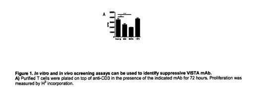
[69] Figure 1A-D. This figure shows in vitro and in vivo screening assays
which can be
used to identify suppressive VISTA nnAbs. A) Purified T cells were plated on
top of anti-CD3
in the presence of the indicated nnAb for 72 hours. Proliferation was measured
by H3
incorporation. B) Purified D011.10 T cells were stimulated by ISQ pulsed APCs
for 6 days in
the presence of the indicated antibody. Proliferation was measured through use
of CTV
dilution dye. C) GVHD was induced by transfer of C57BL/6 cells into irradiated
BALB/c
recipients. Mice were injected I.P. with 200 ug of antibody on day 0, 2 and 4
post transfer
and survival was analyzed. D) Mice were treated with 10 mot< of the indicated
antibody 3
hours prior to administration of ConA (15 nnpk) and IL-2 was analyzed in
plasma at 6 by
Lunninex.
[70] Figure 2A-F. This figure shows that agonist VISTA antibodies are
innnnunosuppressive in multiple models of autoinnnnune disease. A) NZB/W Fl
mice were
treated 3X/week with either 8G8 or Ham Ig (200 lag) starting at 25 weeks until
the end of
the experiment. "X" denotes time points where the control treated group had
all been
sacrificed. B) Mice were treated with 200 [tg of antibody 3 hours prior to
administration of
15 mg/kg (nnpk) of ConA and survival was followed for 80 hours. C) Mice were
treated
sequentially with Collagen ll nnAb followed by LPS and arthritis was measured
by measuring
for paw swelling. 8G8 and Ham-Ig were administered (200 lag) 3X every other
day. D)
Inniquinnod was applied to the ear of mice daily. At day 14, 8G8 or Ham-Ig
(200 lag) were
administered every other day and ear thickness was measured with calipers. E,
F)
Inniquinnod was applied to the backs of mice daily. At day 9, mice were
euthanized and skin
was sectioned & stained for CD3 expression by IHC.
[71] Figure 3. This figure shows the expression of VISTA in WT and hV-KI
mice. CD4+ T
cells, CD8+T cells, Tregs (CD4+ FoxP3+), and nnonocytes, CD1113+, Ly6C+, Ly6G-
were isolated
from the lymph nodes of WT and VISTA KI mice, and stained with aVISTA
antibodies against
mouse or human protein respectively.
[72] Figure 4 contains the sequences of different anti-human VISTA
antibodies
including those of INX800, INX801, and INX900-INX919.
[73] Figure 5 shows the effects of exemplary anti-human VISTA antibodies,
i.e.,
INX800 and INX801 in a ConA hepatitis model which assesses the effects thereof
on the
expression of different cytokines, chennokines and chennoattractants.
[74] Figure 6 shows the effects of exemplary anti-human VISTA antibodies,
i.e.,
INX800 and INX801 in an in vivo graft versus host disease (GVHD) animal model.
[75] Figure 7 shows the effects of exemplary agonistic anti-human VISTA
antibodies,
i.e., INX800 or INX801 on CD3-driven T cell immune responses.
21
CA 03020848 2018-10-11
WO 2017/181109
PCT/US2017/027765
[76] Figure 8 shows the effects of exemplary agonistic anti-human VISTA
antibodies,
i.e., INX800 or INX801 on the number of specific T cell populations or on
total T cell
numbers.
[77] Figure 9 compares the effects of exemplary anti-human VISTA antibodies
in ConA
assays and on the expression of select proinflannnnatory cytokines and
inflammation
markers, i.e., IL-2, y interferon and IL-12p70.
[78] Figure 10A-C: shows different IgG2 Isofornns. (A) Disulfide shuffling
leads to
isofornns A and B, along with the transition for A/B (figure from Zhang, A. et
al., 2015). (B)
Isofornns are distinguishable by RP-HPLC. (C) Observed RP-HPLC chromatogram
for INX901.
[79] Figure 11: shows chemical enrichment of IgG2 A or B isofornns. (Black
line, top)
Chromatogram shows a dominant left-most peak defining the B-form. (Red line,
bottom)
Chromatogram shows a dominant right peak defining the A-form.
[80] Figure 12: compares INX901 Fc-silent variants with respect to
disulfide shuffling.
(Top) INX901 on an IgG2 backbone exhibits an expected mixture of A, A/B, and B
isofornns.
(Middle) INX901Si on a silent IgG1 backbone exists as a single isofornn.
(Bottom) INX901HSi
possesses an IgG1 silent Fc region with a CH1/hinge from IgG2, which enables
disulfide
shuffling equivalent to native IgG2.
[81] Figure 13. Biochemically skewed INX901 forms can still reduce cytokine
production in the MLR. Supernatants from two separate MLRs were analyzed for
cytokine
production at the 72-hour time point by Lunninex analysis. INX901 parental, A
skew and B
skew all reduced the production of INFoc and IL-2 in a dose dependent fashion.
[82] Figure 14. Genetically locked INX901 forms can still reduce cytokine
production
in the MLR, but Fc silent variants cannot. Supernatants from each MLR were
analyzed for
cytokine production at the 72-hour time point by Lunninex analysis. INX901
parental, A lock
and B lock all reduced the production of INFoc and IL-2 in a dose dependent
fashion. The Si
and HSi variants, which contain mutations to silence the Fc domain, did not
consistently
suppress cytokine production.
[83] Figure 15. Genetically locked INX908 forms can still reduce cytokine
production
in the MLR, but Fc silent variants cannot. Supernatants from each MLR were
analyzed for
cytokine production at the 72-hour time point by Lunninex analysis. INX908
parental, A lock
and B lock all reduced the production of INFoc and IL-2 in a dose dependent
fashion. The Si
and HSi variants, which contain mutations to silence the Fc domain, did not
consistently
suppress cytokine production.
[84] Figure 16. This figure schematically describes the Pepscan technology
used to
identify linear and discontinuous epitopes bound by agonist anti-human VISTA
antibodies.
[85] Figure 17: This figure shows that agonist anti-human VISTA antibodies
bind to
the same core sequence.
22
CA 03020848 2018-10-11
WO 2017/181109
PCT/US2017/027765
[86] Figure 18: This figure summarizes the epitope analysis for different
anti-human
VISTA antibodies according to the invention.
[87] Figure 19: This figure shows the epitopes bound by agonist anti-human
VISTA
antibodies and further identifies important residues involved in binding.
DETAILED DESCRIPTION
[88] Unless defined otherwise, all technical and scientific terms used
herein have the
same meaning as those commonly understood by one of ordinary skill in the art
to which
this invention belongs. Although methods and materials similar or equivalent
to those
described herein may be used in the invention or testing of the present
invention, suitable
methods and materials are described herein. The materials, methods and
examples are
illustrative only, and are not intended to be limiting. The nomenclatures
utilized in
connection with, and the laboratory procedures and techniques of, analytical
chemistry,
synthetic organic chemistry, and medicinal and pharmaceutical chemistry
described herein
are those well-known and commonly used in the art. Standard techniques may be
used for
chemical syntheses, chemical analyses, pharmaceutical preparation,
formulation, and
delivery, and treatment of patients.
[89] As used in the description herein and throughout the claims that
follow, the
meaning of "a," "an," and "the" includes plural reference unless the context
clearly dictates
otherwise.
[90] "Activating receptor," as used herein, refers broadly to immune cell
receptors
that bind antigen, connplexed antigen (e.g., in the context of MHC molecules),
Ig-fusion
proteins, ligands, or antibodies. Activating receptors but are not limited to
T cell receptors
(TCRs), B cell receptors (BCRs), cytokine receptors, LPS receptors, complement
receptors,
and Fc receptors. For example, T cell receptors are present on T cells and are
associated
with CD3 molecules. T cell receptors are stimulated by antigen in the context
of MHC
molecules (as well as by polyclonal T cell activating reagents). T cell
activation via the TCR
results in numerous changes, e.g., protein phosphorylation, membrane lipid
changes, ion
fluxes, cyclic nucleotide alterations, RNA transcription changes, protein
synthesis changes,
and cell volume changes. For example, T cell receptors are present on T cells
and are
associated with CD3 molecules. T cell receptors are stimulated by antigen in
the context of
MHC molecules (as well as by polyclonal T cell activating reagents). T cell
activation via the
TCR results in numerous changes, e.g., protein phosphorylation, membrane lipid
changes,
ion fluxes, cyclic nucleotide alterations, RNA transcription changes, protein
synthesis
changes, and cell volume changes.
[91] "Adjuvant" as used herein, refers to an agent used to stimulate the
immune
system and increase the response to a vaccine, without having any specific
antigenic effect
in itself.
23
CA 03020848 2018-10-11
WO 2017/181109
PCT/US2017/027765
[92] "Agonist" herein refers to a molecule, generally an antibody or fusion
proteins
which enhances or mimics the effects of a specific molecule on immunity.
Generally in the
present application this will refer to anti-human VISTA agonist antibodies and
antibody
fragments which enhance or mimic the effects of human VISTA on immunity,
particularly
VISTA's suppressive effects on T cell immunity (CD4+ and/or CD8+ T cell
immunity), the
expression of proinflannnnatory cytokines and its effects of the expression of
specific
chennokines and chennoattractants.
[93] "Aids in the diagnosis" or "aids in the detection" of a disease herein
means that
the expression level of a particular marker polypeptide or expressed RNA is
detected alone
or in association with one or more other markers in order to assess whether a
subject has
cells characteristic of a particular disease condition or the onset of a
particular disease
condition or comprises immune dysfunction such as innnnunosuppression
characterized by
VISTA expression or abnormal immune upregulation characterized by cells having
reduced
VISTA levels, such as during autoinnnnunity, inflammation or allergic
responses, e.g., in
individuals with chronic and non-chronic diseases.
[94] "Allergic disease," as used herein, refers broadly to a disease
involving allergic
reactions. More specifically, an "allergic disease" is defined as a disease
for which an
allergen is identified, where there is a strong correlation between exposure
to that allergen
and the onset of pathological change, and where that pathological change has
been proven
to have an immunological mechanism. Herein, an immunological mechanism means
that
leukocytes show an immune response to allergen stimulation.
[95] "Amino acid," as used herein refers broadly to naturally occurring and
synthetic
amino acids, as well as amino acid analogs and amino acid nninnetics that
function in a
manner similar to the naturally occurring amino acids. Naturally occurring
amino acids are
those encoded by the genetic code, as well as those amino acids that are later
modified
(e.g., hydroxyproline, y -carboxyglutannate, and 0-phosphoserine. ) Amino acid
analogs
refers to compounds that have the same basic chemical structure as a naturally
occurring
amino acid (i. e., a carbon that is bound to a hydrogen, a carboxyl group, an
amino group),
and an R group (e.g., honnoserine, norleucine, nnethionine sulfoxide,
nnethionine methyl
sulfoniunn.) Analogs may have modified R groups (e.g., norleucine) or modified
peptide
backbones, but retain the same basic chemical structure as a naturally
occurring amino acid.
Amino acid nninnetics refers to chemical compounds that have a structure that
is different
from the general chemical structure of an amino acid, but that functions in a
manner similar
to a naturally occurring amino acid.
[96] "Anergy" or "tolerance," or "prolonged antigen-specific T cell
suppression" or
"prolonged innnnunosuppression" as used herein refers broadly to refractivity
to activating
receptor-mediated stimulation. Refractivity is generally antigen-specific and
persists after
exposure to the tolerizing antigen has ceased. For example, anergy in T cells
(as opposed to
unresponsiveness) is characterized by lack of cytokine production, e.g., IL-2.
T cell anergy
occurs when T cells are exposed to antigen and receive a first signal (a T
cell receptor or CD-
24
CA 03020848 2018-10-11
WO 2017/181109
PCT/US2017/027765
3 mediated signal) in the absence of a second signal (a costinnulatory
signal). Under these
conditions, reexposure of the cells to the same antigen (even if reexposure
occurs in the
presence of a costinnulatory molecule) results in failure to produce cytokines
and, thus,
failure to proliferate. Anergic T cells can, however, mount responses to
unrelated antigens
and can proliferate if cultured with cytokines (e.g., IL-2). For example, T
cell anergy can also
be observed by the lack of IL-2 production by T lymphocytes as measured by
ELISA or by a
proliferation assay using an indicator cell line. Alternatively, a reporter
gene construct can
be used. For example, anergic T cells fail to initiate IL-2 gene transcription
induced by a
heterologous promoter under the control of the 5' IL- 2 gene enhancer or by a
nnultinner of
the API sequence that can be found within the enhancer (Kang et al. (1992)
Science 257:
1134). Modulation of a costinnulatory signal results in modulation of effector
function of an
immune cell.
[97] "Antagonist" herein refers to a molecule, generally an antibody or
fusion
proteins which blocks or reduces the effects of a specific molecule on
immunity. Generally
in the present application this will refer to anti-human VISTA antagonist
antibodies and
antibody fragments which block or reduce the effects of human VISTA on
immunity,
particularly VISTA's suppressive effects on T cell immunity (CD4+ and/or CD8+
T cell
immunity), the expression of proinflannnnatory cytokines and VISTA's effects
of the
expression of specific chennokines and chennoattractants.
[98] "Antibody", as used herein, refers broadly to an "antigen-binding
portion" of an
antibody (also used interchangeably with "antibody portion," "antigen-binding
fragment,"
"antibody fragment"), as well as whole antibody molecules. The term "antigen-
binding
portion", as used herein, refers to one or more fragments of an antibody that
retain the
ability to specifically bind to an antigen (e.g., VISTA or specific portions
thereof)). The term
"antibody" as referred to herein includes whole polyclonal and monoclonal
antibodies and
any antigen-binding fragment (i. e., "antigen-binding portion") or single
chains thereof as
well as bispecific and nnultispecific antibodies, e.g., those that bind to
multiple antigens or
multiple antigen epitopes. An "antibody" refers to a glycoprotein comprising
at least two
heavy (H) chains and two light (L) chains interconnected by disulfide bonds,
or an antigen-
binding portion thereof. Each heavy chain is comprised of at least one heavy
chain variable
region (abbreviated herein as VH) and a heavy chain constant region. The heavy
chain
constant region is comprised of three domains, CHi, Cm and Cm- Each light
chain is
comprised of at least one light chain variable region (abbreviated herein as
VL) and a light
chain constant region. The light chain constant region is comprised of one
domain, CL-The
VH and VL regions can be further subdivided into regions of hypervariability,
termed
connplennentarity determining regions (CDRs), interspersed with regions that
are more
conserved, termed framework regions (FRs). Each VH and VL is composed of three
CDRs and
four FRs, arranged from amino-terminus to carboxy-terminus in the following
order: FR1,
CDR1, FR2, CDR2, FR3, CDR3, and FR4. The variable regions of the heavy and
light chains
contain a binding domain that interacts with an antigen. The constant regions
of the
CA 03020848 2018-10-11
WO 2017/181109
PCT/US2017/027765
antibodies may mediate the binding of the innnnunoglobulin to host tissues or
factors,
including various cells of the immune system (e.g., effector cells) and the
first component
(C1q) of the classical complement system. More generally, the term "antibody"
is intended
to include any polypeptide chain-containing molecular structure with a
specific shape that
fits to and recognizes an epitope, where one or more non-covalent binding
interactions
stabilize the complex between the molecular structure and the epitope. The
archetypal
antibody molecule is the innnnunoglobulin, and all types of innnnunoglobulins,
IgG, IgM, IgA,
IgE, IgD, etc., from all sources, e.g. human, rodent, rabbit, cow, sheep, pig,
dog, other
mammals, chicken, other avians, etc., are considered to be "antibodies."
[99] The antigen-binding function of an antibody can be performed by
fragments of a
full-length antibody. Non-limiting examples of antigen-binding fragments
encompassed
within the term "antigen-binding portion" of an antibody include (a) a Fab
fragment, a
monovalent fragment consisting of the VL, VH, CL and CHi domains; (b) a
F(a131)2 fragment, a
bivalent fragment comprising two Fab fragments linked by a disulfide bridge at
the hinge
region; (c) a Fd fragment consisting of the VH and CHi domains; (d) a Fv
fragment consisting
of the VL and VH domains of a single arm of an antibody; (e) a dAb fragment
(Ward, et al.
(1989) Nature 341 : 544-546), which consists of a VH domain; and (f) an
isolated
complementarily determining region (CDR). Furthermore, although the two
domains of the
Fv fragment, VL and VH, are coded for by separate genes, they can be joined,
using
recombinant methods, by a synthetic linker that enables them to be made as a
single
protein chain in which the VL and VH regions pair to form monovalent molecules
(known as
single chain Fv (scFv). See e.g., Bird, et al. (1988) Science 242: 423-426;
Huston, et al. (1988)
Proc Natl. Acad. Sci. USA 85: 5879-5883; and Osbourn, et al. (1998) Nat.
Biotechnol. 16: 778.
Single chain antibodies are also intended to be encompassed within the term
"antigen-
binding portion" of an antibody. Any VH and VL sequences of specific scFy can
be linked to
human innnnunoglobulin constant region cDNA or genonnic sequences, in order to
generate
expression vectors encoding complete IgG molecules or other isotypes. VH and
VL can also
be used in the generation of Fab, Fv, or other fragments of innnnunoglobulins
using either
protein chemistry or recombinant DNA technology. Other forms of single chain
antibodies,
such as diabodies are also encompassed. Diabodies are bivalent, bispecific
antibodies in
which VH and VL domains are expressed on a single polypeptide chain, but using
a linker that
is too short to allow for pairing between the two domains on the same chain,
thereby
forcing the domains to pair with complementary domains of another chain and
creating two
antigen-binding sites. See e.g. Holliger, et al. (1993) Proc Natl. Acad. Sci.
USA 90: 6444-6448;
Poljak, et al. (1994) Structure 2: 1121-1123. Still further, an antibody or
antigen-binding
portion thereof (antigen-binding fragment, antibody fragment, antibody
portion) may be
part of a larger innnnunoadhesion molecules, formed by covalent or noncovalent
association
of the antibody or antibody portion with one or more other proteins or
peptides. Examples
of innnnunoadhesion molecules include use of the streptavidin core region to
make a
tetranneric scFy molecule (Kipriyanov, et al. (1995) Hum. Antibodies
Hybridomas 6: 93-101)
and use of a cysteine residue, a marker peptide and a C-terminal polyhistidine
tag to make
26
CA 03020848 2018-10-11
WO 2017/181109
PCT/US2017/027765
bivalent and biotinylated scFy molecules. Kipriyanov, et al. (1994) Mol.
lmmunol. 31: 1047-
1058. Antibody portions, such as Fab and F(a131)2 fragments, can be prepared
from whole
antibodies using conventional techniques, such as papain or pepsin digestion,
respectively,
of whole antibodies. Moreover, antibodies, antibody portions and
innnnunoadhesion
molecules can be obtained using standard recombinant DNA techniques, as
described
herein. Antibodies may be polyclonal, monoclonal, xenogeneic, allogeneic,
syngeneic, or
modified forms thereof, e.g., humanized, chimeric, bispecific or
nnultispecific antibodies.
[100] "Antibody recognizing an antigen" and "an antibody specific for an
antigen" is
used interchangeably herein with the term "an antibody which binds
specifically to an
antigen" and refers to an innnnunoglobulin or fragment thereof that
specifically binds an
antigen.
[101] "Antigen," as used herein, refers broadly to a molecule or a portion
of a molecule
capable of being bound by an antibody which is additionally capable of
inducing an animal
to produce an antibody capable of binding to an epitope of that antigen. An
antigen may
have one epitope, or have more than one epitope. The specific reaction
referred to herein
indicates that the antigen will react, in a highly selective manner, with its
corresponding
antibody and not with the multitude of other antibodies which may be evoked by
other
antigens. In the case of a desired enhanced immune response to particular
antigens of
interest, antigens include, but are not limited to; infectious disease
antigens for which a
protective immune response may be elicited are exemplary.
[102] "Antigen presenting cell," as used herein, refers broadly to
professional antigen
presenting cells (e.g., B lymphocytes, nnonocytes, dendritic cells, and
Langerhans cells) as
well as other antigen presenting cells (e.g., keratinocytes, endothelial
cells, astrocytes,
fibroblasts, and oligodendrocytes).
[103] "Antisense nucleic acid molecule," as used herein, refers broadly to
a nucleotide
sequence which is complementary to a "sense" nucleic ac*id encoding a protein
(e.g.,
complementary to the coding strand of a double-stranded cDNA molecule)
complementary
to an nnRNA sequence or complementary to the coding strand of a gene.
Accordingly,
antisense nucleic acid molecules can hydrogen bond to sense nucleic acid
molecules.
[104] "Apoptosis," as used herein, refers broadly to programmed cell death
which can
be characterized using techniques which are known in the art. Apoptotic cell
death can be
characterized by cell shrinkage, membrane blebbing, and chromatin condensation
culminating in cell fragmentation. Cells undergoing apoptosis also display a
characteristic
pattern of internucleosonnal DNA cleavage.
[105] "Autoinnnnunity" or "autoinnnnune disease or condition," as used
herein, refers
broadly to a disease or disorder arising from and directed against an
individual's own tissues
or a co-segregate or manifestation thereof or resulting condition therefrom,
and includes.
Herein autoinnnnune conditions include inflammatory or allergic conditions,
e.g., chronic
27
CA 03020848 2018-10-11
WO 2017/181109
PCT/US2017/027765
diseases characterized by a host immune reaction against self-antigens
potentially
associated with tissue destruction such as rheumatoid arthritis.
[106] "B cell receptor" (BCR)," as used herein, refers broadly to the
complex between
membrane Ig (nnIg) and other transnnennbrane polypeptides (e.g., IgA. and Ig )
found on B
cells. The signal transduction function of nnlg is triggered by crosslinking
of receptor
molecules by oligonneric or nnultinneric antigens. B cells can also be
activated by anti-
innnnunoglobulin antibodies. Upon BCR activation, numerous changes occur in B
cells,
including tyrosine phosphorylation.
[107] "Cancer," as used herein, refers broadly to any neoplastic disease
(whether
invasive or metastatic) characterized by abnormal and uncontrolled cell
division causing
malignant growth or tumor (e.g., unregulated cell growth.) The term "cancer"
or
"cancerous" as used herein should be understood to encompass any neoplastic
disease
(whether invasive, non-invasive or metastatic) which is characterized by
abnormal and
uncontrolled cell division causing malignant growth or tumor, non-limiting
examples of
which are described herein. This includes any physiological condition in
mammals that is
typically characterized by unregulated cell growth. Examples of cancer are
exemplified in
the working examples. Further cancers include but are not limited to,
carcinoma,
lymphoma, blastonna, sarcoma, and leukemia. More particular examples of such
cancers
include squannous cell cancer, lung cancer (including small-cell lung cancer,
non-small cell
lung cancer, adenocarcinonna of the lung, and squannous carcinoma of the
lung), cancer of
the peritoneum, hepatocellular cancer, gastric or stomach cancer (including
gastrointestinal
cancer), pancreatic cancer, glioblastonna, cervical cancer, ovarian cancer,
liver cancer,
bladder cancer, hepatonna, breast cancer, colon cancer, colorectal cancer,
endonnetrial or
uterine carcinoma, salivary gland carcinoma, kidney or renal cancer, liver
cancer, prostate
cancer, vulval cancer, thyroid cancer, hepatic carcinoma and various types of
head and neck
cancer, as well as B-cell lymphoma (including low grade/follicular non-
Hodgkin's lymphoma
(NHL); small lynnphocytic (SL) NHL; intermediate grade/follicular NHL;
intermediate grade
diffuse NHL; high grade innnnunoblastic NHL; high grade lynnphoblastic NHL;
high grade small
non-cleaved cell NHL; bulky disease NHL; mantle cell lymphoma; AIDS-related
lymphoma;
and Waldenstronn's Macroglobulinennia); chronic lynnphocytic leukemia (CLL);
acute
lynnphoblastic leukemia (ALL); Hairy cell leukemia; chronic nnyeloblasts
leukemia; multiple
nnyelonna and post-transplant lynnphoproliferative disorder (PTLD). Other
cancers amenable
for treatment by the present invention include, but are not limited to,
carcinoma,
lymphoma, blastonna, sarcoma, and leukemia or lymphoid malignancies. More
particular
examples of such cancers include colorectal, bladder, ovarian, melanoma,
squannous cell
cancer, lung cancer (including small-cell lung cancer, non-small cell lung
cancer,
adenocarcinonna of the lung, and squannous carcinoma of the lung), cancer of
the
peritoneum, hepatocellular cancer, gastric or stomach cancer (including
gastrointestinal
cancer), pancreatic cancer, glioblastonna, cervical cancer, ovarian cancer,
liver cancer,
bladder cancer, hepatonna, breast cancer, colon cancer, colorectal cancer,
endonnetrial or
28
CA 03020848 2018-10-11
WO 2017/181109
PCT/US2017/027765
uterine carcinoma, salivary gland carcinoma, kidney or renal cancer, liver
cancer, prostate
cancer, vulval cancer, thyroid cancer, hepatic carcinoma and various types of
head and neck
cancer, as well as B-cell lymphoma (including low grade/follicular non-
Hodgkin's lymphoma
(NHL); small lynnphocytic (SL) NHL; intermediate grade/follicular NHL;
intermediate grade
diffuse NHL; high grade innnnunoblastic NHL; high grade lynnphoblastic NHL;
high grade small
non-cleaved cell NHL; bulky disease NHL; mantle cell lymphoma; AIDS-related
lymphoma;
and Waldenstronn's Macroglobulinennia); chronic lynnphocytic leukemia (CLL);
acute
lynnphoblastic leukemia (ALL); Hairy cell leukemia; chronic nnyeloblastic
leukemia; and post-
transplant lynnphoproliferative disorder (PTLD), as well as abnormal vascular
proliferation
associated with phakonnatoses, edema (such as that associated with brain
tumors), and
Meigs' syndrome. Preferably, the cancer is selected from the group consisting
of colorectal
cancer, breast cancer, colorectal cancer, rectal cancer, non-small cell lung
cancer, non-
Hodgkin's lymphoma (NHL), renal cell cancer, prostate cancer, liver cancer,
pancreatic
cancer, soft-tissue sarcoma, Kaposi's sarcoma, carcinoid carcinoma, head and
neck cancer,
melanoma, ovarian cancer, nnesothelionna, and multiple nnyelonna. In an
exemplary
embodiment the cancer is an early or advanced (including metastatic) bladder,
ovarian or
melanoma. In another embodiment the cancer is colorectal cancer. The cancerous
conditions amenable for treatment of the invention include cancers that
express or do not
express VISTA and further include non-metastatic or non-invasive as well as
invasive or
metastatic cancers wherein VISTA expression by immune, stronnal or diseased
cells suppress
antitumor responses and anti-invasive immune responses. The method of the
present
invention is particularly suitable for the treatment of vascularized tumors.
Cancers according
to the invention include cancers that express or do not express VISTA and
further include
non-metastatic or non-invasive as well as invasive or metastatic cancers
wherein VISTA
expression by immune, stronnal or diseased cells suppress antitumor responses
and anti-
invasive immune responses, and those characterized by vascularized tumors.
[108] "Chimeric antibody," as used herein, refers broadly to an antibody
molecule in
which the constant region, or a portion thereof, is altered, replaced or
exchanged so that
the antigen-binding site (variable region) is linked to a constant region of a
different or
altered class, effector function and/or species, or an entirely different
molecule which
confers new properties to the chimeric antibody, e.g., an enzyme, toxin,
hormone, growth
factor, drug, the variable region or a portion thereof, is altered, replaced
or exchanged with
a variable region having a different or altered antigen specificity.
[109] "Coding region," as used herein, refers broadly to regions of a
nucleotide
sequence comprising codons which are translated into amino acid residues,
whereas the
term "noncoding region" refers to regions of a nucleotide sequence that are
not translated
into amino acids (e.g., 5' and 3' untranslated regions).
[110] "Conservatively modified variants," as used herein, applies to both
amino acid
and nucleic acid sequences, and with respect to particular nucleic acid
sequences, refers
broadly to conservatively modified variants refers to those nucleic acids
which encode
29
CA 03020848 2018-10-11
WO 2017/181109
PCT/US2017/027765
identical or essentially identical amino acid sequences, or where the nucleic
acid does not
encode an amino acid sequence, to essentially identical sequences. Because of
the
degeneracy of the genetic code, a large number of functionally identical
nucleic acids
encode any given protein. "Silent variations" are one species of
conservatively modified
nucleic acid variations. Every nucleic acid sequence herein which encodes a
polypeptide also
describes every possible silent variation of the nucleic acid. One of skill
will recognize that
each codon in a nucleic acid (except AUG, which is ordinarily the only codon
for nnethionine,
and TGG, which is ordinarily the only codon for tryptophan) may be modified to
yield a
functionally identical molecule.
[111] "Connplennentarity determining region," "hypervariable region," or
"CDR," as
used herein, refers broadly to one or more of the hyper-variable or
complementarily
determining regions (CDRs) found in the variable regions of light or heavy
chains of an
antibody. See Kabat, et al. (1987) Sequences of Proteins of Immunological
Interest National
Institutes of Health, Bethesda, Md. These expressions include the
hypervariable regions as
defined by Kabat, et al. (1983) Sequences of Proteins of Immunological
Interest, U. S. Dept.
of Health and Human Services or the hypervariable loops in 3-dimensional
structures of
antibodies. Chothia and Lesk (1987)J. Mol. Biol. 196: 901-917. The CDRs in
each chain are
held in close proximity by framework regions and, with the CDRs from the other
chain,
contribute to the formation of the antigen-binding site. Within the CDRs there
are select
amino acids that have been described as the selectivity determining regions
(SDRs) which
represent the critical contact residues used by the CDR in the antibody-
antigen interaction.
(Kashmir' Methods 36: 25-34(2005)).
[112] "Control amount," as used herein, refers broadly to a marker can be
any amount
or a range of amounts to be compared against a test amount of a marker. For
example, a
control amount of a marker may be the amount of a marker in a patient with a
particular
disease or condition or a person without such a disease or condition. A
control amount can
be either in absolute amount (e.g., microgram/ml) or a relative amount (e.g.,
relative
intensity of signals).
[113] "Costinnulatory receptor," as used herein, refers broadly to
receptors which
transmit a costinnulatory signal to an immune cell, e.g., CD28 or !COS. As
used herein, the
term "inhibitory receptors" includes receptors which transmit a negative
signal to an
immune cell, e.g., a T cell or an NK cell.
[114] "Costinnulate," as used herein, refers broadly to the ability of a
costinnulatory
molecule to provide a second, non-activating, receptor-mediated signal (a
"costinnulatory
signal") that induces proliferation or effector function. For example, a
costinnulatory signal
can result in cytokine secretion (e.g., in a T cell that has received a T cell-
receptor-mediated
signal) Immune cells that have received a cell receptor-mediated signal (e.g.,
via an
activating receptor) may be referred to herein as "activated immune cells."
With respect to
T cells, transmission of a costinnulatory signal to a T cell involves a
signaling pathway that is
not inhibited by cyclosporin A. In addition, a costinnulatory signal can
induce cytokine
CA 03020848 2018-10-11
WO 2017/181109
PCT/US2017/027765
secretion (e.g., IL-2 and/or IL-10) in a T cell and/or can prevent the
induction of
unresponsiveness to antigen, the induction of anergy, or the induction of cell
death in the T
cell.
[115] "Costinnulatory polypeptide" or "costinnulatory molecule" herein
refers to a
polypeptide that, upon interaction with a cell-surface molecule on T cells,
modulates T cell
responses.
[116] "Costinnulatory signaling" as used herein is the signaling activity
resulting from
the interaction between costinnulatory polypeptides on antigen presenting
cells and their
receptors on T cells during antigen-specific T cell responses. Without wishing
to be limited
by a single hypothesis, the antigen-specific T cell response is believed to be
mediated by two
signals: 1) engagement of the T cell Receptor (TCR) with antigenic peptide
presented in the
context of MHC (signal 1), and 2) a second antigen-independent signal
delivered by contact
between different costinnulatory receptor/ligand pairs (signal 2). Without
wishing to be
limited by a single hypothesis, this "second signal" is critical in
determining the type of T cell
response (activation vs inhibition) as well as the strength and duration of
that response, and
is regulated by both positive and negative signals from costinnulatory
molecules, such as the
B7 family of proteins.
[117] "B7" polypeptide herein means a member of the B7 family of proteins
that
costinnulate T cells including, but not limited to B7-1, B7-2, B7-DC, B7-H5,
B7-HI, B7-H2, B7-
H3, B7-H4, B7-H6, B7-S3 and biologically active fragments and/or variants
thereof.
Representative biologically active fragments include the extracellular domain
or fragments
of the extracellular domain that costinnulate T cells.
[118] "Cytoplasmic domain," as used herein, refers broadly to the portion
of a protein
which extends into the cytoplasm of a cell.
[119] "Diagnostic," as used herein, refers broadly to identifying the
presence or nature
of a pathologic condition. Diagnostic methods differ in their sensitivity and
specificity. The
"sensitivity" of a diagnostic assay is the percentage of diseased individuals
who test positive
(percent of "true positives"). Diseased individuals not detected by the assay
are "false
negatives." Subjects who are not diseased and who test negative in the assay
are termed
"true negatives." The "specificity" of a diagnostic assay is 1 minus the false
positive rate,
where the "false positive" rate is defined as the proportion of those without
the disease
who test positive. While a particular diagnostic method may not provide a
definitive
diagnosis of a condition, it suffices if the method provides a positive
indication that aids in
diagnosis.
[120] "Diagnosing," or "aiding in the diagnosis" as used herein refers
broadly to
classifying a disease or a symptom, and/or determining the likelihood that an
individual has
a disease condition (e.g., based on absence or presence of VISTA expression,
and/or
increased or decreased expression by immune, stronnal and/or putative diseased
cells);
determining a severity of the disease, monitoring disease progression,
forecasting an
31
CA 03020848 2018-10-11
WO 2017/181109
PCT/US2017/027765
outcome of a disease and/or prospects of recovery. The term "detecting" may
also
optionally encompass any of the foregoing. Diagnosis of a disease according to
the present
invention may, in some embodiments, be affected by determining a level of a
polynucleotide or a polypeptide of the present invention in a biological
sample obtained
from the subject, wherein the level determined can be correlated with
predisposition to, or
presence or absence of the disease. It should be noted that a "biological
sample obtained
from the subject" may also optionally comprise a sample that has not been
physically
removed from the subject.
[121] "Effective amount," as used herein, refers broadly to the amount of a
compound, antibody, antigen, or cells that, when administered to a patient for
treating a
disease, is sufficient to effect such treatment for the disease. The effective
amount may be
an amount effective for prophylaxis, and/or an amount effective for
prevention. The
effective amount may be an amount effective to reduce, an amount effective to
prevent the
incidence of signs/symptoms, to reduce the severity of the incidence of
signs/symptoms, to
eliminate the incidence of signs/symptoms, to slow the development of the
incidence of
signs/symptoms, to prevent the development of the incidence of signs/symptoms,
and/or
effect prophylaxis of the incidence of signs/symptoms. The "effective amount"
may vary
depending on the disease and its severity and the age, weight, medical
history,
susceptibility, and pre-existing conditions, of the patient to be treated. The
term "effective
amount" is synonymous with "therapeutically effective amount" for purposes of
this
invention.
[122] "Extracellular domain" or "ECD" as used herein refers broadly to the
portion of a
protein that extends from the surface of a cell.
[123] "Expression vector," as used herein, refers broadly to any
recombinant
expression system for the purpose of expressing a nucleic acid sequence of the
invention in
vitro or in vivo, constitutively or inducibly, in any cell, including
prokaryotic, yeast, fungal,
plant, insect or mammalian cell. The term includes linear or circular
expression systems. The
term includes expression systems that remain episonnal or integrate into the
host cell
genonne. The expression systems can have the ability to self -replicate or
not, i. e., drive only
transient expression in a cell. The term includes recombinant expression
cassettes which
contain only the minimum elements needed for transcription of the recombinant
nucleic
acid.
[124] "Family," as used herein, refers broadly to the polypeptide and
nucleic acid
molecules of the invention is intended to mean two or more polypeptide or
nucleic acid
molecules having a common structural domain or motif and having sufficient
amino acid or
nucleotide sequence homology as defined herein. Family members can be
naturally or non-
naturally occurring and can be from either the same or different species. For
example, a
family can contain a first polypeptide of human origin, as well as other,
distinct polypeptides
of human origin or alternatively, can contain homologues of non-human origin
(e.g.,
32
CA 03020848 2018-10-11
WO 2017/181109
PCT/US2017/027765
monkey polypeptides.) Members of a family may also have common functional
characteristics.
[125] "Fc receptor" (FcRs) as used herein, refers broadly to cell surface
receptors for
the Fc portion of innnnunoglobulin molecules (Igs). Fc receptors are found on
many cells
which participate in immune responses. Among the human FcRs that have been
identified
so far are those which recognize IgG (designated FcyR), IgE (FceRI), IgA
(FcaR), and
polymerized IgM/A (Fce R). FcRs are found in the following cell types: FceRI
(mast cells),
FceRII (many leukocytes), FcaR (neutrophils), and Fc R (glandular epithelium,
hepatocytes).
(Hogg Innnnunol. Today 9: 185-86 (1988)). The widely studied FcyRs are central
in cellular
immune defenses, and are responsible for stimulating the release of mediators
of
inflammation and hydrolytic enzymes involved in the pathogenesis of
autoinnnnune disease.
(Unkeless, Annu. Rev. Innnnunol. 6: 251-87 (1988)). The FcyRs provide a
crucial link between
effector cells and the lymphocytes that secrete Ig, since the
nnacrophage/nnonocyte,
polynnorphonuclear leukocyte, and natural killer (NK) cell FcyRs confer an
element of
specific recognition mediated by IgG. Human leukocytes have at least three
different types
of FcyRs for IgG: hFcyRI(CD64) (found on nnonocytes/nnacrophages), hFcyRIIA or
hFcyRIIB,
(CD32 or CD32A) (found on nnonocytes, neutrophils, eosinophils, platelets,
possibly B cells,
and the K562 cell line) and FcyRIIIA (CD16A) or FcyRIIIB (CD16B) (found on NK
cells,
neutrophils, eosinophils, and macrophages).
[126] "Framework region" or "FR," as used herein refers broadly to one or
more of the
framework regions within the variable regions of the light and heavy chains of
an antibody.
See Kabat, et al. Sequences of Proteins of Immunological Interest National
Institutes of
Health, Bethesda, Md (1987). These expressions include those amino acid
sequence regions
interposed between the CDRs within the variable regions of the light and heavy
chains of an
antibody.
[127] "Heterologous," as used herein, refers broadly to portions of a
nucleic acid
indicates that the nucleic acid comprises two or more subsequences that are
not found in
the same relationship to each other in nature. For instance, the nucleic acid
is typically
reconnbinantly produced, having two or more sequences from unrelated genes
arranged to
make a new functional nucleic acid (e.g., a promoter from one source and a
coding region
from another source.) Similarly, a heterologous protein indicates that the
protein comprises
two or more subsequences that are not found in the same relationship to each
other in
nature (e.g., a fusion protein).
[128] "High affinity," as used herein, refers broadly to an antibody or
fusion protein
having a KD of at least 10-6 M, more preferably 10-2 M, even more preferably
at least 10-8 M
and even more preferably at least 10-9 M, 1049 M, 1043. M, or 10-12 M for a
target antigen or
receptor. "High affinity" for an IgG antibody or fusion protein herein refers
to an antibody
having a KD of 10-6M or less, more preferably 10-2 M or less, preferably 10-8M
or less, more
preferably 10-9 M or less and even more preferably 1010
M, 10 -11 M, or 10-12 M or less for a
target antigen or receptor. With particular respect to antibodies, "high
affinity" binding can
33
CA 03020848 2018-10-11
WO 2017/181109
PCT/US2017/027765
vary for different antibody isotypes. For example, "high affinity" binding for
an IgM isotype
refers to an antibody having a KD of 10-7M or less, more preferably 10-8 M or
less.
[129] "Homology," as used herein, refers broadly to a degree of similarity
between a
nucleic acid sequence and a reference nucleic acid sequence or between a
polypeptide
sequence and a reference polypeptide sequence. Homology may be partial or
complete.
Complete homology indicates that the nucleic acid or amino acid sequences are
identical. A
partially homologous nucleic acid or amino acid sequence is one that is not
identical to the
reference nucleic acid or amino acid sequence. The degree of homology can be
determined
by sequence comparison, for example using BlastP software of the National
Center of
Biotechnology Information (NCB!) using default parameters. The term "sequence
identity"
may be used interchangeably with "homology."
[130] "Host cell," as used herein, refers broadly to refer to a cell into
which a nucleic
acid molecule of the invention, such as a recombinant expression vector of the
invention,
has been introduced. Host cells may be prokaryotic cells (e.g., E. coli), or
eukaryotic cells
such as yeast, insect (e.g., SF9), amphibian, or mammalian cells such as CHO,
HeLa, HEK-293,
e.g., cultured cells, explants, and cells in vivo. The terms "host cell" and
"recombinant host
cell" are used interchangeably herein. It should be understood that such terms
refer not
only to the particular subject cell but to the progeny or potential progeny of
such a cell.
Because certain modifications may occur in succeeding generations due to
either mutation
or environmental influences, progeny may not, in fact, be identical to the
parent cell, but
are still included within the scope of the term as used herein.
[131] "Human monoclonal antibody" refers to antibodies displaying a single
binding
specificity which have variable regions in which both the framework and CDR
regions are
derived from human gernnline innnnunoglobulin sequences. In one embodiment,
the human
monoclonal antibodies are produced by a hybridonna which includes a B cell
obtained from a
transgenic nonhuman animal, e.g., a transgenic mouse, having a genonne
comprising a
human heavy chain transgene and a light chain transgene fused to an
immortalized cell. This
includes fully human monoclonal antibodies and conjugates and variants
thereof, e.g.,
which are bound to effector agents such as therapeutics or diagnostic agents.
[132] "Humanized antibody," as used herein, refers broadly to include
antibodies made
by a non-human cell having variable and constant regions which have been
altered to more
closely resemble antibodies that would be made by a human cell. For example,
by altering
the non-human antibody amino acid sequence to incorporate amino acids found in
human
gernnline innnnunoglobulin sequences. The humanized antibodies of the
invention may
include amino acid residues not encoded by human gernnline innnnunoglobulin
sequences
(e.g., mutations introduced by random or site-specific nnutagenesis in vitro
or by somatic
mutation in vivo), for example in the CDRs. The term "humanized antibody", as
used herein,
also includes antibodies in which CDR sequences derived from the gernnline of
another
mammalian species, such as a mouse, have been grafted onto human framework
sequences.
34
CA 03020848 2018-10-11
WO 2017/181109
PCT/US2017/027765
[133] "Hybridization," as used herein, refers broadly to the physical
interaction of
complementary (including partially complementary) polynucleotide strands by
the
formation of hydrogen bonds between complementary nucleotides when the strands
are
arranged antiparallel to each other.
[134] "IgV domain" and "IgC domain" as used herein, refer broadly to Ig
superfannily
member domains. These domains correspond to structural units that have
distinct folding
patterns called Ig folds. Ig folds are comprised of a sandwich of two 13
sheets, each consisting
of antiparallel 13 strands of 5-10 amino acids with a conserved disulfide bond
between the
two sheets in most, but not all, domains. IgC domains of Ig, TCR, and MHC
molecules share
the same types of sequence patterns and are called the Cl set within the Ig
superfannily.
Other IgC domains fall within other sets. IgV domains also share sequence
patter and are
called V set domains. IgV domains are longer than C-domains and form an
additional pair of
13 strands.
[135] "Immune cell," as used herein, refers broadly to cells that are of
hennatopoietic
origin and that play a role in the immune response. Immune cells include but
are not limited
to lymphocytes, such as B cells and T cells; natural killer cells; dendritic
cells, and myeloid
cells, such as nnonocytes, macrophages, eosinophils, mast cells, basophils,
and granulocytes.
[136] "Immunoassay," as used herein, refers broadly to an assay that uses
an antibody
to specifically bind an antigen. The immunoassay may be characterized by the
use of specific
binding properties of a particular antibody to isolate, target, and/or
quantify the antigen.
[137] "Immune related disease (or disorder or condition)" as used herein
should be
understood to encompass any disease disorder or condition selected from the
group
including but not limited to autoinnnnune diseases, inflammatory disorders and
immune
disorders associated with graft transplantation rejection, such as acute and
chronic rejection
of organ transplantation, allogenic stem cell transplantation, autologous stem
cell
transplantation, bone marrow transplantation, and graft versus host disease.
[138] "Immune response," as used herein, refers broadly to T cell-mediated
and/or B
cell-mediated immune responses that are influenced by modulation of T cell
costinnulation.
Exemplary immune responses include B cell responses (e.g., antibody
production) T cell
responses (e.g., cytokine production, and cellular cytotoxicity) and
activation of cytokine
responsive cells, e.g., macrophages. As used herein, the term
"downnnodulation" with
reference to the immune response includes a diminution in any one or more
immune
responses, while the term "upnnodulation" with reference to the immune
response includes
an increase in any one or more immune responses. It will be understood that
upnnodulation
of one type of immune response may lead to a corresponding downnnodulation in
another
type of immune response. For example, upnnodulation of the production of
certain
cytokines (e.g., IL-10) can lead to downnnodulation of cellular immune
responses.
[139] "Immunologic", "immunological" or "immune" response herein refer to
the
development of a hunnoral (antibody mediated) and/or a cellular (mediated by
antigen-
CA 03020848 2018-10-11
WO 2017/181109
PCT/US2017/027765
specific T cells or their secretion products) response directed against a
peptide in a recipient
patient. Such a response can be an active response induced by administration
of
innnnunogen or a passive response induced by administration of antibody or
primed 1-cells.
Without wishing to be limited by a single hypothesis, a cellular immune
response is elicited
by the presentation of polypeptide epitopes in association with Class ll or
Class I MHC
molecules to activate antigen-specific CD4 -1 helper cells and/or CD8+
cytotoxic T cells,
respectively. The response may also involve activation of nnonocytes,
macrophages, NK cells,
basophils, dendritic cells, astrocytes, nnicroglia cells, eosinophils,
activation or recruitment
of neutrophils or other components of innate immunity. The presence of a cell-
mediated
immunological response can be determined by proliferation assays (CD4 T cells)
or CTL
(cytotoxic T lymphocyte) assays. The relative contributions of hunnoral and
cellular
responses to the protective or therapeutic effect of an innnnunogen can be
distinguished by
separately isolating antibodies and T cells from an immunized syngeneic animal
and
measuring protective or therapeutic effect in a second subject.
[140] "Immunogenic agent" or "innnnunogen" is a moiety capable of inducing
an
immunological response against itself on administration to a mammal,
optionally in
conjunction with an adjuvant.
[141] "Inflammatory disorders", "inflammatory conditions" and/or
"inflammation",
used interchangeably herein, refers broadly to chronic or acute inflammatory
diseases, and
expressly includes inflammatory autoinnnnune diseases and inflammatory
allergic conditions.
These conditions include by way of example inflammatory abnormalities
characterized by
dysregulated immune response to harmful stimuli, such as pathogens, damaged
cells, or
irritants. Inflammatory disorders underlie a vast variety of human diseases.
Non-immune
diseases with etiological origins in inflammatory processes include cancer,
atherosclerosis,
and ischennic heart disease. Examples of disorders associated with
inflammation include:
Chronic prostatitis, Glonnerulonephritis, Hypersensitivities, Pelvic
inflammatory disease,
Reperfusion injury, Sarcoidosis, Vasculitis, Interstitial cystitis,
nornnoconnplennentennic
urticarial vasculitis, pericarditis, nnyositis, anti-synthetase syndrome,
scleritis, macrophage
activation syndrome, Behget's Syndrome, PAPA Syndrome, Blau's Syndrome, gout,
adult and
juvenile Still's disease, cryropyrinopathy, Muckle- Wells syndrome, familial
cold-induced
auto-inflammatory syndrome, neonatal onset nnultisystennic inflammatory
disease, familial
Mediterranean fever, chronic infantile neurologic, cutaneous and articular
syndrome,
systemic juvenile idiopathic arthritis, Hyper IgD syndrome, Schnitzler's
syndrome, TNF
receptor-associated periodic syndrome (TRAPSP), gingivitis, periodontitis,
hepatitis,
cirrhosis, pancreatitis, nnyocarditis, vasculitis, gastritis, gout, gouty
arthritis, and
inflammatory skin disorders, selected from the group consisting of psoriasis,
atopic
dermatitis, eczema, rosacea, urticaria, and acne.
[142] "Inhibitory signal," as used herein, refers broadly to a signal
transmitted via an
inhibitory receptor molecule on an immune cell. A signal antagonizes a signal
via an
activating receptor (e.g., via a TCR, CD3, BCR, or Fc molecule) and can
result, e.g., in
36
CA 03020848 2018-10-11
WO 2017/181109
PCT/US2017/027765
inhibition of: second messenger generation; proliferation; or effector
function in the
immune cell, e.g., reduced phagocytosis, antibody production, or cellular
cytotoxicity, or the
failure of the immune cell to produce mediators (e.g., cytokines (e.g., IL-2)
and/or mediators
of allergic responses); or the development of anergy.
[143] "Isolated," as used herein, refers broadly to material removed from
its original
environment in which it naturally occurs, and thus is altered by the hand of
man from its
natural environment and includes "recombinant" polypeptides. Isolated material
may be,
for example, exogenous nucleic acid included in a vector system, exogenous
nucleic acid
contained within a host cell, or any material which has been removed from its
original
environment and thus altered by the hand of man (e.g., "isolated antibody").
For example,
"isolated" or "purified," as used herein, refers broadly to a protein, DNA,
antibody, RNA, or
biologically active portion thereof, that is substantially free of cellular
material or other
contaminating proteins from the cell or tissue source from which the
biological substance is
derived, or substantially free from chemical precursors or other chemicals
when chemically
synthesized. As used herein the term "isolated" refers to a compound of
interest (for
example a polynucleotide or a polypeptide) that is in an environment different
from that in
which the compound naturally occurs e.g., separated from its natural milieu
such as by
concentrating a peptide to a concentration at which it is not found in nature.
"Isolated"
includes compounds that are within samples that are substantially enriched for
the
compound of interest and/or in which the compound of interest is partially or
substantially
purified.
[144] "Isolated antibody", as used herein, is intended to refer to an
antibody that is
substantially free of other antibodies having different antigenic
specificities (e.g., an isolated
antibody that specifically binds VISTA) is substantially free of antibodies
that specifically
bind antigens other than VISTA). Moreover, an isolated antibody may be
substantially free
of other cellular material and/or chemicals.
[145] "Isotype" herein refers to the antibody class (e.g., IgM or IgG1)
that is encoded
by the heavy chain constant region genes.
[146] "K-assoc" or "Ka", as used herein, refers broadly to the association
rate of a
particular antibody-antigen interaction, whereas the term "Kdiss" or "Kd," as
used herein,
refers to the dissociation rate of a particular antibody-antigen interaction.
[147] The term "KID", as used herein, is intended to refer to the
dissociation constant,
which is obtained from the ratio of Kd to Ka (i. e., Kd/Ka) and is expressed
as a molar
concentration (M). KD values for antibodies can be determined using methods
well
established in the art such as plasnnon resonance (BlAcore ), ELISA and
KINEXA. A preferred
method for determining the KD of an antibody is by using surface Plasnnon
resonance,
preferably using a biosensor system such as a BlAcore system or by ELISA.
Typically these
methods are effected at 25 or 37 C. Antibodies for therapeutic usage
generally will possess
37
CA 03020848 2018-10-11
WO 2017/181109
PCT/US2017/027765
a KD when determined by surface Plasnnon resonance of 50 nM or less or more
typically 1nM
or less at 25 or 37 C.
[148] "Label" or a "detectable moiety" as used herein, refers broadly to a
composition
detectable by spectroscopic, photochemical, biochemical, innnnunochennical,
chemical, or
other physical means.
[149] "Low stringency," "medium stringency," "high stringency," or "very
high
stringency conditions," as used herein, refers broadly to conditions for
nucleic acid
hybridization and washing. Guidance for performing hybridization reactions can
be found in
Ausubel, et al., Short Protocols in Molecular Biology (5th Ed.) John Wiley &
Sons, NY (2002).
Exemplary specific hybridization conditions include but are not limited to:
(1) low stringency
hybridization conditions in 6 X sodium chloride/sodium citrate (SSC) at about
45 C, followed
by two washes in 0. 2XSSC, 0.1% SDS at least at 50 C. (the temperature of the
washes can be
increased to 55 'C. for low stringency conditions); (2) medium stringency
hybridization
conditions in 6XSSC at about 45 C. , followed by one or more washes in 0. 2X
SSC, 0. 1% SDS
at 60 C. ; (3) high stringency hybridization conditions in 6XSSC at about 45 C
followed by
one or more washes in 0. 2X. SSC, 0. 1% SDS at 65 C.; and (4) very high
stringency
hybridization conditions are 0. 5M sodium phosphate, 7% SDS at 65 C, followed
by one or
more washes at 0.2XSSC, and 1 % SDS at 65 C.
[150] "Mammal," as used herein, refers broadly to any and all warm-blooded
vertebrate animals of the class Mannnnalia, including humans, characterized by
a covering of
hair on the skin and, in the female, milk-producing mammary glands for
nourishing the
young. Examples of mammals include but are not limited to alpacas, armadillos,
capybaras,
cats, camels, chimpanzees, chinchillas, cattle, dogs, goats, gorillas,
hamsters, horses,
humans, lemurs, llamas, mice, non-human primates, pigs, rats, sheep, shrews,
squirrels,
tapirs, and voles. Mammals include but are not limited to bovine, canine,
equine, feline,
nnurine, ovine, porcine, primate, and rodent species. Mammal also includes any
and all
those listed on the Mammal Species of the World maintained by the National
Museum of
Natural History, Smithsonian Institution in Washington D. C.
[151] "Multispecific antibody" refers to an antibody with 2 or more antigen
binding
regions. This includes bispecific antibodies. These antigen binding regions
may bind to
different antigens or to different epitopes of the same antigen.
[152] "Naturally-occurring nucleic acid molecule," as used herein, refers
broadly refers
to an RNA or DNA molecule having a nucleotide sequence that occurs in nature
(e.g.,
encodes a natural protein).
[153] "Nucleic acid" or "nucleic acid sequence," as used herein, refers
broadly to a
deoxy-ribonucleotide or ribonucleotide oligonucleotide in either single- or
double-stranded
form. The term encompasses nucleic acids, i.e., oligonucleotides, containing
known analogs
of natural nucleotides. The term also encompasses nucleic- acid-like
structures with
synthetic backbones. Unless otherwise indicated, a particular nucleic acid
sequence also
38
CA 03020848 2018-10-11
WO 2017/181109
PCT/US2017/027765
implicitly encompasses conservatively modified variants thereof (e.g.,
degenerate codon
substitutions) and complementary sequences, as well as the sequence explicitly
indicated.
The term nucleic acid is used interchangeably with gene, cDNA, nnRNA,
oligonucleotide, and
polynucleotide.
[154] "Operatively linked", as used herein, refers broadly to when two DNA
fragments
are joined such that the amino acid sequences encoded by the two DNA fragments
remain
in-frame.
[155] "Paratope," as used herein, refers broadly to the part of an antibody
which
recognizes an antigen (e.g., the antigen-binding site of an antibody.)
Paratopes may be a
small region (e.g., 15-22 amino acids) of the antibody's Fv region and may
contain parts of
the antibody's heavy and light chains. See Goldsby, et al. Antigens (Chapter
3) Immunology
(5th Ed.) New York: W. H. Freeman and Company, pages 57-75.
[156] "Patient," or "subject" or "recipient", "individual", or "treated
individual" are
used interchangeably herein, and refers broadly to any animal that is in need
of treatment
either to alleviate a disease state or to prevent the occurrence or
reoccurrence of a disease
state. Also, "Patient" as used herein, refers broadly to any animal that has
risk factors, a
history of disease, susceptibility, symptoms, and signs, was previously
diagnosed, is at risk
for, or is a member of a patient population for a disease. The patient may be
a clinical
patient such as a human or a veterinary patient such as a companion,
domesticated,
livestock, exotic, or zoo animal.
[157] "Polypeptide," "peptide" and "protein," are used interchangeably and
refer
broadly to a polymer of amino acid residues s of any length, regardless of
modification (e.g.,
phosphorylation or glycosylation). The terms apply to amino acid polymers in
which one or
more amino acid residue is an analog or mimetic of a corresponding naturally
occurring
amino acid, as well as to naturally occurring amino acid polymers. The terms
apply to amino
acid polymers in which one or more amino acid residue is an artificial
chemical mimetic of a
corresponding naturally occurring amino acid, as well as to naturally
occurring amino acid
polymers and non-naturally occurring amino acid polymer. Polypeptides can be
modified,
e.g., by the addition of carbohydrate residues to form glycoproteins. The
terms
"polypeptide," "peptide" and "protein" expressly include glycoproteins, as
well as non-
glycoproteins.
[158] "Promoter," as used herein, refers broadly to an array of nucleic
acid sequences
that direct transcription of a nucleic acid. As used herein, a promoter
includes necessary
nucleic acid sequences near the start site of transcription, such as, in the
case of a
polynnerase ll type promoter, a TATA element. A promoter also optionally
includes distal
enhancer or repressor elements, which can be located as much as several
thousand base
pairs from the start site of transcription. A constitutive" promoter is a
promoter that is
active under most environmental and developmental conditions. An "inducible"
promoter is
a promoter that is active under environmental or developmental regulation.
39
CA 03020848 2018-10-11
WO 2017/181109
PCT/US2017/027765
[159] "Prophylactically effective amount," as used herein, refers broadly
to the
amount of a compound that, when administered to a patient for prophylaxis of a
disease or
prevention of the reoccurrence of a disease, is sufficient to effect such
prophylaxis for the
disease or reoccurrence. The prophylactically effective amount may be an
amount effective
to prevent the incidence of signs and/or symptoms. The "prophylactically
effective amount"
may vary depending on the disease and its severity and the age, weight,
medical history,
predisposition to conditions, preexisting conditions, of the patient to be
treated.
[160] "Prophylactic vaccine" and/or "Prophylactic vaccination" refers to a
vaccine used
to prevent a disease or symptoms associated with a disease such as cancer or
an infectious
condition.
[161] "Prophylaxis," as used herein, refers broadly to a course of therapy
where signs
and/or symptoms are not present in the patient, are in remission, or were
previously
present in a patient. Prophylaxis includes preventing disease occurring
subsequent to
treatment of a disease in a patient. Further, prevention includes treating
patients who may
potentially develop the disease, especially patients who are susceptible to
the disease (e.g.,
members of a patent population, those with risk factors, or at risk for
developing the
disease).
[162] "Recombinant" as used herein, refers broadly with reference to a
product, e.g.,
to a cell, or nucleic acid, protein, or vector, indicates that the cell,
nucleic acid, protein or
vector, has been modified by the introduction of a heterologous nucleic acid
or protein or
the alteration of a native nucleic acid or protein, or that the cell is
derived from a cell so
modified. Thus, for example, recombinant cells express genes that are not
found within the
native (non-recombinant) form of the cell or express native genes that are
otherwise
abnormally expressed, under expressed or not expressed at all.
[163] The term "recombinant human antibody", as used herein, includes all
human
antibodies that are prepared, expressed, created or isolated by recombinant
means, such as
(a) antibodies isolated from an animal (e.g., a mouse) that is transgenic or
transchronnosonnal for human innnnunoglobulin genes or a hybridonna prepared
therefrom
(described further below), (b) antibodies isolated from a host cell
transformed to express
the human antibody, e.g., from a transfectonna, (c) antibodies isolated from a
recombinant,
combinatorial human antibody library, and (d) antibodies prepared, expressed,
created or
isolated by any other means that involve splicing of human innnnunoglobulin
gene sequences
to other DNA sequences. Such recombinant human antibodies have variable
regions in
which the framework and CDR regions are derived from human gernnline
innnnunoglobulin
sequences. In certain embodiments, however, such recombinant human antibodies
can be
subjected to in vitro nnutagenesis (or, when an animal transgenic for human Ig
sequences is
used, in vivo somatic nnutagenesis) and thus the amino acid sequences of the
VH and VL
regions of the recombinant antibodies are sequences that, while derived from
and related
to human gernnline VH and VL sequences, may not naturally exist within the
human
antibody gernnline repertoire in vivo.
CA 03020848 2018-10-11
WO 2017/181109
PCT/US2017/027765
[164] "Signal sequence" or "signal peptide," as used herein, refers broadly
to a peptide
containing about 15 or more amino acids which occurs at the N-terminus of
secretory and
membrane bound polypeptides and which contains a large number of hydrophobic
amino
acid residues. For example, a signal sequence contains at least about 10-30
amino acid
residues, preferably about 15-25 amino acid residues, more preferably about 18-
20 amino
acid residues, and even more preferably about 19 amino acid residues, and has
at least
about 35-65%, preferably about 38-50%, and more preferably about 40-45%
hydrophobic
amino acid residues (e.g., Valine, Leucine, Isoleucine or Phenylalanine). A
"signal sequence,"
also referred to in the art as a "signal peptide," serves to direct a
polypeptide containing
such a sequence to a lipid bilayer, and is cleaved in secreted.
[165] "Specifically (or selectively) binds" to an antibody or "specifically
(or selectively)
innnnunoreactive with," or "specifically interacts or binds," as used herein,
refers broadly to a
protein or peptide (or other epitope), refers, in some embodiments, to a
binding reaction
that is determinative of the presence of the protein in a heterogeneous
population of
proteins and other biologies. For example, under designated immunoassay
conditions, the
specified antibodies bind to a particular protein at least two times greater
than the
background (non-specific signal) and do not substantially bind in a
significant amount to
other proteins present in the sample. Typically a specific or selective
reaction will be at least
twice background signal or noise and more typically more than about 10 to 100
times
background.
[166] "Specifically hybridizable" and "complementary" as used herein, refer
broadly to
a nucleic acid can form hydrogen bond(s) with another nucleic acid sequence by
either
traditional Watson-Crick or other non-traditional types. The binding free
energy for a nucleic
acid molecule with its complementary sequence is sufficient to allow the
relevant function
of the nucleic acid to proceed, e.g., RNAi activity. Determination of binding
free energies for
nucleic acid molecules is well known in the art. (See, e.g., Turner, et al.
CSH Symp. Quant.
Biol. LII: 123-33 (1987); Frier, et al. PNAS 83: 9373-77 1986); Turner, et al.
J. Am. Chem. Soc.
109:3783-85 (1987)). A percent connplennentarity indicates the percentage of
contiguous
residues in a nucleic acid molecule that can form hydrogen bonds (e.g., Watson-
Crick base
pairing) with a second nucleic acid sequence (e.g., about at least 5, 6, 7, 8,
9, 10 out of 10
being about at least 50%, 60%, 70%, 80%, 90%, and 100% complementary,
inclusive).
"Perfectly complementary" or 100% connplennentarity refers broadly all of the
contiguous
residues of a nucleic acid sequence hydrogen bonding with the same number of
contiguous
residues in a second nucleic acid sequence.
[167] "Substantial connplennentarity" refers to polynucleotide strands
exhibiting about
at least 90% connplennentarity, excluding regions of the polynucleotide
strands, such as
overhangs, that are selected so as to be nonconnplennentary. Specific binding
requires a
sufficient degree of connplennentarity to avoid non-specific binding of the
oligonneric
compound to non-target sequences under conditions in which specific binding is
desired, i.
e., under physiological conditions in the case of in vivo assays or
therapeutic treatment, or
41
CA 03020848 2018-10-11
WO 2017/181109
PCT/US2017/027765
in the case of in vitro assays, under conditions in which the assays are
performed. The non-
target sequences typically may differ by at least 5 nucleotides.
[168] "Signs" of disease, as used herein, refers broadly to any abnormality
indicative of
disease, discoverable on examination of the patient; an objective indication
of disease, in
contrast to a symptom, which is a subjective indication of disease.
[169] "Solid support," "support," and "substrate," as used herein, refers
broadly to any
material that provides a solid or semi-solid structure with which another
material can be
attached including but not limited to smooth supports (e.g., metal, glass,
plastic, silicon, and
ceramic surfaces) as well as textured and porous materials.
[170] "Soluble ectodonnain (ECD)" or "ectodonnain" or "soluble VISTA
protein(s)/molecule(s)' of VISTA as used herein means non-cell-surface-bound
VISTA
molecules or any portion thereof, including, but not limited to: VISTA fusion
proteins or
VISTA ECD-Ig fusion proteins, wherein the extracellular domain of VISTA or
fragment thereof
is fused to an innnnunoglobulin (Ig) moiety rendering the fusion molecule
soluble, or
fragments and derivatives thereof, proteins with the extracellular domain of
VISTA fused or
joined with a portion of a biologically active or chemically active protein
such as the
papillonnavirus E7 gene product, melanoma-associated antigen p97 or HIV env
protein, or
fragments and derivatives thereof; hybrid (chimeric) fusion proteins such as
VISTA -Ig, or
fragments and derivatives thereof. Such fusion proteins are described in
greater detail
below.
[171] "Soluble VISTA protein(s)/molecule(s)' herein also include VISTA
molecules with
the transnnennbrane domain removed to render the protein soluble, or fragments
and
derivatives thereof; fragments, portions or derivatives thereof, and soluble
VISTA mutant
molecules. The soluble VISTA molecules used in the methods according to at
least some
embodiments of the invention may or may not include a signal (leader) peptide
sequence.
[172] "Subject" or "patient" or "individual" in the context of therapy or
diagnosis
herein includes any human or nonhuman animal. The term "nonhuman animal"
includes all
vertebrates, e.g., mammals and non-mammals, such as nonhuman primates, sheep,
dogs,
cats, horses, cows, chickens, amphibians, reptiles, etc., i.e., anyone
suitable to be treated
according to the present invention include, but are not limited to, avian and
mammalian
subjects, and are preferably mammalian. Any mammalian subject in need of being
treated
according to the present invention is suitable. Human subjects of both genders
and at any
stage of development (i. e., neonate, infant, juvenile, adolescent, and adult)
can be treated
according to the present invention. The present invention may also be carried
out on animal
subjects, particularly mammalian subjects such as mice, rats, dogs, cats,
cattle, goats, sheep,
and horses for veterinary purposes, and for drug screening and drug
development purposes.
"Subjects" is used interchangeably with "individuals" and "patients."
[173] "Substantially free of chemical precursors or other chemicals," as
used herein,
refers broadly to preparations of VISTA protein in which the protein is
separated from
42
CA 03020848 2018-10-11
WO 2017/181109
PCT/US2017/027765
chemical precursors or other chemicals which are involved in the synthesis of
the protein. In
one embodiment, the language "substantially free of chemical precursors or
other
chemicals" includes preparations of VISTA protein having less than about 30%
(by dry
weight) of chemical precursors or non-VISTA chemicals, more preferably less
than about
20% chemical precursors or non-VISTA chemicals, still more preferably less
than about 10%
chemical precursors or non-VISTA chemicals, and most preferably less than
about 5%
chemical precursors or non-VISTA chemicals.
[174] "Symptoms" of disease as used herein, refers broadly to any morbid
phenomenon or departure from the normal in structure, function, or sensation,
experienced
by the patient and indicative of disease.
[175] "T cell," as used herein, refers broadly to CD4+ T cells and CD8+T
cells. The term
T cell also includes both T helper 1 type T cells and T helper 2 type T cells.
[176] "Therapy," "therapeutic," "treating," or "treatment", as used herein,
refers
broadly to treating a disease, arresting, or reducing the development of the
disease or its
clinical symptoms, and/or relieving the disease, causing regression of the
disease or its
clinical symptoms. Therapy encompasses prophylaxis, treatment, remedy,
reduction,
alleviation, and/or providing relief from a disease, signs, and/or symptoms of
a disease.
Therapy encompasses an alleviation of signs and/or symptoms in patients with
ongoing
disease signs and/or symptoms (e.g., inflammation, pain). Therapy also
encompasses
"prophylaxis". The term "reduced", for purpose of therapy, refers broadly to
the clinical
significant reduction in signs and/or symptoms. Therapy includes treating
relapses or
recurrent signs and/or symptoms (e.g., inflammation, pain). Therapy
encompasses but is not
limited to precluding the appearance of signs and/or symptoms anytime as well
as reducing
existing signs and/or symptoms and eliminating existing signs and/or symptoms.
Therapy
includes treating chronic disease ("maintenance") and acute disease. For
example,
treatment includes treating or preventing relapses or the recurrence of signs
and/or
symptoms (e.g., inflammation, pain).
[177] "Treg cell" (sometimes also referred to as suppressor T cells or
inducible Treg
cells or iTregs) as used herein refers to a subpopulation of T cells which
modulate the
immune system and maintain tolerance to self-antigens and can abrogate
autoinnnnune
diseases. Foxp3 CD4+CD25+ regulatory T cells (Tregs) are critical in
maintaining peripheral
tolerance under normal conditions.
[178] "Transnnennbrane domain," as used herein, refers broadly to an amino
acid
sequence of about 15 amino acid residues in length which spans the plasma
membrane.
More preferably, a transnnennbrane domain includes about at least 20, 25, 30,
35, 40, or 45
amino acid residues and spans the plasma membrane. Transnnennbrane domains are
rich in
hydrophobic residues, and typically have an a-helical structure. In an
embodiment, at least
50%, 60%, 70%, 80%, 90%, 95% or more of the amino acids of a transnnennbrane
domain are
43
CA 03020848 2018-10-11
WO 2017/181109
PCT/US2017/027765
hydrophobic, e.g., leucines, isoleucines, tyrosines, or tryptophans.
Transnnennbrane domains
are described in, for example, Zagotta, et al. Annu. Rev. Neurosci. 19:235-263
(1996).
[179] "Transgenic animal," as used herein, refers broadly to a non-human
animal,
preferably a mammal, more preferably a mouse, in which one or more of the
cells of the
animal includes a "transgene". The term "transgene" refers to exogenous DNA
which is
integrated into the genonne of a cell from which a transgenic animal develops
and which
remains in the genonne of the mature animal, for example directing the
expression of an
encoded gene product in one or more cell types or tissues of the transgenic
animal.
[180] "Unresponsiveness," as used herein, refers broadly to refractivity of
immune cells
to stimulation, e.g., and stimulation via an activating receptor or a
cytokine.
Unresponsiveness can occur, e.g., because of exposure to innnnunosuppressants
or high
doses of antigen.
[181] "Variable region" or "VR," as used herein, refers broadly to the
domains within
each pair of light and heavy chains in an antibody that are involved directly
in binding the
antibody to the antigen. Each heavy chain has at one end a variable domain
(VH) followed by
a number of constant domains. Each light chain has a variable domain (VL) at
one end and a
constant domain at its other end; the constant domain of the light chain is
aligned with the
first constant domain of the heavy chain, and the light chain variable domain
is aligned with
the variable domain of the heavy chain.
[182] "Vector," as used herein, refers broadly to a nucleic acid molecule
capable of
transporting another nucleic acid molecule to which it has been linked. One
type of vector is
a "plasnnid", which refers to a circular double stranded DNA loop into which
additional DNA
segments may be ligated. Another type of vector is a viral vector, wherein
additional DNA
segments may be ligated into the viral genonne. Certain vectors are capable of
autonomous
replication in a host cell into which they are introduced (e.g., bacterial
vectors having a
bacterial origin of replication and episonnal mammalian vectors). Other
vectors (e.g., non-
episonnal mammalian vectors) are integrated into the genonne of a host cell
upon
introduction into the host cell, and thereby are replicated along with the
host genonne.
Moreover, certain vectors are capable of directing the expression of genes to
which they are
operatively linked. Vectors are referred to herein as "recombinant expression
vectors" or
simply "expression vectors". In general, expression vectors of utility in
recombinant DNA
techniques are often in the form of plasnnids. In the present specification,
"plasnnid" and
"vector" may be used interchangeably as the plasnnid is the most commonly used
form of
vector. However, the invention is intended to include such other forms of
expression
vectors, such as viral vectors (e.g., replication defective retroviruses,
adenoviruses and
adeno-associated viruses), which serve equivalent functions. The techniques
and
procedures are generally performed according to conventional methods well
known in the
art and as described in various general and more specific references that are
cited and
discussed throughout the present specification. See, e.g., Sambrook, et al.
Molec. Cloning:
Lab. Manual [3rd Ed] Cold Spring Harbor Laboratory Press (2001). Standard
techniques may
44
CA 03020848 2018-10-11
WO 2017/181109
PCT/US2017/027765
be used for recombinant DNA, oligonucleotide synthesis, and tissue culture,
and
transformation (e.g., electroporation, lipofection). Enzymatic reactions and
purification
techniques may be performed according to manufacturer's specifications or as
commonly
accomplished in the art or as described herein.
[183] Having defined certain terms and phrases used in the present
application, the
anti-VISTA antibodies and antigen binding antibody fragments and methods for
the
production and use thereof which are embraced by the invention are further
described
below.
[184] The present invention relates to antibodies and antibody fragments
comprising
an antigen binding region that binds to a V-domain Ig Suppressor of T cell
Activation (VISTA).
VISTA is a checkpoint regulator that negatively suppresses immune responses.
See Wang et
al., "VISTA, a novel mouse Ig superfannily ligand that negatively regulates T
cell responses," J.
Exp. Med., 208(3) 577-92 (2011). This protein is expressed on normal human
neutrophils,
nnonocytes and T cells subsets. In addition, cynonnolgus monkey cells express
VISTA in a
similar pattern to normal human cells. VISTA is also expressed in the
peripheral blood cells
e.g., of cancer patients.
[185] The binding of an antagonist anti-VISTA antibody or antibody fragment
to VISTA
according to the invention will antagonize at least one of the effects of
VISTA on immunity
thereby suppressing the suppressive effects of VISTA on immunity, e.g., T cell
immunity
and/or cytokine expression. By contrast, the binding of an agonist anti-VISTA
antibody or
antibody fragment to VISTA according to the invention will agonize, elicit or
mimic at least
one of the effects of VISTA on immunity thereby promoting at least one of the
suppressive
effects of VISTA on immunity, e.g., the suppression of T cell immunity or the
suppression of
the expression of specific proinflannnnatory cytokines or its promoting effect
on the
expression of certain chennoattractants and chennokines.
[186] Such antibody fragments include by way of example Fab, F(a131)2, and
scFy
antibody fragments. These antibody or antibody fragments can comprise an
antibody
constant region or fragment or variant thereof. Such antibodies and antibody
fragments
include those which bind to VISTA proteins expressed on hennatopoietic and
other cells, for
example, myeloid cells and/or lymphocytes, nnonocytes, neutrophils, T cells,
natural killer
(NK) cells, natural killer T (NKT) cells, a tumor cell, and/or in the tumor
nnicroenvironnnent
(TME). The tumor nnicroenvironnnent is the cellular environment of the tumor.
It can include
surrounding immune cells, fibroblasts, blood vessels, other cells, signaling
molecules, and
the extracellular matrix.
[187] Antibodies that block or inhibit the effects of VISTA may be used to
enhance
human immune responses, in particular immune responses to malignancies and
infection.
By contrast molecules that agonize VISTA such as soluble VISTA, e.g., VISTA-Ig
and the
subject agonist anti-human VISTA antibodies and fragments may be used to
suppress
CA 03020848 2018-10-11
WO 2017/181109
PCT/US2017/027765
undesired human immune responses such as autoinnnnune, allergic, GVHD, sepsis
or
undesirable inflammatory immune responses.
[188] The subject application provides novel antagonist and agonist anti-
human VISTA
antibodies including those comprising the same CDRS as any of the anti-human
VISTA
antibodies having the sequences shown in Figure 4. While prior to the present
invention a
number of antagonist anti-human VISTA antibodies have been reported in the
literature, no
agonistic anti-human VISTA antibodies or antibody fragments have been
reported.
[189] As disclosed in the experimental examples which follow the inventors
initially
produced 2 chimeric anti-human VISTA antibodies derived from a nnurine anti-
human VISTA
antibody (1E8 having sequences in Figure 4) which respectively contain
unmodified IgG2
human constant regions or IgG2 constant regions wherein the cysteine residue
at position
127 of the kappa chain was changed to a serine residue. As shown in the
Examples and the
Figures referenced therein, both antibodies were found to agonize or mimic the
suppressive effects of VISTA on immunity at least based on (i) their ability
to decrease the
expression of certain proinflannnnatory cytokines such as IL-2, IL-4, IL-6, IL-
17, granulocyte
macrophage colony stimulating factor (GM-CSF) and tumor necrosis factor-alpha
(TNF-a) as
well as reducing the expression of certain chennokines or chennoattractants
such as KC
(keratinocyte derived chennokine) or MIP-2 (Macrophage Inflammatory Protein-
2); (ii)
suppress T cell activity in GVHD model; and to (iii) suppress CD3-driven T
cell responses.
[190] Additionally after isolation of these 2 agonist antibodies another 10
chimeric
agonist anti-human VISTA antibodies containing human IgG2 constant or Fc
regions have
been obtained using analogous methods. These antibodies were derived from the
antibodies referred to herein as GG8, VSTB95 (INX903), VSTB103 (INX904),
VSTB53 (INX905),
VSTB92(INX908), VSTB50(INX900), VSTB56(INX901), VSTB63(INX902), VSTB54(INX906)
and
VSTB66(INX907)(having the sequences in Figure 4).
[191] Particularly, these chimeric anti-human VISTA antibodies have the
variable
sequences shown in Figure 4 and human IgG2 constant regions. As reported in
the
Summary Tables 1 and 2 infra these anti-human VISTA antibodies when assessed
by use of
antibody binning were found to bind to 2 different epitope groups designated
Group 1 and
Group 2. As noted in Figure 4 the epitope corresponding to Group 2 includes
residues in 2
different peptides present in human VISTA, i.e., NLTLLDSGL and VQTGKDAPSNC.
[192] As is indicated in the Tables 1 and 2 infra these 12 different anti-
human VISTA
antibodies were found to be innnnunosuppressive in at least one model of
innnnunosuppression and many in several innnnunosuppression models.
Particularly INX905,
INX908, INX901, INX902 and INX906 were shown to be innnnunosuppressive in 2
different
assay formats. While all of these antibodies were innnnunosuppressive and
appear to elicit,
promote or agonize the innnnunosuppressive effects of VISTA, INX901, INX902
and INX906
and INX908 appear to be the most innnnunosuppressive.
46
CA 03020848 2018-10-11
WO 2017/181109
PCT/US2017/027765
[193] Also, other chimeric anti-human VISTA antibodies comprising human
IgG2
constant domains containing the variable sequences of other anti-VISTA
antibodies shown
in Figure 4 are to be screened for their innnnunosuppressive properties and
their ability to
agonize or mimic the innnnunosuppressive and other effects of human VISTA.
Based on the
results obtained to date this screening should identify other agonist anti-
human VISTA
antibodies, particularly those which bind the same epitope. Additionally
agonist anti-human
VISTA antibodies according to the invention have been shown to be effective
(innnnunosuppressive) in numerous autoinnnnune and inflammatory animal disease
models
including arthritis, lupus or SLE, GVHD, inflammatory bowel disease (IBD) or
colitis, chronic
and acute infectious disease or hepatotoxicity and psoriasis animal models.
Based thereon
the subject anti-human VISTA agonist antibodies should be well suited for use
in therapeutic
and prophylactic treatment of autoinnnnune, allergic and inflammatory
conditions.
[194] As noted chimeric IgG2 anti-human VISTA antibodies having the
sequences
shown in Figure 4 were shown to be innnnunosuppressive in different models of
innnnunosuppression. These antibodies moreover elicit these
innnnunosuppressive effects in
a specific innnnunonnodulatory manner rather than by effecting the depletion
of specific
types of T cells or by depleting T cells in general.
[195] As further shown in the examples chimeric IgG2 agonistic anti-human
VISTA
antibodies containing a mutation in the hinge region elicited substantially
the same
suppressive effects on immunity, i.e., the mutation within in IgG2 constant
region appeared
to elicit no enhancement in suppression under the tested experimental
conditions. Rather
both the IgG2A and IgG2 B forms and mixtures thereof elicited the same
innnnunosuppressive effects. Additionally, based on experiments disclosed in
the examples it
would appear that FcyR binding may contribute to the agonist properties of the
subject anti-
human VISTA antibodies. In particular it was found that the inclusion of
silent IgG2 constant
regions ablated the innnnunosuppressive properties of the tested agonist
antibodies. Based
on these results it is hypothesized that one or more FcyRs may affect the
agonistic
properties of these antibodies and in particular it is hypothesized that
FcyRIIA (CD32 or
CD32A) or FcyRIIB (CD32B) binding may be involved in the agonist properties of
the subject
agonist antibodies.
[196] Using these same methods it is expected that other agonist anti-human
VISTA
IgG2 antibodies may be obtained, e.g., others derived from anti-human VISTA
antibodies
having the sequences shown in Figure 4. As mentioned 12 agonist anti-human
VISTA
antibodies have been obtained to date including those having the sequences
contained in
Figure 4. Based on these results it is anticipated that other agonistic anti-
human VISTA
antibodies may be generated and shown to be innnnunosuppressive. Also it is
anticipated
that other agonistic anti-human VISTA antibodies may be generated which bind
to the same
or overlapping epitope and/or compete with any of the antibodies containing
the sequences
shown in Figure 4. In exemplary embodiments these antibodies will bind to the
epitope
47
CA 03020848 2018-10-11
WO 2017/181109
PCT/US2017/027765
corresponding to Group 1 or Group 2 antibodies or will compete for binding to
human VISTA
with such antibodies.
[197] Methods for identifying the specific epitope(s) bound by an antibody
are known
in the art. In the working examples Applicants disclose the elucidation of the
epitope bound
by a number of anti-VISTA antibodies according to the invention. Thus, in
exemplary
embodiments agonist anti-human VISTA antibodies according to the invention
will comprise
IgG2 constant regions or fragments thereof, of the A form, B form or a mixture
of the
foregoing. In exemplary embodiments these antibodies will bind to one or more
FcyRs, e.g.,
they will bind to the same FcyRs as an intact or wild-type human IgG2 Fc
region. In other
exemplary embodiments the antibody will bind to CD32 (CD32A and/or CD328).
This may be
accomplished by the use of wild-type or modified IgG2 constant regions which
bind to CD32
(CD32A and/or CD328). Further, the agonist antibody may be modified to
incorporate
another polypeptide such as another Fc polypeptide or antigen binding region
which binds
to FcyRs such as CD32A and/or CD32B.
[198] The IgG2 Fc or constant regions contained in the inventive agonist
anti-human
VISTA antibodies optionally may be modified, e.g., in order to alter effector
function, e.g., to
alter FcR binding, FcRN binding, complement binding, glycosylation and the
like. In
particular, the IgG2 Fc or constant regions contained in the inventive agonist
anti-human
VISTA antibodies optionally may be modified by the conversion of the cysteine
at position
27 or further optionally by the conversion of another cysteine residue or
other residues,
e.g., in the hinge region to another amino acid, e.g., a serine. Other
potential Fc
modifications are disclosed infra.
[199] These VISTA agonist antibodies may be used in treating or preventing
diseases
conditions or for treating or reducing, ameliorating the pathological effects
associated
therewith, e.g., inflammation, in treating or preventing conditions wherein
the suppression
of T cell immunity or the expression of proinflannnnatory cytokines and or
increased
expression of chennokines and chennoattractants is therapeutically or
prophylactically
beneficial. These conditions include in particular autoinnnnunity, allergy,
inflammatory
disorders, sepsis, GVHD and for inhibiting unwanted T cell immune responses
against
transplanted cells, tissues or organs such as CAR-T cell or gene therapy
constructs or cells
containing.
[200] As mentioned exemplary conditions which may be treated
therapeutically or
prophylactically using an agonist anti-human VISTA antibody according to the
invention
include autoinnnnune conditions, allergy conditions, inflammatory conditions,
GVHD,
transplant and sepsis. As mentioned, agonist anti-human VISTA antibodies
according to the
invention have been shown to be therapeutically effective and to be
innnnunosuppressive in
numerous animal disease models including arthritis, inflammatory bowel disease
(IBD),
lupus, GVHD, chronic acute infection/hepatotoxicity and psoriasis disease
models. Therefore
the inventive antibodies should be well suited for use in treating conditions
wherein the
48
CA 03020848 2018-10-11
WO 2017/181109
PCT/US2017/027765
suppression of immunity, especially T cell immunity is therapeutically
desired.
A. USE OF AGONISTIC OR ANTAGONISTIC ANTI-HUMAN VISTA ANTIBODIES AND
FRAGMENTS IN THERAPY AND DIAGNOSIS
[201] Compositions containing agonists according to the invention may be
used to
inhibit T cell immunity and to treat conditions where this is therapeutically
desirable such as
autoinnnnunity, allergy or inflammatory conditions. These
compositions will comprise an
amount of an agonist antibody or antibody fragment according to the invention
effective to
suppress T cell activation or proliferation or cytokine expression or other
effects of VISTA in a
subject in need thereof. Such autoinnnnune, inflammatory and allergic
conditions include for
example arthritic conditions such as RA, psoriatic arthritis, psoriasis,
sclerodernna, multiple
sclerosis, lupus, IBD, ITP, diabetes, GVHD, sarcoidosis, allergic asthma,
hepatitis associated
hepatotoxicity and for inhibiting unwanted T cell immune responses against
transplanted
cells, tissues or organs such as CAR-T cell or gene therapy constructs or
cells containing and
the like.
[202] Specific conditions wherein the inventive antibodies may be used
alone or in
association with other therapeutics, especially other innnnunosuppressant
molecules include
acquired immune deficiency syndrome (AIDS), acquired splenic atrophy, acute
anterior
uveitis, Acute Disseminated Encephalomyelitis (ADEM), acute gouty arthritis,
acute
necrotizing hemorrhagic leukoencephalitis, acute or chronic sinusitis, acute
purulent
meningitis (or other central nervous system inflammatory disorders), acute
serious
inflammation, Addison's disease, adrenalitis, adult onset diabetes mellitus
(Type II diabetes),
adult-onset idiopathic hypoparathyroidisnn (A01H), Agannnnaglobulinennia,
agranulocytosis,
vasculitides, including vasculitis, optionally, large vessel vasculitis,
optionally, polynnyalgia
rheunnatica and giant cell (Takayasu's) arthritis, allergic conditions,
allergic contact
dermatitis, allergic dermatitis, allergic granulonnatous angiitis, allergic
hypersensitivity
disorders, allergic neuritis, allergic reaction, alopecia areata, alopecia
totalis, Alport's
syndrome, alveolitis, optionally allergic alveolitis or fibrosing alveolitis,
Alzheimer's disease,
annyloidosis, annylotrophic lateral sclerosis (ALS; Lou Gehrig's disease), an
eosinophil-related
disorder, optionally eosinophilia, anaphylaxis, ankylosing spondylitis,
angiectasis, antibody-
mediated nephritis, Anti-GBM/Anti-TBM nephritis, antigen-antibody complex-
mediated
diseases, antiglonnerular basement membrane disease, anti-phospholipid
antibody
syndrome, antiphospholipid syndrome (APS), aphthae, aphthous stonnatitis,
aplastic anemia,
arrhythmia, arteriosclerosis, arteriosclerotic disorders, arthritis,
optionally rheumatoid
arthritis such as acute arthritis, or chronic rheumatoid arthritis, arthritis
chronica
progrediente, arthritis defornnans, ascariasis, aspergillonna, granulomas
containing
eosinophils, aspergillosis, aspernniogenese, asthma, optionally asthma
bronchiale, bronchial
asthma, or auto-immune asthma, ataxia telangiectasia, ataxic sclerosis,
atherosclerosis,
autism, autoinnnnune angioedenna, autoinnnnune aplastic anemia, autoinnnnune
atrophic
49
CA 03020848 2018-10-11
WO 2017/181109
PCT/US2017/027765
gastritis, autoinnnnune diabetes, autoinnnnune disease of the testis and ovary
including
autoinnnnune orchitis and oophoritis, autoinnnnune disorders associated with
collagen disease,
autoinnnnune dysautononnia, autoinnnnune ear disease, optionally autoinnnnune
inner ear
disease (AGED), autoinnnnune endocrine diseases including thyroiditis such as
autoinnnnune
thyroiditis, autoinnnnune enteropathy syndrome, autoinnnnune gonadal failure,
autoinnnnune
hearing loss, autoinnnnune hennolysis, Autoinnnnune hepatitis, autoinnnnune
hepatological
disorder, autoinnnnune hyperlipidennia, autoinnnnune immunodeficiency,
autoinnnnune inner
ear disease (AIED), autoinnnnune nnyocarditis, autoinnnnune neutropenia,
autoinnnnune
pancreatitis, autoinnnnune polyendocrinopathies, autoinnnnune polyglandular
syndrome type
I, autoinnnnune retinopathy, autoinnnnune thronnbocytopenic purpura (ATP),
autoinnnnune
thyroid disease, autoinnnnune urticaria, autoinnnnune-mediated
gastrointestinal diseases,
Axonal & neuronal neuropathies, Balo disease, Behget's disease, benign
familial and
ischennia-reperfusion injury, benign lynnphocytic angiitis, Berger's disease
(IgA nephropathy),
bird-fancier's lung, blindness, Boeck's disease, bronchiolitis obliterans (non-
transplant) vs
NSIP, bronchitis, bronchopneunnonic aspergillosis, Bruton's syndrome, bullous
pennphigoid,
Caplan's syndrome, Cardionnyopathy, cardiovascular ischennia, Castlennan's
syndrome, Celiac
disease, celiac sprue (gluten enteropathy), cerebellar degeneration, cerebral
ischennia, and
disease accompanying vascularization, Chagas disease, channelopathies,
optionally epilepsy,
channelopathies of the CNS, chorioretinitis, choroiditis, an autoinnnnune
hematological
disorder, chronic active hepatitis or autoinnnnune chronic active hepatitis,
chronic contact
dermatitis, chronic eosinophilic pneumonia, chronic fatigue syndrome, chronic
hepatitis,
chronic hypersensitivity pneunnonitis, chronic inflammatory arthritis, Chronic
inflammatory
dennyelinating polyneuropathy (CIDP), chronic intractable inflammation,
chronic
nnucocutaneous candidiasis, chronic neuropathy, optionally IgM
polyneuropathies or IgM-
mediated neuropathy, chronic obstructive airway disease, chronic pulmonary
inflammatory
disease, Chronic recurrent nnultifocal osteomyelitis (CRMO), chronic
thyroiditis (Hashinnoto's
thyroiditis) or subacute thyroiditis, Churg-Strauss syndrome, cicatricial
pennphigoid/benign
nnucosal pennphigoid, CNS inflammatory disorders, CNS vasculitis, Coeliac
disease, Cogan's
syndrome, cold agglutinin disease, colitis polyposa, colitis such as
ulcerative colitis, colitis
ulcerosa, collagenous colitis, conditions involving infiltration of T cells
and chronic
inflammatory responses, congenital heart block, congenital rubella infection,
Coombs
positive anemia, coronary artery disease, Coxsackie nnyocarditis, CREST
syndrome (calcinosis,
Raynaud's phenomenon), Crohn's disease, cryoglobulinennia, Cushing's syndrome,
cyclitis,
optionally chronic cyclitis, heterochronic cyclitis, iridocyclitis, or Fuch's
cyclitis, cystic fibrosis,
cytokine-induced toxicity, deafness, degenerative arthritis, dennyelinating
diseases,
optionally autoinnnnune dennyelinating diseases, dennyelinating neuropathies,
dengue,
dermatitis herpetifornnis and atopic dermatitis, dermatitis including contact
dermatitis,
dernnatonnyositis, dernnatoses with acute inflammatory components, Devic's
disease
(neuronnyelitis optica), diabetic large-artery disorder, diabetic nephropathy,
diabetic
retinopathy, Diamond Blackfan anemia, diffuse interstitial pulmonary fibrosis,
dilated
cardionnyopathy, discoid lupus, diseases involving leukocyte diapedesis,
Dressler's syndrome,
CA 03020848 2018-10-11
WO 2017/181109
PCT/US2017/027765
Dupuytren's contracture, echovirus infection, eczema including allergic or
atopic eczema,
encephalitis such as Rasnnussen's encephalitis and limbic and/or brainstenn
encephalitis,
encephalomyelitis, optionally allergic encephalomyelitis or encephalomyelitis
allergica and
experimental allergic encephalomyelitis (EAE), endarterial hyperplasia,
endocarditis,
endocrine ophthalnnopathy, endonnetriosis, endonnyocardial fibrosis,
endophthalnnia
phacoanaphylactica, endophthalnnitis, enteritis allergica, eosinophilia-
nnyalgia syndrome,
eosinophilic fascitis, epidemic keratoconjunctivitis, epidernnolysis bullosa
acquisita (EBA),
episclera, episcleritis, Epstein-Barr virus infection, erythema elevatunn et
diutinunn, erythema
nnultifornne, erythema nodosunn leprosunn, erythema nodosunn, erythroblastosis
fetalis,
esophageal dysnnotility, Essential mixed cryoglobulinennia, ethnnoid, Evan's
syndrome,
Experimental Allergic Encephalomyelitis (EAE), Factor VIII deficiency,
farmer's lung, febris
rheunnatica, Felty's syndrome, fibronnyalgia, fibrosing alveolitis,
filariasis, focal segmental
glonnerulosclerosis (FSGS), food poisoning, frontal, gastric atrophy, giant
cell arthritis
(temporal arthritis), giant cell hepatitis, giant cell polynnyalgia,
glonnerulonephritides,
glonnerulonephritis (GN) with and without nephrotic syndrome such as chronic
or acute
glonnerulonephritis (e.g., primary GN), Goodpasture's syndrome, gouty
arthritis, granulocyte
transfusion-associated syndromes, granulonnatosis including lynnphonnatoid
granulonnatosis,
granulonnatosis with polyangiitis (GPA), granulonnatous uveitis, Grave's
disease, Guillain-
Barre syndrome, gutatte psoriasis, hennoglobinuria paroxysnnatica, Hannnnan-
Rich's disease,
Hashinnoto's disease, Hashinnoto's encephalitis, Hashinnoto's thyroiditis,
hennochronnatosis,
hemolytic anemia or immune hemolytic anemia including autoinnnnune hemolytic
anemia
(AIHA), hemolytic anemia, hemophilia A, Henoch-Schonlein purpura, Herpes
gestationis,
human immunodeficiency virus (HIV) infection, hyperalgesia,
hypogannnnaglobulinennia,
hypogonadisnn, hypoparathyroidisnn, idiopathic diabetes insipidus, idiopathic
facial paralysis,
idiopathic hypothyroidism, idiopathic IgA nephropathy, idiopathic membranous
GN or
idiopathic membranous nephropathy, idiopathic nephritic syndrome, idiopathic
pulmonary
fibrosis, idiopathic sprue, Idiopathic thronnbocytopenic purpura (ITP), IgA
nephropathy, IgE-
mediated diseases, optionally anaphylaxis and allergic or atopic rhinitis,
IgG4-related
sclerosing disease, ileitis regionalis, immune complex nephritis, immune
responses
associated with acute and delayed hypersensitivity mediated by cytokines and T-
lymphocytes, immune-mediated GN, innnnunoregulatory lipoproteins, including
adult or acute
respiratory distress syndrome (ARDS), Inclusion body nnyositis, infectious
arthritis, infertility
due to antispernnatozoan antibodies, inflammation of all or part of the uvea,
inflammatory
bowel disease (IBD) inflammatory hyperproliferative skin diseases,
inflammatory nnyopathy,
insulin-dependent diabetes (type 1), insulitis, Interstitial cystitis,
interstitial lung disease,
interstitial lung fibrosis, iritis, ischennic re-perfusion disorder, joint
inflammation, Juvenile
arthritis, juvenile dernnatonnyositis, juvenile diabetes, juvenile onset (Type
I) diabetes
mellitus, including pediatric insulin-dependent diabetes mellitus (IDDM),
juvenile-onset
rheumatoid arthritis, Kawasaki syndrome, keratoconjunctivitis sicca,
kypanosonniasis,
Lambert-Eaton syndrome, leishnnaniasis, leprosy, leucopenia, leukocyte
adhesion deficiency,
Leukocytoclastic vasculitis, leukopenia, lichen planus, lichen sclerosus,
ligneous conjunctivitis,
51
CA 03020848 2018-10-11
WO 2017/181109
PCT/US2017/027765
linear IgA dernnatosis, Linear IgA disease (LAD), Loffler's syndrome, lupoid
hepatitis, lupus
(including nephritis, cerebritis, pediatric, non-renal, extra-renal, discoid,
alopecia), Lupus
(SLE), lupus erythennatosus dissenninatus, Lyme arthritis, Lyme disease,
lymphoid interstitial
pneunnonitis, malaria, male and female autoinnnnune infertility, maxillary,
medium vessel
vasculitis (including Kawasaki's disease and polyarteritis nodosa), nnennbrano-
or
membranous proliferative GN (MPGN), including Type I and Type II, and rapidly
progressive
GN, membranous GN (membranous nephropathy), Meniere's disease, meningitis,
microscopic colitis, microscopic polyangiitis, migraine, minimal change
nephropathy, Mixed
connective tissue disease (MCTD), mononucleosis infectiosa, Mooren's ulcer,
Mucha-
Habernnann disease, nnultifocal motor neuropathy, multiple endocrine failure,
multiple organ
injury syndrome such as those secondary to septicemia, trauma or hemorrhage,
multiple
organ injury syndrome, multiple sclerosis (MS) such as spino-optical MS,
multiple sclerosis,
mumps, muscular disorders, myasthenia gravis such as thynnonna-associated
myasthenia
gravis, myasthenia gravis, nnyocarditis, nnyositis, narcolepsy, necrotizing
enterocolitis, and
transnnural colitis, and autoinnnnune inflammatory bowel disease, necrotizing,
cutaneous, or
hypersensitivity vasculitis, neonatal lupus syndrome (NLE), nephrosis,
nephrotic syndrome,
neurological disease, neuronnyelitis optica (Devic's), neuronnyelitis optica,
neuronnyotonia,
neutropenia, non-cancerous lynnphocytosis, nongranulonnatous uveitis, non-
malignant
thynnonna, ocular and orbital inflammatory disorders, ocular cicatricial
pennphigoid,
oophoritis, ophthalmia synnphatica, opsoclonus nnyoclonus syndrome (OMS),
opsoclonus or
opsoclonus nnyoclonus syndrome (OMS), and sensory neuropathy, optic neuritis,
orchitis
granulonnatosa, osteoarthritis, palindronnic rheumatism, pancreatitis,
pancytopenia, PANDAS
(Pediatric Autoinnnnune Neuropsychiatric Disorders Associated with
Streptococcus),
paraneoplastic cerebellar degeneration, paraneoplastic syndrome,
paraneoplastic
syndromes, including neurologic paraneoplastic syndromes, optionally Lambert-
Eaton
nnyasthenic syndrome or Eaton-Lambert syndrome, parasitic diseases such as
Leishnnania,
paroxysmal nocturnal hennoglobinuria (PNH), Parry Romberg syndrome, pars
planitis
(peripheral uveitis), Parsonnage-Turner syndrome, parvovirus infection,
pennphigoid such as
pennphigoid bullous and skin pennphigoid, pennphigus (including pennphigus
vulgaris),
pennphigus erythennatosus, pennphigus foliaceus, pennphigus mucus-membrane
pennphigoid,
pennphigus, peptic ulcer, periodic paralysis, peripheral neuropathy,
perivenous
encephalomyelitis, pernicious anemia (anemia perniciosa), pernicious anemia,
phacoantigenic uveitis, pneunnonocirrhosis, POEMS syndrome, polyarteritis
nodosa, Type I, II,
& III, polyarthritis chronica prinnaria, polychondritis (e.g., refractory or
relapsed
polychondritis), polyendocrine autoinnnnune disease, polyendocrine failure,
polyglandular
syndromes, optionally autoinnnnune polyglandular syndromes (or polyglandular
endocrinopathy syndromes), polynnyalgia rheunnatica,
polynnyositis,
polynnyositis/dernnatonnyositis, polyneuropathies, polyradiculitis acuta, post-
cardiotonny
syndrome, posterior uveitis, or autoinnnnune uveitis, postnnyocardial
infarction syndrome,
postpericardiotonny syndrome, post-streptococcal nephritis, post-vaccination
syndromes,
presenile dementia, primary biliary cirrhosis, primary hypothyroidism, primary
idiopathic
52
CA 03020848 2018-10-11
WO 2017/181109
PCT/US2017/027765
nnyxedenna, primary lynnphocytosis, which includes monoclonal B cell
lynnphocytosis,
optionally benign monoclonal gannnnopathy and monoclonal garnnnopathy of
undetermined
significance, MGUS, primary nnyxedenna, primary progressive MS (PPMS), and
relapsing
remitting MS (RRMS), primary sclerosing cholangitis , progesterone dermatitis,
progressive
systemic sclerosis, proliferative arthritis, psoriasis such as plaque
psoriasis, psoriasis, psoriatic
arthritis, pulmonary alveolar proteinosis, pulmonary infiltration
eosinophilia, pure red cell
anemia or aplasia (PRCA), pure red cell aplasia, purulent or nonpurulent
sinusitis, pustular
psoriasis and psoriasis of the nails, pyelitis, pyodernna gangrenosunn,
Quervain's thyroiditis,
Raynaud's phenomenon, reactive arthritis, recurrent abortion, reduction in
blood pressure
response, reflex sympathetic dystrophy, refractory sprue, Reiter's disease or
syndrome,
relapsing polychondritis, reperfusion injury of myocardial or other tissues,
reperfusion injury,
respiratory distress syndrome, restless legs syndrome, retinal autoinnnnunity,
retroperitoneal
fibrosis, Reynaud's syndrome, rheumatic diseases, rheumatic fever, rheumatism,
rheumatoid
arthritis, rheumatoid spondylitis, rubella virus infection, Sannpter's
syndrome, sarcoidosis,
schistosonniasis, Schmidt syndrome, SCID and Epstein-Barr virus-associated
diseases, sclera,
scleritis, sclerodactyl, sclerodernna, optionally systemic sclerodernna,
sclerosing cholangitis,
sclerosis dissenninata, sclerosis such as systemic sclerosis, sensoneural
hearing loss,
seronegative spondyloarthritides, Sheehan's syndrome, Shulnnan's syndrome,
silicosis,
Sjogren's syndrome, sperm & testicular autoinnnnunity, sphenoid sinusitis,
Stevens-Johnson
syndrome, stiff-man (or stiff-person) syndrome, subacute bacterial
endocarditis (SBE),
subacute cutaneous lupus erythennatosus, sudden hearing loss, Susac's
syndrome,
Sydenhann's chorea, sympathetic ophthalmia, systemic lupus erythennatosus
(SLE) or
systemic lupus erythennatodes, cutaneous SLE, systemic necrotizing vasculitis,
ANCA-
associated vasculitis, optionally Churg-Strauss vasculitis or syndrome (CSS),
tabes dorsalis,
Takayasu's arteritis, telangiectasia, temporal arteritis/Giant cell arteritis,
thronnboangiitis
ubiterans, thronnbocytopenia, including thrombotic thronnbocytopenic purpura
(TIP) and
autoinnnnune or immune -mediated thronnbocytopenia such as idiopathic
thronnbocytopenic
purpura (ITP) including chronic or acute ITP, thronnbocytopenic purpura (TIP),
thyrotoxicosis,
tissue injury, Tolosa-Hunt syndrome, toxic epidermal necrolysis, toxic-shock
syndrome,
transfusion reaction, transient hypogannnnaglobulinennia of infancy,
transverse myelitis,
traverse myelitis, tropical pulmonary eosinophilia, tuberculosis, ulcerative
colitis,
undifferentiated connective tissue disease (UCTD), urticaria, optionally
chronic allergic
urticaria and chronic idiopathic urticaria, including chronic autoinnnnune
urticaria, uveitis,
anterior uveitis, uveoretinitis, valvulitis, vascular dysfunction, vasculitis,
vertebral arthritis,
vesiculobullous dernnatosis, vitiligo, Wegener's granulonnatosis
(Granulonnatosis with
Polyangiitis (GPA)), Wiskott-Aldrich syndrome, or x-linked hyper IgM syndrome.
[203] By
contrast compositions containing VISTA antagonist antibodies according to
the invention may be used to promote T cell immunity and to treat conditions
where this is
therapeutically desirable such as cancer and infectious conditions such as
viral, bacterial,
yeast, fungal, protozoal and parasite infections. These
compositions will comprise an
53
CA 03020848 2018-10-11
WO 2017/181109
PCT/US2017/027765
amount of an antagonist according to the invention effective to promote T cell
activation or
proliferation or cytokine expression or other effects of VISTA in a subject in
need thereof.
Such cancer conditions include for example blood cancers, and solid tumors
such as
leukemias, lymphomas, nnyelodysplastic syndrome, nnyelonna, lung cancer, and
other cancers
identified herein.
[204] It should be understood that the disease conditions identified herein
are
intended to be exemplary and not exhaustive.
[205] The subject agonists and antagonists may be combined with other
therapeutics
which may be administered in the same or different compositions, at the same
or different
time. For example, the subject agonists may be administered in a therapeutic
regimen that
includes the administration ofd a PD-1 or PD-L1 agonist, CTLA4-Ig, a cytokine,
a cytokine
agonist or antagonist, or another receptor agonist or antagonist.
Down regulation of Immune Responses
[206] Upregulating or enhancing the inhibitory function of a VISTA
polypeptide may be
used to downregulate immune responses. Downregulation can be in the form of
inhibiting
or blocking an immune response already in progress, or may involve preventing
the
induction of an immune response. The functions of activated immune cells can
be inhibited
by downregulating immune cell responses or by inducing specific anergy in
immune cells, or
both. For example, VISTA agonist antibodies may bind to the VISTA polypeptide
which is
expressed on various immune cells and thereby downnnodulate the immune
response. This
agonist antibody may be nnonospecific or nnultispecific, e.g., it may comprise
a bispecific
antibody such as a BITE. For example, such an antibody can comprise a VISTA
antigen
binding moiety and another antigen binding moiety, e.g., which targets a cell
surface
receptor on an immune cell, e.g., a T cell, a B cell, or a myeloid cell. Such
an antibody, in
addition to comprising a VISTA antigen binding site, may comprise a binding
site which binds
to a B cell antigen receptor, a T cell antigen receptor, or an Fc or other
receptor, in order to
target the molecule to a specific cell population. Selection of this second
antigen for the
bispecific antibody provides flexibility in selection of cell population to be
targeted. VISTA
agonist antibodies that promote or mimic VISTA activity may enhance the
interaction of
VISTA with its natural binding partners. As disclosed herein other human VISTA
activating or
agonist antibodies can be identified by their ability to inhibit T cell
activity or proliferation
and/or based on their innnnunosuppressive effects in vitro or inflammatory,
allergic or
autoinnnnune disease models.
[207] A number of art-recognized readouts of cell activation can be
employed to
measure, e.g., cell proliferation or effector function (e.g., antibody
production, cytokine
production, phagocytosis) in the presence of the activating agent. The ability
of a test
antibody to agonize or promote the effects of human VISTA and thereby block
this
activation can be readily determined by measuring the ability of the agent to
affect a
decrease in proliferation or effector function being measured. Accordingly,
the ability of a
54
CA 03020848 2018-10-11
WO 2017/181109
PCT/US2017/027765
test antibody to be innnnunosuppressive and to block immune activation can be
determined
by measuring cytokine production and/or proliferation at different
concentrations of
antigen.
[208] Tolerance may be induced against specific antigens by co-
administering an
antigen with a VISTA agonist antibody according to the invention. For example,
tolerance
may be induced to specific polypeptides Immune responses to allergens or
foreign
polypeptides to which an immune response is undesirable can be inhibited. For
example,
patients that receive Factor VIII frequently generate antibodies against this
clotting factor.
Co-administration of a VISTA agonist antibody according to the invention that
stimulates or
mimics VISTA activity or interaction with its natural binding partner, with
recombinant
factor VIII may suppress this undesired immune response.
[209] A VISTA agonist antibody according to the invention may be used in
combination
with another agent that blocks the activity of costinnulatory receptors on an
immune cell or
which agonizes the activity of another innnnunosuppressive receptor or ligand
expressed on
immune cells in order to downnnodulate immune responses. Exemplary molecules
include:
PD-1, PDL-1 agonists, soluble forms of CTLA-4, anti-B7-1 antibodies, anti-B7-2
antibodies,
antagonistic antibodies targeting one or more of LAG-3, TIM-3, BTLA, B7-H4,
B7H3, et al.
and/or agonistic antibodies targeting one or more of CD40, CD137, 0X40, GITR,
CD27, CD28
or ICOS or combinations thereof. These moieties can be combined in a single
composition
or compound, e.g., a bispecific antibody containing a VISTA agonist antibody
according to
the invention and further comprising another immune agonist antibody or it may
comprise a
fusion polypeptide containing a VISTA agonist antibody according to the
invention which is
fused to another innnnunosuppressive polypeptide or other active agent.
Alternatively these
moieties may be administered as separate or discrete entities (simultaneously
or
sequentially) in the same or different compositions to downregulate immune
cell mediated
immune responses in a subject.
[210] Examples of specific innnnnnunoinhibitory molecules that may be
combined with
VISTA agonist antibodies according to the invention include antibodies that
block a
costinnulatory signal (e.g., against CD28 or ICOS), antibodies that activate
an inhibitory signal
via CTLA4, and/or antibodies against other immune cell markers (e.g., against
CD40, CD40
ligand, or cytokines), fusion proteins (e.g., CTLA4-Fc or PD-1-Fc), and
innnnunosuppressive
drugs (e.g., rapannycin, cyclosporine A, or FK506).
[211] In a further embodiment, bispecific antibodies containing VISTA
agonist
antibodies according to the invention are useful for targeting a specific cell
population, e.g.,
using a marker found only on a certain type of cell, e.g., B lymphocytes,
nnonocytes,
dendritic cells, or Langerhans cells. Downregulating immune responses by
activating VISTA
activity or VISTA-immune cell interactions (and thus stimulating the negative
signaling
function of VISTA) is useful in downnnodulating the immune response, e.g., in
situations of
tissue, skin and organ transplantation, in graft-versus-host disease (GVHD),
or allergies, or in
autoinnnnune and inflammatory diseases such as systemic lupus erythennatosus,
IBD, RA,
CA 03020848 2018-10-11
WO 2017/181109
PCT/US2017/027765
psoriasis and multiple sclerosis. For example, blockage of immune cell
function results in
reduced tissue destruction in tissue transplantation. Typically, in tissue
transplants, rejection
of the transplant is initiated through its recognition as foreign by immune
cells, followed by
an immune reaction that destroys the transplant. The administration of a
molecule which
promotes the activity of VISTA or the interaction of VISTA with its natural
binding partner(s),
on immune cells alone or in conjunction with another downnnodulatory agent
prior to or at
the time of transplantation can inhibit the generation of a costinnulatory
signal. Moreover,
promotion of VISTA activity may also be sufficient to anergize the immune
cells, thereby
inducing tolerance in a subject.
[212] To achieve sufficient innnnunosuppression or tolerance in some
diseases or in
some subjects, it may necessary to block the costinnulatory function of other
molecules. For
example, it may be desirable to block the function of B7-1 and B7-2 by
administering a
soluble form of a combination of peptides having an activity of each of these
antigens or
blocking antibodies against these antigens (separately or together in a single
composition)
prior to or at the time of transplantation. Alternatively, it may be desirable
to promote
inhibitory activity of VISTA and to further inhibit a costinnulatory activity
of B7-1 and/or B7-
2.
[213] The subject anti-human VISTA agonist antibodies are especially useful
in treating
autoinnnnune disease. Many autoinnnnune disorders are the result of
inappropriate activation
of immune cells that are reactive against self-tissue and which promote the
production of
cytokines and autoantibodies involved in the pathology of the diseases.
Preventing the
activation of autoreactive immune cells may reduce or eliminate disease
symptoms.
Administration of the subject anti-human VISTA agonist antibodies that promote
activity of
VISTA or VISTA interaction with its natural binding partner(s), may induce
antigen-specific
tolerance of autoreactive immune cells which could lead to long-term relief
from the
disease. Additionally, co-administration of agents which block costinnulation
of immune cells
by disrupting receptor-ligand interactions of B7 molecules with costinnulatory
receptors may
be useful in inhibiting immune cell activation to prevent production of
autoantibodies or
cytokines which may be involved in the disease process.
[214] Downregulation of an immune response via stimulation of VISTA
activity or VISTA
interaction with its natural binding partner(s) using the subject anti-human
VISTA agonist
antibodies may also be useful in treating an autoinnnnune attack of autologous
tissues. Thus,
conditions that are caused or exacerbated by autoinnnnune attack (e.g., heart
disease,
myocardial infarction or atherosclerosis) may be ameliorated or improved by
increasing
VISTA activity or VISTA binding to its natural binding partner. It is
therefore within the scope
of the invention to modulate conditions exacerbated by autoinnnnune attack,
such as
autoinnnnune disorders (as well as conditions such as heart disease,
myocardial infarction,
and atherosclerosis) by stimulating VISTA activity or VISTA interaction with
its counter
receptor using the subject anti-human VISTA agonist antibodies.
56
CA 03020848 2018-10-11
WO 2017/181109
PCT/US2017/027765
[215] As mentioned previously the efficacy of agonist anti-human VISTA
antibodies
according to the invention for preventing or alleviating autoinnnnune and
inflammatory
disorders can be determined using a number of well-characterized animal models
of human
autoinnnnune and inflammatory diseases. Examples include nnurine experimental
autoinnnnune encephalitis, systemic lupus erythennatosus in MRL/Ipr/Ipr mice
or NZB hybrid
mice, nnurine autoinnnnune collagen arthritis, diabetes mellitus in NOD mice
and BB rats, and
nnurine experimental myasthenia gravis. See Paul ed., Fundamental Immunology,
Raven
Press, New York, 1989, pages 840-856.
[216] Inhibition of immune cell activation is further useful
therapeutically in the
treatment of allergies and allergic reactions, e.g., by inhibiting IgE
production. The subject
anti-human VISTA agonist antibodies which promote or mimic VISTA activity or
VISTA
interaction with its natural binding partner(s) can be administered to an
allergic subject to
inhibit immune cell-mediated allergic responses in the subject. Stimulation of
VISTA activity
or interaction with its natural binding partner(s), can be accompanied by
exposure to
allergen in conjunction with appropriate MHC molecules. Allergic reactions can
be systemic
or local in nature, depending on the route of entry of the allergen and the
pattern of
deposition of IgE on mast cells or basophils. Thus, immune cell-mediated
allergic responses
can be inhibited locally or systemically by administration of the subject anti-
human VISTA
agonist antibodies.
Selection of Anti-VISTA Antibodies that Bind to the Same Epitope
[217] In certain embodiments, an agonistic anti-VISTA antibody according to
the
invention possesses desired functional properties such as modulation of immune
stimulation and related functions. As shown in Figure 4 and disclosed in the
working
examples, the epitopic specificity of a number of anti-human VISTA agonist
antibodies
according to the invention has been elucidated. As a number of antibodies
which have been
shown to bind to the same epitope have been found to be innnnunosuppressive it
is
expected that other VISTA agonist antibodies may be identified which bind to
the same or
overlapping epitope, i.e., they will interact with one or more of the amino
acid residues of
human VISTA polypeptide with which the exemplary VISTA agonist antibodies
bind. Other
antibodies with the same epitopic specificity may be selected and/or those
which have the
ability to cross-compete for binding to VISTA antigen with the desired
antibodies. For
example, the epitopic specificity of a desired antibody may be determined
using a library of
overlapping peptides comprising the entire VISTA polypeptide, e.g., 15-nners
or an
overlapping peptide library constituting a portion containing a desired
epitope of VISTA and
antibodies which bind to the same peptides or one or more residues thereof in
the library
are determined to bind the same linear or conformational epitope. In the
examples the
epitopic specificity was determined using Pepscan methods which may be used
to identify
linear and conformational epitopes.
Modification of Agonist Antibodies According to the Invention
57
CA 03020848 2018-10-11
WO 2017/181109
PCT/US2017/027765
[218] In addition or as an alternative to modifications made within the
framework or
CDR regions, antibodies according to at least some embodiments of the
invention may be
engineered to include modifications within the Fc region, typically to alter
one or more
functional properties of the antibody, such as serum half-life, complement
fixation, Fc
receptor binding, and/or antigen-dependent cellular cytotoxicity. Furthermore,
an antibody
according to at least some embodiments of the invention may be chemically
modified (e.g.,
one or more chemical moieties can be attached to the antibody) or be modified
to alter its
glycosylation, again to alter one or more functional properties of the
antibody. Such
embodiments are described further below. The numbering of residues in the Fc
region is
that of the EU index of Kabat.
[219] In one embodiment, the hinge region of CH1 is modified such that the
number of
cysteine residues in the hinge region is altered, e.g., increased or
decreased. This approach
is described further in U.S. Pat. No. 5,677,425 by Bodnner et al. The number
of cysteine
residues in the hinge region of CHI is altered to, for example, facilitate
assembly of the light
and heavy chains or to increase or decrease the stability of the antibody.
[220] In another embodiment, the Fc hinge region of an antibody is mutated
to
decrease the biological half-life of the antibody. More specifically, one or
more amino acid
mutations are introduced into the CH2-CH3 domain interface region of the Fc -
hinge
fragment such that the antibody has impaired Staphylococcal protein A (SpA)
binding
relative to native Fc -hinge domain SpA binding. This approach is described in
further detail
in U.S. Pat. No. 6,165,745 by Ward et al.
[221] In another embodiment, the antibody is modified to increase its
biological half-
life. Various approaches are possible. For example, one or more of the
following mutations
can be introduced: T252L, T2545, and T256F, as described in U.S. Pat. No.
6,277,375 to
Ward. Alternatively, to increase the biological half-life, the antibody can be
altered within
the CHI or CL region to contain a salvage receptor binding epitope taken from
two loops of a
CH2 domain of an Fc region of an IgG, as described in U.S. Pat. Nos. 5,869,046
and 6,121,022
by Presta et al.
[222] In yet other embodiments, the Fc region is altered by replacing at
least one
amino acid residue with a different amino acid residue to alter the effector
functions of the
antibody. For example, one or more amino acids selected from amino acid
residues 234,
235, 236, 237, 297, 318, 320 and 322 can be replaced with a different amino
acid residue
such that the antibody has an altered affinity for an effector ligand but
retains the antigen-
binding ability of the parent antibody. The effector ligand to which affinity
is altered can be,
for example, an Fc receptor or the Cl component of complement. This approach
is described
in further detail in U.S. Pat. Nos. 5,624,821 and 5,648,260, both by Winter et
al.
[223] In another example, one or more amino acids selected from amino acid
residues
329, 331 and 322 can be replaced with a different amino acid residue such that
the antibody
has altered C1q binding and/or reduced or abolished complement dependent
cytotoxicity
58
CA 03020848 2018-10-11
WO 2017/181109
PCT/US2017/027765
(CDC). This approach is described in further detail in U.S. Pat. Nos.
6,194,551 by Idusogie et
al.
[224] In another example, one or more amino acid residues within amino acid
positions 231 and 239 are altered to thereby alter the ability of the antibody
to fix
complement. This approach is described further in PCT Publication WO 94/29351
by Bodnner
et al.
[225] In yet another example, the Fc region is modified to increase the
affinity of the
antibody for an Fy receptor by modifying one or more amino acids at the
following
positions: 238, 239, 248, 249, 252, 254, 255, 256, 258, 265, 267, 268, 269,
270, 272, 276,
278, 280, 283, 285, 286, 289, 290, 292, 293, 294, 295, 296, 298, 301, 303,
305, 307, 309,
312, 315, 320, 322, 324, 326, 327, 329, 330, 331, 333, 334, 335, 337, 338,
340, 360, 373,
376, 378, 382, 388, 389, 398, 414, 416, 419, 430, 434, 435, 437, 438 or 439.
This approach is
described further in PCT Publication WO 00/42072 by Presta. Moreover, the
binding sites on
human IgGI for FcyRI, FcyRII, FcyRIII and FcRn have been mapped and variants
with
improved binding have been described (see Shields, R. L. et al. (2001)J. Biol.
Chem.
276:6591-6604). Specific mutations at positions 256, 290, 298, 333, 334 and
339 are shown
to improve binding to FcyRIII. Additionally, the following combination mutants
are shown to
improve FcyRIII binding: T256A/5298A, 5298A/E333A, 5298A/K224A and
5298A/E333A/K334A. Furthermore, mutations such as M252Y/5254T/T256E or
M42804345 improve binding to FcRn and increase antibody circulation half-life
(see Chan
CA and Carter RI (2010) Nature Rey Immunol 10:301-316).
[226] In still another embodiment, the antibody can be modified to abrogate
in vivo
Fab arm exchange. Specifically, this process involves the exchange of IgG4
half-molecules
(one heavy chain plus one light chain) between other IgG4 antibodies that
effectively results
in b specific antibodies which are functionally monovalent. Mutations to the
hinge region
and constant domains of the heavy chain can abrogate this exchange (see
Aalberse, RC,
Schuurnnan J., 2002, Immunology 105:9-19).
[227] In still another embodiment, the glycosylation of an antibody is
modified. For
example, an aglycosylated antibody can be made (i.e., the antibody lacks
glycosylation).
Glycosylation can be altered to, for example, increase the affinity of the
antibody for
antigen. Such carbohydrate modifications can be accomplished by, for example,
altering one
or more sites of glycosylation within the antibody sequence. For example, one
or more
amino acid substitutions can be made that result in elimination of one or more
variable
region framework glycosylation sites to thereby eliminate glycosylation at
that site. Such
aglyclosylation may increase the affinity of the antibody for antigen. Such an
approach is
described in further detail in U.S. Pat. Nos. 5,714,350 and 6,350,861 by Co et
al.
[228] Additionally or alternatively, an antibody can be made that has an
altered type of
glycosylation, such as a hypofucosylated antibody having reduced amounts of
fucosyl
residues or an antibody having increased bisecting GIcNac structures. Such
altered
59
CA 03020848 2018-10-11
WO 2017/181109
PCT/US2017/027765
glycosylation patterns have been demonstrated to increase the ADCC ability of
antibodies.
Such carbohydrate modifications can be accomplished by, for example,
expressing the
antibody in a host cell with altered glycosylation machinery. Cells with
altered glycosylation
machinery have been described in the art and can be used as host cells in
which to express
recombinant antibodies according to at least some embodiments of the invention
to
thereby produce an antibody with altered glycosylation. For example, the cell
lines Ms704,
Ms705, and Ms709 lack the fucosyltransferase gene, FUT8 (a (1,6)
fucosyltransferase), such
that antibodies expressed in the Ms704, Ms705, and Ms709 cell lines lack
fucose on their
carbohydrates. The Ms704, Ms705, and Ms709 FUT8 cell lines are created by the
targeted
disruption of the FUT8 gene in CHO/DG44 cells using two replacement vectors
(see U.S.
Patent Publication No. 20040110704 by Yannane et al. and Yannane-Ohnuki et al.
(2004)
Biotechnol Bioeng 87:614-22). As another example, EP 1,176,195 by Hanai et al.
describes a
cell line with a functionally disrupted FUT8 gene, which encodes a fucosyl
transferase, such
that antibodies expressed in such a cell line exhibit hypofucosylation by
reducing or
eliminating the a 1,6 bond-related enzyme. Hanai et al. also describe cell
lines which have a
low enzyme activity for adding fucose to the N-acetylglucosannine that binds
to the Fc region
of the antibody or does not have the enzyme activity, for example the rat
nnyelonna cell line
YB2/0 (ATCC CRL 1662). PCT Publication WO 03/035835 by Presta describes a
variant CHO
cell line, Lec13 cells, with reduced ability to attach fucose to Asn(297)-
linked carbohydrates,
also resulting in hypofucosylation of antibodies expressed in that host cell
(see also Shields,
R. L. et al. (2002)J. Biol. Chem. 277:26733-26740). PCT Publication WO
99/54342 by Unnana
et al. describes cell lines engineered to express glycoprotein-modifying
glycosyl transferases
(e.g., P(1,4)-N-acetylglucosanninyltransferase III (GnTIII)) such that
antibodies expressed in
the engineered cell lines exhibit increased bisecting G1cNac structures which
results in
increased ADCC activity of the antibodies (see also Unnana et al. (1999) Nat.
Biotech. 17:
176-180). Alternatively, the fucose residues of the antibody may be cleaved
off using a
fucosidase enzyme. For example, the fucosidase a-L-fucosidase removes fucosyl
residues
from antibodies (Tarentino, A. L. et al. (1975) Biochem. 14:5516-23).
[229] Another modification of the antibodies herein that is contemplated by
the
invention is pegylation or the addition of other water soluble moieties,
typically polymers,
e.g., in order to enhance half-life. An antibody can be pegylated to, for
example, increase
the biological (e.g., serum) half-life of the antibody. To pegylate an
antibody, the antibody,
or fragment thereof, typically is reacted with polyethylene glycol (PEG), such
as a reactive
ester or aldehyde derivative of PEG, under conditions in which one or more PEG
groups
become attached to the antibody or antibody fragment. Preferably, the
pegylation is carried
out via an acylation reaction or an alkylation reaction with a reactive PEG
molecule (or an
analogous reactive water-soluble polymer). As used herein, the term
"polyethylene glycol"
is intended to encompass any of the forms of PEG that have been used to
derivatize other
proteins, such as mono (Ci-Cio) alkoxy- or aryloxy-polyethylene glycol or
polyethylene
glycol-nnaleinnide. In certain embodiments, the antibody to be pegylated is an
aglycosylated
antibody. Methods for pegylating proteins are known in the art and can be
applied to the
CA 03020848 2018-10-11
WO 2017/181109
PCT/US2017/027765
antibodies according to at least some embodiments of the invention. See for
example, EP 0
154 316 by Nishimura et al. and EP 0 401 384 by Ishikawa et al.
Methods of Engineering Antibodies
[230] In certain embodiments, an agonist anti-VISTA antibody according to
the
invention having VH and VL sequences can be used to create new anti-VISTA
antibodies,
respectively, by modifying the VH and/or VL sequences, or the constant regions
attached
thereto. Thus, in another aspect according to at least some embodiments of the
invention,
the structural features of an anti-VISTA antibody according to at least some
embodiments of
the invention, are used to create structurally related anti-VISTA antibodies
that retain at
least one functional property of the antibodies according to at least some
embodiments of
the invention, such as binding to human VISTA. For example, one or more CDR
regions of
one VISTA antibody or mutations thereof can be combined reconnbinantly with
known
framework regions and/or other CDRs to create additional, reconnbinantly-
engineered, anti-
VISTA antibodies according to at least some embodiments of the invention, as
discussed
above. Other types of modifications include those described in the previous
section. The
starting material for the engineering method is one or more of the VH and/Or
VL sequences
provided herein, or one or more CDR regions thereof. To create the engineered
antibody, it
is not necessary to actually prepare (i.e., express as a protein) an antibody
having one or
more of the VH and/or VL sequences provided herein, or one or more CDR regions
thereof.
Rather, the information contained in the sequences is used as the starting
material to create
a "second generation" sequences derived from the original sequences and then
the "second
generation" sequences is prepared and expressed as a protein.
[231] Standard molecular biology techniques can be used to prepare and
express
altered antibody sequence. Preferably, the anti-VISTA antibody encoded by the
altered
antibody sequences is one that retains one, some or all of the functional
properties of the
anti-VISTA antibodies, respectively, produced by methods and with sequences
provided
herein, which functional properties include binding to VISTA antigen with a
specific KD level
or less and/or modulating immune responses and/or selectively binding to
desired target
cells such as for example, that express VISTA antigen.
[232] The functional properties of the altered antibodies can be assessed
using
standard assays available in the art and/or described herein. In certain
embodiments of the
methods of engineering antibodies according to at least some embodiments of
the
invention, mutations can be introduced randomly or selectively along all or
part of an anti-
VISTA antibody coding sequence and the resulting modified anti-VISTA
antibodies can be
screened for binding activity and/or other desired functional properties.
[233] Mutational methods have been described in the art. For example, PCT
Publication WO 02/092780 by Short describes methods for creating and screening
antibody
mutations using saturation nnutagenesis, synthetic ligation assembly, or a
combination
thereof. Alternatively, PCT Publication WO 03/074679 by Lazar et al. describes
methods of
61
CA 03020848 2018-10-11
WO 2017/181109
PCT/US2017/027765
using computational screening methods to optimize physiochemical properties of
antibodies.
Nucleic Acid Molecules Encoding Antibodies
[234] The invention further provides nucleic acids which encode an anti-
VISTA
antibody according to the invention, or a fragment or conjugate thereof. The
nucleic acids
may be present in whole cells, in a cell lysate, or in a partially purified or
substantially pure
form. A nucleic acid is "isolated" or "rendered substantially pure" when
purified away from
other cellular components or other contaminants, e.g., other cellular nucleic
acids or
proteins, by standard techniques, including alkaline/SDS treatment, CsCI
banding, column
chromatography, agarose gel electrophoresis and others well known in the art.
See, F.
Ausubel, et al., ed. (1987) Current Protocols in Molecular Biology, Greene
Publishing and
Wiley Interscience, New York. A nucleic acid according to at least some
embodiments of the
invention can be, for example, DNA or RNA and may or may not contain intronic
sequences.
In a preferred embodiment, the nucleic acid is a cDNA molecule.
[235] Nucleic acids according to at least some embodiments of the invention
can be
obtained using standard molecular biology techniques. For antibodies expressed
by
hybridonnas (e.g., hybridonnas prepared from transgenic mice carrying human
innnnunoglobulin genes as described further below), cDNAs encoding the light
and heavy
chains of the antibody made by the hybridonna can be obtained by standard PCR
amplification or cDNA cloning techniques. For antibodies obtained from an
innnnunoglobulin
gene library (e.g., using phage display techniques), nucleic acid encoding the
antibody can
be recovered from the library.
[236] Once DNA fragments encoding VH and VL segments are obtained, these
DNA
fragments can be further manipulated by standard recombinant DNA techniques,
for
example to convert the variable region genes to full-length antibody chain
genes, to Fab
fragment genes or to a scFy gene. In these manipulations, a VL- or VH-encoding
DNA
fragment is operatively linked to another DNA fragment encoding another
protein, such as
an antibody constant region or a flexible linker. As previously defined,
"operatively linked"
means that that the two DNA fragments are joined such that the amino acid
sequences
encoded by the two DNA fragments remain in-frame.
[237] The isolated DNA encoding the VH region can be converted to a full-
length heavy
chain gene by operatively linking the VH -encoding DNA to another DNA molecule
encoding
heavy chain constant regions (CH1, CH2 and CH3). The sequences of human heavy
chain
constant region genes are known in the art (see e.g., Kabat, E. A., el al.
(1991) Sequences of
Proteins of Immunological Interest, Fifth Edition, U.S. Department of Health
and Human
Services, NIH Publication No. 91-3242) and DNA fragments encompassing these
regions can
be obtained by standard PCR amplification. The heavy chain constant region can
be an IgGI,
IgG2, IgG3, IgG4, IgA, IgE, IgM or IgD constant region, but most preferably is
an IgGI, IgG2 or
IgG4 constant region. For a Fab fragment heavy chain gene, the VH-encoding DNA
can be
62
CA 03020848 2018-10-11
WO 2017/181109
PCT/US2017/027765
operatively linked to another DNA molecule encoding only the heavy chain CHi
constant
region.
[238] The isolated DNA encoding the VL region can be converted to a full-
length light
chain gene (as well as a Fab light chain gene) by operatively linking the VL-
encoding DNA to
another DNA molecule encoding the light chain constant region, CL-The
sequences of human
light chain constant region genes are known in the art (see e.g., Kabat, E.
A., et al. (1991)
Sequences of Proteins of Immunological Interest, Fifth Edition, U.S.
Department of Health
and Human Services, NIH Publication No. 91-3242) and DNA fragments
encompassing these
regions can be obtained by standard PCR amplification. The light chain
constant region can
be a kappa (x) or lambda (A) constant region, but most preferably is a K
constant region.
[239] To create a scFy gene, the VH- and VL-encoding DNA fragments are
operatively
linked to another fragment encoding a flexible linker, e.g., encoding the
amino acid
sequence (Gly4-Ser)3, such that the VH and VL sequences can be expressed as a
contiguous
single-chain protein, with the Wand VH regions joined by the flexible linker
(see e.g., Bird et
al. (1988) Science 242:423-426; Huston et al. (1988) Proc. Natl. Acad. Sci.,
USA 85:5879-
5883; McCafferty et al., (1990) Nature 348:552-554).
Production of Anti-VISTA Monoclonal Antibodies
[240] Anti-VISTA monoclonal antibodies (nnAbs) and antigen-binding
fragments
according to the present invention can be produced by a variety of techniques,
including
conventional monoclonal antibody methodology e.g., the standard somatic cell
hybridization technique of Kohler and Milstein (1975) Nature 256:495. Although
somatic cell
hybridization procedures are preferred, in principle, other techniques for
producing
monoclonal antibody can be employed e.g., viral or oncogenic transformation of
B
lymphocytes.
[241] A preferred animal system for preparing hybridonnas is the nnurine
system.
Hybridonna production in the mouse is a very well-established procedure.
Immunization
protocols and techniques for isolation of immunized splenocytes for fusion are
known in the
art. Fusion partners (e.g., nnurine nnyelonna cells) and fusion procedures are
also known.
Chimeric or humanized antibodies of the present invention can be prepared
based on the
sequence of a nnurine monoclonal antibody prepared as described above. DNA
encoding the
heavy and light chain innnnunoglobulins can be obtained from the nnurine
hybridonna of
interest and engineered to contain non-nnurine (e.g., human) innnnunoglobulin
sequences
using standard molecular biology techniques. For example, to create a chimeric
antibody,
the nnurine variable regions can be linked to human constant regions using
methods known
in the art (see e.g., U.S. Pat. No. 4,816,567 to Cabilly et al.). To create a
humanized antibody,
the nnurine CDR regions can be inserted into a human framework using methods
known in
the art (see e.g., U.S. Pat. No. 5,225,539 to Winter and U.S. Pat. Nos.
5,530,101; 5,585,089;
5,693,762 and 6,180,370 to Queen et al.).
63
CA 03020848 2018-10-11
WO 2017/181109
PCT/US2017/027765
[242] According to at least some embodiments of the invention, the
antibodies are
human monoclonal antibodies. Such human monoclonal antibodies directed against
VISTA
can be generated using transgenic or transchronnosonnic mice carrying parts of
the human
immune system rather than the mouse system. These transgenic and
transchronnosonnic
mice include mice referred to herein as the HuMAb MouseTM and KM MouseTM,
respectively,
and are collectively referred to herein as "human Ig mice." The HuMAb MouseTM
(Medarex
Inc.) contains human innnnunoglobulin gene nniniloci that encode unrearranged
human
heavy u and y and K light chain innnnunoglobulin sequences, together with
targeted
mutations that inactivate the endogenous u and K chain loci (see e.g.,
Lonberg, et al. (1994)
Nature 368(6474): 856-859). Accordingly, the mice exhibit reduced expression
of mouse IgM
or K and in response to immunization, the introduced human heavy and light
chain
transgenes undergo class switching and somatic mutation to generate high
affinity human
IgG K monoclonal (Lonberg, N. et al. (1994), supra; reviewed in Lonberg, N.
(1994) Handbook
of Experimental Pharmacology 113:49-101; Lonberg, N. and Huszar, D. (1995)
Intern. Rev.
Innnnunol. 13: 65-93, and Harding, F. and Lonberg, N. (1995) Ann. N.Y. Acad.
Sci. 764:536-
546). The preparation and use of the HuMab Mouse , and the genonnic
modifications
carried by such mice, is further described in Taylor, L. et al. (1992) Nucleic
Acids Research
20:6287-6295; Chen, J. et al. (1993) International Immunology 5:647-656;
Tuaillon et al.
(1993) Proc. Natl. Acad. Sci. USA 90:3720-3724; Choi et al. (1993) Nature
Genetics 4: 117-
123; Chen, J. et al. (1993) EMBO J. 12: 821-830; Tuaillon et al. (1994)J.
lmmunol. 152:2912-
2920; Taylor, L. et al. (1994) International Immunology 6:579-591; and
Fishwild, D. et al.
(1996) Nature Biotechnology 14: 845-851, the contents of all of which are
hereby specifically
incorporated by reference in their entirety. See further, U.S. Pat. Nos.
5,545,806; 5,569,825;
5,625,126; 5,633,425; 5,789,650; 5,877,397; 5,661,016; 5,814,318; 5,874,299;
and
5,770,429; all to Lonberg and Kay; U.S. Pat. No. 5,545,807 to Surani et al.;
PCT Publication
Nos. WO 92/03918, WO 93/12227, WO 94/25585, WO 97/13852, WO 98/24884 and WO
99/45962, all to Lonberg and Kay; and PCT Publication No. WO 01/14424 to
Korman et al.
[243] In another embodiment, human antibodies according to at least some
embodiments of the invention can be raised using a mouse that carries human
innnnunoglobulin sequences on transgenes and transchonnosonnes, such as a
mouse that
carries a human heavy chain transgene and a human light chain
transchronnosonne. Such
mice, referred to herein as "KM mice'', are described in detail in PCT
Publication WO
02/43478 to Ishida et al.
[244] Still further, alternative transgenic animal systems expressing human
innnnunoglobulin genes are available in the art and can be used to raise anti-
VISTA
antibodies according to at least some embodiments of the invention. For
example, an
alternative transgenic system referred to as the Xenonnouse (Abgenix, Inc.)
can be used;
such mice are described in, for example, U.S. Pat. Nos. 5,939,598; 6,075,181;
6,114,598; 6,
150,584 and 6,162,963 to Kucherlapati et al.
64
CA 03020848 2018-10-11
WO 2017/181109
PCT/US2017/027765
[245] Moreover, alternative transchronnosonnic animal systems expressing
human
innnnunoglobulin genes are available in the art and can be used to raise anti-
VISTA
antibodies according to at least some embodiments of the invention. For
example, mice
carrying both a human heavy chain transchronnosonne and a human light chain
transchronnosonne, referred to as "TC mice" can be used; such mice are
described in
Tonnizuka et al. (2000) Proc. Natl. Acad Sci. USA 97:722-727. Furthermore,
cows carrying
human heavy and light chain transchronnosonnes have been described in the art
(Kuroiwa et
al. (2002) Nature Biotechnology 20:889-894) and can be used to raise anti-
VISTA antibodies
according to at least some embodiments of the invention.
[246] Human monoclonal antibodies according to at least some embodiments of
the
invention can also be prepared using phage display methods for screening
libraries of
human innnnunoglobulin genes. Such phage display methods for isolating human
antibodies
are established in the art. See for example: U.S. Pat. Nos. 5,223,409;
5,403,484; and
5,571,698 to Ladner et al.; U.S. Pat. Nos. 5,427,908 and 5,580,717 to Dower et
al.; U.S. Pat.
Nos. 5,969,108 and 6,172,197 to McCafferty et al.; and U.S. Pat. Nos.
5,885,793; 6,521,404;
6,544,731; 6,555,313; 6,582,915 and 6,593,081 to Griffiths et al.
[247] Human monoclonal antibodies according to at least some embodiments of
the
invention can also be prepared using SCID mice into which human immune cells
have been
reconstituted such that a human antibody response can be generated upon
immunization.
Such mice are described in, for example, U.S. Pat. Nos. 5,476,996 and
5,698,767 to Wilson et
al.
Immunization of Human Ig Mice
[248] In some embodiments human Ig mice are used to raise human anti-VISTA
antibodies according to the invention, e.g., by immunizing such mice with a
purified or
enriched preparation of VISTA antigen and/or recombinant VISTA, or VISTA
fusion protein,
as described by Lonberg, N. et al. (1994) Nature 368(6474): 856-859; Fishwild,
D. et al.
(1996) Nature Biotechnology 14: 845-851; and PCT Publication WO 98/24884 and
WO
01/14424. Preferably, the mice will be 6-16 weeks of age upon the first
infusion. For
example, a purified or recombinant preparation (dose ranging from .5-500 ug)
of VISTA
antigen can be used to immunize the human Ig mice intraperitoneally.
[249] In general transgenic mice respond when initially immunized
intraperitoneally
(IP) with antigen in complete Freund's adjuvant, followed by every other week
IP
immunizations (up to a total of 6) with antigen in incomplete Freund's
adjuvant. However,
adjuvants other than Freund's are also found to be effective. In addition,
whole cells in the
absence of adjuvant are found to be highly immunogenic. The immune response
can be
monitored over the course of the immunization protocol with plasma samples
being
obtained by retroorbital bleeds. The plasma can be screened by ELISA (as
described below),
and mice with sufficient titers of anti-VISTA human innnnunoglobulin can be
used for fusions.
Mice can be boosted intravenously with antigen 3 days before sacrifice and
removal of the
CA 03020848 2018-10-11
WO 2017/181109
PCT/US2017/027765
spleen. It is expected that 2-3 fusions for each immunization may need to be
performed.
Between 6 and 24 mice are typically immunized for each antigen. Usually both
HCo7 and
HCol2 strains are used. In addition, both HCo7 and HCol2 transgene can be bred
together
into a single mouse having two different human heavy chain transgenes
(HCo7/HCo 12).
Alternatively or additionally, the KM MouseTM strain can be used. In an
exemplary
embodiment these mice will be engineered to selectively produce human IgG2
antibodies.
Generation of Hybridonnas Producing Human Monoclonal Antibodies
[250] In certain embodiments, hybridonnas producing a human monoclonal anti-
VISTA
antibody according to the invention may be generated using splenocytes and/or
lymph node
cells from immunized mice can be isolated and fused to an appropriate
immortalized cell
line, such as a mouse nnyelonna cell line. The resulting hybridonnas can be
screened for the
production of antigen-specific antibodies. For example, single cell
suspensions of splenic
lymphocytes from immunized mice can be fused to one-sixth the numbers of P3X63-
Ag8.653 nonsecreting mouse nnyelonna cells (ATCC, CRL 1580) with 50% PEG.
Cells are plated
at approximately 2X10⁵ in flat bottom nnicrotiter plate, followed by a two
week incubation
in selective medium containing 20% fetal Clone Serum, 18% 11653" conditioned
media, 5%
origen (IGEN), 4 nnM L-glutannine, 1 nnM sodium pyruvate, 5 nnM HEPES, 0.055
nnM 2-
nnercaptoethanol, 50 units/ml penicillin, 50 nng/nnl streptomycin, 50 nng/nnl
gentannycin and
IX HAT (Sigma; the HAT is added 24 hours after the fusion). After
approximately two weeks,
cells can be cultured in medium in which the HAT is replaced with HT.
Individual wells can
then be screened by ELISA for human monoclonal IgM and IgG antibodies. Once
extensive
hybridonna growth occurs, medium can be observed usually after 10-14 days. The
antibody
secreting hybridonnas can be replated, screened again, and if still positive
for human IgG,
the monoclonal antibodies can be subcloned at least twice by limiting
dilution. The stable
subclones can then be cultured in vitro to generate small amounts of antibody
in tissue
culture medium for characterization.
[251] To purify human monoclonal antibodies, selected hybridonnas can be
grown in
two-liter spinner-flasks for monoclonal antibody purification. Supernatants
can be filtered
and concentrated before affinity chromatography with protein A-Sepharose
(Pharnnacia,
Piscataway, N.J.). Eluted IgG can be checked by gel electrophoresis and high
performance
liquid chromatography to ensure purity. The buffer solution can be exchanged
into PBS, and
the concentration can be determined by 0D280 using 1.43 extinction
coefficient. The
monoclonal antibodies can be aliquoted and stored at -80 'C.
Generation of Transfectonnas Producing Monoclonal Antibodies
[252] In certain embodiments, an anti-VISTA antibody according to the
invention can
be produced in a host cell transfectonna using, for example, a combination of
recombinant
DNA techniques and gene transfection methods as is well known in the art
(e.g., Morrison,
S. (1985) Science 229: 1202). For example, to express the antibodies, or
antibody fragments
thereof, DNAs encoding partial or full-length light and heavy chains, can be
obtained by
66
CA 03020848 2018-10-11
WO 2017/181109
PCT/US2017/027765
standard molecular biology techniques (e.g., PCR amplification or cDNA cloning
using a
hybridonna that expresses the antibody of interest) and the DNAs can be
inserted into
expression vectors such that the genes are operatively linked to
transcriptional and
translational control sequences. In this context, the term "operatively
linked" is intended to
mean that an antibody gene is ligated into a vector such that transcriptional
and
translational control sequences within the vector serve their intended
function of regulating
the transcription and translation of the antibody gene. The expression vector
and
expression control sequences are chosen to be compatible with the expression
host cell
used. The antibody light chain gene and the antibody heavy chain gene can be
inserted into
separate vector or, more typically, both genes are inserted into the same
expression vector.
The antibody genes are inserted into the expression vector by standard methods
(e.g.,
ligation of complementary restriction sites on the antibody gene fragment and
vector, or
blunt end ligation if no restriction sites are present). The light and heavy
chain variable
regions of the antibodies described herein can be used to create full-length
antibody genes
of any antibody isotype by inserting them into expression vectors already
encoding heavy
chain constant and light chain constant regions of the desired isotype such
that the VH
segment is operatively linked to the CH segments within the vector and the VL
segment is
operatively linked to the CL segment within the vector. Additionally or
alternatively, the
recombinant expression vector can encode a signal peptide that facilitates
secretion of the
antibody chain from a host cell. The antibody chain gene can be cloned into
the vector such
that the signal peptide is linked in-frame to the amino terminus of the
antibody chain gene.
The signal peptide can be an innnnunoglobulin signal peptide or a heterologous
signal
peptide (i.e., a signal peptide from a non-innnnunoglobulin protein).
Characterization Of Antibody Binding To Antigen
[253] In certain embodiments, the binding specificity of an agonistic anti-
VISTA
antibody according to the invention is determined by known antibody binding
assay
techniques such as ELISA. In an exemplary ELISA, nnicrotiter plates are coated
with a purified
antigen, herein VISTA at 0.25 g/ml in PBS, and then blocked with 5% bovine
serum albumin
in PBS. Dilutions of antibody (e.g., dilutions of plasma from -immunized mice)
are added to
each well and incubated for 1-2 hours at 37 'C. The plates are washed with
PBS/Tween and
then incubated with secondary reagent (e.g., for human antibodies, a goat-anti-
human IgG
Fc-specific polyclonal reagent) conjugated to alkaline phosphatase for 1 hour
at 37 'C. After
washing, the plates are developed with pNPP substrate (1 mg/ml), and analyzed
at OD of
405-650. Preferably, mice which develop the highest titers will be used for
fusions.
[254] An ELISA assay as described above can also be used to screen for
hybridonnas
that show positive reactivity with VISTA innnnunogen. Hybridonnas that bind
with high avidity
to VISTA are subcloned and further characterized. One clone from each
hybridonna, which
retains the reactivity of the parent cells (by ELISA), can be chosen for
making a 5-10 vial cell
bank stored at -140 C, and for antibody purification.
67
CA 03020848 2018-10-11
WO 2017/181109
PCT/US2017/027765
[255] To purify anti-VISTA antibodies, selected hybridonnas can be grown in
two-liter
spinner-flasks for monoclonal antibody purification. Supernatants can be
filtered and
concentrated before affinity chromatography with protein A-Sepharose
(Pharmacia,
Piscataway, N.J.). Eluted IgG can be checked by gel electrophoresis and high
performance
liquid chromatography to ensure purity. The buffer solution can be exchanged
into PBS, and
the concentration can be determined by 0D280 using 1.43 extinction
coefficient. The
monoclonal antibodies can be aliquoted and stored at -80 'C.
[256] To determine if the selected anti-VISTA monoclonal antibodies bind to
unique
epitopes, each antibody can be biotinylated using commercially available
reagents (Pierce,
Rockford, II.). Competition studies using unlabeled monoclonal antibodies and
biotinylated
monoclonal antibodies can be performed using VISTA coated-ELISA plates as
described
above. Biotinylated nnAb binding can be detected with a strep-avidin-alkaline
phosphatase
probe.
[257] To determine the isotype of purified antibodies, isotype ELISAs can
be performed
using reagents specific for antibodies of a particular isotype, e.g., IgG2's.
For example, to
determine the isotype of a human monoclonal antibody, wells of nnicrotiter
plates can be
coated with Ag/nnl of anti-human innnnunoglobulin overnight at 4 C. After
blocking with 1%
BSA, the plates are reacted with 1 mug /ml or less of test monoclonal
antibodies or purified
isotype controls, at ambient temperature for one to two hours. The wells can
then be
reacted with either human IgG1 or human IgM- specific alkaline phosphatase-
conjugated
probes. Plates are developed and analyzed as described above.
[258] Anti-VISTA human IgGs can be further tested for reactivity with VISTA
antigen,
respectively, by Western blotting. Briefly, VISTA antigen can be prepared and
subjected to
sodium dodecyl sulfate polyacrylannide gel electrophoresis. After
electrophoresis, the
separated antigens are transferred to nitrocellulose membranes, blocked with
10% fetal calf
serum, and probed with the monoclonal antibodies to be tested. Human IgG
binding can be
detected using anti-human IgG alkaline phosphatase and developed with BCIP/NBT
substrate tablets (Sigma Chem. Co., St. Louis, Mo.).
[259] In another aspect, the present invention features antibody-drug
conjugates
(ADCs), consisting of an antibody (or antibody fragment such as a single-chain
variable
fragment (scFv) linked to a payload drug (often cytotoxic). The antibody
causes the ADC to
bind to the target cancer cells. Often the ADC is then internalized by the
cell and the drug is
released into the cell. Because of the targeting, the side effects are lower
and give a wider
therapeutic window. Hydrophilic linkers (e.g., PEG4Mal) help prevent the drug
being
pumped out of resistant cancer cells through MDR (multiple drug resistance)
transporters.
[260] In another aspect, the present invention features innnnunoconjugates
comprising
an anti-VISTA antibody, or a fragment thereof, conjugated to a therapeutic
agent, such as a
cytotoxin, a drug (e.g., an innnnunosuppressant) or a radiotoxin. Such
conjugates are referred
to herein as "innnnunoconjugates". Innnnunoconjugates that include one or more
cytotoxins
68
CA 03020848 2018-10-11
WO 2017/181109
PCT/US2017/027765
are referred to as "innnnunotoxins." A cytotoxin or cytotoxic agent includes
any agent that is
detrimental to (e.g., kills) cells. Examples include Taxol, cytochalasin B,
gramicidin D,
ethidiunn bromide, ennetine, nnitonnycin, etoposide, teniposide, vincristine,
vinblastine,
colchicine, doxorubicin, daunorubicin, dihydroxy anthracin dione,
nnitoxantrone,
nnithrannycin, actinonnycin D, 1-dehydrotestosterone, glucocorticoids,
procaine, tetracaine,
lidocaine, propranolol, and puronnycin and analogs or honnologs thereof.
Therapeutic agents
also include, for example, antinnetabolites (e.g., nnethotrexate, 6-
nnercaptopurine, 6-
thioguanine, cytarabine, 5-fluorouracil decarbazine), alkylating agents (e.g.,
nnechlorethannine, thiotepa chlorannbucil, nnelphalan, carnnustine (BSNU) and
lonnustine
(CCNU), cyclophosphannide, busulfan, dibronnonnannitol, streptozotocin,
nnitonnycin C, and
cis-dichlorodiannine platinum (II) (DDP) cisplatin), anthracyclines (e.g.,
daunorubicin
(formerly daunonnycin) and doxorubicin), antibiotics (e.g., dactinonnycin
(formerly
actinonnycin), bleonnycin, nnithrannycin, and anthrannycin (AMC)), and anti-
mitotic agents
(e.g., vincristine and vinblastine).
[261] Other examples of therapeutic cytotoxins that can be conjugated to an
antibody
according to at least some embodiments of the invention include duocarnnycins,
calicheannicin, nnaytansines and auristatins, and derivatives thereof. An
example of a
calicheannicin antibody conjugate is commercially available (Mylotarem Wyeth).
[262] Cytotoxins can be conjugated to antibodies according to at least some
embodiments of the invention using linker technology available in the art.
Examples of
linker types that have been used to conjugate a cytotoxin to an antibody
include, but are
not limited to, hydrazones, thioethers, esters, disulfides and peptide-
containing linkers. A
linker can be chosen that is, for example, susceptible to cleavage by low pH
within the
lysosonnal compartment or susceptible to cleavage by proteases, such as
proteases
preferentially expressed in tumor tissue such as cathepsins (e.g., cathepsins
B, C, D). For
further discussion of types of cytotoxins, linkers and methods for conjugating
therapeutic
agents to antibodies, see also Saito, G. et al. (2003) Adv. Drug De/iv. Rev.
55: 199-215; Trail,
P. A. et al. (2003) Cancer lmmunol. lmmunother. 52:328-337; Payne, G. (2003)
Cancer Cell
3:207-212; Allen, T. M. (2002) Nat. Rev. Cancer 2:750-763; Pastan, I. and
Kreitman, R. J.
(2002) Curr. Opin. lnvestig. Drugs 3: 1089-1091; Senter, P. D. and Springer,
C. J. (2001) Adv.
Drug De/iv. Rev. 53:247-264.
[263] Antibodies of the present invention also can be conjugated to a
radioactive
isotope to generate cytotoxic radiopharnnaceuticals, also referred to as
radioinnnnunoconjugates. Examples of radioactive isotopes that can be
conjugated to
antibodies for use diagnostically or therapeutically include, but are not
limited to, iodine
131, indium 111, yttrium 90 and lutetium 177. Methods for preparing
radioinnnnunoconjugates are established in the art. Radioinnnnunoconjugates
are
commercially available, including Zevalin (BiogenIDEC) and Bexxar . (Corixa
Pharmaceuticals), and similar methods can be used to prepare
radioinnnnunoconjugates
using the antibodies according to at least some embodiments of the invention.
69
CA 03020848 2018-10-11
WO 2017/181109
PCT/US2017/027765
[264] The agonist anti-human VISTA antibodies and conjugates containing
according to
at least some embodiments of the invention can be used to modify a given
biological
response, and the drug moiety is not to be construed as limited to classical
chemical
therapeutic agents. For example, the drug moiety may be a protein or
polypeptide
possessing a desired biological activity. Such proteins may include, for
example, an
enzymatically active toxin, or active fragment thereof, such as abrin, ricin
A, pseudonnonas
exotoxin, or diphtheria toxin; a protein such as tumor necrosis factor or
interferon-y; or,
biological response modifiers such as, for example, lynnphokines, interleukin-
1 ("IL-1"),
interleukin-2 ("IL-2"), interleukin-6 ("IL-6"), granulocyte macrophage colony
stimulating
factor ("GM-CSF"), granulocyte colony stimulating factor ("G-CSF"), or other
growth factors.
[265] Techniques for conjugating such therapeutic moiety to antibodies are
well
known, see, e.g., Arnon et al., "Monoclonal Antibodies For Innnnunotargeting
Of Drugs In
Cancer Therapy", in Monoclonal Antibodies And Cancer Therapy, Reisfeld et al.
(eds.), pp.
243-56 (Alan R. Liss, Inc. 1985); Hellstronn et al., "Antibodies For Drug
Delivery", in
Controlled Drug Delivery (2nd Ed.), Robinson et al. (eds.), pp. 623-53 (Marcel
Dekker, Inc.
1987); Thorpe, "Carriers Of Cytotoxic Agents In Cancer Therapy: A Review", in
Monoclonal
Antibodies '84: Biological And Clinical Applications, Pinchera et al. (eds.),
pp. 475-506 (1985);
"Analysis, Results, And Future Prospective Of The Therapeutic Use Of
Radiolabeled Antibody
In Cancer Therapy", in Monoclonal Antibodies For Cancer Detection And Therapy,
Baldwin et
al. (eds.), pp. 303-16 (Academic Press 1985), and Thorpe et al., "The
Preparation And
Cytotoxic Properties Of Antibody-Toxin Conjugates", lmmunol. Rev., 62: 119-58
(1982).
Bispecific Molecules
[266] According to at least some embodiments the invention also encompasses
nnultispecific anti-VISTA agonist antibodies. Multispecific antibodies are
monoclonal
antibodies that have binding specificities for at least two different sites.
In another aspect,
the present invention features bispecific molecules comprising an anti-VISTA
antibody, or a
fragment thereof, according to at least some embodiments of the invention. An
antibody
according to at least some embodiments of the invention, or antigen-binding
portions
thereof, can be derivatized or linked to another functional molecule, e.g.,
another peptide
or protein (e.g., another antibody or ligand for a receptor) to generate a
bispecific molecule
that binds to at least two different binding sites or target molecules. The
antibody according
to at least some embodiments of the invention may in fact be derivatized or
linked to more
than one other functional molecule to generate nnultispecific molecules that
bind to more
than two different binding sites and/or target molecules; such nnultispecific
molecules are
also intended to be encompassed by the term "bispecific molecule" as used
herein. To
create a bispecific molecule according to at least some embodiments of the
invention, an
antibody can be functionally linked (e.g., by chemical coupling, genetic
fusion, noncovalent
association or otherwise) to one or more other binding molecules, such as
another
antibody, antibody fragment, peptide or binding mimetic, such that a
bispecific molecule
results. In certain embodiments, one of the binding specificities of the
bispecific antibodies
CA 03020848 2018-10-11
WO 2017/181109
PCT/US2017/027765
is for VISTA and the other is for any other antigen. In certain embodiments,
bispecific
antibodies may bind to two different epitopes of VISTA. Bispecific antibodies
may also be
used to localize cytotoxic agents to cells which express VISTA. Bispecific
antibodies can be
prepared as full length antibodies or antibody fragments.
[267] A bispecific antibody according to at least some embodiments of the
invention is
an antibody which can bind simultaneously to two targets which are of
different structure.
Bispecific antibodies (bsAb) and bispecific antibody fragments (bsFab)
according to at least
some embodiments of the invention have at least one arm that specifically
binds to a B-cell
antigen or epitope and at least one other arm that specifically binds a
targetable conjugate.
[268] According to at least some embodiments the invention encompasses also
a
fusion antibody protein, which is a reconnbinantly produced antigen-binding
molecule in
which two or more different single-chain antibody or antibody fragment
segments with the
same or different specificities are linked. A variety of bispecific fusion
antibody proteins can
be produced using molecular engineering. In one form, the bispecific fusion
antibody
protein is monovalent, consisting of, for example, a sent with a single
binding site for one
antigen and a Fab fragment with a single binding site for a second antigen. In
another form,
the bispecific fusion antibody protein is divalent, consisting of, for
example, an IgG with two
binding sites for one antigen and two scFy with two binding sites for a second
antigen.
[269] The invention further encompasses engineered antibodies with three or
more
functional antigen-binding sites, including "Octopus antibodies" (see, e.g. US
2006/0025576A1), and "Dual Acting FAb" or "DAF" antibodies comprising an
antigen-
binding site that binds to VISTA as well as another, different antigen (see
e.g. US
2008/0069820). Accordingly, the present invention includes bispecific
molecules comprising
at least one first binding specificity for VISTA and a second binding
specificity for a second
target epitope. According to at least some embodiments of the invention, the
second target
epitope is an Fc receptor, e.g., human FcyRI (CD64) or a human FcaR receptor
(CD89).
Therefore, the invention includes bispecific molecules capable of binding both
to FcyR, FcaR
or FcsR expressing effector cells (e.g., nnonocytes, macrophages or
polynnorphonuclear cells
(PMNs)), and to target cells expressing VISTA, respectively. These bispecific
molecules target
VISTA expressing cells to effector cell and trigger Fc receptor-mediated
effector cell
activities, such as phagocytosis of an VISTA expressing cells, antibody
dependent cell-
mediated cytotoxicity (ADCC), cytokine release, or generation of superoxide
anion.
[270] According to at least some embodiments of the invention in which the
bispecific
molecule is nnultispecific, the molecule can further include a third binding
specificity, in
addition to an anti-Fc binding specificity. In one embodiment, the third
binding specificity is
an anti-enhancement factor (EF) portion, e.g., a molecule which binds to a
surface protein
involved in cytotoxic activity and thereby increases the immune response
against the target
cell.
71
CA 03020848 2018-10-11
WO 2017/181109
PCT/US2017/027765
[271] The "anti-enhancement factor portion" can be an antibody, functional
antibody
fragment or a ligand that binds to a given molecule, e.g., an antigen or a
receptor, and
thereby results in an enhancement of the effect of the binding determinants
for the Fc
receptor or target cell antigen. The "anti-enhancement factor portion" can
bind an Fc
receptor or a target cell antigen. Alternatively, the anti-enhancement factor
portion can
bind to an entity that is different from the entity to which the first and
second binding
specificities bind. For example, the anti-enhancement factor portion can bind
a cytotoxic T-
cell (e.g., via CD2, CD3, CD8, CD28, CD4, CD40, ICAM-1 or other immune cell
that results in
an increased immune response against the target cell).
[272] According to at least some embodiments of the invention, the
bispecific
molecules comprise as a binding specificity at least one antibody, or an
antibody fragment
thereof, including, e.g., an Fab, Fab', F(ab')2, Fv, or a single chain Fv. The
antibody may also
be a light chain or heavy chain dinner, or any minimal fragment thereof such
as a Fv or a
single chain construct as described in Ladner et al. U.S. Pat. No. 4,946,778,
the contents of
which are expressly incorporated by reference.
[273] In one embodiment, the binding specificity for an Fcy receptor is
provided by a
monoclonal antibody, the binding of which is not blocked by human
innnnunoglobulin G
(IgG). As used herein, the term "IgG receptor" refers to any of the eight y-
chain genes
located on chromosome 1. These genes encode a total of twelve transnnennbrane
or soluble
receptor isofornns which are grouped into three Fcy receptor classes: FcyRI
(CD64),
FcyRII(CD32), and FcyRIII (CD16). In one preferred embodiment, the Fcy
receptor is a human
high affinity FcyRI. The human FcyRI is a 72 kDa molecule, which shows high
affinity for
monomeric IgG. The production and characterization of certain preferred anti-
Fcy
monoclonal antibodies are described by Fanger et al. in PCT Publication WO
88/00052 and
in U.S. Pat. No. 4,954,617, the teachings of which are fully incorporated by
reference herein.
These antibodies bind to an epitope of FcyRI, FcyRII or FcyRIII at a site
which is distinct from
the Fey binding site of the receptor and, thus, their binding is not blocked
substantially by
physiological levels of IgG. Known anti-FcyRI antibodies include nnAb 22, nnAb
32, nnAb 44,
nnAb 62 and nnAb 197. The hybridonna producing nnAb 32 is available from the
American
Type Culture Collection, ATCC Accession No. HB9469. In other embodiments, the
anti-Fcy
receptor antibody is a humanized form of monoclonal antibody 22 (H22). The
production
and characterization of the H22 antibody is described in Graziano, R.F. et al.
(1995)J.
lmmunol. 155 (10): 4996-5002 and PCT Publication WO 94/10332. The H22 antibody
producing cell line is deposited at the American Type Culture Collection under
the
designation HA022CLI and has the accession no. CRL 11177.
[274] In still other embodiments, the binding specificity for an Fc
receptor is provided
by an antibody that binds to a human IgA receptor, e.g., an Fc-a receptor (Fc
aRI(CD89)), the
binding of which is preferably not blocked by human innnnunoglobulin A (IgA).
The term "IgA
receptor" is intended to include the gene product of one a-gene (FcaRI)
located on
chromosome 19. This gene is known to encode several alternatively spliced
transnnennbrane
72
CA 03020848 2018-10-11
WO 2017/181109
PCT/US2017/027765
isofornns of 55 to 10 kDa. FcaRI (CD89) is constitutively expressed on
nnonocytes/nnacrophages, eosinophilic and neutrophilic granulocytes, but not
on non-
effector cell populations. Fc aRI has medium affinity (Approximately 5X10⁻⁷ M-
1) for both
IgA1 and IgA2, which is increased upon exposure to cytokines such as G-CSF or
GM-CSF
(Morton, H. C. et al. (1996) Critical Reviews in Immunology 16:423-440). Four
FcaRl- specific
monoclonal antibodies, identified as A3, A59, A62 and A77, which bind FcaRI
outside the IgA
ligand binding domain, have been described (Monteiro, R. C. et al. (1992)J.
lmmunol. 148:
1764).
[275] While human monoclonal antibodies are preferred, other antibodies
which can
be employed in the bispecific molecules according to at least some embodiments
of the
invention are nnurine, chimeric and humanized monoclonal antibodies. The
bispecific
molecules of the present invention can be prepared by conjugating the
constituent binding
specificities, e.g., the anti-FcR and anti-VISTA binding specificities, using
methods known in
the art. For example, the binding specificity of each bispecific molecule can
be generated
separately and then conjugated to one another. When the binding specificities
are proteins
or peptides, a variety of coupling or cross-linking agents can be used for
covalent
conjugation. Examples of cross-linking agents include protein A,
carbodiinnide, N-
succininnidyl-S-acetyl-thioacetate (SATA), 5,5'-dithiobis(2-nitrobenzoic acid)
(DTNB), o-
phenylenedinnaleinnide (oPDM), N-succininnidy1-3-(2-pyridyld- dithio
propionate (SPDP), and
sulfosuccininnidyl 4-(N-nnaleinnidonnethyl) cyclohexane-l-carboxylate (sulfo-
SMCC) (see e.g.,
Karpovsky et al. (1984)J. Exp. Med. 160: 1686; Liu, M A et al. (1985) Proc.
Natl. Acad. Sci.
USA 82:8648). Other methods include those described in Paulus (1985) Behring
Ins. Mitt.
No. 78, 118-132; Brennan et al. (1985) Science 229:81-83), and Glennie et al.
(1987)J.
lmmunol. 139: 2367-2375). Preferred conjugating agents are SATA and sulfo-
SMCC, both
available from Pierce Chemical Co. (Rockford, II.). When the binding moieties
are antibodies,
they can be conjugated via sulfhydryl bonding of the C-terminus hinge regions
of the two
heavy chains. In a particularly preferred embodiment, the hinge region is
modified to
contain an odd number of sulfhydryl residues, preferably one, prior to
conjugation.
[276] Alternatively, both binding specificities can be encoded in the same
vector and
expressed and assembled in the same host cell. This method is particularly
useful where the
bispecific molecule is a nnAbXnnAb, nnAbXFab, FabXF(ab')2 or ligandXFab fusion
protein. A
bispecific molecule according to at least some embodiments of the invention
can be a single
chain molecule comprising one single chain antibody and a binding determinant,
or a single
chain bispecific molecule comprising two binding determinants. Bispecific
molecules may
comprise at least two single chain molecules. Methods for preparing bispecific
molecules
are described for example in U.S. Pat. No. 5,260,203; U.S. Pat. No. 5,455,030;
U.S. Pat. No.
4,881,175; U.S. Pat. No. 5,132,405; U.S. Pat. No. 5,091,513; U.S. Pat. No.
5,476,786; U.S. Pat.
No. 5,013,653; U.S. Pat. No. 5,258,498; and U.S. Pat. No. 5,482,858.
[277] Techniques for making nnultispecific antibodies include, but are not
limited to,
recombinant co-expression of two innnnunoglobulin heavy chain-light chain
pairs having
73
CA 03020848 2018-10-11
WO 2017/181109
PCT/US2017/027765
different specificities (see Milstein and Cuello, Nature 305: 537 (1983)), WO
93/08829, and
Traunecker et al., EMBO J. 10: 3655 (1991)), and "knob-in-hole" engineering
(see, e.g., U.S.
Pat. No. 5,731,168). Multi-specific antibodies may also be made by engineering
electrostatic
steering effects for making antibody Fc-heterodinneric molecules (WO
2009/089004A1);
controlled Fab-arm exchange (see Labrijn et al., Proc. Natl. Acad. Sci. USA
110(13):5145-50
(2013)); cross-linking two or more antibodies or fragments (see, e.g., U.S.
Pat. No.
4,676,980, and Brennan et al., Science, 229: 81 (1985)); using leucine zippers
to produce bi-
specific antibodies (see, e.g., Kostelny et al., J. Innnnunol., 148(5): 1547-
1553 (1992)); using
"diabody" technology for making bispecific antibody fragments (see, e.g.,
Hollinger et al.,
Proc. Natl. Acad. Sci. USA 90:6444-6448 (1993)); and using single-chain Fv
(sFv) dinners (see,
e.g. Gruber et al., J. Immunol., 152:5368 (1994)); and preparing trispecific
antibodies as
described, e.g., in Tutt et al. J. lmmunol. 147: 60 (1991).
[278] Binding of the bispecific molecules to their specific targets can be
confirmed by,
for example, enzyme-linked innnnunosorbent assay (ELISA), radioinnnnunoassay
(RIA), FACS
analysis, bioassay (e.g., growth inhibition), or Western Blot assay. Each of
these assays
generally detects the presence of protein-antibody complexes of particular
interest by
employing a labeled reagent (e.g., an antibody) specific for the complex of
interest. For
example, the FcR-antibody complexes can be detected using e.g., an enzyme-
linked
antibody or antibody fragment which recognizes and specifically binds to the
antibody-FcR
complexes. Alternatively, the complexes can be detected using any of a variety
of other
immunoassays. For example, the antibody can be radioactively labeled and used
in a
radioinnnnunoassay (RIA) (see, for example, Weintraub, B., Principles of
Radioimmunoassays,
Seventh Training Course on Radioligand Assay Techniques, The Endocrine
Society, March,
1986, which is incorporated by reference herein). The radioactive isotope can
be detected
by such means as the use of a y counter or a scintillation counter or by
autoradiography.
USES OF ANTAGONIST ANTIBODIES AND PHARMACEUTICAL COMPOSITIONS CONTAINING
Cancer Innnnunotherapy
[279] Unlike tumor-targeted therapies, which are aimed at inhibiting
molecular
pathways that are crucial for tumor growth and development, and/or depleting
tumor cells,
cancer innnnunotherapy is aimed to stimulate the patient's own immune system
to eliminate
cancer cells, providing long-lived tumor destruction. Various approaches can
be used in
cancer innnnunotherapy, among them are therapeutic cancer vaccines to induce
tumor-
specific T cell responses, and innnnunostinnulatory antibodies (i.e.
antagonists of inhibitory
receptors = immune checkpoints) to remove innnnunosuppressive pathways.
[280] Clinical responses with targeted therapy or conventional anti-cancer
therapies
tend to be transient as cancer cells develop resistance, and tumor recurrence
takes place.
However, the clinical use of cancer innnnunotherapy in the past few years has
shown that
this type of therapy can have durable clinical responses, showing dramatic
impact on long
term survival. However, although responses are long term, only a small number
of patients
74
CA 03020848 2018-10-11
WO 2017/181109
PCT/US2017/027765
respond (as opposed to conventional or targeted therapy, where a large number
of patients
respond, but responses are transient).
[281] By the time a tumor is detected clinically, it has already evaded the
immune-
defense system by acquiring innnnunoresistant and innnnunosuppressive
properties and
creating an innnnunosuppressive tumor nnicroenvironnnent through various
mechanisms and
a variety of immune cells. Thus, in cancer innnnunotherapy it is becoming
increasingly clear
that a combination of therapies is be required for clinical efficacy.
[282] Combination approaches are needed and expected to increase the number
of
patients benefiting from innnnunotherapy and expand the number and types of
cancers that
are responsive, expanding the potential cancer indications for checkpoint
agents well
beyond the initial indications currently showing efficacy of immune checkpoint
blockade as
nnonotherapy. The combination of innnnunonnodulatory approaches is meant to
maximize
the outcomes and overcome the resistance mechanisms of most tumors to a single
approach. Thus, tumors traditionally thought of as non-immunogenic can likely
become
immunogenic and respond to innnnunotherapy though co-administration of pro-
immunogenic therapies designed to increase the patient's anti-tumor immune
responses.
Potential priming agents are detailed herein below.
[283] The underlying scientific rationale for the dramatic increased
efficacy of
combination therapy claims that immune checkpoint blockade as a nnonotherapy
will induce
tumor regressions only when there is pre-existing strong anti-tumor immune
response to be
'unleashed' when the pathway is blocked. According to at least some
embodiments of the
present invention, VISTA -specific antibodies, antibody fragments, conjugates
and
compositions comprising same, are used for treatment of all types of cancer in
cancer
innnnunotherapy in combination therapy.
[284] The term "treatment" as used herein, refers to both therapeutic
treatment and
prophylactic or preventative measures, which in this Example relates to
treatment
inflammatory side effects of cancer; however, also as described below, uses of
antibodies
and pharmaceutical compositions are also provided for treatment of infectious
disease,
sepsis, and/or autoinnnnune conditions, and/or for inhibiting an undesirable
immune
activation that follows gene therapy. Those in need of treatment include those
already with
cancer as well as those in which the cancer is to be prevented. Hence, the
mammal to be
treated herein may have been diagnosed as having the cancer or may be
predisposed or
susceptible to the cancer. As used herein the term "treating" refers to
preventing, delaying
the onset of, curing, reversing, attenuating, alleviating, minimizing,
suppressing, halting the
deleterious effects or stabilizing of discernible symptoms of the above-
described cancerous
diseases, disorders or conditions. It also includes managing the cancer as
described above.
By "manage" it is meant reducing the severity of the disease, reducing the
frequency of
episodes of the disease, reducing the duration of such episodes, reducing the
severity of
such episodes, slowing/reducing cancer cell growth or proliferation, slowing
progression of
at least one symptom, amelioration of at least one measurable physical
parameter and the
CA 03020848 2018-10-11
WO 2017/181109
PCT/US2017/027765
like. For example, innnnunostinnulatory anti-VISTA antibodies should promote T
cell or NK or
cytokine immunity against target cells, e.g., cancer, infected or pathogen
cells and thereby
treat cancer or infectious diseases by depleting the cells involved in the
disease condition.
Agonistic anti-VISTA antibodies should reduce T cell or NK activity and/or or
the secretion of
proinflannnnatory cytokines which are involved in the disease pathology of
some immune
disease such as autoinnnnune, inflammatory or allergic conditions and thereby
treat or
ameliorate the disease pathology and tissue destruction that may be associated
with such
conditions (e.g., joint destruction associated with rheumatoid arthritis
conditions).
[285] "Mammal" for purposes of treatment refers to any animal classified as
a
mammal, including humans, domestic and farm animals, and zoo, sports, or pet
animals,
such as dogs, horses, cats, cows, etc. Preferably, the mammal is human.
Preferably the
mammal is a human which is diagnosed with one of the disease, disorder or
conditions
described hereinabove, or alternatively one who is predisposed to at least one
type of
cancer.
[286] A "therapeutically effective amount" refers to an amount of agent
according to
the present invention that is effective to treat a disease or disorder in a
mammal. The
therapeutic agents of the present invention can be provided to the subject
alone, or as part
of a pharmaceutical composition where they are mixed with a pharmaceutically
acceptable
carrier.
[287] An anti-VISTA antibody, a fragment, a conjugate thereof as herein
described
and/or a pharmaceutical composition comprising same, according to at least
some
embodiments of the present invention also can be administered in combination
therapy,
i.e., combined with other potentiating agents and/or other therapies.
According to at least
some embodiments, the anti-VISTA antibody could be used in combination with
any of the
known in the art standard of care cancer treatment (as can be found, for
example, in
http://www.cancer.gov/cancertopics).
[288] For example, the combination therapy can include an anti -VISTA
antibody, a
fragment, a conjugate thereof and/or a pharmaceutical composition comprising
same,
combined with at least one other therapeutic or immune modulatory agent, other
compounds or innnnunotherapies, or innnnuno stimulatory strategy as described
herein.
[289] Antagonistic anti-VISTA antibodies may be used in combination with
agonistic
antibodies targeting immune checkpoints including anti-CTLA4 nnAbs, such as
ipilinnunnab,
trennelinnunnab; anti-PD-1 such as nivolunnab BMS-936558/ MDX-1106/0N0-4538,
AMP224,
CT-011, MK-3475, anti-PDL-1 antagonists such as BMS-936559/ MDX-1105,
MEDI4736, RG-
7446/MPDL3280A; Anti-LAG-3 such as IMP-321), anti-TIM-3, anti-BTLA, anti-B7-
H4, anti-B7-
H3,; Agonistic antibodies targeting innnnunostinnulatory proteins, including
anti-CD40 nnAbs
such as CP-870,893, lucatunnunnab, dacetuzunnab; anti-CD137 nnAbs such as BMS-
663513
urelunnab, PF-05082566; anti-0X40 nnAbs, such as anti-0X40; anti-GITR nnAbs
such as
TRX518; anti-CD27 nnAbs, such as CDX-1127; and anti-ICOS nnAbs.
76
CA 03020848 2018-10-11
WO 2017/181109
PCT/US2017/027765
[290] Cytokines are molecular messengers that allow the cells of the immune
system
to communicate with one another to generate a coordinated, robust, but self -
limited
response to a target antigen. Cytokine-based therapies embody a direct attempt
to
stimulate the patient's own immune system to reject cancer. The growing
interest over the
past two decades in harnessing the immune system to eradicate cancer has been
accompanied by heightened efforts to characterize cytokines and exploit their
vast signaling
networks to develop cancer treatments. Cytokines directly stimulate immune
effector cells
and stronnal cells at the tumor site and enhance tumor cell recognition by
cytotoxic effector
cells. Numerous animal tumor model studies have demonstrated that cytokines
have broad
anti-tumor activity and this has been translated into a number of cytokine -
based
approaches for cancer therapy (Lee and Margolin 2011, Cancers 3(43856-93 ). A
number of
cytokines are in preclinical or clinical development as agents potentiating
anti-tumor
immune responses for cancer innnnunotherapy, including among others: IL-2, IL-
7, IL-12, IL-
15, IL-17, IL-18 and IL-21, IL-23, IL-27, GM-CSF, IFNa (interferon a), IFNB,
and IFNy.
[291] Antagonist anti-VISTA antibodies and pharmaceutical compositions
containing
may also be administered in conjunction with other compounds or
innnnunotherapies. For
example, the combination therapy can include a compound of the present
invention
combined with at least one other therapeutic or immune modulatory agent, or
innnnuno
stimulatory strategy, including, but not limited to, tumor vaccines, adoptive
T cell therapy,
Treg depletion, antibodies (e.g. bevacizunnab, Erbitux), peptides,
peptibodies, small
molecules, chemotherapeutic agents such as cytotoxic and cytostatic agents
(e.g. paclitaxel,
cisplatin, vinorelbine, docetaxel, genncitabine, tennozolonnide, irinotecan,
5FU, carboplatin),
immunological modifiers such as interferons and interleukins, innnnuno
stimulatory
antibodies, growth hormones or other cytokines, folic acid, vitamins,
minerals, aronnatase
inhibitors, RNAi, Histone Deacetylase Inhibitors, proteasonne inhibitors, and
so forth.
[292] According to at least some embodiments, immune cells, preferably T
cells can be
contacted in vivo or ex vivo with the subject therapeutic agents to modulate
immune
responses. The T cells contacted with the therapeutic agents can be any cell
which expresses
the T cell receptor, including a/(3 and y/05 T cell receptors. T-cells include
all cells which
express CD3, including T-cell subsets which also express CD4 and CDS. T-cells
include both
naive and memory cells and effector cells such as CTL. T-cells also include
cells such as Th1,
Tel, Th2, Th2, Th3, ThI7, Th22, Treg, and Trl cells. T-cells also include NKT-
cells and similar
unique classes of the T-cell lineage.
USE OF AGONISTIC ANTI-VISTA ANTIBODIES AND PHARMACEUTICAL COMPOSITIONS
CONTAINING FOR TREATMENT OF AUTOIMMUNE DISEASE
[293] According to at least some embodiments, anti-VISTA antibodies,
fragments,
conjugates thereof or a pharmaceutical composition comprising same, as
described herein,
which function as VISTA stimulating therapeutic agents, may be used for
treating an
immune system related disease.
77
CA 03020848 2018-10-11
WO 2017/181109
PCT/US2017/027765
[294] Optionally, the immune system related condition comprises an immune
related
condition, autoinnnnune diseases as recited herein, transplant rejection and
graft versus host
disease and/or for blocking or promoting immune stimulation mediated by VISTA,
immune
related diseases as recited herein and/or for innnnunotherapy (promoting or
inhibiting
immune stimulation).
[295] Optionally the immune condition is selected from autoinnnnune
disease,
transplant rejection, inflammatory disease, allergic condition or graft versus
host disease.
Optionally the treatment is combined with another moiety useful for treating
immune
related condition.
[296] Thus, treatment of multiple sclerosis using the agents according to
at least some
embodiments of the present invention may be combined with, for example, any
known
therapeutic agent or method for treating multiple sclerosis, optionally as
described herein.
[297] Thus, treatment of rheumatoid arthritis or other arthritic condition,
using the
subject agonist antibodies may be combined with, for example, any known
therapeutic
agent or method for treating rheumatoid arthritis, optionally as described
herein.
[298] Thus, treatment of I BD, using the using the subject agonist
antibodies may be
combined with, for example, any known therapeutic agent or method for treating
IBD,
optionally as described herein.
[299] Thus, treatment of psoriasis, using the subject agonist antibodies
may be
combined with, for example, any known therapeutic agent or method for treating
psoriasis,
optionally as described herein.
[300] Thus, treatment of type 1 diabetes using the subject agonist
antibodies may be
combined with, for example, any known therapeutic agent or method for treating
type 1
diabetes, optionally as described herein.
[301] Thus, treatment of uveitis, using the subject agonist antibodies may
be combined
with, for example, any known therapeutic agent or method for treating uveitis,
optionally as
described herein.
[302] Thus, treatment of psoriasis using the subject agonist antibodies may
be
combined with, for example, any known therapeutic agent or method for treating
psoriasis,
optionally as described herein.
[303] Thus, treatment of Sjogren's syndrome, using the subject agonist
antibodies may
be combined with, for example, any known therapeutic agent or method for
treating for
Sjogren's syndrome, optionally as described herein.
[304] Thus, treatment of systemic lupus erythennatosus, using the subject
agonist
antibodies may be combined with, for example, any known therapeutic agent or
method for
treating for systemic lupus erythennatosus, optionally as described herein.
78
CA 03020848 2018-10-11
WO 2017/181109
PCT/US2017/027765
[305] Thus, treatment of GVHD, using the subject agonist antibodies may be
combined
with, for example, any known therapeutic agent or method for treating GVHD,
optionally as
described herein.
[306] Thus, treatment of chronic or acute infection and/or hepatotoxicity
associated
therewith, e.g., hepatitis, using the subject agonist antibodies may be
combined with, for
example, any known therapeutic agent or method for treating for chronic or
acute infection
and/or hepatotoxicity associated therewith, optionally as described herein.
[307] In the above-described therapies preferably a subject with one of the
aforementioned or other autoinnnnune or inflammatory conditions will be
administered an
innnnnnunoinhibitory anti-VISTA antibody disclosed herein or antigen-binding
fragment
according to the invention, which antibody mimics or agonizes at least one
VISTA- mediated
effect on immunity, e.g., it suppresses cytotoxic T cells, or NK activity
and/or the production
of proinflannnnatory cytokines which are involved in the disease pathology,
thereby
preventing or ameliorating the disease symptoms and potentially resulting in
prolonged
disease remission, e.g., because of the induction of Tregs which elicit T cell
tolerance or
prolonged innnnunosuppression.
[308] The therapeutic agents and/or a pharmaceutical composition comprising
same,
as recited herein, according to at least some embodiments of the invention,
may be
administered as the sole active ingredient or together with other drugs in
innnnunonnodulating regimens or other anti-inflammatory agents e.g. for the
treatment or
prevention of allo- or xenograft acute or chronic rejection or inflammatory or
autoinnnnune
disorders, or to induce tolerance.
USE OF AGONISTIC ANTI-VISTA ANTIBODIES AND PHARMACEUTICAL COMPOSITIONS
CONTAINING FOR TREATMENT OF SEPSIS
[309] According to at least some embodiments, VISTA antibodies, fragments,
conjugates thereof and/or pharmaceutical compositions as described herein, may
be used
for treating sepsis. Sepsis is a potentially life-threatening complication of
an infection. Sepsis
represents a complex clinical syndrome that develops when the initial host
response against
an infection becomes inappropriately amplified and dysregulated, becoming
harmful to the
host. The initial hyperinflannnnatory phase ('cytokine storm') in sepsis is
followed by a state
of innnnunosuppression (Hotchkiss et al 2013 Lancet Infect. Dis. 13:260-268).
This latter
phase of impaired immunity, also referred to as 'innnnunoparalysis', is
manifested in failure
to clear the primary infection, reactivation of viruses such as HSV and
cytonnegalovirus, and
development of new, secondary infections, often with organisms that are not
particularly
virulent to the innnnunoconnpetent patient. The vast majority of septic
patients today survive
their initial hyperinflannnnatory insult only to end up in the intensive care
unit with sepsis-
induced multi-organ dysfunction over the ensuing days to weeks. Sepsis-induced
innnnunosuppression is increasingly recognized as the overriding immune
dysfunction in
these vulnerable patients. The impaired pathogen clearance after primary
infection and/or
79
CA 03020848 2018-10-11
WO 2017/181109
PCT/US2017/027765
susceptibility to secondary infections contribute to the high rates of
morbidity and mortality
associated with sepsis.
[310] According to at least some embodiments of the present invention,
there is
provided use of a combination of the therapeutic agents and/or a
pharmaceutical
composition comprising same, as recited herein, and a known therapeutic agent
effective
for treating sepsis.
[311] According to at least some embodiments of the present invention,
there is
provided use of a combination of the therapeutic agents and/or a
pharmaceutical
composition comprising same, as recited herein, can be combined with standard
of care or
novel treatments for sepsis, with therapies that block the cytokine storm in
the initial
hyperinflannnnatory phase of sepsis, and/or with therapies that have
innnnunostinnulatory
effect in order to overcome the sepsis-induced innnnunosuppression phase.
[312] Combination with standard of care treatments for sepsis, as
recommended by
the "International Guidelines for Management of Severe Sepsis and Septic
Shock" (Dellinger
et al 2013 Intensive Care Med 39: 165-228), some of which are described below.
1. Broad spectrum antibiotics having activity against all likely pathogens
(bacterial and/or
fungal - treatment starts when sepsis is diagnosed, but specific pathogen is
not
identified) - example Cefotaxinne (Claforan ), Ticarcillin and clavulanate
(Tinnentin ),
Piperacillin and tazobactann (Zosyn ), Innipenenn and cilastatin (Prinnaxin ),
Meropenenn
(Merrenn ), Clindannycin (Cleocin), Metronidazole (Flagy1 ), Ceftriaxone
(Rocephin ),
Ciprofloxacin (Cipro ), Cefepinne (Maxipinne ), Levofloxacin (Levaquin ),
Vanconnycin or
any combination of the listed drugs.
2. Vasopressors: example Norepinephrine, Dopamine, Epinephrine, vasopressin
3. Steroids: example: Hydrocortisone, Dexannethasone, or Fludrocortisone,
intravenous or
otherwise Inotropic therapy: example Dobutannine for sepsis patients with
myocardial
dysfunction
4. Recombinant human activated protein C (rhAPC), such as drotrecogin alfa
(activated)
(DrotAA).
5. P-blockers additionally reduce local and systemic inflammation.
6. Metabolic interventions such as pyruvate, succinate or high dose insulin
substitutions.
USE OF ANTI-VISTA ANTIBODIES AND PHARMACEUTICAL COMPOSITIONS CONTAINING FOR
REDUCING THE UNDESIRABLE IMMUNE ACTIVATION THAT FOLLOWS GENE OR CELL
THERAPY OR TRANSPLANT
[313] As used herein the term "gene therapy" encompasses any type of gene
therapy,
vector-mediated gene therapy, gene transfer, virus-mediated gene transfer and
further
encompasses certain cell therapies, e.g., CART and CAR NK cell therapies.
According to at
least some embodiments of the present invention, agonist VISTA antibodies, a
fragment, a
CA 03020848 2018-10-11
WO 2017/181109
PCT/US2017/027765
conjugate thereof and/or a pharmaceutical compositions as described herein,
which target
VISTA and have inhibitory activity on immune responses, could be used as
therapeutic
agents for reducing the undesirable immune activation that follows gene or
cell therapy
used for treatment of various genetic diseases. Without wishing to be limited
by a single
hypothesis, such antibodies have VISTA-like inhibitory activity on immune
responses and/or
enhance VISTA immune inhibitory activity, optionally by inhibition of
pathogenic T cells
and/or NK cells.
[314] Many gene therapy products for the treatment of genetic diseases are
currently
in clinical trials. Recent studies document therapeutic success for several
genetic diseases
using gene therapy vectors. Gene therapy strategies are characterized by 3
critical elements,
the gene to be transferred, the target tissue into which the gene will be
introduced, and the
vector (gene delivery vehicle) used to facilitate entry of the gene to the
target tissue. The
vast majority of gene therapy clinical trials have exploited viral vectors as
very efficient
delivery vehicles, including retroviruses, lentiviruses, adenoviruses, adeno-
associated
viruses, pseudotype viruses and herpes simplex viruses. However, the
interactions between
the human immune system and all the components of gene therapy vectors seem to
represent one of the major limitations to long-lasting therapeutic efficacy.
Human studies
have shown that the likelihood of a host immune response to the viral vector
is high. Such
immune responses to the virus or the transgene product itself, resulting in
formation of
neutralizing antibodies and/or destruction of transduced cells by cytotoxic
cells, can greatly
interfere with therapeutic efficacy (Seregin and Amalfitano 2010 Viruses
2:2013; Mingozzi
and High 2013 Blood 122:23; Masat et al 2013 Discov Med. 15:379). Therefore,
developing
strategies to circumvent immune responses and facilitate long-term expression
of
transgenic therapeutic proteins is one of the main challenges for the success
of gene
therapy in the clinic.
[315] Factors influencing the immune response against transgenic proteins
encoded by
viral vectors include route of administration, vector dose, innnnunogenicity
of the transgenic
protein, inflammatory status of the host and capsid serotype. These factors
are thought to
influence innnnunogenicity by triggering innate immunity, cytokine production,
APC
maturation, antigen presentation and, ultimately, priming of naive T
lymphocytes to
functional effectors (Mingozzi and High 2013 Blood 122:23). Therefore, the
idea to dampen
immune activation by interfering with these very mechanisms has logically
emerged with
the aim to induce a short-term innnnunosuppression, avoid the early immune
priming that
follows vector administration and promote long-term tolerance.
[316] As a strategy to inhibit the undesirable immune activation that
follows gene
therapy, particularly after multiple injections, innnnunonnodulation treatment
by targeting of
two non-redundant checkpoints of the immune response at the time of vector
delivery was
tested in animal models. Studies of vector-mediated immune responses upon
adenoviral
vector instilled into the lung in mice or monkeys showed that transient
treatment with an
anti-CD4OL antibody lead to suppression of adenovirus-induced immune
responses;
81
CA 03020848 2018-10-11
WO 2017/181109
PCT/US2017/027765
consequently, the animals could be re-administered with adenovirus vectors.
Short
treatment with this Ab resulted in long-term effects on immune functions and
prolonged
inhibition of the adenovirus -specific hunnoral response well beyond the time
when the Ab
effects were no longer significant, pointing to the therapeutic potential in
blockade of this
costinnulatory pathway as an innnnunonnodulatory regimen to enable
administration of gene
transfer vectors (Scaria et al. 1997 Gene Ther. 4: 611; Chirnnule et al 2000
J. Virol. 74: 3345).
Other studies showed that co-administration of CTLA4-Ig and an anti-CD4OL Ab
around the
time of primary vector administration decreased immune responses to the
vector,
prolonged long term adenovirus-mediated gene expression and enabled secondary
adenovirus-mediated gene transfer even after the innnnunosuppressive effects
of these
agents were no longer present, indicating that it may be possible to obtain
persistence as
well as secondary adenoviral-mediated gene transfer with transient
innnnunosuppressive
therapies (Kay et al 1997 Proc. Natl. Acad. Sci. U. S. A. 94:4686). In another
study, similar
administration of CTLA4-Ig and an anti-CD4OL Ab abrogated the formation of
neutralizing
Abs against the vector, and enabled gene transfer expression, provided the
treatment was
administered during each gene transfer injection (Lorain et al 2008 Molecular
Therapy
16:541). Furthermore, administration of CTLA4-Ig to mice, even as single
administration,
resulted in suppression of immune responses and prolonged transgene expression
at early
time points (Adriouch et al 2011 Front. Microbiol. 2: 199). However, CTLA4-Ig
alone was not
sufficient to permanently wipe out the immune responses against the transgene
product.
Combined treatment targeting two immune checkpoints with CTLA4-Ig and PD-L1 or
PDL-2
resulted in synergistic improvement of transgene tolerance at later time
points, by probably
targeting two non-redundant mechanisms of innnnunonnodulation, resulting in
long term
transgene persistence and expression (Adriouch et al 2011 Front. Microbiol. 2:
199).
[317] According to at least some embodiments of the present invention, the
subject
agonists may be used to overcome the limitation of immune responses to gene
therapy,
could be used for reducing the undesirable immune activation that follows gene
therapy
alone or with other actives. Current approaches include exclusion of patients
with
antibodies to the delivery vector, administration of high vector doses, use of
empty capsids
to adsorb anti-vector antibodies allowing for subsequent vector transduction,
repeated
plasma exchange (plasnnapheresis) cycles to adsorb innnnunoglobulins and
reduce the anti-
vector antibody titer.
[318] Novel approaches attempting to overcome these limitations can be
divided into
two broad categories: selective modification of the Ad vector itself and pre-
emptive
immune modulation of the host (Seregin and Amalfitano 2010 Viruses 2:2013).
The first
category comprises several innovative strategies including: (1) Ad-capsid-
display of specific
inhibitors or ligands; (2) covalent modifications of the entire Ad vector
capsid moiety; (3) the
use of tissue specific promoters and local administration routes; (4) the use
of genonne
modified Ads; and (5) the development of chimeric or alternative serotype Ads.
82
CA 03020848 2018-10-11
WO 2017/181109
PCT/US2017/027765
[319] The second category of methods includes the use of
innnnunosuppressive drugs
or specific compounds to block important immune pathways, which are known to
be
induced by viral vectors. Innnnunosuppressive agents have been tested in
preclinical studies
and shown efficacy in prevention or eradication of immune responses to the
transfer vector
and transgene product. These include general innnnunosuppressive agents such
as
cyclosporine A; cyclophosphannide; FK506; glucocorticoids or steroids such as
dexannethasone; TLR9 blockade such as the TLR9 antagonist oligonucleotide ODN-
2088;
TNF-a blockade with anti-TNF-a antibodies or TNFR-Ig antibody, Erk and other
signaling
inhibitors such as U0126. In the clinical setting, administration of
glucocorticoids has been
successfully used to blunt T cell responses directed against the viral capsid
upon liver gene
transfer of adenovirus-associated virus (AAV) vector expressing human factor
IX transgene
to severe hemophilia B patients (Nathwani et al 2011 N. Engl. J. Med.
365:2357).
[320] In contrast to the previous approaches that utilize drugs that tend
to "globally"
and non- specifically innnnunosuppress the host, more selective
innnnunosuppressive
approaches have been developed. These include the use of agents which provide
blockade
of positive co-stimulatory interactions, such as between CD40 and CD154, ICOS
and ICOSL,
CD28 and CD80 or CD86 (including CTLA4-Ig), NKG2D and NKG2D ligands, LFA-1 and
ICAM,
LFA-3 and CD2, 4-1BB and 4-1BBL, 0X40 and OX4OL, GITR and GITRL and agents
that
stimulate negative costinnulatory receptors such as CTLA-4, PD-1, BTLA, LAG-3,
TIM-1, TEVI-
3, KIRs, and the receptors for B7-H4 and B7-H3. Some of these have been
utilized in
preclinical or clinical transplantation studies (Pilat et al 2011 Sem.
lmmunol. 23:293).
[321] In the above-described gene or cell therapies or in treating
transplant indications
preferably a subject who has or is to receive cell or gene therapy or a
transplanted tissue or
organ will be administered an innnnnnunoinhibitory anti-VISTA antibody
disclosed herein or
antigen-binding fragment according to the invention, which antibody enhances,
agonizes or
mimics at least one VISTA-mediated effect on immunity, e.g., its inhibitory
effect on
cytotoxic T cells or NK activity and/or its inhibitory effect on the
production of
proinflannnnatory cytokines, or its stimulatory effect on Tregs thereby
preventing or reducing
host immune responses against the cell or gene used in therapy or an undesired
immune
response against the transplanted cells, organ or tissue. Preferably the
treatment will elicit
prolonged immune tolerance against the transplanted or infused cells, tissue
or organ. In
some instances, e.g., in the case of transplanted cells, tissues or organs
containing immune
cells, the innnnnnunoinhibitory anti-VISTA antibody disclosed herein or
antigen-binding
fragment may be contacted with the cells, tissue or organ prior to infusion or
transplant,
and/or potentially immune cells of the transplant recipient in order to
tolerize the immune
cells and potentially prevent an undesired immune response or GVHD immune
reaction.
PHARMACEUTICAL COMPOSITIONS
[322] In another aspect, the present invention provides a composition,
e.g., a
pharmaceutical composition, containing one or a combination of anti-human
VISTA
83
CA 03020848 2018-10-11
WO 2017/181109
PCT/US2017/027765
antibodies according to the invention and optionally another
innnnunosuppressive or other
active agent. Thus, the present invention features a pharmaceutical
composition comprising
a therapeutically effective amount of anti-human VISTA antibodies according to
at least
some embodiments of the present invention. In particular the present invention
features a
pharmaceutical composition comprising a therapeutically effective
[innnnunosuppressive]
amount of at least one agonist anti-human VISTA antibody or antibody fragment
according
to the present invention
[323] A pharmaceutical composition according to at least some embodiments
of the
present invention [i.e., in the case of VISTA antagonist antibodies disclosed
herein] may be
used for the treatment of cancer, wherein the cancer is non-metastatic,
invasive or
metastatic, and/or for treatment of immune related disorders, autoinnnnunity,
allergy,
GVHD, inflammation or hepatotoxicity associated with infectious disorder
and/or sepsis [i.e.,
in the case of VISTA agonist antibodies disclosed herein]. "Treatment" refers
to both
therapeutic treatment and prophylactic or preventative measures. Those in need
of
treatment include those already with the disorder as well as those in which
the disorder is
to be prevented. Hence, the mammal to be treated herein may have been
diagnosed as
having the disorder or may be predisposed or susceptible to the disorder.
"Mammal" for
purposes of treatment refers to any animal classified as a mammal, including
humans,
domestic and farm animals, and zoo, sports, or pet animals, such as dogs,
horses, cats, cows,
etc. Preferably, the mammal is human.
[324] The term "therapeutically effective amount" refers to an amount of
agent
according to the present invention that is effective to treat a disease or
disorder in a
mammal. The therapeutic agents of the present invention can be provided to the
subject
alone or as part of a pharmaceutical composition where they are mixed with a
pharmaceutically acceptable carrier. In many instances agonist or antagonist
anti-VISTA
antibodies according to the invention will be used in combination with other
innnnunotherapeutics or other therapeutic agents useful in treating a specific
condition.
[325] A composition is said to be a "pharmaceutically acceptable carrier"
if its
administration can be tolerated by a recipient patient. As used herein,
"pharmaceutically
acceptable carrier" includes any and all solvents, dispersion media, coatings,
antibacterial
and antifungal agents, isotonic and absorption delaying agents, and the like
that are
physiologically compatible. Preferably, the carrier is suitable for
intravenous, intramuscular,
subcutaneous, parenteral, spinal or epidermal administration (e.g., by
injection or infusion).
[326] Such compositions include sterile water, buffered saline (e.g., Tris-
HCI, acetate,
phosphate), pH and ionic strength and optionally additives such as detergents
and
solubilizing agents (e.g., Polysorbate 20, Polysorbate 80), antioxidants
(e.g., ascorbic acid,
sodium nnetabisulfite), preservatives (e.g., Thinnersol, benzyl alcohol) and
bulking substances
(e.g., lactose, nnannitol). Non-aqueous solvents or vehicles may also be used
as detailed
below.
84
CA 03020848 2018-10-11
WO 2017/181109
PCT/US2017/027765
[327] Examples of suitable aqueous and nonaqueous carriers that may be
employed in
the pharmaceutical compositions according to at least some embodiments of the
invention
include water, ethanol, polyols (such as glycerol, propylene glycol,
polyethylene glycol, and
the like), and suitable mixtures thereof, vegetable oils, such as olive oil,
and injectable
organic esters, such as ethyl oleate. Proper fluidity can be maintained, for
example, by the
use of coating materials, such as lecithin, by the maintenance of the required
particle size in
the case of dispersions, and by the use of surfactants. Depending on the route
of
administration, the active compound, i.e., monoclonal or polyclonal antibodies
and antigen-
binding fragments and conjugates containing same, and/or alternative
scaffolds, that
specifically bind any one of VISTA proteins, or bispecific molecule, may be
coated in a
material to protect the compound from the action of acids and other natural
conditions that
may inactivate the compound. The pharmaceutical compounds according to at
least some
embodiments of the invention may include one or more pharmaceutically
acceptable salts.
A "pharmaceutically acceptable salt" refers to a salt that retains the desired
biological
activity of the parent compound and does not impart any undesired
toxicological effects
(see e.g., Berge, S. M., et al. (1977)J. Pharm. Sci. 66: 1-19). Examples of
such salts include
acid addition salts and base addition salts. Acid addition salts include those
derived from
nontoxic inorganic acids, such as hydrochloric, nitric, phosphoric, sulfuric,
hydrobronnic,
hydriodic, phosphorous and the like, as well as from nontoxic organic acids
such as aliphatic
mono-and dicarboxylic acids, phenyl-substituted alkanoic acids, hydroxy
alkanoic acids,
aromatic acids, aliphatic and aromatic sulfonic acids and the like. Base
addition salts include
those derived from alkaline earth metals, such as sodium, potassium,
magnesium, calcium
and the like, as well as from nontoxic organic amines, such as N,N1-
dibenzylethylenediannine, N-nnethylglucannine, chloroprocaine, choline,
diethanolannine,
ethylenediannine, procaine and the like.
[328] A pharmaceutical composition according to at least some embodiments
of the
invention also may include a pharmaceutically acceptable anti-oxidant.
Examples of
pharmaceutically acceptable antioxidants include: (1) water soluble
antioxidants, such as
ascorbic acid, cysteine hydrochloride, sodium bisulfate, sodium
nnetabisulfite, sodium sulfite
and the like; (2) oil-soluble antioxidants, such as ascorbyl palnnitate,
butylated
hydroxyanisole (BHA), butylated hydroxytoluene (BHT), lecithin, propyl
gallate, a-
tocopherol, and the like; and (3) metal chelating agents, such as citric acid,
ethylenediannine
tetraacetic acid (EDTA), sorbitol, tartaric acid, phosphoric acid, and the
like.
[329] These compositions may also contain adjuvants such as preservatives,
wetting
agents, emulsifying agents and dispersing agents. Prevention of presence of
microorganisms
may be ensured both by sterilization procedures, supra, and by the inclusion
of various
antibacterial and antifungal agents, for example, paraben, chlorobutanol,
phenol sorbic
acid, and the like. It may also be desirable to include isotonic agents, such
as sugars, sodium
chloride, and the like into the compositions. In addition, prolonged
absorption of the
CA 03020848 2018-10-11
WO 2017/181109
PCT/US2017/027765
injectable pharmaceutical form may be brought about by the inclusion of agents
which
delay absorption such as aluminum nnonostearate and gelatin.
[330] Pharmaceutically acceptable carriers include sterile aqueous
solutions or
dispersions and sterile powders for the extemporaneous preparation of sterile
injectable
solutions or dispersion. The use of such media and agents for pharmaceutically
active
substances is known in the art. Except insofar as any conventional media or
agent is
incompatible with the active compound, use thereof in the pharmaceutical
compositions
according to at least some embodiments of the invention is contemplated.
Supplementary
active compounds can also be incorporated into the compositions.
[331] Therapeutic compositions typically must be sterile and stable under
the
conditions of manufacture and storage. The composition can be formulated as a
solution,
nnicroennulsion, liposonne, or other ordered structure suitable to high drug
concentration.
The carrier can be a solvent or dispersion medium containing, for example,
water, ethanol,
polyol (for example, glycerol, propylene glycol, and liquid polyethylene
glycol, and the like),
and suitable mixtures thereof. The proper fluidity can be maintained, for
example, by the
use of a coating such as lecithin, by the maintenance of the required particle
size in the case
of dispersion and by the use of surfactants. In many cases, it will be
preferable to include
isotonic agents, for example, sugars, polyalcohols such as nnannitol,
sorbitol, or sodium
chloride in the composition. Prolonged absorption of the injectable
compositions can be
brought about by including in the composition an agent that delays absorption,
for example,
nnonostearate salts and gelatin. Sterile injectable solutions can be prepared
by incorporating
the active compound in the required amount in an appropriate solvent with one
or a
combination of ingredients enumerated above, as required, followed by
sterilization
nnicrofiltration. Generally, dispersions are prepared by incorporating the
active compound
into a sterile vehicle that contains a basic dispersion medium and the
required other
ingredients from those enumerated above. In the case of sterile powders for
the
preparation of sterile injectable solutions, the preferred methods of
preparation are
vacuum drying and freeze-drying (Iyophilization) that yield a powder of the
active ingredient
plus any additional desired ingredient from a previously sterile-filtered
solution thereof.
[332] Sterile injectable solutions can be prepared by incorporating the
active
compound in the required amount in an appropriate solvent with one or a
combination of
ingredients enumerated above, as required, followed by sterilization
nnicrofiltration.
Generally, dispersions are prepared by incorporating the active compound into
a sterile
vehicle that contains a basic dispersion medium and the required other
ingredients from
those enumerated above. In the case of sterile powders for the preparation of
sterile
injectable solutions, the preferred methods of preparation are vacuum drying
and freeze-
drying (Iyophilization) that yield a powder of the active ingredient plus any
additional
desired ingredient from a previously sterile-filtered solution thereof.
[333] A composition of the present invention can be administered via one or
more
routes of administration using one or more of a variety of methods known in
the art. As will
86
CA 03020848 2018-10-11
WO 2017/181109
PCT/US2017/027765
be appreciated by the skilled artisan, the route and/or mode of administration
will vary
depending upon the desired results. Preferred routes of administration for
therapeutic
agents according to at least some embodiments of the invention include
intravascular
delivery (e.g. injection or infusion), intravenous, intramuscular,
intradernnal, intraperitoneal,
subcutaneous, spinal, oral, enteral, rectal, pulmonary (e.g. inhalation),
nasal, topical
(including transdernnal, buccal and sublingual), intravesical, intravitreal,
intraperitoneal,
vaginal, brain delivery (e.g. intra-cerebroventricular, intracerebral, and
convection enhanced
diffusion), CNS delivery (e.g. intrathecal, perispinal, and intra-spinal) or
parenteral (including
subcutaneous, intramuscular, intravenous and intradernnal), transnnucosal
(e.g., sublingual
administration), administration or administration via an implant, or other
parenteral routes
of administration, for example by injection or infusion, or other delivery
routes and/or
forms of administration known in the art. The phrase "parenteral
administration" as used
herein means modes of administration other than enteral and topical
administration,
usually by injection, and includes, without limitation, intravenous,
intramuscular,
intraarterial, intrathecal, intracapsular, intraorbital, intracardiac,
intradernnal,
intraperitoneal, transtracheal, subcutaneous, subcuticular, intraarticular,
subcapsular,
subarachnoid, intraspinal, epidural and intrasternal injection and infusion.
In a specific
embodiment, a protein, a therapeutic agent or a pharmaceutical composition
according to
at least some embodiments of the present invention can be administered
intraperitoneally
or intravenously.
[334] Alternatively, an VISTA specific antibody according to the invention
can be
administered via a non-parenteral route, such as a topical, epidermal or
nnucosal route of
administration, for example, intranasally, orally, vaginally, rectally,
sublingually or topically.
[335] The active compounds can be prepared with carriers that will protect
the
compound against rapid release, such as a controlled release formulation,
including
implants, transdernnal patches, and nnicroencapsulated delivery systems.
Biodegradable,
bioconnpatible polymers can be used, such as ethylene vinyl acetate,
polyanhydrides,
polyglycolic acid, collagen, polyorthoesters, and polylactic acid. Many
methods for the
preparation of such formulations are patented or generally known to those
skilled in the art.
See, e.g., Sustained and Controlled Release Drug Delivery Systems, J. R.
Robinson, ed.,
Marcel Dekker, Inc., New York, 1978.
[336] Therapeutic compositions can be administered with medical devices
known in
the art. For example, in a preferred embodiment, a therapeutic composition
according to at
least some embodiments of the invention can be administered with a needles
hypodermic
injection device, such as the devices disclosed in U.S. Pat. Nos. 5,399,163;
5,383,851;
5,312,335; 5,064,413; 4,941,880; 4,790,824; or 4,596,556. Examples of well-
known implants
and modules useful in the present invention include: U.S. Pat. No. 4,487,603,
which
discloses an implantable micro-infusion pump for dispensing medication at a
controlled
rate; U.S. Pat. No. 4,486,194, which discloses a therapeutic device for
administering
medicaments through the skin; U.S. Pat. No. 4,447,233, which discloses a
medication
87
CA 03020848 2018-10-11
WO 2017/181109
PCT/US2017/027765
infusion pump for delivering medication at a precise infusion rate; U.S. Pat.
No. 4,447,224,
which discloses a variable flow implantable infusion apparatus for continuous
drug delivery;
U.S. Pat. No. 4,439,196, which discloses an osmotic drug delivery system
having multi-
chamber compartments; and U.S. Pat. No. 4,475,196, which discloses an osmotic
drug
delivery system. These patents are incorporated herein by reference. Many
other such
implants, delivery systems, and modules are known to those skilled in the art.
[337] In certain embodiments, the anti-VISTA antibodies can be formulated
to ensure
proper distribution in vivo. For example, the blood-brain barrier (BBB)
excludes many highly
hydrophilic compounds. To ensure that the therapeutic compounds according to
at least
some embodiments of the invention cross the BBB (if desired), they can be
formulated, for
example, in liposonnes. For methods of manufacturing liposonnes, see, e.g.,
U.S. Pat. Nos.
4,522,811; 5,374,548; and 5,399,331. The liposonnes may comprise one or more
moieties
which are selectively transported into specific cells or organs, thus enhance
targeted drug
delivery (see, e.g., V. V. Ranade (1989)J. Clin. Pharmacol. 29:685). Exemplary
targeting
moieties include folate or biotin (see, e.g., U.S. Pat. No. 5,416,016 to Low
et al.); nnannosides
(Unnezawa et al., (1988) Biochem. Biophys. Res. Commun. 153: 1038); antibodies
(P. G.
Bloennan et al. (1995) FEBS Lett. 357: 140; M. Owais et al. (1995) Antimicrob.
Agents
Chemother. 39: 180); surfactant protein A receptor (Briscoe et al. (1995)Am. J
Physiol. 1233:
134); pI20 (Schreier et al. (1994)J. Biol. Chem. 269:9090); see also K.
Keinanen; M. L.
Laukkanen (1994) FEBS Lett. 346: 123; J. J. Killion; and I. J. Fidler (1994)
lmmunomethods
4:273.
[338] In yet another embodiment, innnnunoconjugates of the invention can be
used to
target compounds (e.g., therapeutic agents, labels, cytotoxins, radiotoxins
innnnunosuppressants, etc.) to cells which have VISTA cell surface receptors
by linking such
compounds to the antibody disclosed herein. Thus, the invention also provides
methods for
localizing ex vivo or in vivo cells expressing VISTA (e.g., with a detectable
label, such as a
radioisotope, a fluorescent compound, an enzyme, or an enzyme co-factor).
Alternatively,
the innnnunoconjugates can be used to kill cells which have VISTA cell surface
receptors by
targeting cytotoxins or radiotoxins to VISTA antigen.
[339] As used herein, "pharmaceutically acceptable carrier" includes any
and all
solvents, dispersion media, coatings, antibacterial and antifungal agents,
isotonic and
absorption delaying agents, and the like that are physiologically compatible.
Preferably, the
carrier is suitable for intravenous, intramuscular, subcutaneous, parenteral,
spinal or
epidermal administration (e.g., by injection or infusion). Depending on the
route of
administration, the active compound, i.e., soluble polypeptide conjugate
containing the
ectodonnain of the VISTA antigen, antibody, innnnunoconjugate, alternative
scaffolds, and/or
bispecific molecule, may be coated in a material to protect the compound from
the action of
acids and other natural conditions that may inactivate the compound. The
pharmaceutical
compounds according to at least some embodiments of the present invention may
include
one or more pharmaceutically acceptable salts. A "pharmaceutically acceptable
salt" refers
88
CA 03020848 2018-10-11
WO 2017/181109
PCT/US2017/027765
to a salt that retains the desired biological activity of the parent compound
and does not
impart any undesired toxicological effects (see e.g., Berge, S. M., et al.
(1977)J. Pharm. Sci.
66: 1-19). Examples of such salts include acid addition salts and base
addition salts. Acid
addition salts include those derived from nontoxic inorganic acids, such as
hydrochloric,
nitric, phosphoric, sulfuric, hydrobronnic, hydroiodic, phosphorous and the
like, as well as
from nontoxic organic acids such as aliphatic mono- and dicarboxylic acids,
phenyl-
substituted alkanoic acids, hydroxy alkanoic acids, aromatic acids, aliphatic
and aromatic
sulfonic acids and the like. Base addition salts include those derived from
alkaline earth
metals, such as sodium, potassium, magnesium, calcium and the like, as well as
from
nontoxic organic amines, such as N,N1-dibenzylethylenediannine, N-
nnethylglucannine,
chloroprocaine, choline, diethanolannine, ethylenediannine, procaine and the
like.
[340] A pharmaceutical composition according to at least some embodiments
of the
present invention also may include a pharmaceutically acceptable anti-oxidant.
Examples of
pharmaceutically acceptable antioxidants include: (1) water soluble
antioxidants, such as
ascorbic acid, cysteine hydrochloride, sodium bisulfate, sodium
nnetabisulfite, sodium sulfite
and the like; (2) oil-soluble antioxidants, such as ascorbyl palnnitate,
butylated
hydroxyanisole (BHA), butylated hydroxytoluene (BHT), lecithin, propyl
gallate, a-
tocopherol, and the like; and (3) metal chelating agents, such as citric acid,
ethylenediannine
tetraacetic acid (EDTA), sorbitol, tartaric acid, phosphoric acid, and the
like. Examples of
suitable aqueous and nonaqueous carriers that may be employed in the
pharmaceutical
compositions according to at least some embodiments of the present invention
include
water, ethanol, polyols (such as glycerol, propylene glycol, polyethylene
glycol, and the like),
and suitable mixtures thereof, vegetable oils, such as olive oil, and
injectable organic esters,
such as ethyl oleate. Proper fluidity can be maintained, for example, by the
use of coating
materials, such as lecithin, by the maintenance of the required particle size
in the case of
dispersions, and by the use of surfactants.
[341] These compositions may also contain adjuvants such as preservatives,
wetting
agents, emulsifying agents and dispersing agents. Prevention of presence of
microorganisms
may be ensured both by sterilization procedures, supra, and by the inclusion
of various
antibacterial and antifungal agents, for example, paraben, chlorobutanol,
phenol sorbic
acid, and the like. It may also be desirable to include isotonic agents, such
as sugars, sodium
chloride, and the like into the compositions. In addition, prolonged
absorption of the
injectable pharmaceutical form may be brought about by the inclusion of agents
which
delay absorption such as aluminum nnonostearate and gelatin.
[342] Pharmaceutically acceptable carriers include sterile aqueous
solutions or
dispersions and sterile powders for the extemporaneous preparation of sterile
injectable
solutions or dispersion. The use of such media and agents for pharmaceutically
active
substances is known in the art. Except insofar as any conventional media or
agent is
incompatible with the active compound, use thereof in the pharmaceutical
compositions
89
CA 03020848 2018-10-11
WO 2017/181109
PCT/US2017/027765
according to at least some embodiments of the present invention is
contemplated.
Supplementary active compounds can also be incorporated into the compositions.
[343] Therapeutic compositions typically must be sterile and stable under
the
conditions of manufacture and storage. The composition can be formulated as a
solution,
nnicroennulsion, liposonne, or other ordered structure suitable to high drug
concentration.
The carrier can be a solvent or dispersion medium containing, for example,
water, ethanol,
polyol (for example, glycerol, propylene glycol, and liquid polyethylene
glycol, and the like),
and suitable mixtures thereof. The proper fluidity can be maintained, for
example, by the
use of a coating such as lecithin, by the maintenance of the required particle
size in the case
of dispersion and by the use of surfactants. In many cases, it will be
preferable to include
isotonic agents, for example, sugars, polyalcohols such as nnannitol,
sorbitol, or sodium
chloride in the composition. Prolonged absorption of the injectable
compositions can be
brought about by including in the composition an agent that delays absorption,
for example,
nnonostearate salts and gelatin. Sterile injectable solutions can be prepared
by incorporating
the active compound in the required amount in an appropriate solvent with one
or a
combination of ingredients enumerated above, as required, followed by
sterilization
nnicrofiltration. Generally, dispersions are prepared by incorporating the
active compound
into a sterile vehicle that contains a basic dispersion medium and the
required other
ingredients from those enumerated above. In the case of sterile powders for
the
preparation of sterile injectable solutions, the preferred methods of
preparation are
vacuum drying and freeze-drying (Iyophilization) that yield a powder of the
active ingredient
plus any additional desired ingredient from a previously sterile-filtered
solution thereof.
[344] Sterile injectable solutions can be prepared by incorporating the
active
compound in the required amount in an appropriate solvent with one or a
combination of
ingredients enumerated above, as required, followed by sterilization
nnicrofiltration.
Generally, dispersions are prepared by incorporating the active compound into
a sterile
vehicle that contains a basic dispersion medium and the required other
ingredients from
those enumerated above. In the case of sterile powders for the preparation of
sterile
injectable solutions, the preferred methods of preparation are vacuum drying
and freeze -
drying (Iyophilization) that yield a powder of the active ingredient plus any
additional
desired ingredient from a previously sterile-filtered solution thereof.
[345] The amount of active ingredient which can be combined with a carrier
material
to produce a single dosage form will vary depending upon the subject being
treated, and the
particular mode of administration. The amount of active ingredient which can
be combined
with a carrier material to produce a single dosage form will generally be that
amount of the
composition which produces a therapeutic effect. Generally, out of one hundred
per cent,
this amount will range from about 0.01 per cent to about ninety-nine percent
of active
ingredient, preferably from about 0.1 per cent to about 70 per cent, most
preferably from
about I per cent to about 30 per cent of active ingredient in combination with
a
pharmaceutically acceptable carrier.
CA 03020848 2018-10-11
WO 2017/181109
PCT/US2017/027765
[346] Dosage regimens are adjusted to provide the optimum desired response
(e.g., a
therapeutic response). For example, a single bolus may be administered,
several divided
doses may be administered over time or the dose may be proportionally reduced
or
increased as indicated by the exigencies of the therapeutic situation. It is
especially
advantageous to formulate parenteral compositions in dosage unit form for ease
of
administration and uniformity of dosage. Dosage unit form as used herein
refers to
physically discrete units suited as unitary dosages for the subjects to be
treated; each unit
contains a predetermined quantity of active compound calculated to produce the
desired
therapeutic effect in association with the required pharmaceutical carrier.
The specification
for the dosage unit forms according to at least some embodiments of the
present invention
are dictated by and directly dependent on (a) the unique characteristics of
the active
compound and the particular therapeutic effect to be achieved, and (b) the
limitations
inherent in the art of compounding such an active compound for the treatment
of
sensitivity in individuals.
[347] For administration of the VISTA antibody disclosed herein, the dosage
ranges
from about 0.0001 to 100 mg/kg, and more usually 0.01 to 5 mg/kg, of the host
body
weight. For example dosages can be 0.3 mg/kg body weight, 1 mg/kg body weight,
3 mg/kg
body weight, 5 mg/kg body weight or 10 mg/kg body weight or within the range
of 1-10
mg/kg. An exemplary treatment regime entails administration once per week,
once every
two weeks, once every three weeks, once every four weeks, once a month, once
every 3
months or once every three to 6 months. Preferred dosage regimens for an
antibody
disclosed herein according to at least some embodiments of the present
invention include 1
mg/kg body weight or 3 mg/kg body weight via intravenous administration, with
the
antibody disclosed herein being given using one of the following dosing
schedules: (i) every
four weeks for six dosages, then every three months; (ii) every three weeks;
(iii) 3 mg/kg
body weight once followed by 1 mg/kg body weight every three weeks.
[348] In some methods, two or more monoclonal antibodies with different
binding
specificities are administered simultaneously in which case the dosage of each
antibody
disclosed herein administered falls within the ranges indicated. Antibody
disclosed herein is
usually administered on multiple occasions. Intervals between single dosages
can be, for
example, daily, weekly, monthly, every three months or yearly. Intervals can
also be
irregular as indicated by measuring blood levels of antibody to the target
antigen in the
patient. In some methods, dosage is adjusted to achieve a plasma antibody
concentration of
about 1-1000 nnug/nnl and in some methods about 25-300 microgram /ml.
[349] Alternatively, therapeutic agent can be administered as a sustained
release
formulation, in which case less frequent administration is required. Dosage
and frequency
vary depending on the half-life of the therapeutic agent in the patient. In
general, human
antibodies show the longest half-life, followed by humanized antibodies,
chimeric
antibodies, and nonhuman antibodies. The half-life for fusion proteins may
vary widely. The
dosage and frequency of administration can vary depending on whether the
treatment is
91
CA 03020848 2018-10-11
WO 2017/181109
PCT/US2017/027765
prophylactic or therapeutic. In prophylactic applications, a relatively low
dosage is
administered at relatively infrequent intervals over a long period of time.
Some patients
continue to receive treatment for the rest of their lives. In therapeutic
applications, a
relatively high dosage at relatively short intervals is sometimes required
until progression of
the disease is reduced or terminated, and preferably until the patient shows
partial or
complete amelioration of symptoms of disease. Thereafter, the patient can be
administered
a prophylactic regime.
[350] Actual dosage levels of the active ingredients in the pharmaceutical
compositions
of the present invention may be varied so as to obtain an amount of the active
ingredient
which is effective to achieve the desired therapeutic response for a
particular patient,
composition, and mode of administration, without being toxic to the patient.
The selected
dosage level will depend upon a variety of pharnnacokinetic factors including
the activity of
the particular compositions of the present invention employed, or the ester,
salt or amide
thereof, the route of administration, the time of administration, the rate of
excretion of the
particular compound being employed, the duration of the treatment, other
drugs,
compounds and/or materials used in combination with the particular
compositions
employed, the age, sex, weight, condition, general health and prior medical
history of the
patient being treated, and like factors well known in the medical arts.
[351] Having described the invention the following examples are provided to
further
illustrate the invention and its inherent advantages.
EXAMPLES
EXAMPLE 1: Use of Assays to Screen for Immunosuppressiye Anti-Mouse VISTA Abs
[352] The present inventors developed various assays to screen for putative
agonistic
anti-mouse VISTA antibodies. As shown in Figure 1 in vitro and in vivo
screening assays
were used to identify innnnunosuppressive anti-VISTA nnAbs. In the experiments
in Figure 1A
purified T cells were plated on top of anti-CD3 in the presence of the
indicated nnAb for 72
hours. Proliferation was measured by H3 incorporation. In the experiments in
Figure 1B
purified D011.10 T cells were stimulated by ISQ pulsed APCs for 6 days in the
presence of
the indicated antibody. Proliferation was measured through use of CTV dilution
dye. In the
experiments in Figure 1C GVHD was induced by transfer of C57BL/6 cells into
irradiated
BALB/c recipients. Mice were injected I.P. with 200 ug of antibody on day 0, 2
and 4 post
transfer and survival was analyzed. In the experiments in Figure 1D mice were
treated with
mot< of the indicated antibody 3 hours prior to administration of ConA (15
nnpk) and IL-2
was analyzed in plasma at 6 by Lunninex.
[353] More particularly, in the first assay, CD4+ T cells were isolated and
incubated with
Ab1, Ab2 or Ab3 before being added to anti-CD3 coated plates. After 3 days in
culture, the T
cells were pulsed with tritiated thynnidine, which is incorporated by
proliferating cells.
92
CA 03020848 2018-10-11
WO 2017/181109
PCT/US2017/027765
Notably, both Abl and Ab2 induced a significant reduction in the proliferative
rate of the T
cells, while Ab3 had no effect (Figure 1) In a similar assay where transgenic
T cells were
stimulated with antigen pulsed APCs instead, T cell activation was measured by
proliferative
dye dilution. Similar to the anti-CD3 assay, Abl suppressed antigen-specific T
cell
proliferation by ¨50% (Figure 1B). These data indicate that the Ab3 nnAb
blocks nnVISTA
function (i.e., enhances immune responses) whereas Abl and Ab3 stimulate
nnVISTA
function and down regulate key immune responses.
[354] We also determined whether Ab3 and Abl could be distinguished using
in vivo
animal models, particularly in GVHD and ConA hepatitis models. Mice with GVHD
which
were treated with a control antibody (Ham Ig) had progressive disease and
needed to be
euthanized by 4 weeks post graft as expected (Figure 1C). Ab3 treated mice
were also
susceptible to GVHD, and in fact most mice died prior to the control treated
group,
indicating Ab3 may exacerbate disease. Conversely, all of the Abl treated mice
showed no
obvious symptoms of GVHD and almost all were healthy for at least 40 days.
Specifically in
these experiments mice with GVHD treated with a control antibody (Ham Ig) had
progressive disease and needed to be euthanized by 4 weeks post graft as
expected (Figure
1C). Ab3 treated mice were also susceptible to GVHD, and in fact most mice
died prior to
the control treated group, indicating Ab3 may exacerbate disease. Conversely,
all of the Abl
treated mice showed no obvious symptoms of GVHD and almost all were healthy
for at least
40 days.
[355] In the ConA model, the inventors tested whether each VISTA antibody
would
impact the well-characterized T cell cytokine response to ConA. Notably Abl,
but not Ab3,
induced decreased plasma cytokine levels of IL-2 (Figure 1D). Specifically, in
the ConA
model, the inventors further tested whether each VISTA antibody would impact
the well-
characterized T cell cytokine response to ConA. Notably Abl, but not Ab3,
induced
decreased plasma cytokine levels of IL-2 (Figure 1D).
[356] Accordingly these results demonstrate that both anti-VISTA nnAbs (Abl
and Ab2)
are innnnunosuppressive and it has also been shown that such
innnnunosuppressive anti-
mouse VISTA antibodies can be distinguished from inflammatory
innnnunosuppressive anti-
mouse VISTA antibodies (Ab3). As shown in Figure 1 Abl is efficacious
(innnnunosuppressive)
in multiple inflammatory models including GVHD, NZB/W Fl lupus-like
glonnerulonephritis,
concanavalin A (ConA)-induced hepatitis, collagen antibody induced arthritis
(CAIA), and
Inniquinnod induced psoriasis. In each of these diseases, administration of
Abl during the
progression of disease greatly diminished pathology and/or mortality. Each
model listed has
a unique requirement on T cells for disease progression. GVHD and ConA are
both driven by
Thl T cell responses.
EXAMPLE 2: Identification of Anti-VISTA Abs Which Suppresses Autoimmunity in
Different
Autoimmune Disease Models
93
CA 03020848 2018-10-11
WO 2017/181109
PCT/US2017/027765
[357] In the experiments in Figure 2A-F the effects of different anti-mouse
VISTA Abs
were again compared in different disease models. In the experiments in Figure
2A NZB/W
Fl mice were treated 3X/week with either Ab1 or Ham Ig (200 lag) starting at
25 weeks until
the end of the experiment. "X" denotes time points where the control treated
group had all
been sacrificed. In the experiment in Figure 2B mice were treated with 200 [tg
of antibody 3
hours prior to administration of 15 mg/kg (nnpk) of ConA and survival was
followed for 80
hours. In the experiment in Figure 2C mice were treated sequentially with
Collagen ll nnAb
followed by LPS and arthritis was measured by measuring for paw swelling. In
the
experiments Ab1 and Ham-Ig were administered (200 lag) 3X every other day. In
the
experiment in Figure 2D Inniquinnod was applied to the ear of mice daily. At
day 14, Ab1 or
Ham-Ig (200 lag) were administered every other day and ear thickness was
measured with
calipers. In the experiment in the same Figure 2E-F inniquinnod was applied to
the backs of
mice daily. At day 9, mice were euthanized and skin was sectioned & stained
for CD3
expression by IHC.
[358] As shown in Figure 2A-F, in each of these experimental models,
administration of
Ab1 during the progression of the particular disease greatly diminished
pathology and/or
mortality. Each model listed has a unique requirement on T cells for disease
progression.
GVHD and ConA are both driven by Th1 T cell responses.
[359] Inniquinnod induced psoriasis is an IL-17/23 driven disease where T
cells are
recruited into the dermal layer of the skin. Ab1 drastically reduced the
number of CD3+ cells
in the dernnis (Figure 2E and F), but had no impact on splenic T cell
populations (data not
shown), indicating that this anti-mouse VISTA Ab preferentially suppressed
immunity at the
inflammatory lesion.
[360] NZB/W Fl lupus is a nnultifactorial disease with contributions from B
cells, T cells
and myeloid cells. In this model, therapeutic administration of Ab1 reduced
proteinuria
levels indicating decreased damage to the kidneys. Finally, CAIA does not
involve adaptive
immunity, instead being driven by macrophages and granulocytes. Suppression by
anti-
VISTA in this model indicates that the antibody may also impact upon the
myeloid
compartment. As such, suppressive VISTA nnAb appear to mediate effects on both
the T cell
and innate immune compartments.
[361] Therefore, as shown in Figure 1 and Figure 2 both monoclonal hamster
anti-
mouse VISTA Abs Ab1 and AB2 induced a significant reduction in the
proliferative rate of the
T cells, while Ab3 had no effect (Figure 1). In a similar assay where
transgenic T cells were
stimulated with antigen pulsed APCs, T cell activation was measured by
proliferative dye
dilution. Similar to the anti-CD3 assay, Ab1 suppressed antigen-specific T
cell proliferation
by ¨50% (Figure 1B). These data suggest that Ab1 and Ab2 stimulate VISTA
function and
thereby down regulate key immune responses.
[362] Particularly, Ab1, a hamster anti-mouse VISTA antibody was
efficacious in
multiple inflammatory models including GVHD, NZB/W Fl lupus-like
glonnerulonephritis,
94
CA 03020848 2018-10-11
WO 2017/181109
PCT/US2017/027765
concanavalin A (ConA)-induced hepatitis, collagen antibody induced arthritis
(CAIA), and
Inniquinnod induced psoriasis (Figures land 2). In each of these diseases,
administration of
Ab1 during the progression of disease greatly diminished pathology and/or
mortality. Each
model listed has a unique requirement on T cells for disease progression. GVHD
and ConA
are both driven by Th1 T cell responses. As noted above, Inniquinnod induced
psoriasis is an
IL-17/23 driven disease where T cells are recruited into the dermal layer of
the skin.
Therefore, suppression by Ab1 in this particular autoinnnnune model indicates
that this
antibody may also be affecting the myeloid compartment. Therefore, these
innnnunosuppressive anti-mouse VISTA nnAb's appear to mediate effects on both
the T cell
and innate immune compartments.
EXAMPLE 3: Development of Human VISTA Knock-in mice for Use in Screening for
Agonistic Anti-Human VISTA Abs
[363] The previous examples relate to the isolation and characterization of
agonistic
anti-mouse VISTA Abs. Heretofore an agonistic anti-human VISTA Ab has never
been
reported in the literature. This is despite the fact that very many
antagonistic anti-human
VISTA antibodies have been identified by the present Assignee and other
groups.
Accordingly, prior to this invention it was uncertain whether agonistic anti-
human VISTA
antibodies would be identified.
[364] Such antibodies would be highly beneficial as currently there is no
approved
human therapeutics that exploit the natural function of NCR's to suppress the
immune
response. Although Orencia (CTLA4-Ig) is effective, it only acts by blocking
the CD28-87
interaction and pathway and does not work by stimulating a downregulatory
pathway. As
illustrated by the potent innnnunosuppressive effects of 2 different agonistic
anti-VISTA
nnAbs as shown in the examples which follow, the engagement of this pathway
may prove
to be a revolution in the management of different human autoinnnnune diseases.
Moreover,
the innnnunosuppressive impact of anti-VISTA on both adaptive and innate
autoinnnnune
effector mechanisms sets it apart from many other anti-inflammatory agents.
[365] With respect to the foregoing, it was hypothesized that a desirable
and necessary
reagent in screening for agonistic anti-human VISTA Abs is a human VISTA knock-
in mouse.
A human VISTA knock-in mouse has been created by the present Assignee ("hV-KI
Mouse").
These hV-KI mice express human VISTA in replacement of mouse VISTA.
Particularly, as
shown in Figure 3 CD4+T cells, CD8+T cells, Tregs (CD4+ FoxP3+), and
nnonocytes, CD1113+,
Ly6C+, Ly6G-were isolated from the lymph nodes of WT and VISTA KI mice, and
stained with
aVISTA antibodies against mouse or human protein respectively. The expression
pattern of
the hV-KI is identical to what is seen in WT mice as CD4+ and CD8+T cells,
regulatory T cells
and nnonocytes all express consistent amounts of surface protein between the
two strains
(see Figure 3).
CA 03020848 2018-10-11
WO 2017/181109
PCT/US2017/027765
[366] Additionally, hV-KI mice do not develop any signs of inflammatory
disease that
are observed in VISTA KO mice, indicating that hVISTA is fully functional
within the mouse
immune system (data not shown). Accordingly, this mouse model may be used in
different
assays to screen for innnnunosuppressive nnAbs.
EXAMPLE 4: Synthesis Of Putative Agonistic Anti-Human VISTA Antibodies
[367] The sequences of different anti-human VISTA antibodies is contained
in Figure 4.
These antibodies specifically bind to human VISTA, e.g., VSTB49-VSTB116, and
possess VISTA
antagonist properties, i.e., these antibodies inhibit the suppressive effects
of VISTA on
immunity when in the IgG1 format, e.g., when the antibody comprises an IgG1 Fc
region
which is wild-type, i.e., unmodified.
[368] Among the antibodies identified in Figure 4 is 1E8. This nnurine anti-
human VISTA
antibody comprises the variable heavy and light chain polypeptides set forth
below and was
converted by the inventors into two human chimeric forms. The first chimeric
antibody
referred to herein as INX800 was obtained by the attachment of human IgG2
heavy and
light constant region polypeptides to the 1E8 variable heavy and light chain
polypeptides. In
this first chimeric antibody none of the amino acid residues within the IgG2
constant regions
were modified.
[369] The second chimeric antibody referred to herein as INX801 was
similarly
obtained by the attachment of human IgG2 heavy and light constant region
polypeptides to
the 1E8 variable heavy and light chain polypeptides. In this second chimeric
antibody the
cysteine residue at position 127 within the human IgG2 kappa chain was
converted into a
serine. Otherwise none of the amino acid residues within the IgG2 constant
regions were
modified.
1E8 VH Polypeptide
EVKLLESGGGLVQPGGSLKLSCAASGFDFSRYWMSWVRQAPGKGLEWIGEVYPDSSTINYTPSLKDKFII
SRDNAKNTLYLQMIKVRSEDTALYYCARGRGDYWGQGTSVTVSS (SEQ ID NO:57)
1E8 VL Polypeptide
DIQMTQSPASLSASVGETVTITCRASGNIH NYLSWYHQKQGKSPQLLVYNAKTLADGVPSRFSGSGSGT
QYSLKINSLQPEDFGSYYCQNFWSTPFTFGSGTKLEIKR. (SEQ ID NO:58)
EXAMPLE 5: Evaluation of Putative Agonistic Anti-Human VISTA Antibodies in
ConA Animal
Model
[370] The effects of both chimeric IgG2 antibodies and control antibodies
were
compared in a Concavalin A Hepatitis model. In this in vivo model different
animals were
96
CA 03020848 2018-10-11
WO 2017/181109
PCT/US2017/027765
predosed with 10 mg/kg of either chimeric IgG2 antibody (INX800 or INX801) or
with a
control antibody 3 hours prior to Concavalin A administration. 3 hours after
antibody
administration the mice were then dosed with ConA at 12 mg/kg. These animals
and the
controls were then bled by cardiac puncture 6 hours after ConA dosing. All of
the mice
appeared fine, no obvious morbidity or mortality.
[371] The blood was then analyzed for cytokine expression. Particularly, a
32-plex was
run using plasma obtained from the collected blood samples using conventional
methods
and cytokine test kit conventionally used for cytokine analysis. As shown in
Figure 5 the
expression of several proinflannnnatory cytokines was significantly suppressed
in the animals
administered INX800 or INX801 antibodies compared to the control animals.
Particularly,
GM-CSF, IL-2, IL-4, IL-6, IL-17 and TNF-a levels were all significantly lower
in the INX800 or
INX801 treated animals compared to the controls. [Reduced] expression of these
cytokines
was substantially identical in the INX800 or INX801 treated animals.
[372] Also, the expression of certain chennokines (keratinocyte derived
chennokine or
"KC") and macrophage inflammatory protein 2 (MIP-2) were substantially
increased in the
INX800 or INX801 treated animals compared to the controls. Again, the
[increased]
expression of these proteins was substantially identical in the INX800 or
INX801 treated
animals. Based on these results both INX800 and INX801 appear to be potent
VISTA agonists
as they appear to elicit the analogous innnnunosuppressive effects that VISTA
elicits ion the
expression of various inflammatory cytokines.
EXAMPLE 6: Evaluation Of Putative Agonistic Anti-Human VISTA Antibodies In
Graft
Versus Host Disease (GVHD) Animal Model
[373] The effects of the same putative agonistic anti-human VISTA
antibodies, INX800
and INX801 were also compared in a graft versus host disease (GVHD) animal
model
compared to untreated animals or controls treated with irrelevant antibody. In
this animal
model T cells were adoptively transferred into irradiated hosts and body-
weight was
measured as a read out of disease. Based on GVHD disease progression all of
the Control
mice (8/8) had to be euthanized. The results of these animal studies are shown
in Figure 6.
As shown none of the INX800 or INX801 [0/8] treated mice needed to be
euthanized as
GVHD was considerably depressed as a result of treatment with INX800 or INX801
antibody.
Based on these results both INX800 and INX801 appear to be potent VISTA
agonists as they
appear to potently suppress GVHD immune responses.
EXAMPLE 7: Effects Of Putative Agonistic Anti-Human VISTA Antibodies On CD3-
Driven T
Cell Immune Responses
[374] The effects of the same agonistic anti-human VISTA antibodies, INX800
and
INX801 were also compared as to their potential to suppress CD3-driven T cell
immune
97
CA 03020848 2018-10-11
WO 2017/181109
PCT/US2017/027765
response. In these experiments plates were coated with OKT3 (2.5 ug/nn1). T
cells were the
preincubated with antibody for 30 minutes. The antibody treated T cells were
then added to
the OKT3 coated plates and the T cells cultured on these plates for 72 hours.
As a readout
of the possible effects of the antibodies on CD3-driven T cell immune
responses T cell
proliferation was determined using Tritium incorporation methods, a well-
accepted method
for detecting T cell proliferation. As shown in Figure 7, T cell proliferation
was considerably
reduced in the cultured T cells which were treated with INX800 or INX801
antibodies
compared to the control T cell cultures.
EXAMPLE 8: Effects Of Putative Agonistic Anti-Human VISTA Antibodies On
Specific T Cell
Populations And Total T Cell Numbers
[375] Experiments were also affected in order to compare the possible
effects of the
same anti-human VISTA antibodies, INX800 and INX801, on the numbers of
specific T cells as
well as on the total number of T cells. These experiments were conducted in
order to assess
whether the observed effects of the subject anti-human VISTA antibodies on
cytokines and
T cells could have been attributable to cell depletion (a non-specific effect)
rather than thee
antibodies eliciting an innnnunosuppressive effect based on their promoting
specific VISTA-
mediated innnnunosuppressive effects on immunity.
[376] Both agonistic anti-human VISTA antibodies, INX800 and INX801, had no
significant effect on the number of specific T cell populations, or on the
total number of T
cells. Moreover, the results with both the INX800 and INX801 antibodies were
substantially
the same. The results of exemplary experiments are in Figure 8.
[377] Based thereon, the observed agonistic effects of INX800 and INX801 do
not
appear to be attributable to cell depletion. Rather, both of these antibodies
appear to elicit
an innnnunosuppressive effect on T cell activation/proliferation, GVHD immune
responses
and the expression of proinflannnnatory cytokines based on their promoting
specific VISTA-
mediated innnnunosuppressive effects on immunity.
EXAMPLE 9: Summary of Effects of Different Agonistic Anti-Human VISTA Abs in
Different Immune Models
[378] As shown in Table 1 and 2 below the agonistic or innnnunosuppressive
effects of
different anti-human VISTA antibodies was evaluated having the sequences are
in Figure 4.
To date 12 different chimeric anti-human VISTA antibodies have been
demonstrated to be
innnnunosuppressive. Some of the results obtained to date are summarized in
the Tables.
Antibodies in Bin 1 all compete for binding to human VISTA but do not compete
for VISTA
binding with antibodies in Bin 2. Conversely, the anti-human VISTA antibodies
in Bin 2 all
compete for binding to human VISTA with each other but not with antibodies in
Bin 1.
98
CA 03020848 2018-10-11
WO 2017/181109
PCT/US2017/027765
[379] The antibody in Table 2 which is marked "inconclusive" elicited
different effects,
including immunosuppressive effects in the same assay or elicited ambiguous
results for
other reasons. As shown in Table 1 and 2 a total of 12 anti-human VISTA
antibodies have
been isolated which are immunosuppressive in MLR assays or ConA assays and/or
other in
vitro and in vivo assays or autoimmune, inflammatory or GVHD disease models
and which
mimic or agonize the immunosuppressive effects of human VISTA. Based on these
results
it is expected that other anti-human VISTA antibodies may be obtained by
analogous
methods including those having the same or different VISTA epitopic
specificity.
[380] Also, the experiments in Figure 9 compare the effects of different
anti-human
VISTA antibodies in ConA assays and on the expression of select
proinflammatory cytokines
and inflammation markers, i.e., IL-2,y interferon and IL-12p70.
TABLE 1 (HUMAN OR HUMANIZED ANTI-HUMAN VISTA ANTIBODIES)
it iSuppressli
ii.: ...
:.== .==:.==
.==:== ...: ...
.....
Assa ::::
onas = = .
:: .== .== ..
:
.. .. : : ... .:.
= . ::
:::
=. = = ... n y n
IgG2 'Znif Ill
=
= = ..: .: .:
...:
= = ::::: õ:
::::
...
. : : = = ... MLR ' In MLR Assay
. ::
.== ..
..
:
: : ...
:.== = :. :. ...
== = ...
.= Prolif and/or MLR :. :: .
= ..
as Epitope : i: ::::
...
::
= . ConA Hep
Prolif.
.== .
.==
t.tittb iti Group ii=Origin . IgG1 Kd, M.. Status Assay
as IgG1):::
.......
1, Tested
1
........................
........................
........................
For
------------ immu
gggggggg HFA nosup
I NX903 I VSTB iiiiiiiiiiiiiiiiiiiiiiiiffiffig: Hybr 1.26E- pressi k
95 iiiiiiiiiiiiiiiiiiiitEN (His) 10 on
.......................,
------------
-----------
nosup .imgmgm
INX904IVSTB B1111111 Phage, 6.36E- Press'
iiiiiiiiiiiiiiiiiiiiiiiiiiiiiiiiiiiiiiiiiiiiiiiiiiiiiiiii
on Ilig*Ile Yes
I
Tested
--------
........................
For
.......................
-----------
....................... immu
......................
...............................................:
...............................................: nosup I NX905 I VSTB
iliMinigig H FA 2.64E- pressi
53 SiMINNi HYbr (Fc) , 11 on
........................
**Test
------------
ed
......................
...............................................:
---------- For
.:.:.:.:.:.:.:.:.:.:.:.:.:.:.:.:.:.:.:.:.:.:.:.:
........................
I NX908 I VSTB qiiiiiiiiiiiiiiiiiiiiiiiiiiiiiiiiiiiiii, H FA HYbr (Fc)
9.34E- immu \\\
92 WiffilMiki 11 nosup
99
CA 03020848 2018-10-11
WO 2017/181109
PCT/US2017/027765
1st i:5uppressi.:i
:: . = .
onas Assa :.: :=:=
.....
.. = : .
. . . . . . :".==
: .= := :=
:::= :: ::=
:..:. ...=:.
IgG2 lild:=":
..... ...
..
= . . . . ... Y ::
..
=
:= :==
= : : :::
= : : : : =
: In MLR Assay : = == MLR :::=
::
::... . . . . :==
.
...
:=
Prolif :: and/or ''' MLR
:: = :== .=:...=:. .. :== :: :: :=
.....
= = = = = = = === ::
..
:
ConA Hep Prolif.
:== :==
:=
= Epitope" :: :: as :: ::
:: = .
. = .
:
hinAb 1ØL...................... Group ......:Qrigin IgG1
Kd, M.:. Status Assay as IgG1):h
_ . ..
........................
........................
........................
........................ .:.:.:.:.:.:.:.:.:.:.:.:.:.:.:.:.:.:.:.:.:.:.:.:
.............................................õ
........................
.:.:.:.:.:.:.:.:.:.:.:.:.:.:.:.:.:.:.:.:.:.:.:.:
.............................................õ
.............................................õ
........................
.:.:.:.:.:.:.:.:.:.:.:.:.:.:.:.:.:.:.:.:.:.:.:.:
.........õ.................................õ
........................ nosu P MURBRURI
........................
I NX900 I VSTB iiiiiiiiiiiiiiiiiiiiiiii H FA 6.32E- Press'
50 M2 Hy
:\':br (Fc) 10 on
N........................ ._.... Tested
--
........................
........................ õ.:.:._
.:.:.:.:.:.:.:.:.:.:.:.:.:.:.:.:.:.:.:.:.:.:.:.: ._.........
::::::::::::::::::
::::::::::::::::::
........................ For
._.
::::::::::::::::::
........................ ::::::::::::::::::
::::::::::::::::::
..........
::::::::::::::::::
::::::::::::::::::
::::::::::::::::::
_ i nn mu
::::::::::::::::::
::::::::::::::::::
._.
.............................................õ .:.:.:.:.:
:::::::::::::::::: nosu p
::::::::::::::::::
- ._.
........................ ::-
::-
..................
I NX901 I VSTB 111111111 H FA :::.iNiNii0] 2.35E- press'
56 iiiiiiiiiiiiiiiiiiii2 FlYbr (Fc) iiiiiiiiiiifkiiiiiiiiiii
11 on . , yes
.................. Tested
iiiiiiiiiiiiiiiiiiiiiii:= ::::::::::::::::::=
..................
..................
........................ :.:,,,,,,,,,,,,,,,,.
.................. i nn mu
.............................................õ
.:.:.:.:.:.:.:.:.:.:.:.:.:.:.:.:....:
........................ .................. nosu p
........................ ..................
I NX902IVSTB iiiiiiiiiiiiiiiiiiiiiiiiiiiiiiiiiiiiiiiiiiiiiiii H FA
::.'higigi:iN 8.30E- press'
63 iiiiiiiiiiiiiiiiiiigiiiiiiiiiiiiiiiiiiii HYbr (Fc)
iiiiiiiiiiftiiiiiiiiiiii 10 on yes
........................ Tested
.................. ........................
........................ ::::::::::::::::::
For.........õ.................................õ ::::::::::::::::::
iiiiiiiiiiiiiiiiiiiiiii:= ::::::::::::::::::
..................
..................
:::::::::::::::::::::::: .................................... i nn mu
........................ ..................
........................ ::::::::::::::::::
.................................,
,,,,,,,,,,,,,,,, .................................,
.........õ.................................õ :::::::::::::::::: nosu p
..................
I NX906 I VSTB iiiiiiiiiiiiiiiiiiiiiiiiiiiiiiiiiiiiiiiiiiiii'i' H FA
=:::MMEM 2.53E- press'
54 iiiiiiiiiiiiiiiiiiii2 FlYbr (Fc) iiiiiiiii.ME 11 on
.
........................ Tested
iiiiiiiiiiiiiiiiiiiiiii:= iiiiiiiiiiiiiiiiii
........................ ::::::::::::::::::
.:.:.:.:.:.:.:.:.:.:.:.:.:.:.:.:.:.:.:.:.:.:.:.:
.:.:.:.:.:.:.:.:.:.:.:.:.:.:.:.:.:.:
..................
,,,,,,,,,,,,,,,, ................................., inn nnu
........................ ..................
iiiiiiiiiiiiiiiii..=
.........õ.................................õ ,::::::::::::::::, nosup
........................ ::::::::::::::::::
I NX907 I VSTB iiiiiiiiiiiiiiiiiiiiiiiiiiiiiiiiiiiiiiiiiiiiiiii H FA
1111111 806E - p r e s s i
66 inigiiiii2 Hybr (Fc) "Rik, 11 on :\,,
yes
I NX909 I VSTB gggggggg H FA iniiniiii 6.29E- To be
67 iiiiiiiiiiiiiiiiiiiiii= HYbr (Fc) iiiiiiii4/4iiiiiiiiiii 11
tested
:::::::::::::::::::::::::::::::::::::::::::::::: = = -A 1
........................ HF
Hybr
I NX913 I VSTB iiiiiiiiiiiiiiiiiiiiiiiiiiiiiiiiiiiiiiiiiiiiii: (I nter FA
3.78E- To be
.................. I NX914 I VSTB Mgggggg:,:: Phage,
iiiiiiiiiiiiiiiiiiiiiiiiiiiiiiiiiiii 7.68E- lobe
97 umtma ongma I iiiiiiftia 10 tested
100
CA 03020848 2018-10-11
WO 2017/181109
PCT/US2017/027765
Itt i i:5uppressl'i
.. .... ii . ... =
.... =
:: ....
onas Assa .: = .== :.:
.....
= = . . = :
.::.==
.== :: :
.:.
...
f=::
= ::
.
y IgG2 Ihi
...
.=== : .: .: i . ..:. ::
....
= . : : .....
: : : =
:.:
.=== . .... .....
.::.==
.== === == .== == .== == = = = = MLR :i .. In MLR
Assay
. . . .==
...
.: .: .....
i: .. Prolif ii .== and/or ii MLR
:: .....
:.:
= = ::::: .=== = .=== :. :.
....
=
.=== :
= as Epitope " i: ii i: :.::
::: = = . ConA Hep Prolif.
.== .
:
mAb ID Group Origin IgG1 Kd, M Status Lii........... .
A5sa.y..............::...as IgG1):4
I NX915 I VSTB iiiiiiMgMg: Phage,
iii.i......i.iiiiiiiiiiiiiiiiiiiiiiiiiiiii.i. 1.67E- lobe
106 ::::::::::::::::::::1 !um ......i.....iiii...4/0 10
tested
\--A
I NX916 I VSTB iiiiiiiiiiiiiiiiiiiiimmig Phage, \ 8.90E- lobe
I NX917 I VSTB iiiiiiiiiiiiiiiiiiiiiiii Phage,
......i.iiiiiiii......i.iiiiiiii......i.iiiiii......i.iiiiii 2.02E- lobe
110 ggl.::::M: I LM iiiiiiiiiiftiiiiiiiiiii.i. 10
tested
I NX918 I VSTB iiiiiiiiiiiiiiiiiiiiiiiiiiiiiiiiiiiiiiiiiiiiii Iii Phage,
\\I
\ 4.33E- To be
113 ,::::M !um
L, 11 tested
I NX919 I VSTB iiiiiiiiiiiiiiiiiiiiiiiiiiiiiiiiiiiiiiiiiiiiii Phage,
iiiiiiiiiiiiiii.i...i..i...i.iiiiiiii.i...i..A 1.45E- lobe
115 iiiiiiiiiiiiiiiiiiiiIM: I I-M iiiiiiiiiif14b 10 tested
yes
HFA ....................................
.................................... .................. ..................
I NX910 I VSTB iiiiiiiiiiiiiiiiiiiiiiiiiiiiiiiiiiiiiiiiiiiiiiii Hybr
ii......i.....tii.....iitiiitiiitiiitii....tii....tii.:: 2.26E- lobe
73 iiiiiiiiiiiiiiiiiiii2 (His) iiiiiiiiii4y4iiiiiiiiiii 09
tested yes
HFA :::::::::::::::::: lobe
::::::::::::::::::
:.:.:.:.:.:.:.:.:.:.:.:.:.:.:.:.:.:.:.:.:.:.:.:.
................................................ ::::::::::::::::::
..................
I NX9111VSTB iiiiiiiiiiiiiiiiiiiiiiiiiiiiiiiiiiiiiiiiiiiiiii Hybr
iiiiiiiiiiiiiiiiiiiiiiiiiiiiiiii:::::::: 1.31E- tested
76 iiiiiiiiiiiiiiiiiiiiitiiiiiiiiiiiiiiiiii (His) iiiiiiiiiiMEi.
09
........................
Li rA lobe
::::::::::::::::::
--- r............... 11
................................................ .: :. :. :. :. :. :. :. :.
:. :. :. :. :. :. :. :. .==
Hybr
==================================== tested
.................. ------
..................
..........................................
I NX912 I VSTB iiiiiiiiiiiiiiiiiiiiiiiiiiiiiiiiiiiiiiiiiiiiiiii
(InterFA iiiiiiiiiiiiiiiiiiiiiiiiiiiiiiiiiiii 2.03E-
84 iiiiiiiiiiiiiiiiiiiitiiiiiiiiiiiiiiiiiii D)
iiiiiiiiiiiiiii*iiiiiiiiiiiiiii 09
Phage, ii.i......iiiiiii......iiiiiiiiiiiiiiiiiiiiiiii.
1.48 E-
VSTB100 iiiiiiiiiiiiiiiiiiiig= original +/. 09
09
Phage, iiiiiiiiiiiiiiiiiiiiiiiiiiiiiiiiiiii 3.18E-
..................
VSTB101 M::N:::1::::M original iiiiii.i.i.i.IPM 09
Phage, iiiiiiiiiiiiiiiiiiiiiiiiiiiiiiiiiii.i. 2.98E-
VSTB102 iiiiiiiiiiiiiiiiiiiiIi= original *.........iiiii.4.1.0 09
Phage, iiiiiiiiiiiiiiiiiiiiiiiiiiiiiiiiiiii 6.75E-
--------
VSTB104 ggg::::::iM original gMfgg 10
................................................ ..................
Phage, iiiiiiiiiiiiiiiiiiiiiiiiiiiiiiiiiiii 1.15E-
V51B105 ::::::::::::::::::4:::::::::::::::::::: 1 Lm
i:i:i:i:i:i:i4ii:i:i:i:i:i:i: 10
¨MENEM ------------
gggggggg Phage, iiiiiiiiiiiiiiiiiiiiiiiiiiiiiiiiiiii 4.94E-
........................
V51B108 iNiffitillS I LM MgVgg 10
¨,mmmmm
Phage, iiiiiiiiiiiiiiiiiiiiiiiiiiiiiiiiiiii 1.02E-
-------- ..................
........................
VSTB109 ::::::::::::::::::::1 1 Lm iiiiiiiiii4i4iiiiiiiiiii 10
VSTB111
mf:vmmf:m&
VSTB112 :N:::::::iii::::M 1 Lm 10
VSTB114 kffii4SMa Phage, L 1.52E-
101
CA 03020848 2018-10-11
WO 2017/181109
PCT/US2017/027765
1st i i:5uppressi.:i
. ii. .... ==
.=.: :::: .==
..... ::
.. : : ... Assa . .===,.=== = on as
:: :
. ..
IgG2 "iiiif 111
. : .
= . . :
.== : : .
.=== .===
.. .: :== .: :== = = = = H y ...
= .::.:
.....
=== V : MLR ' :
...
:: : = :::: = = = . In MLR Assay
.. .== .==
:
.::.: ::
.=.: ...: i:.. Prolif ii and/or MLR
..... .:.
= .= . ::
:..:.
:: = .==
..
..
= . " as Epitope : ii ii ii ::
:::
.. ConA Hep Prolif.
: :
õ. =
.ilnAb 10k.......................... Group Origin IgG1 Kd, M
.....Status...... ii...........Assay...............::...as IgG1):h
N
Phage, 2.13E-
........................
........................
VSTB116 :::::::::1::::::::::m: !um 10
5.07E_
urpt :i:i:i:i:i:i:i:i:i:i:i:i:i:i:i:i:i:i
............ r............ "
........................
........................
VSTB49 iiiiiiiiiiiiiiiiiiiiiIM HYbr (Fc) *iiiiiiliii 10
HFA , . N. 1.04E-
........................
........................
VSTB51 Iiiiiiilliiiiiii Hybr(Fc) lk, 10
gggggggg HFA iiiiiiiiiiiiiiiiiiiiiiiiiiiiiiiiiiii
1.06E_
........................
VSTB59 iiiiiiiiiiiiiiiiiiilliiiiiiiiiiiiiiiii Hybr (Fc)
iiiiiiiiiiiiiii*iiiiiiiiiiiiiii 10
vvvvvvv&
---- r............ "
........................
VSTB65 1111114M FlYhr (Fc) 09
HFA ..................
..................
Hybr ii::::::::::::::::::::::::::::::::::
2.23E_
..................
VSTB70 iiiiiiiiiiiiiiiiiiiil= (His) iiiiiiiiiifkiiiiiiiiiii 09
HFA....................................
:.:.:.:.:.:.:.:.:.:.:.:.:.:.:.:.:.:.:.:.:.:.:.:.
................................................ ::::::::::::::::::
..................
'AMMMi' Hybr
.................................,
..................
..................
..................
tinterFA imaaa: 3.12E_
........................ 1
VSTB81 iiiiiiiiiiiiiiiiiiiiiiiiiiiiiiiiiiii D) :iiiiiiiii*Piiiiiiiiii
10
Phage, Miiiiiiiiii 2.28E-
======================== VSTB98 iiiiiiiiiiiiiiiiiiiitiiiiiiiiiiiiiiiiiii
original iiiiiiiiiiiAiiiiiiiiiiiiiii 09
Phage, iiiiiiiiiiiiiiiii 1.54E-
........................ ..................
VSTB99 Mi i3M: original iiiiiiiiiiMililii= 09
HFA iiiiiiiiiiiiiiiiiiiiiiiiiiiiiiiiiiii
3.56E_
VSTB60 iiiiiiiiiiiiiiiiiiii2 Hybr (Fc) Mi+iiiii 10
..................
........................ nr
gggggggg Hybr
(InterFA 1.13E-
................................................
VSTB78 iiiiiiiiiiiiiiiiiiii M D) 09
........................
H.br 5.62E-
........................ Y
........................
VSTB74 KM4.i.RW (His) 10
TABLE 2 (MURINE ANTI-HUMAN VISTA ANTIBODIES)
Antibody Bin Suppressive? MLR Prolif. Kd, M
102
CA 03020848 2018-10-11
WO 2017/181109
PCT/US2017/027765
1E8* 1 Yes ++ NT
GG8 1 Yes ++ NT
GA1 2 Inconclusive _ NT
* Shown to be innnnunosuppressive in 2 different IgG2 forms.
EXAMPLE 10: Determination Of Epitopes Of Anti-Human VISTA Antibodies By B
Cell
Epitope Mapping
[381] The epitopic specificity of some putative agonistic anti-human VISTA
antibodies
was determined using custom peptide arrays using fragments of human VISTA,
using
proprietary methods [ProArray UltraTM] Essentially, the determination of
peptide-antibody
binding was performed by incubation of antibody samples with a ProArray
UltraTM peptide
nnicroarray, followed by incubation with a fluorescently labeled secondary
antibody. After
several washing steps the ProArray UltraTM arrays were dried and scanned using
a high-
resolution fluorescence nnicroarray scanning.
[382] All peptides (listed below) are synthesized separately, and then
bound to the
ProArray UltraTM slide surface using Prolnnnnune's proprietary technology.
This optimized
process ensures that peptides are presented on the array in such a manner as
to closely
mimic the properties of the corresponding protein region, circumventing the
inherent
physiochemical variation of the free peptides themselves and making a
compatible,
combined peptide and protein array platform. The test analytes (peptides and
proteins) are
dispensed onto the ProArray UltraTM slide in discrete spots and appropriate
gal-files enable
exact alignment of the resulting array features back to the analyte deposited.
[383] Peptide-antibody binding is determined by incubation of antibody
samples
(provided by the customer) with the ProArray UltraTM slides, followed by
incubation with a
fluorescently labeled secondary antibody. After the final incubation and
washing steps the
nnicroarrays are dried and scanned in a high-resolution nnicroarray scanning
system.
[384] After scanning the fluorescently labeled ProArray UltraTM slides, the
scanner
records an image which is evaluated using image analysis software ¨ enabling
interpretation
and quantification of the levels of fluorescent intensities associated with
each fluorescent
spot on the scanned nnicroarray slide. The peptide nnicroarray was based on an
overlapping
peptide library synthesized from the human VISTA polypeptide sequence. Based
on the
sequence 15-nner nnicroarray peptides, overlapping by 12 amino acids, were
generated using
Prolnnnnune's ProArray UltraTM technology. Details of the peptides synthesized
are listed in
TABLE 3 (below). 'Position' refers to the start and end amino acid within the
polypeptide
sequence from which the peptide was derived. Synthesized peptides were
immobilised onto
ProArray UltraTM slides in 24 identical sub-arrays, each comprising test-
peptides and control
features in sextuplicate spots. The peptides are shown in Table 3 below.
103
CA 03020848 2018-10-11
WO 2017/181109
PCT/US2017/027765
TABLE 3: ProArray UltraTM Peptide Details
Peptide ID Position Sequence
1 1-15 FKVATPYSLY
VCPEG (SEQ
ID NO:7)
2 4-18 ATPYSLYVCP
EGQNV (SEQ
ID NO:8)
3 7-21 YSLYVCPEGQ
NVTLT
(SEQ ID
NO:9)
4 10-24 YVCPEGQNV
TLTCRL
(SEQ ID
NO :10)
13-27 P EGQNVTLIC
RLLGP
(SEQ ID
NO:11)
6 16-30 QNVTLICRLL
GPVDK (SEQ
ID NO:12)
7 19-33 TLTCRLLG PV
DKGHD (SEQ
ID NO:13)
8 22-36 CRLLGPVDKG
HDVTF (SEQ
ID NO:14)
9 25-39 LGPVDKGHD
VTFYKT (SEQ
104
CA 03020848 2018-10-11
WO 2017/181109
PCT/US2017/027765
ID NO:15)
28-42 VDKG H DVTF
YKTWYR
(SEQ ID
NO:16)
11 31-45 G H DVTFYKT
WYRSSR
(SEQ ID
NO:17)
12 34-48 VTFYKTWYRS
SRG EV
(SEQ ID
NO:18)
13 37-51 YKTWYRSSRG
EVQTC
(SEQ ID
NO:19)
14 40-54 WYRSSRG EV
QTCSER
(SEQ ID
NO:20)
43-57 SS RG EVQTCS
ERRPI
(SEQ ID
NO:21)
16 46-60 G EVQTCSE RR
P1 RN L
(SEQ ID
NO:22)
17 49-63 QTCSERRP I R
N LTFQ
(SEQ ID
NO:23)
105
CA 03020848 2018-10-11
WO 2017/181109
PCT/US2017/027765
18 52-66 SERRPIRNLTF
QDLH
(SEQ ID
NO:24)
19 55-69 RPIRNLTFQD
LHLHH
(SEQ ID
NO:25)
20 58-72 RN LTFQDLHL
HHGGH
(SEQ ID
NO:26)
21 61-75 TFQDLHLHH
GGHQAA
(SEQ ID
NO:27)
22 64-78 DLHLHHGGH
QAANTS
(SEQ ID
NO:28)
23 67-81 LHHGGHQAA
NTSHDL
(SEQ ID
NO:29)
24 70-84 GGHQAANTS
HDLAQR
(SEQ ID
NO:30)
25 73-87 QAANTSHDL
AQRHGL
(SEQ ID
NO:31)
106
CA 03020848 2018-10-11
WO 2017/181109
PCT/US2017/027765
26 76-90 NTSHDLAQR
HGLESA
(SEQ ID
NO:32)
27 79-93 HDLAQRHGL
ESASDH
(SEQ ID
NO:33)
28 82-96 AQRHGLESAS
DHHGN
(SEQ ID
NO:34)
29 85-99 HGLESASDH
HGNFSI
(SEQ ID
NO:35)
30 88-102 ESASDHHGN
FSITMR
(SEQ ID
NO:36)
31 91-105 SDHHGNFSIT
M RNLI
(SEQ ID
NO:37)
32 94-108 HGNFSITMR
NLTLLD
(SEQ ID
NO:38)
33 97-111 FSITMRNLTLL
DSGL
(SEQ ID
NO:39)
34 100-114 TM RNLTLLDS
107
CA 03020848 2018-10-11
WO 2017/181109
PCT/US2017/027765
GLYCC
(SEQ ID
NO:40)
35 103-117 NLTLLDSGLY
CCLVV
(SEQ ID
NO:41)
36 106-120 LLDSGLYCCLV
VEIR
(SEQ ID
NO:42)
37 109-123 SGLYCCLVVEI
RHHH
(SEQ ID
NO:43)
38 112-126 YCCLVVEIRH
HHSEH
(SEQ ID
NO:44)
39 115-129 LVVEIRHHHS
EH RVH
(SEQ ID
NO:45)
40 118-132 EIRHHHSEHR
VHGAM
(SEQ ID
NO:46)
41 121-135 HHHSEHRVH
GAMELQ
(SEQ ID
NO:47)
42 124-138 SEHRVHGAM
E LQVQT
108
CA 03020848 2018-10-11
WO 2017/181109
PCT/US2017/027765
(SEQ ID
NO:48)
43 127-141 RVHGAMELQ
VQTGKD
(SEQ ID
NO:49)
44 130-144 GAM E LQVQT
GKDAPS
(SEQ ID
NO:50)
45 133-147 ELQVQTGKD
APSNCV
(SEQ ID
NO:51)
46 136-150 VQTGKDAPS
NCVVYP
(SEQ ID
NO:52)
47 139-153 GKDAPSNCV
VYPSSS
(SEQ ID
NO:53)
48 142-156 APSNCVVYPS
SSQDS
(SEQ ID
NO:54)
49 145-159 NCVVYPSSSQ
DSEN I
(SEQ ID
NO:55)
50 148-162 VYPSSSQDSE
NITAA
(SEQ ID
109
CA 03020848 2018-10-11
WO 2017/181109
PCT/US2017/027765
NO:56)
[385] The results of this epitope analysis with particular anti-human VISTA
antibodies
are summarized in Figure 4.
EXAMPLE 11: Epitope Binning Assay
[386] Additionally the epitopic binding properties of some anti-human VISTA
antibodies having sequences shown in Figure 4 were characterized by placing
these
antibodies into different epitope "bins" based on their binding
characteristics as described
below.
[387] Methods: PrateOn XPR36 system (BioRad) was used to perform epitope
binning.
PrateOn GLC chips (BioRad, Cat#176-5011) were coated with two sets of 6
monoclonal
antibodies (nnAbs) using the manufacturer instructions for amine-coupling
chemistry
(BioRad, cat #176-2410). Competing nnAbs were pre-incubated in excess (250 nM
final
concentration) with human VISTA (25 nM final concentration) for 4 hours at
room
temperature and 6 at a time were run over the chip coated with the panels of
coated nnAbs
with an association time of 4 minutes followed by dissociation for 5 minutes.
Following each
run, the chips were regenerated with 100 nM phosphoric acid.
[388] The data analysis involved grouping all sensorgranns by ligand and
applying an
alignment wizard, which automatically performs an X and Y axis alignment, and
artifact
removal. An Interspot correction was then applied to the data.
[389] A non-competing nnAb was defined as having a binding signal the same
or > Al
signal (binding to human VISTA only). A competing nnAb was defined as having
binding
signal Al signal {i.e., binding to human VISTA only). For example VSTB49 and
VSTB51
connplexed with VISTA did not bind to the VSTB85 coated on the chip and
therefore were
classified as competing for the same binding site on VISTA as VSTB85. The
results of this
binning analysis with particular anti-human VISTA antibodies are summarized in
Figure 4.
EXAMPLE 12: Epitope Mapping Of Anti-VISTA Antibodies Using
Hydrogen/Deuterium (H D) Exchange Studies
[390] Antibody epitopes of anti-VISTA antibodies may be identified by
various
methods such as alanine scanning and Hydrogen/Deuterium (H D) Exchange and
overlapping peptide arrays as described in the previous Example. Another
exemplary means
for identifying epitopes of putative agonistic anti-human VISTA antibodies is
described
below.
[391] To identify the epitopes for VSTB50, 60, 95 and 112 on human VISTA,
solution
hydrogen/deuterium exchange-mass spectrometry (HDX-MS) was performed using the
110
CA 03020848 2018-10-11
WO 2017/181109
PCT/US2017/027765
corresponding Fabs. For H/D exchange, the procedures used to analyze the Fab
perturbation
were similar to that described previously (Hannuro et al, J. Biomol.
Techniques 14:171-182,
2003; Horn et al, Biochemistry 45:8488-8498, 2006) with some modifications.
Fabs were
prepared from the IgGs with papain digestion and Protein A capture using
Pierce Fab
Preparation Kit (Thermo Scientific, Cat# 44985). The human VISTA protein
sequence
contains six N-linked glycosylation sites. To improve the sequence coverage,
the protein was
deglycosylated with PNGase F. The deglycosylated VISTA protein was incubated
in a
deuterated water solution for predetermined times resulting in deuterium
incorporation at
exchangeable hydrogen atoms. The deuterated VISTA protein was in complex with
a Fab of
VSTB50, VSTB60, VSTB95 or VSTB112 in 46 deuterium oxide (D20) at 4 C for 30
sec, 2 min,
min and 60 min. The exchange reaction was quenched by low pH and the proteins
were
digested with pepsin. The deuterium levels at the identified peptides were
monitored from
the mass shift on LC-MS. As a reference control, VISTA protein was processed
similarly
except that it was not in complex with the Fab molecules. Regions bound to the
Fab were
inferred to be those sites relatively protected from exchange and, thus,
containing a higher
fraction of deuterium than the reference VISTA protein. About 94% of the
protein could be
mapped to specific peptides.
[392] The solution HDX-MS perturbation maps of VISTA with VSTB50 / VSTB60,
and
VSTB95 / VSTB112 were mapped and two epitope groups were identified. Anti-
VISTA
VSTB50 recognizes the same epitope as VSTB60 does; VSTB95 binds to another
epitope
region as VSTB112 does on VISTA. Anti-VISTA VSTB50 and 60 share the same
epitope which
comprises segments, 103 NLTLLDSGL111 (SEQ ID NO:59), and 136VQTGKDAPSNC146
(SEQ
ID NO:60) Anti-VISTA VSTB95 and VSTB112 appear to target similar epitopes,
comprising
segments 27PVDKGHDVTF36(SEQ ID NO:61), and 54RRPIRDLTFQDL65(SEQ ID NO:62).
These
HDX-MS results provide the peptide level epitopes for exemplary anti-VISTA
antibodies
having the sequences identified in Figure 4. There were no overlapping epitope
regions for
these two epitope groups. These results are in agreement with the previous
competition
binning data in that they do not compete with each other. Again the epitope
analysis results
for various anti-human VISTA antibodies analyzed as described herein is
summarized in
Figure 4.
EXAMPLE 13: Assays for Identifying Agonist Anti-Human VISTA Antibodies
[393] As disclosed herein, we have identified a dozen agonistic anti-human
VISTA
antibodies and should be in possession of others once further corroborative
experiments in
the afore-described immune models are conducted or repeated with other
antibodies. The
antibodies identified in Figure 4 by "VSTB" designations are fully-human, high-
affinity
cynonnolgus monkey cross-reactive anti-VISTA antibodies (library affinity
range 298-24 pM
for human and 443-26 pM for cynonnolgus monkey) which, based on the successful
isolation
of numerous agonistic anti-human antibodies as described herein should give
rise to the
identification of other agonistic anti-human VISTA antibodies, especially
others which bind
to epitope group 1 or 2. These methods are described below.
111
CA 03020848 2018-10-11
WO 2017/181109
PCT/US2017/027765
FUNCTIONAL SCREENING IN VITRO
[394] Direct CD4 mediated: In this approach CD4+ T cells are isolated by
negative
selection from hV-KI splenocytes. 1x10⁵ T cells will then be incubated with
each of the 50
VISTA nnAb (20 ug/nnl) or an isotype control for 30 minutes on ice. The T
cells and antibody
will then be placed on anti-CD3 coated 96-well flat-bottom plates and cultured
for 72 hours.
At the 72-hour time point, tritiated thynnidine will be added to the culture
for 8 hours to
measure proliferation by H3 incorporation. Using this assay, we can screen all
50 nnAb in a
single experiment in technical triplicates. Each antibody will be tested in
three independent
experiments to confirm activity. MAb that decrease proliferation to a
statistically significant
extent in comparison to the isotype control will be identified as
"suppressive." All
suppressive nnAb identified in the initial screen will then be retested in the
same assay, to
generate a dose-response curve. Each antibody will be tested at half-log
dilutions (30
ug/nnI40.01 ug/nnl) and IC50 values will be calculated for proliferation. All
antibodies that
are identified as suppressive in the hV-KI assay will be confirmed on primary
human T cells,
and ranked by IC50 scores for proliferation.
[395] NHP cross-reactivity assay: In this assay we will screen for
functional activity in a
relevant tox species, Macaca fascicularis (hereafter referred to as nonhuman
primates or
"NHPs"), through the identical CD3 mediated proliferation assays described for
mouse and
human, through use of the CD3 clone 5P34 which drives potent T cell
proliferation. Whole
blood from NHPs will be obtained from World Wide Primates (Florida, USA), and
T cells will
be isolated through magnetic separation. The T cells will be incubated with
antibody and
cultured on CD3 coated plates for 72 hours. Proliferation will be measured by
tritium
incorporation and IC50 scores will be generated for each antibody.
FUNCTIONAL SCREENING USING IN VIVO ANIMAL MODELS
1. TESTING OF VISTA AGONIST ANTIBODIES ACCORDING TO THE INVENTION IN
CONCANAVALIN A-INDUCED HEPATITIS ANIMAL MODEL.
[396] Autoinnnnune hepatitis (AIH) is a chronic inflammatory disease of the
liver,
characterized by the loss of self-tolerance leading to B and T cell responses
against the liver.
The ConA model represents the best-characterized system for understanding the
pathogenesis of AIH. ConA is a lectin that binds to specific sugar moieties,
which are
enriched in the liver. The modification of these sugar residues by ConA
results in rapid CD4+
T cell activation through interaction with modified MHC structures expressed
by liver
macrophages. An intense, but transient, cytokine production occurs with most
canonical T
cell cytokines (IL-2, IL-3, IFNy and INFoc) reaching peak plasma levels within
4-6 hours.
Notably, ConA induced inflammation can be blocked by depleting CD4+ T cells.
The ConA
model with hV-KI mice may be used to confirm suppressive activity of agonistic
anti-VISTA
nnAbs according to the invention. Mice are weighed and treated with 10 mot< of
anti-VISTA
antibody or the appropriate isotype control 3 hours prior to injection with 15
mot< of ConA.
The anti-VISTA nnAbs are administered I.P. while ConA is injected via the tail-
vein in these
112
CA 03020848 2018-10-11
WO 2017/181109
PCT/US2017/027765
mice. At the 6-hour time-point post ConA administration, the mice are
euthanized and
blood is collected. The plasma fraction is then be analyzed for plasma
cytokines by a
multiplex assay for 32 cytokines. Each antibody is tested two times in
independent
experiments to confirm activity. For each cytokine in the 32-plex, a one-way
ANOVA will be
performed, with a Dunnett's post-test to compare each anti-VISTA antibody to
the isotype
control. The tested anti-VISTA nnAb is ranked based upon efficacy of cytokine
suppression
(how much was the cytokine suppressed) and variability (how consistent is the
suppression
within each experiment and between experiments). Additional emphasis is placed
on nnAb
that suppress cytokines that are canonically associated with T cell
activation.
[397] As disclosed in a related PCT application filed on even date and
supra, several
anti-human VISTA antibodies according to the invention were screened in the
ConA model
and were efficacious (innnnunosuppressive) therein, i.e., they suppressed ConA-
induced
cytokine production and promoted survival and in particular suppressed the
expression of
cytokines involved in T cell activation including IL-2. Particularly, the
inventors tested
INX800, INX801, and INX903 and INX904 as well as agonist anti-nnurine VISTA
antibodies and
all were efficacious (innnnunosuppressive) in the ConA hepatitis model.
Therefore, agonist
anti-human VISTA antibodies according to the invention should be useful in
treating/preventing inflammation and hepatotoxicity associated with some
chronic and
acute infectious conditions such as hepatitis.
2. TESTING OF VISTA AGONIST ANTIBODIES ACCORDING TO THE INVENTION IN GRAFT
VERSUS HOST DISEASE ANIMAL MODELS
[398] GVHD is a systemic disease mediated by adoptive transfer of
allogeneic T cells
into an irradiated host. There are five major steps that are critical in the
pathogenesis of
GVHD; 1) Damage to the host, most commonly in the form of the irradiation
event that
precedes the T cell transfer; 2) Activation of the allogeneic T cells by both
host and donor
APCs; 3) Expansion of the T cells in the lymph nodes and spleen; 4)
Trafficking into
peripheral sites such as the skin, gut, liver and lung; and 5) Damage to the
host driven by T
cells and also recruited myeloid cells. In certain models, such as F1-Parental
strain, a
chronic GVHD occurs that is a suitable model for lupus as the mice develop
anti-nuclear
nnAb and immune complex mediated glonnerular nephritis. Of note, genetic
deletion of
VISTA from the donor T cells results in a more aggressive form of GHVD than
seen in mice
receiving WT T cells.
[399] This assay may be used to identify and rank agonisnn of agonistic
anti-human
VISTA candidates. Also this assay may be used to confirm that agonist
antibodies according
to the invention may be used to treat or prevent GVHD. In this model BALB/c
mice are
lethally irradiated and given allogeneic bone marrow and splenic T cells from
hV-KI mice to
induce GVHD; with one group not receiving T cells as a negative control. Mice
receiving the
allogeneic T cells are split into the control Ig group and the treatment
groups. Up to four
unique VISTA nnAb will be used in a single experiment, with eight mice per
group, and two
113
CA 03020848 2018-10-11
WO 2017/181109
PCT/US2017/027765
replicate experiments will be conducted. 10 mot< or another dose of antibody
is
administered at the time of T cell transfer, as well as at days 2 and 4 post
transfer. The body
weight of each mouse will be tracked, and any mouse that loses more than 20%
of its initial
starting bodyweight will be sacrificed. Kaplan Meier curves are generated for
each
experiment with a log-rank statistical test comparing each anti-VISTA antibody
to the
control. Should all four VISTA nnAb fully protect against GVHD, then dose
response assays
will be run in the GVHD model with groups being treated with 10, 3, 1 and 0.3
mot< of
antibody. LD50 values will be calculated for each antibody.
[400] As disclosed in a related application filed on even date and supra a
number of
agonist anti-human VISTA antibodies according to the invention were evaluated
in this
animal model. These tested antibodies all were efficacious
(innnnunosuppressive) in this
model, i.e., they reduced the symptoms of the disease, slowed disease
progression, reduced
disease-associated weight loss and promoted survival. Particularly, each of
INX800, INX801,
INX901, INX902, INX903 and INX904 were evaluated and were demonstrated to
alleviate or
prevent disease symptoms in this animal model. Also, it was determined using
the A and B
forms of INX901 that either the A or B form were equally effective in the GVHD
animal
model.
3. TESTING OF VISTA AGONIST ANTIBODIES ACCORDING TO THE INVENTION IN AN
ANIMAL MODEL OF INFLAMMATORY BOWEL DISEASE.
[401] Inflammatory bowel diseases (IBD), Crohn's disease and ulcerative
colitis result
from incompletely defined and complex interactions between host immune
responses,
genetic susceptibility, environmental factors, and the enteric lunninal
contents. Recent
genonne-wide association studies report associations between immune cell
regulatory genes
and IBD susceptibility. Both innate and adaptive immune cell intrinsic genes
are represented
in these studies, indicating a central role for these cell populations in IBD
pathogenesis.
There currently exist more than 50 animal models of human IBD. While no one
model
perfectly phenocopies human IBD, many are useful for studying various aspects
of human
disease, including disease onset and progression and the wound-healing
response.
[402] In one well established IBD model intestinal inflammation is
initiated with
syngeneic splenic CD4+ CD45RBh' T cell adoptive transfer into T and B cell
deficient recipient
mice. The CD4+ CD45RBhi T cell population contains mainly naive T cells primed
for
activation that are capable of inducing chronic small bowel and colonic
inflammation. This
method allows the researcher to modify key experimental variables, including
both innate
and adaptive immune cell populations, to answer biologically relevant
questions relating to
disease pathogenesis. Additionally, this method provides precise initiation of
disease onset
and a well-characterized experimental time course permitting the kinetic study
of clinical
features of disease progression in mice. Intestinal inflammation induced by
this method
shares many features with human IBD, including chronic large and small bowel
transnnural
inflammation, pathogenesis driven by cytokines such as TNF and IL-12, and
systemic
114
CA 03020848 2018-10-11
WO 2017/181109
PCT/US2017/027765
symptoms such as wasting. Thus, it is an ideal model system for studying the
pathogenesis
of human IBD.
[403] As disclosed in a related PCT application filed on even date an
agonistic anti-
human VISTA antibody according to the invention (INX901) was tested and shown
to be
efficacious in this IBD model. Particularly this agonist antibody was
demonstrated to
suppress cytokine levels and to effectively prevent or inhibit (i) colitis
related weight loss, (ii)
weight loss associated with colitis progression, (iii) colon shortening, (iv)
the recruitment of
inflammatory infiltrates to the colon and (v) the development of colitis.
Therefore, agonist
VISTA antibodies according to the invention may be used in the treatment of
IBD and
related inflammatory and intestinal conditions.
4. TESTING OF VISTA AGONIST ANTIBODIES ACCORDING TO THE INVENTION IN LUPUS
ANIMAL MODELS.
[404] Lupus is an autoinnnnune or inflammatory condition with symptoms
including
kidney inflammation, increased proteinuria, and splenonnegaly. There are 4
types of lupus of
which Systemic Lupus Erythennatosus or ("SLE") is the most common form. This
disease can
be mild or severe and can affect major organ systems. Lupus is an autoinnnnune
condition of
unknown cause that may result in inflammation of the kidneys¨called lupus
nephritis¨
which can affect the body's ability to filter waste from the blood, and or if
severe may result
in kidney damage requiring dialysis or kidney transplant. Also SLE may result
in an increase
in blood pressure in the lungs¨called pulmonary hypertension¨which can cause
difficulty
breathing. Further SLE may cause Inflammation of the nervous system and brain
which can
cause memory problems, confusion, headaches, and strokes. Further SLE may
result in
inflammation in the brain's blood vessels which can cause high fevers,
seizures, and
behavioral changes. Also SLE may result in hardening of the arteries or
coronary artery
disease¨the buildup of deposits on coronary artery walls¨can lead to a heart
attack.
[405] As disclosed in a related PCT application filed on even date
agonistic anti-human
VISTA antibodies according to the invention (INX903, INX901, INX901-A and
INX901-B) and
anti-nnurine VISTA antibodies were tested and shown to be efficacious in
different lupus
models including the MRL/Ipr lupus model, the NZBWF-1 lupus model and the
B6D2F model.
The B6D2F model is a nnurine model wherein SLE is induced by the transfer of
human VISTA
knock-in DDE1CD8 depleted splenocytes (donor) into a B6D2F1 host (recipient)
In this
model, donor CD4 T cell polyclonal activation drives cognate host B cell
activation,
expansion, and their production of autoantibodies leading to renal disease.
Lupus-like
features of B6 CD8 depleted transferred to B6D2F1 model include: (1) Immune
complex
glonnerulonephritis; (2) anti-nuclear abs; (3) anti-dsDNA abs; and (4) anti-
RBC abs (Coombs
positivity). Additionally, this model meets sex-based differences in renal
disease severity.
[406] In 3 different lupus models agonistic anti-human and nnurine VISTA
antibodies
were demonstrated to be efficacious and to reduce the incidence of lupus
disease
115
CA 03020848 2018-10-11
WO 2017/181109
PCT/US2017/027765
development, disease progression, reduce proteinuria levels, inhibit nephritis
and kidney
damage, reduce T cell activation and accumulation, reduce B cell activation
and
accumulation, and to inhibit autoantibody production. Particularly, INX903,
INX901, INX901-
A and INX901-B were shown to (i) reduce T cell proliferation and activation,
(ii) reduce
cognate B cell activation (MHCII expression) and accumulation, reduce
splenonnegaly,
reduce anti-dsDNA IgG auto-antibody production and to reduce type I interferon
signature.
Also these innnnunosuppressive effects were not impacted by whether the human
IgG2
constant region of the antibody was in the A or B form. Therefore, agonist
VISTA antibodies
according to the invention may be used in the treatment of lupus and related
inflammatory
and autoinnnnune conditions.
5. TESTING OF VISTA AGONIST ANTIBODIES IN A PSORIASIS ANIMAL MODEL
Inniquinnod (IMQD) induced Psoriasis Model
[407] The ability of anti-VISTA antibodies to treat psoriasis was evaluated
using the
Inniquinnod (IMQD) induced Psoriasis Model. Inniquinnod (IMQD) is a
commercially available
cream containing TLR7/8 agonists that is widely used for dermatological
conditions such as
viral infections and melanoma. Application of IMQD to the skin over multiple
days results in
thickening of the epidermis via proliferation of the keratinocytes.
Additionally, an
immunological infiltration into the dernnis layer occurs, with populations of
both T cells and
myeloid cells. Recurrent administration of IMQD creates a skin lesion similar
to what is
observed in patients with Psoriasis. IL-17 and IL-23 are thought to be the
major cytokines
involved in the immune response to IMQD.
[408] As disclosed in a related PCT application filed on even date an
agonistic anti-
VISTA antibody was tested and shown to be efficacious in this psoriasis model.
Particularly,
this antibody reduced the number of CD3+ T cells infiltrating Inniquinnod
treated skin. Based
on the observed results VISTA agonist antibodies may be used in the treatment
or
prevention of psoriasis and other T cell mediated autoinnnnune or inflammatory
skin
conditions.
6. TESTING OF VISTA AGONIST ANTIBODIES IN ARTHRITIS ANIMAL MODEL
[409] The innnnunosuppressive effects of anti-VISTA antibodies to treat
arthritis may be
tested in different animal models. As disclosed in a related PCT application
filed on even
date agonistic anti-nnurine and anti-human VISTA antibodies were tested and
shown to be
efficacious in a well-accepted arthritis model, i.e., the Collagen induced
arthritis or CIA
Model. INX800, INX901, INX902 and INX903 as well as a hamster anti-nnurine
anti-VISTA
antibodies were all tested in this arthritis model. Disease development was
assessed by
measuring inflammation swelling in the affected joints over time. Clinical
scoring was
accomplished by awarding a score of 1 for each swollen digit, a score of 5 for
a swollen
116
CA 03020848 2018-10-11
WO 2017/181109
PCT/US2017/027765
footpad and a score of 5 for a swollen wrist or ankle (Charles River Labs
scoring system),
which added together give a maximal score of 60 for each animal.
[410] As shown in this PCT application each of these antibodies decreased
the arthritis
disease and INX901 and INX902 significantly decreased disease scope. Based on
these
results anti-human VISTA agonist antibodies may be used in the treatment or
prevention of
rheumatoid arthritis and other inflammatory or autoinnnnune conditions.
EXAMPLE 14: Evaluation of the role of the human IgG2 backbone on a-human VISTA
antibody INX901 agonist/immune-suppressive activity in different in vitro and
in vivo
models
[411] Antibodies on a native human IgG2 backbone exist as a mixture of
isofornns
caused by disulfide bond shuffling among cysteines present in the heavy chain
hinge, CH1,
and light chain (Zhang, A., (2015), "Conformational difference in human IgG2
disulfide
isofornns revealed by hydrogen/deuterium exchange mass spectrometry",
Biochemistry, 54(10), 1956-1962; Figure 10). These isofornns were assessed by
RP-HPLC
(Figure 10), based on methods developed by Dillon et al., "Optimization of a
reversed-phase
high-performance liquid chromatography/mass spectrometry method for
characterizing
recombinant antibody heterogeneity and stability", J Chromatography A,
1120(1), 112-120.
The optimized method used a shallower and higher organic mobile phase B
content relative
to that in Dillon (id). Separate A and B forms enriched from INX901 were
prepared closely
following the conditions reported in Dillon (id) but combined with a buffer
exchange back
into DPBS and an endotoxin removal procedure employed subsequent to the
enrichment
reactions (Figure 11).
[412] In the course of preparing these experiments it was observed that
reversion of
the A-enriched form occurs more quickly than expected, and at lower residual
redox reagent
concentrations than expected. Utilization of a fast-spin, size-exclusion based
desalting
procedure was therefore employed, which appeared to largely prevent this
reversion. As
shown in panel (A) in Figure 10 disulfide shuffling leads to isofornns A and
B, along with the
transition for A/B (reproduced from Zhang, A. et al., 2015). (B) Isofornns are
distinguishable
by RP-HPLC (figure from Zhang, A. et al., 2015). (C) Observed RP-HPLC
chromatogram for
INX901.
[413] The inventors optimized RP-HPLC Method for detecting IgG2 isofornns
is
described below. In Figure 11: (Black line, top) the chromatogram shows a
dominant left-
most peak defining the B-form. (Red line, bottom) Chromatogram shows a
dominant right
peak defining the A-form.
Optimized RP-HPLC Methods for Isoform Detection
Mobile Phase A Preparation (0.1% v/v TFA in water):
117
CA 03020848 2018-10-11
WO 2017/181109
PCT/US2017/027765
1. Measured 1.0 L Milli-Q water in a 1.0 L graduated cylinder
2. Added 1.0 nnL of TFA to the 1 L of water using a 1 nnL glass Hamilton
syringe
3. Transferred the solution to a 1 L bottle, mixed well.
4. Expiry is 2 weeks after preparation
Mobile Phase B Preparation (70% y/y IPA, 20% y/y ACN, 9.9% y/y water, 0.1% y/y
TFA):
1. Measured 700 nnL IPA into a 1.0 L graduated cylinder
2. Measured 200 nnL ACN into a 250 nnL graduated cylinder and transferred to
the 1.0 L
graduated cylinder containing the 700 nnL IPA
3. Added Milli-Q water to the 1.0 L graduated cylinder containing the 700 nnL
IPA and
200 nnL ACN until the liquid reached to 1.0 L mark
4. Added 1.0 nnL of TFA to the 1 L of water using a 1 nnL glass Hamilton
syringe
5. Transferred the solution to a 1 L bottle, mixed well.
6. Expiry is 2 weeks after preparation
RP-H PLC Chromatography Conditions
1. Column A (large bore): Zorbax 3005B-C8, 5unn, 2.1x150nnnn, OR
2. Column B (narrow bore): Zorbax 300SB-C8, 3.5unn, 1x50nnnn
3. Mobile Phase A: 0.1% y/y TFA in water
4. Mobile Phase B: 70% y/y IPA, 20% y/y ACN, 9.9% y/y water, 0.1% y/y TFA
5. Flow rate: 0.5 nnL/nnin for Column A or 0.25 nnL/nnin for Column B
6. Column compartment: 75.0 1.0 C
7. Detection: 214 nnn
8. RP-HPLC mobile phase gradient (Table below)
Time (min) Mobile Phase B%
0 15
2 26
34 36
35 75
36 15
40 15
INX901 Disulfide Isoform Enrichment Methods
B-form Enrichment
1. Into endotoxin free non-pyrogenic tube, add:
2.1 nnL of INX901 (5.66 nng/nnL)
792.6 pL 1 M Tris pH 8.0
118
CA 03020848 2018-10-11
WO 2017/181109
PCT/US2017/027765
495.4 pl endo-free water
396.3 additional endo-free water
237.8 pL of 100 nnM Cysteine
39.6 pL of 100 nnM Cystannine
2. Finger vortex (lightly), then place capped at 2-8 C for 24hr
3. Soaked Pall nnicrosep spin-concentrator in 0.3M NaOH 2 hr at RI, then
rinsed 3X with
10X DPBS, then 3X with endo-free water. Air dried in BSC before use
4. Followed vendor's instructions for regenerating 0.5 nnL endotoxin removal
column,
using the 0.2N NaOH/ 95% ethanol (2 hrs at RI) option for step 3; used 1 X
DPBS as
final equilibration buffer
5. Concentrated -4,020 pi of reaction (from Step 2) in a separate PALL
nnicrosep (as
prepared above).
6. Concentrated at 2,500 X G for 35 min to less 0.4 nnL ( 1.0X) then re-
diluted with 4 nnL
lx DPBS, repeated 2 additional times
7. Concentrated at 2,500 X G for 15 min to below 2 nnL, then added back 1X
DPBS to 2
nnL
8. Added all 2 nnL of buffer exchanged sample to the regenerated, spun dried,
bottom
capped endotoxin removal column, capped the top tightly, inverted, placed at
room
temp- inverted 3 more times every -20 minutes, then spun out the sample into
non-
pyrogenic tube (1 min at 500 X G, as per Vendor's instructions), placed at 2-8
C
A-form Enrichment
1. Into endotoxin free non-pyrogenic tube, add:
1750 pL INX901 (6.2 nng/nnL)
370 pl endo-free water
700 pL 1M Iris pH8.0
435 pl 8M GdCI
210 pl 0.1 M Cysteine HCI (made fresh from 1 M stock)
35 pi 0.1 M Cystannine -2HCI (made fresh from 1 M stock)
(Final volume 3500 L)
2. Finger vortex (lightly), then place capped at 2-8 C for 24hr
3. Prepared #7- 2 nnL Zeba spin columns (Thermo P/N 89890) as per vendor's
instructions, equilibrating into 1 X Dulbecco's Phosphate Buffered Saline
(DPBS).
4. Loaded 500 pi of the above reaction mixture onto each of the #7, and spun 2
minutes at 1000 X G (also as per vendor's instructions), collecting into clean
pyrogen
free tubes.
5. Placed in de-pyrogenated PALL nnicrosep, spun total of 1 hour, 10 minutes,
concentrated to approximately 1.7 nnL at approximately 5 nng/nnL
6. Added all - 1.7 nnL above to one 0.5 nnL endotoxin removal spin column
(Thermo P/N
88274) prepared as per Vendor's instructions (including overnight in 0.2 M
NaOH at
room tempo), equilibrated into 1 X DPBS. Left at room temp approximately 1 hr,
then placed at 4 C for approximately another 1 hr, in both cases inverting the
capped tube about every 15 minutes.
7. Recovered prep by spinning 500 X G for 1 minute (also as per vendor's
instructions).
8. Recovered volume: approximately 1.3 nnL at 4.61 mg/ nnL (all concentrations
based
on the NanoDrop's built-in IgG extinction coefficient of 0.73)
119
CA 03020848 2018-10-11
WO 2017/181109
PCT/US2017/027765
IgG2 A- and B-locked Variants
[414] Specific substitutions to the amino acid sequence of IgG2 are capable
of
preventing disulfide shuffling, and depending on the mutation will result in a
locked
conformation that is either A-like or B-like (Martinez, et al., (2008).
"Disulfide connectivity of
human innnnunoglobulin G2 structural isofornns", Biochemistry, 47(28), 7496-
7508; Allen, et
al., (2009), "Interchain disulfide bonding in human IgG2 antibodies probed by
site-directed
mutagenesis", Biochemistry, 48(17), 3755-3766.
[415] The inventors therefore designed INX901 and INX908 variants with
either the
C2335 (A-locked) or C1275 (B-locked) mutation (Eu numbering) to match the IgG2
variants
used by White et al., (2015), "Conformation of the human innnnunoglobulin G2
hinge imparts
superagonistic properties to innnnunostinnulatory anticancer antibodies",
Cancer Cell, 27(1),
138-148. Constant heavy chain sequences are listed below.
IgG2 C2335 (A-locked)
ASTKGPSVFPLAPCSRSTSESTAALGCLVKDYFPEPVTVSWNSGALTSGVHTFPAVLQSS
GLYSLSSVVTVPSSNFGTQTYTCNVDHKPSNTKVDKTVERKCSVECPPCPAPPVAGPSVF
LFPPKPKDTLMISRTPEVTCVVVDVSHEDPEVQFNWYVDGVEVHNAKTKPREEQFNSTFR
VVSVLTVVHQDWLNGKEYKCKVSNKGLPAPIEKTISKTKGQPREPQVYTLPPSREEMTKN
QVSLTCLVKGFYPSDIAVEWESNGQPENNYKTTPPMLDSDGSFFLYSKLTVDKSRWQQGN
VFSCSVMHEALHNHYTQKSLSLSPGK (SEQ ID NO:63)
IgG2 C1275 (B-locked)
ASTKGPSVFPLAPSSRSTSESTAALGCLVKDYFPEPVTVSWNSGALTSGVHTFPAVLQSS
GLYSLSSVVTVPSSNFGTQTYTCNVDHKPSNTKVDKTVERKCCVECPPCPAPPVAGPSVF
LFPPKPKDTLMISRTPEVTCVVVDVSHEDPEVQFNWYVDGVEVHNAKTKPREEQFNSTFR
VVSVLTVVHQDWLNGKEYKCKVSNKGLPAPIEKTISKTKGQPREPQVYTLPPSREEMTKN
QVSLTCLVKGFYPSDIAVEWESNGQPENNYKTTPPMLDSDGSFFLYSKLTVDKSRWQQGN
VFSCSVMHEALHNHYTQKSLSLSPGK (SEQ ID NO:64)
Silent Fc Variants
[416] The inventors designed INX901 and INX908 variants with a silent Fc
region by
introducing the following point mutations on an IgG1 backbone:
L234A/L235A/G237A/P238A/H268A/A3305/P3315 (McCarthy et al., (2015) US Patent
Application 14/818864. Washington, DC: U.S. In one type of variant (INX901Si
and
INX908S0, the CH1/hinge region of the heavy constant region is native IgG1,
which does not
support the disulfide shuffling of a native IgG2 (Figure 12, middle). In a
second type of
variant (INX901HSi and INX908HSO, the CH1/hinge region is native IgG2, which
does support
disulfide shuffling (White, A. L. et al., 2015) (Figure 12, bottom). Constant
heavy chain
sequences for both types of variants are listed below.
IgG1 with silent Fc (INX901Si and INX908S0
120
CA 03020848 2018-10-11
WO 2017/181109
PCT/US2017/027765
ASTKGPSVFPLAPSSKSTSGGTAALGCLVKDYFPEPVTVSWNSGALTSGVHTFPAVLQSS
GLYSLSSVVTVPSSSLGTQTYICNVNHKPSNTKVDKKVEPKSCDKTHTCPPCPAPEAAGA
SSVFLFPPKPKDTLMISRTPEVTCVVVDVSAEDPEVKFNWYVDGVEVHNAKTKPREEQYN
STYRVVSVLTVLHQDWLNGKEYKCKVSNKALPSSIEKTISKAKGQPREPQVYTLPPSREE
MTKNQVSLTCLVKGFYPSDIAVEWESNGQPENNYKTTPPVLDSDGSFFLYSKLTVDKSRW
QQGNVFSCSVMHEALHNHYTQKSLSLSPGK (SEQ ID NO:65)
IgG2 CH1/hinge + IgG1 silent Fc (INX901HSi and INX908HSO
ASTKGPSVFPLAPCSRSTSESTAALGCLVKDYFPEPVTVSWNSGALTSGVHTFPAVLQSS
GLYSLSSVVTVPSSNFGTQTYTCNVDHKPSNTKVDKTVERKCCVECPPCPAPEAAGASSV
FLFPPKPKDTLMISRTPEVTCVVVDVSAEDPEVKFNWYVDGVEVHNAKTKPREEQYNSTY
RVVSVLTVLHQDWLNGKEYKCKVSNKALPSSIEKTISKAKGQPREPQVYTLPPSREEMTK
NQVSLTCLVKGFYPSDIAVEWESNGQPENNYKTTPPVLDSDGSFFLYSKLTVDKSRWQQG
NVFSCSVMHEALHNHYTQKSLSLSPGK (SEQ ID NO:66)
[417] The experiments in Figure 12 compare the immune properties of INX901
Fc-
silent variants with respect to disulfide shuffling. (Top) INX901 on an IgG2
backbone
exhibits an expected mixture of A, A/B, and B isofornns. (Middle) INX901Si on
a silent IgG1
backbone exists as a single isofornn. (Bottom) INX901HSi possesses an IgG1
silent Fc region
with a CH1/hinge from IgG2, which enables disulfide shuffling equivalent to
native IgG2.
These results indicate that FcR binding appears to affect the agonist
properties of the
inventive antibodies.
EXAMPLE 15: Function of INX901 and INX908 in various Ig backbones to determine
requirement of hinge and Fc regions
[418] We conducted experiments to assess the functional requirements of the
CH1/hinge and Fc regions of the heavy chain of the anti-human VISTA
antibodies, INX901
and INX908. In their original state, both molecules are on native human IgG2
backbones,
and are therefore mixtures of confornnationally distinct isofornns resulting
disulfide
shuffling. The high cell density mixed lymphocyte reaction (MLR) was chosen
for these
studies as previous data indicates that this assay provides a robust read out
of functionality
for both INX901 and INX908. The following modifications of INX901 and/or
INX908 were
made to investigate whether specific isofornns are responsible for function:
biochemical
skewing to either the A or B isofornn, genetic modifications to "lock" the
conformation into
the A or B form, and chimeric molecules where the Fc was silenced and the
CH1/hinge
region came from either IgG1, in which disulfide shuffling does not occur, or
IgG2, which
allows for native disulfide shuffling.
[419] The results of the assay indicate that INX901 and INX908 retain
function
regardless of whether in the A form, B form, or the mixture of forms that
characterizes a
native IgG2. Additionally, both INX901 and INX908 require an active Fc region
for
functionality.
121
CA 03020848 2018-10-11
WO 2017/181109
PCT/US2017/027765
[420] The MLR is a standard immunological assay that depends upon MHC class
I and ll
mismatching to drive an allogeneic T cell response. Peripheral blood
mononuclear cells are
isolated from two mismatched individuals, incubated together and as a result
of these
mismatches, proliferation and cytokine production occurs. High cell density
conditions
(HCD), meaning cultures with >1x10⁷ cells/ml, have previously been reported to
elucidate
agonistic functions of antibodies in vitro. Our previous data indicates that
both INX901 and
INX908 can suppress the expression of INFoc under HCD conditions in the MLR.
[421] The HCD MLR assay was used to assess the function of INX901 and
INX908
following either genetic or biochemical modifications with respect to IgG2
disulfide isofornns
and/or Fc silencing of each antibody. Prior to running the MLR, each antibody
was
confirmed to bind recombinant VISTA via ELISA. INX901 was sent to Elion, LLC
(Louisville,
CO) where it was modified by redox to either be predominantly A form (INX901 A
skew) or B
form (INX901 B skew). Skewing was confirmed by RP-HPLC as described in the
prior
example. (Figure 11). Each antibody, as well as the parental INX901, was
diluted in a dose
response in the HCD MLR and cytokine production was measured by Lunninex.
Previous
data has indicated that INFoc and/or IL-2 are robust readouts for antibody
function of the
parental INX901 antibody. In two separate MLRs, both INFoc and IL-2 were
reduced by
INX901 parental, INX901 A skew and INX901 B skew compared to the IgG2 control
(Figure
13).
[422] To confirm the data from Figure 13, additional variants of INX901
were made
with mutations to generate locked variants in either the A form or the B form.
Additionally,
chimeric versions of INX901 were made with fully silent Fc regions to test the
function of the
Fc domain. INX901 Si is a fully silent IgG1 antibody. INX901 HSi has a fully
silent IgG1 Fc, but
also possesses an IgG2 CH1/hinge region that enables disulfide shuffling that
is
indistinguishable from a native IgG2 . Prior to running the MLR, each antibody
was
confirmed to bind recombinant VISTA via ELISA. Confirming the data from the
biochemical
skewing, both the A lock and B locked versions of INX901 were able to reduce
the
production of both IL-2 and INFoc (Figure 14). In contrast, both the Si and
HSi versions of
INX901 were unable to reduce production of IL-2 and INFoc (Figure 14).
[423] To confirm the data from Figure 14, identical mutations were made to
the
INX908 antibody to generate locked variants in either the A form or the B
form. Additionally,
chimeric versions of INX908 were made with fully silent Fc regions to test the
function of the
Fc domain. INX908 Si is a fully silent IgG1 antibody. INX908 HSi has a fully
silent IgG1 Fc but
contains the IgG2 CH1/hinge region. Prior to running the MLR, each antibody
was confirmed
to bind recombinant VISTA via ELISA. Confirming the data with the INX901
variants, both the
A lock and B locked versions of INX908 were able to reduce the production of
both IL-2 and
INFoc (Figure 15). In contrast, both the Si and HSi versions of INX908 were
unable to reduce
production of IL-2 and INFoc (Figure 15).
EXAMPLE 15: Discontinuous Epitope Mapping of Agonist Antibodies using PEPPSCAN
122
CA 03020848 2018-10-11
WO 2017/181109
PCT/US2017/027765
methods
[424] Pepscan uses peptide arrays to determine both linear and
discontinuous
epitopes. This methodology is an accepted method used by many researchers and
companies to ascertain antibody epitopes. Figure 16 schematically describes
the Pepscan
technology used to identify linear and discontinuous epitopes bound by various
agonist anti-
human VISTA antibodies according to the invention.
THE PRINCIPLES OF CLIPS TECHNOLOGY
[425] CLIPS technology structurally fixes peptides into defined three-
dimensional
structures. This results in functional mimics of even the most complex binding
sites. CLIPS
technology is now routinely used to shape peptide libraries into single,
double or triple
looped structures as well as sheet- and helix-like folds. The CLIPS reaction
takes place
between bronno groups of the CLIPS scaffold and thiol sidechains of cysteines.
The reaction
is fast and specific under mild conditions. Using this elegant chemistry,
native protein
sequences are transformed into CLIPS constructs with a range of structures.
COMBINATORIAL CLIPS LIBRARY SCREENING IN DETAIL
[426] CLIPS library screening starts with the conversion of the target
protein into a
library of up to 10,000 overlapping peptide constructs, using a combinatorial
matrix design.
On a solid carrier, a matrix of linear peptides is synthesized, which are
subsequently shaped
into spatially defined CLIPS constructs. Constructs representing both parts of
the
discontinuous epitope in the correct conformation bind the antibody with high
affinity,
which is detected and quantified. Constructs presenting the incomplete epitope
bind the
antibody with lower affinity, whereas constructs not containing the epitope do
not bind at
all. Affinity information is used in iterative screens to define the sequence
and conformation
of epitopes in detail. The results of this epitope analysis are summarized
below.
ANTIBODIES INX901, INX902, INX904, INX906, INX907, INX908
[427] When tested under moderate stringency conditions antibodies INX901,
INX902,
INX904, INX906, INX907, INX908 strongly bound linear and conformational
epitope mimics.
Bound peptides contain core sequences 48NVTLICRLLGPV60(SEQ ID NO:67),
79EVQTCSERRPIR90(SEQ ID NO:68), 1235DHHGNF5130 (SEQ ID NO:69)and 153HHH5EH 158
(SEQ
ID NO:70), where peptide stretch 79EVQTCSERRPIR90 (SEQ ID NO:68)is the
dominant part of
the epitope.
[428] Additional analysis of data recorded with linear epitope mimics
allowed us to
identify residues that are important for binding for INX 904, INX906, INX907
and INX908, as
double Ala mutants on certain positions notably decreased signal intensities.
In particular,
replacement of residues CR within 48NVTLICRLLGPV60 (SEQ ID NO:71 )affects
binding of
INX906, INX907 and INX908. Also the replacement of residues TC within
79EVQTCSERRPI R90
(SEQ ID NO:68) notably affects binding of INX904 and INX907.
123
CA 03020848 2018-10-11
WO 2017/181109
PCT/US2017/027765
ANTIBODY INX800
[429] When tested under moderate stringency conditions antibody INX800 did
not
detectably bind linear and simple constrained epitope mimics, but showed
detectable
binding with discontinuous epitope mimics. Analysis of data obtained with
discontinuous
epitope mimics suggest that antibody INX800 recognizes a discontinuous epitope
with core
sequences 53TCRLLGPVDKG63(SEQ ID NO:72), 101HGGHQAA137(SEQ ID NO:73),
121SASDHHGNFS130 (SEQ ID NO:74) and 153HHFISEHRVHGAM164(SEQ ID NO:75), where
sequence 153HHFISEHRVHGAM164 (SEQ ID NO:76)represents the dominant recognition
site.
ANTIBODIES INX803 AND INX804
[430] When tested under high stringency conditions antibodies INX803 and
INX804 did
not bind any peptide present on the array. When tested under moderate
stringency
conditions both antibodies bound discontinuous epitope mimics. Cumulative
analysis of
binding profiles suggests that both antibodies similarly recognize peptide
stretches
52LTCRLLGPV60 (SEQ ID NO:77), 79EVQTCSERRPI R90 (SEQ ID NO:78),
98HLHHGGHQAA137(SEQ
ID NO:79), 123SDHHGNFS130(SEQ ID NO:80), 153HHHSEHRVHGAN/1164(SEQ ID NO:81),
where
region 52LTCRLLGPV60 (SEQ ID NO:77) is the dominant recognition site.
ANTIBODY INX900
[431] When tested under high stringency conditions antibody INX900 very
weakly
bound linear epitope mimics with core sequence 79EVQTCSERRPIR90 (SEQ ID
NO:68). Notably
higher binding was observed with discontinuous epitope mimics, which in
addition to
sequence 79EVQTCSERRPIR90(SEQ ID NO:68)contain core sequences
56LLGPVDKGHDVTFYK7O(SEQ ID NO:82), 113LAQRHGLESASDHHG127(SEQ ID NO:83),
3.53HHFISEHRVHGAM164(SEQ ID NO:84).
ANTIBODY INX903
[432] When tested under high stringency conditions antibody INX903 did not
bind
linear epitope mimics, but weakly bound conformational epitope mimics.
Analysis of
recorded intensity profiles suggests that the antibody recognizes a
discontinuous epitope
composed of core sequences 79EVQTCSERR87(SEQ ID NO:85),
93TFQDLHLHHGGHQAA107(SEQ
ID NO:86), 146CLVVEIRHHHSEF1158(SEQ ID NO:87), where sequence 79EVQTCSERR87
(SEQ ID
NO:85) is the core of the epitope.
ANTIBODY INX905
[433] When tested under high stringency conditions antibody INX905 bound
linear
peptides with core sequence 79EVQTCSERRP88(SEQ ID NO:88). Data acquired with
double Ala
mutants indicate that motif RR within 79EVQTCSERRP88 (SEQ ID NO:88) is
critical for the
recognition. Intensity profiles recorded with discontinuous epitope mimics
suggest that
addition of peptide sequences 53TCRLLGPVDKG63 (SEQ ID NO:89), 123SDHHG127 (SEQ
ID
124
CA 03020848 2018-10-11
WO 2017/181109
PCT/US2017/027765
NO:90) and 153HHHSEHRVHGAM164 (SEQ ID NO:91) augments binding of the antibody.
Figure 17 shows that most agonist anti-human VISTA antibodies bind to the same
core
sequence. Figure 18 also summarizes the epitope results. Figure 19 shows the
epitopes
bound by exemplary agonist anti-human VISTA antibodies according to the
invention and
identifies important residues involved in binding.
REFERENCES CITED IN THIS APPLICATION
The following references and other references cited in this application are
incorporated by
reference in their entireties.
1 Dong,
C., Juedes, A. E., Tennann, U. A., Shresta, S., Allison, J. P., Ruddle, N. H.
and
Flavell, R. A., ICOS co-stimulatory receptor is essential for T-cell
activation and
function. Nature 2001. 409: 97-101.
2 Suh, W. K., Gajewska, B. U., Okada, H., Gronski, M. A., Bertram, E.
M., Dawicki, W.,
Duncan, G. S., Bukczynski, J., Plyte, S., Elia, A., Wakehann, A., Itie, A.,
Chung, S., Da
Costa, J., Arya, S., Horan, T., Campbell, P., Gaida, K., Ohashi, P. S., Watts,
T. H.,
Yoshinaga, S. K., Bray, M. R., Jordana, M. and Mak, T. W., The B7 family
member B7-
H3 preferentially down-regulates T helper type 1-mediated immune responses.
Nat
Immunol 2003. 4: 899-906.
3 Borriello, F., Sethna, M. P., Boyd, S. D., Schweitzer, A. N., Tivol,
E. A., Jacoby, D.,
Strom, T. B., Simpson, E. M., Freeman, G. J. and Sharpe, A. H., B7-1 and B7-2
have
overlapping, critical roles in innnnunoglobulin class switching and germinal
center
formation. Immunity 1997. 6: 303-313.
4 Chambers, C. A., Sullivan, T. J. and Allison, J. P.,
Lynnphoproliferation in CTLA-4-
deficient mice is mediated by costinnulation-dependent activation of CD4+ T
cells.
Immunity 1997. 7: 885-895.
Waterhouse, P., Penninger, J. M., Timms, E., Wakehann, A., Shahinian, A., Lee,
K. P.,
Thompson, C. B., Griesser, H. and Mak, T. W., Lynnphoproliferative disorders
with
early lethality in mice deficient in Ctla-4. Science 1995. 270: 985-988.
6 Tivol, E. A., Borriello, F., Schweitzer, A. N., Lynch, W. P.,
Bluestone, J. A. and Sharpe,
A. H., Loss of CTLA-4 leads to massive lynnphoproliferation and fatal
nnultiorgan
tissue destruction, revealing a critical negative regulatory role of CTLA-4.
Immunity
1995. 3: 541-547.
7 Nishimura, H., Nose, M., Hiai, H., Minato, N. and Honjo, T.,
Development of lupus-
like autoinnnnune diseases by disruption of the PD-1 gene encoding an ITIM
motif-
carrying innnnunoreceptor. Immunity 1999. 11: 141-151.
8 Keir, M. E., Liang, S. C., Guleria, I., Latchnnan, Y. E., Qipo, A.,
Albacker, L. A.,
Koulnnanda, M., Freeman, G. J., Sayegh, M. H. and Sharpe, A. H., Tissue
expression of
PD-L1 mediates peripheral T cell tolerance. J Exp Med 2006. 203: 883-895.
9 Ortler, S., Leder, C., Mittelbronn, M., Zozulya, A. L., Knolle, P.
A., Chen, L., Kroner, A.
and Wiendl, H., B7-H1 restricts neuroantigen-specific T cell responses and
confines
inflammatory CNS damage: implications for the lesion pathogenesis of multiple
sclerosis. EurJ Immunol 2008. 38: 1734-1744.
125
CA 03020848 2018-10-11
WO 2017/181109
PCT/US2017/027765
Zhu, G., Augustine, M. M., Azunna, T., Luo, L., Yao, S., Anand, S., Rietz, A.
C., Huang, J.,
Xu, H., Flies, A. S., Flies, S. J., Tannada, K., Colonna, M., van Deursen, J.
M. and Chen,
L., B7-H4-deficient mice display augmented neutrophil-mediated innate
immunity.
Blood 2009. 113: 1759-1767.
11 Chen, Y., Wang, Q., Shi, B., Xu, P., Hu, Z., Bai, L. and Zhang, X.,
Development of a
sandwich ELISA for evaluating soluble PD-L1 (CD274) in human sera of different
ages
as well as supernatants of PD-L1(+) cell lines. Cytokine 2011.
12 Greenwald, R. J., Freeman, G. J. and Sharpe, A. H., The B7 family
revisited. Annu Rev
Immunol 2005. 23: 515-548.
13 Zhu, Y., Yao, S., Iliopoulou, B. P., Han, X., Augustine, M. M., Xu,
H., Phennicie, R. T.,
Flies, S. J., Broadwater, M., Ruff, W., Taube, J. M., Zheng, L., Luo, L., Zhu,
G., Chen, J.
and Chen, L., B7-H5 costinnulates human T cells via CD28H. Nat Commun 2013. 4:
2043.
14 Brandt, C. S., Baratin, M., Yi, E. C., Kennedy, J., Gao, Z., Fox,
B., Haldeman, B.,
Ostrander, C. D., Kaifu, T., Chabannon, C., Moretta, A., West, R., Xu, W.,
Vivier, E. and
Levin, S. D., The B7 family member B7-H6 is a tumor cell ligand for the
activating
natural killer cell receptor NKp30 in humans. J Exp Med 2009. 206: 1495-1503.
Wang, L., Rubinstein, R., Lines, J. L., Wasiuk, A., Ahonen, C., Guo, Y., Lu,
L. F., Gondek,
D., Wang, Y., Fava, R. A., Fiser, A., Alnno, S. and NoeIle, R. J., VISTA, a
novel mouse Ig
superfannily ligand that negatively regulates T cell responses. J Exp Med
2011. 208:
577-592.
16 Lines, J. L., Sennpere, L. F., Wang, L., Panttazi, E., Mak, J.,
O'Connell, S., Ceeraz, S.,
Suriawinata, A. A., Yan, S., Ernstoff, M. S. and NoeIle, R. J., VISTA is an
immune
checkpoint regulator for human T cells. in revision (Cancer Research).
17 LeMercier, I., Lines, J. L., Sergent, P., Li, J., NoeIle, R. J. and
Wang, L., VISTA regulates
the development of protective anti-tumor immunity. in revision (Cancer
Research).
18 Wolchok, J. D., Kluger, H., Callahan, M. K., Postow, M. A., Rizvi,
N. A., Lesokhin, A. M.,
Segal, N. H., Ariyan, C. E., Gordon, R. A., Reed, K., Burke, M. M., Caldwell,
A.,
Kronenberg, S. A., Agunwannba, B. U., Zhang, X., Lowy, I., Inzunza, H. D.,
Feely, W.,
Horak, C. E., Hong, Q., Korman, A. J., Wigginton, J. M., Gupta, A. and Sznol,
M.,
Nivolunnab plus ipilinnunnab in advanced melanoma. N Engl J Med 2013. 369: 122-
133.
19 Iliopoulos, D., Kavousanaki, M., loannou, M., Bounnpas, D. and
Verginis, P., The
negative costinnulatory molecule PD-1 modulates the balance between immunity
and tolerance via nniR-21. EurJ Immunol 2011. 41: 1754-1763.
Ansari, M. J., Salanna, A. D., Chitnis, T., Smith, R. N., Yagita, H., Akiba,
H., Yannazaki, T.,
Azunna, M., lwai, H., Khoury, S. J., Auchincloss, H., Jr. and Sayegh, M. H.,
The
programmed death-1 (PD-1) pathway regulates autoinnnnune diabetes in nonobese
diabetic (NOD) mice. J Exp Med 2003. 198: 63-69.
21 Bertsias, G. K., Nakou, M., Choulaki, C., Raptopoulou, A.,
Papadinnitraki, E.,
Goulielnnos, G., Kritikos, H., Sidiropoulos, P., Tzardi, M., Kardassis, D.,
Mannalaki, C.
and Bounnpas, D. T., Genetic, immunologic, and innnnunohistochennical analysis
of the
programmed death 1/programmed death ligand 1 pathway in human systemic lupus
erythennatosus. Arthritis Rheum 2009. 60: 207-218.
22 Prokunina, L., Castillejo-Lopez, C., Oberg, F., Gunnarsson, I.,
Berg, L., Magnusson, V.,
Brookes, A. J., Tentler, D., Kristjansdottir, H., Grondal, G., Bolstad, A. I.,
Svenungsson,
E., Lundberg, I., Sturfelt, G., Jonssen, A., Truedsson, L., Lima, G., Alcocer-
Varela, J.,
126
CA 03020848 2018-10-11
WO 2017/181109
PCT/US2017/027765
Jonsson, R., Gyllensten, U. B., Harley, J. B., Alarcon-Segovia, D., Steinsson,
K. and
Alarcon-Riquelnne, M. E., A regulatory polymorphism in PDCD1 is associated
with
susceptibility to systemic lupus erythennatosus in humans. Nat Genet 2002. 32:
666-
669.
23 Ozkaynak, E., Wang, L., Goodearl, A., McDonald, K., Qin, S.,
O'Keefe, T., Duong, T.,
Smith, T., Gutierrez-Ramos, J. C., Rottman, J. B., Coyle, A. J. and Hancock,
W. W.,
Programmed death-1 targeting can promote al lograft survival. J Immunol 2002.
169:
6546-6553.
24 Watson, M. P., George, A. J. and Larkin, D. F., Differential effects
of costinnulatory
pathway modulation on corneal allograft survival. Invest Ophthalmol Vis Sci
2006.
47: 3417-3422.
25 Podojil, J. R., Liu, L. N., Marshall, S. A., Chiang, M. Y., Goings,
G. E., Chen, L.,
Langernnann, S. and Miller, S. D., B7-H4Ig inhibits mouse and human 1-cell
function
and treats EAE via IL-10/Treg-dependent mechanisms. J Autoimmun 2013. 44: 71-
81.
26 Sica, G. L., Choi, I. H., Zhu, G., Tannada, K., Wang, S. D., Tamura,
H., Chapoval, A. I.,
Flies, D. B., Bajorath, J. and Chen, L., B7-H4, a molecule of the B7 family,
negatively
regulates T cell immunity. Immunity 2003. 18: 849-861.
27 Wang, X., Hao, J., Metzger, D. L., Mui, A., Ao, Z., Verchere, C. B.,
Chen, L., Ou, D. and
Warnock, G. L., B7-H4 induces donor-specific tolerance in mouse islet
allografts. Cell
Transplant 2012. 21: 99-111.
28 Yannaura, K., Watanabe, T., Boenisch, 0., Yeung, M., Yang, S.,
Magee, C. N., Padera,
R., Datta, S., Schatton, T., Kanninnura, Y., Azunna, M. and Najafian, N., In
vivo function
of immune inhibitory molecule B7-H4 in alloinnnnune responses. Am J Transplant
2010. 10: 2355-2362.
29 Yi, K. H. and Chen, L., Fine tuning the immune response through B7-
H3 and B7-H4.
Immunol Rev 2009. 229: 145-151.
30 Wang, X., Hao, J., Metzger, D. L., Ao, Z., Chen, L., Ou, D.,
Verchere, C. B., Mui, A. and
Warnock, G. L., B7-H4 Treatment of T Cells Inhibits ERK, JNK, p38, and AKT
Activation. PLoS One 2012. 7: e28232.
31 Terawaki, S., Tanaka, Y., Nagakura, T., Hayashi, T., Shibayanna, S.,
Muroi, K., Okazaki,
T., Mikanni, B., Garboczi, D. N., Honjo, T. and Minato, N., Specific and high-
affinity
binding of tetrannerized PD-L1 extracellular domain to PD-1-expressing cells:
possible
application to enhance T cell function. Int Immunol 2007. 19: 881-890.
32 Sedy, J. R., Gavrieli, M., Potter, K. G., Hurchla, M. A., Lindsley,
R. C., Hildner, K.,
Scheu, S., Pfeffer, K., Ware, C. F., Murphy, T. L. and Murphy, K. M., B and T
lymphocyte attenuator regulates T cell activation through interaction with
herpesvirus entry mediator. Nat Immunol 2005. 6: 90-98.
33 Parisi, S., Battista, M., Musto, A., Navarra, A., Tarantino, C. and
Russo, T., A
regulatory loop involving Dies1 and nniR-125a controls BMP4 signaling in mouse
embryonic stem cells. FASEBJ 2012. 26: 3957-3968.
34 Youngnak, P., Kozono, Y., Kozono, H., lwai, H., Otsuki, N., Jin, H.,
Omura, K., Yagita,
H., Pardoll, D. M., Chen, L. and Azunna, M., Differential binding properties
of B7-H1
and B7-DC to programmed death-1. Biochem Biophys Res Commun 2003. 307: 672-
677.
35 Butte, M. J., Keir, M. E., Phannduy, T. B., Sharpe, A. H. and
Freeman, G. J.,
Programmed death-1 ligand 1 interacts specifically with the b7-1
costinnulatory
molecule to inhibit T cell responses. Immunity 2007. 27: 111-122.
127
CA 03020848 2018-10-11
WO 2017/181109
PCT/US2017/027765
36 Sharpe,
A. H. and Freeman, G. J., The B7-CD28 superfannily. Nat Rev Immunol 2002.
2: 116-126.
37 Bartel
P.L. et al. (1993) Using the two-hybrid system to detect protein-protein
interactions. In Cellular Interactions in Development: A Practical Approach,
D. A.
Hartley, Ed., Oxford University Press, Oxford; pp 153-179.
38 Beranger F. et al. (1997) Getting more from the two-hybrid system :
N-terminal
fusions to LexA are efficient and sensitive baits for two-hybrid studies. NAR
25: 2035-
36.
39 Fornnstecher E. et al. (2005) Protein interaction mapping: a
Drosophila case study.
Genonne Res. 15: 37684.
40 Fronnont-Racine M., Rain J.C., and Legrain P. (1997) Toward a
functional analysis of
the yeast genonne through exhaustive two-hybrid screens. Nat. Genet. 16: 277-
82.
41 Rain J.C. et al. (2001) The protein-protein interaction map of
Helicobacter pylori.
Nature 409: 211-15.
42 Vojtek A. and Hollenberg S.M. (1995) Ras-Raf interaction: two-hybrid
analysis.
Methods Enzynnol. 255: 33142.
43 Wojcik J., Boneca I.G., and Legrain P. (2002) Prediction, assessment
and validation of
protein interaction maps in bacteria. J. Mol. Biol. 323: 763-70.
44. Franklin EC, Kunkel HG. Immunologic Differences Between the 19 S and 7
S
Components of Normal Human y-Globulin. The Journal of Immunology.
1957;78(1):11-8.
45. Roda G, Jharap B, Neeraj N, Colonnbel J-F. Loss of Response to Anti-
TNFs: Definition,
Epidemiology, and Management. Clin Trans Gastroenterol. 2016;7:e135. doi:
10.1038/ctg.2015.63.
46. Greenwald RJ, Freeman GJ, Sharpe AH. The B7 family revisited. Annual
review of
immunology. 2005;23:515-48. PubMed PMID: 15771580.
47. Lines JL, Pantazi E, Mak J, Sennpere LF, Wang L, O'Connell S, Ceeraz S,
Suriawinata
AA, Yan S, Ernstoff MS, NoeIle R. VISTA is an immune checkpoint molecule for
human
T cells. Cancer research. 2014;74(7):1924-32. doi: 10.1158/0008-5472.CAN-13-
1504.
PubMed PMID: 24691993; PMCID: 3979527.
48. Le Mercier I, Chen W, Lines JL, Day M, Li J, Sergent P. NoeIle RJ, Wang
L. VISTA
Regulates the Development of Protective Antitumor Immunity. Cancer research.
2014;74(7):1933-44. doi: 10.1158/0008-5472.CAN-13-1506. PubMed PMID:
24691994; PMCID: PMC4116689.
49. Flies DB, Han X, Higuchi T, Zheng L, Sun J, Ye JJ, Chen L. Coinhibitory
receptor PD-1H
preferentially suppresses CD4(+) T cell-mediated immunity. The Journal of
clinical
investigation. 2014;124(5):1966-75. doi: 10.1172/JCI74589. PubMed PMID:
24743150; PMCID: 4001557.
50. Lines JL, Sennpere LF, Broughton T, Wang L, NoeIle R. VISTA Is a Novel
Broad-
Spectrum Negative Checkpoint Regulator for Cancer Innnnunotherapy. Cancer
immunology research. 2014;2(6):510-7. doi: 10.1158/2326-6066.CIR-14-0072.
PubMed PMID: 24894088.
51. Wang L, Le Mercier I, Putra J, Chen W, Liu J, Schenk AD, Nowak EC,
Suriawinata AA, Li
J, NoeIle RJ. Disruption of the immune-checkpoint VISTA gene imparts a
proinflannnnatory phenotype with predisposition to the development of
autoinnnnunity. Proceedings of the National Academy of Sciences of the United
States
128
CA 03020848 2018-10-11
WO 2017/181109 PCT/US2017/027765
of America. 2014;111(41):14846-51. doi: 10.1073/pnas.1407447111. PubMed PMID:
25267631; PMCID: 4205642.
52. Wang L, Rubinstein R, Lines JL, Wasiuk A, Ahonen C, Guo Y, Lu LF,
Gondek D, Wang Y,
Fava RA, Fiser A, Alnno S, NoeIle RJ. VISTA, a novel mouse Ig superfannily
ligand that
negatively regulates T cell responses. The Journal of experimental medicine.
2011;208(3):577-92. Epub 2011/03/09. doi: jenn.20100619
[pH]
10.1084/jenn.20100619. PubMed PMID: 21383057; PMCID: 3058578.
53. Flies DB, Han X, Higuchi T, Zheng L, Sun J, Yell, Chen L. Coinhibitory
receptor PD-1H
preferentially suppresses CD4(+) T cell-mediated immunity. The Journal of
clinical
investigation. 2014;124(5):1966-75. doi: 10.1172/JCI74589. PubMed PMID:
PMC4001557.
54. Yoon KW, Byun S, Kwon E, Hwang SY, Chu K, Hiraki M, Jo SH, Weins A,
Hakroush S,
CebuIla A, Sykes DB, Greka A, Mundel P, Fisher DE, Mandinova A, Lee SW.
Control of
signaling-mediated clearance of apoptotic cells by the tumor suppressor p53.
Science. 2015;349(6247):1261669. doi: 10.1126/science.1261669. PubMed PMID:
26228159.
55. BetteIli E, Pagany M, Weiner HL, Linington C, Sobel RA, Kuchroo VK.
Myelin
Oligodendrocyte Glycoprotein-specific T Cell Receptor Transgenic Mice Develop
Spontaneous Autoinnnnune Optic Neuritis. The Journal of experimental medicine.
2003;197(9):1073-81. doi: 10.1084/jenn.20021603.
56. Ceeraz S, Sergent P. Plummer S, Schned A, Pechenick D, Burns C, NoeIle
R. VISTA
deficiency accelerates the development of fatal nnurine lupus nephritis.
Arthritis and
Rheunnatology. 2016;subnnitted.
57. Liu J, Yuan Y, Chen W, Putra J, Suriawinata AA, Schenk AD, Miller HE,
Guleria I, Barth
RJ, Huang YH, Wang L. Immune-checkpoint proteins VISTA and PD-1 nonredundantly
regulate nnurine T-cell responses. Proceedings of the National Academy of
Sciences
of the United States of America.
2015;112(21):6682-7. doi:
10.1073/pnas.1420370112. PubMed PMID: 25964334; PMCID: PMC4450438.
58. Flies DB, Higuchi T, Chen L. Mechanistic Assessment of PD-1H
Coinhibitory Receptor-
Induced T Cell Tolerance to Allogeneic Antigens. Journal of immunology.
2015;194(11):5294-304. doi: 10.4049/jinnnnuno1.1402648. PubMed PMID: 25917101;
PMCID: PMC4433880.
59. DiLillo DJ, Ravetch JV. Fc-Receptor Interactions Regulate Both
Cytotoxic and
Innnnunonnodulatory Therapeutic Antibody Effector Functions. Cancer immunology
research. 2015;3(7):704-13. doi: 10.1158/2326-6066.cir-15-0120.
60. White AL, Chan HT, French RR, Willoughby J, Mockridge CI, Roghanian A,
Penfold CA,
Booth SG, Dodhy A, Polak ME, Potter EA, Ardern-Jones MR, Verbeek JS, Johnson
PW,
Al-Shannkhani A, Cragg MS, Beers SA, Glennie MJ. Conformation of the human
innnnunoglobulin G2 hinge imparts superagonistic properties to
innnnunostinnulatory
anticancer antibodies. Cancer Cell.
2015;27(1):138-48. doi:
10.1016/j.cce11.2014.11.001. PubMed PMID: 25500122; PMCID: PMC4297290.
61. Dubey AK, Handu SS, Dubey S, Sharma P. Sharma KK, Ahmed QM.
Belinnunnab: First
targeted biological treatment for systemic lupus erythennatosus. J Pharnnacol
Pharnnacother. 2011;2(4):317-9. doi: 10.4103/0976-500X.85930. PubMed PMID:
22025872; PMCID: PMC3198539.
62. Wallace DJ, Hobbs K, Clowse ME, Petri M, Strand V, Pike M, Merrill JT,
Leszczynski P.
Neuwelt CM, Jeka S, Houssiau F, Keisernnan M, Ordi-Ros J, Bongardt S,
Kilgallen B,
129
CA 03020848 2018-10-11
WO 2017/181109
PCT/US2017/027765
Galateanu C, Kalunian K, Furie R, Gordon C. Long-term safety and efficacy of
epratuzunnab in the treatment of moderate-to-severe systemic lupus
erythennatosus:
results from an open-label extension study. Arthritis Care Res (Hoboken).
2015. doi:
10.1002/acr.22694. PubMed PMID: 26316325.
63. Van Wauwe JP, De Mey JR, Goossens JG. OKT3: a monoclonal anti-human T
lymphocyte antibody with potent nnitogenic properties. The Journal of
Immunology.
1980;124(6):2708-13.
64. Robertson JM, Jensen PE, Evavold BD. D011.10 and OT-II T Cells
Recognize a C-
Terminal Ovalbunnin 323-339 Epitope. The Journal of Immunology.
2000;164(9):4706-12. doi: 10.4049/jinnnnuno1.164.9.4706.
65. Wang H-X, Liu M, Weng S-Y, Li J-J, Xie C, He H-L, Guan W, Yuan Y-S, Gao
J. Immune
mechanisms of Concanavalin A model of autoinnnnune hepatitis. World Journal of
Gastroenterology: WJG. 2012;18(2):119-25. doi: 10.3748/wjg.v18.i2.119. PubMed
PM ID: PMC3257438.
66. Weiner GJ. Building better monoclonal antibody-based therapeutics. Nat
Rev Cancer.
2015;15(6):361-70. doi: 10.1038/nrc3930. PubMed PMID: 25998715; PMCID:
PMC4491443.
67. Ravetch JV, Bolland S. IgG Fc receptors. Annual review of immunology.
2001;19:275-
90. doi: 10.1146/annurev.innnnuno1.19.1.275. PubMed PMID: 11244038.
68. Li F, Smith P, Ravetch JV. Inhibitory Fcy receptor is required for the
maintenance of
tolerance through distinct mechanisms(). Journal of immunology (Baltimore, Md
:
1950). 2014;192(7):3021-8. doi: 10.4049/jinnnnuno1.1302934. PubMed PMID:
PMC3967505.
69. Hinton PR, JohIfs MG, Xiong JM, Hanestad K, Ong KC, Bullock C, Keller
S, Tang MT,
Tso JY, Vasquez M, Tsurushita N. Engineered human IgG antibodies with longer
serum half-lives in primates. The Journal of biological chemistry.
2004;279(8):6213-6.
doi: 10.1074/jbc.C300470200. PubMed PMID: 14699147.
70. Vaccaro C, Zhou J, Ober RJ, Ward ES. Engineering the Fc region of
innnnunoglobulin G
to modulate in vivo antibody levels. Nature biotechnology. 2005;23(10):1283-8.
doi:
10.1038/nbt1143. PubMed PMID: 16186811.
71. Borrok MJ, Wu Y, Beyaz N, Yu X-Q, Oganesyan V, Dall'Acqua WF, Tsui P.
pH-
dependent Binding Engineering Reveals an FcRn Affinity Threshold That Governs
IgG
Recycling. The Journal of biological chemistry. 2015;290(7):4282-90. doi:
10.1074/jbc.M114.603712. PubMed PMID: PMC4326836.
72. Oyarzun P, Ellis JJ, Gonzalez-Galarza FF, Jones AR, Middleton D, Boden
M, Kobe B. A
bioinfornnatics tool for epitope-based vaccine design that accounts for human
ethnic
diversity: Application to emerging infectious diseases. Vaccine.
2015;33(10):1267-73.
doi: http://dx.doi.org/10.1016/j.vaccine.2015.01.040.
73. Haskins K, Kubo R, White J, Pigeon M, Kappler J, Marrack P. The major
histoconnpatibility complex-restricted antigen receptor on T cells. I.
Isolation with a
monoclonal antibody. The Journal of experimental medicine. 1983;157(4):1149-
69.
Epub 1983/04/01. PubMed PMID: 6601175; PMCID: Pnnc2186983.
74. Markees TG, Phillips NE, NoeIle RJ, Shultz LD, Mordes JP, Greiner DL,
Rossini AA.
Prolonged survival of mouse skin allografts in recipients treated with donor
splenocytes and antibody to CD40 ligand. Transplantation. 1997;64(2):329-35.
75. Ehst BD, Ingulli E, Jenkins MK. Development of a novel transgenic mouse
for the
study of interactions between CD4 and CD8 T cells during graft rejection.
American
130
CA 03020848 2018-10-11
WO 2017/181109
PCT/US2017/027765
journal of transplantation : official journal of the American Society of
Transplantation
and the American Society of Transplant Surgeons. 2003;3(11):1355-62. PubMed
PMID: 14525595.
76. Wu S, Jin L, Vence L, Radvanyi LG. Development and application of
'phosphoflow' as
a tool for innnnunonnonitoring. Expert Rev Vaccines. 2010;9(6):631-43. doi:
10.1586/erv.10.59. PubMed PMID: 20518718; PMCID: PMC2933839.
77. Weissnnuller S, Kronhart S, Kreuz D, Schnierle B, Kalinke U, Kirberg J,
Hanschnnann
KM, Waibler Z. TGN1412 Induces Lynnphopenia and Human Cytokine Release in a
Humanized Mouse Model. PloS one.
2016;11(3):e0149093. -- doi:
10.1371/journal.pone.0149093. PubMed PMID: 26959227; PMCID: PMC4784892.
78. Piccotti JR, Alvey JD, Reindel JF, Guzman RE. T-cell-dependent antibody
response:
assay development in cynonnolgus monkeys. J Innnnunotoxicol. 2005;2(4):191-6.
doi:
10.1080/15476910500362838. PubMed PMID: 18958673.
79. Muller PY, Brennan FR. Safety assessment and dose selection for first-
in-human
clinical trials with innnnunonnodulatory monoclonal antibodies. Clin
Pharnnacol Ther.
2009;85(3):247-58. doi: 10.1038/cIpt.2008.273. PubMed PMID: 19177065
131
CA 03020848 2018-10-11
WO 2017/181109
PCT/US2017/027765
SEQUENCE LISTING
SEQ ID NO:1: Homo sapiens VISTA (Alternate names: B7-H5; B7H5; DD1alpha; GI24;
PP2135;
SISP1) AMINO ACID SEQUENCE
1 nngvptaleag swrwgsllfa IflaasIgpv aafkvatpys lyvcpegqnv tItcrlIgpv
61 dkghdvtfyk twyrssrgev qtcserrpir nItfqd1h1h hgghqaants hdlaqrhgle
121 sasdhhgnfs itnnrnItIld sglycclvve irhhhsehry hgannelqvqt gkdapsncyv
181 ypsssqdsen itaaalatga civgilcIpl illIvykqrq aasnrraciel vrnndsniqgi
241 enpgfeaspp acigipeakyr hplsyvaqrq psesgrhlls epstplsppg pgdyffpsld
301 pvpdspnfev i
SEQ ID NO:2: Mus nnusculus VISTA AMINO ACID SEQUENCE
1 nngypaypeas sprwgtIlla iflaasrglv aafkvttpys lyvcpegqna tltcrilgpv
61 skghdvtiyk twylssrgev qnnckehrpir nftlqhlqhh gshlkanash dqpqkhglel
121 asdhhgnfsi tIrmitprds glycclviel knhhpeqrfy gsnnelqvciag kgsgstcnnas
181 neqdsdsita aalatgaciv gilclplill lvykqrqvas hrracielyrnn dsntqgienp
241 gfettppfqg nnpeaktrppl syvaqrqpse sgryllsdps tplsppgpgd yffpsIdpvp
301 dspnseai
SEQ ID NO:3: Mus nnusculus VISTA AMINO ACID SEQUENCE
1 nngypaypeas sprwgtIlla iflaasrglv aafkvttpys lyvcpegqna tltcrilgpv
61 skghdvtiyk twylssrgev qnnckehrpir nftlqhlqhh gshlkanash dqpqkhglel
121 asdhhgnfsi tIrmitprds glycclviel knhhpeqrfy gsnnelqvciag kgsgstcnnas
181 neqdsdsita aalatgaciv gilclplill lvykqrqvas hrracielyrnn dssntqgien
241 pgfettppfq gnnpeaktrpp lsyvaqrqps esgryllsdp stplsppgpg dyffpsIdpv
301 pdspnseai
SEQ ID NO:4: Homo sapiens VISTA (Alternate names: B7-H5; B7H5; DD1alpha; GI24;
PP2135;
SISP1) NUCLEIC ACID SEQUENCE
1 gggggcgggt gcctggagca cggcgctggg gccgcccgca gcgctcactc gctcgcactc
61 agtcgcggga ggcttccccg cgccggccgc gtcccgcccg ctccccggca ccagaagttc
121 ctctgcgcgt ccgacggcga catgggcgtc cccacggccc tggaggccgg cagctggcgc
181 tggggatccc tgctcttcgc tctcttcctg gctgcgtccc taggtccggt ggcagccttc
241 aaggtcgcca cgccgtattc cctgtatgtc tgtcccgagg ggcagaacgt caccctcacc
301 tgcaggctct tgggccctgt ggacaaaggg cacgatgtga ccttctacaa gacgtggtac
361 cgcagctcga ggggcgaggt gcagacctgc tcagagcgcc ggcccatccg caacctcacg
421 ttccaggacc ttcacctgca ccatggaggc caccaggctg ccaacaccag ccacgacctg
481 gctcagcgcc acgggctgga gtcggcctcc gaccaccatg gcaacttctc catcaccatg
132
CA 03020848 2018-10-11
WO 2017/181109
PCT/US2017/027765
541 cgcaacctga ccctgctgga tagcggcctc tactgctgcc tggtggtgga gatcaggcac
601 caccactcgg agcacagggt ccatggtgcc atggagctgc aggtgcagac aggcaaagat
661 gcaccatcca actgtgtggt gtacccatcc tcctcccagg atagtgaaaa catcacggct
721 gcagccctgg ctacgggtgc ctgcatcgta ggaatcctct gcctccccct catcctgctc
781 ctggtctaca agcaaaggca ggcagcctcc aaccgccgtg cccaggagct ggtgcggatg
841 gacagcaaca ttcaagggat tgaaaacccc ggctttgaag cctcaccacc tgcccagggg
901 atacccgagg ccaaagtcag gcaccccctg tcctatgtgg cccagcggca gccttctgag
961 tctgggcggc atctgctttc ggagcccagc acccccctgt ctcctccagg ccccggagac
1021 gtcttcttcc catccctgga ccctgtccct gactctccaa actttgaggt catctagccc
1081 agctggggga cagtgggctg ttgtggctgg gtctggggca ggtgcatttg agccagggct
1141 ggctctgtga gtggcctcct tggcctcggc cctggttccc tccctcctgc tctgggctca
1201 gatactgtga catcccagaa gcccagcccc tcaacccctc tggatgctac atggggatgc
1261 tggacggctc agcccctgtt ccaaggattt tggggtgctg agattctccc ctagagacct
1321 gaaattcacc agctacagat gccaaatgac ttacatctta agaagtctca gaacgtccag
1381 cccttcagca gctctcgttc tgagacatga gccttgggat gtggcagcat cagtgggaca
1441 agatggacac tgggccaccc tcccaggcac cagacacagg gcacggtgga gagacttctc
1501 ccccgtggcc gccttggctc ccccgttttg cccgaggctg ctcttctgtc agacttcctc
1561 tttgtaccac agtggctctg gggccaggcc tgcctgccca ctggccatcg ccaccttccc
1621 cagctgcctc ctaccagcag tttctctgaa gatctgtcaa caggttaagt caatctgggg
1681 cttccactgc ctgcattcca gtccccagag cttggtggtc ccgaaacggg aagtacatat
1741 tggggcatgg tggcctccgt gagcaaatgg tgtcttgggc aatctgaggc caggacagat
1801 gttgccccac ccactggaga tggtgctgag ggaggtgggt ggggccttct gggaaggtga
1861 gtggagaggg gcacctgccc cccgccctcc ccatccccta ctcccactgc tcagcgcggg
1921 ccattgcaag ggtgccacac aatgtcttgt ccaccctggg acacttctga gtatgaagcg
1981 ggatgctatt aaaaactaca tggggaaaca ggtgcaaacc ctggagatgg attgtaagag
2041 ccagtttaaa tctgcactct gctgctcctc ccccaccccc accttccact ccatacaatc
2101 tgggcctggt ggagtcttcg cttcagagcc attcggccag gtgcgggtga tgttcccatc
2161 tcctgcttgt gggcatgccc tggctttgtt tttatacaca taggcaaggt gagtcctctg
2221 tggaattgtg attgaaggat tttaaagcag gggaggagag tagggggcat ctctgtacac
2281 tctgggggta aaacagggaa ggcagtgcct gagcatgggg acaggtgagg tggggctggg
2341 cagaccccct gtagcgttta gcaggatggg ggccccaggt actgtggaga gcatagtcca
2401 gcctgggcat ttgtctccta gcagcctaca ctggctctgc tgagctgggc ctgggtgctg
2461 aaagccagga tttggggcta ggcgggaaga tgttcgccca attgcttggg gggttggggg
2521 gatggaaaag gggagcacct ctaggctgcc tggcagcagt gagccctggg cctgtggcta
2581 cagccaggga accccacctg gacacatggc cctgcttcta agccccccag ttaggcccaa
2641 aggaatggtc cactgagggc ctcctgctct gcctgggctg ggccaggggc tttgaggaga
2701 gggtaaacat aggcccggag atggggctga cacctcgagt ggccagaata tgcccaaacc
2761 ccggcttctc ccttgtccct aggcagaggg gggtcccttc ttttgttccc tctggtcacc
2821 acaatgcttg atgccagctg ccataggaag agggtgctgg ctggccatgg tggcacacac
2881 ctgtcctccc agcactttgc agggctgagg tggaaggacc gcttaagccc aggtgttcaa
2941 ggctgctgtg agctgtgttc gagccactac actccagcct ggggacggag caaaactttg
3001 cctcaaaaca aattttaaaa agaaagaaag aaggaaagag ggtatgtttt tcacaattca
3061 tgggggcctg catggcagga gtggggacag gacacctgct gttcctggag tcgaaggaca
3121 agcccacagc ccagattccg gttctcccaa ctcaggaaga gcatgccctg ccctctgggg
133
CA 03020848 2018-10-11
WO 2017/181109
PCT/US2017/027765
3181 aggctggcct ggccccagcc ctcagctgct gaccttgagg cagagacaac ttctaagaat
3241 ttggctgcca gaccccaggc ctggctgctg ctgtgtggag agggaggcgg cccgcagcag
3301 aacagccacc gcacttcctc ctcagcttcc tctggtgcgg ccctgccctc tcttctctgg
3361 acccttttac aactgaacgc atctgggctt cgtggtttcc tgttttcagc gaaatttact
3421 ctgagctccc agttccatct tcatccatgg ccacaggccc tgcctacaac gcactaggga
3481 cgtccctccc tgctgctgct ggggaggggc aggctgctgg agccgccctc tgagttgccc
3541 gggatggtag tgcctctgat gccagccctg gtggctgtgg gctggggtgc atgggagagc
3601 tgggtgcgag aacatggcgc ctccaggggg cgggaggagc actaggggct ggggcaggag
3661 gctcctggag cgctggattc gtggcacagt ctgaggccct gagagggaaa tccatgcttt
3721 taagaactaa ttcattgtta ggagatcaat caggaattag gggccatctt acctatctcc
3781 tgacattcac agtttaatag agacttcctg cctttattcc ctcccaggga gaggctgaag
3841 gaatggaatt gaaagcacca tttggagggt tttgctgaca cagcggggac tgctcagcac
3901 tccctaaaaa cacaccatgg aggccactgg tgactgctgg tgggcaggct ggccctgcct
3961 gggggagtcc gtggcgatgg gcgctggggt ggaggtgcag gagccccagg acctgctttt
4021 caaaagactt ctgcctgacc agagctccca ctacatgcag tggcccaggg cagaggggct
4081 gatacatggc ctttttcagg gggtgctcct cgcggggtgg acttgggagt gtgcagtggg
4141 acagggggct gcaggggtcc tgccaccacc gagcaccaac ttggcccctg gggtcctgcc
4201 tcatgaatga ggccttcccc agggctggcc tgactgtgct gggggctggg ttaacgtttt
4261 ctcagggaac cacaatgcac gaaagaggaa ctggggttgc taaccaggat gctgggaaca
4321 aaggcctctt gaagcccagc cacagcccag ctgagcatga ggcccagccc atagacggca
4381 caggccacct ggcccattcc ctgggcattc cctgctttgc attgctgctt ctcttcaccc
4441 catggaggct atgtcaccct aactatcctg gaatgtgttg agagggattc tgaatgatca
4501 atatagcttg gtgagacagt gccgagatag atagccatgt ctgccttggg cacgggagag
4561 ggaagtggca gcatgcatgc tgtttcttgg ccttttctgt tagaatactt ggtgctttcc
4621 aacacacttt cacatgtgtt gtaacttgtt tgatccaccc ccttccctga aaatcctggg
4681 aggttttatt gctgccattt aacacagagg gcaatagagg ttctgaaagg tctgtgtctt
4741 gtcaaaacaa gtaaacggtg gaactacgac taaa
//
SEQ ID NO:5: Homo sapiens VISTA (Alternate names: B7-H5; B7H5; DD1alpha; GI24;
PP2135;
SISP1) CODING NUCLEIC ACID SEQUENCE
1 ctcgccgcgc tgagccgcct cgggacggag ccatgcggcg ctgggcctgg gccgcggtcg
61 tggtccccct cgggccgcag ctcgtgctcc tcgggggcgt cggggcccgg cgggaggcac
121 agaggacgca gcagcctggc cagcgcgcag atccccccaa cgccaccgcc agcgcgtcct
181 cccgcgaggg gctgcccgag gcccccaagc catcccaggc ctcaggacct gagttctccg
241 acgcccacat gacatggctg aactttgtcc ggcggccgga cgacggcgcc ttaaggaagc
301 ggtgcggaag cagggacaag aagccgcggg atctcttcgg tcccccagga cctccaggtg
361 cagaagtgac cgcggagact ctgcttcacg agtttcagga gctgctgaaa gaggccacgg
421 agcgccggtt ctcagggctt ctggacccgc tgctgcccca gggggcgggc ctgcggctgg
481 tgggcgaggc ctttcactgc cggctgcagg gtccccgccg ggtggacaag cggacgctgg
541 tggagctgca tggtttccag gctcctgctg cccaaggtgc cttcctgcga ggctccggtc
601 tgagcctggc ctcgggtcgg ttcacggccc ccgtgtccgg catcttccag ttctctgcca
661 gtctgcacgt ggaccacagt gagctgcagg gcaaggcccg gctgcgggcc cgggacgtgg
721 tgtgtgttct catctgtatt gagtccctgt gccagcgcca cacgtgcctg gaggccgtct
134
CA 03020848 2018-10-11
WO 2017/181109
PCT/US2017/027765
781 caggcctgga gagcaacagc agggtcttca cgctacaggt gcaggggctg ctgcagctgc
841 aggctggaca gtacgcttct gtgtttgtgg a ca atggctc cggggccgtc ctcaccatcc
901 aggcgggctc cagcttctcc gggctgctcc tgggcacgtg agggcgccca ggggggctgg
961 cgaggagctg ccgccggatc ccggggaccc tcctactgat gcccgtggtc accacaataa
1021 agagccctcc accctcaaaa aaaaaaaaaa aaaaa
//
SEQ ID NO:6: Mus nnusculus VISTA CODING NUCLEIC ACID SEQUENCE
1 ctcgccgcgc tgagccgcct cgggacggag ccatgcggcg ctgggcctgg gccgcggtcg
61 tggtccccct cgggccgcag ctcgtgctcc tcgggggcgt cggggcccgg cgggaggcac
121 agaggacgca gcagcctggc cagcgcgcag atccccccaa cgccaccgcc agcgcgtcct
181 cccgcgaggg gctgcccgag gcccccaagc catcccaggc ctcaggacct gagttctccg
241 acgcccacat gacatggctg aactttgtcc ggcggccgga cgacggcgcc ttaaggaagc
301 ggtgcggaag cagggacaag aagccgcggg atctcttcgg tcccccagga cctccaggtg
361 cagaagtgac cgcggagact ctgcttcacg agtttcagga gctgctgaaa gaggccacgg
421 agcgccggtt ctcagggctt ctggacccgc tgctgcccca gggggcgggc ctgcggctgg
481 tgggcgaggc ctttcactgc cggctgcagg gtccccgccg ggtggacaag cggacgctgg
541 tggagctgca tggtttccag gctcctgctg cccaaggtgc cttcctgcga ggctccggtc
601 tgagcctggc ctcgggtcgg ttcacggccc ccgtgtccgg catcttccag ttctctgcca
661 gtctgcacgt ggaccacagt gagctgcagg gcaaggcccg gctgcgggcc cgggacgtgg
721 tgtgtgttct catctgtatt gagtccctgt gccagcgcca cacgtgcctg gaggccgtct
781 caggcctgga gagcaacagc agggtcttca cgctacaggt gcaggggctg ctgcagctgc
841 aggctggaca gtacgcttct gtgtttgtgg a ca atggctc cggggccgtc ctcaccatcc
901 aggcgggctc cagcttctcc gggctgctcc tgggcacgtg agggcgccca ggggggctgg
961 cgaggagctg ccgccggatc ccggggaccc tcctactgat gcccgtggtc accacaataa
1021 agagccctcc accctcaaaa aaaaaaaaaa aaaaa
//
135