Note: Descriptions are shown in the official language in which they were submitted.
CA 03020976 2018-10-12
WO 2017/181135
PCT/US2017/027795
METHODS, SYSTEMS AND KITS FOR IN-PEN ASSAYS
[0001] This application is a non-provisional application claiming the
benefit under 35 U.S.C. 119(e)
of U.S. Provisional Application No. 62/323,500, filed on April 15, 2016, and
of U.S. Provisional
Application No. 62/364,568, filed on July 20, 2016, each of which disclosures
is herein incorporated by
reference in its entirety.
BACKGROUND
[0002] The embodiments disclosed herein are generally directed towards,
systems, apparatuses and
methods for optically measuring a quantity or quality parameter of a micro-
object confined within a
defined area. More specifically, there is a need for imaging systems or
methods that can accurately
determine the quantity of an analyte produced by a micro-object confined in a
chamber within a
microfluidic assembly.
SUMMARY
[0003] In one aspect, a system is provided for determining a quantity of
analyte produced by a
biological micro-object. The system can comprise an image acquisition unit.
The image acquisition
unit can comprise a microfluidic device holder capable of securing a
microfluidic device, wherein the
microfluidic device includes a flow region and a plurality of sequestration
pens that are fluidically
connected to the flow region. Each of the plurality of sequestration pens can
hold one or more
biological micro-objects. The image acquisition unit can further comprise an
imaging element
configured to capture one or more assay images of the plurality of
sequestration pens and the flow
region of the microfluidic device. The system can further comprise an image
processing unit
communicatively connected to the image acquisition unit. The image processing
unit can comprise an
area of interest determination engine configured to receive each captured
assay image and define an area
of interest for each sequestration pen depicted in the assay image. The area
of interest can include an
image area corresponding to an area within the sequestration pen that is most
sensitive for measuring
analyte concentration fluctuations, is least sensitive to the position of
biological micro-objects in the
sequestration pen when analyte fluctuations are measured, and extends along an
axis of diffusion
between the sequestration pen and the flow region. The image processing unit
can further comprise a
scoring engine configured to analyze at least a portion of the image area
within the area of interest of
each sequestration pen, to determine scores that are indicative of the
quantity of analyte in each
sequestration pen.
1
CA 03020976 2018-10-12
WO 2017/181135
PCT/US2017/027795
[0004] In another aspect, a method is provided for determining a quantity
of analyte produced by a
biological micro-object. The method can comprise the step of receiving imaging
data of a microfluidic
device that includes a flow region and a plurality of sequestration pens that
are fluidically connected to
the flow region. The imaging data can include an analyte assay image and one
or both of a background
noise image and a signal reference image. The method can further comprise
defining an area of interest
for each sequestration pen. The area of interest can include an image area
within the sequestration pen
that is most sensitive for measuring analyte concentration fluctuations, is
least sensitive to the position
of biological micro-objects in the sequestration pen when analyte fluctuations
are measured, and extends
along an axis of diffusion between the sequestration pen and the flow region.
The method can even
further comprise determining scores that are indicative of the quantity of
analyte in each sequestration
pen by analyzing at least a portion of the image area of the area of interest
for each sequestration pen.
[0005] In another aspect, a non-transitory computer-readable medium is
provided in which a
program is stored for causing a computer to perform an image processing method
for determining a
quantity of analyte produced by a biological micro-object. The method can
comprise receiving imaging
data of a microfluidic device that includes a flow region and a plurality of
sequestration pens that are
fluidically connected to the flow region. The imaging data can include an
analyte assay image and one
or both of a background noise image and a signal reference image. The method
can further comprise
defining an area of interest for each sequestration pen. The area of interest
can include an image area
within the sequestration pen that is most sensitive for measuring analyte
concentration fluctuations, is
least sensitive to the position of biological micro-objects in the
sequestration pen when analyte
fluctuations are measured, and extends along an axis of diffusion between the
sequestration pen and the
flow region. The method can even further comprise determining scores that are
indicative of the
quantity of analyte in each sequestration pen by analyzing at least a portion
of the image area of the area
of interest for each sequestration pen.
[0006] In another aspect, a method of assessing a level of secretion of an
analyte by a biological
micro-object, or a population of biological micro-objects generated therefrom
is provided, the method
including: introducing the biological micro-object into a sequestration pen of
a microfluidic device,
wherein the microfluidic device includes an enclosure having a flow region,
where the sequestration pen
is fluidically connected to the flow region, and wherein sequestration pen
contains a first fluidic
medium; allowing the biological micro-object, or the population of biological
micro-objects generated
therefrom, to secrete the analyte into the first fluidic medium within the
sequestration pen; introducing a
2
CA 03020976 2018-10-12
WO 2017/181135
PCT/US2017/027795
second fluidic medium into the flow region, wherein the second fluidic medium
contains a plurality of
reporter molecules, and where each reporter molecule includes: a binding
component configured to bind
the secreted analyte; and a detectable label; allowing a portion of the
plurality of reporter molecules to
diffuse into the sequestration pen and bind to the analyte secreted therein,
thereby producing a plurality
of reporter molecule: secreted analyte (RMSA) complexes; and detecting
reporter molecules located
within an area of interest within the microfluidic device, wherein the area of
interest includes at least a
portion of the sequestration pen.
[0007] In another aspect, a method of clonal line development is provided,
the method including:
introducing an individual biological micro-object into each of a plurality of
sequestration pens of a
microfluidic device, where the microfluidic device further includes an
enclosure having a flow region,
and where each of the sequestration pens of the plurality is fluidically
connected to the flow region and
contains a first fluidic medium; allowing each biological micro-object, or a
clonal population of
biological micro-objects generated therefrom, to secrete an analyte into the
first fluidic medium
contained in the corresponding sequestration pen; introducing a second fluidic
medium into the flow
region, where the second fluidic medium includes a plurality of reporter
molecules, where each reporter
molecule includes a binding component configured to bind the secreted analyte;
and a detectable label;
allowing a portion of the plurality of reporter molecules to diffuse into each
sequestration pen of the
plurality and bind to at least a portion of the analyte secreted therein,
thereby producing a plurality of
reporter molecule:secreted analyte (RMSA) complexes in each of the plurality
of sequestration pens;
detecting, for each sequestration pen of the plurality, an intensity of a
signal emanating from a
corresponding area of interest, where the area of interest includes at least a
portion of the corresponding
sequestration pen, and where at least a portion of the signal emanating from
the area of interest emanates
from the detectable label of reporter molecules located within the area of
interest; determining, for each
sequestration pen of the plurality, a score based upon the detected signal
intensity emanating from the
corresponding area of interest; selecting a set of sequestration pens from the
plurality of sequestration
pens, where each sequestration pen of the set has a score indicating that the
biological micro-object, or
clonal population, contained therein is a top analyte producer; exporting from
the microfluidic device
one or more biological micro-objects contained within each sequestration pen
of the set of selected
sequestration pens; expanding the exported one or more biological micro-
objects from each
sequestration pen of the set of selected sequestration pens in corresponding
reaction vessels; and
3
CA 03020976 2018-10-12
WO 2017/181135
PCT/US2017/027795
determining a level of analyte secreted in each corresponding reaction vessel,
thereby determining a
level of secretion for each biological micro-object or clonal population.
[0008] In yet another aspect, a kit for evaluation of secretion levels of
analyte of a biological micro-
object or a population of biological micro-objects generated therefrom is
provided, including: a
microfluidic device including an enclosure having a flow region; and a
sequestration pen, where the
sequestration pen is fluidically connected to the flow region, and where the
flow region and the
sequestration pen is configured to contain a fluidic medium; and a reporter
molecule comprising a
detectable label and a binding component configured to bind the analyte.
BRIEF DESCRIPTION OF THE DRAWINGS
[0009] For a more complete understanding of the principles disclosed
herein, and the advantages
thereof, reference is now made to the following descriptions taken in
conjunction with the
accompanying drawings, in which:
[0010] Figure 1A illustrates an example of a system for use with a
microfluidic device and
associated control equipment according to some embodiments of the disclosure.
[0011] Figures 1B and 1C illustrate a microfluidic device according to some
embodiments of the
disclosure.
[0012] Figures 2A and 2B illustrate isolation pens according to some
embodiments of the disclosure.
[0013] Figure 2C illustrates a detailed sequestration pen according to some
embodiments of the
disclosure.
[0014] Figures 2D-F illustrate sequestration pens according to some other
embodiments of the
disclosure.
[0015] Figure 2G illustrates a microfluidic device according to an
embodiment of the disclosure.
[0016] Figure 2H illustrates a coated surface of the microfluidic device
according to an embodiment
of the disclosure.
[0017] Figure 3A illustrates a specific example of a system for use with a
microfluidic device and
associated control equipment according to some embodiments of the disclosure.
[0018] Figure 3B illustrates an imaging device according to some
embodiments of the disclosure.
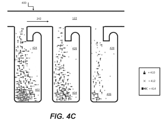
[0019] Figures 4A-4C are graphical representations of an assay according to
some embodiments of
the disclosure.
[0020] Figures 5A-5C are graphical illustrations of an assay according to
some other embodiments
of the disclosure.
4
CA 03020976 2018-10-12
WO 2017/181135
PCT/US2017/027795
[0021] Figure 6 is a schematic illustration of diffusion characteristics
within a chamber of a
microfluidic device according to some embodiments of the disclosure.
[0022] Figures 7A-7B are graphical representations of calculated diffusion
rates of molecules
according to some embodiments of the disclosure.
[0023] Figures 8A-8B are graphical representations of calculated and
experimentally confirmed
diffusion rates of molecules according to some embodiments of the disclosure.
[0024] Figures 9A-9B are graphical representation of diffusion
characteristics within a chamber of a
microfluidic device according to some embodiments of the disclosure.
[0025] Figures 10A-10B are graphical representation of diffusion
characteristics within a chamber
of a microfluidic device according to some other embodiments of the
disclosure.
[0026] Figures 11A-11B are graphical representation of diffusion
characteristics within a chamber
of a microfluidic device according to yet other embodiments of the disclosure.
[0027] Figures 12A-12C are graphical and photographic representations of
diffusion characteristics
within a chamber of a microfluidic device and an area of interest for
assessing levels of secretion of a
product from a biological micro-object, according to some embodiments of the
disclosure.
[0028] Figures 13A-13B depict photographic images of a microfluidic device
before and after
normalization according to some embodiments of the disclosure.
[0029] Figures 14A-14C are graphical and photographic representations of
assay images within a
microfluidic device and assay data for an area of interest thereof, according
to some embodiments of the
disclosure.
[0030] Figure 15 is a graphical representation of an overlay of median
intensity values for a plurality
of chambers within a microfluidic device, according to some embodiments of the
disclosure.
[0031] Figure 16A and 16B are graphical representations of analyte
secretion by biological micro-
objects disposed within a microfluidic device, according to some embodiments
of the disclosure.
[0032] Figure 17 illustrates steps performed to quantify an amount of
analyte secreted by a
biological micro-object present in sequestrations pens according to some
embodiments of the present
disclosure.
[0033] Figure 18 illustrates a sequence of steps performed to calculate an
absolute or relative value
representing the amount of analyte secreted by a biological micro-object
according to some
embodiments of the disclosure.
CA 03020976 2018-10-12
WO 2017/181135
PCT/US2017/027795
[0034] Figure 19 illustrates steps performed to assess an absolute or
relative value representing the
amount of analyte secreted by a clonal population of cells according to some
embodiments of the
disclosure.
[0035] Figure 20 is a graphical representation of a titration curve
generated according to some
embodiments of the disclosure.
[0036] Figure 21 is a photographic representation of a normalized assay
image of a portion of
microfluidic device including pen identification and assay scores according to
some embodiments of the
disclosure.
[0037] Figure 22 is a photographic and a graphical representation of a
course of a culturing and
assay sequence according to some embodiments of the disclosure.
[0038] Figures 23A-23B are graphical representation of assay values for all
the chambers of a
microfluidic device according to some embodiments of the disclosure.
[0039] Figure 24 is a graphical representation of correlation between assay
values for clonal
populations in selected chambers of a microfluidic device and the respective
scaled up clonal population
according to some embodiments of the disclosure.
[0040] Figure 25 is a block diagram that illustrates a computer system, in
accordance with various
embodiments.
[0041] Figure 26 is a schematic diagram of a system for assessing a
quantity of analyte, in
accordance with various embodiments
[0042] Figure 27 is a schematic diagram of a system for assessing a
quantity of analyte, in
accordance with various embodiments.
[0043] Figure 28 is a cross-section of a chamber of a micro-fluidic device,
in accordance with
various embodiments.
[0044] Figure 29 and 30 are exemplary flowcharts depicting a method for
determining a quantity of
analyte, in accordance with various embodiments.
[0045] It is to be understood that the figures are not necessarily drawn to
scale, nor are the objects in
the figures necessarily drawn to scale in relationship to one another. The
figures are depictions that are
intended to bring clarity and understanding to various embodiments of
apparatuses, systems, and
methods disclosed herein. Wherever possible, the same reference numbers will
be used throughout the
drawings to refer to the same or like parts. Moreover, it should be
appreciated that the drawings are not
intended to limit the scope of the present teachings in any way.
6
CA 03020976 2018-10-12
WO 2017/181135
PCT/US2017/027795
DETAILED DESCRIPTION
[0046] This specification describes exemplary embodiments and applications
of the disclosure. The
disclosure, however, is not limited to these exemplary embodiments and
applications or to the manner in
which the exemplary embodiments and applications operate or are described
herein. Moreover, the
figures may show simplified or partial views, and the dimensions of elements
in the figures may be
exaggerated or otherwise not in proportion. In addition, as the terms "on,"
"attached to," "connected to,"
"coupled to," or similar words are used herein, one element (e.g., a material,
a layer, a substrate, etc.)
can be "on," "attached to," "connected to," or "coupled to" another element
regardless of whether the one
element is directly on, attached to, connected to, or coupled to the other
element or there are one or more
intervening elements between the one element and the other element. Also,
unless the context dictates
otherwise, directions (e.g., above, below, top, bottom, side, up, down, under,
over, upper, lower,
horizontal, vertical, "x," "y," "z," etc.), if provided, are relative and
provided solely by way of example
and for ease of illustration and discussion and not by way of limitation. In
addition, where reference is
made to a list of elements (e.g., elements a, b, c), such reference is
intended to include any one of the
listed elements by itself, any combination of less than all of the listed
elements, and/or a combination of
all of the listed elements. Section divisions in the specification are for
ease of review only and do not
limit any combination of elements discussed.
[0047] Where dimensions of microfluidic features are described as having a
width or an area, the
dimension typically is described relative to an x-axial and/or y-axial
dimension, both of which lie within
a plane that is parallel to the substrate and/or cover of the microfluidic
device. The height of a
microfluidic feature may be described relative to a z-axial direction, which
is perpendicular to a plane
that is parallel to the substrate and/or cover of the microfluidic device. In
some instances, a cross
sectional area of a microfluidic feature, such as a channel or a passageway,
may be in reference to a x-
axial/z-axial, a y-axial/z-axial, or an x-axial/y-axial area.
[0048] As used herein, "substantially" means sufficient to work for the
intended purpose. The term
"substantially" thus allows for minor, insignificant variations from an
absolute or perfect state,
dimension, measurement, result, or the like such as would be expected by a
person of ordinary skill in
the field but that do not appreciably affect overall performance. When used
with respect to numerical
values or parameters or characteristics that can be expressed as numerical
values, "substantially" means
within ten percent.
[0049] The term "ones" means more than one.
7
CA 03020976 2018-10-12
WO 2017/181135
PCT/US2017/027795
[0050] As used herein, the term "plurality" can be 2, 3, 4, 5, 6, 7, 8, 9,
10, or more.
[0051] As used herein, the term "disposed" encompasses within its meaning
"located."
[0052] As used herein, a "microfluidic device" or "microfluidic apparatus"
is a device that includes
one or more discrete microfluidic circuits configured to hold a fluid, each
microfluidic circuit comprised
of fluidically interconnected circuit elements, including but not limited to
region(s), flow path(s),
channel(s), chamber(s), and/or pen(s), and at least one port configured to
allow the fluid (and,
optionally, micro-objects suspended in the fluid) to flow into and/or out of
the microfluidic device.
Typically, a microfluidic circuit of a microfluidic device will include a flow
region, which may include a
microfluidic channel, and at least one chamber, and will hold a volume of
fluid of less than about 1 mL,
e.g., less than about 750, 500, 250, 200, 150, 100, 75, 50, 25, 20, 15, 10, 9,
8, 7, 6, 5, 4, 3, or 2 L. In
certain embodiments, the microfluidic circuit holds about 1-2, 1-3, 1-4, 1-5,
2-5, 2-8, 2-10, 2-12, 2-15,
2-20, 5-20, 5-30, 5-40, 5-50, 10-50, 10-75, 10-100, 20-100, 20-150, 20-200, 50-
200, 50-250, or 50-300
L. The microfluidic circuit may be configured to have a first end fluidically
connected with a first port
(e.g., an inlet) in the microfluidic device and a second end fluidically
connected with a second port (e.g.,
an outlet) in the microfluidic device.
[0053] As used herein, a "nanofluidic device" or "nanofluidic apparatus" is
a type of microfluidic
device having a microfluidic circuit that contains at least one circuit
element configured to hold a
volume of fluid of less than about 1 [tL, e.g., less than about 750, 500, 250,
200, 150, 100, 75, 50, 25,
20, 15, 10, 9, 8, 7, 6, 5, 4, 3, 2, 1 nL or less. A nanofluidic device may
comprise a plurality of circuit
elements (e.g., at least 2, 3, 4, 5, 6, 7, 8, 9, 10, 15, 20, 25, 50, 75, 100,
150, 200, 250, 300, 400, 500, 600,
700, 800, 900, 1000, 1500, 2000, 2500, 3000, 3500, 4000, 4500, 5000, 6000,
7000, 8000, 9000, 10,000,
or more). In certain embodiments, one or more (e.g., all) of the at least one
circuit elements is
configured to hold a volume of fluid of about 100 pL to 1 nL, 100 pL to 2 nL,
100 pL to 5 nL, 250 pL to
2 nL, 250 pL to 5 nL, 250 pL to 10 nL, 500 pL to 5 nL, 500 pL to 10 nL, 500 pL
to 15 nL, 750 pL to 10
nL, 750 pL to 15 nL, 750 pL to 20 nL, 1 to 10 nL, 1 to 15 nL, 1 to 20 nL, 1 to
25 nL, or 1 to 50 nL. In
other embodiments, one or more (e.g., all) of the at least one circuit
elements are configured to hold a
volume of fluid of about 20 nL to 200nL, 100 to 200 nL, 100 to 300 nL, 100 to
400 nL, 100 to 500 nL,
200 to 300 nL, 200 to 400 nL, 200 to 500 nL, 200 to 600 nL, 200 to 700 nL, 250
to 400 nL, 250 to 500
nL, 250 to 600 nL, or 250 to 750 nL.
[0054] A microfluidic device or a nanofluidic device may be referred to
herein as a "microfluidic
chip" or a "chip"; or "nanofluidic chip" or "chip".
8
CA 03020976 2018-10-12
WO 2017/181135
PCT/US2017/027795
[0055] A "microfluidic channel" or "flow channel" as used herein refers to
flow region of a
microfluidic device having a length that is significantly longer than both the
horizontal and vertical
dimensions. For example, the flow channel can be at least 5 times the length
of either the horizontal or
vertical dimension, e.g., at least 10 times the length, at least 25 times the
length, at least 100 times the
length, at least 200 times the length, at least 500 times the length, at least
1,000 times the length, at least
5,000 times the length, or longer. In some embodiments, the length of a flow
channel is about 100,000
microns to about 500,000 microns, including any value therebetween. In some
embodiments, the
horizontal dimension is about 100 microns to about 1000 microns (e.g., about
150 to about 500
microns) and the vertical dimension is about 25 microns to about 200 microns,
(e.g., from about 40 to
about 150 microns). It is noted that a flow channel may have a variety of
different spatial configurations
in a microfluidic device, and thus is not restricted to a perfectly linear
element. For example, a flow
channel may be, or include one or more sections having, the following
configurations: curve, bend,
spiral, incline, decline, fork (e.g., multiple different flow paths), and any
combination thereof In
addition, a flow channel may have different cross-sectional areas along its
path, widening and
constricting to provide a desired fluid flow therein. The flow channel may
include valves, and the
valves may be of any type known in the art of microfluidics. Examples of
microfluidic channels that
include valves are disclosed in U.S. Patents 6,408,878 and 9,227,200, each of
which is herein
incorporated by reference in its entirety.
[0056] As used herein, the term "obstruction" refers generally to a bump or
similar type of structure
that is sufficiently large so as to partially (but not completely) impede
movement of target micro-objects
between two different regions or circuit elements in a microfluidic device.
The two different
regions/circuit elements can be, for example, the connection region and the
isolation region of a
microfluidic sequestration pen.
[0057] As used herein, the term "constriction" refers generally to a
narrowing of a width of a circuit
element (or an interface between two circuit elements) in a microfluidic
device. The constriction can be
located, for example, at the interface between the isolation region and the
connection region of a
microfluidic sequestration pen of the instant disclosure.
[0058] As used herein, the term "transparent" refers to a material which
allows visible light to pass
through without substantially altering the light as is passes through.
[0059] As used herein, the term "micro-object" refers generally to any
microscopic object that may
be isolated and/or manipulated in accordance with the present disclosure. Non-
limiting examples of
9
CA 03020976 2018-10-12
WO 2017/181135
PCT/US2017/027795
micro-objects include: inanimate micro-objects such as microparticles;
microbeads (e.g., polystyrene
beads, LuminexTM beads, or the like); magnetic beads; microrods; microwires;
quantum dots, and the
like; biological micro-objects such as cells; biological organelles; vesicles,
or complexes; synthetic
vesicles; liposomes (e.g., synthetic or derived from membrane preparations);
lipid nanorafts, and the
like; or a combination of inanimate micro-objects and biological micro-objects
(e.g., microbeads
attached to cells, liposome-coated micro-beads, liposome-coated magnetic
beads, or the like). Beads
may include moieties/molecules covalently or non-covalently attached, such as
fluorescent labels,
proteins, carbohydrates, antigens, small molecule signaling moieties, or other
chemical/biological
species capable of use in an assay. Lipid nanorafts have been described, for
example, in Ritchie et al.
(2009) "Reconstitution of Membrane Proteins in Phospholipid Bilayer
Nanodiscs," Methods Enzymol.,
464:211-231.
[0060] As used herein, the term "cell" is used interchangeably with the
term "biological cell." Non-
limiting examples of biological cells include eukaryotic cells, plant cells,
animal cells, such as
mammalian cells, reptilian cells, avian cells, fish cells, or the like,
prokaryotic cells, bacterial cells,
fungal cells, protozoan cells, or the like, cells dissociated from a tissue,
such as muscle, cartilage, fat,
skin, liver, lung, neural tissue, and the like, immunological cells, such as T
cells, B cells, natural killer
cells, macrophages, and the like, embryos (e.g., zygotes), oocytes, ova, sperm
cells, hybridomas,
cultured cells, cells from a cell line, cancer cells, infected cells,
transfected and/or transformed cells,
reporter cells, and the like. A mammalian cell can be, for example, from a
human, a mouse, a rat, a
horse, a goat, a sheep, a cow, a primate, or the like.
[0061] A colony of biological cells is "clonal" if all of the living cells
in the colony that are capable
of reproducing are daughter cells derived from a single parent cell. In
certain embodiments, all the
daughter cells in a clonal colony are derived from the single parent cell by
no more than 10 divisions. In
other embodiments, all the daughter cells in a clonal colony are derived from
the single parent cell by no
more than 14 divisions. In other embodiments, all the daughter cells in a
clonal colony are derived from
the single parent cell by no more than 17 divisions. In other embodiments, all
the daughter cells in a
clonal colony are derived from the single parent cell by no more than 20
divisions. The term "clonal
cells" refers to cells of the same clonal colony.
[0062] As used herein, a "colony" of biological cells refers to 2 or more
cells (e.g. about 2 to about
20, about 4 to about 40, about 6 to about 60, about 8 to about 80, about 10 to
about 100, about 20 to
CA 03020976 2018-10-12
WO 2017/181135
PCT/US2017/027795
about 200, about 40 to about 400, about 60 to about 600, about 80 to about
800, about 100 to about
1000, or greater than 1000 cells).
[0063] As used herein, the term "maintaining (a) cell(s)" refers to
providing an environment
comprising both fluidic and gaseous components and, optionally a surface, that
provides the conditions
necessary to keep the cells viable and/or expanding.
[0064] As used herein, the term "expanding" when referring to cells, refers
to increasing in cell
number.
[0065] A "component" of a fluidic medium is any chemical or biochemical
molecule present in the
medium, including solvent molecules, ions, small molecules, antibiotics,
nucleotides and nucleosides,
nucleic acids, amino acids, peptides, proteins, sugars, carbohydrates, lipids,
fatty acids, cholesterol,
metabolites, or the like.
[0066] As used herein, "capture moiety" is a chemical or biological
species, functionality, or motif
that provides a recognition site for a micro-object. A selected class of micro-
objects may recognize the
in situ-generated capture moiety and may bind or have an affinity for the in
situ-generated capture
moiety. Non-limiting examples include antigens, antibodies, and cell surface
binding motifs.
[0067] As used herein, "flowable polymer" is a polymer monomer or macromer
that is soluble or
dispersible within a fluidic medium (e.g., a pre-polymer solution). The
flowable polymer may be input
into a microfluidic flow region and flow with other components of a fluidic
medium therein.
[0068] As used herein, "photoinitiated polymer" refers to a polymer (or a
monomeric molecule that
can be used to generate the polymer) that upon exposure to light, is capable
of crosslinking covalently,
forming specific covalent bonds, changing regiochemistry around a rigidified
chemical motif, or
forming ion pairs which cause a change in physical state, and thereby forming
a polymer network. In
some instances, a photoinitiated polymer may include a polymer segment bound
to one or more
chemical moieties capable of crosslinking covalently, forming specific
covalent bonds, changing
regiochemistry around a rigidified chemical motif, or forming ion pairs which
cause a change in
physical state. In some instances, a photoinitiated polymer may require a
photoactivatable radical
initiator to initiate formation of the polymer network (e.g., via
polymerization of the polymer).
[0069] As used herein, "antibody" refers to an immunoglobulin (Ig) and
includes both polyclonal
and monoclonal antibodies; primatized (e.g., humanized); murine; mouse-human;
mouse-primate; and
chimeric; and may be an intact molecule, a fragment thereof (such as scFv, Fv,
Fd, Fab, Fab' and F(ab)'2
fragments), or multimers or aggregates of intact molecules and/or fragments;
and may occur in nature or
11
CA 03020976 2018-10-12
WO 2017/181135
PCT/US2017/027795
be produced, e.g., by immunization, synthesis or genetic engineering. An
"antibody fragment," as used
herein, refers to fragments, derived from or related to an antibody, which
bind antigen and which in
some embodiments may be derivatized to exhibit structural features that
facilitate clearance and uptake,
e.g., by the incorporation of galactose residues. This includes, e.g., F(ab),
F(ab)'2, scFv, light chain
variable region (VL), heavy chain variable region (VH), and combinations
thereof.
[0070] As used herein in reference to a fluidic medium, "diffuse" and
"diffusion" refer to
thermodynamic movement of a component of the fluidic medium down a
concentration gradient.
[0071] The phrase "flow of a medium" means bulk movement of a fluidic medium
primarily due to
any mechanism other than diffusion. For example, flow of a medium can involve
movement of the
fluidic medium from one point to another point due to a pressure differential
between the points. Such
flow can include a continuous, pulsed, periodic, random, intermittent, or
reciprocating flow of the liquid,
or any combination thereof When one fluidic medium flows into another fluidic
medium, turbulence
and mixing of the media can result.
[0072] The phrase "substantially no flow" refers to a rate of flow of a
fluidic medium that, averaged
over time, is less than the rate of diffusion of components of a material
(e.g., an analyte of interest) into
or within the fluidic medium. The rate of diffusion of components of such a
material can depend on, for
example, temperature, the size of the components, and the strength of
interactions between the
components and the fluidic medium.
[0073] As used herein in reference to different regions within a
microfluidic device, the phrase
"fluidically connected" means that, when the different regions are
substantially filled with fluid, such as
fluidic media, the fluid in each of the regions is connected so as to form a
single body of fluid. This
does not mean that the fluids (or fluidic media) in the different regions are
necessarily identical in
composition. Rather, the fluids in different fluidically connected regions of
a microfluidic device can
have different compositions (e.g., different concentrations of solutes, such
as proteins, carbohydrates,
ions, or other molecules) which are in flux as solutes move down their
respective concentration
gradients and/or fluids flow through the microfluidic device.
[0074] As used herein, a "flow path" refers to one or more fluidically
connected circuit elements
(e.g. channel(s), region(s), chamber(s) and the like) that define, and are
subject to, the trajectory of a
flow of medium. A flow path is thus an example of a swept region of a
microfluidic device. Other
circuit elements (e.g., unswept regions) may be fluidically connected with the
circuit elements that
comprise the flow path without being subject to the flow of medium in the flow
path.
12
CA 03020976 2018-10-12
WO 2017/181135
PCT/US2017/027795
[0075] As used herein, "isolating a micro-object" confines a micro-object
to a defined area within
the microfluidic device.
[0076] A microfluidic (or nanofluidic) device can comprise "swept" regions
and "unswept" regions.
As used herein, a "swept" region is comprised of one or more fluidically
interconnected circuit elements
of a microfluidic circuit, each of which experiences a flow of medium when
fluid is flowing through the
microfluidic circuit. The circuit elements of a swept region can include, for
example, regions, channels,
and all or parts of chambers. As used herein, an "unswept" region is comprised
of one or more
fluidically interconnected circuit element of a microfluidic circuit, each of
which experiences
substantially no flux of fluid when fluid is flowing through the microfluidic
circuit. An unswept region
can be fluidically connected to a swept region, provided the fluidic
connections are structured to enable
diffusion but substantially no flow of media between the swept region and the
unswept region. The
microfluidic device can thus be structured to substantially isolate an unswept
region from a flow of
medium in a swept region, while enabling substantially only diffusive fluidic
communication between
the swept region and the unswept region. For example, a flow channel of a
micro-fluidic device is an
example of a swept region while an isolation region (described in further
detail below) of a microfluidic
device is an example of an unswept region.
[0077] The capability of biological micro-objects (e.g., biological cells)
to produce specific
biological materials (e.g., proteins, such as antibodies) can be assayed in
such a microfluidic device. In
a specific embodiment of an assay, sample material comprising biological micro-
objects (e.g., cells) to
be assayed for production of an analyte of interest can be loaded into a swept
region of the microfluidic
device. Ones of the biological micro-objects (e.g., mammalian cells, such as
human cells) can be
selected for particular characteristics and disposed in unswept regions. The
remaining sample material
can then be flowed out of the swept region and an assay material flowed into
the swept region. Because
the selected biological micro-objects are in unswept regions, the selected
biological micro-objects are
not substantially affected by the flowing out of the remaining sample material
or the flowing in of the
assay material. The selected biological micro-objects can be allowed to
produce the analyte of interest,
which can diffuse from the unswept regions into the swept region, where the
analyte of interest can react
with the assay material to produce localized detectable reactions, each of
which can be correlated to a
particular unswept region. Any unswept region associated with a detected
reaction can be analyzed to
assess which, if any, of the biological micro-objects in the unswept region
are sufficient producers of the
analyte of interest.
13
CA 03020976 2018-10-12
WO 2017/181135
PCT/US2017/027795
[0078] Microfluidic devices and systems for operating and observing such
devices. Figure 1A
illustrates an example of a microfluidic device 100 and a system 150 which can
be used for maintaining,
isolating, assaying or culturing biological micro-objects. A perspective view
of the microfluidic device
100 is shown having a partial cut-away of its cover 110 to provide a partial
view into the microfluidic
device 100. The microfluidic device 100 generally comprises a microfluidic
circuit 120 comprising a
flow path 106 through which a fluidic medium 180 can flow, optionally carrying
one or more micro-
objects (not shown) into and/or through the microfluidic circuit 120. Although
a single microfluidic
circuit 120 is illustrated in Figure 1A, suitable microfluidic devices can
include a plurality (e.g., 2 or 3)
of such microfluidic circuits. Regardless, the microfluidic device 100 can be
configured to be a
nanofluidic device. As illustrated in Figure 1A, the microfluidic circuit 120
may include a plurality of
microfluidic sequestration pens 124, 126, 128, and 130, where each
sequestration pens may have one or
more openings in fluidic communication with flow path 106. In some embodiments
of the device of
Figure 1A, the sequestration pens may have only a single opening in fluidic
communication with the
flow path 106. As discussed further below, the microfluidic sequestration pens
comprise various
features and structures that have been optimized for retaining micro-objects
in the microfluidic device,
such as microfluidic device 100, even when a medium 180 is flowing through the
flow path 106. Before
turning to the foregoing, however, a brief description of microfluidic device
100 and system 150 is
provided.
[0079] As generally illustrated in Figure 1A, the microfluidic circuit 120
is defined by an enclosure
102. Although the enclosure 102 can be physically structured in different
configurations, in the example
shown in Figure lA the enclosure 102 is depicted as comprising a support
structure 104 (e.g., a base), a
microfluidic circuit structure 108, and a cover 110. The support structure
104, microfluidic circuit
structure 108, and cover 110 can be attached to each other. For example, the
microfluidic circuit
structure 108 can be disposed on an inner surface 109 of the support structure
104, and the cover 110
can be disposed over the microfluidic circuit structure 108. Together with the
support structure 104 and
cover 110, the microfluidic circuit structure 108 can define the elements of
the microfluidic circuit 120.
[0080] The support structure 104 can be at the bottom and the cover 110 at
the top of the
microfluidic circuit 120 as illustrated in Figure 1A. Alternatively, the
support structure 104 and the
cover 110 can be configured in other orientations. For example, the support
structure 104 can be at the
top and the cover 110 at the bottom of the microfluidic circuit 120.
Regardless, there can be one or
more ports 107 each comprising a passage into or out of the enclosure 102.
Examples of a passage
14
CA 03020976 2018-10-12
WO 2017/181135
PCT/US2017/027795
include a valve, a gate, a pass-through hole, or the like. As illustrated,
port 107 is a pass-through hole
created by a gap in the microfluidic circuit structure 108. However, the port
107 can be situated in other
components of the enclosure 102, such as the cover 110. Only one port 107 is
illustrated in Figure 1A
but the microfluidic circuit 120 can have two or more ports 107. For example,
there can be a first port
107 that functions as an inlet for fluid entering the microfluidic circuit
120, and there can be a second
port 107 that functions as an outlet for fluid exiting the microfluidic
circuit 120. Whether a port 107
function as an inlet or an outlet can depend upon the direction that fluid
flows through flow path 106.
[0081] The support structure 104 can comprise one or more electrodes (not
shown) and a substrate
or a plurality of interconnected substrates. For example, the support
structure 104 can comprise one or
more semiconductor substrates, each of which is electrically connected to an
electrode (e.g., all or a
subset of the semiconductor substrates can be electrically connected to a
single electrode). The support
structure 104 can further comprise a printed circuit board assembly ("PCBA").
For example, the
semiconductor substrate(s) can be mounted on a PCBA.
[0082] The microfluidic circuit structure 108 can define circuit elements
of the microfluidic circuit
120. Such circuit elements can comprise spaces or regions that can be fluidly
interconnected when
microfluidic circuit 120 is filled with fluid, such as flow regions (which may
include or be one or more
flow channels), chambers, pens, traps, and the like. In the microfluidic
circuit 120 illustrated in Figure
1A, the microfluidic circuit structure 108 comprises a frame 114 and a
microfluidic circuit material 116.
The frame 114 can partially or completely enclose the microfluidic circuit
material 116. The frame 114
can be, for example, a relatively rigid structure substantially surrounding
the microfluidic circuit
material 116. For example, the frame 114 can comprise a metal material.
[0083] The microfluidic circuit material 116 can be patterned with cavities
or the like to define
circuit elements and interconnections of the microfluidic circuit 120. The
microfluidic circuit material
116 can comprise a flexible material, such as a flexible polymer (e.g. rubber,
plastic, elastomer, silicone,
polydimethylsiloxane ("PDMS"), or the like), which can be gas permeable. Other
examples of materials
that can compose microfluidic circuit material 116 include molded glass, an
etchable material such as
silicone (e.g. photo-patternable silicone or "PPS"), photo-resist (e.g., 5U8),
or the like. In some
embodiments, such materials¨and thus the microfluidic circuit material 116¨can
be rigid and/or
substantially impermeable to gas. Regardless, microfluidic circuit material
116 can be disposed on the
support structure 104 and inside the frame 114.
CA 03020976 2018-10-12
WO 2017/181135
PCT/US2017/027795
[0084] The cover 110 can be an integral part of the frame 114 and/or the
microfluidic circuit
material 116. Alternatively, the cover 110 can be a structurally distinct
element, as illustrated in Figure
1A. The cover 110 can comprise the same or different materials than the frame
114 and/or the
microfluidic circuit material 116. Similarly, the support structure 104 can be
a separate structure from
the frame 114 or microfluidic circuit material 116 as illustrated, or an
integral part of the frame 114 or
microfluidic circuit material 116. Likewise, the frame 114 and microfluidic
circuit material 116 can be
separate structures as shown in Figure 1A or integral portions of the same
structure.
[0085] In some embodiments, the cover 110 can comprise a rigid material.
The rigid material may
be glass or a material with similar properties. In some embodiments, the cover
110 can comprise a
deformable material. The deformable material can be a polymer, such as PDMS.
In some
embodiments, the cover 110 can comprise both rigid and deformable materials.
For example, one or
more portions of cover 110 (e.g., one or more portions positioned over
sequestration pens 124, 126, 128,
130) can comprise a deformable material that interfaces with rigid materials
of the cover 110. In some
embodiments, the cover 110 can further include one or more electrodes. The one
or more electrodes can
comprise a conductive oxide, such as indium-tin-oxide (ITO), which may be
coated on glass or a
similarly insulating material. Alternatively, the one or more electrodes can
be flexible electrodes, such
as single-walled nanotubes, multi-walled nanotubes, nanowires, clusters of
electrically conductive
nanoparticles, or combinations thereof, embedded in a deformable material,
such as a polymer (e.g.,
PDMS). Flexible electrodes that can be used in microfluidic devices have been
described, for example,
in U.S. 2012/0325665 (Chiou et al.), the contents of which are incorporated
herein by reference. In
some embodiments, the cover 110 can be modified (e.g., by conditioning all or
part of a surface that
faces inward toward the microfluidic circuit 120) to support cell adhesion,
viability and/or growth. The
modification may include a coating of a synthetic or natural polymer. In some
embodiments, the cover
110 and/or the support structure 104 can be transparent to light. The cover
110 may also include at least
one material that is gas permeable (e.g., PDMS or PPS).
[0086] Figure 1A also shows a system 150 for operating and controlling
microfluidic devices, such
as microfluidic device 100. System 150 includes an electrical power source
192, an imaging device 194
(incorporated within imaging module 164, where device 194 is not illustrated
in Figure 1A, per se), and
a tilting device 190 (part of tilting module 166, where device 190 is not
illustrated in Figure 1A).
[0087] The electrical power source 192 can provide electric power to the
microfluidic device 100
and/or tilting device 190, providing biasing voltages or currents as needed.
The electrical power source
16
CA 03020976 2018-10-12
WO 2017/181135
PCT/US2017/027795
192 can, for example, comprise one or more alternating current (AC) and/or
direct current (DC) voltage
or current sources. The imaging device 194 (part of imaging module 164,
discussed below) can
comprise a device, such as a digital camera, for capturing images inside
microfluidic circuit 120. In
some instances, the imaging device 194 further comprises a detector having a
fast frame rate and/or high
sensitivity (e.g. for low light applications). The imaging device 194 can also
include a mechanism for
directing stimulating radiation and/or light beams into the microfluidic
circuit 120 and collecting
radiation and/or light beams reflected or emitted from the microfluidic
circuit 120 (or micro-objects
contained therein). The emitted light beams may be in the visible spectrum and
may, e.g., include
fluorescent emissions. The reflected light beams may include reflected
emissions originating from an
LED or a wide spectrum lamp, such as a mercury lamp (e.g. a high pressure
mercury lamp) or a Xenon
arc lamp. As discussed with respect to Figure 3B, the imaging device 194 may
further include a
microscope (or an optical train), which may or may not include an eyepiece.
[0088] System 150 further comprises a tilting device 190 (part of tilting
module 166, discussed
below) configured to rotate a microfluidic device 100 about one or more axes
of rotation. In some
embodiments, the tilting device 190 is configured to support and/or hold the
enclosure 102 comprising
the microfluidic circuit 120 about at least one axis such that the
microfluidic device 100 (and thus the
microfluidic circuit 120) can be held in a level orientation (i.e. at 00
relative to x- and y-axes), a vertical
orientation (i.e. at 90 relative to the x-axis and/or the y-axis), or any
orientation therebetween. The
orientation of the microfluidic device 100 (and the microfluidic circuit 120)
relative to an axis is referred
to herein as the "tilt" of the microfluidic device 100 (and the microfluidic
circuit 120). For example, the
tilting device 190 can tilt the microfluidic device 100 at 0.10, 0.2 , 0.3 ,
0.4 , 0.5 , 0.6 , 0.7 , 0.8 , 0.9 ,
1 , 2 , 3 , 40, 50, 100, 15 , 20 , 25 , 30 , 35 , 40 , 45 , 50 , 55 , 60 , 65
, 70 , 75 , 80 , 90 relative to
the x-axis or any degree therebetween. The level orientation (and thus the x-
and y-axes) is defined as
normal to a vertical axis defined by the force of gravity. The tilting device
can also tilt the microfluidic
device 100 (and the microfluidic circuit 120) to any degree greater than 90
relative to the x-axis and/or
y-axis, or tilt the microfluidic device 100 (and the microfluidic circuit 120)
180 relative to the x-axis or
the y-axis in order to fully invert the microfluidic device 100 (and the
microfluidic circuit 120).
Similarly, in some embodiments, the tilting device 190 tilts the microfluidic
device 100 (and the
microfluidic circuit 120) about an axis of rotation defined by flow path 106
or some other portion of
microfluidic circuit 120.
17
CA 03020976 2018-10-12
WO 2017/181135
PCT/US2017/027795
[0089] In some instances, the microfluidic device 100 is tilted into a
vertical orientation such that the
flow path 106 is positioned above or below one or more sequestration pens. The
term "above" as used
herein denotes that the flow path 106 is positioned higher than the one or
more sequestration pens on a
vertical axis defined by the force of gravity (i.e. an object in a
sequestration pen above a flow path 106
would have a higher gravitational potential energy than an object in the flow
path). The term "below" as
used herein denotes that the flow path 106 is positioned lower than the one or
more sequestration pens
on a vertical axis defined by the force of gravity (i.e. an object in a
sequestration pen below a flow path
106 would have a lower gravitational potential energy than an object in the
flow path).
[0090] In some instances, the tilting device 190 tilts the microfluidic
device 100 about an axis that is
parallel to the flow path 106. Moreover, the microfluidic device 100 can be
tilted to an angle of less
than 90 such that the flow path 106 is located above or below one or more
sequestration pens without
being located directly above or below the sequestration pens. In other
instances, the tilting device 190
tilts the microfluidic device 100 about an axis perpendicular to the flow path
106. In still other
instances, the tilting device 190 tilts the microfluidic device 100 about an
axis that is neither parallel nor
perpendicular to the flow path 106.
[0091] System 150 can further include a media source 178. The media source
178 (e.g., a container,
reservoir, or the like) can comprise multiple sections or containers, each for
holding a different fluidic
medium 180. Thus, the media source 178 can be a device that is outside of and
separate from the
microfluidic device 100, as illustrated in Figure 1A. Alternatively, the media
source 178 can be located
in whole or in part inside the enclosure 102 of the microfluidic device 100.
For example, the media
source 178 can comprise reservoirs that are part of the microfluidic device
100.
[0092] Figure 1A also illustrates simplified block diagram depictions of
examples of control and
monitoring equipment 152 that constitute part of system 150 and can be
utilized in conjunction with a
microfluidic device 100. As shown, examples of such control and monitoring
equipment 152 include a
master controller 154 comprising a media module 160 for controlling the media
source 178, a motive
module 162 for controlling movement and/or selection of micro-objects (not
shown) and/or medium
(e.g., droplets of medium) in the microfluidic circuit 120, an imaging module
164 for controlling an
imaging device 194 (e.g., a camera, microscope, light source or any
combination thereof) for capturing
images (e.g., digital images), and a tilting module 166 for controlling a
tilting device 190. The control
equipment 152 can also include other modules 168 for controlling, monitoring,
or performing other
18
CA 03020976 2018-10-12
WO 2017/181135
PCT/US2017/027795
functions with respect to the microfluidic device 100. As shown, the equipment
152 can further include
a display device 170 and an input/output device 172.
[0093] The master controller 154 can comprise a control module 156 and a
digital memory 158.
The control module 156 can comprise, for example, a digital processor
configured to operate in
accordance with machine executable instructions (e.g., software, firmware,
source code, or the like)
stored as non-transitory data or signals in the memory 158. Alternatively, or
in addition, the control
module 156 can comprise hardwired digital circuitry and/or analog circuitry.
The media module 160,
motive module 162, imaging module 164, tilting module 166, and/or other
modules 168 can be similarly
configured. Thus, functions, processes acts, actions, or steps of a process
discussed herein as being
performed with respect to the microfluidic device 100 or any other
microfluidic apparatus can be
performed by any one or more of the master controller 154, media module 160,
motive module 162,
imaging module 164, tilting module 166, and/or other modules 168 configured as
discussed above.
Similarly, the master controller 154, media module 160, motive module 162,
imaging module 164,
tilting module 166, and/or other modules 168 may be communicatively coupled to
transmit and receive
data used in any function, process, act, action or step discussed herein.
[0094] The media module 160 controls the media source 178. For example, the
media module 160
can control the media source 178 to input a selected fluidic medium 180 into
the enclosure 102 (e.g.,
through an inlet port 107). The media module 160 can also control removal of
media from the enclosure
102 (e.g., through an outlet port (not shown)). One or more media can thus be
selectively input into and
removed from the microfluidic circuit 120. The media module 160 can also
control the flow of fluidic
medium 180 in the flow path 106 inside the microfluidic circuit 120. For
example, in some
embodiments media module 160 stops the flow of media 180 in the flow path 106
and through the
enclosure 102 prior to the tilting module 166 causing the tilting device 190
to tilt the microfluidic device
100 to a desired angle of incline.
[0095] The motive module 162 can be configured to control selection,
trapping, and movement of
micro-objects (not shown) in the microfluidic circuit 120. As discussed below
with respect to Figures
1B and 1C, the enclosure 102 can comprise a dielectrophoresis (DEP),
optoelectronic tweezers (OET)
and/or opto-electrowetting (OEW) configuration (not shown in Figure 1A), and
the motive module 162
can control the activation of electrodes and/or transistors (e.g.,
phototransistors) to select and move
micro-objects (not shown) and/or droplets of medium (not shown) in the flow
path 106 and/or
sequestration pens 124, 126, 128, 130.
19
CA 03020976 2018-10-12
WO 2017/181135
PCT/US2017/027795
[0096] The imaging module 164 can control the imaging device 194. For
example, the imaging
module 164 can receive and process image data from the imaging device 194.
Image data from the
imaging device 194 can comprise any type of information captured by the
imaging device 194 (e.g., the
presence or absence of micro-objects, droplets of medium, accumulation of
label, such as fluorescent
label, etc.). Using the information captured by the imaging device 194, the
imaging module 164 can
further calculate the position of objects (e.g., micro-objects, droplets of
medium) and/or the rate of
motion of such objects within the microfluidic device 100.
[0097] The tilting module 166 can control the tilting motions of tilting
device 190. Alternatively, or
in addition, the tilting module 166 can control the tilting rate and timing to
optimize transfer of micro-
objects to the one or more sequestration pens via gravitational forces. The
tilting module 166 is
communicatively coupled with the imaging module 164 to receive data describing
the motion of micro-
objects and/or droplets of medium in the microfluidic circuit 120. Using this
data, the tilting module
166 may adjust the tilt of the microfluidic circuit 120 in order to adjust the
rate at which micro-objects
and/or droplets of medium move in the microfluidic circuit 120. The tilting
module 166 may also use
this data to iteratively adjust the position of a micro-object and/or droplet
of medium in the microfluidic
circuit 120.
[0098] In the example shown in Figure 1A, the microfluidic circuit 120 is
illustrated as comprising a
microfluidic channel 122 and sequestration pens 124, 126, 128, 130. Each pen
comprises an opening to
channel 122, but otherwise is enclosed such that the pens can substantially
isolate micro-objects inside
the pen from fluidic medium 180 and/or micro-objects in the flow path 106 of
channel 122 or in other
pens. The walls of the sequestration pen extend from the inner surface 109 of
the base to the inside
surface of the cover 110 to provide enclosure. The opening of the pen to the
microfluidic channel 122 is
oriented at an angle to the flow 106 of fluidic medium 180 such that flow 106
is not directed into the
pens. The flow may be tangential or orthogonal to the plane of the opening of
the pen. In some
instances, pens 124, 126, 128, 130 are configured to physically corral one or
more micro-objects within
the microfluidic circuit 120. Sequestration pens in accordance with the
present disclosure can comprise
various shapes, surfaces and features that are optimized for use with DEP,
OET, OEW, fluid flow,
and/or gravitational forces, as will be discussed and shown in detail below.
[0099] The microfluidic circuit 120 may comprise any number of microfluidic
sequestration pens.
Although five sequestration pens are shown, microfluidic circuit 120 may have
fewer or more
sequestration pens. As shown, microfluidic sequestration pens 124, 126, 128,
and 130 of microfluidic
CA 03020976 2018-10-12
WO 2017/181135
PCT/US2017/027795
circuit 120 each comprise differing features and shapes which may provide one
or more benefits useful
for maintaining, isolating, assaying or culturing biological micro-objects. In
some embodiments, the
microfluidic circuit 120 comprises a plurality of identical microfluidic
sequestration pens.
[00100] In the embodiment illustrated in Figure 1A, a single channel 122
and flow path 106 is shown.
However, other embodiments may contain multiple channels 122, each configured
to comprise a flow
path 106. The microfluidic circuit 120 further comprises an inlet valve or
port 107 in fluid
communication with the flow path 106 and fluidic medium 180, whereby fluidic
medium 180 can access
channel 122 via the inlet port 107. In some instances, the flow path 106
comprises a single path. In
some instances, the single path is arranged in a zigzag pattern whereby the
flow path 106 travels across
the microfluidic device 100 two or more times in alternating directions.
[00101] In some instances, microfluidic circuit 120 comprises a plurality
of parallel channels 122 and
flow paths 106, wherein the fluidic medium 180 within each flow path 106 flows
in the same direction.
In some instances, the fluidic medium within each flow path 106 flows in at
least one of a forward or
reverse direction. In some instances, a plurality of sequestration pens is
configured (e.g., relative to a
channel 122) such that the sequestration pens can be loaded with target micro-
objects in parallel.
[00102] In some embodiments, microfluidic circuit 120 further comprises one
or more micro-object
traps 132. The traps 132 are generally formed in a wall forming the boundary
of a channel 122, and may
be positioned opposite an opening of one or more of the microfluidic
sequestration pens 124, 126, 128,
130. In some embodiments, the traps 132 are configured to receive or capture a
single micro-object
from the flow path 106. In some embodiments, the traps 132 are configured to
receive or capture a
plurality of micro-objects from the flow path 106. In some instances, the
traps 132 comprise a volume
approximately equal to the volume of a single target micro-object.
[00103] The traps 132 may further comprise an opening which is configured
to assist the flow of
targeted micro-objects into the traps 132. In some instances, the traps 132
comprise an opening having a
height and width that is approximately equal to the dimensions of a single
target micro-object, whereby
larger micro-objects are prevented from entering into the micro-object trap.
The traps 132 may further
comprise other features configured to assist in retention of targeted micro-
objects within the trap 132. In
some instances, the trap 132 is aligned with and situated on the opposite side
of a channel 122 relative to
the opening of a microfluidic sequestration pen, such that upon tilting the
microfluidic device 100 about
an axis parallel to the microfluidic channel 122, the trapped micro-object
exits the trap 132 at a
trajectory that causes the micro-object to fall into the opening of the
sequestration pen. In some
21
CA 03020976 2018-10-12
WO 2017/181135
PCT/US2017/027795
instances, the trap 132 comprises a side passage 134 that is smaller than the
target micro-object in order
to facilitate flow through the trap 132 and thereby increase the likelihood of
capturing a micro-object in
the trap 132.
[00104] In some embodiments, dielectrophoretic (DEP) forces are applied
across the fluidic medium
180 (e.g., in the flow path and/or in the sequestration pens) via one or more
electrodes (not shown) to
manipulate, transport, separate and sort micro-objects located therein. For
example, in some
embodiments, DEP forces are applied to one or more portions of microfluidic
circuit 120 in order to
transfer a single micro-object from the flow path 106 into a desired
microfluidic sequestration pen. In
some embodiments, DEP forces are used to prevent a micro-object within a
sequestration pen (e.g.,
sequestration pen 124, 126, 128, or 130) from being displaced therefrom.
Further, in some
embodiments, DEP forces are used to selectively remove a micro-object from a
sequestration pen that
was previously collected in accordance with the embodiments of the current
disclosure. In some
embodiments, the DEP forces comprise optoelectronic tweezer (OET) forces.
[00105] In other embodiments, optoelectrowetting (OEW) forces are applied
to one or more positions
in the support structure 104 (and/or the cover 110) of the microfluidic device
100 (e.g., positions helping
to define the flow path and/or the sequestration pens) via one or more
electrodes (not shown) to
manipulate, transport, separate and sort droplets located in the microfluidic
circuit 120. For example, in
some embodiments, OEW forces are applied to one or more positions in the
support structure 104
(and/or the cover 110) in order to transfer a single droplet from the flow
path 106 into a desired
microfluidic sequestration pen. In some embodiments, OEW forces are used to
prevent a droplet within
a sequestration pen (e.g., sequestration pen 124, 126, 128, or 130) from being
displaced therefrom.
Further, in some embodiments, OEW forces are used to selectively remove a
droplet from a
sequestration pen that was previously collected in accordance with the
embodiments of the current
disclosure.
[00106] In some embodiments, DEP and/or OEW forces are combined with other
forces, such as flow
and/or gravitational force, so as to manipulate, transport, separate and sort
micro-objects and/or droplets
within the microfluidic circuit 120. For example, the enclosure 102 can be
tilted (e.g., by tilting device
190) to position the flow path 106 and micro-objects located therein above the
microfluidic
sequestration pens, and the force of gravity can transport the micro-objects
and/or droplets into the pens.
In some embodiments, the DEP and/or OEW forces can be applied prior to the
other forces. In other
embodiments, the DEP and/or OEW forces can be applied after the other forces.
In still other instances,
22
CA 03020976 2018-10-12
WO 2017/181135
PCT/US2017/027795
the DEP and/or OEW forces can be applied at the same time as the other forces
or in an alternating
manner with the other forces.
[00107] Figures 1B, 1C, and 2A-2H illustrates various embodiments of
microfluidic devices that can
be used in the practice of the embodiments of the present disclosure. Figure
1B depicts an embodiment
in which the microfluidic device 200 is configured as an optically-actuated
electrokinetic device. A
variety of optically-actuated electrokinetic devices are known in the art,
including devices having an
optoelectronic tweezer (OET) configuration and devices having an opto-
electrowetting (OEW)
configuration. Examples of suitable OET configurations are illustrated in the
following U.S. patent
documents, each of which is incorporated herein by reference in its entirety:
U.S. Patent No. RE 44,711
(Wu et al.) (originally issued as U.S. Patent No. 7,612,355); and U.S. Patent
No. 7,956,339 (Ohta et al.).
Examples of OEW configurations are illustrated in U.S. Patent No. 6,958,132
(Chiou et al.) and U.S.
Patent Application Publication No. 2012/0024708 (Chiou et al.), both of which
are incorporated by
reference herein in their entirety. Yet another example of an optically-
actuated electrokinetic device
includes a combined OET/OEW configuration, examples of which are shown in U.S.
Patent Publication
Nos. 20150306598 (Khandros et al.) and 20150306599 (Khandros et al.) and their
corresponding PCT
Publications W02015/164846 and W02015/164847, all of which are incorporated
herein by reference
in their entirety.
[00108] Examples of microfluidic devices having pens in which biological
micro-objects can be
placed, cultured, and/or monitored have been described, for example, in US
2014/0116881 (application
no. 14/060,117, filed October 22, 2013), US 2015/0151298 (application no.
14/520,568, filed October
22, 2014), and US 2015/0165436 (application no. 14/521,447, filed October 22,
2014), each of which is
incorporated herein by reference in its entirety. US application nos.
14/520,568 and 14/521,447 also
describe exemplary methods of analyzing secretions of cells cultured in a
microfluidic device. Each of
the foregoing applications further describes microfluidic devices configured
to produce dielectrophoretic
(DEP) forces, such as optoelectronic tweezers (OET) or configured to provide
opto-electro wetting
(OEW). For example, the optoelectronic tweezers device illustrated in Figure 2
of US 2014/0116881 is
an example of a device that can be utilized in embodiments of the present
disclosure to select and move
an individual biological micro-object or a group of biological micro-objects.
[00109] Microfluidic device motive configurations. As described above, the
control and
monitoring equipment of the system can comprise a motive module for selecting
and moving objects,
such as micro-objects or droplets, in the microfluidic circuit of a
microfluidic device. The microfluidic
23
CA 03020976 2018-10-12
WO 2017/181135
PCT/US2017/027795
device can have a variety of motive configurations, depending upon the type of
object being moved and
other considerations. For example, a dielectrophoresis (DEP) configuration can
be utilized to select and
move micro-objects in the microfluidic circuit. Thus, the support structure
104 and/or cover 110 of the
microfluidic device 100 can comprise a DEP configuration for selectively
inducing DEP forces on
micro-objects in a fluidic medium 180 in the microfluidic circuit 120 and
thereby select, capture, and/or
move individual micro-objects or groups of micro-objects. Alternatively, the
support structure 104
and/or cover 110 of the microfluidic device 100 can comprise an electrowetting
(EW) configuration for
selectively inducing EW forces on droplets in a fluidic medium 180 in the
microfluidic circuit 120 and
thereby select, capture, and/or move individual droplets or groups of
droplets.
[00110] One example of a microfluidic device 200 comprising a DEP
configuration is illustrated in
Figures 1B and 1C. While for purposes of simplicity Figures 1B and 1C show a
side cross-sectional
view and a top cross-sectional view, respectively, of a portion of an
enclosure 102 of the microfluidic
device 200 having a region/chamber 202, it should be understood that the
region/chamber 202 may be
part of a fluidic circuit element having a more detailed structure, such as a
growth chamber, a
sequestration pen, a flow region, or a flow channel. Furthermore, the
microfluidic device 200 may
include other fluidic circuit elements. For example, the microfluidic device
200 can include a plurality
of growth chambers or sequestration pens and/or one or more flow regions or
flow channels, such as
those described herein with respect to microfluidic device 100. A DEP
configuration may be
incorporated into any such fluidic circuit elements of the microfluidic device
200, or select portions
thereof. It should be further appreciated that any of the above or below
described microfluidic device
components and system components may be incorporated in and/or used in
combination with the
microfluidic device 200. For example, system 150 including control and
monitoring equipment 152,
described above, may be used with microfluidic device 200, including one or
more of the media module
160, motive module 162, imaging module 164, tilting module 166, and other
modules 168.
[00111] As seen in Figure 1B, the microfluidic device 200 includes a
support structure 104 having a
bottom electrode 204 and an electrode activation substrate 206 overlying the
bottom electrode 204, and a
cover 110 having a top electrode 210, with the top electrode 210 spaced apart
from the bottom electrode
204. The top electrode 210 and the electrode activation substrate 206 define
opposing surfaces of the
region/chamber 202. A medium 180 contained in the region/chamber 202 thus
provides a resistive
connection between the top electrode 210 and the electrode activation
substrate 206. A power source
212 configured to be connected to the bottom electrode 204 and the top
electrode 210 and create a
24
CA 03020976 2018-10-12
WO 2017/181135
PCT/US2017/027795
biasing voltage between the electrodes, as required for the generation of DEP
forces in the
region/chamber 202, is also shown. The power source 212 can be, for example,
an alternating current
(AC) power source.
[00112] In certain embodiments, the microfluidic device 200 illustrated in
Figures 1B and 1C can
have an optically-actuated DEP configuration. Accordingly, changing patterns
of light 218 from the
light source 216, which may be controlled by the motive module 162, can
selectively activate and
deactivate changing patterns of DEP electrodes at regions 214 of the inner
surface 208 of the electrode
activation substrate 206. (Hereinafter the regions 214 of a microfluidic
device having a DEP
configuration are referred to as "DEP electrode regions.") As illustrated in
Figure 1C, a light pattern
218 directed onto the inner surface 208 of the electrode activation substrate
206 can illuminate select
DEP electrode regions 214a (shown in white) in a pattern, such as a square.
The non-illuminated DEP
electrode regions 214 (cross-hatched) are hereinafter referred to as "dark"
DEP electrode regions 214.
The relative electrical impedance through the DEP electrode activation
substrate 206 (i.e., from the
bottom electrode 204 up to the inner surface 208 of the electrode activation
substrate 206 which
interfaces with the medium 180 in the flow region 106) is greater than the
relative electrical impedance
through the medium 180 in the region/chamber 202 (i.e., from the inner surface
208 of the electrode
activation substrate 206 to the top electrode 210 of the cover 110) at each
dark DEP electrode region
214. An illuminated DEP electrode region 214a, however, exhibits a reduced
relative impedance
through the electrode activation substrate 206 that is less than the relative
impedance through the
medium 180 in the region/chamber 202 at each illuminated DEP electrode region
214a.
[00113] With the power source 212 activated, the foregoing DEP
configuration creates an electric
field gradient in the fluidic medium 180 between illuminated DEP electrode
regions 214a and adjacent
dark DEP electrode regions 214, which in turn creates local DEP forces that
attract or repel nearby
micro-objects (not shown) in the fluidic medium 180. DEP electrodes that
attract or repel micro-objects
in the fluidic medium 180 can thus be selectively activated and deactivated at
many different such DEP
electrode regions 214 at the inner surface 208 of the region/chamber 202 by
changing light patterns 218
projected from a light source 216 into the microfluidic device 200. Whether
the DEP forces attract or
repel nearby micro-objects can depend on such parameters as the frequency of
the power source 212 and
the dielectric properties of the medium 180 and/or micro-objects (not shown).
[00114] The square pattern 220 of illuminated DEP electrode regions 214a
illustrated in Figure 1C is
an example only. Any pattern of the DEP electrode regions 214 can be
illuminated (and thereby
CA 03020976 2018-10-12
WO 2017/181135
PCT/US2017/027795
activated) by the pattern of light 218 projected into the microfluidic device
200, and the pattern of
illuminated/activated DEP electrode regions 214 can be repeatedly changed by
changing or moving the
light pattern 218.
[00115] In some embodiments, the electrode activation substrate 206 can
comprise or consist of a
photoconductive material. In such embodiments, the inner surface 208 of the
electrode activation
substrate 206 can be featureless. For example, the electrode activation
substrate 206 can comprise or
consist of a layer of hydrogenated amorphous silicon (a-Si:H). The a-Si:H can
comprise, for example,
about 8% to 40% hydrogen (calculated as 100 * the number of hydrogen atoms /
the total number of
hydrogen and silicon atoms). The layer of a-Si:H can have a thickness of about
500 nm to about 2.0 m.
In such embodiments, the DEP electrode regions 214 can be created anywhere and
in any pattern on the
inner surface 208 of the electrode activation substrate 206, in accordance
with the light pattern 218. The
number and pattern of the DEP electrode regions 214 thus need not be fixed,
but can correspond to the
light pattern 218. Examples of microfluidic devices having a DEP configuration
comprising a
photoconductive layer such as discussed above have been described, for
example, in U.S. Patent No. RE
44,711 (Wu et al.) (originally issued as U.S. Patent No. 7,612,355), the
entire contents of which are
incorporated herein by reference.
[00116] In other embodiments, the electrode activation substrate 206 can
comprise a substrate
comprising a plurality of doped layers, electrically insulating layers (or
regions), and electrically
conductive layers that form semiconductor integrated circuits, such as is
known in semiconductor fields.
For example, the electrode activation substrate 206 can comprise a plurality
of phototransistors,
including, for example, lateral bipolar phototransistors, each phototransistor
corresponding to a DEP
electrode region 214. Alternatively, the electrode activation substrate 206
can comprise electrodes (e.g.,
conductive metal electrodes) controlled by phototransistor switches, with each
such electrode
corresponding to a DEP electrode region 214. The electrode activation
substrate 206 can include a
pattern of such phototransistors or phototransistor-controlled electrodes. The
pattern, for example, can
be an array of substantially square phototransistors or phototransistor-
controlled electrodes arranged in
rows and columns, such as shown in Fig. 2B. Alternatively, the pattern can be
an array of substantially
hexagonal phototransistors or phototransistor-controlled electrodes that form
a hexagonal lattice.
Regardless of the pattern, electric circuit elements can form electrical
connections between the DEP
electrode regions 214 at the inner surface 208 of the electrode activation
substrate 206 and the bottom
electrode 210, and those electrical connections (i.e., phototransistors or
electrodes) can be selectively
26
CA 03020976 2018-10-12
WO 2017/181135
PCT/US2017/027795
activated and deactivated by the light pattern 218. When not activated, each
electrical connection can
have high impedance such that the relative impedance through the electrode
activation substrate 206
(i.e., from the bottom electrode 204 to the inner surface 208 of the electrode
activation substrate 206
which interfaces with the medium 180 in the region/chamber 202) is greater
than the relative impedance
through the medium 180 (i.e., from the inner surface 208 of the electrode
activation substrate 206 to the
top electrode 210 of the cover 110) at the corresponding DEP electrode region
214. When activated by
light in the light pattern 218, however, the relative impedance through the
electrode activation substrate
206 is less than the relative impedance through the medium 180 at each
illuminated DEP electrode
region 214, thereby activating the DEP electrode at the corresponding DEP
electrode region 214 as
discussed above. DEP electrodes that attract or repel micro-objects (not
shown) in the medium 180 can
thus be selectively activated and deactivated at many different DEP electrode
regions 214 at the inner
surface 208 of the electrode activation substrate 206 in the region/chamber
202 in a manner determined
by the light pattern 218.
[00117] Examples of microfluidic devices having electrode activation
substrates that comprise
phototransistors have been described, for example, in U.S. Patent No.
7,956,339 (Ohta et al.) (see, e.g.,
device 300 illustrated in Figures 21 and 22, and descriptions thereof), the
entire contents of which are
incorporated herein by reference. Examples of microfluidic devices having
electrode activation
substrates that comprise electrodes controlled by phototransistor switches
have been described, for
example, in U.S. Patent Publication No. 2014/0124370 (Short et al.) (see,
e.g., devices 200, 400, 500,
600, and 900 illustrated throughout the drawings, and descriptions thereof),
the entire contents of which
are incorporated herein by reference.
[00118] In some embodiments of a DEP configured microfluidic device, the
top electrode 210 is part
of a first wall (or cover 110) of the enclosure 102, and the electrode
activation substrate 206 and bottom
electrode 204 are part of a second wall (or support structure 104) of the
enclosure 102. The
region/chamber 202 can be between the first wall and the second wall. In other
embodiments, the
electrode 210 is part of the second wall (or support structure 104) and one or
both of the electrode
activation substrate 206 and/or the electrode 210 are part of the first wall
(or cover 110). Moreover, the
light source 216 can alternatively be used to illuminate the enclosure 102
from below.
[00119] With the microfluidic device 200 of Figures 1B-1C having a DEP
configuration, the motive
module 162 can select a micro-object (not shown) in the medium 180 in the
region/chamber 202 by
projecting a light pattern 218 into the microfluidic device 200 to activate a
first set of one or more DEP
27
CA 03020976 2018-10-12
WO 2017/181135
PCT/US2017/027795
electrodes at DEP electrode regions 214a of the inner surface 208 of the
electrode activation substrate
206 in a pattern (e.g., square pattern 220) that surrounds and captures the
micro-object. The motive
module 162 can then move the in situ-generated captured micro-object by moving
the light pattern 218
relative to the microfluidic device 200 to activate a second set of one or
more DEP electrodes at DEP
electrode regions 214. Alternatively, the microfluidic device 200 can be moved
relative to the light
pattern 218.
[00120] In other embodiments, the microfluidic device 200 can have a DEP
configuration that does
not rely upon light activation of DEP electrodes at the inner surface 208 of
the electrode activation
substrate 206. For example, the electrode activation substrate 206 can
comprise selectively addressable
and energizable electrodes positioned opposite to a surface including at least
one electrode (e.g., cover
110). Switches (e.g., transistor switches in a semiconductor substrate) may be
selectively opened and
closed to activate or inactivate DEP electrodes at DEP electrode regions 214,
thereby creating a net DEP
force on a micro-object (not shown) in region/chamber 202 in the vicinity of
the activated DEP
electrodes. Depending on such characteristics as the frequency of the power
source 212 and the
dielectric properties of the medium (not shown) and/or micro-objects in the
region/chamber 202, the
DEP force can attract or repel a nearby micro-object. By selectively
activating and deactivating a set of
DEP electrodes (e.g., at a set of DEP electrodes regions 214 that forms a
square pattern 220), one or
more micro-objects in region/chamber 202 can be trapped and moved within the
region/chamber 202.
The motive module 162 in Figure 1A can control such switches and thus activate
and deactivate
individual ones of the DEP electrodes to select, trap, and move particular
micro-objects (not shown)
around the region/chamber 202. Microfluidic devices having a DEP configuration
that includes
selectively addressable and energizable electrodes are known in the art and
have been described, for
example, in U.S. Patent Nos. 6,294,063 (Becker et al.) and 6,942,776 (Medoro),
the entire contents of
which are incorporated herein by reference.
[00121] As yet another example, the microfluidic device 200 can have an
electrowetting (EW)
configuration, which can be in place of the DEP configuration or can be
located in a portion of the
microfluidic device 200 that is separate from the portion which has the DEP
configuration. The EW
configuration can be an opto-electrowetting configuration or an electrowetting
on dielectric (EWOD)
configuration, both of which are known in the art. In some EW configurations,
the support structure 104
has an electrode activation substrate 206 sandwiched between a dielectric
layer (not shown) and the
bottom electrode 204. The dielectric layer can comprise a hydrophobic material
and/or can be coated
28
CA 03020976 2018-10-12
WO 2017/181135
PCT/US2017/027795
with a hydrophobic material, as described below. For microfluidic devices 200
that have an EW
configuration, the inner surface 208 of the support structure 104 is the inner
surface of the dielectric
layer or its hydrophobic coating.
[00122] The dielectric layer (not shown) can comprise one or more oxide
layers, and can have a
thickness of about 50 nm to about 250 nm (e.g., about 125 nm to about 175 nm).
In certain
embodiments, the dielectric layer may comprise a layer of oxide, such as a
metal oxide (e.g., aluminum
oxide or hafnium oxide). In certain embodiments, the dielectric layer can
comprise a dielectric material
other than a metal oxide, such as silicon oxide or a nitride. Regardless of
the exact composition and
thickness, the dielectric layer can have an impedance of about 10 kOhms to
about 50 kOhms.
[00123] In some embodiments, the surface of the dielectric layer that faces
inward toward
region/chamber 202 is coated with a hydrophobic material. The hydrophobic
material can comprise, for
example, fluorinated carbon molecules. Examples of fluorinated carbon
molecules include perfluoro-
polymers such as polytetrafluoroethylene (e.g., TEFLON ) or poly(2,3-
difluoromethylenyl-
perfluorotetrahydrofuran) (e.g., CYTOPTm). Molecules that make up the
hydrophobic material can be
covalently bonded to the surface of the dielectric layer. For example,
molecules of the hydrophobic
material can be covalently bound to the surface of the dielectric layer by
means of a linker such as a
siloxane group, a phosphonic acid group, or a thiol group. Thus, in some
embodiments, the hydrophobic
material can comprise alkyl-terminated siloxane, alkyl-termination phosphonic
acid, or alkyl-terminated
thiol. The alkyl group can be long-chain hydrocarbons (e.g., having a chain of
at least 10 carbons, or at
least 16, 18, 20, 22, or more carbons). Alternatively, fluorinated (or
perfluorinated) carbon chains can
be used in place of the alkyl groups. Thus, for example, the hydrophobic
material can comprise
fluoroalkyl-terminated siloxane, fluoroalkyl-terminated phosphonic acid, or
fluoroalkyl-terminated thiol.
In some embodiments, the hydrophobic coating has a thickness of about 10 nm to
about 50 nm. In other
embodiments, the hydrophobic coating has a thickness of less than 10 nm (e.g.,
less than 5 nm, or about
1.5 to 3.0 nm).
[00124] In some embodiments, the cover 110 of a microfluidic device 200
having an electrowetting
configuration is coated with a hydrophobic material (not shown) as well. The
hydrophobic material can
be the same hydrophobic material used to coat the dielectric layer of the
support structure 104, and the
hydrophobic coating can have a thickness that is substantially the same as the
thickness of the
hydrophobic coating on the dielectric layer of the support structure 104.
Moreover, the cover 110 can
comprise an electrode activation substrate 206 sandwiched between a dielectric
layer and the top
29
CA 03020976 2018-10-12
WO 2017/181135
PCT/US2017/027795
electrode 210, in the manner of the support structure 104. The electrode
activation substrate 206 and the
dielectric layer of the cover 110 can have the same composition and/or
dimensions as the electrode
activation substrate 206 and the dielectric layer of the support structure
104. Thus, the microfluidic
device 200 can have two electrowetting surfaces.
[00125] In some embodiments, the electrode activation substrate 206 can
comprise a photoconductive
material, such as described above. Accordingly, in certain embodiments, the
electrode activation
substrate 206 can comprise or consist of a layer of hydrogenated amorphous
silicon (a-Si:H). The a-
Si:H can comprise, for example, about 8% to 40% hydrogen (calculated as 100 *
the number of
hydrogen atoms / the total number of hydrogen and silicon atoms). The layer of
a-Si:H can have a
thickness of about 500 nm to about 2.0 m. Alternatively, the electrode
activation substrate 206 can
comprise electrodes (e.g., conductive metal electrodes) controlled by
phototransistor switches, as
described above. Microfluidic devices having an opto-electrowetting
configuration are known in the art
and/or can be constructed with electrode activation substrates known in the
art. For example, U.S.
Patent No. 6,958,132 (Chiou et al.), the entire contents of which are
incorporated herein by reference,
discloses opto-electrowetting configurations having a photoconductive material
such as a-Si:H, while
U.S. Patent Publication No. 2014/0124370 (Short et al.), referenced above,
discloses electrode activation
substrates having electrodes controlled by phototransistor switches.
[00126] The microfluidic device 200 thus can have an opto-electrowetting
configuration, and light
patterns 218 can be used to activate photoconductive EW regions or
photoresponsive EW electrodes in
the electrode activation substrate 206. Such activated EW regions or EW
electrodes of the electrode
activation substrate 206 can generate an electrowetting force at the inner
surface 208 of the support
structure 104 (i.e., the inner surface of the overlaying dielectric layer or
its hydrophobic coating). By
changing the light patterns 218 (or moving microfluidic device 200 relative to
the light source 216)
incident on the electrode activation substrate 206, droplets (e.g., containing
an aqueous medium,
solution, or solvent) contacting the inner surface 208 of the support
structure 104 can be moved through
an immiscible fluid (e.g., an oil medium) present in the region/chamber 202.
[00127] In other embodiments, microfluidic devices 200 can have an EWOD
configuration, and the
electrode activation substrate 206 can comprise selectively addressable and
energizable electrodes that
do not rely upon light for activation. The electrode activation substrate 206
thus can include a pattern of
such electrowetting (EW) electrodes. The pattern, for example, can be an array
of substantially square
EW electrodes arranged in rows and columns, such as shown in Fig. 2B.
Alternatively, the pattern can
CA 03020976 2018-10-12
WO 2017/181135
PCT/US2017/027795
be an array of substantially hexagonal EW electrodes that form a hexagonal
lattice. Regardless of the
pattern, the EW electrodes can be selectively activated (or deactivated) by
electrical switches (e.g.,
transistor switches in a semiconductor substrate). By selectively activating
and deactivating EW
electrodes in the electrode activation substrate 206, droplets (not shown)
contacting the inner surface
208 of the overlaying dielectric layer or its hydrophobic coating can be moved
within the
region/chamber 202. The motive module 162 in Figure 1A can control such
switches and thus activate
and deactivate individual EW electrodes to select and move particular droplets
around region/chamber
202. Microfluidic devices having a EWOD configuration with selectively
addressable and energizable
electrodes are known in the art and have been described, for example, in U.S.
Patent No. 8,685,344
(Sundarsan et al.), the entire contents of which are incorporated herein by
reference.
[00128] Regardless of the configuration of the microfluidic device 200, a
power source 212 can be
used to provide a potential (e.g., an AC voltage potential) that powers the
electrical circuits of the
microfluidic device 200. The power source 212 can be the same as, or a
component of, the power
source 192 referenced in Fig. 1. Power source 212 can be configured to provide
an AC voltage and/or
current to the top electrode 210 and the bottom electrode 204. For an AC
voltage, the power source 212
can provide a frequency range and an average or peak power (e.g., voltage or
current) range sufficient to
generate net DEP forces (or electrowetting forces) strong enough to trap and
move individual micro-
objects (not shown) in the region/chamber 202, as discussed above, and/or to
change the wetting
properties of the inner surface 208 of the support structure 104 (i.e., the
dielectric layer and/or the
hydrophobic coating on the dielectric layer) in the region/chamber 202, as
also discussed above. Such
frequency ranges and average or peak power ranges are known in the art. See,
e.g., US Patent No.
6,958,132 (Chiou et al.), US Patent No. RE44,711 (Wu et al.) (originally
issued as US Patent No.
7,612,355), and US Patent Application Publication Nos. U52014/0124370 (Short
et al.),
U52015/0306598 (Khandros et al.), and U52015/0306599 (Khandros et al.).
[00129] Sequestration pens. Non-limiting examples of generic sequestration
pens 224, 226, and 228
are shown within the microfluidic device 230 depicted in Figures 2A-2C. Each
sequestration pen 224,
226, and 228 can comprise an isolation structure 232 defining an isolation
region 240 and a connection
region 236 fluidically connecting the isolation region 240 to a channel 122.
The connection region 236
can comprise a proximal opening 234 to the microfluidic channel 122 and a
distal opening 238 to the
isolation region 240. The connection region 236 can be configured so that the
maximum penetration
depth of a flow of a fluidic medium (not shown) flowing from the microfluidic
channel 122 into the
31
CA 03020976 2018-10-12
WO 2017/181135
PCT/US2017/027795
sequestration pen 224, 226, 228 does not extend into the isolation region 240.
Thus, due to the
connection region 236, a micro-object (not shown) or other material (not
shown) disposed in an isolation
region 240 of a sequestration pen 224, 226, 228 can thus be isolated from, and
not substantially affected
by, a flow of medium 180 in the microfluidic channel 122.
[00130] The sequestration pens 224, 226, and 228 of Figures 2A-2C each have
a single opening
which opens directly to the microfluidic channel 122. The opening of the
sequestration pen opens
laterally from the microfluidic channel 122. The electrode activation
substrate 206 underlays both the
microfluidic channel 122 and the sequestration pens 224, 226, and 228. The
upper surface of the
electrode activation substrate 206 within the enclosure of a sequestration
pen, forming the floor of the
sequestration pen, is disposed at the same level or substantially the same
level of the upper surface the of
electrode activation substrate 206 within the microfluidic channel 122 (or
flow region if a channel is not
present), forming the floor of the flow channel (or flow region, respectively)
of the microfluidic device.
The electrode activation substrate 206 may be featureless or may have an
irregular or patterned surface
that varies from its highest elevation to its lowest depression by less than
about 3 microns, 2.5 microns,
2 microns, 1.5 microns, 1 micron, 0.9 microns, 0.5 microns, 0.4 microns, 0.2
microns, 0.1 microns or
less. The variation of elevation in the upper surface of the substrate across
both the microfluidic channel
122 (or flow region) and sequestration pens may be less than about 3%, 2%, 1%.
0.9%, 0.8%, 0.5%,
0.3% or 0.1% of the height of the walls of the sequestration pen or walls of
the microfluidic device.
While described in detail for the microfluidic device 200, this also applies
to any of the microfluidic
devices 100, 230, 250, 280, 290, 300, 400, 500, 900, 1000, 1100, 1200
described herein.
[00131] The microfluidic channel 122 can thus be an example of a swept
region, and the isolation
regions 240 of the sequestration pens 224, 226, 228 can be examples of unswept
regions. As noted, the
microfluidic channel 122 and sequestration pens 224, 226, 228 can be
configured to contain one or more
fluidic media 180. In the example shown in Figures 2A-2B, the ports 222 are
connected to the
microfluidic channel 122 and allow a fluidic medium 180 to be introduced into
or removed from the
microfluidic device 230. Prior to introduction of the fluidic medium 180, the
microfluidic device may
be primed with a gas such as carbon dioxide gas. Once the microfluidic device
230 contains the fluidic
medium 180, the flow 242 of fluidic medium 180 in the microfluidic channel 122
can be selectively
generated and stopped. For example, as shown, the ports 222 can be disposed at
different locations
(e.g., opposite ends) of the microfluidic channel 122, and a flow 242 of
medium can be created from one
port 222 functioning as an inlet to another port 222 functioning as an outlet.
32
CA 03020976 2018-10-12
WO 2017/181135
PCT/US2017/027795
[00132] Figure 2C illustrates a detailed view of an example of a
sequestration pen 224 according to
the present disclosure. Examples of micro-objects 246 are also shown.
[00133] As is known, a flow 242 of fluidic medium 180 in a microfluidic
channel 122 past a proximal
opening 234 of sequestration pen 224 can cause a secondary flow 244 of the
medium 180 into and/or out
of the sequestration pen 224. To isolate micro-objects 246 in the isolation
region 240 of a sequestration
pen 224 from the secondary flow 244, the length Lcon of the connection region
236 of the sequestration
pen 224 (i.e., from the proximal opening 234 to the distal opening 238) should
be greater than the
penetration depth Dp of the secondary flow 244 into the connection region 236.
The penetration depth
Dp of the secondary flow 244 depends upon the velocity of the fluidic medium
180 flowing in the
microfluidic channel 122 and various parameters relating to the configuration
of the microfluidic
channel 122 and the proximal opening 234 of the connection region 236 to the
microfluidic channel 122.
For a given microfluidic device, the configurations of the microfluidic
channel 122 and the opening 234
will be fixed, whereas the rate of flow 242 of fluidic medium 180 in the
microfluidic channel 122 will be
variable. Accordingly, for each sequestration pen 224, a maximal velocity
Vinax for the flow 242 of
fluidic medium 180 in channel 122 can be identified that ensures that the
penetration depth Dp of the
secondary flow 244 does not exceed the length Lc0 of the connection region
236. As long as the rate of
the flow 242 of fluidic medium 180 in the microfluidic channel 122 does not
exceed the maximum
velocity Vmax, the resulting secondary flow 244 can be limited to the
microfluidic channel 122 and the
connection region 236 and kept out of the isolation region 240. The flow 242
of medium 180 in the
microfluidic channel 122 will thus not draw micro-objects 246 out of the
isolation region 240. Rather,
micro-objects 246 located in the isolation region 240 will stay in the
isolation region 240 regardless of
the flow 242 of fluidic medium 180 in the microfluidic channel 122.
[00134] Moreover, as long as the rate of flow 242 of medium 180 in the
microfluidic channel 122
does not exceed V., the flow 242 of fluidic medium 180 in the microfluidic
channel 122 will not move
miscellaneous particles (e.g., microparticles and/or nanoparticles) from the
microfluidic channel 122
into the isolation region 240 of a sequestration pen 224. Having the length
Lcon of the connection region
236 be greater than the maximum penetration depth Dp of the secondary flow 244
can thus prevent
contamination of one sequestration pen 224 with miscellaneous particles from
the microfluidic channel
122 or another sequestration pen (e.g., sequestration pens 226, 228 in Fig.
2D).
[00135] Because the microfluidic channel 122 and the connection regions 236
of the sequestration
pens 224, 226, 228 can be affected by the flow 242 of medium 180 in the
microfluidic channel 122, the
33
CA 03020976 2018-10-12
WO 2017/181135
PCT/US2017/027795
microfluidic channel 122 and connection regions 236 can be deemed swept (or
flow) regions of the
microfluidic device 230. The isolation regions 240 of the sequestration pens
224, 226, 228, on the other
hand, can be deemed unswept (or non-flow) regions. For example, components
(not shown) in a first
fluidic medium 180 in the microfluidic channel 122 can mix with a second
fluidic medium 248 in the
isolation region 240 substantially only by diffusion of components of the
first medium 180 from the
microfluidic channel 122 through the connection region 236 and into the second
fluidic medium 248 in
the isolation region 240. Similarly, components (not shown) of the second
medium 248 in the isolation
region 240 can mix with the first medium 180 in the microfluidic channel 122
substantially only by
diffusion of components of the second medium 248 from the isolation region 240
through the
connection region 236 and into the first medium 180 in the microfluidic
channel 122. In some
embodiments, the extent of fluidic medium exchange between the isolation
region of a sequestration pen
and the flow region by diffusion is greater than about 90%, 91%, 92%, 93%, 94%
95%, 96%, 97%,
98%, or greater than about 99% of fluidic exchange. The first medium 180 can
be the same medium or
a different medium than the second medium 248. Moreover, the first medium 180
and the second
medium 248 can start out being the same, then become different (e.g., through
conditioning of the
second medium 248 by one or more cells in the isolation region 240, or by
changing the medium 180
flowing through the microfluidic channel 122).
[00136] The maximum penetration depth Dp of the secondary flow 244 caused by
the flow 242 of
fluidic medium 180 in the microfluidic channel 122 can depend on a number of
parameters, as
mentioned above. Examples of such parameters include: the shape of the
microfluidic channel 122 (e.g.,
the microfluidic channel can direct medium into the connection region 236,
divert medium away from
the connection region 236, or direct medium in a direction substantially
perpendicular to the proximal
opening 234 of the connection region 236 to the microfluidic channel 122); a
width Mich (or cross-
sectional area) of the microfluidic channel 122 at the proximal opening 234;
and a width Wcor, (or cross-
sectional area) of the connection region 236 at the proximal opening 234; the
velocity V of the flow 242
of fluidic medium 180 in the microfluidic channel 122; the viscosity of the
first medium 180 and/or the
second medium 248, or the like.
[00137] In some embodiments, the dimensions of the microfluidic channel 122
and sequestration
pens 224, 226, 228 can be oriented as follows with respect to the vector of
the flow 242 of fluidic
medium 180 in the microfluidic channel 122: the microfluidic channel width
\Arch (or cross-sectional
area of the microfluidic channel 122) can be substantially perpendicular to
the flow 242 of medium 180;
34
CA 03020976 2018-10-12
WO 2017/181135
PCT/US2017/027795
the width Wcor, (or cross-sectional area) of the connection region 236 at
opening 234 can be substantially
parallel to the flow 242 of medium 180 in the microfluidic channel 122; and/or
the length Lcon of the
connection region can be substantially perpendicular to the flow 242 of medium
180 in the microfluidic
channel 122. The foregoing are examples only, and the relative position of the
microfluidic channel 122
and sequestration pens 224, 226, 228 can be in other orientations with respect
to each other.
[00138] As illustrated in Figure 2C, the width Won _ _ of the connection
region 236 can be uniform from
c
the proximal opening 234 to the distal opening 238. The width Won __ of the
connection region 236 at the
c
distal opening 238 can thus be any of the values identified herein for the
width Won __ of the connection
c
region 236 at the proximal opening 234. Alternatively, the width Won __ of the
connection region 236 at
c
the distal opening 238 can be larger than the width Won _ _ of the connection
region 236 at the proximal
c
opening 234.
[00139] As illustrated in Figure 2C, the width of the isolation region 240
at the distal opening 238 can
be substantially the same as the width Won __ of the connection region 236 at
the proximal opening 234.
c
The width of the isolation region 240 at the distal opening 238 can thus be
any of the values identified
herein for the width Won __ of the connection region 236 at the proximal
opening 234. Alternatively, the
c
width of the isolation region 240 at the distal opening 238 can be larger or
smaller than the width W0cn
of the connection region 236 at the proximal opening 234. Moreover, the distal
opening 238 may be
smaller than the proximal opening 234 and the width Won _ _ of the connection
region 236 may be
c
narrowed between the proximal opening 234 and distal opening 238. For example,
the connection
region 236 may be narrowed between the proximal opening and the distal
opening, using a variety of
different geometries (e.g. chamfering the connection region, beveling the
connection region). Further,
any part or subpart of the connection region 236 may be narrowed (e.g. a
portion of the connection
region adjacent to the proximal opening 234).
[00140] Figures 2D-2F depict another exemplary embodiment of a microfluidic
device 250
containing a microfluidic circuit 262 and flow channels 264, which are
variations of the respective
microfluidic device 100, circuit 132 and channel 134 of Figure 1A. The
microfluidic device 250 also
has a plurality of sequestration pens 266 that are additional variations of
the above-described
sequestration pens 124, 126, 128, 130, 224, 226 or 228. In particular, it
should be appreciated that the
sequestration pens 266 of device 250 shown in Figures 2D-2F can replace any of
the above-described
sequestration pens 124, 126, 128, 130, 224, 226 or 228 in devices 100, 200,
230, 280, 290, 300 400, 500,
900, 1000, 1100, 1200. Likewise, the microfluidic device 250 is another
variant of the microfluidic
CA 03020976 2018-10-12
WO 2017/181135
PCT/US2017/027795
device 100, and may also have the same or a different DEP configuration as the
above-described
microfluidic device 100, 200, 230, 280, 290, 300, 400, 500, 900, 1000, 1100,
1200 as well as any of the
other microfluidic system components described herein.
[00141] The microfluidic device 250 of Figures 2D-2Fcomprises a support
structure (not visible in
Figures 2D-2F, but can be the same or generally similar to the support
structure 104 of device 100
depicted in Figure 1A), a microfluidic circuit structure 256, and a cover (not
visible in Figures 2D-2F,
but can be the same or generally similar to the cover 122 of device 100
depicted in Figure 1A). The
microfluidic circuit structure 256 includes a frame 252 and microfluidic
circuit material 260, which can
be the same as or generally similar to the frame 114 and microfluidic circuit
material 116 of device 100
shown in Figure 1A. As shown in Figure 2D, the microfluidic circuit 262
defined by the microfluidic
circuit material 260 can comprise multiple channels 264 (two are shown but
there can be more) to which
multiple sequestration pens 266 are fluidically connected.
[00142] Each sequestration pen 266 can comprise an isolation structure 272,
an isolation region 270
within the isolation structure 272, and a connection region 268. From a
proximal opening 274 at the
microfluidic channel 264 to a distal opening 276 at the isolation structure
272, the connection region 268
fluidically connects the microfluidic channel 264 to the isolation region 270.
Generally, in accordance
with the above discussion of Figures 2B and 2C, a flow 278 of a first fluidic
medium 254 in a channel
264 can create secondary flows 282 of the first medium 254 from the
microfluidic channel 264 into
and/or out of the respective connection regions 268 of the sequestration pens
266.
[00143] As illustrated in Figure 2E, the connection region 268 of each
sequestration pen 266
generally includes the area extending between the proximal opening 274 to a
channel 264 and the distal
opening 276 to an isolation structure 272. The length Lc0 of the connection
region 268 can be greater
than the maximum penetration depth Dp of secondary flow 282, in which case the
secondary flow 282
will extend into the connection region 268 without being redirected toward the
isolation region 270 (as
shown in Figure 2D). Alternatively, at illustrated in Figure 2F, the
connection region 268 can have a
length Lc0 that is less than the maximum penetration depth Dp, in which case
the secondary flow 282
will extend through the connection region 268 and be redirected toward the
isolation region 270. In this
latter situation, the sum of lengths Lc1 and La of connection region 268 is
greater than the maximum
penetration depth Dp, so that secondary flow 282 will not extend into
isolation region 270. Whether
length Lcon of connection region 268 is greater than the penetration depth Dp,
or the sum of lengths Lc1
and La of connection region 268 is greater than the penetration depth Dp, a
flow 278 of a first medium
36
CA 03020976 2018-10-12
WO 2017/181135
PCT/US2017/027795
254 in channel 264 that does not exceed a maximum velocity V. will produce a
secondary flow having
a penetration depth Dp, and micro-objects (not shown but can be the same or
generally similar to the
micro-objects 246 shown in Figure 2C) in the isolation region 270 of a
sequestration pen 266 will not be
drawn out of the isolation region 270 by a flow 278 of first medium 254 in
channel 264. Nor will the
flow 278 in channel 264 draw miscellaneous materials (not shown) from channel
264 into the isolation
region 270 of a sequestration pen 266. As such, diffusion is the only
mechanism by which components
in a first medium 254 in the microfluidic channel 264 can move from the
microfluidic channel 264 into a
second medium 258 in an isolation region 270 of a sequestration pen 266.
Likewise, diffusion is the
only mechanism by which components in a second medium 258 in an isolation
region 270 of a
sequestration pen 266 can move from the isolation region 270 to a first medium
254 in the microfluidic
channel 264. The first medium 254 can be the same medium as the second medium
258, or the first
medium 254 can be a different medium than the second medium 258.
Alternatively, the first medium
254 and the second medium 258 can start out being the same, then become
different, e.g., through
conditioning of the second medium by one or more cells in the isolation region
270, or by changing the
medium flowing through the microfluidic channel 264.
[00144]
As illustrated in Figure 2E, the width Mich of the microfluidic channels 264
(i.e., taken
transverse to the direction of a fluid medium flow through the microfluidic
channel indicated by arrows
278 in Figure 2D) in the microfluidic channel 264 can be substantially
perpendicular to a width Wconl of
the proximal opening 274 and thus substantially parallel to a width Wc0n2 of
the distal opening 276. The
width Wcord of the proximal opening 274 and the width Wc0n2 of the distal
opening 276, however, need
not be substantially perpendicular to each other. For example, an angle
between an axis (not shown) on
which the width Wconl of the proximal opening 274 is oriented and another axis
on which the width
Wc0n2 of the distal opening 276 is oriented can be other than perpendicular
and thus other than 90 .
Examples of alternatively oriented angles include angles of: about 30 to
about 90 , about 45 to about
90 , about 60 to about 90 , or the like.
[00145] In various embodiments of sequestration pens (e.g. 124, 126, 128,
130, 224, 226, 228, or
266), the isolation region (e.g. 240 or 270) is configured to contain a
plurality of micro-objects. In other
embodiments, the isolation region can be configured to contain only one, two,
three, four, five, or a
similar relatively small number of micro-objects. Accordingly, the volume of
an isolation region can be,
for example, at least 1x10⁶, 2x10⁶, 4x10⁶, 6x10⁶ cubic microns, or more.
37
CA 03020976 2018-10-12
WO 2017/181135
PCT/US2017/027795
[00146] In various embodiments of sequestration pens, the width \Arch of
the microfluidic channel
(e.g., 122) at a proximal opening (e.g. 234) can be about 50-1000 microns, 50-
500 microns, 50-400
microns, 50-300 microns, 50-250 microns, 50-200 microns, 50-150 microns, 50-
100 microns, 70-500
microns, 70-400 microns, 70-300 microns, 70-250 microns, 70-200 microns, 70-
150 microns, 90-400
microns, 90-300 microns, 90-250 microns, 90-200 microns, 90-150 microns, 100-
300 microns, 100-250
microns, 100-200 microns, 100-150 microns, or 100-120 microns. In some other
embodiments, the
width \Arch of the microfluidic channel (e.g., 122) at a proximal opening
(e.g. 234) can be about 200-800
microns, 200-700 microns, or 200-600 microns. The foregoing are examples only,
and the width \Arch of
the microfluidic channel 122 can be any width within any of the endpoints
listed above. Moreover, the
Mich of the microfluidic channel 122 can be selected to be in any of these
widths in regions of the
microfluidic channel other than at a proximal opening of a sequestration pen.
[00147] In some embodiments, a sequestration pen has a height of about 30
to about 200 microns, or
about 50 to about 150 microns. In some embodiments, the sequestration pen has
a cross-sectional area
of about 1 x10⁴ ¨3 x10⁶ square microns, 2 x10⁴ ¨2 x10⁶ square microns, 4 x10⁴
¨ 1 x10⁶ square
microns, 2 x10⁴¨ 5 x10⁵ square microns, 2 x10⁴¨ 1 x10⁵ square microns or about
2 x10⁵ ¨ 2x10⁶ square
microns.
[00148] In various embodiments of sequestration pens, the height Elch of
the microfluidic channel
(e.g.,122) at a proximal opening (e.g., 234) can be a height within any of the
following heights: 20-100
microns, 20-90 microns, 20-80 microns, 20-70 microns, 20-60 microns, 20-50
microns, 30-100 microns,
30-90 microns, 30-80 microns, 30-70 microns, 30-60 microns, 30-50 microns, 40-
100 microns, 40-90
microns, 40-80 microns, 40-70 microns, 40-60 microns, or 40-50 microns. The
foregoing are examples
only, and the height Hai of the microfluidic channel (e.g.,122) can be a
height within any of the
endpoints listed above. The height Hal of the microfluidic channel 122 can be
selected to be in any of
these heights in regions of the microfluidic channel other than at a proximal
opening of a sequestration
pen.
[00149] In various embodiments of sequestration pens a cross-sectional area
of the microfluidic
channel ( e.g., 122) at a proximal opening (e.g., 234) can be about 500-50,000
square microns, 500-
40,000 square microns, 500-30,000 square microns, 500-25,000 square microns,
500-20,000 square
microns, 500-15,000 square microns, 500-10,000 square microns, 500-7,500
square microns, 500-5,000
square microns, 1,000-25,000 square microns, 1,000-20,000 square microns,
1,000-15,000 square
microns, 1,000-10,000 square microns, 1,000-7,500 square microns, 1,000-5,000
square microns, 2,000-
38
CA 03020976 2018-10-12
WO 2017/181135
PCT/US2017/027795
20,000 square microns, 2,000-15,000 square microns, 2,000-10,000 square
microns, 2,000-7,500 square
microns, 2,000-6,000 square microns, 3,000-20,000 square microns, 3,000-15,000
square microns,
3,000-10,000 square microns, 3,000-7,500 square microns, or 3,000 to 6,000
square microns. The
foregoing are examples only, and the cross-sectional area of the microfluidic
channel (e.g., 122) at a
proximal opening (e.g., 234) can be any area within any of the endpoints
listed above.
[00150] In various embodiments of sequestration pens, the length Lcon of
the connection region (e.g.,
236) can be about 1-600 microns, 5-550 microns, 10-500 microns, 15-400
microns, 20-300 microns, 20-
500 microns, 40-400 microns, 60-300 microns, 80-200 microns, or about 100-150
microns. The
foregoing are examples only, and length Lcon of a connection region (e.g.,
236) can be in any length
within any of the endpoints listed above.
[00151] In various embodiments of sequestration pens, the width Won __ of a
connection region (e.g.,
c
236) at a proximal opening (e.g., 234) can be about 20-500 microns, 20-400
microns, 20-300 microns,
20-200 microns, 20-150 microns, 20-100 microns, 20-80 microns, 20-60 microns,
30-400 microns, 30-
300 microns, 30-200 microns, 30-150 microns, 30-100 microns, 30-80 microns, 30-
60 microns, 40-300
microns, 40-200 microns, 40-150 microns, 40-100 microns, 40-80 microns, 40-60
microns, 50-250
microns, 50-200 microns, 50-150 microns, 50-100 microns, 50-80 microns, 60-200
microns, 60-150
microns, 60-100 microns, 60-80 microns, 70-150 microns, 70-100 microns, or 80-
100 microns. The
foregoing are examples only, and the width Won __ of a connection region
(e.g., 236) at a proximal
c
opening (e.g., 234) can be different than the foregoing examples (e.g., any
value within any of the
endpoints listed above).
[00152] In various embodiments of sequestration pens, the width Won __ of a
connection region (e.g.,
c
236) at a proximal opening (e.g., 234) can be at least as large as the largest
dimension of a micro-object
(e.g.,biological cell which may be a T cell, B cell, or an ovum or embryo)
that the sequestration pen is
intended for. The foregoing are examples only, and the width Won __ of a
connection region (e.g., 236) at
c
a proximal opening (e.g., 234) can be different than the foregoing examples
(e.g., a width within any of
the endpoints listed above).
[00153] In various embodiments of sequestration pens, the width Wpr of a
proximal opening of a
connection region may be at least as large as the largest dimension of a micro-
object (e.g., a biological
micro-object such as a cell) that the sequestration pen is intended for. For
example, the width Wpr may
be about 50 microns, about 60 microns, about 100 microns, about 200 microns,
about 300 microns or
39
CA 03020976 2018-10-12
WO 2017/181135
PCT/US2017/027795
may be about 50-300 microns, about 50-200 microns, about 50 -100 microns,
about 75- 150 microns,
about 75-100 microns, or about 200- 300 microns.
[00154] In various embodiments of sequestration pens, a ratio of the length
Lc0 of a connection
region (e.g., 236) to a width Won __ of the connection region (e.g., 236) at
the proximal opening 234 can
c
be greater than or equal to any of the following ratios: 0.5, 1.0, 1.5, 2.0,
2.5, 3.0, 3.5, 4.0, 4.5, 5.0, 6.0,
7.0, 8.0, 9.0, 10.0, or more. The foregoing are examples only, and the ratio
of the length Lcon of a
connection region 236 to a width Won __ of the connection region 236 at the
proximal opening 234 can be
c
different than the foregoing examples.
[00155] In various embodiments of microfluidic devices 100, 200, 23, 250,
280, 290, 300, 400, 500,
900, 1000, 1100, 1200, V. can be set around 0.2, 0.5, 0.7, 1.0, 1.3, 1.5, 2.0,
2.5, 3.0, 3.5, 4.0, 4.5, 5.0,
5.5, 6.0, 6.7, 7.0, 7.5, 8.0, 8.5, 9.0, 10, 11, 12, 13, 14, or 15
microliters/sec.
[00156] In various embodiments of microfluidic devices having sequestration
pens, the volume of an
isolation region (e.g., 240) of a sequestration pen can be, for example, at
least 5x10⁵, 8x10⁵, 1x10⁶,
2x10⁶, 4x10⁶, 6x10⁶, 8x10⁶, 1x10⁷, 5x10⁷, 1x10⁸, 5x10⁸, or 8x10⁸ cubic
microns, or more. In various
embodiments of microfluidic devices having sequestration pens, the volume of a
sequestration pen may
be about 5x10⁵, 6x10⁵, 8x10⁵, 1x10⁶, 2x10⁶, 4x10⁶, 8x10⁶, 1x10⁷, 3x10⁷, 5x10⁷,
or about 8x10⁷ cubic
microns, or more. In some other embodiments, the volume of a sequestration pen
may be about 1
nanoliter to about 50 nanoliters, 2 nanoliters to about 25 nanoliters, 2
nanoliters to about 20 nanoliters,
about 2 nanoliters to about 15 nanoliters, or about 2 nanoliters to about 10
nanoliters.
[00157] In various embodiment, the microfluidic device has sequestration
pens configured as in any
of the embodiments discussed herein where the microfluidic device has about 5
to about 10
sequestration pens, about 10 to about 50 sequestration pens, about 100 to
about 500 sequestration pens;
about 200 to about 1000 sequestration pens, about 500 to about 1500
sequestration pens, about 1000 to
about 2000 sequestration pens, about 1000 to about 3500 sequestration pens,
about 3000 to about 7000
sequestration pens, about 5000 to about 10,000 sequestration pens, about 9,000
to about 15,000
sequestration pens, or about 12, 000 to about 20,000 sequestration pens. The
sequestration pens need
not all be the same size and may include a variety of configurations (e.g.,
different widths, different
features within the sequestration pen).
[00158] In various embodiments, sequestration pens 424, 426, 428, 524, 526,
528, 624, 924, 1024,
1124, 1126, 1424, 1426 may have any of the features, dimensions or components
as described herein, in
any combination.
CA 03020976 2018-10-12
WO 2017/181135
PCT/US2017/027795
[00159] Figure 2G illustrates a microfluidic device 280 according to one
embodiment. The
microfluidic device 280 illustrated in Figure 2G is a stylized diagram of a
microfluidic device 100. In
practice the microfluidic device 280 and its constituent circuit elements
(e.g. channels 122 and
sequestration pens 128) would have the dimensions discussed herein. The
microfluidic circuit 120
illustrated in Figure 2G has two ports 107, four distinct channels 122 and
four distinct flow paths 106.
The microfluidic device 280 further comprises a plurality of sequestration
pens opening off of each
channel 122. In the microfluidic device illustrated in Figure 2G, the
sequestration pens have a geometry
similar to the pens illustrated in Figure 2C and thus, have both connection
regions and isolation regions.
Accordingly, the microfluidic circuit 120 includes both swept regions (e.g.
channels 122 and portions of
the connection regions 236 within the maximum penetration depth Dp of the
secondary flow 244) and
non-swept regions (e.g. isolation regions 240 and portions of the connection
regions 236 not within the
maximum penetration depth Dp of the secondary flow 244).
[00160] Figures 3A through 3B shows various embodiments of system 150 which
can be used to
operate and observe microfluidic devices (e.g. 100, 200, 230, 250, 280, 290,
300, 400, 500, 900, 1000,
1100, 1200) according to the present disclosure. As illustrated in Figure 3A,
the system 150 can include
a structure ("nest") 300 configured to hold a microfluidic device 100 (not
shown), or any other
microfluidic device described herein. The nest 300 can include a socket 302
capable of interfacing with
the microfluidic device 320 (e.g., an optically-actuated electrokinetic device
100) and providing
electrical connections from power source 192 to microfluidic device 320. The
nest 300 can further
include an integrated electrical signal generation subsystem 304. The
electrical signal generation
subsystem 304 can be configured to supply a biasing voltage to socket 302 such
that the biasing voltage
is applied across a pair of electrodes in the microfluidic device 320 when it
is being held by socket 302.
Thus, the electrical signal generation subsystem 304 can be part of power
source 192. The ability to
apply a biasing voltage to microfluidic device 320 does not mean that a
biasing voltage will be applied
at all times when the microfluidic device 320 is held by the socket 302.
Rather, in most cases, the
biasing voltage will be applied intermittently, e.g., only as needed to
facilitate the generation of
electrokinetic forces, such as dielectrophoresis or electro-wetting, in the
microfluidic device 320.
[00161] As illustrated in Figure 3A, the nest 300 can include a printed
circuit board assembly (PCBA)
322. The electrical signal generation subsystem 304 can be mounted on and
electrically integrated into
the PCBA 322. The exemplary support includes socket 302 mounted on PCBA 322,
as well.
41
CA 03020976 2018-10-12
WO 2017/181135
PCT/US2017/027795
[00162] Typically, the electrical signal generation subsystem 304 will
include a waveform generator
(not shown). The electrical signal generation subsystem 304 can further
include an oscilloscope (not
shown) and/or a waveform amplification circuit (not shown) configured to
amplify a waveform received
from the waveform generator. The oscilloscope, if present, can be configured
to measure the waveform
supplied to the microfluidic device 320 held by the socket 302. In certain
embodiments, the
oscilloscope measures the waveform at a location proximal to the microfluidic
device 320 (and distal to
the waveform generator), thus ensuring greater accuracy in measuring the
waveform actually applied to
the device. Data obtained from the oscilloscope measurement can be, for
example, provided as feedback
to the waveform generator, and the waveform generator can be configured to
adjust its output based on
such feedback. An example of a suitable combined waveform generator and
oscilloscope is the Red
PitayaTM.
[00163] In certain embodiments, the nest 300 further comprises a controller
308, such as a
microprocessor used to sense and/or control the electrical signal generation
subsystem 304. Examples
of suitable microprocessors include the ArduinoTM microprocessors, such as the
Arduino NanoTM. The
controller 308 may be used to perform functions and analysis or may
communicate with an external
master controller 154 (shown in Figure 1A) to perform functions and analysis.
In the embodiment
illustrated in Figure 3A the controller 308 communicates with a master
controller 154 through an
interface 310 (e.g., a plug or connector).
[00164] In some embodiments, the nest 300 can comprise an electrical signal
generation subsystem
304 comprising a Red PitayaTM waveform generator/oscilloscope unit ("Red
Pitaya unit") and a
waveform amplification circuit that amplifies the waveform generated by the
Red Pitaya unit and passes
the amplified voltage to the microfluidic device 100. In some embodiments, the
Red Pitaya unit is
configured to measure the amplified voltage at the microfluidic device 320 and
then adjust its own
output voltage as needed such that the measured voltage at the microfluidic
device 320 is the desired
value. In some embodiments, the waveform amplification circuit can have a
+6.5V to -6.5V power
supply generated by a pair of DC-DC converters mounted on the PCBA 322,
resulting in a signal of up
to 13 Vpp at the microfluidic device 100.
[00165] As illustrated in Figure 3A, the support structure 300 (e.g., nest)
can further include a thermal
control subsystem 306. The thermal control subsystem 306 can be configured to
regulate the
temperature of microfluidic device 320 held by the support structure 300. For
example, the thermal
control subsystem 306 can include a Peltier thermoelectric device (not shown)
and a cooling unit (not
42
CA 03020976 2018-10-12
WO 2017/181135
PCT/US2017/027795
shown). The Peltier thermoelectric device can have a first surface configured
to interface with at least
one surface of the microfluidic device 320. The cooling unit can be, for
example, a cooling block (not
shown), such as a liquid-cooled aluminum block. A second surface of the
Peltier thermoelectric device
(e.g., a surface opposite the first surface) can be configured to interface
with a surface of such a cooling
block. The cooling block can be connected to a fluidic path 314 configured to
circulate cooled fluid
through the cooling block. In the embodiment illustrated in Figure 3A, the
support structure 300
comprises an inlet 316 and an outlet 318 to receive cooled fluid from an
external reservoir (not shown),
introduce the cooled fluid into the fluidic path 314 and through the cooling
block, and then return the
cooled fluid to the external reservoir. In some embodiments, the Peltier
thermoelectric device, the
cooling unit, and/or the fluidic path 314 can be mounted on a casing 312of the
support structure 300. In
some embodiments, the thermal control subsystem 306 is configured to regulate
the temperature of the
Peltier thermoelectric device so as to achieve a target temperature for the
microfluidic device 320.
Temperature regulation of the Peltier thermoelectric device can be achieved,
for example, by a
thermoelectric power supply, such as a PololuTM thermoelectric power supply
(Pololu Robotics and
Electronics Corp.). The thermal control subsystem 306 can include a feedback
circuit, such as a
temperature value provided by an analog circuit. Alternatively, the feedback
circuit can be provided by
a digital circuit.
[00166] In some embodiments, the nest 300 can include a thermal control
subsystem 306 with a
feedback circuit that is an analog voltage divider circuit (not shown) which
includes a resistor (e.g., with
resistance 1 kOhm+/-0.1 %, temperature coefficient +/-0.02 ppm/CO) and a NTC
thermistor (e.g., with
nominal resistance 1 kOhm+/-0.01 %). In some instances, the thermal control
subsystem 306 measures
the voltage from the feedback circuit and then uses the calculated temperature
value as input to an on-
board PID control loop algorithm. Output from the PD control loop algorithm
can drive, for example,
both a directional and a pulse-width-modulated signal pin on a PololuTM motor
drive (not shown) to
actuate the thermoelectric power supply, thereby controlling the Peltier
thermoelectric device.
[00167] The nest 300 can include a serial port 324 which allows the
microprocessor of the controller
308 to communicate with an external master controller 154 via the interface
310 (not shown). In
addition, the microprocessor of the controller 308 can communicate (e.g., via
a Plink tool (not shown))
with the electrical signal generation subsystem 304 and thermal control
subsystem 306. Thus, via the
combination of the controller 308, the interface 310, and the serial port 324,
the electrical signal
generation subsystem 304 and the thermal control subsystem 306 can communicate
with the external
43
CA 03020976 2018-10-12
WO 2017/181135
PCT/US2017/027795
master controller 154. In this manner, the master controller 154 can, among
other things, assist the
electrical signal generation subsystem 304 by performing scaling calculations
for output voltage
adjustments. A Graphical User Interface (GUI) (not shown) provided via a
display device 170 coupled
to the external master controller 154, can be configured to plot temperature
and waveform data obtained
from the thermal control subsystem 306 and the electrical signal generation
subsystem 304, respectively.
Alternatively, or in addition, the GUI can allow for updates to the controller
308, the thermal control
subsystem 306, and the electrical signal generation subsystem 304.
[00168] As discussed above, system 150 can include an imaging device 194.
In some embodiments,
the imaging device 194 comprises a light modulating subsystem 330 (See Figure
3B). The light
modulating subsystem 330 can include a digital mirror device (DMD) or a
microshutter array system
(MSA), either of which can be configured to receive light from a light source
332 and transmits a subset
of the received light into an optical train of microscope 350. Alternatively,
the light modulating
subsystem 330 can include a device that produces its own light (and thus
dispenses with the need for a
light source 332), such as an organic light emitting diode display (OLED), a
liquid crystal on silicon
(LCOS) device, a ferroelectric liquid crystal on silicon device (FLCOS), or a
transmissive liquid crystal
display (LCD). The light modulating subsystem 330 can be, for example, a
projector. Thus, the light
modulating subsystem 330 can be capable of emitting both structured and
unstructured light. In certain
embodiments, imaging module 164 and/or motive module 162 of system 150 can
control the light
modulating subsystem 330.
[00169] In certain embodiments, the imaging device 194 further comprises a
microscope 350. In
such embodiments, the nest 300 and light modulating subsystem 330 can be
individually configured to
be mounted on the microscope 350. The microscope 350 can be, for example, a
standard research-grade
light microscope or fluorescence microscope. Thus, the nest 300 can be
configured to be mounted on
the stage 344 of the microscope 350 and/or the light modulating subsystem 330
can be configured to
mount on a port of microscope 350. In other embodiments, the nest 300 and the
light modulating
subsystem 330 described herein can be integral components of microscope 350.
[00170] In certain embodiments, the microscope 350 can further include one
or more detectors 348.
In some embodiments, the detector 348 is controlled by the imaging module 164.
The detector 348 can
include an eye piece, a charge-coupled device (CCD), a camera (e.g., a digital
camera), or any
combination thereof. If at least two detectors 348 are present, one detector
can be, for example, a fast-
frame-rate camera while the other detector can be a high sensitivity camera.
Furthermore, the
44
CA 03020976 2018-10-12
WO 2017/181135
PCT/US2017/027795
microscope 350 can include an optical train configured to receive reflected
and/or emitted light from the
microfluidic device 320 and focus at least a portion of the reflected and/or
emitted light on the one or
more detectors 348. The optical train of the microscope can also include
different tube lenses (not
shown) for the different detectors, such that the final magnification on each
detector can be different.
[00171] In certain embodiments, imaging device 194 is configured to use at
least two light sources.
For example, a first light source 332 can be used to produce structured light
(e.g., via the light
modulating subsystem 330) and a second light source 334 can be used to provide
unstructured light.
The first light source 332 can produce structured light for optically-actuated
electrokinesis and/or
fluorescent excitation, and the second light source 334 can be used to provide
bright field illumination.
In these embodiments, the motive module 164 can be used to control the first
light source 332 and the
imaging module 164 can be used to control the second light source 334. The
optical train of the
microscope 350 can be configured to (1) receive structured light from the
light modulating subsystem
330 and focus the structured light on at least a first region in a
microfluidic device, such as an optically-
actuated electrokinetic device, when the device is being held by the nest 300,
and (2) receive reflected
and/or emitted light from the microfluidic device and focus at least a portion
of such reflected and/or
emitted light onto detector 348. The optical train can be further configured
to receive unstructured light
from a second light source and focus the unstructured light on at least a
second region of the
microfluidic device, when the device is held by the nest 300. In certain
embodiments, the first and
second regions of the microfluidic device can be overlapping regions. For
example, the first region can
be a subset of the second region. In other embodiments, the second light
source 334 may additionally or
alternatively include a laser, which may have any suitable wavelength of
light. The representation of the
optical system shown in Figure 3B is a schematic representation only, and the
optical system may
include additional filters, notch filters, lenses and the like. When the
second light source 334 includes
one or more light source(s) for brightfield and/or fluorescent excitation, as
well as laser illumination the
physical arrangement of the light source(s) may vary from that shown in Figure
3B, and the laser
illumination may be introduced at any suitable physical location within the
optical system. The
schematic locations of light source 334 and light source 332/light modulating
subsystem 330 may be
interchanged as well.
[00172] In Figure 3B, the first light source 332 is shown supplying light
to a light modulating
subsystem 330, which provides structured light to the optical train of the
microscope 350 of system 355
(not shown). The second light source 334 is shown providing unstructured light
to the optical train via a
CA 03020976 2018-10-12
WO 2017/181135
PCT/US2017/027795
beam splitter 336. Structured light from the light modulating subsystem 330
and unstructured light from
the second light source 334 travel from the beam splitter 336 through the
optical train together to reach a
second beam splitter (or dichroic filter 338, depending on the light provided
by the light modulating
subsystem 330), where the light gets reflected down through the objective 336
to the sample plane 342.
Reflected and/or emitted light from the sample plane 342 then travels back up
through the objective 340,
through the beam splitter and/or dichroic filter 338, and to a dichroic filter
346. Only a fraction of the
light reaching dichroic filter 346 passes through and reaches the detector
348.
[00173] In some embodiments, the second light source 334 emits blue light.
With an appropriate
dichroic filter 346, blue light reflected from the sample plane 342 is able to
pass through dichroic filter
346 and reach the detector 348. In contrast, structured light coming from the
light modulating
subsystem 330 gets reflected from the sample plane 342, but does not pass
through the dichroic filter
346. In this example, the dichroic filter 346 is filtering out visible light
having a wavelength longer than
495 nm. Such filtering out of the light from the light modulating subsystem
330 would only be complete
(as shown) if the light emitted from the light modulating subsystem did not
include any wavelengths
shorter than 495 nm. In practice, if the light coming from the light
modulating subsystem 330 includes
wavelengths shorter than 495 nm (e.g., blue wavelengths), then some of the
light from the light
modulating subsystem would pass through filter 346 to reach the detector 348.
In such an embodiment,
the filter 346 acts to change the balance between the amount of light that
reaches the detector 348 from
the first light source 332 and the second light source 334. This can be
beneficial if the first light source
332 is significantly stronger than the second light source 334. In other
embodiments, the second light
source 334 can emit red light, and the dichroic filter 346 can filter out
visible light other than red light
(e.g., visible light having a wavelength shorter than 650 nm).
[00174] Coating solutions and coating agents. Without intending to be
limited by theory,
maintenance of a biological micro-object (e.g., a biological cell) within a
microfluidic device (e.g., a
DEP-configured and/or EW-configured microfluidic device) may be facilitated
(i.e., the biological
micro-object exhibits increased viability, greater expansion and/or greater
portability within the
microfluidic device) when at least one or more inner surfaces of the
microfluidic device have been
conditioned or coated so as to present a layer of organic and/or hydrophilic
molecules that provides the
primary interface between the microfluidic device and biological micro-
object(s) maintained therein. In
some embodiments, one or more of the inner surfaces of the microfluidic device
(e.g. the inner surface
of the electrode activation substrate of a DEP-configured microfluidic device,
the cover of the
46
CA 03020976 2018-10-12
WO 2017/181135
PCT/US2017/027795
microfluidic device, and/or the surfaces of the circuit material) may be
treated with or modified by a
coating solution and/or coating agent to generate the desired layer of organic
and/or hydrophilic
molecules.
[00175] The coating may be applied before or after introduction of
biological micro-object(s), or may
be introduced concurrently with the biological micro-object(s). In some
embodiments, the biological
micro-object(s) may be imported into the microfluidic device in a fluidic
medium that includes one or
more coating agents. In other embodiments, the inner surface(s) of the
microfluidic device (e.g., a DEP-
configured microfluidic device) are treated or "primed" with a coating
solution comprising a coating
agent prior to introduction of the biological micro-object(s) into the
microfluidic device.
[00176] In some embodiments, at least one surface of the microfluidic
device includes a coating
material that provides a layer of organic and/or hydrophilic molecules
suitable for maintenance and/or
expansion of biological micro-object(s) (e.g. provides a conditioned surface
as described below). In
some embodiments, substantially all the inner surfaces of the microfluidic
device include the coating
material. The coated inner surface(s) may include the surface of a flow region
(e.g., channel), chamber,
or sequestration pen, or a combination thereof. In some embodiments, each of a
plurality of
sequestration pens has at least one inner surface coated with coating
materials. In other embodiments,
each of a plurality of flow regions or channels has at least one inner surface
coated with coating
materials. In some embodiments, at least one inner surface of each of a
plurality of sequestration pens
and each of a plurality of channels is coated with coating materials.
[00177] Coating agent/Solution. Any convenient coating agent/coating
solution can be used,
including but not limited to: serum or serum factors, bovine serum albumin
(BSA), polymers,
detergents, enzymes, and any combination thereof
[00178] Polymer-based coating materials. The at least one inner surface may
include a coating
material that comprises a polymer. The polymer may be covalently or non-
covalently bound (or may be
non-specifically adhered) to the at least one surface. The polymer may have a
variety of structural
motifs, such as found in block polymers (and copolymers), star polymers (star
copolymers), and graft or
comb polymers (graft copolymers), all of which may be suitable for the methods
disclosed herein.
[00179] The polymer may include a polymer including alkylene ether
moieties. A wide variety of
alkylene ether containing polymers may be suitable for use in the microfluidic
devices described herein.
One non-limiting exemplary class of alkylene ether containing polymers are
amphiphilic nonionic block
copolymers which include blocks of polyethylene oxide (PEO) and polypropylene
oxide (PPO) subunits
47
CA 03020976 2018-10-12
WO 2017/181135
PCT/US2017/027795
in differing ratios and locations within the polymer chain. Pluronic polymers
(BASF) are block
copolymers of this type and are known in the art to be suitable for use when
in contact with living cells.
The polymers may range in average molecular mass Mw from about 2000Da to about
20KDa. In some
embodiments, the PEO-PPO block copolymer can have a hydrophilic-lipophilic
balance (HLB) greater
than about 10 (e.g. 12-18). Specific Pluronic polymers useful for yielding a
coated surface include
Pluronic L44, L64, P85, and F127 (including F127NF). Another class of
alkylene ether containing
polymers is polyethylene glycol (PEG Mw <100,000Da) or alternatively
polyethylene oxide (PEO,
Mw>100,000). In some embodiments, a PEG may have an Mw of about 1000Da,
5000Da, 10,000Da or
20,000Da.
[00180] In other embodiments, the coating material may include a polymer
containing carboxylic
acid moieties. The carboxylic acid subunit may be an alkyl, alkenyl or
aromatic moiety containing
subunit. One non-limiting example is polylactic acid (PLA). In other
embodiments, the coating material
may include a polymer containing phosphate moieties, either at a terminus of
the polymer backbone or
pendant from the backbone of the polymer. In yet other embodiments, the
coating material may include
a polymer containing sulfonic acid moieties. The sulfonic acid subunit may be
an alkyl, alkenyl or
aromatic moiety containing subunit. One non-limiting example is polystyrene
sulfonic acid (PSSA) or
polyanethole sulfonic acid. In further embodiments, the coating material may
include a polymer
including amine moieties. The polyamino polymer may include a natural
polyamine polymer or a
synthetic polyamine polymer. Examples of natural polyamines include spermine,
spermidine, and
putrescine.
[00181] In other embodiments, the coating material may include a polymer
containing saccharide
moieties. In a non-limiting example, polysaccharides such as xanthan gum or
dextran may be suitable to
form a material which may reduce or prevent cell sticking in the microfluidic
device. For example, a
dextran polymer having a size about 3kDa may be used to provide a coating
material for a surface within
a microfluidic device.
[00182] In other embodiments, the coating material may include a polymer
containing nucleotide
moieties, i.e. a nucleic acid, which may have ribonucleotide moieties or
deoxyribonucleotide moieties,
providing a polyelectrolyte surface. The nucleic acid may contain only natural
nucleotide moieties or
may contain unnatural nucleotide moieties which comprise nucleobase, ribose or
phosphate moiety
analogs such as 7-deazaadenine, pentose, methyl phosphonate or
phosphorothioate moieties without
limitation.
48
CA 03020976 2018-10-12
WO 2017/181135
PCT/US2017/027795
[00183] In yet other embodiments, the coating material may include a
polymer containing amino acid
moieties. The polymer containing amino acid moieties may include a natural
amino acid containing
polymer or an unnatural amino acid containing polymer, either of which may
include a peptide, a
polypeptide or a protein. In one non-limiting example, the protein may be
bovine serum albumin (BSA)
and/or serum (or a combination of multiple different sera) comprising albumin
and/or one or more other
similar proteins as coating agents. The serum can be from any convenient
source, including but not
limited to fetal calf serum, sheep serum, goat serum, horse serum, and the
like. In certain embodiments,
BSA in a coating solution is present in a concentration from about 1 mg/mL to
about 100 mg/mL,
including 5 mg/mL, 10 mg/mL, 20 mg/mL, 30 mg/mL, 40 mg/mL, 50 mg/mL, 60 mg/mL,
70 mg/mL,
80 mg/mL, 90 mg/mL, or more or anywhere in between. In certain embodiments,
serum in a coating
solution may be present in a concentration of about 20% (v/v) to about 50%
v/v, including 25%, 30%,
35%, 40%, 45%, or more or anywhere in between. In some embodiments, BSA may be
present as a
coating agent in a coating solution at 5 mg/mL, whereas in other embodiments,
BSA may be present as a
coating agent in a coating solution at 70 mg/mL. In certain embodiments, serum
is present as a coating
agent in a coating solution at 30%. In some embodiments, an extracellular
matrix (ECM) protein may
be provided within the coating material for optimized cell adhesion to foster
cell growth. A cell matrix
protein, which may be included in a coating material, can include, but is not
limited to, a collagen, an
elastin, an RGD-containing peptide (e.g. a fibronectin), or a laminin. In yet
other embodiments, growth
factors, cytokines, hormones or other cell signaling species may be provided
within the coating material
of the microfluidic device.
[00184] In some embodiments, the coating material may include a polymer
containing more than one
of alkylene oxide moieties, carboxylic acid moieties, sulfonic acid moieties,
phosphate moieties,
saccharide moieties, nucleotide moieties, or amino acid moieties. In other
embodiments, the polymer
conditioned surface may include a mixture of more than one polymer each having
alkylene oxide
moieties, carboxylic acid moieties, sulfonic acid moieties, phosphate
moieties, saccharide moieties,
nucleotide moieties, and/or amino acid moieties, which may be independently or
simultaneously
incorporated into the coating material.
[00185] Covalently linked coating materials. In some embodiments, the at
least one inner surface
includes covalently linked molecules that provide a layer of organic and/or
hydrophilic molecules
suitable for maintenance/expansion of biological micro-object(s) within the
microfluidic device,
providing a conditioned surface for such cells.
49
CA 03020976 2018-10-12
WO 2017/181135
PCT/US2017/027795
[00186] The covalently linked molecules include a linking group, wherein
the linking group is
covalently linked to one or more surfaces of the microfluidic device, as
described below. The linking
group is also covalently linked to a moiety configured to provide a layer of
organic and/or hydrophilic
molecules suitable for maintenance/expansion of biological micro-object(s).
[00187] In some embodiments, the covalently linked moiety configured to
provide a layer of organic
and/or hydrophilic molecules suitable for maintenance/expansion of biological
micro-object(s) may
include alkyl or fluoroalkyl (which includes perfluoroalkyl) moieties; mono-
or polysaccharides (which
may include but is not limited to dextran); alcohols (including but not
limited to propargyl alcohol);
polyalcohols, including but not limited to polyvinyl alcohol; alkylene ethers,
including but not limited to
polyethylene glycol; polyelectrolytes ( including but not limited to
polyacrylic acid or polyvinyl
phosphonic acid); amino groups (including derivatives thereof, such as, but
not limited to alkylated
amines, hydroxyalkylated amino group, guanidinium, and heterocylic groups
containing an
unaromatized nitrogen ring atom, such as, but not limited to morpholinyl or
piperazinyl); carboxylic
acids including but not limited to propiolic acid (which may provide a
carboxylate anionic surface);
phosphonic acids, including but not limited to ethynyl phosphonic acid (which
may provide a
phosphonate anionic surface); sulfonate anions; carboxybetaines;
sulfobetaines; sulfamic acids; or amino
acids.
[00188] In various embodiments, the covalently linked moiety configured to
provide a layer of
organic and/or hydrophilic molecules suitable for maintenance/expansion of
biological micro-object(s)
in the microfluidic device may include non-polymeric moieties such as an alkyl
moiety, a substituted
alkyl moiety, such as a fluoroalkyl moiety (including but not limited to a
perfluoroalkyl moiety), amino
acid moiety, alcohol moiety, amino moiety, carboxylic acid moiety, phosphonic
acid moiety, sulfonic
acid moiety, sulfamic acid moiety, or saccharide moiety. Alternatively, the
covalently linked moiety
may include polymeric moieties, which may be any of the moieties described
above.
[00189] In some embodiments, the covalently linked alkyl moiety may
comprise carbon atoms
forming a linear chain (e.g., a linear chain of at least 10 carbons, or at
least 14, 16, 18, 20, 22, or more
carbons) and may be an unbranched alkyl moiety. In some embodiments, the alkyl
group may include a
substituted alkyl group (e.g., some of the carbons in the alkyl group can be
fluorinated or
perfluorinated). In some embodiments, the alkyl group may include a first
segment, which may include
a perfluoroalkyl group, joined to a second segment, which may include a non-
substituted alkyl group,
where the first and second segments may be joined directly or indirectly
(e.g., by means of an ether
CA 03020976 2018-10-12
WO 2017/181135
PCT/US2017/027795
linkage). The first segment of the alkyl group may be located distal to the
linking group, and the second
segment of the alkyl group may be located proximal to the linking group.
[00190] In other embodiments, the covalently linked moiety may include at
least one amino acid,
which may include more than one type of amino acid. Thus, the covalently
linked moiety may include a
peptide or a protein. In some embodiments, the covalently linked moiety may
include an amino acid
which may provide a zwitterionic surface to support cell growth, viability,
portability, or any
combination thereof.
[00191] In other embodiments, the covalently linked moiety may include at
least one alkylene oxide
moiety, and may include any alkylene oxide polymer as described above. One
useful class of alkylene
ether containing polymers is polyethylene glycol (PEG Mw <100,000Da) or
alternatively polyethylene
oxide (PEO, Mw>100,000). In some embodiments, a PEG may have an Mw of about
1000Da, 5000Da,
10,000Da or 20,000Da.
[00192] The covalently linked moiety may include one or more saccharides.
The covalently linked
saccharides may be mono-, di-, or polysaccharides. The covalently linked
saccharides may be modified
to introduce a reactive pairing moiety which permits coupling or elaboration
for attachment to the
surface. Exemplary reactive pairing moieties may include aldehyde, alkyne or
halo moieties. A
polysaccharide may be modified in a random fashion, wherein each of the
saccharide monomers may be
modified or only a portion of the saccharide monomers within the
polysaccharide are modified to
provide a reactive pairing moiety that may be coupled directly or indirectly
to a surface. One exemplar
may include a dextran polysaccharide, which may be coupled indirectly to a
surface via an unbranched
linker.
[00193] The covalently linked moiety may include one or more amino groups.
The amino group may
be a substituted amine moiety, guanidine moiety, nitrogen-containing
heterocyclic moiety or heteroaryl
moiety. The amino containing moieties may have structures permitting pH
modification of the
environment within the microfluidic device, and optionally, within the
sequestration pens and/or flow
regions (e.g., channels).
[00194] The coating material providing a conditioned surface may comprise
only one kind of
covalently linked moiety or may include more than one different kind of
covalently linked moiety. For
example, the fluoroalkyl conditioned surfaces (including perfluoroalkyl) may
have a plurality of
covalently linked moieties which are all the same, e.g., having the same
linking group and covalent
attachment to the surface, the same overall length, and the same number of
fluoromethylene units
51
CA 03020976 2018-10-12
WO 2017/181135
PCT/US2017/027795
comprising the fluoroalkyl moiety. Alternatively, the coating material may
have more than one kind of
covalently linked moiety attached to the surface. For example, the coating
material may include
molecules having covalently linked alkyl or fluoroalkyl moieties having a
specified number of
methylene or fluoromethylene units and may further include a further set of
molecules having charged
moieties covalently attached to an alkyl or fluoroalkyl chain having a greater
number of methylene or
fluoromethylene units, which may provide capacity to present bulkier moieties
at the coated surface. In
this instance, the first set of molecules having different, less sterically
demanding termini and fewer
backbone atoms can help to functionalize the entire substrate surface and
thereby prevent undesired
adhesion or contact with the silicon/silicon oxide, hafnium oxide or alumina
making up the substrate
itself. In another example, the covalently linked moieties may provide a
zwitterionic surface presenting
alternating charges in a random fashion on the surface.
[00195] Conditioned surface properties. Aside from the composition of the
conditioned surface,
other factors such as physical thickness of the hydrophobic material can
impact DEP force. Various
factors can alter the physical thickness of the conditioned surface, such as
the manner in which the
conditioned surface is formed on the substrate (e.g. vapor deposition, liquid
phase deposition, spin
coating, flooding, and electrostatic coating). In some embodiments, the
conditioned surface has a
thickness of about mm to about 10nm; about 1 nm to about 7 nm; about mm to
about 5nm; or any
individual value therebetween. In other embodiments, the conditioned surface
formed by the covalently
linked moieties may have a thickness of about 10 nm to about 50 nm. In various
embodiments, the
conditioned surface prepared as described herein has a thickness of less than
lOnm. In some
embodiments, the covalently linked moieties of the conditioned surface may
form a monolayer when
covalently linked to the surface of the microfluidic device (e.g., a DEP
configured substrate surface) and
may have a thickness of less than 10 nm (e.g., less than 5 nm, or about 1.5 to
3.0 nm). These values are
in contrast to that of a surface prepared by spin coating, for example, which
may typically have a
thickness of about 30nm. In some embodiments, the conditioned surface does not
require a perfectly
formed monolayer to be suitably functional for operation within a DEP-
configured microfluidic device.
[00196] In various embodiments, the coating material providing a
conditioned surface of the
microfluidic device may provide desirable electrical properties. Without
intending to be limited by
theory, one factor that impacts robustness of a surface coated with a
particular coating material is
intrinsic charge trapping. Different coating materials may trap electrons,
which can lead to breakdown
of the coating material. Defects in the coating material may increase charge
trapping and lead to further
52
CA 03020976 2018-10-12
WO 2017/181135 PCT/US2017/027795
breakdown of the coating material. Similarly, different coating materials have
different dielectric
strengths (i.e. the minimum applied electric field that results in dielectric
breakdown), which may
impact charge trapping. In certain embodiments, the coating material can have
an overall structure (e.g.,
a densely-packed monolayer structure) that reduces or limits that amount of
charge trapping.
[00197] In addition to its electrical properties, the conditioned surface
may also have properties that
are beneficial in use with biological molecules. For example, a conditioned
surface that contains
fluorinated (or perfluorinated) carbon chains may provide a benefit relative
to alkyl-terminated chains in
reducing the amount of surface fouling. Surface fouling, as used herein,
refers to the amount of
indiscriminate material deposition on the surface of the microfluidic device,
which may include
permanent or semi-permanent deposition of biomaterials such as protein and its
degradation products,
nucleic acids and respective degradation products and the like.
[00198] Unitary or Multi-part conditioned surface. The covalently linked
coating material may be
formed by reaction of a molecule which already contains the moiety configured
to provide a layer of
organic and/or hydrophilic molecules suitable for maintenance/expansion of
biological micro-object(s)
in the microfluidic device, as is described below. Alternatively, the
covalently linked coating material
may be formed in a two-part sequence by coupling the moiety configured to
provide a layer of organic
and/or hydrophilic molecules suitable for maintenance/expansion of biological
micro-object(s) to a
surface modifying ligand that itself has been covalently linked to the
surface.
[00199] Methods of preparing a covalently linked coating material. In some
embodiments, a
coating material that is covalently linked to the surface of a microfluidic
device (e.g., including at least
one surface of the sequestration pens and/or flow regions) has a structure of
Formula 1 or Formula 2.
When the coating material is introduced to the surface in one step, it has a
structure of Formula 1, while
when the coating material is introduced in a multiple step process, it has a
structure of Formula 2.
moiety
moiety
CG
(L)n
coating material (L)n
coating material
LG LG
0 0
DEP substrate DEP
substrate
or _______________________________________________________________
Formula 1 Formula 2
53
CA 03020976 2018-10-12
WO 2017/181135
PCT/US2017/027795
[00200] The coating material may be linked covalently to oxides of the
surface of a DEP-configured
or EW- configured substrate. The DEP- or EW- configured substrate may comprise
silicon, silicon
oxide, alumina, or hafnium oxide. Oxides may be present as part of the native
chemical structure of the
substrate or may be introduced as discussed below.
[00201] The coating material may be attached to the oxides via a linking
group ("LG"), which may be
a siloxy or phosphonate ester group formed from the reaction of a siloxane or
phosphonic acid group
with the oxides. The moiety configured to provide a layer of organic and/or
hydrophilic molecules
suitable for maintenance/expansion of biological micro-object(s) in the
microfluidic device can be any
of the moieties described herein. The linking group LG may be directly or
indirectly connected to the
moiety configured to provide a layer of organic and/or hydrophilic molecules
suitable for
maintenance/expansion of biological micro-object(s) in the microfluidic
device. When the linking group
LG is directly connected to the moiety, optional linker ("L") is not present
and n is 0. When the linking
group LG is indirectly connected to the moiety, linker L is present and n is
1. The linker L may have a
linear portion where a backbone of the linear portion may include 1 to 200 non-
hydrogen atoms selected
from any combination of silicon, carbon, nitrogen, oxygen, sulfur and/or
phosphorus atoms, subject to
chemical bonding limitations as is known in the art. It may be interrupted
with any combination of one
or more moieties, which may be chosen from ether, amino, carbonyl, amido,
and/or phosphonate groups,
arylene, heteroarylene, or heterocyclic groups. In some embodiments, the
backbone of the linker L may
include 10 to 20 atoms. In other embodiments, the backbone of the linker L may
include about 5 atoms
to about 200 atoms; about 10 atoms to about 80 atoms; about 10 atoms to about
50 atoms; or about 10
atoms to about 40 atoms. In some embodiments, the backbone atoms are all
carbon atoms.
[00202] In some embodiments, the moiety configured to provide a layer of
organic and/or hydrophilic
molecules suitable for maintenance/expansion of biological micro-object(s) may
be added to the surface
of the substrate in a multi-step process, and has a structure of Formula 2, as
shown above. The moiety
may be any of the moieties described above.
[00203] In some embodiments, the coupling group CG represents the resultant
group from reaction
of a reactive moiety Rx and a reactive pairing moiety Rpx (i.e., a moiety
configured to react with the
reactive moiety Rx). For example, one typical coupling group CG may include a
carboxamidyl group,
which is the result of the reaction of an amino group with a derivative of a
carboxylic acid, such as an
activated ester, an acid chloride or the like. Other CG may include a
triazolylene group, a carboxamidyl,
thioamidyl, an oxime, a mercaptyl, a disulfide, an ether, or alkenyl group, or
any other suitable group
54
CA 03020976 2018-10-12
WO 2017/181135
PCT/US2017/027795
that may be formed upon reaction of a reactive moiety with its respective
reactive pairing moiety. The
coupling group CG may be located at the second end (i.e., the end proximal to
the moiety configured to
provide a layer of organic and/or hydrophilic molecules suitable for
maintenance/expansion of
biological micro-object(s) in the microfluidic device) of linker L, which may
include any combination of
elements as described above. In some other embodiments, the coupling group CG
may interrupt the
backbone of the linker L. When the coupling group CG is triazolylene, it may
be the product resulting
from a Click coupling reaction and may be further substituted (e.g., a
dibenzocylcooctenyl fused
triazolylene group).
[00204] In some embodiments, the coating material (or surface modifying
ligand) is deposited on the
inner surfaces of the microfluidic device using chemical vapor deposition. The
vapor deposition process
can be optionally improved, for example, by pre-cleaning the cover 110, the
microfluidic circuit material
116, and/or the substrate (e.g., the inner surface 208 of the electrode
activation substrate 206 of a DEP-
configured substrate, or a dielectric layer of the support structure 104 of an
EW-configured substrate),
by exposure to a solvent bath, sonication or a combination thereof
Alternatively, or in addition, such
pre-cleaning can include treating the cover 110, the microfluidic circuit
material 116, and/or the
substrate in an oxygen plasma cleaner, which can remove various impurities,
while at the same time
introducing an oxidized surface (e.g. oxides at the surface, which may be
covalently modified as
described herein). Alternatively, liquid-phase treatments, such as a mixture
of hydrochloric acid and
hydrogen peroxide or a mixture of sulfuric acid and hydrogen peroxide (e.g.,
piranha solution, which
may have a ratio of sulfuric acid to hydrogen peroxide from about 3:1 to about
7:1) may be used in place
of an oxygen plasma cleaner.
[00205] In some embodiments, vapor deposition is used to coat the inner
surfaces of the microfluidic
device 200 after the microfluidic device 200 has been assembled to form an
enclosure 102 defining a
microfluidic circuit 120. Without intending to be limited by theory,
depositing such a coating material
on a fully-assembled microfluidic circuit 120 may be beneficial in preventing
delamination caused by a
weakened bond between the microfluidic circuit material 116 and the electrode
activation substrate 206
dielectric layer and/or the cover 110. In embodiments where a two-step process
is employed the surface
modifying ligand may be introduced via vapor deposition as described above,
with subsequent
introduction of the moiety configured provide a layer of organic and/or
hydrophilic molecules suitable
for maintenance/expansion of biological micro-object(s). The subsequent
reaction may be performed by
exposing the surface modified microfluidic device to a suitable coupling
reagent in solution.
CA 03020976 2018-10-12
WO 2017/181135
PCT/US2017/027795
[00206] Figure 2H depicts a cross-sectional view of a microfluidic device
290 having an exemplary
covalently linked coating material providing a conditioned surface. As
illustrated, the coating materials
298 (shown schematically) can comprise a monolayer of densely-packed molecules
covalently bound to
both the inner surface 294 of a base 286, which may be a DEP substrate, and
the inner surface 292 of a
cover 288 of the microfluidic device 290. The coating material 298 can be
disposed on substantially all
inner surfaces 294, 292 proximal to, and facing inwards towards, the enclosure
284 of the microfluidic
device 290, including, in some embodiments and as discussed above, the
surfaces of microfluidic circuit
material (not shown) used to define circuit elements and/or structures within
the microfluidic device
290. In alternate embodiments, the coating material 298 can be disposed on
only one or some of the
inner surfaces of the microfluidic device 290.
[00207] In the embodiment shown in Figure 2H, the coating material 298 can
include a monolayer of
organosiloxane molecules, each molecule covalently bonded to the inner
surfaces 292, 294 of the
microfluidic device 290 via a siloxy linker 296. Any of the above-discussed
coating materials 298 can
be used (e.g. an alkyl-terminated, a fluoroalkyl terminated moiety, a PEG-
terminated moiety, a dextran
terminated moiety, or a terminal moiety containing positive or negative
charges for the organosiloxy
moieties), where the terminal moiety is disposed at its enclosure-facing
terminus (i.e. the portion of the
monolayer of the coating material 298 that is not bound to the inner surfaces
292, 294 and is proximal to
the enclosure 284).
[00208] In other embodiments, the coating material 298 used to coat the
inner surface(s) 292, 294 of
the microfluidic device 290 can include anionic, cationic, or zwitterionic
moieties, or any combination
thereof. Without intending to be limited by theory, by presenting cationic
moieties, anionic moieties,
and/or zwitterionic moieties at the inner surfaces of the enclosure 284 of the
microfluidic circuit 120, the
coating material 298 can form strong hydrogen bonds with water molecules such
that the resulting water
of hydration acts as a layer (or "shield") that separates the biological micro-
objects from interactions
with non-biological molecules (e.g., the silicon and/or silicon oxide of the
substrate). In addition, in
embodiments in which the coating material 298 is used in conjunction with
coating agents, the anions,
cations, and/or zwitterions of the coating material 298 can form ionic bonds
with the charged portions of
non-covalent coating agents (e.g. proteins in solution) that are present in a
medium 180 (e.g. a coating
solution) in the enclosure 284.
[00209] In still other embodiments, the coating material may comprise or be
chemically modified to
present a hydrophilic coating agent at its enclosure-facing terminus. In some
embodiments, the coating
56
CA 03020976 2018-10-12
WO 2017/181135
PCT/US2017/027795
material may include an alkylene ether containing polymer, such as PEG. In
some embodiments, the
coating material may include a polysaccharide, such as dextran. Like the
charged moieties discussed
above (e.g., anionic, cationic, and zwitterionic moieties), the hydrophilic
coating agent can form strong
hydrogen bonds with water molecules such that the resulting water of hydration
acts as a layer (or
"shield") that separates the biological micro-objects from interactions with
non-biological molecules
(e.g., the silicon and/or silicon oxide of the substrate).
[00210] Further details of appropriate coating treatments and modifications
may be found at U.S.
Application Serial No. 15/135,707, filed on April 22, 2016, and is
incorporated by reference in its
entirety.
[00211] Additional system components for maintenance of viability of cells
within the
sequestration pens of the microfluidic device. In order to promote growth
and/or expansion of cell
populations, environmental conditions conducive to maintaining functional
cells may be provided by
additional components of the system. For example, such additional components
can provide nutrients,
cell growth signaling species, pH modulation, gas exchange, temperature
control, and removal of waste
products from cells.
[00212] Assaying an analyte secreted by a biological micro-object. In some
embodiments, the
disclosure provides methods, systems and devices for quantifying a biological
molecule present in
sequestration pens. In some embodiments, the biological molecule is a secreted
analyte of a biological
cell or any other biological micro-organism capable of producing a secreted
analyte.
[00213] In the bioproduction industry, one severe problem is the expense,
time and difficulty in
identifying clonal populations having desired levels of production and growth
habits when employing
the currently available instrumentation and workflows. For example, developing
a new antibody
production line can take many months of work and cost millions of dollars in
personnel, equipment and
materials. The ability to screen and identify promising clones within a
microfluidic device, very early in
expanding populations, such as 3, 4, 5, 6, or 7 days after seeding individual
founding cells, as described
herein, can offer significant time and cost advantages. It has been discovered
by Applicant, that the
nanofluidic environment, particularly one based on sequestration pens, as
described herein, provides
exemplary isolation of clonal populations from each other, permitting the
ability to obtain assay results
from each individual clonal population without contamination from other clonal
populations located
within the microfluidic device. It has also been discovered that assays to
determine the relative or
absolute amount of a secreted analyte using the methods described herein, even
when performed at an
57
CA 03020976 2018-10-12
WO 2017/181135
PCT/US2017/027795
early stage of clonal expansion, can be correlated to production of the
desired secreted analyte at more
typical macroscale scale of expansion (e.g, shake flasks, etc.). Further, the
ability to screen individual
clones at such an early stage can also permit identification of desired clones
meeting specific
requirements of growth rate and/or more robust production (for example, highly
productive clones
which are more resistant to levels of a material in the culturing environment
such as metabolic waste
products or exhausted nutrients).
[00214] Another advantage discovered by Applicant is that more complete
exploration of a plurality
of cells as potential founding cells for a clonal population can be made
without use of excessive
resources because the nanofluidic chambers (e.g., sequestration pens)
described here permit
simultaneous growth/assay for up to thousands of individual founding cells at
the same time in
extremely small volumes.
[00215] Additionally, the nanofluidic environment described here permits
examination of the effects
of specific conditions upon cells, with feedback from repeated assays. For
example, conditions and
materials, such as culture medium, more closely related to large scale
production of a secreted product
of a cell (an analyte in the methods herein) may be used to find and
characterize the most suitable clones
for further examination. In another example, diverse stimulation protocols for
B-cell antibody
stimulation may be examined in a more reproducible manner, and may be assayed
in order to more
comparably assess the benefits of one protocol over another.
[00216] Detection and quantification using diffusion profiles. As described
herein, the amount of
a secreted analyte of a biological micro-object may be quantified using a
reporter molecule that binds to
the secreted analyte. The reporter molecule includes a binding component that
binds the secreted
analyte to be quantified and a signal component that is used to detect a
quantity of the reporter molecule.
The reporter molecule has a higher diffusion rate in its unbound state (e.g.,
not bound to a secreted
analyte) than in its bound state (e.g., bound to one or more molecules of the
secreted analyte). In some
embodiments, the difference in the diffusion rate between the unbound and
bound reporter molecules
will be a function of the size of secreted analyte molecule(s) that the
reporter molecule binds to. In
some embodiments, the reporter molecule may bind the secreted analyte in a
conformation that slows
the rate of diffusion. For example, the reporter molecule may bind multiple
copies of the secreted
analyte in a conformation in which the secreted analyte is aggregated and
diffuses slowly due, in part, to
its conformation. The methods described herein exploit the differences in the
rate of diffusion between
58
CA 03020976 2018-10-12
WO 2017/181135
PCT/US2017/027795
the reporter molecule (unbound) and the bound reporter molecule:analyte
complex (RMSA) to quantify
the amount of the secreted analyte.
[00217] Diffusion assay under flow conditions in the microfluidic channel.
Figures 4A-4C
illustrate an assay according to some embodiments of the disclosure. In Figure
4A, reporter molecules
412, each having a detectable label, are introduced into the microfluidic
channel 122 by flowing a fluid
containing a concentration of the reporter molecules 412 within flow 242 into
the channel 122 of
microfluidic device 400. Sequestration pens 424, 426, 428 are each fluidically
connected to the
microfluidic channel 122 containing various numbers of cells 402, 404, 406
secreting a biological
analyte 410. Each of sequestration pens 424, 426, 428 include a connection
region 436 and an isolation
region 440 (sequestration pen 424 is the only pen so labeled, just for
clarity). The connection region 436
and isolation region 440 have properties as described above, and limit the
contact of materials
introduced into the channel 122 (e.g., within isolation region 440, materials
flowing within the channel
122 may enter the isolation region only by diffusion, not by flow directly
into the isolation region.) At
the time point illustrated in Figure 4A, the molecules of the secreted analyte
410 are proximal to the
cells.
[00218] Figure 4B illustrates the same region of the microfluidic device as
in Figure 4A at a later
time point. The reporter molecules 412 can rapidly diffuse within the channel
122 and sequestration
pens 424, 426, 428 such that the concentration of the reporter molecules 412
equilibrate between the
channel 122 and the interiors of the sequestration pens 424, 426, 428. As
illustrated in Figure 4B, the
reporter molecules 412 have reached a steady-state concentration to be
substantially uniform within the
sequestration pens 424, 426, 428. The flow 242 of medium containing reporter
molecule 412 is replaced
by flow 242 of medium containing no reporter molecule 412, and the channel 122
does not contain
significant amounts of reporter molecule.
[00219] As the reporter molecules 412 within each sequestration pen 424,
426, 428 contact the
molecules of secreted analyte 410, the reporter molecules 412 can bind to the
analyte 410, forming a
reporter molecule:analyte complex 414, and providing a localized detectable
signal that is related to the
quantity of the secreted analyte 410. As flow 242 continues, reporter
molecules diffuse out of the
sequestration pen, entering the channel 122 and are exported out of the
microfluidic device. However,
as shown in Figure 4B, diffusion of reporter molecule:analyte complex 410 is
slower than that of
unbound reporter molecule 412, due to its greater molecular weight (and
effective size) and
differentially does not diffuse as rapidly out of the sequestration pens 424,
426, 428.
59
CA 03020976 2018-10-12
WO 2017/181135
PCT/US2017/027795
[00220] Figure 4C illustrates the same region of the microfluidic device as
in Figures 4A and 4B at
yet a later time point at which the secreted analyte 410 and reporter
molecule: analyte complexes 414
are diffusing from the source of the secreted analyte 410 (e.g., cells 402,
404, 406) to the channel 122.
Flow 242 continues within the channel 122, thereby permitting reporter
molecule 412 to diffuse out of
each sequestration pen 424, 426, 428 more rapidly than reporter
molecule:analyte complex 410.
[00221] The reporter molecule: analyte complex diffuses more slowly because
secreted analyte
molecules 410 may have a greater molecular weight (and associated effective
size in solution) than the
reporter molecules 412. In embodiments where the secreted analyte is an
antibody and the reporter
molecule is a peptide or aptamer, the difference in molecular weight is
significant. In any case, the
weight (and accordingly, the size) of the bound reporter molecule:analyte
complex 414 is greater than
that of the unbound reporter molecule 412 and, therefore, the reporter
molecule:analyte complex 414 can
diffuse more slowly than the unbound reporter molecule 412, providing a
distinct diffusion profile and
associated detectable signal, relative to the uniform signal provided by the
unbound reporter molecules
412. Additionally, the biological micro-objects 502, 504, 506 continue to
secrete the analyte 410,
providing more targets for binding with reporter molecules 412 which are still
disposed within the
sequestration pens 524,526,528. A time point can be selected where the
percentage of unbound reporter
molecules diffusing or already diffused out of the sequestration pen exceeds a
threshold value,
permitting imaging of detectable signal from substantially or predominately
only reporter
molecule:analyte complex 414 within each sequestration pen 424, 426, 428. In
some embodiments, an
assay image is acquired when the amount of unbound reporter molecules 412 that
have diffused out of
the sequestration pen is about 10%, 15%, 20%, 25%, 30%, 35%, 40%, 45%, 50%,
55%, 60%, 65%,
70%, 75%, 80%, 85%, 90%, or any range defined by two of the foregoing values.
Alternatively, or in
addition, in some embodiments an assay image is acquired when the amount of
unbound reporter
molecules 412 that have diffused out of the sequestration pen is about 1.25X,
1.5X, 2.0X, 2.5X, 3.0X,
3.5X, 4.0X, 4.5X, 5.0X, 7.5X, 10X, 25X, 50X, or 100X greater than an amount of
bound reporter
molecule:analyte complexes 414 that have diffused out of the sequestration pen
[00222] The detectable signals obtained in the assay image may be
proportional to the number of
biological micro-objects in the pens. Sequestration pen 424 is illustrated as
containing 6 biological
micro-objects, sequestration pen 426 is illustrated as containing 4 biological
micro-objects and
sequestration pen 428 is illustrated as containing 2 biological micro-objects,
and in some embodiments,
the assay signal from the respective sequestration pens may be proportional to
those numbers of cells. In
CA 03020976 2018-10-12
WO 2017/181135
PCT/US2017/027795
some embodiments, secretion of the analyte may depend on the cell cycle state
at the assay signal
acquisition time, and the signal from each of a plurality of sequestration
pens may not be substantially
proportional to the number of cells within each sequestration pen.
Additionally, as illustrated in Figure
4C, while all of the cells 402, 404, 406 are secreting the analyte 410,
different populations of cells (e.g.,
different clones) may secrete the biological analyte 410 at varying rates.
Therefore the amount of analyte
410 produced (and the resulting intensity of the diffusion profile signal
detected from the reporter
molecule:analyte complex 414) may not be the same, pen to pen, even when
normalized for the number
of cells 402, 404, 406 present in each pen. One or more assay images may be
obtained during this
period of time, which may be used to characterize the amount of secreted
analyte 410 within the imaged
sequestration pens 424, 426, 428. Description of the analysis performed to
arrive at relative or absolute
quantification of the amount of secreted analyte 410 produced follows below.
[00223] Diffusion assay under non-flow conditions in the microfluidic
channel. Figures 5A
through 5C illustrate an assay according to one embodiment of the disclosure.
In Figure 5A, reporter
molecules 412, each having a detectable label, are introduced into the
microfluidic channel 122 of
microfluidic device 500 by flowing a fluid containing a concentration of the
reporter molecules 412 into
the channel 122. Figure 5A also shows sequestration pens 524, 526, 528
fluidically connected to the
microfluidic channel 122 containing various numbers of cells 502, 504, 506
secreting a biological
analyte 410. Each of sequestration pens 524, 526, 528 include a connection
region 536 and an isolation
region 540 (sequestration pen 524 is the only pen labeled, just for clarity).
The connection region 536
and isolation region 540 have properties as described above, and limit the
contact of materials
introduced into the channel 122 (e.g, within isolation region 540, materials
flowing within the channel
122 may enter the isolation region only by diffusion, not by flow directly
into the isolation region.) At
the time point illustrated in Figure 5A, the molecules of the secreted analyte
410 are proximal to the
cells.
[00224] Figure 5B illustrates the same region of the microfluidic device as
in Figure 5A at a later
time point. The reporter molecules 412 can rapidly diffuse within the channel
122 and sequestration
pen5524, 526, 528 such that the concentration of the reporter molecules 412
equilibrate between the
channel 122 and the interiors of the sequestration pens 524, 526, 528. As
illustrated in Figure 5B, the
reporter molecules 412 have reached a steady-state concentration equilibrium
such that the concentration
of the unbound reporter molecules 412 can be substantially uniform in the
sequestration pens 524, 526,
528 and the channel 122. The flow in the channel is stopped when the
concentration of reporter
61
CA 03020976 2018-10-12
WO 2017/181135
PCT/US2017/027795
molecules 412 is equilibrated into the sequestration pens 524, 526, 528. As
the reporter molecules 412
contact the molecules of secreted analyte 410, the reporter molecules 412 can
bind to the analyte 410,
forming a reporter molecule:analyte complex 414, and providing a localized
detectable signal that is
related to the quantity of the secreted analyte 410.
[00225] Figure 5C illustrates the same region of the microfluidic device as
in Figures 5A and 5B at
yet a later time point at which the secreted analyte 410 and reporter
molecule: analyte complexes 414
are diffusing from the source of the secreted analyte 410 (e.g., cells 502,
504, 506) to the channel 122.
There is no flow in the channel 122 at this time point.
[00226] As above, the secreted analyte molecules 410 may have a greater
molecular weight (and
associated effective size in solution) than the reporter molecules 412.
Therefore, the reporter
molecule:analyte complex 414 can diffuse more slowly than the unbound reporter
molecule 412,
providing a distinct diffusion profile and associated detectable signal,
relative to the uniform signal
provided by the unbound reporter molecules 412. Additionally, the biological
micro-objects 502, 504,
506 continue to secrete the analyte 410, providing more targets for binding
with reporter molecules 412
which are still disposed within the sequestration pens 524,526,528.
[00227] The diffusion profiles and/or associated signals may be
proportional to the number of
biological micro-objects in the pens. Sequestration pen 524 is illustrated as
containing 6 biological
micro-objects, sequestration pen 526 is illustrated as containing 4 biological
micro-objects and t
sequestration pen 528 is illustrated as containing 2 biological micro-objects.
, In some other
embodiments, however, the cells 502, 504, 506 in respective sequestration pens
524, 526, 528 may
secrete the analyte 410 at about the same rate, and the resulting intensity of
detected signal from the
reporter molecule:analyte complexes 414 may be proportional to the number of
cells 502, 504, 506
present in each sequestration pen. However, secretion of the analyte may
depend on the cell cycle state
at the assay signal acquisition time. Further, as illustrated in Figure 5C,
while all of the cells 502, 504,
506 are secreting the analyte, different populations of cells (e.g., different
clones) may secrete the
biological analyte 410 at varying rates. Therefore the amount of analyte 410
produced (and the resulting
intensity of the diffusion profile signal detected from the reporter
molecule:analyte complex 414) may
not be the same, pen to pen, even when normalized for the number of cells 502,
504, 506 present in each
pen. One or more assay images may be obtained during this period of time,
which may be used to
characterize the amount of secreted analyte 410 within the imaged
sequestration pens 524, 526, 528.
62
CA 03020976 2018-10-12
WO 2017/181135
PCT/US2017/027795
Description of the analysis performed to arrive at relative or absolute
quantification of the amount of
secreted analyte 410 produced follows below.
[00228] Secreted analytes. An analyte secreted by a biological micro-object
may be a protein, a
saccharide, a nucleic acid, an organic molecule having a molecular weight of
less than 3Kd, a vesicle, a
virus, and any combination thereof A secreted analyte may be a naturally
expressed analyte (e.g.,
natively expressed) or may be a bioengineered analyte (e.g., a product
resulting from gene insertion,
deletion, modification and the like). A secreted analyte that is a nucleic
acid may be a ribonucleic or a
deoxynucleic acid, may include natural or unnatural nucleotides. A secreted
analyte that is a virus may
be a viral particle, a vector or a phage. A secreted analyte that is a
saccharide may be a mono-, di- or
polysaccharide. Non-limiting examples of saccharides may include glucose,
trehalose, mannose,
arabinose, fructose, ribose, xanthan or chitosan. A secreted small, organic
molecule may include but is
not limited to biofuels, oils, polymers, or pharmaceutics such as macrolide
antibiotics. A secreted
analyte that is a protein can be an antibody or fragment of an antibody. A
secreted analyte that is a
protein can be a blood protein, such as an albumin, a globulin (e.g., a1pha2-
macroglobulin, gamma
globulin, beta-2 microglobulin, haptoglobulin), a complement protein (e.g.,
component 3 or 4),
transferrin, prothrombin, alpha 1 antitrypsin, and the like; a hormone, such
as insulin, glucagon,
somatostatin, growth hormone, growth factors (e.g., FGF, HGF, NGF, EGF, PDGF,
TGF,
Erythropoietin, IGF, TNF), follicle stimulating hormone, luteinizing hormone,
leptin, and the like; a
fibrous protein, such as a silk or an extracellular matrix protein (e.g., a
fibronectin, laminin, collagen,
elastin, vitronectin, tenascin, versican, bone sialoprotein); an enzyme, such
as a metalloprotease (e.g.,
matrix metalloproteinase (MMP)) or other type of protease (e.g., serine
protease, cysteine protease,
threonine protease, aspartic protease, glutamic protease, asparagine peptide
lyase), an amylase, a
cellulase, a catalase, a pectinase, and the like; a bacterial, yeast, or
protozoan protein; a plant protein; o
or a viral protein, such as a capsid or envelope protein. A secreted analyte
that is a protein can be an
antibody, fragment of an antibody, an enzyme (including but not limited to a
proteolytic enzyme), an
engineered (normally intracellular protein) protein, such as for example,
albumin, and/or a structural
protein including but not limited to silkworm silk or spider silk). This list
is not limiting and any protein
that may be engineered to be secreted may be evaluated by the methods. The
secreted analyte may be an
antibody-drug conjugate. A non-limiting example of a secreted analyte that may
have a combination of
a protein, a saccharide, a nucleic acid, an organic molecule having a
molecular weight of less than 3Kd,
and/or a virus, can include a proteoglycan or glycoprotein.
63
CA 03020976 2018-10-12
WO 2017/181135
PCT/US2017/027795
[00229] Reporter molecules and their characteristics. A reporter molecule
may include a binding
component designed to bind the secreted analyte and also may include a
detectable label. The binding
component may be any suitable binding partner configured to bind the secreted
analyte. The binding
component may be a protein, a peptide, a nucleic acid or small organic
molecule having a molecular
weight less than 3Kd. For example, the binding component can be a nucleic acid
sequence that
specifically binds another nucleic acid sequence or a peptide that
specifically binds a protein (e.g. an
epitope that recognizes a specific antibody). In some embodiments, the binding
component can non-
specifically bind a family of secreted analytes of a biological micro-object.
For example, the binding
component can be a peptide that specifically binds to an IgG domain or a
nucleic acid that binds to a
domain present in a family of nucleic acid sequences. In some embodiments, the
reporter molecule may
be multi-valent, comprising more than one binding component to bind more than
one copy of the
secreted analyte or to more than one member of a family of secreted analytes.
For ease of discussion,
the term secreted analyte as used herein can refer to either a specific
secreted analyte molecule or a
family of secreted analytes. The stoichiometry of the RMSA complex can
therefore vary. For example,
a reporter molecule binding one copy of the secreted analyte may have a RMSA
complex with a 1:1
stoichiometry. Alternatively, the RMSA complex may have a 2:1, 3:1, 4:1, 2:2,
4:2, or other
stoichiometry of the reporter molecule: secreted analyte. The reporter
molecule may have any suitable
molecular weight, with the provision that the apparent "size", as defined by
diffusion characteristics of
the reporter molecule: analyte complex which depend upon molecular weight, is
sufficiently "larger"
than the reporter molecule itself to observe differential diffusion between
unbound reporter molecules
and the RMSA complexes. The reporter molecule may have a molecular weight that
is about 10 %,
20%, 30%, 40%, 50%, 60%, 70%, 80%, 90%, or the same as the molecular weight of
the secreted
analyte. In some embodiments, the molecular weight of the reporter molecule is
less than about 50%,
40%, 30%, 20%, 10% of the molecular weight of the secreted analyte. The
molecular weight of the
RMSA complex may be at least 2X, 4X, 10X, 20X, 30X, 40X, 50X or any number
therebetween, greater
than the molecular weight of the reporter molecule. The molecular weight of
the RMSA complex may
be 2-fold, 4-fold or 50-fold greater than the molecular weight of the unbound
reporter molecule.
[00230] Reporter molecules for one class of secreted analytes: antibodies.
Reporter molecules
suitable for binding to antibodies include proteins, peptides and aptamers
configured to bind regions of
an IgG. A non-limiting list of binding components suitable for use within a
reporter molecule to detect
an antibody is shown in Table 1.
64
CA 03020976 2018-10-12
WO 2017/181135
PCT/US2017/027795
[00231] Table 1. Compounds as binding components of a reporter molecule to
detect antibodies.
CPD Affinity for IgG Molecular Identification
Weight (MW)
1 nanomolar 42kDa Protein A-AF594, PierceTM
Recombinant Protein A
(ThermoFisher Cat. # 77674)
2 nanomolar 65kDa Protein G- AF594, PierceTM
Recombinant Protein G
(ThermoFisher Cat. # 21193)
3 NA (Fc) Approx. 2.4kDa SEQ. ID NO. 9
4 100nM (Fc) Approx. 2.4 kDa SEQ. ID NO. 10
75 nm (Fc of hIgG) Approx .8 kDa SEQ. ID NO. 11
Aptamer-AF594, Apta-IndexTM
(Apt.8, ID#44, Aptagen, LLC.)
6 8.6 nm (Fc) Approx. 12kDa Aptamer IgG Fc CO2 #369
(Base Pair
Technologies ATWO018
7 NA, (Fc) ¨2kDa to about SEQ. ID. NO. 1
4.5 kDa
8 NA, (Fc) ¨2kDa to about SEQ. ID. NO. 2
4.5 kDa
9 NA, (Fc) ¨2kDa to about SEQ. ID. NO. 3
4.5 kDa
NA, (Fc) 2kDa to about SEQ. ID. NO. 4
4.5 kDa
11 NA, (Fc) ¨ 2kDa to about SEQ. ID. NO. 5
2.4 kDa
12 NA, (Fc) ¨ 2kDa to about SEQ. ID. NO. 6
2.4 kDa
13 NA, (Fc) ¨ 2kDa to about SEQ. ID. NO. 7
CA 03020976 2018-10-12
WO 2017/181135
PCT/US2017/027795
2.4 kDa
14 NA, (Fc) ¨2.4 kDa SEQ. ID. NO. 8
[00232] Any of CPDs 1-14 can be used in the assays described herein. Some
of the above listed
CPDs are small peptides which are known to bind to the Fc domain of IgG (For
CPD 4 and 7-14, see
DeLano WL, et al. (2000), Science 287:1279-1283, and U.S Patent No. 7608681B2,
the disclosure of
each of which is incorporated herein by reference in its entirety).
[00233] CPD 3 has a structure of Asp Ser Ala Trp His Leu Gly Glu Leu Val Trp
Cys Thr.
[00234] CPD4 has a structure of Asp Cys Ala Trp His Leu Gly Glu Leu Val Trp
Cys Thr.
[00235] CPD 7 has a structure of Xaai-Xaa2-Xaa3-Xaa4-Xaa5-Xaa6-Xaa7-Xaa8-
Xaa9-Xaaio-Xaaii-
Leu-Val-Trp-Cys-Xaa16-Xaa17-Xaais-Xaa19-Xaa20, where: Xaai is any amino acid
or absent; Xaa2 is any
amino acid or absent; Xaa3 is any amino acid or absent; Xaa4 is any amino acid
or absent; Xaa5 is Cys or
Ser; Xaa6 is any amino acid; Xaa7 is any amino acid; Xaa8 is any amino acid;
Xaa9 is any amino acid;
Xaaio is any amino acid; Xaaii is any amino acid; Xaa16 is any amino acid or
absent; Xaa17 is any amino
acid or absent; Xaa18 is any amino acid or absent; Xaa19 is any amino acid or
absent; and Xaa20 is any
amino acid or absent.
[00236] CPD 8 has a structure of Xaai-Xaa2-Xaa3-Xaa4-Xaa5-Xaa6-Xaa7-Xaa8-
Xaa9-Gly-Glu-Leu-
Val-Trp-Cys-Xaa16-Xaa17-Xaais-Xaa19-Xaa20, where: Xaai is any amino acid or
absent; Xaa2 is any
amino acid or absent; Xaa3 is any amino acid or absent; Xaa4 is any amino acid
or absent; Xaa5 is Cys or
Ser; Xaa6 is any amino acid; Xaa7 is any amino acid; Xaa8 is any amino acid;
Xaa9 is any amino acid;
Xaa16 is any amino acid or absent; Xaa17 is any amino acid or absent; Xaa18 is
any amino acid or absent;
Xaa19 is any amino acid or absent; and Xaa20 is any amino acid or absent.
[00237] CPD 9 has a structure of Xaai-Xaa2-Xaa3-Xaa4-Xaa5-Xaa6-Xaa7-Xaa8-
Xaa9-Gly-Glu-Leu-
Val-Trp-Cys-Xaa16-Xaa17-Xaa18-Xaa19-Xaa20, where: Xaai is any amino acid or
absent; Xaa2 is any
amino acid or absent; Xaa3 is any amino acid or absent; Xaa4 is any amino acid
or absent; Xaa5 is Cys or
Ser; Xaa6 is Ala, Ser, or Thr; Xaa7 is Trp or Tyr; Xaa8 is His or Trp; Xaa9 is
Leu or Met; Xaa16 is any
amino acid or absent; Xaa17 is any amino acid or absent; Xaa18 is any amino
acid or absent; Xaa19 is any
amino acid or absent; and Xaa20 is any amino acid or absent.
[00238] CPD 10 has a structure of Xaai-Xaa2-Xaa3-Xaa4-Xaa5-Xaa6-Xaa7-Xaa8-
Xaa9-Gly-Glu-Leu-
Val-Trp-Cys-Xaa16-Xaa17-Xaais-Xaa19-Xaa20, where: Xaai is any amino acid or
absent; Xaa2 is any
amino acid or absent; Xaa3 is any amino acid or absent; Xaa4 is Ser, Arg, or
Asp; Xaa5 is Cys or Ser;
Xaa6 is Ala, Ser, or Thr; Xaa7 is Trp or Tyr; Xaa8 is His or Trp; Xaa9 is Leu
or Met; Xaa16 is Glu, Ser,
66
CA 03020976 2018-10-12
WO 2017/181135
PCT/US2017/027795
Thr, or Val; Xaa17 is any amino acid or absent; Xaa18 is any amino acid or
absent; Xaa19 is any amino
acid or absent; and Xaa20 is any amino acid or absent.
[00239] CPD 11 has a structure of Xaa1-Xaa2-Xaa3-Xaa4-Xaa5-Xaa6- Xaa7-Xaa8-
Leu-Val-Trp-Cys-
Xaa13, where: Xaai is any amino acid or absent; Xaa2 is Cys or Ser; Xaa3 is
any amino acid; Xaa4 is any
amino acid; Xaa5 is any amino acid; Xaa6 is any amino acid; Xaa7 is any amino
acid; Xaa8 is any amino
acid; and Xaan is any amino acid or absent.
[00240] CPD 12 has a structure of Xaai-Xaa2-Xaa3-Xaa4-Xaa5-Xaa6-Gly-Glu-Leu-
Val-Trp-Cys-
Xaa13, where: Xaai is any amino acid or absent; Xaa2 is Cys or Ser; Xaa3 is
any amino acid; Xaa4 is any
amino acid; Xaa5 is any amino acid; Xaa6 is any amino acid; and Xaan is any
amino acid or absent.
[00241] CPD 13 has a structure of Xaai-Xaa2-Xaa3-Xaa4-Xaa5-Xaa6-Gly-Glu-Leu-
Val-Trp-Cys-
Xaa13, where: Xaai is any amino acid or absent; Xaa2 is Cys or Ser; Xaa3 is
Ala, Ser, or Thr; Xaa4 is Trp
or Tyr; Xaa5 is His or Trp; Xaa6 is Leu or Met; and Xaan is any amino acid or
absent.
[00242] CPD 14 has a structure of Xaai-Xaa2-Xaa3-Xaa4-Xaa5-Xaa6-Gly-Glu-Leu-
Val-Trp-Cys-
Xaa13, where: Xaai is Ser, Arg, or Asp; Xaa2 is Cys or Ser; Xaa3 is Ala, Ser,
or Thr; Xaa4 is Trp or Tyr;
Xaa5 is His or Trp; Xaa6 is Leu or Met; and Xaan is Glu, Ser, Thr, or Val.
[00243] The binding component is not limited to a material having a high
affinity (e.g., nanomolar as
is known for CPD 1 and CPD 2 of Table 1) for IgG. In some embodiments, binding
components having
affinities greater than about 100 millimolar or 1 micromolar may be
successfully used in this diffusion
based assay to detect antibodies.
[00244] For other types of secreted analytes, different types of binding
components of reporter
molecules may be used. For example, an irreversible protease inhibitor may be
used to detect a
proteolytic enzyme, such as a fluoromethyl ketone inhibitor for serine or
cysteine proteases. Aptamers
to engineered analytes such as saccharides or macrolide antibiotics may be
used. Antibodies or
fragments thereof may be used to detect albumins, structural proteins, or
macrolide antibiotics. Any
suitable binding component to a secreted analyte may be used as is known in
the art.
[00245] Detectable label. The reporter molecule may also include a visible,
luminescent,
phosphorescent, or fluorescent detectable label. In some embodiments, the
detectable label may be a
fluorescent label. Any suitable fluorescent label may be used, including but
not limited to fluorescein,
rhodamine, cyanine, phenanthrene or any other class of fluorescent dye label.
Some examples of useful
fluorescent dye labels include fluorescein (available as a thioisocyanate
active species for labelling of
the binding component of the reporter molecule) Alexa Fluor 594 ((AF594,
ThermoFisher Scientific,
67
CA 03020976 2018-10-12
WO 2017/181135
PCT/US2017/027795
Cat. No. A20004 (NHS ester)) MW 819.8, Ex/Em590/617 nm) or HiLyte FluorTM 555
(AnaSpec Inc.,
Cat. # AS-81250) MW 869, Ex/Em 550/566 nm (Cy3 filter). In some embodiments,
the reporter
molecule, such as an aptamer or capture oligonucleotide, may include a FRET
labeled oligonucleotide,
which may include but is not limited to a molecular beacon, dual hybridization
probe, Scorpion , or
Eclipse probe. A FRET labeled oligonucleotide probe or probe pair may include
fluorescent labels
that do not fluoresce until a hybridization event takes place. In some
embodiments, the detectable label
is covalently attached directly or indirectly to the binding component of the
reporter molecule. In some
other embodiments, a capture oligonucleotide may be a binding component of a
reporter molecule and
either an intrinsic or extrinsic fluorescent dye may be the detectable label,
such that the detectable label
of the reporter molecule may not be detectable until the capture
oligonucleotide binds the analyte, for
example, an intercalating dye. In some embodiments, a detectable label of a
reporter molecule may not
be detectable until after the RMSA complex has formed, as the detectable
signal is shifted to a new
wavelength not present prior to binding. In some embodiments, such as an
intercalating dye covalently
attached to the binding component of the reporter molecule. In other
embodiments, the detectable label
may be an isotope.
[00246] In yet other embodiments, the detectable label and the binding
component is a single moiety,
for example a protein or nucleic acid that provides a detectable signal (e.g.
a self-detectable protein such
as a green fluorescent protein (GFP), or a ribonucleic acid aptamer such as
"Spinach", which is an RNA
equivalent to GFP. Spinach incorporates 3,5-difluoro-4-hydroxybenzylidene
imidazolinone (DFHBI) as
the fluorescent detectable label.
[00247] Diffusion modelling. The methods described herein utilize models
and observations related
to differential diffusion of secreted analytes from the isolation region of a
sequestration pen to the flow
region (e.g., microfluidic channel). A number of software programs may be used
in modelling the
behaviors of secreted analytes of a biological micro-object including, but not
limited to COMSOL ,
MATLAB and/or various numerical modeling and computer-assisted design tools.
[00248] Figure 6 shows a model of one type of sequestration pen having one
biological cell (602)
placed at the base of the sequestration pen 624, within the isolation region
640, at a point distal to the
opening of the sequestration pen 624 to the channel 122. Lines of diffusion
610 show the trajectory of
diffusion of a secreted analyte of the cell 602 from the isolation region 640
through the connection
region 636 to the channel 122. It can be seen that as diffusing materials
passage through the connection
68
CA 03020976 2018-10-12
WO 2017/181135
PCT/US2017/027795
region the lines of diffusion concentrated and flow linearly to the channel
122. The rate of diffusion is
defined by the secreted analyte's coefficient of diffusion and can be modeled
as follows.
[00249] The diffusion coefficient, D for a specific secreted analyte is
defined as:
D = (1/j) kT (Equation 1)
where fis a frictional coefficient, k is the Boltzman constant, and T is the
absolute temperature.
The frictional coefficientfis dependent upon the viscosity (i) of the solvent
in which the secreted
analyte is diffusing and on the size and shape of the secreted analyte. A
secreted analyte having a
spherical sphere has a minimalized frictional coefficient, but a
nonsymmetrical shape such as that of an
antibody or other protein with defined structural constraints will result in
larger/ Additionally, if the
secreted analyte has interactions with the solvent such as hydrogen bonding or
waters of hydration
associated with the secreted analyte, the frictional coefficient will also be
increased. Some genericized
diffusion coefficients are shown in Table 2.
[00250] Table 2. Exemplary diffusion coefficients.
Generic material Diffusion Coefficient
Small molecule (< 1kDa) in water 1-1.5 x 10⁻⁵ CM2 S-1
Small protein ( < 20kDa 10-6 CM2 S-1
[00251] Diffusion of the secreted analyte can be represented by the
following equation:
<x2> = q,Dt (Equation 2)
where <x2> is the mean squared displacement, and x is the mean distance from a
selected starting
point of travel over time t. The value of q, depends on whether diffusion is
being evaluated in 1, 2, or 3
dimensions.
[00252] With these equations, the time for a reporter molecule to diffuse
in and out of a sequestration
pen of defined configuration and the time for the RMSA complex can be
modelled, and is shown in
Figures 7A and 7B for an antibody secreted analyte, having a molecular weight
in the range of about
150 kDa. In Figure 7B, curve 712 models the behavior of a small peptide like
CPD 3, molecular weight
of about 2.5kDa, where the small peptide is calculated to be capable of
diffusing into and equilibrating
within a sequestration pen configured like the sequestration pen 424, 524, 624
and the like (Figures 4A-
C, 5 and 6 respectively) from the channel in within 25-30 min. In contrast,
curve 714 models the
behavior of the much larger CPD 1, having a molecular weight of about 45 kDa.
This larger molecule
69
CA 03020976 2018-10-12
WO 2017/181135
PCT/US2017/027795
presents more opportunities to interact with the solvent, and full
equilibration is not reached somewhere
between 45-50 min.
[00253] In Figure 7B, the behavior of four different species is shown for
diffusion out of a
sequestration pen configured like that of 424, 624. In this graph, curve 716
shows the calculated rate of
diffusion out of a sequestration pen configured like that of sequestration pen
424, 524, 624, and the like,
for the small peptide CPD 3. Small peptide CPD 3 is substantially eliminated
from the sequestration pen
by about 25 min. In contrast, when CPD 3 is bound to the secreted analyte IgG
(MS 150 kDa), curve
722 (triangular shape) shows the calculated diffusion behavior for the RMSA
complex containing CPD
3, where a small amount of RMSA complex remains after 60 min. Curve 718
(diamond shape) shows
the calculated diffusion behavior for CPD 1 (protein A, a 45kDa protein),
which accordingly requires
over 50 min to diffuse out substantially completely. When this protein
complexes with the secreted
antibody (MW 150 kDa) curve 724 (dashed segment) shows a similarly slow rate
of diffusion to that of
CPD 3: IgG complex, and still shows complex remaining after 60 min.
[00254] Figure 8A shows the calculation of the difference between reporter
molecule and RMSA
complexes for each of the two different binding components of the reporter
molecule. Curve 826 shows
the assay time optimization for the maximal difference in concentration within
the sequestration pen for
the CPD 3: CPD 3/IgG pairing, in order to observe the maximum signal arising
from the RMSA
complex and the minimal signal due to unbound reporter molecule, which appears
to be at about 15
minutes of restored medium flow in the microfluidic channel, which exports any
diffused material out of
the microfluidic device. Curve 826 shows the difference curve for CPD 1,
showing the difference in
concentration between the unbound CDP-1 and RMSA complex containing CPD 1
within the
sequestration pen, and showing that the maximized difference is out at a later
timepoint, sometime after
25 minutes or longer. Figure 8B shows the experimental time for diffusion in
(upper graph) and
diffusion out (lower graph) for unbound CPD 1, showing reasonable correlation
with the calculated
values. These sets of modelling and executed experiments shows that it is
possible to find optimized
points in time for observation of a detectable signal from substantially the
reporter molecule: secreted
analyte complex in order to assess the levels of analyte secretion within a
specific sequestration pen.
[00255] Selection of a region along an axis of diffusion. Figures 9A-B and
10A-B show modelling
experiments used to determine a region from which to extract quantitative
measurements (either relative
or absolute) from assay images. In Figures 9A-B, modelling of diffusion flow
and resultant fluorescent
signal intensity from the RMSA complex was performed to consider the effect of
location of biological
CA 03020976 2018-10-12
WO 2017/181135
PCT/US2017/027795
cell 902, 904, 906 within sequestration pen 924 of microfluidic device 900,
which is similar to
sequestration pen 424, 524, 624. The effect was modeled using a location for
cell 902 at about 25
microns from the base of the sequestration pen (0 microns), which is distal to
the opening of the
sequestration pen 924 to the channel 122; cell 904 was modeled at a distance
about 100 microns from
the base of the sequestration pen 924; and cell 906 was modeled at a distance
about 180 microns from
the base of the sequestration pen 924 (see horizontal axis of Figure 9A and
Fig 9B). Each cell is
modeled to lie along a center axis of diffusion trajectory towards the opening
of the sequestration pen,
illustrated by line 952, for modelling simplicity. Each of these positions lie
within the isolation region
940 of the sequestration pen, and well away from the connection region 936 of
the sequestration pen,
therefore insuring that signal intensity detected from cells 902, 904, 906
within the isolation region 940
are not affected by flow effects from flow 242 in the channel 122. The y axis
of Figure 9A represents
the normalized concentration of reporter molecule (or equivalently RMSA
complex, as this experiment
relies only upon fluorescence intensities detected.) As shown in Figure 9A,
the intensity of the
fluorescent signal (modelling includes the constraint that 902, 904, 906 are
all producing secreted
analyte at the same rate) is highest for cell 902, as the fluorescently
labeled complex is diffusing in a
more monodirectional manner than that of 904, 906, due to its location near
the base of the sequestration
pen 924. Cells 904, 906 have more capacity to have labeled RMSA complexes
diffusing in all
directions. What is determined by this model is that a region can be
identified, where the signal
intensity is most sensitive to changes in fluorescent signal intensity due to
changes in labeled species'
concentrations (e.g., RMSA complex), and least sensitive to the exact location
of cell 902, 904, 906,
which is region 944, which lies along the axis of diffusion between the
sequestration pen 924 and the
channel 122, as indicated both in Figure 9A and 9B. Cell position insensitive
region 944 is at least a
portion of an area of interest (AO') used to assess the relative or absolute
amount of a secreted analyte
of a biological micro-object within a sequestration pen 924. In some
embodiments, an AOI may include
additional portions of the sequestration pen 924 and/or the channel 122.
[00256] Sequestration pen optimized for large populations of cells. Figure
10A and 10B illustrates
a similarly constructed modelling experiment as shown in Figures 9A and B for
a differently configured
sequestration pen 1024. The effect of location of biological cell 1002, 1004,
1006 within sequestration
pen 1024 (which is similar to sequestration pen 224, 226, 228) of microfluidic
device 1000 is shown.
The effect was modeled using a location for cell 1002 at about 25 microns from
the base of the
sequestration pen (0 microns), which is distal to the opening of the
sequestration pen 1024 to the channel
71
CA 03020976 2018-10-12
WO 2017/181135
PCT/US2017/027795
122; cell 1004 was modeled at a distance about 100 microns from the base of
the sequestration pen
1024; and cell 1006 was modeled at a distance about 180 microns from the base
of the sequestration pen
1024 (see horizontal axis of Figure 10A and Fig 10B). Each cell is modeled to
lie along a center axis of
diffusion trajectory towards the opening of the sequestration pen, illustrated
by line 1052, for modelling
simplicity. Each of these positions lie within the isolation region 1040 of
the sequestration pen, and well
away from the connection region 1036 of the sequestration pen, therefore
insuring that signal intensity
detected from cells 1002, 1004, 1006 within the isolation region 1040 are not
affected by flow effects
from flow 242 in the channel 122. The y axis of Figure 10A represents the
normalized concentration of
reporter molecule (or equivalently RMSA complex, as this experiment relies
only upon fluorescence
intensities detected), and the discussion of the concentrations shown are as
above for Figure 9A. What
is determined by this model is that a region can be identified, where the
signal intensity is most sensitive
to changes in fluorescent signal intensity due to changes in labeled species'
concentrations (e.g.,RMSA
complex), and least sensitive to the exact location of cell 1002, 1004, 1006,
which is region 1044,
which lies along the axis of diffusion between the sequestration pen 1024 and
the channel 122, as
indicated both in Figure 10A and 10B. Cell position insensitive region 1044 is
at least a portion of an
AOI used to assess the relative or absolute amount of a secreted analyte of a
biological micro-object
within a sequestration pen 1024. In some embodiments, an AOI may include
additional portions of the
sequestration pen 1024 and/or the channel 122 which are located along the axis
of diffusion between the
sequestration pen 1024 and the channel.
[00257] In some embodiments, the geometry of a sequestration pen may be
altered to provide an
optimal diffusion profile of a secreted analyte. Figure 11A illustrates a
section of a microfluidic device
1100 comprising a channel 122 and sequestration pens 1124, 1126 designed to
provide an optimized
diffusion profile. Specifically, the sequestration pens 1124, 1126 have
isolation regions which can
accommodate a large number of biological micro-objects 1102, which may be
useful in providing a
larger signal intensity for use in assessing the quantity (relative or
absolute) of secreted analytes of the
cells 1102.
[00258] In some embodiments, the isolation region 1140 of sequestration pen
1124 may
accommodate a volume ranging from 0.1 to 100 nL. In a specific embodiment, as
shown in Figure 11B,
the isolation region1140 may hold a volume of 6 nL. The sequestration pens
1124, 1126 can
accommodate as many as 100, 200, 300, 400 or 500 micro-objects. In some
embodiments, the
sequestration pens may accommodate a maximum of 300-400 micro-objects.
72
CA 03020976 2018-10-12
WO 2017/181135
PCT/US2017/027795
[00259] The sequestration pens 1124, 1126, each have a connection region
1136 that is configured to
separate biological micro-objects 1102 in the isolation region 1140 from the
connection region 1136,
creating sufficient distance for the secreted analyte to diffuse away from its
source (e.g., one of the
biological micro-objects 1102 secreting the analyte). This separation reduces
interference or overlap of
localized signal from RMSAcomplexes still associated at or on the biological
micro-obejct 1102 (e.g.,
not freely diffusing) with its diffusion trajectory along line of anticipated
diffusion trajectory 1130. By
eliminating this overlap, concentration values generated from the at least a
portion of the AOI or the
entire AOI will represent signal from the bound reporter molecule as it
diffuses. In some embodiments,
the connection region 1136 is separated from the isolation region 1140 by
constriction of the connection
region 1136 relative to the isolation region 1140. In some embodiments, the
connection region 1136
will have a width ranging from 10-30 microns and a length ranging from 40 to
200 microns. In a
specific embodiment, the connection region 1136 is 20 microns in width and
range from 100 to 200
microns in length.
[00260] Figure 11B depicts the flux lines 1120 and concentration gradient
lines 110 of secreted
analyte of biological micro-object 1102 from within sequestration pen 1124,
through the connection
region 1136 and out to channel 122. Portions of the connection region 1136 may
be selected as at least a
portion of an AOI and may be part of the region which is insensitive to cell
location and is sensitive to
variance in intensities observed in the Assay image.
[00261] Assessing an Area of Interest (A0I). Figure 12A shows a schematic
representation of an
AOI from which data is extracted for the determination of the relative or
absolute amount of a secreted
analyte from a biological micro-object. The AOI 1250 is selected to encompass:
a region in the isolation
region 1240, region in the connection region 1236 (of sequestration pen 1224
in microfluidic device
1200); and a portion of the channel 122, all of which is aligned along the
axis of diffusion from the
sequestration pen 1224 to the channel 122. In this embodiment, flow 242 is
present in the microfluidic
channel 122, reducing any detectable signal within the portion of the channel
incorporated within the
AOI. The selection of the point at which the AOI ends within the sequestration
pen is made to prevent
overlap with the biological object 1202 which secretes the analyte, and from
which detectable signal
emanates. As shown in Figure 12A, lines of diffusion 1210 are directed towards
the connection region
1236 and become aligned with the axis of diffusion as the connection region
1236 is entered.
Concentration gradient lines 1220 are shown as well.
73
CA 03020976 2018-10-12
WO 2017/181135
PCT/US2017/027795
[00262] Figure 12B is a photograph showing the Assay Image for a
sequestration pen 1224, having an
identification number 1260 of "327", which indicates its location within the
microfluidic device 1200.
The identification number assists in correlating brightfield and fluorescence
image locations, and also
assists users to select, manipulate and export cells from a selected
sequestration pen. Sequestration pen
1224 of Figure 12B has one biological micro-object 1202 present within the
isolation region (not
labelled). The Assay Image clearly shows extensive amount of fluorescence
signal within the
sequestration pen, emanating from biological micro-object 1202. The AOI 1250
is shown
photographically imposed, and is aligned along the axis of diffusion and
centered along the line of
diffusion trajectory 1252. The AOI is 20 pixels wide, which is chosen
depending on the width of the
connection region 126 (not labelled in Figure 12B) and is divided into 20 sub-
regions. An AOI may
have other pixel sizes to each sub-region and the number of sub- regions may
vary from about 1 to about
50. The sub-region 1254 of the AOI is the sub-region located furthest away
from the channel 122 of all
the sub-regions of the AOI, but is selected to not overlap with the biological
micro-object 1202. The
sub-region at the second end of the AOI is sub-region 1258, which is located
within the channel 122.
Importantly, the group of sub-regions 1256 is the Cell position insensitive
region 944, 1044 of Figures
9A-B and 10A-B, from which the detected fluorescence is used to assess the
relative or absolute amount
of a secreted analyte of a biological micro-object within a sequestration pen.
[00263] Figure 12C shows a graphical representation of the fluorescence
detected in the AOI, where
the values on the horizontal axis represent sub-regions 1 (corresponding to
sub-region 1254 of Figure
12B), the sub-region at the most proximal end of the AOI to the biological
micro-object 1202, and sub-
region 20 corresponds to the sub-region 1258 of Figure 12B, at the most
proximal end of the AOI in the
channel 122. The amount of detected fluorescence in the AOI is proportional to
the amount of secreted
analyte. A variety of mathematical operations may be used to extract
information about the relative or
absolute amount of the secreted analyte and is discussed in detail in sections
below.
[00264] Normalization of the Assay image. Before the Assay Image can be
processed to assess
relative or absolute amounts of a secreted analyte, the raw Assay Image may be
normalized. Figure 13A
shows a raw Assay Image which displays error such variance and non-linearity
of system components
such as the light source(s), the Light Modulating Subsystem (e.g. the DMD),
the image capture device
(e.g. the camera).
74
CA 03020976 2018-10-12
WO 2017/181135
PCT/US2017/027795
[00265] Method A for assays conducted under flow conditions. In one
embodiment, the raw
Assay Image may be normalized by subtracting both a Dark Reference image and a
Signal Reference
image correction from each pixel in the raw Assay Image as in the following
equation:
Normalized Assay value= Assay intensity value¨ Dark Reference
Signal Reference value ¨ Dark Reference
(Equation 3)
[00266] The Dark Reference image may be obtained by imaging the
microfluidic device before
flowing any medium into the device. Autofluorescence errors and other system
errors can be corrected
by subtracting the Dark Reference value at each pixel. The Signal Reference
Image may correct for roll
off, photobleaching errors or camera errors, and is obtained by flowing
reporter molecule, or just the
reporter molecule throughout the microfluidic device to reach an equilibrated
concentration of the
reporter molecule or fluorescent label. Each pixel in the raw Assay Image may
be corrected in this
manner, before extracting the fluorescence data for quantitation purposes. The
normalized Assay Image
is shown in Figure 13B.
[00267] Method B for some embodiments of assays conducted under non-flow
conditions. As a
first step in normalization, the Dark Reference image, as described above, was
subtracted from the
image of the microfluidic device with the bound and unbound reporter molecules
present to produce an
"dark reference subtracted image."
[00268] As a second step, portions of the raw Assay image of Figure 13A where
the bound and
unbound reporter molecules are not present (i.e. walls defining the
sequestration pens and channels in
the microfluidic device) were removed or "masked" from the auto-fluorescence
subtracted image to
produce a "masked dark reference subtracted image." As understood by those
skilled in the art, this step
also could be performed before the subtraction of auto-fluorescence.
[00269] As a third step in generating the normalized image of Figure 13A,
the intensity value for each
pixel in the masked, auto-fluorescence subtracted image was divided by the
global average intensity
calculated based on all pixels in the masked, auto-fluorescence subtracted
image. By dividing the
intensity value for each pixel by the global average intensity, an image or
similar data structure (e.g. a
matrix) comprising a gain correction factor for each pixel is generated ("gain
correction image") is for
each pixel of the image is produced. Other methods of producing a gain
correction image are well
known to those skilled in the art.
[00270] As a fourth step in generating the normalized image depicted in
Figure 13A, the gain-
correction image was subject to a smoothing algorithm to reduce random noise.
This step may not be
CA 03020976 2018-10-12
WO 2017/181135
PCT/US2017/027795
employed in some embodiments of the method. Specifically, the gain-correction
image was subject to a
box-filter smoothing algorithm that used a 9-pixel by 9-pixel box-filter that
accounts for the masked
portions of the image in generating a local average for each pixel. As can be
appreciated by those
skilled in the art, other smoothing algorithms such as mean filtering,
Gaussian filtering, gradient
weighting filtering, sequence statistical filtering, robust smoothing
filtering, Crimmins noise removal
filtering, edge preserved filtering and self-adaptive median filtering may be
used.
[00271] As a fifth step in generating the normalized photograph depicted in
Figure 13B, the smoothed
gain-correction image may be multiplied by the auto-fluorescence subtracted
image to produce a
normalized image.
[00272] These methods may combine any of the foregoing steps and methods in
the same or different
sequence.
[00273] Method C for some embodiments of assays conducted under non-flow
conditions.
Another method of normalizing the image may be used, depending on the
substantially uniform
concentration unbound reporter molecule within the channel due to its greater
rate of diffusion over that
of the bound RMSA complex. The brightness of the channels may be used to
normalize the image to
correct for the errors described above.
[00274] Therefore, in an alternate embodiment, the normalized image of
Figure 13B can be obtained
using the brightness in the channels proximal to the pens to correct for any
variance in the amount of
brightness across the view of regions of the microfluidic device. This method
of normalization relies on
the fact that the channels are not expected to have any analyte (or any RMSA
complex) present and
therefore can be performed using any area of the microfluidic device that does
not have the analyte
present.
[00275] In order to normalize based on the channel intensity, as a first
step, a region of the channel R
that is not expected to have any analyte present in it is identified for each
sequestration pen. In some
embodiments, this region R can be a pre-defined region R corresponding to an
area of the channel above
the pen. In other embodiments, the region R for each sequestration pen can be
identified based on other
information or calculated based on the image.
[00276] For each region of the channel R, a brightness value BR is computed
based on the pixels
within the region. Prior to computing the brightness values, the image used to
calculate the brightness
value may be subtracted, masked or otherwise processed as discussed above. In
some embodiments, BR
is the average brightness value of the pixels within the region R.
76
CA 03020976 2018-10-12
WO 2017/181135
PCT/US2017/027795
[00277] After the average brightness value BR for each region R is
computed, the image of the pens
and channels may be partitioned into a series of areas A, where each area A
encompasses a respective
region R. This area may be computed so that a region R is in the center of an
area A. In a specific
embodiment, the areas A may be computed by generating a Voronoi diagram or a
Delauney
triangulation of the centroids of each region R. In other embodiments, each
region R need not be
centered in its respective area A and can be computed based on pre-defined
areas segmenting the
microfluidic device. For each area A, a gain-correction factor is calculated
based on the maximum
brightness value calculated for the brightest region BRõ,,, divided by the
brightness value BR for the
region R associated with the area A. The gain-correction factor may be used to
generate a gain-
correction image which can be multiplied against another image (e.g. the auto-
fluorescence subtracted
image) to produce a normalized image. The gain-correction factor image may
also be smoothed as
described above prior to use in normalization.
[00278] Quantification of the assay signal. In some embodiments, the
diffusion profile of the
RMPC may be used to quantify the amount of the RMPC present in the
sequestration pen. The diffusion
profile provides a series of values ("concentration values") that represent
the concentration of the RMPC
as it diffuses from its source to the channel.
[00279] After identification of the AOI, other transformations may be
applied. For example, the
pixels in each line may be processing by discarding outlier and/or aberrant
pixels, other forms of
global/local normalization, space conversion, and transforming the space of
the pixel (e.g. from a multi-
dimensional space to a two-dimensional space or vice-versa).
[00280] Depending on the embodiment, the intensity values may be used in
different ways to
calculate the concentration values. In some embodiments, the AOI may be
sampled at fixed points to
generate a set of concentration values corresponding to the intensity values
at the fixed points. In some
embodiments, the AOI may be segmented in a series of segments and the median
or mean intensity of
each segment may be calculated. Based on the embodiment and the degree of
resolution required, the
number of concentration values calculated can be as low as 1 and as high as
the number of pixels in the
line representing the diffusion trajectory.
[00281] Depending on the embodiment, the concentration values may be combined
in different ways
in order to quantify the amount of signal from the bound reporter molecule
(and therefore the amount of
secreted analyte) present. In some embodiments, the concentration values may
be plotted to assess
whether concentration values exhibit characteristics consistent with a
diffusion profile. Depending on
77
CA 03020976 2018-10-12
WO 2017/181135
PCT/US2017/027795
the embodiment, a number of algorithms may be used to fit a line to the
concentration values and
calculate characteristics of the line such as the slope and error associated
with the line. Suitable line-
fitting algorithms include: least-squares, polynomial fit, curve-fitting, and
erfc fitting. Other algorithms
are known to those skilled in the art. Methods of transforming fluorescence
intensity values to obtain
concentration values is described more fully below.
[00282] Figure 14A is an Assay Image (photograph) of one sequestration pen
1424, having an
identification number "1107", and wherein a line of anticipated diffusion
trajectory 1452 is shown. An
AOI 1450 is projected onto the Assay Image, and in this example, has a width
of about 12 pixels, and it
was segmented into 20 equal segments along the axis defined by the line
(segments not shown). The
median intensity for each of the 20 equal segments was calculated and then
plotted as the concentration
value in the graph of Figure 14B. On the horizontal axis of the graphs, the
segment numbers 1-20 are
numbered according to their distance from the source (i.e. the cells secreting
the secreted analyte), with
the segment numbers having a low number representing the segment of the AOI
closest to the cells in
the region of the sequestration pen most distal from the channel.
[00283] Figure 14B depicts a series of curves representing concentration
values for a set of
sequestration pens, which were generated according to the method discussed in
the previous paragraph
and other sections following. To generate the series of curves shown in Figure
14B, the concentration
values generated for each sequestration pen were not normalized based on the
number of cells in the
sequestration pen. However, in alternate embodiments, the concentration values
and resultant curves
may be normalized based on the number of cells in each sequestration pen. As
shown in Figure 14B, the
slope of the curve (of concentration values) for each pen may be used to
assess the relative amount of
the secreted analyte present in each sequestration pen. In other words, the
slope may be used as a score
such that sequestration pens can be ranked and ordered relative to each other,
and "slope" and "score",
in some embodiments herein, may be used interchangeably. In some instances,
the score may be
referred to as a secretion score. More specifically, in instances where the
secreted analyte is produced
by a biological micro-object (e.g. cell) present in the sequestration pens,
the slopes may be used to
assess the relative ability of the cells in each sequestration pen to produce
the secreted analyte (e.g. the
relative ability of cells to secrete an antibody). As discussed below, a
relative or absolute amount of the
secreted analyte may be calculated using different methods, including summing
all the points in the sub-
region of the AOI which is insensitive to the positions of the cells in the
sequestration pen and is most
78
CA 03020976 2018-10-12
WO 2017/181135
PCT/US2017/027795
sensitive to variance in fluorescence intensity observed (e.g., regions 1256
of Figure 12B, 1044 of
Figures 10A-B, and 944 of Figures 9A-B).
[00284] In addition, the shape of the curve may be evaluated to assess
whether the concentration
values for each pen conform to expected parameters or indicate systemic error.
For example, the shape
of the curve labelled "Pen 1497" in Figure 14B does not correspond to the
shape of the curves observed
for the other sequestration pens whereas the shape of the curve labelled "Pen
1107" does corresponds to
the expected diffusion profile. As shown in Figure 14A, Pen 1107 had a visible
gradient of reporter
molecule from its sequestration pen to the channel which resulted in its curve
corresponding to an
expected diffusion profile. As shown in Figure 14C, a sequestration pen 1426,
having identification no.
Pen 1497, has a line of anticipated diffusion trajectory 1452and AOI 1450.
However, sequestration pen
1426 is proximal to a channel containing a bubble, where the meniscus 1401 of
the bubble appears in the
image as a white ellipse. The presence of the bubble results in the aberrant
curve for Pen 1497 depicted
in Figure 14B. In various embodiments, the region of the segmented AOI that
linear regression may be
applied may be selected to be segments (sub-regions) 9-13, which as discussed
above encompass
portions of the connection region and have been identified to be most
sensitive to fluorescence intensity
variance and most insensitive to the location of biological micro-objects
within the sequestration pen.
[00285] Figure 15 shows an overlay of a plurality of curves representing
intensity values (and thereby
concentration values) derived via any of the methods described herein,
obtained from a plurality of
sequestration pens within a microfluidic device. The intensity values of each
point in each curve,
plotted against the vertical axis of the graph, have been normalized for ease
of overlay. The values
along the horizontal axis start with a value of "y" equal to zero,
representing the first pixel in the y
dimension of each AOI (and is physically located within the channel of the
microfluidic device and
outside of the sequestration pen, similarly to the AOIs shown in Figures 9A-B,
10A-B, 12A-b, and 14A
and C. The points along the horizontal axis marked "200" correspond to the
last pixel in each AOI of the
plurality of sequestration pens, which is the boundary of the AOI closest to
the cells secreting analyte,
and hence the source from which the detectable signal from RMPCs emanates. The
concentration
values obtained from the portion 1544 of the AOI that is least sensitive to
the position of cells within the
sequestration pen and most sensitive to the variance in fluorescence
intensities is shown in the portion of
the curve associated with y values between about 90 and about 130, as shown.
It can be seen that a
mathematical operation imposing a linear shape in this region, and extracting
the slope thereof, closely
represents the state of the data.
79
CA 03020976 2018-10-12
WO 2017/181135
PCT/US2017/027795
[00286] Performing the assay across the nanofluidic device containing
thousands of clonal
populations, each derived from a single cell placed into a separate
sequestration pen, can provide
quantification of each of the clonal populations. As shown in Figures 16A and
B, the ability to find rare
high producing clones is enhanced. If it is assumed that distribution of
titers from a randomly secreting
pool of cells is well described by Poisson statistics, then the titer
distribution should fit to a gamma
distribution. In Figure 16A, the curve superimposed over the bar graph
distribution of titers (which are
obtained from the scores and normalized for number of cells present in each
sequestration pen of the
plurality, and expressed in Arbitrary Units (A.U.) shows good agreement. There
is a great majority of
clonal populations expressing analyte from less than 50 to less than 100 A.U,
and very few individual
titers out in the high range of 250 A. U. and over. The same data is now shown
plotting the relative
specific productivity against rate of growth (along the horizontal axis). The
curves superimposed on the
graph show lines of constant titer, which again show that the majority of
clones whether they are fast or
slow growing clones, express the analyte at less than 100 A.U. and are not the
desirable highly
producing clones sought for cell line development. Only a few clones
identified within the regions 1670,
1680, and 1690 are the rare high producers. However, these clones are not the
fastest producing clones
arising out of the originally seeded single cells. If these cells were mixed
in with other cells as part of a
larger growth environment, such as a well plate or a shaker flask, these rare,
highly producing clones
would most likely be overgrown by the faster growing, less productive clones.
Trying to identify these
clones if one attempted selection of random single cell sets for expansion,
would require a massive
sampling effort with massive input of resources to grow up the number of cells
that would be required to
have the probability of seeing them. In the system provided here, the titer
(or score), may be obtained
for all of the clonal populations, and the physical location of the productive
clones is known (see Figure
21, below). Further, only the selected clones may be selected and physically
moved for further
expansion/subcloning; selection and movement may be performed individually to
prevent contamination
by other cell populations. The opportunity to screen all of the clones arising
from the originally seeded
cells provides a greatly improved process for screening and selecting cells
that secrete a desired analyte.
[00287] Methods. A method for assessing a level of secretion of an analyte
of a biological micro-
objects, or a population of biological micro-objects generated therefrom is
provided, the method
including: introducing the biological micro-object into a sequestration pen of
a microfluidic device,
where the microfluidic device includes an enclosure having a flow region,
wherein the sequestration pen
is fluidically connected to the flow region, and wherein sequestration pen
contains a first fluidic
CA 03020976 2018-10-12
WO 2017/181135
PCT/US2017/027795
medium; allowing the biological micro-object, or the population of biological
micro-objects generated
therefrom, to secrete the analyte into the first fluidic medium within the
sequestration pen; introducing a
second fluidic medium into the flow region, where the second fluidic medium
contains a plurality of
reporter molecules, and where each reporter molecule includes a binding
component configured to bind
the secreted analyte; and a detectable label; allowing a portion of the
plurality of reporter molecules to
diffuse into the sequestration pen and bind to the analyte secreted therein,
thereby producing a plurality
of reporter molecule: secreted analyte (RMSA) complexes; and detecting
reporter molecules located
within an area of interest within the microfluidic device, wherein the area of
interest includes at least a
portion of the sequestration pen.
[00288] In some embodiments, the flow region may also contain the first
fluidic medium. In other
embodiments, the flow region may contain a fluidic medium different from the
first fluidic medium.
[00289] In some embodiments, the reporter molecule may bind the secreted
analyte, thereby forming
a RMSA complex that may have a stoichiometry of 1:1, 2:1, 3:1, 4:1, 2:2, 4:2,
and the like, of the
reporter molecule: secreted analyte of the RMSA complex.
[00290] In various embodiments of the method for assessing a level of
secretion of an analyte,
detecting reporter molecules may include detecting unbound reporter molecules
as well as detecting
reporter molecules that are part of RMSA complexes.
[00291] In various embodiments, the sequestration pen may have an isolation
region and a connection
region fluidically connecting the isolation region to the flow region, wherein
the isolation region and the
connection region are configured such that components of a fluidic medium in
the isolation region are
exchanged with components of a fluidic medium in the flow region substantially
only by diffusion.
[00292] In various embodiments of the method for assessing a level of
secretion of an analyte, the
method further includes expanding the biological micro-object within the
sequestration pen into a clonal
population of biological micro-objects.
[00293] In various embodiments, the method may further include perfusing
the flow region with a
culturing medium, where the perfusing occurs after introducing the biological
micro-object into the
sequestration pen and before introducing the second fluidic medium into the
flow region. In some
embodiments, the culturing medium may be the same as the first medium.
[00294] In various embodiments, the culturing medium may include one or
more of a soluble feeder
cell component, a defined dissolved oxygen component, defined pH component, an
exhausted growth
medium component, and/or a soluble stimulatory component. In some embodiments,
viability of the
81
CA 03020976 2018-10-12
WO 2017/181135
PCT/US2017/027795
cells being cultured within the microfluidic device may be improved by
including a portion of the
supernatant culture medium of feeder cells that provide auxiliary biomolecules
that stimulate or
otherwise support the cells culturing within the microfluidic device. The
feeder cells themselves may
not be present within the microfluidic device but may be cultured in standard
reaction vessels.
Harvesting and delivery of portions of the culture medium conditioned by the
presence of the feeder
cells to the microfluidic device may be performed. In other embodiments, the
amount of dissolved
oxygen may be measured and altered as desired, which may a facile process
within the microfluidic
environment described here, as compared to such adjustment in culture
wellplates, shake flasks and the
like. In some other embodiments, the pH of the culture medium within the
microfluidic environment
may be monitored and altered, again a more facile process than in plasticware
standardly used.
[00295] In yet other embodiments, exhausted growth medium may be added to the
microfluidic
environment, which can act as a selection mechanism to analyze which clones
within the microfluidic
environment may still produce the secreted analyte more readily or may be used
to approximate the
scaleup environment of various types of reaction vessels, which may include
wellplates, shaker flasks
and bioreactors. In yet other embodiments, soluble stimulatory components such
as antibodies
(including but not limited to CD28), cytokines, growth factors, and the like,
which may stimulate the
cells within the microfluidic environment to produce more rapidly or to
produce different analytes than
prior to introduction of the stimulatory component. In other embodiments, one
or more compounds
and/or reagents configured to prevent the cells from adhering to each other
and the pens may be added to
the culture medium.
[00296] In some embodiments, one or more of these additions to the culture
medium may confer a
selection pressure on one or more of the cells within the sequestration pens.
[00297] In various embodiments, introducing the second fluidic medium into
the flow region includes
flowing the second fluidic medium through the flow region for a first period
of time. In some
embodiments, the first period of time may be based on modelling of a diffusion
profile for unbound
reporter molecules. In some embodiments, the first period of time may be about
30 to about 60 minutes.
[00298] The method may further include introducing a third fluidic medium
into the flow region,
wherein the third fluidic medium does not include reporter molecules; and
allowing at least a portion of
unbound reporter molecules to diffuse out of the sequestration pen, where
detecting the reporter
molecules located within the area of interest occurs at a time selected such
that an amount of unbound
reporter molecules that have diffused out of the sequestration pen is at least
2X greater than an amount
82
CA 03020976 2018-10-12
WO 2017/181135
PCT/US2017/027795
of RMSA complexes that have diffused out of the sequestration pen. Detecting
may include detecting
unbound reporter molecules and detecting the reporter molecules that are part
of the RMSA complexes.
In various embodiments, introducing the third fluidic medium into the flow
region may include flowing
the third fluidic medium through the flow region for a second period of time.
In some embodiments, the
second period of time may be selected based on modelling of diffusion profiles
for unbound reporter
molecules and RMSA complexes.
[00299] In various embodiments, the area of interest may include at least a
portion of the
sequestration pen aligned along an axis of diffusion from within the
sequestration pen to out into the
flow region. In various embodiments, detecting the reporter molecules located
within the area of
interest may include measuring an intensity of a detectable signal coming from
the area of interest,
wherein at least some of the detectable signal emanates from the detectable
label of reporter molecules
located within the area of interest. In some embodiments, at least 50%, 55%,
60%, 65%, 70%, 75%,
80%, 85%, 90%, 95%, 98%, or more of the detectable signal emanates from the
detectable label of
reporter molecules located within the area of interest. In some embodiments,
detecting the reporter
molecules located within the area of interest further may include determining
a background-subtracted
signal intensity by subtracting an intensity of a background signal from the
measured intensity of the
detectable signal. The background signal may not be measured every time
reporter molecules are
detected. In some embodiments, the background signal may be pre-determined
based on
known/standard conditions (e.g., chip type, location of sequestration pen in
the chip, type of detectable
label, components of first fluidic medium).
[00300] The method may further include measuring an intensity of a
background signal within the
area of interest, at a time prior to introducing the biological micro-object
into the sequestration pen. In
various embodiments, the measured intensity of the detectable signal or the
background-subtracted
signal intensity may be normalized for a number of cells observed within the
sequestration pen.
[00301] In various embodiments, the method may further include quantifying
the level of secretion of
the analyte. Quantifying the level of the secretion of the produce may be
based on any of a number of
measurements, such as the measured intensity of the detectable signal or the
background-subtracted
signal intensity, either of which may be normalized for vignetting in the
field of view. The method may
further include providing a secretion score for the sequestration pen. The
secretion score may be
determined according to any of the methods in the sections following which
describe methods of
processing the detected and/or normalized fluorescence signal.
83
CA 03020976 2018-10-12
WO 2017/181135
PCT/US2017/027795
[00302] In various embodiments, the secreted analyte may have a molecular
weight at least twice as
great as a molecular weight of the reporter molecules. In some embodiments,
the secreted analyte may
have a molecular weight at least four times greater than a molecular weight of
the reporter molecules. In
other embodiments, the secreted analyte may have a molecular weight at least
ten times greater than a
molecular weight of the reporter molecule.
[00303] In various embodiments, the binding component of the reporter
molecule may include at least
one amino acid and/or at least one nucleic acid. In some embodiments, the
binding component of the
reporter molecule may include a peptide or protein. In some embodiments, the
binding component of
the reporter molecule may include a peptide having the sequence of any one of
SEQ ID NOs: 1 to 10. In
some other embodiments, the binding component of the reporter molecule
comprises protein A, protein
G, or an IgG-binding fragment of protein A or protein G. In various
embodiments, the binding
component of the reporter molecule may include an aptamer.
[00304] In various embodiments, the detectable label may include a visible,
luminescent,
phosphorescent, or fluorescent label. In some embodiments, the detectable
label may be a fluorescent
label.
[00305] In various embodiments, the analyte secreted by the biological
micro-object may include a
protein, a saccharide, a nucleic acid, an organic molecule other than a
protein, saccharide, or nucleic
acid, a vesicle, or a virus. In some embodiments, the analyte secreted by the
biological micro-object
may be an antibody. In other embodiments, the analyte secreted by the
biological micro-object may be a
protein other than an antibody.
[0001] In various embodiments, the microfluidic device may include a plurality
of sequestration pens,
where the step of disposing may include disposing a biological micro-object
within at least a portion of
the plurality of sequestration pens. In various embodiments, the method may
further include a step of
comparing a level of secretion for each sequestration pen of a sub-set of the
sequestration pens of the
plurality of sequestration pens. The method may further include a step of
comparing scores of more
than one sequestration pens of the plurality of sequestration pens. In some
embodiments, the method
may further include a step of quantifying the level of secretion. In various
embodiments, the method
may further include a step of selecting one or more of the plurality of
sequestration pens and exporting
the biological micro-object or the population of biological micro-objects
generated therefrom out of the
selected one or more sequestration pens. In various embodiments, this method
allows for subcloning
and comparative analysis of subclones, by further expanding and assaying the
resultant subclone
84
CA 03020976 2018-10-12
WO 2017/181135
PCT/US2017/027795
populations. This may be accomplished by moving selected clonal population to
another set of
sequestration pens within the microfluidic device and expanding again for each
individual cell of the
selected population. In other embodiments, the method may further include a
step of exporting the
selected biological micro-object or the population of biological micro-objects
generated therefrom out of
the microfluidic device. In various embodiments, the step of export from
either the sequestration pens to
the channel or from the sequestration pen and/or channel out of the
microfluidic device may be
performed on each selected sequestration pen individually (e.g., cells from a
set of selected sequestration
pens may be exported in a series of export steps, one sequestration pen at a
time). In some
embodiments, the cells which are disposed within a sequestration pen can come
from a previously
assayed sequestration pen, allowing for subcloning and comparative analysis of
subclones. For
example, an absolute or relative value of a specific antibody may be used to
select and expand cells that
produce a high volume of the specific antibody. Similarly, an absolute or
relative value of a family of
proteins (e.g. antibodies with an IgG domain) may be used to selected and
expand cells that produce a
high volume of the antibody. In some embodiments, all the cells from a
sequestration pen associated
with a relative or absolute value representing the amount of a secreted
analyte will be selected and
expanded in the same sequestration pen or other contained area of the chip. In
other embodiments, one
or more of the cells from the same sequestration pen associated with a
relative or absolute value
representing the amount of a secreted analyte will be selected and expanded in
different sequestration
pens. In some embodiments, the above discussed steps to generate the relative
or absolute value may be
repeatedly performed ( lx, 2X, 3X, 4X, or more times) on the expanded cells.
[0002] In another embodiment, application of this method may permit
examination of the effects of
specific conditions upon cells, with feedback from repeated assays. For
example, conditions and
materials more closely related to large scale production of a secreted analyte
may be used, in order to
find and characterize the most suitable clones for further examination. In
another example, diverse
stimulation protocols for B-cell antibody stimulation may be examined in a
more reproducible manner,
and may be assayed in order to more comparably assess the benefits of one
protocol over another.
[00306] Turning now to Figure 17, Figure 17 illustrates functions performed
to quantify an amount of
secreted analyte present in sequestrations pens according to some embodiments
of the present
disclosure.
[00307] In box 1702, biological micro-objects that produce the secreted
analyte are maintained in one
or more sequestration pens in the microfluidic device. For example, the
biological micro-objects may be
CA 03020976 2018-10-12
WO 2017/181135
PCT/US2017/027795
cultured within the sequestration pens or loaded into the sequestration pens
using a variety of means
including gravity and/or dielectrophoretic forces, which may be optically
actuated. Each pen may
contain a single biological micro-object or a plurality of biological micro-
objects. The plurality of
biological micro-objects may be a clonal population of biological micro-
objects (e.g. a clonal population
of cells) generated therefrom the single biological micro-object or may be a
heterogeneous population of
biological micro-objects.
[00308] In box 1704, a reporter molecule having a signal component and a
binding component that
binds the secreted analyte is provided to the channel and the sequestration
pens. For example, a reporter
molecule may be flowed into the channel and allowed to diffuse into
sequestration pens opening to the
channel. Other means of providing the reporter molecule to the channel can be
used.
[00309] In box 1706, the reporter molecule is allowed to diffuse within the
microfluidic device (e.g.
within the channel and sequestration pens) until it reaches a steady-state
concentration equilibrium in its
unbound state. Depending on the molecular weight of the reporter molecule, the
amount of time needed
to achieve a steady-state concentration equilibrium can vary.
[00310] In box 1708, the reporter molecule binds secreted analytes present
in the sequestration pen.
In some embodiments, flow is resumed within the channel and unbound reporter
molecule diffuses out
of the sequestration pen.
[00311] In box 1710, an image is generated of the sequestration pen(s) and
channel(s) comprising the
unbound reporter molecules and RMSA complexes. Depending on the signal
component of the reporter
molecule, it may be necessary to subject the microfluidic device to a specific
light (e.g. subjecting a
fluorophore to a specific frequency of light) or introduce an additional
reagent to visualize the signal
component.
[00312] In box 1712, the image of the sequestration pen(s) and channel(s)
is analyzed to calculate an
amount of the secreted analyte present in the sequestration pen(s).
[00313] Now turning to Figure 18, Figure 18 illustrates functions performed
to calculate an absolute
or relative value representing the amount of secreted analyte according to
some embodiments of the
disclosure.
[00314] In box 1802, an image of the microfluidic device comprising
channel(s) and sequestration
pen(s) is normalized to correct for systemic error. As discussed above, a
number of different
normalization algorithms may be used to correct for systemic error. In some
embodiments, a gain-
correction factor is used to normalize the image. In some embodiments, the
amount of fluorescence
86
CA 03020976 2018-10-12
WO 2017/181135
PCT/US2017/027795
signal present in the channel adjacent to a sequestration pen is used to
normalize the image. In some
embodiments, an auto-fluorescence image is subtracted from the image of the
microfluidic device during
normalization.
[00315] In box 1804, a line representing the axis of anticipated diffusion
trajectory from the source of
the secreted analyte within a pen (e.g. cells within a pen) to the channel
proximal to the pen is identified.
An AOI is identified that is aligned along the axis of anticipated diffusion
trajectory, and extends from
within the sequestration pen to into the channel. At least a portion of the
AOI includes a region having
the greatest sensitivity to signal intensity while also being insensitive to
cell location within the
sequestration pen. As discussed above, the AOI and the respective region
having greatest sensitivity to
signal/insensitive to cell location may be determined by computationally
modeling a number of different
parameters including but not limited to: the geometry of the sequestration
pen, the position of the source
of the secreted analyte within the pen, the molecular weight of the secreted
analyte and the presence (or
absence) of a flow within the channel.
[00316] In box 1806, one or more concentration values are generated based
on the at AOI containing
the at least a portion of the AOI that is insensitive to cell location and is
most sensitive to signal
variance. According to the embodiment, the concentration values may be
calculated based on sampling
pixels within the AOI or segmenting the AOI into groups of pixels.
[00317] In box 1808, the one or more concentration values are used to
calculate a relative or an
absolute value representing the amount of secreted analyte present in each
sequestration pen. As
discussed above, the one or more concentration values calculated for a given
sequestration pen may be
normalized based on the number of biological micro-objects (e.g. cells)
present in each sequestration
pen. In some embodiments, the one or more concentration values may be used to
generate a curve or
other composite value representing the diffusion profile from the source of
the secreted analyte to the
channel. In these embodiments, a slope of a line fitted to the curve of
concentration values (or other
composite value) may assess a secretion score associated with sequestration
pens and may be used to
assess the amount of secreted analyte present in each sequestration pen
relative to the other sequestration
pens (i.e. a relative value of the secreted analyte).
[00318] In another aspect, a method of clonal line development is provided,
the method including:
introducing an individual biological micro-object into each of a plurality of
sequestration pens of a
microfluidic device, where the microfluidic device further includes an
enclosure having a flow region,
and where each of the sequestration pens of the plurality is fluidically
connected to the flow region and
87
CA 03020976 2018-10-12
WO 2017/181135
PCT/US2017/027795
contains a first fluidic medium; allowing each biological micro-object, or a
clonal population of
biological micro-objects generated therefrom, to secrete an analyte into the
first fluidic medium
contained in the corresponding sequestration pen; introducing a second fluidic
medium into the flow
region, where the second fluidic medium includes a plurality of reporter
molecules, where each reporter
molecule includes a binding component configured to bind the secreted analyte;
and a detectable label;
allowing a portion of the plurality of reporter molecules to diffuse into each
sequestration pen of the
plurality and bind to at least a portion of the analyte secreted therein,
thereby producing a plurality of
reporter molecule:secreted analyte (RMSA) complexes in each of the plurality
of sequestration pens;
detecting, for each sequestration pen of the plurality, an intensity of a
signal emanating from a
corresponding area of interest, where the area of interest includes at least a
portion of the corresponding
sequestration pen, and where at least a portion of the signal emanating from
the area of interest emanates
from the detectable label of reporter molecules located within the area of
interest; determining, for each
sequestration pen of the plurality, a score based upon the detected signal
intensity emanating from the
corresponding area of interest; selecting a set of sequestration pens from the
plurality of sequestration
pens, where each sequestration pen of the set has a score indicating that the
biological micro-object, or
clonal population, contained therein is a top analyte producer; exporting from
the microfluidic device
one or more biological micro-objects contained within each sequestration pen
of the set of selected
sequestration pens; expanding the exported one or more biological micro-
objects from each
sequestration pen of the set of selected sequestration pens in corresponding
reaction vessels; and
determining a level of analyte secreted in each corresponding reaction vessel,
thereby determining a
level of secretion for each biological micro-object or clonal population. A
top analyte producer may be
one of the top 50% of producers. In some embodiments, a top analyte producer
produces analytes at a
rate amongst the top 55%, 60%, 65%, 70%, 75%, 80%, 85%, 90%, 92%, 94%, 95%,
96%, 97%, 98%,
99% producing clones or higher. Alternatively, a top producer could produce
analyte at greater than a
threshold amount.
[00319] In various embodiments, the score can be the intensity of signal
emanating from the
corresponding area of interest, or it can be calculated based upon the
intensity of signal emanating from
the corresponding area of interest.
[00320] Each sequestration pen of the plurality may have an isolation
region and a connection region
fluidically connecting the isolation region to the flow region, and the
isolation region and the connection
88
CA 03020976 2018-10-12
WO 2017/181135
PCT/US2017/027795
region may be configured such that components of a fluidic medium in the
isolation region are
exchanged with components of a fluidic medium in the flow region substantially
only by diffusion.
[00321] In various embodiments, the method further includes expanding the
individual biological
micro-object within some or all sequestration pens of the plurality into a
clonal population of biological
micro-objects. In various embodiments, the method further includes perfusing
the flow region with a
culturing medium, where the perfusing occurs after introducing the individual
biological micro-objects
into the plurality of sequestration pens and before introducing the second
fluidic medium into the flow
region. The culturing medium may be the same as the first medium. Perfusing
may be performed
continuously or intermittently.
[00322] In some embodiments, the culturing medium may include one or more
of a soluble feeder
cell component, a defined dissolved oxygen component, defined pH component, an
exhausted growth
medium component, and/or a soluble stimulatory component. In some embodiments,
viability of the
cells being cultured within the microfluidic device may be improved by
including a portion of the
supernatant culture medium of feeder cells that provide auxiliary biomolecules
that stimulate or
otherwise support the cells culturing within the microfluidic device. The
feeder cells themselves may
not be present within the microfluidic device but may be cultured in standard
reaction vessels.
Harvesting and delivery of portions of the culture medium conditioned by the
presence of the feeder
cells to the microfluidic device may be performed. In other embodiments, the
amount of dissolved
oxygen may be measured and altered as desired, which may a facile process
within the microfluidic
environment described here, as compared to such adjustment in culture
wellplates, shake flasks and the
like. In some other embodiments, the pH of the culture medium within the
microfluidic environment
may be monitored and altered, again a more facile process than in plasticware
standardly used.
[00323] In yet other embodiments, exhausted growth medium may be added to the
microfluidic
environment, which can act as a selection mechanism to analyze which clones
within the microfluidic
environment may still produce the secreted analyte more readily or may be used
to approximate the
scaleup environment of various types of reaction vessels, which may include
wellplates, shaker flasks
and bioreactors. In yet other embodiments, soluble stimulatory components such
as antibodies
(including but not limited to CD28), cytokines, growth factors, and the like,
which may stimulate the
cells within the microfluidic environment to produce more rapidly or to
produce different analytes than
prior to introduction of the stimulatory component. In other embodiments, one
or more compounds
89
CA 03020976 2018-10-12
WO 2017/181135
PCT/US2017/027795
and/or reagents configured to prevent the cells from adhering to each other
and the pens may be added to
the culture medium.
[00324] In various embodiments of the method, introducing the second
fluidic medium into the flow
region may include flowing the second fluidic medium through the flow region
for a first period of time.
The first period of time may be selected based on modelling of a diffusion
profile for unbound reporter
molecules. In some embodiments, the first period of time may be about 30 to
about 60 minutes.
[00325] In various embodiments, the method further includes: introducing a
third fluidic medium into
the flow region, where the third fluidic medium does not comprise reporter
molecules; and allowing at
least a portion of unbound reporter molecules to diffuse out of the
sequestration pen, where detecting the
intensity of the signal emanating from the corresponding area of interest of
each sequestration pen of the
plurality occurs at a time selected such that an amount of unbound reporter
molecules that have diffused
out of the sequestration pen is at least 2X greater than an amount of RMSA
complexes that have
diffused out of the sequestration pen. In some embodiments, introducing the
third fluidic medium into
the flow region may include flowing the third fluidic medium through the flow
region for a second
period of time. In some embodiments, the second period of time may be selected
based on modelling of
diffusion profiles for unbound reporter molecules and RMSA complexes. In some
embodiments, the
second period of time may be about 20 to about 50 minutes.
[00326] In various embodiments of the method, the area of interest may
include at least a portion of
the sequestration pen aligned along an axis of diffusion from within the
sequestration pen to out into the
flow region.
[00327] In various embodiments of the method, detecting the intensity of
the signal emanating from
the corresponding area of interest of each sequestration pen of the plurality
may include subtracting an
intensity of a background signal from the measured intensity of the detectable
signal to determine a
background-subtracted signal intensity. The background signal may not be
measured every time
reporter molecules are detected. In some embodiments, the background signal
may be pre-determined
based on known/standard conditions (e.g., chip type, location of sequestration
pen in the chip, type of
detectable label, components of first fluidic medium).
[00328] In various embodiments, the method may further include measuring an
intensity of a
background signal within the corresponding area of interest of each
sequestration pen of the plurality, at
a time prior to introducing the biological micro-objects into the
sequestration pens. In some
CA 03020976 2018-10-12
WO 2017/181135
PCT/US2017/027795
embodiments, the measured intensity of the detectable signal or the background-
subtracted signal
intensity may be normalized for a number of cells observed within the
corresponding sequestration pen.
[00329] In various embodiments, the scores of the plurality of
sequestration pens are determined
according to any of the methods in the sections following which describe
methods of processing the
detected and/or normalized fluorescence signal.
[00330] In various embodiments, the secreted analyte may have a molecular
weight at least twice as
great as a molecular weight of the reporter molecules. In some embodiments,
the secreted analyte may
have a molecular weight at least four times greater than a molecular weight of
the reporter molecules. In
other embodiments, the secreted analyte may have a molecular weight at least
ten times greater than a
molecular weight of the reporter molecule.
[00331] In various embodiments, the binding component of the reporter
molecule may include at least
one amino acid and/or at least one nucleic acid. In some embodiments, the
binding component of the
reporter molecule may include a peptide or protein. In some embodiments, the
binding component of
the reporter molecule may include a peptide having the sequence of any one of
SEQ ID NOs: 1 to 10. In
some other embodiments, the binding component of the reporter molecule
comprises protein A, protein
G, or an IgG-binding fragment of protein A or protein G. In various
embodiments, the binding
component of the reporter molecule may include an aptamer.
[00332] In various embodiments, the detectable label may include a visible,
luminescent,
phosphorescent, or fluorescent label. In some embodiments, the detectable
label may be a fluorescent
label.
[00333] In various embodiments, the analyte secreted by the biological
micro-object may include a
protein, a saccharide, a nucleic acid, an organic molecule other than a
protein, saccharide, or nucleic
acid, a vesicle, or a virus. In some embodiments, the analyte secreted by the
biological micro-object
may be an antibody. In other embodiments, the analyte secreted by the
biological micro-object may be a
protein other than an antibody.
[00334] In various embodiments, the reaction vessels may be wells in a well-
plate, shaker flasks, or
bio-reactors. The reaction vessels may have a volume greater than about 20
microliters, about 100
microliters, about 1 milliliter, about 10 milliliters, about 100 mL, about 1
L, or more. A bio-reactor may
have one or more of the following features: closed loop control of pH and
dissolved oxygen (DO) with
independent control of 02 and CO2, automated liquid handling for reactor set-
up, feeds, base addition
and sampling, which may more closely approximate the environment of a reactor
used for mass
91
CA 03020976 2018-10-12
WO 2017/181135
PCT/US2017/027795
production of a secreted analyte, which may have a volume of 20 L, 50 L, 50
gal, 200 gal, or more. The
bio-reactor may have a relatively small volume such as 10 mL or 15 mL (e.g.,
an ambr15Tm (TAP
Biosystems) bioreactor). A bio-reactor may have integrated viability analysis
capabilities.
[00335] Figure 19 illustrates functions performed to assess an absolute or
relative value representing
the amount of secreted analyte in a clonal population of cells according to
some embodiments of the
disclosure.
[00336] At box 1902, a single cell is selected for expansion. As discussed
above, a cell may be
selected based on the results of an assay or the cell may be selected based on
other characteristics such
as a phenotype and/or morphology.
[00337] At box 1904, the single cell is expanded into a clonal population
of cells. In some
embodiments, aspects of the clonal population of cells may be analyzed as the
cells proliferate. For
example, the rate of proliferation, the morphology of the cells and cell
adhesion may be analyzed to
assess the overall health and/or viability of the cells.
[00338] At box 1906, the absolute or relative value of a secreted analyte
produced by the clonal
population of cells is assessed. In some embodiments, the absolute or relative
value may be assessed as
described above with respect to Figures 12A-C and 15. In some embodiments, the
absolute or relative
value may be assessed using other methods, such as those described in U.S.
Patent Application No.
14/964,025, the entirety of which is herein incorporated by reference.
[00339] At box 1908, one or more of the cells from the clonal population of
cells may be selected
based on the absolute or relative value of the secreted analyte produced by
the clonal population of cells.
In some embodiments, the one or more cells may be selected based on aspects of
the clonal population
of cells that are observed during cell proliferation as discussed above. In
some embodiments, the
selected cells may be exported for analysis or further expansion (e.g.
expansion as a cell line to produce
a secreted analyte). As discussed above, in some embodiments, the process of
expanding a single cell
and analyzing a clonal population for a production of a secreted analyte may
be repeated to assess the
absolute or relative amount of a second secreted analyte or to assess whether
the single cell stably
produces the secreted analyte quantified at box 1906.
[00340] Absolute value of secreted analyte concentration: titration curve.
In some embodiments,
a theoretical model of diffusion may be used to generate an absolute value
based on the one or more
concentration values and/or a known quantity of the secreted analyte of a
biological micro-object in one
of the pens. Depending on the embodiment, different theoretical models of
diffusion may be used to
92
CA 03020976 2018-10-12
WO 2017/181135
PCT/US2017/027795
calculate an absolute value of the analyte based on the one or more
concentration values. Depending on
the embodiment, the theoretical model may model various phenomena or evaluate
different assumptions.
[00341] In some embodiments, a titration curve may be used to generate an
absolute value of a
secreted analyte of a biological micro-object. In these instances, various
known amounts of the analyte
may be introduced into the microfluidic device and used to generate absolute
values representing the
known amounts of the analyte. The absolute values representing the known
amounts of the analyte may
be used to generate a titration curve demonstrating, in part, a linear
relationship between the absolute
values and the various known amounts of the analyte. In some embodiments, a
number of absolute
values corresponding to known amounts of the analyte may be generated such
that the titration curve
contains a "dynamic range" showing the upper and lower bounds of accurate
quantification of the
analyte given various system parameters (i.e. the highest and lowest amount of
the analyte that produces
an absolute value having a linear relationship).
[00342] Depending on the embodiment, various methods of replicating an
anticipated diffusion
profile may be used to allow the concentration values for the known
concentrations of analyte to be
generated in the same manner as the analyte that is generated at a source in
the sequestration pen (e.g. by
a cell in a sequestration pen). In some embodiments, varying known
concentrations of the analyte of
interest are incubated with the reporter molecule. In most embodiments, the
concentration of the
reporter molecule will be in excess of the amount of reporter molecule
necessary to bind all copies of the
analyte. In some embodiments, the concentration of reporter molecule will be
roughly 5-200 times the
amount necessary to bind all copies of the analyte. However, this range can
vary based on the binding
affinity of the reporter molecule for the analyte. For example, in
embodiments, where the reporter
molecule has a strong binding affinity for the analyte, the concentration of
the reporter molecule may
range from 2-200 times the amount necessary to bind all copies of the analyte.
In a specific embodiment
where a FITC-labelled CPD 4 (Table 1) is used to bind IgG, the concentration
of the FITC-labelled CPD
4 may range from 5-100 times the amount necessary to bind all copies of IgG.
The method is not
limited to the use of CPD 4 but may use any reporter molecule suitable for the
diffusion assay itself. For
example, fluorescently labeled CPD 1, CPD 2, CPD 3, CPD 5, CPD 6 (Table 1) may
be used to generate
a titration curve, and the fluorophore may be any suitably chosen fluorophore
such as Alexa Fluor 594
or HiLyte Fluor Tm 555.
[00343] In some embodiments, an anticipated diffusion profile may be
generated by providing the
unbound reporter molecule and reporter molecule: analyte complex to the
sequestration pens and
93
CA 03020976 2018-10-12
WO 2017/181135
PCT/US2017/027795
channels of the microfluidic device for a sufficient time to allow the unbound
and bound reporter
molecule: analyte complex to enter the sequestration pens (i.e. perfusing the
unbound and bound
reporter molecule throughout the microfluidic device). After the RMSA complex
and unbound reporter
molecule is perfused throughout the microfluidic device, the channels are
provided with a flow of
another medium which eliminates (i.e. flushes) the RMSA complex and unbound
reporter molecule
from the channels and the swept regions of the sequestration pens. The RMSA
complexes and unbound
reporter molecules then diffuse from the sequestration pen to the channels.
However, as discussed
above, the unbound reporter molecules have a higher rate of diffusion than the
RMSA complex
Therefore, the unbound reporter molecules reach an equilibrium through the
microfluidic device (i.e.
have a same concentration in the channels and the sequestration pens) much
faster than the RMSA
complexes. This difference allows for the quantification of concentration
values based on the median
intensity values for sub-regions of an AOI (area of interest) as described
above with respect to Figures
12A-C and 15, and discussed below with respect to the specific data
manipulation.
[00344] Figure 20 depicts a titration curve generated according to a
specific embodiment.
Specifically, Figure 20 depicts a series of absolute values (labelled as "On-
Chip Assay Titer Scores" on
the y-axis) that correspond to known amounts of IgG (shown in micrograms/mL on
the x-axis).
Specifically, the known amounts of IgG used to generate the titration curve
shown in Figure 20 were
0.001526, 0.003052, 0.006104, 0.012207, 0.024414, 0.048828, 0.097656,
0.195313, 0.390625, 0.78125,
1.5625, 3.125, 6.25, 12.5 micrograms/ml.
[00345] To generate the absolute values representing the known amounts of
IgG (labelled Assay
Scores) shown in Figure 20, known amounts of IgG were incubated in a solution
containing FITC-
labelled CPD 4. To ensure binding and detection of all copies of IgG, 6 times
the amount of the FITC-
labelled CPD 4 needed to bind all copies of IgG was included in the solution.
The microfluidic device
was then perfused for 45 minutes with the solution followed by flushing the
channels with a cell line
media (ThermoFisher CD CHO media) at a velocity of 10 microliters/second.
After flushing the
channels, the FITC-labelled CPD 4 and the IgG were allowed to diffuse from
sequestration pen to the
channel for 10 minutes. The microfluidic device is then imaged and used to
generate Assay Scores for
each of the sequestration pens in the microfluidic device as described below.
This process was initially
performed with the highest amount of IgG (i.e. 12.5 micrograms/mL) and
consecutively repeated using
lower amounts of IgG each time.
94
CA 03020976 2018-10-12
WO 2017/181135
PCT/US2017/027795
[00346] After imaging the microfluidic device, the Assay Scores for each known
amount of IgG were
calculated by taking the average of the individual absolute values generated
for each sequestration pen
in the microfluidic device. Each of the individual absolute values was
generated by taking the slope of
the concentration values generated for the sequestration pen as described
above with respect to Figures
12-15. Specifically, the slope for each sequestration pen was calculated based
on the concentration
values generated based on the selected AOI. Prior to generating the
concentration values, the image was
normalized and subjected to a gain-correction factor as described above with
respect to Figures 13A-B.
After the slopes were calculated for each sequestration pen, the average of
all the slopes for all of the
sequestration pens in the microfluidic device was used as the Assay Score.
[00347] Once generated, a titration curve, such as that depicted in Figure
20 may be used to generate
absolute values of unknown quantities of secreted analyte. As shown in Figure
20, the Assay Scores in
the titration curve may be fitted with any line-fitting algorithm to produce a
slope equation that defines
the relationship between the absolute value (i.e. Assay Scores) and known
amounts of the analyte (i.e.
IgG). The slope equation can then be used to generate an absolute value
representing the amount of
analyte present under experimental conditions (i.e. cells producing unknown
amounts of the analyte),
given the Assay Scores observed under experimental conditions. To generate the
titration curve shown
in Figure 20, a logarithmic fit model using 95% confidence intervals based on
the standard deviation of
the Assay Scores was generated using Tableau Software.
[00348] As shown in Figure 20, the Assay Scores for the known amounts of IgG
demonstrate a linear
relationship starting at approximately 1 microgram/ml. That is, the Assay
Scores demonstrate a
proportional increase in response to the increasing amounts of IgG. At
concentrations below 1
mcirogram/ml, no linear relationship is observed. Accordingly, the titration
curve shown in Figure 20
shows a dynamic range with a lower limit of accurate quantification at
approximately lmicrogram/ml.
The titration curve shown in Figure 20 does not demonstrate an upper limit, as
the Assay Score
corresponding to the highest amount of IgG is within the range of Assay Score
that exhibit a linear
relationship with the known amounts of IgG.
[00349] As shown in the curve depicted in Figure 20, the Assay Scores
typically observed under
experimental conditions (i.e. cells within sequestration pens secreting IgG)
are marked on the titration
curve in gray and labelled "Typical Range of Assay Titers." As the Assay
Scores observed under
experimental conditions are within the range of Assay Scores that demonstrate
linear relationships with
CA 03020976 2018-10-12
WO 2017/181135
PCT/US2017/027795
the known amounts of IgG, the slope generated for Figure 20 can be used to
calculate the amount of IgG
that is typically produced under experimental conditions.
[00350] Kits. Kits may be provided for evaluation of secretion levels of an
analyte of a biological
micro-object or a population of biological micro-objects generated therefrom,
including a microfluidic
device comprising an enclosure having a flow region; and a sequestration pen,
wherein the sequestration
pen is fluidically connected to the flow region, and wherein the flow region
and the sequestration pen is
configured to contain a fluidic medium; and a reporter molecule comprising a
detectable label and a
binding component configured to bind the analyte.
[00351] In various embodiments of the kit, the sequestration pen of the
microfluidic device may have
an isolation region and a connection region fluidically connecting the
isolation region to the flow region,
where the isolation region and the connection region are configured such that
components of the fluidic
medium are exchanged between the flow region and the isolation region of the
sequestration pen
substantially only by diffusion. In various embodiments, the enclosure of the
microfluidic device may
include a base upon which the flow region and the sequestration pen are
disposed. In some
embodiments, the base of the enclosure may include a substrate having a
dielectrophoretic
configuration. The dielectrophoretic configuration may be optically actuated.
In various embodiments,
the flow region may be a channel. In some embodiments, the microfluidic device
may include a
plurality of sequestration pens, which may be configured like any
sequestration pen as described herein.
In some embodiments, at least one inner surface of the microfluidic device
includes a covalently
modified surface. In various embodiments, the microfluidic device of the kit
may be configured like any
microfluidic device described herein and may have any component, dimensions,
and/or multiplicity of
microfluidic circuit elements in any combination.
[00352] In various embodiments of the kit, the binding component of the
reporter molecule may
include at least one amino acid and/or at least one nucleic acid. In some
embodiments, the binding
component of the reporter molecule may include a peptide or protein. In
various embodiments, the
peptide or a protein binding component may be a peptide or a protein that
binds human or murine IgG.
In some embodiments, the binding component of the reporter molecule may be any
of CPD 1, CPD 2,
CPD 3, CPD 4, CPD 7, CPD 8, CPD 9, CPD 10, CPD 11, CPD 12, CPD 13 or CPD 14
(See Table 1). In
some embodiments the binding component of the reporter molecule may be CPD 1,
CPD 2, CPD 3 or
CPD 4 (See Table 1). In some embodiments, the protein binding component that
binds human or
murine IgG may be CPD 1 or CPD 2 (See Table 1). In other embodiments, the
binding component of
96
CA 03020976 2018-10-12
WO 2017/181135
PCT/US2017/027795
the reporter molecule includes an aptamer. In various embodiments, the aptamer
may be CPD 5 or CPD
6 (Table 1). In some embodiments, the aptamer binding component of the
reporter molecule binds to Fc
of an IgG.
[00353] In various embodiments of the kit, the detectable label of the
reporter molecule may include a
visible, luminescent, phosphorescent, or fluorescent label. In some
embodiments, the detectable label is
a fluorescent label. The fluorescent label may be a rhodamine, fluorescein, or
cyanine fluorescent dye.
[00354] In various embodiments of the kit, the kit may also include a
fluidic medium. The fluidic
medium may be configured to maintain, expand or provide selective pressure to
the biological micro-
object or the population of biological micro-objects generated therefrom.
[00355] In various embodiments of the kit, the kit may also include a
reagent configured to condition
one or more surfaces of the microfluidic device. In some embodiments, the
reagent may be configured
to covalently modify the one or more surfaces of the microfluidic device.
[00356] Computer-Implemented System. FIG. 25 is a block diagram that
illustrates a computer
system 3100, upon which embodiments of the present teachings may be
implemented. In various
embodiments of the present teachings, computer system 3100 can include a bus
3102 or other
communication mechanism for communicating information, and a processor 3104
coupled with bus
3102 for processing information. In various embodiments, computer system 3100
can also include a
memory 3106, which can be a random access memory (RAM) or other dynamic
storage device, coupled
to bus 3102 for determining instructions to be executed by processor 3104.
Memory 3106 also can be
used for storing temporary variables or other intermediate information during
execution of instructions
to be executed by processor 3104. In various embodiments, computer system 3100
can further include a
read only memory (ROM) 3108 or other static storage device coupled to bus 3102
for storing static
information and instructions for processor 3104. A storage device 3110, such
as a magnetic disk or
optical disk, can be provided and coupled to bus 3102 for storing information
and instructions.
[00357] In various embodiments, computer system 3100 can be coupled via bus
3102 to a display
3112, such as a cathode ray tube (CRT) or liquid crystal display (LCD), for
displaying information to a
computer user. An input device 3114, including alphanumeric and other keys,
can be coupled to bus
3102 for communicating information and command selections to processor 3104.
Another type of user
input device is a cursor control 3116, such as a mouse, a trackball or cursor
direction keys for
communicating direction information and command selections to processor 3104
and for controlling
cursor movement on display 3112. This input device 3114 typically has two
degrees of freedom in two
97
CA 03020976 2018-10-12
WO 2017/181135
PCT/US2017/027795
axes, a first axis (i.e., x) and a second axis (i.e., y), that allows the
device to specify positions in a plane.
However, it should be understood that input devices 3114 allowing for 3
dimensional (x, y and z) cursor
movement are also contemplated herein.
[00358] Consistent with certain implementations of the present teachings,
results can be provided by
computer system 3100 in response to processor 3104 executing one or more
sequences of one or more
instructions contained in memory 3106. Such instructions can be read into
memory 3106 from another
computer-readable medium or computer-readable storage medium, such as storage
device 3110.
Execution of the sequences of instructions contained in memory 3106 can cause
processor 3104 to
perform the processes described herein. Alternatively hard-wired circuitry can
be used in place of or in
combination with software instructions to implement the present teachings.
Thus implementations of the
present teachings are not limited to any specific combination of hardware
circuitry and software.
[00359] The term "computer-readable medium" (e.g., data store, data
storage, etc.) or "computer-
readable storage medium" as used herein refers to any media that participates
in providing instructions
to processor 3104 for execution. Such a medium can take many forms, including
but not limited to, non-
volatile media, volatile media, and transmission media. Examples of non-
volatile media can include, but
are not limited to, optical, solid state, magnetic disks, such as storage
device 3110. Examples of volatile
media can include, but are not limited to, dynamic memory, such as memory
3106. Examples of
transmission media can include, but are not limited to, coaxial cables, copper
wire, and fiber optics,
including the wires that comprise bus 3102.
[00360] Common forms of computer-readable media include, for example, a
floppy disk, a flexible
disk, hard disk, magnetic tape, or any other magnetic medium, a CD-ROM, any
other optical medium,
punch cards, paper tape, any other physical medium with patterns of holes, a
RAM, PROM, and
EPROM, a FLASH-EPROM, any other memory chip or cartridge, or any other
tangible medium from
which a computer can read.
[00361] In addition to computer readable medium, instructions or data can
be provided as signals on
transmission media included in a communications apparatus or system to provide
sequences of one or
more instructions to processor 3104 of computer system 3100 for execution. For
example, a
communication apparatus may include a transceiver having signals indicative of
instructions and data.
The instructions and data are configured to cause one or more processors to
implement the functions
outlined in the disclosure herein. Representative examples of data
communications transmission
98
CA 03020976 2018-10-12
WO 2017/181135
PCT/US2017/027795
connections can include, but are not limited to, telephone modem connections,
wide area networks
(WAN), local area networks (LAN), infrared data connections, NFC connections,
etc.
[00362] It should be appreciated that the methodologies described herein
flow charts, diagrams and
accompanying disclosure can be implemented using computer system 3100 as a
standalone device or on
a distributed network of shared computer processing resources such as a cloud
computing network.
[00363] Analyte Quantifier System. In accordance with various embodiments,
systems and
methods for determining a quantity of analyte produced by a micro-object are
disclosed. The analyte
can include, for example, secretions from a micro-object, where the micro-
object can be a biological
micro-object. The analyte can include, for example, a protein, a saccharide, a
nucleic acid, antibody,
antigen, an organic molecule other than a protein, saccharide or nucleic acid,
a vesicle, or a virus. The
quantity of analyte can be a relative quantity as will be discussed below.
[00364] FIG. 26 is a schematic diagram of a system for determining a
quantity of analyte produced by
a micro-object, in accordance with various embodiments. As depicted herein,
the system 3200 can
include an image acquisition unit 3202, an image processing unit 3204, and a
display 3212 for
outputting data and receiving user input via an associated input device (not
pictured).
[00365] Image acquisition unit 3202 (such as, but not limited to, imaging
module 164 depicted in
FIG. 1 above) can include a microfluidic device holder 3214 (such as, but not
limited to, support
structure 104 and 300 depicted in FIGS. 1 and 3B above) and an imaging element
3216 (such as, but not
limited to, imaging device 194 referenced above).
[00366] Microfluidic device holder 3214 can be oriented and designed to
secure a microfluidic
device. The microfluidic device can include any of the various examples
described herein (such as, but
not limited to, microfluidic device 200, 230, 250, 280, 290, 320, 400, 500,
900, 1000, 1100 and 1200
depicted in FIGS. 1B-1C, 2A-2B, 2D, 2G-2H, 3A, 4A-4C, 5A-5C, 9B, 10B, 11A-11B
and 12A-12B
above). Alternatively, holder 3214 can integrated with the microfluidic
device. The microfluidic device
can include a flow region and a chamber, or plurality of chambers, which can
be fluidically connected to
the flow region, wherein each of the chambers can hold one or more micro-
objects. As previously
noted, the chambers can be, for example, sequestration pens. It should be
appreciated that the chambers
can be of on any shape, size or orientation as required by the particular
application that they are used for.
As discussed previously, the flow region can be a single microfluidic channel,
or a plurality of
microfluidic flow channels (such as, but not limited to, channel 122 as
depicted in FIGS. 1A and 2A-2C
above, and flow channels 264 as depicted in FIGS. 2D-2F above), which provide
a single flow path or a
99
CA 03020976 2018-10-12
WO 2017/181135
PCT/US2017/027795
plurality of flow paths (such as, but not limited to, flow path 106 depicted
in FIGS. 1A and 2B above,
and flow of medium 242 and 278 and depicted in FIGS. 2C-2D, 4A-4C, 5A, 9B,
10B, 12A above). The
flow region can be in fluid communication with a single, or a plurality of
chambers. Alternatively, the
flow region may be in fluid communication with the single chamber, or a
plurality of chambers, via a
reversible closure such as, for example, a valve. The flow region can be
configured to receive a flow of
material via an inlet as previously described. The flow of material can
include, for example, a flow of
micro-objects, binding agent or reagents, or a flow of medium including the
material. The microfluidic
device can further be configured to receive a flow of a binding agent (such
as, for example, a reporter
molecule) through the flow path and into the chambers. The binding agent may
emit electromagnetic
radiation, such as light emitted by a detectable label of the binding agent
(e.g., fluorescence, UV, etc.),
upon binding to the analyte. The analyte can include, for example secretions
from the micro-object,
where the micro-object can be a biological micro-object. The analyte can
include, for example, a
protein, a saccharide, a nucleic acid, antibody, antigen, an organic molecule
other than a protein,
saccharide or nucleic acid, a vesicle, or a virus.
[00367] Imaging element 216 can be configured to capture one or more assay
images 3222 (see FIG.
27) of the plurality of chambers and the flow region of the microfluidic
device. Imaging element 3216
can further be configured to capture one or more corresponding background
images 3218 (see FIG. 27)
and/or one or more corresponding signal reference images 3220 (see FIG. 3) to
be analyzed and
implemented in conjunction with the one or more assay images 3222 as discussed
in detail below.
[00368] The background image 3218 can be taken by imaging element 3216
prior to any foreign
matter (such as, for example, micro-objects, binding agent, or other reagents)
being introduced into the
microfluidic device. In so doing, the background image 3218 captures any
background noise in the
device, particularly in an area of interest, further discussed below.
Background noise can be due to, for
example, artifacts, or instrument setup and imaging parameters¨for example,
light from the excitation
source, camera noise, and ambient light. Background noise can also be due to
background fluorescence
imparted by, for example, auto-fluorescence of samples, vessels, imaging
media, or the fluorescence
resulting from fluorophores not bound to specific targets. What image area is
included in the
background image depends on how that image is implemented on the system going
forward. For
example, as will be described in detail below, depending on the calibration
methods used, a different
background image area may be desired.
100
CA 03020976 2018-10-12
WO 2017/181135
PCT/US2017/027795
[00369] The signal reference image 3220 can be taken by imaging element
3216 after a binding agent
is introduced into the chambers to a level such that the binding agent
concentration equilibrates in the
area of interest ("AOI"). In so doing, the signal reference image 3220
captures image acquisition
distortions in the device. Such distortions can stem from, for example,
microfluidic or imaging element
design. Image distortion types can include, for example, image edge effects,
perspective distortion,
barrel distortion, pincushion distortion, mustache distortion, and chromatic
aberration. The signal
reference image area can include an image of the AOI, the flow region
proximate the chamber and
associated AOI, or both. What image area is included in the signal reference
image depends on how that
image is implemented by the system going forward. For example, as provided in
further detail below,
depending on the calibration methods implemented by the system, a different
signal reference image
area may be utilized.
[00370] Image processing unit 3204 of system 3200 of FIG. 26 can be
communicatively connected to
the image acquisition unit 3202. In various embodiments, image processing unit
3204 can include an
area of interest determination engine 3206 and a scoring engine 3210. It
should be appreciated that
each component (e.g., engine, module, etc.) depicted as part of image
processing unit 3204 (and
described herein) can be implemented as hardware, firmware, software, or any
combination thereof.
[00371] In various embodiments, the image processing unit 3204 can be
implemented as an integrated
instrument system assembly with the image acquisition unit 3202. That is, the
image processing unit
3204 and image acquisition unit 3202 can be housed in the same housing
assembly and communicate via
conventional device/component connection means (e.g. serial bus, optical
cabling, electrical cabling,
etc.).
[00372] In various embodiments, image processing unit 3204 can be
implemented as a standalone
computing device (as shown above in Figure 25) that is communicatively
connected to the image
acquisition unit 3202 via an optical, serial port, network or modem
connection. For example, the image
processing unit can be connected via a LAN or WAN connection that allows for
the transmission of
imaging data acquired by the image acquisition unit 3202 to the image
processing unit 3204 of analysis.
[00373] In various embodiments, the functions of image processing unit 3204
can be implemented on
a distributed network of shared computer processing resources (such as a cloud
computing network) that
is communicatively connected to the image acquisition unit 3202 via a WAN (or
equivalent) connection.
For example, the functionalities of image processing unit 3204 can be divided
up to be implemented in
one or more computing nodes on a cloud processing service such as AMAZON WEB
SERVICESTM.
101
CA 03020976 2018-10-12
WO 2017/181135
PCT/US2017/027795
[00374] The area of interest determination engine 3206 can be designed and
configured to receive the
captured assay image from imaging element 3216 and define an AOI for each
chamber depicted in the
assay image. The area of interest determination engine 3206 can be programmed
to define an
appropriate AOI by including, within the AOI, an image area within the chamber
that is most sensitive
for measuring analyte concentration fluctuations. For example, this would be
an area within the
chamber where the smallest fluctuations in electromagnetic radiation, such as
light emissions (e.g.,
fluorescence, UV, etc.), can be measured by the imaging element 3216. Even
further, the image area
can include an image area that is least sensitive to the position of micro-
objects in the chamber when
analyte fluctuations are measured. For example, this would be an area within
the chamber where the
sensitivity of measurements of electromagnetic radiation, such as light
emissions (e.g., fluorescence,
UV, etc.), is least affected by the presence of micro-objects in the chamber.
The AOI can even further
be defined to extend along an axis of diffusion 3302 (see FIG. 28), between
the respective chamber and
the flow region in fluid communication with the respective chamber.
[00375] In an exemplary embodiment depicted in FIG. 28, axis of diffusion
3302 can be determined
using spatial information about the microfluidic device and its corresponding
chambers. That spatial
information can be derived from a CAD model contained in system 3200,
associated software, or
separate software package to produce a defined unaligned AOI 3224. Calibration
algorithms for the
imaging element and microfluidic device can then map the image data to this
CAD model to obtain the
appropriate axis of diffusion 3302. Applying a spatial correction transform
3226 based from this
mapping, area of interest determination engine 3206 can produce a set of
aligned AOIs 3228. The AOI
can be determined automatically from the area of interest determination engine
3206 or can be
determined manually via user input into display 3212.
[00376] Scoring engine 3210 of system 3200 of FIG. 26 can be designed and
configured to analyze at
least a portion of the image area within the area of interest of each chamber
to determine scores that are
indicative of the quantity of analyte in each chamber. Moreover, since the
score can be a dimensionless
value that can be compared by scoring engine 3210 to other scores to indicate
a relative quantity, or
concentration of analyte, the score determined can be converted into units of
concentration for the user.
[00377] To determine a score, scoring engine 3210 can use various models.
Some models, as
discussed below, can be those that utilize, for example, fluorescence data
that quantifies the amount of
binding agent (such as, for example, a reporter molecule) that binds to the
analyte in each chamber, the
flow region, or both. The analyte can include, for example, secretions from
the micro-object within a
102
CA 03020976 2018-10-12
WO 2017/181135
PCT/US2017/027795
chamber, where the micro-object can be a biological micro-object. Scoring
engine 3210 can use the
bound reporter molecule data (such as, for example, fluorescence values),
particularly across an AOI, to
determine a score for a respective chamber, which is indicative of the
quantity of analyte in that
chamber. Non-limiting examples of scoring models include applying linear
regression analysis to light
emission data (e.g., fluorescence values or some other type of detectable
signal) over a portion of the
image area of the AOI of each chamber, applying a sigmoidal model to the AOI,
using average intensity
of the AOI that is invariant to the position of the biological micro-objects
emitting the analyte of
interest, or integrating light emission data (e.g., fluorescence values or
some other type of detectable
signal) over a portion of the image area of the AOI of each chamber.
[00378] Sigmoidal modeling, for example, approximates the diffusion
gradient in the AOI by
sigmoid, or logistic, curves, equations and details. A quantitation model
using a combination of
parameters, such as, for example, growth rate, difference between asymptotes,
and the inflection point
location, may yield the necessary accuracy and/or precision. The model's
parameters could be estimated,
for example, by nonlinear regression or curvilinear regression depending on
the exact form of the
sigmoid curve used. Common model parameter estimation techniques include, for
example, Levenberg-
Marquardt, simplex, and simulated annealing. Heuristic techniques can be used
to initialize the
parameters to further assist in ensuring convergence during iterative fitting
techniques such as nonlinear
regression. For example, upper and lower asymptotes can be crudely estimated
by the averages of sub-
regions at the extremes of the AOI.
[00379] Alternatively, as depicted in the embodiment of FIG. 27, prior to
applying the above scoring
models over an AOI, scoring engine 3210 can partition the aligned AOI 3228
into separate segments
3230 along the axis of diffusion 3302 (see FIG. 28). FIG. 4 depicts an example
of a portion of a
microfluidic device, including a flow region 3308, chamber 3306, axis of
diffusion 3302, a plurality of
segments 3304 and a micro-object 3310. The number of segments 3304 can vary as
needed to perform
the requisite scoring model. The number of segments 3304 can be, for example,
20. For segments
3304, scoring engine 3210 can compute a median value 3232, wherein the value
can be, for example,
electromagnetic radiation values, such as fluorescent values, indicative of
amount of binding agent (such
as, for example, reporter molecule) that binds to analyte within each chamber
3306, the flow region
3308, or both. The analyte may include, for example, secretions from the micro-
object 3310, where the
micro-object can be a biological micro-object. The scoring engine 3210 can
then determine a subset of
segments 3304 via a subsection quantification process 3234 based on a set of
parameters.
103
CA 03020976 2018-10-12
WO 2017/181135
PCT/US2017/027795
[00380] The scoring engine 3210 can determine a subset of segments 3304 via
a subsection
quantification process 3234 using a set of instructions encoded into, or
provided remotely to (for
example, wirelessly, remote software program, user input), the scoring engine
3210. The set of
instructions can be based on, for example, previous numerical simulations
conducted using different
combinations of, for example, micro-object types, micro-object counts in a
chamber, segment counts,
subsection counts and subsection locations. Using this data, instructions can
be encoded that associate
the micro-object of interest with the various numerical simulations to
determine the appropriate subset
of segments for the analysis of the biological micro-object of interest.
[00381] The subset of segments 3304 can include any group of segments
within the total segment
count necessary to determine a score for said chamber 3306. For example, based
on the provided set of
parameters or instructions, the scoring engine 3210 could identify bins 9-13
as the subset of bins used in
determining the score for a specific chamber. Applying scoring models, such as
those described
previously, the scoring engine 3210 can then determine a score, such as a
secretion score 3236, for said
chamber.
[00382] Alternatively, image processing unit 3204 can further include a
calibration engine 3208 as
depicted in the embodiment of FIG. 26 and the embodiment of FIG. 27.
Calibration engine 3208 can be
designed and configured to apply an AOI normalization process 3238 of each
chamber for image
distortions caused by background noise from the microfluidic device and during
assay image capture.
The resulting calibrated image AOI can then be scored by scoring engine 3210.
As stated above,
background noise can be due to, for example, artifacts, or instrument setup
and imaging parameters¨for
example, light from the excitation source, camera noise, and ambient light.
Background noise can also
be due to background fluorescence imparted by, for example, auto-fluorescence
of samples, vessels,
imaging media, or the fluorescence resulting from fluorophores not bound to
specific targets. Image
distortions during assay image capture can stem from, for example,
microfluidic device design or
imaging device design. Image distortion types can include, for example, image
edge effects, projector
non-uniformity, camera vignette, perspective distortion, barrel distortion,
pincushion distortion,
mustache distortion, and chromatic aberration.
[00383] Calibration engine 3208 can be designed and configured to normalize
the AOI of each
chamber, or at least the image area of the AOI of each chamber, for image
distortions from the
microfluidic device before and/or introduced during assay image capture.
Calibration engine 3208 can
accomplish this by subtracting the background image from the assay image
and/or signal reference
104
CA 03020976 2018-10-12
WO 2017/181135
PCT/US2017/027795
image, and accounting for image acquisition distortions captured in the signal
reference image. The
resulting normalized image AOI can then be scored by scoring engine 3210.
[00384] Various models exist for normalizing an image for feature
extraction and anomaly detection.
In an embodiment, data exclusion via statistical inference can remove
anomalies prior to normalizing the
AOI. Anomalies such as foreign material, which may have very low or very high
intensities, can be
detected with basic statistical transformations such as computing the z-score
of a given data point within
the distribution of all AOI data.
[00385] In an embodiment, data exclusion via statistical inference can
remove anomalies prior to
normalizing the AOI. Because an ideal diffusion profile generally has a
constant value along any line
orthogonal to the axis of diffusion, it is possible to statistically infer
whether anomalies, such as foreign
material, exist in the AOI and to exclude those data points from modeling.
Each data point in the AOI
can be transformed into a z-score, which is indicative of its probability of
occurrence due to random
variation. For example, given an AOI where / is an intensity value at a given
point, II. is the mean
intensity value, a is the standard deviation, y denotes the direction of
diffusion and x is orthogonal to it,
a z-score at a given point can be calculated via equation (1):
y 1.1y
( 1) Zxy = _______________________________________
0-
[00386] The z-scores produced by the equation above can be used to exclude
data with z-score
magnitudes greater than a given threshold. This process can be repeated to
iteratively remove anomalies
of different sizes and intensities.
[00387] In an embodiment, piecewise modeling can remove anomalies prior to
normalizing the AOI.
Based upon the principle that the diffusion profile is ideally constant along
any direction orthogonal to
the axis of diffusion, one can fit an analytical model independently across
the N columns of the AOI. In
an ideal system, these models would all yield the same parameter estimates. In
practice, they will be
normally distributed. However, in the case of anomalies, such as misalignment
or the presence of
foreign material, there will be additional modalities in the distribution of
parameter estimates. By
combining information such as the correlation of the models to the AOI-
column's data with the
prevalence of a given parameter's magnitude within the distribution of all
parameter estimates, one may
determine which models reflect an abnormal result and thus should be excluded
from further analysis.
105
CA 03020976 2018-10-12
WO 2017/181135
PCT/US2017/027795
This may be used in conjunction with the z-score technique described above to
appropriately normalize
the data of interest.
[00388] In an embodiment, normalized values for the assay image
('Corrected) at a point x,y can be
produced captured using background image "a" and signal reference image "c"
data, according to
equation (2):
/(x, y) ¨ a (x, y)
(2) 1Corrected(x1Y) =
C (X, y) ¨ a(x, y)
[00389] In an embodiment, a G score is produced to normalize all data
points in an AOI. After
capturing a background image "a" and signal reference image "c", a correcting
factor, "G", can be
calculated according to equation (3):
(E
(3) G (x,y) =
(c (x, y) ¨ a (x, y))
[00390] The correcting factor G can then be applied as follows to determine
normalized values for the
assay image ('Corrected) according to equation (4) for scoring of the AOI:
(4) 'Corrected (x,y) = G(x,v)(1
\-Original(x,Y) ¨ a (x, Y))
[00391] Using normalized values for the assay image, scoring engine 3210
can be further designed
and configured to analyze a portion of the normalized area of interest of each
chamber to determine
scores that are indicative of the quantity of analyte in each chamber. As
discussed in detail above,
examples of scoring models include applying linear regression analysis to a
portion of the normalized
area of interest of each chamber, or integrating fluorescence values (or some
other type of detectable
signal) over a portion of the normalized area of interest of each chamber.
[00392] Alternatively, rather than using a signal reference image and a
background image of the AOI,
calibration engine 3208 may apply the above embodiments for calibration using
a signal reference image
and/or background image of the flow region proximate the chamber(s) and
associated AOI(s), as well as
other areas of the microfluidic device that do not contain biological micro-
objects. These "non-AOI"
106
CA 03020976 2018-10-12
WO 2017/181135
PCT/US2017/027795
images can be used in conjunction with assay image data to normalize the assay
image data as discussed
in detail above.
[00393] In accordance with various embodiments, image acquisition unit 3202
and image processing
unit 3204 can be integrated into a single physical unit. Alternatively, image
acquisition unit 3202 and
image processing unit 3204 can be separably oriented, provided in independent
units such that units are
still communicatively connected to exchange information.
[00394] Each component of image processing unit 3204 described above may be
hardware or may
partially or entirely be a software module.
[00395] FIG. 29 is an exemplary flowchart showing a method for predicting
determining a quantity of
analyte produced by a micro-object. As depicted herein, step 3410 details an
exemplary workflow that
can be utilized by area of interest engine 3206 of image acquisition unit 3202
of system 3200 of FIG. 26.
In step 3410, area of interest engine 3206 receives imaging data of a
microfluidic device that includes a
flow region and a plurality of chambers that are fluidically connected to the
flow region, wherein the
imaging data includes a background noise image, a signal reference image and
an analyte assay image.
The background image can be taken by imaging element 3216 prior to any foreign
matter (such as, for
example, micro-objects, binding agent, or other reagents) being introduced
into the microfluidic device.
In so doing, the background image captures any background noise associated
with system 3200 and
image captures of areas on the device. Examples of background noise are
described previously. The
signal reference image can be taken by imaging element 3216 after binding
agent is introduced into the
chambers to a level such that the binding agent concentration equilibrates in
the device. In so doing, the
signal reference image captures image acquisition distortions associated with
system 3200 and
associated image captures of areas on the device.
[00396] The received imaging data can include, for example, fluorescence
emission data determined
from fluorescence emitted from the binding agent (such as, for example, a
reporter molecule) binding to
an analyte in the one or more chambers, flow region, or both. The analyte can
include, for example,
secretions from the micro-objects, where the micro-objects can be biological
micro-objects. Secretions
from the biological micro-objects can include, for example, a protein, a
saccharide, a nucleic acid, an
organic molecule having a molecular weight of less than 3Kd, or a virus. As
previously noted, the
chambers can be, for example, sequestration pens.
[00397] As depicted herein, steps 3420 and 3430 detail an exemplary
workflow that can be utilized
by scoring engine 3210 of image processing unit 3204 of system 3200 of FIG.
26. In step 3420, image
107
CA 03020976 2018-10-12
WO 2017/181135
PCT/US2017/027795
processing unit 3204 can define an area of interest ("AOI") for each chamber.
The AOI can include an
image area within the chamber that is most sensitive for measuring analyte
concentration fluctuations.
The AOI can further include an image area that is least sensitive to the
position of micro-objects in the
chamber when analyte fluctuations are measured and, even further, the image
area can extend along an
axis of diffusion between the chamber and the flow region.
[00398] In step 3430, scoring engine 3210 can determine scores that are
indicative of the quantity of
analyte in each chamber by analyzing a portion of the AOI for each chamber. To
determine a score per
chamber, scoring engine 3210 can use various models as discussed above. Some
models can be those
that utilize, for example, fluorescence data that quantify the amount of
binding agent (such as, for
example, a reporter molecule) that binds to an analyte. The analyte can
include, for example, secretions
from the micro-object within a chamber, where the micro-object can be a
biological micro-object.
Scoring engine 3210 can use the bound reporter molecule data (or fluorescence
values), particularly
across an AOI, to determine a score for a respective chamber, which is
indicative of the quantity of
analyte in that chamber. Non-limiting examples of scoring models include
applying linear regression
analysis to a portion of the normalized area of interest of each chamber, or
integrating fluorescence
values (or some other type of detectable signal) over a portion of the
normalized area of interest of each
chamber.
[00399] FIG. 30 illustrates a calibration method that can be applied to
imaging data to obtain a
normalized AOI for each of the chambers in the analyte assay image. As
depicted herein, steps 3510
through 3530 detail an exemplary workflow for the calibration method that can
be utilized by calibration
engine 3208 of image processing unit 3204 of FIG. 26.
[00400] In step 3510, calibration engine 3208 can receive imaging data from
the image acquisition
unit 3202, which can include imaging data of the background image, signal
reference image and analyte
assay image. As discussed previously, imaging data can be in the form of
fluorescence values from an
area of interest per chamber on a microfluidic device. The imaged fluorescence
can be that which
originates from background noise (for the background image), from binding
agent that fills the area of
interest (for the signal reference image, or from emissions by the binding
agent (such as, for example, a
reporter molecule) that binds to analyte that may include, for example,
secretions from the biological
micro-object present in a chamber.
[00401] In step 3520, calibration engine 3208 can subtract background image
values from signal
reference image and analyte assay image values. In so doing, any background
noise already present in
108
CA 03020976 2018-10-12
WO 2017/181135
PCT/US2017/027795
the system is removed from the signal reference image and analyte assay image
values to obtain
background corrected values for both images no longer affected by the noise.
[00402] In step 3530, calibration engine 3208 can further correct the
analyte assay image values by
comparing background corrected values of the signal reference image and
analyte assay image values to
account for image acquisition distortions, previously described, that would be
identified though the
signal reference image. The comparison would produce normalized analyte assay
image values,
particularly within the AOI. Examples of associated formulas and calculations
to determine the
normalized values are provided above.
[00403] Using the normalized data, scoring engine 3210 can determine scores
that are indicative of
the quantity of analyte in each chamber by analyzing a portion of the now
normalized AOI for each
chamber.
[00404] The methodologies described herein may be implemented by various
means depending upon
the application. For example, these methodologies may be implemented in
hardware, firmware,
software, or any combination thereof For a hardware implementation, the
processing unit may be
implemented within one or more application specific integrated circuits
(ASICs), digital signal
processors (DSPs), digital signal processing devices (DSPDs), programmable
logic devices (PLDs),
field programmable gate arrays (FPGAs), processors, controllers, micro-
controllers, microprocessors,
electronic devices, other electronic units designed to perform the functions
described herein, or a
combination thereof.
[00405] In various embodiments, the methods of the present teachings may be
implemented as
firmware and/or a software program and applications written in conventional
programming languages
such as C, C++, etc. If implemented as firmware and/or software, the
embodiments described herein
can be implemented on a non-transitory computer-readable medium in which a
program is stored for
causing a computer to perform the methods described above. It should be
understood that the various
engines described herein can be provided on a computer system, such as
computer system 3100 of FIG.
1, whereby processor 3104 would execute the analyses and determinations
provided by these engines,
subject to instructions provided by any one of, or a combination of, memory
components
3106/3108/3110 and user input provided via input device 3114.
[00406] While the present teachings are described in conjunction with
various embodiments, it is not
intended that the present teachings be limited to such embodiments. On the
contrary, the present
109
CA 03020976 2018-10-12
WO 2017/181135
PCT/US2017/027795
teachings encompass various alternatives, modifications, and equivalents, as
will be appreciated by
those of skill in the art.
[00407] Further, in describing various embodiments, the specification may
have presented a method
and/or process as a particular sequence of steps. However, to the extent that
the method or process does
not rely on the particular order of steps set forth herein, the method or
process should not be limited to
the particular sequence of steps described. As one of ordinary skill in the
art would appreciate, other
sequences of steps may be possible. Therefore, the particular order of the
steps set forth in the
specification should not be construed as limitations on the claims. In
addition, the claims directed to the
method and/or process should not be limited to the performance of their steps
in the order written, and
one skilled in the art can readily appreciate that the sequences may be varied
and still remain within the
spirit and scope of the various embodiments.
[00408] The embodiments described herein, can be practiced with other
computer system
configurations including hand-held devices, microprocessor systems,
microprocessor-based or
programmable consumer electronics, minicomputers, mainframe computers and the
like. The
embodiments can also be practiced in distributing computing environments where
tasks are performed
by remote processing devices that are linked through a network.
[00409] It should also be understood that the embodiments described herein
can employ various
computer-implemented operations involving data stored in computer systems.
These operations are
those requiring physical manipulation of physical quantities. Usually, though
not necessarily, these
quantities take the form of electrical or magnetic signals capable of being
stored, transferred, combined,
compared, and otherwise manipulated. Further, the manipulations performed are
often referred to in
terms, such as producing, identifying, determining, or comparing.
[00410] Any of the operations that form part of the embodiments described
herein are useful machine
operations. The embodiments, described herein, also relate to a device or an
apparatus for performing
these operations. The systems and methods described herein can be specially
constructed for the
required purposes or it may be a general purpose computer selectively
activated or configured by a
computer program stored in the computer. In particular, various general
purpose machines may be used
with computer programs written in accordance with the teachings herein, or it
may be more convenient
to construct a more specialized apparatus to perform the required operations.
[00411] Certain embodiments can also be embodied as computer readable code
on a computer
readable medium. The computer readable medium is any data storage device that
can store data, which
110
CA 03020976 2018-10-12
WO 2017/181135
PCT/US2017/027795
can thereafter be read by a computer system. Examples of the computer readable
medium include hard
drives, network attached storage (NAS), read-only memory, random-access
memory, CD-ROMs, CD-
Rs, CD-RWs, magnetic tapes, and other optical, FLASH memory and non-optical
data storage devices.
The computer readable medium can also be distributed over a network coupled
computer systems so that
the computer readable code is stored and executed in a distributed fashion.
EXPERIMENTAL
[00412] System and device: An OptoSelect Tm device, a nanofluidic device
manufactured by
Berkeley Lights, Inc. and controlled by an optical instrument which was also
manufactured by Berkeley
Lights, Inc. were employed. The instrument includes: a mounting stage for the
chip coupled to a
temperature controller; a pump and fluid medium conditioning component; and an
optical train
including a camera and a structured light source suitable for activating
phototransistors within the chip.
The OptoSelect device includes a substrate configured with
OptoElectroPositioning (OEPTM)
technology, which provides a phototransistor-activated OET force. The chip
also included a plurality of
microfluidic channels, each having a plurality of NanoPenTM chambers (or
sequestration pens) fluidically
connected thereto. The volume of each sequestration pen is around lx106 cubic
microns.
[00413] Biological cells. CHO cells engineered to express a human antibody
were used. Cell
numbers and viability were counted and cell density was adjusted to 5x10⁵/m1
for loading the cells onto
the OptoSelect device.
[00414] Device priming. 250 microliters of 100% carbon dioxide is flowed in
to the OptoSelect
device at a rate of 12 microliters/sec, followed by 250 microliters of PBS
containing 0.1% Pluronic
F27 (Life Technologies Cat# P6866) flowed in at 12 microliters/sec, and
finally 250 microliters of
PBS flowed in at 12 microliters/sec. Introduction of the culture medium
follows.
[00415] Media: CD CHO medium (ThermoFisher Scientific Cat. # 10743029), a
commercially
available protein-free and serum-free medium, chemically defined medium was
used.
[00416] Media perfusion during culture. Medium is perfused through the
OptoSelect device
according to either of the following two methods:
1. Perfuse at 0.01 microliters/sec for 2h; perfuse at 2 microliters/sec for 64
sec; and
repeat.
2. Perfuse at 0.02 microliters/sec for 100 sec; stop flow 500 sec; perfuse at
2
microliters/sec for 64 sec; and repeat.
111
CA 03020976 2018-10-12
WO 2017/181135
PCT/US2017/027795
Example 1: Assessing the relative production of an antibody using a peptide
reporter molecule.
[00417] Reporter molecule. An IgG binding peptide having a molecular weight of
2.4Kd, N-
terminally labeled with HiLyte FluorTm 555 NHS ester (AnaSpec Inc., Cat. # AS-
81251, 869da (MW of
free acid), Ex/Em 550/566 nm (Cy3 filter)).
[00418] Dark Reference image collection: Prior to introduction of cells,
the OptoSelect device was
imaged first with no medium nor reporter molecule present, obtaining the Dark
Reference image used in
a process as described herein which removes background and normalizes the
image of each NanoPen
chamber.
[00419] Signal Reference image collection: Culture medium containing the N-
terminally labeled
HiLyte FluorTm 555 IgG binding peptide (reporter molecule) at a concentration
of 1 microgram/ml was
flowed into the OptoSelect device for 45 min at 0.005 microliters/sec until
the fluorescent compound
diffused and achieved an equilibrated distribution between the NanoPen
chambers and the microfluidic
channel. The Signal Reference image was acquired at that time. The OptoSelect
device was then
flushed with culture medium at 0.03 microliters/sec having no reporter
molecule for 25 min. This period
of flushing ensured that the reporter molecules had substantially completely
diffused out of the NanoPen
chamber, leaving none or insignificant amounts of reporter molecules remaining
within the NanoPen
chambers. The Signal Reference image may alternatively be obtained by flowing
the fluorescent dye
itself at the same molar concentration, and does not require that
fluorescently labeled reporter molecule
be employed.
[00420] Introducing secreting cells into the microfluidic device. CHO cells
were introduced into
the OptoSelect device and selectively placed into the Nanopen chambers using
dielectrophoretic forces
of the OEP technology of the device. The cells were disposed one cell per
NanoPen chamber. Culture
medium was perfused as above, for a period of 6 days. Brightfield images were
taken daily to record cell
expansion within each NanoPen chamber. The selection of a 6 day culture period
prior to a first assay
may be varied depending on the particular requirements of the biological cells
and secreted analyte. It
may be desired to assay (which may include a brightfield image) each day of an
extended culture period,
or one or more assays may be performed on selected days during the culturing
period.
[00421] Assay Signal Collection. As an initial step of the assay, a
brightfield image was obtained to
correlate cell number and position within each NanoPen chamber. After
collection of the brightfield
image, the fluorescent reporter molecule at a concentration of 1 microgram/ml
was flowed into the
microfluidic channel 0.05 microliters/sec for a period of 45 min, affording
sufficient time for reporter
112
CA 03020976 2018-10-12
WO 2017/181135
PCT/US2017/027795
molecule to diffuse fully into each NanoPen chamber. After introduction of the
reporter molecule to the
NanoPen chamber, flow of culture medium containing no fluorescent reporter
molecule was resumed at
0.03 microliters/sec for a period of 25 minutes, based on the diffusion rate
as determined above. A
fluorescence image was obtained. The assay may be repeated if desired, over
additional periods of
culturing/expansion as determined to be suitable for the particular cells
and/or secreted analyte
therefrom.
[00422] Determination of relative production of analyte. An area of
interest (AO') along the axis
of diffusion from within the NanoPen chamber was identified within each
Nanopen chamber which
encompasses an area of about 20 pixels wide and 200 pixels in length, where
the lower (first) end of the
AOI was chosen to be within the isolation region at a selected distance from
the base of the NanoPen
chamber distal to the opening into the microfluidic channel, where no cells
were disposed. The second
(upper end) of the length of the AOI was selected to be within the
microfluidic channel itself, which
ensured that the pixels residing within the AOI and within the channel
substantially have no signal. The
width of the AOI is centered along a trajectory of anticipated diffusion from
the isolation region of the
NanoPen chamber out to the channel of the OptoSelect Device. As described
herein, the AOI was sub-
divided into 20 sub-regions (bins), each having a width of 20 pixels and a
length along the anticipated
diffusion trajectory of 10 pixels.
[00423] The fluorescent Assay image was normalized/calibrated as described
herein using the Dark
Reference and the Signal Reference images to reduce system errors, and roll
off of signal image due to
imperfect illumination of the field of view. Each pixel in the AOI is
processed as:
Normalized Assay value= Assay intensity value¨ Dark Reference
Signal Reference value ¨ Dark Reference
[00424] The median intensity for each of the 20 sub-regions was determined
by adding the signal
intensities for each pixel in the sub-region. A representation of the curve
resulting from plotting the
normalized median intensity values for each sub-region, is shown in Figure
12B, where the x axis lists
sub-regions 1 to 20. Sub- region 1 is the sub-region of the AOI most distal
from the channel, and sub-
region 20 is the sub-region of the AOI that is furthest into the channel. A
linear regression was
performed upon the section of the curve plotting the normalized median
intensity values for sub-regions
9-13 (region 1156, in Figure 12B). As described above, these sub-regions were
determined to be within
the region where the signal intensity observed was insensitive to the position
of the biological cells
within the lower (most distal from the channel) portion of the isolation
region of the NanoPen chamber.
113
CA 03020976 2018-10-12
WO 2017/181135
PCT/US2017/027795
The value of the slope obtained from this operation was used as a score, in
arbitrary units (A.U.). Larger
slopes (score) indicated greater secretion of analyte by the cells within that
NanoPen chamber.
[00425] As shown in Figure 21, an identification number and a score is
indicated for each of the
NanoPen chambers visible (top number is the identification number for that
NanoPen chamber, and the
lower number is the score for that NanoPen chamber) clearly correlated with
the intensity of signal
observed. Nanopen chambers 563 and 941, with low scores of 10.01 and 8.67
respectively, had at least
one cell producing analyte, but with low amounts of reporter molecule:
antibody complex diffusing out
of each of these NanoPen chambers. NanoPen chambers 563 and 949, with scores
of 13.15 and 17.26
respectively, showed mid- range scores. Finally, NanoPen chambers 560, 566,
and 942, having scores
of 25.26, 29.99 and 27.95 respectively, produced greater amounts of the
antibody analyte. The scores
shown here are not corrected for the number of cells present. However, that
calculation may be imposed
on the scores shown here. Either raw scores or cell-count-corrected scores may
be used to more easily
rank NanoPen chambers to assist in deciding on which NanoPen chambers to be
further examined in the
course of the effort to develop highly productive cell lines. Other methods of
calculating a rate of
concentration change from within the NanoPen chamber to the channel may also
be employed such as
area under the curve or other methods described herein to quantify the level
of production of secreted
analyte within each NanoPen chamber.
[00426] Measure of relative productivity. Scores may be corrected for the
number of cells per
NanoPen chamber, as shown in Figures 22A-B. In this experiment, the cell type,
media, reporter
molecule, pre-culturing image acquisition, culturing conditions, and assay
conditions were the same as
above. Figure 22A shows a single NanoPen chamber 2124 for which a brightfield
image was acquired
at day 0, and on days 3,4, 5, 6, as shown in the images in the top row of
Figure 21. Additionally, an
assay as described above was performed on each of days 3, 4, 5, 6 and the
fluorescent images for each of
days 3, 4, 5, and 6 for the same single NanoPen chamber2124 are shown aligned
under the
corresponding brightfield image for that day. The brightfield image was used
to count the number of
cells present, which may be performed in an automated process, showing for the
selected NanoPen
chamber: day 0 (1 cell); day 3 (8 cells); day 4 (25 cells); day 5 (65 cells);
day 6 (123 cells), as the clonal
population expanded. The assay scores, obtained as described above
(representing the negative slope)
steadily increased, as well, day 3(195 A.U.); day 4(566 A.U.); day 5 (1431
A.U.); day 6 (2842).
Accordingly, on day 3, eight cells in the NanoPen chamber resulted in a score
of 195 (A.U.). On day 4,
the same NanoPen chamber now had 25 cells, resulting in a score of 566 (A.U.).
On day 5, the same
114
CA 03020976 2018-10-12
WO 2017/181135
PCT/US2017/027795
NanoPen chamber had 65 cells, resulting in a score of 1431 (A.U.). Finally, on
day 6, the same
NanoPen chamber had 123 cells, resulting in a score of 2843 (A.U.). The graph
of Figure 22B shows the
assay scores (y- axis) plotted against the assay timepoint in days (x-axis)
since the start of cell culture
within the NanoPen chamber. The absolute scores were divided by the number of
cells present at that
timepoint to provide a score normalized to the number of cells in the chamber.
This yielded a
normalized measure of productivity (rQp) of cells in the selected NanoPen
chamber, which remained in
a range between 22.6 to 24.1 A.U. per cell.
Example 2. Correlation of the in-situ scoring of antibody production with
macroscale production
and cell line development.
[00427] System and device: as above.
[00428] Cells: CHO cells as in Experiment 1.
[00429] Media: as in Experiment 1.
[00430] Reporter molecule: as in Experiment 1.
[00431] Culturing was performed for 6 days, and the diffusion based assay
using a HiLyte FluorTm
555 labeled IgG binding peptide having a molecular weight of 2.4Kd was
performed as in Experiment 1.
Analysis to assign a score based on the intensities of signal observed within
the AOI as defined herein
was performed. Scores were assessd for each Nanopen chamber within the
OptoSelect device. In
Figures 22A and 22B, each NanoPen chamber of the OptoSelect device was
plotted, where the
horizontal axis is the titer obtained on chip (the "score", or in this case,
the slope of the median intensity
values along the cell position insensitive sub-region (sub-regions 9-13 of 1-
20 for the entire AOI)).
Each NanoPen chamber was plotted on the y-axis of the graph (Figure 23A)
representing the number of
cells counted in the respective NanoPen chamber at the time of assay (obtained
from the brightfield
image). A first selection was made by selecting three groups of NanoPen
chambers having: low (cutoff
less than 800 A.U.) scores for secretion of analyte; mid-levels scores for
analyte secretion (from just less
than 800 A.U. to about 1400 A.U.) and high scores for analyte secretion (from
about 1400 to about 2400
A.U.). Within each of these selected groups, there were NanoPen chambers
having large, medium and
low numbers of cells.
[00432] A further selection was included, as shown in Figure 23B, to select
NanoPen chambers
having fast growth; chambers showing a medium rate of cell doubling, and a
third group was selected to
have only slowly expanding numbers of cells. In each of these groups, there
were representatives of
high, medium and low levels of analyte production (scores range across the
entire horizontal axis of
115
CA 03020976 2018-10-12
WO 2017/181135
PCT/US2017/027795
Figure 23B. Selection of individual NanoPen chambers within in one of all nine
sub-types of
growth/secretion profiles was made and the selected pens (each holding a
separate clonal population)
was exported individually, first to well plates. Titers were obtained via an
ELISA assay to IgG. A
further selection of low, medium and highly secreting wells containing clonal
populations were
introduced to 125 ml shaker flasks and scaled up further.
[00433] The clonal populations in the scaled up 125m1 shaker flasks were
assayed via ELISA assay
for IgG. Selected clones are shown in Figure 24, where the titer of each 125m1
shaker flask is
represented on the y-axis and the on chip titer (score in A. U. as obtained in
the assay described above)
for the respective NanoPen chamber from which the cells originated, is shown
on the x-axis. A first
group 2405 originated from cells within Nanopen chambers having low on-chip
titers (scores). Group
2405 did not include any high producing clones once scaled to 125 ml shaker
flask. A second group
2415 originated from cells within Nanopen chambers having a medium range of on-
chip titer (score) and
demonstrated a mid range of ELISA titer values from the 125 shaker flask. The
last group 2425, has
high titer values, all being higher than the pool average of 139.1
micrograms/mL, and correlate back to
cells from NanoPen chambers having on-chip titers (score in AU.) that also
were high. It was therefore
demonstrated that for cells in NanoPen chambers having medium to high on chip
titers (scores, or, in a
particular embodiment, a slope) there is good correlation to the level of
titer obtained in the macroscale
population. Therefore, the assay performed within an OptoSelect device as
described herein yielded a
meaningful approach to more rapidly identify greater numbers of highly
productive clones for cell line
development. Additionally, as discussed above with regard to Figure 16B, the
ability to screen each
clonal population individually affords the ability to identify productive
clones that may not grow as
rapidly as other non-productive clonal populations. These slower growing, more
productive clones
would have low probability of being identified under condition of bulk
expansion.
Example 3. Assessing the relative production of an antibody using an aptamer.
[00434] System and device: as above.
[00435] Cells: CHO cells as in Experiment 1.
[00436] Media: as in Experiment 1.
[00437] Reporter molecule: Aptamer for human immunoglobulin G, (Apta-Index'
Apt 8, ID# 44,
a 23-mer, MW. 7.4Kd, affinity for the Fc domain, Aptagen L.L.C. Sequence: 5'-
rGp-rGp-rAp-rGp-
rGp-fUp-fCp-fCp-rGp-rAp-rAp-rAp-rGp-rGp-rAp-fCp-fUp-fCp-fCp-3'. In the
sequence notation, a r-
prefix indicates a ribonucleotide; f- prefix indicates a 2-Fluoro nucleotide; -
p suffix indicates phosphate;
116
CA 03020976 2018-10-12
WO 2017/181135
PCT/US2017/027795
and G, A, C, U are standard nucleotide abbreviations. It is labeled with Alexa
Fluor 594 (AF594,
ThermoFisher Scientific, Cat. No. A20004 (NHS ester)) MW 819.8,
Ex/Em590/617nm).
[00438] Culture medium containing either Alexa Fluor 594 or Alexa Fluor
594 labelled aptamer at
a concentration of 2 micrograms/ml is flowed into the OptoSelect device for 45
min until the fluorescent
compound diffuses and achieves an equilibrated distribution between the
NanoPen chambers and the
microfluidic channel. The Signal Reference image is acquired. The OptoSelect
device is then flushed
with culture medium having no reporter molecule for 30 min at 0.03
microliters/sec. This period of
flushing ensures that the reporter molecules substantially completely diffuse
out of the NanoPen
chamber.
[00439] CHO cells are introduced into the OptoSelect device and selectively
placed into the Nanopen
chambers using dielectrophoretic forces of the OEP technology of the device.
The cells are disposed
one cell per NanoPen chamber. Culture medium is perfused as above, for a
period of 6 days. Brightfield
images are taken daily to record cell expansion within each NanoPen chamber.
Assays to detect
antibody production are performed each day of day 3,4, 5 and 6 of the
experiment.
[00440] Assay Signal Collection. A brightfield image is obtained to
correlate cell number and
position within each NanoPen chamber. After collection of the brightfield
image, fluorescent reporter
molecule, Aptamer -AlexaFluor 594, at a concentration of 2 microgram/ml is
flowed into the
microfluidic channel for a period of 50 min, affording sufficient time for
reporter molecule to diffuse
fully into each NanoPen chamber. After introduction of the reporter molecule
to the NanoPen chamber,
flow of culture medium containing no reporter molecule is resumed at 0.03
microliters/sec for a period
of 30 minutes, based on the diffusion rate for a molecule of approximately
7Kd. A fluorescence image
is obtained.
[00441] An area of interest (AO') along the axis of diffusion from within
the NanoPen chamber is
identified within each Nanopen chamber which encompasses an area of about 20
pixels wide and 200
pixels in length, situated as described above in Experiment 1. The width of
the AOI is centered along a
trajectory of anticipated diffusion from the isolation region of the NanoPen
chamber out to the channel
of the OptoSelect Device. The AOI is sub-divided into 20 sub-regions (bins or
segments), each having a
width of 20 pixels and a length along the anticipated diffusion trajectory of
10 pixels. The fluorescent
Assay image is normalized/calibrated as described herein using the Dark
Reference and the Signal
Reference images to reduce system errors, and roll off of signal image due to
imperfect illumination of
the field of view.
117
CA 03020976 2018-10-12
WO 2017/181135
PCT/US2017/027795
[00442] The median intensity for each of the 20 sub-regions is determined
by adding the signal
intensities for each pixel in the sub-region. A curve of the normalized median
intensity values for each
sub-region, is generated and a linear regression is performed upon the section
of the curve plotting the
normalized median intensity values for sub-regions 9-13. The value of the
slope obtained from this
operation is used as a score, in arbitrary units (AU.). It is expected that a
select number of individual
NanoPen chambers of the 3500 total NanoPen chambers of the OptoSelect device
have scores greater
than 200- 250 A.U. and are selected to be exported for expansion and further
development.
[00443] Although specific embodiments and applications of the disclosure
have been described in this
specification, these embodiments and applications are exemplary only, and many
variations are possible.
Recitation of Selected Embodiments
[00444] Embodiment 1. A system for determining a quantity of analyte
produced by a biological
micro-object, comprising: an image acquisition unit, comprising: a
microfluidic device holder capable of
securing a microfluidic device, wherein the microfluidic device includes a
flow region and a plurality of
sequestration pens that are fluidically connected to the flow region, wherein
each of the plurality of
sequestration pens can hold one or more biological micro-objects, and an
imaging element configured to
capture one or more assay images of the plurality of sequestration pens and
the flow region of the
microfluidic device; and an image processing unit communicatively connected to
the image acquisition
unit, comprising: an area of interest determination engine configured to
receive each captured assay
image and define an area of interest for each sequestration pen depicted in
the assay image, wherein the
area of interest includes an image area corresponding to an area within the
sequestration pen that is most
sensitive for measuring analyte concentration fluctuations, is least sensitive
to the position of biological
micro-objects in the sequestration pen when analyte fluctuations are measured,
and extends along an
axis of diffusion between the sequestration pen and the flow region, and a
scoring engine configured to
analyze at least a portion of the image area within the area of interest of
each sequestration pen, to
determine scores that are indicative of the quantity of analyte in each
sequestration pen.
[00445] Embodiment 2. The system of Embodiment 1, further comprising: a
calibration engine
configured to normalize at least the image area of the area of interest of
each sequestration pen for image
distortions caused by background noise and/or introduced during assay image
capture.
[00446] Embodiment 3. The system of Embodiment 1 or 2, wherein the imaging
element is further
configured to capture one or more corresponding background images and one or
more corresponding
signal reference images.
118
CA 03020976 2018-10-12
WO 2017/181135
PCT/US2017/027795
[00447] Embodiment 4. The system of Embodiment 3, wherein the calibration
engine is configured to
normalize at least the image area of the area of interest of each
sequestration pen for image distortions
by subtracting the corresponding background image from the assay image; and/or
wherein the
calibration engine is configured to normalize at least the image area of the
area of interest of each
sequestration pen for image distortions by accounting for image acquisition
distortions captured in the
corresponding signal reference image.
[00448] Embodiment 5. The system of any one of Embodiments 2-4, wherein the
scoring engine is
configured to analyze at least a portion of the normalized image area of the
area of interest of each
sequestration pen to determine scores that are indicative of the quantity of
analyte in each sequestration
pen.
[00449] Embodiment 6. The system of Embodiment 5, wherein the scoring engine
is configured to
apply a linear regression analysis to light intensity values over a portion of
the normalized image area of
the area of interest of each sequestration pen to determine scores that are
indicative of the quantity of
analyte in each sequestration pen.
[00450] Embodiment 7. The system of Embodiment 5, wherein the scoring engine
is configured to
integrate light intensity values over a portion of the normalized area of
interest of each sequestration pen
to determine scores that are indicative of the quantity of analyte in each
sequestration pen.
[00451] Embodiment 8. The system of any one of Embodiments 1 to 7, wherein the
image
acquisition unit and the image processing unit are separately oriented.
[00452] Embodiment 9. The system of any one of Embodiments 1 to 7, wherein the
image
acquisition unit and the image processing unit are integrated into a single
unit.
[00453] Embodiment 10. The system of any one of Embodiments 1 to 9, wherein
the area of interest
is automatically defined by the image processing unit.
[00454] Embodiment 11. The system of any one of Embodiments 1 to 10, wherein
the microfluidic
device is configured to receive a flow of a binding agent that binds to
analyte produced by the biological
micro-objects and comprises a detectable label, and wherein the scoring engine
is configured to
determine analyte quantity in each sequestration pen based on an amount of
light emitted by the
detectable label of the binding agent, as determined from the assay image.
[00455] Embodiment 12. A method for determining a quantity of analyte
produced by a biological
micro-object, comprising: receiving imaging data of a microfluidic device that
includes a flow region
and a plurality of sequestration pens that are fluidically connected to the
flow region, wherein the
119
CA 03020976 2018-10-12
WO 2017/181135
PCT/US2017/027795
imaging data includes an analyte assay image and one or both of a background
noise image and a signal
reference image; defining an area of interest for each sequestration pen,
wherein the area of interest
includes an image area within the sequestration pen that is most sensitive for
measuring analyte
concentration fluctuations, is least sensitive to the position of biological
micro-objects in the
sequestration pen when analyte fluctuations are measured, and extends along an
axis of diffusion
between the sequestration pen and the flow region, and determining scores that
are indicative of the
quantity of analyte in each sequestration pen by analyzing at least a portion
of the image area of the area
of interest for each sequestration pen.
[00456] Embodiment 13. The method of Embodiment 12, wherein the imaging data
comprises light
emission data determined from light emitted from a reporter molecule that
binds to the analyte produced
by the biological micro-objects.
[00457] Embodiment 14. The method of Embodiment 12 or 13, further comprising:
normalizing at
least the image area of the area of interest for each of the sequestration
pens in the analyte assay image
by subtracting out background noise captured in the background noise image;
and/or normalizing at least
the image area of the area of interest for each of the sequestration pens in
the analyte assay image by
accounting for image acquisition distortions captured in the signal reference
image.
[00458] Embodiment 15. The method of Embodiments 14, wherein determining
scores that are
indicative of the quantity of analyte in each sequestration pen further
comprises analyzing at least a
portion of the normalized image area of the area of interest for each
sequestration pen.
[00459] Embodiment 16. The method of Embodiments 14, wherein determining
scores that are
indicative of the quantity of analyte in each sequestration pen further
comprises applying a linear
regression analysis to light emission data over at least a portion of the
normalized image area of the area
of interest of each sequestration pen.
[00460] Embodiment 17. The method of Embodiments 14, wherein determining
scores that are
indicative of the quantity of analyte in each sequestration pen further
comprises integrating light
emission data over at least a portion of the normalized image area of the area
of interest of each
sequestration pen.
[00461] Embodiment 18. The method of any one of Embodiments 12 to 17, wherein
the analyte
comprises a protein, a saccharide, a nucleic acid, an organic molecule other
than a protein, saccharide or
nucleic acid, a vesicle, or a virus.
120
CA 03020976 2018-10-12
WO 2017/181135
PCT/US2017/027795
[00462] Embodiment 19. A non-transitory computer-readable medium in which a
program is stored
for causing a computer to perform an image processing method for determining a
quantity of analyte
produced by a biological micro-object, the method comprising: receiving
imaging data of a microfluidic
device that includes a flow region and a plurality of sequestration pens that
are fluidically connected to
the flow region, wherein the imaging data includes an analyte assay image and
one or both of a
background noise image and a signal reference image; defining an area of
interest for each sequestration
pen, wherein the area of interest includes an image area within the
sequestration pen that is most
sensitive for measuring analyte concentration fluctuations, is least sensitive
to the position of biological
micro-objects in the sequestration pen when analyte fluctuations are measured,
and extends along an
axis of diffusion between the sequestration pen and the flow region, and
determining scores that are
indicative of the quantity of analyte in each sequestration pen by analyzing
at least a portion of the
image area of the area of interest for each sequestration pen.
[00463] Embodiment 20. The method of Embodiment 19, wherein the imaging data
comprises light
emission data determined from light emitted from a reporter molecule that
binds to from the analyte
produced by the biological micro-objects.
[00464] Embodiment 21. The method of Embodiment 19 or 20, further comprising:
normalizing at
least the image area of the area of interest for each of the sequestration
pens in the analyte assay image
by subtracting out background noise captured in the background noise image;
and/or normalizing at least
the image area of the area of interest for each of the sequestration pens in
the analyte assay image by
accounting for image acquisition distortions captured in the signal reference
image.
[00465] Embodiment 22. The method of Embodiments 21, wherein determining
scores that are
indicative of the quantity of analyte in each sequestration pen further
comprises analyzing at least a
portion of the normalized image area of the area of interest for each
sequestration pen.
[00466] Embodiment 23. The method of Embodiments 21, wherein determining
scores that are
indicative of the quantity of analyte in each sequestration pen further
comprises applying a linear
regression analysis to the light emission data from at least a portion of the
normalized image area of the
area of interest of each sequestration pen.
[00467] Embodiment 24. The method of Embodiments 21, wherein determining
scores that are
indicative of the quantity of analyte in each sequestration pen further
comprises integrating light
emission data over at least a portion of the normalized image area of the area
of interest of each
sequestration pen.
121
CA 03020976 2018-10-12
WO 2017/181135
PCT/US2017/027795
[00468] Embodiment 25. The method of any one of Embodiments 19 to 24, wherein
the analyte
comprises a protein, a saccharide, a nucleic acid, an organic molecule other
than a protein, saccharide or
nucleic acid, a vesicle, or a virus.
[00469] Embodiment 26. A method of assessing a level an analyte produced
(and/or secreted) by a
biological micro-object, or a population of biological micro-objects generated
therefrom, the method
comprising: introducing the biological micro-object into a sequestration pen
of a microfluidic device,
wherein the microfluidic device comprises an enclosure having a flow region,
wherein the sequestration
pen is fluidically connected to the flow region, and wherein sequestration pen
contains a first fluidic
medium; allowing the biological micro-object, or the population of biological
micro-objects generated
therefrom, to secrete the analyte into the first fluidic medium within the
sequestration pen; introducing a
second fluidic medium into the flow region, wherein the second fluidic medium
comprises a plurality of
reporter molecules, and wherein each reporter molecule comprises a binding
component configured to
bind the secreted analyte and a detectable label; allowing a portion of the
plurality of reporter molecules
to diffuse into the sequestration pen and bind to the analyte secreted
therein, thereby producing a
plurality of reporter molecule: secreted analyte (RMSA) complexes; and
detecting reporter molecules
located within an area of interest within the microfluidic device, wherein the
area of interest includes at
least a portion of the sequestration pen.
[00470] Embodiment 27. The method of embodiment 26, wherein the sequestration
pen has an
isolation region and a connection region fluidically connecting the isolation
region to the flow region,
and wherein the isolation region and the connection region are configured such
that components of a
fluidic medium in the isolation region are exchanged with components of a
fluidic medium in the flow
region substantially only by diffusion.
[00471] Embodiment 28. The method of embodiment 27 further comprising
expanding the biological
micro-object within the sequestration pen into a clonal population of
biological micro-objects.
[00472] Embodiment 29. The method of embodiment 28, further comprising
perfusing the flow
region with a culturing medium, wherein the perfusing occurs after introducing
the biological micro-
object into the sequestration pen and before introducing the second fluidic
medium into the flow region.
[00473] Embodiment 30. The method of embodiment 29, wherein the culturing
medium comprises
one or more of a soluble feeder cell component, a defined dissolved oxygen
component, defined pH
component, an exhausted growth medium component, and/or a soluble stimulatory
component.
122
CA 03020976 2018-10-12
WO 2017/181135
PCT/US2017/027795
[00474] Embodiment 31. The method of any one of embodiments 26 to 30, wherein
introducing the
second fluidic medium into the flow region comprises flowing the second
fluidic medium through the
flow region for a first period of time.
[00475] Embodiment 32. The method of embodiment 31, wherein the first
period of time is about 30
to about 60 minutes.
[00476] Embodiment 33. The method of any one of embodiments 26 to 32, further
comprising:
introducing a third fluidic medium into the flow region, wherein the third
fluidic medium does not
comprise reporter molecules; and allowing at least a portion of unbound
reporter molecules to diffuse
out of the sequestration pen, wherein detecting the reporter molecules located
within the area of interest
occurs at a time selected such that an amount of unbound reporter molecules
that have diffused out of
the sequestration pen is at least 2X greater than an amount of RMSA complexes
that have diffused out
of the sequestration pen.
[00477] Embodiment 34. The method of embodiment 33, wherein introducing the
third fluidic
medium into the flow region comprises flowing the third fluidic medium through
the flow region for a
second period of time.
[00478] Embodiment 35. The method of embodiment 34, wherein the second period
of time is
selected based on modelling of diffusion profiles for unbound reporter
molecules and RMSA complexes.
[00479] Embodiment 36. The method of embodiment 34, wherein the second period
of time is about
20 to about 50 minutes.
[00480] Embodiment 37. The method of any one of embodiments 26 to 36, wherein
the area of
interest comprises at least a portion of the sequestration pen aligned along
an axis of diffusion from
within the sequestration pen to out into the flow region.
[00481] Embodiment 38. The method of any one of embodiments 26 to 37, wherein
detecting the
reporter molecules located within the area of interest comprises measuring an
intensity of a detectable
signal coming from the area of interest, wherein at least some of the
detectable signal emanates from the
detectable label of reporter molecules located within the area of interest.
[00482] Embodiment 39. The method of embodiment 38, wherein detecting the
reporter molecules
located within the area of interest further comprises determining a background-
subtracted signal
intensity by subtracting an intensity of a background signal from the measured
intensity of the detectable
signal.
123
CA 03020976 2018-10-12
WO 2017/181135
PCT/US2017/027795
[00483] Embodiment 40. The method of embodiment 39 further comprising
measuring an intensity of
a background signal within the area of interest, at a time prior to
introducing the biological micro-object
into the sequestration pen.
[00484] Embodiment 41. The method of any one of embodiments 38 to 40, wherein
the measured
intensity of the detectable signal or the background-subtracted signal
intensity is normalized for a
number of cells observed within the sequestration pen.
[00485] Embodiment 42. The method of any one of embodiments 26 to 41 further
comprising
quantifying the level of secretion of the analyte.
[00486] Embodiment 43. The method of any one of embodiments 26 to 42 further
comprising
providing a secretion score for the sequestration pen.
[00487] Embodiment 44. The method of embodiment 43, wherein the secretion
score is determined
according to any of the methods of claims 12 to 18.
[00488] Embodiment 45. The method of any one of embodiments 26 to 44, wherein
the secreted
analyte has a molecular weight at least twice as great as a molecular weight
of the reporter molecules.
[00489] Embodiment 46. The method of any one of embodiments 26 to 44, wherein
the secreted
analyte has a molecular weight at least four times greater than a molecular
weight of the reporter
molecules.
[00490] Embodiment 47. The method of any one of embodiments 26 to 44, wherein
the secreted
analyte has a molecular weight at least ten times greater than a molecular
weight of the reporter
molecule.
[00491] Embodiment 48. The method of any one of embodiments 26 to 47, wherein
the binding
component of the reporter molecule comprises at least one amino acid and/or at
least one nucleic acid.
[00492] Embodiment 49. The method of embodiment 48, wherein the binding
component of the
reporter molecule comprises a peptide or protein.
[00493] Embodiment 50. The method of embodiment 49, wherein the binding
component of the
reporter molecule comprises a peptide having the sequence of any one of SEQ ID
NOs: 1 to 10.
[00494] Embodiment 51. The method of embodiment 49, wherein the binding
component of the
reporter molecule comprises protein A, protein G, or an IgG-binding fragment
of protein A or protein G.
[00495] Embodiment 52. The method of any one of embodiments 26 to 51, wherein
the binding
component of the reporter molecule comprises an aptamer.
124
CA 03020976 2018-10-12
WO 2017/181135
PCT/US2017/027795
[00496] Embodiment 53. The method of any one of embodiments 26 to 52, wherein
the detectable
label comprises a visible, luminescent, phosphorescent, or fluorescent label.
[00497] Embodiment 54. The method of any one of embodiments 26 to 52, wherein
the detectable
label is a fluorescent label.
[00498] Embodiment 55. The method of any one of embodiments 26 to 54, wherein
the analyte
secreted by the biological micro-object comprises a protein, a saccharide, a
nucleic acid, an organic
molecule other than a protein, saccharide, or nucleic acid, a vesicle, or a
virus.
[00499] Embodiment 56. The method of any one of embodiments 26 to 55, wherein
the analyte
secreted by the biological micro-object is an antibody or, optionally, a
glycosylated antibody.
[00500] Embodiment 57. The method of any one of embodiments 26 to 55, wherein
the analyte
secreted by the biological micro-object is a protein other than an antibody
which, optionally, is a
glycosylated protein.
[00501] Embodiment 58. The method of any one of embodiments 26 to 57, wherein
the microfluidic
device comprises a plurality of sequestration pens, wherein a biological micro-
object is introduced into
at least two sequestration pens of the plurality, and wherein the remainder of
the method is carried out
with respect to each of the at least two sequestration pens.
[00502] Embodiment 591. The method of embodiment 58, further comprising
comparing a level of
secretion for sequestration pens of the at least two sequestration pens of the
plurality.
[00503] Embodiment 60. The method of embodiment 58, further comprising
comparing secretion
scores of more than one sequestration pen of the plurality of sequestration
pens.
[00504] Embodiment 61. The method of any one of embodiments 58 to 60, further
comprising:
selecting one or more of the at least two sequestration pens; and exporting
one or more biological micro-
objects from each of the selected sequestration pens.
[00505] Embodiment 62. The method of embodiment 61, wherein the one or more
biological micro-
objects from each of the selected sequestration pens are further exported out
of the microfluidic device.
[00506] Embodiment 63. The method of embodiment 61 or 62, wherein the selected
sequestration
pens are exported individually.
[00507] Embodiment 64. A method of clonal line development, the method
comprising: introducing
an individual biological micro-object into each of a plurality of
sequestration pens of a microfluidic
device, wherein the microfluidic device further comprises an enclosure having
a flow region, and
wherein each of the sequestration pens of the plurality is fluidically
connected to the flow region and
125
CA 03020976 2018-10-12
WO 2017/181135
PCT/US2017/027795
contains a first fluidic medium; allowing each biological micro-object, or a
clonal population of
biological micro-objects generated therefrom, to secrete an analyte into the
first fluidic medium
contained in the corresponding sequestration pen; introducing a second fluidic
medium into the flow
region, wherein the second fluidic medium comprises a plurality of reporter
molecules, wherein each
reporter molecule comprises a binding component configured to bind the
secreted analyte and a
detectable label; allowing a portion of the plurality of reporter molecules to
diffuse into each
sequestration pen of the plurality and bind to at least a portion of the
analyte secreted therein, thereby
producing a plurality of reporter molecule:secreted analyte (RMSA) complexes
in each of the plurality
of sequestration pens; detecting, for each sequestration pen of the plurality,
an intensity of a signal
emanating from a corresponding area of interest, wherein the area of interest
includes at least a portion
of the corresponding sequestration pen, and wherein at least a portion of the
signal emanating from the
area of interest emanates from the detectable label of reporter molecules
located within the area of
interest; determining, for each sequestration pen of the plurality, a score
based upon the detected signal
intensity emanating from the corresponding area of interest; selecting a set
of sequestration pens from
the plurality of sequestration pens, wherein each sequestration pen of the set
has a score indicating that
the biological micro-object, or clonal population, contained therein is a top
analyte producer; exporting
from the microfluidic device one or more biological micro-objects contained
within each sequestration
pen of the set of selected sequestration pens; expanding the exported one or
more biological micro-
objects from each sequestration pen of the set of selected sequestration pens
in corresponding reaction
vessels; and determining a level of analyte secreted in each corresponding
reaction vessel, thereby
determining a level of secretion for each biological micro-object or clonal
population.
[00508] Embodiment 65. The method of embodiment 64, wherein each sequestration
pen of the
plurality has an isolation region and a connection region fluidically
connecting the isolation region to the
flow region, and wherein the isolation region and the connection region are
configured such that
components of a fluidic medium in the isolation region are exchanged with
components of a fluidic
medium in the flow region substantially only by diffusion.
[00509] Embodiment 66. The method of embodiment 65 further comprising
expanding the individual
biological micro-object within some or all sequestration pens of the plurality
into a clonal population of
biological micro-objects.
[00510] Embodiment 67. The method of embodiment 66, further comprising
perfusing the flow
region with a culturing medium, wherein the perfusing occurs after introducing
the individual biological
126
CA 03020976 2018-10-12
WO 2017/181135
PCT/US2017/027795
micro-objects into the plurality of sequestration pens and before introducing
the second fluidic medium
into the flow region.
[00511] Embodiment 68. The method of embodiment 67, wherein the culturing
medium comprises
one or more of a soluble feeder cell component, a defined dissolved oxygen
component, defined pH
component, an exhausted growth medium component, and/or a soluble stimulatory
component.
[00512] Embodiment 69. The method of any one of embodiments 64 to 68, wherein
introducing the
second fluidic medium into the flow region comprises flowing the second
fluidic medium through the
flow region for a first period of time.
[00513] Embodiment 70. The method of embodiment 69, wherein the first
period of time is about 30
to about 60 minutes.
[00514] Embodiment 71. The method of any one of embodiments 64 to 70, further
comprising:
introducing a third fluidic medium into the flow region, wherein the third
fluidic medium does not
comprise reporter molecules; and allowing at least a portion of unbound
reporter molecules to diffuse
out of the sequestration pen, wherein detecting the intensity of the signal
emanating from the
corresponding area of interest of each sequestration pen of the plurality,
occurs at a time selected such
that an amount of unbound reporter molecules that have diffused out of the
sequestration pen is at least
2X greater than an amount of RMSA complexes that have diffused out of the
sequestration pen.
[00515] Embodiment 72. The method of embodiment 71, wherein introducing the
third fluidic
medium into the flow region comprises flowing the third fluidic medium through
the flow region for a
second period of time.
[00516] Embodiment 73. The method of embodiment 72, wherein the second period
of time is
selected based on modelling of diffusion profiles for unbound reporter
molecules and RMSA complexes.
[00517] Embodiment 74. The method of embodiment 73, wherein the second period
of time is about
20 to about 50 minutes.
[00518] Embodiment 75. The method of any one of embodiments 64 to 74, wherein
the area of
interest comprises at least a portion of the sequestration pen aligned along
an axis of diffusion from
within the sequestration pen to out into the flow region.
[00519] Embodiment 76. The method of any one of embodiments 64 to 75, wherein
detecting the
intensity of the signal emanating from the corresponding area of interest of
each sequestration pen of the
plurality comprises subtracting an intensity of a background signal from the
measured intensity of the
detectable signal to determine a background-subtracted signal intensity.
127
CA 03020976 2018-10-12
WO 2017/181135
PCT/US2017/027795
[00520] Embodiment 77. The method of embodiment 76 further comprising
measuring an intensity of
a background signal within the corresponding area of interest of each
sequestration pen of the plurality,
at a time prior to introducing the biological micro-objects into the
sequestration pens.
[00521] Embodiment 78. The method of embodiment 76 or 77, wherein the measured
intensity of the
detectable signal or the background-subtracted signal intensity is normalized
for a number of cells
observed within the corresponding sequestration pen.
[00522] Embodiment 79. The method of embodiment 64, wherein the scores of the
plurality of
sequestration pens are determined according to any of the methods of claims 12
to 18.
[00523] Embodiment 80. The method of any one of embodiments 64 to 79, wherein
the secreted
analyte has a molecular weight at least twice as great as a molecular weight
of the reporter molecules.
[00524] Embodiment 81. The method of any one of embodiments 64 to 79, wherein
the secreted
analyte has a molecular weight at least four times greater than a molecular
weight of the reporter
molecules.
[00525] Embodiment 82. The method of any one of embodiments 64 to 79, wherein
the secreted
analyte has a molecular weight at least ten times greater than a molecular
weight of the reporter
molecule.
[00526] Embodiment 83. The method of any one of embodiments 64 to 82, wherein
the binding
component of the reporter molecule comprises at least one amino acid and/or at
least one nucleic acid.
[00527] Embodiment 84. The method of embodiment 83, wherein the binding
component of the
reporter molecule comprises a peptide or protein.
[00528] Embodiment 85. The method of embodiment 84, wherein the binding
component of the
reporter molecule comprises a peptide having the sequence of any one of SEQ ID
NOs: 1 to 10.
[00529] Embodiment 86. The method of embodiment 84, wherein the binding
component of the
reporter molecule comprises protein A, protein G, or an IgG-binding fragment
of protein A or protein G.
[00530] Embodiment 87. The method of any one of embodiments 64 to 86, wherein
the binding
component of the reporter molecule comprises an aptamer.
[00531] Embodiment 88. The method of any one of embodiments 64 to 87, wherein
the detectable
label comprises a visible, luminescent, phosphorescent, or fluorescent label.
[00532] Embodiment 89. The method of any one of embodiments 64 to 87, wherein
the detectable
label is a fluorescent label.
128
CA 03020976 2018-10-12
WO 2017/181135
PCT/US2017/027795
[00533] Embodiment 90. The method of any one of embodiments 64 to 89, wherein
the analyte
secreted by the biological micro-object comprises a protein, a saccharide, a
nucleic acid, an organic
molecule other than a protein, saccharide, or nucleic acid, a vesicle, or a
virus.
[00534] Embodiment 91. The method of any one of embodiments 64 to 90, wherein
the analyte
secreted by the biological micro-object is an antibody or, optionally, a
glycosylated antibody.
[00535] Embodiment 92. The method of any one of embodiments 64 to 90, wherein
the analyte
secreted by the biological micro-object is a protein other than an antibody
which, optionally, is a
glycosylated protein.
[00536] Embodiment 93. The method of any one of embodiments 64 to 92, wherein
the reaction
vessels are wells in a well-plate, shaker flasks, or bio-reactors.
[00537] Embodiment 94. A kit for evaluation of levels of an analyte
secreted by a biological micro-
object, or a population of biological micro-objects generated therefrom, the
kit comprising:a
microfluidic device comprising an enclosure having a flow region and a
plurality of sequestration pens,
wherein each sequestration pen is fluidically connected to the flow region,
and wherein the flow region
and the sequestration pens are configured to contain a fluidic medium; and a
reporter molecule
comprising a detectable label and a binding component configured to bind the
analyte.
[00538] Embodiment 95. The kit of embodiment 94, wherein each sequestration
pen of the plurality
has an isolation region and a connection region fluidically connecting the
isolation region to the flow
region, wherein the isolation region and the connection region are configured
such that components of a
fluidic medium in the isolation region are exchanged with components of a
fluidic medium in the flow
region substantially only by diffusion.
[00539] Embodiment 96. The kit of embodiment 94 or 95, wherein the binding
component of the
reporter molecule comprises at least one amino acid and/or at least one
nucleic acid.
[00540] Embodiment 97. The kit of embodiment 96, wherein the binding component
of the reporter
molecule comprises a peptide or protein.
[00541] Embodiment 98. The kit of embodiment 97, wherein the binding component
of the reporter
molecule comprises a peptide having the sequence of any one of SEQ ID NOs: 1
to 10.
[00542] Embodiment 99. The kit of embodiment 97, wherein the binding component
of the reporter
molecule comprises protein A, protein G, or an IgG-binding fragment of protein
A or protein G.
[00543] Embodiment 100. The kit of embodiment 96, wherein the binding
component of the reporter
molecule comprises an aptamer.
129
CA 03020976 2018-10-12
WO 2017/181135
PCT/US2017/027795
[00544] Embodiment 101. The kit of any one of embodiments 94 to 100,
wherein the detectable label
comprises a visible, luminescent, phosphorescent, or fluorescent label.
[00545] Embodiment 102. The kit of any one of embodiments 94 to 100,
wherein the detectable label
is a fluorescent label.
[00546] Embodiment 103. The kit of any one of embodiments 94 to 102 further
comprising a fluidic
medium.
[00547] Embodiment 104. The kit of embodiment 103, wherein the fluidic
medium is configured to
maintain, expand or provide selective pressure to the biological micro-object
or the population of
biological micro-objects generated therefrom.
[00548] Embodiment 105. The kit of any one of embodiments 94 to 104, further
comprising a reagent
configured to condition one or more surfaces of the microfluidic device.
[00549] Embodiment 106. The kit of embodiment 105, wherein the reagent is
configured to
covalently modify the one or more surfaces of the microfluidic device.
[00550] Embodiment 107. The method of any one of embodiments 26 to 63, wherein
the area of
interest comprises an image area corresponding to an area within the
sequestration pen that is most
sensitive for measuring analyte concentration fluctuations, is least sensitive
to the position of biological
micro-objects in the sequestration pen when analyte fluctuations are measured,
and extends along an
axis of diffusion between the sequestration pen and the flow region.
[00551] Embodiment 108. The method of embodiment 107, wherein the area of
interest consists
essentially of the image area.
[00552] Embodiment 109. The method of any one of embodiments 26 to 63 or 107
to 108, wherein
the method is automated.
[00553] Embodiment 110. A non-transitory computer-readable medium in which a
program is stored
for causing a computer to direct a system to perform a method for determining
a quantity of analyte
produced by a biological micro-object, wherein the method is any one of the
methods of embodiments
26 to 63 or 107 to 108.
[00554] Embodiment 111. The non-transitory computer-readable medium of
embodiment 110,
wherein the system is any one of the systems of embodiments 1 to 11.
[00555] Embodiment 112. The method of any one of embodiments 64 to 93, wherein
the area of
interest comprises an image area corresponding to an area within the
sequestration pen that is most
sensitive for measuring analyte concentration fluctuations, is least sensitive
to the position of biological
130
CA 03020976 2018-10-12
WO 2017/181135
PCT/US2017/027795
micro-objects in the sequestration pen when analyte fluctuations are measured,
and extends along an
axis of diffusion between the sequestration pen and the flow region.
[00556] Embodiment 113. The method of embodiment 112, wherein the area of
interest consists
essentially of the image area.
[00557] Embodiment 114. The method of any one of embodiments 64 to 93 or 112
to 113, wherein
the method is automated.
[00558] Embodiment 115. A non-transitory computer-readable medium in which
a program is stored
for causing a computer to direct a system to perform at least part of a method
for clonal line
development, wherein the method is any one of the methods of embodiments 64 to
93 or 112 to 113, and
wherein the system performs at least the steps up until and including
exporting from the microfluidic
device the one or more biological micro-objects contained within each
sequestration pen of the set of
selected sequestration pens.
[00559] Embodiment 116. The non-transitory computer-readable medium of
embodiment 115,
wherein the system is any one of the systems of embodiments 1 to 11.
131