Note: Descriptions are shown in the official language in which they were submitted.
CA 03021765 2018-10-22
WO 2017/189733 PCT/US2017/029651
METHODS AND COMPOSITIONS FOR THE TREATMENT OF DEGENERATE
BONE
CROSS REFERENCE TO RELATED APPLICATIONS
[0001] This application claims the benefit of priority of U.S. Provisional
Application No.
62/328,313, filed April 27, 2016, the contents of which are hereby
incorporated by reference.
FIELD OF INVENTION
[0002] The present disclosure relates to methods and compositions for the
treatment of
degenerate bone in a patient. In some embodiments, the methods and
compositions disclosed
herein are useful in the treatment, prevention, or in delaying the progression
of a bone disease
linked to bone degeneration, such as osteoarthritis ("OA"), rheumatoid
arthritis, and
avascular necrosis.
BACKGROUND
[0003] Areas of degenerate bone can lead to a host of issues for patients.
For example,
the onset and progression of symptomatic OA, rheumatoid arthritis, and
avascular necrosis
are thought to be linked to areas of degenerate bone in or adjacent to the
affected area. While
the etiologies of these diseases are different, each is often associated with
significant pain and
loss of function. Slowing, arresting, and repairing bone degeneration can
reduce pain and
slow, prevent, or reverse disease progression.
[0004] One example of a pathology thought to be linked to degenerate bone
is OA.
Osteoarthritis is the most common form of arthritis, affecting the hands,
knees, hips, spine,
and other joints, and is a leading cause of lost productivity, estimated to
affect approximately
27 million Americans. Arthritis Foundation: What is Osteoarthritis, available
at
http://www.arthritis.org/about-arthritisitypeslosteoarthritisiwhat-is-
osteoarthritis.plip (last
visited April 12, 2017), the contents of which are incorporated herein by
reference in their
entirety. OA results in damage to cartilage in the joint, pain, swelling, and
movement
problems. As OA progresses, bone in the region begins to degenerate, resulting
in bone spurs
and further inflammation. The etiology of OA is not fully understood, but is
thought to
include causes such as trauma (e.g., fractures), degeneration, inflammation,
ischemia,
1
CA 03021765 2018-10-22
WO 2017/189733 PCT/US2017/029651
congenital joint abnormalities, metabolic defects, endocrine and neuropathic
diseases, and
infections.
[0005] Patients who initially present with painful bone disease linked to
bone
degeneration are usually treated non-surgically. Non-surgical treatments are
modestly
effective at temporarily relieving pain, but are not risk free. For example,
pharmacologic
intervention (e.g., non-steroidal anti-inflammatory drugs) has been reported
to be associated
with significant complications, such as gastric ulcers, strokes and heart
attacks. Generally
speaking, non-surgical interventions are only efficacious for alleviating the
pain caused by
bone disease and do not slow or prevent disease progression.
[0006] When patients fail non-surgical treatment for bone disease, surgical
intervention,
whether invasive or minimally invasive, is often recommended. Current invasive
surgical
approaches aim to alter the biomechanical forces on areas of the affected
joint, either by
shifting weight from an area of damaged cartilage to an area of healthy
cartilage by
osteotomy or other means, or by completely replacing the joint and restoring
biomechanical
function with the use of joint replacement hardware. Minimally invasive
surgical approaches
include the treatment of areas of degenerate bone, such as bone marrow lesions
("BMLs"),
whose presence has been associated with the onset and progression of OA. See,
e.g.,
Sharkey, P. F. et al. Am. I Orthop. (Belle Mead NJ) 2012, 4/(9), 413-17, the
contents of
which are incorporated herein by reference in their entirety. Minimally
invasive prior art
treatments for bone degeneration include the injection of a variety of calcium
phosphate
cements ("CPCs") into the area of degenerate bone, such that the CPCs
biomechanically
stabilize the joint. See, e.g., Hisatome, T. et al., I Biomed. Mater. Res.
2002, 59(3), 490-98
(creating subchondral access into the femoral condyle while preserving
articular cartilage by
using an augmentation material such as a CPC because of its mechanical
strength and using
the CPC to fill a large defect to prevent collapse and provide the necessary
support to
preserve the articular cartilage); Chatterjee, D. et at. Cl/n. Orthop. Relat.
Res. 2015, 473(7),
2334-42 (disclosing injection of a CPC with a pore size of 150-500 p.m into
the subchondral
bone in order to improve its structural integrity and biomechanical strength),
the contents of
all of the foregoing of which are incorporated herein by reference in their
entireties. Other
prior art CPCs have been used for fracture fixation or to fill bony voids or
gaps of the skeletal
system (e.g., extremities, craniofacial, spine, and pelvis). See, e.g.,
Nishizuka, T. et at. PLoS
One 2014, 9(8), e104603, the contents of which are incorporated herein by
reference in their
entirety.
2
CA 03021765 2018-10-22
WO 2017/189733 PCT/US2017/029651
[0007] Importantly, these prior art treatments have significant drawbacks
when used to
treat bone diseases such as OA. For example, invasive surgical approaches
carry
considerable risk, including infection, deep vein thrombosis, and ¨ in extreme
cases ¨ death.
Moreover, total joint replacements are effective for only approximately 20
years. Prior art
minimally invasive treatments for bone disease have also been shown to be
ineffective in
patients with more advanced bone degeneration. See, e.g., Chatterjee, D. et
at. Cl/n. Orthop.
Relat. Res. 2015, 473(7), 2334-42, the contents of which are incorporated
herein by reference
in their entirety. Finally, use of both invasive and non-invasive prior art
treatments that
provide for biomechanical stabilization of bone result in significant pain
post-operatively.
[0008] Furthermore, prior art treatments that provide for biomechanical
stabilization of
bone also do not address the causative factors of bone disease characterized
by bone
degeneration. During the onset and progression of bone disease, bone in the
affected area is
subject to insult by inflammatory and/or non-inflammatory mediators. These
mediators
emanate from the joint space and pass through channels in the subchondral bone
plate that
link the joint space and the affected area of bone. The influx of these
mediators causes
degeneration of the bone and fluid accumulation within the weakened trabecular
structure,
and results in intense pain due to activation of nociceptors in the
subchondral bone. The
insult to the bone in the affected area is worsened in more advanced cases of
bone disease
because of the destruction of at least a portion of the articular cartilage,
which in turn results
in an increased flow of mediators from the joint space, through the cortical
bone plate, and
into the affected area of bone.
[0009] CPCs used in the treatment of bone disease require several features
in order to
effectively treat the affected area of bone, including injectability,
flowability, settability,
cohesion, and adhesion to bone. Unfortunately, conventional CPCs typically are
lacking with
respect to one or more of the desired characteristics, which has hindered the
development of
CPCs capable of being administered to a desired anatomical location in a
minimally invasive
manner. CPCs are typically formed by mixing a solid and a liquid to obtain a
paste suitable
for injection which later sets and cures after administration into the
affected area of bone.
Prior art CPCs are designed to have a high compressive strength and elastic
modulus so as to
provide biomechanical stabilization to the affected area of bone. Such CPCs
are generally
made with high solid-to-liquid ratios, which results in high compressive
strengths and elastic
moduli and generally lower porosity, but these CPCs offer poor injectability
due to the
required high injection pressures and poor flowability such that the materials
do not
3
CA 03021765 2018-10-22
WO 2017/189733 PCT/US2017/029651
adequately fill the space in the affected area. CPCs made with these high
solid-to-liquid
ratios can also dewater during injection, leaving cement solid in the
instrumentation and
preventing the CPC from setting and curing in situ. Attempts to address these
issues by
preparing CPCs with lower solid-to-liquid ratios have resulted in poor
cohesiveness and a
lack of setting and/or curing post-administration due to the hydrophilic
nature of the CPC and
its tendency to mix with body fluids. Additionally, even when materials made
with these
lower solid-to-liquid ratios are capable of setting, they do not maintain
cohesion or adhesion
to bone, such that that they do not remain in the affected area after
administration, and instead
flow through the porous bone structure.
[0010] As a result of these challenges, a very limited number of CPCs are
available that
have the desired combination of providing biomechanical stability to the
affected area while
maintaining injectability and flowability to fill the affected area of bone.
See, e.g.,
Subchondroplasty Procedure AccuFill Bone Substitute Material (BSM),
available at
littp ://subchondroplasty corn/healthcare-professional sbsni littni (last
visited April 18, 2017);
see also Tofighi, A. et al. I Biomimetics Biomat. Tissue Eng'g 2009, 2, 39-28,
the contents
of all of the foregoing of which are incorporated herein by reference in their
entireties.
Unfortunately, these CPCs, which are used to treat bone disease, cure to form
biomaterials
with a high degree of porosity, resulting in significant pain post-
operatively. See, e.g., Farr,
J.; Cohen, S. B. Oper. Tech. Sports Med. 2013, 21(2), 138-43; Eliaz, N.;
Metoki, N.
Materials 2017, 10, 334, the contents of all of the foregoing of which are
incorporated herein
by reference in their entireties.
[0011] While the prior art has provided certain CPCs that include a
carbohydrate, these
materials have short setting times or are made with high powder-to-liquid
ratios and are
accordingly insufficiently intermixable to provide for facile preparation and
administration
directly from syringes. See, e.g., Pek, Y. S. et al. Biomat. 2009, 30, 822-28;
Ahmadzadeh-
Asl, S. et al. Adv. Applied Ceramics 2011, 110(6), 340-45; the contents of all
of the foregoing
of which are incorporated herein by reference in their entireties.
[0012] Accordingly, there is a need in the art for more safe and
efficacious treatment
options that address the underlying causes of bone disease associated with
degenerate bone
with less risk and side effects than prior art methods.
4
CA 03021765 2018-10-22
WO 2017/189733 PCT/US2017/029651
SUMMARY OF INVENTION
[0013] Methods and compositions for the treatment of degenerate bone in a
patient in
need thereof are disclosed herein.
[0014] In one aspect, disclosed herein is an injectable biomaterial
comprising a solid
component and a liquid component comprising a carbohydrate, wherein the
injectable
biomaterial sets and cures to form an apatitic crystal structure after mixing
of the solid
component and the liquid component.
[0015] In another aspect, disclosed herein is a method for making an
injectable
biomaterial comprising creating the liquid component by providing a liquid
solution,
adjusting the pH of the liquid solution with a pH adjusting agent, and
dissolving the
carbohydrate in the liquid solution to form a the liquid component; providing
the solid
component; and mixing the liquid component and the solid component to form the
injectable
biomaterial.
[0016] In some embodiments, the injectable biomaterial sets over a period
of time. In
some embodiments, the injectable biomaterial cures over a period of time. In
some
embodiments, the injectable biomaterial sets prior to completely curing.
[0017] In some embodiments, the solid component comprises at least one of a
metal
phosphate and a metal carbonate. In some embodiments, the solid component
comprises a
reactive calcium phosphate. In some embodiments, the solid component comprises
at least
one of a-tricalcium phosphate (Ca3(PO4)2), calcium carbonate (CaCO3), and
monocalcium
phosphate monohydrate (Ca(H2PO4)2 H20). In some embodiments, the solid
component
comprises 70-90% alpha tricalcium phosphate, 10-20% calcium carbonate, and 0.5-
2%
calcium phosphate monobasic monohydrate (mass/mass). In some embodiments, the
solid
component comprises 80-89% alpha tricalcium phosphate, 11-19% calcium
carbonate, and
0.75-1.5% calcium phosphate monobasic monohydrate (mass/mass). In some
embodiments,
the solid component comprises 82-86% alpha tricalcium phosphate, 13-16%
calcium
carbonate, and 0.9-1.2% calcium phosphate monobasic monohydrate (mass/mass).
In some
embodiments, the solid component comprises 84.3% alpha tricalcium phosphate,
14.7%
calcium carbonate, and 1.02% calcium phosphate monobasic monohydrate
(mass/mass). In
some embodiments, the solid component further comprises at least one ionic
compound of at
least one oligoelement occurring naturally in a human body. In some
embodiments, the at
least one ionic compound comprises a cation selected from the group consisting
of Nat, ICP,
CA 03021765 2018-10-22
WO 2017/189733 PCT/US2017/029651
Mg', Ca', Sr', 1-1+, and mixtures thereof. In further embodiments, the at
least one ionic
compound comprises an anion selected from the group consisting of P043-, HP042-
, H2PO4-,
f-,r-N - T__Tr,r-N
P2074 , 2, , S0 42, , HSO4 , Cr, OW, F-, Si044-, and mixtures hereof
[0018] In some embodiments, the liquid component further comprises a salt.
In some
embodiments, the salt is a metal salt. In some embodiments, the salt is
selected from a
phosphate salt, a silicate salt, a chloride salt, a hydroxide salt, and
mixtures thereof. In some
embodiments, the salt comprises at least one of sodium phosphate dibasic,
sodium silicate,
sodium chloride, and calcium hydroxide.
[0019] In some embodiments, the carbohydrate is selected from the group
consisting of
dextran, alginate, carboxymethylcellulose, and hyaluronic acid. In some
embodiments, the
carbohydrate is hyaluronic acid, or an ester, acylurea, acyl isourea,
disulfide, or amide
thereof. In some embodiments, the hyaluronic acid is selected from the group
consisting of
hyaluronan, sodium hyaluronate, potassium hyaluronate, magnesium hyaluronate,
calcium
hyaluronate, ammonium hyaluronate, and combinations thereof In some
embodiments, the
hyaluronic acid comprises at least one cross-link. In some embodiments, the
hyaluronic acid
is derived from bacteria or animals. In some embodiments, the hyaluronic acid
comprises a
sulfated hyaluronic acid, or ester, acylurea, acyl isourea, carbomer,
disulfide, or amide
thereof. In some embodiments, the hyaluronic acid comprises an N-sulfated
hyaluronic acid,
or ester, acylurea, acyl isourea, carbomer, disulfide, or amide thereof In
some embodiments,
the hyaluronic acid comprises a hyaluronic ester. In some embodiments, the
hyaluronic ester
is a esterified in an amount from about 20 to 100%. In some embodiments, the
non-esterified
hyaluronic acid is salified with an organic or an inorganic base.
[0020] In some embodiments, the carbohydrate is water-soluble. In some
embodiments,
the liquid component is in the form of a hydrogel.
[0021] In some embodiments, the carbohydrate is present in the injectable
biomaterial at
a concentration of about 0.1 to about 100 mg/mL. In some embodiments, the
carbohydrate is
present in the injectable biomaterial at a concentration of about 0.1 to about
50 mg/mL. In
some embodiments, the carbohydrate is present in the injectable biomaterial at
a
concentration of about 0.1 to about 10 mg/mL. In some embodiments, the
carbohydrate is
present in the injectable biomaterial at a concentration of about 1 to about
10 mg/mL. In
some embodiments, the carbohydrate is present in the injectable biomaterial at
a
concentration of about 2 to about 10 mg/mL. In some embodiments, the
carbohydrate is
present in the injectable biomaterial at a concentration of about 4 to about 8
mg/mL. In some
6
CA 03021765 2018-10-22
WO 2017/189733 PCT/US2017/029651
embodiments, the carbohydrate is present in the injectable biomaterial at a
concentration of
about 5 to about 7 mg/mL.
[0022] In some embodiments, the carbohydrate has a molecular weight of from
about
0.90 x 10⁶ Da to about 1.0 x 10⁷ Da. In some embodiments, the carbohydrate has
a molecular
weight of from about 0.90 x 10⁶ Da to about 5.0 x 10⁶ Da. In some embodiments,
the
carbohydrate has a molecular weight of from about 0.90 x 10⁶ Da to about 4.0 x
106 Da. In
some embodiments, the carbohydrate has a molecular weight of from about 0.90 x
106 Da to
about 3.0 x 10⁶ Da. In some embodiments, the carbohydrate has a molecular
weight of from
about 1.5 x 10⁶ Da to about 3.0 x10⁶ Da. In some embodiments, the carbohydrate
has a
molecular weight of from about 1.7 x 10⁶ Da to about 2.5 x 10⁶ Da. In some
embodiments,
the carbohydrate is hyaluronic acid having a molecular weight of about 0.90 x
106 Da and is
present at a concentration of about 6.0 mg/mL. In some embodiments, the
carbohydrate is
hyaluronic acid having a molecular weight of about 1.7 x 10⁶ Da and is present
at a
concentration of about 6.0 mg/mL. In some embodiments, the carbohydrate is
hyaluronic
acid having a molecular weight of about 2.6 x 10⁶ Da and is present at a
concentration of
about 6.0 mg/mL.
[0023] In some embodiments, the molecular weight of the carbohydrate is
stable for at
least 3 months. In some embodiments, the molecular weight of the carbohydrate
is stable for
at least 6 months. In some embodiments, the molecular weight of the
carbohydrate is stable
for at least 1 year. In some embodiments, the molecular weight of the
carbohydrate is stable
for at least 2 years. In some embodiments, the molecular weight of the
carbohydrate is stable
for at least 3 years. In some embodiments, the molecular weight of the
carbohydrate is stable
for at least 4 years. In some embodiments, the molecular weight of the
carbohydrate is stable
for at least 5 years.
[0024] In some embodiments, the ratio of solid component to liquid
component is about 3
to about 1 by mass. In some embodiments, the ratio of solid component to
liquid component
is about 2 to about 1 by mass. In some embodiments, the ratio of solid
component to liquid
component is about 1.5 to about 1 by mass. In some embodiments, the ratio of
solid
component to liquid component is about 1 to about 1 by mass.
[0025] In some embodiments, the injectable biomaterial is injectable
through a needle or
cannula prior to initially setting. In some embodiments, the needle or cannula
has a size of at
least 21 gauge. In some embodiments, the needle or cannula has a size of at
least 20 gauge.
In some embodiments, the needle or cannula has a size of at least 18 gauge. In
some
7
CA 03021765 2018-10-22
WO 2017/189733 PCT/US2017/029651
embodiments, the needle or cannula has a size of at least 16 gauge. In some
embodiments,
the needle or cannula has a size of at least 15 gauge. In some embodiments,
the needle or
cannula has a size of at least 14 gauge. In some embodiments, the needle or
cannula has a
size of at least 12 gauge. In some embodiments, the needle or cannula has a
size of at least
gauge.
[0026] In some embodiments, the injectable biomaterial does not dewater
when being
dispensed through a needle or cannula. In some embodiments, the injectable
biomaterial
does not seize when being dispensed through a needle or cannula.
[0027] In some embodiments, the injectable biomaterial is cohesive. In some
embodiments, the injectable biomaterial remains cohesive during its initial
setting time.
[0028] In some embodiments, the injectable biomaterial adheres to bone. In
some
embodiments, the injectable biomaterial remains adhesive to the bone during
its initial setting
time.
[0029] In some embodiments, the injectable biomaterial is workable for less
than about
60 minutes after the mixing of the solid component and the liquid component.
In some
embodiments, the injectable biomaterial is workable for less than about 50
minutes after the
mixing of the solid component and the liquid component. In some embodiments,
the
injectable biomaterial is workable for less than about 40 minutes after the
mixing of the solid
component and the liquid component. In some embodiments, the injectable
biomaterial is
workable for less than about 30 minutes after the mixing of the solid
component and the
liquid component. In some embodiments, the injectable biomaterial is workable
for less than
about 20 minutes after the mixing of the solid component and the liquid
component. In some
embodiments, the injectable biomaterial is workable for less than about 10
minutes after the
mixing of the solid component and the liquid component. In some embodiments,
the
injectable biomaterial is workable for less than about 5 minutes after the
mixing of the solid
component and the liquid component. In some embodiments, the injectable
biomaterial is
workable for less than about 4 minutes after the mixing of the solid component
and the liquid
component. In some embodiments, the injectable biomaterial is workable for
less than about
3 minutes after the mixing of the solid component and the liquid component. In
some
embodiments, the injectable biomaterial is workable for less than about 2
minutes after the
mixing of the solid component and the liquid component. In some embodiments,
the
injectable biomaterial is workable for less than about 1 minute after the
mixing of the solid
component and the liquid component.
8
CA 03021765 2018-10-22
WO 2017/189733 PCT/US2017/029651
[0030] In some embodiments, the injectable biomaterial initially sets in
less than about 60
minutes after mixing the solid component and the liquid component. In some
embodiments,
the injectable biomaterial initially sets in less than in less than about 50
minutes after mixing
the solid component and the liquid component. In some embodiments, the
injectable
biomaterial initially sets in less than in less than about 40 minutes after
mixing the solid
component and the liquid component. In some embodiments, the injectable
biomaterial
initially sets in less than in less than about 30 minutes after mixing the
solid component and
the liquid component. In some embodiments, the injectable biomaterial
initially sets in less
than in less than about 20 minutes after mixing the solid component and the
liquid
component. In some embodiments, the injectable biomaterial initially sets in
less than in less
than about 10 minutes after mixing the solid component and the liquid
component. In some
embodiments, the injectable biomaterial initially sets in less than in less
than about 5 minutes
after mixing the solid component and the liquid component. In some
embodiments, the
injectable biomaterial initially sets in less than in less than about 4
minutes after mixing the
solid component and the liquid component. In some embodiments, the injectable
biomaterial
initially sets in less than in less than about 3 minutes after mixing the
solid component and
the liquid component. In some embodiments, the injectable biomaterial
initially sets in less
than in less than about 2 minutes after mixing the solid component and the
liquid component.
In some embodiments, the injectable biomaterial initially sets in less than in
less than about 1
minute after mixing the solid component and the liquid component.
[0031] In some embodiments, the injectable biomaterial cures completely in
less than
about 96 hours after the mixing of the solid component and the liquid
component. In some
embodiments, the injectable biomaterial cures completely in less than about 72
hours after the
mixing of the solid component and the liquid component. In some embodiments,
the
injectable biomaterial cures completely in less than about 48 hours after the
mixing of the
solid component and the liquid component. In some embodiments, the injectable
biomaterial
cures completely in less than about 24 hours after the mixing of the solid
component and the
liquid component. In some embodiments, the injectable biomaterial cures
completely in less
than about 12 hours after the mixing of the solid component and the liquid
component. In
some embodiments, the injectable biomaterial cures completely in less than
about 6 hours
after the mixing of the solid component and the liquid component. In some
embodiments, the
injectable biomaterial cures completely in less than about 5 hours after the
mixing of the solid
component and the liquid component. In some embodiments, the injectable
biomaterial cures
9
CA 03021765 2018-10-22
WO 2017/189733 PCT/US2017/029651
completely in less than about 4 hours after the mixing of the solid component
and the liquid
component. In some embodiments, the injectable biomaterial cures completely in
less than
about 3 hours after the mixing of the solid component and the liquid
component. In some
embodiments, the injectable biomaterial cures completely in less than about 2
hours after the
mixing of the solid component and the liquid component. In some embodiments,
the
injectable biomaterial cures completely in less than about 1 hour after the
mixing of the solid
component and the liquid component.
[0032] In some embodiments, the initial setting and curing of the
injectable biomaterial
does not result in a gaseous release.
[0033] In some embodiments, the injectable biomaterial does not
significantly alter the
pH of the adjacent fluids when disposed in a patient.
[0034] In some embodiments, the initial setting curing of the injectable
biomaterial does
not significantly alter the temperature of the adjacent fluids when disposed
in a patient.
[0035] In some embodiments, the curing of the injectable biomaterial yields
an apatitic
crystal structure substantially consistent with that of hydroxyapatite. In
some embodiments,
the curing of the injectable biomaterial yields an apatitic crystal structure
that is at least about
90% hydroxyapatite. In some embodiments, the curing of the injectable
biomaterial yields an
apatitic crystal structure that is at least about 95% hydroxyapatite. In some
embodiments, the
curing of the injectable biomaterial yields an apatitic crystal structure that
is at least about
96% hydroxyapatite. In some embodiments, the curing of the injectable
biomaterial yields an
apatitic crystal structure that is at least about 97% hydroxyapatite. In some
embodiments, the
injectable biomaterial yields an apatitic crystal structure that is at least
about 98%
hydroxyapatite. In some embodiments, the curing of the injectable biomaterial
yields an
apatitic crystal structure that is at least about 99% hydroxyapatite. In some
embodiments, the
curing of the injectable biomaterial yields an apatitic crystal structure that
is greater than
about 99% hydroxyapatite.
[0036] In some embodiments, the fully set and cured injectable biomaterial
has a molar
Ca/P ratio of about 1 to about 2. In some embodiments, the fully set and cured
injectable
biomaterial has a molar Ca/P ratio of about 1.3 to about 1.8. In some
embodiments, the fully
set and cured injectable biomaterial has a molar Ca/P ratio of about 1.4 to
about 1.7. In some
embodiments, the fully set and cured injectable biomaterial has a molar Ca/P
ratio of about
1.5 to about 1.7. In some embodiments, the fully set and cured injectable
biomaterial has a
molar Ca/P ratio of about 1.5 to about 1.667.
CA 03021765 2018-10-22
WO 2017/189733 PCT/US2017/029651
[0037] In some embodiments, the fully set and cured injectable biomaterial
has a
compressive strength of less about 20 MPa. In some embodiments, the fully set
and cured
injectable biomaterial has a compressive strength of less about 15 MPa. In
some
embodiments, the fully set and cured injectable biomaterial has a compressive
strength of less
about 10 MPa. In some embodiments, the fully set and cured injectable
biomaterial has a
compressive strength of less about 9 MPa. In some embodiments, the fully set
and cured
injectable biomaterial has a compressive strength of less about 8 MPa. In some
embodiments, the fully set and cured injectable biomaterial has a compressive
strength of less
about 7 MPa. In some embodiments, the fully set and cured injectable
biomaterial has a
compressive strength of less about 6 MPa. In some embodiments, the fully set
and cured
injectable biomaterial has a compressive strength of less about 5 MPa. In some
embodiments, the fully set and cured injectable biomaterial has a compressive
strength of less
about 4 MPa. In some embodiments, the fully set and cured injectable
biomaterial has a
compressive strength of less about 3 MPa. In some embodiments, the fully set
and cured
injectable biomaterial has a compressive strength of less about 2 MPa. In some
embodiments, the fully set and cured injectable biomaterial has a compressive
strength of less
about 1 MPa.
[0038] In some embodiments, the fully set and cured injectable biomaterial
has an elastic
modulus of less than about 5 GPa. In some embodiments, the fully set and cured
injectable
biomaterial has an elastic modulus of less than about 4 GPa. In some
embodiments, the fully
set and cured injectable biomaterial has an elastic modulus of less than about
3 GPa. In some
embodiments, the fully set and cured injectable biomaterial has an elastic
modulus of less
than about 2 GPa. In some embodiments, the fully set and cured injectable
biomaterial has an
elastic modulus of less than about 1 GPa. In some embodiments, the fully set
and cured
injectable biomaterial has an elastic modulus of less than about 0.5 GPa. In
some
embodiments, the fully set and cured injectable biomaterial has an elastic
modulus of less
than about 0.25 GPa.
[0039] In some embodiments, the injectable biomaterial has a viscosity of
about 5 Pas
and about 30 Pas immediately after mixing the solid component and the liquid
component,
when measured at room temperature. In some embodiments, the injectable
biomaterial has a
viscosity of about 5 Pas and about 20 Pa. s immediately after mixing the solid
component
and the liquid component, when measured at room temperature. In some
embodiments, the
injectable biomaterial has a viscosity of about 5 Pas and about 18 Pa. s
immediately after
11
CA 03021765 2018-10-22
WO 2017/189733 PCT/US2017/029651
mixing the solid component and the liquid component, when measured at room
temperature.
In some embodiments, the injectable biomaterial does not biomechanically
stabilize bone.
[0040] In some embodiments, the fully set and cured injectable biomaterial
has a true
density of about 1 g/cm3to about 4 g/cm3. In some embodiments, the fully set
and cured
injectable biomaterial has a true density of about 1.5 g/cm3to about 3.5
g/cm3. In some
embodiments, the fully set and cured injectable biomaterial has a true density
of about 1.83
g/cm3to about 3.14 g/cm3. In some embodiments, the fully set and cured
injectable
biomaterial has a true density of about 2 g/cm3to about 3 g/cm3.
[0041] In some embodiments, the fully set and cured injectable biomaterial
comprises a
median pore diameter of less than about 1 um. In some embodiments, the fully
set and cured
injectable biomaterial comprises a median pore diameter of less than about 0.8
um. In some
embodiments, the fully set and cured injectable biomaterial comprises a median
pore
diameter of less than about 0.6 um. In some embodiments, the fully set and
cured injectable
biomaterial comprises a median pore diameter of less than about 0.5 um. In
some
embodiments, the fully set and cured injectable biomaterial comprises a median
pore
diameter of less than about 0.4 um. In some embodiments, the fully set and
cured injectable
biomaterial comprises a median pore diameter of less than about 0.2 um. In
some
embodiments, the fully set and cured injectable biomaterial comprises a median
pore
diameter of less than about 0.15 um.
[0042] In some embodiments, the fully set and cured injectable biomaterial
comprises a
total porous area of less than about 4 m2/g. In some embodiments, the fully
set and cured
injectable biomaterial comprises a total porous area of less than about 3
m2/g. In some
embodiments, the fully set and cured injectable biomaterial comprises a total
porous area of
less than about 2 m2/g.
[0043] In some embodiments, the fully set and cured injectable biomaterial
comprises a
porosity sufficient to prevent diffusional passage of at least one of
inflammatory mediators
and non-inflammatory mediators.
[0044] In some embodiments, the fully set and cured injectable biomaterial
is
osteoinductive.
[0045] In some embodiments, the fully set and cured injectable biomaterial
is
osteoconductive.
[0046] In some embodiments, the fully set and cured injectable biomaterial
is resorbable.
12
CA 03021765 2018-10-22
WO 2017/189733 PCT/US2017/029651
[0047] In some embodiments, the curing of the injectable biomaterial yields
less than
about 5% calcium oxide. In some embodiments, the curing of the injectable
biomaterial
yields less than about 4% calcium oxide. In some embodiments, the curing of
the injectable
biomaterial yields less than about 3% calcium oxide. In some embodiments, the
curing of the
injectable biomaterial yields less than about 2% calcium oxide. In some
embodiments, the
curing of the injectable biomaterial yields less than about 1% calcium oxide.
[0048] In some embodiments, the liquid component is sterile. In some
embodiments, the
solid component is sterile.
[0049] In some embodiments, the injectable biomaterial is intermixable.
[0050] In some embodiments, the pH adjusting agent is selected from an
organic acid and
an inorganic acid. In some embodiments, the pH adjusting agent is selected
from the group
consisting of citric acid, formic acid, acetic acid, and mixtures thereof. In
some
embodiments, the pH adjusting agent s selected from the group consisting of
hydrochloric
acid, phosphoric acid, nitric acid, and mixtures thereof
[0051] In some embodiments, providing the solid component further comprises
drying
the solid component. In some embodiments, the drying comprises exposing the
solid
component to heat over a period of time. In some embodiments, the heat
comprises at least
about 165 C. In some embodiments, the period of time comprises at least about
12 hours.
[0052] In another aspect, disclosed herein is a method of treating an
affected area of a
bone in a patient in need thereof, the method comprising identifying the
affected area in the
bone of the patient; creating in the bone an incision through a cortical wall
of the bone to
provide access to a degenerate cancellous space in the affected area of the
bone;
administering a volume of an injectable biomaterial of any preceding claim
through the
incision through the cortical wall of the bone and into the degenerate
cancellous space.
[0053] In some embodiments, the affected area of bone is adjacent to a
joint of the patient
in which the patient is experiencing a joint pathology. In some embodiments,
the joint
pathology is a pathology of the knee, shoulder, wrist, hand, spine, ankle,
elbow, or hip. In
some embodiments, the joint pathology is selected from the group consisting of
pain,
osteoarthritis, rheumatoid arthritis, avascular necrosis, and combinations
thereof In some
embodiments, the method is for the treatment of osteoarthritis in a joint of
the patient. In
some embodiments, the osteoarthritis has a Kellgren Lawrence (KL) grade of 1-
3. In some
embodiments, the joint pathology is not related to joint instability.
13
CA 03021765 2018-10-22
WO 2017/189733 PCT/US2017/029651
[0054] In some embodiments, the affected area exhibits at least one of
inflammatory or
degradative changes as a result of at least one of inflammatory mediators and
non-
inflammatory mediators.
[0055] In some embodiments, the inflammatory or degradative changes are
identified by
MRI. In some embodiments, the MRI is a T2 MRI.
[0056] In some embodiments, the inflammatory or degradative changes are
disposed in
cancellous bone.
[0057] In some embodiments, the affected area is disposed between about 0
inches and
about 5 inches from the joint of the patient. In some embodiments, the
affected area is
disposed between about 0 inches and about 4 inches from the joint of the
patient. In some
embodiments, the affected area is disposed between about 0 inches and about 3
inches from
the joint of the patient. In some embodiments, the affected area is disposed
between about 0
inches and about 2 inches from the joint of the pat In some embodiments, the
affected area is
disposed between about 0 inches and about 1 inch from the joint of the
patient. In some
embodiments, the affected area is disposed between about 0 inches and about 20
mm from
the joint of the patient. In some embodiments, the affected area is disposed
between about 0
mm and about 10 mm from the joint of the patient. In some embodiments, the
affected area
is disposed between about 0 mm and about 5 mm from the joint of the patient.
In some
embodiments, the affected area is disposed between about 0 mm and about 1 mm
from the
joint of the patient.
[0058] In some embodiments, the incision is percutaneous.
[0059] In some embodiments, providing the access to the cancellous space
comprises
creating a channel in the bone of the patient to couple the incision in the
cortical wall of the
bone to the cancellous space comprising the affected area. In some
embodiments, the
channel is perpendicular to the long axis of the bone. In some embodiments,
the channel is
not perpendicular to the long axis of the bone. In some embodiments, the
channel is within
about 5 inches from the proximal subchondral plate. In some embodiments, the
channel is
within about 4 inches from the proximal subchondral plate. In some
embodiments, the
channel is within about 3 inches from the proximal subchondral plate. In some
embodiments,
the channel is within about 2 inches from the proximal subchondral plate. In
some
embodiments, the channel is within about 1 inches from the proximal
subchondral plate. In
some embodiments, the channel is within about 20 mm of the proximal
subchondral plate. In
some embodiments, the channel is within about 10 mm of the proximal
subchondral plate. In
14
CA 03021765 2018-10-22
WO 2017/189733 PCT/US2017/029651
some embodiments, the channel is within about 5 mm of the proximal subchondral
plate. In
some embodiments, the channel is within about 1 mm of the proximal subchondral
plate. In
some embodiments, the channel is accessed by a cannula that is positioned and
inserted
without the need for additional targeting instrumentation.
[0060] In some embodiments, the method further comprises decompressing and
aspirating the contents of the affected area prior to administration of the
injectable
biomaterial to the affected area. In some embodiments, the decompression and
aspiration
reduces localized inflammation in the affected area. In some embodiments, the
decompression and aspiration reduces intraosseous pressure in the affected
area. In some
embodiments, the contents comprise a fluid. In some embodiments, the fluid
comprises at
least one of inflammatory mediators and non-inflammatory mediators.
[0061] In some embodiments, the at least one inflammatory mediator
comprises at least
one of bradykinin, histamine, prostaglandins, lactic acid, substance P,
vasoactive intestinal
peptide, calcitonin gene related peptide (CGRP), and mixtures thereof. In some
embodiments, the at least one inflammatory mediator comprises an inflammatory
cytokine.
In some embodiments, the inflammatory cytokine is selected from the group
consisting of
AIMP1 (SCYE1), BMP2, CD4OLG (TNFSF5), CSF1 (MCSF), CSF2 (GM-CSF), CSF3
(GCSF), FASLG (TNFSF6), GM-CSF, IFNA2, IFNG, IL-1, IL-6, IL-8, IL-15, IL-16,
IL-17,
IL-18, IFN-y, LTA (TNFB), LTB, MIF, NAMPT, OSM, SPP1, TGF-f3, TNF, TNF-a,
TNFSF10 (TRAIL), TNFSF11 (RANKL), TNFSF13, TNFSF13B, TNFSF4 (0X4OL),
VEGFA, and mixtures thereof In some embodiments, the at least one non-
inflammatory
mediator comprises a proteolytic enzyme. In some embodiments, the proteolytic
enzyme is
selected from the group consisting of matrix metalloproteinases (MMPs), tissue
inhibitors of
metalloproteinases (TIMPs), a disintegrin and metalloproteinase with
thrombospondin motifs
(ADAM-TS), and mixtures thereof. In some embodiments, the inflammatory
mediator
comprises an inflammatory chemokine. In some embodiments, the inflammatory
chemokine
is selected from the group consisting of C5, CCL1 (1-309), CCL11 (eotaxin),
CCL13 (MCP-
4), CCL15 (MIP-1d), CCL16 (HCC-4), CCL17 (TARC), CCL2 (MCP-1), CCL20 (MIP-3a),
CCL22 (MDC), CCL23 (MPIF-1), CCL24 (MPIF-2, Eotaxin-2, MPIF-2, Eotaxin-2),
CCL26
(eotaxin-3), CCL3 (MIP-1A), CCL4 (MIP-1B), CCL5 (RANTES), CCL7 (MCP-3), CCL8
(MCP-2), CX3CL1, CXCL1 (GRO1, GRO-alpha, SCYB1), CXCL10 (INP10), CXCL11 (I-
TAC, IP-9), CXCL12 (SDF1), CXCL13, CXCL2 (GRO2, GRO-beta, SCYB2), CXCL3,
CXCL5 (ENA-78, LIX), CXCL6 (GCP-2), CXCL9 (MIG), and mixtures thereof In some
CA 03021765 2018-10-22
WO 2017/189733 PCT/US2017/029651
embodiments, the inflammatory mediator comprises an interleukin. In some
embodiments,
the interleukin is selected from the group consisting of IL13, IL15, IL16,
IL17A, IL17C,
IL17F, IL1A, IL1B, IL1RN, IL21, IL27, IL3, IL33, IL5, IL7, CXCL8, IL9, and
mixtures
thereof. In some embodiments, the inflammatory mediator comprises an
inflammatory
mediator selected from the group consisting of bradykinin, calcitonin gene
related peptide
(CGRP), histamine, lactic acid, nerve growth factor (NGF), prostaglandins,
substance P,
vasoactive intestinal peptide, and mixtures thereof
[0062] In some embodiments, the injectable biomaterial is administered
through a
cannula or needle. In some embodiments, the needle or cannula has a size of at
least 21
gauge. In some embodiments, the needle or cannula has a size of at least 20
gauge. In some
embodiments, the needle or cannula has a size of at least 18 gauge. In some
embodiments,
the needle or cannula has a size of at least 16 gauge. In some embodiments,
the needle or
cannula has a size of at least 15 gauge. In some embodiments, the needle or
cannula has a
size of at least 14 gauge. In some embodiments, the needle or cannula has a
size of at least
12 gauge. In some embodiments, the needle or cannula has a size of at least 10
gauge.
[0063] In some embodiments, the injectable biomaterial does not dewater
when being
dispensed through the needle or cannula.
[0064] In some embodiments, the injectable biomaterial does not seize when
being
dispensed through the needle or cannula.
[0065] In some embodiments, the injectable biomaterial is administered
through a
steerable cannula to minimize surgical damage.
[0066] In some embodiments, the injectable biomaterial is injected into the
affected area
while minimally disrupting the subchondral plate.
[0067] In some embodiments, the injectable biomaterial is injected into a
layer between
about 0 mm and about 20 mm above or below the affected area while minimally
disrupting
the subchondral plate. In some embodiments, the injectable biomaterial is
injected into a
layer between about 0 mm and about 10 mm above or below the affected area
while
minimally disrupting the subchondral plate. In some embodiments, the
injectable biomaterial
is injected into a layer between about 0 mm and about 5 mm above or below the
affected area
while minimally disrupting the subchondral plate. In some embodiments, the
injectable
biomaterial is injected into a layer between about 0 mm and about 1 mm above
or below the
affected area while minimally disrupting the subchondral plate.
16
CA 03021765 2018-10-22
WO 2017/189733
PCT/US2017/029651
[0068] In some embodiments, the injectable biomaterial is administered to
an area that is
not intrinsic to the structural stability of the bone.
[0069] In some embodiments, the method further comprises arthroscopically
examining
the joint space post-injection to ensure an absence of the injectable
biomaterial in the joint.
[0070] In some embodiments, the injectable biomaterial flows into the
porosity of
cancellous bone during administration into the affected area.
[0071] In some embodiments, the injectable biomaterial remains cohesive and
substantially fills bone voids during administration into the affected area.
[0072] In some embodiments, the injectable biomaterial at least partially
coats the
interface between the cancellous space and an adjacent joint to provide a
protective layer
upon setting.
[0073] In some embodiments, the injectable biomaterial prevents diffusional
passage of
at least one of inflammatory mediators and non-inflammatory mediators from the
adjacent
joint space into the affected area.
[0074] In some embodiments, the protective layer provides a sacrificial
layer for
osteoclasts to consume during bone remodeling.
[0075] In some embodiments, the administration of the injectable
biomaterial does not
cause stress shielding resulting in the weakening of the unloaded bone.
[0076] In some embodiments, the method does not cause substantial post-
operative pain.
[0077] In some embodiments, the method decreases pain in the joint.
[0078] In some embodiments, the method slows the progression of
osteoarthritis in the
joint.
[0079] In some embodiments, the method is for the treatment of rheumatoid
arthritis in a
joint of the patient. In some embodiments, the method slows the progression of
rheumatoid
arthritis in the joint.
[0080] In some embodiments, the method slows the progression of avascular
necrosis in
the joint.
[0081] In another aspect, disclosed herein is a kit comprising a solid
component and a
liquid component for preparing an injectable biomaterial as disclosed herein
and instructions
for use of the same.
[0082] In some embodiments, the instructions are for a method of treating
an affected
area of a bone in a patient in need thereof.
17
CA 03021765 2018-10-22
WO 2017/189733 PCT/US2017/029651
[0083] In some embodiments, the treatment is for pain, osteoarthritis,
rheumatoid
arthritis, avascular necrosis, or combinations thereof
[0084] In some embodiments, the solid component and the liquid component
are disposed
in separate sterile containers.
[0085] In some embodiments, the packaging configuration allows the solid
component
and the liquid component to remain stable at 2 C ¨ 25 C for at least about
three months. In
some embodiments, the packaging configuration allows the solid component and
the liquid
component to remain stable at 2 C ¨ 25 C for at least about six months. In
some
embodiments, the packaging configuration allows the solid component and the
liquid
component to remain stable at 2 C ¨ 25 C for at least about one year. In
some
embodiments, the packaging configuration allows the solid component and the
liquid
component to remain stable at 2 C ¨ 25 C for at least about two years. In
some
embodiments, the packaging configuration allows the solid component and the
liquid
component to remain stable at 2 C ¨ 25 C for at least about three years. In
some
embodiments, the packaging configuration allows the solid component and the
liquid
component to remain stable at 2 C ¨ 25 C for at least about four years. In
some
embodiments, the packaging configuration allows the solid component and the
liquid
component to remain stable at 2 C ¨ 25 C for at least about five years.
[0086] In some embodiments, the liquid component is disposed in a sterile
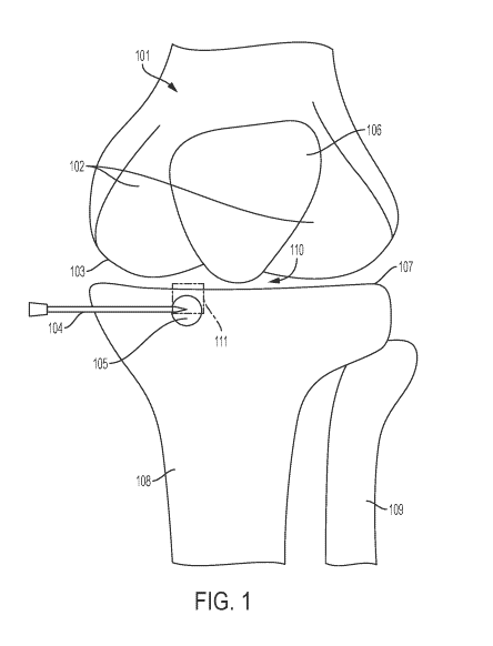
syringe.
[0087] In some embodiments, the solid component is disposed in a syringe
possessing an
integrated mixing device for in situ mixing of premeasured portions of the
solid component
and the liquid component to form the injectable biomaterial. In some
embodiments, the
syringe is sterile.
[0088] In some embodiments, the kit further comprises a Luer-Lock.
[0089] In some embodiments, the kit further comprises an end cap.
BRIEF DESCRIPTION OF THE DRAWINGS
[0090] FIG. 1 shows a schematic of a human knee comprising an area of
degenerate
bone.
[0091] FIGs. 2A-E show a schematic callout of an area comprising a portion
of the
degenerate bone of FIG. 1
18
CA 03021765 2018-10-22
WO 2017/189733 PCT/US2017/029651
[0092] FIGs. 3A-B show an injectable biomaterial according to the present
disclosure as
compared to an injectable biomaterial lacking a carbohydrate, each in
phosphate buffered
saline.
[0093] FIGs. 4A-D show injectable biomaterials according to the present
disclosure as
compared to an injectable biomaterial lacking a carbohydrate, each in
phosphate buffered
saline.
[0094] FIGs. 5A-D show an injectable biomaterial according to the present
disclosure as
compared to an injectable biomaterial lacking a carbohydrate, after removal of
excess
phosphate buffered saline.
[0095] FIGs. 6A-D show cross-sections of sawbone injected with injectable
biomaterials
according to the present disclosure as contrasted with sawbone injected with
an injectable
biomaterial lacking a carbohydrate.
[0096] FIGs. 7A-B show cross-sections of sawbone injected with injectable
biomaterials
according to the present disclosure as contrasted with sawbone injected with
an injectable
biomaterial lacking a carbohydrate.
[0097] FIGs. 8A-B show the results of a diffusion barrier experiment
comparing control
with an injectable biomaterial lacking a carbohydrate and an injectable
biomaterial according
to the present disclosure.
[0098] FIG. 9 shows an x-ray powder diffractogram of an injectable
biomaterial, post
setting and curing, according to the present disclosure.
[0099] FIG. 10 shows a Fourier Transform Infrared ("FT-IR") spectrograph of
an
injectable biomaterial according to the present disclosure.
[0100] FIGs. 11A-C show scanning electron microscopy ("SEM") images of an
injectable biomaterial according to the present disclosure after setting and
curing.
[0101] FIGs. 12A-B show micro-computed tomography ("micro CT") images from
different planes taken 6 weeks after administration of an injectable
biomaterial according to
the present disclosure into degenerate bone generated in skeletally mature New
Zealand
White rabbits.
[0102] FIG. 13 shows an image of an injectable biomaterial according to the
present
disclosure injected into canine femoral condyle.
[0103] FIG. 14 shows and image of an injectable biomaterial according to
the present
disclosure injected into human cadaver bone.
19
CA 03021765 2018-10-22
WO 2017/189733 PCT/US2017/029651
DETAILED DESCRIPTION OF THE INVENTION
[0104] The present disclosure relates to methods and compositions for the
treatment of
degenerate bone in a patient. In some embodiments, the methods and
compositions disclosed
herein are useful in the treatment, prevention, or in delaying the progression
of a bone disease
linked to bone degeneration, such as osteoarthritis ("OA"), rheumatoid
arthritis, and
avascular necrosis.
[0105] The inventors have surprisingly discovered that the addition of a
carbohydrate to
an injectable biomaterial provides for an injectable, intermixable, flowable,
settable, curable,
cohesive composition that adheres to bone. Further, the injectable
biomaterials disclosed
herein possess a low porosity and high dimensional stability that is desirable
for use in the
minimally invasive treatment of various bone diseases, a property absent in
the biomaterials
of the prior art.
[0106] Without wishing to be bound by theory, the inventors posit that a
low porosity and
high dimensional stability injectable biomaterial serves to arrest biochemical
communication
involving the joint space and the adjacent bone. The inventors posit that this
biochemical
communication is responsible for the onset of a number of symptoms of bone
degeneration,
including pain and fluid accumulation, and that continued bone degeneration
can facilitate the
progression of bone diseases linked to bone degeneration, such as OA,
rheumatoid arthritis,
and avascular necrosis. By way of example, as the cartilage and synovium
become injured,
whether from trauma or overuse, a milieu of inflammatory and/or non-
inflammatory
mediators is released from both tissues. These mediators enter the bone
through channels in
the cortical bone plane, stimulating pain through nociceptor activation, fluid
accumulation,
and degenerative changes in the affected area of bone (such as by the
formation of a bone
marrow lesion). The inventors hypothesize that, as a result of this
biochemical
communication between the joint space and the adjacent bone, the homeostasis
of the
affected area of bone is disturbed, resulting in dysregulation in the
synthesis and degradation
of bone proteins, and a degenerative positive feedback loop responsible for
the progression of
pathologies such as OA is formed. Blocking the effect of the inflammatory
and/or non-
inflammatory mediators causing bone degeneration is therefore essential to
treating,
preventing, and/or delaying the progression of bone disease.
[0107] Accordingly, the inventors set out to produce an injectable
biomaterial with low
porosity and high dimensional stability, but which were able to be prepared
using lower
CA 03021765 2018-10-22
WO 2017/189733 PCT/US2017/029651
solid-to-liquid ratios, allowing for their use in the minimally invasive
treatment of bone
disease linked to bone degeneration. During their research, the inventors
found that existing
techniques used to produce such injectable materials generally resulted in
materials
demonstrating poor cohesion, adhesion, setting and curing properties,
rendering such
materials unsuitable for use in the minimally invasive treatment of bone
disease. Similarly,
those prior techniques generally resulted in materials with high porosity that
would be poorly
suited to arresting biochemical communication between the affected area of
bone and the
adjacent joint space. See, e.g., Eliaz, N.; Metoki, N. Materials 2017, /0,
334, at 13, the
contents of which are incorporated by reference herein in their entirety. The
inventors then
surprisingly discovered that the addition of a carbohydrate to an injectable
biomaterial
allowed for a material that could be prepared with lower solid-to-liquid
ratios but was
nonetheless able to set, cure, maintain cohesiveness and adherency to bone,
and that
possessed low porosity and high dimensional stability as compared to
injectable biomaterials
prepared without an added carbohydrate. In other words, the inventors
discovered that the
addition of a carbohydrate provided for an injectable biomaterial having the
critical
combination of injectability, intermixability, flowability, settability and
curability, while
maintaining the desired properties of cohesivity, adhesivity, low porosity,
and high
dimensional stability. Accordingly, in one embodiment, the present disclosure
provides
injectable biomaterials that can be prepared using a lower solid-to-liquid
ratio than previously
possible while maintaining the cohesiveness, adherency to bone, low porosity,
and high
dimensional stability of materials traditionally made with a higher solid-to-
liquid ratio.
Accordingly, the disclosed injectable biomaterials are able to stem the flow
and prevent
ingress of inflammatory and non-inflammatory mediators into the affected area
of bone from
an adjacent joint space while not sacrificing the desirable ability to
intermix, be injected, flow
into, remain and set and cure in the area of degenerate bone to be treated
without being
cleared by the function of normal body fluid exchange. These materials provide
this
surprising and unique combination of properties and, contrary to the common
knowledge in
the art, which focused on the provision of high compressive strength and
elastic modulus
materials, unexpectedly allow for superior treatment of bone disease linked to
bone
degeneration as compared to prior compositions.
[0108] Without wishing to be bound by theory, the inventors posit that the
injectable
biomaterials disclosed herein serve to treat the underlying causes of bone
disease linked to
bone degeneration by eliminating and preventing recurrence of biochemical
communication
21
CA 03021765 2018-10-22
WO 2017/189733 PCT/US2017/029651
between the affected area of bone and the adjacent joint space. The injectable
biomaterials
disclosed herein provide a barrier between the affected area of bone and the
adjacent joint
space that prevents the ingress of inflammatory and/or non-inflammatory
mediators from the
joint space, arresting degradation mediated by, e.g., proteolytic enzymes and
inflammatory
cytokines, and allowing the bone to recover via normal dynamics (bone
resorption). In
contrast, prior injectable biomaterials addressed the symptoms, but not the
underlying causes
of bone disease linked to bone degeneration.
[0109] Furthermore, the disclosed injectable biomaterials possess lower
compressive
strength than the materials typically used in the prior art. Without wishing
to be bound by
theory, the inventors posit that, counter to the prevailing knowledge in the
art, provision of an
injectable biomaterial that does not biomechanically stabilize an affected
area of bone
provides for superior outcomes in the treatment, prevention, and slowing the
progression of
bone disease linked to bone degeneration. Conventional wisdom in the art
indicated that
injectable biomaterials (such as CPCs) with high compressive strength were
required to
properly treat joint pathologies by providing biomechanical stabilization.
Contrary to this
conventional wisdom, the inventors have surprisingly discovered that by
providing a weaker
injectable biomaterial that does not provide biomechanical support to the
affected area, the
disclosed methods and compositions do not artificially alter the biomechanical
forces to
which the joint is exposed, providing a superior methodology to address such
pathologies.
Without wishing to be bound by theory, the inventors posit that the provision
of
biomechanical support to an area of degenerate bone can be detrimental by
shifting stress and
strain to otherwise healthy tissue, potentially resulting in the spread of
bone disease and/or its
symptoms in accordance with Wolff s Law. The inventors posit that this is due
the
impossibility of exact recreation of healthy biomechanics, which necessarily
relates on undue
strain on other locations. For example, areas of high stress will become
thicker and stiffer
whereas areas of low stress will resorb according to Wolff s Law, which states
that bone in a
healthy person or animal will adapt to the loads under which it is placed.
Based on Wolff s
Law, if loading on a particular bone increases, the bone will remodel itself
over time to
become stronger to resist that sort of loading. However, these normal
biological responses
are not possible in bone disease because the degenerative biochemical feedback
loop present
in those conditions adversely affect, and can completely cease, normal bone
remodeling
processes. Accordingly, by not providing biomechanical support, the disclosed
methods and
compositions allow for physiological conditions that permit the affected area
of bone to
22
CA 03021765 2018-10-22
WO 2017/189733 PCT/US2017/029651
recover and rebuild through natural bone remodeling, thus slowing and/or
reversing the
progression of bone disease, and preventing the onset and progression of bone
diseases such
as symptomatic OA, rheumatoid arthritis, and avascular necrosis, as well as
preventing
spread of the disease or symptoms to adjacent healthy tissues.
[0110] In some embodiments, the strength of the fully set and cured
injectable
biomaterial is less than that provided by the injectable biomaterials of the
prior art. For
example, certain prior art injectable biomaterials possess a compressive
strength of
approximately 50 MPa, which is 4-10 times greater than the average 5-15 MPa
compressive
strength of healthy cancellous bone. See, e.g., Norian SRS Tiabia plateau
fractures,
Synthes , available at
htt )1/,'`Www rch.orau upoadedijieq/Mai n/Content/ortho/N oriati SRS Tibia
qateau fractui-
es.pdf (last visited April 24, 2017), the contents of which are incorporated
herein by reference
in their entirety. In some embodiments, the strength of the fully set and
cured injectable
biomaterial is characterized by one or more of compressive strength and
elastic modulus. In
some embodiments, the fully set and cured injectable biomaterial has a
compressive strength
of less about 20 MPa. In some embodiments, the fully set and cured injectable
biomaterial
has a compressive strength of less about 15 MPa. In some embodiments, the
fully set and
cured injectable biomaterial has a compressive strength of less about 10 MPa.
In some
embodiments, the fully set and cured injectable biomaterial has a compressive
strength of less
about 9 MPa. In some embodiments, the fully set and cured injectable
biomaterial has a
compressive strength of less about 8 MPa. In some embodiments, the fully set
and cured
injectable biomaterial has a compressive strength of less about 7 MPa. In some
embodiments, the fully set and cured injectable biomaterial has a compressive
strength of less
about 6 MPa. In some embodiments, the fully set and cured injectable
biomaterial has a
compressive strength of less about 5 MPa. In some embodiments, the fully set
and cured
injectable biomaterial has a compressive strength of less about 4 MPa. In some
embodiments, the fully set and cured injectable biomaterial has a compressive
strength of less
about 3 MPa. In some embodiments, the fully set and cured injectable
biomaterial has a
compressive strength of less about 2 MPa. In some embodiments, the fully set
and cured
injectable biomaterial has a compressive strength of less about 1 MPa.
[0111] In further embodiments, the elastic modulus of the fully set and
cured injectable
biomaterial is less than that provided by the injectable biomaterials of the
prior art. In further
embodiments, the elastic modulus of the fully set and cured injectable
biomaterial is close to
23
CA 03021765 2018-10-22
WO 2017/189733 PCT/US2017/029651
that of healthy subchondral bone. Without wishing to be bound by theory, the
inventors posit
that provision of an injectable biomaterial having an elastic modulus similar
to that of healthy
subchondral bone reduces the risk of altering natural biomechanics and
resulting in further
bone degradation in accordance with Wolff s Law (i.e., stress shielding). See,
e.g., Eliaz, N.;
Metoki, N. Materials 2017, /0, 334, at 3. For example, the elastic modulus of
samples of
human subchondral bone has been reported to be about 1.15 GPa. See, e.g.,
Choi, K. et at.
Biomech. 1990, 23(11), 1103-13; see also Brown, T. D.; Vrahas, M. S. I
Orthoped. Res.
1984, 2(1), 32-38 (reporting an apparent elastic modulus of 1.372 GPa for
machined caps of
subchondral bone); Mente, P. L.; Lewis, J. L. I Orthoped. Res. 1994, /2(5),
637-47
(reporting an elastic modulus calculated from "pure" bovine subchondral bone
beams of 2.3
1.5 GPa), the contents of all of the foregoing of which are incorporated
herein by reference in
their entireties. In some embodiments, the fully set and cured injectable
biomaterial has an
elastic modulus of less than about 5 GPa. In some embodiments, the fully set
and cured
injectable biomaterial has an elastic modulus of less than about 4 GPa. In
some
embodiments, the fully set and cured injectable biomaterial has an elastic
modulus of less
than about 3 GPa. In some embodiments, the fully set and cured injectable
biomaterial has an
elastic modulus of less than about 2 GPa. In some embodiments, the fully set
and cured
injectable biomaterial has an elastic modulus of less than about 1 GPa. In
some
embodiments, the fully set and cured injectable biomaterial has an elastic
modulus of less
than about 0.5 GPa. In some embodiments, the fully set and cured injectable
biomaterial has
an elastic modulus of less than about 0.25 GPa.
[0112] Moreover, the inventors surprisingly discovered that the disclosed
injectable
biomaterials set and cure without significant gaseous emission. Without
wishing to be bound
by theory, the inventors posit that the absence of gaseous release during
setting and curing
post-administration results in an injectable biomaterial does not expand
during setting and
curing, thus reducing or eliminating post-operative pain as compared with
prior art materials.
Furthermore, the inventors posit that the absence of gaseous release during
curing and setting
post-administration facilitates the formation of a structure with the desired
decreased porosity
as compared with prior art materials. The inventors posit that the provision
of an injectable
biomaterial that does not comprise bicarbonate may serve to prevent gaseous
release and thus
results in a biomaterial with decreased porosity that does not cause post-
operative pain.
24
CA 03021765 2018-10-22
WO 2017/189733 PCT/US2017/029651
Definitions
[0113] The term "adherent to bone" as used herein with reference to an
injectable
biomaterial refers to the materials demonstration of sufficient affinity for
bone such that it is
not readily cleared away from the site of injection by bodily fluids.
[0114] The term "bone disease" as used herein refers to a disease,
condition, or pathology
in a patient that is linked to bone degeneration. For example, the bone
disease can affect a
joint adjacent to a degenerate bone.
[0115] The term "cohesive" as used herein with reference to an injectable
biomaterial
refers to the ability of a material to adhere to itself and be molded such
that it non-transiently
maintains its shape over a period of time and fills an affected area of
degenerate bone after
injection.
[0116] The term "cure" as used herein with reference to an injectable
biomaterial refers
to the process whereby the components of the injectable biomaterial chemically
and
physically react to form the final desired crystal structure. A material that
is "cured" no
longer undergoes appreciable changes in compressive strength or porosity. The
terms "cure
time" and "curing time" as used herein with reference to an injectable
biomaterial refer to the
time at which the injectable biomaterial is fully cured. In some embodiments,
the injectable
biomaterials disclosed herein cure to form an apatitic crystal structure.
[0117] The term "decompression" as used herein refers to a procedure to
remove pressure
on a structure.
[0118] The term "degenerate tissue" as used herein refers to tissue that
has undergone a
change to a lower or less functionally active form.
[0119] The term "degenerate bone" as used herein refers to an area of bone
that has
undergone a change to a lower or less functionally active form. In some
embodiments,
degenerate bone exhibits at least one change selected from (1) formation of
higher volume
fraction trabeculae relative to normal bone; (2) decreased mineral-to-matrix
and carbonate-to-
matrix values relative to normal bone; (3) increased intraosseous fluid
accumulation relative
to normal bone; and (4) increased infiltration into the marrow space of a
fibrous collagen
network relative to normal bone.
[0120] The term "dewater" as used herein with reference to an injectable
biomaterial
refers to separation of the solid component and the liquid component.
CA 03021765 2018-10-22
WO 2017/189733 PCT/US2017/029651
[0121] The term "flowable" as used herein with reference to an injectable
biomaterial
refers to any generally incompressible material which may be caused to flow
under pressure
or gravity.
[0122] The term "inflammatory mediator" as used herein refers to a
biological
component that induces an inflammatory response in an animal or a human.
Inflammatory
mediators include, but are not limited to, chemokines, such as C5, CCL1 (1-
309), CCL11
(eotaxin), CCL13 (MCP-4), CCL15 (MIP-1d), CCL16 (HCC-4), CCL17 (TARC), CCL2
(MCP-1), CCL20 (MIP-3a), CCL22 (MDC), CCL23 (MPIF-1), CCL24 (MPIF-2, Eotaxin-
2,
MPIF-2, Eotaxin-2), CCL26 (eotaxin-3), CCL3 (MIP-1A), CCL4 (MIP-1B), CCL5
(RANTES), CCL7 (MCP-3), CCL8 (MCP-2), CX3CL1, CXCL1 (GRO1, GRO-alpha,
SCYB1), CXCL10 (INP10), CXCL11 (I-TAC, IP-9), CXCL12 (SDF1), CXCL13, CXCL2
(GRO2, GRO-beta, SCYB2), CXCL3, CXCL5 (ENA-78, LIX), CXCL6 (GCP-2), CXCL9
(MIG); interleukins, such as IL13, IL15, IL16, IL17A, IL17C, IL17F, ILlA,
IL1B, IL1RN,
IL21, IL27, IL3, IL33, IL5, IL7, CXCL8, IL9; cytokines such as AIMP1 (SCYE1),
BMP2,
CD4OLG (TNFSF5), CSF1 (MCSF), CSF2 (GM-CSF), CSF3 (GCSF), FASLG (TNFSF6),
GM-CSF, IFNA2, IFNG, IL-1, IL-6, IL-8, IL-15, IL-16, IL-17, IL-18, IFN-y, LTA
(TNFB),
LTB, MIF, NAMPT, OSM, SPP1, TGF-f3, TNF, TNF-a, TNFSF10 (TRAIL), TNFSF11
(RANKL), TNFSF13, TNFSF13B, TNFSF4 (0X4OL), VEGFA; and other inflammatory
mediators, such as bradykinin, calcitonin gene related peptide (CGRP),
histamine, lactic acid,
nerve growth factor (NGF), prostaglandins, substance P, and vasoactive
intestinal peptide;
and mixtures thereof. Other inflammatory mediators are known to those skilled
in the art.
[0123] The term "initial setting time" as used herein with reference to an
injectable
biomaterial refers to the shortest amount of time between (1) the mixing of
the solid
component and the liquid component and (2) the point where a 1 lb. (454 g)
Gilmore Needle,
having a tip diameter of 1/24 inch (1.06 mm) does not penetrate a sample of
the injectable
biomaterial of uniform thickness of 3/16 inch (5 mm), the length of the needle
tip, in less than
1 minute. See ASTM C414-03 at 7.2, 8.2 (reapproved 2012), the contents of
which are
incorporated herein by reference in their entirety. The term "set" or
"settable" as used herein
with reference to an injectable biomaterial refers to the ability of the
injectable material to
transition from that state achieved at point (1) to point (2) described above.
[0124] The term "injectable" as used herein with reference to an injectable
biomaterial
refers to a material that is capable of being extruded from a syringe using no
more than
acceptable hand pressure applied to a plunger rod. Acceptable hand pressure is
known to
26
CA 03021765 2018-10-22
WO 2017/189733 PCT/US2017/029651
those of skill in the art. In some embodiments, the injectable biomaterial
must be capable of
being extruded from a syringe without the use of mechanical advantage, such as
a screw-
driven syringe. In some embodiments, the syringe is a 3 mL syringe. In some
embodiments,
the syringe is a 5 mL syringe. In some embodiments, the syringe is a 10 mL
syringe. In
some embodiments, the syringe is a 14 mL syringe. In some embodiments, an
injectable
biomaterial is able to be extruded from a syringe using no more than 15 lb.
extrusion force at
a rate of 6 mL/minute. In some embodiments, an injectable biomaterial is able
to be extruded
from a syringe using no more than 10 lb. extrusion force at a rate of 6
mL/minute. In some
embodiments, an injectable biomaterial is able to be extruded from a syringe
using no more
than 7.5 lb. extrusion force at a rate of 6 mL/minute. In some embodiments, an
injectable
biomaterial is able to be extruded from a syringe using no more than 5 lb.
extrusion force at a
rate of 6 mL/minute. In some embodiments, the syringe is coupled to an 11
gauge cannula.
In some embodiments, the syringe is coupled to an 14 gauge cannula. In some
embodiments,
the syringe is coupled to a 15 gauge cannula. In some embodiments, the syringe
is coupled to
an 18 gauge cannula. In some embodiments, the syringe is coupled to a 21 gauge
cannula. In
some embodiments, the syringe is coupled to a cannula and the injectable
biomaterial must
flow down the length of the cannula in its entirety without seizing or
dewatering to be
considered injectable. In some embodiments, the cannula is about 11 cm long.
In some
embodiments, the cannula is about 15 cm long.
[0125] The term "intermixable" as used herein with reference to an
injectable biomaterial
refers to the ability to achieve full mixing of the solid component and the
liquid component
when each is disposed in a 5 mL syringe, wherein the syringes are coupled and
the contents
extruded between the syringes for 1 minute using hand pressure with no visible
seizing or
dewatering.
[0126] The term "macroporous" as used herein with reference to an
injectable biomaterial
refers to a material that possess pores which are visible macroscopically.
[0127] The term "median pore diameter" as used herein refers to the pore
diameter
corresponding to 50% total intrusion volume from a cumulative intrusion volume
vs.
diameter plot. See Webb, P. An introduction to the physical characterization
of materials by
mercury intrusion porosimetry with emphasis on reduction and presentation of
experimental
data, Micromeritics Instrument Corp. (2001), the contents of which are
incorporated herein
by reference in their entirety.
27
CA 03021765 2018-10-22
WO 2017/189733 PCT/US2017/029651
[0128] The term "osteoconductive" as used herein with reference to an
injectable
biomaterial refers to the ability of an osteogenic material to serve as a
substrate, scaffold or
framework supporting new bone growth that is perpetuated by the native bone.
[0129] The term "osteogenic" as used herein with reference to an injectable
biomaterial
refers to the ability of a material to promote the growth of new bone tissue.
Exemplary
osteogenic materials include, but are not limited to, bone marrow aspirate,
bone marrow
aspirate concentrate, platelet-rich plasma, platelet-poor plasma, somatic cell
autografts, stem
cell autografts, stem cell allografts, and mixtures thereof. Other exemplary
osteogenic
materials are known to those skilled in the art.
[0130] The term "osteoinductive" as used herein with reference to an
injectable
biomaterial refers to the ability of an osteogenic material to recruit cells
from the host that
have the potential for forming new bone and repairing bone tissue.
Osteoinductive injectable
biomaterials according to the present disclosure stimulate osteoprogenitor
cells to
differentiate into osteoblasts that then begin new bone formation. Exemplary
osteoinductive
materials include, but are not limited to, BMP2, BMP7, BMP9, PDGF, and P15.
See, e.g.,
Neiva, R. F. et at. I Periodontal. 2008, 79(2), 291-99, the contents of which
are incorporated
herein by reference in their entirety. Other osteoinductive materials are
known to those
skilled in the art.
[0131] The term "proteolytic enzymes" as used herein refers to enzymes
capable of
breaking down various proteins. Proteolytic enzymes include, but are not
limited to, matrix
metalloproteinases (MMPs), tissue inhibitors of metalloproteinases (TIMPs), a
disintegrin
and metalloproteinase with thrombospondin motifs (ADAM-TS), and mixtures
thereof
Other proteolytic enzymes are known to those skilled in the art.
[0132] The term "patient" as used herein refers to humans and non-humans
such as
primates, pets and farm animals.
[0133] The term "resorbable" as used herein with reference to an injectable
biomaterial
refers to the ability of a material to be broken down and assimilated back
into the body over
time.
[0134] The term "self-setting" as used herein with reference to an
injectable biomaterial
refers to the ability of the material to form an apatitic crystal structure as
a result of the
mixing of the solid component and the liquid component. In contrast, certain
prior art
materials provide an apatitic crystal structure, e.g., by mixing pre-formed
hydroxyapatite with
a cement-forming material. Moreover, certain such cement-forming materials
(e.g., swellable
28
CA 03021765 2018-10-22
WO 2017/189733 PCT/US2017/029651
polymers) lack dimensional stability and are permeable, and are thus
inappropriate for
preventing ingress of inflammatory and/or non-inflammatory mediators from an
adjacent
joint space.
[0135] The term "seize" as used herein with reference to an injectable
biomaterial refers
to the rapid conversion of the injectable biomaterial to a material that is
uninjectable.
[0136] The term "stable" as used herein with reference to an injectable
biomaterial refers
to the ability of the injectable biomaterial, or its precursors, to maintain
sufficient physical
and/or chemical properties such that it still functions for its intended
purpose in accordance
with the present disclosure after a period of time has elapsed. For example,
stability includes,
but is not limited to, the ability of the injectable biomaterial to mix, set,
and/or cure.
[0137] The terms "subchondral bone plate" and "cortical bone plate" are
used herein
interchangeably, and refer to the thin cortical lamella lying immediately
beneath the calcified
cartilage.
[0138] The term "total porous area" as used herein refers to the total sum
of all pore wall
area derived from the volume of each incremental intrusion step. Assuming
cylindrical
pores, the wall area of each step i is Ai= 4 Vi/Di, where Vi = pore volume and
Di = pore
diameter. See Webb, P. An introduction to the physical characterization of
materials by
mercury intrusion porosimetry with emphasis on reduction and presentation of
experimental
data, Micromeritics Instrument Corp. (2001), the contents of which are
incorporated herein
by reference in their entirety.
[0139] The terms "treatment," "treating," "treat," "therapy,"
"therapeutic," and the like
are used herein to refer generally to attempting to obtain a desired
pharmacological,
biological, and/or physiological effect. The effect may be prophylactic in
terms of
completely or partially preventing or delaying the onset of a condition or
symptom thereof
and/or may be therapeutic in terms of a partial or complete stabilization,
amelioration, or
remedying of the condition or symptom.
[0140] The term "true density" as used herein refers to the mass divided by
the solid
volume or true skeletal volume. True density is usually determined after the
substance has
been reduced to a particle size so small that it accommodates no internal
voids. See Webb, P.
An introduction to the physical characterization of materials by mercury
intrusion
porosimetry with emphasis on reduction and presentation of experimental data,
Micromeritics Instrument Corp. (2001), the contents of which are incorporated
herein by
29
CA 03021765 2018-10-22
WO 2017/189733 PCT/US2017/029651
reference in their entirety. True density can be measured, e.g., by helium
pycnometry (such
as by use of an AccuPyc 11 1340 pycnometer).
[0141] The term "working time" as used herein with reference to an
injectable
biomaterial refers to the maximum amount of time between (1) the mixing of the
solid
component and the liquid component and (2) the point where the injectable
biomaterial is no
longer workable. The injectable biomaterial is workable if, after the elapsed
time after
mixing the solid component and the liquid component for one minute and
injecting into an
area of degenerate bone (or a substitute such as a sawbone model), the
injectable biomaterial
flows to fill the porosity of the area of administration prior to setting. The
injectable
biomaterial is no longer workable if, after the elapsed time after mixing the
solid component
and the liquid component for one minute and injecting into an area of
degenerate bone (or a
substitute such as a sawbone model, the injectable biomaterial sets prior to
filling the porosity
of the area of administration. Tests for working time are known to those
skilled in the art.
See, e.g., ASTM C414-03 at 7.2, 8.2 (reapproved 2012), the contents of which
are
incorporated herein by reference in their entirety.
Bone Structure
[0142] Bone disease often affects the whole of the adjacent joint. FIG. 1
shows a
schematic of a joint affected by bone disease. Femur 101 comprises cartilage
102 and
femoral articular surface 103, while patella 106 is shown above the joint
space 110. The
lower portion of FIG. 1 shows fibula 109 and tibia 108, the latter of which
comprises an area
of degenerate bone 105 into which a cannula 104 is inserted. A portion of
tibial articular
surface 107 is encompassed by callout 111, which is shown in greater detail in
the schematics
of FIGs. 2A-E, discussed in more detail as follows.
[0143] Subchondral bone plays a crucial role in the initiation and
progression of bone
diseases such as OA. The term "subchondral bone" as used herein refers to the
bony
components lying distal to calcified cartilage. Subchondral bone comprises the
subchondral
bone plate (aka cortical bone plate) and subchondral trabecular bone (aka
cancellous bone).
The subchondral bone plate and cancellous bone are not divided by a sharp
border, but
nonetheless are distinct anatomic entities.
[0144] As shown in FIGs. 2A-E, the subchondral bone plate is a thin
cortical lamella,
lying immediately beneath the calcified cartilage 205. This bone plate is not
an impenetrable
structure, but rather possesses a marked porosity. It is invaded by channels
that provide a
CA 03021765 2018-10-22
WO 2017/189733 PCT/US2017/029651
direct link between articular calcified cartilage and subchondral trabecular
bone. A high
number of arterial and venous vessels and/or nerves (collectively 202),
penetrate through the
channels and send branches into calcified cartilage 205. The distribution and
intensity of the
channels depend not only on the age of the tissues, but also on the magnitude
of the
compressive forces transmitting through calcified cartilage and subchondral
bone within and
between joints. These channels are preferentially concentrated in the heavily
stressed areas
of the joint. Channel shape and diameter also differs with the thickness of
the cortical plate.
Channels are narrower and form a tree-like mesh in regions where the plate is
thicker, while
they tend to be wider and resemble ampullae where the subchondral bone plate
is thinner.
[0145] Arising from the subchondral bone plate is the supporting
trabeculae, which are
structures that are part of and support the trabecular bone, and also include
deeper bone
structure. Subchondral trabecular bone exerts important shock-absorbing and
supportive
functions in normal joints, and may also be important for calcified cartilage
nutrient supply
and metabolism. Relative to the subchondral bone plate 207, subchondral
trabecular bone
(not shown, but disposed distal the joint space from the subchondral bone
plate 207) is more
porous and metabolically active, containing blood vessels, sensory nerves, and
bone marrow.
Subchondral trabecular bone has an inhomogeneous structure that varies with
the distance
from the articular surface 203/208. It exhibits significant structural and
mechanical
anisotropy; that is, the bone trabeculae show preferential spatial orientation
and parallelism.
[0146] Subchondral bone is a dynamic structure and is uniquely adapted to
the
mechanical forces imposed across the joint. In addition to bone density
patterns and
mechanical properties, subchondral bone also dynamically adjusts trabecular
orientation and
scale parameters in a precise relationship with principal stress. Mechanical
stress also
modifies the contour and shape of subchondral bone by means of bone modeling
and
remodeling. Subchondral bone and calcified cartilage are dynamic stress-
bearing structures
that play complementary roles in load-bearing of joints. Subchondral bone
supports
overlying articular cartilage 210 and distributes mechanical loads across
joint surfaces with a
gradual transition in stress and strain. Stiffened and less pliable
subchondral bone tends to
transmit increased loads to overlying cartilage, leading to secondary
cartilage damage and
degeneration. The load transmitted to underlying bone is substantially
increased after
articular cartilage damage or loss.
[0147] Articular cartilage overlies subchondral bone, and provides a vital
function of
maintaining homeostasis of the joint environment. It encompasses superficial
non-calcified
31
CA 03021765 2018-10-22
WO 2017/189733 PCT/US2017/029651
cartilage 210 and deeper calcified cartilage 205. Calcified cartilage 205 is
permeable to small
molecule transport, and plays an important role in the biochemical interaction
between non-
calcified cartilage 210 and subchondral bone. It is separated from non-
calcified cartilage 210
by a boundary called the "tidemark" (204), a dynamic structure that appears as
a basophilic
line in histological sections. The tidemark 204 represents the mineralization
front of calcified
cartilage 205, and provides a gradual transition between the two dissimilar
cartilage regions.
Continuous collagen fibrils cross the tidemark, indicating the strong link
between non-
calcified cartilage 210 and calcified cartilage 205. There is also a sharp
borderline between
calcified cartilage 205 and subchondral bone, called the "cement line" (206).
Unlike the
tidemark 204, however, no continuous collagen fibrils cross the cement line.
[0148] Given the intimate contact between articular cartilage 210 and
subchondral bone,
they form a closely composited functional unit called the "osteochondral
junction" (200).
The osteochondral junction 200 is peculiarly complex, and consists of a layer
of non-calcified
cartilage 210, the tidemark 204, calcified cartilage 205, the cement line 206
and subchondral
bone.
[0149] In some embodiments, the affected area of bone is disposed about 50
mm or less
from the joint articular surface proximal to the affected area of bone. In
some embodiments,
the affected area of bone is disposed about 40 mm or less from the joint
articular surface
proximal to the affected area of bone. In some embodiments, the affected area
of bone is
disposed about 30 mm or less from the joint articular surface proximal to the
affected area of
bone. In some embodiments, the affected area of bone is disposed about 20 mm
or less from
the joint articular surface proximal to the affected area of bone. In some
embodiments, the
affected area of bone is disposed about 15 mm or less from the joint articular
surface
proximal to the affected area of bone. In some embodiments, the affected area
of bone is
disposed about 10 mm or less from the joint articular surface proximal to the
affected area of
bone. In some embodiments, the affected area of bone is disposed about 9 mm or
less from
the joint articular surface proximal to the affected area of bone. In some
embodiments, the
affected area of bone is disposed about 8 mm or less from the joint articular
surface proximal
to the affected area of bone. In some embodiments, the affected area of bone
is disposed
about 7 mm or less from the joint articular surface proximal to the affected
area of bone. In
some embodiments, the affected area of bone is disposed about 6 mm or less
from the joint
articular surface proximal to the affected area of bone. In some embodiments,
the affected
area of bone is disposed about 5 mm or less from the joint articular surface
proximal to the
32
CA 03021765 2018-10-22
WO 2017/189733 PCT/US2017/029651
affected area of bone. In some embodiments, the affected area of bone is
disposed about 4
mm or less from the joint articular surface proximal to the affected area of
bone. In some
embodiments, the affected area of bone is disposed 3 mm or less from the joint
articular
surface proximal to the affected area of bone. In some embodiments, the
affected area of
bone is disposed about 2 mm or less from the joint articular surface proximal
to the affected
area of bone. In some embodiments, the affected area of bone is disposed about
1 mm or less
from the joint articular surface proximal to the affected area of bone.
Etiology of Bone Disease
[0150] Articular cartilage is both aneural and avascular. As such,
cartilage is incapable
of directly generating pain, stiffness (e.g., either the symptom of pain on
moving a joint, the
symptom of loss of range of motion, or the physical sign of reduced range of
motion), or any
of the symptoms that patients with bone disease typically describe. In
contrast, the
subchondral bone, periosteum, periarticular ligaments, periarticular muscle
spasm, synovium
and joint capsule are all richly innervated and can be the source of
nociception in bone
disease. Furthermore, cross-talk (i.e., biochemical communication) between
subchondral
bone, articular cartilage 210, and joint space 211 is crucial for the
initiation and progression
of bone disease linked to bone degeneration, in terms of pain, function and
pathology.
Alterations of any tissue will modulate the properties and functions of other
parts of the
osteochondral junction 200. There is intensive stress transfer and biochemical
cross-talk
across this region which plays a role in maintenance and degeneration of the
joint.
[0151] The permeability of calcified cartilage 205 and subchondral bone
plate 207 allows
crossover communication, and provides connecting channels between subchondral
bone and
the joint space 211. In vivo studies showed that prostaglandins, leukotrienes
and various
growth factors released by osteoblasts during subchondral bone remodeling
could reach
overlying articular cartilage 210. Conversely, inflammatory and osteoclast
stimulation
factors released by articular cartilage 210 also leads to subchondral bone
deterioration
through increased bone remodeling in bone disease.
[0152] During bone disease linked to bone degeneration, functional units of
joints
comprising cartilage and subchondral bone can undergo uncontrolled catabolic
and anabolic
remodeling processes to adapt to local biochemical and biological signals.
Changes in
cartilage and subchondral bone are not merely secondary manifestations of bone
disease but
are active components of the disease, contributing to its severity. Increased
vascularization
33
CA 03021765 2018-10-22
WO 2017/189733 PCT/US2017/029651
and formation of microcracks in joints during bone disease have suggested the
facilitation of
movement of molecules from the joint space to subchondral bone and vice versa
through the
synovial and bone marrow fluids. Several biological factors and signaling
molecules
produced from both tissues may passage from one zone to another, affecting
homeostasis of
neighboring tissue. Secreted cytokines, growth factors and signaling molecules
form
cartilage-bone biochemical units play modulatory roles to alter
pathophysiology of joints
during bone disease through pathways such as WNT (wingless type), BMP (bone
morphogenic protein), TGF-(3 (transforming growth factor (3) and MAPK (mitogen-
activated
protein kinase) signaling. The close proximity of cartilage and subchondral
bone provides an
ample opportunity to induce physical and functional alteration in each other
through
molecular interaction.
[0153] As a result of the biochemical cross-talk between the joint space
211 and
subchondral bone, a degenerative biochemical response is initiated which
accelerates as
biomechanical changes begin to manifest themselves in patients with bone
disease. Both the
subchondral cortical plate 207 and cancellous bone show distinct differences
in their behavior
during progression of bone disease and hence must be regarded as separate
units to
understand the joint deformation events. During progression of bone disease,
subchondral
bone turnover can be 20-fold increase compared to normal bone turnover.
Subchondral bone
in bone disease patients secrete high levels of alkaline phosphatase (ALP),
osteocalcin,
osteopontin, IL-6, IL-8, and progressive ankylosis protein homolog (ANKH),
urokinase
plasminogen activator (uPA), prostaglandin and growth factors, such as IGF-1,
IGF-2 and
TGF-(3 and Type 1 collagen compared to normal subchondral bone. These secreted
biochemical factors contribute to bone formation, suggesting an enhanced bone
anabolic
activity of subchondral bone osteoblasts, exemplified by formation of
osteophytes and the
sclerosis observed in bone disease. However, the bone forming activity of
subchondral bone
is not necessarily accompanied by equivalent mineralization. Unmineralized
immature new
bone formation may lead to abundant osteoids in the subchondral bone (both at
the level of
the cortical plate and at the level of the trabecular bone) resulting in the
opposite effect on
tissue properties.
[0154] Inflammatory mediators present in synovial fluid also contribute to
catabolic
activities of chondrocytes leading to remodeling of the cartilage
extracellular matrix.
Chemokines, cytokines and proteases secreted from chondrocytes and present in
the synovial
fluid alter biochemical (e.g., catabolic) and functional abilities of
cartilage. During bone
34
CA 03021765 2018-10-22
WO 2017/189733 PCT/US2017/029651
disease, chondrocytes have been found to secrete TNF-a, IL-1, IL10 converting
enzyme
(caspase-1) and type 1 IL-1 receptor. The concentration at which IL-1 is
synthesized by
chondrocytes is capable of inducing the activation of proteolytic enzymes,
such as matrix
metalloproteinases (MNIPs), aggrecanases, a disintegrin and metalloproteinase
with
thrombospondin motifs (ADAM-TS) and other catabolic genes in regions of matrix
depletion
in affected cartilage. Furthermore, under these conditions, chondrocytes are
stimulated to
express molecules that are associated with chondrocyte hypertrophy and
terminal
differentiation, like VEGF, runt-related transcription factor 2 (RUNX2) and
MMP-13.
Secretion of angiogenic factors such as VEGF increase vascularity within the
deep layers of
articular cartilage facilitating, together with the presence of microcracks,
molecular transport
of inflammatory and/or non-inflammatory mediators by diffusion from the joint
space and
into the articular cartilage and the subchondral bone. Fine, unmyelinated
nerves (C-fibers
and sympathetic nerves) accompany also these vessels and enervate normally
aneural tissues,
a source of bone disease pain. In addition, IL-6, in combination with other
cytokines like
IL10, can switch osteoblasts from a normal phenotype to a sclerotic phenotype.
All these
actors potentiate and stimulate the process of bone remodeling, altering the
physiology of
subchondral bone.
[0155] These inflammatory and/or non-inflammatory mediators also affect
nociception in
the synovial tissue and the subchondral bone. Nociceptors encompass a broad
range of
receptors for ligands that change the properties of these neurons, such that
they require lower
thresholds to fire action potentials or even fire spontaneously when the
receptors are engaged.
These ligands include, but are not limited to, cytokines, chemokines,
neuropeptides and
prostaglandins, which in some embodiments all form part of the biochemical
milieu in the
affected joint. As a result of this peripheral sensitization, joint movement
within the normal
range becomes painful (a phenomenon known as mechanical allodynia).
[0156] Furthermore, inflammatory mediators such as bradykinin, histamine,
prostaglandins, lactic acid, substance P, vasoactive intestinal peptide, nerve
growth factor
(NGF), and calcitonin gene related peptide (CGRP) are released into the joint
from e.g.,
synovial fibroblasts and migrate into the subchondral bone and synovium,
activating the
nociceptors, located in those regions. These mediators reduce the firing
threshold of the
nociceptors, making them more likely to respond to both non-noxious and
noxious painful
stimuli. As the disease progresses, more and more of these mediators
accumulate in the joint
CA 03021765 2018-10-22
WO 2017/189733 PCT/US2017/029651
migrate into the subchondral space and synovium, thereby triggering a self-
perpetuating cycle
of pain generation.
[0157] As the damage progresses, degeneration of the subchondral bone
becomes
radiographically visible. The severity of the joint damage on the radiograph
may bear little
relation to the severity of the pain experienced. However, utilizing imaging
modalities such
as magnetic resonance imaging ("MM"), significant structural associations such
as bone
degeneration, sub-articular bone attrition, synovitis and effusion have been
related to knee
pain.
[0158] Without wishing to be bound by theory, the inventors posit that
methods and
compositions disclosed herein address these issues by breaking the biological
communication
between the joint space and the affected area of bone by optionally aspirating
inflammatory
and/or non-inflammatory mediators from the subchondral bone and subsequently
filling the
interconnected pores of the affected area with an injectable biomaterial. FIG.
2A shows a
schematic of an exemplary embodiment of a diseased joint that can be treated
according to
the present disclosure. FIGs. 2A-E are a callout of the area of degenerate
bone 105 shown in
FIG. 1. Osteochondral junction 200 comprises an area of degenerate bone 209,
surrounded
by blood vessels/and or nerves 202. A biological fluid comprising inflammatory
and/or non-
inflammatory mediators 201 have permeated from the joint space 211 past the
articular
surface 203/208 through articular cartilage 210, tidemark 204, calcified
cartilage 205 and
have collected in degenerate area of bone 209. In an exemplary embodiment
shown in FIG.
2B, cannula 211 is inserted into the area of degenerate bone 209 and the
biological fluid 201
is aspirated through the cannula 211 and removed. As shown in FIG. 2C,
injectable
biomaterial 212 prepared according to the present disclosure fills the area of
degenerate bone
209 in the affected area. As shown in FIG. 2D, the injectable biomaterial
cures to form a low
porosity, high dimensional stability material 213, thereby halting biochemical
communication
to and from the joint space and protecting the affected area and surrounding
cells. Moreover,
the injectable biomaterial remains in place for a period of time, preventing
the re-infiltration
of these inflammatory and/or non-inflammatory mediators. Accordingly,
biochemical
communication between the affected area of bone and the joint space is
arrested by blocking
the porosity connecting the affected area of bone and joint space, temporally
breaking
biochemical communication and allowing the affected area of bone to recover.
By shielding
the affected area of bone from these actors, arthritic degeneration of the
joint is prevented.
The latter results from the prevention of further biochemical degeneration of
the bone and of
36
CA 03021765 2018-10-22
WO 2017/189733 PCT/US2017/029651
the adjacent meniscal and cartilage tissues, and alleviation of the
corresponding pain in the
joint. Furthermore, certain injectable biomaterials according to the present
disclosure provide
for biomechanical repair of the affected area, such as by the provision of
osteoconductive
and/or osteoinductive surfaces to encourage natural bone healing processes,
shifting the
damage/repair equilibrium toward repair. This exemplary embodiment is shown in
FIG. 2E,
where osteoclasts 214 resorb the cured injectable biomaterial 213 and
osteoblasts 215 begin
to build new, healthy bone 216. Importantly, the compositions and methods
disclosed herein
achieve their intended effects, namely cessation of the underlying causes of
bone disease
linked to bone degeneration, without further altering the biomechanical
stability of the joint
being treated or causing significant pain post-operatively that can be
associated with volume
expansion from gas. They therefore offer several advantages over prior art
treatments for
bone disease.
Identification of Bone for Treatment
[0159] In some embodiments, an affected area for treatment according to the
compositions and methods disclosed herein is identified by the use of MRI. In
further
embodiments, the MRI is a knee MRI. In further embodiments, the MM is an ankle
MM. In
some embodiments, the MRI is weight-bearing MRI. In some embodiments, the MRI
is open
MRI. In some embodiments, the MIR is upright open MM. In further embodiments,
the
MRI is low field strength MRI. In further embodiments, the MRI is an ultra-
high field MM.
In further embodiments, the MRI is extremity MM. In further embodiments, the
MM is
whole body scanner MRI. In some embodiments, the affected area is identified
by
hyperintense signals on T2-weighted fat saturated MM images. In some
embodiments, MRI
Tip value, an indicator of early cartilage degradation, is elevated in
cartilage overlying bone
marrow lesions, with the level of cartilage degradation proportional to Tip
signal intensity in
a bone marrow lesion. In further embodiments, the affected area is identified
using
Technetium-99 bone scans. In some embodiments, the affected area is identified
using
fluoroscopy.
[0160] Without wishing to be bound by theory, the inventors posit that
affected areas of
bone thought to be associated with arthritis are disposed less than 50 mm, 10
mm or 1 mm
from the joint. Accordingly, in some embodiments, the methods and compositions
disclosed
herein are used to treat affected areas of bone which are disposed between
about 0 mm to
about 50 mm from the joint. In further embodiments, the affected area of bone
is disposed
37
CA 03021765 2018-10-22
WO 2017/189733 PCT/US2017/029651
between about 0 mm to about 10 mm from the joint. In still further
embodiments, the
affected area of bone is disposed between about 0 mm to about 1 mm from the
joint. In some
embodiments, the affected area of bone is disposed about 40 mm or less from
the joint. In
some embodiments, the affected area of bone is disposed about 20 mm or less
from the joint.
In some embodiments, the affected area of bone is disposed about 15 mm or less
from the
joint. In some embodiments, the affected area of bone is disposed about 10 mm
or less from
the joint. In some embodiments, the affected area of bone is disposed about 9
mm or less
from the joint. In some embodiments, the affected area of bone is disposed
about 8 mm or
less from the joint. In some embodiments, the affected area of bone is
disposed about 7 mm
or less from the joint. In some embodiments, the affected area of bone is
disposed about 6
mm or less from the joint. In some embodiments, the affected area of bone is
disposed about
mm or less from the joint. In some embodiments, the affected area of bone is
disposed
about 4 mm or less from the joint. In some embodiments, the affected area of
bone is
disposed 3 mm or less from the joint. In some embodiments, the affected area
of bone is
disposed about 2 mm or less from the joint. In some embodiments, the affected
area of bone
is disposed about 1 mm or less from the joint.
[0161] In some embodiments, the affected area of bone comprises a bone
marrow lesion.
In further embodiments, the affected area of bone comprises degenerate
cancellous bone
space.
[0162] In some embodiments, the location of administration of injectable
biomaterial is
determined by studying a previously captured image of the affected area. In
further
embodiments, the location of administration of injectable biomaterial is
determined using
additional guidance during surgery. In some embodiments, the additional
guidance
comprises real-time fluoroscopic imaging. In further embodiments, the
additional guidance
comprises robotic devices. In further embodiments, the additional guidance
comprises braces
for maintaining the joint in a position consistent with previously captured
images of the joint.
In further embodiments, the additional guidance comprises the use of one or
more labels. In
some embodiments, the one or more labels comprise radioactive labels. In some
embodiments, the radioactive labels comprise Technetium-99. In some
embodiments, the
one or more labels comprise radioactive label fiducial markers.
38
CA 03021765 2018-10-22
WO 2017/189733 PCT/US2017/029651
Iniectable Biomaterials
[0163] The injectable biomaterial disclosed herein is a physiologically
compatible
material that fills the porosity in the affected area of bone which is
symptomatic of bone
disease. Certain properties of the injectable biomaterial, namely that it is
injectable,
intermixable, flowable, is cohesive, and adherent to bone, allow the
biomaterial to fill the
porosity that exists in the affected area of bone without being readily
cleared away by bodily
fluids.
[0164] The injectable biomaterials disclosed herein comprise a solid
component and a
liquid component that comprises a carbohydrate. The injectable biomaterials
disclosed herein
can be prepared by mixing of the solid component and the liquid component. The
injectable
biomaterials disclosed herein set and cure to form an apatitic crystal
structure after mixing of
the solid component and the liquid component. The solid component provides a
solid
material, or mix of materials, that reacts when combined with the liquid
component to form
the apatitic crystal structure. The liquid component provides a medium for the
components
of the solid component to mix and react to form the apatitic crystal
structure. In some
embodiments, the solid component comprises a calcium phosphate. In some
embodiments,
the liquid component comprises water. In some embodiments, the solid component
and/or
the liquid component include additional components.
[0165] The liquid component of the injectable biomaterials disclosed herein
includes a
carbohydrate. While the carbohydrate itself may not be a liquid, when provided
as a
constituent of the liquid component, it is dissolved or suspended therein. In
some
embodiments, the liquid component is in the form of a gel or a hydrogel.
[0166] In some embodiments, the solid component and the liquid component
are mixed at
a particular ratio to achieve the desired injectable biomaterial. In some
embodiments, the
ratio of solid component to liquid component is about 3 to about 1 by mass. In
some
embodiments, the ratio of solid component to liquid component is about 2 to
about 1 by mass.
In some embodiments, the ratio of solid component to liquid component is about
1.5 to about
1 by mass. In some embodiments, the ratio of solid component to liquid
component is about
1 to about 1 by mass. In some embodiments, these ratios of solid component to
liquid
component provide an injectable biomaterial that is injectable. While prior
art materials
comprising a solid component and a liquid component comprising a carbohydrate
are
disclosed, these materials utilize a higher powder-to-liquid ratio than is
achievable according
39
CA 03021765 2018-10-22
WO 2017/189733 PCT/US2017/029651
to the present disclosure. See, e.g., Ahmadzadeh-Asl, S. et at. Adv. Applied
Ceramics 2011,
110(6) , 340-45, the contents of which are incorporated herein by reference in
their entirety.
Accordingly, these materials are not intermixable and require high extrusion
forces to
dispense the entirety of the material, resulting in compositions that cannot
be readily
administered in a minimally invasive fashion with the ease of the presently
disclosed
injectable biomaterials. In contrast, the injectable biomaterials of the
present disclosure are
readily intermixable, such that the solid component and liquid component can
be provided in
separate syringes, which are coupled to one another, the contents intermixed
and then directly
administered to an area of degenerate bone. This property not only provides
for an injectable
biomaterial with additional convenience, ease of mixing and use, but also
reduced chance of
contamination. By contrast, prior art materials made using higher powder-to-
liquid ratios
lack intermixability such that they must be manually mixed in a container and
subsequently
transferred to a syringe, such as by a spatula, increasing the changes for
contamination and
compromising of sterility. See, e.g., Ahmadzadeh-Asl, S. et at. Adv. Applied
Ceramics 2011,
110(6) , 340-45, at 341, the contents of which are incorporated herein by
reference in their
entirety. The inventors have surprisingly discovered that, contrary to
conventional wisdom,
the reduced powder-to-liquid ratios achievable with the injectable
biomaterials according to
the present disclosure are still able to set in a commercially feasible time,
remain cohesive,
adhesive to bone, and provide the reduced strength desired by the inventors to
prevent
biomechanical stabilization. Moreover, prior art materials that utilized
carbohydrates began
with materials capable of setting, and added a carbohydrate to improve
flowability and
injectability. In contrast, the inventors surprisingly discovered that the
addition of a
carbohydrate to injectable biomaterials that were not capable of setting or
remaining cohesive
surprisingly were able to set and remain cohesive by virtue of the
carbohydrate addition.
[0167] Exemplary injectable biomaterials according to the present
disclosure can
comprise calcium phosphate cements, settable polymers such as lysine
diisocyanates,
proteins, such as collagen, gelatin and their derivatives, and carbohydrates,
such as
hyaluronic acid, alginates, chitosan, cellulose, dextran and their
derivatives. In further
embodiments, the injectable biomaterial comprises bone, such as autografts,
allografts, and
artificial or synthetic bone substitutes. In certain embodiments, the
injectable biomaterial
comprises one or more of platelet-rich plasma ("PRP"), platelet-poor plasma
("PPP"), bone
marrow aspirate ("BMA"), bone marrow aspirate concentrate ("BMAC"), or cell
lysates. In
further embodiments, the injectable biomaterial comprises at least one
polymeric material.
CA 03021765 2018-10-22
WO 2017/189733 PCT/US2017/029651
[0168] In some embodiments, the injectable biomaterials disclosed herein
are self-setting.
In some embodiments, the self-setting injectable biomaterials disclosed herein
set and cure to
form an fully set and cured injectable biomaterial that has a major phase of
hydroxyapatite.
In further embodiments, the major phase is at least 95% hydroxyapatite. See
ASTM F1185-
03 (reapproved 2014), at 4.2; ISO 13175-3 (2012), at 4.2.2, the contents of
all of the
foregoing of which are incorporated herein by reference in their entireties.
[0169] In some embodiments, the injectable biomaterials disclosed herein
are flowable.
In contrast, prior art materials lack sufficient flowability to be able to be
injected into an area
of degenerate bone such that they flow into and substantially fill the area.
While some prior
art materials were capable of being injected, they cease flowing when
backpressure ceases.
Accordingly, these materials tend to stay resident at the immediate location
of administration,
rather than flowing to fill in the increased porosity present in the area of
degenerate bone.
Additionally, these materials tend to set prior to administration in full,
and/or seize in the
instrumentation. Accordingly, these materials were incapable of providing the
requisite
barrier to prevent influx of inflammatory and/or non-inflammatory mediators
from the
adjacent joint space.
[0170] In some embodiments, the solid component can comprise multiple
constituents.
In some embodiments, the constituents react to form an apatitic crystal
structure. In some
embodiments, the constituents are provided as solid powders. In some
embodiments, the
particle sizes of the powders of the constituents can be adjusted to effect
the desired setting
and curing times. For example, the particle size of constituents can be
reduced to provide for
faster reaction with other constituents, consequently shortening the initial
setting time.
Conversely, the particle size of constituents can be increased to provide for
slower reaction
with other constituents, consequently lengthening the initial setting time.
See, e.g., Bohner,
M. et at. I Mater. Chem. 2008, 18, 5669-75, the contents of which are
incorporated herein by
reference in their entirety. In some embodiments, the solid component is
substantially free of
bicarbonate.
[0171] In accordance with certain aspects of the present invention, the
injectable material
can be injected into bone to fill the affected area. In some embodiments, the
injection volume
of biomaterial is from about 1 to about 6 mL. In some embodiments, the
injection volume of
biomaterial is about 6 mL. In some embodiments, the injection volume of
biomaterial is
about 5 mL. In some embodiments, the injection volume of biomaterial is about
4 mL. In
some embodiments, the injection volume of biomaterial is about 3 mL. In some
41
CA 03021765 2018-10-22
WO 2017/189733 PCT/US2017/029651
embodiments, the injection volume of biomaterial is about 2 mL. In some
embodiments, the
injection volume of biomaterial is about 1 mL. Without wishing to be bound by
theory, the
inventors posit that the injectable biomaterial disclosed herein shields the
affected area of
bone from inflammatory and/or non-inflammatory mediators, thereby arresting,
preventing
and/or reversing degeneration of the joint. These effects result from the
prevention of further
biochemical degeneration of the bone and of the adjacent meniscal and
cartilage tissues, and
alleviation of the corresponding pain in the joint. Furthermore, the
injectable biomaterials
disclosed herein provide for biomechanical repair of the affected area, such
as by the
provision of osteoconductive and/or osteoinductive surfaces to encourage
natural bone
healing processes, shifting the damage/repair equilibrium toward repair.
Importantly, the
injectable biomaterials disclosed herein achieve their attended effects,
namely cessation of
the underlying causes of bone disease, without further altering the
biomechanical stability of
the joint being treated or causing significant pain post-operatively that can
be associated with
volume expansion from gas.
[0172] In some embodiments, the injectable biomaterial has suitable
viscosity such that it
can be injected into the affected area from a syringe through an 10-21 gauge
cannula. In
some embodiments, the injectable biomaterial can be injected using pressure
applied by no
more than average hand and finger strength. Typical ranges of average hand and
finger
strength are known in the art, and are disclosed, for example, in DiDomenico,
A.; Nussbaum,
M. A. Ergonomics 2003, 46(15), 1531-1548, the contents of which are hereby
incorporated
by reference in their entirety. In some embodiments, an injectable biomaterial
is able to be
extruded from a syringe using no more than 15 lb. extrusion force at a rate of
6 mL/minute.
In some embodiments, an injectable biomaterial is able to be extruded from a
syringe using
no more than 10 lb. extrusion force at a rate of 6 mL/minute. In some
embodiments, an
injectable biomaterial is able to be extruded from a syringe using no more
than 5 lb. extrusion
force at a rate of 6 mL/minute.
[0173] In some embodiments the flowability comprises sufficient flowability
such that
the injectable biomaterial flows into porosity in the bone in the affected
area prior to initially
setting and/or curing. In some embodiments, the injectable biomaterial flows
into porosity in
the bone as a result of hand pressure applied to the syringe from which it is
expelled.
[0174] In some embodiments, the injectable biomaterial disclosed herein is
cohesive.
The cohesiveness of the injectable biomaterial manifests in the ability of the
material to resist
phase separation (e.g., dewatering) over the time required to prepare and
inject the material
42
CA 03021765 2018-10-22
WO 2017/189733 PCT/US2017/029651
into the affected area, while simultaneously maintaining sufficient
flowability such that the
material can still be injected and caused to flow through the porosity of the
degenerate bone.
[0175] In some embodiments, the liquid component has a pH that can be
modified to
achieve the desired working, initial setting, and curing times. See, e.g.,
Bohner, M. et at.
Mater. Chem. 2008, 18, 5669-75, the contents of which are incorporated herein
by reference
in their entirety. For example, if lower working, initial setting, and curing
times are desired,
the pH of the liquid component may be lowered. In some embodiments, the pH of
the liquid
component is between about 3 and about 8. In some embodiments, the pH of the
liquid
component is between about 3 and about 7. In some embodiments, the pH of the
liquid
component is between about 3 and about 6. In some embodiments, the pH of the
liquid
component is between about 4 and about 6. In some embodiments, the pH of the
liquid
component is between about 5 and about 6. In some embodiments, the pH of the
liquid
components is about 6. In some embodiments, the pH of the liquid component is
adjusted
using a pH adjusting agent. In some embodiments, the pH adjusting agent is
selected from an
organic acid and an inorganic acid. In some embodiments, the pH adjusting
agent is selected
from the group consisting of citric acid, formic acid, acetic acid, and
mixtures thereof In
some embodiments, the pH adjusting agent s selected from the group consisting
of
hydrochloric acid, phosphoric acid, nitric acid, and mixtures thereof In some
embodiments,
the pH adjusting agent is citric acid.
[0176] In some embodiments, the liquid component comprises a salt whose
concentration
may also be modified to modify the desired working, initial setting, and
curing times. For
example, if lower working, initial setting, and curing times are desired, the
salt concentration
of the setting solution may be raised. In some embodiments, the liquid
solution comprises a
salt present at a concentration of about 0.01 to about 10 M. In further
embodiments, the
concentration of the salt is from about 0.1 to about 1 M. In further
embodiments, the
concentration of the salt is from about 0.2 to about 0.4 M. In further
embodiments, the
concentration of the salt is from about 0.3 M. In some embodiments, the salt
is sodium
phosphate dibasic, sodium silicate, sodium chloride, calcium hydroxide, or
mixtures thereof.
In some embodiments, the salt is sodium phosphate dibasic.
[0177] In some embodiments, the liquid component comprises water as the
solvent.
[0178] In some embodiments, the injectable biomaterial cures to form a
material having a
molar calcium to phosphorus ("Ca/P") ratio of about 1 to about 2. In further
embodiments,
the material has a molar Ca/P ratio of about 1.3 to about 1.8. In further
embodiments, the
43
CA 03021765 2018-10-22
WO 2017/189733
PCT/US2017/029651
material has a molar Ca/P ratio of about 1.4 to about 1.7. In further
embodiments, the
material has a molar Ca/P ratio of about 1.5 to about 1.7. In further
embodiments, the
material has a molar Ca/P ratio of about 1.5 to about 1.667. Ca/P ratios can
be determined
according to methods known in the art. In some embodiments, the Ca/P ratio is
calculated
theoretically. In some embodiments, the Ca/P ratio is calculated using
inductively-coupled
plasma mass spectroscopy ("ICP-MS"). In some embodiments, the Ca/P ratio is
calculated
using ion chromatography.
[0179] In
some embodiments, the injectable biomaterial disclosed herein is adherent to
bone. In some embodiments, injectable biomaterial demonstrates sufficient
adherence to
bone such that it remains resident at the location of administration for
sufficient time to
prevent the re-infiltration of inflammatory and/or non-inflammatory mediators
and to allow
the damaged bone to heal. In some embodiments, the injectable biomaterial
remains
substantially resident at the location of administration for up to about 30
days. In some
embodiments, the injectable biomaterial remains substantially resident at the
location of
administration for up to about 2 months. In some embodiments, the injectable
biomaterial
remains substantially resident at the location of administration for up to
about 3 months. In
some embodiments, the injectable biomaterial remains substantially resident at
the location of
administration for up to about 6 months. In some embodiments, the injectable
biomaterial
remains substantially resident at the location of administration for up to
about one year. In
some embodiments, the injectable biomaterial remains substantially resident at
the location of
administration for up to about 18 months. In some embodiments, the injectable
biomaterial
remains substantially resident at the location of administration for up to
about 2 years. In
some embodiments, the injectable biomaterial remains substantially resident at
the location of
administration for up to about 30 months. In some embodiments, the injectable
biomaterial
remains substantially resident at the location of administration for up to
about 3 years. In
some embodiments, the injectable biomaterial is resident at the location of
administration
until the injectable biomaterial is completely resorbed. Resorption is the
process by which
osteoclasts break down the injectable biomaterial and replace it with healthy
bone. See, e.g.,
Sheikh, Z. et al. Materials, 2015,8, 7913-25. In some embodiments, the
adherence of the
injectable biomaterial to bone prevents the permeation of inflammatory and/or
non-
inflammatory mediators into the affected area, allowing the degenerate bone to
heal and/or
repair. In some embodiments, degenerate bone healing and/or repair prevents
further
cartilage damage.
44
CA 03021765 2018-10-22
WO 2017/189733 PCT/US2017/029651
[0180] In some embodiments, the injectable biomaterial is resorbable over
time. In some
embodiments, the injectable biomaterial is completely resorbed in about 30
days. In some
embodiments, the injectable biomaterial is completely resorbed in about 2
months. In some
embodiments, the injectable biomaterial is completely resorbed in about 3
months. In some
embodiments, the injectable biomaterial is completely resorbed in about 6
months. In some
embodiments, the injectable biomaterial is completely resorbed in about one
year. In some
embodiments, the injectable biomaterial is completely resorbed in about 18
months. In some
embodiments, the injectable biomaterial is completely resorbed in about 2
years. In some
embodiments, the injectable biomaterial is completely resorbed in about 30
months. In some
embodiments, the injectable biomaterial is completely resorbed in about 3
years.
[0181] In some embodiments, the injectable biomaterial disclosed herein is
macroporous
when set or cured. Macroporosity allows for the infiltration of endogenous
cells from the
host. Without wishing to be bound by theory, the inventors posit that
macroporosity allows
endogenous cells to stimulate bone remodeling at the affected area.
[0182] In some embodiments, the injectable biomaterial possesses sufficient
cohesion
prior to setting and curing such that it remains in the location of
administration, but lacks the
compressive strength post-curing that would be required to substantially alter
or support the
existing biomechanics of the joint or biomechanically stabilize the affected
area.
[0183] Consequently, an injection that changes the stress distribution
within a bone will
change the structure of the bone as well. Moreover, the provision of
biomechanical support
to an area of degenerate bone alone fails to address the underlying causes of
bone disease
and/or its symptoms and can lead to increases in biomechanical instability in
other areas of
the joint according to Wolff s law. Accordingly, the methods and compositions
of the
present disclosure do not directly prevent further degeneration of the bone by
providing
biomechanical support, but instead provide a biochemical environment to the
affected area of
bone which shields the bone from the effect of inflammatory and/or non-
inflammatory actors
and thus allows the bone to naturally heal and restore its original condition
without Wolff s
Law intervention.
[0184] In some embodiments, the injectable biomaterial disclosed herein
includes an
osteoinductive component. Osteoinductive injectable biomaterials according to
the present
disclosure have the ability to cause precursor cells (such as osteoprogenitors
or mesenchymal
stem cells) to differentiate into osteoblasts that then begin new bone
formation in the affected
area, thus facilitating rebuilding of healthy bone. Exemplary osteoinductive
components
CA 03021765 2018-10-22
WO 2017/189733 PCT/US2017/029651
suitable for use in the injectable biomaterials disclosed herein include, but
are not limited to,
bone morphogenetic proteins ("BMP," e.g., rhBMP-2), transforming growth
factors (e.g.,
transforming growth factor beta or "TGF-beta"), osteoblast cells, polymers
such as
hyaluronic acid, poly-hydroxyethylmethacrylate ("Poly-HEMA"); metals such as
titanium;
various forms of calcium phosphates including hydroxyapatite, tricalcium
phosphate, natural
ceramics such as hydroxyapatite and hydroxyapatite/calcium carbonate,
including those
derived from coral exoskeleton; synthetic non-sintered calcium phosphate
ceramics such as
tricalcium phosphate, dicalcium phosphate dihydrate, dicalcium phosphate
anhydrous,
hydroxyapatite, biphasic calcium phosphate, and octacalcium phosphate;
synthetic sintered
calcium phosphates such as pyrophosphate, hydroxyapatite, biphasic calcium
phosphate,
tricalcium phosphate, and carbonated apatite; other ceramics such as aluminum
oxide,
Bioglass , and Pyrex ; composites such as hydroxyapatite/poly(D,L-lactide),
and various
other organic species known to induce bone formation by those of skill in the
art. In some
embodiments, the injectable biomaterial is prepared using a dilute suspension
of type I
collagen. In some embodiments, the osteoinductive component is BMP. In some
embodiments, the osteoinductive component is TGF-beta. In some embodiments,
the
osteoinductive component is selected from PRP, PPP, BMA conditional media,
BMAC,
BMA lysate, cell lysates, and mixtures thereof.
[0185] In some embodiments, the injectable biomaterials disclosed herein
including an
osteoconductive component. Osteoconductive injectable biomaterials according
to the
present disclosure provide a scaffold or framework for new bone growth that is
perpetuated
by the native bone, thus facilitating rebuilding of healthy bone. Osteoblasts
from native bone
are supported by osteoconductive injectable biomaterials as they form new
bone. Exemplary
osteoconductive components suitable for use in the injectable biomaterials
disclosed herein
include, but are not limited to, demineralized bone matrix ("DBM"), collagen,
autograft,
allograft, synthetic scaffolds, and mixtures thereof.
[0186] In some embodiments, osteoconductive and/or osteoinductive
properties are
provided by bone marrow, blood plasma, morselized bone of the patient, or
commercially
available materials. In some embodiments, osteoconductive and/or
osteoinductive properties
are provided by hydroxyapatite, tricalcium phosphate, CaSO4, and/or other
materials known
to those of skill in the art.
[0187] In some embodiments, the injectable biomaterials disclosed herein
have a working
time sufficient to allow a person skilled in the art to administer the
injectable biomaterial to
46
CA 03021765 2018-10-22
WO 2017/189733 PCT/US2017/029651
an affected area in a patient after mixing of the solid component and the
liquid component
prior to the transition of the injectable biomaterial to a material that it is
no longer injectable.
[0188] In some embodiments, the properties of the injectable biomaterial
disclosed herein
are obtained after injection. For example, in some embodiments the injectable
biomaterial is
less adherent to bone prior to injection, but becomes more adherent to bone
after injection
into the affected area. In some embodiments, the injectable biomaterial
becomes more
adherent to bone after initially setting. In some embodiments, the injectable
biomaterial
becomes more adherent to bone after curing.
[0189] In some embodiments, the setting or curing of the injectable
biomaterial is not
significantly exothermic. In some embodiments, the setting or curing of the
injectable
biomaterial is isothermic. Release of heat during the setting and/or curing of
the injectable
biomaterial in situ can result in damage to the surrounding tissues, and thus
an injectable
biomaterial that sets and/or cures isothermically can prevent damage to these
tissues.
[0190] In some embodiments, the carbohydrate is selected from the group
consisting of
dextran, alginate, carboxymethylcellulose, and hyaluronic acid. In some
embodiments, the
carbohydrate is hyaluronic acid. In some embodiments, inclusion of hyaluronic
acid in the
injectable biomaterial improves the intermixability, flowability and cohesion
of the injectable
biomaterial, while providing a material that sets and cures to form an
apatitic crystal
structure. In some embodiments, the inclusion of hyaluronic acid in the
injectable
biomaterial weakens the injectable biomaterial to prevent biomechanical
stabilization. In
some embodiments, the injectable biomaterial including hyaluronic acid
disclosed herein
exhibit anti-inflammatory properties, which function to dampen the
inflammatory milieu
causing destruction of the subchondral bone.
[0191] In some embodiments, the injectable biomaterial bonds to the bone.
In further
embodiments, the injectable biomaterial attaches to the bone. In further
embodiments, the
injectable biomaterial adheres to the bone. In some embodiments, the bonds are
formed by
biological processes in situ.
[0192] In certain embodiments, the injectable biomaterial is in the form of
a fluid. In
further embodiments, the injectable biomaterial is in the form of a viscous
liquid having a
viscosity between about 5 Pa. s and about 30 Pa. s at room temperature. In
some
embodiments, the viscosity is between about 5 Pa. s to about 25 Pas. In some
embodiments,
the viscosity is between about 5 Pa s to about 24 Pa. s. In some embodiments,
the viscosity is
between about 5 Pa. s to about 23 Pas. In some embodiments, the viscosity is
between about
47
CA 03021765 2018-10-22
WO 2017/189733 PCT/US2017/029651
Pas to about 22 Pas. In some embodiments, the viscosity is between about 5 Pa.
s to about
21 Pas. In some embodiments, the viscosity is between about 5 Pas to about 20
Pas. In
some embodiments, the viscosity is between about 5 Pa. s to about 19 Pas. In
some
embodiments, the viscosity is between about 5 Pa. s to about 18 Pas. In some
embodiments,
the viscosity is between about 5 Pa. s to about 17 Pas. In some embodiments,
the viscosity is
between about 5 Pa. s to about 16 Pas. In some embodiments, the viscosity is
between about
5 Pas to about 15 Pas. In some embodiments, the viscosity is between about 5
Pa. s to about
14 Pas. In some embodiments, the viscosity is between about 5 Pas to about 13
Pas. In
some embodiments, the viscosity is between about 5 Pa. s to about 12 Pas. In
some
embodiments, the viscosity is between about 5 Pa s to about 11 Pa. s. In some
embodiments,
the viscosity is between about 5 Pa. s to about 10 Pas. In some embodiments,
viscosity is
measured immediately after the mixing of the solid component and the liquid
component. In
further embodiments, the injectable biomaterial is in the form of a semi-
solid. In further
embodiments, the injectable biomaterial is in the form of a gel. In further
embodiments, the
injectable biomaterial is in the form of a hydrogel. In further embodiments,
the injectable
biomaterial is in the form of a dispersion. In further embodiments, the
injectable biomaterial
is in the form of a slurry.
[0193] In some embodiments, the injectable biomaterial remains in its
originally-injected
state after preparation and/or injection. In further embodiments, the
injectable biomaterial
initially sets to a less fluid state after preparation and/or injection.
[0194] In some embodiments, the injectable biomaterial converts from a
liquid to form a
semi-solid after preparation and/or injection. In some embodiments, the
injectable
biomaterial converts from a liquid to form a semi-solid after preparation
and/or injection over
a working time. In some embodiments, the injectable biomaterial converts from
a liquid to
form a semi-solid after preparation and/or injection over an initial setting
time. In some
embodiments, the injectable biomaterial converts from a liquid to form a semi-
solid after
preparation and/or injection over a curing time. In some embodiments, the
injectable
biomaterial converts from a liquid to form a gel after preparation and/or
injection. In some
embodiments, the injectable biomaterial converts from a liquid to form a gel
after preparation
and/or injection over a working time. In some embodiments, the injectable
biomaterial
converts from a liquid to form a gel after preparation and/or injection over
an initial setting
time. In some embodiments, the injectable biomaterial converts from a liquid
to form a gel
after preparation and/or injection over a curing time. In some embodiments,
the injectable
48
CA 03021765 2018-10-22
WO 2017/189733 PCT/US2017/029651
biomaterial converts from a liquid to form a solid after preparation and/or
injection. In some
embodiments, the injectable biomaterial converts from a liquid to form a solid
after
preparation and/or injection over a working time. In some embodiments, the
injectable
biomaterial converts from a liquid to form a solid after preparation and/or
injection over an
initial setting time. In some embodiments, the injectable biomaterial converts
from a liquid
to form a solid after preparation and/or injection over a curing time.
[0195] In some embodiments, the injectable biomaterial is provided in a
syringe. In some
embodiments, the injectable biomaterial is provided in a syringe that is
coupled to a cannula.
In some embodiments, the injectable biomaterial is injected into the bone so
as to form an
injectable biomaterial in situ. In some embodiments, an opening is created in
the bone prior
to injection of the injectable biomaterial.
[0196] In some embodiments, the injectable biomaterial is formed in a
syringe. In some
embodiments, the solid component is disposed in a first syringe. In some
embodiments, the
liquid component is disposed in a second syringe. In some embodiments, at
least one of the
first and the second syringes comprises an integrated mixing system. In some
embodiments,
the injectable biomaterial is provided by injecting the contents of the second
syringe into the
first syringe, thereby combining the solid component and the liquid component.
In some
embodiments, the solid component and the liquid component are mixed by
repeated extrusion
between the first and second syringes. In some embodiments, the solid
component and the
liquid component are mixed by use of the integrating mixing system. Integrated
mixing
systems are known in the art, such as the Medmix P-System and F-system. See,
e.g., Bone-
Cement Delivery System (P-System), available at htip medmix chiportfolio-
itern/bone-cement-delively-systern-p-systern/ (last visited April 20, 2017).
In some
embodiments, the first syringe is coupled to the second syringe. In some
embodiments, the
coupling is by Luer lock. In some embodiments, the Luer-Lock is then
disconnected and the
first syringe is capped with an end cap. In some embodiments, the mixture in
the first syringe
is then mixed using the integrated mixing system to form the injectable
biomaterial.
[0197] In some embodiments, the injectable biomaterial and/or the
containers in which it
or its precursors are stored are sterile. In some embodiments, the sterility
comprises a
condition in which an object has a sterility assurance level (SAL) of 10-3 or
less. In further
embodiments, the sterility comprises a condition in which an object has a SAL
of 10-6 or less.
In some embodiments, the SAL is determined in accordance with current FDA
guidelines for
49
CA 03021765 2018-10-22
WO 2017/189733 PCT/US2017/029651
medical devices. In some embodiments, the injectable material is sterile for
up to up to 5
years.
[0198] In some embodiments, the injectable biomaterial has a shelf life of
at least about 3
months. In some embodiments, the injectable biomaterial has a shelf life of at
least about 6
months. In some embodiments, the injectable biomaterial has a shelf life of at
least about 1
year. In some embodiments, the injectable biomaterial has a shelf life of at
least about 18
months. In some embodiments, the injectable biomaterial has a shelf life of at
least about 2
years. In some embodiments, the injectable biomaterial has a shelf life of at
least about 3
years. In some embodiments, the injectable biomaterial has a shelf life of at
least about 4
years. In some embodiments, the injectable biomaterial has a shelf life of at
least about 5
years.
Methods of Treatment
[0199] The compositions and methods disclosed herein are useful for the
treatment of
degenerate bone in a patient. In some embodiments, the degenerate bone is
disposed in an
affected area of bone. In some embodiments, the affected area or bone is a
region of bone
that exhibits inflammatory and/or degradative changes as a result of
inflammatory and/or
non-inflammatory mediators. In some embodiments, the methods and compositions
disclosed herein are useful for the treatment of bone disease in a patient.
[0200] In some embodiments, the methods and compositions disclosed herein
are useful
for the treatment of j oint pain in an affected area. In some embodiments, the
methods and
compositions disclosed herein are useful for the treatment of bone pain in an
affected area. In
some embodiments, the methods and compositions disclosed herein are useful for
the
treatment of arthritic pain in an affected area. In some embodiments, the
affected area is a
knee. In further embodiments, the affected area is a hip. In further
embodiments, the
affected area is a shoulder. In further embodiments, the affected area is an
ankle. In further
embodiments, the affected area is a wrist. In further embodiments, the
affected area is an
elbow. In further embodiments, the affected area is a vertebrae. In further
embodiments, the
affected area is a hand.
[0201] In some embodiments, the methods and compositions disclosed herein
are useful
for the treatment of arthritis in an affected joint. In some embodiments, the
arthritis is OA.
In some embodiments, the arthritis is rheumatoid arthritis. In some
embodiments, the
affected joint is a knee. In further embodiments, the affected joint is a hip.
In further
CA 03021765 2018-10-22
WO 2017/189733 PCT/US2017/029651
embodiments, the affected joint is a shoulder. In further embodiments, the
affected joint is an
ankle. In further embodiments, the affected joint is a wrist. In further
embodiments, the
affected joint is an elbow. In further embodiments, the affected joint is a
vertebrae. In
further embodiments, the affected joint is a join proximal to a hand.
[0202] In some embodiments, the methods and compositions disclosed herein
are useful
for the treatment of avascular necrosis. In some embodiments, the affected
joint is a knee. In
further embodiments, the affected joint is a hip. In further embodiments, the
affected joint is
a shoulder. In further embodiments, the affected joint is an ankle. In further
embodiments,
the affected joint is a wrist. In further embodiments, the affected joint is
an elbow. In further
embodiments, the affected joint is a vertebrae. In further embodiments, the
affected joint is a
joint proximal to a hand.
[0203] In some embodiments, the methods and compositions disclosed herein
are useful
for the treatment of focal osteochondral defects in an affected bone. In some
embodiments,
the affected bone is a femoral condyle. In some embodiments, the affected bone
is a humeral
head. In some embodiments, the affected bone is a talus. In some embodiments,
the affected
bone is a capitellum of the humerus. In some embodiments, the affected bone is
an elbow.
In some embodiments, the affected bone is a wrist. In some embodiments, the
affected bone
is a hand bone. In some embodiments, the affected bone is a toe. In some
embodiments, the
methods and compositions disclosed herein are useful for the treatment of a
femoral head. In
some embodiments, the methods and compositions disclosed herein are useful for
the
treatment of an acetabulum. In some embodiments, the methods and compositions
disclosed
herein are useful for the treatment of a tibial plateau.
[0204] In some embodiments, the affected area is a tight, pressure-filled
environment.
Accordingly, in some embodiments, the methods disclosed herein comprise the
step of
decompressing or aspirating the affected area. In some embodiments, the step
of obtaining
access to the affected area provides for decompression of the affected area.
[0205] In some embodiments, the compositions and methods disclosed herein
are used to
fill bony voids or gaps that are not intrinsic to the stability of the bony
structure. Typically,
these bony voids or gaps are not regions with poor biomechanical integrity.
[0206] In some embodiments, the methods disclosed herein break the
biochemical
communication between the joint space and the affected area of bone. In some
embodiments,
the methods disclosed herein provide a protective coating around the affected
area of bone
against inflammatory and/or non-inflammatory mediators. In some embodiments,
the
51
CA 03021765 2018-10-22
WO 2017/189733 PCT/US2017/029651
methods disclosed herein decrease pain associated with arthritis. In some
embodiments, the
methods disclosed herein slow the progression of arthritis. In some
embodiments, the
methods disclosed herein stop the progression of arthritis.
[0207] In some embodiments, the injectable biomaterial is administered to
the affected
area via a cannula. In some embodiments, the cannula is between 10 and 21
gauge. In some
embodiments, the cannula has an integrated trocar to allow for penetration of
the cortical
bone and the formation of a fluid-tight seal in the affected area. In some
embodiments, the
cannula is fenestrated to allow for directional injection of the injectable
biomaterial. In some
embodiments, the cannula is non-fenestrated. In some cases, the cannula is
designed to
facilitate either directional injection of the biomaterial through a
fenestrations located in the
wall of the cannula or through an opening at the distal end of the cannula. In
accordance
with this type of system, an outer cannula and two inner cannulas are
provided. The outer
cannula is provided with fenestrations in the wall of the cannula and an open
distal tip. The
first inner cannula is provided with fenestrations in the wall of the cannula
and a closed distal
tip. The second inner cannula includes an opening in the distal tip and no
fenestrations in the
side wall of the cannula. The user can then select the first inner cannula for
insertion into the
outer cannula, whereby when the first inner cannula is coupled to a syringe
and the contents
extruded, directional injection is achieved. Alternatively, the user can
select the second inner
cannula for insertion into the outer cannula, whereby when the second inner
cannula is
coupled to a syringe and the contents extruded, injection through the distal
tip is achieved.
Fenestrated and non-fenestrated cannulas, such as the Ranfac Bone Marrow
Aspiration and
Access Needles are known in the art. In some embodiments, the cannula is
steerable to
minimize surgical damage due to the need to create more intrusive access into
affected areas
that are in difficult-to-access places. Steerable cannulas, such as the Osseon
Osseoflex SN,
are known in the art. In some embodiments, after injection the syringe is
removed from the
cannula and a rod is used to push the remaining injectable biomaterial
disposed in the cannula
into the affected area.
[0208] In some embodiments, the injectable biomaterial is injected without
increasing
post-operative intra-osseous pressure, resulting in no or minimal post-
operative pain that can
be associated with volume expansion from gas.
[0209] In accordance with certain aspects, the procedure produces short
term pain
reduction in < 7 days and continue to reduce pain and prevent total joint
replacement for > 2
52
CA 03021765 2018-10-22
WO 2017/189733 PCT/US2017/029651
years, as measured by visual analog scored (VAS) or any other clinical
accepted measure for
pain and/or function.
[0210] In some embodiments, patients are required to maintain partial
weight bearing and
use ambulatory aids post-operatively. In some embodiments, full weight bearing
is permitted
post-operatively. In some embodiments, post-intervention physical therapy is
required. In
some embodiments, patients require routine post intervention care, observation
and follow-
up.
[0211] In some embodiments, the methods disclosed herein further comprise
the
application of electrical stimulation to the bone to promote bone healing.
Kits
[0212] In another aspect, the present disclosure provides a kit comprising
an injectable
biomaterial and instructions for use of the same.
[0213] In some embodiments, the kit comprises two syringes. In some
embodiments, a
solid component is disposed in a first syringe and a liquid component is
disposed in a second
syringe. When mixed together, the solid component and liquid component form
the
injectable biomaterial. In some embodiments, at least one of the syringes in
the kit comprises
an integrated mixing device for in situ mixing of premeasured portions of
ingredients from
each of the first and second syringes, wherein the ingredients form the
injectable biomaterial
upon combination. In some embodiments, the kit comprises a Luer lock. In some
embodiments, the kit comprises an end cap. In some embodiments the kit
comprises a
cannula. In some embodiment, the cannula comprises and inner cannula and an
outer
cannula. In some embodiments, at least one of the syringes is disposed in a
sealed pouch to
protect the contents from moisture. In some embodiments, the pouch is
constructed of foil
reinforced with nylon.
[0214] In some embodiments, the kit comprises bone tools. In some
embodiments, the
bone tools are adapted to provide a channel in the bone into which the
injectable biomaterial
is injected. In some embodiments, the kit comprises a bone filler to seal the
open end of the
channel in the bone in which the injectable biomaterial is injected. In some
embodiments, the
kit comprising the bone tools is distinct from the kit comprising the solid
component and the
liquid component.
[0215] In some embodiments, at least a portion of the kit and its contents
are sterile. In
some embodiments, the sterility comprises a condition in which an object has a
sterility
53
CA 03021765 2018-10-22
WO 2017/189733 PCT/US2017/029651
assurance level (SAL) of 10-3 or less. In further embodiments, the sterility
comprises a
condition in which an object has a SAL of 10-6 or less. In some embodiments,
the SAL is
determined in accordance with current FDA guidelines for medical devices. In
some
embodiments, the syringes are sterile.
EXAMPLES
[0216] The following examples further describe and demonstrate embodiments
within the
scope of the present disclosure. The examples are given solely for the purpose
of illustration
and are not to be construed as limitations of the present disclosure, as many
variations thereof
are possible without departing from the spirit and scope of the disclosure.
Example 1: Diagnosis of a Patient in Need of Treatment
[0217] Described herein is an exemplary diagnosis of a patient in need of
treatment for
bone disease according to the present disclosure.
[0218] A patient presents with pain in a joint, for example a knee joint.
Pain and activity
are evaluated using a clinical score such as KOOS, IKDC, and/or Tegner Lysholm
Activity
Scale, which reveals increased pain and decreased function relative to an
unaffected joint.
See, e.g., Collins, N. J. et at. Arthritis Care Res. (Hoboken) 2011, 63(011),
S208-228, the
contents of which are incorporated herein by reference in their entirety.
Conventional
radiography does not reveal an obvious cause thereof. Accordingly, the patient
undergoes T2
Mill to identify an area of bone degeneration, visible as an intense white
area in the MM
output.
Example 2: Method of Treating a Patient
[0219] Described herein is an exemplary method of treatment for bone
disease in a
patient in need thereof according to the present disclosure.
[0220] The surgical area is draped and cleaned using standard surgical
protocols. The leg
of the patient is abducted and a mini-fluoroscopy unit is placed so that
appropriate
anteroposterior and lateral views of the knee can be obtained. The appropriate
starting site is
identified based on the location of the affected area of bone. Cannula
trajectory is determined
and an incision is made in the skin of the patient at a location that allows
for access to the
affected area of bone.
54
CA 03021765 2018-10-22
WO 2017/189733 PCT/US2017/029651
[0221] A trocar of a bone marrow aspiration needle is used to access an
area adjacent to
the affected area and the tip of the bone marrow aspiration needle is inserted
and punched
through the remaining cortex and into or adjacent the affected area of bone.
Optionally,
fluoroscopy is present in the operating room to allow for verification of
instrument location.
Optionally, a K-wire is used to drill through the cortex to the site of
injection and a cannula is
placed over the K-wire. The trocar is removed from the cannula and the
contents of the
affected area of bone are optionally aspirated, such as by suction.
Optionally, an outer
cannula and two inner cannulas are provided. The outer cannula is provided
with
fenestrations in the wall of the cannula and an open distal tip. The first
inner cannula is
provided with fenestrations in the wall of the cannula and a closed distal
tip. The second
inner cannula includes an opening in the distal tip and no fenestrations in
the side wall of the
cannula. The user can then select the first inner cannula for insertion into
the outer cannula,
whereby when the first inner cannula is coupled to a syringe and the contents
extruded,
directional injection is achieved. Alternatively, the user can select the
second inner cannula
for insertion into the outer cannula, whereby when the second inner cannula is
coupled to a
syringe and the contents extruded, injection through the distal tip is
achieved.
[0222] Next, a syringe comprising an injectable biomaterial according to
the present
disclosure is prepared and coupled to the cannula. Pressure is applied to the
syringe causing
the injectable biomaterial to be injected through the cannula in an amount
sufficient to fill the
degenerate area of bone in the affected area. Administration can be
perpendicular relative to
the long axis of the bone or at an angle relative to the long axis of the
bone. The injectable
biomaterial is optionally allowed to sit in the affected area for 5-30 minutes
before removal
of the cannula. During this time, the working time and/or initial setting time
of the injectable
biomaterial can expire. Initial setting of the injectable biomaterial can
reduce clearance of
the injectable biomaterial from the affected area by fluids, such as bodily
fluids or fluids from
surgical irrigation. During removal, additional injectable biomaterial can
optionally be
extruded into the space to fill the space in which the cannula was resident.
[0223] Final fluoroscopic images are obtained to confirm the appropriate
location of the
injected biomaterial and an arthroscope is inserted into the knee to verify
that no injectable
biomaterial has extravasated into the capsule (e.g., that no extrusion of the
injectable
biomaterial occurs into the joint space post-implantation, such as via the
microcracks).
CA 03021765 2018-10-22
WO 2017/189733 PCT/US2017/029651
[0224] Arthroscopy is optionally used to address other correctable issues
(e.g., meniscal
tears, osteophytes, etc.) during the procedure. The cannula is removed from
the patient and
the incision is closed, such as by simple sutures.
[0225] After clinical evaluation after a period of one month, the patient
shows an
improvement in clinical scores, such as KOOS, IKDC, and/or Tegner Lysholm
Activity
Scale.
Example 3: Exemplary Solid Components
[0226] Described herein are exemplary solid components according to the
present
disclosure.
Solid Component 1
[0227] A 98.5 g batch of solid component was made as follows. Separate
amounts of
83.0 g of alpha tricalcium phosphate ("a-TCP," Ca3(PO4)2), 14.5 g calcium
carbonate
(CaCO3), and 1.00 g calcium phosphate monobasic monohydrate ("monocalcium
phosphate
monohydrate," Ca(H2PO4)2 H20) were weighed out as powders and separately dried
at a
temperature of at least 165 C overnight, for at least 12 hours. The dried
powders were then
combined in ajar and mixed by hand shaking for 10 minutes to produce a 98.5 g
batch of
Solid Component 1 containing 84.3% alpha tricalcium phosphate, 14.7% calcium
carbonate,
and 1.02% calcium phosphate monobasic monohydrate (mass/mass).
[0228] Aliquots of the resulting solid component were then dispensed into
sterile syringes
comprising integrated mixing devices (Medmix Systems AG, Rotkreuz,
Switzerland). Into 3
mL sterile syringes were dispensed 1.50 g of Solid Component 1. Into 14 mL
sterile syringes
were dispensed 4.00 g of Solid Component 1.
[0229] Particle size analysis was conducted on the component powders using
a Malvern
MasterSizer 2000 to ensure compliance with a set of exemplary particle size
specifications as
detailed in Table 1 below.
Table 1: Exemplary Measurements of Particle Size of Solid Component
Constituents
Component rho (aM) D90 (PM)
a-TCP 1.85 5.27
CaCO3 2.01 5.73
Ca(H2PO4)2 13.17 75.88
56
CA 03021765 2018-10-22
WO 2017/189733 PCT/US2017/029651
[0230] Particle size of alpha-TCP and calcium carbonate were measured via
laser
diffraction of a water dispersion. Particle size of calcium phosphate
monobasic monohydrate
were measured by laser diffraction of an isopropanol dispersion. Particle size
measurements
were conducted in accordance with USP <429>, the contents of which are
incorporated
herein by reference in the entirety. Particle size analysis was performed
using MasterSizer
2000 software. The particle sizes measured were found to be in the acceptable
range for each
component.
Solid Component 2
[0231] A 100. g batch of solid component was made as follows. Separate
amounts of
83.0 g of alpha tricalcium phosphate ("a-TCP," Ca3(PO4)2), 16.0 g calcium
carbonate
(CaCO3), and 1.00 g calcium phosphate monobasic monohydrate ("monocalcium
phosphate
monohydrate," Ca(H2PO4)2 H20) were weighed out as powders and separately dried
at a
temperature of at least 165 C overnight, for at least 12 hours. The dried
powders were then
combined in ajar and mixed by hand shaking for 10 minutes to produce a 100. g
batch of
Solid Component 2 containing 83.0% alpha tricalcium phosphate, 16.0% calcium
carbonate,
and 1.00% calcium phosphate monobasic monohydrate (mass/mass).
[0232] Aliquots of the resulting solid component were then dispensed into
sterile syringes
comprising integrated mixing devices (Medmix Systems AG, Rotkreuz,
Switzerland). Into 3
mL sterile syringes were dispensed 1.50 g of Solid Component 2. Into 14 mL
sterile syringes
were dispensed 4.00 g of Solid Component 2.
Example 4: Exemplary Liquid Components
[0233] Described herein are exemplary liquid components according to the
present
disclosure.
Control Liquid Component
[0234] A control liquid component lacking a carbohydrate was prepared by
dissolving
sodium phosphate dibasic in sterile water for injection to a concentration of
0.30 M. Once
fully dissolved, the pH of the solution was adjusted to about pH 6 using
citric acid.
[0235] Aliquots of the resulting liquid component were then dispensed into
sterile
syringes (Becton Dickinson, Franklin Lakes, NJ). Into 3 mL sterile syringes
were dispensed
1.50 mL of the Control Liquid Component. Into 5 mL sterile syringes were
dispensed 4.00
mL of the Control Liquid Component.
57
CA 03021765 2018-10-22
WO 2017/189733
PCT/US2017/029651
Liquid Component 1
[0236] A liquid component comprising hyaluronic acid was prepared by
dissolving
sodium phosphate dibasic in sterile water for injection to a concentration of
0.30 M. Once
fully dissolved, the pH of the solution was adjusted to about pH 6 using
citric acid. Sodium
hyaluronate having an average molecular weight of 0.90 x 10⁶ was added to a
final
concentration of 6.0 mg/mL.
[0237] Aliquots of the resulting liquid component were then dispensed into
sterile
syringes (Becton Dickinson, Franklin Lakes, NJ). Into 3 mL sterile syringes
was dispensed
1.50 mL of the liquid component. Into 5 mL sterile syringes was dispensed 4.00
mL of the
liquid component.
Liquid Component 2
[0238] A liquid component comprising hyaluronic acid was prepared by
dissolving
sodium phosphate dibasic in sterile water for injection to a concentration of
0.30 M. Once
fully dissolved, the pH of the solution was adjusted to about pH 6 using
citric acid. Sodium
hyaluronate having an average molecular weight of 1.7 x 10⁶ was added to a
final
concentration of 6.0 mg/mL.
[0239] Aliquots of the resulting liquid component were then dispensed into
sterile
syringes (Becton Dickinson, Franklin Lakes, NJ). Into 3 mL sterile syringes
was dispensed
1.50 mL of the liquid component. Into 5 mL sterile syringes was dispensed 4.00
mL of the
liquid component.
Liquid Component 3
[0240] A liquid component comprising hyaluronic acid was prepared by
dissolving
sodium phosphate dibasic in sterile water for injection to a concentration of
0.30 M. Once
fully dissolved, the pH of the solution was adjusted to about pH 6 using
citric acid. Sodium
hyaluronate having an average molecular weight of 2.6 x 10⁶ was added to a
final
concentration of 6.0 mg/mL.
[0241] Aliquots of the resulting liquid component were then dispensed into
sterile
syringes (Becton Dickinson, Franklin Lakes, NJ). Into 3 mL sterile syringes
was dispensed
1.50 mL of the liquid component. Into 5 mL sterile syringes was dispensed 4.00
mL of the
liquid component.
Liquid Component 4
[0242] A liquid component comprising alginic acid is prepared by dissolving
sodium
phosphate dibasic in sterile water for injection to a concentration of 0.30 M.
Once fully
58
CA 03021765 2018-10-22
WO 2017/189733
PCT/US2017/029651
dissolved, the pH of the solution is adjusted to about pH 6 using citric acid.
Sodium alginate
(Sigma-Aldrich, St. Louis, MO) is added to a final concentration of 6.0 mg/mL.
[0243] Aliquots of the resulting liquid component are then dispensed into
sterile syringes
(Becton Dickinson, Franklin Lakes, NJ). Into 3 mL sterile syringes is
dispensed 1.50 mL of
the liquid component. Into 5 mL sterile syringes is dispensed 4.00 mL of the
liquid
component.
Liquid Component 5
[0244] A liquid component comprising chitosan is prepared by dissolving
sodium
phosphate dibasic in sterile water for injection to a concentration of 0.30 M.
Once fully
dissolved, the pH of the solution is adjusted to about pH 6 using citric acid.
Medium
molecular weight chitosan (Sigma-Aldrich, St. Louis, MO) is added to a final
concentration
of 6.0 mg/mL.
[0245] Aliquots of the resulting liquid component are then dispensed into
sterile syringes
(Becton Dickinson, Franklin Lakes, NJ). Into 3 mL sterile syringes is
dispensed 1.50 mL of
the liquid component. Into 5 mL sterile syringes is dispensed 4.00 mL of the
liquid
component.
Liquid Component 6
[0246] A liquid component comprising cellulose is prepared by dissolving
sodium
phosphate dibasic in sterile water for injection to a concentration of 0.30 M.
Once fully
dissolved, the pH of the solution is adjusted to about pH 6 using citric acid.
Microcrystalline
cellulose (Sigma-Aldrich, St. Louis, MO) is added to a final concentration of
6.0 mg/mL.
[0247] Aliquots of the resulting liquid component are then dispensed into
sterile syringes
(Becton Dickinson, Franklin Lakes, NJ). Into 3 mL sterile syringes is
dispensed 1.50 mL of
the liquid component. Into 5 mL sterile syringes is dispensed 4.00 mL of the
liquid
component.
Liquid Component 7
[0248] A liquid component comprising dextran is prepared by dissolving
sodium
phosphate dibasic in sterile water for injection to a concentration of 0.30 M.
Once fully
dissolved, the pH of the solution is adjusted to about pH 6 using citric acid.
Dextran from
Leuconostoc spp. with a relative molecular weight of 450,000-650,000 (Sigma-
Aldrich, St.
Louis, MO) is added to a final concentration of 6.0 mg/mL.
[0249] Aliquots of the resulting liquid component are then dispensed into
sterile syringes
(Becton Dickinson, Franklin Lakes, NJ). Into 3 mL sterile syringes is
dispensed 1.50 mL of
59
CA 03021765 2018-10-22
WO 2017/189733 PCT/US2017/029651
the liquid component. Into 5 mL sterile syringes is dispensed 4.00 mL of the
liquid
component.
Example 5: Preparation of Injectable Biomaterials
[0250] Described herein is the preparation of exemplary injectable
biomaterials according
to the present disclosure.
[0251] The injectable biomaterials indicated in Table 2 below were prepared
according to
the following procedure. A first syringe containing the indicated solid
component described
in Example 3 and comprising an integrated mixing device was coupled via Luer
lock to a
second syringe containing the indicated liquid component described in Example
4. The
contents of the first syringe were expelled into the second syringe. The Luer
lock and the
second syringe were removed and an end cap was coupled to the first syringe.
The integrated
mixing device was actuated and the contents of the first syringe were mixed
for one minute.
The resultant injectable biomaterial can then be dispensed by removal of the
end cap and
extrusion of the contents, either directly or via a cannula or syringe coupled
to the first
syringe.
Table 2: Exemplary Injectable Biomaterials
Solid Component Liquid Component
Name
(amount) (amount)
Control Injectable 1 (1 50 Control (1.50 mL)
Biomaterial . g)
Injectable Biomaterial 1 1(1.50 g) 1(1.50 mL)
Injectable Biomaterial 2 1(1.50 g) 2 (1.50 mL)
Injectable Biomaterial 3 1(1.50 g) 3 (1.50 mL)
Injectable Biomaterial 4 1 (4.00 g) 1 (4.00 mL)
Injectable Biomaterial 5 1 (4.00 g) 2 (4.00 mL)
Injectable Biomaterial 6 1 (4.00 g) 3 (4.00 mL)
Example 6: Comparison of Injectable Biomaterials Including and Lacking a
Carbohydrate
[0252] Described herein is a comparison of exemplary injectable
biomaterials according
to the present disclosure with materials lacking a carbohydrate.
[0253] Samples of the Control Injectable Biomaterial and Injectable
Biomaterial 3 were
prepared as indicated in Example 5. Immediately after preparation, the
syringes containing
CA 03021765 2018-10-22
WO 2017/189733 PCT/US2017/029651
each composition were coupled to 18 gauge needles. The contents of the
respective syringes
were each expelled into separate vials, each containing 10.0 mL phosphate
buffered saline
("PBS") at 37 C. The vials were placed on a shaker plate at 125 rpm for 15
minutes. After
removal from the shaker plate, the vials were immediately photographed as
shown in FIGs.
3A-B. As shown in FIG. 3A, the Control Injectable Biomaterial (i.e., lacking a
carbohydrate)
produced an un-set powder mixture that was partly suspended in solution and
evenly settled
at the bottom of the vial. The uniform settling of the material indicates a
lack of setting and
cohesiveness, and indicates this material is unsuitable for treatment of
degenerate bone as
disclosed herein. In contrast, FIG. 3B demonstrates that an injectable
biomaterial according
to the present disclosure (i.e., Injectable Biomaterial 3) is cohesive and
sets to form a material
suitable for the treatment of degenerate bone as disclosed herein.
Example 7: Second Comparison of Injectable Biomaterials Including and Lacking
a
Carbohydrate
[0254] Described herein is a comparison of exemplary injectable
biomaterials according
to the present disclosure with materials lacking a carbohydrate.
[0255] In another example comparing the properties of injectable
biomaterials according
to the present disclosure made using carbohydrates of varying molecular
weights with
materials lacking a carbohydrate, compositions of the Control Injectable
Biomaterial,
Injectable Biomaterial 1, Injectable Biomaterial 2, and Injectable Biomaterial
3 were
separately prepared as indicated in Example 5. Immediately after preparation,
the syringes
contain each composition were coupled to 18 gauge needles. The contents of the
respective
syringes were each expelled into separate vials, each containing 10.0 mL PBS
at 37 C. The
vials were placed on a shaker plate at 125 rpm for 15 minutes. After removal
from the shaker
plate, the vials were immediately photographed as shown in FIGs. 4A-D. As
shown in FIG.
4A, the Control Injectable Biomaterial (i.e., lacking a carbohydrate) produces
an un-set
powder mixture partly suspended in solution and evenly settled at the bottom
of the vial. The
uniform settling of the material indicates a lack of setting and cohesiveness,
and indicates this
material is unsuitable for treatment of degenerate bone as disclosed herein.
In contrast, FIG.
4B (Injectable Biomaterial 1), FIG. 4C (Injectable Biomaterial 2), and FIG. 4D
(Injectable
Biomaterial 3) demonstrate that injectable biomaterials according to the
present disclosure
made utilizing carbohydrates having a variety of molecular weights are
cohesive and set to
form materials suitable for the treatment of degenerate bone as disclosed
herein.
61
CA 03021765 2018-10-22
WO 2017/189733 PCT/US2017/029651
[0256] FIGs. 5A-D show the same materials as FIGs. 4A-D after removal of
excess PBS
by pipette, washing of the material with additional PBS (3 x 10 mL) and drying
(100 C,
3 hours). As shown in FIG. 5A, the Control Injectable Biomaterial (i.e.,
lacking a
carbohydrate) produces an un-set loose powder mixture evenly distributed over
the bottom of
the vial. The uniform settling of the material indicates a lack of setting and
cohesiveness, and
indicates this material is unsuitable for treatment of degenerate bone as
disclosed herein. In
contrast, FIGs. 5B-D demonstrate that injectable biomaterials according to the
present
disclosure made utilizing carbohydrates having a range of molecular weights
are cohesive
and set to form materials suitable for the treatment of degenerate bone as
disclosed herein.
[0257] Mass of the injectable biomaterials shown in FIGs. 4A-D and FIGs. 5A-
D were
measured prior to and after removal of excess liquid. The Control Injectable
Biomaterial
(i.e., lacking a carbohydrate) shown in FIG. 4A and FIG. 5A retained only 54%
of its mass,
representing a considerable loss in the amount of material formed. In
contrast, the injectable
biomaterials according to the present disclosure shown in FIGs. 4B-D and FIGs.
5B-D
retained 92%, 95% and 88% of their masses, respectively, demonstrating a much
higher yield
of the desired material.
Example 8: Evaluation of Injectable Biomaterials in Sawbone
[0258] Described herein is the evaluation of exemplary injectable
biomaterials according
to the present disclosure when injected into sawbone as compared with
materials lacking a
carbohydrate. Sawbone provides a useful model for the evaluation of the
performance of
injectable biomaterials in patient bone as it mimics the porosity of
cancellous bone. See, e.g.,
Patel, P. S. D. et at. BMC Musculoskeletal Disorders 2008, 9, 137, the
contents of which are
incorporated herein in their entirety.
[0259] Sawbones Open Cell Block 15 PCF ( 1522-524, Pacific Research
Laboratories,
Vashon Island, WA) was separated into several sample blocks using a hacksaw.
On a level,
flat face of each sample block was drilled a hole approximately 6 mm in
diameter and 12 mm
deep to simulate a degenerate area of bone. Each sample block was submerged
into PBS at
37 C. 18 gauge needles were coupled to syringes containing compositions of
the Control
Injectable Biomaterial, Injectable Biomaterial 1, Injectable Biomaterial 2,
and Injectable
Biomaterial 3, separately prepared as indicated in Example 5. Each needle was
inserted into
the side of a sample block and into the simulated degenerate area of bone, and
the
compositions were injected. After sitting for 15 minutes in the heated PBS
solution, the
62
CA 03021765 2018-10-22
WO 2017/189733 PCT/US2017/029651
sample blocks were removed from the medium and washed with deionized water to
remove
cement debris, in part to mimic normal function of bodily fluids. The blocks
were shaken to
remove excess water, dried using compressed air and photographed as shown in
FIGs. 6A-D.
As shown in FIG. 6A, no perceptible amount of the Control Injectable
Biomaterial (i.e.,
lacking a carbohydrate) remained and set in the defect, leaving the defect
substantially
unaffected. In contrast, as shown in FIGs. 6B-D, Injectable Biomaterial 1,
Injectable
Biomaterial 2, and Injectable Biomaterial 3 having various molecular weights
demonstrated
cohesion and set to substantially fill the defects in the sample blocks of
sawbone. These
results demonstrate the utility of the injectable biomaterials according to
the present
disclosure in retaining cohesiveness and adhering to a bone-like substance to
effectively fill
and protect a material that mimics an area of degenerate bone in a patient.
Example 9: Second Evaluation of Injectable Biomaterials in Sawbone
[0260] Described herein is the evaluation of exemplary injectable
biomaterials according
to the present disclosure when injected into sawbone as compared with
materials lacking a
carbohydrate.
[0261] In another example comparing the properties of injectable
biomaterials according
to the present disclosure with materials lacking a carbohydrate, compositions
of the Control
Injectable Biomaterial and Injectable Biomaterial 3 were separately prepared
as indicated in
Example 5. Two roughly cylindrical samples of Sawbones Open Cell Block 15 PCF
( 1522-
524, Pacific Research Laboratories, Vashon Island, WA) were prepared using a
hacksaw. On
a level, flat face of each sample block was drilled a hole approximately 6 mm
in diameter and
12 mm deep to simulate a degenerate area of bone. Each sample block was
submerged into
PBS at 37 C. 18 gauge needles were coupled to syringes containing
compositions of the
Control Injectable Biomaterial and Injectable Biomaterial 3. Each needle was
inserted into
the side of a sample block and into the simulated degenerate area of bone, and
the
compositions were injected. After sitting for 15 minutes in the heated PBS
solution, the
sample blocks were removed from the medium and washed with deionized water to
remove
cement debris, in part to mimic normal function of bodily fluids. The blocks
were shaken to
remove excess water, dried using compressed air, cut cross-sectionally through
the simulated
defect using a hacksaw, photographed as shown in FIGs. 7A-B. As shown in FIG.
7A, little
of the Control Injectable Biomaterial (i.e., lacking a carbohydrate) remained
and set in the
defect, leaving the defect substantially unaffected. In contrast, as shown in
FIG. 7B,
63
CA 03021765 2018-10-22
WO 2017/189733 PCT/US2017/029651
Injectable Biomaterial 3, prepared according to the present disclosure, was
cohesive and set
to substantially fill the defect in sawbone. These results demonstrate the
utility of the
injectable biomaterials according to the present disclosure in retaining
cohesiveness and
adhering to a bone-like substance to effectively fill and protect a material
that mimics an area
of degenerate bone in a patient.
Example 10: Evaluation of Diffusional Permeability of Injectable Biomaterials
[0262] Described herein is the evaluation of the diffusional permeability
properties of
exemplary injectable biomaterials according to the present disclosure as
compared with, inter
alia, a control composition lacking a carbohydrate.
[0263] In an in vitro experiment, the diffusional permeability of
injectable biomaterials
according to the present disclosure was compared to injectable biomaterials
lacking a
carbohydrate. This experiment utilized Transwells (Corning Inc., Corning,
NY), two-part
trays assembly that comprise a lower compartment with multiple receiving wells
that couple
with an upper compartment that includes corresponding wells that engage with
the receiving
wells but include a membrane at their base. See Transwell Permeable Supports
Selection
and Use Guide, available at
http ://C STE edi a2 corning.com/LifeSei en c e slIV1 edia/pdf/tra ns well gui
de.p df (last visited April
24, 2017). Transwells are useful to measure the permeability test materials
deposited on
the membranes by filling the receiving wells with a control liquid, affixing
the upper tray to
the lower tray, and placing a liquid containing a solute (e.g., a color
indicator) on top of the
test materials. The amount of solute that penetrates the test materials and
membranes to
diffuse into the control liquid in the lower wells can then be assessed,
qualitatively or
quantitatively (such as by absorption spectroscopy).
[0264] In this experiment, three permeability tests were conducted: (1) a
control
membrane including no injectable biomaterial (shown in column (i) of FIGs. 8A-
B), (2) a
membrane treated with Control Injectable Biomaterial (i.e., lacking a
carbohydrate) (shown
in column (ii) of FIGs. 8A-B), and (3) a membrane treated with Injectable
Biomaterial 3
(shown in column (iii) of FIGs. 8A-B). The lower well was filled with PBS and
the upper
tray affixed to the lower tray. The Control Injectable Biomaterial and
Injectable Biomaterial
3 were prepared as disclosed in Example 5. These materials were extruded from
the syringe
onto each membrane as applicable, and 1 mL of a 0.026 M (0.25 g/40 mL)
solution of
alizarin red was then added on top of the material in each of the upper tray
wells. The tray
64
CA 03021765 2018-10-22
WO 2017/189733 PCT/US2017/029651
assembly was then incubated overnight at 37 C and photographed as shown in
FIG. 8A. The
upper tray was then removed and the results photographed in FIG. 8B. As shown
in in FIGs.
8A-B, the dyed liquid was able to penetrate the membrane in column (i) as well
as the
membrane treated with Control Injectable Biomaterial in column (ii), but was
substantially
blocked from penetrating the membrane treated with Injectable Biomaterial 3
according to
the present disclosure shown in column (iii). These results demonstrate the
effectiveness of
the diffusional barrier provided by the injectable biomaterials according to
the present
disclosure as compared with control compositions lacking a carbohydrate.
[0265] Quantification of this experiment is conducted by preparing stock
solutions of
alizarin red and creating a calibration curve by measuring the absorption of
various dilutions
at a wavelength of 450 nm. Final solutions passed through Transwells are then
measured at
450 nm and correlated to a specific absorbance based on extrapolation. Results
expressed as
percentage permeated are expected to results demonstrate the effectiveness of
the diffusional
barrier provided by the injectable biomaterials according to the present
disclosure as
compared with control compositions lacking a carbohydrate.
Example 11: Methods for Testing Working Time and Injectability
[0266] Described herein are tests for working time and injectability of
exemplary
injectable biomaterials according to the present disclosure.
[0267] Working time and injectability are assessed using procedures
detailed in ASTM
C414-03 at 7.2, 8.2 (reapproved 2012), the contents of which are incorporated
herein by
reference in their entirety as disclosed above. Briefly, approximately 0.5 oz
(15 g) portions
of the injectable biomaterial are extruded at desired intervals and troweled
onto smooth, clean
and dry horizontal surfaces. The injectable biomaterial is considered workable
if it stays in
the applied position without following the trowel, or without curling behind
the trowel while
spreading. The injectable biomaterial is no longer workable when it fails to
stay in the
applied position while spreading.
[0268] Working time and injectability were also tested using a 14 mL
syringe of
Injectable Biomaterial 6, prepared as disclosed in Example 5. After
preparation, the syringe
was allowed to sit for 5 minutes. The syringe was coupled to a 15 gauge
cannula and the
contents were able to be expelled using normal hand pressure. Required
pressure for
injectability is also measured using an Instron 3342.
CA 03021765 2018-10-22
WO 2017/189733 PCT/US2017/029651
[0269] Working time was also tested on a sample of Injectable Biomaterial
3, prepared as
indicated in Example 5. The entire volume of the injectable biomaterial was
extruded on a
clean surface and formed into a ball. The material was considered workable
when the surface
was tacky to the touch of a dry gloved hand, as indicated by a visible residue
present on the
finger of the glove. The material was no longer considered workable when the
surface was
no longer tacky to the touch of a dry gloved hand, as indicated by no visible
residue present
on the finger of the glove. Testing for tackiness was repeated every 15
seconds. The
working time was determined to be approximately 6 minutes, 15 seconds.
Example 12: Methods for Testing Viscosity
[0270] Described herein are tests for viscosity of exemplary injectable
biomaterials
according to the present disclosure.
[0271] A sample of Injectable Biomaterial 1 was prepared as indicated in
Example 5.
The resulting material was extruded directly onto a Model AR 1000 rheometer at
25 C. A
60 mm 1-degree stainless steel plate was used with a truncation gap of 28 p.m
at a sheer rate
of 1 s-1. Once stabilized after 10 seconds, the viscosity was recorded. The
viscosity is
reported as 18.69 Pas.
Example 13: Methods for Testing Cohesion
[0272] Described herein is a test for cohesion of exemplary injectable
biomaterials
according to the present disclosure.
[0273] A sample of Injectable Biomaterial 3 was prepared as indicated in
Example 5.
Immediately after preparation, a syringe containing the composition was
coupled to an 18
gauge needle. The contents of the syringe was expelled into a vial containing
10.0 mL PBS
at 37 C. The material was visually cohesive and did not break apart in
solution.
Example 14: Methods for Testing Initial Setting Time
[0274] Described herein is a test for initial setting time of exemplary
injectable
biomaterials according to the present disclosure.
[0275] Injectable Biomaterial 3 was made according to Example 5. A 14 gauge
cannula
was coupled to the syringe and the contents expelled onto an aluminum dish.
The surface
was struck off evenly using a straight-edge spatula. The remainder of the
material was spread
out in the mixing pan to a uniform thickness of 3/16 inch (5 mm). The pan was
submerged in
PBS at 37 C for 15 minutes and then removed. After removal, a 1 lb. (454 g)
Gilmore
66
CA 03021765 2018-10-22
WO 2017/189733 PCT/US2017/029651
Needle, having a tip diameter of 1/24 inch (1.06 mm) penetrated the sample the
length of the
needle tip, in greater than 1 minute, establishing an initial setting time of
no more than 15
minutes. See ASTM C414-03 at 7.2, 8.2 (reapproved 2012), the contents of which
are
incorporated herein by reference in their entirety.
[0276] Additional testing at 15 second intervals provides a more refined
assessment of
initial setting time.
Example 15: Methods for Testing Extrusion Force
[0277] Described herein is a test extrusion force for exemplary injectable
biomaterials
according to the present disclosure.
[0278] An injectable biomaterial according to the present disclosure is
prepared as
disclosed herein. The material is loaded into a 10 mL syringe with an internal
tip diameter of
800 mm, and the syringe is loaded into a computer-controlled extrusion force
testing
machine, such as a Zwick/Roell-HCr 25/400, set at a crosshead speed of 5 mm
min-1. The
injectable biomaterial is extruded by a compressive load vertically mounted on
top of the
plunger. The injectable biomaterials according to the present disclosure are
expected to be
completely extruded using a peak force less than about 150 N.
Example 16: X-Ray Diffraction Testing of Phase Composition
[0279] Described herein are methods for testing the phase of exemplary
injectable
biomaterials according to the present disclosure. ASTM and ISO requirements
mandate a
minimum hydroxyapatite content of 95% of the crystalline phases and a maximum
mass
fraction of calcium oxide of 1% of the crystalline phases. See ASTM F1185-03
(reapproved
2014), at 4.2; ISO 13175-3 (2012), at 4.2.2, the contents of all of the
foregoing of which are
incorporated herein by reference in their entireties.
[0280] Injectable Biomaterial 3 was made according to Example 5. The sample
was
injected into PBS at 37 C and allowed to sit for approximately 16 hours. The
resulting solid
was collected, ground in a zirconia mortar and pestle, and sintered (1 hour,
1,100 C) before
being allowed to cool to room temperature and being subjected to analysis by x-
ray
diffraction ("XRD") at 1.2 /min with a scan range of 020 using a Rigaku Mini
flex II desktop
X-ray diffraction machine model 2005H302. As shown in FIG. 9, only crystalline
phases
were detected, and an absence of amorphous material. Additionally,
characteristic peaks for
hydroxyapatite were identified and the percentage hydroxyapatite was
determined to be
greater than 99%. See International Center for Diffraction Data ("ICDD") 9-
342. The
67
CA 03021765 2018-10-22
WO 2017/189733 PCT/US2017/029651
amount of calcium oxide was found to be less than 1%. See ICDD 4-777, the
contents of
which are incorporated herein by reference in their entirety.
Example 17: FT-IR Testing of Phase Composition
[0281] Described herein are methods for testing the phase of exemplary
injectable
biomaterials according to the present disclosure.
[0282] Injectable Biomaterial 3 was made according to Example 5. The sample
was
injected into PBS at 37 C and allowed to sit for approximately 16 hours. The
resulting solid
was collected, ground in a zirconia mortar and pestle, and sintered (1 hour,
1,100 C) before
being allowed to cool to room temperature and being and subjected to FTIR. As
shown in
FIG. 10, characteristic bands were observed for hydroxyapatite as follows:
P043- absorption
bands at 563, 598, 961, 1019, and 1086 cm-1; OH- bands are present at 629 and
3570 cm-1.
No other bands were observed. This spectrum indicated that hydroxyapatite was
formed with
no other mineral phases present.
Example 18: Scanning Electron Microscopy of Injectable Biomaterial
[0283] Described herein are scanning electron micrographs of injectable
biomaterials
according to the present disclosure.
[0284] A sample of Injectable Biomaterial 3 was prepared as indicated in
Example 5.
The resulting material was injected into PBS at 37 C and allowed to sit for
approximately 16
hours. After drying (100 C for 3 hours), the samples were fractured and
imaged by scanning
electron microscopy ("SEM"). As shown in FIGs. 11A-C, representative SEM
images show
nano-sized, interlocking, interconnected crystalline structures typical to
that of a
hydroxyapatite crystal structure.
Example 19: Methods for Elemental Analysis
[0285] Described herein are methods for testing the elemental composition
of exemplary
injectable biomaterials according to the present disclosure. ASTM and ISO
requirements
mandate maximum content of certain elements for compositions used for bone
cement. See
ASTM F1185-03 (reapproved 2014), at 4.3; ISO 13175-3 (2012), at 4.1, the
contents of all of
the foregoing of which are incorporated herein by reference in their
entireties.
[0286] Injectable Biomaterial 3 was made according to Example 5. The sample
was
injected into PBS at 37 C and allowed to sit for approximately 16 hours. The
resulting solid
was collected and ground in a zirconia mortar and pestle. The material was
then divided,
68
CA 03021765 2018-10-22
WO 2017/189733 PCT/US2017/029651
with Sample 1 being sintered (1 hour, 1,100 C) before being allowed to cool
to room
temperature, and Sample 2 not being sintered. Both samples were then subjected
to analysis
by ICP-MS. Triplicate tests were run for each sample to confirm the results.
Results were
substantially the same, and in compliance with specification, both with and
without sintering.
Table 3 provides the results of these experiments.
Table 3: Results of ICP-MS Elemental Analysis of Injectable Biomaterial 3 With
and
Without Sintering
Specification Sample 1 (ppm) Sample 2 (ppm)
Element
(upper limit, ppm) Run 1 Run 2 Run 3 Run 1 Run 2 Run 3
Pb 30 0.1 0.1 0.1 0.2 0.1 0.1
Hg 5 0.03 0.05 0.05 0.4 0.4 0.4
As 3 0.04 0.05 0.04 0.1 0.1 0.1
Cd 5 0.02 0.03 0.03 <0.3 <0.3 <0.3
Example 20: Methods for Testing Setting Reaction Temperature
[0287] Described herein are exemplary test methods for measuring the
setting reaction
temperature of injectable biomaterials according to the present disclosure.
[0288] The energetic characteristics of the setting reaction are tested on
an injectable
biomaterial prepared in accordance with Example 5. The resulting material is
extruded into
an exothermic heat mold made of polytetrafluoroethylene (PTFE),
poly(ethyleneterephthalate), polyoxymethylene, high density polyethylene, or
ultra-high
molecular weight polyethylene (UHMWPE) and equipped with a No. 24 gage wire
thermocouple, or similar device, positioned with its junction in the center of
the mold at a
height of 3.0 mm in the internal cavity. The plunger is immediately seated
with a C-clamp or
suitable press to produce the 6.0 mm specimen height. Upon producing plunger
seating,
excess material and the C-clamp or press are removed for the remainder of the
procedure.
The temperature is continuously recorded with respect to time from the onset
of mixing the
liquid component and the solid component until cooling is observed. The
maximum
temperature recorded is reported to the nearest 1 C.
At least three independent samples are tested. See ASTM 451-16, at 7.6, the
contents of
which are incorporated herein by reference in their entirety. The results are
expected to
indicate that the setting reaction is substantially isothermic and does not
significantly change
the temperature in the immediate vicinity of setting material.
69
CA 03021765 2018-10-22
WO 2017/189733 PCT/US2017/029651
Example 21: Methods for Testing Compressive Strength
[0289] Described herein are exemplary methods for testing the compressive
strength of
injectable biomaterials according to the present disclosure.
[0290] The compressive strength of Injectable Biomaterial 3 prepared in
accordance with
Example 5 was tested. A 14 gauge cannula was coupled to the syringe and the
mixture was
expelled into a mold submerged in 37 C PBS using hand pressure to produce
test specimens
in the shape of cylinders approximately 12 mm high and 6 mm in diameter. After
24 hours,
the samples were removed and filed so that the ends of each sample were plane
parallel. The
samples were then subjected to compressive strength testing using an Omega
Force Gauge
Model DFG35-100 used to read the force of failure at 12.7 mm/minute, which
provided the
compressive strength. See ASTM F451-16, at 7.9, the contents of which are
incorporated
herein by reference in their entirety. A total of three compressive strength
readings were
recorded for each sample. The results indicated a compressive strength of 5.7
1 MPa.
Example 22: Methods for Testing Dimensional Stability
[0291] Described herein are exemplary methods for testing the dimensional
stability of
injectable biomaterials according to the present disclosure.
[0292] An injectable biomaterial is produced according to the Examples
disclosed herein.
After five minutes from the initiation of mixing have elapsed, an 14 gauge
cannula is coupled
to the syringe and the mixture is expelled into a mold approximately 12 mm
high and 6 mm
in diameter submerged in 37 C PBS using hand pressure. After 15 minutes, the
samples are
ejected from the mold and their height and diameter measured using digital
calipers. The
samples are then submerged in 37 C PBS for a further 24 hours after which
their height and
diameter is re-measured. The sample is then dried in an oven (60 C, 24
hours). A total of
three readings are recorded for each sample during the first, second, and
third measurements.
The results are expected to indicate no significant change in height or
diameter between the
first, second, and third readings, indicating high dimensional stability. The
maximum change
in any dimension over the three tests is expected to be less than 10%, less
than 7.5%, less
than 5%, or less than 3%.
Example 23: Methods of Testing Bone Formation In Vivo
[0293] Described herein are in vivo tests of bone formation in rabbits
using exemplary
injectable biomaterials according to the present disclosure.
CA 03021765 2018-10-22
WO 2017/189733 PCT/US2017/029651
[0294] The skin of skeletally mature New Zealand White rabbits was opened
and the
periosteum reflected using a periosteal elevator in the medial aspect of the
distal femur. Bi-
lateral critical size defects (6 mm in diameter and 10 mm deep) were created
using a burr
with a 6 mm flat drill and controlled with a depth indicator. The medial
epicondyle was used
as an anatomical landmark. The defects were prepared under saline irrigation
to minimize
thermal damage. The defects were flushed with sterile saline during
preparation and at
completion to remove residual bone. The defects were filled with approximately
0.3 mL
Injectable Biomaterial 6 (prepared according to Example 5) using a spatula to
the height of
the original cortex. The skin was closed using 3-0 Dexon. Animals were given
post-
operative analgesia and returned to their holding cages. The animals were free
to mobilize
and weight-bear post-operatively as tolerated. Animals were monitored daily,
to include
changes in skin, fur, eyes, and mucous membranes, as well as behavior pattern
and central
nervous system and somatomotor activity.
[0295] At 6-12 weeks post-implantation the animals were euthanized and the
right and
left femora harvested and photographed with a digital camera. The general
integrity of the
skin incision was examined along with the macroscopic and underlining
subcutaneous
tissues. Internal organs (e.g., heart, liver, lungs, and spleen) were excised,
photographed, and
preserved in 10% neutral buffered formalin (NBF) until further processing.
After fixation,
the internal organs were embedded, sectioned and stained with hematoxylin and
eosin
(H&E). The harvested femora were radiographed in the anteroposterior and
lateral planes
using an HP Faxitron and high resolution mammography film at 24 kV for 45
seconds.
Micro-computed tomography ("micro CT") slices were also taken for
representative animals
using an Inveon in vivo micro-computer tomography scanner in order to obtain
high
resolution images of bone formation and test article resorption. The distal
femurs were
scanned and the raw images reconstructed to DICOM data using Siemens software.
Images
were examined in the axial, sagittal, and coronal planes to assess the overall
quality of the
healing sites and any local reactions. As shown in FIGs. 12A-B, the injectable
biomaterial
(shown in solid white) remains resident at the site of administration 6 weeks
post-
implantation.
[0296] Additional analysis is conducted at 6, 12, 18 and 26 weeks post-
implantation to
determine if new bone formation is detected by micro-computed tomography.
71
CA 03021765 2018-10-22
WO 2017/189733 PCT/US2017/029651
Example 24: Administration of Injectable Biomaterials to Canines
[0297] Described herein are in vivo tests of exemplary injectable
biomaterials according
to the present disclosure in canines.
[0298] Animals were anesthetized by intramuscular administration of
Acepromazine
Maleate Injection as a pre-medication at a dose of 0.5 mg/lb. Subsequently,
animals were
administered Telazolg (Tiletamine HC1 and Zolazepam HC1, Zoetis)
intramuscularly as an
anesthetic at a dose of 4.5 mg/lb. to allow endotracheal tube insertion prior
to isoflurane
anesthesia at a rate of 1.5-2%. Finally, animals were administered
buprenorphine injection
intramuscularly at a dose of 1-3 g/lb. to ensure pain free surgery. All hair
around the stifle
joint was shaved, and extra hair was removed from the surgical site using
alcohol soaked
gauze. Final surgical site cleaning was achieved using a 2% chlorhexidine/70%
isopropyl
preparation stick with tint to ensure coverage, starting in the middle of the
surgical site and
applied clockwise ever expanding circles until entire site was clean.
[0299] Two medial or lateral incisions were made to expose the fascia over
the distal
femur and proximal tibia. Through these incisions, and one at a time, a 15
gauge, four inch
cannula was driven into the bone of either the distal femur or proximal tibia
by hand pressure.
Live fluoroscopic imaging confirmed appropriate placement. The trocar was
removed from
the cannula and a syringe containing Injectable Biomaterial 3 prepared as
described in
Example 5 was coupled to the cannula, followed by extrusion of the material
from the
syringe, through the cannula, and into the site of administration. Injection
was monitored by
fluoroscopy successful injection was achieved.
[0300] The injectable biomaterial was allowed to set for 15 minutes post-
initiation of
mixing. The cannula was left in place. Animals were euthanized using
intravenous
Euthasolg at a dose of 0.1 mL/lb. The fascia were then dissected to expose the
bone. Bone
was be removed from the animal by sawing above and below the involved joint.
The
resultant specimens were set in epoxy and sectioned along the sagittal plane.
As shown in
FIG. 13, the cured injectable material 1302 intruded into the existing
porosity of the
cancellous bone in the femoral condyle. The cannula 1301 is also shown still
resident at the
site of administration.
Example 25: Administration of Injectable Biomaterials to Human Cadaver Bones
[0301] Described herein are in vivo tests of exemplary injectable
biomaterials according
to the present disclosure in human cadaver bones.
72
CA 03021765 2018-10-22
WO 2017/189733 PCT/US2017/029651
[0302] Surgeries were performed on the knee joint of human cadavers.
Specimens were
positioned appropriately and an incision was made on the medial and lateral
sides to allow
insertion of a cannula into either the distal femur or tibial plateau. An 11
gauge outer cannula
was driven into the cancellous bone, the trocar removed, and an inner 15 gauge
cannula was
inserted into the outer cannula to allow for either injection through an open
distal tip or a
lateral fenestration. An arthroscope was positioned in the knee to monitor for
extravasation
of the cement into the joint space and a fluoroscope was positioned to allow
visualization of
the positioning of the surgical instrumentation and injectable biomaterial.
[0303] A syringe containing Injectable Biomaterial 3 prepared as described
in Example 5
was coupled to the inner cannula, followed by extrusion of the material from
the syringe,
through the cannula, and into the site of administration using normal hand
strength, with no
back pressure hindering injection. No leakage of the injectable material into
the joint space
or from the surgical instrumentation was noted. Dissection into the cancellous
space post-
injection was performed. As shown in FIG. 14, the cured injectable biomaterial
1402
intruded into the existing porosity of the cancellous bone of human distal
femur cadaver
bone. Void 1401 shows the location in which the cannula was placed.
INCORPORATION BY REFERENCE
[0304] The entire disclosure of each of the patent documents, including
certificates of
correction, patent application documents, scientific articles, governmental
reports, websites,
and other references referred to herein is incorporated by reference in its
entirety for all
purposes.
COMBINATIONS
[0305] It is appreciated that certain features of the present disclosure,
which are, for
clarity, described in the context of separate embodiments, may also be
provided in
combination in a single embodiment.
[0306] Conversely, various features of the present disclosure, which are,
for brevity,
described in the context of a single embodiment, may also be provided
separately or in any
suitable sub-combination. All combinations of the embodiments are specifically
embraced
by the present disclosure and are disclosed herein just as if each and every
combination was
individually and explicitly disclosed. In addition, all sub-combinations
listed in the
73
CA 03021765 2018-10-22
WO 2017/189733 PCT/US2017/029651
embodiments describing such variables are also specifically embraced by the
present
disclosure and are disclosed herein just as if each and every such sub-
combination of factors
was individually and explicitly disclosed herein.
EQUIVALENTS
[0307] As will be apparent to one of ordinary skill in the art from a
reading of this
disclosure, the present disclosure can be embodied in forms other than those
specifically
disclosed above without departing from the spirit or essential characteristics
thereof The
particular embodiments described above are, therefore, to be considered as
illustrative and
not restrictive or limiting of the present disclosure. Those skilled in the
art will recognize, or
be able to ascertain, using no more than routine experimentation, numerous
equivalents to the
specific embodiments described herein. The scope of the disclosure is as set
forth in the
appended claims and equivalents thereof. All changes that come within the
meaning and
range of equivalency of the claims are intended to be embraced therein, rather
than being
limited to the examples contained in the foregoing description.
74