Note: Descriptions are shown in the official language in which they were submitted.
CA 03022430 2018-10-26
WO 2017/189890 PCT/US2017/029917
ELEC TRO THERAPEUTIC TREATMENT
CROSS-REFERENCE TO RELATED APPLICATIONS
[0001] This application claims priority to and the benefit of U.S.
Provisional Patent
Application Nos. 62/328,204 and 62/328,201, both filed April 27, 2016, where
the contents of
both are hereby incorporated by reference in their entireties as if fully set
forth herein.
FIELD OF THE INVENTION
[0002] The present invention relates to systems and methods for locating,
assessing,
diagnosing, treating and monitoring of musculoskeletal disorders, soft tissue
injuries, pain and other areas of dysfunctional tissue in patients, and more
particularly
systems for locating, assessing, diagnosing, treating and monitoring of
musculoskeletal disorders, soft tissue injuries, pain and other areas of
dysfunctional
tissue in patients using a combination of electrical stimulation and imaging
tools.
BACKGROUND
[0003] When a muscle (or other soft tissue) is injured or exists in a
setting of inflammation
for any reason, that tissue becomes dysfunctional. This dysfunctional tissue
is fixed in spasm,
meaning the muscle fibers are shortened and locked (unable to relax). These
fibers become
inhibited and unresponsive to the central nervous system's attempt to
stimulate them to relax
because the signals generated from the nerves are not strong enough. This
dysfunctional area of
the muscle eventually stops contracting and can no longer perform properly
during any
movement pattern. As a result, other areas of the muscle or an alternate
muscle(s) must make up
for this dysfunctional tissue. This usually leads to pain and injury in these
and other areas as
well. Moreover, abnormal motor patterns develop as these other muscles attempt
to compensate
for this dysfunction. Left untreated, these abnormal compensation patterns may
become the
default movement as time goes on.
[0004] It has long been known that the central nervous system operates
significantly based
on electrical impulses. The central nervous system works in two directions,
both transmitting
feeling sensation and pain to the brain, and in firing muscles responsive to
impulses from the
brain. It has also long been known that non-biological sources of electrical
stimulation can be
used to control certain muscles. For instance, the pacemaker works on this
principle.
Transcutaneous electrical stimulation has also been used in a variety of
devices i.e. for
increasing strength, density, size and endurance in muscles or the temporary
relief
1
CA 03022430 2018-10-26
WO 2017/189890 PCT/US2017/029917
of pain. In most applications, the placement of the electrodes and the
electrical signal applied
are pre-selected based upon a desired result or the site of where pain is
felt.
[0005] Recently, there has been much interest in leveraging electrical
stimulation to treat
dysfunctional tissues and relieve pain and discomfort associated therewith,
due to
musculoskeletal disorders or soft tissue injuries. However, while providing
electrical
stimulation is relatively straightforward, diagnosing a dysfunctional tissue
site requiring such
electrical stimulation treatment and assessing the results of the electrical
stimulation treatment
can be relatively difficult.
BRIEF DESCRIPTION OF THE DRAWINGS
[0006] FIG. 1 is a block diagram of a system for implementing the various
embodiments;
[0007] FIGs. 2A-2C show some exemplary configurations for the system of
FIG. 1;
[0008] FIG. 3A-3I show other exemplary configurations for the system of
FIG. 1;
[0009] FIG. 4A-4C show other exemplary configurations for the system of
FIG. 1;
[0010] FIG. 5 is a flow chart of steps in a process for treating a patient
in accordance with
the various embodiments;
[0011] FIG. 6 is a flow chart illustrating a Doppler analysis according to
the various
embodiments;
[0012] FIG. 7 is a flow chart illustrating a sonoelastography analysis
according to the
various embodiments;
[0013] FIG. 8 is a schematic diagram of the processes and sub-processes
involved in a
method according to the various embodiments;
[0014] FIG. 9 is flow chart illustrating a treatment based on ultrasound
temperature
according to the various embodiments;
[0015] FIG. 10 is flow chart illustrating a treatment for torn tissues
according to the various
embodiments; and
[0016] FIGs. 11A and 11B show a computing device that can be configured to
implement
the various embodiments.
DETAILED DESCRIPTION
[0017] The present invention is described with reference to the attached
figures, wherein like
reference numerals are used throughout the figures to designate similar or
equivalent elements.
The figures are not drawn to scale and they are provided merely to illustrate
the instant
invention. Several aspects of the invention are described below with reference
to example
applications for illustration. It should be understood that numerous
specific details,
2
CA 03022430 2018-10-26
WO 2017/189890 PCT/US2017/029917
relationships, and methods are set forth to provide a full understanding of
the invention. One
having ordinary skill in the relevant art, however, will readily recognize
that the invention can
be practiced without one or more of the specific details or with other
methods. In other
instances, well-known structures or operations are not shown in detail to
avoid obscuring the
invention. The present invention is not limited by the illustrated ordering of
acts or events, as
some acts may occur in different orders and/or concurrently with other acts or
events.
Furthermore, not all illustrated acts or events are required to implement a
methodology in
accordance with the present invention.
[0018] In view of the foregoing, the present invention is directed to
systems and methods for
assessing or diagnosing, treating and monitoring musculoskeletal disorders,
soft tissue
injuries, pain and other areas of dysfunctional tissue in patients using
electrical
stimulation and/or ultrasound imaging, applying appropriate electrical
stimulation based
on such assessments and diagnoses, and evaluating the results of the
electrical stimulation to
determine whether additional stimulation is required.
[0019] In particular, the systems and methods described herein involve
performing
methodologies in real-time (or near-real time) that enable locating of site of
an injury or other
dysfunction requiring treatment and then measuring and assessing the results
of the
treatment. In some configurations, this can involve maintaining a precise
location of the target
site for treatment, i.e., the target site of injury or other dysfunction. It
is this inability to perform
such actions that are significant contributors to the difficulty experienced
using existing
processes.
[0001] To address the limitations and drawbacks of conventional mechanisms
for locating,
assessing, diagnosing, treating and monitoring musculoskeletal disorders, soft
tissue injuries,
pain and other areas of dysfunctional tissue, various embodiments are directed
to systems and
methods for performing a neuromuscular electrical stimulation in combination
with an imaging
modality, such as ultrasound imaging. The ultrasound imaging functions may be
designed to
image soft tissue structures such as muscles, blood vessels, nerves and the
like in a manner that
may be suitable for locating and/or treating dysfunctional tissue. For
example, the ultrasound
imaging capabilities of the system may provide such spatial and contrast
resolution that is
sufficient to distinguish nerves and blood vessels from surrounding tissue to
a degree that is
appropriate for the intended application. Additional features, such as the
ability to produce an
ultrasound image having color flow mode, may be provided. It should be noted
that while the
various embodiments will be primarily described with respect to ultrasound
imaging, the various
embodiments are not limited in this regard. Rather, the present disclosure
contemplates that
3
CA 03022430 2018-10-26
WO 2017/189890 PCT/US2017/029917
other types of existing and future imaging methodologies providing similar
results may be used
instead.
[0020] The techniques described herein for identifying treatment locations
using electrical
stimulation may be referred to as "Neuromuscular Interactive Stimulation" or
NIS. NIS
involves a dynamic electrical stimulus with search capabilities that can
locate dysfunctional
tissue, which can be the source of pain and/or limited or restricted range of
motion. Once
dysfunctional tissue is located, treatment involves placing electrodes at the
dysfunctional tissue
sites enabling the clinician to treat the source of the pain and/or restricted
range of motion versus
where the pain is felt. Superimposing electrical stimulation onto voluntary
muscular contractions
¨ the patient performs the body motion that engages the muscles associated
with where the
dysfunctional tissue is found while the NIS stimulus is applied.
[0021] The systems and methods described herein are capable of performing
electrical
stimulation such as, for example, NIS as discussed above. It will be
appreciated that NIS is
discussed herein as one example of an electrical stimulation technique, and
that an embodiment
contemplates that the system may be used in connection with any type of
electrical stimulation.
The system may display information relating to the NIS features of the system
on a display
where ultrasound information and/or an ultrasound image may also be displayed.
In addition, a
probe of the system may include NIS controls, and/or the NIS electrodes
themselves. The probe
may be cordless or corded, depending on the application.
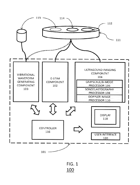
[0022] Turning first to FIG. 1 there is shown a block diagram of a system
100 for
implementing a method in accordance with one aspect of the present invention.
The system 100
includes a housing 101 for the various components of system 100. The housing
101 can be of
any size, including hand held or portable sizes, and the components of system
100 can be sized
accordingly. The various components can include an electrical stimulation (e-
stim) component
102 to generate and provide the electrical stimulation for the patient. The e-
stim component 102
can be configured in a variety of ways. Exemplary e-stim components and
operation thereof are
described in U.S. Patent Nos. 5,107,835, 5,109,848, 8,768,474, and U.S. Patent
Application
Publication No. 2011/0082524, the contents of all of which are hereby
incorporated by reference
in their entirety.
[0023] The e-stim component 102 can be coupled to one or more electrodes
114 for
providing the electrical stimulation treatment to the patient.
[0024] The components can further include an ultrasound (US) imaging
component 104 for
performing US scanning or sonography. The US imaging component 104 can be
coupled to a
transducer 112 for generating and receiving sound waves in a patient. Like a
conventional US
imaging component, the US imaging component 104 can be configured to include a
grayscale or
4
CA 03022430 2018-10-26
WO 2017/189890 PCT/US2017/029917
B-mode ultrasonography processor 106 for producing typical US images. That is,
images in
which the structure or architecture of the patient by analyzing the strength
and time elapsed for
an echo of sound pulses directed into the patient. However, the US imaging
component 104 can
further include a sonoelastography processor 108 and a Doppler image processor
110. The
sonoelastography processor 108 can be configured to analyze shear waves
generated in a patient
and estimate tissue modulus, i.e., tissue stiffness.
[0023] One of the most important characteristics of tissue performance is
its elasticity. An
appropriately elastic or supple tissue will perform optimally, while one which
is not sufficiently
elastic (e.g., stiff or rigid tissues) will offer reduced performance. The
elastic modulus is not
something that can be seen with normal ultrasound. Sonoelastography enables
the measurement
of tissue modulus, giving a better indication of tissue dysfunction than a
visual image.
[0024] The Doppler image processor 110 can be configured to utilize color,
power, or
spectral Doppler analysis of Doppler measurements to see and evaluate blood
flow. In the
various embodiments, all three processors can be concurrently used to generate
images that thus
represent structure, stiffness, and blood flow in soft tissues.
[0025] As shown in FIG. 1, the transducer 112 and electrodes 114 are
incorporated into a
single head unit 111 coupled to the components in housing via, for example,
wiring or cabling.
Thus, the same head unit can be utilized to perform imaging plus the
subsequent electrical
stimulation treatment. Such a configuration is advantageous if the clinician
believes there
is dysfunctional tissue, i.e. a muscle tear or strain, at the site where pain
is felt by
the patient in that is unnecessary to use the electrodes 114 to search for the
muscle tear or
strain after the transducer 112 is utilized to locate a location for treatment
in the patient. In
particular, the electrodes are "pre-positioned" and electrical stimulation
treatment can be
immediately applied. Further, since no repositioning is needed, the electrical
stimulation
treatment can be applied more accurately. Finally, since no repositioning is
needed, the area of
interest can be immediately reevaluated using the transducer 112 and
additional treatments can
be provided without the need to reposition the head unit 111 on the patient.
[0026] Alternatively, this same type of confirmation is advantageous if
Neuromuscular
Interactive Stimulation is used to locate dysfunctional tissue. In such a
configuration, since
there is direct feedback from the patient once the area with dysfunctional
tissue is located, it
does not require special skills to identify the dysfunctional tissues via US.
As a result, an area of
dysfunctional tissue may be found more quickly. Further, since the transducer
112 is collocated,
the transducer 112 can be immediately applied to verify or more closely
examine the
dysfunctional tissues without needing to reposition the head unit on the
patient 111.
CA 03022430 2018-10-26
WO 2017/189890 PCT/US2017/029917
[0027] The same head unit shown in Figure 1 can also be utilized to use
electrical
stimulation to locate areas of dysfunctional tissue that exists at distances
away from where the
patient feels pain, and then the same head is used to perform imaging plus the
subsequent
electrical stimulation treatment. Such a configuration is advantageous when
the clinician desires
to search for dysfunctional tissue that exists away from where pain is felt by
the patient, in that it
is unnecessary to position the transducer 112 after the electrodes 114 are
utilized to locate a
location for treatment in the patient. Rather, the transducer is "pre-
positioned" and ultrasound
imaging can immediately be performed followed by electrical stimulation
treatment. Further,
since no repositioning is needed, the ultrasound imaging can be performed
quicker and more
accurately since less areas needs to be scanned using the transducer. Finally,
since no
repositioning is needed, the area of interest can be immediately reevaluated
and additional
treatments can be provided without the need to reposition the head unit 111 on
the patient.
[0028] Additionally, in some configurations, the system 100 can incorporate
in housing 101
a vibrational waveform generating component 103 and a vibrational transducer
113 device
coupled thereto for generating the shear waves needed for sonoelastography.
The transducer
112 can then detect the shear waves. The component 103 can be configured for
generated low
frequency waves (-1-10 kHz). In some configurations, as shown in FIG. 1, the
vibrational
transducer 113 can be a separate device from head unit 111. This allows the
shear waves to be
introduced into the patient at different points of the patient's body, which
may be necessary
depending on the suspected location of injury. However, as also shown in FIG.
1, the
vibrational transducer 113 could also be incorporated into head unit 111. The
system of FIG. 1
can perform the sonoelastography in a variety of modes. One mode of operation
of
sonoelastography is discussed, at least in part, by Bharat and Varghese in
"Radiofrequency
electrode vibration-induced shear wave imaging for tissue modulus estimation:
A simulation
study." The Journal of the Acoustical Society of America. 2010;128(4):1582-
1585.
doi:10.1121/1.3466880, the contents of which are hereby incorporated by
reference in their
entirety.
[0029] In addition to the foregoing components, system 100 can also include
in housing 101
a controller 116 for coordinating and controlling operations of the e-stim
component 102 and the
US imaging component 104. The housing 101 can also include a display 118 for
displaying
images and other information to users. Although shown in FIG. 1 as being
directly connected to
the controller, the display can be concurrently or alternatively coupled to
the US imaging
component 104. The system can also include a user interface 120 with human
interface
elements (not shown) such as a keyboard or keypad, a pointing selection
device, a touchscreen,
or any other elements suitable for providing user input to controller 116 for
controlling the
6
CA 03022430 2018-10-26
WO 2017/189890 PCT/US2017/029917
various components of system 100. However, in some implementations, the user
interface for
system 100 can be a separate computer, tablet, or smartphone in communication
with system
100.
[0030] In FIG. 1, system 100 is illustrated using a particular combination
of components in
housing 101. However, in the various embodiments, the system 100 can be
implemented using
more or less components than shown in FIG. 1 while achieving the same
functionality.
[0031] As noted above, the system 100 includes a head unit 111 with at
least the transducer
112 for US imaging and the electrodes 114 for electrical stimulation
treatment. Thus, the head
unit 111 and system 100 can be configured in a variety of ways. Two examples
are illustrated in
FIGs. 2 and 3.
[0032] Turning first to FIG. 2A, there is shown a first exemplary
configuration for the
system of FIG. 1. As in FIG. 1, the configuration of FIG. 2A provides a
housing 101 for
components 102, 103, and 104, as well as having a display 118 and a user
interface 120. As also
shown in FIG. 1, the configuration of FIG. 2 also includes a head unit 111 and
a separate
vibrational transducer 113, coupled by wiring or cabling to the appropriate
components in
housing 101.
[0033] The head unit 111 is configured to support both US imaging and
electrical
stimulation treatment in a compact unit that is easy to use. For example, the
head unit 111 can
include, as shown in FIG. 2A, an enable button 202 to active electrical
stimulation. In operation,
the system can be configured for US imaging by default and the head unit 111
can be moved
over the patient until a region of interest (i.e., the region to be treated)
is located via the US
imaging. Then, while visualizing the region of interest, the enable button 202
can be activated
to cause the electrical stimulation to be applied. When deactivated, the head
unit can resume
imaging. In some configurations, switching between US imaging and electrical
stimulation can
be completely automated. In other configurations, user intervention or control
can be required.
[0034] Additionally, as shown in the inset of FIG. 2A, both the transducer
112 for US
imaging and the electrodes 114 for electrical stimulation treatment can be
incorporated into the
same face 204 of head unit 111. The arrangement of transducer 112 and
electrodes can vary in
the various embodiments. However, in particular embodiments, a ring-type
structure can be
used. That is, a central portion of the face 204 can include the transducer
112, such as in the
form of a piezo-electric element array. This central portion can then be
surrounded by an
electrode 114 in the form of an electrical stimulation ring. However, the
various embodiments
are not limited to this design and any arrangement of transducers and
electrodes can be used in
the various embodiments.
7
CA 03022430 2018-10-26
WO 2017/189890 PCT/US2017/029917
[0035] Variations on the arrangement of FIG. 2A are possible to provide
additional
functionality. For example, FIG. 2B shows a similar arrangement to that of
FIG. 2A. However,
in FIG. 2B, removable electrode is provided. This type of arrangement is
discussed below in
greater detail with respect to FIGs. 4A-4C. FIG. 2C shows yet another
arrangement similar to
that of FIG. 2A. In FIG. 2C, housing 101 also contains components 105 for
supporting
electromyography (EMG).
[0036] As used herein, EMG refers to the electrodiagnostic medicine
technique for
evaluating and recording the electrical activity produced by skeletal muscles.
Component 105
can be an instrument called an electromyograph to produce a record called an
electromyogram.
The electromyograph detects, via the EMG electrodes attached thereto, the
electric potential
generated by muscle cells when these cells are electrically or neurologically
activated.
[0037] Thus, one or more EMG electrodes 212 can be coupled to the EMG
components 105.
In such configurations, these can be one or more EMG percutaneous recording
needle
electrodes, one or more EMG surface recording electrodes, one or more EMG
transcutaneous
stimulation electrodes, or a combination of these. In these configurations,
the EMG electrodes
can be controlled from the main unit 101 or from controls 202 in the head unit
111.
[0038] It should be noted that in the various embodiments in which controls
are included in
a probe or head unit 111, such controls can be used to adjust and control a
variety of settings.
These include, but are not limited to, gain or depth adjustment for the US
transducer 112 control
of frequency, power and polarity adjustment for e-stim and, sound, volume,
protocol and mode
adjustment/selection for EMG operations.
[0039] Turning next to FIG. 3A, there is shown another exemplary
configuration for the
system of FIG. 1. In particular, FIG. 3A shows an implementation of head unit
111. Like head
unit 111 in FIG. 1, head unit 111 in FIG. 3A also includes a transducer 112
for US imaging and
electrodes 114 for providing electrical stimulation. However, as shown in FIG.
3A, head unit
can be configured to include a flexible wrap or strap 302, a swivel 304, and a
rotatable end
portion 306. The use and operation of these components is illustrated in FIGs.
3B, 3C, and 3D.
[0040] First, as shown in FIG. 3B, the head unit 111 is positioned over a
patient so that the
US transducer 112 is positioned for imaging and to allow the head unit 111 to
be moved over the
surface of the patient's skin to locate a region of interest. Next, as shown
in FIG. 3C, once a
region of interest is identified, the strap 302 can be used to secure the head
unit 111 in place.
Thereafter, as shown in FIG. 3D, the rotatable end portion 306 can be rotated
to provide the
electrode 114 at the skin's surface. Finally, the electrical stimulation can
be applied. If further
US imaging and/or electrical stimulation is required, the rotatable head
portion 306 can be
alternated appropriately.
8
CA 03022430 2018-10-26
WO 2017/189890 PCT/US2017/029917
[0041] Alternatively, first, as shown in FIG. 3E, the head unit 111 is
positioned over a
patient so that the electrode 114 is positioned for scanning and to allow the
head unit 111 to be
moved over the surface of the patient's skin to locate a region of interest.
Next, as shown in FIG.
3F, once a region of interest is identified, the strap 302 can be used to
secure the head unit 111
in place. Next, as shown in FIG. 3G, the rotatable end portion 306 can be
rotated to provide the
US transducer 112 access to the skin's surface for visualization and
measurement of the
treatment area via ultrasound. Next, as shown in FIG. 3H, the rotatable end
portion 306 can be
rotated to provide the electrode 114 access to the patient's skin. Finally,
the electrical
stimulation can be applied. If further US imaging and/or electrical
stimulation is required, the
rotatable head portion 306 can be alternated appropriately.
[0042] It should be noted that rather than strap 302, any other means of
securing the position
of the head unit 111 relative to a patient can be used. For example, the head
unit can be attached
to a mechanical or robotic arm or other device that allows repositioning of
the head unit 111 at a
fixed location.
[0043] Although the configurations above show a wireline connection between
head unit
111 and the main unit 101, in other configurations a wireless or a combination
of wireless and
wireline connections can be used. This is illustrated in FIG. 31. As shown in
FIG. 31, the head
unit 111 can communicate with main unit 101 via wireless links 310.
Additionally, controls 308
can be provided at head unit 111 to improve usability when using wireless
links 310. However,
in some configurations, the head unit 111 can be controlled from main unit
101.
[0044] The configurations of FIG. 2 and FIG. 3A are presented solely as
examples and for
ease of illustration. Other configurations for head unit can be provided in
the various
embodiments. For example, such additional configurations are illustrated in
FIGs. 4A-4C and 5.
[0045] Turning first to FIG. 4A, there is shown one exemplary configuration
400 for the
system of FIG. 1. The configuration of FIG. 4A is similar to that of FIGs. 2
and 3A. Thus, the
configuration 400 includes a probe or head unit 111, with a US transducer 112
and electrode(s)
114, which is coupled to a main unit or housing 101. The main unit 101 can
include or be
coupled to, as described above with respect to FIG. 1, controls or a user
interface 120 and a
display 118. The probe 111 can also include controls 402 for operating the
system from the
probe 111. The probe 111 and the main unit 101 can be communicatively coupled
via a wireline
communications link 404 or wireless communication links 406. The probe unit
111 can be
powered via the main unit 101 in some configurations and powered independently
in other
configurations.
[0046] In the configuration of FIG. 4A, the US transducer 112 is located at
one end of the
probe 111 and the electrode 114 is placed over the US transducer 114. In such
a configuration,
9
CA 03022430 2018-10-26
WO 2017/189890 PCT/US2017/029917
the electrode 114 can be configured such that the US transducer 112 is
accessible to provide and
receive beams. For example as shown in FIG. 4B, the electrode 114 can be
configured with one
or more openings 502 to permit beams to propagate to and from the US
transducer 112. It
should be noted that while FIG. 4B shows one exemplary configuration for the
electrode 114,
the various embodiments are not limited in this regard. Rather, the electrode
114 can be
configured in a variety of ways to permit propagation of the beams for the US
transducer 112.
[0047] In some configurations, this arrangement of the US transducer 112
and the electrode
114 can be utilized to perform the methods described herein in substantially a
similar fashion as
described above with respect to FIGs. 2 and 3A-3H.
[0048] In other configurations, the arrangement in FIG. 4B can be
configured to perform a
proper placement of multiple electrodes prior to treatment. That is, the
electrode 114 can be
removable coupled to the probe 111, mechanically and electrically. This can be
performed via
one or more clips or other types of fasteners for establishing both mechanical
and electrical
connections. Thus, in accordance with the methods described above, the correct
location for the
electrode 114 on a patient can be identified and the electrode 114 can simply
detach from the
probe 111 to remain in place. Thereafter, another electrode 115 can be
attached to the probe unit
111 and positioned on the patient as discussed above. This process can be
repeated until all
electrodes 114 and 115 are positioned. Finally, treatment can be provided.
In such configurations, the electrodes 114 and 115 can be configured to
operate via a wireless
connection 408 to the main unit 101, as shown in FIG. 4A. Alternative, the
electrodes 114, 115
can be coupled, for example, as shown in FIG. 4C to the main unit 101 via
wireline connections.
[0049] It should be noted that the foregoing configurations are presented
solely by way of
example and not by way of limitation. Accordingly, systems in accordance with
the various
embodiments may include more or less components than shown above. For example,
as shown
above in some configurations, wireless or wireline connections are provided.
However, wireline
or wireless connections can be provided in any of the embodiments. In another
example, some
of the configurations above show the use of EMG components and electrodes.
However, EMG
components and electrodes can be provided in any of the embodiments.
[0050] Having described various components of a system for implementing the
methods of
the various embodiments, attention is directed to FIGs. 5. 6, 7, 8, which
discuss the methods of
the various embodiments in greater detail.
[0051] Turning first to FIG. 5, there is shown a flowchart of steps in an
exemplary procedure
for treatment of dysfunctional tissues in a patient. The method begins at step
502 where a
patient presents with some type of pain, injury, or loss or restricted range
of motion. The
physician or other healthcare worker can then perform a clinical examination
at step 504 to
CA 03022430 2018-10-26
WO 2017/189890 PCT/US2017/029917
determine presence or absence of dysfunctional tissue according to clinical
criteria, apply
pressure algometry to determine pain pressure threshold, and/or perform
differential diagnosis
for presence of dysfunctional tissue and to generally locate a location on the
patient for
treatment.
[0052] Thereafter, the method 500 can proceed to step 506, where the head
unit 111 is
positioned on the patient. If sonoelastography is also being performed, the
vibrational
transducer 113 is also positioned on the patient. The method of positioning
the head unit 111 on
the patient can depend on the clinical examination at step 504. This step can
also involve the
application of US liquid or gel to facilitate imaging.
[0053] Once the head unit 111 is positioned at step 506, the method can
proceed to step 508.
Step 508 first involves visualization of the dysfunctional tissue. Namely
obtaining both
structural and functional information. For example, using information gleaned
from the US
imaging (structural) and Doppler and/or sonoelastography analyses
(functional), as described
above with respect to FIG. 1, the precise location of the injury can be
identified, as well as the
type of injury. For example, the Doppler imaging will identify areas of
unusual blood flow, the
sonoelastography analysis will identify areas of unusual stiffness in muscle
or other soft tissues,
and the US imaging can be used to identify the locations of these, as well as
identify any
structural issues. In some implementations, the dysfunctional tissues can be
identified
automatically via software. In other implementations, the data is merely
presented to the user
and the user then reviews the data to identify the dysfunctional tissues.
[0054] In some configurations, electrical stimulation can be used at steps
506 and 508 to
identify dysfunctional tissues. For example, as discussed above, Neuromuscular
Interactive
Stimulation can be used to identify the dysfunctional tissues. In such a
configuration, electrical
stimulation is provided via electrodes 114 and based on level of discomfort or
pain produced by
the electrical stimulation, one can identify the dysfunctional tissues, as
discussed above.
[0055] After the dysfunctional tissues have been identified at step 508,
the method can
proceed to step 512. However, the method can optionally first proceed to step
510, where the
electrodes for an e-stim component, such as electrodes 114 coupled to e-stim
component 102 of
system 100, are positioned appropriately for the dysfunctional tissues
identified at step 508. For
example, the electrodes 114 can be rotated into place, as discussed above with
respect to FIGs.
3A-3D. Regardless, at step 512, electrical stimulation is provided via an
appropriate treatment
protocol.
[0056] In some configurations, the appropriate treatment protocol can be
automatically
selected by the system 100 based on the results of step 508. In other
configurations, the system
11
CA 03022430 2018-10-26
WO 2017/189890 PCT/US2017/029917
can provide one or more recommendations for the appropriate treatment
protocol. However, the
user ultimately selects one for actual use.
[0057] Once the treatment at step 512 is complete, the method can proceed
to step 514.
There, the area of treatment is re-visualized. This can involve repeating step
506 and 508 as
needed. Thereafter, based on the information gleaned from the revisualization
at step 514, the
effectiveness of treatment can be evaluated at step 516. In particular, the
information, before
and after treatment, can be compared to determine if treatment was effective.
In some
implementations, the evaluation can be performed automatically via software.
In other
implementations, the data is merely presented to the user and the user then
reviews the data to
evaluate the results of the treatment.
[0058] After the evaluation, the method 500 can proceed in various ways.
For example, the
method can proceed to step 518, where the treatment session is concluded. At
this point, the
patient can be provided instructions, including instructions for further
treatment. Alternatively,
at step 520, a treatment session is immediately repeated, possibly multiple
times. Regardless of
the scheduling of the treatment sessions, they can be repeated until the
dysfunctional tissue is
reduced or eradicated, until pain is eliminated, until the injury is healed,
or until normal function
and/or range of motion is restored.
[0059] In some configurations, the determination that additional treatment
is needed can be
performed automatically. That is, if the comparison at step 516 does not
indicate a sufficient
change in structure or function or fails to meet any other criteria, the
system 100 can be
configured to automatically restart treatment. The treatment can continue
until the criteria for
discontinuing treatment is met. Such criteria can also involve halting
treatment to avoid
excessive treatment of the patient.
[0060] As discussed above, step 508 involves visualization and
identification of
dysfunctional tissue using sonoelastography and Doppler analyses. These
processes are
described with respect to FIGs. 6 and 7. FIG. 6 schematically outlines the
steps for Doppler
analysis 600 and FIG. 7 schematically outlines the steps for sonoelastography
analysis 700.
[0061] As shown in FIG. 6, the Doppler analysis 600 first involves
obtaining blood flow
waveforms at step 602. Doppler imaging has been used to assess blood flow in
the
neighborhood of myofascial trigger points (MTrPs) yielding blood flow scores
of the vascular
bed and adjacent soft tissue that effectively distinguish MTrPs. E.g. a
constricted vascular bed
and an enlarged vascular volume can be explained by observed flow waveforms
with retrograde
diastolic flow. Next, at step 604, the blood flow waveforms are utilized to
calculate a pulsality
index (PI) for the area being imaged. In some implementations, software can be
provided to
collect data, interpret and provide the PI result. PI = [PSV-MDV]/mean
velocity, where PSV is
12
CA 03022430 2018-10-26
WO 2017/189890 PCT/US2017/029917
peak systolic velocity and MDV is minimum diastolic velocity. Finally, based
on the calculated
pulsality index, an assessment of the health of the area being imaged. In some
implementations,
the calculations and evaluation of health can be performed automatically via
software. For
example, software can assign a blood flow waveform score (BFS) based upon a
range from
normal arterial flow to abnormal high resistance flow with retrograde
diastolic flow. In other
implementations, the data is merely presented to the user and the user then
reviews the data to
determine health.
[0062] As shown in FIG. 7, the sonoelastography analysis 700 begins at step
702 by
obtaining elasticity data for deep fascia tissues. This can involve generating
low frequency
(<1000Hz) shear waves from an external source (or possibly by the e-stim
device). The waves
are generated so that they propagate through the region of interest and their
peak vibration
amplitude is evaluated to obtain elasticity data. Thereafter, based on this
elasticity data, the
stiffness of muscles and other soft tissues in the imaged area can be
calculated at step 704.
Finally, based on the calculated stiffness, areas of nodules can be calculated
and used to
differentiate between active and latent MTrPs and thus identify or visualize
areas for treatment.
[0063] The present disclosure contemplates that the systems and methods
described herein
can be used for a wide range of dysfunctions, and not solely active or latent
MTrPs or other
conditions explicitly specified herein. Reference to active or latent MTrPs or
other express
conditions is solely for ease of illustration and understanding.
[0064] Although the procedure in FIG. 5 describes in general the treatment
protocol/method
of the various embodiments, FIG. 8 describes in greater detail operations at
the various
components of a system configured in accordance with the various embodiments.
[0065] FIG. 8 is a schematic of the sub-processes involved in the process
of FIG. 7. Going
left to right across the top row, the main sub-processes are identified. The
second or middle row
breaks down the main sub-processes, where appropriate further. The third or
bottom row
indicates the data obtain from the sub-processes.
[0066] The main sub-processes are as follows. First, a visualization sub-
process is
performed to identify and characterize the dysfunctional tissues. Next, e-stim
therapy is
performed according to the results of the visualization. Thereafter, the
visualization is repeated.
Finally, the initial and final results are compared to evaluate the effect of
the e-stim treatment.
[0067] Each of the visualization and re-visualization sub-process involve,
as discussed
above, several imaging/analysis components, as shown in the middle row. The
first component
is the grayscale/B-mode imaging, as discuss above, to obtain structure
information. As shown in
the bottom row, this can involve obtaining size information, depth and
location information,
information regarding adjacent anatomy, and echogenicity. The second component
is other data
13
CA 03022430 2018-10-26
WO 2017/189890 PCT/US2017/029917
collection via Doppler analyses and/or sonoelastography analyses in order to
obtain stiffness
and/or pulsality index values. Optionally, as illustrated in FIG. 8, the data
collected during the
re-visualization can involve obtaining changes in values.
[0068]
Now turning to FIG. 9, there is shown a flowchart of steps in an exemplary
procedure
for treatment of dysfunctional tissues in a patient based on monitoring of
ultrasound
temperature. Ultrasound temperature can be obtained in a variety of ways. One
exemplary
methodology for obtaining ultrasound temperature is described in U.S. Patent
No. 8,016,757, to
Peter J. Kaczkowski and Ajay Anand, issued September 13, 2011, the contents of
which are
hereby incorporated by reference in their entirety. However, the present
disclosure contemplates
that any other method for obtaining ultrasound temperature can be used in the
various
embodiments without limitation. The method 900 begins at step 902 with a setup
of the system
for scanning and treating a patient.
After the system is setup, electrode scanning can be
performed at step 904, as discussed above, until an area of dysfunctional
tissue is located at step
906.
[0069]
Once the area of dysfunctional tissue is located at step 906, the ultrasound
temperatures of the dysfunctional tissue and the surrounding areas can be
recorded at step 908.
Thereafter, at step 910, the area with the dysfunctional tissue can be treated
with e-stim. After
the treatment at step 910, the ultrasound temperatures can again be recorded
at step 912.
[0070]
The method then moves on to step 914. At step 914, a determination is made as
to
whether a decrease in the temperature of the dysfunctional tissue is detected.
If no decrease is
detected, then the method proceeds to step 916, where the parameters for e-
stim are modified.
Thereafter, the method returns to step 910 for additional stimulation using
the new parameters
and the temperature is monitored at steps 912 and 914 until a temperature drop
is detected.
Once the temperature drop is detected at step 914, the method proceeds to step
918.
[0071]
At step 918, further electrical stimulation can be provided. Thereafter,
temperature is
measured again at step 920. Thereafter, at step 922 it is determined whether
the temperature of
the dysfunctional tissue is equal to that of the surrounding tissue. If not,
the method 900 repeats
steps 918-922 until the temperature is equal. Once the temperature of the
dysfunctional tissue is
equal to that of the surrounding tissue, the method proceeds to step 924,
where treatment is
ended. In some configurations, if an increase in temperature is detected
during steps 918-922,
the method 900 can be configured to return to step 916, so that the parameters
can be adjusted.
[0072]
Now turning to FIG. 10, there is shown a flowchart of steps in an exemplary
procedure for treatment of torn tissues in a patient based on monitoring of
ultrasound imaging.
The method 1000 begins at step 1002 with a setup of the system for scanning
and treating a
patient. After the system is setup, ultrasound imaging can be performed at
step 1004 to identify
14
CA 03022430 2018-10-26
WO 2017/189890 PCT/US2017/029917
and locate torn tissue in an area with pain. Thereafter, the size of the torn
tissue and images of
the torn tissue can be recorded at step 1006.
[0073] Once the area of torn tissue is located at step 1004 and
measurements and images are
obtained at step 1006, the area with the torn tissue (i.e., the area of pain)
can be treated with e-
stim. After the treatment at step 1010, additional images and measurements of
the torn tissue
can be recorded at step 1012.
[0074] The method then moves on to step 1014. At step 1014, a determination
is made as to
whether a decrease in the size of the tear is detected. If no decrease is
detected, then the method
proceeds to step 1016, where the parameters for e-stim are modified.
Thereafter, treatment and
monitoring are repeated with steps 1008-1016 until a decrease in the size of
the tear is detected
at step 1014. Once the decrease is detected at step 1014, the method proceeds
to step 1018.
[0075] At step 1018, further treatment of the site is provided with the
existing parameters.
Thereafter, additional imaging is performed at step 1020 to determine if the
torn tissue has been
healed. If at step 1022, the tissue is not yet healed, the method can repeat
steps 1018-1022 until
healing is observed. Once the tissues are healed, the method can end at step
1024. In some
configurations, if an increase in temperature is detected during steps 918-
922, the method 900
can be configured to return to step 916, so that the parameters can be
adjusted.
[0076] FIG. 11A, and FIG. 11B illustrate exemplary possible system
embodiments. The
more appropriate embodiment will be apparent to those of ordinary skill in the
art when
practicing the present technology. Persons of ordinary skill in the art will
also readily appreciate
that other system embodiments are possible.
[0077] FIG. 11A illustrates a conventional system bus computing system
architecture 1100
wherein the components of the system are in electrical communication with each
other using a
bus 1105. Exemplary system 1100 includes a processing unit (CPU or processor)
1110 and a
system bus 1105 that couples various system components including the system
memory 1115,
such as read only memory (ROM) 1120 and random access memory (RAM) 1125, to
the
processor 1110. The system 1100 can include a cache of high-speed memory
connected directly
with, in close proximity to, or integrated as part of the processor 1110. The
system 1100 can
copy data from the memory 1115 and/or the storage device 1130 to the cache
1112 for quick
access by the processor 1110. In this way, the cache can provide a performance
boost that
avoids processor 1110 delays while waiting for data. These and other modules
can control or be
configured to control the processor 1110 to perform various actions. Other
system memory
1115 may be available for use as well. The memory 1115 can include multiple
different types of
memory with different performance characteristics. The processor 1110 can
include any general
purpose processor and a hardware module or software module, such as module 1
1132, module 2
CA 03022430 2018-10-26
WO 2017/189890 PCT/US2017/029917
1134, and module 3 1136 stored in storage device 1130, configured to control
the processor
1110 as well as a special-purpose processor where software instructions are
incorporated into the
actual processor design. The processor 1110 may essentially be a completely
self-contained
computing system, containing multiple cores or processors, a bus, memory
controller, cache, etc.
A multi-core processor may be symmetric or asymmetric.
[0078] To enable user interaction with the computing device 1100, an input
device 1145 can
represent any number of input mechanisms, such as a microphone for speech, a
touch-sensitive
screen for gesture or graphical input, keyboard, mouse, motion input, speech
and so forth. An
output device 1135 can also be one or more of a number of output mechanisms
known to those
of skill in the art. In some instances, multimodal systems can enable a user
to provide multiple
types of input to communicate with the computing device 1100. The
communications interface
1140 can generally govern and manage the user input and system output. There
is no restriction
on operating on any particular hardware arrangement and therefore the basic
features here may
easily be substituted for improved hardware or firmware arrangements as they
are developed.
[0079] Storage device 1130 is a non-volatile memory and can be a hard disk
or other types
of computer readable media which can store data that are accessible by a
computer, such as
magnetic cassettes, flash memory cards, solid state memory devices, digital
versatile disks,
cartridges, random access memories (RAMs) 1125, read only memory (ROM) 1120,
and hybrids
thereof.
[0080] The storage device 1130 can include software modules 1132, 1134,
1136 for
controlling the processor 1110. Other hardware or software modules are
contemplated. The
storage device 1130 can be connected to the system bus 1105. In one aspect, a
hardware module
that performs a particular function can include the software component stored
in a computer-
readable medium in connection with the necessary hardware components, such as
the processor
1110, bus 1105, display 1135, and so forth, to carry out the function.
[0081] FIG. 11B illustrates a computer system 1150 having a chipset
architecture that can be
used in executing the described method and generating and displaying a
graphical user interface
(GUI). Computer system 1150 is an example of computer hardware, software, and
firmware
that can be used to implement the disclosed technology. System 1150 can
include a processor
1155, representative of any number of physically and/or logically distinct
resources capable of
executing software, firmware, and hardware configured to perform identified
computations.
Processor 1155 can communicate with a chipset 1160 that can control input to
and output from
processor 1155. In this example, chipset 1160 outputs information to output
1165, such as a
display, and can read and write information to storage device 1170, which can
include magnetic
media, and solid state media, for example. Chipset 1160 can also read data
from and write data
16
CA 03022430 2018-10-26
WO 2017/189890 PCT/US2017/029917
to RAM 1175. A bridge 1180 for interfacing with a variety of user interface
components 1185
can be provided for interfacing with chipset 1160. Such user interface
components 1185 can
include a keyboard, a microphone, touch detection and processing circuitry, a
pointing device,
such as a mouse, and so on. In general, inputs to system 1150 can come from
any of a variety of
sources, machine generated and/or human generated.
[0082] Chipset 1160 can also interface with one or more communication
interfaces 1190 that
can have different physical interfaces. Such communication interfaces can
include interfaces for
wired and wireless local area networks, for broadband wireless networks, as
well as personal
area networks. Some applications of the methods for generating, displaying,
and using the GUI
disclosed herein can include receiving ordered datasets over the physical
interface or be
generated by the machine itself by processor 1155 analyzing data stored in
storage 1170 or
1175. Further, the machine can receive inputs from a user via user interface
components 1185
and execute appropriate functions, such as browsing functions by interpreting
these inputs using
processor 1155.
[0083] It can be appreciated that exemplary systems 1100 and 1150 can have
more than one
processor 1110 or be part of a group or cluster of computing devices networked
together to
provide greater processing capability.
[0084] For clarity of explanation, in some instances the present technology
may be presented
as including individual functional blocks including functional blocks
comprising devices, device
components, steps or routines in a method embodied in software, or
combinations of hardware
and software.
[0085] In some embodiments the computer-readable storage devices, mediums,
and
memories can include a cable or wireless signal containing a bit stream and
the like. However,
when mentioned, non-transitory computer-readable storage media expressly
exclude media such
as energy, carrier signals, electromagnetic waves, and signals per se.
[0086] Methods according to the above-described examples can be implemented
using
computer-executable instructions that are stored or otherwise available from
computer readable
media. Such instructions can comprise, for example, instructions and data
which cause or
otherwise configure a general purpose computer, special purpose computer, or
special purpose
processing device to perform a certain function or group of functions.
Portions of computer
resources used can be accessible over a network. The computer executable
instructions may be,
for example, binaries, intermediate format instructions such as assembly
language, firmware, or
source code. Examples of computer-readable media that may be used to store
instructions,
information used, and/or information created during methods according to
described examples
17
CA 03022430 2018-10-26
WO 2017/189890 PCT/US2017/029917
include magnetic or optical disks, flash memory, USB devices provided with non-
volatile
memory, networked storage devices, and so on.
[0087] Devices implementing methods according to these disclosures can
comprise
hardware, firmware and/or software, and can take any of a variety of form
factors. Typical
examples of such form factors include laptops, smart phones, small form factor
personal
computers, personal digital assistants, and so on. Functionality described
herein also can be
embodied in peripherals or add-in cards. Such functionality can also be
implemented on a
circuit board among different chips or different processes executing in a
single device, by way
of further example.
[0088] The instructions, media for conveying such instructions, computing
resources for
executing them, and other structures for supporting such computing resources
are means for
providing the functions described in these disclosures.
[0089] While various embodiments of the present invention have been
described above, it
should be understood that they have been presented by way of example only, and
not limitation.
Numerous changes to the disclosed embodiments can be made in accordance with
the disclosure
herein without departing from the spirit or scope of the invention. Thus, the
breadth and scope
of the present invention should not be limited by any of the above described
embodiments.
Rather, the scope of the invention should be defined in accordance with the
following claims
and their equivalents.
[0090] Although the invention has been illustrated and described with
respect to one or more
implementations, equivalent alterations and modifications will occur to others
skilled in the art
upon the reading and understanding of this specification and the annexed
drawings. In addition,
while a particular feature of the invention may have been disclosed with
respect to only one of
several implementations, such feature may be combined with one or more other
features of the
other implementations as may be desired and advantageous for any given or
particular
application.
[0091] The terminology used herein is for the purpose of describing
particular embodiments
only and is not intended to be limiting of the invention. As used herein, the
singular forms "a",
"an" and "the" are intended to include the plural forms as well, unless the
context clearly
indicates otherwise. Furthermore, to the extent that the terms "including",
"includes", "having",
"has", "with", or variants thereof are used in either the detailed description
and/or the claims,
such terms are intended to be inclusive in a manner similar to the term
"comprising."
[0092] Unless otherwise defined, all terms (including technical and
scientific terms) used
herein have the same meaning as commonly understood by one of ordinary skill
in the art to
which this invention belongs. It will be further understood that terms, such
as those defined in
18
CA 03022430 2018-10-26
WO 2017/189890 PCT/US2017/029917
commonly used dictionaries, should be interpreted as having a meaning that is
consistent with
their meaning in the context of the relevant art and will not be interpreted
in an idealized or
overly formal sense unless expressly so defined herein.
19