Note: Descriptions are shown in the official language in which they were submitted.
CA 03022515 2018-10-29
WO 2017/191560 PCT/IB2017/052544
1
ANTIBODIES RECOGNIZING TAU
CROSS REFERENCE TO RELATED APPLICATIONS
[0001] This application is related to US Provisional Application No.
62/330,789 filed May 2,
2016, which is incorporated by reference in its entirety for all purposes.
REFERENCE TO A SEQUENCE LISTING
[0002] The Sequence Listing written in file 497107SEQLIST.txt is 65 kilobytes,
was created
on May 2, 2017, and is hereby incorporated by reference.
BACKGROUND OF THE INVENTION
[0003] Tau is a well-known human protein that can exist in phosphorylated
forms (see, e.g.,
Goedert, Proc. Natl. Acad. Sci. U.S.A. 85:4051-4055(1988); Goedert, EMBO J.
8:393-
399(1989); Lee, Neuron 2:1615-1624(1989); Goedert, Neuron 3:519-526(1989);
Andreadis,
Biochemistry 31:10626-10633(1992). Tau has been reported to have a role in
stabilizing
microtubules, particularly in the central nervous system. Total tau (t-tau, L
e., phosphorylated
and unphosphorylated forms) and phospho-tau (p-tau, i.e., phosphorylated tau)
are released by
the brain in response to neuronal injury and neurodegeneration and have been
reported to occur
at increased levels in the CSF of Alzheimer's patients relative to the general
population (Jack et
al., Lancet Neurol 9: 119-28 (2010)).
[0004] Tau is the principal constituent of neurofibrillary tangles, which
together with plaques
are a hallmark characteristic of Alzheimer's disease. The tangles constitute
abnormal fibrils
measuring 10 nm in diameter occurring in pairs wound in a helical fashion with
a regular
periodicity of 80 nm. The tau within neurofibrillary tangles is abnormally
phosphorylated
(hyperphosphorylated) with phosphate groups attached to specific sites on the
molecule. Severe
involvement of neurofibrillary tangles is seen in the layer II neurons of the
entorhinal cortex, the
CA1 and subicular regions of the hippocampus, the amygdala, and the deeper
layers (layers III,
CA 03022515 2018-10-29
WO 2017/191560 PCT/IB2017/052544
2
V, and superficial VI) of the neocortex in Alzheimer's disease.
Hyperphosphorylated tau has
also been reported to interfere with microtubule assembly, which may promote
neuronal network
breakdown.
[0005] Tau inclusions are part of the defining neurophathology of several
neurodegenerative
diseases including Alzheimer's disease, frontotemporal lobar degeneration,
progressive
supranuclear palsy and Pick's disease.
BRIEF SUMMARY OF THE CLAIMED INVENTION
[0006] In one aspect, the invention provides an isolated monoclonal antibody
that specifically
binds to tau. Examples of such antibodies bind to an epitope within amino acid
residues 199-213
or 262-276 of SEQ ID NO:3 (con-esponding to amino acid residues 257-271 or 320-
334,
respectively, of SEQ ID NO:1).
[0007] Some such antibodies compete for binding to human tau with antibody
3D6. Some
such antibodies bind to the same epitope on human tau as 3D6.
[0008] Some antibodies comprise three light chain CDRs as and three heavy
chain CDRs of
monoclonal antibody 3D6, wherein 3D6 is a mouse antibody characterized by a
heavy chain
variable region having an amino acid sequence comprising SEQ ID NO: 7 and a
light chain
variable region having an amino acid sequence comprising SEQ ID NO: 11. In
some antibodies,
the three heavy chain CDRs are as defined by Kabat/Chothia Composite (SEQ ID
NOs: 8, 9, and
10) and the three light chain CDRs are as defined by Kabat/Chothia Composite
(SEQ ID NOs:
12, 13, and 14).
[0009] For example, the antibody can be 3D6 or a chimeric, veneered, or
humanized form
thereof. In some such antibodies, the variable heavy chain has > 85% identity
to human
sequence. In some such antibodies, the variable light chain has? 85% identity
to human
sequence. In some such antibodies, each of the variable heavy chain and
variable light chain has
> 85% identity to human germline sequence.
[0010] In some such antibodies, the mature heavy chain variable region
comprises the three
heavy chain CDRs are as defined by Kabat/Chothia Composite (SEQ ID NOs: 8, 9,
and 10) and
the three light chain CDRs are as defined by Kabat/Chothia Composite (SEQ ID
NOs: 12, 13,
CA 03022515 2018-10-29
WO 2017/191560 PCT/IB2017/052544
3
and 14); provided that position H27 is occupied by F or Y, H28 is occupied by
N or T, H29 is
occupied by I or F, H30 is occupied by K or T, position H51 is occupied by I
or V, position H54
is occupied by N or D, position H60 is occupied by D or A, H61 is occupied by
P or E, and H102
is occupied by F or Y. In some such antibodies, CDR-H1 has an amino acid
sequence
comprising SEQ ID NO: 42. In some such antibodies, CDR-H1 has an amino acid
sequence
comprising SEQ ID NO: 58. In some such antibodies, CDR-H1 has an amino acid
sequence
comprising SEQ ID NO: 59. In some such antibodies, CDR-H1 has an amino acid
sequence
comprising SEQ ID NO: 60. In some such antibodies, CDR-H2 has an amino acid
sequence
comprising SEQ ID NO: 43. In some such antibodies, CDR-H2 has an amino acid
sequence
comprising SEQ ID NO: 61. In some such antibodies, CDR-H2 has an amino acid
sequence
comprising SEQ ID NO: 62. In some such antibodies, CDR-H2 has an amino acid
sequence
comprising SEQ ID NO: 63. In some such antibodies, CDR-H2 has an amino acid
sequence
comprising SEQ ID NO: 64. In some such antibodies, CDR-H3 has an amino acid
sequence
comprising SEQ ID NO: 65.
[0011] In some such antibodies, the antibody is a humanized antibody. In some
such
antibodies, CDR-H1 has an amino acid sequence comprising SEQ ID NO: 42 and CDR-
H2 has
an amino acid sequence comprising SEQ ID NO: 43. In some such antibodies, CDR-
H1 has an
amino acid sequence comprising SEQ ID NO: 42 and CDR-H2 has an amino acid
sequence
comprising SEQ ID NO:61. In some such antibodies, CDR-H1 has an amino acid
sequence
comprising SEQ ID NO: 42 and CDR-H2 has an amino acid sequence comprising SEQ
ID
NO:64. In some such antibodies, CDR-H1 has an amino acid sequence comprising
SEQ ID NO:
42, CDR-H2 has an amino acid sequence comprising SEQ ID NO:63, and CDR-H3 has
an amino
acid sequence comprising SEQ ID NO:65. In some such antibodies, CDR-H1 has an
amino acid
sequence comprising SEQ ID NO: 58 and CDR-H2 has an amino acid sequence
comprising SEQ
ID NO:62. In some such antibodies, CDR-H1 has an amino acid sequence
comprising SEQ ID
NO: 59 and CDR-H2 has an amino acid sequence comprising SEQ ID NO:63. In some
such
antibodies, CDR-H1 has an amino acid sequence comprising SEQ ID NO: 60 and CDR-
H2 has
an amino acid sequence comprising SEQ ID NO:62.
[0012] Some antibodies are a humanized or chimeric 3D6 antibody that
specifically binds to
human tau, wherein 3D6 is a mouse antibody characterized by a mature heavy
chain variable
CA 03022515 2018-10-29
WO 2017/191560 PCT/IB2017/052544
4
region of SEQ ID NO:7 and a mature light chain variable region of SEQ ID NO:
11. Some such
antibodies are a humanized antibody comprising a humanized mature heavy chain
variable
region comprising the three heavy chain CDRs of 3D6 and a humanized mature
light chain
variable region comprising the three light chain CDRs of 3D6. In some such
antibodies, the
CDRs are of a definition selected from the group of Kabat, Chothia,
Kabat/Chothia Composite,
AbM and Contact.
[0013] In some such antibodies the humanized mature heavy chain variable
region comprises
the three Kabat/Chothia Composite heavy chain CDRs of 3D6 (SEQ ID NOs: 8-10)
and the
humanized mature light chain variable region comprises the three Kabat/Chothia
Composite light
chain CDRs of 3D6 (SEQ ID NOs: 12-14).
[0014] In some such antibodies, the humanized mature heavy chain variable
region comprises
the three Kabat heavy chain CDRs of 3D6 (SEQ ID NO:32, SEQ ID NO:9, and SEQ ID
NO:10)
and the humanized mature light chain variable region comprises the three Kabat
light chain
CDRs of 3D6 (SEQ ID NOs: 12-14).
[0015] In some such antibodies, the humanized mature heavy chain variable
region comprises
the three Chothia heavy chain CDRs of 3D6 (SEQ ID NO:33, SEQ ID NO:34, and SEQ
ID
NO:10) and the humanized mature light chain variable region comprises the
three Chothia light
chain CDRs of 3D6 (SEQ ID NOs: 12-14).
[0016] In some such antibodies, the humanized mature heavy chain variable
region comprises
the three AbM heavy chain CDRs of 3D6 (SEQ ID NO:8, SEQ ID NO:35, and SEQ ID
NO:10))
and the humanized mature light chain variable region comprises the three AbM
light chain CDRs
of 3D6 (SEQ ID NOs: 12-14).
[0017] In some such antibodies, the humanized mature heavy chain variable
region comprises
the three Contact heavy chain CDRs of 3D6 (SEQ ID NOs:39-41) and the humanized
mature
light chain variable region comprises the three Contact light chain CDRs of
3D6 (SEQ ID
NOs:36-38).
[0018] In some antibodies, the humanized mature heavy chain variable region
has an amino
acid sequence at least 90% identical to any one of SEQ ID NOs:15-19 and SEQ ID
NOs:46-57
and a humanized mature light chain variable region having an amino acid
sequence at least 90%
CA 03022515 2018-10-29
WO 2017/191560 PCT/IB2017/052544
identical to any one of SEQ ID NOs: 20-23, except that position H17 can be T
or S, and position
H20 can be I or V.
[0019] In some such antibodies, at least one of the following positions in the
VH region is
occupied by the amino acid as specified: H38 is occupied by R and H93 is
occupied by S. In
some such antibodies, positions H38 and H93 in the VH region are occupied by R
and S,
respectively.
[0020] In some such antibodies, at least one of the following positions in the
VH region is
occupied by the amino acid as specified: H38 is occupied by R, H43 is occupied
by Q, H83 is
occupied by T, and H93 is occupied by S. In some such antibodies, positions
H38, H43, H83,
and H93 in the VH region are occupied by R, Q, T, and S, respectively.
[0021] In some antibodies, at least one of the following positions in the VH
region is occupied
by the amino acid as specified: H12 is occupied by V, H24 is occupied by A,
H48 is occupied by
I, H67 is occupied by A, H80 is occupied by L, H81 is occupied by Q, and H91
is occupied by F.
In some antibodies, positions H12, H24, H48, H67, H80, H81, and H91 in the VH
region are
occupied by V, A, I, A, L, Q, and F, respectively.
[0022] In some antibodies at least one of the following positions in the VH
region is occupied
by the amino acid as specified: H13 is occupied by R and H66 is occupied by K.
In some
antibodies, positions H13 and H66 in the VH region are occupied by R and K,
respectively.
[0023] In some antibodies, at least one of the following positions in the VH
region is occupied
by the amino acid as specified: H40 is occupied by R and H82a is occupied by
G. In some
antibodies, positions H40 and H82a in the VH region are occupied by R and G,
respectively.
[0024] In some antibodies, at least one of the following positions in the VH
region is occupied
by the amino acid as specified: H42 is occupied by E and H76 is occupied by N.
In some
antibodies, positions H42 and H76 in the VH region are occupied by E and N,
respectively.
[0025] In some antibodies, at least one of the following positions in the VH
region is occupied
by the amino acid as specified: H40 is occupied by R, H82a is occupied by G,
and H83 is
occupied by T. In some antibodies, positions H40, H82a, and H83 in the VH
region are
occupied by R, G, and T, respectively.
CA 03022515 2018-10-29
WO 2017/191560 PCT/IB2017/052544
6
[0026] In some antibodies, position H12 in the VH region is occupied by V.
[0027] In some antibodies, position H80 in the VH region is occupied by L.
[0028] In some antibodies, at least one of the following positions in the VH
region is occupied
by the amino acid as specified: H24 is occupied by A, H48 is occupied by I,
H67 is occupied by
A, H80 is occupied by L, and H91 is occupied by F. In some antibodies,
positions H24, H48,
H67, H80, and H91 in the VH region are occupied by A, I, A, L, and F,
respectively.
[0029] In some antibodies, at least one of the following positions in the VH
region is occupied
by the amino acid as specified: H43 is occupied by Q, and H81 is occupied by
Q. In some
antibodies, positions H43 and H81 in the VH region are occupied by Q, and Q,
respectively.
[0030] In some antibodies, at least one of the following positions in the VH
region is occupied
by the amino acid as specified: H24 is occupied by A, and H91 is occupied by
F. In some
antibodies, positions H24 and H91 in the VH region are occupied by A and F,
respectively.
[0031] In some antibodies, at least one of the following positions in the VH
region is occupied
by the amino acid as specified: H13 is occupied by R , H17 is occupied by L,
H29 is occupied by
F, H42 is occupied by E, H43 is occupied by Q, H61 is occupied by E, H76 is
occupied by N,
H80 is occupied by L, H81 is occupied by Q. In some antibodies, positions H13,
H17, H29,
H42, H43, H61, H76, H80, and H81 in the VH region are occupied by R, L, F, E,
Q, E, N, L, and
Q, respectively.
[0032] In some antibodies, at least one of the following positions in the VH
region is occupied
by the amino acid as specified: H24 is occupied by A, H28 is occupied by T,
H48 is occupied by
I, H54 is occupied by D, H60 is occupied by A, H67 is occupied by A, H80 is
occupied by L,
and H91 is occupied by F. In some antibodies, positions H24, H28, H48, H54,
H60, H67, H80,
and H91 in the VH region are occupied by A, T, I, D, A, A, L, and F,
respectively.
[0033] In some antibodies, at least one of the following positions in the VH
region is occupied
by the amino acid as specified:: H10 is occupied by D, H17 is occupied by L,
H24 is occupied by
A, H28 is occupied by T, H43 is occupied by Q, H48 is occupied by I, H60 is
occupied by A,
H61 is occupied by E, H91 is occupied by F, H108 is occupied by T, and H109 is
occupied by L.
CA 03022515 2018-10-29
WO 2017/191560 PCT/IB2017/052544
7
In some antibodies, positions H10, H17, H24, H28, H43, H48, H60, H61, H91,
H108, and H109
in the VH region are occupied by D, L, A, T, Q, I, A, E, F, T, and L,
respectively.
[0034] In some antibodies, at least one of the following positions in the VH
region is occupied
by the amino acid as specified: H17 is occupied by L, H27 is occupied by Y,
H29 is occupied by
F, and H61 is occupied by E. In some antibodies, positions H17, H27, H29, and
H61 in the VH
region are occupied by L, Y, F, and E, respectively.
[0035] In some antibodies, at least one of the following positions in the VH
region is occupied
by the amino acid as specified: H17 is occupied by L, H27 is occupied by Y,
H29 is occupied by
F, H61 is occupied by E, H76 is occupied by N, and H82a is occupied by G. In
some antibodies,
positions H17, H27, H29, H61, H76, and H82a in the VH region are occupied by
L, Y, F, E, N,
and G, respectively.
[0036] In some antibodies, at least one of the following positions in the VH
region is occupied
by the amino acid as specified: H12 is occupied by V, H17 is occupied by L,
H24 is occupied by
A, H43 is occupied by Q, H48 is occupied by I, H83 is occupied by T, and H91
is occupied by F.
In some antibodies, positions H12, H17, H24, H43, H48, H83, and H9lin the VH
region are
occupied by V, L, A, Q, I, T, F, respectively.
[0037] In some antibodies, at least one of the following positions in the VH
region is occupied
by the amino acid as specified: H12 is occupied by V, H24 is occupied by A,
H48 is occupied
by I, H67 is occupied b A, H80 is occupied by L, H83 is occupied by T, and H91
is occupied by
F. In some antibodies, positions H12, H24, H48, H67, H80, H83, and H91 in the
VH region are
occupied by V, A, I, A, L, T, and F, respectively.
[0038] In some antibodies, at least one of the following positions in the VH
region is occupied
by the amino acid as specified: H10 is occupied by E or D, H12 is occupied by
K or V, H13 is
occupied by K or R, H17 is occupied by T, L or S, H24 is occupied by V or A,
H27 is occupied
by F or Y, H28 is occupied by N or T, H29 is occupied by I or F, H30 is
occupied by K or T,
H38 is occupied by Q or R, H40 is occupied by A or R, H42 is occupied by G or
E, H43 is
occupied by K or Q, H48 is occupied by M or I, H51 is occupied by V or I, H54
is occupied by
N or D, H60 is occupied by D or A, H61 is occupied by P or E, H66 is occupied
by R or K, H67
is occupied by V or A, H76 is occupied by D or N, H80 is occupied by M or L,
H81 is occupied
CA 03022515 2018-10-29
WO 2017/191560 PCT/IB2017/052544
8
by E or Q, H82a is occupied by S or G, H83 is occupied by T or R, H91 is
occupied by Y or F,
H93 is occupied by A or S, H102 is occupied by F or Y, H108 is occupied by T
or L, H109 is
occupied by L or V. In some antibodies, positions H12, H13, H17, H24, H38,
H42, H43, H48,
H66, H67, H76, H80, H81, H83, H91, and H93 in the VH region are occupied by V,
R, L, A, R,
E, Q, I, K, A, N, L, Q, T, F, and S, respectively.
[0039] In some antibodies, positions H38, H42, H43, H76, H83, and H93 in the
VH region are
occupied by R, E, Q, N, T, and S, respectively. In some antibodies, positions
H12, H13, H17,
H24, H38, H40, H42, H43, H48, H66, H67, H76, H80, H81, H82A, H83, H91, and H93
in the
VH region are occupied by V, R, L, A, R, R, E, Q, I, K, A, N, L, Q, G, T, F,
and S, respectively.
In some antibodies, positions H12, H24, H38, H40, H43, H48, H67, H80, H81,
H82A, H83,
H91, and H93 in the VH region are occupied by V, A, R, R, Q, I, A, L, Q, G, T,
F, and S,
respectively. In some antibodies, positions H12, H24, H28, H38, H40, H43, H48,
H54, H60,
H67, H80, H81, H82A, H83, H91, and H93 in the VH region are occupied by V, A,
T, R, R, Q, I,
D, A, A, L, Q, G, T, F, and S, respectively.
[0040] In some antibodies, positions H12, H24, H28, H38, H40, H48, H51, H54,
H60, H67,
H80, H82A, H83, H91, and H93 in the VH region are occupied by V, A, T, R, R,
I, V, D, A, A,
L, G, T, F, and S, respectively. In some antibodies, positions H12, H24, H28,
H38, H40, H48,
H54, H60, H67, H80, H82A, H83, H91, and H93 in the VH region are occupied by
V, A, T, R,
R, I, D, A, A, L, G, T, F, and S, respectively. In some antibodies, positions
H13, H17, H24,
H29, H38, H40, H42, H43, H54, H61, H76, H80, H81, H82A, H83, H91, and H93 in
the VH
region are occupied by R, L, A, F, R, R, E, Q, N, E, N, L, Q, G, T, F, and S,
respectively.
[0041] In some antibodies, positions H13, H17, H24, H27, H28, H29, H30, H38,
H40, H42,
H43, H51, H54, H60, H61, H76, H80, H81, H82A, H83, H91, and H93 in the VH
region are
occupied by R, L, A, Y, T, F, T, R, R, E, Q, V, D, A, E, N, L, Q, G, T, F, and
S, respectively. In
some antibodies, positions H10, H12, H17, H24, H28, H38, H40, H42, H43, H48,
H54, H60,
H61, H76, H80, H82A, H83, H91, H93, H108, and H109 in the VH region are
occupied by D, V,
L, A, T, R, R, E, Q, I, N, A, E, N, L, G, T, F, S, T, and L, respectively. In
some antibodies,
positions H10, H12, H17, H24, H28, H38, H40, H43, H48, H51, H54, H60, H61,
H82A, H83,
CA 03022515 2018-10-29
WO 2017/191560 PCT/IB2017/052544
9
H91, H93, H102, H108, and H109 in the VH region are occupied by D, V, L, A, T,
R, R, Q, I, V,
D, A, E, G, T, F, S, Y, T, and L, respectively.
[0042] In some antibodies, positions H38 and H93 in the VH region are occupied
by R and S,
respectively. In some antibodies, positions H17, H27, H29, H38, H61, H76,
H82A, and H93 in
the VH region are occupied by L, Y, F, R, E, N, G, and S, respectively. In
some antibodies,
positions H17, H27, H28, H29, H30, H38, H51, H54, H60, H61, H76, H82A, and H93
in the VH
region are occupied by L, Y, T, F, T, R, V, D, A, E, N, G, and S,
respectively.
[0043] In some antibodies, positions H12, H38, H40, H48, H66, H67, H76, H80,
H82A, H83,
and H93 in the VH region are occupied by V, R, R, I, K, A, N, L, G, T, and S,
respectively.
In some antibodies, positions H12, H17, H27, H29, H38, H40, H61, H80, H82A,
H83, and H93
in the VH region are occupied by V, L, Y, F, R, R, E, L, G, T, and S,
respectively. In some
antibodies, positions H12, H17, H27, H28, H29, H30, H38, H40, H51, H54, H60,
H61, H80,
H82A, H83, and H93 in the VH region are occupied by V, L, Y, T, F, T, R, R, V,
D, A, E, L, G,
T, and S, respectively.
[0044] In some antibodies, at least one of the following positions in the VL
region is occupied
by the amino acid as specified: L36 is occupied by L, L37 is occupied by L,
and L100 is
occupied by G. In some antibodies, positions L36, L37, and L100 in the VL
region are occupied
by L, L, and G, respectively.
[0045] In some antibodies, at least one of the following positions in the VL
region is occupied
by the amino acid as specified: L12 is occupied by S and L45 is occupied by K.
In some
antibodies, positions L12 and L45 in the VL region are occupied by S and K,
respectively.
[0046] In some antibodies, at least one of the following positions in the VL
region is occupied
by the amino acid as specified: L2 is V or I, L7 is S or T, L12 is P or S, L15
is L or I, L36 is L,
L37 is L, L45 is R or K, L60 is D or S, L100 is G.
[0047] In some antibodies, positions L12, L36, L37, L45, and L100 in the VL
region are
occupied by S, L, L, K, and G, respectively. In some antibodies, positions
L36, L37, and L100
in the VL region are occupied by L, L and G, respectively. In some antibodies,
positions L36,
L37, L60, and L100 in the VL region are occupied by L, L, S, and G,
respectively. In some
CA 03022515 2018-10-29
WO 2017/191560 PCT/IB2017/052544
antibodies, positions L2, L7, L12, L15, L36, L37, L45, and L100 in the VL
region are occupied
by I, T, S, I, L, L, K, and G, respectively. In some antibodies, positions L2,
L7, L12, L15, L36,
L37, L45, and L100 in the VL region are occupied by V, S, P, L, L, L, R, and
G, respectively.
[0048] Some antibodies comprise a mature heavy chain variable region having an
amino acid
sequence at least 95% identical to any one of SEQ ID NOs: 15-19, 46-57 and a
mature light
chain variable region having an amino acid sequence at least 95% identical to
any one of SEQ ID
NOs: 20-23, except that position H17 can be T or S, and position H20 can be I
or V.. Some
antibodies comprise a mature heavy chain variable region having an amino acid
sequence at least
98% identical to any one of SEQ ID NOs: 15-19, 46-57 and a mature light chain
variable region
having an amino acid sequence at least 98% identical to any one of SEQ ID NOs:
20-23, except
that position H17 can be T or S, and position H20 can be I or V.
[0049] In some antibodies, the mature heavy chain variable region has an amino
acid sequence
of any one of SEQ ID NOs:15-19 and SEQ ID NOs:46-57 and the mature light chain
variable
region has an amino acid sequence of any one of SEQ ID NOs:20-23. In some
antibodies, the
mature heavy chain variable region has an amino acid sequence of SEQ ID NO:15
and the
mature light chain variable region has an amino acid sequence of SEQ ID NO:20.
In some
antibodies, the mature heavy chain variable region has an amino acid sequence
of SEQ ID
NO:15 and the mature light chain variable region has an amino acid sequence of
SEQ ID NO:21.
In some antibodies, the mature heavy chain variable region has an amino acid
sequence of SEQ
ID NO:15 and the mature light chain variable region has an amino acid sequence
of SEQ ID
NO:22. In some antibodies, the mature heavy chain variable region has an amino
acid sequence
of SEQ ID NO:15 and the mature light chain variable region has an amino acid
sequence of SEQ
ID NO:23.
[0050] In some antibodies, the mature heavy chain variable region has an amino
acid sequence
of SEQ ID NO:16 and the mature light chain variable region has an amino acid
sequence of SEQ
ID NO:20. In some antibodies, the mature heavy chain variable region has an
amino acid
sequence of SEQ ID NO:16 and the mature light chain variable region has an
amino acid
sequence of SEQ ID NO:21. In some antibodies, the mature heavy chain variable
region has an
amino acid sequence of SEQ ID NO:16 and the mature light chain variable region
has an amino
CA 03022515 2018-10-29
WO 2017/191560 PCT/IB2017/052544
11
acid sequence of SEQ ID NO:22. In some antibodies, the mature heavy chain
variable region
has an amino acid sequence of SEQ ID NO:16 and the mature light chain variable
region has an
amino acid sequence of SEQ ID NO:23.
[0051] In some antibodies, the mature heavy chain variable region has an amino
acid sequence
of SEQ ID NO:17 and the mature light chain variable region has an amino acid
sequence of SEQ
ID NO:20. In some antibodies, the mature heavy chain variable region has an
amino acid
sequence of SEQ ID NO:17 and the mature light chain variable region has an
amino acid
sequence of SEQ ID NO:21. In some antibodies, the mature heavy chain variable
region has an
amino acid sequence of SEQ ID NO:17 and the mature light chain variable region
has an amino
acid sequence of SEQ ID NO:22. In some antibodies, the mature heavy chain
variable region
has an amino acid sequence of SEQ ID NO:17 and the mature light chain variable
region has an
amino acid sequence of SEQ ID NO:23.
[0052] In some antibodies, the mature heavy chain variable region has an amino
acid sequence
of SEQ ID NO:18 and the mature light chain variable region has an amino acid
sequence of SEQ
ID NO:20. In some antibodies, the mature heavy chain variable region has an
amino acid
sequence of SEQ ID NO:18 and the mature light chain variable region has an
amino acid
sequence of SEQ ID NO:21. In some antibodies, the mature heavy chain variable
region has an
amino acid sequence of SEQ ID NO:18 the mature light chain variable region has
an amino acid
sequence of SEQ ID NO:22. In some antibodies, the mature heavy chain variable
region has an
amino acid sequence of SEQ ID NO:18 and the mature light chain variable region
has an amino
acid sequence of SEQ ID NO:23.
[0053] In some antibodies, the mature heavy chain variable region has an amino
acid sequence
of SEQ ID NO:19 and the mature light chain variable region has an amino acid
sequence of SEQ
ID NO:20. In some antibodies, the mature heavy chain variable region has an
amino acid
sequence of SEQ ID NO:19 and the mature light chain variable region has an
amino acid
sequence of SEQ ID NO:21. In some antibodies, the mature heavy chain variable
region has an
amino acid sequence of SEQ ID NO:19 and the mature light chain variable region
has an amino
acid sequence of SEQ ID NO:22. In some antibodies, the mature heavy chain
variable region
CA 03022515 2018-10-29
WO 2017/191560 PCT/IB2017/052544
12
has an amino acid sequence of SEQ ID NO:19 and the mature light chain variable
region has an
amino acid sequence of SEQ ID NO:23.
[0054] In some antibodies, the mature heavy chain variable region has an amino
acid sequence
of SEQ ID NO:46 and the mature light chain variable region has an amino acid
sequence of SEQ
ID NO:20. In some antibodies, the mature heavy chain variable region has an
amino acid
sequence of SEQ ID NO:46 and the mature light chain variable region has an
amino acid
sequence of SEQ ID NO:21. In some antibodies, the mature heavy chain variable
region has an
amino acid sequence of SEQ ID NO:46 and the mature light chain variable region
has an amino
acid sequence of SEQ ID NO:22. In some antibodies, the mature heavy chain
variable region
has an amino acid sequence of SEQ ID NO:46 and the mature light chain variable
region has an
amino acid sequence of SEQ ID NO:23.
[0055] In some antibodies, the mature heavy chain variable region has an amino
acid sequence
of SEQ ID NO:47 and the mature light chain variable region has an amino acid
sequence of SEQ
ID NO:20. In some antibodies, the mature heavy chain variable region has an
amino acid
sequence of SEQ ID NO:47 and the mature light chain variable region has an
amino acid
sequence of SEQ ID NO:21. In some antibodies, the mature heavy chain variable
region has an
amino acid sequence of SEQ ID NO:47 and the mature light chain variable region
has an amino
acid sequence of SEQ ID NO:22. In some antibodies, the mature heavy chain
variable region
has an amino acid sequence of SEQ ID NO:47 and the mature light chain variable
region has an
amino acid sequence of SEQ ID NO:23.
[0056] In some antibodies, the mature heavy chain variable region has an amino
acid sequence
of SEQ ID NO:48 and the mature light chain variable region has an amino acid
sequence of SEQ
ID NO:20. In some antibodies, the mature heavy chain variable region has an
amino acid
sequence of SEQ ID NO:48 and the mature light chain variable region has an
amino acid
sequence of SEQ ID NO:21. In some antibodies, the mature heavy chain variable
region has an
amino acid sequence of SEQ ID NO:48 and the mature light chain variable region
has an amino
acid sequence of SEQ ID NO:22. In some antibodies, the mature heavy chain
variable region
has an amino acid sequence of SEQ ID NO:48 and the mature light chain variable
region has an
amino acid sequence of SEQ ID NO:23.
CA 03022515 2018-10-29
WO 2017/191560 PCT/IB2017/052544
13
[0057] In some antibodies, the mature heavy chain variable region has an amino
acid sequence
of SEQ ID NO:49 and the mature light chain variable region has an amino acid
sequence of SEQ
ID NO:20. In some antibodies, the mature heavy chain variable region has an
amino acid
sequence of SEQ ID NO:49 and the mature light chain variable region has an
amino acid
sequence of SEQ ID NO:21. In some antibodies, the mature heavy chain variable
region has an
amino acid sequence of SEQ ID NO:49 and the mature light chain variable region
has an amino
acid sequence of SEQ ID NO:22. In some antibodies, the mature heavy chain
variable region
has an amino acid sequence of SEQ ID NO:49 and the mature light chain variable
region has an
amino acid sequence of SEQ ID NO:23.
[0058] In some antibodies, the mature heavy chain variable region has an amino
acid sequence
of SEQ ID NO:50 and the mature light chain variable region has an amino acid
sequence of SEQ
ID NO:20. In some antibodies, the mature heavy chain variable region has an
amino acid
sequence of SEQ ID NO:50 and the mature light chain variable region has an
amino acid
sequence of SEQ ID NO:21. In some antibodies, the mature heavy chain variable
region has an
amino acid sequence of SEQ ID NO:50 and the mature light chain variable region
has an amino
acid sequence of SEQ ID NO:22. In some antibodies, the mature heavy chain
variable region
has an amino acid sequence of SEQ ID NO:50 and the mature light chain variable
region has an
amino acid sequence of SEQ ID NO:23.
[0059] In some antibodies, the mature heavy chain variable region has an amino
acid sequence
of SEQ ID NO:51 and the mature light chain variable region has an amino acid
sequence of SEQ
ID NO:20. In some antibodies, the mature heavy chain variable region has an
amino acid
sequence of SEQ ID NO:51 and the mature light chain variable region has an
amino acid
sequence of SEQ ID NO:21. In some antibodies, the mature heavy chain variable
region has an
amino acid sequence of SEQ ID NO:51 and the mature light chain variable region
has an amino
acid sequence of SEQ ID NO:22. In some antibodies, the mature heavy chain
variable region
has an amino acid sequence of SEQ ID NO:51 and the mature light chain variable
region has an
amino acid sequence of SEQ ID NO:23.
[0060] In some antibodies, the mature heavy chain variable region has an amino
acid sequence
of SEQ ID NO:52 and the mature light chain variable region has an amino acid
sequence of SEQ
CA 03022515 2018-10-29
WO 2017/191560 PCT/IB2017/052544
14
ID NO:20. In some antibodies, the mature heavy chain variable region has an
amino acid
sequence of SEQ ID NO:52 and the mature light chain variable region has an
amino acid
sequence of SEQ ID NO:21. In some antibodies, the mature heavy chain variable
region has an
amino acid sequence of SEQ ID NO:52 and the mature light chain variable region
has an amino
acid sequence of SEQ ID NO:22. In some antibodies, the mature heavy chain
variable region
has an amino acid sequence of SEQ ID NO:52 and the mature light chain variable
region has an
amino acid sequence of SEQ ID NO:23.
[0061] In some antibodies, the mature heavy chain variable region has an amino
acid sequence
of SEQ ID NO:53 and the mature light chain variable region has an amino acid
sequence of SEQ
ID NO:20. In some antibodies, the mature heavy chain variable region has an
amino acid
sequence of SEQ ID NO:53 and the mature light chain variable region has an
amino acid
sequence of SEQ ID NO:21. In some antibodies, the mature heavy chain variable
region has an
amino acid sequence of SEQ ID NO:53 and the mature light chain variable region
has an amino
acid sequence of SEQ ID NO:22. In some antibodies, the mature heavy chain
variable region
has an amino acid sequence of SEQ ID NO:53 and the mature light chain variable
region has an
amino acid sequence of SEQ ID NO:23.
[0062] In some antibodies, the mature heavy chain variable region has an amino
acid sequence
of SEQ ID NO:54 and the mature light chain variable region has an amino acid
sequence of SEQ
ID NO:20. In some antibodies, the mature heavy chain variable region has an
amino acid
sequence of SEQ ID NO:54 and the mature light chain variable region has an
amino acid
sequence of SEQ ID NO:21. In some antibodies, the mature heavy chain variable
region has an
amino acid sequence of SEQ ID NO:54 and the mature light chain variable region
has an amino
acid sequence of SEQ ID NO:22. In some antibodies, the mature heavy chain
variable region
has an amino acid sequence of SEQ ID NO:54 and the mature light chain variable
region has an
amino acid sequence of SEQ ID NO:23.
[0063] In some antibodies, the mature heavy chain variable region has an amino
acid sequence
of SEQ ID NO:55 and the mature light chain variable region has an amino acid
sequence of SEQ
ID NO:20. In some antibodies, the mature heavy chain variable region has an
amino acid
sequence of SEQ ID NO:55 and the mature light chain variable region has an
amino acid
CA 03022515 2018-10-29
WO 2017/191560 PCT/IB2017/052544
sequence of SEQ ID NO:21. In some antibodies, the mature heavy chain variable
region has an
amino acid sequence of SEQ ID NO:55 and the mature light chain variable region
has an amino
acid sequence of SEQ ID NO:22. In some antibodies, the mature heavy chain
variable region
has an amino acid sequence of SEQ ID NO:55 and the mature light chain variable
region has an
amino acid sequence of SEQ ID NO:23.
[0064] In some antibodies, the mature heavy chain variable region has an amino
acid sequence
of SEQ ID NO:56 and the mature light chain variable region has an amino acid
sequence of SEQ
ID NO:20. In some antibodies, the mature heavy chain variable region has an
amino acid
sequence of SEQ ID NO:56 and the mature light chain variable region has an
amino acid
sequence of SEQ ID NO:21. In some antibodies, the mature heavy chain variable
region has an
amino acid sequence of SEQ ID NO:56 and the mature light chain variable region
has an amino
acid sequence of SEQ ID NO:22. In some antibodies, the mature heavy chain
variable region
has an amino acid sequence of SEQ ID NO:56 and the mature light chain variable
region has an
amino acid sequence of SEQ ID NO:23.
[0065] In some antibodies, the mature heavy chain variable region has an amino
acid sequence
of SEQ ID NO:57 and the mature light chain variable region has an amino acid
sequence of SEQ
ID NO:20. In some antibodies, the mature heavy chain variable region has an
amino acid
sequence of SEQ ID NO:57 and the mature light chain variable region has an
amino acid
sequence of SEQ ID NO:21. In some antibodies, the mature heavy chain variable
region has an
amino acid sequence of SEQ ID NO:57 and the mature light chain variable region
has an amino
acid sequence of SEQ ID NO:22. In some antibodies, the mature heavy chain
variable region
has an amino acid sequence of SEQ ID NO:57 and the mature light chain variable
region has an
amino acid sequence of SEQ ID NO:23.
[0066] For example, the antibody can be a chimeric antibody. For example, the
antibody can
be a veneered antibody.
[0067] The antibody can be an intact mouse, chimeric, veneered or humanized
antibody or a
binding fragment, single-chain antibody Fab fragment, Fab'2 fragment, or
single chain Fv.
Some of the antibodies have a human IgG1 isotype, while others may have a
human IgG2 or
IgG4 isotype. Some antibodies have the mature light chain variable region
fused to a light chain
CA 03022515 2018-10-29
WO 2017/191560 PCT/IB2017/052544
16
constant region and the mature heavy chain variable region fused to a heavy
chain constant
region. The heavy chain constant region of some antibodies is a mutant form of
a natural human
heavy chain constant region which has reduced binding to a Fcy receptor
relative to the natural
human heavy chain constant region.
[0068] Some antibodies may have at least one mutation in the constant region,
such as a
mutation that reduces complement fixation or activation by the constant
region, for example, a
mutation at one or more of positions 241, 264, 265, 270, 296, 297, 318, 320,
322, 329 and 331 by
EU numbering. Some antibodies have an alanine at positions 318, 320 and 322.
Some
antibodies can be at least 95% w/w pure. The antibody can be conjugated to a
therapeutic,
cytotoxic, cytostatic, neurotrophic, or neuroprotective agent.
[0069] In another aspect, the invention provides a pharmaceutical composition
comprising any
of the antibodies disclosed herein and a pharmaceutically-acceptable carrier.
[0070] In another aspect, the invention provides a nucleic acid encoding the
heavy chain
and/or light chain of any of the antibodies disclosed herein, a recombinant
expression vector
comprising the nucleic acid and a host cell transformed with the recombinant
expression vector.
[0071] In yet another aspect, the invention provides methods of humanizing any
non-human
antibody described herein, for example, mouse antibody 3D6, wherein 3D6 is
characterized by a
mature heavy chain variable region of SEQ ID NO:7 and a mature light chain
variable region of
SEQ ID NO:11. Such methods can involve selecting one or more acceptor
antibodies,
synthesizing a nucleic acid encoding a humanized heavy chain comprising CDRs
of the mouse
heavy chain and a nucleic acid encoding a humanized light chain comprising
CDRs of the mouse
antibody light chain, and expressing the nucleic acids in a host cell to
produce a humanized
antibody.
[0072] Methods of producing antibodies, such as a humanized, chimeric or
veneered antibody,
for example humanized, chimeric or veneered forms of 3D6, are also provided.
In such methods,
cells transformed with nucleic acids encoding the heavy and light chains of
the antibody are
cultured so that the cells secrete the antibody. The antibody can then be
purified from the cell
culture media.
CA 03022515 2018-10-29
WO 2017/191560 PCT/IB2017/052544
17
[0073] Cell lines producing any of the antibodies disclosed herein can be
produced by
introducing a vector encoding heavy and light chains of the antibody and a
selectable marker into
cells, propagating the cells under conditions to select for cells having
increased copy number of
the vector, isolating single cells from the selected cells; and banking cells
cloned from a single
cell selected based on yield of antibody.
[0074] Some cells can be propagated under selective conditions and screened
for cell lines
naturally expressing and secreting at least 100 mg/L/106 cells/24 hours.
Single cells can be
isolated from the selected cells. Cells cloned from a single cell can then be
banked. Single cells
can be selected based on desirable properties, such as the yield of the
antibody. Exemplary cell
lines are cell lines expressing 3D6.
[0075] The invention also provides methods of inhibiting or reducing
aggregation of tau in a
subject having or at risk of developing a tau-mediated amyloidosis, comprising
administering to
the subject an effective regime of an antibody disclosed herein, thereby
inhibiting or reducing
aggregation of tau in the subject. Exemplary antibodies include humanized
versions of 3D6.
[0076] Also provided are methods of treating or effecting prophylaxis of a tau-
related disease
in a subject, comprising administering an effective regime of an antibody
disclosed herein and
thereby treating or effecting prophylaxis of the disease. Examples of such a
disease are
Alzheimer's disease, Down's syndrome, mild cognitive impairment, primary age-
related
tauopathy, postencephalitic parkinsonism, posttraumatic dementia or dementia
pugilistica, Pick's
disease, type C Niemann-Pick disease, supranuclear palsy, frontotemporal
dementia,
frontotemporal lobar degeneration, argyrophilic grain disease, globular glial
tauopathy,
amyotrophic lateral sclerosis/parkinsonism dementia complex of Guam,
corticobasal
degeneration (CBD), dementia with Lewy bodies, Lewy body variant of Alzheimer
disease
(LBVAD), or progressive supranuclear palsy (PSP). In some methods, the tau-
related disease is
Alzheimer's disease. In some methods the patient is an ApoE4 canier.
[0077] Also provided are methods of reducing aberrant transmission of tau
comprising
administering an effective regime of an antibody disclosed herein and thereby
reducing
transmission of tau.
CA 03022515 2018-10-29
WO 2017/191560 PCT/IB2017/052544
18
[0078] Also provided are methods of inducing phagocytosis of tau comprising
administering
an effective regime of an antibody disclosed herein and thereby inducing
phagocytosis of tau.
[0079] Also provided are methods of inhibiting tau aggregation or deposition
comprising
administering an effective regime of an antibody disclosed herein thereby
inhibiting tau
aggregation or deposition.
[0080] Also provided are methods of inhibiting formation of tau tangles
comprising
administering an effective regime of an antibody disclosed herein.
[0081] The invention also provides a method of detecting tau protein deposits
in a subject
having or at risk of a disease associated with tau aggregation or deposition
comprising
administering to a subject an antibody disclosed herein, and detecting the
antibody bound to tau
in the subject. Examples of such a disease are Alzheimer's disease, Down's
syndrome, mild
cognitive impairment, primary age-related tauopathy, postencephalitic
parkinsonism,
posttraumatic dementia or dementia pugilistica, Pick's disease, type C Niemann-
Pick disease,
supranuclear palsy, frontotemporal dementia, frontotemporal lobar
degeneration, argyrophilic
grain disease, globular glial tauopathy, amyotrophic lateral
sclerosis/parkinsonism dementia
complex of Guam, corticobasal degeneration (CBD), dementia with Lewy bodies,
Lewy body
variant of Alzheimer disease (LBVAD), or progressive supranuclear palsy (PSP).
In some
embodiments, the antibody is administered by intravenous injection into the
body of the subject.
In some embodiments, the antibody is administered directly to the brain of the
subject by
intracranial injection or by drilling a hole through the skull of the subject.
In some embodiments,
the antibody is labeled. In some embodiments, the antibody is labeled with a
fluorescent label, a
paramagnetic label, or a radioactive label. In some embodiments, the
radioactive label is
detected using positron emission tomography (PET) or single-photon emission
computed
tomography (SPECT).
[0082] The invention also provides a method of measuring efficacy of treatment
in a subject
being treated for a disease associated with tau aggregation or deposition,
comprising measuring a
first level of tau protein deposits in the subject prior to treatment by
administering to a subject an
antibody disclosed herein, and detecting a first amount of the antibody bound
to tau in the
subject, administering the treatment to the subject, measuring a second level
of tau protein
CA 03022515 2018-10-29
WO 2017/191560 PCT/IB2017/052544
19
deposits in the in subject after treatment by administering to a subject the
antibody, and detecting
the antibody bound to tau in the subject, wherein a decrease in the level of
tau protein deposits
indicates a positive response to treatment.
[0083] The invention also provides a method of measuring efficacy of treatment
in a subject
being treated for a disease associated with tau aggregation or deposition,
comprising measuring a
first level of tau protein deposits in the subject prior to treatment by
administering to a subject an
antibody disclosed herein, and detecting a first amount of antibody bound to
tau in the subject,
administering the treatment to the subject, measuring a second level of tau
protein deposits in the
in subject after treatment by administering to a subject the antibody, and
detecting a second
amount of antibody bound to tau in the subject, wherein no change in the level
of tau protein
deposits or a small increase in tau protein deposits indicates a positive
response to treatment.
BRIEF DESCRIPTION OF THE DRAWINGS
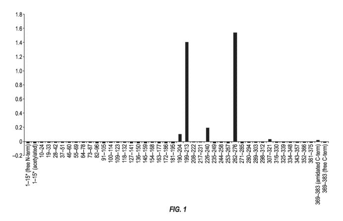
[0084] Figure 1 depicts the results of experiments designed to map the
epitope(s) bound by the
murine 3D6 monoclonal antibody.
[0085] Figure 2 depicts an alignment of heavy chain variable regions of the
mouse 3D6
antibody and humanized versions of the 3D6 antibody (VHvl, VHv2, VHvlb,
VHvlbAl 1, and
VHv5) . The consensus amino acid sequence among the heavy chain variable
regions of the
mouse 3D6 antibody and humanized versions of the 3D6 antibody (VHvl, VHv2,
VHvlb,
VHvlbAl 1, and VHv5) is labeled "Majority.' The CDRs as defined by
Kabat/Chothia
Composite are in boldface. Positions in heavy chain variable regions of the
mouse 3D6 antibody
and humanized versions of the 3D6 antibody (VHvl, VHv2, VHvlb, VHvl bAl 1, and
VHv5)
where amino acid residues differ from the "Majority" sequence are boxed.
[0086] Figure 3 depicts an alignment of light chain variable regions of the
mouse 3D6
antibody and humanized versions of the 3D6 antibody. The consensus amino acid
sequence
between the light chain variable regions of the mouse 3D6 antibody and
selected humanized 3D6
antibodies is labeled "Majority.' The CDRs as defined by Kabat are in
boldface. Positions in
light chain variable regions of the mouse 3D6 antibody and humanized versions
of the 3D6
antibody where amino acid residues differ from the "Majority" sequence are
boxed.
CA 03022515 2018-10-29
WO 2017/191560 PCT/IB2017/052544
[0087] Figures 4A and 4B depict an alignment of heavy chain variable regions
of the mouse
3D6 antibody and humanized versions of the 3D6 antibody (VHvl, VHvlb,
VHvlbAll,
VHv1bA11B6G2, VHv1bA11B6H3, VHvlc, VHvld, VHvle, VHvlf, VHv2, VHv3, VHv3b,
VHv3c, VHv4, VHv4b, VHv4c, and VHv5). The consensus amino acid sequence among
the
heavy chain variable regions of the humanized versions of the 3D6 antibody
(VHvl, VHv lb,
VHvlbAll, VHv1bA11B6G2, VHv1bA11B6H3, VHvlc, VHvld, VHvle, VHvlf, VHv2,
VHv3, VHv3b, VHv3c, VHv4, VHv4b, VHv4c, and VHv5) is labeled "Majority.' The
CDRs as
defined by Kabat/Chothia Composite are in boldface. Positions in heavy chain
variable regions
of the humanized versions of the 3D6 antibody (VHvl, VHvlb, VHvlbAll,
VHv1bA11B6G2,
VHv1bA11B6H3, VHvlc, VHvld, VHvle, VHvlf, VHv2, VHv3, VHv3b, VHv3c, VHv4,
VHv4b, VHv4c, and VHv5) where amino acid residues differ from the "Majority"
sequence are
boxed.
[0088] Figures 5A, 5B, and 5C depict results of ELISA screening assays for
selected mouse
monoclonal anti-tau antibodies.
[0089] Figure 6 depicts binding kinetics for selected mouse monoclonal anti-
tau antibodies to
recombinant human tau.
[0090] Figure 7 depicts results of functional blocking assays for selected
mouse monoclonal
anti-tau antibodies.
[0091] Figure 8 depicts results of disaggregation assays for selected mouse
monoclonal anti-
tau antibodies.
[0092] Figure 9 depicts results of experiments showing that 3D6 and 5G8
immunocapture tau
from human Alzheimer's disease tissue.
[0093] Figures 10A, 10B, and 10C depict results of experiments showing
affinity of
humanized 3D6 variants toward tau.
CA 03022515 2018-10-29
WO 2017/191560 PCT/IB2017/052544
21
BRIEF DESCRIPTION OF THE SEQUENCES
[0094] SEQ ID NO:1 sets forth the amino acid sequence of an isoform of human
tau (Swiss-
Prot P10636-8).
[0095] SEQ ID NO:2 sets forth the amino acid sequence of an isoform of human
tau (Swiss-
Prot P10636-7).
[0096] SEQ ID NO:3 sets forth the amino acid sequence of an isoform of human
tau (Swiss-
Prot P10636-6), (4RON human tau).
[0097] SEQ ID NO:4 sets forth the amino acid sequence of an isoform of human
tau (Swiss-
Prot P10636-5)
[0098] SEQ ID NO:5 sets forth the amino acid sequence of an isoform of human
tau (Swiss-
Prot P10636-4).
[0099] SEQ ID NO:6 sets forth the amino acid sequence of an isoform of human
tau (Swiss-
Prot P10636-2).
[0100] SEQ ID NO: 7 sets forth the amino acid sequence of the heavy chain
variable region of
the mouse 3D6 antibody.
[0101] SEQ ID NO: 8 sets forth the amino acid sequence of Kabat/Chothia
composite CDR-
H1 of the mouse 3D6 antibody.
[0102] SEQ ID NO:9 sets forth the amino acid sequence of Kabat CDR-H2 of the
mouse 3D6
antibody.
[0103] SEQ ID NO: 10 sets forth the amino acid sequence of Kabat CDR-H3 of the
mouse
3D6 antibody.
[0104] SEQ ID NO: 11 sets forth the amino acid sequence of the light chain
variable region of
the mouse 3D6 antibody and of the mouse 6A10 antibody.
[0105] SEQ ID NO: 12 sets forth the amino acid sequence of Kabat CDR-L1 of the
mouse
3D6 antibody and of the mouse 6A10 antibody.
CA 03022515 2018-10-29
WO 2017/191560 PCT/IB2017/052544
22
[0106] SEQ ID NO: 13 sets forth the amino acid sequence of Kabat CDR-L2 of the
mouse
3D6 antibody and of the mouse 6A10 antibody.
[0107] SEQ ID NO: 14 sets forth the amino acid sequence of Kabat CDR-L3 of the
mouse
3D6 antibody and of the mouse 6A10 antibody.
[0108] SEQ ID NO:15 sets forth the amino acid sequence of heavy chain variable
region of the
humanized 3D6 antibody hu3D6VHv1.
[0109] SEQ ID NO:16 sets forth the amino acid sequence of heavy chain variable
region of the
humanized 3D6 antibody hu3D6VHv2.
[0110] SEQ ID NO:17 sets forth the amino acid sequence of heavy chain variable
region of the
humanized 3D6 antibody hu3D6VHv lb.
[0111] SEQ ID NO:18 sets forth the amino acid sequence of heavy chain variable
region of the
humanized 3D6 antibody hu3D6VHv1bA1 1.
[0112] SEQ ID NO:19 sets forth the amino acid sequence of heavy chain variable
region of the
humanized 3D6 antibody hu3D6VHv5:
[0113] SEQ ID NO:20 sets forth the amino acid sequence of the light chain
variable region of
the humanized 3D6 antibody hu3D6VLv1.
[0114] SEQ ID NO:21 sets forth the amino acid sequence of the light chain
variable region of
the humanized 3D6 antibody hu3D6VLv2.
[0115] SEQ ID NO:22 sets forth the amino acid sequence of the light chain
variable region of
the humanized 3D6 antibody hu3D6VLv3.
[0116] SEQ ID NO:23 sets forth the amino acid sequence of the light chain
variable region of
the humanized 3D6 antibody hu3D6VLv4.
[0117] SEQ ID NO:24 sets forth the amino acid sequence of the heavy chain
variable acceptor
Acc.# BAC01986.1.
[0118] SEQ ID NO:25 sets forth the amino acid sequence of the heavy chain
variable acceptor
Acc.# IMGT# IGHV1-69-2*01.
CA 03022515 2018-10-29
WO 2017/191560 PCT/IB2017/052544
23
[0119] SEQ ID NO:26 sets forth the amino acid sequence of the heavy chain
variable acceptor
Acc.# IMGT#IGKJ1*01.
[0120] SEQ ID NO:27 sets forth the amino acid sequence of the light chain
variable acceptor
Acc. # IMGT#IGKV2-30*02
[0121] SEQ ID NO:28 sets forth the amino acid sequence of the light chain
variable acceptor
Acc. # IMGT#IGKI2*01.
[0122] SEQ ID NO:29 sets forth the amino acid sequence of the light chain
variable acceptor
Acc. # AAZ09048.1.
[0123] SEQ ID NO: 30 sets forth a nucleic acid sequence encoding the heavy
chain variable
region of the mouse 3D6 antibody.
[0124] SEQ ID NO: 31 sets forth a nucleic acid sequence encoding the light
chain variable
region of the mouse 3D6 antibody.
[0125] SEQ ID NO: 32 sets forth the amino acid sequence of Kabat CDR-H1 of the
mouse
3D6 antibody.
[0126] SEQ ID NO: 33 sets forth the amino acid sequence of Chothia CDR-H1 of
the mouse
3D6 antibody.
[0127] SEQ ID NO: 34 sets forth the amino acid sequence of Chothia CDR-H2 of
the mouse
3D6 antibody.
[0128] SEQ ID NO: 35 sets forth the amino acid sequence of AbM CDR-H2 of the
mouse 3D6
antibody.
[0129] SEQ ID NO: 36 sets forth the amino acid sequence of Contact CDR-L1 of
the mouse
3D6 antibody.
[0130] SEQ ID NO: 37 sets forth the amino acid sequence of Contact CDR-L2 of
the mouse
3D6 antibody.
[0131] SEQ ID NO: 38 sets forth the amino acid sequence of Contact CDR-L3 of
the mouse
3D6 antibody.
CA 03022515 2018-10-29
WO 2017/191560 PCT/IB2017/052544
24
[0132] SEQ ID NO: 39 sets forth the amino acid sequence of Contact CDR-H1 of
the mouse
3D6 antibody.
[0133] SEQ ID NO: 40 sets forth the amino acid sequence of Contact CDR-H2 of
the mouse
3D6 antibody.
[0134] SEQ ID NO: 41 sets forth the amino acid sequence of Contact CDR-H3 of
the mouse
3D6 antibody.
[0135] SEQ ID NO: 42 sets forth the amino acid sequence of an alternate Kabat-
Chothia
Composite CDR-H1 of a humanized 3D6 antibody (derived from hu3D6VHv5,
hu3D6VHvlbAl1B6G2, hu3D6VHvlbAl1B6H3, hu3D6VHv1e, and hu3D6VHv1f).
[0136] SEQ ID NO: 43 sets forth the amino acid sequence of an alternate Kabat
CDR-H2 of a
humanized 3D6 antibody (derived from hu3D6VHv5 and hu3D6VHvlbAl1B6H3).
[0137] SEQ ID NO:44 sets forth the consensus amino acid sequence among the
heavy chain
variable regions of the mouse 3D6 and selected humanized 3D6 antibodies (VHvl,
VHv2,
VHvlb, VHvlbAll, and VHv5) (labeled "Majority' in Figure 2).
[0138] SEQ ID NO:45 sets forth the consensus amino acid sequence between the
light chain
variable regions of the mouse 3D6 and selected humanized 3D6 antibodies
(labeled "Majority' in
Figure 3).
[0139] SEQ ID NO:46 sets forth the amino acid sequence of heavy chain variable
region of the
humanized 3D6 antibody hu3D6VHvlbAl1B6G2.
[0140] SEQ ID NO:47 sets forth the amino acid sequence of heavy chain variable
region of the
humanized 3D6 antibody hu3D6VHvlbAl1B6H3.
[0141] SEQ ID NO:48 sets forth the amino acid sequence of heavy chain variable
region of the
humanized 3D6 antibody hu3D6VHv1c.
[0142] SEQ ID NO:49 sets forth the amino acid sequence of heavy chain variable
region of the
humanized 3D6 antibody hu3D6VHv1d.
[0143] SEQ ID NO:50 sets forth the amino acid sequence of heavy chain variable
region of the
humanized 3D6 antibody hu3D6VHv1e.
CA 03022515 2018-10-29
WO 2017/191560 PCT/IB2017/052544
[0144] SEQ ID NO:51 sets forth the amino acid sequence of heavy chain variable
region of the
humanized 3D6 antibody hu3D6VHv1f.
[0145] SEQ ID NO:52 sets forth the amino acid sequence of heavy chain variable
region of the
humanized 3D6 antibody hu3D6VHv3.
[0146] SEQ ID NO:53 sets forth the amino acid sequence of heavy chain variable
region of the
humanized 3D6 antibody hu3D6VHv3b.
[0147] SEQ ID NO:54 sets forth the amino acid sequence of heavy chain variable
region of the
humanized 3D6 antibody hu3D6VHv3c.
[0148] SEQ ID NO:55 sets forth the amino acid sequence of heavy chain variable
region of the
humanized 3D6 antibody hu3D6VHv4.
[0149] SEQ ID NO:56 sets forth the amino acid sequence of heavy chain variable
region of the
humanized 3D6 antibody hu3D6VHv4b.
[0150] SEQ ID NO:57 sets forth the amino acid sequence of heavy chain variable
region of the
humanized 3D6 antibody hu3D6VHv4c.
[0151] SEQ ID NO: 58 sets forth the amino acid sequence of an alternate Kabat-
Chothia
Composite CDR-H1 of a humanized 3D6 antibody (derived from hu3D6VH1c).
[0152] SEQ ID NO: 59 sets forth the amino acid sequence of an alternate Kabat-
Chothia
Composite CDR-H1 of a humanized 3D6 antibody (derived from hu3D6VHv1d,
hu3D6VHv3c,
and hu3D6VHv4c).
[0153] SEQ ID NO: 60 sets forth the amino acid sequence of an alternate Kabat-
Chothia
Composite CDR-H1 of a humanized 3D6 antibody (derived from hu3D6VHv3b and
hu3D6VHv4b).
[0154] SEQ ID NO: 61 sets forth the amino acid sequence of an alternate Kabat
CDR-H2 of a
humanized 3D6 antibody (derived from hu3D6VHvlbAl1B6G2).
[0155] SEQ ID NO: 62 sets forth the amino acid sequence of an alternate Kabat
CDR-H2 of a
humanized 3D6 antibody (derived from hu3D6VHv1c, hu3D6VHv3b, AND hu3D6VHv4b.
CA 03022515 2018-10-29
WO 2017/191560 PCT/IB2017/052544
26
[0156] SEQ ID NO: 63 sets forth the amino acid sequence of an alternate Kabat
CDR-H2 of a
humanized 3D6 antibody (derived from hu3D6VHvld, hu3D6VHvlf, hu3D6VHv3c, and
hu3D6VHv4c).
[0157] SEQ ID NO: 64 sets forth the amino acid sequence of an alternate Kabat
CDR-H2 of a
humanized 3D6 antibody (derived from hu3D6VHv le).
[0158] SEQ ID NO: 65 sets forth the amino acid sequence of an alternate Kabat
CDR-H3 of a
humanized 3D6 antibody (derived from hu3D6VHv1f).
SEQ ID NO:66 sets forth the amino acid sequence of the heavy chain variable
region of the
mouse 6A10 antibody.
[0159] SEQ ID NO: 67 sets forth the amino acid sequence of Kabat/Chothia
composite CDR-
H1 of the mouse 6A10 antibody.
[0160] SEQ ID NO:68 sets forth the amino acid sequence of Kabat CDR-H2 of the
mouse
6A10 antibody.
[0161] SEQ ID NO: 69 sets forth the amino acid sequence of Kabat CDR-H3 of the
mouse
6A10 antibody.
[0162] SEQ ID NO:70 sets for the amino acid sequence of the VH region of mouse
antibody
(pdb code 1CR9) used as a structure template for heavy chain humanization.
[0163] SEQ ID NO:71 sets forth the consensus amino acid sequence among the
heavy chain
variable regions of the selected humanized 3D6 antibodies (VHvl, VHvlb,
VHvlbAll,
VHvlbAl1B6G2, VHvlbAl1B6H3, VHvlc, VHvld, VHvle, VHvlf, VHv2, VHv3, VHv3b,
VHv3c, VHv4, VHv4b, VHv4c, and VHv5) (labeled "Majority' in Figures 4A and
4B).
[0164] SEQ ID NO: 72 sets forth the amino acid sequence of the heavy chain of
a chimeric
3D6 antibody.
[0165] SEQ ID NO: 73 sets forth the amino acid sequence of the light chain of
a chimeric 3D6
antibody.
CA 03022515 2018-10-29
WO 2017/191560 PCT/IB2017/052544
27
DEFINITIONS
[0166] Monoclonal antibodies or other biological entities are typically
provided in isolated
form. This means that an antibody or other biologically entity is typically at
least 50% w/w pure
of interfering proteins and other contaminants arising from its production or
purification but does
not exclude the possibility that the monoclonal antibody is combined with an
excess of
pharmaceutically acceptable carrier(s) or other vehicle intended to facilitate
its use. Sometimes
monoclonal antibodies are at least 60%, 70%, 80%, 90%, 95% or 99% w/w pure of
interfering
proteins and contaminants from production or purification. Often an isolated
monoclonal
antibody or other biological entity is the predominant macromolecular species
remaining after its
purification.
[0167] Specific binding of an antibody to its target antigen means an affinity
and/or avidity of
at least 106, io7, 108, i09, 1010, r11,
u or 1012 M-1. Specific binding is detectably higher
in
magnitude and distinguishable from non-specific binding occurring to at least
one unrelated
target. Specific binding can be the result of formation of bonds between
particular functional
groups or particular spatial fit (e.g., lock and key type) whereas nonspecific
binding is usually
the result of van der Waals forces. Specific binding does not however
necessarily imply that an
antibody binds one and only one target.
[0168] The basic antibody structural unit is a tetramer of subunits. Each
tetramer includes two
identical pairs of polypeptide chains, each pair having one "light" (about 25
kDa) and one
"heavy" chain (about 50-70 kDa). The amino-terminal portion of each chain
includes a variable
region of about 100 to 110 or more amino acids primarily responsible for
antigen recognition.
This variable region is initially expressed linked to a cleavable signal
peptide. The variable
region without the signal peptide is sometimes referred to as a mature
variable region. Thus, for
example, a light chain mature variable region means a light chain variable
region without the
light chain signal peptide. The carboxy-terminal portion of each chain defines
a constant region
primarily responsible for effector function.
[0169] Light chains are classified as either kappa or lambda. Heavy chains are
classified as
gamma, mu, alpha, delta, or epsilon, and define the antibody's isotype as IgG,
IgM, IgA, IgD and
IgE, respectively. Within light and heavy chains, the variable and constant
regions are joined by
CA 03022515 2018-10-29
WO 2017/191560 PCT/IB2017/052544
28
a "J" region of about 12 or more amino acids, with the heavy chain also
including a "D" region
of about 10 or more amino acids. See generally, Fundamental Immunology, Paul,
W., ed., 2nd
ed. Raven Press, N.Y., 1989, Ch. 7 (incorporated by reference in its entirety
for all purposes).
[0170] An immunoglobulin light or heavy chain variable region (also referred
to herein as a
"light chain variable domain" ("VL domain") or "heavy chain variable domain"
("VH domain"),
respectively) consists of a "framework" region interrupted by three
"complementarity
determining regions" or "CDRs." The framework regions serve to align the CDRs
for specific
binding to an epitope of an antigen. The CDRs include the amino acid residues
of an antibody
that are primarily responsible for antigen binding. From amino-terminus to
carboxyl-terminus,
both VL and VH domains comprise the following framework (FR) and CDR regions:
FR1,
CDR1, FR2, CDR2, FR3, CDR3, and FR4. CDRs 1, 2, and 3 of a VL domain are also
referred
to herein, respectively, as CDR-L1, CDR-L2, and CDR-L3; CDRs 1, 2, and 3 of a
VH domain
are also referred to herein, respectively, as CDR-H1, CDR-H2, and CDR-H3.
[0171] The assignment of amino acids to each VL and VH domain is in accordance
with any
conventional definition of CDRs. Conventional definitions include, the Kabat
definition (Kabat,
Sequences of Proteins of Immunological Interest (National Institutes of
Health, Bethesda, MD,
1987 and 1991), the Chothia definition (Chothia & Lesk, J. MoL Biol. 196:901-
917, 1987;
Chothia et al., Nature 342:878-883, 1989); a composite of Chothia Kabat CDR in
which CDR-
H1 is a composite of Chothia and Kabat CDRs; the AbM definition used by Oxford
Molecular's
antibody modelling software; and, the contact definition of Martin et al
(bioinfo.org.uk/abs) (see
Table 1). Kabat provides a widely used numbering convention (Kabat numbering)
in which
corresponding residues between different heavy chains or between different
light chains are
assigned the same number. When an antibody is said to comprise CDRs by a
certain definition
of CDRs (e.g., Kabat) that definition specifies the minimum number of CDR
residues present in
the antibody (i.e., the Kabat CDRs). It does not exclude that other residues
falling within another
conventional CDR definition but outside the specified definition are also
present. For example,
an antibody comprising CDRs defined by Kabat includes among other
possibilities, an antibody
in which the CDRs contain Kabat CDR residues and no other CDR residues, and an
antibody in
which CDR H1 is a composite Chothia-Kabat CDR H1 and other CDRs contain Kabat
CDR
residues and no additional CDR residues based on other definitions.
CA 03022515 2018-10-29
WO 2017/191560 PCT/IB2017/052544
29
Table 1
Conventional Definitions of CDRs Using Kabat Numbering
Composite of
Chothia
Loop Kabat Chothia & AbM Contact
Kabat
Li L24--L34 L24--L34 L24--L34 L24--L34 L30--L36
L2 L50--L56 L50--L56 L50--L56 L50--L56 L46--L55
L3 L89--L97 L89--L97 L89--L97 L89--L97 L89--L96
H1 H31--H35B H26--H32..H34* H26--H35B* H26--H35B H30--H35B
H2 H50--H65 H52--H56 H50--H65 H50--H58 H47--H58
H3 H95--H102 H95--H102 H95--H102 H95--H102 H93--H101
*CDR-H1 by Chothia can end at H32, H33, or H34 (depending on the length of
the loop). This is because the Kabat numbering scheme places insertions of
extra
residues at 35A and 35B, whereas Chothia numbering places them at 31A and
31B. If neither H35A nor H35B (Kabat numbering) is present, the Chothia CDR-
H1 loop ends at H32. If only H35A is present, it ends at H33. If both H35A and
H35B are present, it ends at H34.
[0172] The term "antibody" includes intact antibodies and binding fragments
thereof
Typically, fragments compete with the intact antibody from which they were
derived for specific
CA 03022515 2018-10-29
WO 2017/191560 PCT/IB2017/052544
binding to the target including separate heavy chains, light chains Fab, Fab',
F(abl)2, F(ab)c,
Dabs, nanobodies, and Fv. Fragments can be produced by recombinant DNA
techniques, or by
enzymatic or chemical separation of intact immunoglobulins. The term
"antibody" also includes
a bispecific antibody and/or a humanized antibody. A bispecific or
bifunctional antibody is an
artificial hybrid antibody having two different heavy/light chain pairs and
two different binding
sites (see, e.g., Songsivilai and Lachmann, Clin. Exp. Immunol., 79:315-321
(1990); Kostelny et
al., J. Immunol., 148:1547-53 (1992)). In some bispecific antibodies, the two
different
heavy/light chain pairs include a humanized 3D6 heavy chain/light chain pair
and a heavy
chain/light chain pair specific for a different epitope on tau than that bound
by 3D6.
[0173] In some bispecific antibodies, one heavy chain/light chain pair is a
humanized 3D6
antibody as further disclosed below and the other heavy chain/light chain pair
is from an
antibody that binds to a receptor expressed on the blood brain barrier, such
as an insulin receptor,
an insulin-like growth factor (IGF) receptor, a leptin receptor, or a
lipoprotein receptor, or a
transferrin receptor (Friden et al., Proc. NatL Acad. Sci. USA 88:4771-4775,
1991; Friden et al.,
Science 259:373-377, 1993). Such a bispecific antibody can be transferred
cross the blood brain
barrier by receptor-mediated transcytosis. Brain uptake of the bispecific
antibody can be further
enhanced by engineering the bi-specific antibody to reduce its affinity to the
blood brain barrier
receptor. Reduced affinity for the receptor resulted in a broader distribution
in the brain (see,
e.g., Atwal et al., Sci. Trans. Med. 3, 84ra43, 2011; Yu et al., Sci. Trans.
Med. 3, 84ra44, 2011).
[0174] Exemplary bispecific antibodies can also be: (1) a dual-variable-domain
antibody
(DVD-Ig), where each light chain and heavy chain contains two variable domains
in tandem
through a short peptide linkage (Wu et al., Generation and Characterization of
a Dual Variable
Domain Immunoglobulin (DVD-IgTm) Molecule, In: Antibody Engineering, Springer
Berlin
Heidelberg (2010)); (2) a Tandab, which is a fusion of two single chain
diabodies resulting in a
tetravalent bispecific antibody that has two binding sites for each of the
target antigens; (3) a
flexibody, which is a combination of scFvs with a diabody resulting in a
multivalent molecule;
(4) a so-called "dock and lock" molecule, based on the "dimerization and
docking domain" in
Protein Kinase A, which, when applied to Fabs, can yield a trivalent
bispecific binding protein
consisting of two identical Fab fragments linked to a different Fab fragment;
or (5) a so-called
Scorpion molecule, comprising, e.g., two scFvs fused to both termini of a
human Fc-region.
CA 03022515 2018-10-29
WO 2017/191560 PCT/IB2017/052544
31
Examples of platforms useful for preparing bispecific antibodies include BiTE
(Micromet),
DART (MacroGenics), Fcab and Mab2 (F-star), Fc-engineered IgG1(Xencor) or
DuoBody
(based on Fab arm exchange, Genmab).
[0175] The term "epitope" refers to a site on an antigen to which an antibody
binds. An
epitope can be formed from contiguous amino acids or noncontiguous amino acids
juxtaposed by
tertiary folding of one or more proteins. Epitopes formed from contiguous
amino acids (also
known as linear epitopes) are typically retained on exposure to denaturing
solvents whereas
epitopes formed by tertiary folding (also known as conformational epitopes)
are typically lost on
treatment with denaturing solvents. An epitope typically includes at least 3,
and more usually, at
least 5 or 8-10 amino acids in a unique spatial conformation. Methods of
determining spatial
conformation of epitopes include, for example, x-ray crystallography and 2-
dimensional nuclear
magnetic resonance. See, e.g., Epitope Mapping Protocols, in Methods in
Molecular Biology,
Vol. 66, Glenn E. Morris, Ed. (1996).
[0176] Antibodies that recognize the same or overlapping epitopes can be
identified in a
simple immunoassay showing the ability of one antibody to compete with the
binding of another
antibody to a target antigen. The epitope of an antibody can also be defined X-
ray
crystallography of the antibody bound to its antigen to identify contact
residues. Alternatively,
two antibodies have the same epitope if all amino acid mutations in the
antigen that reduce or
eliminate binding of one antibody reduce or eliminate binding of the other.
Two antibodies have
overlapping epitopes if some amino acid mutations that reduce or eliminate
binding of one
antibody reduce or eliminate binding of the other.
[0177] Competition between antibodies is determined by an assay in which an
antibody under
test inhibits specific binding of a reference antibody to a common antigen
(see, e.g., Junghans et
al., Cancer Res. 50:1495, 1990). A test antibody competes with a reference
antibody if an
excess of a test antibody (e.g., at least 2x, 5x, 10x, 20x or 100x) inhibits
binding of the reference
antibody by at least 50% as measured in a competitive binding assay. Some test
antibodies
inhibit binding of the references antibody by at least 75%, 90% or 99%.
Antibodies identified by
competition assay (competing antibodies) include antibodies binding to the
same epitope as the
CA 03022515 2018-10-29
WO 2017/191560 PCT/IB2017/052544
32
reference antibody and antibodies binding to an adjacent epitope sufficiently
proximal to the
epitope bound by the reference antibody for steric hindrance to occur.
[0178] The term "pharmaceutically acceptable" means that the carrier, diluent,
excipient, or
auxiliary is compatible with the other ingredients of the formulation and not
substantially
deleterious to the recipient thereof.
[0179] The term "patient" includes human and other mammalian subjects that
receive either
prophylactic or therapeutic treatment.
[0180] An individual is at increased risk of a disease if the subject has at
least one known risk-
factor (e.g., genetic, biochemical, family history, and situational exposure)
placing individuals
with that risk factor at a statistically significant greater risk of
developing the disease than
individuals without the risk factor.
[0181] The term "biological sample" refers to a sample of biological material
within or
obtainable from a biological source, for example a human or mammalian subject.
Such samples
can be organs, organelles, tissues, sections of tissues, bodily fluids,
peripheral blood, blood
plasma, blood serum, cells, molecules such as proteins and peptides, and any
parts or
combinations derived therefrom. The term biological sample can also encompass
any material
derived by processing the sample. Derived material can include cells or their
progeny.
Processing of the biological sample may involve one or more of filtration,
distillation, extraction,
concentration, fixation, inactivation of interfering components, and the like.
[0182] The term "control sample" refers to a biological sample not known or
suspected to
include tau-related disease-affected regions, or at least not known or suspect
to include diseased
regions of a given type. Control samples can be obtained from individuals not
afflicted with the
tau-related disease. Alternatively, control samples can be obtained from
patients afflicted with
the tau-related disease. Such samples can be obtained at the same time as a
biological sample
thought to comprise the tau-related disease or on a different occasion. A
biological sample and a
control sample can both be obtained from the same tissue. Preferably, control
samples consist
essentially or entirely of normal, healthy regions and can be used in
comparison to a biological
sample thought to comprise tau-related disease-affected regions. Preferably,
the tissue in the
control sample is the same type as the tissue in the biological sample.
Preferably, the tau-related
CA 03022515 2018-10-29
WO 2017/191560 PCT/IB2017/052544
33
disease-affected cells thought to be in the biological sample arise from the
same cell type (e.g.,
neurons or glia ) as the type of cells in the control sample.
[0183] The term "disease" refers to any abnormal condition that impairs
physiological
function. The term is used broadly to encompass any disorder, illness,
abnormality, pathology,
sickness, condition, or syndrome in which physiological function is impaired,
irrespective of the
nature of the etiology.
[0184] The term "symptom" refers to a subjective evidence of a disease, such
as altered gait, as
perceived by the subject. A "sign" refers to objective evidence of a disease
as observed by a
physician.
[0185] The term "positive response to treatment" refers to a more favorable
response in an
individual patient or average response in a population of patients relative to
an average response
in a control population not receiving treatment.
[0186] For purposes of classifying amino acids substitutions as conservative
or
nonconservative, amino acids are grouped as follows: Group I (hydrophobic side
chains): met,
ala, val, leu, ile; Group II (neutral hydrophilic side chains): cys, ser, thr;
Group III (acidic side
chains): asp, glu; Group IV (basic side chains): asn, gln, his, lys, arg;
Group V (residues
influencing chain orientation): gly, pro; and Group VI (aromatic side chains):
trp, tyr, phe.
Conservative substitutions involve substitutions between amino acids in the
same class. Non-
conservative substitutions constitute exchanging a member of one of these
classes for a member
of another.
[0187] Percentage sequence identities are determined with antibody sequences
maximally
aligned by the Kabat numbering convention. After alignment, if a subject
antibody region (e.g.,
the entire mature variable region of a heavy or light chain) is being compared
with the same
region of a reference antibody, the percentage sequence identity between the
subject and
reference antibody regions is the number of positions occupied by the same
amino acid in both
the subject and reference antibody region divided by the total number of
aligned positions of the
two regions, with gaps not counted, multiplied by 100 to convert to
percentage.
CA 03022515 2018-10-29
WO 2017/191560 PCT/IB2017/052544
34
[0188] Compositions or methods "comprising" or "including" one or more recited
elements
may include other elements not specifically recited. For example, a
composition that
"comprises" or "includes" an antibody may contain the antibody alone or in
combination with
other ingredients.
[0189] Designation of a range of values includes all integers within or
defining the range, and
all subranges defined by integers within the range.
[0190] Unless otherwise apparent from the context, the term "about"
encompasses
insubstantial variations, such as values within a standard margin of error of
measurement (e.g.,
SEM) of a stated value.
[0191] Statistical significance means p0.05.
[0192] The singular forms of the articles "a," "an," and "the" include plural
references unless
the context clearly dictates otherwise. For example, the term "a compound" or
"at least one
compound" can include a plurality of compounds, including mixtures thereof.
DETAILED DESCRIPTION
I. General
[0193] The invention provides antibodies that bind to tau. Some antibodies
specifically bind to
an epitope within residues 199-213 or 262-276 of SEQ ID NO:3 (corresponding to
residues 257-
271 or 320-334, respectively, of SEQ ID NO:1).. Some antibodies bind to tau
irrespective of
phosphorylation state. Some antibodies of the invention serve to inhibit or
delay tau-associated
pathologies and associated symptomatic deterioration. Although an
understanding of mechanism
is not required for practice of the invention, a reduction in toxicity may
occur as a result of the
antibody inducing phagocytosis of tau, inhibiting tau from inter or
intramolecular aggregation, or
from binding to other molecules, by stabilizing a non-toxic conformation, by
inhibiting
intercellular or intracellular transmission of pathogenic tau forms, by
blockade of tau
phosphorylation, by preventing binding of tau to cells, or by inducing
proteolytic cleavage of tau,
among other mechanisms. The antibodies of the invention or agents that induce
such antibodies
can be used in methods of treating or effecting prophylaxis of Alzheimer's and
other diseases
associated with tau.
CA 03022515 2018-10-29
WO 2017/191560 PCT/IB2017/052544
II. Target Molecules
[0194] Unless otherwise apparent from the context, reference to tau means a
natural human
form of tau including all isoforms irrespective of whether posttranslational
modification (e.g.,
phosphorylation, glycation, or acetylation) is present. There are six major
isoforms (splice
variants) of tau occurring in the human brain. The longest of these variants
has 441 amino acids,
of which the initial met residue is cleaved. Residues are numbered according
to the 441 isoform.
Thus, for example, reference to a phosphorylation at position 404 means
position 404 of the 441
isoform, or corresponding position of any other isoform when maximally aligned
with the 441
isoform. The amino acid sequences of the isoforms and Swiss-Prot numbers are
indicated below.
P10636-8 (SEQ ID NO:1)
10 20 30 40 50 60
MAEPRQEFEV MEDHAGTYGL GDRKDQGGYT MHQDQEGDTD AGLKESPLQT PTEDGSEEPG
70 80 90 100 110 120
SETSDAKSTP TAEDVTAPLV DEGAPGKQAA AQPHTEIPEG TTAEEAGIGD TPSLEDEAAG
130 140 150 160 170 180
HVTQARMVSK SKDGTGSDDK KAKGADGKTK IATPRGAAPP GQKGQANATR IPAKTPPAPK
190 200 210 220 230 240
TPPSSGEPPK SGDRSGYSSP GSPGTPGSRS RTPSLPTPPT REPKKVAVVR TPPKSPSSAK
250 260 270 280 290 300
SRLQTAPVPM PDLKNVKSKI GSTENLKHQP GGGKVQIINK KLDLSNVQSK CGSKDNIKHV
310 320 330 340 350 360
PGGGSVQIVY KPVDLSKVTS KCGSLGNIHH KPGGGQVEVK SEKLDFKDRV QSKIGSLDNI
370 380 390 400 410 420
THVPGGGNKK IETHKLTFRE NAKAKTDHGA EIVYKSPVVS GDTSPRHLSN VSSTGSIDMV
430 440
DSPQLATLAD EVSASLAKQG L
P10636-7 (SEQ ID NO:2)
10 20 30 40 50 60
MAEPRQEFEV MEDHAGTYGL GDRKDQGGYT MHQDQEGDTD AGLKESPLQT PTEDGSEEPG
70 80 90 100 110 120
SETSDAKSTP TAEAEEAGIG DTPSLEDEAA GHVTQARMVS KSKDGTGSDD KKAKGADGKT
130 140 150 160 170 180
KIATPRGAAP PGQKGQANAT RIPAKTPPAP KTPPSSGEPP KSGDRSGYSS PGSPGTPGSR
190 200 210 220 230 240
SRTPSLPTPP TREPKKVAVV RTPPKSPSSA KSRLQTAPVP MPDLKNVKSK IGSTENLKHQ
250 260 270 280 290 300
PGGGKVQIIN KKLDLSNVQS KCGSKDNIKH VPGGGSVQIV YKPVDLSKVT SKCGSLGNIH
310 320 330 340 350 360
HKPGGGQVEV KSEKLDFKDR VQSKIGSLDN ITHVPGGGNK KIETHKLTFR ENAKAKTDHG
370 380 390 400 410
AEIVYKSPVV SGDTSPRHLS NVSSTGSIDM VDSPQLATLA DEVSASLAKQ GL
CA 03022515 2018-10-29
WO 2017/191560
PCT/IB2017/(1525-14
36
P10636-6 (4RON human tau) (SEQ ID NO:3)
20 30 40 50 60
MAEPRQEFEV MEDHAGTYGL GDRKDQGGYT MHQDQEGDTD AGLKAEEAGI GDTPSLEDEA
70 80 90 100 110 120
AGHVTQARMV SKSKDGTGSD DKKAKGADGK TKIATPRGAA PPGQKGQANA TRIPAKTPPA
130 140 150 160 170 180
PKTPPSSGEP PKSGDRSGYS SPGSPGTPGS RSRTPSLPTP PTREPKKVAV VRTPPKSPSS
190 200 210 220 230 240
AKSRLQTAPV PMPDLKNVKS KIGSTENLKH QPGGGKVQII NKKLDLSNVQ SKCGSKDNIK
250 260 270 280 290 300
HVPGGGSVQI VYKPVDLSKV TSKCGSLGNI HHKPGGGQVE VKSEKLDFKD RVQSKIGSLD
310 320 330 340 350 360
NITHVPGGGN KKIETHKLTF RENAKAKTDH GAEIVYKSPV VSGDTSPRHL SNVSSTGSID
370 380
MVDSPQLATL ADEVSASLAK QGL
P10636-5 (SEQ ID NO:4)
10 20 30 40 50 60
MAEPRQEFEV MEDHAGTYGL GDRKDQGGYT MHQDQEGDTD AGLKESPLQT PTEDGSEEPG
70 80 90 100 110 120
SETSDAKSTT TAEDVTAPLV DEGAPGKQAA AQPHTEIPEG TTAEEAGIGD TPSLEDEAAG
130 140 150 160 170 180
HVTQARMVSK SKDGTGSDDK KAKGADGKTK IATPRGAAPP GQKGQANATR IPAKTPPAPK
190 200 210 220 230 240
TPPSSGEPPK SGDRSGYSSP GSPGTPGSRS RTPSLPTPPT REPKKVAVVR TPPKSPSSAK
250 260 270 280 290 300
SRLQTAPVPM PDLKNVKSKI GSTENLKHQP GGGKVQIVYK PVDLSKVTSK CGSLGNIHHK
310 320 330 340 350 360
PGGGQVEVKS EKLDFKDRVQ SKIGSLDNIT HVPGGGNKKI ETHKLTFREN AKAKTDHGAE
370 380 390 400 410
IVYKSPVVSG DTSPRHLSNV SSTGSIDMVD SPQLATLADE VSASLAKQGL
P10636-4 (SEQ ID NO:5)
10 20 30 40 50 60
MAEPRQEFEV MEDHAGTYGL GDRKDQGGYT MHQDQEGDTD AGLKESPLQT PTEDGSEEPG
70 80 90 100 110 120
SETSDAKSTP TAEAEEAGIG DTPSLEDEAA GHVTQARMVS KSKDGTGSDD KKAKGADGKT
130 140 150 160 170 180
KIATPRGAAP PGQKGQANAT RIPAKTPPAP KTPPSSGEPP KSGDRSGYSS PGSPGTPGSR
190 200 210 220 230 240
SRTPSLPTPP TREPKKVAVV RTPPKSPSSA KSRLQTAPVP MPDLKNVKSK IGSTENLKHQ
250 260 270 280 290 300
PGGGKVQIVY KPVDLSKVTS KCGSLGNIHH KPGGGQVEVK SEKLDFKDRV QSKIGSLDNI
310 320 330 340 350 360
THVPGGGNKK IETHKLTFRE NAKAKTDHGA EIVYKSPVVS GDTSPRHLSN VSSTGSIDMV
370 380
DSPQLATLAD EVSASLAKQG L
P10636-2 (SEQ ID NO:6)
CA 03022515 2018-10-29
WO 2017/191560 PCT/IB2017/052544
37
20 30 40 50 60
MAEPRQEFEV MEDHAGTYGL GDRKDQGGYT MHQDQEGDTD AGLKAEEAGI GDTPSLEDEA
70 80 90 100 110 120
AGHVTQARMV SKSKDGTGSD DKKAKGADGK TKIATPRGAA PPGQKGQANA TRIPAKTPPA
130 140 150 160 170 180
PKTPPSSGEP PKSGDRSGYS SPOSPGTPGS RSRTPSLPTP PTREPKKVAV VRTPPKSPSS
190 200 210 220 230 240
AKSRLQTAPV PMPDLKNVKS KIGSTENLKH QPGGGKVQIV YKPVDLSKVT SKCGSLGNIH
250 260 270 280 290 300
HKPGGGQVEV KSEKLDFKDR VQSKIGSLDN ITHVPGGGNK KIETHKLTFR ENAKAKTDHG
310 320 330 340 350
AEIVYKSPVV SGDTSPRHLS NVSSTGSIDM VDSPQLATLA DEVSASLAKQ GL
[0195] Reference to tau includes known natural variations about 30 of which
are listed in the
Swiss-Prot database and permutations thereof, as well as mutations associated
with tau
pathologies, such as dementia, Pick's disease, supranuclear palsy, etc. (see,
e.g., Swiss-Prot
database and Poorkaj, et al. Ann Neurol. 43:815-825 (1998)). Some examples of
tau mutations
numbered by the 441 isoform are a lysine to threonine mutation at amino acid
residue 257
(K257T), an isoleucine to valine mutation at amino acid position 260 (1260V);
a glycine to valine
mutation at amino acid position 272 (G272V); an asparagine to lysine mutation
at amino acid
position 279 (N279K); an asparagine to histidine mutation at amino acid
position 296 (N296H);
a proline to serine mutation at amino acid position 301 (P30 1S); a proline to
leucine mutation at
amino acid 301 (P301L); a glycine to valine mutation at amino acid position
303 (G303V); a
serine to asparagine mutation at position 305 (5305N); a glycine to serine
mutation at amino acid
position 335 (G3355); a valine to methionine mutation at position 337 (V337M);
a glutamic acid
to valine mutation at position 342 (E342V); a lysine to isoleucine mutation at
amino acid
position 369 (K3691); a glycine to arginine mutation at amino acid position
389 (G389R); and an
arginine to tryptophan mutation at amino acid position 406 (R406W).
[0196] Tau can be phosphorylated at one or more amino acid residues including
tyrosine at
amino acid positions 18, 29, 97, 310, and 394 serine at amino acid positions
184, 185, 198, 199,
CA 03022515 2018-10-29
WO 2017/191560 PCT/IB2017/052544
38
202, 208, 214, 235, 237, 238, 262, 293, 324, 356, 396, 400, 404, 409, 412,
413, and 422; and
threonine at amino acids positions 175, 181, 205, 212, 217, 231, and 403.
Unless otherwise apparent from context, reference to tau, or their fragments
includes the natural
human amino acid sequences including isoforms, mutants, and allelic variants
thereof.
III. Antibodies
A. Binding Specificity and Functional Properties
[0197] The invention provides antibodies that bind to tau. Some antibodies
specifically bind to
an epitope within residues 199-213 of 383 amino acid 4RON human tau protein
(SEQ ID NO:3)
(corresponding to residues 257-271 of SEQ ID NO:1). Some antibodies
specifically bind to an
epitope within residues 262-276 of 383 amino acid 4RON human tau protein (SEQ
ID NO:3)
(corresponding to residues 320-334 of SEQ ID NO:1). Some antibodies bind to
tau irrespective
of phosphorylation state. Some antibodies bind to an epitope not including a
residue subject to
phosphorylation. These antibodies can be obtained by immunizing with a tau
polypeptide
purified from a natural source or recombinantly expressed. Antibodies can be
screened for
binding tau in unphosphorylated form as well as a form in which one or more
residues
susceptible to phosphorylation are phosphorylated. Such antibodies preferably
bind with
indistinguishable affinities or at least within a factor of 1.5, 2 or 3-fold
to phosphorylated tau
compared to non-phosphorylated tau (i.e., are "pan-specific"). 3D6 is an
example of a pan-
specific monoclonal antibody. The invention also provides antibodies binding
to the same
epitope as any of the foregoing antibodies, such as, for example, the epitope
of 3D6. Also
included are antibodies competing for binding to tau with any of the foregoing
antibodies, such
as, for example, competing with 3D6.
[0198] The above-mentioned antibodies can be generated de novo by immunizing
with a
peptide including residues 199-213 or 262-276 of SEQ ID NO:3 (corresponding to
residues 257-
271 or 320-334, respectively, of SEQ ID NO:1) or by immunizing with a full
length tau
polypeptide or fragment thereof comprising such residues and screening for
specific binding to a
peptide including such residues. Such peptides are preferably attached to a
heterologous
conjugate molecule that helps elicit an antibody response to the peptide.
Attachment can be
direct or via a spacer peptide or amino acid. Cysteine is used as a spacer
amino acid because its
CA 03022515 2018-10-29
WO 2017/191560 PCT/IB2017/052544
39
free SH group facilitates attachment of a carrier molecule. A polyglycine
linker (e.g., 2-6
glycines), with or without a cysteine residue between the glycines and the
peptide can also be
used. The carrier molecule serves to provide a T-cell epitope that helps
elicit an antibody
response against the peptide. Several carriers are commonly used particularly
keyhole limpet
hemocyanin (KLH), ovalbumin and bovine serum albumin (BSA). Peptide spacers
can be added
to peptide immunogen as part of solid phase peptide synthesis. Carriers are
typically added by
chemical cross-linking. Some examples of chemical crosslinkers that can be
used include cross-
N-maleimido-6-aminocaproyl ester or m-maleimidobenzoyl-N-hydroxysuccinimide
ester (MBS)
(see for example, Harlow, E. et al., Antibodies: A Laboratory Manual, Cold
Spring Harbor
Laboratory Press, Cold Spring Harbor, N.Y. 1988; Sinigaglia et al., Nature,
336:778-780 (1988);
Chicz et al., J. Exp. Med., 178:27-47 (1993); Hammer et al., Cell 74:197-203
(1993); Falk K. et
al., Immunogenetics, 39:230-242 (1994); WO 98/23635; and, Southwood et al. J.
Immunology,
160:3363-3373 (1998)). The carrier and spacer if present can be attached to
either end of the
immunogen.
[0199] A peptide with optional spacer and carrier can be used to immunize
laboratory animals
or B-cells as described in more detail below. Hybridoma supernatants can be
tested for ability to
bind one or more peptides including residues 199-213 or 262-276 of SEQ ID NO:3
(corresponding to residues 257-271 or 320-334, respectively, of SEQ ID NO:1)
and/or
phosphorylated and non-phosphorylated forms of tau, such as, for example, a
full-length isoform
of tau with position 404 in phosphorylated form. The peptide can be attached
to a carrier or
other tag to facilitate the screening assay. In this case, the carrier or tag
is preferentially different
than the combination of spacer and carrier molecule used for immunization to
eliminate
antibodies specific for the spacer or carrier rather than the tau peptide. Any
of the tau isoforms
can be used.
[0200] The invention provides monoclonal antibodies binding to epitopes within
tau. An
antibody designated 3D6 is one such exemplary mouse antibody. Unless otherwise
apparent
from context, reference to 3D6 should be understood as referring to any of the
mouse, chimeric,
veneered, and humanized forms of this antibody. The antibody has been
deposited as [DEPOSIT
NUMBER]. This antibody specifically binds within amino acid residues 199-213
or 262-276 of
the 383 amino acid 4RON human tau protein (SEQ ID NO:3) (corresponding to
amino acid
CA 03022515 2018-10-29
WO 2017/191560 PCT/IB2017/052544
residues 257-271 or 320-334, respectively, of SEQ ID NO:1). This antibody is
further
characterized by its ability to bind both phosphorylated and unphosphorylated
tau, both non-
pathological and pathological forms and conformations of tau, and
misfolded/aggregated forms
of tau. An antibody designated 6A10 is one such exemplary mouse antibody.
Unless otherwise
apparent from context, reference to 6A10 should be understood as referring to
any of the mouse,
chimeric, veneered, and humanized forms of this antibody. Kabat/Chothia
Composite CDRs of
the heavy chain of 6A10 are designated SEQ ID NOs: 67, 68, and 69,
respectively, and Kabat
CDRs of the light chain of 6A10 are designated SEQ ID NOs: 12, 13, and 14,
respectively.
Mouse 6A10 shares 82.1% of VH sequence identity and 100% VL sequence identity
with the
VH chain and VL chain, respectively, of mouse 3D6.
[0201] Some antibodies of the invention bind to the same or overlapping
epitope as an
antibody designated 3D6. The sequences of the heavy and light chain mature
variable regions of
this antibody are designated SEQ ID NOs: 7 and 11, respectively. Other
antibodies having such
a binding specificity can be produced by immunizing mice with tau or a portion
thereof
including the desired epitope (e.g. 199-213 or 262-276 of SEQ ID NO:3, con-
esponding to
residues 257-271 or 320-334, respectively, of SEQ ID NO:1) and screening
resulting antibodies
for binding to tau optionally in competition with an antibody having the
variable regions of
mouse 3D6 (IgG1 kappa). Fragments of tau including the desired epitope can be
linked to a
carrier that helps elicit an antibody response to the fragment and/or be
combined with an
adjuvant the helps elicit such a response. Such antibodies can be screened for
differential
binding to tau or a fragment thereof compared with mutants of specified
residues. Screening
against such mutants more precisely defines the binding specificity to allow
identification of
antibodies whose binding is inhibited by mutagenesis of particular residues
and which are likely
to share the functional properties of other exemplified antibodies. The
mutations can be
systematic replacement substitution with alanine (or serine if an alanine is
present already) one
residue at a time, or more broadly spaced intervals, throughout the target or
throughout a section
thereof in which an epitope is known to reside. If the same set of mutations
significantly reduces
the binding of two antibodies, the two antibodies bind the same epitope.
[0202] Antibodies having the binding specificity of a selected murine antibody
(e.g., 3D6) can
also be produced using a variant of the phage display method. See Winter, WO
92/20791. This
CA 03022515 2018-10-29
WO 2017/191560 PCT/IB2017/052544
41
method is particularly suitable for producing human antibodies. In this
method, either the heavy
or light chain variable region of the selected murine antibody is used as a
starting material. If,
for example, a light chain variable region is selected as the starting
material, a phage library is
constructed in which members display the same light chain variable region
(i.e., the murine
starting material) and a different heavy chain variable region. The heavy
chain variable regions
can for example be obtained from a library of rearranged human heavy chain
variable regions. A
phage showing strong specific binding for tau or a fragment thereof (e.g., at
least 108 and
preferably at least 109 M-1) is selected. The heavy chain variable region from
this phage then
serves as a starting material for constructing a further phage library. In
this library, each phage
displays the same heavy chain variable region (i.e., the region identified
from the first display
library) and a different light chain variable region. The light chain variable
regions can be
obtained for example from a library of rearranged human variable light chain
regions. Again,
phage showing strong specific binding for tau or a fragment thereof are
selected. The resulting
antibodies usually have the same or similar epitope specificity as the murine
starting material.
[0203] Kabat/Chothia Composite CDRs of the heavy chain of 3D6 are designated
SEQ ID
NOs: 8, 9, and 10, respectively, and Kabat CDRs of the light chain of 3D6 are
designated SEQ
ID NOs: 12, 13, and 14, respectively.
[0204] Table 2 indicates the 3D6 CDRs as defined by Kabat, Chothia, Composite
of Chothia
and Kabat (also referred to herein as "Kabat/Chothia Composite"), AbM, and
Contact.
CA 03022515 2018-10-29
WO 2017/191560 PCT/IB2017/052544
42
Table 2: 3D6 CDRs as defined by Kabat, Chothia, Composite of Chothia and
Kabat, AbM, and
Contact
Composite of
Loop Kabat Chothia Chothia AbM Contact
& Kabat
L24--L34 L24--L34 L24--L34 L24--L34 L30--L36
Li
SEQ ID NO:12 SEQ ID NO:12 SEQ ID NO:12 SEQ ID NO:12 SEQ ID NO:
36
L50--L56 L50--L56 L50--L56 L50--L56 L46--L55
L2
SEQ ID NO:13 SEQ ID NO: 13 SEQ ID NO: 13 SEQ ID NO: 13 SEQ ID
NO: 37
L89--L97 L89--L97 L89--L97 L89--L97 L89--L96
L3
SEQ ID NO: 14 SEQ ID NO: i4 SEQ ID NO: 14 SEQ ID NO: 14 SEQ ID
NO:38
H31--H35B H26--H32 H26--H35B H26--H35B H30--H35B
H1
SEQ ID NO: 32 SEQ ID NO:33 SEQ ID NO:8 SEQ ID NO:8 SEQ ID NO:39
H50--H65 H52--H56 H50--H65 H50--H58 H47--H58
H2
SEQ ID NO: 9 SEQ ID NO:34 SEQ ID NO: 9 SEQ ID NO:35 SEQ ID
NO:40
H95--H102 H95--H102 H95--H102 H95--H102 H93--H101
H3
SEQ ID NO: 10 SEQ ID NO: 10 SEQ ID NO: 10 SEQ ID NO: 10 SEQ ID
NO:41
[0205] Other antibodies can be obtained by mutagenesis of cDNA encoding the
heavy and
light chains of an exemplary antibody, such as 3D6. Monoclonal antibodies that
are at least
70%, 80%, 90%, 95%, 96%, 97%, 98%, or 99% identical to 3D6 in amino acid
sequence of the
mature heavy and/or light chain variable regions and maintain its functional
properties, and/or
which differ from the respective antibody by a small number of functionally
inconsequential
amino acid substitutions (e.g., conservative substitutions), deletions, or
insertions are also
CA 03022515 2018-10-29
WO 2017/191560 PCT/IB2017/052544
43
included in the invention. Monoclonal antibodies having at least one or all
six CDR(s) as
defined by any conventional definition, but preferably Kabat, that are 90%,
95%, 99% or 100%
identical to con-esponding CDRs of 3D6 are also included.
[0206] The invention also provides antibodies having some or all (e.g., 3, 4,
5, and 6) CDRs
entirely or substantially from 3D6. Such antibodies can include a heavy chain
variable region
that has at least two, and usually all three, CDRs entirely or substantially
from the heavy chain
variable region of 3D6 and/or a light chain variable region having at least
two, and usually all
three, CDRs entirely or substantially from the light chain variable region
of3D6. The antibodies
can include both heavy and light chains. A CDR is substantially from a
corresponding 3D6 CDR
when it contains no more than 4, 3, 2, or 1 substitutions, insertions, or
deletions, except that
CDR-H2 (when defined by Kabat) can have no more than 6, 5, 4, 3, 2, or 1
substitutions,
insertions, or deletions. Such antibodies can have at least 70%, 80%, 90%,
95%, 96%, 97%,
98%, or 99% identity to 3D6 in the amino acid sequence of the mature heavy
and/or light chain
variable regions and maintain their functional properties, and/or differ from
3D6 by a small
number of functionally inconsequential amino acid substitutions (e.g.,
conservative
substitutions), deletions, or insertions.
[0207] Some antibodies identified by such assays can bind to monomeric,
misfolded,
aggregated, phosphorylated, or unphosphorylated forms of tau or otherwise.
Likewise, some
antibodies are immunoreactive on non-pathological and pathological forms and
conformations of
tau.
B. Non-Human Antibodies
[0208] The production of other non-human antibodies, e.g., murine, guinea pig,
primate, rabbit
or rat, against tau or a fragment thereof (e.g., amino acid residues 199-213
or 262-276 of SEQ ID
NO:3) (corresponding to amino acid residues 257-271 or 320-334, respectively,
of SEQ ID
NO:1) can be accomplished by, for example, immunizing the animal with tau or a
fragment
thereof. See Harlow & Lane, Antibodies, A Laboratory Manual (CSHP NY, 1988)
(incorporated by reference for all purposes). Such an immunogen can be
obtained from a natural
source, by peptide synthesis, or by recombinant expression. Optionally, the
immunogen can be
administered fused or otherwise complexed with a carrier protein. Optionally,
the immunogen
CA 03022515 2018-10-29
WO 2017/191560 PCT/IB2017/052544
44
can be administered with an adjuvant. Several types of adjuvant can be used as
described below.
Complete Freund's adjuvant followed by incomplete adjuvant is preferred for
immunization of
laboratory animals. Rabbits or guinea pigs are typically used for making
polyclonal antibodies.
Mice are typically used for making monoclonal antibodies. Antibodies are
screened for specific
binding to tau or an epitope within tau (e.g., an epitope comprising one or
more of amino acid
residues 199-213 or 262-276 of SEQ ID NO:3) (corresponding to amino acid
residues 257-271
or 320-334, respectively, of SEQ ID NO:1). Such screening can be accomplished
by
determining binding of an antibody to a collection of tau variants, such as
tau variants containing
amino acid residues 199-213 or 262-276 of SEQ ID NO: 3 (corresponding to amino
acid residues
257-271 or 320-334, respectively, of SEQ ID NO:1) or mutations within these
residues, and
determining which tau variants bind to the antibody. Binding can be assessed,
for example, by
Western blot, FACS or ELISA.
C. Humanized Antibodies
[0209] A humanized antibody is a genetically engineered antibody in which CDRs
from a non-
human "donor" antibody are grafted into human "acceptor" antibody sequences
(see, e.g.,
Queen, US 5,530,101 and 5,585,089; Winter, US 5,225,539; Carter, US 6,407,213;
Adair, US
5,859,205; and Foote, US 6,881,557). The acceptor antibody sequences can be,
for example, a
mature human antibody sequence, a composite of such sequences, a consensus
sequence of
human antibody sequences, or a germline region sequence. Thus, a humanized
antibody is an
antibody having at least three, four, five or all CDRs entirely or
substantially from a donor
antibody and variable region framework sequences and constant regions, if
present, entirely or
substantially from human antibody sequences. Similarly a humanized heavy chain
has at least
one, two and usually all three CDRs entirely or substantially from a donor
antibody heavy chain,
and a heavy chain variable region framework sequence and heavy chain constant
region, if
present, substantially from human heavy chain variable region framework and
constant region
sequences. Similarly a humanized light chain has at least one, two and usually
all three CDRs
entirely or substantially from a donor antibody light chain, and a light chain
variable region
framework sequence and light chain constant region, if present, substantially
from human light
chain variable region framework and constant region sequences. Other than
nanobodies and
dAbs, a humanized antibody comprises a humanized heavy chain and a humanized
light chain.
CA 03022515 2018-10-29
WO 2017/191560 PCT/IB2017/052544
A CDR in a humanized antibody is substantially from a corresponding CDR in a
non-human
antibody when at least 85%, 90%, 95% or 100% of corresponding residues (as
defined by any
conventional definition but preferably defined by Kabat) are identical between
the respective
CDRs. The variable region framework sequences of an antibody chain or the
constant region of
an antibody chain are substantially from a human variable region framework
sequence or human
constant region respectively when at least 85%, 90%, 95% or 100% of
corresponding residues
defined by Kabat are identical. To be classified as humanized under the 2014
World Health
Organization (WHO) International non-proprietary names (INN) definition of
humanized
antibodies, an antibody must have at least 85% identity to human germline
antibody sequences
(i.e., prior to somatic hypermutation). Mixed antibodies are antibodies for
which one antibody
chain (e.g., heavy chain) meets the threshold but the other chain (e.g., light
chain) does not meet
the threshold. An antibody is classified as chimeric if neither chain meets
the threshold, even
though the variable framework regions for both chains were substantially human
with some
murine backmutations. See, Jones et al. (2016) The INNs and outs of antibody
nonproprietary
names, mAbs 8:1, 1-9, DOI: 10.1080/19420862.2015.1114320. See also "WHO-INN:
International nonproprietary names (INN) for biological and biotechnological
substances (a
review)" (Internet) 2014. Available from: http://www.
whoint/medicines/services/inn/BioRev2014.pdf), incorporated herein by
reference. For the
avoidance of doubt, the term "humanized" as used herein is not intended to be
limited to the
2014 WHO INN definition of humanized antibodies. Some of the humanized
antibodies
provided herein have at least 85% sequence identity to human germline
sequences and some of
the humanized antibodies provided herein have less than 85% sequence identity
to human
germline sequences. Some of the heavy chains of the humanized antibodies
provided herein have
from about 60% to 100% sequence identity to human germ line sequences, such
as, for example,
in the range of about 60% to 69%, 70% to 79%, 80% to 84%, or 85% to 89%. Some
heavy
chains fall below the 2014 WHO INN definition and have, for example, about
64%, 65%, 66%,
67%, 68%, 69%, 70%, 71%, 72%, 73%, 74%, 75%, 76%, 77%, 78%, 79%, 80%, 81%, or
82%,
83%, or 84% sequence identity to human germ line sequences, while other heavy
chains meet
the 2014 WHO INN definition and have about 85%, 86%, 87%, 88%, 89% or greater
sequence
identity to human germ line sequences. Some of the light chains of the
humanized antibodies
CA 03022515 2018-10-29
WO 2017/191560 PCT/IB2017/052544
46
provided herein have from about 60% to 100% sequence identity to human germ
line sequences,
such as, for example, in the range of about 80% to 84% or 85% to 89%. Some
light chains fall
below the 2014 WHO INN definition and have, for example, about 81%, 82%, 83%
or 84%
sequence identity to human germ line sequences, while other light chains meet
the 2014 WHO
INN definition and have about 85%, 86%, 87%, 88%, 89% or greater sequence
identity to human
germ line sequences. Some humanized antibodies provided herein that are
"chimeric" under the
2014 WHO INN definition have heavy chains with less than 85% identity to human
germ line
sequences paired with light chains having less than 85% identity to human germ
line sequences.
Some humanized antibodies provided herein are "mixed" under the 2014 WHO INN
definition,
for example, having a heavy chain with at least 85% sequence identity to human
germ line
sequences paired with a light chain having less than 85% sequence identity to
human germ line
sequences, or vice versa. Some humanized antibodies provided herein meet the
2014 WHO INN
definition of "humanized" and have a heavy chain with at least 85% sequence
identity to human
germ line sequences paired with a light chain having at least 85% sequence
identity to human
germ line sequences. Additional humanized antibodies of the invention meet the
2014 WHO
INN definition of "mixed" and include antibodies having a mature heavy chain
having an amino
acid sequence of any of SEQ ID NOs:15-19 and SEQ ID NOs:46-57 paired with a
mature light
chain having an amino acid sequence of any of SEQ ID NOs:20-23.
[0210] Although humanized antibodies often incorporate all six CDRs (defined
by any
conventional definition but preferably as defined by Kabat) from a mouse
antibody, they can also
be made with less than all CDRs (e.g., at least 3, 4, or 5 CDRs) from a mouse
antibody (e.g.,
Pascalis et al., J. Immunol. 169:3076, 2002; Vajdos et al., J. of MoL Biol.,
320: 415-428, 2002;
Iwahashi et al., Mol. Immunol. 36:1079-1091, 1999; Tamura et al, J. Immunol.,
164:1432-1441,
2000).
[0211] In some antibodies only part of the CDRs, namely the subset of CDR
residues required
for binding, termed the SDRs, are needed to retain binding in a humanized
antibody. CDR
residues not contacting antigen and not in the SDRs can be identified based on
previous studies
(for example residues H60-H65 in CDR H2 are often not required), from regions
of Kabat CDRs
lying outside Chothia hypervariable loops (Chothia, J. MoL Biol. 196:901,
1987), by molecular
modeling and/or empirically, or as described in Gonzales et al., MoL Immunol.
41: 863, 2004. In
CA 03022515 2018-10-29
WO 2017/191560 PCT/IB2017/052544
47
such humanized antibodies at positions in which one or more donor CDR residues
is absent or in
which an entire donor CDR is omitted, the amino acid occupying the position
can be an amino
acid occupying the corresponding position (by Kabat numbering) in the acceptor
antibody
sequence. The number of such substitutions of acceptor for donor amino acids
in the CDRs to
include reflects a balance of competing considerations. Such substitutions are
potentially
advantageous in decreasing the number of mouse amino acids in a humanized
antibody and
consequently decreasing potential immunogenicity and/or for meeting the WHO
INN definition
of "humanized". However, substitutions can also cause changes of affinity, and
significant
reductions in affinity are preferably avoided. Positions for substitution
within CDRs and amino
acids to substitute can also be selected empirically.
[0212] The human acceptor antibody sequences can optionally be selected from
among the
many known human antibody sequences to provide a high degree of sequence
identity (e.g., 65-
85% identity) between a human acceptor sequence variable region frameworks and
corresponding variable region frameworks of a donor antibody chain.
[0213] An example of an acceptor sequence for the heavy chain is the human
mature heavy
chain variable region with NCBI accession code BAC01986.1 (SEQ ID NO:24). An
example of
an acceptor sequence for the heavy chain is the human mature heavy chain
variable region with
NCBI accession code IMGT# IGHV1-69-2*01 (SEQ ID NO:25). An example of an
acceptor
sequence for the heavy chain is the human mature heavy chain variable region
with NCBI
accession code IMGT#IGKI1*01 (SEQ ID NO:26). IMGT# IGHV1-69-2*01 (SEQ ID
NO:25)
shares the canonical form of mouse 3D6 heavy chain CDR-H1 and H2. IMGT# IGHV1-
69-2*01
(SEQ ID NO:25) and IMGT#IGKI1*01 (SEQ ID NO:26) and both belong to human heavy
chain
subgroup 1. An example of an acceptor sequence for the light chain is the
human mature light
chain variable region with NCBI accession code IMGT#IGKV2-30*02 (SEQ ID
NO:27). An
example of an acceptor sequence for the light chain is the human mature light
chain variable
region with NCBI accession code IMGT#IGK12*01 (SEQ ID NO:28). An example of an
acceptor sequence for the light chain is the human mature light chain variable
region with NCBI
accession code AAZ09048.1 (SEQ ID NO:29). IMGT#IGKV2-30*02 (SEQ ID NO:27) has
the
same canonical classes for CDR-L1, CDR-L2 and L3 as mouse 3D6. IMGT#IGKV2-
30*02
(SEQ ID NO:27) and IMGT#IGK12*01 (SEQ ID NO:28) belong to human kappa subgroup
2.
CA 03022515 2018-10-29
WO 2017/191560 PCT/IB2017/052544
48
[0214] If more than one human acceptor antibody sequence is selected, a
composite or hybrid
of those acceptors can be used, and the amino acids used at different
positions in the humanized
light chain and heavy chain variable regions can be taken from any of the
human acceptor
antibody sequences used. For example, the human mature heavy chain variable
regions with
NCBI accession codes IMGT# IGHV1-69-2*01 (SEQ ID NO:25) and BAC01986.1 (SEQ ID
NO:24) were used as acceptor sequences for humanization of the 3D6 mature
heavy chain
variable region. Examples of positions in which these two acceptors differ
include positions H17
(T or S) and H20 (I or V). Humanized versions of the3D6 heavy chain variable
region can
include either amino acid at either of these positions.
[0215] Certain amino acids from the human variable region framework residues
can be
selected for substitution based on their possible influence on CDR
conformation and/or binding
to antigen. Investigation of such possible influences is by modeling,
examination of the
characteristics of the amino acids at particular locations, or empirical
observation of the effects
of substitution or mutagenesis of particular amino acids.
[0216] For example, when an amino acid differs between a murine variable
region framework
residue and a selected human variable region framework residue, the human
framework amino
acid can be substituted by the equivalent framework amino acid from the mouse
antibody when
it is reasonably expected that the amino acid:
(1) noncovalently binds antigen directly;
(2) is adjacent to a CDR region or within a CDR as defined by Chothia but not
Kabat;
(3) otherwise interacts with a CDR region (e.g., is within about 6 A of a CDR
region),
(e.g., identified by modeling the light or heavy chain on the solved structure
of a
homologous known immunoglobulin chain); or
(4) is a residue participating in the VL-VH interface.
[0217] The invention provides humanized forms of the murine 3D6 antibody
including 17
exemplified humanized heavy chain mature variable regions (hu3D6VHv1 (SEQ ID
NO:15),
hu3D6VHv2 (SEQ ID NO:16), hu3D6VHv1b (SEQ ID NO:17), hu3D6VHv1bA11 (SEQ ID
NO:18), hu3D6VHv5 (SEQ ID NO:19), hu3D6VHv1bA11B6G2 (SEQ ID NO:46) ,
CA 03022515 2018-10-29
WO 2017/191560 PCT/IB2017/052544
49
hu3D6VHvlbAl1B6H3 (SEQ ID NO:47), hu3D6VHv1c (SEQ ID NO:48), hu3D6VHv1d (SEQ
ID NO:49), hu3D6VHv1e (SEQ ID NO:50), hu3D6VHv1f (SEQ ID NO:51), hu3D6VHv3
(SEQ
ID NO:52), hu3D6VHv3b (SEQ ID NO:53) , hu3D6VHv3c (SEQ ID NO:54), hu3D6VHv4
(SEQ ID NO:55), hu3D6VHv4b (SEQ ID NO:56), and hu3D6VHv4c (SEQ ID NO:57)) and
4
exemplified humanized light chain mature variable regions (hu3D6VLv1 (SEQ ID
NO:20),
hu3D6VLv2 (SEQ ID NO:21, hu3D6VLv3 (SEQ ID NO:22), and hu3D6VLv4 (SEQ ID
NO:23)).
[0218] In an embodiment, humanized sequences are generated using a two-stage
PCR protocol
that allows introduction of multiple mutations, deletions, and insertions
using QuikChange site-
directed mutagenesis [Wang, W. and Malcolm, B.A. (1999) BioTechniques 26:680-
682)].
[0219] Framework residues from classes (1) through (3) as defined by Queen, US
5,530,101,
are sometimes alternately referred to as canonical and vernier residues.
Framework residues that
help define the conformation of a CDR loop are sometimes referred to as
canonical residues
(Chothia & Lesk, J. MoL Biol. 196:901-917 (1987); Thornton & Martin, J. Mol.
Biol. 263:800-
815 (1996)). Framework residues that support antigen-binding loop
conformations and play a
role in fine-tuning the fit of an antibody to antigen are sometimes referred
to as vernier residues
(Foote & Winter, J. MoL Biol 224:487-499 (1992)).
[0220] Other framework residues that are candidates for substitution are
residues creating a
potential glycosylation site. Still other candidates for substitution are
acceptor human
framework amino acids that are unusual for a human immunoglobulin at that
position. These
amino acids can be substituted with amino acids from the equivalent position
of the mouse donor
antibody or from the equivalent positions of more typical human
immunoglobulins.
[0221] Other framework residues that are candidates for substitution are N-
terminal glutamine
residues (Q) that may be replaced with glutamic acid (E) to minimize potential
for pyroglutamate
conversion [ Y. Diana Liu, et al., 2011, J. Biol. Chem., 286: 11211-11217].
Glutamic acid (E)
conversion to pyroglutamate (pE) occurs more slowly than from glutamine (Q).
Because of the
loss of a primary amine in the glutamine to pE conversion, antibodies become
more acidic.
Incomplete conversion produces heterogeneity in the antibody that can be
observed as multiple
CA 03022515 2018-10-29
WO 2017/191560 PCT/IB2017/052544
peaks using charge-based analytical methods. Heterogeneity differences may
indicate a lack of
process control.
[0222] Exemplary humanized antibodies are humanized forms of the mouse 3D6,
designated
Hu3D6.
[0223] The mouse antibody 3D6 comprises mature heavy and light chain variable
regions
having amino acid sequences comprising SEQ ID NO: 7 and SEQ ID NO:11,
respectively. The
invention provides 17 exemplified humanized mature heavy chain variable
regions:
hu3D6VHv1, hu3D6VHv2, hu3D6VHv1b, hu3D6VHv1bA11, hu3D6VHv5,
hu3D6VHv1bA11B6G2, hu3D6VHv1bA11B6H3, hu3D6VHv lc, hu3D6VHv1d, hu3D6VHv le,
hu3D6VHv1f, hu3D6VHv3, hu3D6VHv3b, hu3D6VHv3c, hu3D6VHv4, hu3D6VHv4b, and
hu3D6VHv4c. The invention further provides 4 exemplified human mature light
chain variable
regions: hu3D6VLv1, hu3D6VLv2, hu3D6VLv3, and VLv4. Figures 2 and 3 show
alignments
of the heavy chain variable region and light chain variable region,
respectively, of murine 3D6
and various humanized antibodies.
[0224] For reasons such as possible influence on CDR conformation and/or
binding to antigen,
mediating interaction between heavy and light chains, interaction with the
constant region, being
a site for desired or undesired post-translational modification, being an
unusual residue for its
position in a human variable region sequence and therefore potentially
immunogenic, getting
aggregation potential, and other reasons, the following 30 variable region
framework positions
were considered as candidates for substitutions in the 4 exemplified human
mature light chain
variable regions and the 17 exemplified human mature heavy chain variable
regions, as further
specified in the examples: L2 (V2I), L7 (57T), L12 (P125), L15 (L15I), L36
(F36L), L37
(Q37L), L45 (R45K), L60 (D605), L100 (Q100G), H10 (E10D), H12 (K12V), H13
(K13R), H17
(T17L), H24 (V24A), H38 (Q38R), H40 (A4OR), H42 (G42E), H43 (K43Q), H48
(M48I), H66
(R66K), H67 (V67A), H76 (D76N), H80 (M8OL), H81 (E81Q), H82a (582a-G), H83
(R83T),
H91 (Y91F), H93 (A935), H108 (L108T), and H109 (V109L). The following 9
variable region
CDR positions were considered as candidates for substitutions in the 17
exemplified human
mature heavy chain variable regions, as further specified in the examples: H27
(F27Y), H28
(N28T), H29 (I29F), H30 (K30T), H51 (I5 1V), H54 (N54D), H60 (D60A), H61
(P61E), and
CA 03022515 2018-10-29
WO 2017/191560 PCT/IB2017/052544
51
H102 (F102Y). In some humanized 3D6 antibodies, Kabat-Chothia Composite CDR-H1
has an
amino acid sequence comprising SEQ ID NO: 42, and Kabat CDR-H2 has an amino
acid
sequence comprising SEQ ID NO: 43. In some humanized 3D6 antibodies, Kabat-
Chothia
Composite CDR-H1 has an amino acid sequence comprising SEQ ID NO: 42, and
Kabat CDR-
H2 has an amino acid sequence comprising SEQ ID NO: 61. In some humanized 3D6
antibodies, Kabat-Chothia Composite CDR-H1 has an amino acid sequence
comprising SEQ ID
NO: 58 and Kabat CDR-H2 has an amino acid sequence comprising SEQ ID NO: 62.
In some
humanized 3D6 antibodies, Kabat-Chothia Composite CDR-H1 has an amino acid
sequence
comprising SEQ ID NO: 42, Kabat CDR-H2 has an amino acid sequence comprising
SEQ ID
NO: 64, and Kabat CDR-H3 has an amino acid sequence comprising SEQ ID NO:65.
In some
humanized 3D6 antibodies, Kabat-Chothia Composite CDR-H1 has an amino acid
sequence
comprising SEQ ID NO: 42 and Kabat CDR-H2 has an amino acid sequence
comprising SEQ ID
NO:64. In some humanized 3D6 antibodies, Kabat-Chothia Composite CDR-H1 has an
amino
acid sequence comprising SEQ ID NO: 59 and Kabat CDR-H2 has an amino acid
sequence
comprising SEQ ID NO:63. In some humanized 3D6 antibodies, Kabat-Chothia
Composite
CDR-H1 has an amino acid sequence comprising SEQ ID NO: 60 and Kabat CDR-H2
has an
amino acid sequence comprising SEQ ID NO:62.
[0225] Here, as elsewhere, the first-mentioned residue is the residue of a
humanized antibody
formed by grafting Kabat CDRs or a composite Chothia-Kabat CDR in the case of
CDR-H1 into
a human acceptor framework, and the second-mentioned residue is a residue
being considered
for replacing such residue. Thus, within variable region frameworks, the first
mentioned residue
is human, and within CDRs, the first mentioned residue is mouse.
[0226] Exemplified antibodies include any permutations or combinations of the
exemplified
mature heavy and light chain variable regions VHvl/VLvl, VHv1NLv2, VHv1/VLv3,
VHvl/VLv4, VHv2NLv1, VHv2NLv2, VHv2/VLv3, VHv2/VLv4, VHvlb/VLvl,
VHv1bNLv2, VHvlb/VLv3, VHv1bNLv4, VHv lbAll/VLvl, VHv1bA11/VLv2,
VHv1bA11/VLv3, VHv1bA11/VLv4, VHv5NLv1, VHv5/VLv2, VHv5/VLv3, VHv5NLv4,
VHv1bA11B6G2NLv1, VHvlbAl1B6G2NLv2, VHvlbAl1B6G2NLv3,
VHv1bA11B6G2NLv4,VHv1bA11B6H3/VLv1, VHv1bA11B6H3/VLv2,
VHv1bA11B6H3NLv3, VHv1bA11B6H3NLv4, VHvlc/VLvl, VHv1c/VLv2, VHv1cNLv3,
CA 03022515 2018-10-29
WO 2017/191560 PCT/IB2017/052544
52
VHvicNLv4, VHvld/VLvl, VHvld/VLv2, VHvld/VLv3, VHvld/VLv4, VHvleNLvl,
VHvleNLv2, VHvle/VLv3, VHvle/VLv4, VHvlfNLvl, VHvlfNLv2, VHvlf/VLv3,
VHv1fNLv4, VHv3/VLv1, VHv3/VLv2, VHv3NLv3, VHv3/VLv4, VHv3bNLv1,
VHv3bNLv2, VHv3b/VLv3, VHv3bNLv4, VHv3c/VLv1, VHv3c/VLv2, VHv3cNLv3,
VHv3cNLv4, VHv4/VLv1, VHv4/VLv2, VHv4/VLv3, VHv4NLv4, VHv4b/VLv1,
VHv4bNLv2, VHv4b/VLv3, VHv4bNLv4, VHv4c/VLv1, VHv4c/VLv2, VHv4cNLv3, or
VHv4cNLv4.
[0227] The invention provides variants of the 3D6 humanized antibody in which
the
humanized mature heavy chain variable region shows at least 90%, 95%, 96%,
97%, 98%, or
99% identity to hu3D6VHv1 (SEQ ID NO:15), hu3D6VHv2 (SEQ ID NO:16), hu3D6VHv1b
(SEQ ID NO:17), hu3D6VHv1bA11 (SEQ ID NO:18), hu3D6VHv5 (SEQ ID NO:19),
hu3D6VHvlbAl1B6G2 (SEQ ID NO:46) , hu3D6VHvlbAl1B6H3 (SEQ ID NO:47),
hu3D6VHv1c (SEQ ID NO:48), hu3D6VHv1d (SEQ ID NO:49), hu3D6VHv1e (SEQ ID
NO:50), hu3D6VHv1f (SEQ ID NO:51), hu3D6VHv3 (SEQ ID NO:52), hu3D6VHv3b (SEQ
ID
NO:53) , hu3D6VHv3c (SEQ ID NO:54), hu3D6VHv4 (SEQ ID NO:55), hu3D6VHv4b (SEQ
ID NO:56), and hu3D6VHv4c (SEQ ID NO:57) and the humanized mature light chain
variable
region shows at least 90%, 95%, 96%, 97%, 98%, or 99% identity to hu3D6VLv1
(SEQ ID
NO:20), hu3D6VLv2 (SEQ ID NO:21, hu3D6VLv3 (SEQ ID NO:22), and hu3D6VLv4 (SEQ
ID NO:23). In some such antibodies at least 1,2, 3, 4, 5, 6, 7, 8, 9, 10, 11,
12, 13, 14, 15, 16, 17,
18, 19, 20, 21, 22, 23, 24, 25, 26, 27, 28, 29, 30, 31, 32, 33, 34, 35, 36,
37, 38, or all 39 of the
backmutations or other mutations in SEQ ID NOs:15-19 , SEQ ID NO:20-23, and
SEQ ID
NO:46-57) are retained.
[0228] In some humanized 3D6 antibodies, at least one of the following
positions in the VH
region is occupied by the amino acid as specified: H38 is occupied by R and
H93 is occupied by
S. In some humanized 3D6 antibodies, positions H38 and H93 in the VH region
are occupied
by, R and S, respectively.
[0229] In some humanized 3D6 antibodies, at least one of the following
positions in the VH
region is occupied by the amino acid as specified: H38 is occupied by R, H43
is occupied by Q,
CA 03022515 2018-10-29
WO 2017/191560 PCT/IB2017/052544
53
H83 is occupied by T, and H93 is occupied by S. In some humanized 3D6
antibodies, positions
H38, H43, H83, and H93 in the VH region are occupied by, R, Q, T, and S,
respectively.
[0230] In some humanized 3D6 antibodies, at least one of the following
positions in the VH
region is occupied by the amino acid as specified: H12 is occupied by V, H24
is occupied by A,
H48 is occupied by I, H67 is occupied by A, H80 is occupied by L, H81 is
occupied by Q, and
H91 is occupied by F. In some humanized 3D6 antibodies, positions H12, H24,
H48, H67, H80,
H81, and H91 in the VH region are occupied by V, A, I, A, L, Q, and F,
respectively.
[0231] In some humanized 3D6 antibodies, at least one of the following
positions in the VH
region is occupied by the amino acid as specified: H13 is occupied by R and
H66 is occupied by
K. In some humanized 3D6 antibodies, positions H13 and H66 in the VH region
are occupied by
R and K, respectively.
[0232] In some humanized 3D6 antibodies, at least one of the following
positions in the VH
region is occupied by the amino acid as specified: H40 is occupied by R and
H82a is occupied
by G. In some humanized 3D6 antibodies, positions H40 and H82a in the VH
region are
occupied by R and G, respectively.
[0233] In some humanized 3D6 antibodies, at least one of the following
positions in the VH
region is occupied by the amino acid as specified: H42 is occupied by E and
H76 is occupied by
N. In some humanized 3D6 antibodies, positions H42 and H76 in the VH region
are occupied by
E and N, respectively.
[0234] In some humanized 3D6 antibodies, at least one of the following
positions in the VH
region is occupied by the amino acid as specified: H40 is occupied by R, H82a
is occupied by G,
and H83 is occupied by T. In some humanized 3D6 antibodies, positions H40,
H82a, and H83 in
the VH region are occupied by R, G, and T, respectively.
[0235] In some humanized 3D6 antibodies, H12 in the VH region is occupied by
V.
[0236] In some humanized 3D6 antibodies, H80 in the VH region is occupied by
L.
[0237] In some humanized 3D6 antibodies, at least one of the following
positions in the VH
region is occupied by the amino acid as specified: H24 is occupied by A, H48
is occupied by I,
H67 is occupied by A, H80 is occupied by L, and H91 is occupied by F In some
humanized 3D6
CA 03022515 2018-10-29
WO 2017/191560 PCT/IB2017/052544
54
antibodies, positions H24, H48, H67, H80, and H91 in the VH region are
occupied by A, I, A, L,
and F, respectively.
[0238] In some humanized 3D6 antibodies, at least one of the following
positions in the VH
region is occupied by the amino acid as specified: H43 is occupied by Q, and
H81 is occupied
by Q. In some humanized 3D6 antibodies, positions H43 and H81 in the VH region
are occupied
by Q, and Q, respectively.
[0239] In some humanized 3D6 antibodies, at least one of the following
positions in the VH
region is occupied by the amino acid as specified: H24 is occupied by A, and
H91 is occupied
by F. In some humanized 3D6 antibodies, positions H24 and H91 in the VH region
are occupied
by A and F, respectively.
[0240] In some humanized 3D6 antibodies, at least one of the following
positions in the VH
region is occupied by the amino acid as specified: H13 is occupied by R, H17
is occupied by L,
H29 is occupied by F, H42 is occupied by E, H43 is occupied by Q, H61 is
occupied by E, H76
is occupied by N, H80 is occupied by L, H81 is occupied by Q. In some
humanized 3D6
antibodies, positions H13, H17, H29, H42, H43, H61, H76, H80, and H81 in the
VH region are
occupied by R, L, F, E, Q, E, N, L, and Q, respectively.
[0241] In some humanized 3D6 antibodies, at least one of the following
positions in the VH
region is occupied by the amino acid as specified: H24 is occupied by A, H28
is occupied by T,
H48 is occupied by I, H54 is occupied by D, H60 is occupied by A, H67 is
occupied by A, H80
is occupied by L, and H91 is occupied by F. In some humanized 3D6 antibodies,
positions H24,
H28, H48, H54, H60, H67, H80, and H91 in the VH region are occupied by A, T,
I, D, A, A, L,
and Fõ respectively.
[0242] In some humanized 3D6 antibodies, at least one of the following
positions in the VH
region is occupied by the amino acid as specified: H10 is occupied by D, H17
is occupied by L,
H24 is occupied by A, H28 is occupied by T, H43 is occupied by Q, H48 is
occupied by I, H60
is occupied by A, H61 is occupied by E, H91 is occupied by F, H108 is occupied
by T, and H109
is occupied by L. In some humanized 3D6 antibodies, positions H10, H17, H24,
H28, H43, H48,
H60, H61, H91, H108, and H109 in the VH region are occupied by D, L, A, T, Q,
I, A, E, F, T,
and L, respectively.
CA 03022515 2018-10-29
WO 2017/191560 PCT/IB2017/052544
[0243] In some humanized 3D6 antibodies, at least one of the following
positions in the VH
region is occupied by the amino acid as specified: H17 is occupied by L, H27
is occupied by Y,
H29 is occupied by F, and H61 is occupied by E In some humanized 3D6
antibodies, positions
H17, H27, H29, and H61 in the VH region are occupied by L, Y, F, and E,
respectively.
[0244] In some humanized 3D6 antibodies, at least one of the following
positions in the VH
region is occupied by the amino acid as specified: H17 is occupied by L, H27
is occupied by Y,
H29 is occupied by F, H61 is occupied by E, H76 is occupied by N, and H82a is
occupied by G.
[0245] In some humanized 3D6 antibodies, positions H17, H27, H29, H61, H76,
and H82a in
the VH region are occupied by L, Y, F, E, N, and G, respectively.
[0246] In some humanized 3D6 antibodies, at least one of the following
positions in the VH
region is occupied by the amino acid as specified: H12 is occupied by V, H17
is occupied by L,
H24 is occupied by A, H43 is occupied by Q, H48 is occupied by I, H83 is
occupied by T, and
H91 is occupied by F. In some humanized 3D6 antibodies, positions H12, H17,
H24, H43, H48,
H83, and H9lin the VH region are occupied by V, L, A, Q, I, T, F,
respectively.
[0247] In some humanized 3D6 antibodies, at least one of the following
positions in the VH
region is occupied by the amino acid as specified: H12 is occupied by V, H24
is occupied by A,
H48 is occupied by I, H67 is occupied b A, H80 is occupied by L, H83 is
occupied by T, and
H91 is occupied by F. In some humanized 3D6 antibodies, positions H12, H24,
H48, H67, H80,
H83, and H91 in the VH region are occupied by V, A, I, A, L, T, and F,
respectively.
[0248] In some humanized 3D6 antibodies, at least one of the following
positions in the VH
region is occupied by the amino acid as specified: H10 is occupied by E or D,
H12 is occupied
by K or V, H13 is occupied by K or R, H17 is occupied by T, L or S, H24 is
occupied by V or A,
H27 is occupied by F or Y, H28 is occupied by N or T, H29 is occupied by I or
F, H30 is
occupied by K or T, H38 is occupied by Q or R, H40 is occupied by A or R, H42
is occupied by
G or E, H43 is occupied by K or Q, H48 is occupied by M or I, H51 is occupied
by V or I, H54
is occupied by N or D, H60 is occupied by D or A, H61 is occupied by P or E,
H66 is occupied
by R or K, H67 is occupied by V or A, H76 is occupied by D or N, H80 is
occupied by M or L,
H81 is occupied by E or Q, H82a is occupied by S or G, H83 is occupied by T or
R, H91 is
CA 03022515 2018-10-29
WO 2017/191560 PCT/IB2017/052544
56
occupied by Y or F, H93 is occupied by A or S, H102 is occupied by F or Y,
H108 is occupied
by T or L, H109 is occupied by L or V.
[0249] In some humanized 3D6 antibodies, positions H12, H13, H17, H24, H38,
H42, H43,
H48, H66, H67, H76, H80, H81, H83, H91, and H93 in the VH region are occupied
by V, R, L,
A, R, E, Q, I, K, A, N, L, Q, T, F, and S, respectively, as in hu3D6VHv1. In
some humanized
3D6 antibodies, positions H38, H42, H43, H76, H83, and H93 in the VH region
are occupied by
R, E, Q, N, T, and S, respectively, as in hu3D6VHv2. In some humanized 3D6
antibodies,
positions H12, H13, H17, H24, H38, H40, H42, H43, H48, H66, H67, H76, H80,
H81, H82A,
H83, H91, and H93 in the VH region are occupied by V, R, L, A, R, R, E, Q, I,
K, A, N, L, Q, G,
T, F, and S, respectively, as in hu3D6VHv1b. In some humanized 3D6 antibodies,
positions
H12, H24, H38, H40, H43, H48, H67, H80, H81, H82A, H83, H91, and H93 in the VH
region
are occupied by V, A, R, R, Q, I, A, L, Q, G, T, F, and S, respectively, as in
hu3D6VHv1bA1 1.
In some humanized 3D6 antibodies, positions H12, H24, H28, H38, H40, H43, H48,
H54, H60,
H67, H80, H81, H82A, H83, H91, and H93 in the VH region are occupied by V, A,
T, R, R, Q, I,
D, A, A, L, Q, G, T, F, and S, respectively, as in hu3D6VHv5. In some
humanized 3D6
antibodies, positions H12, H24, H28, H38, H40, H48, H51, H54, H60, H67, H80,
H82A, H83,
H91, and H93 in the VH region are occupied by V, A, T, R, R, I, V, D, A, A, L,
G, T, F, and S,
respectively, as in hu3D6VHvlbAl1B6G2. In some humanized 3D6 antibodies,
positions H12,
H24, H28, H38, H40, H48, H54, H60, H67, H80, H82A, H83, H91, and H93 in the VH
region
are occupied by V, A, T, R, R, I, D, A, A, L, G, T, F, and S, respectively, as
in
hu3D6VHv1bA11B6H3. In some humanized 3D6 antibodies, positions H13, H17, H24,
H29,
H38, H40, H42, H43, H54, H61, H76, H80, H81, H82A, H83, H91, and H93 in the VH
region
are occupied by R, L, A, F, R, R, E, Q, N, E, N, L, Q, G, T, F, and S,
respectively, as in
hu3D6VHv1c. In some humanized 3D6 antibodies, positions H13, H17, H24, H27,
H28, H29,
H30, H38, H40, H42, H43, H51, H54, H60, H61, H76, H80, H81, H82A, H83, H91,
and H93 in
the VH region are occupied by R, L, A, Y, T, F, T, R, R, E, Q, V, D, A, E, N,
L, Q, G, T, F, and
S, respectively, as in hu3D6VHv1d. In some humanized 3D6 antibodies, positions
H10, H12,
H17, H24, H28, H38, H40, H42, H43, H48, H54, H60, H61, H76, H80, H82A, H83,
H91, H93,
H108, and H109 in the VH region are occupied by D, V, L, A, T, R, R, E, Q, I,
N, A, E, N, L, G,
T, F, S, T, and L, respectively, as in, hu3D6VHv1e. In some humanized 3D6
antibodies,
CA 03022515 2018-10-29
WO 2017/191560 PCT/IB2017/052544
57
positions H10, H12, H17, H24, H28, H38, H40, H43, H48, H51, H54, H60, H61,
H82A, H83,
H91, H93, H102, H108, and H109 in the VH region are occupied by D, V, L, A, T,
R, R, Q, I, V,
D, A, E, G, T, F, S, Y, T, and L, respectively, as in hu3D6VHv1f. In some
humanized 3D6
antibodies, positions H38 and H93 in the VH region are occupied by R and S,
respectively, as in
hu3D6VHv3. In some humanized 3D6 antibodies, positions H17, H27, H29, H38,
H61, H76,
H82A, and H93 in the VH region are occupied by L, Y, F, R, E, N, G, and S,
respectively, as in
hu3D6VHv3b. In some humanized 3D6 antibodies, positions H17, H27, H28, H29,
H30, H38,
H51, H54, H60, H61, H76, H82A, and H93 in the VH region are occupied by L, Y,
T, F, T, R,
V, D, A, E, N, G, and S, respectively, as in hu3D6VHv3c. In some humanized 3D6
antibodies,
positions H12, H38, H40, H48, H66, H67, H76, H80, H82A, H83, and H93 in the VH
region are
occupied by V, R, R, I, K, A, N, L, G, T, and S, respectively, as in
hu3D6VHv4. In some
humanized 3D6 antibodies, positions H12, H17, H27, H29, H38, H40, H61, H80,
H82A, H83,
and H93 in the VH region are occupied by V, L, Y, F, R, R, E, L, G, T, and S,
respectively, as in
hu3D6VHv4b. In some humanized 3D6 antibodies, positions H12, H17, H27, H28,
H29, H30,
H38, H40, H51, H54, H60, H61, H80, H82A, H83, and H93 in the VH region are
occupied by V,
L, Y, T, F, T, R, R, V, D, A, E, L, G, T, and S, respectively, as in
hu3D6VHv4c.
[0250] In some humanized 3D6 antibodies, at least one of the following
positions in the VL
region is occupied by the amino acid as specified: L36 is occupied by L, L37
is occupied by L,
and L100 is occupied by G. In some humanized 3D6 antibodies, positions L36,
L37, and L100
region are occupied by L, L, and G, respectively.
[0251] In some humanized 3D6 antibodies, at least one of the following
positions in the VL
region is occupied by the amino acid as specified: L12 is occupied by S and
L45 is occupied by
K. In some humanized 13D6 antibodies, positions L12 and L45 in the VL region
are occupied
by S and K, respectively.
[0252] In some humanized 3D6 antibodies, at least one of the following
positions in the VL
region is occupied by the amino acid as specified: L2 is V or I, L7 is S or T,
L12 is P or S, L15 is
L or I, L36 is L, L37 is L, L45 is R or K, L60 is D or S, L100 is G.
[0253] In some humanized 3D6 antibodies, positions L12, L36, L37, L45, and
L100 in the VL
region are occupied by S, L, L, K, and G, respectively, as in hu3D6VLv 1. In
some humanized
CA 03022515 2018-10-29
WO 2017/191560 PCT/IB2017/052544
58
3D6 antibodies, positions L36, L37, and L100 in the VL region are occupied by
L, L and G,
respectively, as in hu3D6VLv2. In some humanized 3D6 antibodies, positions
L36, L37, L60,
and L100 in the VL region are occupied by L, L, S, and G, respectively, as in
hu3D6VLv3. In
some humanized 3D6 antibodies, positions L2, L7, L12, L15, L36, L37, L45, and
L100 in the
VL region are occupied by I, T, S, I, L, L, K, and G, respectively, as in
hu3D6VLv4. In some
humanized 3D6 antibodies, positions L2, L7, L12, L15, L36, L37, L45, and L100
in the VL
region are occupied by V, S, P, L, L, L, R, and G, respectively.
[0254] In some humanized 3D6 antibodies, the variable heavy chain has 85%
identity to
human sequence. In some humanized 3D6 antibodies, the variable light chain has
85% identity
to human sequence. In some humanized 3D6 antibodies, each of the the variable
heavy chain
and variable light chain has 85% identity to human germline sequence. In some
humanized
3D6 antibodies, the three heavy chain CDRs are as defined by Kabat/Chothia
Composite (SEQ
ID NOs: 8, 9, and 10) and the three light chain CDRs are as defined by
Kabat/Chothia
Composite (SEQ ID NOs: 12, 13, and 14); provided that position H27 is occupied
by F or Y,
H28 is occupied by N or T, position H29 is occupied by I or F, position H30 is
occupied by K or
T, position H51 is occupied by I or V, position H54 is occupied by N or D,
position H60 is
occupied by D or A, position H61 is occupied by P or E, and position H102 is
occupied by F or
Y. In some humanized 3D6 antibodies, Kabat/Chothia Composite CDR-H1 has an
amino acid
sequence comprising SEQ ID NO: 42, SEQ ID NO:58, SEQ ID NO:69, or SEQ ID
NO:60. In
some humanized 3D6 antibodies, Kabat CDR-H2 has an amino acid sequence
comprising SEQ
ID NO: 43, SEQ ID NO:61, SEQ ID NO:62, SEQ ID NO:63, or SEQ ID NO:64. In some
humanized 3D6 antibodies, Kabat CDR-H3 has an amino acid sequence comprising
SEQ ID
NO:65.
[0255] The CDR regions of such humanized antibodies can be identical or
substantially
identical to the CDR regions of 3D6, The CDR regions can be defined by any
conventional
definition (e.g., Chothia, or composite of Chothia and Kabat) but are
preferably as defined by
Kabat.
[0256] Variable regions framework positions are in accordance with Kabat
numbering unless
otherwise stated. Other such variants typically differ from the sequences of
the exemplified
CA 03022515 2018-10-29
WO 2017/191560 PCT/IB2017/052544
59
Hu3D6 heavy and light chains by a small number (e.g., typically no more than
1, 2, 3, 5, 10, or
15) of replacements, deletions or insertions. Such differences are usually in
the framework but
can also occur in the CDRs. ,
[0257] A possibility for additional variation in humanized 3D6 variants is
additional
backmutations in the variable region frameworks. Many of the framework
residues not in
contact with the CDRs in the humanized mAb can accommodate substitutions of
amino acids
from the corresponding positions of the donor mouse mAb or other mouse or
human antibodies,
and even many potential CDR-contact residues are also amenable to
substitution. Even amino
acids within the CDRs may be altered, for example, with residues found at the
corresponding
position of the human acceptor sequence used to supply variable region
frameworks. In addition,
alternate human acceptor sequences can be used, for example, for the heavy
and/or light chain.
If different acceptor sequences are used, one or more of the backmutations
recommended above
may not be performed because the corresponding donor and acceptor residues are
already the
same without backmutations.
[0258] Preferably, replacements or backmutations in humanized 3D6 variants
(whether or not
conservative) have no substantial effect on the binding affinity or potency of
the humanized
mAb, that is, its ability to bind to tau.
[0259] The humanized 3D6 antibodies are further characterized by their ability
to bind both
phosphorylated and unphosphorylated tau and misfolded/aggregated forms of tau.
D. Chimeric and Veneered Antibodies
[0260] The invention further provides chimeric and veneered forms of non-human
antibodies,
particularly the 3D6 antibodies of the examples.
[0261] A chimeric antibody is an antibody in which the mature variable regions
of light and
heavy chains of a non-human antibody (e.g., a mouse) are combined with human
light and heavy
chain constant regions. Such antibodies substantially or entirely retain the
binding specificity of
the mouse antibody, and are about two-thirds human sequence. In an embodiment,
a chimeric
3D6 antibody has a heavy chain amino acid sequence of SEQ ID NO: 72 and a
light chain amino
acid sequence of SEQ ID NO:73.
CA 03022515 2018-10-29
WO 2017/191560 PCT/IB2017/052544
[0262] A veneered antibody is a type of humanized antibody that retains some
and usually all
of the CDRs and some of the non-human variable region framework residues of a
non-human
antibody but replaces other variable region framework residues that may
contribute to B- or T-
cell epitopes, for example exposed residues (Padlan, Mol. Immunol. 28:489,
1991) with residues
from the corresponding positions of a human antibody sequence. The result is
an antibody in
which the CDRs are entirely or substantially from a non-human antibody and the
variable region
frameworks of the non-human antibody are made more human-like by the
substitutions.
Veneered forms of the 3D6 antibody are included in the invention.
E. Human Antibodies
[0263] Human antibodies against tau or a fragment thereof (e.g., amino acid
residues 199-213
or 262-276 of SEQ ID NO:3, conesponding to amino acid residues 257-271 or 320-
334,
respectively, of SEQ ID NO:1) are provided by a variety of techniques
described below. Some
human antibodies are selected by competitive binding experiments, by the phage
display method
of Winter, above, or otherwise, to have the same epitope specificity as a
particular mouse
antibody, such as one of the mouse monoclonal antibodies described in the
examples. Human
antibodies can also be screened for a particular epitope specificity by using
only a fragment of
tau, such as a tau fragment containing only amino acid residues 199-213 or 262-
276 of SEQ ID
NO:3 (corresponding to amino acid residues 257-271 or 320-334, respectively,
of SEQ ID NO:1)
as the target antigen, and/or by screening antibodies against a collection of
tau variants, such as
tau variants containing various mutations within amino acid residues 199-213
or 262-276 of SEQ
ID NO:3 (corresponding to amino acid residues 257-271 or 320-334,
respectively, of SEQ ID
NO:1).
[0264] Methods for producing human antibodies include the trioma method of
Oestberg et al.,
Hybridoma 2:361-367 (1983); Oestberg, U.S. Patent No. 4,634,664; and Engleman
et al., US
Patent 4,634,666, use of transgenic mice including human immunoglobulin genes
(see, e.g.,
Lonberg et al., W093/12227 (1993); US 5,877,397; US 5,874,299; US 5,814,318;
US 5,789,650;
US 5,770,429; US 5,661,016; US 5,633,425; US 5,625,126; US 5,569,825; US
5,545,806;
Neuberger, Nat. Biotechnol. 14:826 (1996); and Kucherlapati, WO 91/10741
(1991)) phage
display methods (see, e.g., Dower et al., WO 91/17271; McCafferty et al., WO
92/01047; US
CA 03022515 2018-10-29
WO 2017/191560 PCT/IB2017/052544
61
5,877,218; US 5,871,907; US 5,858,657; US 5,837,242; US 5,733,743; and US
5,565,332); and
methods described in WO 2008/081008 (e.g., immortalizing memory B cells
isolated from
humans, e.g., with EBV, screening for desired properties, and cloning and
expressing
recombinant forms).
F. Selection of Constant Region
[0265] The heavy and light chain variable regions of chimeric, veneered or
humanized
antibodies can be linked to at least a portion of a human constant region. The
choice of constant
region depends, in part, whether antibody-dependent cell-mediated
cytotoxicity, antibody
dependent cellular phagocytosis and/or complement dependent cytotoxicity are
desired. For
example, human isotypes IgG1 and IgG3 have complement-dependent cytotoxicity
and human
isotypes IgG2 and IgG4 do not. Human IgG1 and IgG3 also induce stronger cell
mediated
effector functions than human IgG2 and IgG4. Light chain constant regions can
be lambda or
kappa. Numbering conventions for constant regions include EU numbering
(Edelman, G.M. et
al., Proc. Natl. Acad. USA, 63, 78-85 (1969)), Kabat numbering (Kabat,
Sequences of Proteins
of Immunological Interest (National Institutes of Health, Bethesda, MD, 1991,
IMGT unique
numbering (Lefranc M.-P. et al., IMGT unique numbering for immunoglobulin and
T cell
receptor constant domains and Ig superfamily C-like domains, Dev. Comp.
Immunol., 29, 185-
203 (2005), and IMGT exon numbering (Lefranc, supra).
[0266] One or several amino acids at the amino or carboxy terminus of the
light and/or heavy
chain, such as the C-terminal lysine of the heavy chain, may be missing or
derivatized in a
proportion or all of the molecules. Substitutions can be made in the constant
regions to reduce or
increase effector function such as complement-mediated cytotoxicity or ADCC
(see, e.g., Winter
et al., US Patent No. 5,624,821; Tso et al., US Patent No. 5,834,597; and
Lazar et al., Proc. Natl.
Acad. Sci. USA 103:4005, 2006), or to prolong half-life in humans (see, e.g.,
Hinton et al., J.
Biol. Chem. 279:6213, 2004). Exemplary substitutions include a Gln at position
250 and/or a
Leu at position 428 (EU numbering is used in this paragraph for the constant
region) for
increasing the half-life of an antibody. Substitution at any or all of
positions 234, 235, 236
and/or 237 reduce affinity for Fcy receptors, particularly FcyRI receptor
(see, e.g., US
6,624,821). An alanine substitution at positions 234, 235, and 237 of human
IgG1 can be used
CA 03022515 2018-10-29
WO 2017/191560 PCT/IB2017/052544
62
for reducing effector functions. Some antibodies have alanine substitution at
positions 234, 235
and 237 of human IgG1 for reducing effector functions. Optionally, positions
234, 236 and/or
237 in human IgG2 are substituted with alanine and position 235 with glutamine
(see, e.g., US
5,624,821) . In some antibodies, a mutation at one or more of positions 241,
264, 265, 270, 296,
297, 322, 329, and 331 by EU numbering of human IgG1 is used. In some
antibodies, a
mutation at one or more of positions 318, 320, and 322 by EU numbering of
human IgG1 is
used. In some antibodies, positions 234 and/or 235 are substituted with
alanine and/or position
329 is substituted with glycine. In some antibodies, positions 234 and 235 are
substituted with
alanine. In some antibodies, the isotype is human IgG2 or IgG4.
[0267] Antibodies can be expressed as tetramers containing two light and two
heavy chains, as
separate heavy chains, light chains, as Fab, Fab', F(ab')2, and Fv, or as
single chain antibodies in
which heavy and light chain mature variable domains are linked through a
spacer.
[0268] Human constant regions show allotypic variation and isoallotypic
variation between
different individuals, that is, the constant regions can differ in different
individuals at one or
more polymorphic positions. Isoallotypes differ from allotypes in that sera
recognizing an
isoallotype bind to a non-polymorphic region of a one or more other isotypes.
Thus, for
example, another heavy chain constant region is of IgG1 G1m3with or without
the C-terminal
lysine. Reference to a human constant region includes a constant region with
any natural
allotype or any permutation of residues occupying positions in natural
allotypes.
G. Expression of Recombinant Antibodies
[0269] A number of methods are known for producing chimeric and humanized
antibodies
using an antibody-expressing cell line (e.g., hybridoma). For example, the
immunoglobulin
variable regions of antibodies can be cloned and sequenced using well known
methods. In one
method, the heavy chain variable VH region is cloned by RT-PCR using mRNA
prepared from
hybridoma cells. Consensus primers are employed to the VH region leader
peptide
encompassing the translation initiation codon as the 5 primer and a g2b
constant regions specific
3' primer. Exemplary primers are described in U.S. patent publication US
2005/0009150 by
Schenk et al. (hereinafter "Schenk"). The sequences from multiple,
independently derived
clones can be compared to ensure no changes are introduced during
amplification. The sequence
CA 03022515 2018-10-29
WO 2017/191560 PCT/IB2017/052544
63
of the VH region can also be determined or confirmed by sequencing a VH
fragment obtained by
RACE RT-PCR methodology and the 3' g2b specific primer.
[0270] The light chain variable VL region can be cloned in an analogous
manner. In one
approach, a consensus primer set is designed for amplification of VL regions
using a 5' primer
designed to hybridize to the VL region encompassing the translation initiation
codon and a 3'
primer specific for the Ck region downstream of the V-J joining region. In a
second approach,
5'RACE RT-PCR methodology is employed to clone a VL encoding cDNA. Exemplary
primers
are described in Schenk, supra. The cloned sequences are then combined with
sequences
encoding human (or other non-human species) constant regions.
[0271] In one approach, the heavy and light chain variable regions are re-
engineered to encode
splice donor sequences downstream of the respective VDJ or VJ junctions and
are cloned into a
mammalian expression vector, such as pCMV-h71 for the heavy chain and pCMV-Mcl
for the
light chain. These vectors encode human 71 and Ck constant regions as exonic
fragments
downstream of the inserted variable region cassette. Following sequence
verification, the heavy
chain and light chain expression vectors can be co-transfected into CHO cells
to produce
chimeric antibodies. Conditioned media is collected 48 hours post-transfection
and assayed by
western blot analysis for antibody production or ELISA for antigen binding.
The chimeric
antibodies are humanized as described above.
[0272] Chimeric, veneered, humanized, and human antibodies are typically
produced by
recombinant expression. Recombinant polynucleotide constructs typically
include an expression
control sequence operably linked to the coding sequences of antibody chains,
including naturally
associated or heterologous expression control elements, such as a promoter.
The expression
control sequences can be promoter systems in vectors capable of transforming
or transfecting
eukaryotic or prokaryotic host cells. Once the vector has been incorporated
into the appropriate
host, the host is maintained under conditions suitable for high level
expression of the nucleotide
sequences and the collection and purification of the crossreacting antibodies.
[0273] These expression vectors are typically replicable in the host organisms
either as
episomes or as an integral part of the host chromosomal DNA. Commonly,
expression vectors
CA 03022515 2018-10-29
WO 2017/191560 PCT/IB2017/052544
64
contain selection markers, e.g., ampicillin resistance or hygromycin
resistance, to permit
detection of those cells transformed with the desired DNA sequences.
[0274] E. coli is one prokaryotic host useful for expressing antibodies,
particularly antibody
fragments. Microbes, such as yeast, are also useful for expression.
Saccharomyces is a yeast
host with suitable vectors having expression control sequences, an origin of
replication,
termination sequences, and the like as desired. Typical promoters include 3-
phosphoglycerate
kinase and other glycolytic enzymes. Inducible yeast promoters include, among
others,
promoters from alcohol dehydrogenase, isocytochrome C, and enzymes responsible
for maltose
and galactose utilization.
[0275] Mammalian cells can be used for expressing nucleotide segments encoding
immunoglobulins or fragments thereof See Winnacker, From Genes to Clones, (VCH
Publishers, NY, 1987). A number of suitable host cell lines capable of
secreting intact
heterologous proteins have been developed, and include CHO cell lines, various
COS cell lines,
HeLa cells, HEK293 cells, L cells, and non-antibody-producing myelomas
including Sp2/0 and
NSO. The cells can be nonhuman. Expression vectors for these cells can include
expression
control sequences, such as an origin of replication, a promoter, an enhancer
(Queen et al.,
Immunol. Rev. 89:49 (1986)), and necessary processing information sites, such
as ribosome
binding sites, RNA splice sites, polyadenylation sites, and transcriptional
terminator sequences.
Expression control sequences can include promoters derived from endogenous
genes,
cytomegalovirus, SV40, adenovirus, bovine papillomavirus, and the like. See Co
et al., J.
Immunol. 148:1149 (1992).
[0276] Alternatively, antibody coding sequences can be incorporated in
transgenes for
introduction into the genome of a transgenic animal and subsequent expression
in the milk of the
transgenic animal (see, e.g., U.S. Pat. No. 5,741,957; U.S. Pat. No.
5,304,489; and U.S. Pat. No.
5,849,992). Suitable transgenes include coding sequences for light and/or
heavy chains operably
linked with a promoter and enhancer from a mammary gland specific gene, such
as casein or
beta lactoglobulin.
[0277] The vectors containing the DNA segments of interest can be transferred
into the host
cell by methods depending on the type of cellular host. For example, calcium
chloride
CA 03022515 2018-10-29
WO 2017/191560 PCT/IB2017/052544
transfection is commonly utilized for prokaryotic cells, whereas calcium
phosphate treatment,
electroporation, lipofection, biolistics, or viral-based transfection can be
used for other cellular
hosts. Other methods used to transform mammalian cells include the use of
polybrene,
protoplast fusion, liposomes, electroporation, and microinjection. For
production of transgenic
animals, transgenes can be microinjected into fertilized oocytes or can be
incorporated into the
genome of embryonic stem cells, and the nuclei of such cells transferred into
enucleated oocytes.
[0278] Having introduced vector(s) encoding antibody heavy and light chains
into cell culture,
cell pools can be screened for growth productivity and product quality in
serum-free media.
Top-producing cell pools can then be subjected of FACS-based single-cell
cloning to generate
monoclonal lines. Specific productivities above 50 pg or 100 pg per cell per
day, which
correspond to product titers of greater than 7.5 g/L culture, can be used.
Antibodies produced by
single cell clones can also be tested for turbidity, filtration properties,
PAGE, IEF, UV scan, HP-
SEC, carbohydrate-oligosaccharide mapping, mass spectrometry, and binding
assay, such as
ELISA or Biacore. A selected clone can then be banked in multiple vials and
stored frozen for
subsequent use.
[0279] Once expressed, antibodies can be purified according to standard
procedures of the art,
including protein A capture, HPLC purification, column chromatography, gel
electrophoresis
and the like (see generally, Scopes, Protein Purification (Springer-Verlag,
NY, 1982)).
[0280] Methodology for commercial production of antibodies can be employed,
including
codon optimization, selection of promoters, selection of transcription
elements, selection of
terminators, serum-free single cell cloning, cell banking, use of selection
markers for
amplification of copy number, CHO terminator, or improvement of protein titers
(see, e.g., US
5,786,464; US 6,114,148; US 6,063,598; US 7,569,339; W02004/050884;
W02008/012142;
W02008/012142; W02005/019442; W02008/107388; W02009/027471; and US 5,888,809).
IV. Active Immunogens
[0281] An agent used for active immunization serves to induce in a patient the
same types of
antibody described in connection with passive immunization above. Agents used
for active
immunization can be the same types of immunogens used for generating
monoclonal antibodies
in laboratory animals, e.g., a peptide of 3-15 or 3-12 or 5-12, or 5-8
contiguous amino acids from
CA 03022515 2018-10-29
WO 2017/191560 PCT/IB2017/052544
66
a region of tau corresponding to residues 199-213 or 262-276 of SEQ ID NO:3
(con-esponding to
residues 257-271 or 320-334, respectively, of SEQ ID NO:1), such as, for
example, a peptide
including residues 199-213 or 262-276 of SEQ ID NO:3 (corresponding to
residues 257-271 or
320-334, respectively, of SEQ ID NO:1). For inducing antibodies binding to the
same or
overlapping epitope as 3D6, the epitope specificity of these antibodies can be
mapped (e.g., by
testing binding to a series of overlapping peptides spanning tau). A fragment
of tau consisting of
or including or overlapping the epitope can then be used as an immunogen. Such
fragments are
typically used in unphosphorylated form.
[0282] The heterologous carrier and adjuvant, if used may be the same as used
for generating
monoclonal antibody, but may also be selected for better pharmaceutical
suitability for use in
humans. Suitable carriers include serum albumins, keyhole limpet hemocyanin,
immunoglobulin
molecules, thyroglobulin, ovalbumin, tetanus toxoid, or a toxoid from other
pathogenic bacteria,
such as diphtheria (e.g., CRM197), E. coli, cholera, or H. pylon, or an
attenuated toxin
derivative. T cell epitopes are also suitable carrier molecules. Some
conjugates can be formed
by linking agents of the invention to an immunostimulatory polymer molecule
(e.g., tripalmitoyl-
S-glycerine cysteine (Pam3Cys), mannan (a mannose polymer), or glucan (a 0 12
polymer)),
cytokines (e.g., IL-1, IL-1 alpha and 0 peptides, IL-2, y-INF, IL-10, GM-CSF),
and chemokines
(e.g., MIP1-a and (3, and RANTES). Immunogens may be linked to the carriers
with or without
spacers amino acids (e.g., gly-gly). Additional carriers include virus-like
particles. Virus-like
particles (VLPs), also called pseudovirions or virus-derived particles,
represent subunit structures
composed of multiple copies of a viral capsid and/or envelope protein capable
of self-assembly
into VLPs of defined spherical symmetry in vivo. (Powilleit, et al., (2007)
PLoS ONE
2(5):e415.) Alternatively, peptide immunogens can be linked to at least one
artificial T-cell
epitope capable of binding a large proportion of MHC Class II molecules., such
as the pan DR
epitope ("PADRE"). PADRE is described in US 5,736,142, WO 95/07707, and
Alexander J et al,
Immunity, 1:751-761(1994). Active immunogens can be presented in multimeric
form in which
multiple copies of an immunogen and/or its carrier are presented as a single
covalent molecule.
[0283] Fragments are often administered with pharmaceutically acceptable
adjuvants. The
adjuvant increases the titer of induced antibodies and/or the binding affinity
of induced
antibodies relative to the situation if the peptide were used alone. A variety
of adjuvants can be
CA 03022515 2018-10-29
WO 2017/191560 PCT/IB2017/052544
67
used in combination with an immunogenic fragment of tau to elicit an immune
response.
Preferred adjuvants augment the intrinsic response to an immunogen without
causing
conformational changes in the immunogen that affect the qualitative form of
the response.
Preferred adjuvants include aluminum salts, such as aluminum hydroxide and
aluminum
phosphate, 3 De-O-acylated monophosphoryl lipid A (MPLTm) (see GB 2220211
(RIBI
ImmunoChem Research Inc., Hamilton, Montana, now part of Corixa). StimulonTM
QS-21 is a
triterpene glycoside or saponin isolated from the bark of the Quillaja
Saponaria Molina tree
found in South America (see Kensil et al., in Vaccine Design: The Subunit and
Adjuvant
Approach (eds. Powell & Newman, Plenum Press, NY, 1995); US 5,057,540),
(Aquila
BioPharmaceuticals, Framingham, MA; now Antigenics, Inc., New York, NY). Other
adjuvants
are oil in water emulsions (such as squalene or peanut oil), optionally in
combination with
immune stimulants, such as monophosphoryl lipid A (see Stoute et al., N. Engl.
J. Med. 336, 86-
91(1997)), pluronic polymers, and killed mycobacteria. Ribi adjuvants are oil-
in-water
emulsions. Ribi contains a metabolizable oil (squalene) emulsified with saline
containing Tween
80. Ribi also contains refined mycobacterial products which act as
immunostimulants and
bacterial monophosphoryl lipid A. Another adjuvant is CpG (WO 98/40100).
Adjuvants can be
administered as a component of a therapeutic composition with an active agent
or can be
administered separately, before, concurrently with, or after administration of
the therapeutic
agent.
[0284] Analogs of natural fragments of tau that induce antibodies against tau
can also be used.
For example, one or more or all L-amino acids can be substituted with D amino
acids in such
peptides. Also the order of amino acids can be reversed (retro peptide).
Optionally a peptide
includes all D-amino acids in reverse order (retro-inverso peptide). Peptides
and other
compounds that do not necessarily have a significant amino acid sequence
similarity with twi
peptides but nevertheless serve as mimetics of tau peptides and induce a
similar immune
response. Anti- idiotypic antibodies against monoclonal antibodies to tau as
described above can
also be used. Such anti-Id antibodies mimic the antigen and generate an immune
response to it
(see Essential immunology, Roit ed., Filac.kwell Scientific Publications, Palo
Alto, CA 6th ed., p.
181).
CA 03022515 2018-10-29
WO 2017/191560 PCT/IB2017/052544
68
[0285] Peptides (and optionally a carrier fused to the peptide) can also be
administered in the
form of a nucleic acid encoding the peptide and expressed in situ in a
patient. A nucleic acid
segment encoding an immunogen is typically linked to regulatory elements, such
as a promoter
and enhancer that allow expression of the DNA segment in the intended target
cells of a patient.
For expression in blood cells, as is desirable for induction of an immune
response, promoter and
enhancer elements from light or heavy chain immunoglobulin genes or the CMV
major
intermediate early promoter and enhancer are suitable to direct expression.
The linked regulatory
elements and coding sequences are often cloned into a vector. Antibodies can
also be
administered in the form of nucleic acids encoding the antibody heavy and/or
light chains. If
both heavy and light chains are present, the chains are preferably linked as a
single chain
antibody. Antibodies for passive administration can also be prepared e.g., by
affinity
chromatography from sera of patients treated with peptide immunogens.
[0286] The DNA can be delivered in naked form (i.e., without colloidal or
encapsulating
materials). Alternatively a number of viral vector systems can be used
including retroviral
systems (see, e.g., Lawrie and Tumin, Cur. Opin. Genet. Develop. 3, 102-109
(1993));
adenoviral vectors {see, e.g., Bett et al, J. Virol. 67, 591 1(1993)); adeno-
associated virus
vectors {see, e.g., Zhou et al., J. Exp. Med. 179, 1867 (1994)), viral vectors
from the pox family
including vaccinia virus and the avian pox viruses, viral vectors from the
alpha virus genus such
as those derived from Sindbis and Semliki Forest Viruses (see, e.g., Dubensky
et al., J. Virol. 70,
508-519 (1996)), Venezuelan equine encephalitis virus (see US 5,643,576) and
rhabdoviruses,
such as vesicular stomatitis virus (see WO 96/34625)and papillomaviruses (Ohe
et al., Human
Gene Therapy 6, 325-333 (1995); Woo et al, WO 94/12629 and Xiao & Brandsma,
Nucleic
Acids. Res. 24, 2630-2622 (1996)).
[0287] DNA encoding an immunogen, or a vector containing the same, can be
packaged into
liposomes. Suitable lipids and related analogs are described by US 5,208,036,
US 5,264,618, US
5,279,833, and US 5,283,185. Vectors and DNA encoding an immunogen can also be
adsorbed
to or associated with particulate carriers, examples of which include
polymethyl methacrylate
polymers and polylactides and poly(lactide-co-glycolides), (see, e.g., McGee
et al., J. Micro
Encap. 1996).
CA 03022515 2018-10-29
WO 2017/191560 PCT/IB2017/052544
69
H. Antibody Screening Assays
[0288] Antibodies can be initially screened for the intended binding
specificity as described
above. Active immunogens can likewise be screened for capacity to induce
antibodies with such
binding specificity. In this case, an active immunogen is used to immunize a
laboratory animal
and the resulting sera tested for the appropriate binding specificity.
[0289] Antibodies having the desired binding specificity can then be tested in
cellular and
animal models. The cells used for such screening are preferentially neuronal
cells. A cellular
model of tau pathology has been reported in which neuroblastoma cells are
transfected with a
four-repeat domain of tau, optionally with a mutation associated with tau
pathology (e.g., delta
K280, see Khlistunova, Current Alzheimer Research 4, 544-546 (2007)). In
another model, tau
is induced in the neuroblastoma N2a cell line by the addition of doxycyclin.
The cell models
enable one to study the toxicity of tau to cells in the soluble or aggregated
state, the appearance
of tau aggregates after switching on tau gene expression, the dissolution of
tau aggregates after
switching the gene expression off again, and the efficiency of antibodies in
inhibiting formation
of tau aggregates or dis aggregating them.
[0290] Antibodies or active immunogens can also be screened in transgenic
animal models of
diseases associated with tau. Such transgenic animals can include a tau
transgene (e.g., any of
the human isoforms) and optionally a human APP transgene among others, such as
a kinase that
phosphorylates tau, ApoE, presenilin or alpha synuclein. Such transgenic
animals are disposed
to develop at least one sign or symptom of a disease associated with tau.
[0291] An exemplary transgenic animal is the K3 line of mice (Itner et al.,
Proc. Natl. Acad.
Sci. USA 105(41):15997-6002 (2008)). These mice have a human tau transgene
with a K 369 I
mutation (the mutation is associated with Pick's disease) and a Thy 1.2
promoter. This model
shows a rapid course of neurodegeneration, motor deficit and degeneration of
afferent fibers and
cerebellar granule cells. Another exemplary animal is the JNPL3 line of mice.
These mice have
a human tau transgene with a P301L mutation (the mutation is associated with
frontotemporal
dementia) and a Thy 1.2 promoter (Taconic, Germantown, N.Y., Lewis, et al.,
Nat Genet.
25:402-405 (2000)). These mice have a more gradual course of
neurodegeneration. The mice
develop neurofibrillary tangles in several brain regions and spinal cord,
which is hereby
CA 03022515 2018-10-29
WO 2017/191560 PCT/IB2017/052544
incorporated by reference in its entirety). This is an excellent model to
study the consequences of
tangle development and for screening therapy that may inhibit the generation
of these aggregates.
Another advantage of these animals is the relatively early onset of pathology.
In the homozygous
line, behavioral abnormalities associated with tau pathology can be observed
at least as early as 3
months, but the animals remain relatively healthy at least until 8 months of
age. In other words,
at 8 months, the animals ambulate, feed themselves, and can perform the
behavioral tasks
sufficiently well to allow the treatment effect to be monitored. Active
immunization of these
mice for 6-13 months with - Al wI KLH-PHF-1 generated titers of about 1,000
and showed
fewer neurofibrillary tangles, less pSer422, and reduced weight loss relative
to untreated control
ice.
[0292] The activity of antibodies or active agents can be assessed by various
criteria including
reduction in amount of total tau or phosphorylated tau, reduction in other
pathological
characteristics, such as amyloid deposits of AP, and inhibition or delay or
behavioral deficits.
Active immunogens can also be tested for induction of antibodies in the sera.
Both passive and
active immunogens can be tested for passage of antibodies across the blood
brain barrier into the
brain of a transgenic animal. Antibodies or fragments inducing an antibody can
also be tested in
non-human primates that naturally or through induction develop symptoms of
diseases
characterized by tau. Tests on an antibody or active agent are usually
performed in conjunction
with a control in which a parallel experiment is conduct except that the
antibody or active agent
is absent (e.g., replaced by vehicle). Reduction, delay or inhibition of signs
or symptoms disease
attributable to an antibody or active agent under test can then be assessed
relative to the control.
V. Patients Amenable to Treatment
[0293] The presence of neurofibrillary tangles has been found in several
diseases including
Alzheimer's disease, Down's syndrome, mild cognitive impairment, primary age-
related
tauopathy, postencephalitic parkinsonism, posttraumatic dementia or dementia
pugilistica, Pick's
disease, type C Niemann-Pick disease, supranuclear palsy, frontotemporal
dementia,
frontotemporal lobar degeneration, argyrophilic grain disease, globular glial
tauopathy,
amyotrophic lateral sclerosis/parkinsonism dementia complex of Guam,
corticobasal
degeneration (CBD), dementia with Lewy bodies, Lewy body variant of Alzheimer
disease
CA 03022515 2018-10-29
WO 2017/191560 PCT/IB2017/052544
71
(LBVAD), and progressive supranuclear palsy (PSP). The present regimes can
also be used in
treatment or prophylaxis of any of these diseases. Because of the widespread
association
between neurological diseases and conditions and tau, the present regimes can
be used in
treatment or prophylaxis of any subject showing elevated levels of tau or
phosphorylated tau
(e.g., in the CSF) compared with a mean value in individuals without
neurological disease. The
present regimes can also be used in treatment or prophylaxis of neurological
disease in
individuals having a mutation in tau associated with neurological disease. The
present methods
are particularly suitable for treatment or prophylaxis of Alzheimer's disease,
and especially in
patients.
[0294] Patients amenable to treatment include individuals at risk of disease
but not showing
symptoms, as well as patients presently showing symptoms. Patients at risk of
disease include
those having a known genetic risk of disease. Such individuals include those
having relatives
who have experienced this disease, and those whose risk is determined by
analysis of genetic or
biochemical markers. Genetic markers of risk include mutations in tau, such as
those discussed
above, as well as mutations in other genes associated with neurological
disease. For example,
the ApoE4 allele in heterozygous and even more so in homozygous form is
associated with risk
of Alzheimer's disease. Other markers of risk of Alzheimer's disease include
mutations in the
APP gene, particularly mutations at position 717 and positions 670 and 671
referred to as the
Hardy and Swedish mutations respectively, mutations in the presenilin genes,
PS1 and PS2, a
family history of AD, hypercholesterolemia or atherosclerosis. Individuals
presently suffering
from Alzheimer's disease can be recognized by PET imaging, from characteristic
dementia, as
well as the presence of risk factors described above. In addition, a number of
diagnostic tests are
available for identifying individuals who have AD. These include measurement
of CSF tau or
phospho-tau and Af342 levels. Elevated tau or phospho-tau and decreased Af342
levels signify
the presence of AD. Some mutations associated with Parkinson's disease.
Ala30Pro or Ala53, or
mutations in other genes associated with Parkinson's disease such as leucine-
rich repeat kinase,
PARK8. Individuals can also be diagnosed with any of the neurological diseases
mentioned
above by the criteria of the DSM IV TR.
[0295] In asymptomatic patients, treatment can begin at any age (e.g., 10, 20,
30). Usually,
however, it is not necessary to begin treatment until a patient reaches 40,
50, 60 or 70 years of
CA 03022515 2018-10-29
WO 2017/191560 PCT/IB2017/052544
72
age. Treatment typically entails multiple dosages over a period of time.
Treatment can be
monitored by assaying antibody levels over time. If the response falls, a
booster dosage is
indicated. In the case of potential Down's syndrome patients, treatment can
begin antenatally by
administering therapeutic agent to the mother or shortly after birth.
I. Nucleic Acids
[0296] The invention further provides nucleic acids encoding any of the heavy
and light chains
described above (e.g., SEQ ID NOs: 7, 11, 15-19, and SEQ ID NOs: 20-23, SEQ ID
NOs: 46-57,
and SEQ ID NO:66). Optionally, such nucleic acids further encode a signal
peptide and can be
expressed with the signal peptide linked to the constant region. Coding
sequences of nucleic
acids can be operably linked with regulatory sequences to ensure expression of
the coding
sequences, such as a promoter, enhancer, ribosome binding site, transcription
termination signal,
and the like. The nucleic acids encoding heavy and light chains can occur in
isolated form or can
be cloned into one or more vectors. The nucleic acids can be synthesized by,
for example, solid
state synthesis or PCR of overlapping oligonucleotides. Nucleic acids encoding
heavy and light
chains can be joined as one contiguous nucleic acid, e.g., within an
expression vector, or can be
separate, e.g., each cloned into its own expression vector.
J. Conjugated Antibodies
[0297] Conjugated antibodies that specifically bind to antigens, such as tau,
are useful in
detecting the presence of tau; monitoring and evaluating the efficacy of
therapeutic agents being
used to treat patients diagnosed with Alzheimer's disease, Down's syndrome,
mild cognitive
impairment, primary age-related tauopathy, postencephalitic parkinsonism,
posttraumatic
dementia or dementia pugilistica, Pick's disease, type C Niemann-Pick disease,
supranuclear
palsy, frontotemporal dementia, frontotemporal lobar degeneration,
argyrophilic grain disease,
globular glial tauopathy, amyotrophic lateral sclerosis/parkinsonism dementia
complex of Guam,
corticobasal degeneration (CBD), dementia with Lewy bodies, Lewy body variant
of Alzheimer
disease (LBVAD), or progressive supranuclear palsy (PSP); inhibiting or
reducing aggregation
of tau; inhibiting or reducing tau fibril formation; reducing or clearing tau
deposits; stabilizing
non-toxic conformations of tau; or treating or effecting prophylaxis of
Alzheimer's disease,
Down's syndrome, mild cognitive impairment, primary age-related tauopathy,
postencephalitic
CA 03022515 2018-10-29
WO 2017/191560 PCT/IB2017/052544
73
parkinsonism, posttraumatic dementia or dementia pugilistica, Pick's disease,
type C Niemann-
Pick disease, supranuclear palsy, frontotemporal dementia, frontotemporal
lobar degeneration,
argyrophilic grain disease, globular glial tauopathy, amyotrophic lateral
sclerosis/parkinsonism
dementia complex of Guam, corticobasal degeneration (CBD), dementia with Lewy
bodies,
Lewy body variant of Alzheimer disease (LBVAD), or progressive supranuclear
palsy (PSP) in a
patient. For example, such antibodies can be conjugated with other therapeutic
moieties, other
proteins, other antibodies, and/or detectable labels. See WO 03/057838; US
8,455,622. Such
therapeutic moieties can be any agent that can be used to treat, combat,
ameliorate, prevent, or
improve an unwanted condition or disease in a patient, such as Alzheimer's
disease, Down's
syndrome, mild cognitive impairment, primary age-related tauopathy,
postencephalitic
parkinsonism, posttraumatic dementia or dementia pugilistica, Pick's disease,
type C Niemann-
Pick disease, supranuclear palsy, frontotemporal dementia, frontotemporal
lobar degeneration,
argyrophilic grain disease, globular glial tauopathy, amyotrophic lateral
sclerosis/parkinsonism
dementia complex of Guam, corticobasal degeneration (CBD), dementia with Lewy
bodies,
Lewy body variant of Alzheimer disease (LBVAD), or progressive supranuclear
palsy (PSP).
[0298] Conjugated therapeutic moieties can include cytotoxic agents,
cytostatic agents,
neurotrophic agents, neuroprotective agents, radiotherapeutic agents,
immunomodulators, or any
biologically active agents that facilitate or enhance the activity of the
antibody. A cytotoxic
agent can be any agent that is toxic to a cell. A cytostatic agent can be any
agent that inhibits
cell proliferation. A neurotrophic agent can be any agent, including chemical
or proteinaceous
agents, that promotes neuron maintenance, growth, or differentiation. A
neuroprotective agent
can be agent, including chemical or proteinaceous agents, that protects
neurons from acute insult
or degenerative processes. An immunomodulator can be any agent that stimulates
or inhibits the
development or maintenance of an immunologic response. A radiotherapeutic
agent can be any
molecule or compound that emits radiation. If such therapeutic moieties are
coupled to a tau-
specific antibody, such as the antibodies described herein, the coupled
therapeutic moieties will
have a specific affinity for tau-related disease-affected cells over normal
cells. Consequently,
administration of the conjugated antibodies directly targets cancer cells with
minimal damage to
surrounding normal, healthy tissue. This can be particularly useful for
therapeutic moieties that
CA 03022515 2018-10-29
WO 2017/191560 PCT/IB2017/052544
74
are too toxic to be administered on their own. In addition, smaller quantities
of the therapeutic
moieties can be used.
[0299] Some such antibodies can be modified to act as immunotoxins. See, e.g.,
U.S. Patent
No. 5,194,594. For example, ricin, a cellular toxin derived from plants, can
be coupled to
antibodies by using the bifunctional reagents S-acetylmercaptosuccinic
anhydride for the
antibody and succinimidyl 3-(2-pyridyldithio)propionate for ricin. See
Pietersz et al., Cancer
Res. 48(16):4469-4476 (1998). The coupling results in loss of B-chain binding
activity of ricin,
while impairing neither the toxic potential of the A-chain of ricin nor the
activity of the antibody.
Similarly, saporin, an inhibitor of ribosomal assembly, can be coupled to
antibodies via a
disulfide bond between chemically inserted sulfhydryl groups. See Polito et
al., Leukemia
18:1215-1222 (2004).
[0300] Some such antibodies can be linked to radioisotopes. Examples of
radioisotopes
include, for example, yttrium
90 (90y), indium) o (111In), 1311, 99mTc, radiosilver-111,
radiosilver-199, and Bismuth213. Linkage of radioisotopes to antibodies may be
performed with
conventional bifunction chelates. For radiosilver-111 and radiosilver-199
linkage, sulfur-based
linkers may be used. See Hazra et al., Cell Biophys. 24-25:1-7 (1994). Linkage
of silver
radioisotopes may involve reducing the immunoglobulin with ascorbic acid. For
radioisotopes
such as 111In and 90Y, ibritumomab tiuxetan can be used and will react with
such isotopes to
form 111In-ibritumomab tiuxetan and 90Y-ibritumomab tiuxetan, respectively.
See Witzig,
Cancer Chemother. Pharmacol., 48 Suppl 1:S91-S95 (2001).
[0301] Some such antibodies can be linked to other therapeutic moieties. Such
therapeutic
moieties can be, for example, cytotoxic, cytostatic, neurotrophic, or
neuroprotective. For
example, antibodies can be conjugated with toxic chemotherapeutic drugs such
as maytansine,
geldanamycin, tubulin inhibitors such as tubulin binding agents (e.g.,
auristatins), or minor
groove binding agents such as calicheamicin. Other representative therapeutic
moieties include
agents known to be useful for treatment, management, or amelioration of
Alzheimer's disease,
Down's syndrome, mild cognitive impairment, primary age-related tauopathy,
postencephalitic
parkinsonism, posttraumatic dementia or dementia pugilistica, Pick's disease,
type C Niemann-
Pick disease, supranuclear palsy, frontotemporal dementia, frontotemporal
lobar degeneration,
CA 03022515 2018-10-29
WO 2017/191560 PCT/IB2017/052544
argyrophilic grain disease, globular glial tauopathy, amyotrophic lateral
sclerosis/parkinsonism
dementia complex of Guam, corticobasal degeneration (CBD), dementia with Lewy
bodies,
Lewy body variant of Alzheimer disease (LBVAD), or progressive supranuclear
palsy (PSP).
[0302] Antibodies can also be coupled with other proteins. For example,
antibodies can be
coupled with Fynomers. Fynomers are small binding proteins (e.g., 7 kDa)
derived from the
human Fyn SH3 domain. They can be stable and soluble, and they can lack
cysteine residues
and disulfide bonds. Fynomers can be engineered to bind to target molecules
with the same
affinity and specificity as antibodies. They are suitable for creating multi-
specific fusion
proteins based on antibodies. For example, Fynomers can be fused to N-terminal
and/or C-
terminal ends of antibodies to create bi- and tri-specific FynomAbs with
different architectures.
Fynomers can be selected using Fynomer libraries through screening
technologies using FACS,
Biacore, and cell-based assays that allow efficient selection of Fynomers with
optimal properties.
Examples of Fynomers are disclosed in Grabulovski et al., J. Biol. Chem.
282:3196-3204 (2007);
Bertschinger et al., Protein Eng. Des. SeL 20:57-68 (2007); Schlatter et al.,
MAbs. 4:497-508
(2011); Banner et al., Acta. Crystallogr. D. Biol. Crystallogr. 69(Pt6):1124-
1137 (2013); and
Brack et al., MoL Cancer Ther. 13:2030-2039 (2014).
[0303] The antibodies disclosed herein can also be coupled or conjugated to
one or more other
antibodies (e.g., to form antibody heteroconjugates). Such other antibodies
can bind to different
epitopes within tau or can bind to a different target antigen.
[0304] Antibodies can also be coupled with a detectable label. Such antibodies
can be used,
for example, for diagnosing Alzheimer's disease, Down's syndrome, mild
cognitive impairment,
primary age-related tauopathy, postencephalitic parkinsonism, posttraumatic
dementia or
dementia pugilistica, Pick's disease, type C Niemann-Pick disease,
supranuclear palsy,
frontotemporal dementia, frontotemporal lobar degeneration, argyrophilic grain
disease, globular
glial tauopathy, amyotrophic lateral sclerosis/parkinsonism dementia complex
of Guam,
corticobasal degeneration (CBD), dementia with Lewy bodies, Lewy body variant
of Alzheimer
disease (LBVAD), or progressive supranuclear palsy (PSP), and/or for assessing
efficacy of
treatment. Such antibodies are particularly useful for performing such
determinations in subjects
having or being susceptible to Alzheimer's disease, Down's syndrome, mild
cognitive
CA 03022515 2018-10-29
WO 2017/191560 PCT/IB2017/052544
76
impairment, primary age-related tauopathy, postencephalitic parkinsonism,
posttraumatic
dementia or dementia pugilistica, Pick's disease, type C Niemann-Pick disease,
supranuclear
palsy, frontotemporal dementia, frontotemporal lobar degeneration,
argyrophilic grain disease,
globular glial tauopathy, amyotrophic lateral sclerosis/parkinsonism dementia
complex of Guam,
corticobasal degeneration (CBD), dementia with Lewy bodies, Lewy body variant
of Alzheimer
disease (LBVAD), or progressive supranuclear palsy (PSP), or in appropriate
biological samples
obtained from such subjects. Representative detectable labels that may be
coupled or linked to
an antibody include various enzymes, such as horseradish peroxidase, alkaline
phosphatase, beta-
galactosidase, or acetylcholinesterase; prosthetic groups, such
streptavidin/biotin and
avidin/biotin; fluorescent materials, such as umbelliferone, fluorescein,
fluorescein
isothiocyanate, rhodamine, dichlorotriazinylamine fluorescein, dansyl chloride
or phycoerythrin;
luminescent materials, such as luminol; bioluminescent materials, such as
luciferase, luciferin,
and aequorin; radioactive materials, such as radiosilver-111, radiosilver-199,
Bismuth213, iodine
(1311, 1251, , 123=1 1211,),
carbon (14C), sulfur (5S), tritium (3H), indium (1 isiu, oqu, 111th,),
technetium (99Tc), thallium (2oiTi), gallium (68Ga, 67Ga), palladium (1 3Pd),
molybdenum (99Mo),
xenon (133Xe), fluorine (18F), 153sm, 177Lu, 159Gd, 149pm, 140La, 175yb,
166H0, 90y, 47sc, 186Re,
188Re, 142pr, 105Rh, 97Ru, 68ue, 7 5 co, 65zn, 85sr, 32p, 153Gd, 169yb, 51cr,
54mn, 75se, 113Sn, and
-
'Tin; positron emitting metals using various positron emission tomographies;
nonradioactive
paramagnetic metal ions; and molecules that are radiolabelled or conjugated to
specific
radioisotopes.
[0305] Linkage of radioisotopes to antibodies may be performed with
conventional bifunction
chelates. For radiosilver-111 and radiosilver-199 linkage, sulfur-based
linkers may be used. See
Hazra et al., Cell Biophys. 24-25:1-7 (1994). Linkage of silver radioisotopes
may involve
reducing the immunoglobulin with ascorbic acid. For radioisotopes such as
111In and 90Y,
ibritumomab tiuxetan can be used and will react with such isotopes to form
111In-ibritumomab
tiuxetan and 90Y-ibritumomab tiuxetan, respectively. See Witzig, Cancer
Chemother.
Pharmacol., 48 Suppl 1:S91-S95 (2001).
[0306] Therapeutic moieties, other proteins, other antibodies, and/or
detectable labels may be
coupled or conjugated, directly or indirectly through an intermediate (e.g., a
linker), to an
antibody of the invention. See e.g., Arnon et al., "Monoclonal Antibodies For
Immunotargeting
CA 03022515 2018-10-29
WO 2017/191560 PCT/IB2017/052544
77
Of Drugs In Cancer Therapy," in Monoclonal Antibodies And Cancer Therapy,
Reisfeld et al.
(eds.), pp. 243-56 (Alan R. Liss, Inc. 1985); Hellstrom et al., "Antibodies
For Drug Delivery," in
Controlled Drug Delivery (2nd Ed.), Robinson et al. (eds.), pp. 623-53 (Marcel
Dekker, Inc.
1987); Thorpe, "Antibody Carriers Of Cytotoxic Agents In Cancer Therapy: A
Review," in
Monoclonal Antibodies 84: Biological And Clinical Applications, Pinchera et
al. (eds.), pp. 475-
506 (1985); "Analysis, Results, And Future Prospective Of The Therapeutic Use
Of
Radiolabeled Antibody In Cancer Therapy," in Monoclonal Antibodies For Cancer
Detection
And Therapy, Baldwin et al. (eds.), pp. 303-16 (Academic Press 1985); and
Thorpe et al.,
Immunol. Rev., 62:119-58 (1982). Suitable linkers include, for example,
cleavable and non-
cleavable linkers. Different linkers that release the coupled therapeutic
moieties, proteins,
antibodies, and/or detectable labels under acidic or reducing conditions, on
exposure to specific
proteases, or under other defined conditions can be employed.
VI. Pharmaceutical Compositions and Methods of Use
[0307] In prophylactic applications, an antibody or agent for inducing an
antibody or a
pharmaceutical composition the same is administered to a patient susceptible
to, or otherwise at
risk of a disease (e.g., Alzheimer's disease) in regime (dose, frequency and
route of
administration) effective to reduce the risk, lessen the severity, or delay
the onset of at least one
sign or symptom of the disease. In particular, the regime is preferably
effective to inhibit or
delay tau or phospho-tau and paired filaments formed from it in the brain,
and/or inhibit or delay
its toxic effects and/or inhibit/or delay development of behavioral deficits.
In therapeutic
applications, an antibody or agent to induce an antibody is administered to a
patient suspected of,
or already suffering from a disease (e.g., Alzheimer's disease) in a regime
(dose, frequency and
route of administration) effective to ameliorate or at least inhibit further
deterioration of at least
one sign or symptom of the disease. In particular, the regime is preferably
effective to reduce or
at least inhibit further increase of levels of tau, phosphor-tau, or paired
filaments formed from it ,
associated toxicities and/or behavioral deficits.
[0308] A regime is considered therapeutically or prophylactically effective if
an individual
treated patient achieves an outcome more favorable than the mean outcome in a
control
population of comparable patients not treated by methods of the invention, or
if a more favorable
CA 03022515 2018-10-29
WO 2017/191560 PCT/IB2017/052544
78
outcome is demonstrated in treated patients versus control patients in a
controlled clinical trial
(e.g., a phase II, phase II/III or phase III trial) at the p <0.05 or 0.01 or
even 0.001 level.
[0309] Effective doses of vary depending on many different factors, such as
means of
administration, target site, physiological state of the patient, whether the
patient is an ApoE
carrier, whether the patient is human or an animal, other medications
administered, and whether
treatment is prophylactic or therapeutic.
[0310] Exemplary dosage ranges for antibodies are from about 0.01 to 60 mg/kg,
or from
about 0.1 to 3 mg/kg or 0.15-2 mg/kg or 0.15-1.5 mg/kg, of patient body
weight. Antibody can
be administered such doses daily, on alternative days, weekly, fortnightly,
monthly, quarterly, or
according to any other schedule determined by empirical analysis. An exemplary
treatment
entails administration in multiple dosages over a prolonged period, for
example, of at least six
months. Additional exemplary treatment regimes entail administration once per
every two
weeks or once a month or once every 3 to 6 months.
[0311] The amount of an agent for active administration varies from 0.1-500
lag per patient
and more usually from 1-100 or 1-10 lag per injection for human
administration. The timing of
injections can vary significantly from once a day, to once a year, to once a
decade. A typical
regimen consists of an immunization followed by booster injections at time
intervals, such as 6
week intervals or two months. Another regimen consists of an immunization
followed by
booster injections 1, 2 and 12 months later. Another regimen entails an
injection every two
months for life. Alternatively, booster injections can be on an irregular
basis as indicated by
monitoring of immune response.
[0312] Antibodies or agents for inducing antibodies are preferably
administered via a
peripheral route (i.e., one in which an administered or induced antibody
crosses the blood brain
barrier to reach an intended site in the brain. Routes of administration
include topical,
intravenous, oral, subcutaneous, intraarterial, intracranial, intrathecal,
intraperitoneal, intranasal,
intraocular, or intramuscular. Preferred routes for administration of
antibodies are intravenous
and subcutaneous. Preferred routes for active immunization are subcutaneous
and intramuscular.
This type of injection is most typically performed in the arm or leg muscles.
In some methods,
CA 03022515 2018-10-29
WO 2017/191560 PCT/IB2017/052544
79
agents are injected directly into a particular tissue where deposits have
accumulated, for example
intracranial injection.
[0313] Pharmaceutical compositions for parenteral administration are
preferably sterile and
substantially isotonic and manufactured under GMP conditions. Pharmaceutical
compositions
can be provided in unit dosage form (i.e., the dosage for a single
administration).
Pharmaceutical compositions can be formulated using one or more
physiologically acceptable
carriers, diluents, excipients or auxiliaries. The formulation depends on the
route of
administration chosen. For injection, antibodies can be formulated in aqueous
solutions,
preferably in physiologically compatible buffers such as Hank's solution,
Ringer's solution, or
physiological saline or acetate buffer (to reduce discomfort at the site of
injection). The solution
can contain formulatory agents such as suspending, stabilizing and/or
dispersing agents.
Alternatively antibodies can be in lyophilized form for constitution with a
suitable vehicle, e.g.,
sterile pyrogen-free water, before use.
[0314] The present regimes can be administered in combination with another
agent effective in
treatment or prophylaxis of the disease being treated. For example, in the
case of Alzheimer's
disease, the present regimes can be combined with immunotherapy against AP
(WO/2000/072880), cholinesterase inhibitors or memantine or in the case of
Parkinson's disease
immunotherapy against alpha synuclein WO/2008/103472, Levodopa, dopamine
agonists,
COMT inhibitors, MAO-B inhibitors, Amantadine, or anticholinergic agents.
[0315] Antibodies are administered in an effective regime meaning a dosage,
route of
administration and frequency of administration that delays the onset, reduces
the severity,
inhibits further deterioration, and/or ameliorates at least one sign or
symptom of a disorder being
treated. If a patient is already suffering from a disorder, the regime can be
referred to as a
therapeutically effective regime. If the patient is at elevated risk of the
disorder relative to the
general population but is not yet experiencing symptoms, the regime can be
referred to as a
prophylactically effective regime. In some instances, therapeutic or
prophylactic efficacy can be
observed in an individual patient relative to historical controls or past
experience in the same
patient. In other instances, therapeutic or prophylactic efficacy can be
demonstrated in a
CA 03022515 2018-10-29
WO 2017/191560 PCT/IB2017/052544
preclinical or clinical trial in a population of treated patients relative to
a control population of
untreated patients.
[0316] Exemplary dosages for an antibody are 0.1-60 mg/kg (e.g., 0.5, 3, 10,
30, or 60 mg/kg),
or 0.5-5 mg/kg body weight (e.g., 0.5, 1, 2, 3, 4 or 5 mg/kg) or 10-4000 mg or
10-1500 mg as a
fixed dosage. The dosage depends on the condition of the patient and response
to prior
treatment, if any, whether the treatment is prophylactic or therapeutic and
whether the disorder is
acute or chronic, among other factors.
[0317] Administration can be parenteral, intravenous, oral, subcutaneous,
intra-arterial,
intracranial, intrathecal, intraperitoneal, topical, intranasal or
intramuscular. Some antibodies
can be administered into the systemic circulation by intravenous or
subcutaneous administration.
Intravenous administration can be, for example, by infusion over a period such
as 30-90 mm.
[0318] The frequency of administration depends on the half-life of the
antibody in the
circulation, the condition of the patient and the route of administration
among other factors. The
frequency can be daily, weekly, monthly, quarterly, or at irregular intervals
in response to
changes in the patient's condition or progression of the disorder being
treated. An exemplary
frequency for intravenous administration is between weekly and quarterly over
a continuous
cause of treatment, although more or less frequent dosing is also possible.
For subcutaneous
administration, an exemplary dosing frequency is daily to monthly, although
more or less
frequent dosing is also possible.
[0319] The number of dosages administered depends on whether the disorder is
acute or
chronic and the response of the disorder to the treatment. For acute disorders
or acute
exacerbations of a chronic disorder, between 1 and 10 doses are often
sufficient. Sometimes a
single bolus dose, optionally in divided form, is sufficient for an acute
disorder or acute
exacerbation of a chronic disorder. Treatment can be repeated for recurrence
of an acute
disorder or acute exacerbation. For chronic disorders, an antibody can be
administered at regular
intervals, e.g., weekly, fortnightly, monthly, quarterly, every six months for
at least 1, 5 or 10
years, or the life of the patient.
A. Diagnostics and Monitoring Methods
In Vivo Imaging, Diagnostic Methods, and Optimizing Immunotherapy
CA 03022515 2018-10-29
WO 2017/191560 PCT/IB2017/052544
81
[0320] The invention provides methods of in vivo imaging tau protein deposits
(e.g.,
neurofibrillary tangles and tau inclusions) in a patient. The methods work by
administering a
reagent, such as antibody that binds tau (e.g., a mouse, humanized, chimeric
or veneered 3D6
antibody), to the patient and then detecting the agent after it has bound.
Antibodies binding to an
epitope of tau within amino acid residues 199-213 or 262-276 of SEQ ID NO:3
(corresponding
to amino acid residues 257-271 or 320-334, respectively, of SEQ ID NO:1)are
preferred. In
some methods, the antibody binds to an epitope within amino acid residues 199-
213 of SEQ ID
NO:3 (corresponding to amino acid residues 257-271 of SEQ ID NO:1) , or within
amino acids
262-276 of SEQ ID NO:3 (corresponding to amino acid residues 320-334 of SEQ ID
NO:1) . A
clearing response to the administered antibodies can be avoided or reduced by
using antibody
fragments lacking a full-length constant region, such as Fabs. In some
methods, the same
antibody can serve as both a treatment and diagnostic reagent.
[0321] Diagnostic reagents can be administered by intravenous injection into
the body of the
patient, or directly into the brain by intracranial injection or by drilling a
hole through the skull.
The dosage of reagent should be within the same ranges as for treatment
methods. Typically, the
reagent is labeled, although in some methods, the primary reagent with
affinity for tau is
unlabeled and a secondary labeling agent is used to bind to the primary
reagent. The choice of
label depends on the means of detection. For example, a fluorescent label is
suitable for optical
detection. Use of paramagnetic labels is suitable for tomographic detection
without surgical
intervention. Radioactive labels can also be detected using positron emission
tomography (PET)
or single-photon emission computed tomography (SPECT).
[0322] The methods of in vivo imaging of tau protein deposits are useful to
diagnose or
confirm diagnosis of a tauopathy, such as Alzheimer's disease, frontotemporal
lobar
degeneration, progressive supranuclear palsy and Pick's disease, or
susceptibility to such a
disease. For example, the methods can be used on a patient presenting with
symptoms of
dementia. If the patient has abnormal neurofibrillary tangles, then the
patient is likely suffering
from Alzheimer's disease. Alternatively, if the patient has abnormal tau
inclusions, then
depending on the location of the inclusions, the patient may be suffering from
frontotemporal
lobar degeneration. The methods can also be used on asymptomatic patients.
Presence of
abnormal tau protein deposits indicates susceptibility to future symptomatic
disease. The
CA 03022515 2018-10-29
WO 2017/191560 PCT/IB2017/052544
82
methods are also useful for monitoring disease progression and/or response to
treatment in
patients who have been previously diagnosed with a tau-related disease.
[0323] Diagnosis can be performed by comparing the number, size, and/or
intensity of labeled
loci, to corresponding baseline values. The base line values can represent the
mean levels in a
population of undiseased individuals. Baseline values can also represent
previous levels
determined in the same patient. For example, baseline values can be determined
in a patient
before beginning tau immunotherapy treatment, and measured values thereafter
compared with
the baseline values. A decrease in values relative to baseline signals a
positive response to
treatment.
[0324] In some patients, diagnosis of a tauopathy may be aided by performing a
PET scan. A
PET scan can be performed using, for example, a conventional PET imager and
auxiliary
equipment. The scan typically includes one or more regions of the brain known
in general to be
associated with tau protein deposits and one or more regions in which few if
any deposits are
generally present to serve as controls.
[0325] The signal detected in a PET scan can be represented as a
multidimensional image.
The multidimensional image can be in two dimensions representing a cross-
section through the
brain, in three dimensions, representing the three dimensional brain, or in
four dimensions
representing changes in the three dimensional brain over time. A color scale
can be used with
different colors indicating different amounts of label and, inferentially, tau
protein deposit
detected. The results of the scan can also be presented numerically, with
numbers relating to the
amount of label detected and consequently amount of tau protein deposits. The
label present in a
region of the brain known to be associated with deposits for a particular
tauopathy (e.g.,
Alzheimer's disease) can be compared with the label present in a region known
not to be
associated with deposits to provide a ratio indicative of the extent of
deposits within the former
region. For the same radiolabeled ligand, such ratios provide a comparable
measure of tau
protein deposits and changes thereof between different patients.
[0326] In some methods, a PET scan is performed concurrent with or in the same
patient visit
as an MRI or CAT scan. An MRI or CAT scan provides more anatomical detail of
the brain than
a PET scan. However, the image from a PET scan can be superimposed on an MRI
or CAT scan
CA 03022515 2018-10-29
WO 2017/191560 PCT/IB2017/052544
83
image more precisely indicating the location of PET ligand and inferentially
tau deposits relative
to anatomical structures in the brain. Some machines can perform both PET
scanning and MRI
or CAT scanning without the patient changing positions between the scans
facilitating
superimposition of images.
[0327] Suitable PET ligands include radiolabeled antibodies of the invention
(e.g., a mouse,
humanized, chimeric or veneered 3D6 antibody). The radioisotope used can be,
for example,
co, N-13, ols, Fls, or 1123. The interval between administering the PET ligand
and performing
the scan can depend on the PET ligand and particularly its rate of uptake and
clearing into the
brain, and the half- life of its radiolabel.
[0328] PET scans can also be performed as a prophylactic measure in
asymptomatic patients or
in patients who have symptoms of mild cognitive impairment but have not yet
been diagnosed
with a tauopathy but are at elevated risk of developing a tauopathy. For
asymptomatic patients,
scans are particularly useful for individuals considered at elevated risk of
tauopathy because of a
family history, genetic or biochemical risk factors, or mature age.
Prophylactic scans can
commence for example, at a patient age between 45 and 75 years. In some
patients, a first scan
is performed at age 50 years.
[0329] Prophylactic scans can be performed at intervals of for example,
between six months
and ten years, preferably between 1-5 years. In some patients, prophylactic
scans are performed
annually. If a PET scan performed as a prophylactic measure indicates
abnormally high levels of
tau protein deposits, immunotherapy can be commenced and subsequent PET scans
performed as
in patients diagnosed with a tauopathy. If a PET scanned performed as a
prophylactic measure
indicates levels of tau protein deposits within normal levels, further PET
scans can performed at
intervals of between six months and 10 years, and preferably 1-5 years, as
before, or in response
to appearance of signs and symptoms of a tauopathy or mild cognitive
impairment. By
combining prophylactic scans with administration of tau-directed immunotherapy
if and when an
above normal level of tau protein deposits is detected, levels of tau protein
deposits can be
reduced to, or closer to, normal levels, or at least inhibited from increasing
further, and the
patient can remain free of the tauopathy for a longer period than if not
receiving prophylactic
CA 03022515 2018-10-29
WO 2017/191560 PCT/IB2017/052544
84
scans and tau-directed immunotherapy (e.g., at least 5, 10, 15 or 20 years, or
for the rest of the
patient's life).
[0330] Normal levels of tau protein deposits can be determined by the amount
of
neurofibrillary tangles or tau inclusions in the brains of a representative
sample of individuals in
the general population who have not been diagnosed with a particular tauopathy
(e.g.,
Alzheimer's disease) and are not considered at elevated risk of developing
such disease (e.g., a
representative sample of disease-free individuals under 50 years of age).
Alternatively, a normal
level can be recognized in an individual patient if the PET signal according
to the present
methods in a region of the brain in which tau protein deposits are known to
develop is not
different (within the accuracy of measurement) from the signal from a region
of the brain in
which it is known that such deposits do not normally develop. An elevated
level in an individual
can be recognized by comparison to the normal levels (e.g., outside mean and
variance of a
standard deviation) or simply from an elevated signal beyond experimental
error in a region of
the brain associated with tau protein deposits compared with a region not
known to be associated
with deposits. For purposes of comparing the levels of tau protein deposits in
an individual and
population, the tau protein deposits should preferably be determined in the
same region(s) of the
brain, these regions including at least one region in which tau protein
deposits associated with a
particular tauopathy (e.g., Alzheimer's disease) are known to form. A patient
having an elevated
level of tau protein deposits is a candidate for commencing immunotherapy.
[0331] After commencing immunotherapy, a decrease in the level of tau protein
deposits can
be first seen as an indication that the treatment is having the desired
effect. The observed
decrease can be, for example, in the range of 1-100%, 1-50%, or 1-25% of the
baseline value.
Such effects can be measured in one or more regions of the brain in which
deposits are known to
form or can be measured from an average of such regions. The total effect of
treatment can be
approximated by adding the percentage reduction relative to baseline to the
increase in tau
protein deposits that would otherwise occur in an average untreated patient.
[0332] Maintenance of tau protein deposits at an approximately constant level
or even a small
increase in tau protein deposits can also be an indication of response to
treatment albeit a
suboptimal response. Such responses can be compared with a time course of
levels of tau
CA 03022515 2018-10-29
WO 2017/191560 PCT/IB2017/052544
protein deposits in patients with a particular tauopathy (e.g., Alzheimer's
disease) that did not
receive treatment, to determine whether the immunotherapy is having an effect
in inhibiting
further increases of tau protein deposits.
[0333] Monitoring of changes in tau protein deposits allows adjustment of the
immunotherapy
or other treatment regime in response to the treatment. PET monitoring
provides an indication of
the nature and extent of response to treatment. Then a determination can be
made whether to
adjust treatment and if desired treatment can be adjusted in response to the
PET monitoring.
PET monitoring thus allows for tau-directed immunotherapy or other treatment
regime to be
adjusted before other biomarkers, MRI or cognitive measures have detectably
responded. A
significant change means that comparison of the value of a parameter after
treatment relative to
basement provides some evidence that treatment has or has not resulted in a
beneficial effect. In
some instances, a change of values of a parameter in a patient itself provides
evidence that
treatment has or has not resulted in a beneficial effect. In other instances,
the change of values,
if any, in a patient, is compared with the change of values, if any, in a
representative control
population of patients not undergoing immunotherapy. A difference in response
in a particular
patient from the normal response in the control patient (e.g., mean plus
variance of a standard
deviation) can also provide evidence that an immunotherapy regime is or is not
achieving a
beneficial effect in a patient.
[0334] In some patients, monitoring indicates a detectable decline in tau
protein deposits but
that the level of tau protein deposits remains above normal. In such patients,
if there are no
unacceptable side effects, the treatment regime can be continued as is or even
increased in
frequency of administration and/or dose if not already at the maximum
recommended dose.
[0335] If the monitoring indicates levels of tau protein deposits in a patient
have already been
reduced to normal, or near-normal, levels of tau protein deposits, the
immunotherapy regime can
be adjusted from one of induction (i.e., that reduces the level of tau protein
deposits) to one of
maintenance (i.e. , that maintains tau protein deposits at an approximately
constant level). Such
a regime can be affected by reducing the dose and or frequency of
administering
immunotherapy.
CA 03022515 2018-10-29
WO 2017/191560 PCT/IB2017/052544
86
[0336] In other patients, monitoring can indicate that immunotherapy is having
some
beneficial effect but a suboptimal effect. An optimal effect can be defined as
a percentage
reduction in the level of tau protein deposits within the top half or quartile
of the change in tau
protein deposits (measured or calculated over the whole brain or
representative region(s) thereof
in which tau protein deposits are known to form) experienced by a
representative sample of
tauopathy patients undergoing immunotherapy at a given time point after
commencing therapy.
A patient experiencing a smaller decline or a patient whose tau protein
deposits remains constant
or even increases, but to a lesser extent than expected in the absence of
immunotherapy (e.g., as
inferred from a control group of patients not administered immunotherapy) can
be classified as
experiencing a positive but suboptimal response. Such patients can optionally
be subject to an
adjustment of regime in which the dose and or frequency of administration of
an agent is
increased.
[0337] In some patients, tau protein deposits may increase in similar or
greater fashion to tau
deposits in patients not receiving immunotherapy. If such increases persist
over a period of time,
such as 18 months or 2 years, even after any increase in the frequency or dose
of agents,
immunotherapy can if desired be discontinued in favor of other treatments.
[0338] The foregoing description of diagnosing, monitoring, and adjusting
treatment for
tauopathies has been largely focused on using PET scans. However, any other
technique for
visualizing and/or measuring tau protein deposits that is amenable to the use
of tau antibodies of
the invention (e.g., a mouse, humanized, chimeric or veneered 3D6 antibody)
can be used in
place of PET scans to perform such methods.
[0339] Also provided are methods of detecting an immune response against tau
in a patient
suffering from or susceptible to diseases associated with tau. The methods can
be used to
monitor a course of therapeutic and prophylactic treatment with the agents
provided herein. The
antibody profile following passive immunization typically shows an immediate
peak in antibody
concentration followed by an exponential decay. Without a further dose, the
decay approaches
pretreatment levels within a period of days to months depending on the half-
life of the antibody
administered. For example, the half-life of some human antibodies is of the
order of 20 days.
CA 03022515 2018-10-29
WO 2017/191560 PCT/IB2017/052544
87
[0340] In some methods, a baseline measurement of antibody to tau in the
subject is made
before administration, a second measurement is made soon thereafter to
determine the peak
antibody level, and one or more further measurements are made at intervals to
monitor decay of
antibody levels. When the level of antibody has declined to baseline or a
predetermined
percentage of the peak less baseline (e.g., 50%, 25% or 10%), administration
of a further dose of
antibody is administered. In some methods, peak or subsequent measured levels
less background
are compared with reference levels previously determined to constitute a
beneficial prophylactic
or therapeutic treatment regime in other subjects. If the measured antibody
level is significantly
less than a reference level (e.g., less than the mean minus one or,
preferably, two standard
deviations of the reference value in a population of subjects benefiting from
treatment)
administration of an additional dose of antibody is indicated.
[0341] Also provided are methods of detecting tau in a subject, for example,
by measuring tau
in a sample from a subject or by in vivo imaging of tau in a subject. Such
methods are useful to
diagnose or confirm diagnosis of diseases associated with tau, or
susceptibility thereto. The
methods can also be used on asymptomatic subjects. The presence of tau
indicates susceptibility
to future symptomatic disease. The methods are also useful for monitoring
disease progression
and/or response to treatment in subjects who have been previously diagnosed
with Alzheimer's
disease, Down's syndrome, mild cognitive impairment, primary age-related
tauopathy,
postencephalitic parkinsonism, posttraumatic dementia or dementia pugilistica,
Pick's disease,
type C Niemann-Pick disease, supranuclear palsy, frontotemporal dementia,
frontotemporal lobar
degeneration, argyrophilic grain disease, globular glial tauopathy,
amyotrophic lateral
sclerosis/parkinsonism dementia complex of Guam, corticobasal degeneration
(CBD), dementia
with Lewy bodies, Lewy body variant of Alzheimer disease (LBVAD), or
progressive
supranuclear palsy (PSP).
[0342] Biological samples obtained from a subject having, suspected of having,
or at risk of
having Alzheimer's disease, Down's syndrome, mild cognitive impairment,
primary age-related
tauopathy, postencephalitic parkinsonism, posttraumatic dementia or dementia
pugilistica, Pick's
disease, type C Niemann-Pick disease, supranuclear palsy, frontotemporal
dementia,
frontotemporal lobar degeneration, argyrophilic grain disease, globular glial
tauopathy,
amyotrophic lateral sclerosis/parkinsonism dementia complex of Guam,
corticobasal
CA 03022515 2018-10-29
WO 2017/191560 PCT/IB2017/052544
88
degeneration (CBD), dementia with Lewy bodies, Lewy body variant of Alzheimer
disease
(LBVAD), or progressive supranuclear palsy (PSP) can be contacted with the
antibodies
disclosed herein to assess the presence of tau. For example, levels of tau in
such subjects may be
compared to those present in healthy subjects. Alternatively, levels of tau in
such subjects
receiving treatment for the disease may be compared to those of subjects who
have not been
treated for Alzheimer's disease, Down's syndrome, mild cognitive impairment,
primary age-
related tauopathy, postencephalitic parkinsonism, posttraumatic dementia or
dementia
pugilistica, Pick's disease, type C Niemann-Pick disease, supranuclear palsy,
frontotemporal
dementia, frontotemporal lobar degeneration, argyrophilic grain disease,
globular glial
tauopathy, amyotrophic lateral sclerosis/parkinsonism dementia complex of
Guam, corticobasal
degeneration (CBD), dementia with Lewy bodies, Lewy body variant of Alzheimer
disease
(LBVAD), or progressive supranuclear palsy (PSP). Some such tests involve a
biopsy of tissue
obtained from such subjects. ELISA assays may also be useful methods, for
example, for
assessing tau in fluid samples.
VII. Kits
[0343] The invention further provides kits (e.g., containers) comprising an
antibody disclosed
herein and related materials, such as instructions for use (e.g., package
insert). The instructions
for use may contain, for example, instructions for administration of the
antibody and optionally
one or more additional agents. The containers of antibody may be unit doses,
bulk packages
(e.g., multi-dose packages), or sub-unit doses.
[0344] Package insert refers to instructions customarily included in
commercial packages of
therapeutic products that contain information about the indications, usage,
dosage,
administration, contraindications and/or warnings concerning the use of such
therapeutic
products
[0345] Kits can also include a second container comprising a pharmaceutically-
acceptable
buffer, such as bacteriostatic water for injection (BWFI), phosphate-buffered
saline, Ringer's
solution and dextrose solution. It can also include other materials desirable
from a commercial
and user standpoint, including other buffers, diluents, filters, needles, and
syringes.
CA 03022515 2018-10-29
WO 2017/191560 PCT/IB2017/052544
89
VIII. Other Applications
[0346] The antibodies can be used for detecting tau, or fragments thereof, in
the context of
clinical diagnosis or treatment or in research. For example, the antibodies
can be used to detect
the presence of tau in a biological sample as an indication that the
biological sample comprises
tau deposits. Binding of the antibodies to the biological sample can be
compared to binding of
the antibodies to a control sample. The control sample and the biological
sample can comprise
cells of the same tissue origin. Control samples and biological samples can be
obtained from the
same individual or different individuals and on the same occasion or on
different occasions. If
desired, multiple biological samples and multiple control samples are
evaluated on multiple
occasions to protect against random variation independent of the differences
between the
samples. A direct comparison can then be made between the biological sample(s)
and the
control sample(s) to determine whether antibody binding (i.e., the presence of
tau) to the
biological sample(s) is increased, decreased, or the same relative to antibody
binding to the
control sample(s). Increased binding of the antibody to the biological
sample(s) relative to the
control sample(s) indicates the presence of tau in the biological sample(s).
In some instances,
the increased antibody binding is statistically significant. Optionally,
antibody binding to the
biological sample is at least 1.5-fold, 2-fold, 3-fold, 4-fold, 5-fold, 10-
fold, 20-fold, or 100-fold
higher than antibody binding to the control sample.
[0347] In addition, the antibodies can be used to detect the presence of the
tau in a biological
sample to monitor and evaluate the efficacy of a therapeutic agent being used
to treat a patient
diagnosed with Alzheimer's disease, Down's syndrome, mild cognitive
impairment, primary
age-related tauopathy, postencephalitic parkinsonism, posttraumatic dementia
or dementia
pugilistica, Pick's disease, type C Niemann-Pick disease, supranuclear palsy,
frontotemporal
dementia, frontotemporal lobar degeneration, argyrophilic grain disease,
globular glial
tauopathy, amyotrophic lateral sclerosis/parkinsonism dementia complex of
Guam, corticobasal
degeneration (CBD), dementia with Lewy bodies, Lewy body variant of Alzheimer
disease
(LBVAD), or progressive supranuclear palsy (PSP). A biological sample from a
patient
diagnosed with Alzheimer's disease, Down's syndrome, mild cognitive
impairment, primary
age-related tauopathy, postencephalitic parkinsonism, posttraumatic dementia
or dementia
pugilistica, Pick's disease, type C Niemann-Pick disease, supranuclear palsy,
frontotemporal
CA 03022515 2018-10-29
WO 2017/191560 PCT/IB2017/052544
dementia, frontotemporal lobar degeneration, argyrophilic grain disease,
globular glial
tauopathy, amyotrophic lateral sclerosis/parkinsonism dementia complex of
Guam, corticobasal
degeneration (CBD), dementia with Lewy bodies, Lewy body variant of Alzheimer
disease
(LBVAD), or progressive supranuclear palsy (PSP) is evaluated to establish a
baseline for the
binding of the antibodies to the sample (i.e., a baseline for the presence of
the tau in the sample)
before commencing therapy with the therapeutic agent. In some instances,
multiple biological
samples from the patient are evaluated on multiple occasions to establish both
a baseline and
measure of random variation independent of treatment. A therapeutic agent is
then administered
in a regime. The regime may include multiple administrations of the agent over
a period of time.
Optionally, binding of the antibodies (i.e., presence of tau) is evaluated on
multiple occasions in
multiple biological samples from the patient, both to establish a measure of
random variation and
to show a trend in response to immunotherapy. The various assessments of
antibody binding to
the biological samples are then compared. If only two assessments are made, a
direct
comparison can be made between the two assessments to determine whether
antibody binding
(i.e., presence of tau) has increased, decreased, or remained the same between
the two
assessments. If more than two measurements are made, the measurements can be
analyzed as a
time course starting before treatment with the therapeutic agent and
proceeding through the
course of therapy. In patients for whom antibody binding to biological samples
has decreased
(i.e., the presence of tau), it can be concluded that the therapeutic agent
was effective in treating
the Alzheimer's disease, Down's syndrome, mild cognitive impairment, primary
age-related
tauopathy, postencephalitic parkinsonism, posttraumatic dementia or dementia
pugilistica, Pick's
disease, type C Niemann-Pick disease, supranuclear palsy, frontotemporal
dementia,
frontotemporal lobar degeneration, argyrophilic grain disease, globular glial
tauopathy,
amyotrophic lateral sclerosis/parkinsonism dementia complex of Guam,
corticobasal
degeneration (CBD), dementia with Lewy bodies, Lewy body variant of Alzheimer
disease
(LBVAD), or progressive supranuclear palsy (PSP) in the patient. The decrease
in antibody
binding can be statistically significant. Optionally, binding decreases by at
least 1%, 2%, 3%,
4%, 5%, 10%, 15%, 20%, 30%, 40%, 50%, 60%, 70%, 80%, 90%, or 100%. Assessment
of
antibody binding can be made in conjunction with assessing other signs and
symptoms of
Alzheimer's disease, Down's syndrome, mild cognitive impairment, primary age-
related
CA 03022515 2018-10-29
WO 2017/191560 PCT/IB2017/052544
91
tauopathy, postencephalitic parkinsonism, posttraumatic dementia or dementia
pugilistica, Pick's
disease, type C Niemann-Pick disease, supranuclear palsy, frontotemporal
dementia,
frontotemporal lobar degeneration, argyrophilic grain disease, globular glial
tauopathy,
amyotrophic lateral sclerosis/parkinsonism dementia complex of Guam,
corticobasal
degeneration (CBD), dementia with Lewy bodies, Lewy body variant of Alzheimer
disease
(LBVAD), or progressive supranuclear palsy (PSP).
[0348] The antibodies can also be used as research reagents for laboratory
research in detecting
tau, or fragments thereof. In such uses, antibodies can be labeled with
fluorescent molecules,
spin-labeled molecules, enzymes, or radioisotopes, and can be provided in the
form of kit with
all the necessary reagents to perform the detection assay. The antibodies can
also be used to
purify tau, or binding partners of tau, e.g., by affinity chromatography.
[0349] All patent filings, websites, other publications, accession numbers and
the like cited
above or below are incorporated by reference in their entirety for all
purposes to the same extent
as if each individual item were specifically and individually indicated to be
so incorporated by
reference. If different versions of a sequence are associated with an
accession number at
different times, the version associated with the accession number at the
effective filing date of
this application is meant. The effective filing date means the earlier of the
actual filing date or
filing date of a priority application referring to the accession number if
applicable. Likewise if
different versions of a publication, website or the like are published at
different times, the
version most recently published at the effective filing date of the
application is meant unless
otherwise indicated. Any feature, step, element, embodiment, or aspect of the
invention can be
used in combination with any other unless specifically indicated otherwise.
Although the present
invention has been described in some detail by way of illustration and example
for purposes of
clarity and understanding, it will be apparent that certain changes and
modifications may be
practiced within the scope of the appended claims.
EXAMPLES
Example 1. Identification of tau Monoclonal Antibodies
[0350] Monoclonal antibodies against tau were generated as follows.
Immunizations were
performed with either recombinant N-terminally His-tagged 383 a.a. human tau
(4RON),
CA 03022515 2018-10-29
WO 2017/191560 PCT/IB2017/052544
92
containing a P30 1S mutation [immunogen A] or recombinant 383 a.a. human tau
(4R0N),
containing a P30 1S mutation, lacking an N-terminal His-tag [immunogen B].
Immunogens were
emulsified in RIBI adjuvant.
[0351] Five week old female Balb/c mice were intraperitoneally immunized with
25 g of
immunogen A on day 0, and 'Ong of immunogen A each on days 7, 14, 21, 27, 34,
48, 55, and
62. Mice were immunized with 'Ong of immunogen B on days 76 and 90. On days 43
and 98,
mice were bled and titered against immunogen A; on day 101 the animals with
highest titers
were boosted with a terminal immunization of 50 g immunogen B, which was
delivered V2
intraperitoneally and V2 intravenously. Fused hybridomas were screened via
ELISA against both
immunogens, and positives with the highest signal were epitope mapped (see
Example 2). 3D6
reacted to peptides corresponding to amino acid residues 199-213 of SEQ ID
NO:3 and to amino
acid residues. 262-276 of SEQ ID NO:3 (following numbering of the longest CNS
isoform of
tau, this corresponds to amino acid residues 257-271 of SEQ ID NO:1 and to
amino acid residues
320-334 of SEQ ID NO:1).
Example 2. Epitope mapping of antibody 3D6
[0352] A range of overlapping biotinylated peptides spanning the entire 383aa
4RON human
tau protein were used for mapping the murine 3D6 antibody. Additional peptides
were used to
model potential post-translational modifications of the C- and N-terminal ends
of the protein.
[0353] Biotinylated peptides were bound to separate wells of a streptavidin-
coated ELISA
plate. The plate was blocked and treated with murine 3D6, followed by
incubation with a
horseradish peroxidase-conjugated anti-mouse antibody. After thorough washing,
OPD was
applied to the plate and allowed to develop. The plate was read at 450nm
absorbance.
Background subtraction was performed with absorbance values from wells
containing no
primary antibody, and a threshold for positive binding was set to 0.2
absorbance units.
Positive binding was detected for the peptide spanning amino acid residues 199-
213 (of SEQ ID
NO:3), and amino acid residues 262-276 (of SEQ ID NO:3). Using the numbering
of the full-
length 4R2N human tau protein (441 amino acids) these peptides correspond to
amino acid
residues 257-271 (of SEQ ID NO:1) and 320-334 (of SEQ ID NO:1). (Figure 1)
CA 03022515 2018-10-29
WO 2017/191560 PCT/IB2017/052544
93
Example 3. Design of Humanized 3D6 Antibodies
[0354] The starting point or donor antibody for humanization was the mouse
antibody 3D6.
The heavy chain variable amino acid sequence of mature m3D6 is provided as SEQ
ID NO:7.
The light chain variable amino acid sequence of mature m3D6 is provided as SEQ
ID NO: ii.
The heavy chain Kabat/Chothia Composite CDR1, CDR2, and CDR3 amino acid
sequences are
provided as SEQ ID NOs:8-10, respectively. The light chain Kabat CDR1, CDR2,
and CDR3
amino acid sequences are provided as SEQ ID NOs12-14 respectively. Kabat
numbering is used
throughout.
[0355] The variable kappa (Vk) of 3D6 belongs to mouse Kabat subgroup 2 which
corresponds
to human Kabat subgroup 2 and the variable heavy (Vh) to mouse Kabat subgroup
2c which
corresponds to human Kabat subgroup 1 [Kabat E.A., et al., (1991), Sequences
of Proteins of
Immunological Interest, Fifth Edition. NIH Publication No. 91-3242]. 16
residue Chothia CDR-
Li belongs to canonical class 4, 7 residue Chothia CDR-L2 to class 1, 9
residue Chothia CDR-
L3 to class 1 in Vk [Martin A.C, and Thornton J.M. (1996) J. Mol. Biol.
263:800-15. [Martin &
Thornton, 1996]. 10 residue Chothia CDR-H1 belongs to class 1, 17 residue
Chothia CDR-H2 to
class 2 [Martin & Thornton, 1996]]. CDR-H3 has no canonical classes. A search
was made over
the protein sequences in the PDB database [Deshpande N, et al., (2005) Nucleic
Acids Res. 33:
D233-7.] to find structures which would provide a rough structural model of
3D6. To build up a
Fv model of 3D6, a .structure of an antibody binding to an epitope that
participates in the PrPC
to PrPSc conformation change (pdb code 1CR9; SEQ ID NO: 70) [Kanyo, Z.F., et
al. (1999).
J.Mol..Biol. 293: 855-863] with a resolution of 2.0A was used. It retained the
same canonical
structure for the loops as 3D6. Following 2015 INN antibody humanization rule
[Jones,T.D., et
al, (2016), The INNs and outs of antibody nonproprietary names. mAbs
(http://dx.doi.org/10.1080/19420862.2015.1114320)], a search of IMGT database
allowed
selection of suitable human germline frameworks into which to graft the murine
CDRs. For Vk,
a human kappa light chain with IMGT#IGKV2-30*02 (human germline VH sequence)
and
IMGT#IGK12*01 (human germline VJ sequence) were chosen. IMGT#IGKV2-30*02 has
the
same canonical classes for CDR-L1, CDR-L2 and L3 as mouse 3D6. IMGT#IGKV2-
30*02 and
IMGT#IGK12*01 belong to human kappa subgroup 2. Human VL acceptor AAZ09048.1
was
also applied. For Vh, human heavy chain with IMGT# IGHV1-69-2*01 (human
germline VH
CA 03022515 2018-10-29
WO 2017/191560 PCT/IB2017/052544
94
sequence) and IMGT#IGKII*01 (human germline VJ sequence) were chosen, which
both
belong to human heavy chain subgroup 1. IMGT# IGHV1-69-2*01 shares the
canonical form of
mouse 3D6 CDR-H1 and H2. For VH design 1 and 2, human VH acceptor BAC01986.1
from a
search of the non-redundant protein sequence database from NCBI was also
applied (following
the previous INN humanization rule).
[0356] Following 2015 INN antibody humanization rule [Jones,T.D.,et al,
(2016), The INNs
and outs of antibody nonproprietary names. mAbs
(hdp://dx.doi.org/10.1080/19420862.2015.1114320)], a search of IMGT database
allowed
selection of suitable human germline frameworks into which to graft the murine
CDRs. Heavy
and light chain variant sequences resulting from antibody humanization process
were further
aligned to human germ line sequences using IMGT Domain GapAlign tool to assess
the
humanness of the heavy and light chain as outlined by WHO INN committee
guidelines. (WHO-
INN: International nonproprietary names (INN) for biological and
biotechnological substances (a
review) (Internet) 2014. Available from: http://www.
who.int/medicines/services/inn/BioRev2014.pdf) Residues were changed to align
with
corresponding human germ line sequence, where possible, to enhance humanness.
[0357] 17 humanized heavy chain variable region variants and 4 humanized light
chain
variable region variants were constructed containing different permutations of
substitutions
hu3D6VHv1 (SEQ ID NO:15); hu3D6VHv2 (SEQ ID NO:16);, hu3D6VHv1b (SEQ ID
NO:17);
hu3D6VHv1bA11 (SEQ ID NO:18); hu3D6VHv5 (SEQ ID NO:19); hu3D6VHv1bA11B6G2
(SEQ ID NO:46); hu3D6VHvlbAl1B6H3 (SEQ ID NO:47); hu3D6VHv1c (SEQ ID NO:48);
hu3D6VHv1d (SEQ ID NO:49); hu3D6VHv1e (SEQ ID NO:50); hu3D6VHv1f (SEQ ID
NO:51); hu3D6VHv3 (SEQ ID NO:52); hu3D6VHv3b (SEQ ID NO:53); hu3D6VHv3c (SEQ
ID NO:54); hu3D6VHv4 (SEQ ID NO:55); hu3D6VHv4b (SEQ ID NO:56); and hu3D6VHv4c
(SEQ ID NO:57); and hu3D6VLv1, hu3D6VLv2, hu3D6VLv3, and hu3D6VLv4 (SEQ ID
NOs:20-23, respectively) (Tables 3 and 4). The exemplary humanized Vk and Vh
designs, with
backmutations and other mutations based on selected human frameworks, are
shown in Tables 3
and 4, respectively. The gray-shaded areas in Tables 3 and 4 indicate the CDRs
as defined by
Kabat/Chothia Composite. SEQ ID NOs: 15-19, SEQ ID NOs: 20-23, SEQ ID NOs: 46-
57
contain backmutations and other mutations as shown in Table 5. The amino acids
at positions in
CA 03022515 2018-10-29
WO 2017/191560 PCT/IB2017/052544
hu3D6VHv1, hu3D6VHv2, hu3D6VHv1b, hu3D6VHv1bA11, hu3D6VHv5,
hu3D6VHv1bA11B6G2, hu3D6VHv1bA11B6H3, hu3D6VHvlc, hu3D6VHv1d, hu3D6VHvle,
hu3D6VHv1f, hu3D6VHv3, hu3D6VHv3b, hu3D6VHv3c, hu3D6VHv4, hu3D6VHv4b, and
hu3D6VHv4c are listed in Table 6. The amino acids at positions in hu3D6VLv1,
hu3D6VLv2,
hu3D6VLv3, and hu3D6VLv4 at are listed in Table 7. The percentage humanness
for
humanized VH chains hu3D6VHv1, hu3D6VHv2, hu3D6VHv1b, hu3D6VHv1bA11,
hu3D6VHv5, hu3D6VHv1bAl1B6G2, hu3D6VHvlbAl1B6H3, hu3D6VHvlc, hu3D6VHv1d,
hu3D6VHv1e, hu3D6VHv1f, hu3D6VHv3, hu3D6VHv3b, hu3D6VHv3c, hu3D6VHv4,
hu3D6VHv4b, and hu3D6VHv4c (SEQ ID NOs: 15-19, 46-57 respectively) and
humanized VL
chains hu3D6VLv1, hu3D6VLv2, hu3D6VLv3, and hu3D6VLv4 (SEQ ID NOs:20-23,
respectively) is shown in Table 8.
I-1 I-A lID 00 ,-.1 al LI1 -P UJ 1µ)
I-1 Kabat residue #
0
t..)
o
,-,
i- i- up oo -...1 cm Ln .1.
w 1µ) I-1 Linear residue # --4
,-,
yD
,-,
-11 -11 -11 -11 -11 -11
-11 -11 -11 -11 -11 u,
FR or CDR
IL' IL' IL' IL' IL' IL' IL' IL' IL' IL' IL' o
m (iS) 1¨ iC) < m Murine 3D6 VH (SEQ ID
NO: 7)
< m > m (r) g) < ' g) < m Hu VH
Acceptor FR Acc. # IMGT# IGHV1-69-2*01 (SEQ ID NO: 25)
Hu VH Acceptor FR Acc. # I MGT# IGHJ1*01 (SEQ ID NO: 26)
< m > m v) g) < 1¨ p < m
h3D6VHv1 (SEQ ID NO:15)
< m > m v) g) < 1¨ p < m
h3D6VHv2 (SEQ ID NO:16)
P
< m > m (i) p < 1¨ g) < m
h3D6VHv1b (SEQ ID NO: 17) 0
.
< > m v) r.) < 1¨ g) < m
h3D6VHv1bA11 (SEQ ID NO: 18) 1-3
fl,
rõ
N)
m
u,
cr
c., .
< m > m (i) g) < 1¨ p < m
h3D6VHv5 (SEQ ID NO: 19) eT N)
w
,
.3
,
< m > m v) g) < 1¨ p < m
h3D6VHv1bA11B6G2 (SEQ ID NO: 46) ,
.
,
N)
< m > m v) g) < 1¨ p < m
h3D6VHv1bA11B6H3 (SEQ ID NO: 47) .
< m > m v) g) < 1¨ p < m
h3D6VHv1c (SEQ ID NO: 48)
< m > m v)g) < 1¨ p< m
h3D6VHv1d (SEQ ID NO: 49)
< o > m (iS) p < 1¨ p < m
h3D6VHv1e (SEQ ID NO: 50)
< o > m (iS) g) < 1¨ g) <
m h3D6VHv1f (SEQ ID NO: 51)
1-d
< m > m v) g) < 1¨ p < m
h3D6VHv3 (SEQ ID NO: 52) n
,-i
< m > m (i) p < ,¨ p < m
h3D6VHv3b (SEQ ID NO: 53) 5
,..,
=
< m > m v, g) < ,- g) < m h3D6VHv3c (SEQ
ID NO: 54)
--4
o
< m > m (1W) g) < 1¨ p < m
h3D6VHv4 (SEQ ID NO: 55) u,
t..)
u,
< m > m (1W) 1¨ g) < m
h3D6VHv4b (SEQ ID NO: 56) .6.
.6.
< m > m (1W) r.) < 1¨ iC) <
m h3D6VHv4c (SEQ ID NO: 57)
NJ NJ NJ NJ I-1 I-1 I-1 I-1 I-1
I-1 I-1 I-1 Ka bat residue #
UJ I-1 0 lD 00 CY) LI1 UJ
NJ NJ NJ NJ I-1 I-1 I-1 I-1 I-1
I-1 I-1 I-1 Linear residue #
UJ NJ I-1 0 lD 00 CY) LI1 UJ NJ
-11 -11 -11 -11 -11 -11 -11 -11 -11 -11 -11 -
11
FR or CDR
< > m -0 < Murine 3D6 VH (SEQ
ID NO: 7)
(r) <H > m -c) Hu VH Acceptor FR Acc. # IMGT#
IGHV1-69-2*01 (SEQ ID NO: 25)
Hu VH Acceptor FR Acc. # IMGT# IGHJ1*01 (SEQ ID NO: 26)
< < > m -o < h3D6VHv1 (SEQ ID
NO:15)
< < > m -0 h3D6VHv2 (SEQ ID
NO:16)
¨ h3D6VHv1b (SEQ ID
NO: 17)
(11 ¨ 7 < H > m -0 < h3D6VHv1bA11 (SEQ ID
NO: 18)
u,
(11
¨ 7 < H > m -0 < h3D6VHv5 (SEQ ID
NO: 19)
¨ 7 < H > m -0 < h3D6VHv1bA11B6G2 (SEQ
ID NO: 46)
¨ 7 < H > m -0 < h3D6VHv1bA11B6H3 (SEQ
ID NO: 47)
¨ h3D6VHv1c (SEQ ID
NO: 48)
¨ h3D6VHv1d (SEQ ID
NO: 49)
¨ h3D6VHv1e (SEQ ID
NO: 50)
¨ h3D6VHv1f (SEQ ID
NO: 51)
(11
1-d
¨ 7 < H > m -0 h3D6VHv3 (SEQ ID
NO: 52)
¨ h3D6VHv3b (SEQ ID
NO: 53)
¨ h3D6VHv3c (SEQ ID
NO: 54)
¨ < H > m -0 < h3D6VHv4 (SEQ
ID NO: 55)
¨ h3D6VHv4b (SEQ ID
NO: 56)
¨ h3D6VHv4c (SEQ ID
NO: 57)
i0.4.:: UJ NJ NJ NJ NJ NJ NJ
Ka bat residue #
0
UJ U.) U.) NJ NJ NJ NJ NJ NJ
NJ I-A 0 lr) CO --.1 at VI
-P Linear residue # --.1
,-,
yD
,-,
FR or CDR
i u 1.-. 1-, 1-, 1--. 1-, i- i- o
o 7.< - Z -n G) v) >
Murine 3D6 VH (SEQ ID NO: 7)
'4
o -1 -n H -< G) v) < Hu VH Acceptor
FR Acc. # IMGT# IGHV1-69-2*01 (SEQ ID NO: 25)
:
Hu VH Acceptor FR Acc. # IMGT# IGHJ1*01 (SEQ ID NO: 26)
o -A- - Z -n G) (r) >
h3D6VHv1 (SEQ ID NO:15)
o 7< - Z -n G) v) <
h3D6VHv2 (SEQ ID NO:16)
P
0 7: - Z -n G) V) > h3D6VHv1b (SEQ ID
NO: 17) 0
.i,..;,
.
"
o 7.< - Z -n G) v) >
h3D6VHv1bA11 (SEQ ID NO: 18) "
u,
co
.
o -
H -n G) v) > " h3D6VHv5 (SEQ ID NO: 19) .
,
,
o - H -n G) (r) >
h3D6VHv1bA11B6G2 (SEQ ID NO: 46) ,
,
"
o - H -n G) v) >
h3D6VHv1bA11B6H3 (SEQ ID NO: 47) -
o -n z -n G) v) >
h3D6VHv1c (SEQ ID NO: 48)
o -I -n -I -< G) v) >
h3D6VHv1d (SEQ ID NO: 49)
o - h3D6VHv1e (SEQ ID
NO: 50)
c-.) ... - -I -n G) V) > h3D6VHv1f (SEQ ID
NO: 51)
7.)
.. - z -n m v) < h3D6VHv3 (SEQ ID NO: 52)
n
1-i
z -< c-) v) < h3D6VHv3b (SEQ ID NO:
53) 5
4
n.)
GI v) < h3D6VHv3c (SEQ ID NO:
54) o
,-,
--.1
c-.) ... - z -n G) v) < h3D6VHv4 (SEQ ID
NO: 55)
4
n.)
C) -n Z -< 601 v) < h3D6VHv4b (SEQ ID
NO: 56) .6.
.6.
.i,..;,
h3D6VHv4c (SEQ ID NO: 57)
UJ w
Ka bat residue #
0 :
k...)
UJ UJ UJ UJ :: UJ
1-,
w Linear residue # --4
,-,
,.z
,-,
-71 -71 2 x niponiponiponipon FR or
CDR (A
NJ NJ 1.-A 1 C) 1.-1 I i::) 1-.1 i C) 1.-A I C) 1.-1 I C)
0
0
< * I r- -< Murine 3D6 VH
(SEQ ID NO: 7)
4:4-444, 4.444
Hu VH Acceptor FR Acc. # IMGT# IGHV1-69-2*01 (SEQ ID NO: 25)
Hu VH Acceptor FR Acc. # IMGT# IGHJ1*01 (SEQ ID NO: 26)
< * I r- -< h3D6VHv1
(SEQ ID NO:15)
< * I r- -< h3D6VHv2
(SEQ ID NO:16)
P
< * I r- -< h3D6VHv1b (SEQ ID
NO: 17) 0
'11..111111. 14.1444
tk. 0
<
ND * I I- .< h3D6VHv1bA11 (SEQ ID NO:
18) "
u,
< * I 1- -<
h3D6VHv5 (SEQ ID NO: 19)
" ,
.3
< * I 1-
-< ,
,
h3D6VHv1bA11B6G2 (SEQ ID NO: 46)
.
,
"
I 1- -< h3D6VHv1bA11B6H3 (SEQ
ID NO: 47) .
< * I 1- -< h3D6VHv1c
(SEQ ID NO: 48)
I 1- -< h3D6VHv1d (SEQ ID
NO: 49)
< * I 1- -< h3D6VHv1e
(SEQ ID NO: 50)
h3D6VHv1f (SEQ ID NO: 51)
1-d
< * . I r- -<
h3D6VHv3 (SEQ ID NO: 52)
n
,= .ii ,.ii . ,!,---
,-i
= h3D6VHv3b (SEQ ID
NO: 53) 5
: k...)
< * I r- -<
h3D6VHv3c (SEQ ID NO: 54) o
,-,
= , ,.i!i
t,--- --4
,= h3D6VHv4 (SEQ ID
NO: 55) o
co,
k...)
=.7.7.7 .77, ¨
< * ''!' 14.1444 I r-
-< h3D6VHv4b (SEQ ID NO: 56) 4=.
4=.
:!!! tk.
< * I r- -< h3D6VHv4c
(SEQ ID NO: 57)
lD 00 *--.1 CTI U'l -P UJ NJ I-1 0
l.D oo Ka bat residue #
0
t..)
o
Lo oo *--.1 CTI U'l -P UJ NJ
I-1 0 l.D oo Linear residue # --4
,-,
yD
,-,
-11 T1 -11 -11 -11 -11 -11
-11 -11 -11 -11 -11 Ul
R.) R.) R.) R.) R.) R.) R.) R.) R.)
R.) R.) R.) FR or CDR
o
G) ¨ * m r M JO m -o 7 o JO 7 o Murine
3D6 VH (SEQ ID NO: 7)
Hu VH Acceptor FR Acc. # IMGT# IGHV1-69-2*01 (SEQ ID NO: 25)
Hu VH Acceptor FR Acc. # IMGT# IGHJ1*01 (SEQ ID NO: 26)
m ¨ * m 1¨ m g) m -0 > )0 oo h3D6VHv1
(SEQ ID NO:15)
m E * m 1¨ m g) m -0 > )0 oo h3D6VHv2
(SEQ ID NO:16)
P
G) ¨ * m r M JO m -o oo JO oo
h3D6VHv1b (SEQ ID NO: 17)
.
N)
G) ¨
,-,
* m r M JO M -o oo JO Po
h3D6VHv1bA11 (SEQ ID NO: 18) rõ
.
,
o u,
o
m ¨ * m 1¨ M g) M -o 7 o JO Po
h3D6VHv5 (SEQ ID NO: 19) N)
.
,
.3
,
m ¨ * m 1¨ m m -o po p po h3D6VHv1bA11B6G2 (SEQ
ID NO: 46) ,
.
,
N)
m ¨ * m 1¨ m m -o po p po h3D6VHv1bA11B6H3 (SEQ
ID NO: 47) -
G) E * rn r M JO m -o Po g) Po
h3D6VHv1c (SEQ ID NO: 48)
G) E * m 1¨ m )0 m -0 70 )0 70
h3D6VHv1d (SEQ ID NO: 49)
G) ¨ * rn r M JO m -o oo JO 7 o
h3D6VHv1e (SEQ ID NO: 50)
m ¨ * m 1¨ m )0 m -0 oo p Po h3D6VHv1f
(SEQ ID NO: 51)
1-d
m E * m 1¨ m m -o > g) Po h3D6VHv3 (SEQ ID
NO: 52) n
,-i
m E * m ,- m m -0 > g) Po h3D6VHv3b (SEQ ID
NO: 53) 5
,..,
m E * m ,- m m -0 > g) Po h3D6VHv3c (SEQ ID
NO: 54) o
,-,
--4
o
6-) ¨ * m 1¨ m m -0 oo )0 oo h3D6VHv4 (SEQ ID
NO: 55) u,
t..)
u,
h3D6VHv4b (SEQ ID NO: 56)
.6.
.6.
h3D6VHv4c (SEQ ID NO: 57)
LJ1 lrl VI
Ka bat residue #
o
ui trt Ln ui : Lri
1¨,
-P w r..) i¨N cz) Linear residue
# --.1
,-,
,-,
i 70 n i 73 nixinixnipar) FR or CDR
u,
c7,
4,J , 0 NJ, ONJ, ONJ, ONJ, 0
0
'.i.
-
* : Murine 3D6 VH (SEQ
ID NO: 7)
m -0 o < :: I¨ :: Hu VH Acceptor FR Acc. # I MGT#
IGHV1-69-2*01 (SEQ ID NO: 25) Hu VH Acceptor FR Acc. # IMGT# IGHJ1*01 (SEQ ID
NO: 26)
h3D6VHv1 (SEQ ID NO:15)
* : h3D6VHv2 (SEQ ID
NO:16)
_
* : h3D6VHv1b (SEQ ID
NO: 17) 0
,i,44 14.144
2
M 17 0 -
* : h3D6VHv1bA11 (SEQ ID
NO: 18) "
,-,
.
o t4
¨ : * h3D6VHv5 (SEQ ID
NO: 19) " h3D6VHv1bA11B6G2 (SEQ ID NO: 46) ,
,
m -0 o _
.
* :
h3D6VHv1bA11B6H3 (SEQ ID NO: 47)
m -o o _
h3D6VHv1c (SEQ ID NO: 48)
m -0 o < * : h3D6VHv1d (SEQ ID
NO: 49)
m -o o _
h3D6VHv1e (SEQ ID NO: 50)
h3D6VHv1f (SEQ ID NO: 51)
h3D6VHv3 (SEQ ID NO: 52)
n
,-i
_
* h3D6VHv3b (SEQ ID
NO: 53) 5
h3D6VHv3c (SEQ ID NO: 54)
o
,-,
--.1
* h3D6VHv4 (SEQ ID
NO: 55) o
u,
l!t,. m
u,
_
* h3D6VHv4b (SEQ ID
NO: 56) .6.
.6.
,i,.1..
h3D6VHv4c (SEQ ID NO: 57)
ii c..,8 Fit ::4 gil gi ii W ii Ka
bat residue #
o
CFI tri tit Ln Q.) : lil
1-,
0 lf) CO '',I Cr) tit % Linear
residue # --.1
,-,
yD
,-,
iponxxniponixiniponipon FR or CDR
u,
1..)1 C:)n./I On./101../1C)1=-)10
o
i..:
Murine 3D6 VH (SEQ ID NO: 7)
==iit
_ H m m
: Hu VH Acceptor FR Acc. # IMGT# IGHV1-69-2*01 (SEQ ID NO: 25)
Hu VH Acceptor FR Acc. # IMGT# IGHJ1*01 (SEQ ID NO: 26)
h3D6VHv1 (SEQ ID NO:15)
h3D6VHv2 (SEQ ID NO:16)
P
h3D6VHv1b (SEQ ID NO: 17)
0
rõ
h3D6VHv1bA11 (SEQ ID NO: 18)
rõ
,-,
.
-< < H 0 C) 0
n.)
h3D6VHv5 (SEQ ID NO: 19)
" ,
0
,
h3D6VHv1bA11B6G2 (SEQ ID NO: 46)
,
,
-< < H 0 c) 0
.
h3D6VHv1bA11B6H3 (SEQ ID NO: 47)
z
h3D6VHv1c (SEQ ID NO: 48)
-< < H 0 c) 0
h3D6VHv1d (SEQ ID NO: 49)
z
h3D6VHv1e (SEQ ID NO: 50)
h3D6VHv1f (SEQ ID NO: 51)
z
h3D6VHv3 (SEQ ID NO: 52)
n
t!,---
,-i
z .m h3D6VHv3b (SEQ ID
NO: 53)
,..,
h3D6VHv3c (SEQ ID NO: 54)
o
,-,
;,,,---
--.1
z h3D6VHv4 (SEQ ID
NO: 55) o
u,
t..)
=M ______
z : h3D6VHv4b (SEQ ID
NO: 56) .6.
.6.
4
h3D6VHv4c (SEQ ID NO: 57)
,
_______________________________________________________________________________
_______________
Ka bat residue #
Linear residue #
--4
,-,
yD
,-,
Trl Trl Trl Trl Tr1 "T1
x 73 n U1
(a) (a) (a) (a) L.) (3) (3)
I7J 1=-) i 0 1\-) I c.) FR or CDR
o
¨ H > m p -n 7: -10 0 Murine
3D6 VH (SEQ ID NO: 7)
¨ H < 7o M g) -n 7: m > ..1 Hu VH Acceptor
FR Acc. # IMGT# IGHV1-69-2*01 (SEQ ID NO: 25)
t::m '!iH.', :::n -,..!===
Hu VH Acceptor FR Acc. # IMGT# IGHJ1*01 (SEQ ID NO: 26)
¨ H > M g) -n 7.- -0 0
h3D6VHv1 (SEQ ID NO:15)
¨ H < 7o m p -n 7: -10 0
h3D6VHv2 (SEQ ID NO:16)
P
¨ H > M g) -n 7.- -0
0 .. h3D6VHv1b (SEQ ID NO: 17) .. .
rõ
¨ H > 70 M p -n 7: -10
0 h3D6VHv1bA11 (SEQ ID NO: 18) rõ
,-,
.
,
¨ H > 7o G") )0 -n 7: -o >
h3D6VHv5 (SEQ ID NO: 19)
" ,
¨ H > 7o m p -n
-o > ,
h3D6VHv1bA11B6G2 (SEQ ID NO: 46)
,
,
rõ
¨ H > 7o G") )0 -n 7: -o
> h3D6VHv1bA11B6H3 (SEQ ID NO: 47) .
tr---
- H < 7o m p -n m 0 :
h3D6VHv1c (SEQ ID NO: 48)
*--
- H < 7o G") )0 m 7.- m > :
h3D6VHv1d (SEQ ID NO: 49)
tr---
- H < 7o m p -n m > h3D6VHv1e
(SEQ ID NO: 50)
..,.:__
¨ H < 7o M p -n rn >
h3D6VHv1f (SEQ ID NO: 51)
:44---
- H < 7o m p -n -o
0 h3D6VHv3 (SEQ ID NO: 52) n
,-i
¨ H < 70 c) g) -n rn
0 : h3D6VHv3b (SEQ ID NO: 53) 5
,r--
,..,
¨ H < 7o c) g::, -n rn
> h3D6VHv3c (SEQ ID NO: 54) o
,-,
!:!
--4
¨ H > m p -n -o 0
h3D6VHv4 (SEQ ID NO: 55) o
¨
¨ H < 7o m p -n m
0 h3D6VHv4b (SEQ ID NO: 56) .6.
.6.
¨ H < 7o m p -n 7: m > h3D6VHv4c
(SEQ ID NO: 57)
00 00 *...1 *...1 *...1 *...1 *...1 *...1
*...1 *...1 *...1 *...1 Ka bat residue #
i- o up oc) =-..1 C:1) (Il -P UJ NJ I-1 0
0
N
o
00 00 00 *...1 *...1 *...1 *...1 *...1 *...1 *...1
*...1 *...1
NJ I-1 0 lD 00 *--..1 0") U'l
-P U.) NJ I-1 Linear residue #
--4
,-,
yD
,-,
71 71 71 71 71 71 71 71 71 71 71 71 Ul
(a) (a) (a) (a) (a) (a) (a) (a) (a)
(a) (a) (a) FR or CDR
o
-< > H Z (I) (I) H 0 > H Murine 3D6
VH (SEQ ID NO: 7)
m E - > H 0 H L''' H > H Hu VH
Acceptor FR Acc. # IMGT# IGHV1-69-2*01 (SEQ ID NO: 25)
Hu VH Acceptor FR Acc. # IMGT# IGHJ1*01 (SEQ ID NO: 26)
-< > H z H v) H o > H h3D6VHv1 (SEQ
ID NO:15)
rnE-<>HZ H(AHODH h3D6VHv2 (SEQ ID NO:16)
P
-< > H Z H (I) H 0 > H
h3D6VHv1b (SEQ ID NO: 17)
.
N)
g) 1¨ -< > H 0 H (i) H 0 > H
h3D6VHv1bA11 (SEQ ID NO: 18) ,-, rõ
.
o t4
4,,
> H o H v) H o > H h3D6VHv5 (SEQ ID
NO: 19) N)
.
,
.3
,
m 1¨ -< > H o H v) H o > H
h3D6VHv1bA11B6G2 (SEQ ID NO: 46) ,
.
,
N)
m 1¨ -< > H o H v) H o > H
h3D6VHv1bA11B6H3 (SEQ ID NO: 47) .
> H Z H (I) H 0 > H h3D6VHv1c (SEQ ID
NO: 48)
g) 1¨ -< > H z H v) H o > H h3D6VHv1d
(SEQ ID NO: 49)
m 1¨ -< > H z H v) H o > H h3D6VHv1e
(SEQ ID NO: 50)
rnE-<>-10-1(AHODH h3D6VHv1f (SEQ ID NO: 51)
1-d
rnE-<>-10-1(AHODH h3D6VHv3 (SEQ ID NO: 52) n
,-i
rnE-<>HZ H(AHODH h3D6VHv3b (SEQ ID NO: 53) 5
,..,
=
m E -< > H z H (./., H o > H h3D6VHv3c
(SEQ ID NO: 54)
--4
o
m 1¨ -< > H z H v) H o > H
h3D6VHv4 (SEQ ID NO: 55) u,
t..)
u,
> H o H v) H o > H h3D6VHv4b (SEQ ID
NO: 56)
> H o H v) H o > H h3D6VHv4c (SEQ ID
NO: 57)
Oo 00 00
LD 00 oo oo oo oo oo oo N.) oo Ka bat residue
#
O LD 00 *..1 c:n VI -P LAJ r) co > ^) 0
t..)
o
LD LD LD LD LD 00 00 00 00 00 00 00 1¨,
-P LAJ Ni i-1 0 LD 00 *..1 c:n
VI -P LAJ Linear residue # --4
,¨,
vD
,¨,
71 71 71 71 71 71 71 71 71 71 71 71 Ul
(a) (a) (a) (a) (a) (a) (a) (a)
(a) (a) (a) (a) FR or CDR o,
o
-< < > H 0 m (iS) H 1¨
(iS) G) 1¨ Murine 3D6 VH (SEQ ID NO: 7)
-< < > H 0 m (/) 70 1¨ Lr) Lr) 1¨
Hu VH Acceptor FR Acc. # I MGT# IGHV1-69-2*01 (SEQ ID NO: 25)
Hu VH Acceptor FR Acc. # IMGT# IGHJ1*01 (SEQ ID NO: 26)
-< < > H 0 m v) H 1¨
(iS) v) 1¨ h3D6VHv1 (SEQ ID NO:15)
-< < > H 0 m Lr) H 1¨
(iS) v) 1¨ h3D6VHv2 (SEQ ID NO:16)
P
-< < > H o m cr) H 1¨
(iS) G) 1¨ h3D6VHv1b (SEQ ID NO: 17) .
.
N)
-< < > H 0 m j1 H 1¨ v)
G) 1¨ h3D6VHv1bA11 (SEQ ID NO: 18) ,¨, rõ
.
o t4
u,
-< < > H 0 m (/) H 1¨
(iS) m 1¨ h3D6VHv5 (SEQ ID NO: 19) N)
.
,
.3
,
-< < > H 0 m (..r) H 1¨ v) m 1¨
h3D6VHv1bA11B6G2 (SEQ ID NO: 46) ,
.
,
N)
-< < > H 0 m v) H 1¨ v) m
1¨ h3D6VHv1bA11B6H3 (SEQ ID NO: 47) .
-< < > H 0 m Li) H 1¨
(iS) G) 1¨ h3D6VHv1c (SEQ ID NO: 48)
-< < > H 0 m (/) H 1¨
(iS) M 1¨ h3D6VHv1d (SEQ ID NO: 49)
-< < > H 0 m CA H I¨
(iS) M 1¨ h3D6VHv1e (SEQ ID NO: 50)
-< < > H 0 m (/) H 1¨
(iS) G) r h3D6VHv1f (SEQ ID NO: 51)
1-d
-< < > H 0 m v) 70 r
(J) V) r h3D6VHv3 (SEQ ID NO: 52) n
,-i
-< < > H 0 rn V) 70 r
(J) M r h3D6VHv3b (SEQ ID NO: 53) 5
,..,
-< < > H 0 rn V) 70 I¨
(/) M I¨ h3D6VHv3c (SEQ ID NO: 54) o
,¨,
--4
-< < > H 0 rn V) H r
(iS) M r h3D6VHv4 (SEQ ID NO: 55) o
u,
t..)
u,
-< < > H 0 rn V) H r
(iS) M r h3D6VHv4b (SEQ ID NO: 56) .6.
.6.
-< < > H 0 m (/) H 1¨
(iS) G) r h3D6VHv4c (SEQ ID NO: 57)
Ka bat residue #
0
t...)
o
I-1 I-A CD CD CD CD CD
I-' 0 0 0 CD 00 "..1 al Li,
Linear residue # --4
,-,
r
,.0
:
I,
FR or CDR
c4,
r- H v) r) 71 Murine 3D6 VH (SEQ ID
NO: 7)
14444;1444444444- 14.1.1. 14.1.1. kilt.-
H > n -< Hu VH Acceptor FR Acc. # I MGT#
IGHV1-69-2*01 (SEQ ID NO: 25)
71 - rrl >
Hu VH Acceptor FR Acc. # IMGT# IGHJ1*01 (SEQ ID NO: 26)
r- H v) r) 71 h3D6VHv1 (SEQ ID
NO:15)
r- H cr) r) -< h3D6VHv2 (SEQ ID
NO:16)
r- H cr) r) 71 h3D6VHv1b (SEQ ID NO:
17) 0
411114.111111.
1.1.1.1.14 0
ND
I- H I.r) r) 71 h3D6VHv1bA11 (SEQ ID
NO: 18) ,-, "
u,
,
o u,
1¨ H cr) r) 71
01
h3D6VHv5 (SEQ ID NO: 19)
" ,
.3
1¨ H cr) r) 71
1
h3D6VHv1bA11B6G2 (SEQ ID NO: 46)
,
,
1¨ H cr) r) 71 h3D6VHv1bA11B6H3 (SEQ ID
NO: 47) .
1¨ H cr) r) 71
h3D6VHv1c (SEQ ID NO: 48)
1¨ H cr) r) 71 h3D6VHv1d (SEQ ID NO:
49)
1¨ H cr) r) 71
h3D6VHv1e (SEQ ID NO: 50)
r- H v) r) 71
h3D6VHv1f (SEQ ID NO: 51)
1-d
r- v) r) -< h3D6VHv3 (SEQ ID NO:
52) n
, H .ii.
,yi.---- ,-i :
r- H v) r) -< h3D6VHv3b (SEQ ID NO:
53) 5
r- H v) (-) -< h3D6VHv3c (SEQ ID NO:
54) o
,-,
,.i!i yi.----
--4
:
r- = H v) (-) -< h3D6VHv4 (SEQ ID NO:
55) o
v.,
k....)
... 77--
un
r- 1.1.1.1.14 H v) (-) -< h3D6VHv4b (SEQ ID NO:
56) 4=.
4=.
411114.111111.
r- H cr) r) -< h3D6VHv4c (SEQ ID NO:
57)
0 0 0 0 0 0 La ::::: ts).
0 ::: Ka bat residue
#
o o c) 0 : :: kip co"
. rn 0 r) co > o :::::
: 0
õ
k....)
I...A I...A 1.=A 1....1
:: 1,
Ul c 0 4:. 0 UJ NJ
Linear residue #
4
1-L
ixinix)nipar) FR CDR co:,
c., ow. or 0 o
:
Murine 3D6 VH (SEQ ID NO: 7)
44, 4.444 4.444
::
Hu VH Acceptor FR Acc. # IMGT# IGHV1-69-2*01 (SEQ ID NO: 25)
Hu VH Acceptor FR Acc. # IMGT# IGHJ1*01 (SEQ ID NO: 26)
h3D6VHv1 (SEQ ID NO:15)
h3D6VHv2 (SEQ ID NO:16)
P
:
14.144 14.1444 14.1444 h3D6VHv1b (SEQ ID
NO: 17) 0
ND
h3D6VHv1bA11 (SEQ ID NO: 18)
,- "
u,
,
o u,
h3D6VHv5 (SEQ ID NO: 19)
" ,
h3D6VHv1bA11B6G2 (SEQ ID NO: 46)
,
,
,
"
h3D6VHv1bA11B6H3 (SEQ ID NO: 47)
h3D6VHv1c (SEQ ID NO: 48)
h3D6VHv1d (SEQ ID NO: 49)
h3D6VHv1e (SEQ ID NO: 50)
h3D6VHv1f (SEQ ID NO: 51)
1-o
h3D6VHv3 (SEQ ID NO: 52)
n
h3D6VHv3b (SEQ ID NO: 53)
5
,.!* =77, ,.i,,i,iik ,,---
k....)
h3D6VHv3c (SEQ ID NO: 54)
o
,-
4 ii.i!i,.'. 4.ii.
......=- ',...7"-- ---1
h3D6VHv4 (SEQ ID NO: 55)
o
* k.! ,=.77, ,=i,.,i,iik ,,iiii.---
k....)
h3D6VHv4b (SEQ ID NO: 56)
4=,
4=,
14.144 14.1444 14.1444 1.I.
h3D6VHv4c (SEQ ID NO: 57)
C C o 0 o c IC mo 0 Ka bat residue
#
UJ NJ 1--1 0 0 0
7-i 0
N
I-A I-A 1--A
00 0 -'I 0 Cn 0
Linear residue # --4
,-,
¨
yD
,-,
71 ixnixn FR or CDR
u,
.-P wipwip :
o
* 7 o Murine 3D6 VH
(SEQ ID NO: 7)
Hu VH Acceptor FR Acc. # IMGT# IGHV1-69-2*01 (SEQ ID NO: 25)
,,,,...
:
* 1 0 :: Hu VH Acceptor FR Acc. #
IMGT# IGHJ1*01 (SEQ ID NO: 26)
7-1 o h3D6VHv1 (SEQ ID
NO:15)
* -ri o h3D6VHv2 (SEQ
ID NO:16)
P
h3D6VHv1b (SEQ ID NO: 17)
0
14.1.
0
ND
h3D6VHv1bA11 (SEQ ID NO: 18)
"
cio
7-1 o
h3D6VHv5 (SEQ ID NO: 19)
" ,
.3
h3D6VHv1bA11B6G2 (SEQ ID NO: 46)
,
,
"
7-1 o h3D6VHv1bA11B6H3 (SEQ
ID NO: 47) .
* 71 o h3D6VHv1c
(SEQ ID NO: 48)
7-1 o h3D6VHv1d (SEQ ID
NO: 49)
* m o h3D6VHv1e
(SEQ ID NO: 50)
h3D6VHv1f (SEQ ID NO: 51)
h3D6VHv3 (SEQ ID NO: 52)
1-d
n
=
4. ,-i
* 71 o
h3D6VHv3b (SEQ ID NO: 53) 5
,..,
h3D6VHv3c (SEQ ID NO: 54)
o
,-,
--4
* 71 o
h3D6VHv4 (SEQ ID NO: 55) o
u,
u,
h3D6VHv4b (SEQ ID NO: 56)
.6.
.6.
14.1.
h3D6VHv4c (SEQ ID NO: 57)
I-A I-A I-A I-A I-A I-A I-A I-A i-A
o o o o o o Ka bat
residue #
NJ I-1 0 la 00 *--.1 al
U'l -P 0
N
0
Linear residue #
-4
,-,
,-,
71 71 71 71 71 71 71 71
71 u,
FR or CDR
o,
71 71 71 71 71 71 71 71 71 o
cr) < H 1¨ H H m g) m Murine 3D6 VH
(SEQ ID NO: 7)
Hu VH Acceptor FR Acc. # I MGT# IGHV1-69-2*01 (SEQ ID NO: 25)
cr) < H < 1¨ H m )0 m Hu VH Acceptor FR Acc. #
IMGT# IGHJ1*01 (SEQ ID NO: 26)
cr) < H < 1¨ H m )0 m h3D6VHv1 (SEQ
ID NO:15)
cr) < H < 1¨ H m )0 m h3D6VHv2 (SEQ
ID NO:16)
P
(..r) < H < 1¨ H m )0 m
h3D6VHv1b (SEQ ID NO: 17) 0
.
"
(.11 < H < 1¨ H m g) m
h3D6VHv1bA11 (SEQ ID NO: 18) ,-, N) .
o t4
(.11 < H < 1¨ H m )0 m
h3D6VHv5 (SEQ ID NO: 19) N)
0
I-'
00
I
V ) < H < I- H M JO M
h3D6VHv1bA11B6G2 (SEQ ID NO: 46) ,
.
,
"
M < H < 1¨ H m )0 m h3D6VHv1bA11B6H3 (SEQ
ID NO: 47) .
cr) < H < 1¨ H m )0 m h3D6VHv1c (SEQ
ID NO: 48)
cr) < H < 1¨ H m g) m h3D6VHv1d (SEQ
ID NO: 49)
cr) < H 1¨ H H m g) m h3D6VHv1e (SEQ
ID NO: 50)
cr) < H 1¨ H H m g) m h3D6VHv1f (SEQ
ID NO: 51)
1-d
cr) < H < 1¨ H m )0 m
h3D6VHv3 (SEQ ID NO: 52) n
1-i
v) < H < 1¨ H m Jo m h3D6VHv3b (SEQ ID
NO: 53) 5
,..,
=
v) < H < ,- H M )0 M h3D6VHv3c (SEQ ID
NO: 54)
-4
o
cr) < H < 1¨ H m g) m
h3D6VHv4 (SEQ ID NO: 55) u,
t..)
u,
cr) < H < 1¨ H m g) m h3D6VHv4b (SEQ
ID NO: 56)
v) < H < 1¨ H m g) m h3D6VHv4c (SEQ ID
NO: 57)
i-
i- Ka bat residue #
(J..) 0
t..)
o
00 i- Linear residue # --4
,-,
yD
,-,
T1 u,
il
FR or CDR
(i) Murine 3D6 VH (SECI ID NO: 7)
Hu VH Acceptor FR Acc. # IMGT# IGHV1-69-2*01 (SECI ID NO: 25)
(i) Hu VH Acceptor FR Acc. # IMGT# IGHJ1*01 (SECI ID NO: 26)
(i) h3D6VHv1 (SECI ID NO:15)
(i) h3D6VHv2 (SECI ID NO:16)
P
(i) h3D6VHv1b (SECI ID NO: 17) 0
.
N)
(i) h3D6VHv1bA11 (SECI ID NO: 18) ,-,
rõ
.
,
,-, u,
o
(i) h3D6VHv5 (SECI ID NO: 19) N)
.
,
.3
,
(i) h3D6VHv1bA11B6G2 (SECI ID NO: 46) ,
.
,
N)
(i) h3D6VHv1bA11B6H3 (SECI ID NO: 47) .
(i) h3D6VHv1c (SECI ID NO: 48)
(i) h3D6VHv1d (SECI ID NO: 49)
(i) h3D6VHv1e (SECI ID NO: 50)
(i) h3D6VHv1f (SECI ID NO: 51)
1-d
(i) h3D6VHv3 (SECI ID NO: 52) n
,-i
(i) h3D6VHv3b (SECI ID NO: 53) 5
,..,
=
(i) h3D6VHv3c (SECI ID NO: 54)
--4
o
(i) h3D6VHv4 (SECI ID NO: 55) u,
t..)
u,
(i) h3D6VHv4b (SECI ID NO: 56) .6.
.6.
(i) h3D6VHv4c (SECI ID NO: 57)
14 tJ tJ FJ
u r=-=, too > ---.1 r.,.
',al ' C.k) f,-) ,¨' 0 ,.0 Co .--.1 Cr, LA -P W f,..) 1¨, 0 Kabat
residue #
o
:=== co., t_,õ _
,,,õ ,__, Linear residue# 1,, t,õ_, 1-,
::*=-= ,--, o -..r. --.1 c-i, ,..A _.== ck) t,-) ,-, c) o oc -...) cps LA _1.
w t,-) ,--, c) --.1
1-,
,k
_______________________________________________________________________________
_____________________________ o
1-,
o
FR or CDR
o
rrrrrrrr
:
...=
.=
=
Murine 3D6 VL (SEQ
ID NO: 11)
==: ..
=. Hu VL Acceptor Fr
=
.=== ..
Acc. # IMGT#IGKV2-
# < r" cA C c4 .,, 4iii n v) ¨ v) > -0 c c r -i c -0 r v) r -0 v) c -i 4 c c
iz1
30*02 (SEQ ID NO:
=
P = :: ..
:..
= =:: ..
== .
27) .
,..
"
Hu VL Acceptor Fr
u,
1-,
..... .... ....
..... .. ..... .... .. .:. Acc. # et
. ,
IMGT#IGKJ2*01 4'
' ,
== = = = = = ===
= = = = = = = = = = = = = =
= === = = = (SEQ ID NO: 28) ,
= = " "
= " ,,,
= hu3D6VLv1 (SEQ ID
::=:::
NO: 20)
*. :=:=::
,,,---
hu3D6VLv2 (SEQ ID
=::.: NO: 21)
=
. IV
:
.:
,-i
=
hu3D6VLv3 (SEQ ID 5
NO: 22)
o
= . ,,
kkk--- o
un
hu3D6VLv4 (SEQ ID
un
==::.:
NO: 23) .6.
..,../1 'al '..1) .../1 ...A ...11 '..1) -P -P -P -
P -P -P -P -P -P -P W W W W C.k) . W '...)
'...0 ,......) W I ..) ( J
:.c, ,..r, -L= ...kJ t .) ¨ .' co .--.] co= t.al
-i. w t.) ,¨ o co .--.] co= t..n -P.= ',.) 1,1 ,¨ ..."7. ,I.,- :7:,
M Kabat residue #
rri.'
`i ,--- ----
= == 0
.1$ N
0
if; 1
a' ;,--.' ..,-1-2 :=.,i;! 1.!.] ...3! ;....,^, `_-1,", `c,", LL!.3 LA
8- t t ,, :,, bit ..1, t t t -4- 7- ; ',-?; ,,,;.-.:,' ' '-
:,-,' .;--',J= ',..-.,- r_. '=..,..,, it Linear resid #
ue
1-,
--4
1-,
es, es, es, es es es, es,
vi
d d d d 9 d d 6 dd e 6 d d d
9 c,
x x x x x '1-71t '1-71t '1-71t '1-71t Ill Ill Ill
Ill Ill '1-71t '1-71t Ill Ill '1-71t '1-71t x x x x
FR or CDR o
r r i- r r r. r " " " " L') " " " r r r r r n r
I.) LI 14 IJ IJ FJ 14 . .'.
,
:::: = ...
..
.. ..
= .:.:
=
:.
:...
.:=:=A Murine 3D6 VL (SEQ
3rpr c/; < 1.11-,-,r 7ci '-1:1 v)f0C)'-o 7ci3Or r Zr-e.µ.. n u
.... ::: Hu VL Acceptor Fr
..
:.::::=
::::õ.......::: Acc. # IMGT#IGKV2-
r -C. -1 Z C: Izi
========== 30*02 (SEQ ID NO:
...: ... P
.. ..
= =
== = .
27) .
õ.......:::: :::
õ..... = . = ,,
=3 r.,^'
. ... ..
= = = ........ ........
. . .. .. ... .. . . ..
.. .. ,
M
Hu VL Acceptor Fr
..... ...: ...: ..... .. ..... ..
Acc. #
IMGT#IGKJ2*01 .i.
, :.:: ::= ::= ::= ::= ::= :.:: ::= ::= :.: .:::=
::=
= = = = = =
= = = - = ,
,
..... ...: ...: ..... .. ..... ..
= = = = = = = = = === =
(SEQ ID NO :: = = = = = ::: =
= = = :: :: ::: :: :: : 28) ,
::::::::::;::: :.= - ==========
:::::::: , ,
hu3D6VLv1 (SEQ ID
NO: 20)
= ______________ .= *. ,,
ii ..
._:,,:i :::::=========
hu3D6VLv2 (SEQ ID
ik 1:7 r' cr H<::: r''='= ,- ,-, r 7ci 7ci '0 c/)
e0 C '0 7ci C r r z :: r =-<. i C p :::::::w.:::
=
, ::: NO: 21)
=
iii 00
:.
,-i
..
hu3D6VLv3 (SEQ ID 5
w
.....
:,,,,.....,,, NO: 22) o
=
.= -4
.4444, ,-, - =,;=- --.4
_____________________________________________ o
: n.) ..
,,,,,:. ==========
hu3D6VLv4 (SEQ ID vi
* c) t- c./.; :: < c.7.! ,== ,-, r '.0 cf) C C.) '71
C r r Z r- -,.', -1 C: cci '.0% .6.
i.........:::
::.........:::::.........:::::.........::::::.........:::::::.......:::::::....
.....:::::..................
Oo Oo Oo Oo Oo Oo Oo Co --A --A --A --A --A --A --A --A --A --A CP\ CPs CPs
O's O's O's O's O's O's CP\ LA LA LA
1¨,
0 .S:) Oo --A CPs LA -P W l,) 1¨, 0 .S:) Oo --A cr.\ LA .1. w t,.) ,¨, c)
,.c) oc -.3 Kabat residue #
0
r..)
o
00 -.3 -.3 -.3 -.3 -.3 -.3 -.3 -.3 -.3 -.3 cps cr.\ cr.\ cps cps cps cps
1¨,
,¨,
c) ,.c) oc -.3 cr.\ LA .1. w t,.) ,¨, c) ,.c) oc -.3 cr.\ LA .1. w t,.) ,¨,
c) ,.c) oc -.3 cr.\ LA .1. w Linear residue # --.1
1¨,
1¨,
un
cA
FR or CDR
o
w w w w w w w w w w w w w w w w w w w w w w w w w w w w w w w
Murine 3D6 VL (SEQ
ID NO: 11)
Hu VL Acceptor Fr
Acc. # IMGT#IGKV2-
,-
30*02 (SEQ ID NO:
P
27)
.
,..
N)
N)
tg 1¨, u,
Hu VL Acceptor Fr
Acc. #
Fr IV
0
I-`
IMGT#IGKJ2*01 4' ' ,
,
(SEQ ID NO: 28)
0
N)
hu3D6VLv1 (SEQ ID
NO: 20)
hu3D6VLv2 (SEQ ID
NO: 21)
IV
n
,-i
hu3D6VLv3 (SEQ ID
5
r..)
NO: 22)
o
1¨,
--.1
o
un
r..)
hu3D6VLv4 (SEQ ID
un
.6.
NO: 23)
.6.
CA 03022515 2018-10-29
WO 2017/191560
PCT/IB2017/052544
114
Table 4
CY 2,
4# 4# w
= oc
"., 4 CY CY CY CY
14 14 s. * CV
-cs -cs A =-1 I-1
=¨
14 4# C:' ,...-- o ,...., =-1
......, CV ....., en
rk
R ,: ec., ,¨, ,-.1
...,C7te c..) 4# = 6 6 6 6
ct ,
rx :1_,) 4 4 4 4
a= 14 14 A I¨I E. co
: w = : I¨I 4# g c... w4 ,44 ,44 ,44
,44
=- =-
".
= *
o = vi en en en en
., rn
4 4 4 4
88 94 Fr3 C C C C C C
::.:.
89 . 95 CDR-L3 : ' W Mi i
.......................... pi w.::. ::: ::: INF:: :.w :31Air::
....
ii
90 96 CDR- L3 Q Q Q Q Q Q
i!
...........................................:
............................................
ii 91 97 ('DR- L3 (3 G G G
(3 G
.:.::
ii 92 98 CDR-L3 T T T T T I
93 99 CDR- L3 H H H H H H
============================================
............................................
ii 94 100 CDR-L3 F W F F F F
ii
............................................
...........................................:
95 101 ('DR-L3 P P P P P P
,! ...
: 95A CDR-L3
.......... .................................:
.................... ....................
95B CDR-L3
........ .......... ..................................
............................................ ..................
................ ..................
........ .......... ..................................
............................................ ..................
................ ..................
95C CDR-L3
.......... .......... ..................................
............................................
............................................ ....................
.................... ....................
.......... ..........
.................................. ....................
.................... ....................
95DCDR-L3 .......... ..................................
............................................ ....................
95E CDR-L3
..................................
:.:.:.:.:.:.:.:.:.:. ::::::i
95F CDR-L3
96 102 CDR- L3 Y Y Y Y Y Y
================================== ........
.................................. ...... ........
97 103 CDR-L3 I X T X ::: ::: if.: .:
.: __ X.: __ .:. : __ T.:
98 104 Fr4 F F F F F F
99 105 Fr4 G G G G G
G
100 106 Fr4 G Q G G G
G
101 107 Fr4 G G G G G
G
102 108 Fr4 T T T T T
T
103 109 Fr4 K K K K K
K
104 110 Fr4 L L L L L
L
105 111 Fr4 E E E E E
E
106 112 Fr4 I I I I I I
106A 113 Fr4 K K K K K K
107 114 Fr4 R R R R
R
CA 03022515 2018-10-29
WO 2017/191560
PCT/IB2017/052544
115
[0358] Table 5: Va, VL Backmutations and Other Mutations for Humanized 3D6
CA 03022515 2018-10-29
WO 2017/191560 PCT/IB2017/052544
116
Changes from Acceptor Framework
VH or Vi. Variant VH or Vi. Exon
Acceptor Sequence (or CDR) Residues (based on
Kabat/Chothia Composite CDRs)
NCBI accession code IMGT# IGHV1-69- H12, H13, H17, H24, H38, H42,
Hu3D6VHv
2*01 (SEQ (ID NO: 25); NCBI accession H43, H48, H66, H67, H76, H80,
(SEQ ID NO:15)
code IMGT# IGHT1*01 (SEQ (ID NO: 26) H81, H83, H91, H93
NCBI accession code IMGT# IGHV1-69-
Hu3D6VHv2
2*01 (SEQ (ID NO: 25); NCBI accession H38, H42, H43, H76, H83, H93
(SEQ ID NO:16)
code IMGT# IGHT1*01 (SEQ (ID NO: 26)
NCBI accession code IMGT# IGHV1-69- H12, H13, H17, H24, H38, H40,
Hu3D6VHv lb
2*01 (SEQ (ID NO: 25); NCBI accession H42, H43, H48, H66, H67, H76,
(SEQ ID NO:17)
code IMGT# IGH.T1*01 (SEQ (ID NO: 26) H80, H81, H82A, H83, H91, H93
NCBI accession code IMGT# IGHV1-69- H12, H24, H38, H40, H43, H48,
Hu3D6VHv lbA 1 1
2*01 (SEQ (ID NO: 25); NCBI accession H67, H80, H81, H82A, H83, H91,
(SEQ ID NO:18)
code IMGT# IGHT1*01 (SEQ (ID NO: 26) H93
NCBI accession code IMGT# IGHV1-69- H12, H24, H28, H38, H40, H43,
Hu3D6VHv5
2*01 (SEQ (ID NO: 25); NCBI accession H48, H54, H60, H67, H80, H81,
(SEQ ID NO:19)
code IMGT# IGH.T1*01 (SEQ (ID NO: 26) H82A, H83, H91, H93
NCBI accession code IMGT# IGHV1-69- H12, H24, H28, H38, H40, H48,
Hu 3D6VHvlbAl1B6G2
2*01 (SEQ (ID NO: 25); NCBI accession H51, H54, H60, H67, H80, H82A,
(SEQ ID NO:46)
code IMGT# IGHT1*01 (SEQ (ID NO: 26) H83, H91, H93
NCBI accession code IMGT# IGHV1-69- H12, H24, H28, H38, H40, H48,
Hu 3D6VHvlbAl1B6H3
2*01 (SEQ (ID NO: 25); NCBI accession H54, H60, H67, H80, H82A, H83,
(SEQ ID NO:47)
code IMGT# IGHT1*01 (SEQ (ID NO: 26) H91, H93
NCBI accession code IMGT# IGHV1-69- H13, H17, H24, H29, H38, H40,
Hu3D6VHv lc
2*01 (SEQ (ID NO: 25); NCBI accession H42, H43, H54, H61, H76, H80,
(SEQ ID NO:48)
code IMGT# IGH.T1*01 (SEQ (ID NO: 26) H81, H82A, H83, H91, H93
H13, H17, H24, H27, H28, H29,
NCBI accession code IMGT# IGHV1-69-
Hu3D6VHv 1 d H30, H38, H40, H42, H43, H51,
2*01 (SEQ (ID NO: 25); NCBI accession
(SEQ ID NO:49) H54, H60, H61, H76, H80, H81,
code IMGT# IGHT1*01 (SEQ (ID NO: 26)
H82A, H83, H91, H93
H10, H12, H17, H24, H28, H38,
NCBI accession code IMGT# IGHV1-69-
Hu3D6VHv le H40, H42, H43, H48, H54, H60,
2*01 (SEQ (ID NO: 25); NCBI accession
(SEQ ID NO:50) H61, H76, H80, H82A, H83, H91,
code IMGT# IGHT1*01 (SEQ (ID NO: 26)
H93, H108, H109
H10, H12, H17, H24, H28, H38,
NCBI accession code IMGT# IGHV1-69- H40, H43, H48, H51, H54, H60,
Hu3D6VHv 1 f
2*01 (SEQ (ID NO: 25); NCBI accession H61, H82A, H83, H91, H93, H102,
(SEQ ID NO:51)
code IMGT# IGHT1*01 (SEQ (ID NO: 26) H108, H109
NCBI accession code IMGT# IGHV1-69-
Hu3D6VHv3
2*01 (SEQ (ID NO: 25); NCBI accession H38, H93
(SEQ ID NO:52)
code IMGT# IGHT1*01 (SEQ (ID NO: 26)
NCBI accession code IMGT# IGHV1-69-
Hu3D6VHv3b H17, H27, H29, H38, H61, H76,
2*01 (SEQ (ID NO: 25); NCBI accession
(SEQ ID NO:53) H82A, H93
code IMGT# IGHT1*01 (SEQ (ID NO: 26)
NCBI accession code IMGT# IGHV1-69- H17, H27, H28, H29, H30, H38,
Hu3D6VHv3c
2*01 (SEQ (ID NO: 25); NCBI accession H51, H54, H60, H61, H76, H82A,
(SEQ ID NO:54)
code IMGT# IGHT1*01 (SEQ (ID NO: 26) H93
CA 03022515 2018-10-29
WO 2017/191560 PCT/IB2017/052544
117
Changes from Acceptor Framework
VH or Vi. Variant VH or Vi. Exon
Acceptor Sequence (or CDR) Residues (based on
Kabat/Chothia Composite CDRs)
NCBI accession code IMGT# IGHV1-69-
Hu3D6VHv4 H12, H38, H40, H48, H66, H67,
2*01 (SEQ (ID NO: 25); NCBI accession
(SEQ ID NO:55) H76, H80, H82A, H83, H93
code IMGT# IGHJ1*01 (SEQ (ID NO: 26)
NCBI accession code IMGT# IGHV1-69-
Hu3D6VHv4b H12, H17, H27, H29, H38, H40,
2*01 (SEQ (ID NO: 25); NCBI accession
(SEQ ID NO:56) H61, H80, H82A, H83, H93
code IMGT# IGHJ1*01 (SEQ (ID NO: 26)
NCBI accession code IMGT# IGHV1-69- H12, H17, H27, H28, H29, H30,
Hu3D6VHv4c
2*01 (SEQ (ID NO: 25); NCBI accession H38, H40, H51, H54, H60, H61,
(SEQ ID NO:57)
code IMGT# IGHJ1*01 (SEQ (ID NO: 26) H80, H82A, H83, H93
NCBI accession code IMGT#IGKV2-
Hu3D6VLv1
30*02 (SEQ (ID NO: 27); NCBI accession L12, L36, L37, L45, L100
(SEQ ID NO:20)
code IMGT#IGKJ2*01 (SEQ (ID NO: 28)
NCBI accession code IMGT#IGKV2-
Hu3D6VLv2
30*02 (SEQ (ID NO: 27); NCBI accession L36, L37, L100
(SEQ ID NO:21)
code IMGT#IGKJ2*01 (SEQ (ID NO: 28)
NCBI accession code IMGT#IGKV2-
Hu3D6VLv3
30*02 (SEQ (ID NO: 27); NCBI accession L36, L37, L60, L100
(SEQ ID NO:22)
code IMGT#IGKJ2*01 (SEQ (ID NO: 28)
NCBI accession code IMGT#IGKV2-
Hu3D6VLv4 L2 L7 L12 L15 L36 L37 L45
30*02 (SEQ (ID NO: 27); NCBI accession " " " '
(SEQ ID NO:23) L100
code IMGT#IGKJ2*01 (SEQ (ID NO: 28)
CA 03022515 2018-10-29
WO 2017/191560 PCT/IB2017/052544
118
[0359] Table 6: Kabat Numbering of Framework (or CDR) Residues (based on
Kabat/Chothia Composite CDRs) for Backmutations and Other Mutations in Heavy
Chains of Humanized 3D6 Antibodies
=c;eq 72, ,11 `,=1
'4D
=14 *7
"CS
cE3' YvDvD,,,,, 7
r4;4 r4;4 en a r; en
en en en en en en en en en
"c5 11
c.)
H10 E
EEEEEEEEEDDEEEEEE
H12 K V
VK V V V V VKK V VKK K V V V
H13 K
RK RK KK K R RK KK K KKKK
H17 T
L S L T T T TLLLL TLL TLL
H24 V A
A V A A A A A A A A A V V V V V V
H27 Y
FF FF FF FF YF FF YYF YY
H28 T
NNNNT T TNT T TNNTNNT
H29 F
IIIIIIIFFIIIFFIFF
H30 T
KKKKKKKK TKKKK T K K T
H38 Q
RR RR RR RR RR RR R R R RR
H40 A
A AR R RR RR RR R A A AR RR
H42 G
EEEGGGGEEEGGGGGGG
H43 K
QQQQQKKQQQQKKKKKK
H48 M
IM II II IMM I IMMMIMM
H51 V
II II IV I IV IV I IV I IV
H54 D
NNNNDDDNDNDNNDNND
H60 A
DDDD A A AD A A ADD ADD A
z z z z z z z z z z Residue
0
7ci rri iz1 < rri (Heavy Chain)IMGT# IGHV1-69-
2*01
(Heavy Chain) IMGT# IGHJ1*01
cr
C/) ti 1-3 C Ct Z Mouse 3D6
= t r1 r1Ct Z Hu3D6 VHvl
= t- T1rt 4 z < pc, Hu3D6
VHv2
= t r1 r11-3 C Ct Z Hu3D6 VHvlb
= t r11-3 C 7ci Hu3D6 VHvlbAll
cf)r1c pc, -0 Hu3D6 VHv5
= t r11-3C tT1 tpci
Hu3D6VHv1bA11B6G2
t T1
Cf) ti 1-3C tT1 t Hu3D6VHv1bA11B6H3 0
t- r1Cf) ti 1-3 C Ct Z <7Ci Hu 3D6 VHvic
= t- r11-3 C Ct Z <7ci Hu 3D6 VHvld
C r1 t Z <7Ci Hu 3D6 VHvle
C r14 iz1 < pc, rt Hu 3D6
VHvlf
= t- T17cir14 CI 7ci Hu 3D6 VHv3
< c') PciC t 4 z c tt Hu 3D6 VHv3b
< Pci rt 4 z c pc, rt Hu
3D6 VHv3c
T1 r1 t Z Hu 3D6 VHv4
Cr1 tr1 Hu 3D6 VHv4b
Cr1 tr1 Hu 3D6 VHv4c
CA 03022515 2018-10-29
WO 2017/191560 PCT/IB2017/052544
120
[0360] Table 7: Kabat Numbering of Framework Residues (based on Kabat/Chothia
Composite CDRs) for Backmutations and Other Mutations in Light Chains of
Humanized
3D6 Antibodies
Table 7
es,
o
*
c,
r'? *
'*1
i-c-?,
i-c-?, esi rn 7r
^as C.. cle
...
f:44e --E Zz
4
4 C.)
14 .5.L
.i¨
L2 V V V V V I
L7 S T S S S T
L12 P S S P P S
L15 L I L L L I
L36 F L L L L L
L37 Q L L L L L
L45 R K K R R K
L60 D D D D S D
L100 Q G G G G G
CA 03022515 2018-10-29
WO 2017/191560
PCT/IB2017/052544
121
[0361] Table 8: Percentage Humanness of Heavy and Light Chains of Humanized
3D6
Antibodies
V11 or Vt. Variant % Humanness
Hu3D6VHv1 (SEQ ID NO:15) 70.40%
Hu3D6VHv2 (SEQ ID NO:16) 79.60%
Hu3D6VHv1b (SEQ ID NO:17) 69.40%
Hu3D6VHv1bA11 (SEQ ID NO:18) 74.50%
Hu3D6VHv5 (SEQ ID NO:19) 77.60%
Hu3D6VHvlbAl1B6G2 (SEQ ID NO:46) 80.6%
Hu3D6VHvlbAl1B6H3 (SEQ ID NO:47) 79.6%
Hu3D6VHv1c (SEQ ID NO:48)
Hu3D6VHv1d (SEQ ID NO:49) 81.6%
Hu3D6VHv1e (SEQ ID NO:50)
Hu3D6VHv1f (SEQ ID NO:51) 80.6%
Hu3D6VHv3 (SEQ ID NO:52) 85.7%
Hu3D6VHv3b (SEQ ID NO:53) 85.7%
Hu3D6VHv3c (SEQ ID NO:54) 90.8%
Hu3D6VHv4 (SEQ ID NO:55) 76.5%
Hu3D6VHv4b(SEQ ID NO:56) 82.7%
Hu3D6VHv4c (SEQ ID NO:57) 87.8%
Hu3D6VLv1 (SEQ ID NO:20) 87%
Hu3D6VLv2 (SEQ ID NO:21) 89%
Hu3D6VLv3 (SEQ ID NO:22) 88%
Hu3D6VLv4 (SEQ ID NO:23) 87%
CA 03022515 2018-10-29
WO 2017/191560 PCT/IB2017/052544
122
[0362] Positions at which canonical, vernier, or interface residues differ
between mouse and
human acceptor sequences are candidates for substitution. Examples of
canonical/CDR
interacting residues include Kabat residues H24, H27, H29, H34, H38, and H67
in Tables 3 and
4. Examples of interface/packing (VH+VL) residues include Kabat residues H40,
H43, H48,
H91, H93, L36, L98, and L100 in Tables 3 and 4.
[0363] The rationales for selection of the positions indicated in Table 4 in
the light chain
variable region as candidates for substitution are as follows.
[0364] Q100G is a mutation of a residue that contacts an interface residue.
F36L and M89W
are mutations of an interface residue. V2I is a frequency/germ-line aligning
mutation. S7T is a
mutation of a residue that is similar to S and may have similar
immunogenicity. P12S is a
mutation to try to increase contact with E105. L15I is a mutation to a residue
that is similar to L.
R45K is a mutation to a residue that is similar to R. D6OS is a mutation to S
as D is a predicted
protease cleavage site.
[0365] The rationales for selection of the positions indicated in Table 3 in
the heavy chain
variable region as candidates for substitution are as follows.
[0366] V24A is a mutation of a HCDR1 canonical residue. V67A is a mutation of
a residue
that contacts HCDR2. Q38R is a mutation of a canonical/CDR interacting
residue. A4OR,
K43Q, and M48I are mutations of residues that contact interface residues. Y91F
and A935 are
mutations of interface residues. K12V, M8OL, and E81Q are frequency based back-
mutations or
germ-line aligning mutations. S82aG is a mutation to test for increased
binding.
[0367] S at Kabat residue H17 in hu3D6VHv2 was acquired from human VH acceptor
Acc. #
BAC01986.1.
[0368] V at Kabat residue H20 in hu3D6VHv1 and hu3D6VHv2 were acquired from
human
VH acceptor Acc. # BAC01986.1. Y at Kabat residue H102 in hu3D6VHv1f was
acquired from
the Mus VH structure template (PDB#1CR9_H; SEQ ID NO:70) and from mouse anti-
tau
monoclonal antibody 6A10.
CA 03022515 2018-10-29
WO 2017/191560 PCT/IB2017/052544
123
[0369] Humanized sequences are generated using a two-stage PCR protocol that
allows
introduction of multiple mutations, deletions, and insertions using QuikChange
site-directed
mutagenesis [Wang, W. and Malcolm, B.A. (1999) BioTechniques 26:680-682).
[0370] The designs based on these human frameworks were:
VARIABLE KAPPA
[0371] hu3D6VLv1 (SEQ ID NO: 20):
DVVMTQSPLSLSVTLGQPASISCKSSQSLLDSDGKTYLNWLLQRPGQSPKRLIYLVSKLD
SGVPDRFS GSGSGTDFTLKISRVEAEDVGVYYCWQGTHFPYTFGGGTKLEIKR
[0372] hu3D6VLv2 (SEQ ID NO: 21):
DVVMTQSPLSLPVTLGQPASISCKSSQSLLDSDGKTYLNWLLQRPGQSPRRLIYLVSKLD
SGVPDRFS GSGSGTDFTLKISRVEAEDVGVYYCWQGTHFPYTFGGGTKLEIKR
[0373] hu3D6VLv3 (SEQ ID NO: 22):
DVVMTQSPLSLPVTLGQPASISCKSSQSLLDSDGKTYLNWLLQRPGQSPRRLIYLVSKLD
SGVPSRFSGSGSGTDFTLKISRVEAEDVGVYYCWQGTHFPYTFGGGTKLEIKR
[0374] hu3D6VLv4 (SEQ ID NO: 24):
DIVMTQTPLSLSVTIGQPASISCKSS QSLLDSDGKTYLNWLLQKPGQSPKRLIYLVSKLDS
GVPDRFSGS GSGTDFTLKISRVEAEDVGVYYCWQGTHFPYTFGGGTKLEIKR
VARIABLE HEAVY
[0375] hu3D6VHv1 (SEQ ID NO: 15):
EVQLVQSGAEVVRPGALVKVSCKASGFNIKDYYLHWVRQAPEQGLEWIGWIDPENGDT
VYDPKFQGKATITADTSTNTAYLQLSSLTSEDTAVYFCSTLDFWGQGTLVTVSS
[0376] hu3D6VHv2 (SEQ ID NO: 16):
EVQLVQSGAEVKKPGASVKVSCKVSGFNIKDYYLHWVRQAPEQGLEWMGWIDPENGD
TVYDPKFQGRVTITADTSTNTAYMELSSLTSEDTAVYYCSTLDFWGQGTLVTVSS
[0377] hu3D6VHv1b (SEQ ID NO: 17):
CA 03022515 2018-10-29
WO 2017/191560 PCT/IB2017/052544
124
EVQLVQS GAEVVRP GALV KIS C KAS GFNI KD YYLHWVRQRPEQGLEWIGWIDPENGDT
VYDPKFQGKATITADTSTNTAYLQLGSLTSEDTAVYFCSTLDFWGQGTLVTVSS
[0378] hu3D6VHv1bA11 (SEQ ID NO: 18):
EVQLVQSGAEVVKPGATVKISCKASGFNIKDYYLHWVRQRPGQGLEWIGWIDPENGDT
VYDPKFQGRATITADTS TDTAYLQLGS LTS EDTAVYFCS TLD FWGQGTLVTVS S
[0379] hu3D6VHv5 (SEQ ID NO: 19):
EVQLVQSGAEVVKPGATVKISCKASGFTIKDYYLHWVRQRPGQGLEWIGWIDPEDGDT
VYAP KFQGRATITADTS TDTAYLQLGS LTS EDTAVYFCS TLD FWGQGTLVTVS S
[0380] hu3D6VHvlbAl1B6G2 (SEQ ID NO:46):
EVQLVQSGAEVVKPGATVKISCKASGFTIKDYYLHWVRQRPGKGLEWIGWVDPEDGDT
VYAP KFQGRATITADTS TDTAYLELGS LTS EDTAVYFCS TLD FWGQGTLVTVS S
[0381] hu3D6VHvlbAl1B6H3 (SEQ ID NO:47):
EVQLVQSGAEVVKPGATVKISCKASGFTIKDYYLHWVRQRPGKGLEWIGWIDPEDGDT
VYAP KFQGRATITADTS TDTAYLELGS LTS EDTAVYFCS TLD FWGQGTLVTVS S
[0382] hu3D6VHv1c (SEQ ID NO: 48):
EVQLVQSGAEVKRPGALVKISCKASGFNFKDYYLHWVRQRPEQGLEWMGWIDPENGD
TVYDEKFQGRVTITADTS TNTAYLQLGS LT S EDTAVYFCS TLDFWGQGTLVTVS S
[0383] hu3D6VHv1d (SEQ ID NO: 49):
EVQLVQSGAEVKRPGALVKISCKASGYTFTDYYLHWVRQRPEQGLEWMGWVDPEDGD
TVYAEKFQGRVTITADTS TNTAYLQLGS LT S EDTAVYFCS TLDFWGQGTLVTVS S
[0384] hu3D6VHv1e (SEQ ID NO: 50:
EVQLVQSGADVvkPGALVKISCKASGFTIKDYYLHWVRQRPEQGLEWIGWIDPENGDTV
YAEKFQGRVTITADTSTNTAYLeLGSLTSEDTAVYFCSTLDFWGQGTTLTVSS
[0385] hu3D6VHv1f (SEQ ID NO: 51):
EVQLVQS GADVV KPGALV KIS C KAS GFTIKDYYLHWVRQRPGQGLEWIGWVDPEDGD
TVYAEKFQGRVTITADTSTDTAYMELGSLTSEDTAVYFCSTLDYWGQGTTLTVSS
[0386] hu3D6VHv3 (SEQ ID NO:52):
CA 03022515 2018-10-29
WO 2017/191560 PCT/IB2017/052544
125
EVQLVQSGAEVKKPGATVKISCKVSGFNIKDYYLHWVRQAPGKGLEWMGWIDPENGD
TVYDPKFQGRVTITADTSTDTAYMELSSLRSEDTAVYYCSTLDFWGQGTLVTVSS
[0387] hu3D6VHv3b (SEQ ID NO:53):
EVQLVQSGAEVKKPGALVKISCKVSGYNFKDYYLHWVRQAPGKGLEWMGWIDPENG
DTVYDEKFQGRVTITADTSTNTAYMELGSLRSEDTAVYYCSTLDFWGQGTLVTVSS
[0388] hu3D6VHv3c (SEQ ID NO: 54):
EVQLVQSGAEVKKPGALVKISCKVSGYTFTDYYLHWVRQAPGKGLEWMGWVDPEDG
DTVYAEKFQGRVTITADTSTNTAYMELGSLRSEDTAVYYCSTLDFWGQGTLVTVSS
[0389] hu3D6VHv4 (SEQ ID NO: 55):
EVQLVQSGAEVVKPGATVKISCKVSGFNIKDYYLHWVRQRPGKGLEWIGWIDPENGDT
VYDPKFQGKATITADTSTNTAYLELGSLTSEDTAVYYCSTLDFWGQGTLVTVSS
[0390] hu3D6VHv4b (SEQ ID NO: 56):
EVQLVQSGAEVVKPGALVKISCKVSGYNFKDYYLHWVRQRPGKGLEWMGWIDPENGD
TVYDEKFQGRVTITADTSTDTAYLELGSLTSEDTAVYYCSTLDFWGQGTLVTVSS
[0391] hu3D6VHv4c (SEQ ID NO: 57):
EVQLVQSGAEVVKPGALVKISCKVSGYTFTDYYLHWVRQRPGKGLEWMGWVDPEDG
DTVYAEKFQGRVTITADTSTDTAYLELGSLTSEDTAVYYCSTLDFWGQGTLVTVSS
[0392] Example 4. Mouse monoclonal antibodies bind tau in ELISA assays
[0393] Methods: Indirect ELISA: 96-well polystyrene plates were coated with
capture
antibodies anti-6xHis (Figure 5A) or polyclonal anti-tau (Dako #A0024, Figure
5B) suspended in
1xPBS for 2 hr at RT or 16 hr at 4 C. Coating was removed, and plates were
blocked for 1 hr
with 1%BSA in 1xPBS, followed by incubation with human recombinant tau, either
with (Figure
5A) or without (Figure 5B) a polyhistidine tag at the N-terminus of the
protein. After washing,
plates were incubated with indicated antibodies, washed, and incubated with
HRP-conjugated
goat anti-mouse secondary antibody. Plates were developed with TMB, and A450
was measured
with a plate reader.
CA 03022515 2018-10-29
WO 2017/191560 PCT/IB2017/052544
126
[0394] Sandwich ELISA: 96-well polystyrene plates were coated with anti-mouse
antibodies
in 1xPBS for 2 hr at RT or 16 hr at 4 C. Coating was removed, and plates were
blocked for 1 hr
with 1%BSA in 1xPBS. The plate was next incubated with the Indicated
antibodies at identical
concentrations, diluted in 0.1% BSA in 1xPBS. Plates were successively treated
with human tau,
polyclonal rabbit anti-tau (Dako #A0024), and HRP-conjugated goat anti-rabbit
antibody, all
diluted in 0.1%BSA in PBS with washes occurring between each step.
Streptavidin-HRP was
added, plates were developed with TMB, and A450 was measured with a plate
reader. See Figure
5C.
[0395] Results: A panel of hybridoma-produced antibodies were assayed for
binding to tau via
a number of different ELISA formats. Detection of tau was confirmed using an
indirect format,
using tau protein immobilized by its N-terminally fused polyhistidine tag
(Figure 5A). Binding to
the native, untagged protein was also confirmed (Figure 5B). To assess the
solution affinity of
the various antibodies, a sandwich ELISA format was used in which tested
hybridoma antibodies
were used as capture reagents (Figure 5C).
[0396] Example 5. Affinity of mouse monoclonal antibodies to tau
[0397] Methods: SPR analysis was performed using a Biacore T200 to determine
the binding
kinetics of murine antibodies to recombinant human tau. To prepare a sensor
surface, anti-mouse
antibody (GE Life Sciences) was immobilized on sensor chip CM5 via amine
coupling, and
antibody was captured at a level to ensure maximum binding of 50 RU. Various
concentrations
of recombinant tau ranging from 10-0.14 nM were passed over the captured
ligand at a flow rate
of 50 L/min in running buffer (HBS + 0.05% P-20, 1 mg/mL BSA), for 180 sec
association and
900 sec dissociation. Data were double-referenced to both an irrelevant sensor
not containing
antibody ligand, and 0 nM analyte concentration to account for the
dissociation of ligand from
the capture moiety. Data was then analyzed using a global 1:1 fit.
[0398] Results: Multiple murine antibodies were selected based on their
performance in a
battery of ELISA assays, and their binding affinities were assessed via SPR.
Antibodies were
tested in parallel sets, and their binding association and dissociation rates
were compared to
select the highest binder to recombinant human tau. The highest binding
affinity was observed
with antibody clone 3D6. Binding affinities are shown in Figure 6.
CA 03022515 2018-10-29
WO 2017/191560 PCT/IB2017/052544
127
[0399] Example 6. Mouse monoclonal antibodies prevent binding of human tau to
the surface
of immortalized neuronal cells
[0400] Methods: Inhibition_of Tau Binding to B103 Neuroblastoma Cells with
anti-Tau
Monoclonal Antibodies
1. Resuspend B103 cells in PBS at 5 x 10⁵ cells/mt. Plate 50 L of cell
suspension per
well in a MSD High Bind plate. This results in 25K cells/well. Cover the plate
and allow cells to
attach at 37 C, 5% CO2, for 2 hrs.
2. Following cell attachment, remove PBS from wells by inverting plate and
gently
tapping to remove excess buffer. Add 50 L of 3% MSD Blocker A in PBS or other
suitable
blocking buffer to each well and incubate plate at RT for 1 hr without
shaking.
3. During the plate blocking step co-incubate Tau and anti-Tau antibodies
as follows:
a. Start with anti-Tau antibody at 2 mg/mL and serial dilute in PBS, 1:2,
for 7
additional dilutions.
b. Dilute Tau to 20 nM in PBS. The Tau concentration will be constant in
each well.
c. Mix the Tau and anti-Tau antibody, 1:1, for a final Tau concentration of
10 nM
and a starting concentration of anti-Tau of 1 mg/mL.
d. Incubate the mixture for approximately 1 hr at RT with shaking (600rpm).
4. After plate blocking, step 2, remove blocking buffer from wells by
inverting plate and
gently tapping and wash plate 2x with PBS using a multichannel pipette. Ensure
excess buffer is
completely removed. Cool the plated cells to 4 C prior to adding the Tau: anti-
Tau complexes.
5. Add 50 L of cooled complex, step 3, to the plated cells and incubate on
ice for 30
minutes.
6. Wash plate 2x with chilled PBS as previously described.
7. Add 50 L per well of the 16B5.SULFO-TAG for detection of cell surface
bound Tau.
Incubate for 30 minutes on ice.
CA 03022515 2018-10-29
WO 2017/191560 PCT/IB2017/052544
128
8. Wash plate 2x with chilled PBS again as previously described.
9. Add 150 [it per well of 1X Read Buffer T Without Surfactant (diluted in
H20) and
read immediately on the MSD SECTORTm 600 instrument. Avoid introducing bubbles
when
adding read buffer.
10. Report the MSD signals vs. concentration of anti-Tau.
[0401] Antibodies tested were anti-tau antibodies 3D6, 16G7, 3H9, 4C5, and
5G8, and isotype
control.
[0402] Results:
[0403] Decreasing SulfoTag anti-tau signal occurring with increasing test
antibody indicates
functional blocking of the binding of tau to neuronal cell surfaces. No
blocking was observed
with isotype control, 16G7, or 3H9. Increasing amounts of functional blocking
activity were
observed with 4C5, 5G8, and 3D6. 3D6 demonstrated the deepest blocking
activity of the
antibodies tested. See Figure 7.
[0404] Example 7. Disaggregation activity
[0405] Methods: Aggregation of recombinant tau¨Purified recombinant tau with
an N-
terminal 6xHis tag was combined with equimolar amounts of low-molecular weight
heparin in
1xPBS (pH 7.4), and incubated at 37 C for 96 hr on a nutator. Aggregation of
the sample was
confirmed by binding to Thioflavin T.
Incubation with antibodies¨Antibodies were incubated with aggregated,
recombinant tau at the
indicated molar ratios incubated at 37 C for 96 hr without rotation or
nutation. At the end of the
experiment, aggregation was measured by incubating samples with 25 mM
Thioflavin T, and
measuring emitted fluorescence (450/482 ex/em). Signals were background
subtracted to buffer
samples.
[0406] Results: As shown in Figure 8, 3D6 preferentially disassembles intact
tau fibrils.
Varying molar ratios of 3D6 (triangles), isotype control (circles) and 16G7
(squares) were
incubated with amyloid-containing tau fibrils for 96 hours. At the end of this
period, the extent of
aggregation was assessed by binding to Thioflavin T. 3D6 preferentially
decreases the Thioflavin
CA 03022515 2018-10-29
WO 2017/191560 PCT/IB2017/052544
129
T signal present in the sample, compared to both an isotype control antibody
as well as to 16G7,
an anti-tau antibody that binds to a different region of tau.
[0407] Example 8. 3D6 and 5G8 immunocapture tau from human disease tissue.
[0408] Methods: High-salt soluble protein fractions were prepared to 1 mg/ml.
For each
immunoprecipitation, 200 lag of sample was used. 'Ong of the indicated
antibody (either an
isotype control, 3D6, or anti-tau antibody 5G8) was added to the high-salt
sample preparations,
and incubated for 2 hr. Protein G magnetic beads were then added to the
mixtures, and incubated
for a further hour to capture antibody/antigen complexes. Samples were
thoroughly washed with
1xPBS, and beads were boiled in reducing/denaturing sample buffer to release
captured proteins.
Resulting samples were resolved by SDS-PAGE and Western blotting was performed
using a
polyclonal anti-tau antibody (Dako, #A0024).
[0409] Results: As shown in Figure 9, 3D6 and 5G8 immunoprecipitated tau from
Alzheimer
disease tissue. High-salt soluble fractions were immunoprecipitated with the
indicated antibody,
and detected with a polyclonal anti-tau antibody directed towards a separate
region of the tau
molecule from the binding sites for 3D6 and tau antibody A. 3D6 robustly
captured tau from this
fraction. The input (high-salt soluble sample) is shown at right.
[0410] Example 9. Immunohistochemistry immunoreactivity of 3D6,
[0411] Frontotemporal cortices were obtained from patients without
neurodegenerative disease
or with Alzheimer disease, which was confirmed upon post-mortem assessment.
Immunohistochemistry was performed on lightly acetone-fixed, 10um slide-
mounted
cryosections. All staining steps were performed using a Leica BOND Rx
autostainer, using Leica
consumables. Either murine or a human form of 3D6 was incubated with tissue
sections followed
by addition of species-appropriate secondary antibodies conjugated to an HRP
polymer. To
prevent non-specific binding of endogenous immunoglobulin when using humanized
antibodies
on human tissue, the antibodies were non-covalently labeled with a biotin-
conjugated anti-human
monovalent Fab fragment in vitro before incubation on tissue. Tissue labeled
with the primary
antibody-biotin Fab fragment complex was further amplified using an avidin-
biotin amplification
system (Vector Laboratories, Burlingame, CA). The staining was visualized with
a DAB
chromogen, which produced a brown deposit. Negative control consisted of
performing the
CA 03022515 2018-10-29
WO 2017/191560 PCT/IB2017/052544
130
entire immunohistochemical procedure on adjacent sections with an IgG isotype
control
antibody.
[0412] Antibodies tested were murine CD6, chimeric 3D6 (which contained VH and
VL from
the murine antibody with human constant regions, heavy chain SEQ ID NO:72 and
light chain
SEQ ID NO:73), and humanized variant hu3D6VHv5/hu3D6VLv2.
[0413] Staining performed with murine, chimeric, and humanized forms of 3D6
were
qualitatively compared and assessed for the strength and intensity of
staining, as well as
localization of immunoreactivity. Intensity of staining was similar for
chimeric and humanized
forms of 3D6, and displayed similar localization patterns compared with the
murine form of the
antibody. Tau was detected in neurofibrillary tangles, fibrils, neuropil
threads, and in
degenerating axons. There was also notable somal staining detected.
[0414] Example 10. Affinity of humanized variants towards tau
[0415] Methods ;Indirect ELISA 96-well polystyrene plates were coated with
human
recombinant tau suspended in 1xPBS for 2h at RT or 16h at 4 C. Coating was
removed, and
plates were blocked for lh with 1%BSA in 1xPBS. Indicated antibodies at 1
iag/mL in 0.1%
BSA in 1xPBS were added to plates for 1 hour followed by washing, and HRP-
conjugated goat
anti-human antibody was added. Plates were developed with TMB, and A450 was
measured with
a plate reader.
[0416] Sandwich ELISA 96-well polystyrene plates were coated with anti-human
antibodies in
1xPBS for 2 hr at RT or 16 hr at 4 C. Coating was removed, and plates were
blocked for 1 hr
with 1%BSA in 1xPBS. Indicated antibodies at varying concentrations as
indicated, diluted in
0.1% BSA in 1xPBS were added to plates for 1 hour followed by washing, and
biotinylated
recombinant human tau diluted in 0.1% BSA in 1xPBS was added. After washing,
Streptavidin-
HRP was added, plates were developed with TMB, and A450 was measured with a
plate reader.
[0417] SPR analysis was performed using a Biacore T200 to determine the
binding kinetics of
h3D6-VHv5-L2 to recombinant human tau. To prepare a sensor surface, anti-human
antibody
(GE Life Sciences) was immobilized on sensor chip CM5 via amine coupling, and
CA 03022515 2018-10-29
WO 2017/191560 PCT/IB2017/052544
131
hu3D6VHv5/hu3D6VLv2 was captured at a level to ensure maximum binding of 50
RU. Various
concentrations of recombinant tau ranging from 10-0.14 nM were passed over the
captured
ligand at a flow rate of 50 aL/min in running buffer (HBS + 0.05% P-20, 1
mg/mL BSA), for
180s association and 900s dissociation. Data were double-referenced to both an
irrelevant sensor
not containing antibody ligand, and 0 nM analyte concentration to account for
the dissociation of
ligand from the capture moiety. Data was then analyzed using a global 1:1 fit.
[0418] Antibodies tested were chimeric 3D6 (comprising murine VH and VL
regions with
human constant regions; SEQ ID NO:72 (heavy chain) and SEQ ID NO:73 (light
chain), and
humanized variants (hu3D6VHv lb/ hu3D6VLv2, hu3D6VHv1c/ hu3D6VLv2, hu3D6VHv1d/
hu3D6VLv2, hu3D6VHv 1 e/ hu3D6VLv2, hu3D6VHv1f/ hu3D6VLv2, hu3D6VHv2/
hu3D6VLv2, hu3D6VHv3/ hu3D6VLv2, hu3D6VHv3b/ hu3D6VLv2, hu3D6VHv3c/
hu3D6VLv2, hu3D6VHv4/ hu3D6VLv2, hu3D6VHv4b/ hu3D6VLv2, hu3D6VHv4c/
hu3D6VLv2, hu3D6VHv5/hu3D6VLv2, hu3D6VHvlbB6A11/ hu3D6VLv2,
hu3D6VHv1bB6A11B6G2/ hu3D6VLv2, hu3D6VHv1bB6A11B6H2/ hu3D6VLv2).
[0419] Figure 10A: Multiple candidate sequences from the humanization of 3D6
were tested
via ELISA to compare binding to recombinant human tau. Absorbance values were
compared
and normalized to the value for chimeric-3D6, which contained VH and VL from
the murine
antibody. Multiple candidate sequences had similar binding when compared to
chimeric
antibody, with hu3D6VHv5/hu3D6VLv2 displaying the highest reactivity.
[0420] Figure 10B: Binding of the hu3D6VHv5/hu3D6VLv2 was compared with
chimeric-
3D6 via a concentration curve in a sandwich ELISA, with chimeric-3D6 and
hu3D6VHv5/hu3D6VLv2used as tau capture reagents. Both antibodies displayed
similar binding
curves, indicating similar affinities.
[0421] Figure 10C: Affinity of hu3D6VHv5/hu3D6VLv2 was determined by SPR
analysis.
Varying concentrations of recombinant tau were flowed over immobilized
antibody, and
resulting sensorgrams were analyzed using a global 1:1 fit. The kinetic
affinity, association, and
dissociation rates are shown here, and compare very favorably with murine 3D6
values.
CA 03022515 2018-10-29
WO 2017/191560 PCT/IB2017/052544
132
Listing of Sequences
[0422] P10636-8 (SEQ ID NO:1)
MAEPRQEFEVMEDHAGTYGLGDRKDQGGYTMHQDQEGDTDAGLKESPLQTPTEDGSE
EPGSETSDAKSTPTAEDVTAPLVDEGAPGKQAAAQPHTEIPEGTTAEEAGIGDTPSLEDE
AAGHVTQARMVS KS KDGTGSDDKKAKGADGKTKIATPRGAAPPGQKGQANATRIPAK
TPPAPKTPPSS GEPPKSGDRS GYS S PGS PGTPGS RS RTPS LPTPPTREPKKVAVVRTPP KS P
SSAKSRLQTAPVPMPDLKNVKS KIGSTENLKHQPGGGKVQIINKKLDLSNV QS KCGS KD
NIKHVPGGGS VQIVYKPVDLS KVTSKCGSLGNIHHKPGGGQVEVKSEKLDFKDRVQS KI
GS LDNITHVPGGGNKKIETHKLTFRENAKAKTDHGAEIVYKS PVVS GDTS PRHLS NVS S T
GSIDMVDSPQLATLADEVSAS LAKQGL
[0423] P10636-7 (SEQ ID NO:2)
MAEPRQEFEVMEDHAGTYGLGDRKDQGGYTMHQDQEGDTDAGLKESPLQTPTEDGSE
EPGS ET S DAKS TPTAEAEEAGI GDTPS LEDEAAGHVTQARMVS KS KDGTGSDDKKAKG
ADGKTKIATPRGAAPPGQKGQANATRIPAKTPPAPKTPPSSGEPPKSGDRSGYSSPGSPG
TPGS RS RTPS LPTPPTREPKKVAVVRTPPKS PS S AKS RLQTAPVPMPD L KNV KS KIGS TEN
LKHQPGGGKVQIINKKLDLSNVQS KCGSKDNIKHVPGGGSVQIVYKPVDLS KVTS KCGS
LGNIHH KP GGGQVEV KS EKLD FKDRVQS KIGSLDNITHVPGGGNKKIETHKLTFRENAK
AKTDHGAEIVYKSPVVSGDTSPRHLSNVSSTGSIDMVDSPQLATLADEVSASLAKQGL
[0424] P10636-6 (4RON human tau) (SEQ ID NO:3)
MAEPRQEFEVMEDHAGTYGLGDRKDQGGYTMHQDQEGDTDAGLKAEEAGIGDTPSLE
DEAAGHVTQARMVS KS KD GTGS DD KKAKGAD G KT KIATPRGAAPPGQ KGQANATRIP
AKTPPAPKTPPSS GEPPKSGDRS GYS S PGS PGTPGS RS RTPS LPTPPTREPKKVAVVRTPP K
SPSSAKSRLQTAPVPMPDLKNVKS KIGSTENLKHQPGGGKVQIINKKLDLSNVQS KCGS
KDNIKHVPGGGS VQIVYKPVDLS KVTSKCGSLGNIHHKPGGGQVEVKSEKLDFKDRVQ
S KIGS LDNITHVP GGGN KKIETHKLTFRENAKAKTDHGAEIVY KS PVVS GDTS PRHLS NV
SSTGSIDMVDSPQLATLADEVSASLAKQGL
[0425] P10636-5 (SEQ ID NO:4)
CA 03022515 2018-10-29
WO 2017/191560 PCT/IB2017/052544
133
MAEPRQEFEVMEDHAGTYGLGDRKDQGGYTMHQDQEGDTDAGLKESPLQTPTEDGSE
EPGSETSDAKSTPTAEDVTAPLVDEGAPGKQAAAQPHTEIPEGTTAEEAGIGDTPSLEDE
AAGHVTQARMVS KS KDGTGSDDKKAKGADGKTKIATPRGAAPPGQKGQANATRIPAK
TPPAPKTPPSS GEPPKSGDRS GYSSPGSPGTPGSRSRTPSLPTPPTREPKKVAVVRTPPKSP
SSAKSRLQTAPVPMPDLKNVKS KIGSTENLKHQPGGGKVQIVYKPVDLS KVTS KCGSLG
NIHHKPGGGQVEVKSEKLDFKDRVQS KIGSLDNITHVPGGGNKKIETHKLTFRENAKAK
TDHGAEIVYKSPVVSGDTSPRHLSNVSSTGSIDMVDSPQLATLADEVS ASLAKQGL
[0426] P10636-4 (SEQ ID NO:5)
MAEPRQEFEVMEDHAGTYGLGDRKDQGGYTMHQDQEGDTDAGLKESPLQTPTEDGSE
EPGSETSDAKSTPTAEAEEAGIGDTPSLEDEAAGHVTQARMVS KS KDGTGSDDKKAKG
ADGKTKIATPRGAAPPGQKGQANATRIPAKTPPAPKTPPSSGEPPKSGDRSGYSSPGSPG
TPGSRS RTPSLPTPPTREPKKVAVVRTPPKS PS S AKS RLQTAPVPMPD L KNVKS KIGS TEN
LKHQPGGGKVQIVYKPVDLS KVTSKCGSLGNIHHKPGGGQVEVKSEKLDFKDRVQS KI
GSLDNITHVPGGGNKKIETHKLTFRENAKAKTDHGAEIVYKSPVVSGDTSPRHLSNVSST
GSIDMVDSPQLATLADEVSAS LAKQGL
[0427] P10636-2 (SEQ ID NO:6)
MAEPRQEFEVMEDHAGTYGLGDRKDQGGYTMHQDQEGDTDAGLKAEEAGIGDTPSLE
DEAAGHVTQARMVS KS KDGTGSDDKKAKGADGKTKIATPRGAAPPGQKGQANATRIP
AKTPPAPKTPPSS GEPPKSGDRS GYSSPGSPGTPGSRSRTPSLPTPPTREPKKVAVVRTPPK
SPSSAKSRLQTAPVPMPDLKNVKS KIGSTENLKHQPGGGKVQIVYKPVDLS KVTSKCGS
LGNIHHKPGGGQVEVKSEKLDFKDRVQS KIGSLDNITHVPGGGNKKIETHKLTFRENAK
AKTDHGAEIVYKSPVVSGDTSPRHLSNVSSTGSIDMVDSPQLATLADEVSASLAKQGL
[0428] SEQ ID NO:7; Murine 3D6 VH amino acid sequence:
EVQLQQSGADLVRPGALVKLSCKASGFNIKDYYLHWVRQRPEQGLEWIGWIDPENGDT
VYDPKFQGKATITADTS S NTAYLQLGSLTS EDTAVYFCS TLD FWGQGTTLTVS S
[0429] SEQ ID NO:8; Kabat/Chothia HCDR1:
GFNIKDYYLH
[0430] SEQ ID NO:9; Kabat HCDR2:
CA 03022515 2018-10-29
WO 2017/191560 PCT/IB2017/052544
134
WIDPENGDTVYDPKFQG
[0431] SEQ ID NO:10; Kabat HCDR3:
LDF
[0432] SEQ ID NO:11; Murine 3D6 VL amino acid sequence:
DVVMTQTPLTLSVTIGQPASISCKSSQSLLDSDGKTYLNWLLQRPGQSPKRLIYLVSKLD
SGVPDRFTGSGSGTDFTLKISRVEAEDLGVYYCWQGTHFPYTFGGGTKLEIKR
[0433] SEQ ID NO:12; Murine Kabat LCDR1:
KSSQSLLDSDGKTYLN
[0434] SEQ ID NO:13; Murine Kabat LCDR2:
LVSKLDS
[0435] SEQ ID NO:14; Murine Kabat LCDR3:
WQGTHFPYT
[0436] SEQ ID NO:15; hu3D6VHv1:
EVQLVQSGAEVVRPGALVKVSCKASGFNIKDYYLHWVRQAPEQGLEWIGWIDPENGDT
VYDPKFQGKATITADTSTNTAYLQLSSLTSEDTAVYFCSTLDFWGQGTLVTVSS
[0437] SEQ ID NO:16; hu3D6VHv2:
EVQLVQSGAEVKKPGASVKVSCKVSGFNIKDYYLHWVRQAPEQGLEWMGWIDPENGD
TVYDPKFQGRVTITADTSTNTAYMELSSLTSEDTAVYYCSTLDFWGQGTLVTVSS
[0438] SEQ ID NO:17; hu3D6VHv lb:
EVQLVQSGAEVVRPGALVKISCKASGFNIKDYYLHWVRQRPEQGLEWIGWIDPENGDT
VYDPKFQGKATITADTSTNTAYLQLGSLTSEDTAVYFCSTLDFWGQGTLVTVSS
[0439] SEQ ID NO:18; hu3D6VHv1bA11:
EVQLVQSGAEVVKPGATVKISCKASGFNIKDYYLHWVRQRPGQGLEWIGWIDPENGDT
VYDPKFQGRATITADTSTDTAYLQLGSLTSEDTAVYFCSTLDFWGQGTLVTVSS
[0440] SEQ ID NO:19; hu3D6VHv5:
CA 03022515 2018-10-29
WO 2017/191560 PCT/IB2017/052544
135
EVQLVQSGAEVVKPGATVKISCKASGFTIKDYYLHWVRQRPGQGLEWIGWIDPEDGDT
VYAP KFQGRATITADTS TDTAYLQLGS LTS EDTAVYFCS TLD FWGQGTLVTVS S
[0441] SEQ ID NO:20; hu3D6VLv1:
DVVMT QS PLS LS VTLGQPAS IS C KS S QS LLD S DGKTYLNWLLQRPGQSPKRLIYLVS KLD
SGVPDRFS GS GS GTDFTLKIS RVEAEDVGVYYCWQGTHFPYTFGGGTKLEIKR
[0442] SEQ ID NO:21; hu3D6VLv2:
DVVMT QS PLS LPVTLGQPAS IS C KS S QS LLD S DGKTYLNWLLQRPGQSPRRLIYLVSKLD
SGVPDRFS GS GS GTDFTLKIS RVEAEDVGVYYCWQGTHFPYTFGGGTKLEIKR
[0443] SEQ ID NO:22; hu3D6VLv3:
DVVMT QS PLS LPVTLGQPAS IS C KS S QS LLD S DGKTYLNWLLQRPGQSPRRLIYLVSKLD
SGVPSRFSGSGSGTDFTLKISRVEAEDVGVYYCWQGTHFPYTFGGGTKLEIKR
[0444] SEQ ID NO:23; hu3D6VLv4:
DIVMTQTPLSLSVTIGQPASISCKSS QSLLDSDGKTYLNWLLQKPGQSPKRLIYLVS KLDS
GVPDRFS GS GS GTDFTL KIS RVEAEDVGVYYCWQGTHFPYTFGGGTKLEIKR
[0445] SEQ ID NO:24 ; heavy chain variable acceptor Acc.# BAC01986.1
QVQLQQSGAEVKKPGS SVKVSCKASGGTFGSYAISWVRQAPGQGLEWMGRIIPILGIAT
YAQ KFQGRVTITAD KS TS TAYMD LS S LRS ED TAVYYCARGKGEFEGMDVWGQGTTVT
VSS
[0446] SEQ ID NO:25 ; heavy chain variable acceptor Acc.# IMGT# IGHV1-69-2*01
EVQLVQSGAEVKKPGATVKISCKVSGYTFTDYYMHWVQQAPGKGLEWMGLVDPEDG
ETIYAEKFQGRVTITADTSTDTAYMELSSLRSEDTAVYYCAT
[0447] SEQ ID NO:26 ; heavy chain variable acceptor Acc.# IMGT#IGKII*01
QHWGQGTLVTVSS
[0448] SEQ ID NO:27; light chain variable acceptor Acc. # IMGT#IGKV2-30*02
Acc. #
IMGT#IGKV2-30*02
CA 03022515 2018-10-29
WO 2017/191560 PCT/IB2017/052544
136
DVVMT QS PLS LPVTLGQPAS IS CRS S QS LVHS D GNTYLNWFQQRPGQS PRRLIY KVS NRD
SGVPDRFS GS GS GTDFTLKIS RVEAEDVGVYYCMQGTHWP
[0449] SEQ ID NO:28 ; light chain variable acceptor Acc. # IMGT#IGK12*01
YTFGQGTKLEIK
[0450] SEQ ID NO:29; Light chain variable acceptor Acc. # AAZ09048.1
DVVMT QS PLS LTVTLGQPASIS CRS S QS LVYS D GNTYLNWFQQRPGQS PRRLIYRVS HW
DSGVPDRFS GS GS GTDFTLKIS RVEAEDVGVYYCMQGTYWPLTFGQGT KLEIK
[0451] SEQ ID NO:30 ; Murine 3D6 VH nucleic acid sequence:
GAGGTTCAGCTGCAGCAGTCTGGGGCTGACCTTGTGAGGCCAGGGGCCTTAGTCAA
GTTGTCCTGCAAAGCTTCTGGCTTCAACATTAAAGACTACTATTTGCACTGGGTGAG
GCAGAGGCCTGAACAGGGCCTGGAGTGGATTGGATGGATTGATCCTGAGAATGGTG
ATACTGTATATGACCCGAAGTTCCAGGGCAAGGCCACTATAACAGCAGACACATCC
TCCAATACAGCCTACCTGCAGCTCGGCAGCCTGACATCTGAGGACACTGCCGTCTAT
TTCTGTTCTACCCTTGACTTCTGGGGCCAAGGCACCACTCTCACAGTCTCCTCA
[0452] SEQ ID NO:31; Murine 3D6 VL nucleic acid sequence:
GATGTTGTGATGACCCAGACTCCACTCACTTTGTCGGTTACCATTGGACAACCAGCC
TCCATCTCTTGCAAGTCAAGTCAGAGCCTCTTAGATAGTGATGGAAAGACATATTTG
AATTGGTTGTTACAGAGGCCAGGCCAGTCTCCAAAGCGCCTAATCTATCTGGTGTCT
AAACTGGACTCTGGAGTCCCTGACAGGTTCACTGGCAGTGGATCAGGGACAGATTTC
ACACTGAAAATCAGCAGAGTGGAGGCTGAGGATTTGGGAGTTTATTATTGCTGGCA
AGGTACACATTTTCCGTACACGTTCGGAGGGGGGACCAAGCTGGAAATAAAACGT
[0453] SEQ ID NO:32; Murine CDR-H1 Kabat
DYYLH
[0454] SEQ ID NO:33; Murine CDR-H1 Chothia
GFNIKDY
[0455] SEQ ID NO:34; Murine CDR-H2 Chothia
CA 03022515 2018-10-29
WO 2017/191560 PCT/IB2017/052544
137
DPENGD
[0456] SEQ ID NO:35; Murine CDR-H2 AbM
WIDPENGDTV
[0457] SEQ ID NO:36; Murine CDR-L1 Contact
KTYLNWL
[0458] SEQ ID NO:37; Murine CDR-L2 Contact
RLIYLVSKLD
[0459] SEQ ID NO:38; Murine CDR-L3 Contact
WQGTHFPY
[0460] SEQ ID NO:39; Murine CDR-H1 Contact
KDYYLH
[0461] SEQ ID NO:40; Murine CDR-H2 Contact
WIGWIDPENGDTV
[0462] SEQ ID NO:41; Murine CDR-H3 Contact
STLD
[0463] SEQ ID NO: 42; Alternate Kabat-Chothia CDR-H1
GFTIKDYYLH
[0464] SEQ ID NO: 43; Alternate Kabat CDR-H2
WIDPEDGDTVYAPKFQG
[0465] SEQ ID NO:44; consensus VH amino acid sequence from Figure 2
EVQLVQSGAEVVXPGALVKISCKASGFNIKDYYLHWVRQRPEQGLEWIGWIDPENGDT
VYDPKFQGXATITADTSTNTAYLQLGSLTSEDTAVYFCSTLDFWGQGTLVTVSS
[0466] SEQ ID NO:45; consensus VL amino acid sequence
CA 03022515 2018-10-29
WO 2017/191560 PCT/IB2017/052544
138
DVVMT QS PLS LS VTLGQPAS IS C KS S QS LLD S DGKTYLNWLLQRPGQSPKRLIYLVS KLD
SGVPDRFS GS GS GTDFTLKIS RVEAEDVGVYYCWQGTHFPYTFGGGTKLEIKR
[0467] SEQ ID NO:46; hu3D6VHv lbAl1B6G2:
EVQLVQSGAEVVKPGATVKISCKASGFTIKDYYLHWVRQRPGKGLEWIGWVDPEDGDT
VYAP KFQGRATITADTS TDTAYLELGS LTS EDTAVYFCS TLD FWGQGTLVTVS S
[0468] SEQ ID NO:47; hu3D6VHv lbAl1B6H3:
EVQLVQSGAEVVKPGATVKISCKASGFTIKDYYLHWVRQRPGKGLEWIGWIDPEDGDT
VYAP KFQGRATITADTS TDTAYLELGS LTS EDTAVYFCS TLD FWGQGTLVTVS S
[0469] SEQ ID NO:48; hu3D6VHv lc:
EVQLVQS GAEV KRP GALV KIS C KAS GFNFKD YYLHWVRQRPE QGLEWMGWIDPENGD
TVYDEKFQGRVTITADTS TNTAYLQLGS LT S EDTAVYFCS TLDFWGQGTLVTVS S
[0470] SEQ ID NO:49; hu3D6VHv ld:
EVQLVQS GAEV KRP GALV KIS C KAS GYTFTDYYLHWVRQRPEQGLEWMGWVDPED GD
TVYAEKFQGRVTITADTS TNTAYLQLGS LT S EDTAVYFCS TLDFWGQGTLVTVS S
[0471] SEQ ID NO:50; hu3D6VHv le:
EVQLVQSGADVvkPGALVKISCKASGFTIKDYYLHWVRQRPEQGLEWIGWIDPENGDTV
YAEKFQGRVTITADTSTNTAYLeLGSLTSEDTAVYFCSTLDFWGQGTTLTVSS
[0472] SEQ ID NO:51; hu3D6VHv1f:
EVQLVQS GADVV KPGALV KIS C KAS GFTIKDYYLHWVRQRPGQGLEWIGWVDPEDGD
TVYAEKFQGRVTITADTSTDTAYMELGSLTSEDTAVYFCSTLDYWGQGTTLTVSS
[0473] SEQ ID NO:52; hu3D6VHv3:
EVQLVQSGAEVKKPGATVKISCKVSGFNIKDYYLHWVRQAPGKGLEWMGWIDPENGD
TVYDPKFQGRVTITADTS TDTAYMELS S LRS EDTAVYYCS TLD FWGQGTLVTVS S
[0474] SEQ ID NO:53; hu3D6VHv3b:
EVQLVQSGAEVKKPGALVKISCKVSGYNFKDYYLHWVRQAPGKGLEWMGWIDPENG
DTVYDEKFQGRVTITADTSTNTAYMELGSLRSEDTAVYYCSTLDFWGQGTLVTVSS
CA 03022515 2018-10-29
WO 2017/191560 PCT/IB2017/052544
139
[0475] SEQ ID NO:54; hu3D6VHv3c:
EVQLVQSGAEVKKPGALVKISCKVSGYTFTDYYLHWVRQAPGKGLEWMGWVDPEDG
DTVYAEKFQGRVTITADTSTNTAYMELGSLRSEDTAVYYCSTLDFWGQGTLVTVSS
[0476] SEQ ID NO:55; hu3D6VHv4:
EVQLVQSGAEVVKPGATVKISCKVSGFNIKDYYLHWVRQRPGKGLEWIGWIDPENGDT
VYDPKFQGKATITADTSTNTAYLELGSLTSEDTAVYYCSTLDFWGQGTLVTVSS
[0477] SEQ ID NO:56; hu3D6VHv4b:
EVQLVQSGAEVVKPGALVKISCKVSGYNFKDYYLHWVRQRPGKGLEWMGWIDPENGD
TVYDEKFQGRVTITADTSTDTAYLELGSLTSEDTAVYYCSTLDFWGQGTLVTVSS
[0478] SEQ ID NO:57; hu3D6VHv4c:
EVQLVQSGAEVVKPGALVKISCKVSGYTFTDYYLHWVRQRPGKGLEWMGWVDPEDG
DTVYAEKFQGRVTITADTSTDTAYLELGSLTSEDTAVYYCSTLDFWGQGTLVTVSS
[0479] SEQ ID NO: 58; Alternate Kabat-Chothia CDR-H1 (derived from hu3D6VH1c).
GFNFKDYYLH
[0480] SEQ ID NO: 59; Alternate Kabat-Chothia CDR-HI, (derived from
hu3D6VHv1d,
hu3D6VHv3c, and hu3D6VHv4c).
GYTFTDYYLH
[0481] SEQ ID NO: 60; Alternate Kabat-Chothia CDR-H1 (derived from hu3D6VHv3b
and
hu3D6VHv4b)
GYNFKDYYLH
[0482] SEQ ID NO: 61; Alternate Kabat CDR-H2 (derived from hu3D6VHvlbAl1B6G2).
WVDPEDGDTVYAPKFQG
[0483] SEQ ID NO: 62, Alternate Kabat CDR-H2 (derived from hu3D6VHvlc,
hu3D6VHv3b, AND hu3D6VHv4b.
WIDPENGDTVYDEKFQG
CA 03022515 2018-10-29
WO 2017/191560 PCT/IB2017/052544
140
[0484] SEQ ID NO: 63; Alternate Kabat CDR-H2 derived from hu3D6VHv1d,
hu3D6VHv1f,
hu3D6VHv3c, and hu3D6VHv4c).
WVDPEDGDTVYAEKFQG
[0485] SEQ ID NO: 64; Alternate Kabat CDR-H2 (derived from hu3D6VHv1e).
WIDPENGDTVYAEKFQG
[0486] SEQ ID NO: 65; Alternate Kabat CDR-H3 (derived from hu3D6VHv1f)
LDY
SEQ ID NO:66; heavy chain variable region of the mouse 6A10 antibody.
EVQLQQSGAELVRSGASVKLSCTASGLNIKDYYIHWVKQRPEQGLEWIGWIDPENDDTE
YAPKFQGRATLTTDTSSNTAYLQLSSLTSEDTAVYYCTPLDYWGQGTSVTVSS
[0487] SEQ ID NO: 67; Kabat/Chothia composite CDR-H1 of the mouse 6A10
antibody.
GLNIKDYYIH
[0488] SEQ ID NO:68; Kabat CDR-H2 of the mouse 6A10 antibody.
WIDPENDDTEYAPKFQG
[0489] SEQ ID NO: 69; Kabat CDR-H3 of the mouse 6A10 antibody
LDY
[0490] SEQ ID NO:70; Mus VH structure template (PDB#1CR9_H)
KVKLQQSGAELVRSGASVKLSCTASGFNIKDYYIQWVKQRPEQGLEWIGWIDPENGNSEYAPRF
QGKATMTADTLSNTAYLQLSSLTSEDTAVYYCNADLHDYWGQGTTLTVSS
[0491] SEQ ID NO:71; consensus VH amino acid sequence from Figures 4A and 4B
EVQLVQSGAEVVKPGALVKISCKASGFNIKDYYLHWVRQRPGQGLEWIGWIDPENGDT
VYDPKFQGRVTITADTSTNTAYLELGSLTSEDTAVYFCSTLDFWGQGTLVTVSS
[0492] SEQ ID NO: 72; heavy chain of chimeric 3D6 antibody
EVQLQQSGADLVRPGALVKLSCKASGFNIKDYYLHWVRQRPEQGLEWIGWIDPENGDT
VYDPKFQGKATITADTSSNTAYLQLGSLTSEDTAVYFCSTLDFWGQGTTLTVSSASTKG
CA 03022515 2018-10-29
WO 2017/191560 PCT/IB2017/052544
141
PS VFPLAPS S KS TS GGTAALGCLVKDYFPEPVTVS WNS GALTSGVHTFPAVLQSSGLYSL
SSVVTVPS S S LGTQTYICNVNHKPS NTKVD KKVEP KS CD KTHTCPPCPAPELLGGP S VFL
FPP KP KDTLMIS RTPEVTCVVVD VS HEDPEVKFNWYVD GVEVHNAKTKPREEQYNS TY
RVVS VLTVLHQDWLNG KEY KC KVS N KALPAPIEKTI S KAKGQPREPQVYTLPPS REEMT
KNQVS LTCLV KGFYPS DI AVEWE S NGQPENNYKTTPPVLD S D GS FFLYS KLTVD KS RWQ
QGNVFS CS VMHEALHNHYT Q KS LS LS PG K
[0493] SEQ ID NO: 73; light chain of chimeric 3D6 antibody
DVVMTQTPLTLS VTIGQPAS IS C KS S QS LLD S D GKTYLNWLLQRPGQS P KRLIYLVS KLD
S GVPDRFT GS GS GTDFTLKIS RVEAEDLGVYYCWQGTHFPYTFGGGTKLEI KRTVAAPS
VFIFPPS DEQL KS GTAS VVCLLNNFYPREAKV QWKVDNALQS GNS QESVTEQDSKDS TY
S LS S TLTLS KADYE KH KVYACEVTHQGLS S PVTKS FNRGEC