Note: Descriptions are shown in the official language in which they were submitted.
CA 03023256 2018-11-05
WO 2017/201239
PCT/US2017/033255
METHOD FOR DETECTION OF A PCR PRODUCT
CROSS-REFERENCE TO RELATED APPLICATIONS
[0001] This application claims the benefit of and priority to U.S. Provisional
Patent
Application Serial No. 62/337,917, filed May 18, 2016 and entitled "NESTED
DETECTION OF PCR PRODUCT," the entirety of which is incorporated herein by
reference.
BACKGROUND
[0002] The subject matter disclosed herein relates to a polymerase chain
reaction (PCR)
process and, more particularly, to a method for nested detection of a PCR
product.
[0003] PCR is a technique that allows for replicating and amplifying trace
amounts of
DNA fragments into quantities that are sufficient for analysis. As such, PCR
can be used
in a variety of applications, such as DNA sequencing and detecting DNA
fragment in
samples, such as for detection of pathogens in samples.
[0004] In operation, PCR involves the use of a series of repeated temperature
changes or
cycles that cause the DNA to melt or denature, yielding two single-stranded
DNA
molecules that then act as templates. Primers, short DNA fragments, containing
sequences complementary to a target region of DNA along with a DNA polymerase,
are
used to selectively repeat amplification for a particular DNA region or
sequence.
Typically, two primers are included in a reaction mixture. The primers are
single-
stranded sequences, but are shorter than the length of the target region of
DNA. The
primers bind to a complementary part of the DNA strand and the DNA polymerase
binds
to the primer-DNA hybrid and begins DNA formation of a new DNA strand
complementary to the DNA template strand. The process is repeated until
multiple
copies of the DNA strands have been created.
[0005] However, in some instances, the primers can be subject to hetero-
dimerization, in
which sequences of the primer bind to each other, rather than to the DNA,
resulting in
short chains of dimers or artifact amplification products, known as primer
dimers. These
artifact products can form in the early stages of PCR and subsequently be
amplified.
1
CA 03023256 2018-11-05
WO 2017/201239
PCT/US2017/033255
[0006] An electronic sensor for detection of specific target nucleic acid
molecules can
include capture probes immobilized on a sensor surface between a set of paired
electrodes. An example of a system and method for detecting target nucleic
acid
molecules is described in U.S. Patent No. 7,645,574, the entirety of which is
herein
incorporated by reference. Following PCR, amplified products or amplicons
derived
from targeted pathogen sequences are captured by the probes via a 5' single-
stranded tail,
which was incorporated in the molecules during the amplification process by
the use of
primers made with an internal replication block. Nano-gold clusters,
functionalized with
a second capture oligonucleotide having a complementary sequence to a
universal 5'-tail
tagged onto the other end of the amplified product, are used for localized
hybridization to
only sensor sites having captured amplification products. Subsequently, using
a short
treatment with a gold developer reagent, the nano-gold clusters serve as
catalytic
nucleation sites for metallization, which cascades into the development of a
fully
conductive film. The presence of the gold film shorts the gap between the
electrodes and
is measured by a drop in resistance, allowing the presence of the captured
amplification
products can then be measured. However, primer-only artifact products or
possible
amplicons derived from spurious nucleic acid molecules, with both primers
having the
requisite 5' tails, can react with such sensors in the same way a DNA target
would and
can result in false positive results.
SUMMARY
[0007] A method for detecting a nucleic acid molecule in a biological sample
includes
amplifying a nucleic acid molecule to generate an amplicon having a single 5'-
tail and
hybridizing the 5'-tail to one of a plurality of capture probes on a surface
of a sensor.
The amplicon is converted to a single strand molecule and a target-specific
catalyst
cluster is bound to the single strand molecule. The catalyst cluster is
subjected to
metallization in order to detect a target nucleic acid.
[0008] In an embodiment, a method for detecting a target nucleic acid molecule
in a
sample with a sensor is disclosed. The sensor includes a first electrode and a
second
electrode coupled to a sensor surface in a spaced apart arrangement and a
plurality of
2
CA 03023256 2018-11-05
WO 2017/201239
PCT/US2017/033255
capture probes coupled to the sensor surface between the first electrode and
the second
electrode. The method includes performing nucleic acid molecule amplification
via
polymerase chain reaction (PCR) using a first primer having a 5'-tail and a
second primer
having no 5'-tail to form a plurality of double-stranded amplicons having a
first strand
with a 5'-tail and a second strand with no tail and hybridizing the plurality
of amplicons
to the plurality of capture probes. The method further includes converting the
plurality of
amplicons to a plurality of single strand molecules and binding a catalyst
cluster to an
interior section of each of the plurality of single strand molecules. In
addition, the
method includes contacting the plurality of single strand molecules having a
catalyst
cluster bound thereon with a metal or metal alloy to deposit the metal or
metal alloy on
the catalyst cluster and determining if an electrical current can be carried
between the
electrodes. The electrical current between the electrodes indicates the
presence of the
target nucleic acid molecule in the sample.
[0009] In another embodiment, a method for preparing a nucleic acid molecule
detector
is disclosed. The nucleic acid molecule detector includes a first electrode
and a second
electrode coupled to a sensor surface in a spaced apart arrangement and a
plurality of
capture probes coupled to the sensor surface between the first electrode and
the second
electrode. The method includes receiving a biological sample, amplifying a
nucleic acid
molecule within the biological sample to generate an amplicon having a single
5'-tail,
and binding the 5'-tail of the amplicon to one of the plurality of capture
probes. The
method additionally includes employing an exonuclease to digest one strand of
the
amplicon to convert the amplicon to a single strand molecule and synthesizing
a target-
specific catalyst cluster. The method further includes contacting the catalyst
cluster with
the single strand molecule of the amplicon to bind the target-specific
catalyst cluster to an
interior region of the single strand molecule.
[0010] An advantage that may be realized in the practice of some disclosed
embodiments
is reduction or elimination of false positives due to the formation of primer-
dimer
artifacts or other unintended amplification products.
3
CA 03023256 2018-11-05
WO 2017/201239
PCT/US2017/033255
[0011] The above embodiments are exemplary only. Other embodiments are within
the
scope of the disclosed subject matter.
BRIEF DESCRIPTION OF THE DRAWINGS
[0012] So that the manner in which the features of the invention can be
understood, a
detailed description of the invention may be had by reference to certain
embodiment,
some of which are illustrated in the accompanying drawings. It is to be noted,
however,
that the drawings illustrate only certain embodiments of this invention and
are therefore
not to be considered limiting of its scope, for the scope of the disclosed
subject matter
encompasses other embodiments as well. The drawings are not necessarily to
scale,
emphasis generally being placed upon illustrating the features of certain
embodiments of
the invention. In the drawings, like numerals are used to indicate like parts
throughout
the various views.
[0013] FIG. 1 is an illustration of an embodiment of a nucleic acid molecule
sensor
surface;
[0014] FIG. 2 is a flowchart illustrating a method of detecting nucleic acid
molecules;
[0015] FIG. 3 is an illustration of an embodiment of an amplicon having a
single 5' tail;
[0016] FIG. 4 is an illustration of the sensor surface of FIG. 1 having the
amplicon of
FIG. 3 coupled thereto;
[0017] FIG. 5 is an illustration of the sensor surface of FIG. 4 with the
amplicon
converted to a single strand molecule;
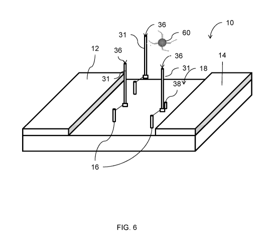
[0018] FIG. 6 is an illustration of the sensor surface of FIG. 5 with a
catalyst cluster
coupled to the single strand molecule;
[0019] FIG. 7A is an illustration of an embodiment of a method of forming a
catalyst
cluster;
[0020] FIG. 7B is an illustration of an embodiment of another method of
forming a
catalyst cluster;
[0021] FIG. 8A is an illustration of an embodiment of a method of forming a
multiplexed
catalyst cluster;
4
CA 03023256 2018-11-05
WO 2017/201239
PCT/US2017/033255
[0022] FIG. 8B is an illustration of an embodiment of a method of mixing
multiplexed
catalyst clusters;
[0023] FIG. 8C is an illustration of an embodiment of a method of multiple
site nesting
of multiplexed catalyst clusters on single strand amplicon molecules;
[0024] FIG. 9 is an illustration of a portion of a ribosomal gene used for
testing;
[0025] FIG. 10 is a photograph of microchip surfaces resulting from testing
the effects of
time on exonuclease digestion of amplicon strands;
[0026] FIG. 11 is a photograph of microchip surfaces resulting from testing
the effects of
varying exonuclease concentration;
[0027] FIG. 12 is a photograph of microchip surfaces comparing the results of
two
different catalyst cluster reagents;
[0028] FIG. 13A is a photograph of a gel analysis of replicates of RT-PCR for
dengue
viral RNA in multiplexed reaction using pan-flavivirus primers mixed with
Cal/Bun
primers; and
[0029] FIG. 13B is a photograph of a gel analysis of replicates of RT-PCR for
LaCrosse
RNA in multiplexed reaction using pan-flavivirus primers mixed with Cal/Bun
virus
primers.
DETAILED DESCRIPTION
[0030] FIG. 1 illustrates an embodiment of a detector sensor microchip 10. In
this
embodiment, the microchip 10 includes a first electrode 12 and a second
electrode 14
positioned so that the first 12 and second 14 electrode do not contact each
other, with a
plurality of capture probes 16 in the form of a functionalized oxide surface
allowing
attachment and immobilization of capture probe molecules 16 on the sensor
surface 18
between the first electrode 12 and the second electrode 14. The capture probes
16 are
designed to capture PCR amplified products via interaction with 5' tails
incorporated
during the amplification process.
[0031] FIG. 2 illustrates an embodiment of a method 20 for detection of target
nucleic
acid molecules. At block 22, target nucleic acid molecules collected from a
biological
sample are amplified via PCR. The biological sample could be any suitable type
of
CA 03023256 2018-11-05
WO 2017/201239
PCT/US2017/033255
material, such as blood, mucous, and skin, among others. It is to be
understood that any
suitable type of PCR methodology can be employed. In this embodiment, two
primers
are used during the PCR process. The first primer is synthesized as a 5'-
tailed
oligonucleotide with an internal replication block and the second primer is a
non-tailed
oligonucleotide. In an example, the second primer has a 5' phosphate group.
Multiple
cycles are performed until a plurality of double-stranded amplicons 30
(replications of the
target nucleic acid molecules) are formed having a first strand 31 with a 5'
tail 32 and a
second strand 33 extended from the non-tailed primer, as illustrated in FIG.
3. In an
example, illustrated in FIG. 3, the second strand 33 has a 5'-phosphate group
34. In
another example, not illustrated, the second strand 33 does not have a 5'-
phosphate
group. Returning to FIG. 2, at block 24, the amplicons 30 are hybridized to
the capture
probes 16 on the surface of the detector microchip 10, illustrated in FIG. 1.
In particular,
the 5' tails of the amplicons 30 bind to the capture probes 16, as illustrated
in FIG. 4.
[0032] At block 26, the hybridized amplicons 30 are converted to single strand
molecules
36, as illustrated in FIG. 5. In an example, rather than employing heat, which
would
denature the amplicons 30 off of the capture probes 16, a 5'-to-3' directional
helicase is
used to convert the amplicons 30 to single strand molecules 36. In another
example, an
exonuclease is employed to convert the amplicons 30 to single strand molecules
36. In
this example, the exonuclease has 5' to 3' directionality and preferentially
digests the
strand 33 extended from the non-tail primer. Because the tailed strand 31 is
bound to the
capture probe 16, the exonuclease cannot access the 5' end of the strand 31,
and therefore
cannot digest the strand 31, preventing degradation of the tailed strand 31.
In an
example, depending on nuclease processivity or steric hindrance at a
particular distance
from the sensor surface 18, the strand 33 may experience incomplete digestion,
resulting
in a remaining portion 38 of the digested strand 33. In an example, the strand
33
extended from the non-tail primer is synthesized with a 5' phosphate and the
exonuclease
is a lambda exonuclease.
[0033] Digestion of the strand 33 exposes the internal sequence region of the
tailed
strand 31. At block 28 of the method 20 (FIG. 2), a catalyst reagent, such as
a gold
6
CA 03023256 2018-11-05
WO 2017/201239
PCT/US2017/033255
catalyst reagent, is directly hybridized to the tailed strand 31, as
illustrated in FIG. 6. In
an embodiment, the catalyst reagent is in the form of catalyst clusters. In an
embodiment,
a single catalyst cluster 60 binds to the tailed strand 31. In another
embodiment, and
depending on the length of the tailed strand 31, a plurality of catalyst
clusters 60 bind to
each tailed strand 31.
[0034] Since a generic oligonucleotide is not suitable for binding to internal
sequences
within the amplicons, the catalyst clusters are target-specific, i.e., the
catalyst clusters
bind to specific target sequences in the strand 31. Because the primer-dimer
artifacts do
not include these target sequences, the catalyst clusters 60 do not bind to
primer-dimer
artifacts, thus avoiding potential false positive measurements. As illustrated
in FIG. 7A,
in one example, a thiol-modified oligonucleotide 40 can be reacted with a
catalytic
cluster 42 to form a catalyst cluster 44 functionalized with at least 20
oligonucleotides.
In another example, illustrated in FIG. 7B, a universal oligonucleotide 46
that possesses a
universal cluster binding sequence 45 and amplicon-specific bind sequence 47
can be
hybridized with a base generic cluster 48 to form a catalyst cluster 50. As a
result, each
new oligonucleotide is concatenated at the 3'-end with an adaptor specific
sequence
complementary to the cluster generic oligonucleotide. While the resulting
catalyst cluster
50 in FIG. 7B is depicted as having four hybridized adaptor oligonucleotides
52, it is to
be understood that the actual number of adaptors will depend on cluster size,
which limits
the number of capture oligonucleotides on the base cluster, and the
stoichiometry of
adaptor oligonucleotides added to the cluster.
[0035] As illustrated in FIG. 8A, the base generic clusters 48 can be prepared
with
mixtures of probe oligonucleotides A, B, C, D to form a catalyst cluster 54
with
multiplexing capabilities. Depending on cluster size, which determines the
surface area
available for functionalization, it is possible to load multiple different
probes onto a
single cluster. In order to support efficient metallization reactions, the
ratio for the
number of each probe type per cluster may require optimization. As illustrated
in FIG.
8B, tailored mixtures of multiplexed clusters 54 can be created. The mixtures
of
7
CA 03023256 2018-11-05
WO 2017/201239
PCT/US2017/033255
multiplexed clusters 54 can both multiplex for a large number of targeted
amplicons and
populate each targeted amplicon with multiple clusters, as illustrated in FIG.
8C.
[0036] Returning to FIG. 2, at block 30, metallization of the catalyst
clusters 60, which
serve as catalytic nucleation sites, can be performed to form a conductive
film and
resistance between the electrodes 12, 14 (FIG. 1) can be measured to detect
target nucleic
acid molecules at block 32. In an example, the catalyst clusters 60 are gold
clusters and a
gold developer reagent is applied to the catalyst clusters 60 to cascade into
the
development of the conductive film, which in this example is a gold film. In
this
example, the presence of the gold film electrically shorts the gap between the
electrodes
12, 14 and is measured by a drop in resistance. In this example, a negative
sensor has a
resistance of more than one million ohms and a positive sensor has a
resistance of about
one thousand ohms.
EXAMPLE:
[0037] An existing test developed for plasmodium faliciprium was used to test
the
method 20 described above. FIG. 9 is a screenshot taken from the INVITROGENTm
program showing a portion of the 18S ribosomal gene to which primers (shown in
bold
lettering) are designed. Typically, the two primers, forward primer 60 and
reverse primer
62, used in PCR for this method each have a sensor and catalyst binding 5'-
tail.
However, using the method 20 described above, a new reverse primer 64 located
downstream of the original reverse primer 62 was identified. Synthesis of the
new
reverse primer 64 omitted the 5' tail for catalyst binding while including a
5'-phosphorl
group to facilitate exonuclease degradation.
[0038] Because the 5'-tail on the original reverse primer 62 hybridizes to a
universal
catalyst reagent, modification of the catalyst cluster for use with the method
20 was
accomplished by pre-hybridization of the original reverse primer
oligonucleotide onto a
universal cluster to form target-specific catalyst gold clusters. Preparation
of these
target-specific catalyst gold clusters included a heated incubation with 1000
fold molar
excess of the primer 62, cooling to room temperature, and removal of unbound
excess
oligonucleotide by washing, repeated twice, via high-speed centrifugation and
8
CA 03023256 2018-11-05
WO 2017/201239
PCT/US2017/033255
resuspension with the final reagent buffer. Similarly, a second catalyst
reagent 66 with
specificity towards a different sequence element located upstream of the first
cluster
binding sequence was prepared. The ability of this second cluster to effect
metallization
and detection served as only a preliminary attempt to assess the processivity
of the
exonuclease in the digestion of sensor-bound amplicons. With this particular
derived
amplicon, the nuclease must have digested to within at least 95 nucleotides of
completely
degrading the extraneous DNA strand, leaving a single-stranded tract of about
eighty
nucleotides available for cluster binding.
[0039] Select 5' to 3' exonucleases with suitable properties to perform a
digestion as per
the method 20 were assessed. Two suitable commercially available exonucleases,
Lambda and T7, were identified. Lambda was used in this example. FIGs. 10 and
11
illustrate the results of this experiment.
[0040] FIG. 10 illustrates the results of time coarse experiments of Lambda
exonuclease
treatments on derived amplicons hybridized to microchips. All microchips in
Panel A
and Panel B were hybridized for five minutes at 45 degrees Celsius with 100 ng
of the
amplicon, washed twice by dipping into a 10 mL volume of hybridization buffer,
and
dried under a nitrogen gas stream. In a humidified petri dish held in a 37
degree Celsius
incubator, the microchips were spotted with 25 tL Lambda reaction solution
containing
one unit of the exonuclease. One unit of exonuclease is defined as the amount
of enzyme
required to produce 10 nmol of acid-soluble deoxyribonucleotide from a double-
stranded
substrate in a total reaction volume of 50 !IL in 30 minutes at 37 degrees
Celsius in 1X
lambda Exonuclease Reaction Buffer with 1 tg sonicated duplex [3I-1]-DNA.
After
incubation for a designated time, indicated above each microchip, the
reactions were
quenched by transferring the microchips into a petri dish with 20 mL of
hybridization
buffer. Subsequently, for metallization, catalyst hybridization with clusters
modified
with the primer oligonucleotide was performed for five minutes at 45 degrees
Celsius,
two washes were performed by swirling the chips in petri dishes containing
hybridization
buffer, and then gold development was performed for four minutes at 55 degrees
Celsius
with 25 !IL of developer reagent placed on the chip surface. FIG. 11
illustrates the effect
9
CA 03023256 2018-11-05
WO 2017/201239
PCT/US2017/033255
of titration of the Lambda exonuclease on the gold development of target
amplicons that
were hybridized on the surface of the microchips. The microchips were
hybridized as
described above with regard to FIG. 10. However, the units of Lambda
exonuclease used
on each microchip illustrated in FIG. 11 was varied as indicated above each
microchip.
[0041] One finding of these experiments was that the Lambda exonuclease could
not
digest the 5'-tail primer extended strand, which would have caused a loss in
the capacity
to capture the catalyst reagent. This finding is supported by the longer time
coarse
digestion experiment illustrated in FIG. 10, which shows there is no loss of
chip
metallization, even with the most extensive Lambda incubation time of 300
minutes.
[0042] The ability of the adaptor modified clusters to hybridize to a more
internal site
within the 5'-tailed strand were investigated by preparation of the second
cluster reagent,
described above. This second cluster was formed to bind about thirty
nucleotides
proximal of the hybridization site of the first cluster. The results of this
investigation of
illustrated in FIG. 12, which compares development of a first microchip 70
processed
with the first cluster reagent and a second microchip 72 processed with the
second cluster
reagent. Comparison of the first microchip 70 and second microchip 72 shows
that both
clusters performed equally well yielding comparable gold spots on the
respective treated
microchips. In addition, the results established that, on a majority of the
hybridized
amplicons, the Lambda exonuclease digested a minimum of ninety bases and was
not
inhibited while approaching the microchip surface. In addition, the longer
stretch of
single-stranded DNA with which the second cluster interacted did not appear to
affect
bind of the second cluster relative to binding of the first cluster.
[0043] Using the method described above for Plasmodium falciparum (P I), fifty
test
cartridge runs were performed. Negative and positive samples consisted
respectively of
either 10 of water or a 1 blood
culture of P I (105) cells diluted in a buffer to 10
L. After pipetting and sealing a sample into the test cartridge, a fully
automated assay,
including sample preparation, PCR amplification, and microchip hybridization,
nuclease
digestion, and metallization reactions, was carried out. Post
assay electrical
measurements were performed by removal of each microchip board from the
cartridge
CA 0302 3256 2018-11-05
WO 2017/201239
PCT/US2017/033255
after each test run and visually inspecting each microchip before placing the
microchip
on a probe station for collection of electrical results. Table 1 presents the
raw data from
the fifty assays. As indicated by "NO TEST" in the "results" column of Table
1, certain
assays were excluded due to machine failure or operator mistakes, as further
indicated in
the "Notes" column in Table 1. Table 2 indicates the percentages of correct
true positives
and negatives obtained. As illustrated in Table 2, using a modified, single-
tailed
amplicons resulted in 100% correct negative measurements and 88% correct
positive
measurements.
TABLE 1:
E-test # of shorted sensors
Date TEST TARGET CTL NCOMP RESULT Notes
3/14/16 POS 10 0 0 CORRECT
3/14/16 POS 10 0 0 CORRECT
3/14/16 NEG 0 0 0 CORRECT
3/14/16 NEG 0 0 0 CORRECT
3/14/16 NEG 0 0 0 CORRECT
3/15/16 POS 0 0 0 INCORRECT No sign of target development -
Benchtop development indicates SP-PCR ok
3/15/16 POS 0 0 0 NO TEST -- Cartridge leaked. Benchtop
development of hyb buffer indicates SP-PCR ok
3/15/16 NEG NO TEST PCR Temp Timeout
3/15/16 NEG 0 0 0 CORRECT
3/15/16 NEG 0 0 0 CORRECT
3/15/16 NEG 0 0 0 CORRECT
3/16/16 POS 10 0 0 CORRECT
3/16/16 POS 10 0 0 CORRECT
3/16/16 NEG 0 0 0 CORRECT
3/17/16 POS 10 0 0 CORRECT
3/17/16 POS 10 0 0 CORRECT
3/17/16 NEG 0 0 0 CORRECT
3/17/16 NEG 0 0 0 CORRECT
3/18/16 POS 10 0 0 CORRECT
3/18/16 POS 10 0 0 CORRECT
3/18/16 NEG 0 0 0 CORRECT
3/18/16 NEG 0 0 0 CORRECT
3/18/16 NEG 0 0 0 CORRECT
3/21/16 POS 10 0 0 CORRECT
3/21/16 POS 10 0 0 CORRECT
3/21/16 NEG 0 0 0 NO TEST Developer misloaded - turned out
ok when completed on benchtop
3/21/16 NEG 0 0 0 CORRECT
3/21/16 NEG 0 0 0 CORRECT
3/22/16 POS 10 0 0 CORRECT
3/22/16 POS 10 0 0 CORRECT
3/22/16 NEG 0 0 0 CORRECT
3/22/16 NEG 0 0 0 CORRECT
3/22/16 NEG 0 0 0 CORRECT
3/23/16 POS 9 0 0 CORRECT
3/23/16 POS 0 0 0 NO TEST Weak development - machine ran
unusually slow (SD card issues)
3/23/16 POS 10 0 0 CORRECT
3/23/16 NEG 0 0 0 CORRECT
3/23/16 NEG 0 0 0 CORRECT
3/23/16 POS 10 0 0 CORRECT
3/23/16 POS 10 0 0 CORRECT
3/23/16 POS 10 0 0 CORRECT
3/28/16 POS 0 0 0 INCORRECT -- No development
3/28/16 POS 0 0 0 INCORRECT No development
3/28/16 POS NO TEST Motor COM error, Motor timeout
3/28/16 POS 10 0 0 CORRECT
3/28/16 NEG NO TEST Syringe timeout error
3/31/16 POS 10 0 0 CORRECT
3/31/16 POS 10 0 0 CORRECT
3/31/16 POS 6 0 0 CORRECT Light development
3/31/16 POS 10 0 0 CORRECT
11
CA 03023256 2018-11-05
WO 2017/201239
PCT/US2017/033255
TABLE 2:
50 Total runs
32 Positives
23 Negatives
4 Runs discarded for reader failures
92.0% Mechanical performance
1 Runs excluded for operator reagent misload
1 Positive run excluded for mechanical leak
19 19 Correct Negatives: Negative Runs 100.0%
22 25 Correct Positives: Positive Runs 88.0%
41 44 Aggregate 93.2%
EXAMPLE 2:
[0044] Various combinations of multiplexed reverse transcription PCR (RT-PCR)
were
assessed using primer sets designed for development of a pan-flavivirus/pan-
alphavirus/pan-bunyavirus test. A total of nine primers were used in this
assay, with all
assayed materials derived from the homogenization, using ultrasonically driven
bead-
beating, of pooled six to eight mosquitoes that were spiked or non spiked with
a virus,
VEE-TC83, Dengue, or LaCrosse Virus. Nucleic acid material was isolated from
the
homogenates using magnetic particle purification, desalted by gel-filtration,
and used in
the RT-PCR amplification with the appropriate multiplexed primer sets, either
pan-
flavivirus plus bunyavirus primers or alpha primers, to generate single-
stranded 5'-tailed
amplicons. The findings indicate that the primer sets for the pan-alpha and
pan-flavivirus
tests inhibit one another during PCR amplification. To address this problem,
the test
cartridge provides two separate PCR chambers allowing for interfering primer
sets to be
run separately and mixed prior to hybridization on the sensor chip. FIG. 13A
illustrates
gel analysis of gene replicates of RT-PCR for dengue viral RNA (lanes 1-4),
purified
from spike mosquitoes, in multiplexed reaction using pan-flavivirus primers
mixed with
the Cal/Bun primers. Lane 5 is a negative control with no RNA input. FIG. 13B
illustrates gel analysis of RT-PCR for LaCrosse RNA (lanes 1-5), purified from
spiked
12
CA 03023256 2018-11-05
WO 2017/201239
PCT/US2017/033255
mosquitoes, in multiplexed reaction using pan-flavivirus primers mixed with
the Cal/Bun
primers. Lane 6 is a negative control, no RNA input. The findings indicate
that
combinations of pan-bunyavirus and pan-flavivirus primers are compatible in
PCR
reactions.
[0045] Possible advantages of the above described method include reduction or
elimination of false positive measurements due to primer-dimer artifact
formation.
[0046] While the present invention has been particularly shown and described
with
reference to certain exemplary embodiments, it will be understood by one
skilled in the
art that various changes in detail may be effected therein without departing
from the spirit
and scope of the invention that can be supported by the written description
and drawings.
Further, where exemplary embodiments are described with reference to a certain
number
of elements it will be understood that the exemplary embodiments can be
practiced
utilizing either less than or more than the certain number of elements.
[0047] The patentable scope of the invention is defined by the claims, and ay
include
other examples that occur to those skilled in the art. Such other examples are
intended to
be within the scope of the claims if they have structural elements that do not
differ from
the literal language of the claims, or if they include equivalent structural
elements with
insubstantial differences from the literal language of the claims.
[0048] To the extent that the claims recite the phrase "at least one of' in
reference to a
plurality of elements, this recitation is intended to mean at least one or
more of the listed
elements, and is not limited to at least one of each element. For example, "at
least one of
an element A, element B, and element C," is intended to indicate element A
alone, or
element B alone, or element C alone, or any combination thereof "At least one
of
element A, element B, and element C" is not intended to be limited to at least
one of an
element A, at least one of an element B, and at least one of an element C.
13