Note: Descriptions are shown in the official language in which they were submitted.
1
HUMAN ANATOMIC MODELS FOR USE IN SURGICAL SIMULATION
HAVING SYNTHETIC TISSUE PLANES
FIELD OF THE INVENTION
The present invention relates to a human anatomic model for use in
surgical simulation using conventional surgical tools to manipulate various
simulated
anatomical structures of the model, and more particularly the present
invention relates
to a method of manufacturing the human anatomic model in which the human
anatomic
model simulates tissue planes between the different anatomical structures
being
represented.
BACKGROUND
Creation of models of human anatomy has garnered increasing interest.
This is particularly true in medicine where traditionally cadaveric specimens
have been
used during surgical training. There has been increased demand for surgical
simulation
using models due to the benefits of patient safety, lower cost, greater
availability, and
long shelf life.
One of the most challenging aspects of developing surgical models is
creating realistic, life like tissue planes. This is critical to creating
synthetic tissues that
act in a manner similar to live human or animal tissue when practicing
surgery. This has
been a major deficiency of currently available commercial models.
The process of creating tissue planes has applicability for varied human
tissue. Instances described here in the human eye (including such structures
vitreous,
sclera, Tenon's capsule, conjunctiva, extraocular muscle, crystalline lens,
capsular
complex), skin (including epidermis, dermis, and subcutaneous fat), muscle,
vascular
structures (arteries and veins including intima, media and adventitia),
nerves, fat
(including orbital and subcutaneous), bone and peri-osteum.
CA 3023531 2018-11-08
2
SUMMARY OF THE INVENTION
According to one aspect of the present invention there is provided a
surgical eye model assembly for simulating eye surgery using surgical tools,
the
assembly comprising:
a surgical body formed of material which can be readily cut using the
surgical tools, the surgical body defining a central axis extending from a
rear mounting
side to a front side which is generally semicircular about the central axis so
as to be
representative of an ocular globe;
a plurality of suspension arms extending radially outward from the surgical
body in proximity to the rear side at circumferentially spaced apart locations
about the
central axis;
a rigid base frame comprising a central recess and a plurality of mounting
locations at circumferentially spaced apart locations about the central
recess;
the suspensions arms being coupled under tension to respective ones of
the mounting locations on the rigid base frame such that the surgical body is
resiliently
suspended within the central recess so as to allow for translating movement of
the
surgical body along the central axis relative to the base frame and so as to
allow for
angular deflection of the central axis relative to the base frame.
In one embodiment, the surgical body may be formed of a cadaveric eye
supported on a mounting ring, in which the plurality of suspension arms extend
radially
outwardly from the mounting ring. More preferably however, the surgical body
is formed
of a synthetic resilient material.
The assembly may further include a face plate adapted to be mounted on
the base frame and defining an eye opening therein for alignment with the
surgical
body, in which the face plate further comprises two diametrically opposed
eyelid
CA 3023531 2018-11-08
3
portions for overlapping respective portions of the front side of the surgical
body which
frictionally engage the surgical body and which pre-tension the suspension
arms. More
particularly, the face plate may have an outer surface which is shaped to
represent at
least a cheekbone area, a nose, and a brow portion of a human face surrounding
an
eye opening in the face plate. When the face plate is coupled to the base
frame, the
face plate preferably frictionally engages the surgical body so as to
pretension the
suspension arms by deflecting the surgical body downwardly into the respective
cavity
in the base frame.
The assembly may further include a rigid base element fixed to the
surgical body at a rear of the surgical body in which the suspension arms
protrude
radially from the surgical body at a location below the rigid base element.
The surgical body described above may further comprise:
a core member representative of a sclera; and
a plurality of muscular strands formed of resilient material which are
elongate between opposing first and second ends which are mounted in fixed
relation
to the core member such that an intermediate portion of each muscular strand
is
uncoupled relative to the core member and extends under tension partway about
a
circumference of the core member.
In one instance, the first and second ends of the muscular strands
comprise front and rear ends in which the front ends of the muscular strands
are joined
to the core member at the front side of the body at circumferentially spaced
positions
about the central axis, and the rear ends of the muscular strands are coupled
in fixed
relation to the core member at the rear mounting side of the body so as to be
under
tension about the core member between the front and rear ends whereby the
muscular
strands simulate rectus muscles. When a rigid base element fixed to the
surgical body
CA 3023531 2018-11-08
4
at a rear of the surgical body, the muscular strands may be fixed at the
second ends
thereof on the rigid base element at the rear of surgical body.
Alternatively, (i) the first ends of the muscular strands may be fixed onto
a common first side of core member, (ii) the intermediate portions may extend
circumferentially about diametrically opposing portions of the core member,
and (iii) the
second ends may be mounted on rigid support elements protruding from a second
side
of the core member opposite the first side of the core member whereby the
muscular
strands simulate superior and inferior oblique muscles.
In either instance of the configuration of the muscular strands, preferably
a lubricant layer is provided between the muscular strands and the core
member.
A conjunctiva sheet may also be joined between the front and rear sides
of the core member described above with a lubricant layer received between the
conjunctiva sheet and the core member.
The surgical body described above may further comprise: (i) a core
member representative of a sclera; (ii) a plurality of muscular strands
supported on the
core member to extend partway about a circumference thereof; and (iii) a
conjunctiva
sheet spanning over the core member and the muscular strands between the front
and
rear sides of the body; in which the conjunctiva sheet is fixedly coupled at a
central
location on the sheet to the core member at the front side of the body and
being fixedly
coupled in relation to the core member at the rear side of the body; and in
which the
conjunctiva sheet includes an intermediate portion between the front and rear
sides
which spans over the muscular strands which is supported in floating
relationship
relative to the core member and the muscular strands.
Preferably a lubricant layer is also received between the conjunctiva
sheet and the core member.
CA 3023531 2018-11-08
5
The surgical body described above may further comprise: (i) a spherical
outer shell representative of a sclera formed of a first resilient material
having a wall
thickness which is less than 10% of an outer diameter of the outer shell; and
(ii) a core
material filling the spherical outer shell, the core material comprising a
second resilient
material which is softer than the first resilient material forming the outer
shell.
The surgical body may yet further comprise an iris member representative
of an iris and a cornea member representative of a cornea which are supported
at the
front side of the body by a conjunctiva sheet forming an envelope surrounding
the
surgical body.
The surgical body may also further comprise: (a) a core member
representative of a sclera; and (b) a lens member representative of an ocular
lens which
is supported on the core member at the front side of the body, in which the
lens member
comprises: (i) a nucleus formed of silicone; and (ii) a plurality of cortex
layers in
sequential layers of silicone surrounding the nucleus, each cortex layer being
applied
when the previously applied layer has only partially cured such that the
cortex layers
are partially adhered to one another while remaining separable from one
another using
the surgical tools.
The surgical body may yet further comprise: (i) a core member
representative of a sclera; (ii) a lens member representative an ocular lens;
and (iii) a
capsular envelope representative of a capsule receiving the lens member
therein and
supporting the lens member on the core member at the front side of the body;
in which
the lens member comprises a plurality of layers of silicone material and the
capsular
envelope comprises a layer of silicone material which is thinner than the
layers of
silicone material forming the lens member and which is loosely bonded to the
lens
member such that the capsular envelope is less resistant to shearing from the
lens
CA 3023531 2018-11-08
6
member than the layers of the lens member are from one another.
According to another aspect of the invention there is provided a method
of constructing an anatomical model assembly representative of a human
anatomical
structure having a plurality of different tissue planes, the method
comprising:
forming a first layer of silicone in a shape representative of a first
anatomical tissue plane and allowing the silicone of the first layer to at
least partially
cure;
applying a second layer of silicone to the first layer in a shape
representative of a second anatomical tissue plane and allowing the silicone
of the
second layer to at least partially cure such that the second layer is bonded
to the first
layer by a prescribed bonding force which less than a shear force required to
tear the
second layer.
The method may further include: (i) applying the second layer in an
uncured state onto the first layer when the first layer has not yet fully
cured. (ii) applying
a lubricant between the first and second layers, (iii) diluting the silicone
of the second
layer relative to the first layer, (iv) diluting the silicone with silicone
oil or with a silicone
solvent, (v) applying adhesive to first layer before applying of the second
layer, and/or
(vi) adding an additive such as microfibres to one of the layers to increase
density
thereof relative to the other layer.
In one example, the first layer may be an elongate core member
representative of an intima of a vascular structure, the second layer may be
applied
about a full circumference and along a full length of the elongate core member
so as to
be representative of tunica media, and a third layer may be applied about a
full
circumference of the second layer along a full length of the elongate core
member so
as to be representative of a tunica external.
CA 3023531 2018-11-08
7
According to another important independent aspect of the present
invention there is provided a surgical eye model assembly for simulating eye
surgery
using surgical tools, the assembly comprising:
a surgical body formed of material which can be readily cut using the
surgical tools, the surgical body defining a central axis extending from a
rear mounting
side to a front side which is generally semicircular about the central axis so
as to be
representative of an ocular globe;
a base frame defining a ocular cavity receiving the surgical body therein
such that the surgical body is readily releasable from the base frame;
a face plate formed of resilient material releasably mounted onto the base
frame, the face plate having an outer surface which is shaped to represent at
least a
cheekbone area, a nose, and a brow portion of a human face surrounding an eye
opening in the face plate;
the eye opening of the faceplate being reduced in size relative to a
circumference of the surgical body so as to define a lower eyelid portion and
an upper
eyelid portion which partially overall diametrically opposing portions of the
semicircular
front side of the surgical body respectively.
According to another important independent aspect of the present
invention there is provided a surgical eye model assembly for use with a base
frame in
a surgical eye model for simulating eye surgery using surgical tools, the base
frame
defining an ocular cavity receiving the surgical model therein such that the
surgical
model is readily releasable from the base frame, the model comprising:
a surgical body formed of material which can be readily cut using the
surgical tools, the surgical body defining a central axis extending from a
rear mounting
side to a front side which is generally semicircular about the central axis so
as to be
CA 3023531 2018-11-08
8
representative of an ocular globe;
the surgical body comprising:
a core member representative of a sclera; and
a plurality of muscular strands formed of resilient material which
are elongate between opposing first and second ends which are mounted in fixed
relation to the core member such that an intermediate portion of each muscular
strand
is uncoupled relative to the core member and extends under tension partway
about a
circumference of the core member.
According to another important independent aspect of the present
invention there is provided a surgical eye model assembly for use with a base
frame in
a surgical eye model for simulating eye surgery using surgical tools, the base
frame
defining an ocular cavity receiving the surgical model therein such that the
surgical
model is readily releasable from the base frame, the model comprising:
a surgical body formed of material which can be readily cut using the
surgical tools, the surgical body defining a central axis extending from a
rear mounting
side to a front side which is generally semicircular about the central axis so
as to be
representative of an ocular globe;
the surgical body comprising:
a core member representative of a sclera; and
a plurality of muscular strands formed of resilient material which
are elongate between opposing front and rear ends, in which the front ends of
the
muscular strands being joined to the core member at the front side of the body
at
circumferentially spaced positions about the central axis, and the rear ends
of the
muscular strands being coupled in fixed relation to the core member at the
rear
mounting side of the body so as to be under tension about the core member
between
CA 3023531 2018-11-08
9
the front and rear ends.
In this instance, the assembly may further include a lubricant between the
muscular strands and the core member. The assembly may yet further include a
conjunctiva sheet joined between the front and rear sides of the core member
and a
lubricant received between the conjunctiva sheet and the core member.
According to another important independent aspect of the present
invention there is provided a surgical eye model assembly for use with a base
frame in
a surgical eye model for simulating eye surgery using surgical tools, the base
frame
defining an ocular cavity receiving the surgical model therein such that the
surgical
model is readily releasable from the base frame, the model comprising:
a surgical body formed of material which can be readily cut using the
surgical tools, the surgical body defining a central axis extending from a
rear mounting
side to a front side which is generally semicircular about the central axis so
as to be
representative of an ocular globe;
the surgical body comprising:
a core member representative of a sclera;
a plurality of muscular strands supported on the core member at
circumferentially spaced apart positions about the central axis of the body;
and
a conjunctiva sheet spanning over the core member and the
muscular strands between the front and rear sides of the body;
the conjunctiva sheet being fixedly coupled at a central location on
the sheet to the core member at the front side of the body and being fixedly
coupled in
relation to the core member at the rear side of the body;
the conjunctiva sheet including an intermediate portion between
the front and rear sides which spans over the muscular strands and which is
supported
CA 3023531 2018-11-08
10
in an uncoupled and floating relationship relative to the core member and the
muscular
strands.
The assembly in this instance may further include a lubricant received
between the conjunctiva sheet and the core member.
According to another important independent aspect of the present
invention there is provided a surgical eye model assembly for use with a base
frame in
a surgical eye model for simulating eye surgery using surgical tools, the base
frame
defining an ocular cavity receiving the surgical model therein such that the
surgical
model is readily releasable from the base frame, the model comprising:
a surgical body formed of material which can be readily cut using the
surgical tools, the surgical body defining a central axis extending from a
rear mounting
side to a front side which is generally semicircular about the central axis so
as to be
representative of an ocular globe;
the surgical body comprising:
a spherical outer shell representative of a sclera formed of a first
resilient material having a wall thickness which is less than 10% of an outer
diameter
of the outer shell; and
a core material filling the spherical outer shell, the core material
comprising a second resilient material which is softer than the first
resilient material
forming the outer shell.
According to another important independent aspect of the present
invention there is provided a surgical eye model assembly for use with a base
frame in
a surgical eye model for simulating eye surgery using surgical tools, the base
frame
defining an ocular cavity receiving the surgical model therein such that the
surgical
model is readily releasable from the base frame, the model comprising:
CA 3023531 2018-11-08
11
a surgical body formed of material which can be readily cut using the
surgical tools, the surgical body defining a central axis extending from a
rear mounting
side to a front side which is generally semicircular about the central axis so
as to be
representative of an ocular globe;
the surgical body comprising a core member representative of a sclera
and a lens member representative of an ocular lens which is supported on the
core
member at the front side of the body;
the lens member comprising:
a nucleus formed of silicone; and
a plurality of cortex layers in sequential layers of silicone
surrounding the nucleus, each cortex layer being applied when the previously
applied
layer has only partially cured such that the cortex layers are partially
adhered to one
another while remaining separable from one another using the surgical tools.
According to another important independent aspect of the present
invention there is provided a surgical eye model assembly for use with a base
frame in
a surgical eye model for simulating eye surgery using surgical tools, the base
frame
defining an ocular cavity receiving the surgical model therein such that the
surgical
model is readily releasable from the base frame, the model comprising:
a surgical body formed of material which can be readily cut using the
surgical tools, the surgical body defining a central axis extending from a
rear mounting
side to a front side which is generally semicircular about the central axis so
as to be
representative of an ocular globe;
the surgical body comprising a core member representative of a sclera, a
lens member representative an ocular lens, and a capsular envelope
representative of
a capsule receiving the lens member therein and supporting the lens member on
the
CA 3023531 2018-11-08
12
core member at the front side of the body;
the lens member comprising a plurality of layers of silicone material; and
the capsular envelope comprising a layer of silicone material which is
thinner than the layers of silicone material forming the lens member and which
is loosely
bonded to the lens member such that the capsular envelope is less resistant to
shearing
from the lens member than the layers of the lens member are from one another.
The assembly in this instance may further include an iris member
representative of an iris and a cornea member representative of a cornea which
are
moulded separately from the lens member and which are supported at the front
side of
the body by a conjunctiva sheet forming an envelope surrounding the surgical
body.
According to another important independent aspect of the present
invention there is provided a surgical eye model assembly for simulating eye
surgery
using surgical tools, the assembly comprising:
a central body which is generally spherical in shape about a central axis
spanning between a rear side and a front side of the central body so as to be
representative of an ocular globe;
a conjunctiva sheet comprising two opposing side portions spanning over
diametrically opposing sides of the central body from the rear side to the
front side of
the central body so as to define an eyelid opening at the front side of the
central body
between front edges of the opposing side portions of the conjunctiva sheet
respectively;
a tarsal plate member including two opposing side portions spaced apart
at diametrically opposing locations relative to the eyelid opening
therebetween, each of
the side portions of the tarsal plate member overlaying a forward portion of a
respective
one of the side portions of the conjunctiva sheet respectively;
a muscle member including two opposing side portions spaced apart at
CA 3023531 2018-11-08
13
diametrically opposing locations relative to the eyelid opening therebetween,
each of
the side portions of the muscle member overlaying a forward portion of a
respective
one of the side portions of the tarsal plate member respectively;
the tarsal plate member being adhesively bonded to the conjunctiva
sheet; and
the muscle member being loosely bonded to underlying members
including the tarsal plate member such that the muscle member is less
resistant to
shearing from the underlying members than the tarsal plate member is resistant
to
shearing from the conjunctiva sheet.
In this instance, the assembly may further include an orbital septum
membrane received between the muscle member and the conjunctiva sheet.
According to another important independent aspect of the present
invention there is provided a surgical eye model assembly for simulating eye
surgery
using surgical tools, the assembly comprising:
a base frame formed of rigid material and defining a central socket
including perimeter walls surrounding a central recess and an annular rim
portion
extending about an exterior opening through which the central recess is
accessible
such that the central socket is representative of an ocular socket;
a central body received within the central socket, the central body being
generally spherical in shape about a central axis spanning between a rear side
and a
front side of the central body so as to be representative of an ocular globe;
an elongate multilayer strand of material representative of a
neurovascular structure received within the central socket of the base frame;
and
a plurality of resilient strands of material representative of orbital fat
received between the rear side of the central body and perimeter walls of the
central
CA 3023531 2018-11-08
14
socket in the base frame.
In this instance, the assembly may further include an outer frame
representative of a human skull and locating a frame socket therein within
which the
base frame is received in mating connection.
The assembly can also include an elongate nerve strand of material
representative of an optical nerve received within the central socket of the
base frame,
and/or an elongate vascular strand of material representative of a vascular
structure
received within the central socket of the base frame.
BRIEF DESCRIPTION OF THE DRAWINGS
Various embodiments of the invention will now be described in
conjunction with the accompanying drawings in which:
Figure 1 illustrates a mold for forming the core material of a surgical body
representative of an ocular globe;
Figure 2 illustrates application of an outer shell to the surgical body of
Figure 1 to be representative of a sclera;
Figure 3 illustrates a mold for molding muscles strands representative of
muscles which are attached to the surgical body of figure 1;
Figure 4 illustrates the molded muscles strands secured under tension
about the surgical body of figure 1;
Figure 5 illustrates the molding of an iris member, a pupil member and a
cornea member to represent corresponding structures of a human eye;
Figure 6 illustrates a mold for molding a conjunctiva sheet to be applied
to the surgical body of Figure 1;
Figure 7 illustrates a mold for molding suspension arms integrally onto the
surgical body of figure 1;
CA 3023531 2018-11-08
15
Figure 8 shows the suspension arms supporting the surgical body of
figure 1 on a base frame;
Figure 9 and Figure 10 illustrate an underside of a face plate of the model
assembly and a top side of the face plate after mounting onto the base frame
of Figure
8 respectively;
Figure 11 illustrates a molded multi-layer lens member for optional use on
the surgical body of Figure 1;
Figure 12 schematically represents a human eyelid structure to be
represented by some embodiments of the model assemblies described herein;
Figure 13 illustrates a mold for forming the central body in the model
assembly of Figure 35;
Figure 14 illustrates a mould for forming a conjunctiva sheet about the
central body of figure 13;
Figure 15 illustrates the mould of figure 14 with the conjunctiva sheet
partially formed therein;
Figure 16 and Figure 17 illustrate sequential folding of the two opposing
side portions of the conjunctiva sheet about the central body;
Figure 18 illustrates a separately moulded tarsal plate member having two
opposing side portions for incorporation into the mould assembly of figure 35;
Figure 19 and Figure 20 illustrates the placement of the tarsal plate
member onto the conjunctiva sheet of figure 17 before and after folding of the
edges of
the conjunctiva sheet over two opposing side portions of the tarsal plate
member
respectively;
Figure 21 illustrates an orbital septum membrane and a muscle member
which are representative of an orbital septum and a muscle respectively for
CA 3023531 2018-11-08
16
incorporation into the model of figure 35;
Figure 22 illustrates placement of the orbital septum member of figure 21
beneath opposing side portions of the tarsal plate member;
Figure 23 illustrates placement of the muscle member of figure 21 to lay
over top of various underlying structures in the model of the figure 35;
= Figure 24 illustrates placement of an outer skin layer to form the
outermost layer in the model of figure 35;
Figure 25 illustrates an outer frame which is representative of a human
head according to the model of figure 36 for receiving the model assembly of
figure 35
therein;
Figure 26 is a schematic representation of the bone structure of an orbital
socket which is represented by the base frame in the model assembly of figure
36;
Figure 27 is a schematic representation of a vascular structure which is
modelled according to some embodiments of the invention;
Figure 28 illustrates a front view the base frame according to the model
of figure 36 which is schematically represented in the figure 26;
Figure 29 illustrates a rear view of the base frame according to figure 28;
Figure 30 illustrates the base frame according to the model of figure 36
with the central body, the tarsal plate member, the muscle member, and the
outer skin
layer according to the model of figure 35 received therein;
Figure 31 illustrates the eyelid model assembly according to figure 35
received within a base frame according to figure 36 which is further received
within the
outer frame according to figure 25, shown in use during a simulated surgery;
Figure 32 illustrates a further view outer frame receiving the base frame
and eyelid model assembly according to Figure 35 therein, as well as
incorporating the
CA 3023531 2018-11-08
17
eye model assembly of Figures 33 therein;
Figure 33 illustrate an eye model assembly which is formed according to
the steps of Figures 1 to 11 for simulating a strabismus surgery according to
the present
invention;
Figure 34 illustrates a base frame and corresponding face plate for
receiving the eye model assembly of Figure 33 therein;
Figure 35 illustrates an eyelid structure model assembly which can
optionally co-operate with the eye model assembly according to Figure 33;
Figure 36 is complete model assembly including an outer frame
representative of a human head, a base frame representative of an orbital bone
structure received in the outer frame, an eyelid structure model assembly
overlaid on
the base frame, and optionally the eye model assembly according to Figure 33
received
within the base frame;
Figure 37 is a top plan view of a rigid base element that can be attached
at the rear of the surgical body according to an alternative embodiment of the
present
invention;
Figure 38 is a perspective view of the rigid base element according to
Figure 37;
Figure 39 is a partly sectional side view of the model when using the rigid
base element according to the embodiment of Figure 37 in which the conjunctiva
sheet
is shown in cross section for illustrative purposes; and
Figure 40 is a top plan view of the model when using the rigid base
element according to the embodiment of Figure 37 in which an outline of the
conjunctiva
sheet is shown in broken line for illustrative purposes.
In the drawings like characters of reference indicate corresponding parts
CA 3023531 2018-11-08
18
in the different figures.
DETAILED DESCRIPTION
Referring to the accompanying figures there is illustrated a method of
manufacturing an anatomic model assembly for use in simulating surgery using
conventional surgical tools such as scalpels, etc. The model assembly is
primarily
formed of a resilient material, for example silicone, which can be readily cut
and
manipulated for placement of stitches therein and the like using the
conventional
surgical tools during a simulated surgery.
Although various examples of surgical anatomic model assemblies are
described herein and illustrated in the accompany figures, in each instance a
human
anatomic model is provided primarily formed of silicone material to represent
tissues
intended to be cut and/or manipulated by conventional surgical tools during a
simulated
surgical procedure. The silicone material can be used in different densities
to simulate
different tissue types. For more rigid anatomical structures being
represented, other
materials such as more rigid plastic material may be used for simulating bone
and the
like for example.
Typically, the silicone material is moulded in one or more layers to
represent each tissue type of a simulated anatomical structure, either by
moulding the
layers separately for subsequent joining, or by moulding a core or first layer
initially
followed by application of additional layers to the core or first layer.
Additional layers
may be applied over a first layer when the first layer is fully or partially
cured by either
directly applying the second layer in and uncured state to the first layer, or
by applying
an additional coating layer therebetween, or by modifying the composition of
the
silicone material of the second layer relative to the first layer. Examples
include use of
a lubricant material applied between two separately cured layers, use of a
silicone oil
CA 3023531 2018-11-08
19
layer between silicone layers, use of an adhesive material between silicone
layers, or
use of the various additives including silicone oil, or a silicone
solvent/thinner, or fibers
which can decrease or increase the density or thickness of one silicone layer
relative
to a previous layer. In the resulting anatomic model assembly, the first layer
defines a
first tissue plane and the second layer applied subsequently thereto defines a
second
tissue plane which is partially bonded or adhered to the first layer.
Typically, the
prescribed bonding force which defines the bonding between the layers is less
than the
shear force required to tear either one of the layers so that the layers can
be readily
separated from one another with surgical tools during a simulated surgical
procedure
while individual layers remain substantially intact. When multiple layers are
provided
representing a variety of different structures, different techniques can be
applied
between different ones of the layers to provide varying degrees of adhesion
between
multiple different layers of silicone representative of different tissue
planes within the
anatomic model.
Turning initially to figures 33 and 34, the model assembly of a human eye
is illustrated which is consistent with the manufacturing steps of figures 1
through 11.
The model assembly in this instance includes a surgical body 20 formed of a
spherical
core material 22 and a spherical shell 24 substantially fully surrounding the
core
material. The surgical body is spherical about a central axis spanning from a
rear
mounting side 26 to a front side 28 which is generally semi-spherical in shape
about
the central axis. The core material 22 comprises a gel material which is
softer than the
material forming the outer shell and includes an integral stem attached
thereto which
protrudes from the rear mounting side 26. The outer shell 24 fully surrounds
the core
material 22 as well as the protruding stem at the rear side. The core material
represents
the vitreous of the eye with the outer shell 24 representing the sclera. The
outer shell
CA 3023531 2018-11-08
20
is fully bonded to the core material across the full interface therebetween.
A plurality of elongate muscle strands 30 are moulded of resilient silicone
material and are attached to the surgical body 22 to represent the muscles
attached to
the ocular globe of a human eye. In the illustrated embodiment, four elongate
muscle
strands are provided on the model assembly at evenly spaced apart positions
about
the circumference of the central axis such that each muscle strand includes a
forward
end bonded by integral moulding to the outer shell 24 at the front side 28 so
as to be
spaced radially outward from the central axis. Each muscle strand 30 extends
rearwardly about the outer shell for subsequent joining to the mounting stem
at the rear
mounting side 26 of the surgical body by a suitable mounting band 32 clamped
about
the mounting stem. The muscle strands are pulled under tension about the
surgical
body between the front end bonded at the front side of the body to the rear
end clamped
by the band 32 at the rear side of the body. In this manner, each strand
includes an
intermediate portion between the front and rear ends that are fixed relative
to the core
member, in which the intermediate portion extends partway about a
circumference of
the core member while remaining uncoupled and floating relative to the core
member.
Using the moulds illustrated in figure 5, an iris member 34, a pupil member
36 and a cornea member 38 are separately moulded and then joined together at
the
front side of the surgical body 20 so as to be concentric with the central
axis thereof.
Optionally, a lens member 40 according to figure 11, which is described
in further detail below, may be also moulded of silicone material and
supported
concentrically at the front side of the surgical body prior to application of
the iris member
34, pupil member 36 and cornea member 38.
Using the mould according to figure 6, a conjunctiva sheet 42 can be
separately moulded and then joined to the surgical body 20 after moulding. The
CA 3023531 2018-11-08
21
conjunctiva sheet 42 is an elongate tubular shape with an end wall enclosing
one end
of the tubular structure which locates the cornea 38, the pupil member 36 and
the iris
member 34 therein. The tubular member is mounted concentrically about the
central
axis of the surgical body and is wrapped tightly about the circumference of
the surgical
body so as to be substantially devoid of any air gaps therebetween from the
front side
to the rear side of the surgical body 20. An additional clamping band 44 can
be used at
the rear side for securing the rear portion of the moulded conjunctiva sheet
42 at the
rear side of the surgical body 20. A lubricant material can be used to fill
the gaps
between the conjunctiva sheet 42 and the outer shell 24 of the surgical body,
as well
as surrounding the muscle strands 30 also received therebetween. In this
manner, the
conjunctiva sheet includes an intermediate portion defined between the
opposing front
and rear ends of the sheet that are fixed relative to the core member, in
which the
intermediate portion remains uncoupled and floating relative to the core
member.
The model assembly of figure 33 further includes a plurality of suspension
arms 46 extending radially outward from the central axis of the surgical body
at four
evenly spaced apart positions in a circumferential direction. Each suspension
arm is an
elongate strip of resilient silicone material which is integrally moulded at
the inner end
thereof to the exterior of the conjunctiva sheet 42 at the mounting location
of the
clamping band 44 at the rear side 26 of the surgical body. The suspension arms
lie in
a common plane which is perpendicular to the central axis of the surgical body
20. Each
support arm extends radially outward from the inner end thereof which is
bonded by
integral moulding to the rear side of the surgical body to a respective outer
end having
a mounting aperture therein for mounting on to a suitable base frame 48.
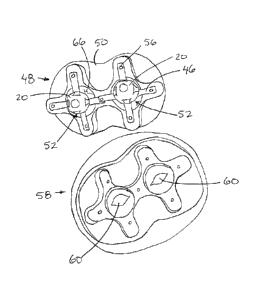
As shown in figure 34, the base frame 48 includes a bottom plate 50
defining the bottom side of the frame. A central recess 52 is defined by a
plurality of
CA 3023531 2018-11-08
22
perimeter walls 54 extending upwardly from the bottom plate 50 about the
perimeter of
the central recess 52. A set of four mounting posts 56 are mounted at evenly
spaced
apart positions about the circumference of a central axis of the central
recess, generally
within a common plane spaced above the flat bottom of the bottom plate 50. The
location of the mounting posts 56 corresponds approximately to the location of
the
mounting apertures in the four support arms 46 of the model assembly according
to
figure 33 when the support arms are slightly resiliently stretched in length
and mounted
under tension.
An outer face plate 58 according to figures 9 and 10 can be mounted over
top of the base frame 48 after suspension of the surgical body 20 on the base
frame.
The faceplate is formed of resilient material which is releasably mounted onto
the base
frame, for example by including a set of sockets for alignment with the
mounting posts
56 respectively. The faceplate has an outer surface which is shaped to
represent, for
example, a cheekbone area, part of a nose, and a brow portion of a human face
surrounding an eye opening 60 of the faceplate which aligns with the surgical
body 20
representing a human eye.
As illustrated in figure 9, the base frame may be shaped to define two
spaced apart central recesses which are identical to one another and each
receive a
respective one of two surgical bodies 20 therein. In this instance the
faceplate may
represent a full face of a person with a pair of eye openings therein for
alignment with
the two surgical bodies 20 respectively.
In each instance, the eye openings 60 in the faceplate are reduced in area
relative to the circumference of the surgical body 20 about the central axis
thereof. In
this manner, when the eye opening is bounded by diametrically opposing ones of
a
lower eyelid portion 62 and an upper eyelid portion 64, the eyelid portions
partially
CA 3023531 2018-11-08
23
overlap diametrically opposing portions of the semicircular front side of the
surgical
body respectively. In addition to the overlap, the eyelid portions of the
faceplate
frictionally engage the front side of the surgical body 20 and act to tension
the support
arms by pushing the surgical body 20 further down into the corresponding
central
recess 52 in the base frame. When the surgical bodies 20 are suspended in the
manner
described above, each surgical body is resiliently suspended within the
respective
central recess so as to allow for some translating movement of the body along
the
central axis relative the base frame and so as to allow for angular deflection
of the
central axis of the surgical body away from the central axis of the base frame
and away
from a normal vertical orientation perpendicular to the bottom plate 50 of the
base
frame. The resilient support arms act to bias the surgical body back to the
normal
position in which the central axis is perpendicular to the bottom plate. The
frictional
engagement of the eyelid portions provides a dampening effect which resists
displacement of the surgical body away from the normal orientation.
Optionally, a
suitable gel material 66 may be placed within the bottom of the central recess
52
between the surgical body 20 and the bottom of the base frame to provide some
additional dampening to resist movement of the surgical body away from the
normal
position.
When optionally using a lens member according to figure 11, initially a
central nucleus member 80 is formed of silicone and allowed to partially set
before
repeated dipping to form subsequent layers 82 with each subsequent layer being
only
partially cured before application of the next layer. A capsular member 84 is
then formed
about the multiple layers 82 of the lens member by creating a much thinner
layer of
silicone. The capsular member 84 that forms an envelope about the cortex
layers 82 is
thinner than the cortex layers and is loosely bonded to the outermost cortex
layer such
CA 3023531 2018-11-08
24
that the capsular envelope is less resistant to shearing from the lens member
than the
layers of the lens member are from one another.
Turning now to figure 35, there is illustrated a model assembly
representative of a human eyelid structure according to the manufacturing
steps of
figures 12 through 24. The model assembly of figure 35 may use a basic
spherical
central body 100 in the centre thereof, or alternatively the surgical body 20
including
the core material 22 and the outer shell 24 according to figure 33 with a lens
member
40 thereon may be used as the central body 100.
The central body 100 in this instance is initially positioned within the mould
of figure 14 to mould a conjunctiva sheet 102 having two opposing side
portions 104
spanning over diametrically opposing sides of the central body from the rear
side to the
front side thereof so as to define an eyelid opening at the front side of the
central body
between the front edges of the opposing side portions of the conjunctiva sheet
102
respectively.
A tarsal plate member 106 according to figure 18 is shaped to be
generally annular about a central opening to define two opposing side portions
108
which are spaced apart at diametrically opposing locations relative to the
eyelid opening
therebetween. Each of the side portions of the tarsal plate member overlays a
forward
portion of a respective one of the side portions of the conjunctiva sheet
respectively.
The forwardmost edges of the two opposing side portions 104 of the conjunctiva
sheet
are folded back over the inner edge of the tarsal plate member that defines
the central
opening therein.
An orbital septum membrane 110 and a muscle member 112 are each
separately moulded of silicone material as shown in figure 21. Each of these
components is generally C-shaped about a central opening for alignment with
the
CA 3023531 2018-11-08
25
corresponding eyelid opening in the mold assembly described above. Opposing
side
portions 114 of the orbital septum at opposing ends of the C-shape thereof are
positioned so that the inner edges thereof are adhered to and received beneath
the
outer perimeter portions of the two side portions of the tarsal plate member
respectively.
The muscle member 112 is subsequently laid over top of the forward edges of
the
conjunctiva sheet 102 which have been folded over the inner edges of the
tarsal plate
member. The muscle member similarly includes two opposing side portions 116
spaced
apart at diametrically opposing locations relative to the eyelid opening
therebetween in
which each of the side portions of the muscle member overlays a forward
portion of a
respective one of the side portions of the tarsal plate member respectively.
Suitable
adhesive, for example uncured silicone which is permitted to cure, can be used
to
ensure a tightly adherent connection between the conjunctiva sheet and the
tarsal plate
member while providing a loosely adherent bond between the muscle member and
the
underlying structures.
A faceplate 118 formed of resilient silicone representative of skin is again
provided similar to the faceplate 58 of the previous embodiments. The
faceplate 18
further defines the eyelid opening between an upper eyelid portion and a lower
eyelid
portion which are laid over and bonded to respective ones of the two side
portions of
the muscle member at the forward edges of the conjunctiva sheet 102
respectively.
The eyelid structure described above can be incorporated into the model
assembly of figure 34, or optionally may be incorporated into a more complex
outer
frame 140 according to the model assembly of figure 36.
In the model assembly according to figure 36, a base frame 142 is
provided of rigid material which includes a central socket 144 formed therein
so as to
have perimeter walls surrounding a recessed cavity which is open at the top
side
CA 3023531 2018-11-08
26
through an exterior opening defined by an annular rim portion 146 of the
central socket
formed in the base frame. The central socket is shaped to be representative of
an orbital
cavity for receiving an ocular globe therein similar to the central body 100
or the surgical
body 20 described above.
As in previous embodiments, the central body is generally spherical in
shape about a central axis spanning between a rear side and a front side of
the central
body. In addition to receiving the central body therein, the central socket
144 also
includes a plurality of resilient strands 148 formed of silicone material
which are
intended to be representative of orbital fat. In addition, an elongate nerve
strand 150
formed of moulded synthetic material and an elongate multilayer strand 152
moulded
of synthetic material to be representative of a vascular structure according
to figure 27
are also placed within the central socket so as to be representative of nerves
and
vascular structures within the anatomical model. The orbital fat is typically
coated with
a lubricant material to allow free movement of the resilient strands 148
relative to one
another to optimally simulate human orbital fat. Once the above structures are
placed
into the central socket together with the central body, some or all of the
components of
the eyelid model assembly described above can be further positioned within the
central
socket followed by placement of the faceplate 118 covering over the exterior
opening
of the central socket with the exception of the eyelid opening in the
faceplate through
which the central body representing the ocular globe is visible. The outer
frame 140 in
this instance is representative of a human head and is typically formed of
rigid material,
for example a 3-D printed plastic. A pocket 154 is formed in the outer frame
140 at the
location of one of the eyes to receive the base frame 142 therein such that
the outer
simulated skin surface of the faceplate 118 is substantially flush and aligned
with the
corresponding outer surface of the outer frame 140.
CA 3023531 2018-11-08
27
Formation of a multilayer strand 152 according to figure 27 can be initiated
by providing an elongate pipe which is dipped into a dense silicone to create
a core
layer 170 representative of an intima. A variable adherence is created by
allowing
partial curing of the core layer 170 by application of an additional layer 172
of silicone
about the full circumference and along the full length of the core layer which
forms the
tunica media. A softer silicone or silicone foam can then be applied about the
full
circumference and along the full length of the additional layer 172 to form an
outermost
layer 174 to be representative of the loose tunica external. The various
techniques
described herein to create different densities among the different layers or
to provide
different degrees of adherence between the different layers can be used,
particularly
when the multi-layer strand 152 is created to be representative of a vascular
structure.
Turning now to Figures 37 through 40, the surgical body 20 according to
the first embodiment of figure 33 may be modified to include a rigid base
element 200
in place of the band 32 which secures the rectus muscles 30 at the rear end of
the body
20. As best shown in figures 37 and 38, the rigid base element comprises a
rigid collar
202 including a central opening therein which receives the stem protruding
from the
rear of the core body therethrough in the mounted position. A plurality of
teeth 204 are
provided at the inner surface of the collar to protrude radially inwardly
while being
elongate in the axial direction. The teeth 204 serve to grip the stem
protruding from the
rear of the core body 20 which is received through the collar. The collar 202
also
includes a set of four lugs 206 which protrude radially outwardly from the
outer surface
of the collar at evenly spaced positions about the circumference thereof. A
pair of rigid
support arms 208 also extended generally radially outwardly from the collar
such that
the pair of arms 208 are angularly offset from one another by approximately 90
with
each arm being centered in the circumferential direction relative to a
corresponding pair
CA 3023531 2018-11-08
28
of the lugs 206. Each arm is thus angularly offset by approximately 45
relative to the
lugs of the corresponding pair. Each of the arms 208 also extends radially
outward at a
slope offset in a common axial direction which is forwardly towards the front
end of the
core body received within the rigid base element 200. A suitable retention
slot is formed
at the free end of each of the support arms 208. When the core body is mounted
onto
the rigid base element 200, the free ends of the support arms 208 lie in a
common
horizontal plane extending through the center of the core body perpendicularly
to the
central axis thereof and perpendicularly to the stem.
The rigid base element receives the core body 12 mounted thereon
subsequent to the sclera 24 being formed onto the core body and subsequent to
the
front ends of the rectus muscles 30 being attached at the forward end of the
core body.
The rectus muscles 30 are each aligned with a corresponding one of the lugs
and are
stretched under tension to span from respective forward ends integrally molded
at the
front-end of the core body to respective rear ends which are fixed onto
respective ones
of the lugs 206, for example by forming an aperture in the strand into which a
corresponding lug 206 is hooked or received. Each rectus muscle 30 thus
includes an
intermediate portion which extends partway about the circumference of the core
body
in an uncoupled and floating relationship relative to the core body. Attaching
the rear
ends of the strands 32 to the respective lugs 206 allows the tension in the
strands to be
individually controlled.
In this embodiment, a pair of oblique muscle strands 31 are also provided
in which the oblique muscle strands 31 are each fixed to the core body by
integrally
molding the first ends of the strands at a common lateral side of the core
body
approximately within a common plane as the free ends of the two support arms
208.
The oblique muscle strands 31 comprise elongate resilient silicone members
similarly
CA 3023531 2018-11-08
29
to the rectus muscle strands 30. Each of the oblique muscle strands can then
be
stretched about a respective portion of the circumference of the core body so
that the
opposing second ends of the strands 31 can be adjustably secured within the
respective
retention slots at the free ends of the two arms 208 respectively. The
configuration of
the rigid base element allows each of the rectus muscles 30 and the oblique
muscle
strands 31 to be individually tensioned to extend partway about the
circumference of
the core body 20 while remaining in an uncoupled and floating relationship
relative to
the core body at an intermediate portion between opposing first and second
ends which
are fixed in relation to the core body.
Use of a lubricant material can surround the intermediate portions of the
muscles so as to be located in a layer between the muscles and the core body
as well
as being located in a layer between the muscles and any additional layers
overlapping
the muscles. Subsequent to mounting of the muscle strands under tension, the
core
body in this instance is again enveloped by a conjunctiva sheet 42 which is
fixed at the
forward end of the core body as described above with regard to the first
embodiment of
figure 33. The conjunctiva sheet fully surrounds and envelops all of the
rectus muscles
30, the oblique muscle strands 31, and the rigid base element 200 upon which
they are
tensioned. An additional clamping band 44 can then be secured directly below
the rigid
base element 200 to fix the conjunctiva sheet 42 at the rear of the core body
similarly
to the first embodiment according to figure 33. The core body in this instance
is again
joined to a plurality of suspension arms 46 which support the core body on a
base frame
in a similar manner to the previous embodiment.
Various particulars with regard to the method of construction of the above
noted model assemblies will now be described in the following.
Strabismus Model
CA 3023531 2018-11-08
30
This model, shown in Figures 33 and 34, is designed for practicing
strabismus surgery. In this surgery eye muscles are recessed or resected
(moved
forward or back) on the globe when someone has misaligned eyes (esotropia,
exotropia).
Below is outlined the process for creating a multilaminar eye model for
use in strabismus and other ophthalmic surgeries.
The model has many features that are unique and / or beneficial for
practicing strabismus and other surgeries. These include:
A) a soft interior which simulates the vitreous cavity with its
compressibility;
B) a rigid sclera which is can hold a suture;
C) extra-ocular muscles which are adhered to the sclera, under tension,
and can be cut and re-sutured to the globe as in strabismus surgery;
D) a conjunctiva which overlays the muscles and globe which must be
dissected through during ophthalmic surgery;
E) a base holder which allows for globe movement and rotation during
simulated surgery;
F) a silicone face which enables proper hand positioning during surgery;
#1 Creation of vitreous / clobe
Ecoflex Gel Silicone (Shore hardness 000-35) is poured in to a 3 part
mould. as shown in Figure 1.
#2 Creation of sclera
Globes are dipped in Dragon Skin 10A silicone (Shore hardness 10A) and
allowed to cure on a rack 2-3 times. This creates a soft to hard interface to
mimic globe
compressibility as shown in Figure 2.
CA 3023531 2018-11-08
31
#3 Creation of muscles
The globe is placed upright in the mould. The muscles are poured and it
makes contact with the globe at the proximal end (Dragon Skin). After curing
the
muscles are pulled back and secured with a plastic ring. The muscles are now
under
tension. They will now 'retract back when cut just as they do in strabismus
surgery. A
light layer of silicone oil is applied under the muscles so that a more
realistic tissue
plane is created when 'hooking' the muscle. Silicone oil can also be added to
the sclera
before dipping to create thinner weaker tissue to make suture placement more
difficult.
An additive of micro-fibres can also be mixed with the silicone to change the
tissue
property / feel of the sclera.
In an additional actualization of the model the superior oblique and inferior
oblique muscles are also attached to the model as shown in Figures 39 and 40.
#4 creation of cornea, iris and pupil
The cornea, iris and pupil are all poured separately (Dragon Skin) as
shown in Figure 5. An alternate method is to laser print an iris coloured
sheet and
adhere it with the silicone corneal cap.
#5 creation of conjunctiva
The cornea, iris and pupil are all assembled in the bottom of the
conjunctiva mould shown in Figure 6. Then Dragon skin is poured in to the
mould, and
the 'plunger' part of the mould is used to compress the silicone and form a
curved shape
and allowed to cure.
An alternate method is to dip the tip of the conjunctiva mould pictured in
Figure 6 and simply set it upright.
#6 attaching the cornea-iris-conjunctiva to the globe
The cured cornea-iris-conjunctiva mould is then attached to the globe with
CA 3023531 2018-11-08
32
a small amount of silicone (dragon skin) and allowed to cure. Once this is
done Silicone
thinner is used to lubricate under the muscles and conjunctiva to allow for
realistic
movement of the muscle hook later during simulated surgery. Next the
conjunctiva is
folded back and secured with a plastic ring. This effect creates a realistic
tissue plane
between the conjunctiva, muscles and sclera.
#7 creation of four arm holders
The assembled globe is then placed in a final holder and silicone is
poured which adheres to the conjunctiva. These four limbs will be attached to
the base =
plate according to Figure 7.
#8 creation of base and face plate and mounting of globe
The base is created from a single one piece mould which is poured using
urethane based plastic.
The globe is then mounted on to four pegs on the base plate as shown in
Figure 8. This allows the globe to rotate as it does in the human during
surgery.
Although used here for strabismus surgery, this could allow rotation of the
globe during
any ophthalmic surgery. The silicone arms also allow for movement of the eye
when
pushed on as it acts as an elastic sling for the model eye.
#9 face plate attachment
The face plate is dragon skin silicone which is then made from a two piece
mould. Then the face mould is place over top and fits snugly on to the base as
shown
in Figures 9 and 10. The face plate provides counter traction and anatomic
fidelity in
placing your hands during surgery.
Cataract Model
A variation of the strabismus model used for cataract and other anterior
segment surgeries. In addition to the features listed in the strabismus model,
the
CA 3023531 2018-11-08
33
following additional features are provided.
#1 Creation of Lens
A multilaminated lens according to Figure 11 is created which acts in a
manner similar to the human lens. The human lens has multiple layers of
nucleus and
cortex which are removed as pieces during modern cataract surgery.
To create the lens a central nucleus of silicone is poured. It is allowed to
partially set and then is dipped again and allowed in silicone. This process
is repeated
until the lens is the appoximate thickenss of the human lens. This process
creates
variable adhesion between the layers which mimics the human eye.
One variation of this process is to spray a silicone lubricant between the
layers. This also varies the adhesion.
Another variation adds micro-fibres to the lens to create different densities
of lens.
Another variation adds silicone oil to make ultrathin layers.
#2 Creation of capsule
The human lens sits within a capsular bag complex. After the lens is
created a silicone non-adhesive is sprayed over it. Then the lens is placed in
a mould
and a thin layer of silicone is created by pouring it over the mould and
allowed to drip
and an inclination of 45 degrees. This creates an ultrathin layer with a
tissue plane
between the lens and capsule.
In another variation a silicone thinner is added to the mix before pouring
on to the lens to make the capsule ultrathin.
#3 adhering capsular complex to front of sclera
The capsular complex with lens is then adhered to the front of the sclera
(described previously in strabismus model steps 1 and 2) with silicone glue.
CA 3023531 2018-11-08
34
#4 Creation of anterior segment
The iris and cornea are then created and adhered over to of the lens
complex. The major difference to the strabismus model is that there is a space
left
between the iris and cornea centrally known as the 'anterior chamber',
analagous to
that of the human eye.
#5 Addition of muscles, conjunctiva and mounting in the face plate
The remainder of the processing of the eyes is the same as that in steps
5-9 described in strabismus surgery.
Cadaveric Model
An additional base plate is created similar to the one featured in the
strabimsus model of Figure 33. It includes a cut side that surgical tubing can
be
connected to a syringe. A flat plate embedded in step #7 of the strabismus arm
holders
allows a cadaveric eye to be mounted in the base and utilized to practice
ophthalmic
surgery.
Anatomic Model of orbital bone, and eyelid structures
Described in the following is the process for constructing the multi
laminated eyelid structure accordingly to Figure 35. The difficulty with
creating
structures like the eye lid or the lip is that they have several densely
packed layers as
shown in Figure 12, each with a complex folded 3D shape, and the level of
adherence
between the layers is variable, from strong adherence to no adherence.
Challenges in
the manufacturing process are defined below and followed by a pictorial
demonstration
of the process.
The first major problem is creating the inner layer of the multilayered
structure. The inner layer is a complex 3D structure and the material, usually
silicone
rubber, cannot be cast directly into this shape. The liquid will simply not
stay in position
CA 3023531 2018-11-08
35
during the curing process. Second this inner layer will always be adjacent to
another
silicone layer, and curing silicone will always fuse to another adjacent
silicone layer. So
lets use our eyelid structure as an example. The inner layer, the tarsal
conjunctiva, is
directly opposite the globe (eyeball) and the bulbar conjunctiva. They are
essentially
continuous with a potential space in between, meaning they will always be in
contact
unless you pull on the eyelid and the space opens up between them. There can
be no
adherence between the tarsal conjunctiva (the inner layer of the eyelid) and
the eyeball
(the adjacent layer) yet they need to be form fitting to the point without any
gaps.
The second challenge is that some of the layers are tightly adherent, such
as the conjunctiva to the tarsal plate, and some layers are loosely adherent
such as the
muscles and underlying fat. These loosely adherent layers are key surgical
landmarks
and allow for the operation to be performed and so have to be accurately
replicated. As
previously discussed curing silicone layers on top of each other will cause
fusion and
create an inseparable bond. You would think that the answer would be to form
the
layers separately and then use a loose adhesive to laminate them, however
there is no
such commercially available adhesive. So in conventional silicone modelling
your
layers would be either fused completely or not fused at all.
To create the loose adherence required the layers are formed separately
using a very fast cure silicone and then laminated at the critical time just
before they
are fully cured. When laminated at this time the layers will form a loose
adherence,
allowing for surgical separation without destruction of either layer.
This method is not always available because it is time sensitive and
accordingly a novel silicone adhesive is also being developed as well to form
loose
adherence between cured silicone layers.
Here is the process step by step with images
CA 3023531 2018-11-08
36
#1 Creation of the globe (eyeball). Poured silicone into a standard two-
part mould as shown in Figure 13.
#2 Creation of the conjunctive (inner layer of the eyelid). The previously
created globe is inserted into the conjunctival mould shown in figure 14.
Silicone is then
poured and fully cured as shown in Figure 15. This creates two flat sheets of
conjunctiva
which are attached to the globe. The shape of these flat sheets is critical,
is they must
fold into a complex 3D shape.
#3 Folding of the conjunctiva to create the potential space between the
conjunctiva (inner eyelid layer) and the globe (eyeball). The flat sheets of
conjunctiva
are folded onto the globe and adhesive is used only at the bottom edge to seal
the apex
of the potential space according to Figures 16 and 17. In the above eyelid
cross section
image this area of adherence is labeled "cul-de-sac".
#4 Lamination of the tarsal plate to the conjunctiva. A separately moulded
tarsal plate as shown in Figure 18 is laminated to the conjunctiva. Note the
conjunctiva
must wrap under the tarsal plate and then tightly wrap the edge of the tarsal
plate onto
the anterior surface as shown in Figures 19 and 20.
#5 Lamination of the orbital septum and orbicularis muscle. The
orbicularis muscle and orbital septum are formed separately as shown in Figure
21 and
allowed to cure to the critical point right before full cure, then the septum
is quickly glued
to the undersurface of the tarsal plate as shown in Figure 22 and the muscle
is
laminated on top of this as shown in Figure 23. This creates the loose
adherence
between the muscle and the septum. The muscle is glued to the edge of the
reflected
conjunctiva.
#6 Lamination of the skin to the eyelid margin and the muscle. The final
step is to glue the outer skin layer to the underlying muscle and eyelid
margin as shown
CA 3023531 2018-11-08
37
in Figure 24.
Fracture Model Modification
The eyelid structure can be over-layed upon a model skull / orbit to
simulate orbital fracture repairs according to the model of Figure 36.
#1 Model head. A model head is printed in plastic or PLA as shown in
Figure 25.
#2 Bone Orbit. The orbital walls which consist of bone are printed in PLA
with the fractures present according to Figure 26.
#3 Neurovascular structures. Arteries, veins and nerves are molded. In
the simplest iteration a small pipe of variable diameter is dipped in silicone
and allowed
to cure. More complex arteries can be created by an alternate method. The
intima is
first created by dipping the pipe in to dense silicone. A variable adherence
is created
by allowing partial curing of the intima and then applying an additional layer
to form the
tunica media. Lastly a softer silicone or silicone foam can be applied
external to this to
form the loose tunica external according to Figure 27. A silicone thinner can
be added
to form different thicknesses of silicone. Silicone non-adhesive can also be
applied to
create variable interfaces.
#4 Orbital Fat. Orbital fat is made by pouring silicone in to a phalange like
mould. The interdigitation of the mould allows the fat to move dynamically as
it does in
the human orbit. Addition of a silicone lubricant is applied and allows the
orbital fat to
move more freely.
#5 Placing neurovascular and orbital fat in orbit bone according to Figure
28 and Figure 29.
#6 placement of orbit bone in face plate and overlaying of eyelids results
in a structure according to Figure 30. The bony orbit is placed in the face
plate
CA 3023531 2018-11-08
38
according to Figure 31 and Figure 32. The eyelids described previously are
adhered to
the faceplate.
Since various modifications can be made in my invention as herein above
described, and many apparently widely different embodiments of same made, it
is
intended that all matter contained in the accompanying specification shall be
interpreted
as illustrative only and not in a limiting sense.
CA 3023531 2018-11-08