Note: Descriptions are shown in the official language in which they were submitted.
CA 03024518 2018-11-15
AMELIORATION AND TREATMENT OF PERINATAL BRAIN DAMAGE
WITH PLURI POTENT STEM CELLS
FIELD
The present invention relates to a cell preparation for regenerative medicine.
More
specifically, it relates to an effective cell preparation for treatment of
perinatal brain
damage containing pluripotent stem cells, and to a novel treatment method.
BACKGROUND
Perinatal brain damage refers to brain damage such as cerebral palsy, mental
development delay, epilepsy or sensory disorder that is caused by some
abnormality
arising during the fetal or neonatal period, and its causes and pathology are
still
incompletely understood. Research has recently been progressing in the field
of perinatal
medicine in regard to fetal hypoxia, leading to greater focus on hypoxia as a
cause of
damage. When hypoxia progresses, the fetus exhibits various metabolic
reactions and
further falls into a state of metabolic insufficiency, resulting in damage to
the organs
including the brain, with death expected to occur in some cases. Preventing
hypoxia
during delivery is therefore thought to be clearly linked with preventing
perinatal brain
damage including cerebral palsy. However, it is difficult to prevent hypoxia
during
delivery, and it is said that hypoxia is responsible for approximately 20% of
all cerebral
palsy cases (NPL 1).
Research has recently been progressing in the field of regenerative medicine
on a
host of cell therapies using stem cells, and as clinical applications begin to
emerge, it is
hoped that stem cells will be used for improvement and treatment of perinatal
brain
damage (NPL 2). Embryonic stem cells (ES cells), neural stem/progenitor cells
(NSPC),
induced pluripotent stem cells (iPS cells) and umbilical cord blood stem cells
(UCBC) are
known types of stem cells that are expected to have potential in clinical
applications for
central nervous system diseases related to perinatal brain damage. The present
inventors
have previously co-administered proliferated NSPCs cultured from rat fetal
brains together
with a chondroitin sulfate-degrading enzyme into the cerebral ventricle of a
perinatal
hypoxic ischemic encephalopathy (HIE) rat model, and found that the infarct
area
decreases significantly compared to an untreated group and to a group
administered neural
stem cells alone (NPL 3). It has also been confirmed by the present inventors
that
CA 03024518 2018-11-15
intraperitoneal administration of UCBCs to an HIE rat model temporarily
reduces hypoxic
ischemic encephalopathy.
The bone marrow-derived mesenchymal cells (MSCs) fraction has been isolated
from adults, and it is known to have the ability to differentiate into bone,
cartilage,
adipocytes, neurons and skeletal muscle, for example (NPL 4 and 5). However,
MSCs are
heterogeneous cell populations, the nature of their differentiation potency is
unknown, and
they have wide variation in therapeutic effect. iPS cells (PTL 1) have been
reported as
adult pluripotent stem cells, but establishing iPS cells requires the very
complex procedure
of introducing specific genes or specific compounds into the skin fibroblast
fraction
(mesenchymal cell fraction) of somatic cells, while iPS cells could also have
high tumor-
forming potential, and therefore high hurdles stand in the way of their
clinical application.
Research by Prof. Dezawa, one of the present inventors, has demonstrated that
the
pluripotency of the mesenchymal cells fraction is exhibited by pluripotent
stem cells
(Multilineage-differentiating Stress Enduring cells, or Muse cells) that
express SSEA-3
(Stage-Specific Embryonic Antigen-3) as a surface antigen, which are present
in the
mesenchymal cell fraction and can be obtained without operation of induction
and that this
holds potential for application in treatment of diseases by tissue
regeneration. It has also
been found that Muse cells can be enriched by treating the mesenchymal cells
fraction
with one or more of different types of stress treatments (PTL 2, NPL 6).
However, it has
not yet been demonstrated that the expected therapeutic effect can be obtained
using Muse
cells for amelioration and/or treatment of perinatal brain damage.
CITATION LIST
PATENT LITERATURE
[PTL 1] Japanese Patent Publication No. 4183742
[PTL 2] International Patent Publication No. W02011/007900
NON PATENT LITERATURE]
[NPL 1] Ikeda, T., Non to Hattatsu, Vol. 43, p. 206-2 I 0 or 2011
[NPL 2] Sato, Y., Shusanki Igaku, Vol. 41, 1531-1536, 2011
[NPL 31 Sato, Y., et al., Report Sci., Vol. 15, p. 613-620(2008)
[NPL 4] Dezawa, M., et al., J. Clin. Invest., Vol. 113, p. 1701-1710(2004)
[NPL 5] Dezawa, M., et al., Science, Vol. 309, p. 314-317(2005)
[NPL 6] Wakao, S, et al., Proc. Natl. Acad. Sci. USA, Vol. 108, p. 9875-
9880(2011)
2
SUMMARY
TECHNICAL PROBLEM
It is an object of the present invention to provide a novel medical use for
pluripotent stem cells (i.e., Muse cells) in regenerative medicine. More
specifically, it is an
object of the present invention to provide a cell preparation and
pharmaceutical
composition that include Muse cells and are effective for treating perinatal
brain damage,
as well as a novel treatment method.
SOLUTION TO PROBLEM
The present inventors have found that preparing a perinatal hypoxic-ischemic
encephalopathy (HIE) rat model, and administering Muse cells by intravenous
injection 72
hours after hypoxic treatment, ameliorates perinatal brain damage (including
learning
disability and motor disability, for example), and the present invention has
thus been
completed.
Specifically, the present invention provides the following.
[1] A cell preparation for amelioration and/or treatment of perinatal hypoxic-
ischemic encephalopathy (HIE), comprising pluripotent stem cells positive for
SSEA-3 as
an active ingredient, which has been isolated from mesenchymal tissue of a
body or
cultured mesenchymal cells, wherein the pluripotent stem cells have all of the
following
properties:
(i) CD105 positive;
(ii) low or non-existent telomerase activity;
(iii) the ability to differentiate into any of the three germ layers;
(iv) no neoplastic proliferation; and
(v) self-renewal ability.
[2] The cell preparation according to [1], comprising a cell fraction wherein
pluripotent stem cells positive for SSEA-3 have been concentrated by external
stress
treatment, which is any one or a combination of: protease treatment, culturing
in a low
oxygen concentration, culturing under low-phosphate conditions, culturing with
low serum
concentration, culturing under low nutritive conditions, culturing under
exposure to heat
shock, culturing at low temperature, freezing treatment, culturing in the
presence of a
hazardous substance, culturing in the presence of active oxygen, culturing
under
mechanical stimulation, culturing with agitating treatment, culturing with
pressure
3
CA 3024518 2020-03-09
treatment, or physical impact.
[3] The cell preparation according to [1] or [2], wherein the pluripotent stem
cells
are CD117-negative and CD146-negative.
[4] The cell preparation according to any one of [1] to [3], wherein the
pluripotent
stem cells are CD117-negative, CD146-negative, NG2-negative, CD34-negative,
vWF-
negative and CD271-negative.
[5] The cell preparation according to any one of [1] to [4], wherein the
pluripotent
stem cells are CD34-negative, CD117-negative, CD146-negative, CD271-negative,
NG2-
negative, vWF-negative, Sox10-negative, Snail-negative, Slug-negative, Tyrpl-
negative
and Dct-negative.
[6] The cell preparation according to any one of [1] to [5], wherein the
perinatal
brain damage is one or more of learning disability, motor disability, cerebral
palsy,
behavior disorder, mental development abnormality, sensory disorder, speech
disorder,
epilepsy, dysphagia and abnormal respiratory control.
[7] The cell preparation according to any one of [1] to [6], wherein the
pluripotent
stem cells have the ability to engraft into brain tissue.
[8] The use of a therapeutically effective amount of the cell preparation
according
to any one of [1] to [7], for improving, treating or preventing perinatal
hypoxic-ishcemic
enceplalopathy (HIE) in a human neonate, infant or child.
[9] The use of [8] wherein the cell preparation contains unit doses of
approximately 1 x 10⁵ cells/individual to approximately 1 x 10⁸ cells per
human neonate,
infant or child.
[10] The use of [8] wherein the cell preparation contains unit doses of stem
cells in
an amount of cells per body weight of approximately 3 x 10⁴ cells/kg to
approximately 3 x
107 cells/kg per neonate, infant or child.
ADVANTAGEOUS EFFECTS OF INVENTION
The present invention can drastically minimize perinatal brain damage in a
subject
(child) suffering from perinatal brain damage, by a brain tissue-regenerating
mechanism in
which Muse cells are administered through the veins or by another route to
selectively
accumulate them in damaged brain tissue, and the Muse cells differentiate to
brain tissue-
forming cells within the tissue.
4
CA 3024518 2020-03-09
BRIEF DESCRIPTION OF DRAWINGS
FIG. 1 shows the results of confirming engraftment of Muse cells in the brain
tissue when administered to an HIE rat model (postnatal age of 48 days; 10
days after
administration).
FIG. 2 shows the results of a Rota-rod treadmill test for motor function in an
HIE
4a
CA 3024518 2020-03-09
CA 03024518 2018-11-15
rat model, using a Muse cell-administered group ("Muse"), a group with
addition of a cell
suspension (physiological saline) alone ("vehicle") and a sham-operated group
("sham")
("Short period": I month after birth; "Long period": 5 months after birth).
FIG. 3 shows the results of an open field test for emotional behavior
(hyperactivity,
etc.) in an I-HE rat model, using a Muse cell-administered group ("Muse"), a
group with
addition of a cell suspension (physiological saline) alone ("vehicle") and a
sham-operated
group ("sham") ("Short period": 1 month after birth; "Long period": 5 months
after birth).
FIG. 4 shows the results of a shuttle avoidance test for improvement in
learning
disability in an HIE rat model, using a Muse cell-administered group ("Muse"),
a group
with addition of a cell suspension (physiological saline) alone ("vehicle")
and a sham-
operated group ("sham") ("Short period": 1 month after birth; "Long period": 5
months
after birth).
FIG. 5 shows the results of a novel object recognition test for improvement in
memory learning and visual cognitive memory in an HIE rat model, using a Muse
cell-
administered group ("Muse"), a group with addition of a cell suspension
(physiological
saline) alone ("vehicle") and a sham-operated group ("sham") ("Short period":
1 month
after birth; "Long period": 5 months after birth).
FIG. 6A shows the results of a cylinder test for improvement in motor function
in
an FILE rat model, using a Muse cell-administered group ("Muse"), a group with
addition
of a cell suspension (physiological saline) alone ("vehicle") and a sham-
operated group
("sham") (5 months after birth).
FIG. 6B shows the results of a cylinder test for improvement in motor function
in
an HIE rat model, using a Muse cell-administered group ("Muse"), a group with
addition
of a cell suspension (physiological saline) alone ("vehicle") and a sham-
operated group
("sham") (5 months after birth).
FIG. 7 shows the results of a Rota-rod treadmill test for motor function in an
HIE
rat model, using a Muse cell-administered group ("Muse"), a non-Muse cell-
administered
group ("non Muse"), a group with addition of a cell suspension (physiological
saline)
alone ("vehicle") and a sham-operated group ("sham") (5 months after birth).
FIG. 8 shows the results of an open field test for emotional behavior
(hyperactivity,
etc.) in an HIE rat model, using a Muse cell-administered group ("Muse"), a
non-Muse
cell-administered group ("non Muse"), a group with addition of a cell
suspension
(physiological saline) alone ("vehicle") and a sham-operated group ("sham") (5
months
5
CA 03024518 2018-11-15
after birth).
FIG. 9 shows the results of a shuttle avoidance test for improvement in
learning
disability in an HIE rat model, using a Muse cell-administered group ("Muse"),
a non-
Muse cell-administered group ("non Muse"), a group with addition of a cell
suspension
(physiological saline) alone ("vehicle") and a sham-operated group ("sham") (5
months
after birth).
FIG. 10 shows the results of a novel object recognition test for improvement
in
memory learning and visual cognitive memory in an HIE rat model, using a Muse
cell-
administered group ("Muse"), a non-Muse cell-administered group ("non Muse"),
a group
with addition of a cell suspension (physiological saline) alone ("vehicle")
and a sham-
operated group ("sham") (5 months after birth).
FIG. 11 shows the results of a cylinder test for improvement in motor function
in
an HIE rat model, using a Muse cell-administered group ("Muse"), a non-Muse
cell-
administered group ("non Muse"), a group with addition of a cell suspension
(physiological saline) alone ("vehicle") and a sham-operated group ("sham") (5
months
afterbirth).
FIG. 12 shows the results for engraftment volume in each tissue 2 weeks after
administration to an HIE rat model, using a Muse cell-administered group
("Muse) and a
non-Muse cell-administered group ("non Muse").
DESCRIPTION OF EMBODIMENTS
The present invention relates to a cell preparation and pharmaceutical
composition
for amelioration and/or treatment of perinatal brain damage, containing SSEA-3
positive
pluripotent stem cells (Muse cells), and to a novel treatment method. The
present
invention will now be explained in greater detail.
1. Applicable diseases and their diagnosis
The present invention is directed toward amelioration and treatment of
perinatal
brain damage using a cell preparation or pharmaceutical composition containing
SSEA-3
positive pluripotent stem cells (Muse cells). The term "perinatal brain
damage" generally
refers to brain damage occurring in the perinatal period (in humans, the
period from 22
weeks after pregnancy until up to 7 days after child birth), and it means, for
example, brain
damage associated with intrapartum hypoxic-ischemic encephalopathy or
successive
systemic inflammatory response syndrome due to viral or bacterial infection.
According to
6
CA 03024518 2018-11-15
the present invention, however, the period during which cerebral palsy caused
by hypoxic-
ischemic encephalopathy manifests as a symptom (for example, 2-3 years of age
for
humans) is also within the range of application. More specifically, according
to the present
invention, the target of amelioration and treatment is perinatal brain damage
produced in
human neonates (up to 28 days after birth), infants (up to 1 year after birth)
and children (1
to 6 years after birth).
The term "hypoxic-ischemic encephalopathy" refers to brain damage due to low
oxygen and reduced flow of arterial blood, which is a cause of neonatal death,
cerebral
s palsy and n failure mluerentoarl reeatrabrodialtmioonn.
oTxhiedecaptoisiessonininegluodfethhyepmoxoitaherersduultriinugg
fthroenfieatanlessttahgees,ia,
cardiacal
hypotension due to lumbar anesthesia or inferior aortic compression by the
uterus,
hypertonic uterine dysfunction due to overdosage of oxytocin (a labor inducing
agent),
umbilical cord blood flow disturbance, placental dysfunction and placental
abruption. It
may also be caused by hypoxia due to severe postpartum hemorrhage, shock,
brain
damage, anesthesia, trauma, congenital heart disease or pulmonary
insufficiency. In
mature infants (in humans, this refers to neonates having a birth weight of
about 3,000 g
and body height of about 50 cm, having developed after elapse of 10 months in
the womb
to a state in which they can survive outside of the womb), it leads to
cerebral cortical
necrosis or parasagittal ischemia, and in premature infants it leads to
periventricular
.. softening or intraventricular hemorrhage. Cerebral edema is a complication
in severe
cases. This condition is characterized by pallid skin, cyanosis, apnea,
bradycardia, reduced
muscle tone and unresponsiveness to stimuli. Cerebral edema may occur within
24 hours
after birth, resulting in brain stem compression. Prognosis is poor, and
death, cerebral
palsy, mental retardation and severe physical or mental disorder may result.
The term "cerebral palsy" refers to irreversible brain damage produced during
the
developmental stage of the brain (in humans, this is the period from the 13th
day of
pregnancy to a postnatal age of 48 days), being associated with nonprogressive
lesions,
and one of its signs being motor function disorder which usually manifests by
3 years of
age. More specifically, it generally refers to brain damage occurring up to
the neonatal
period. The causes are divided according to the period in which damage occurs,
namely:
(a) prenatal causes such as intrauterine infection, placental dysfunction,
fetal
cerebrovascular disease and hereditary causes, (b) causes at birth such as
intrapartuin
mechanical injury, cerebral hemorrhage, anoxia, hypoxia and cerebral
circulation
7
CA 03024518 2018-11-15
disturbance, and (c) postnatal causes such as severe jaundice (nuclear
icterus), intracranial
infection and cerebral hemorrhage. Classification depends on the type of
paralysis. Muscle
tone problems include ankylosis (spasm), stiffness, disorder, athetosis
(maintaining certain
postures, or involuntary movements taking place with intention of movement)
and atonia,
and broadened paralysis may include quadriplegia, hemipleaia, diplegia,
paraplegia,
double hemiplegia and monoplegia. Complications include intellectual
disability
(including learning disability), epileptic seizure, cranial nerve damage and
speech
disorder. The brain lesions caused by hypoxic-ischemic encephalopathy in
mature infants
are usually cerebral cortical stratified necrosis, basal nuclear necrosis,
cerebral infarction,
white matter softening or bridging hilar necrosis, with necrosis of the brain
stem also often
observed, and clinically, basal nuclear necrosis is a cause of athetoid-type
cerebral palsy
while cerebral infarction is a cause of spasmodic quadriplegia and hem
iplegia. Brain stem
necrosis usually has poor prognosis and leads to death in infancy, and even
with survival,
it results in dysphagia and abnormal respiratory control.
Diagnosis of an applicable disease for amelioration and treatment with the
cell
preparation and pharmaceutical composition of the present invention is first
carried out by
a physician according to a hypothermia therapy entry criteria flow chart,
since
hypothermia therapy is effective for neonatal hypoxie-ischemic encephalopathy.
More
specifically, diagnosis is based on whether or not the neurological symptoms
at child birth
fall under the assessment items listed as criteria A (objective finding of
whole body
hypoxia or isehemia), and criteria B (subjective finding of encephalopathy).
When criteria
A and criteria B apply, a diagnosis of perinatal brain damage may be made.
Criteria A: Child birth after at least 36 weeks of gestation, and satisfying
at least
one of the following conditions.
- Apgar score of5 at 10 minutes after birth
- Requirement for continuous neonatal resuscitation (tracheal intubation,
positive
pressure ventilation, etc.) for 10 minutes or longer
- pH of <7.0 in blood gas (umbilical cord blood, artery, vein, peripheral
capillary)
within 60 minutes after birth
- Base deficit of 16 mmol/L or greater in blood gas (umbilical cord blood,
artery,
vein, peripheral capillary) within 60 minutes after birth
Neonates satisfying criteria A are then evaluated for the presence of
abnormalities
by neurological clinical examination for B.
8
CA 03024518 2018-11-15
Criteria B: Moderate to severe encephalopathy (corresponding to a Sarnat
classification of i.e. impaired consciousness (somnolence, torpor, coma)
and at least
one of the following symptoms (preferably by inspection by a neonatologist or
pediatric
neurologist highly familiar with neonatal hypoxic-ischemic encephalopathy)
- Reduced muscle tone
- Abnormal reflex including "doll's eye" reflection or pupil reflex
abnormality
- Reduced or absent sucking reflex
- Clinical spasm
Ultrasonography, MR1, CT, electroencephalogram and laser Doppler blood flow
meter analysis may also be used if necessary in order to obtain physiological
findings
pertaining to the brain. Perinatal brain damage may be diagnosed when an
abnormal
finding is obtained using such apparatuses. Diagnosis of an applicable disease
can also be
made based on direct observation of learning disability or motor disability.
According to
the present invention, the cell preparation and pharmaceutical composition
described
.. below are administered to (or "transplanted in", as it may also be termed
hereunder) a
subject for treatment of the applicable disease, to allow amelioration and/or
treatment of
the applicable disease. The term "amelioration" here means alleviating or
suppressing
progression of symptoms accompanying perinatal brain damage, and preferably it
means
alleviating symptoms to an extent that is not a problem for daily living
activities. The term
"treatment" refers to suppressing or completely eliminating symptoms of
perinatal brain
damage.
2. Cell preparation and pharmaceutical composition
(1) Pluripotent stem cells
The pluripotent stem cells to be used in the cell preparation and
pharmaceutical
composition of the present invention are typically cells whose existence in
the human
body was discovered by Prof. Dezawa, one of the present inventors, and which
are named
"Muse (Multilineage-differentiating Stress Enduring) cells". Muse cells can be
obtained
from bone marrow fluid and adipose tissue (Ogura, F., et al., Stem Cells Dev.,
Nov 20,
2013 (Epub) (published on Jan 17, 2014)) or from skin tissue such as dermal
connective
tissue, and they are widely dispersed throughout the connective tissue of
various organs.
The cells have the properties of both pluripotent stem cells and mesenchymal
stem cells,
and are identified as being double-positive for the cell surface markers "SSEA-
3 (Stage-
specific embryonic antigen-3)" and "CD105". Therefore, Muse cells or cell
populations
9
CA 03024518 2018-11-15
containing Muse cells, for example, can be isolated from body tissue using
these antigen
markers. Muse cells are also stress-tolerant, and can be concentrated from
mesenchymal
tissue or cultured mesenchymal cells by different types of stress treatments.
A cell fraction
with Muse cells enriched by stress treatment may be used as the cell
preparation of the
present invention. The methods of separation and identification of Muse cells,
and their
features, are disclosed in detail in International Patent Publication No.
W02011/007900.
Also, as reported by Wakao et al. (2011, ibid.), when mescnchymal cells are
cultured from
the bone marrow or skin and used as a parent population of Muse cells, all of
the SSEA-3
positive cells are also CD105-positive. Consequently, when Muse cells are
isolated from
.. mesenchymal tissue of a body or cultured mesenchymal stem cells for the
cell preparation
and pharmaceutical composition of the present invention, the Muse cells may be
used after
purification with SSEA-3 alone as the antigen marker. Throughout the present
specification, pluripotent stem cells (Muse cells) or a cell population
containing Muse
cells, isolated from mesenchymal tissue of a body or cultured mesenchymal
tissue using
SSEA-3 as the antigen marker, and which can be used in a cell preparation and
pharmaceutical composition for amelioration and/or treatment of perinatal
brain damage,
may be referred to simply as "SSEA-3 positive cells". Also throughout the
present
specification, "non-Muse cells" refers to cells that are present in
mesenchymal tissue of a
body or cultured mesenchymal tissue, and are the remainder of "SSEA-3 positive
cells". In
the Examples provided below, the non-Muse cells used are cell populations
obtained by
removing the SSEA-3 and CD105-positive cells from MSC by the method described
in
International Patent Publication No. W02011/007900 for separation and
identification of
human Muse cells.
In brief. Muse cells or a cell population containing Muse cells can be
isolated from
body tissue (for example, mesenchymal tissue) using only antibody for the cell
surface
marker SSEA-3, or using antibodies for both SSEA-3 and CDI05. The term "body"
here
means "mammalian body". According to the present invention, the "body" does
not
include a fertilized ovum or an embryo at a developmental stage before the
blastocyst
stage, but it does include an embryo at the developmental stage from the
blastocyst stage
onward, including the fetus or blastocyst. The mammal is not limited and may
be a
primate such as human or monkey, a rodent such as a mouse, rat, rabbit or
guinea pig, or a
cat, dog, sheep, pig, cow, horse, donkey, goat or ferret. The Muse cells to be
used in the
cell preparation and pharmaceutical composition of the present invention are
clearly
CA 03024518 2018-11-15
distinguished from embryonic stem cells (ES cells) or iPS cells based on
separation from
body tissue using a direct marker. The term "mesenchyrnal tissue" refers to
tissue from the
bone, synovial membrane, fat, blood, bone marrow, skeletal muscle, dermis,
ligament,
tendon, dental pulp, umbilical cord or umbilical cord blood, or tissues
present in various
organs. For example, the Muse cells may be obtained from the bone marrow or
skin or
adipose tissue. Preferably, mesenchymal tissue of a body is harvested and the
Muse cells
are isolated from the tissue and used. The separating means mentioned above
may be used
to separate Muse cells from cultured mesenchymal cells such as Fibroblasts or
bone
marrow-derived MSCs. The Muse cells to be used for the cell preparation and
pharmaceutical composition of the present invention may be either autologous
or allogenie
with respect to the recipient.
As mentioned above, Muse cells or a cell population containing Muse cells can
be
isolated from body tissue using SSEA-3 positivity. or double positivity for
SSEA-3 and
CD105, as indicators, but human adult skin is known to include various types
of stem cells
and progenitor cells. However, Muse cells are not identical to these cells.
Such stem cells
and progenitor cells include skin-derived precursors (SKP), neural crest stem
cells
(NCSC), melanoblasts (MB), perivaseular cells (PC), endothelial precursor
cells (EP) and
adipose-derived stem cells (ADSC). Muse cells can be separated out as being
"non-
expressing" for the markers unique to these cells. More specifically, Muse
cells can be
separated by using non-expression for at least one, and for example, 2, 3, 4,
5, 6, 7, 8, 9,
10 or 11 among 11 markers selected from the group consisting of CD34 (EP and
ADSC
marker), CD117 (c-kit) (MB marker), CD146 (PC and ADSC marker), CD271 (NGFR)
(NCSC marker), NG2 (PC marker), vWF factor (von Willebrand factor) (EP
marker),
Sox10 (NCSC marker), Snail (SKP marker), Slug (SKP marker), Tyrpl (MB marker)
and
Dct (MB marker). As a non-limitative example, non-expression of CD117 and
CD146
may be used as the indicator for separation, non-expression of CD117, CD146,
NG2,
CD34, vWF and CD271 may be used as the indicator, or non-expression of all of
the
aforementioned 11 markers may be used as the indicator for separation.
The Muse cells having the aforementioned features to be used for the cell
preparation and pharmaceutical composition of the present invention may have
at least one
property selected from the group consisting of the following:
(i) low or non-existent telomerase activity;
(ii) having the ability to differentiate into any of the three germ layers;
11
CA 03024518 2018-11-15
(iii) exhibiting no neoplastic proliferation; and
(iv) having self-renewal ability.
According to one aspect of the present invention, the Muse cells to be used
for the cell
preparation and pharmaceutical composition of the present invention have all
of these
properties. As regards (i), "low or non-existent telomerase activity", this
refers to low or
non-detectable telomerase activity when using a TRAPEZE XL telomerase
detection kit
(Millipore), for example. "Low" telomerase activity is, for example, either
telomerase
activity on the same level as human fibroblasts, which are somatic cells, or
telomerase
activity of 1/5 and preferably no greater than 1/10 of that of Hela cells. In
regard to (ii),
the Muse cells have the ability to differentiate into the three germ layers
(endoderm,
mesoderm and ectoderm) in vitro and in vivo, and by induction culturing in
vitro, for
example, they can differentiate into hepatocytes, neurons, skeletal muscle
cells, smooth
muscle cells, osteocytes or adipocytes. They may also exhibit the ability to
differentiate
into the three germ layers in the case of transplanting in vivo into the
testes. They also
have the ability to migrate and engraft onto damaged organs (heart, skin,
spine, liver,
muscle, etc.), by administration into the body via intravenous injection, and
differentiate
into specific cells of the corresponding tissue. In regard to (iii), the Muse
cells have the
property of proliferating at a rate of about every 1.3 days in suspension
culture, and
growing in suspension culture from a single cell to form an embryoid-like cell
mass, slow
down the growth at about 14 days; however, when the embryoid-like cell mass is
carried
into adhesion culture, cell growth resumes and the proliferated cells spread
out from the
cell mass. They also have the property of not generating teratomas at least
for 6 months
after transplantation into the testes. In regard to (iv), Muse cells have self-
renewal (auto-
replicating) ability. The term "self-renewal" means that cells in the embryoid-
like cell
mass obtained by culturing a single Muse cell in suspension culture can be
confirmed to
differentiate into cells of all 3 germ layers, and also that when a single
cell from the
embryoid-like cell mass is again carried into a suspension culture, it forms a
next
generation embryoid-like cell mass, and reproduce differentiation into three
germ layers as
well as embryoid-like cell mass in the suspension culture can be confirmed.
Self-renewal
may be observed once or as several repeated cycles.
In addition, a cell fraction containing Muse cells to be used in the cell
preparation
of the present invention may be a cell fraction having the SSEA-3 positive and
CD105-
positive pluripotent stem cells concentrated, obtained by a method of applying
external
12
CA 03024518 2018-11-15
stress treatment to mesenchymal tissue of a body or cultured mesenchymal
cells, causing
the cells other than the external stress-resistant cells to die, and
recovering the surviving
cells, the cell fraction having at least one and preferably all of the
following properties.
(i) SSEA-3 positivity;
(ii) CDI05-positivity;
(iii) low or non-existent telomerase activity;
(iv) having the ability to differentiate into any of the three germ layers;
(v) exhibiting no neoplastic proliferation; and
(vi) having self-renewal ability.
The external stress may be any one or a combination of: protease treatment,
culturing in a low oxygen concentration, culturing under low-phosphate
conditions,
culturing with low serum concentration, culturing under low nutritive
conditions, culturing
under exposure to heat shock, culturing at low temperature, freezing
treatment, culturing
in the presence of a hazardous substance, culturing in the presence of active
oxygen,
culturing under mechanical stimulation, culturing with agitating treatment,
culturing with
pressure treatment, or physical impact. For example, the treatment time with a
protease is
preferably a total of 0.5 to 36 hours to apply external stress to the cells.
The protease
concentration may be the concentration used when the cells adhering to a
culture vessel
are detached, when the cell mass is dispersed into individual cells, or when
individual cells
are recovered from tissue. The protease is preferably a serine protease,
aspartic acid
protease, cysteine protease, metalloprotease, glutamic acid protease or N-
terminal
threonine protease. The protease is also preferably trypsin, collagenase or
dispase.
Muse cells having the aforementioned features, which are to be used in the
cell
preparation of the present invention, are administered to the body by
intravenous
administration or the like, after which they engraft onto damaged brain
tissue, as described
below. It is thought that the Muse cells differentiate into tissue-compatible
cells thus
ameliorating and/or treating perinatal brain damage.
(2) Preparation and use of cell preparation and pharmaceutical composition
Although not limited thereto, the cell preparation and pharmaceutical
composition
of the present invention may be obtained by suspending the Muse cells or a
cell population
containing Muse cells obtained by (I) above, in physiological saline or an
appropriate
buffer (for example, phosphate-buffered physiological saline). In this case,
when the
number of Muse cells isolated from autologous or allogenic tissue is low, the
cells may be
13
CA 03024518 2018-11-15
cultured before administration for growth until the prescribed cell density is
obtained. As
already reported (International Patent Publication No. W02011/007900), Muse
cells do
not undergo neoplastic transformation, and therefore even if the cells
recovered from body
tissue are in undifferentiated form, they have low tumorigenicity and are
safe. There are
no particular restrictions on culturing of the recovered Muse cells, and it
may be carried
out in ordinary growth medium (for example, a-Minimal Essential Medium (cc-
MEM)
containing 10% newborn calf serum). More specifically, referring to
International Patent
Publication No. W02011/007900, suitable medium and additives (for example,
antibiotics
and serum) may be selected for culturing and growth of the Muse cells, and a
solution
containing the prescribed density of Muse cells may be prepared. When a cell
preparation
or pharmaceutical composition of the present invention is to be administered
to a human
patient, roughly several milliliters of bone marrow fluid may be harvested
from human
iliac bone, and for example, the bone marrow-derived MSCs may be cultured as
adherent
cells from the bone marrow fluid to increase them to a number of cells
allowing separation
of an effective therapeutic amount of Muse cells, after which the Muse cells
may be
separated out with SSEA-3 antigen marker as the indicator, and autologous or
al logenic
Muse cells prepared as a cell preparation. As an alternative example, Muse
cells that have
been separated using SSEA-3 antigen marker as the indicator, and the cells
cultured to
increase them to an effective therapeutic amount, may then be prepared as a
cell
preparation of autologous or allogenic Muse cells.
For use of the Muse cells in a cell preparation or pharmaceutical composition,
dimethyl sulfoxide (DMSO) or serum albumin may be added to the cell
preparation or
pharmaceutical composition to protect the cells, and an antibiotic or the like
may be added
to prevent infiltration and growth of bacteria. In addition, other
pharmaceutically
acceptable components (for example, carriers, excipients, disintegrators,
buffering agents,
emulsifying agents, suspending agents, soothing agents, stabilizers,
preservatives,
antiseptic agents, physiological saline and the like), or cells or components
other than
Muse cells that are present among MSCs, may be added to the cell preparation
or
pharmaceutical composition. A person skilled in the art may add such factors
and
chemical agents to the cell preparation and pharmaceutical composition in
appropriate
concentrations.
The number of Muse cells in the cell preparation and pharmaceutical
composition
to be prepared may be appropriately adjusted so as to obtain the desired
effect for
14
CA 03024518 2018-11-15
amelioration and/or treatment of perinatal brain damage (for example,
improvement in
learning disability or improvement in motor disability), in consideration of
the target
gender, age and body weight, the state of the affected area, and the state of
the cells to be
used. In Example 3 below, a perinatal hypoxic-ischemic encephalopathy (HIE)
rat model
was used to examine the effects of transplantation of Muse cells, and in a rat
model of
approximately 15 to 20 g body weight, a very excellent effect was obtained by
administration of SSEA3 positive cells at 1 x l0 cells/rat (individual). Based
on these
results, it is expected that for human neonates (within 28 days after birth),
infants (less
than 1 year after birth) or children (Ito 6 years after birth), administration
of cells in an
amount of approximately 3 x 10⁴ cells/kg to approximately 3 x 10⁷ cells/kg per
individual, based on weight, should yield an excellent effect. For example,
for an infant
with a body weight of about 3,000 g, an estimated dose of about I x 105
cells/individual to
about 1 x 10⁸ cells/individual would be expected to be effective. however, in
order to
avoid an embolization by administration of cells into blood vessels, the SSEA-
3 positive
.. cells may be added to the cell preparation at no greater than 1 x 10⁷
cells/individual, for
example, as the amount per single administration. Here, "individual" includes,
but is not
limited to, a rat or human. The cell preparation and pharmaceutical
composition of the
present invention may be administered several times (for example, 2 to 10
times) at
appropriate intervals (for example, twice a day, once a day, twice a week,
once a week or
once every 2 weeks), until the desired therapeutic effect is obtained.
Therefore, the
therapeutically effective amount, while depending on the condition of the
subject, is
preferably a dose of I x 104 cells to 1 x 10⁷ cells per individual,
administered 1 to 10
times, for example. The total amount of administration per individual is not
restricted, and
may be 1 x 10⁵ cells to 1 x 10⁸ cells, I x 105 cells to 5 x 10⁷ cells, 1 x 10⁵
cells to 1 x 10⁷
.. cells, 1 x 10⁵ cells to 5 x 10⁶ cells, 1 x 10⁵ cells to 1 x 10⁶ cells, 5 x
105 cells to 1 x 10⁸
cells, 5 x 10⁵ cells to 5 x 10⁷ cells, 5 x 10⁵ cells to 1 x 10⁷ cells, 5 x 10⁵
cells to 5 x 10⁶
cells, 5 x 10⁵ cells to 1 x 10⁶ cells, 1 x 10⁶ cells to 1 x 10⁸ cells, 1 x 10⁶
cells to 5 x 10⁷
cells, 1 x 10⁶ cells to 1 x 10⁷ cells or 1 x 10⁶ cells to 5 x 10⁶ cells.
The target of amelioration and treatment by the cell preparation and
pharmaceutical composition of the present invention is perinatal brain damage
associated
with learning disability and motor disability, and the period for
administration may be
after diagnosis of perinatal brain damage by neurological symptoms at birth,
or
ultrasonography or MRI of the head, and within several months immediately
after
diagnosis. According to the present invention, however, although the cell
preparation is
preferably administered immediately after injury, the effect of the cell
preparation of the
present invention may still be expected at a later period after injury, such
as 1 hour, 1
days, 1 week, one month, 3 months or 6 months after injury, for example.
Furthermore,
since the Muse cells to be used have been confirmed in experiments by the
present
inventors to not elicit an immune response even when allogenically derived,
they may be
suitably administered until the desired effect for amelioration and treatment
of perinatal
brain damage is obtained. As demonstrated in Example 3 below, for improvement
of
perinatal brain damage by Muse cells using an HIE rat model, the therapeutic
effect of
improvement in perinatal brain damage over a long period (5 to 6 months after
birth) tends
to be more notable than the therapeutic effect over a short period (one month
after birth),
based on behavioral evaluation.
3. Creation of perinatal hypoxic-ischemic encephalopathy (HIE) rat model
An HIE rat model may be constructed and used to examine the amelioration and
treatment effect on perinatal brain damage (for example, learning disability
and motor
disability) by the cell preparation of the present invention, as described
herein. The rats to
be used in the model are not restricted, and may generally be Wistar/ST rats
or Sprague
Dawley (SD) rats. Methods of creating HIE rat models are publicly known, and
the HIE
rat model may be created by the method of Rice et al. (Ann. Neurol., vol. 9,
131-
141(1981)), for example. The site of infarct in a rat model created by this
method can be
evaluated using a two-dimensional imaging laser flowmeter or MRI to determine
the
presence of hypoxic ischemic encephalopathy.
The Muse cells to be used in the cell preparation and pharmaceutical
composition
of the present invention have the property of accumulating at disease sites.
For
administration of the cell preparation or pharmaceutical composition,
therefore, there is no
restriction on the mode of administration (for example, intraperitoneal,
intramuscular, or
local injection at the affected area), or the type of blood vessel for
administration (vein or
artery). The method for confirming whether the administered Muse cells have
reached and
engrafted to the affected site may be, for example, creation of Muse cells
previously
subjected to gene transfer so as to express a fluorescent protein (for
example, green
fluorescent protein (GFP)), and after administration to the body, observation
may be
carried out using a system that can detect fluorescence (for example, an IVISR
Imaging
System (product of Phanna International, Inc.)), to confirm of the dynamics of
the Muse
16
CA 3024518 2020-03-09
CA 03024518 2018-11-15
cells. Since the Muse cells to be used in the cell preparation and
pharmaceutical
composition of the present invention are human-derived, they are heterogenous
with
respect to rats. In an experiment in which allogenic cells are administered to
an animal
model, an immunosuppressive agent (such as cyclosporin) may be administered
either
before or simultaneously with administration of the allogenic cells to
suppress in vivo
rejection of the heterogenous cells.
4. Amelioration and treatment effect by Muse cells in HIE rat model
According to an embodiment of the present invention, the cell preparation and
pharmaceutical composition of the present invention can ameliorate and/or
treat perinatal
brain damage in mammals including humans, and its various associated symptoms.
Using
an HIE rat model created as described above according to the present
invention,
improvement in symptoms by Muse cells in experimental perinatal brain damaged
rats can
be examined and the effect of the Muse cells can be evaluated. Specifically,
the evaluation
method may employ a common experimental system for evaluation of brain
function using
.. rats, examples of which include, for behavioral evaluation, the Rota-rod
Test, Open Field
Test, Shuttle Avoidance Test, Novel Object Recognition Test, Cylinder Test,
Cat Walk
Test and Morris Water Maze Test.
The "Rota-rod test" is a test using an apparatus that measures the
coordination of
motor function and static sense of a test animal. Specifically, the test
animal is placed on
an apparatus having a rotating rod that is capable of constant acceleration,
and it is
gradually accelerated from slow rotation. The time during which the test
animal can walk
to match the rotation without falling off from the rotating rod is determined.
Repeated
testing allows the motor learning function to be examined.
The "open field test" is based on placement of the test animal in a novel
fixed
space (for example, a 60 (W) x 60 (D) x 40 (H) cm box) and observation of the
emotional
behavior of the test animal, such as exploratory behavior. The observer may
actually
record the behavior of the test animal while photographing it with a video
camera,
extracting data and evaluating the indicators of activity/affectivity (for
example, traveling
distance, rest time and center dwelling rate).
The "shuttle avoidance test" is a test using an apparatus for observation of
conditioned response behavior of a test animal to sound or light, and it
allows evaluation
of learning disability. With the test animal in one room, a sound is produced
or a light is
flashed, and then a current is circulated through the floor and the test
animal escapes to
17
CA 03024518 2018-11-15
another room to avoid the electrical stimulation. Repeating this procedure
allows
evaluation of the presence or absence of learning disability in the test
animal.
In the "novel object recognition test", the test animal is allowed to freely
explore a
space in which two objects have been placed, and then one of the objects is
replaced with
a new object and the change in exploration time by the test animal for the new
object is
measured, to allow evaluation of memory learning and visual cognitive memory.
In the "cylinder test", the test animal is placed in a cylinder and the
left/right
difference in exploratory spontaneous motion of the paws on the wall of the
cylinder is
observed, to allow observation of motor function impairment.
In the "cat walk test", the test animal is caused to walk on a transparent
glass plate
with interior LED lighting, and video images of the foot soles taken from
below with only
the contact surface illuminated, based on the principle of total internal
reflection, are
analyzed by personal computer software, to allow evaluation of leg movement
and the
walking condition of the test animal.
The present invention will now be explained in more specific detail through
the
following examples, with the understanding that the present invention is in no
way limited
by the examples.
EXAMPLES
Example 1: Creation of perinatal hypoxic-ischemic encephalopathy (HIE) rat
model and administration of Muse cells
The protocol for the experimental animals used in this research was approved
by
the Committee for Animal Experiments of Nagoya University, Faculty of
Medicine.
Wistar/ST rats (young) were obtained from Japan SLC, Inc. (Shizuoka
Prefecture, Japan),
and were placed in cages with free access to food and water and reared with a
12-hour
light/dark cycle. The animal room and experimental space were kept at a
constant 23 C.
An HIE rat model was created by the method described by Rice et al. (Ann.
Neurol., vol. 9, 131-141(1981)), using Wistar/ST rats (postnatal age of 7
days). Each rat
was anesthetized by isoflurane inhalation. The left carotid artery was then
doubly ligated
and the ligated area was cut out. After resting for 1 hour with the mother,
they were placed
in a low oxygen environment of 8% 02 at 37 C for 60 minutes, and then returned
to their
mother in the animal room that had been kept at 23 C. After 72 hours, the
treatment group
was administered Muse cells (1 x 10⁴ cells/individual) through the right
carotid artery. In
18
CA 03024518 2018-11-15
the control group, the left carotid artery was ligated in the same manner as
the treatment
group and a low oxygen state was provided, but instead of Muse cells,
physiological saline
alone was administered at the same volume. For the sham group, left carotid
artery
ligation and low oxygen were not carried out.
Example 2: Preparation of Muse cells and confirmation of engraftment onto
brain
tissue
Muse cells were obtained by the method described in International Patent
Publication No. W02011/007900, relating to separation and identification of
human Muse
cells. In order to confirm engrafting to brain tissue of the Muse cells used
for
transplantation, the lentivirus-GFP gene was transferred into the Muse cells
prior to
administration so that the cells were labeled by green fluorescent protein
(GFP). Cell
populations containing the GET-labeled Muse cells and Muse cells were
separated by
FACS, as GFP/SSEA-3 double positive cells. The Muse cells were then
administered in
the manner described in Example I.
Engrafting to brain tissue by the Muse cells was confirmed in the brains of
the 20-
day-old rats that had been administered the Muse cells. After euthanizing the
rats, the
brains were excised and brain tissue slices were prepared by a common method.
Primary
antibody was reacted with GFP, and then peroxidase-labeled secondary antibody
was
reacted. Substrate for peroxidase (3, 3'-diaminobenzidine tetrahydrochloride)
was added,
and the Muse cells that had engrafted to the brain tissue were detected based
on coloration
(Fig. I). The colored images of Fig. 1 show different regions (1 and 2) of
sliced brain
tissue (damaged left brain). The cells indicated by arrows at top are
transplanted Muse
cells. At bottom is shown a negative control that had not been reacted with
antibody.
Based on these results, Muse cells transplanted through the carotid artery of
rats were
confirmed to have engrafted onto damaged brain tissue. Similarly, rats were
euthanized 2
weeks after administration of Muse cells and non-Muse cells in the same
manner, and the
engraftment of Muse cells in the affected hemisphere was confirmed (Fig. 12).
It is seen
that the Muse cells engrafted more notably to damaged brain tissue than in the
system
administered other.
Example 3: Evaluation of improvement in perinatal brain damage by
transplantation of Muse cells
Behavioral evaluation of an HIE rat model in which Muse cells had been
transplanted was carried out in the following manner.
19
CA 03024518 2018-11-15
(1) Rota-rod test
A commonly known apparatus (Rat Rota-Rod 47700 by UGO Basile) was used for
measurement of the motor function coordination and static sense of test
animals, to
examine the improvement in perinatal brain damage by transplantation of the
Muse cells.
The test evaluation measures the time until the rats fall from a rotating
stage, with
repetition of the test (total of 4 times), and with the indicator of
improvement in motor
learning function being lengthening of the time until falling. The tested HIE
rats were in 3
groups: a Muse cell-administered group, a group administered physiological
saline alone
and a sham group, and the evaluation was conducted for each over both a short
period (one
month after birth) and a long period (5 to 6 months after birth). The results
are shown in
Fig. 2. In both the short period and long period, the Muse cell-administered
group showed
improvement in motor learning disability function compared to the group
administered
physiological saline alone (vehicle) (Fig. 2, left), with a more notable
improving effect
being seen in the long period than in the short period (Fig. 2, right). These
results suggest
that Muse cells can improve motor learning disability function, as a type of
perinatal brain
damage.
Another Rota-rod test was conducted in the same manner as mentioned above for
HIE rats (5 months after birth), with a Muse cell-administered group, a
vehicle group and
a sham group, and also a group administered non-Muse cells. The results are
shown in Fig.
7. Improvement in motor learning disability function was seen in the Muse cell-
administered group, but in the newly added non-Muse cell-administered group,
no
improvement in motor learning disability function was seen, similar to the
vehicle. This
suggested that Muse cells exhibit an excellent effect of improvement in motor
learning
disability function compared to non-Muse cells.
(2) Open field test
Different types of rats were placed in a box having a novel fixed space, and
the
emotional behavior (hyperactivity, etc.) of the test animals was observed. The
behavior of
the test animals was photographed with a video camera (ANY-maze Video Tracking
Software, product of Muromachi Kikai Co., Ltd.), and the traveling distance,
immobile
time and center section residence time of the test animals during a fixed
period of time
were measured (top, middle and bottom of Fig. 3, respectively). Rats placed in
a novel
space have longer traveling distance due to anxiety, but the traveling
distance is reduced
when they adapt to the space. When each rat group was compared, no significant
CA 03024518 2018-11-15
difference was found between the vehicle and Muse group during the short
period, but
during the long period, reduced activity was seen in the Muse group compared
to the
vehicle group, with a similar tendency also found in the sham group as well.
This
suggested that administration of Muse cells can improve emotional behavior of
test
animals.
Another open field test was conducted in the same manner as mentioned above
for
HIE rats (5 months after birth), with a Muse cell-administered group, a
vehicle group and
a sham group, and also a group administered non-Muse cells. The results are
shown in Fig.
8, with the mean speed of the test animals also being measured. In regard to
the mean
speed, the speed of movement of rats placed in a novel space increases in
proportion to
anxiety, while the speed is lower with low or lack of anxiety. Notable
improvement was
seen in the Muse cell-administered group in all of the test systems, but in
the newly added
non-Muse cell-administered group, no improvement in emotional behavior was
seen,
similar to the vehicle. This suggested that Muse cells exhibit an excellent
effect of
improvement in emotional behavior compared to non-Muse cells.
(3) Shuttle avoidance test
A shuttle avoidance test was conducted to evaluate improvement in learning
disability with different types of rats. The apparatus used for the evaluation
was a MED-
PC Version IV/MED-APA-DIM by Med Associates Inc., and the avoidance rate of
the rats
to electrical stimulation was measured a total of 5 times (Fig. 4), by a
common method.
The rats in all of the groups learned to avoid the stimulation, but a
significant difference
was seen in the Muse cell group compared to the vehicle group in both the
short period
and the long period. The avoidance rate of the Muse cell group was also about
the same as
that of the sham group. This suggested that administration of Muse cells
improves learning
disability of test animals.
Another shuttle avoidance test was conducted in the same manner as described
above for HIE rats (5 months after birth), with a Muse cell-administered
group, a vehicle
group and a sham group, and also a group administered non-Muse cells. The
results are
shown in Fig. 9. Improvement in learning disability was seen in the Muse cell-
administered group, but in the newly added non-Muse cell-administered group,
no
improvement in learning disability was seen, similar to the vehicle. This
suggested that
Muse cells exhibit an excellent effect of improvement in learning disability
compared to
non-Muse cells.
21
CA 03024518 2018-11-15
(4) Novel object recognition test
A commonly employed apparatus was used for the novel object recognition test
(ANY-maze Video Tracking Software, product of Muromachi Kikai Co., Ltd.).
Different
rats were allowed to freely explore a space in which two objects (familiar
objects) had
been placed, for a fixed period of time, after which one of the objects was
replaced with a
novel object, and the difference in exploration time of the rat for the new
object was
measured. The DI (Discrimination Index) was used as the index for evaluation.
Specifically, calculation was by DI = (TN - TF)/(TN + TN) (where TF:
exploration time
for familiar object, TN: exploration time for novel object). A higher DI value
indicates
.. higher interest for the object. In the short period, the Muse cell group
had a significant
difference over the vehicle group, but the DI was still <0, indicating less
interest in the
novel object. In the long period, however, the Muse cell group had a
significant difference
over the vehicle group and had increased interest for the novel object similar
to the sham
group (Fig. 5). These results suggested the possibility that memory learning
or visual
cognitive memory is improved by administration of Muse cells.
Another shuttle avoidance test was conducted in the same manner as described
above for HIE rats (5 months after birth), with a Muse cell-administered
group, a vehicle
group and a sham group, and also a group administered non-Muse cells. The
results are
shown in Fig. 10. high interest for the novel object was exhibited in the Muse
cell-
administered group, but in the newly added group which had been administered
the non-
Muse cells, no such notable effect was seen as in the Muse cell-administered
group. This
suggested that Muse cells exhibit an effect of increasing interest in novel
objects, as
compared to non-Muse cells.
(5) Cylinder test
Different rats were placed in a cylinder (diameter: 20 cm), and the right/left
difference in exploratory activity of the paws on the wall between the healthy
side (left
paw) and affected side (right paw) was confirmed. The index used for the
evaluation was
"non-impaired paw preference". Specifically, calculation was: Non-impaired paw
preference (%)= (R - L)/(R + L + B) x 100 (where R: use frequency of right
paw, L: use
.. frequency of left paw, B: frequency use of both paws). A higher value
indicates higher
frequency of use of the right paw (affected side) when upright. The results
for the long
period are shown in Fig. 6A. The results show a significant difference for the
Muse cell
group compared to the vehicle group, and very high frequency of use of the
right paw
22
(affected side). This suggested that motor function is improved by
administration of Muse
cells.
Fig. 6B is a more common representation of Fig. 6A as used in research
articles.
Specifically, calculation was: Non-impaired paw preference (%) = (L - R)/(R +
L + B) x
100 (where R: use frequency of right paw, L: use frequency of left paw, B:
frequency use
of both paws). A higher value indicates higher frequency of use of the left
paw (healthy
side) when upright.
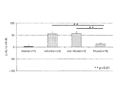
Another cylinder test was conducted in the same manner as described above for
HIE rats (5 months after birth), with a Muse cell-administered group, a
vehicle group and
a sham group, and also a group administered non-Muse cells. In this test, the
healthy side
of the rats was the right paw and the affected side was the left paw. The
results are shown
in Fig. 11. Improvement in motor function was seen in the Muse cell-
administered group,
but in the newly added non-Muse cell-administered group, no improvement in
motor
function was seen, similar to the vehicle. This suggested that Muse cells
exhibit an
excellent effect of improvement in motor function compared to non-Muse cells.
INDUSTRIAL APPLICABILITY
The cell preparation and pharmaceutical composition of the present invention
can
be applied for amelioration and treatment of perinatal brain damage such as
learning
disability and motor disability, by administration to an HIE rat model.
The specific embodiments of the present invention were explained in the
present
specification for the purpose of example, and it will be easily appreciated by
a person
skilled in the art that various modifications may be employed such as are not
outside of the
spirit and scope of the present invention.
23
CA 3024518 2020-03-09