Note: Descriptions are shown in the official language in which they were submitted.
CA 03024623 2018-11-16
WO 2017/201310 PCT/US2017/033368
ROBOTIC SURGICAL DEVICES, SYSTEMS
AND RELATED METHODS
CROSS-REFERENCE TO RELATED APPLICATION(S)
[001] This application claims priority to U.S. Provisional Application No.
62/338,375, filed on
May 18, 201 6 and entitled "Robotic Surgical Devices, Systems and Related
Methods," which is hereby
incorporated by reference in its entirety under 35 U.S.C. 1 19(e).
TECHNICAL FIELD
[002] The embodiments disclosed herein relate to various medical devices
and related
components, including robotic and/or in vivo medical devices and related
components. Certain
embodiments include various robotic medical devices, including robotic devices
that are disposed within a
body cavity and positioned using a support component disposed through an
orifice or opening in the body
cavity. Further embodiments relate to methods and devices for operating the
above devices.
BACKGROUND
[003] Invasive surgical procedures are essential for addressing various
medical conditions.
When possible, minimally invasive procedures such as laparoscopy are preferred
.
[004] However, known minimally invasive technologies such as laparoscopy
are limited in
scope and complexity due in part to 1) mobility restrictions resulting from
using rigid tools inserted through
access ports, and 2) limited visual feedback. Known robotic systems such as
the da Vinci Surgical
System (available from Intuitive Surgical , Inc., located in Sunnyvale, CA)
are also restricted by the access
ports, as well as having the additional disadvantages of being very large,
very expensive, unavailable in
most hospitals, and having limited sensory and mobility capabilities.
[005] There is a need in the art for improved surgical methods, systems,
and devices.
BRIEF SUMMARY OF THE INVENTION
[006] Discussed herein are various robotic surgical systems, including
certain systems having
camera lumens configured to receive various camera systems. Further
embodiments relate to surgical
insertion devices configured to be used to insert various surgical devices
into a cavity of a patient while
maintaining insufflations of the cavity.
[007] In one Example, a robotic surgical system , including : a robotic
surgical device
including : a device body including front and back sides and a distal end and
a proximal end ; first and
second shoulder joints operably coupled to the distal end of the device body;
a first robotic arm operably
coupled to the first shoulder joint; and a second robotic arm operably coupled
to the second shoulder
joint; and a camera component, including a flexible section and a distal
imager, where the first and
second robotic arms are constructed and arranged so as to be positioned on the
front or back sides of the
body. -1-
CA 03024623 2018-11-16
=
WO 2017/201310 PCT/US2017/033368
[008] Implementations may include one or more of the following features.
The robotic
surgical system where the surgical device includes at least one actuator. The
robotic surgical system
where the first and second robotic arms include at least one motor disposed
within each of the first and
second robotic arms. The robotic surgical system further including a support
device configured to remote
center the robotic surgical device. The robotic surgical system further
including an surgical console. The
robotic surgical system where the camera is disposed through a lumen defined
in the robotic surgical
device. The robotic surgical system where the camera is configured to be an
adjustable height camera.
The robotic surgical system where the camera is constructed and arranged to be
capable of pitch and
yaw. The robotic surgical system where the distal camera tip is configured to
orient to a define
workspace. The robotic surgical system where the camera includes lights. The
robotic surgical system
where the robotic surgical device further includes first and second end
effectors. The robotic surgical
system where the first robotic arm further includes an upper arm and a
forearm. The robotic surgical
system where the first robotic arm further includes: a first arm upper arm ; a
first arm elbow joint; and a
first arm lower arm, where the first arm upper arm is configured to be capable
of roll, pitch and yaw
relative to the first shoulder joint and the first arm lower arm is configured
to be capable of yaw relative to
the first arm upper arm by way of the first arm elbow joint. The surgical
robotic system where the first
robotic arm further includes at least one first arm actuator disposed within
the first robotic arm. The
robotic surgical system where the second robotic arm further includes: a
second arm upper arm ; \a
second arm elbow joint; and a second arm lower arm, where the second arm upper
arm is configured to
be capable of roll, pitch and yaw relative to the second shoulder joint and
the second arm lower arm is
configured to be capable of yaw relative to the second arm upper arm by way of
the second arm elbow
joint. The surgical robotic system where the second robotic arm further
includes at least one second arm
actuator disposed within the second robotic arm . The surgical robotic system
where the first and second
arms include at least one motor disposed in each arm . The surgical robotic
system further including at
least one PCB disposed within at least one of the first or second robotic arms
and in operational
communication with at least one of the first robotic arm and second robotic
arm , where the PCB is
configured to perform yaw and pitch functions.
[009] One Example includes A robotic surgical system, including: a robotic
surgical device
including: a device body including : a distal end; a proximal end; a front
side; and a back side; first and
second shoulder joints operably coupled to the distal end of the device body;
a first robotic arm operably
coupled to the first shoulder joint; and a second robotic arm operably coupled
to the second shoulder
joint; and a camera component, including : a shaft; an imager; and a flexible
section operably coupling the
imager to the shaft, where the first and second robotic arms are constructed
and arranged so as to be
positioned on the front or back sides of the body. Implementations may include
one or more of the
following features. The robotic surgical system where the first robotic arm
further includes an upper arm
and a forearm . The robotic surgical system where the first robotic arm
further includes: a first arm upper
arm ; a first arm elbow joint; and a first arm lower arm, where the first arm
upper arm is configured to be
-2-
CA 03024623 2018-11-16
WO 2017/201310 PCT/US2017/033368
capable of roll, pitch and yaw relative to the first shoulder joint and the
first arm lower arm is configured to
be capable of yaw relative to the first arm upper arm by way of the first arm
elbow joint. The surgical
robotic system where the first robotic arm further includes at least one first
arm actuator disposed within
the first robotic arm . The robotic surgical system where the second robotic
arm further includes: a second
arm upper arm ; a second arm elbow joint; and a second arm lower arm, where
the second arm upper arm
is configured to be capable of roll, pitch and yaw relative to the second
shoulder joint and the second arm
lower arm is configured to be capable of yaw relative to the second arm upper
arm by way of the second
arm elbow joint. The surgical robotic system where the second robotic arm
further includes at least one
second arm actuator disposed within the second robotic arm. The surgical
robotic system where the first
and second arms include at least one motor disposed in each arm. The surgical
robotic system further
including at least one PCB disposed within at least one of the first or second
robotic arms and in
operational communication with at least one of the first robotic arm and
second robotic arm, where the
PCB is configured to perform yaw and pitch functions. Implementations of the
described techniques may
include hardware, a method or process, or computer software on a computer-
accessible medium.
[010] Another Example includes A robotic surgical system, including: a
robotic surgical
device including : a device body including : a distal end; a proximal end, and
a camera lumen defined
within the device body, the camera lumen including : a proximal lumen opening
in the proximal end of the
device body; a socket portion defined distally of the proximal lumen opening,
the socket portion including
a first diameter and a first coupling component; an extended portion defined
distally of the socket portion,
the extended portion having a second, smaller diameter; and a distal lumen
opening in the distal end of
the device body, the distal lumen opening defined at a distal end of the
extended portion ; first and second
shoulder joints operably coupled to the distal end of the device body; a first
robotic arm operably coupled
to the first shoulder joint; and a second robotic arm operably coupled to the
second shoulder joint; and a
camera component, including an elongate tube operably coupled to the handle,
where the elongate tube
is configured and sized to be positionable through the extended portion, the
elongate tube including: a
shaft; an imager; and a flexible section operably coupling the optical section
to the rigid section, where
the elongate tube has a length such that at least the optical section is
configured to extend distally from
the distal lumen opening when the camera component is positioned through the
camera lumen.
[01 1] Implementations may include one or more of the following
features. The surgical
robotic system where the first and second arms include at least one motor
disposed in each arm. The
surgical robotic system further including at least one PCB disposed within at
least one of the first or
second robotic arms and in operational communication with at least one of the
first robotic arm and
second robotic arm , where the PCB is configured to perform yaw and pitch
functions.
[012] While multiple embodiments are disclosed, still other embodiments
of the present
invention will become apparent to those skilled in the art from the following
detailed description, which
shows and describes illustrative embodiments of the invention. As will be
realized, the invention is
capable of modifications in various obvious aspects, all without departing
from the spirit and scope of the
-3-
CA 03024623 2018-11-16
WO 2017/201310 PCT/US2017/033368
present invention. Accordingly, the drawings and detailed description are to
be regarded as illustrative in
nature and not restrictive.
BRIEF DESCRI PTION OF THE DRAWINGS
[013] FIG. 1A is a front view of a surgical device, according to one
embodiment.
[014] FIG. 1B is a front view of the device of FIG. 1A inserted into the
body cavity.
[015] FIG. 2 is a front view of a surgical device, according to one
embodiment.
[016] FIG. 3 is a three-quarters perspective view of the robot of the
implementation of FIG. 2
without the camera.
[017] FIG. 4 is a three-quarters perspective view of the camera of the
implementation of FIG. 2
without the robot.
[018] FIG. 5A is a close-up perspective view of a surgical device,
according to one
embodiment.
[019] FIG. 5B is front view of the embodiment of FIG. 5A, wherein the arms
and camera are in
the "insertion" position.
[020] FIG. 6A is a perspective view of a surgical device showing various
workspaces for the
arms, according to one embodiment.
[021] FIG. 6B is a further perspective view of the surgical device of FIG.
6A, showing the
workspace of one arm.
[022] FIG. 7A is a side view of the robot according to one embodiment,
showing the range of
motion of the arms and the associated workspaces, according to one embodiment.
[023] FIG. 78 is a top view of the implementation of FIG. 7A, showing the
range of motion of
the arms and the associated workspaces.
[024] FIG. 7C is a perspective view of the implementation of FIG. 7A,
showing the range of
motion of the arms and the associated workspaces.
[025] FIG. 8A is a rear perspective view of one implementation of a
surgical device, showing
the positioning of the arms to the ahead and behind the device, according to
one embodiment.
(026] FIG. 8B is a three-quarters rear view of the device of FIG. 8A,
showing several possible
arm positions.
[027] FIG. 80 is a lower perspective front view of the device showing the
arm positions of FIG.
8B.
[028] FIG. 9 is a perspective view of a surgical device according to one
embodiment showing
the camera and arms oriented in a central "down" work position.
[029] FIG. 10 is a front view of the device of FIG. 9 showing the arms in
an central "up"
position.
-4-
CA 03024623 2018-11-16
WO 2017/201310 PCT/US2017/033368
[030] FIG. 11 is a perspective view of a surgical device according to one
embodiment showing
the arms in a "down" position.
[031] FIG. 12A is a top view of a surgical device, according to one
implementation.
[032] FIG. 12B is a top view of a surgical device, according to another
implementation .
[033] FIG. 120 is a front view of a surgical device, according to one
implementation.
[034] FIG. 12D is a front view of a surgical device, according to another
implementation.
[035] FIG. 12E is a side view of a surgical device, according to one
implementation.
[036] FIG. 12F is a side view of a surgical device, according to another
implementation.
[037] FIG. 13A is a perspective view of a surgical device according to one
embodiment,
showing the movement of the first joint.
[038] FIG. 13B is a perspective view of a surgical device according to one
embodiment,
showing the movement of the second joint.
[039] FIG. 130 is a perspective view of a surgical device according to one
embodiment,
showing the movement of the third joint.
[040] FIG. 13D is a perspective view of a surgical device according to one
embodiment,
showing the movement of the fourth joint.
[041] FIG. 14 is a perspective view of a surgical robotic device showing
the internal
components, according to one implementation.
[042] FIG. 15 is a front view showing the internal components of the body
and shoulders,
according to one embodiment.
[043] FIG. 16 is a perspective view showing the internal components of the
body, according to
one embodiment
[044] FIG. 17 is a perspective view showing the internal components of the
shoulders,
according to one embodiment.
[045] FIG. 18 is a side view showing the internal components of the
shoulders, according to
one embodiment.
[046] FIG. 19 is a reverse perspective view showing the internal components
of the body and
shoulders, according to one embodiment.
[047] FIG. 20 is a perspective view showing the internal components of the
upper arm,
according to one embodiment.
[048] FIG. 2 1 is a perspective view showing further internal components of
the upper arm,
according to one embodiment.
[049] FIG. 22 is a front view showing further internal components of the
upper arm, according
to one embodiment.
[050] FIG. 23 is a perspective view showing further internal components of
the upper arm,
according to one embodiment.
-5-
CA 03024623 2018-11-16
=
WO 2017/201310 PCT/US2017/033368
[051] FIG. 24 is a perspective view showing internal components of the
lower arm , according to
one embodiment.
[052] FIG. 25 is a perspective view showing further internal components of
the upper arm,
according to one embodiment.
[053] FIG. 26 is a perspective view showing further internal components of
the upper arm,
according to one embodiment.
[054] FIG. 27 is a perspective view showing yet further internal components
of the upper arm,
according to one embodiment.
[055] FIG. 28A is a front perspective view of a surgical device having an
articulating camera,
according to one embodiment.
[056] FIG. 28B is a close-up perspective view of the camera of FIG. 28A
showing a variety of
possible movements.
[057] FIG. 280 is a front view of a robotic device and camera having
adjustable depth,
according to one embodiment.
[058] FIG. 28D is a close up view of the device lumen and camera shaft
showing the adjustable
depth mechanism, according to one implementation, showing the camera in an
"up" position.
[059] FIG. 28E is a front view of the robot and camera, according to the
implementations of
FIGS. 28C and 28D.
[060] FIG. 28F is a front view of a robotic device and camera having
adjustable depth,
according to one embodiment.
[061] FIG. 28G is a close up view of the device lumen and camera shaft
showing the
adjustable depth mechanism , according to one implementation, showing the
camera in an "down"
position.
[062] FIG. 28H is a front view of the robot and camera, according to the
implementations of
FIGS. 28F and 28G.
[063] FIG. 281 is a cross-sectional view of the body lumen, according to
one embodiment.
[064] FIGS. 29A-B depict surgical device workspaces and fields of view,
according to
exemplary implementations.
[065] FIGS. 30A-F depict a surgical device and zero-degree camera through a
range of
possible positions, according to one implementation.
[066] FIGS. 3 lick-F depict a surgical device and thirty degree camera
through a range of
possible positions, according to another implementation.
[067] FIGS. 32A-F depict a surgical device and sixty degree camera through
a range of
possible positions, according to one implementation.
[068] FIGS. 33A-C depict a surgical device and camera through a range of
possible positions
with an "S-scope" configuration , according to one implementation.
-6-
CA 03024623 2018-11-16
WO 2017/201310 PCT/US2017/033368
[069] FIG. 34A is one implementation of the articulating camera tip.
[070] FIG. 346 is another implementation of the articulating camera tip.
[071] FIG. 34C is yet another implementation of the articulating camera
tip.
[072] FIGS. 35A-35C are side views of the surgical device and camera
showing the movement
of the camera between various positions, according to several embodiments.
[073] FIGS. 36A-36C are side views of the surgical device end effectors,
according to one
implementation.
[074] FIG, 37 is a front view of the surgical device on a support
structure, according to one
implementation.
[075] FIG. 38 is a perspective view of the surgical device on a support
structure, according to
one implementation.
[076] FIG. 39 is a cross-sectional view of the surgical device at the
insertion point, according to
one implementation.
[077] FIG. 40A is a perspective view of the surgical device on a support
structure, according to
one implementation.
[078] FIG. 40B is a side view of the surgical device on a support
structure, according to one
implementation.
[079] FIG. 4 1A is a perspective view of the surgical device on a support
structure, according to
one implementation.
[080] FIG. 4 1B is a further perspective view of the surgical device on a
support structure,
according to the implementation of FIG .4 1A.
[081] FIG. 42A is a perspective view of the surgical device on another
support structure,
according to one implementation.
[082] FIG. 426 is a further perspective view of the surgical device on a
support structure,
according to the implementation of FIG. 42A.
[083] FIG. 42C is yet a further perspective view of the surgical device on
a support structure,
according to the implementation of FIG. 42A.
[084] FIG. 43 is a side view of the surgical device on yet another support
structure, according
to one implementation.
[085] FIG. 44 is yet a further perspective view of the surgical device on a
support structure,
according to another implementation.
[086] FIG. 45 is a perspective view of the surgical device on a support
robot, according to
another implementation.
[087] FIG. 46 is a perspective view of the surgical device on a support
robot, according to
another implementation.
-7-
CA 03024623 2018-11-16
WO 2017/201310 PCT/IJS2017/033368
[088] FIG. 45 is a perspective view of the surgical device on a ball joint
support structure,
according to another implementation.
[089] FIGS. 48A through 48D-2 show side and top views of a support
structure positioning the
surgical device, according to one implementation.
[090] FIG. 49 is a perspective view of a support structure positioning the
surgical device,
according to one implementation.
[091] FIG. 50A is a perspective view of another support structure
positioning the surgical
device, according to one implementation.
[092] FIG. 50B is a side view of another support structure positioning the
surgical device,
according to one implementation.
[093] FIG. 50C is a side view of another support structure positioning the
surgical device,
according to one implementation.
[094] FIG. 50D is a side view of another support structure positioning the
surgical device,
according to one implementation.
[095] FIG. 51 is a perspective view of another support structure
positioning the surgical device,
according to one implementation.
[096] FIG. 52A is a side view of another support structure positioning the
surgical device,
according to one implementation.
[097] FIG. 52B is a perspective view of another support structure
positioning the surgical
device, according to one implementation.
[098] FIG. 52C is a perspective view of another support structure
positioning the surgical
device, according to one implementation.
[099] FIG. 52D is a perspective view of another support structure
positioning the surgical
device, according to one implementation.
[0100] FIG. 52E is a perspective view of another support structure
positioning the surgical
device, according to one implementation.
[0101] FIG. 52F is a perspective view of another support structure
positioning the surgical
device, according to one implementation.
[0102] FIG. 53 is a perspective view of the surgical console, according to
one implementation.
[0103] FIG. 54 is a schematic view of a surgical system, according to one
implementation.
[0104] FIG. 55 is another schematic view of a surgical system, according
to one implementation.
DETAILED DESCRIPTION
[0105] The various systems and devices disclosed herein relate to devices
for use in medical
procedures and systems. More specifically, various embodiments relate to
various medical devices,
including robotic devices and related methods and systems.
-8-
CA 03024623 2018-11-16
WO 2017/201310
PCT/US2017/033368
[0106] It is
understood that the various embodiments of robotic devices and related methods
and systems disclosed herein can be incorporated into or used with any other
known medical devices,
systems, and methods.
[0107] It is
understood that the various embodiments of robotic devices and related methods
and systems disclosed herein can be incorporated into or used with any other
known medical devices,
systems, and methods. For example, the various embodiments disclosed herein
may be incorporated
into or used with any of the medical devices and systems disclosed in
copending U.S. Applications
11/766,683 (filed on June 2 1, 2007 and entitled "Magnetically Coupleable
Robotic Devices and Related
Methods"), 11/766,720 (filed on June 21, 2007 and entitled "Magnetically
Coupleable Surgical Robotic
Devices and Related Methods"), 11/966,741 (filed on December 28, 2007 and
entitled "Methods,
Systems, and Devices for Surgical Visualization and Device Manipulation"),
61/030,588 (filed on February
22, 2008), 12/1 71,41 3 (filed on July 11, 2008 and entitled "Methods and
Systems of Actuation in Robotic
Devices"), 12/1 92,663 (filed August 15, 2008 and entitled Medical Inflation,
Attachment, and Delivery
Devices and Related Methods"), 12/1 92,779 (filed on August 15, 2008 and
entitled "Modular and
Cooperative Medical Devices and Related Systems and Methods"), 12/324,364
(filed November 26, 2008
and entitled "Multifunctional Operational Component for Robotic Devices"),
61/640,879 (filed on May 1,
201 2), 13/493,725 (filed June 11, 201 2 and entitled "Methods, Systems, and
Devices Relating to Surgical
End Effectors" ), 13/546,831 (filed July 11, 201 2 and entitled "Robotic
Surgical Devices, Systems, and
Related Methods"), 6 1/680,809 (filed August 8, 201 2), 13/573,849 (filed
October 9, 201 2 and entitled
"Robotic Surgical Devices, Systems, and Related Methods"), 13/738,706 (filed
January 10, 201 3 and
entitled "Methods, Systems, and Devices for Surgical Access and Insertion"),
13/833,605 (filed March 15,
201 3 and entitled "Robotic Surgical Devices, Systems, and Related Methods"),
13/839,422 (filed March
15, 201 3 and entitled "Single Site Robotic Devices and Related Systems and
Methods"), 13/834,792 (filed
March 15, 201 3 and entitled "Local Control Robotic Surgical Devices and
Related Methods"), 14/208,51 5
(filed March 13, 201 4 and entitled "Methods, Systems, and Devices Relating to
Robotic Surgical Devices,
End Effectors, and Controllers"), 14/21 0,934 (filed March 14, 201 4 and
entitled "Methods, Systems, and
Devices Relating to Force Control Surgical Systems), 14/21 2,686 (filed March
14,201 4 and entitled
"Robotic Surgical Devices, Systems, and Related Methods"), and 14/334,383
(filed July 17, 201 4 and
entitled "Robotic Surgical Devices, Systems, and Related Methods"), and U.S.
Patents 7,492, 116 (filed on
October 3 1, 2007 and entitled "Robot for Surgical Applications"), 7,772,796
(filed on April 3, 2007 and
entitled "Robot for Surgical Applications"), and 8,179,073 (issued May 15, 201
1, and entitled "Robotic
Devices with Agent Delivery Components and Related Methods"), U.S. Published
Application No.
201 6/00741 20 (filed September 14, 201 5, and entitled "Quick-Release End
Effectors and Related
Systems and Methods"), U.S. Published Application No. 201 6/01 35898 (filed
November 11, 201 5 entitled
"Robotic Device with Compact Joint Design and Related Systems and Methods"),
U.S. Patent Application
No. 15/227,81 3 (filed August 3,201 6 and entitled "Robotic Surgical Devices,
Systems, and Related
Methods"), U.S. Provisional Application No. 62/379,344 (filed August 25, 201 6
and entitled "Quick-
-9-
CA 03024623 2018-11-16
WO 2017/201310 PCT/US2017/033368
Release End Effector Tool Interface and Related Systems and Methods"), U.S.
Provisional Application
No. 62/425, 149 (filed November 22, 201 6 and entitled "Improved Gross
Positioning Device and Related
Systems and Methods"), U.S. Provisional Application No. 62/427,357 (filed
November 29, 201 6 and
entitled "Controller with User Presence Detection and Related Systems and
Methods"), U.S. Provisional
Application No. 62/433,837 (filed December 14, 201 6 and entitled "Releasable
Attachment Device for
Coupling to Medical Devices and Related Systems and Methods"), and U.S.
Provisional Application No.
62/381 ,299 (filed August 30, 201 6 and entitled "Robotic Device with Compact
Joint Design and an
Additional Degree of Freedom and Related Systems and Methods")a all of which
are hereby incorporated
herein by reference in their entireties.
[0108] Certain device and system implementations disclosed in the
applications listed above can
be positioned within a body cavity of a patient in combination with a support
component similar to those
disclosed herein. An "in vivo device" as used herein means any device that can
be positioned, operated,
or controlled at least in part by a user while being positioned within a body
cavity of a patient, including
any device that is coupled to a support component such as a rod or other such
component that is
disposed through an opening or orifice of the body cavity, also including any
device positioned
substantially against or adjacent to a wall of a body cavity of a patient,
further including any such device
that is internally actuated (having no external source of motive force), and
additionally including any
device that may be used laparoscopically or endoscopically during a surgical
procedure. As used herein,
the terms "robot," and "robotic device" shall refer to any device that can
perform a task either
automatically or in response to a command.
[0109] Certain embodiments provide for insertion of the present invention
into the cavity while
maintaining sufficient insuffiation of the cavity. Further embodiments
minimize the physical contact of the
surgeon or surgical users with the present invention during the insertion
process. Other implementations
enhance the safety of the insertion process for the patient and the present
invention. For example, some
embodiments provide visualization of the present invention as it is being
inserted into the patient's cavity
to ensure that no damaging contact occurs between the system/device and the
patient. In addition,
certain embodiments allow for minimization of the incision size/length.
Further implementations reduce
the complexity of the access/insertion procedure and/or the steps required for
the procedure. Other
embodiments relate to devices that have minimal profiles, minimal size, or are
generally minimal in
function and appearance to enhance ease of handling and use.
[0110] Certain implementations disclosed herein relate to "combination"
or "modular" medical
devices that can be assembled in a variety of configurations. For purposes of
this application, both
"combination device" and "modular device" shall mean any medical device having
modular or
interchangeable components that can be arranged in a variety of different
configurations. The modular
components and combination devices disclosed herein also include segmented
triangular or
quadrangular-shaped combination devices. These devices, which are made up of
modular components
(also referred to herein as "segments") that are connected to create the
triangular or quadrangular
-10-
CA 03024623 2018-11-16
=
WO 2017/201310 PCT/US2017/033368
configuration, can provide leverage and/or stability during use while also
providing for substantial payload
space within the device that can be used for larger components or more
operational components. As with
the various combination devices disclosed and discussed above, according to
one embodiment these
triangular or quadrangular devices can be positioned inside the body cavity of
a patient in the same
fashion as those devices discussed and disclosed above.
[01 11] Certain embodiments disclosed or contemplated herein can be used
for colon resection,
a surgical procedure performed to treat patients with lower gastrointestinal
diseases such as diverticulitis,
Crohn's disease, inflammatory bowel disease and colon cancer. Approximately
two-thirds of known colon
resection procedures are performed via a completely open surgical procedure
involving an 8- to 12-inch
incision and up to six weeks of recovery time. Because of the complicated
nature of the procedure,
existing robot-assisted surgical devices are rarely used for colon resection
surgeries, and manual
laparoscopic approaches are only used in one-third of cases. In contrast, the
various implementations
disclosed herein can be used in a minimally invasive approach to a variety of
procedures that are typically
performed 'open' by known technologies, with the potential to improve clinical
outcomes and health care
costs. Further, the various implementations disclosed herein can be used in
place of the known
mainframe-like laparoscopic surgical robots that reach into the body from
outside the patient. That is the
less-invasive robotic systems, methods, and devices disclosed herein feature
small, self-contained
surgical devices that are inserted in their entireties through a single
incision in the patient's abdomen.
Designed to utilize existing tools and techniques familiar to surgeons, the
devices disclosed herein will not
require a dedicated operating room or specialized infrastructure, and, because
of their much smaller size,
are expected to be significantly less expensive than existing robotic
alternatives for laparoscopic surgery.
Due to these technological advances, the various embodiments herein could
enable a minimally invasive
approach to procedures performed in open surgery today.
[01 1 2] The various embodiments are disclosed in additional detail in the
attached figures, which
include some written description therein.
[01 1 3] . The various system embodiments described herein are used to
perform robotic surgery.
The systems are used for general surgery applications in the abdominal cavity,
including colon resection.
In certain implementations, the various systems described herein are based on
and/or utilize techniques
used in manual laparoscopic surgery including insufflation of the abdominal
cavity and the use of ports to
insert tools into the abdominal cavity.
[01 14] Major components of the various system embodiments include a robot
and a surgeon
control console. The robot implementations are configured to be inserted into
the insufflated abdominal
cavity. Certain robot embodiments have an integrated camera system that
captures a view of the surgical
target. The surgeon can then use that view on a display to help control the
robot's movements. In certain
implementations, the camera is designed so that it can be removed so it can be
cleaned and used in
other applications.
-11-
CA 03024623 2018-11-16
WO 2017/201310 PCT/US2017/033368
[01 15] The surgeon console, according to some embodiments, has a display
to view the
feedback from the camera. This display can also have overlays to provide some
additional information to
the surgeon including the robot's state and other information. The console can
also have a touch screen
used to control various system functions. In addition, the various console
embodiments can also have
user input devices (e.g. haptic joysticks) that the surgeon can use to control
the movement of the robot's
arms and other movement. Further, the console can also has one or more pedals
used to control various
robot control and functions.
[01 16] In other embodiments as will be discussed in further detail
herein, the system can include
disposable or permanent sleeves, an electro-surgery cautery generator, an
insertion port, a support
arm/structure, a camera, remote surgical displays, end-effectors (tools), an
interface pod, a light source,
and other support components.
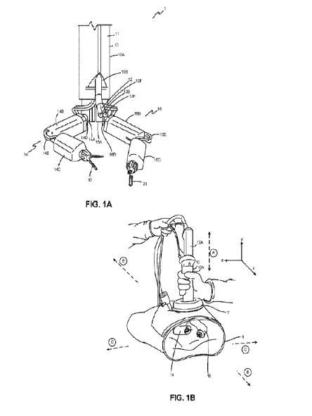
[01 17] FIGS. 1A and 1B depict one embodiment of the system 1 with a robot
or robotic device
with a camera 12. As shown in FIG. 1A, the robotic device 10 has two robotic
arms 14, 16 operably
coupled thereto and a camera component or "camera" 12 disposed between the two
arms 14, 16 and
positionable therein. That is, device 10 has a first (or "right") arm 14 and a
second (or "left) arm 16, both
of which are operably coupled to the device 10 as discussed in additional
detail below. The device 10 as
shown has a casing (also referred to as a "cover" or "enclosure") 11. The
device 10 is also referred to as
a "device body" 10A and has two rotatable cylindrical components (also
referred to as "shoulders" or
"turrets") : a first (or "right") shoulder 14A and a second (or "left")
shoulder 16A. Each arm 14, 16 also has
an upper arm (also referred to herein as an "inner arm," "inner arm assembly,"
"inner link," "inner link
assembly," "upper arm assembly," "first link," or "first link assembly") 14B,
16B, and a forearm (also
referred to herein as an "outer arm," "outer arm assembly," "outer link,"
"outer link assembly," "forearm
assembly," "second link," or "second link assembly") 14C, 16C. The right upper
arm 14B is operably
coupled to the right shoulder 14A of the body 10A at the right shoulder joint
14D and the left upper arm
16B is operably coupled to the left shoulder 16A of the body 10 at the left
shoulder joint 16D. Further, for
each arm 14, 16, the forearm 14C, 16C is rotatably coupled to the upper arm
14B, 16B at the elbow joint
14E, 16E.
[01 18] As shown in FIG. 16, the robotic device 10 has been inserted into
a model of the
abdominal cavity 6 through a gel port 7 in a fashion similar to the way it
would be inserted into a patient's
abdominal cavity 6. The gel port 7 allows for an irregularly shaped robotic
device 10 to be inserted while
maintaining insufflation pressure. In this implementation, a standard manual
laparoscopic port 7 is used
in addition to the robot 10. Alternatively, two or more such ports can be
utilized (not shown). In a further
alternative, no standard manual laparoscopic ports are used.
[01 19] In FIG. 1B, the device body 10A is shown having been inserted in a
ventral-dorsal
orientation into the abdominal cavity such that the longitudinal body axis (as
is shown by reference arrow
A) is generally perpendicular relative to the rostrocaudal/anteroposterior and
mediolateral axes (reference
arrows B and C, respectively) . It is understood that following insertion, the
device body 10A can be
-12-
CA 03024623 2018-11-16
WO 2017/201310 PCT/US2017/033368
variously positioned, so as to be rotated, tilted or angled relative to the
cavity 6 to alter the device
workspace and access various regions of the cavity, as is described in detail
below in relation to FIGS.
6A-8C.
[0120] FIG. 2 shows the robot with the integrated camera system ,
according to one
embodiment. The robot of FIG. 2 has two arms 14, 16 and a body 10A (or torso)
having a distal end 10B
and proximal end 10C. The arms 14, 16 each have active degrees of freedom and
an additional active
joint 14F, 16F to actuate the end effectors, or tools 18, 20. It is understood
that more or less degrees of
freedom could be included. The device in this embodiment has a connection line
8 (also referred to as a
"pigtail cable") (partially shown) that includes electrical power,
electrocautery, and
information/communication signals. In certain implementations, the device has
distributed control
electronics and software to help control the device 10. Some buttons can be
included to support insertion
and extraction of the device into and out of the abdominal cavity. in this
embodiment, the integrated
camera 12 is also shown inserted in the device body 10A. When inserted into
the body 10A, the camera
12 has a handle or body 12A that extends proximally from the proximal body end
10C and a flexible
camera imager 12B extending from the distal body end 10B.
[0121] FIGS. 3 and 4 depict the robotic device 10 with the camera
assembly 12 removed,
according to one embodiment. In these embodiments, and as shown in FIG. 2 and
FIGS. 3-4, the
camera imager 12B is designed to be positioned between the two arms 14, 16 and
capture that view
between the two arms 14, 16. in these implementations, the camera 12 extends
through the robot body
10A such that the camera imager 12B exits near the joints between the body and
the robotic arms (the
"shoulder" joints 14A, 16A). The camera 12 has a flexible, steerable tip 120
to allow the user to adjust
the viewing direction. The end effectors 18, 2 0 on the distal end of the arms
14, 16 can include various
tools 18, 20 (scissors, graspers, needle drivers, etc). In certain
embodiments, the tools 18, 2 0 are
designed to be removable by a small twist of the tool knob that couples the
end effector to the arm 14, 16.
[0122] As is shown in FIGS. 3-4, the camera assembly 12 has a handle 12A
and a long shaft
12D with the camera imager 12B at the distal tip 12C. In various
implementations, the flexible tip 12C
and therefore camera imager 12B can be steered or otherwise moved in two
independent directions in
relation to the shaft 120 at a flexible section 12E (black section on shaft)
to change the direction of view.
In certain implementations, the camera 12 has some control buttons 12F as
shown. in some
embodiments, the camera assembly 12 can be used independently of the robotic
device 10 as shown in
FIG. 4.
[0123] Alternatively, the assembly can be inserted into the robot 10
though a lumen 10D defined
through the body 10A of the robotic device 10 as shown. In certain
embodiments, the lumen 10D
includes a seal/port 10E to ensure that the patient's cavity remains
insufflated (as shown in relation to
FIG. 1B). According to one embodiment, the robotic device 10 can have a sensor
to determine if the
camera is positioned in the camera lumen 10D of the device 10.
-13-
CA 03024623 2018-11-16
WO 2017/201310 PCT/US2017/033368
[0124] FIG. 5 depicts a robotic device 10 according to one embodiment in
a configuration in
which the positionable arms 14, 16 are positioned such that the tools 18,20
are positioned in line with the
camera tip 12C. That is, in this embodiment the arms 14, 16 are disposed in
the workspace so as to be
within the field of view of the camera imager') 2B (designated by reference
lines "VI" and 'V2"). In the
implementation of FIG. 5, the device 10 is positioned within the cavity of the
patient at an angle - that is,
such that the longitudinal axis of the device body 10A (designated by
reference line A) is not
perpendicular to the body of the patient (as shown, for example, in FIG. 1B).
[0125] In the implementation of FIG. 5A, the device body 10A is therefore
oriented so as to have
a "top," "upper," or "front" side 22 and a "bottom ," "lower," or "back" side
24. It is understood that further
configurations are possible, and as described in detail herein, the camera 12
and arms 14, 16 are
capable of extending into either side 22, 24 so as to provide large workspaces
without the need to rotate
the device body 10A.
[0126] In the implementation shown in FIG. 58, the arms 14, 16 of the
robotic device 10 are
positioned in an "insertion" configuration. As shown, in the insertion
configuration, the arms 14, 16 and
camera 12 are all primarily aligned with the robotic device body 10A such that
the longitudinal axes of
each of the components are substantially parallel to one another (as shown by
reference arrow I) for
insertion through the port (as is shown ,for example, in FIG. 1B at 7). It is
understood that the insertion
configuration minimizes the overall "footprint" of the device 10, so as to
allow the smallest possible
incision. In certain implementations, during insertion the device 10 can be
passed through a variety of
positions while being inserted, as has been previously described in U.S.
Patent Application No.
15/227,81 3 filed August 3, 201 6 and entitled "Robotic Surgical Devices,
Systems, and Related Methods,"
which is incorporated by reference herein in its entirety.
[0127] A principle advantage of the system 1 in certain implementations is
a wide workspace
range for the arms, including embodiments wherein the arms are positioned
"behind" the device. In use,
increasing the workspace range of each of the arms can reduce the need to
reposition to the device, and
therefore lead to greater efficiency and faster total surgery times and
recovery. Several implementations
showing the increased arm range are described herein.
[0128] FIGS. 6A, 68, 7A, 78, and 7C schematically depict the entire
workspace 30 as well as
the individual reachable workspaces 30A, 30B of each of the arms 14, 16 of a
robotic device 10,
according to certain embodiments. In these embodiments, "workspace" 30 means
the space 30 around
the robotic device 10 in which either arm and / or end effector 18, 20 can
move, access, and perform its
function within that space.
[0129] More specifically, FIG. 6A depicts a perspective view of the device
body 10A and further
schematically shows the entire workspace 30 as well as the individual
workspaces 30A, 30B of the first
arm 14 and second arm 16, respectively. Note that the each arm 14, 16 has a
range of motion and
corresponding workspace 30A, 30B that extends from the front 22 of the device
to the back 24 of the
device 10. Thus, the first arm 14 equally to the front 22 and the back 24,
through about 180 of space
-14-
CA 03024623 2018-11-16
WO 2017/201310 PCT/US2017/033368
relative to the axis of the device body 10A for each arm 14, 16. This
workspace 30 allows the robotic
device to work to the front 22 and back 24 equally well without having to
reposition the body 10A.
[0130] As best shown in FIG. 6B, the overlap of the ranges of motion for
the individual arms in
these implementations also enables an intersecting workspace 30C (as is also
shown in FIG. 6A). It is
understood that the intersecting workspace 300 in these implementations
encompasses the workspace
30C reachable by both arms 14, 16 and end effectors 18,20 in any individual
device 10 position. Again,
in these implementations, the intersecting workspace 30C includes a range of
about 180 of space
relative to the axis of the device body 10A.
[0131] FIG. 7A depicts a side view of the device body 10A and further
schematically shows the
workspace 30A of the first arm 14. Note that the first arm 14 has a range of
motion that extends from the
front 22 of the device to the back 24 of the device 10. Thus, the first arm 14
equally to the front 22 and
the back 24. This allows the robotic device to work to the front 22 and back
24 equally well without
having to reposition the body 10A. With respect to the actual position of the
arms 14, 16, FIG. 7A depicts
the first arm 14 extending out from the front 22 of the device while the
second arm 16 is extending out
from the back 24.
[0132] Similarly, FIGS. 7B and 70 depict different views of the device
body 10A and arms 14, 16
of FIG. 7A. For example, FIG. 78 depicts atop view of the body 10A and arms
14, 16. In this
embodiment, both the workspace 30A of the first arm 14 and the workspace 30B
of the second arm 16
are shown from atop view. Further, FIG. 7C depicts the body 10A and arms 14,
16 from a perspective
view that shows another angle of the workspaces 30A, 30B.
[01 33] in each of FIGS. 7A-70, the same configuration of the body 10A and
arms 14, 16 is
shown, with the first arm 14 extending out from the front 22 of the device
while the second arm 16 is
extending out from the back 24 (as best shown in FIG. 7A). This wide range of
motion demonstrated by
the workspaces 30A, 30B for both of its arms 14, 16 gives the robotic device
10 .a relatively large
workspace when compared to the length of its arms 14, 16.
[0134] FIGS. 8A, 8B, and 80 further depict the wide range of motion that
can be achieved by the
arms of this specific device 10, according to one embodiment. FIG. 8A depicts
a perspective view of the
back of the device 10 in which the arms 14, 16 are both depicted in a single
position that is substantially
similar to that depicted in FIGS. 7A-7C: a first arm 14 extends away from the
front 22 of the device body
10A, while the second arm 16 extends away from the back 24 of the device body
10A.
[0135] FIG. 8B depicts a side view of the device 10 in which the first
arm 14 is depicted in
multiple different positions, including a first position 14-1 , a second
position 14-2, a third position 14-3,
and a fourth position 14-4, thereby providing some examples of the range of
motion of which the arms (in
this case, the first arm 14) are capable.
[0136] The implementation of FIG. 80 depicts a perspective front view of
the device 10 in which
the first arm 14 is again depicted in the same positions as shown in FIG. 8B,
including the first 14-1 ,
second 14-2, third 14-3, and fourth 14-4 positions within the workspace 30A.
One of skill in the art would
-15-
CA 03024623 2018-11-16
=
WO 2017/201310 PCT/US2017/033368
appreciate that many additional positions between those shown are also
possible, and that these
positions of the first arm 14 are also possible for the second arm 16.
[0137] FIG. 9 is a perspective front view of an implementation of the
device 10 with an
articulating, or flexible camera 12 extending from the distal end 10B of the
device body 10A. In these
implementations, the camera 12 has a distal lens 12B on the tip portion 120,
as well as a flexible sheath
15 enclosing the flexible section 12E. In FIG. 9A, the camera 12 and arms are
generally oriented in a
slightly "down" working position, wherein the tip portion 12C is oriented away
from the front 22 of the body
10A. Again, it is understood that in these implementations, the camera 12 can
therefore be positioned to
best view the end effectors, or tools 18, 20. It is further understood that in
these implementations the
robot 10 exits the body on the forward surface 22.
[0138] FIG. 910 depicts a further implementation of the device 10 with
the arms in an "up" or
"normal" position, where the camera is angled slightly toward the front 22 of
the body 10A. Further, the
device of FIG. 10 has proximal sleeve attachments 32, 34 between the shoulders
14A, 16A and device
body 10A. The sleeve attachments 32, 34 can be "grooves," where two flanges
32A, 32B, 34A, 348 are
disposed around each shoulder shaft 36, 38. It is understood that flanges 32A,
32B, 34A, 34B are
configured or otherwise constructed and arranged so that a permanent and / or
disposable sleeve (not
shown, but as is discussed in the incorporated references) can be attached and
held in place between the
respective flanges 32A, 32B, 34A, 34B. Corresponding distal mating areas 40,
42 for each sleeve (not
shown) are disposed on the distal ends of the forearms 14C, 160 and at the
base of each tool 18, 20.
[0139] FIG. 11 depicts a further implementation of a robot 10 having arms
14 , 16 positioned
substantially "down," compared to the positions of FIGS. 9 and 10. That is, in
FIG. lithe camera tip
120 is oriented perpendicularly from the longitudinal axis (reference arrow A)
of the robot body 10A on
the back side 24 (as opposed to the front side 22) within a region of the
workspace 30, and that the
camera 12 disposed such that the arms 14, 16, and more specifically the tools,
or end effectors 18,20
are within the field of view (shown generally with reference arrow V). In this
implementation, various
operations cables 45 are also shown as being connected to the device body 10A
and camera 12.
[0140] FIGS. 12A-F depict alternate implementations of the robot 10-1 ,
10-2. In the first
implementation, and as shown in FIGS. 12A, 120 and 12E, the robot 10-1 has a
sloped distal body 106-1
portion 48 the camera 12 extends from within. in the second implementation, as
shown in FIGS. 12B,
12D and 12F, the robot 10-2 camera 12 extends from the distal body end 108-2.
In these
implementations, the arms 14, 16 have generally cylindrical upper links, or
shoulders 14A, 16A disposed
in parallel - laterally and separately - on the distal body end 10B such that
there is a "gap" or opening 46
between the shoulders 14A, 16A. In these implementations, the camera 12
extends from the distal end of
the device body 106 within the opening 46, so as to be directly between the
generally cylindrical
shoulders 14A, 16A and equidistant between the front side 22 and back side 24.
In these
implementations, the camera 12 can therefore be curved to view forward and
rearward equally, as is
shown, for example, in relation to FIG. 6A-80.
-16-
CA 03024623 2018-11-16
WO 2017/201310 PCT/US2017/033368
[0141] FIGS. 13-30 depict the internal components of the body 10A, which
is shown in these
figures without its casing or housing 11. It is understood that in use, these
implementations are covered,
as is shown in relation to FIG. 1A. FIGS. 13-30 include the internal
structural or support components of
the body 10A. These components maintain the structure of the body 12 and
provide structural support for
the components disposed therein.
[0142] In use, there are many ways to actuate the robot 10 and its
associated components, such
as DC motors, AC motors, Permanent magnet DC motors, brushless motors,
pneumatics, cables to
remote motors, hydraulics, and the like. A more detailed description of one
possible system is described
in relation to FIGS. 13-30. Other technologies described in the previously-
filed and incorporated
applications and patents can also be implemented to actuate the various
components, as would be
understood.
[0143] FIG. 13 shows an implementation of the robot 10 and each joint of
one arm - here, the
left arm 16. it is understood that the right arm 14 of this implementation is
a mirror image of the left 16. It
is understood that the internal components in the left arm 16 that
operate/control/actuate the left arm 16
are substantially the same as those depicted and described herein and that the
descriptions provided
below apply equally to those components as well.
[0144] In the implementation of FIG. 14, a shoulder yaw joint 100
actuates a yaw joint 100 in the
robot shoulder 14A, 16A. In this implementation , the robot 10 also has a
shoulder pitch joint 102, that is,
a pitch joint 102 on the robot shoulder 14A, 16A. In these implementations, an
upper arm roll joint 104, an
elbow joint 106, and a tool roll joint 108 are also provided which enable the
range of motion described in
relation to Table 1, below. In various implementations, a tool actuation joint
(not shown) interfaces with
the tool (not shown) to actuate open and close of the tool, as has been
previously described.
[0145] In various implementations, these joints 100, 102, 104, 106 have
practical defined
ranges of motions that, together with the robot geometry, lead to the final
workspace of the robot 10. For
the examples given herein, the joint limits allow for a significant robot
workspace, as is described above.
This workspace allows the various implementations of the robot to use both
arms and hands effectively in
several locations within the body cavity of the patient. The joint ranges of
motion defined in the
implementations of FIGS. 13A-27 are given in Table 1. It is understood that
further ranges are possible,
and so this set of ranges is not limiting, but rather representative of a
particular embodiment. Further,
alternate embodiments are possible.
[0146] The direction of rotation and zero positions are shown in FIGS. 13A-
D. In FIGS. 13A-D,
the robot 10 is shown with each of the first four angles in the zero location.
In these implementations,
each joint (the shoulder yaw joint 100, shoulder roll joint 102, upper arm
roll joint 104 and elbow joint 106)
is shown with an axis of rotation (dotted) and a zero location. An arrow is
then used to indicate the
direction of positive joint angle about the axis of rotation. Since the tool
roll joint 108 and tool actuation
jointsl 09 are allow continuous rotation the zero location is arbitrary and
not shown.
Table 1: Joint Ranges of Motion
-17-
CA 03024623 2018-11-16
WO 2017/201310 PCT/US2017/033368
Joint No. Range of Motion
1 -90 to +90
2 -90 to +30
3 -90 to +90
4 Oto 150
Continuous
6 Continuous
[0147] In the implementation of FIG. 14 , the body 10A and each link
(meaning the upper arm
16B, and forearm 16C) contain Printed Circuit Boards ("PCBs") 110, 112, 114
that have embedded
sensor, amplification, and control electronics. One PCB is in each forearm and
upper arm and two PCBs
are in the body. Each PCB also has a full 6 axis accelerometer-based Inertial
Measurement Unit and
temperature sensors that can be used to monitor the temperature of the motors.
Each joint can also have
either an absolute position sensor or an incremental position sensor or both.
In certain implementations,
the some joints contain both absolute position sensors (magnetic encoders) and
incremental sensors (hall
effect). In other implementations, certain joints only have incremental
sensors. These sensors are used
for motor control. The joints could also contain many other types of sensors.
A more detailed description
of one possible method is included here.
[0148] In this implementation, a larger PCB 110 is mounted to the
posterior side of the body
10A. This body PCB 110 controls the motors 116 in the base link, or body 10A
(the shoulder yaw joint
100 and shoulder pitch joint 102 for left and right arms, respectively). Each
upper arm has a PCB 112 to
control the upper arm roll joint 104 and elbow joint 106. Each forearm has a
PCB 114 to control the tool
roll joint 108 and tool actuation joint (not shown) . In the implementation of
FIG. 14, each PCB 110, 112,
114 also has a full six axis accelerometer-based inertial measurement unit and
several temperature
sensors that can be used to monitor the temperature of the various motors
described herein.
[01 49] In these embodiments, each joint 100, 102, 104, 106, 108 can also
have either an
absolute position sensor or an incremental position sensor or both, as
described and otherwise disclosed
in U.S. Provisional Application 61,680,809, filed on August 8, 201 2, which is
hereby incorporated herein
by reference in its entirety. In one implementation, and as shown in FIG. 15
and elsewhere the various
actuators or motors 116, 130, 154, 178 described herein have at least one
temperature sensor 101
disposed on the surface of the motor, for example by temperature-sensitive
epoxy, such that the
temperature sensors (as shown in FIG. 22 at 10 1) can collect temperature
information from each actuator
for transmission to the control unit, as discussed below. In one embodiment,
any of the motors discussed
and depicted herein can be brush or brushless motors. Further, the motors can
be, for example, 6 mm, 8
mm, or 10 mm diameter motors. Alternatively, any known size that can be
integrated into a medical
device can be used. In a further alternative, the actuators can be any known
actuators used in medical
-18-
CA 03024623 2018-11-16
WO 2017/201310 PCT/US2017/033368
devices to actuate movement or action of a component. Examples of motors that
could be used for the
motors described herein include the EC 10 BLDC + G P iCA Planetary Gearhead,
EC 8 BLDC + GP8A
Planetary Gearhead, or EC 6 BLDC + GP6A Planetary Gearhead, all of which are
commercially available
from Maxon Motors, located in Fall River, MA. There are many ways to actuate
these motions, such as
with DC motors, AC motors, permanent magnet DC motors, brushless motors,
pneumatics, cables to
remote motors, hydraulics, and the like. Further implementations can be used
in conjunction with the
various systems, methods and devices disclosed in U.S. Patent Application No.
15/227,81 3 filed August
3,201 6 and entitled "Robotic Surgical Devices, Systems, and Related Methods,"
which is incorporated by
reference in its entirety.
[01 50] In this implementation, joints 1-4 have both absolute position
sensors (magnetic
encoders) and incremental sensors (hall effect). Joints 5 & 6 only have
incremental sensors. These
sensors are used for motor control. It is understood that the joints could
also contain many other types of
sensors, as have been described in detail in the incorporated applications and
references.
[01 511 According to one implementation, certain other internal components
depicted in the
implementation of FIGS. 15-16 are configured to actuate the rotation of the
shoulder yaw joint 100 of the
body 10A around axis 1, as shown in FIG. 14. It is understood that two of each
of the described
components are used - one for each arm - but for ease of description, in
certain depictions and
descriptions, only one is used.
[01 52] As best shown in FIG. 15, a shoulder yaw joint 100 motor 116 and
gearhead combination
drives a motor gear 117 first spur gear set 118, which is best shown in FIG.
16, The first spur gear set
118 drives a shaft supported by bearings 120 to drive a second spur gear set
122. in turn, this second
spur gear set 122 drives an output shaft 124 that is also supported by
bearings 126. This output shaft
124 then drives a turret 14A, 16A (representing the shoulder of the robot 10)
such that the shoulder 16A
rotates around axis 1, as best shown in FIG. 14.
[01 53] According to one implementation, certain internal components
depicted in the
implementation of FIGS. 17-1 9 are configured to actuate the shoulder pitch
joint 102 of the body 10A and
/ or shoulder 14A, 16A around axis 2, as is shown in FIG. 14. In these
implementations, the pitch joint
102 is constructed and arranged to pivot the output link 140 so as to move the
upper arm (not shown)
relative to the shoulder 14A, 16A.
[01 54] In this implementation, a motor 130 and gearhead combination
drives a motor gear 131
and spur gear 132 that in turn drives a first shaft 134. This shaft 134 then
drives a bevel (or miter) gear
pair 136, 137 inside the shoulder turret (depicted in FIG. 19). The bevel (or
miter) gear pair 136, 137
accordingly drives a helical spur set 138, 139 directly connected to the
shoulder pitch joint 102 output link
140, such that the upper arm 16B rotates around axis 2, as best shown in FIG.
14. In this
implementation, the shoulder yaw joint 100 and the shoulder pitch joint 102
therefore have coupled
motion. In these implementations, a plurality of bearings 14 1 support the
various gears and other
components, as has been previously described.
-19-
CA 03024623 2018-11-16
WO 2017/201310 PCT/IJS2017/033368
[01 55] FIGS. 20-23 depict various internal components of the upper arm
16B constructed and
arranged for the movement and operation of the arm 16. In various
implementations, multiple actuators
or motors 142, 154 are disposed within the housing (not shown) of the forearm
160. FIGS. 24-27 depict
various internal components of the forearm 16C constructed and arranged for
the movement and
operation of the end effectors. In various implementations, multiple actuators
or motors 175, 178 are
disposed within the housing (not shown) of the forearm 16C.
[01 56] In one implementation, and as shown in FIG. 22 and elsewhere the
various actuators or
motors 116, 130, 154, 178 described herein have at least one temperature
sensor 101 disposed on the
surface of the motor, for example by temperature-sensitive epoxy, such that
the temperature sensors can
collect temperature information from each actuator for transmission to the
control unit, as discussed
below. In one embodiment, any of the motors discussed and depicted herein can
be brush or brushless
motors. Further, the motors can be, for example, 6 mm, 8 mm, or 10 mm diameter
motors. Alternatively,
any known size that can be integrated into a medical device can be used. In a
further alternative, the
actuators can be any known actuators used in medical devices to actuate
movement or action of a
component. Examples of motors that could be used for the motors described
herein include the EC 10
BLDC +GpicA Planetary Gearhead, EC 8 BLDC + GP8A Planetary Gearhead, or EC 6
BLDC + GP6A
Planetary Gearhead, all of which are commercially available from Maxon Motors,
located in Fall River,
MA. There are many ways to actuate these motions, such as with DC motors, AC
motors, permanent
magnet DC motors, brushless motors, pneumatics, cables to remote motors,
hydraulics, and the like.
[01 57] One implementation of the internal components of the upper arm 16B
constructed and
arranged to actuate the upper arm roll joint 104 is shown in FIGS. 20-21 . In
this implementation, a motor
142 and gearhead combination controlled by a PCB 112 drives a motor gear 143
and corresponding spur
gear 144 where the output spur gear 144 is supported by a shaft 148 and
bearings 150. The output shaft
152 and output spur gear 144 can have a mating feature 146 that mates to the
shoulder pitch joint 102
output link 140 (shown in FIG. 17).
[01 58] One implementation of the internal components of the upper arm 16B
configured to
operate the elbow joint 106 is shown in FIGS. 22-23. In this implementation, a
base motor 154 directly
drives a driven spur gear set that includes three gears 156, 158, 160. This
spur gear set 156, 158, 160
transfers the axis of rotation from the axis of the motor 154 to the axis of a
worm gear 166.
[01 59] As best shown in FIG. 23, the output spur gear 160 from this set
drives a motor gearhead
162 that drives a worm shaft 164 that has a worm gear 166 mounted on it. This
worm gear 166 then
drives a worm wheel 168 that is connected to the Joint 4 output shaft 170. It
should also be noted that
the upper arm unit (as shown in FIG. 22) shows a curved concave region 172 on
the right side. it is
understood that this region 172 is configured to allow for a larger motion of
Joint 4 so as to allowi the
forearm to pass through the region 172.
[01 60] One implementation of the internal components of the forearm 160
configured or
otherwise constructed and arranged to operate the tool roll joint 108 is shown
in FIGS. 24-25. In these
-20-
CA 03024623 2018-11-16
WO 2017/201310 PCT/US2017/033368
implementations, the tool roll joint 108 drives a tool lumen 174 that holds
the tool (shown, for example, at
18, 20 in FIGS. 1A-1 B). The tool lumen 174 is designed to mesh with the roll
features on the tool to
cause the tool to rotate about its axis, as shown as axis 5 in FIG. 14. In
this implementation, a tool roll
motor 175 with a gearhead is used to drive a motor gear 176 and spur gear
chain with two gears 177A,
177B. The last gear of this chain 177B is rigidly mounted to the tool lumen
174, so as to rotate the inner
surface 174A of the tool lumen, and correspondingly any inserted end effector.
[0161] One implementation of a tool actuation joint 109 is shown in FIGS.
26-27. In this
implementation, the Joint 6 motor 178 does not visibly move the robot.
Instead, this tool actuation joint
109 drives a female spline 184 that interfaces with the tool (Shown, for
example, at 18, 20 in FIGS. 1A-
1B) and is configured to actuate the end effector to open and close. This
rotation of the end effector arms
such that the end effector opens and closes is also called "tool drive." The
actuation, in one aspect, is
created as follows. An actuator 178 is provided that is, in this
implementation, a motor assembly 178.
The motor assembly 178 is operably coupled to the motor gear 180, which is a
spur gear in this
embodiment. The motor gear 180 is coupled to first 182 and second 183 driven
gears such that rotation
of the motor gear 180 causes rotation of the driven gears 182, 183. The driven
gears 182, 183 are fixedly
coupled to a female tool spline 184, which is supported by bearing pair 186.
The female tool spline 184 is
configured to interface with a male tool spline feature on the end effector to
open/close the tool as
directed.,
[0162] According to one implementation, the end effector (shown at FIGS.
1A-1 B at 18, 20) can
be quickly and easily coupled to and uncoupled from the forearm 16C in the
following fashion. With both
the roll and drive axes fixed or held in position, the end effector 18,20 can
be rotated, thereby coupling or
uncoupling the threads (not shown). That is, if the end effector is rotated in
one direction, the end effector
is coupled to the forearm 16B, and if it is rotated in the other direction,
the end effector is uncoupled from
the forearm 16B.
[0163] Various implementations of the system 10 are also designed to
deliver energy to the end
effectors so as to cut and coagulate tissue during surgery. This is sometimes
called cautery and can
come in many electrical forms as well as thermal energy, ultrasonic energy,
and RF energy all of which
are intended for the robot.
[0164] in exemplary implementations of the system 1 and various devices
10, the camera 12 is
configured or otherwise constructed and arranged to allow for both pitch
(meaning "up" and "down")
movements and yaw (meaning "side to side" movements) within the workspace 30,
and in exemplary
implementations, the yaw or "pan" functionality is accomplished via mechanical
articulation at the distal tip
12C, rather than via rotating the camera shaft 12D and / or handle 12A, as has
been done previously.
Accordingly, various implementations of the camera component 12 of this
implementation have two
mechanical degrees of freedom : yaw (look left/right) and tilt (look up/down).
In use, the camera
component 12 has pan and tilt functionality powered and controlled by the
actuators and electronics in the
handle 12A, as has been previously described in U.S. Patent Application No.
15/227,81 3. In these
-21-
CA 03024623 2018-11-16
WO 2017/201310 PCT/US2017/033368
implementations of the system, the camera 12 is therefore able to allow the
user to observe the device
arms and end effectors throughout the expanded workspace. Several devices,
systems and methods
allowing for this improved range of vision and camera movement are described
herein.
[0165] Various implementations and components of the camera are shown in
FIGS. 28A-36C
and elsewhere. As discussed above, the camera 12 of certain implementations is
designed to function
with the robot 10, as is shown in FIG. 2. The robot camera 12 can also be used
independent of the robot,
as shown FIG. 4. In various implementations, the camera 12 is inserted into
the proximal end 10C of the
robot body 10A, and as is shown in FIG. 28A, the camera tip 12C exits through
the distal end 106 of the
robot body 10A near the attachment location between the body and arms, as
described above in relation
to FIG. 6. In certain implementations, and as discussed in relation to FIG . 3
, a seal 10E is included in the
robot body 10A so as not to lose insuffiation when the camera 12 is removed
from the robot 10. Several
diameters are possible, but one implementation has a 5 mm camera that is
inserted into a 6 mm lumen
10D in the robot, as is shown in FIG. 28A.
[0166] In the implementations of FIGS. 28A-B, the camera 12 is designed
to flex in two
independent degrees of freedom at the distal end 12C. This allows the user to
visualize the robot tools at
any position within the robot workspace via the imager 12B, as shown at 1 -V
in FIG. 28B. In these
implementations, the robot lumen 10D may be centered with respect to the robot
body 10A, as shown in
FIGS. 28A-B, allowing for symmetric points of view with respect to the robot
arms, or it may be more
anterior, as shown in the implementation of FIG. 1A, or posterior or in other
locations.
[0167] Additionally, as shown in FIGS. 28A-28B the camera 12 tip 12C
contains one or more
lighting components1 2F to light the viewing target (as discussed in relation
to FIG. 1). In these
implementations, the lighting components 12F can be illuminated via an
independent light box or some
other known light source (not shown, but one non-limiting example is high
bright LEDs) in the camera
handle or other forms of light sources. The light can then be directed through
the camera shaft 12 via
fiber optic cables, as has been previously described, for example in relation
to U.S. Patent Application
No. 15/227,81 3 filed August 3,201 6 and entitled "Robotic Surgical Devices,
Systems, and Related
Methods," which is incorporated by reference.
[0168] An additional feature of certain implementations allows the camera
12 to be inserted into
the body 10A with various depths. These implementations allow for better
visualization during various
activities. For example, FIGS. 28C-28E, 28F-28H and FIG . 281 show several
implementations of a
camera 12 that can be inserted at several depths, which can include fixed
locations to hold the camera 12
using one ore more projections 70 such as spring balls 70 disposed on the
exterior surface of the camera
body 12A, and corresponding fixed ring detents 72 (best shown in FIG. 281)
disposed at a variety of
depths inside the body lumen 10D. In use, the detents 72 that engage the balls
70 at various degrees of
insertion depth (reference arrow H). This would allow the camera to be more
proximal with respect to the
robot arms (FIGS. 28C-E) or more distal with respect to the robot arms (FIG.
28F-28H). It is understood
-22-
CA 03024623 2018-11-16
WO 2017/201310 PCT/IJS2017/033368
that in alternate implementations, other methods of disposing the camera 12
are possible, including a
continuous movement and other systems actuated with various actuation and
control mechanisms.
[0169] In various implementations of the camera handle 12, over molds may
be provided for
user comfort. Various connector and button and pigtail combinations are
possible. In certain
implementations, the camera handle 12A holds at least one motor to actuate the
flexible tip 120. In one
version these motors can then be controlled via the surgeon console (as
described below) or other input
devices to control the motion of the camera 12. This control could also
include other camera functions
such as zoom, brightness, contrast, light intensity, and many other features.
[0170] As shown in FIGS. 29A-29B, the camera system's flexible
articulated tip 120 allows the
camera 12 to achieve fields of view (reference arrow V) over substantially all
of the robot workspace 30.
In these implementations, a cross section of one possible workspace in the
sagittal plane is shown. FIGS.
29A-29B demonstrate the movement of the robot arms 14, 16 can move about a
large workspace 30 and
the camera system 12 must be able to visualize the robot tools 18, 20 at all
times.
[01 71] FIGS. 30A-330 depict several embodiments of the device 10, wherein
the camera 12 is
alternately oriented to allow for consistent tool visualization throughout the
surgical theater. It is
understood that this visualization requirement can be met through various
implementations, and that
many imager configurations are possible.
[0172] The imager 12B-1 of the implementations of FIGS. 30A-30F is
referred to as a "zero
degree scope" imager 12B-1 , meaning that the line of viewing (shown with
reference area V) is aligned
normally with the distal tip 120 of the camera 12. FIGS. 30A-30F depict the
sagittal plane of a robot 10
design with the camera 120 having a zero degree imager 12B-1 following the
motion of the robot 10 from
"behind" (at -90 ) the robot 10 (FIG. 30A) to "bellow" (at 0 ) the robot (at
FIG. 300) and in "front" (at 90 )
of the robot 602 at FIG. 30F. FIGS. 30B, 300 and 30E depict the device 10 at -
60 , -45 , and 45 ,
respectively. It is understood that in the implementation of FIGS. 30A-30F,
the camera tip 120 is oriented
so as to place the end effector 20 into the field of view V at each position.
[01 73] The imager 12B-2 of the implementations of FIGS. 31A-31 F is
referred to as a "30
degree scope" imager 12B-2, meaning that the line of viewing (shown with
reference area V) is aligned
30 from the distal tip 120 of the camera 12, as would be understood by one of
skill in the art. FIGS. 3 1A-
3 1 F depict the sagittal plane of a robot 10 design with the camera 120
having a zero degree imager 12B
following the motion of the robot 10 from "behind" (at -90 ) the robot 10 (FIG
.31A) to "bellow" (at 0 ) the
robot (at FIG. 31D) and in "front" (at 90 ) of the robot 602 at FIG. 31F.
FIGS. 316, 310 and 31E depict the
device 10 at -60 , -45 , and 45 , respectively. It is understood that in the
implementation of FIGS. 3 1A-
3 1 F, the camera tip 120 is oriented so as to place the end effector 20 into
the field of view V at each
position.
[0174] The imager 12B-3 of the implementations of FIGS. 32A-32F is
referred to as a "60
degree scope" imager 12B-3, meaning that the line of viewing (shown with
reference area V) is aligned
60 from the distal tip 120 of the camera 12, as would be understood by one of
skill in the art. FIGS. 32A-
-23-
CA 03024623 2018-11-16
WO 2017/201310 PCT/US2017/033368
32F depict the sagittal plane of a robot 10 design with the camera 12C having
a zero degree imager 128
following the motion of the robot 10 from "behind" (at -900) the robot 10
(FIG. 32A) to "bellow" (at 0 ) the
robot (at FIG. 32D) and in "front" (at 90 ) of the robot 10 at FIG. 32F. FIGS.
32B, 32C and 32E depict the
device 10 at -60 , -45 , and 45 , respectively. It is understood that in the
implementation of FIGS. 32A-
32F, the camera tip 12C is oriented so as to place the end effector 20 into
the field of view V at each
position.
[0175] FIGS. 33A-33B depict an alternate implementation of the robot 10
wherein the distal
camera imager 128 and tip 120 can make an "S-curve" shape. This implementation
may require an extra
actuated degree of freedom in certain implementations, but it is understood
that it has the ability to
provide improved viewpoints (shown by reference area V) by allowing the camera
12B to be moved from
the plane of (or otherwise being coaxial with) the robot arms 16 and end
effectors 20. It is understood
that there are various advantages to offsetting the camera tip 120 axis from
any individual arm 14, 16 or
end effector axis, such as to view various internal tissues, organs and the
like within the surgical theater.
[0176] Turning to the articulation of the camera tip 120, FIGS. 34A-340
depict various internal
components and devices used to achieve the camera 12 movements shown in FIGS.
31A-33B and
elsewhere. Again, because of the large workspaces possible in certain
implementations (as discussed
for example in relation to FIGS. 6A-68 at 30) exemplary implementations of the
camera 12 are configured
or otherwise constructed and arranged to allow for both pitch (meaning "up"
and "down") movements and
pan or yaw (meaning "side to side" movements) within the workspace 30. In
these implementations of
the system ,the camera is therefore able to allow the user to observe the
device arms and end effectors
throughout the expanded workspace. Several devices, systems and methods
allowing for this improved
range of vision and camera movement are described herein. As would be
understood by one of skill in
the art, the present examples are non-limiting, and are shown for purposes of
illustration without the
protective sheath (shown, for example, in FIG. 9A at 15).
[0177] The pitch and yaw articulation of the camera tip 120 can be
achieved through various
implementations, as shown in FIGS. 34A-340. FIGS. 34A-348 show continuum
mechanisms. In the
implementation of FIG. 34A, the camera is able to articulate at the tip 120.
In this implementation, the
camera tip 120 via an articulating portion 202 defining a camera lumen 204 and
comprising a plurality of
openings 206A, 206B on either side of the portion so as to allow the device to
flex in the possible
directions (as shown by reference arrows A and B. it is understood that in
these implementations, the
articulating portion 202 can be caused to move or articulate in either
direction (A or B) via cables 208A,
208B disposed through the camera lumen 204 and actuated via motors disposed
within the camera
handle 12A. It is further understood that additional components such as wires,
fiber optics and the like
can also be disposed through this lumen 204.
[0178] In the implementation of FIG. 34B, the articulating portion has
several spacers 212
surrounding an internal tube 214 defining a camera lumen 204. In these
implemenations, a plurality of
cables 208A, 208B, 2080, 208D are disposed through openings 216A, 216B, 2160,
216D in the spacers
-24-
CA 03024623 2018-11-16
WO 2017/201310 PCT/US2017/033368
212. As would be appreciated by one of skill in the art, in these
implementations the cables are fixedly
attached to the most distal spacer 2 12 and are allowed to pass through the
more proximal spacers, such
that proximal movement of the cables 208 results in articulation of the
portion 202. Various methods for
urging the cables 208 proximally have been previously described, for example
in relation to U.S. Patent
Application No. 15/227,81 3 filed August 3,201 6 and entitled "Robotic
Surgical Devices, Systems, and
Related Methods," which is incorporated by reference.
[0179] The implementation of FIG. 34C has a "stack" of interlocking
linkages 220 disposed
within the portion 202. In these implementations, the linkages 220 have
corresponding vertical 222A and
horizontal 222B articulating links on adjacent links 220A, 220B that are
configured to allow the proper
degrees of freedom , as would be understood and appreciated by one of skill in
the art. In these
implementations, cables (not shown) can be run through openings 224 in the
links 222, as has been
previously described. it is understood that these various implementations of
the articulating portion allow
for the adjustment of camera pitch and yaw in various degrees of freedom so as
to enable the camera to
view several fields of view within the workspace without repositioning the
camera body or device.
[0180] Further, the depth to which the camera 12 is inserted into the
device 10 can be varied.
FIGS. 35A-C show how the depths of the camera 12 can be varied to change the
vantage point
(reference arrow V). For example, as shown in FIG. 35A, the camera 12 can be
fully inserted into the
robot 10A with the imager 12B coaxial with the lumen 10D during insertion to
"self visualize" the insertion
process. In use, self visualization allows the user to view the tool tips
during insertion . When in this
"insertion" position, the imager 12B reaches the maximum distance from the
"plunge line" 230 (shown by
reference arrow A).
[0181] As shown in FIGS. 35B-350, a forward working position (FIG . 35B)
and a backward
working position (FIG. 35C) are also possible, with the field of view
(reference area V) adjusted
correspondingly. In the depicted implementation, the camera 12 motion can be
manual or motorized and
controlled. As is also shown in FIGS. 35B-350, in certain implementations of
the device 10 where the
camera extends from a portion on the front side of the device (like that shown
in FIG. 1A), the camera tip
depth will vary between frontward and backward viewing positions, as is
designated by reference arrow
B. In certain implementations, and as is also described in relation to FIGS.
28A-I , the height of the
camera 12 within the workspace can also be adjusted to correct for this
discrepancy.
[0182] Various implementations of the system have a variety of tools, or
end effectors 18,20
disposed at the distal ends of the arms. Exemplary implementations feature
interchangeable end
effectors or "hands". In these implementations, the robot "hands" can include
various tools such as
scissors, graspers, needle drivers, and the like. In various implementations,
the tools are designed to be
removable by a small twist of the tool knob 250, such as via a 1/4 turn
bayonet connection. The tools
generally have two actuated and controlled degrees of freedom with respect to
the forearm. It is
understood that in various implementations, the tools can also have no degrees
of freedom or one or
more degrees of freedom. In various implementations, the tools are controlled
via the user input devices
-25-
CA 03024623 2018-11-16
WO 2017/201310 PCT/US2017/033368
on the control console, as has been previously described. The first degree of
freedom allows the tools to
roll about their own axis (shown at reference arrow R). One type of tool used
in this robot has one degree
of freedom. This tool 18, 20, shown in FIG. 36A-B, is based on hook cautery
from manual laparoscopic
tools, and has a roll interface 252 and monopolar slip ring 254. Certain
implementations of the tool 18, 2 0
can roll (reference arrow R), but does not have an open close function. Many
additional end effector
implementations are contemplated herein, as are described in the several
incorporated references.
[0183] In use, according to certain implementations, the distal end 10B
of the device body 10A
and arms 14, 16 are disposed within the patient body cavity, so as to be
operated remotely by the user
via console, as is described below. The user - typically a surgeon -positions
the device 10 body within
the cavity at a fixed initial starting position, and in some implementations,
is thereafter able to re-position
the device as desired. In certain implementations, and as described herein,
the various support systems
described herein utilize "remote center" or "point tracing" approaches to
maintain the desired position and
orientation of the robot relative to a specific point through re-positioning,
such as a remote point and / or
the incision or insertion point. In certain implementations, the remote
centering is maintained by
constraining the movements of the support structure as it moves through
several degrees of freedom ,
while certain point tracing implementations impose additional movements onto
the support structure to
maintain the position. It is understood that certain implementations can
involve combinations of these
and other approaches. Several illustrative systems and methods for securing,
positioning and
repositioning the device 10 are described herein.
[0184] As shown in FIG. 37, in various implementations the robot 10 can
be supported in place
with FIG. 37 shows one method or device for supporting the robot 10 with a
known clamp/support system
302 attached to the operating room table 303. The clamp system 302 allows for
significant adjustment of
the location of the robot in all six degrees of freedom possible for the robot
body. It is understood that
other known, commercially-available support systems can be used to hold any
robotic device
embodiment disclosed or contemplated herein (such as, for example, robot 10).
Such known devices
typically hold manual laparoscopic instruments such as scopes, tools, and
retractors, and can similarly be
used to clamp to or otherwise support the robot 10 or other such robotic
device embodiments.
[0185] FIGS. 38-39 show one embodiment of a remote center mechanism 304,
sometimes
called a "point tracing mechanism ," or "positioning system" that could be
used to support the robot 10.
One advantage of the remote center mechanism 304, in accordance with one
implementation, is that the
mechanism 304 can be used to move the device 10 while a single point of the
robot 10 assembly remains
in the same location: the remote center 3 18 of the mechanism 304 as best
shown in FIG. 38. In use, the
mechanism 304 is typically positioned such that the remote center 3 18 is
positioned at the insertion point
315 in the patient, as best shown in FIG. 39. With the remote center 3 18 at
the insertion point 315, the
robot 10 has about three degrees of freedom about this insertion point 3 18
and one in/out translation
through the insertion point 315 and port 301 . In these implementations, the
insertion point 315 can be
adjusted in several ways such as by moving the mechanism 304 with respect to
the operating room bed
-26-
CA 03024623 2018-11-16
WO 2017/201310 PCT/US2017/033368
rail to align the remote center 3 18 with the insertion point 3 15 on the
patient. The remote center 3 18
results, in one embodiment, from all joints of the mechanism 304 (shown at
Joint 1, 2, 3, & 4 in FIG. 38),
being designed to intersect with that remote center 3 18. As shown in FIG. 38
according to one
implementation, joints 1-3 are rotational joints (in which Joint 2 is a
special parallelogram mechanism)
and joint 4 is a translational joint that controls the robot insertion depth
into the abdominal cavity.
According to any remote center mechanism implementation as disclosed or
contemplated herein, the
remote center 318 can eliminate or reduce mechanical interference between the
robot 10 and the
abdominal wall 316 that might be created when the robot 10 is being moved.
[0186] FIGS. 40A and 40B show the positioning of the robot 10 with
respect to the abdominal
wall 316, according to certain implementations. In these implementations, a
remote center positioning
device 304 (and any other positioning device embodiment disclosed or
contemplated herein) allow the
robotic device 10 to access the full extent of the workspace 30 within the
cavity 316. In these
implementations, the positioning device 304 has several linkages and links
305, 306, 307, 308, 309
including a support link 3 10 in mechanical communication with the device 10
and joints 3 ii, 3 12, 3 13
including a support joint 314 in mechanical communication with the support
link 310. In these
implementations, the links 305, 306, 307, 308, 309, 310 and joints 3 11,3 12,
313, 314 are in mechanical
communication with one another and with a support pivot 319, so as to be
capable of movement in at
least three degrees of freedom , and with the rotation of the device 10, a
fourth degree of freedom.
[0187] That is, the positioning device 304 makes it possible to position
the robotic device 10
within the patient's cavity 3 16 with the body 10A of the device 10 positioned
through the incision 3 15 (or
port disposed in the incision 315) such that the end effectors 18, 2 0
attached to the arms 14, 16 of the
robotic device 10 can reach any desired location in the workspace 30 while the
links 305, 306, 307, 308,
309, 310 and joints 3 1 1,3 12, 313, 314 of the positioning device 304
function to create the remote center
3 18 where the device body 10A passes through the incision 3 15 such that all
movements of the robotic
device 22 pass through the remote center 3 18 at a single point, such as the
insertion point 3 15. In other
words, regardless of the positioning of the links 305, 306, 307, 308, 309, 3
10 and joints 3 11, 3 12, 3 13,
314 and the resulting positioning of the robotic device 10 within the
patient's cavity 316, the portion of the
device body 10A at the incision 315 (the remote center 318) remains in the
same position in all three axes
(through the incision 3 15) as a result of the positioning device 304. This
allows operation of a robotic
device (such as robotic device 10) within a cavity (such as cavity 316) such
that the end effectors (such
as end effectors 18, 20) can reach any desired location within the cavity
while the entire device 10 is
connected to the positioning device 304 via a device body 10A that passes
through and never moves
from a single point (remote center 3 18) at the incision 3 15, thereby making
it possible to operate and
position the device 10 through that single incision (such as incision 3 15).
Another advantage is that the
positioning device 304 makes it possible to use the single in vivo robotic
device within the patient's cavity
instead of the multiple arms of the known Da VinciTM system extending from the
patient's cavity and
thereby taking up a great deal of workspace outside the body of the patient.
-27-
CA 03024623 2018-11-16
WO 2017/201310 PCT/US2017/033368
[0188] FIGS. 41A and 4 1B show further implementations of the support
device 304 that can be
used to support the robot 10. In these implementations, one or more motors 301
A, 301 B can be
operationally integrated with a support mechanism 304 such that the links 305,
306, 307; 308, 309, 310
and joints 3 11, 312 , 3 13 , 3 14. It is understood that in these
implementations, the motors 301 A, 301 B are
able to drive the linkages into various controlled positions, that is to
"point trace" on the incision point 318
through three or four (including device roll) degrees of freedom. That is, the
actuators or motors 301 A,
301 B can be configured to drive the links 305, 306, 307, 308, 309, 310 and
joints 311, 3 12 , 3 13, 314 in a
coordinated fashion through yaw, pitch and rotational degrees of freedom , so
as to maintain the position
the robot 10 relative to the remote point 3 18 .
[0189] The support structure 304 of FIGS. 42A-420 also utilizes one or
more motors 301 A,
301 B to maintain the position of the device 10 relative to the remote point
318, according to certain
implementations. Again, in these implementations, the support structure 304
has links 305, 306, 307,
308, 309, 310 and joints 311, 3 12 , 3 13, 314, includinng a tracked joint 326
that is in operational
communication with a pitch track 322 having a track opening 324. It is
understood that in these
implementations, the movement of the links 305, 306, 307, 308, 309, 3 10 urges
the support joint 326
through various positions on the track opening 324 to reposition the device 10
while point tracing at the
remote point 318. It is understood that many implementations of the linkages
and / orjoints are possible.
[0190] The implementations of FIGS. 43 and 44 depict a positioning and
support structure
embodiment referred to as the "desk lamp" 304. It is understood that this
implementation has similar
kinematics to a desk lamp, in that in these implementations, the links 330,
332, 334, 336, 338, 340, 342,
344, 346 are able to move in a controlled fashion relative to the handle 12A
and/ or robot 10, so as to
adjust the pitch or other position of the robot 10 while maintaining a
consistent position relative to the
insertion point 3 18. In certain implementations, springs can be used to
counterbalance the weight of the
robot 10. As shown in FIG. 44, in certain of these support devices 304, a
plurality of cables 350, 352, 354
can be used to drive the linkages, such as via an actuated spindle 360 or
other device. That is, various
implementations, actuators 301 A, 301 B can be operationally connected to a
cables 350, 352, 354 to drive
these motions of the links 330, 332, 334, 336, 338, 340, 342, 344, 346.
[0191] Of course all of the support mechanisms described herein can be
actuated with electric
motors or other actuators. Each joint, or any combination of the joints, could
be driven by an electric
motor. Sensors could also be used at some or all of the joints to create a
control system. This control
system can then be connected to the robot control system so that the support
mechanism control and the
robot control could be coordinated to allow both systems to work together so
as to extend the workspace
of the robotic device through the robot controls (or other controls) on the
console or in a separate control
system.
[0192] As shown in FIG. 45, in further alternate implementations, the
robotic device 10 can be
supported by an exterior robot 360. Here, the robotic device 10 is supported
by an external robot arm
362 having several links 362, 364, 366 that have one or more degrees of
freedom each, and can be used
-28-
CA 03024623 2018-11-16
WO 2017/201310 PCT/US2017/033368
to remote center or point trace the robot during the surgical procedure. In
various implementations, the
arm(s) are actively controlled by motors, sensors, and a control system , such
as that described herein. It
is understood that this external robot 360 in certain implementations can be
another surgical robot 360,
an industrial robot, or a custom robot. It is further understood that the
external robot 360 in this system 1
could be used in conjunction with other surgical devices and robotic surgical
systems, such as
laparoscopes 3365 or other known surgical tools and devices. Another version
of the external robot
support robot 360 could be a parallel linkage external robot 370, as is shown
in the implementation of
FIG. 46.
[0193] The parallel linkage external robot 370 of FIG . 46 has an above-
mounted robot 370 that
in certain implementations is mounted to the ceiling above the surgical
theater. In various
implementations, a plurality of radially-disposed proximal links 372 that are
actuated by the robot 370 viat
actuation joints 371 . These proximal links 372 are in mechanical
communication with corresponding joints
374 that are in turn supporting or otherwise positioning support arms 376. in
these implementations, the
support arms are in mechanical and / or operational communication with the
surgical robot 10 by way of a
support joint 378, such that the movement of the actuation joints 371 is
sufficient to urge the support joint
laterally, rotationally and/or vertically so as to urge the robot 10 into
various additional positions.
[0194] FIG. 47 depicts a further alternative embodiment using a ball-like
joint 380 supported by a
support bar 382 to provide adequate degrees of freedom to the robot 10 near
the insertion point 3 18. In
this implementation, the ball-like joint can be used to adjust the three
rotations and one translation (in/out)
of the robot 10, as would be understood by one of skill. It is further
understood that in certain
implementations, a lever lock could be used to unclamp the ball and allow all
four degrees of freedom to
move.
[0195] As shown in FIGS. 48A-48D-2, in further alternate implementations,
a "hangman" support
structure 400 is used to support the robot 10. In this implementation, a
curved, pivoting support staff 402
is attached to the operating room table 303 and extends above the patient
cavity 3 16. In this
implementation, the support staff 404 is in operational communication with a
suspended, articulating "J-
hook" 404 that extends over the patient. In this implementation, the J-hook
has an additional telescoping
link 406 withball joints 408, 410 at either end and is used to support and
position the robot 10. In various
implementations, and as shown in FIGS. 48B-1 through 48D-2, rotational
movement of the support staff
causes corresponding movement of the J-hook 404 and associated link 406 and
joints 408, 4 10 so as to
"swing" the hangman 400 and, in turn, the device 10 about a central position 3
18. It is understood that
many alternate constructions are possible.
[0196] FIG. 49 shows a further alternate implementation showing a
rotating support (also
referred to as a "Lazy Susan support") 420 for the robot. In these
implementations, the robot (not shown)
is supported by a support arm 422 (similar to FIG. 37, for example) that
allows for positioning or
adjustment of the support 420 in relation to the insertion point 424 in the
patient. That is, a support ring
425 is coupled to a distal end of the support arm 422 and can be positioned
adjacent to or on the
-29-
CA 03024623 2018-11-16
1
WO 2017/201310 PCT/US2017/033368
insertion point 424 of the patient. As is understood in the art, the insertion
point 424 can be an incision or
a natural orifice in the patient. The support 420 has a "yaw" degree of
freedom in the form of a rotational
ring 426 that is rotatable in relation to the support ring 425 around the
insertion point 424. Further, the
support 420 has a "pitch" degree of freedom by way of the cross-links 428 that
are rotatable around an
axis that is transverse to the axis of the rotatable ring 426. Coupling plates
430 are rotatably attached to
the cross-links 428 and are configured to couple to the sides of a robotic
device (such as, for example,
device 10). According to one implementation, the coupling plates 430 can be
any coupling components
capable of coupling to a robotic device. The robot (not shown) can be inserted
at different depths using
the plates 430, which are attached to the cross-links 428 with a passive joint
that allows for errors in
acting about the insertion point 424 introduced by variations in the abdominal
wall thickness. More
specifically, each of the cross-links 428 are rotatably coupled at one end to
the rotational ring 426 and
rotatably coupled at the other end to the plates 430, thereby making it
possible for the robot (such as
robot 10) to be moveable so as to address any unknown abdominal wall
thickness. In one embodiment,
the cross-links 428 can be any elongate members that can be rotatably coupled
to the rotational ring 426
and the coupling plates 430.
[0197] An alternate rotating support 440 implementation for a device (such
as device 10) is
shown in FIGS. 50A-D. Here, a support ring 444 supported by two support arms
448 and an open arc
pitch track (also referred to herein as a "pitch frame") 446 moveably coupled
to the ring 444 provides both
yaw (y) and pitch (p) degrees of freedom as shown in FIG. 50A. More
specifically, the pitch track 446 has
a coupling component 447 that is slidably coupled to the support ring 444 such
that the pitch track 446
can slide along the ring 444 to different positions around the ring 444 as
best shown in FIGS. 50B-50D,
thereby providing the yaw (y) degree of freedom for the device 10 in which the
device 10 can be rotated
around as shown. It is understood that the coupling component 447 can be any
mechanism or device
that can be slidably coupled to the support ring 444 to allow the pitch track
446 to coupleably slide along
the ring 444 as described herein.
[0198] The pitch frame 446 can be slidably positioned on the ring 444 and
selectively locked into
the desired position or location on the ring 444. Further, a carriage 452 is
provided that is slidably
coupled to the pitch track 446 and which receives the robotic device 10. That
is, the robotic device 10
can be slidably coupled to the carriage 452. The carriage 452 can slide along
the pitch track 446 in the
direction indicated by reference letter p and can be selectively locked into
the desired position or location
on the track 446, thereby providing the pitch degree of freedom for the device
10 when coupled thereto.
Further, because the device 10 is coupled to the carriage 452 such that it can
be slidably positioned in the
carriage 452 and selectively locked into the desired position in the carriage,
the carriage 452 provides the
translational degree of freedom for the device 10. The pitch track 446,
according to one embodiment, can
be any mechanism or device to which the carriage 452 or the robotic device 10
can be slidably coupled
so as to provide the pitch degree of freedom. In this implementation, the
pitch track 446 has a first arm
446A and a second arm 4466 that are positioned to define a track space 449
therebetween such that the
=
-30-
CA 03024623 2018-11-16
WO 2017/201310 PCT/US2017/033368
carriage 452 can be slidably coupled to the first and second arms 446A, 446B
and slide along the track
space 449. In various embodiments, the two arms 446A, 4466 are curved in an
arc as shown to provide
for the pitch degree of freedom such that the carriage 452 moves along the arc
and thereby transfers the
pitch degree of freedom to the device 10.
[0199] In certain alternative embodiments, the ring 44 can be supported
by one support arm or
three or more support arms. In this implementation , the two support arms 448
are positioned to align the
ring 444 with the insertion point 450 (which can, as with other embodiments,
be an incision or a natural
orifice).
[0200] Another implementation of a robotic device support 460 can be seen
in FIG. 51. In this
embodiment, the device support 460 has two frames: a first frame ("first
track," "pitch frame," or "pitch
track") 462 and a second frame ("second track," "roll frame," or "roll track")
464. The first track 462 is
made up of two arms 462A, 4628 that are positioned to define a track space 463
therebetween such that
the second track 464 can be moveably coupled to the first and second arms
462A, 462B and move along
the track space 463. In various embodiments, the two arms 462A, 4628 are
curved in an arc as shown
such that the second track 464 moves along the arc. In this implementation
,each of the two arms 462A,
462B has a gear track 465A, 4658 coupled to the arms 462A, 4628 as shown such
that the second track
464 can couple at each end to the gear tracks 465A, 465B and thereby move
along the two arms 462A,
462B.
[0201] The second track 464 is made up of two arms 464A, 464B that are
positioned to define a
track space 467 therebetween such that a carriage 466 can be moveably coupled
to the first and second
arms 464A, 464B and move along the track space 467. In various embodiments,
the two arms 464A,
464B are curved in an arc as shown such that the carriage 466 moves along the
arc. In this
implementation, each of the two arms 464A, 464B has a gear track 469A, 469B
coupled to the arms
464A, 464B as shown such that the carriage 466 can couple to the gear tracks
469A, 4698 and thereby
move along the two arms 464A, 464B. The two arms 464A, 464B have coupling
components 468A, 468B
at each end thereof that are configured to couple to the arms 462A, 462B (and
related gear tracks 465A,
465B) of the first frame 462. More specifically, in this embodiment, the
coupling components 468A, 468B
have motors and gears (not shown) that allow for the coupling components 468A,
468B to move along
the gear tracks 465A, 465B. That is, the gears (not shown) in the coupling
components 468A, 4686 are
coupled to the gear tracks 465A, 465B respectively and the motors (not shown)
can actuate those gears
to turn in the appropriate direction to cause the second track 464 to move
along the two arms 462A, 462B
of the first track 462.
[0202] The carriage 466 is configured to receive the robotic device 10 in
a fashion similar to the
carriage 452 discussed above with respect to FIGS. 50A-50D. That is, the
carriage 466 is moveably
coupled to the second track 464 and receives the robotic device 10 such that
the robotic device 10 can be
slidably coupled to the carriage 466. The carriage 466 in this embodiment has
motors and gears (not
shown) that allow for the carriage 466 to move along the gear tracks 469A,
469B of the second track 464
-31-
CA 03024623 2018-11-16
=
WO 2017/201310 PCT/US2017/033368
in a fashion similar to the coupling components 468A, 468B described above.
Alternatively, the first and
second tracks 462, 464 can each be any mechanism or device to which the second
track 464 or carriage
466 can be slidably coupled.
[0203] According to one implementation, the two frames 462, 464 can
provide for three degrees
of freedom. That is, the second frame 464 can move along the first track space
463 via the coupling
components 468A, 468B moving along the first and second arms 462A, 462B,
thereby providing the pitch
degree of freedom for the device 10 as represented by the arrow P. Further,
the carriage 466 can move
along the second track space 467 by moving along the first and second arms
464A, 464B, thereby
providing the roll degree of freedom for the device 10 as represented by the
arrow R. In addition, the
device 10 is slideably positioned in the carriage 466 such that it can moved
translationally toward and
away from the surgical space, thereby providing the translational degree of
freedom for the device 10. It
is also understood that a fourth degree of freedom can be provided by coupling
this support 460 to a
rotatable support ring (such as the ring 444 discussed above) to achieve a yaw
degree of freedom ,
thereby providing for positioning the robot 10 in three degrees of freedom
(pitch, roll, and yaw as
described herein) around a center of rotation 470, along with the
translational degree of freedom.
[0204] FIG. 52 depicts another support embodiment 500 having a track 502
along which the
robotic device 10 can move in a similar fashion to the carriage embodiments
discussed above. It is
understood that the track 502 can have any of the features described above
with respect to other track
embodiments, A handle 504 is coupled to one end of the track 502 and can slide
the track 502
translationally or rotate the track 502. More specifically, the handle 504 has
an inner component 504B
and an outer component 504A that is slideable in relation to the inner
component 504B. Further, the
handle 504 is coupled to the track 502 such that when the outer component 504A
is slid in relation to the
inner component 504B, the outer component 504A moves the track 502 in the same
translational
direction as indicated by arrow T. For example, when the outer component 504A
is urged distally toward
the surgical space (represented by the sphere S), the track 502 is also urged
toward the surgical space in
the direction reflected by arrow T, and when the outer component 504A is urged
away, the track 502 is
also urged away. in addition, the entire handle 504 can also be rotated around
its own longitudinal axis,
thereby urging the track 502 to rotate in the same direction as arrow P,
thereby resulting in the pitch
degree of freedom. Further, the device 10 can be slidably or otherwise
moveably coupled to the track
502 such that it can be urged translationally toward or away from the surgical
space and can be rotated
around its own longitudinal axis.
[0205] A further support embodiment 520 is depicted in FIG. 52B. In this
embodiment, the
support 520 has two tracks 522, 524 that are coupled or in parallel." That is,
the support 520 has a
single carriage 526 that is coupled to both the first and second tracks 522,
524, thereby resulting in
coupled movement of the carriage 526 in relation to the two tracks 522, 524.
It is understood that the two
tracks 522, 524 can be structured in a similar fashion to and have similar
features to the previous track
embodiments discussed above. Further, the carriage 526 can be similar to the
previously described
-32-
CA 03024623 2018-11-16
WO 2017/201310 PCT/US2017/033368
carriage embodiments, except with respect to the fact that the instant
carriage 526 is directly coupled to
both of the tracks 522, 524 as depicted. That is, in this implementation, the
carriage 526 has two portions
(or segments) : a top or first portion 526A that is moveably coupled to the
second track 524 and a bottom
or second portion 526B that is moveably coupled to the first track 522.
[0206] When the carriage 526 slides along the first track 522, the second
track 524 and the
robot 10 rotate as reflected in arrow A. When the carriage 526 slides along
the second track 524, the first
track 522 and the robot 10 rotate as reflected in arrow B. Further, as in
other carriage embodiments
discussed above, the carriage 526 receives the robotic device 10 such that the
robotic device 10 can be
slidably coupled to the carriage 526, thereby providing the translational
degree of freedom for the device
10. In addition, according to certain embodiments, the two tracks 522, 524 can
be coupled to a rotational
support ring 528 such that both the tracks 522, 524 (along with the carriage
526 and device 10) can rotate
with the ring 528 or in relation to the ring 528 in a fashion similar to the
rotational ring embodiments
discussed above.
[0207] FIG. 520 depicts a further implementation of a support 540. In this
implementation, the
support 540 has a single track 542 that is rotatably positioned on a ring
support 544. A carriage 546 is
moveably coupled to the track 542. It is understood that the track 542 can be
structured in a similar
fashion to and have similar features to the previous track embodiments
discussed above. Further, the
carriage 546 can be similar to the previously described carriage embodiments.
[0208] When the carriage 546 slides along the track 542, the robot 10
rotates as reflected by
arrow A. When the track 542 is rotated in relation to the support ring 544
(or, alternatively, the ring 544 is
rotated), the carriage 546 and the robot 10 rotate as reflected in arrow B.
Further, as in other carriage
embodiments discussed above, the carriage 546 receives the robotic device 10
such that the robotic
device 10 can be slidably coupled to the carriage 546, thereby providing the
translational degree of
freedom for the device 10.
[0209] Another embodiment of a robotic device support 560 can be seen in
FIG. 52D. In this
embodiment, the device support 560 has two frames: a first frame or track 562
and a second frame or
track 564. The two frames 562, 564 are coupled to each other in a fashion
similar to the frames 462, 464
in the support 460 discussed in detail above. That is, the second track 564
can be moveably coupled to
and move along the first track 562. Either or both of the tracks 562, 564 can
have gear tracks as
described above. Alternatively, the tracks 562, 564 can have any configuration
disclosed or
contemplated herein with respect to tracks. in certain implementations, the
second track 564 has coupling
components (not shown) at each end that are configured to moveably couple to
the first frame 562.
Alternatively, the second track 546 can be moveably coupled to the first track
562 in any fashion.
[021 0] According to one embodiment, the device 10 can be coupled to the
support 560 via a
carriage (not shown), which can be configured according to any carriage
embodiment disclosed or
contemplated herein. Alternatively, the device 10 can be coupled directly to
the track 564 such that the
device 10 can be movably coupled to the track 564. As such, the device 10 can
move along the track
-33-
CA 03024623 2018-11-16
WO 2017/201310 PCT/U82017/033368
564 as reflected by arrow A, can move toward or away from the surgical space,
resulting in the
translational degree of freedom as reflected by arrow T, and can rotate around
its own longitudinal axis as
reflected by arrow R. In addition, the second track 564 can move along the
first track 562, as reflected by
arrow B. It is also understood that a further degree of freedom can be
provided by coupling this support
560 to a rotatable support ring (such as any of the support ring embodiments
discussed above).
[021 1] FIG. 52E depicts another embodiment of a support 580. In this
implementation, the
support 580 utilizes ball joints. That is, the support has a first or upper
ring 582 and a second or lower
ring 584 that are coupled by three arms 586A, 586B, 586C. Each of the three
arms 586A, 586B, 586C
has ball joints 588 at each end, such that the three arms 586A, 586B, 5860 are
coupled at one end to the
first ring 582 via ball joints 588 and at the other end to the second ring 584
via ball joints 588. The robot
is coupled to the second ring 584 as shown. In one embodiment, the robot 10 is
slidably coupled to
the second ring 584 in a fashion similar to the carriage embodiments above
such that the robot 10 can be
slid toward or away from the surgical space, thereby resulting in a
translational degree of freedom .
[0212] It is understood that the configuration of the three arms 586A-C
coupled to the two rings
582, 584 via ball joints can result in a single center of rotation for the
robotic device 10 at some point
below the second ring 584. As such, if the support 580 is positioned above a
patient, the center of
rotation can be aligned with the surgical insertion point (such as an
incision) in a fashion similar to above
support embodiments.
[0213] A further implementation of a robotic device support 600 is shown
in FIG. 52F. In this
embodiment, the device support 600 has two frames: a first frame or track 602
and a second frame or
track 604. The two frames 602, 604 are coupled to each other in a fashion
similar to the frames 462, 464
in the support 460 or the frames 562, 564 in the support 560, both of which
are discussed in detail above.
That is, the second track 604 can be moveably coupled to and move along the
first track 602. A carriage
606 is moveably coupled to move along the second track 604. Either or both of
the tracks 602, 604 can
have gear tracks as described above. Alternatively, the frames 602, 604 can
have any configuration
disclosed or contemplated herein with respect to frames. In certain
implementations, the second track
604 has coupling components 608A, 6086 at each end that are configured to
moveably couple to the first
frame 602. Alternatively, the second track 604 can be moveably coupled to the
first track 602 in any
fashion.
[0214] The carriage 606 (and thus the device 10) can move along the
second frame 604 as
reflected by arrow A, can move toward or away from the surgical space in
relation to the carriage 606,
resulting in the translational degree of freedom as reflected by arrow T, and
can rotate around its own
longitudinal axis as reflected by arrow R. In addition, the second track 604
can move along the first track
602, as reflected by arrow B. It is also understood that a further degree of
freedom can be provided by
coupling this support 600 to a rotatable support ring (such as any of the
support ring embodiments
discussed above).
-34-
CA 03024623 2018-11-16
WO 2017/201310 PCT/US2017/033368
[0215] One control console 720 implementation is shown in FIG. 53, with a
main display 722
that shows the view from the robot camera (such as robotic device 10). A
secondary touch screen 724
below the main display is used to interface with various functions of the
robot, camera, and system. Two
haptic hand controllers 726, 728 are used as user input devices in this
embodiment. These haptic hand
controllers 726, 728 are capable of measuring the motion of the surgeon's
hands as applied at the
controllers 726, 728 and applying forces and torques to those hands so as to
indicate various information
to the surgeon through this haptic feedback. The console 720 also has pedals
730 to control various
functions of the robot. The height of the surgeon console 720 can be varied to
allow the surgeon to sit or
stand. Further discussion of the operation of the haptic feedback can be found
in relation to U.S. Patent
Application No. 15/227,81 3 and the other applications incorporated by
reference herein.
[0216] FIG. 54 shows various interoperability and wiring possibilities for
the system 1. Many
concepts are possible, but three exemplary embodiments are given here in the
context of FIG. 54. In one
wiring implementation, the surgeon console 720 (or any other console disclosed
or contemplated herein)
interfaces with the electrosurgical generator 740. Then a "monster cable" 742
connects the surgeon
console 720 to a breakout connector 744 near the surgical environment. The
camera 746 and robot 10
are then connected to the breakout connector 744. In this scenario, the energy
of the electrosurgical unit
740 is routed through the surgeon console 720 prior to being sent to the robot
10. In this implementation,
no return pad is provided.
[0217] Alternatively, according to another wiring concept.a return pad 748
is provided that is
coupled to the breakout connector 744 such that the monopolar electrosurgical
energy is routed through
the breakout connector 744, the monster cable 742, and the console 720 before
returning to the
electrosurgical generator 740.
[0218] In a further wiring alternative, the return pad 748 is coupled to
the electrosurgical
generator 740 such that the energy of the electrosurgical unit is routed
through the surgeon console 720
prior to being sent to the robot 10 as a result of the monopolar
electrosurgical energy being routed directly
back to the electrosurgical generator 740.
[0219] In other embodiments, the system 1 can have a cabling connector
enclosure or cluster
with an interface box positioned at one of several possible locations on the
system 1. For example, FIG.
55 depicts the system 1 with an interface box (also referred to herein as a
"pod") 760 hung on the table
rail of the surgical table 762. In this embodiment, the system 1 has support
electronics and equipment
such as cautery, light, and other functions 764 that are coupled to the
interface box 760. The console
720 is also coupled to the interface box 760. The pod 760 simplifies
connections of the robot 1 in the
surgical area. The pod 760 can be sterile, or not sterile and covered with a
sterile drape, or not sterile at
all. The function of the pod 760 is to simplify the cabling required in the
surgical space and to simplify the
connection of the robot and camera Ito the surgeon console 720. The interface
box 760 can be hung on
the surgical table 762 inside or outside the sterile field. The box 760 in
some embodiments has indicators
such as lights or screens (not shown) that inform the user that a proper
connection has been made and
-35-
CA 03024623 2018-11-16
1
WO 2017/201310 PCT/US2017/033368
give other forms of feedback to the user. The pod 760 can also have an
interface in the form of buttons,
touchscreens, or other interface mechanisms to receive input from the user.
[0220] In certain alternative embodiments, the pod 760 can be placed on
the floor next to or at
some distance from the surgical table 762. Alternatively, the pod 760 can be
hung or connected to other
locations or placed on the floor outside the sterile field.
[0221] One use of this can be to mount the pod to the bed rail and then
at a later time to bring in
the sterile robot and camera. The robot and camera pigtails can then be handed
to a non-sterile person to
connect to the pod. This allows for a clean interface between the sterile and
non-sterile field. The pod end
could also be draped so that it could enter the sterile field and be robot and
camera connections can be
assembled at a sterile table so it can then be brought fully functional and
sterile to the surgeon at the
bedside.
[0222] The interface box can also be connected to other support
electronics and equipment
such as cautery, light, and other functions, and the an interface box can be
designed to be on the floor or
another location outside the sterile field with support electronics.
[0223] Although the disclosure has been described with reference to
preferred embodiments,
persons skilled in the art will recognize that changes may be made in form and
detail without departing
from the spirit and scope of the disclosed apparatus, systems and methods.
-36-