Note: Descriptions are shown in the official language in which they were submitted.
CA 03024774 2018-11-19
WO 2017/198863 PCT/EP2017/062191
CELL SURFACE MARKER-DEPLETION IN A
SAMPLE PROCESSING DEVICE
FIELD OF THE DISCLOSURE
The present disclosure relates to a sample processing device including a
plurality of segments
and at least one segment configured to perform a cell surface marker -
depletion step of a whole
blood sample.
BACKGROUND
Currently, quantification of HIV-1 by viral load testing in resource-limited
settings is
performed in centralized laboratories by PCR-based methods using plasma
samples. Dried
blood spots have been employed as an alternative sample type, and while such
alternatives may
be beneficial for various reasons, the suitability of dried blood samples for
viral load testing is
questionable and depending on the assay used, proviral DNA and intracellular
viral RNA
present in dried blood spots interferes with RNA quantification.
In resource-limited settings or other field-based testing with unreliable or
no access to
phlebotomy or lab equipment, the collection and processing of plasma is
challenging or
impossible. Point of care testing is an alternative that has the potential to
facilitate care for the
greatest number of patients and point of care devices can be used to analyze
whole blood
samples easily collected using finger or heel prick and processing without
additional
instrumentation. However, there is a poor correlation in HIV viral loads
measured by RT-PCR
for whole blood samples vs. plasma. The T-cell subpopulation of white blood
cells in whole
blood has HIV proviral DNA, mRNA, and cell-associated virions. Hence, the
quantification
with whole blood is often higher than with plasma, especially at low titers.
This can lead to
what would otherwise be a viral load below the clinical threshold of 1e3
copies/ml to greater
than 1e3 copies/ml. These results have been observed with fresh and frozen
whole blood, as
well as dried blood spots.
Because of the difficulties with collecting plasma samples in resource-limited
settings, it is
desirable to find a solution for a molecular point of care viral load test
that gives quantitative
results comparable to plasma.
SUMMARY
The methods described herein are used to conduct a quantitative analysis of a
whole blood
sample for viral or tumor load. The whole blood sample used in the method
comprises a
CA 03024774 2018-11-19
WO 2017/198863 PCT/EP2017/062191
2
plurality of cells including a viral or tumor cell surface marker, and the
method comprises: (a)
adding the whole blood sample to a device or a component thereof that does not
include a filter,
wherein the device or component includes a surface including immobilized anti-
viral or anti-
tumor cell surface marker antibodies; (b) mixing the surface and sample in the
device to form a
depleted sample, wherein the depleted sample comprises < 5% cells including
the cell surface
marker and the mixing step is performed under conditions that do not lyse
cells in the sample;
and (c) measuring, in the device, viral or tumor load in the depleted sample.
In one aspect, a method of conducting a quantitative analysis of a whole blood
sample for a
target nucleic acid is provided, wherein said whole blood sample comprises a
plurality of cells
including a cell surface marker associated with the target nucleic acid, said
method comprising
(a) adding said whole blood sample to a device comprising a tube defining a
fluid flow channel
and comprising a surface including immobilized anti-cell surface marker
antibodies; (b) mixing
said surface and sample in said device to form a depleted sample, wherein said
depleted sample
comprises < 5% cells including said cell surface marker and said mixing step
is performed
without a filter in the flow channel and under conditions that do not lyse
cells in said sample;
and (c) measuring, in said device, the target nucleic acid in said depleted
sample.
In another aspect, a method of conducting a quantitative analysis of a whole
blood sample for
tumor load is provided, wherein said whole blood sample comprises a plurality
of cells
including a tumor cell surface marker, and the method comprises: (a) adding
the whole blood
sample to a device or a component thereof that does not include a filter,
wherein the device or
component includes a surface including immobilized anti-tumor cell surface
marker antibodies;
(b) mixing the surface and sample in the device to form a depleted sample,
wherein the
depleted sample comprises < 5% cells including the cell surface marker and the
mixing step is
performed under conditions that do not lyse cells in the sample; and (c)
measuring, in the
device, tumor load in the depleted sample.
In yet another aspect, a method of conducting a quantitative analysis of a
whole blood sample
for viral load is provided, wherein said whole blood sample comprises a
plurality of cells
including a viral cell surface marker, and the method comprises: (a) adding
the whole blood
sample to a device or a component thereof that does not include a filter,
wherein the device or
component includes a surface including immobilized anti-viral cell surface
marker antibodies;
(b) mixing the surface and sample in the device to form a depleted sample,
wherein the
depleted sample comprises < 5% cells including the cell surface marker and the
mixing step is
performed under conditions that do not lyse cells in the sample; and (c)
measuring, in the
device, viral load in the depleted sample.
CA 03024774 2018-11-19
WO 2017/198863 PCT/EP2017/062191
3
In a further aspect a method of conducting a quantitative analysis of a whole
blood sample for
viral or tumor load is provided, wherein said whole blood sample comprises a
plurality of cells
including a cell surface marker for a virus or tumor, said method comprising:
(a) adding said
whole blood sample to a device comprising a tube defining a fluid flow channel
comprising a
surface including immobilized anti-cell surface marker antibodies; (b) mixing
said surface and
sample in said device to form a depleted sample, wherein said depleted sample
comprises < 5%
cells including said cell surface marker and said mixing step is performed
without a filter in the
flow channel and under conditions that do not lyse cells in said sample; and
(c) measuring, in
said device, the viral or tumor load in said depleted sample.
In one embodiment, said device comprises a sample pre-treatment compartment
positioned
therein, said sample pre-treatment compartment comprising, from a proximate to
a distal end, a
first flanking segment, an inner segment, and a second flanking segment, and
said mixing step
comprises selectively compressing one or more segments of said sample pre-
treatment
compartment to form a flow channel in the sample pre-treatment compartment
such that the
inner segment flow channel diameter is less than the diameter of the flow
channel in the first
and second flanking segments. In one embodiment, said inner segment flow
channel diameter
is between 25-50% of the diameter of the diameter of the flow channel in the
first and second
flanking segments. In one embodiment, said inner segment flow channel diameter
is about 33%
of the diameter of the diameter of the flow channel in the first and second
flanking segments. In
one embodiment, said device comprises a sample pre-treatment compartment and
said surface
is an inner wall of said pre-treatment compartment. In one embodiment, said
depleted sample
comprises < 2.5% cells including said cell surface marker. In another
embodiment, said
depleted sample comprises < 1% cells including said cell surface marker. In
one embodiment,
the method further comprises separating, in said device, said depleted sample
from cell surface
marker cell-immobilized surface following step (b). In one embodiment, said
method achieves
a limit of detection of < 100 copies/mL of virus or tumor. In one embodiment,
said cell surface
marker is selected from the group consisting of CD4, CD45, beta-microglobulin,
or mixtures
thereof. In certain embodiments, said cell surface marker is CD4. In one
embodiment, said viral
or tumor load measurement is linearly related to a viral or tumor load
measurement,
.. respectively, in a plasma sample taken from a patient providing said whole
blood sample.
In a specific embodiment the method is performed in a device configured to
perform a nucleic
acid analysis of one or more viral or tumor-associated target oligonucleotide
sequences,
wherein the device comprises a sample pre-treatment compartment lacking a
filter and
CA 03024774 2018-11-19
WO 2017/198863 PCT/EP2017/062191
4
comprising a surface including immobilized anti- viral or anti-tumor cell
surface marker
antibodies, the method comprising
a. adding the whole blood sample to the sample pre-treatment compartment;
b. subjecting the sample pre-treatment compartment to conditions sufficient to
mix the
surface and sample to form a depleted sample and cell-surface marker cell-
bound
surface, wherein the depleted sample comprises < 5% cells including the viral
or tumor
cell surface marker and the mixing is performed under conditions that do not
lyse cells
in the sample;
c. separating the depleted sample from the surface;
d. transferring the depleted sample from the sample pre-treatment compartment
to a
nucleic acid analysis region in the device;
e. subjecting the depleted sample to the nucleic acid analysis in the
device; and
f. detecting, in the device, the one or more target oligonucleotides in
the depleted sample;
and
g. calculating the viral or tumor load based on the detection step (f).
In one embodiment, a method performed in a device configured to perform a
nucleic acid
analysis of one or more viral target oligonucleotide sequences is provided,
wherein the device
comprises a sample pre-treatment compartment lacking a filter and comprising a
surface
including immobilized anti- viral cell surface marker antibodies, the method
comprising
a. adding the whole blood sample to the sample pre-treatment compartment;
b. subjecting the sample pre-treatment compartment to conditions sufficient to
mix the
surface and sample to form a depleted sample and cell-surface marker cell-
bound
surface, wherein the depleted sample comprises < 5% cells including the viral
cell
surface marker and the mixing is performed under conditions that do not lyse
cells in
the sample;
c. separating the depleted sample from the surface;
d. transferring the depleted sample from the sample pre-treatment compartment
to a
nucleic acid analysis region in the device;
e. subjecting the depleted sample to the nucleic acid analysis in the
device; and
f. detecting, in the device, the one or more viral target oligonucleotides in
the depleted
sample; and
g. calculating the viral load based on the detection step (f).
In one embodiment, a method performed in a device configured to perform a
nucleic acid
analysis of one or more tumor target oligonucleotide sequences is provided,
wherein the device
CA 03024774 2018-11-19
WO 2017/198863 PCT/EP2017/062191
comprises a sample pre-treatment compartment lacking a filter and comprising a
surface
including immobilized anti- tumor cell surface marker antibodies, the method
comprising
a. adding the whole blood sample to the sample pre-treatment compartment;
b. subjecting the sample pre-treatment compartment to conditions sufficient to
mix the
5 surface and sample to form a depleted sample and cell-surface marker
cell-bound
surface, wherein the depleted sample comprises <5% cells including the viral
cell
surface marker and the mixing is performed under conditions that do not lyse
cells in
the sample;
c. separating the depleted sample from the surface;
d. transferring the depleted sample from the sample pre-treatment compartment
to a
nucleic acid analysis region in the device;
e. subjecting the depleted sample to the nucleic acid analysis in the
device; and
f. detecting, in the device, the one or more tumor target oligonucleotides in
the depleted
sample; and
h. calculating the tumor load based on the detection step (f).
In another aspect, a device configured to perform a quantitative nucleic acid
analysis of one or
more target oligonucleotide sequences in a whole blood sample is provided
comprising a
plurality of cells including a cell surface marker, the device comprising (a)
a sample pre-
treatment compartment lacking a filter and comprising magnetic particles
including
immobilized cell surface marker antibodies; and (b) a nucleic acid analysis
region comprising
one or more additional compartments each configured to conduct one or more
steps of said
nucleic acid analysis comprising reagent preparation, target enrichment,
inhibitor removal,
nucleic acid extraction, amplification and real-time detection; wherein the
sample pre-treatment
compartment is configured to generate a depleted sample comprising < 5% cells
including the
cell surface marker.
In one embodiment, a device configured to perform a quantitative PCR analysis
of one or more
target oligonucleotide sequences in a whole blood sample is provided
comprising a plurality of
cells including a cell surface marker, said device comprising a tube defining
a fluid flow
channel and a plurality of segments positioned therein, said device including:
(a) a first set of
segments in said fluid flow channel defining a sample pre-treatment
compartment comprising,
from a proximate to a distal end, a first flanking segment, an inner segment,
and a second
flanking segment, and anti-cell surface marker antibodies immobilized in one
or more of said
segments, wherein said sample pre-treatment compartment does not include a
filter; (b) a
second set of segments in said fluid flow channel defining a PCR analysis
region adjacent to
CA 03024774 2018-11-19
WO 2017/198863 PCT/EP2017/062191
6
said sample pre-treatment compartment, said PCR analysis region comprising one
or more
additional segments each configured to conduct one or more steps of said PCR
analysis
comprising reagent preparation, target enrichment, inhibitor removal, nucleic
acid extraction,
amplification and real-time detection; and (c) a plurality of compression
members operably
connected with said plurality of segments and configured to selectively
compress one or more
segments of said sample pre-treatment compartment to form a flow channel in
the sample pre-
treatment compartment such that the inner segment flow channel diameter is
less than the
diameter of the flow channel in the first and second flanking segments;
wherein said device is
configured to generate a depleted sample in the sample pre-treatment
compartment comprising
<5% cells including said cell surface marker. In a specific embodiment, the
device comprises a
sample pre-treatment compartment positioned therein, said sample pre-treatment
compartment
comprising, from a proximate to a distal end, a first flanking segment, an
inner segment, and a
second flanking segment, and said mixing step comprises selectively
compressing one or more
segments of said sample pre-treatment compartment to form a flow channel in
the sample pre-
treatment compartment such that the inner segment flow channel diameter is
less than the
diameter of the flow channel in the first and second flanking segments. In a
particular
embodiment, the inner segment flow channel diameter is between 25-50% of the
diameter of
the diameter of the flow channel in the first and second flanking segments,
and more
specifically, the inner segment flow channel diameter is about 33% of the
diameter of the
diameter of the flow channel in the first and second flanking segments.
In one embodiment, the device configured to perform a quantitative PCR
analysis of one or
more viral or tumor target oligonucleotide sequences in a whole blood sample
comprising a
plurality of cells including a cell surface marker for a virus or tumor, said
device comprising a
tube defining a fluid flow channel and a plurality of segments positioned
therein, said device
including: (a) a first set of segments in said fluid flow channel defining a
sample pre-treatment
compartment comprising, from a proximate to a distal end, a first flanking
segment, an inner
segment, and a second flanking segment, and anti- cell surface marker
antibodies immobilized
in one or more of said segments, wherein said sample pre-treatment compartment
does not
include a filter; (b) a second set of segments in said fluid flow channel
defining a PCR analysis
region adjacent to said sample pre-treatment compartment, said PCR analysis
region
comprising one or more additional segments each configured to conduct one or
more steps of
said PCR analysis comprising reagent preparation, target enrichment, inhibitor
removal, nucleic
acid extraction, amplification and real-time detection; and (c) a plurality of
compression
members operably connected with said plurality of segments and configured to
selectively
CA 03024774 2018-11-19
WO 2017/198863 PCT/EP2017/062191
7
compress one or more segments of said sample pre-treatment compartment to form
a flow
channel in the sample pre-treatment compartment such that the inner segment
flow channel
diameter is less than the diameter of the flow channel in the first and second
flanking segments;
wherein said device is configured to generate a depleted sample in the sample
pre-treatment
compartment comprising < 5% cells including said cell surface marker. In one
embodiment of
the device, said inner segment flow channel diameter is between 25-50% of the
diameter of the
diameter of the flow channel in the first and second flanking segments. In one
embodiment,
said inner segment flow channel diameter is about 33% of the diameter of the
diameter of the
flow channel in the first and second flanking segments. In one embodiment,
said device has a
limit of detection of < 100 copies/mL of virus or tumor. In one embodiment,
said cell surface
marker is selected from the group consisting of CD4, CD45, beta-microglobulin,
or mixtures
thereof. In certain embodiments, said cell surface marker is CD4.
In specific embodiments of the methods and devices disclosed herein, the
plurality of cells
includes monocytes and T-cells and said surface binds to the cell surface
marker on said
monocytes and T-cells. The depleted sample can comprise <2.5% cells, and more
specifically
< 1% cells including the cell surface marker. The method can achieve a limit
of detection of
< 200, and more particularly, < 100 copies/mL of target. The cell surface
marker may be
selected from the group consisting of CD4, CD45, beta-microglobulin, or
mixtures thereof and
the surface can be a particle, e.g., a magnetic particle, or an inner wall of
a pre-treatment
compartment of the device. The devices and methods may be used to assess the
quantity of a
target nucleic acid, including but not limited to, the viral load of HIV in a
sample, as well as the
viral load of other viruses, including but not limited to hepatitis (e.g.,
hepatitis A (HAV), B
(HBV), C (HCV), and E (HEV), and in particular, hepatitis B and C), Epstein-
Barr (EBV),
West Nile Virus (WNV), Cytomegalovirus (CMV), Japanese Encephalitis (JNV),
Chikungunya
(CHIK), Dengue Fever, BK Virus, Zika, Babesia, and combinations thereof In a
specific
embodiment, the devices and methods are used to assess the viral load of HIV,
HBV, or HCV.
BRIEF DESCRIPTION OF THE FIGURES
Fig. 1 shows an exemplary sample processing device.
Figs. 2A-2B show the dynamic range of the cobas0 LIATO HIV Quantitative assay
in plasma.
Figs. 3A-3B show the dynamic range of the cobas0 LIATO HIV Quantitative assay
in whole
blood.
Fig. 4 shows the limits of detection (LOD) of the cobas0 LIATO HIV
Quantitative assay in
plasma and whole blood.
CA 03024774 2018-11-19
WO 2017/198863 PCT/EP2017/062191
8
Fig. 5 shows HIV viral load measurements of five patient samples including
plasma, whole
blood (WB), and whole blood treated with CD4 antibody particles
(WB/particles). CD4
antibody particles treatment improved viral load correlation of two samples
(numbers 3 and 5).
Figs. 6A-6B show how CD4 antibody particle pre-treatment improves whole blood
and plasma
viral load correlation. In Fig. 6A, viral load correlation before CD4 antibody
particle treatment
is shown and in Fig. 6B, viral load correlation after CD4 antibody pre-
treatment is shown.
Figs. 7A-7D show the configuration of the sample pre-treatment compartment of
a sample
processing device (Fig. 7A) configured to perform conventional lysis (Fig.
7B), harsh lysis (Fig.
7C), and cell depletion (Fig. 7D).
Figs. 8A-8B show the compression member motor speed used in conventional and
harsh lysis
(Figs. 8A) versus that used in cell depletion (Fig. 8B).
DETAILED DESCRIPTION
Definitions
Unless otherwise defined herein, scientific and technical terms used in
connection with the
present disclosure shall have the meanings that are commonly understood by
those of ordinary
skill in the art. Further, unless otherwise required by context, singular
terms shall include
pluralities and plural terms shall include the singular. The articles "a" and
"an" are used herein
to refer to one or to more than one (i.e., to at least one) of the grammatical
object of the article.
By way of example, "an element" means one element or more than one element.
The terms "detect," "detecting," "detection," and similar terms are used in
this application to
broadly refer to a process or discovering or determining the presence or an
absence, as well as a
degree, quantity, or level, or probability of occurrence of something. For
example, the term
"detecting" when used in reference to a target nucleic acid sequence, can
denote discovery or
determination of the presence, absence, level or quantity, as well as a
probability or likelihood
of the presence or absence of the sequence. It is to be understood that the
expressions
"detecting presence or absence", "detection of presence or absence" and
related expressions
include qualitative and quantitative detection. For example, quantitative
detection includes the
determination of level, quantity or amounts of HIV-associated nucleic acid
sequences in a
sample.
The terms "nucleic acid," "polynucleotide," and "oligonucleotide" refer to
polymers of
nucleotides (e.g., ribonucleotides or deoxyribo-nucleotides) and includes
naturally-occurring
(adenosine, guanidine, cytosine, uracil and thymidine), non-naturally
occurring, and modified
nucleic acids. The term is not limited by length (e.g., number of monomers) of
the polymer. A
nucleic acid may be single-stranded or double-stranded and will generally
contain 5'-3'
CA 03024774 2018-11-19
WO 2017/198863 PCT/EP2017/062191
9
phosphodiester bonds, although in some cases, nucleotide analogs may have
other linkages.
Monomers are typically referred to as nucleotides. The term "non-natural
nucleotide" or
"modified nucleotide" refers to a nucleotide that contains a modified
nitrogenous base, sugar or
phosphate group, or that incorporates a non-natural moiety in its structure.
Examples of non-
natural nucleotides include dideoxynucleotides, biotinylated, aminated,
deaminated, alkylated,
benzylated and fluorophor-labeled nucleotides.
The term "primer" refers to a short nucleic acid (an oligonucleotide) that
acts as a point of
initiation of polynucleotide strand synthesis by a nucleic acid polymerase
under suitable
conditions. Polynucleotide synthesis and amplification reactions typically
include an
appropriate buffer, dNTPs and/or rNTPs, and one or more optional cofactors,
and are carried
out at a suitable temperature. A primer typically includes at least one target-
hybridized region
that is at least substantially complementary to the target sequence. This
region of is typically
about 15 to about 40 nucleotides in length. A "primer pair" refers to a
forward primer and
reverse primer (sometimes called 5' and 3' primers) that are complementary to
opposite strands
of a target sequence and designed to amplify the target sequence. The forward
and reverse
primers are arranged within an amplifiable distance of each other on the
target sequence, e.g.,
about 10-5000 nucleotides, or about 25-500 nucleotides.
As used herein, "probe" means any molecule that is capable of selectively
binding to a
specifically intended target biomolecule, for example, a nucleic acid sequence
of interest to be
.. bound, captured or hybridized by the probe.
The words "complementary" or "complementarity" refer to the ability of a
nucleic acid in a
polynucleotide to form a base pair with another nucleic acid in a second
polynucleotide. For
example, the sequence 5'-A-G-T-3' (5'-A-G-U-3' for RNA) is complementary to
the sequence
3'-T-C-A-5' (3'-U-C-A-5' for RNA). Complementarity may be partial, in which
only some of
the nucleic acids match according to base pairing, or complete, where all the
nucleic acids
match according to base pairing. A probe or primer is considered "specific
for" a target
sequence if it is at least partially complementary to the target sequence.
Depending on the
conditions, the degree of complementarity to the target sequence is typically
higher for a
shorter nucleic acid such as a primer (e.g., greater than 80%, 90%, 95%, or
higher) than for a
longer sequence.
The term "amplification conditions" or similar expressions refer to conditions
in a nucleic acid
amplification reaction (e.g., PCR amplification) that allow for hybridization
and template-
dependent extension of the primers. The term "amplicon" refers to a nucleic
acid molecule that
contains all or a fragment of the target nucleic acid sequence and that is
formed as the product
CA 03024774 2018-11-19
WO 2017/198863 PCT/EP2017/062191
of in vitro amplification by any suitable amplification method. Various PCR
conditions are
described in PCR Strategies (Innis et al., 1995, Academic Press, San Diego,
CA) at Chapter 14;
PCR Protocols: A Guide to Methods and Applications (Innis et al., Academic
Press, NY, 1990).
The term "thermostable nucleic acid polymerase" or "thermostable polymerase"
refers to a
5 polymerase enzyme, which is relatively stable at elevated temperatures when
compared, for
example, to polymerases from E. coli. A thermostable polymerase is suitable
for use under
temperature cycling conditions typical of the polymerase chain reaction
("PCR"). Exemplary
thermostable polymerases include those from Thermus thermophilus, Thermus
caldophilus,
Thermus sp. Z05 (see, e.g., U.S. Patent No. 5,674,738) and mutants of the
Thermus sp. Z05
10 polymerase, Thermus aquaticus, Thermus flavus, Thermus filiformis, Thermus
sp. sps17,
Deinococcus radiodurans, Hot Spring family B/clone 7, Bacillus
stearothermophilus, Bacillus
caldotenax, Thermotoga maritima, Thermotoga neapolitana and Thermosipho
africanus, and
modified versions thereof.
The term "sample" or "biological sample" refers to any composition containing
or presumed to
contain nucleic acid from an individual. The term includes purified or
separated components of
cells, tissues, or blood, e.g., DNA, RNA, proteins, cell-free portions, or
cell lysates. In a
specific embodiment, analysis is conducted on whole blood samples. As used
herein, a "whole
blood sample" includes blood drawn from the body from which no constituent,
such as plasma
or platelets, has been removed. Generally, the sample is unmodified except for
the presence of
an anticoagulant. A sample can also refer to other types of biological
samples, e.g., plasma,
serum, blood components (buffy coat), and dried blood spots. Samples also may
include
constituents and components of in vitro cultures of cells obtained from an
individual, including
cell lines.
The term "kit" refers to any manufacture (e.g., a package or a container)
including at least one
device comprising a solid support, as described herein for specifically
amplifying, capturing,
tagging/converting or detecting a target nucleic acid sequence as described
herein. The kit can
further include instructions for use, supplemental reagents and/or components
or modules used
in the method described herein or a step thereof
Methods
This disclosure provides a method of conducting a quantitative analysis of a
whole blood
sample for a target nucleic acid sequence, wherein the sample comprises a
plurality of cells
including a cell surface marker. In a particular embodiment, the target
nucleic acid sequence is
viral and the cell surface marker is a viral cell surface marker. The method
includes the
following steps:
CA 03024774 2018-11-19
WO 2017/198863 PCT/EP2017/062191
11
a. adding the whole blood sample to a device comprising a surface including
immobilized
anti-cell surface marker antibodies;
b. mixing the surface and sample in the device to form a depleted sample,
wherein the
depleted sample comprises <5% cells including the cell surface marker and the
mixing
step is performed under conditions that do not lyse cells in the sample; and
c. measuring, in the device, amount of target nucleic acid sequence in the
depleted sample.
If the target nucleic acid sequence is viral, the amount of target nucleic
acid sequence in the
depleted sample is correlated with the viral load in the sample.
Alternatively, if the target
nucleic acid sequence is associated with a tumor, an anti-tumor cell surface
marker antibody is
used on the surface in step (a), and the amount of target nucleic acid
sequence in the deplete
sample is correlated with the tumor load in the sample.
In a particular embodiment, the mixing step is performed without filtering the
cell surface
markers from the sample.
The whole blood samples tested in the methods described herein comprise a
plurality of cell
.. types that include a cell surface marker, including but not limited to
monocytes and T-cells, as
well as macrophages and dendritic cells. In a specific embodiment, the
plurality of cells
includes monocytes and T-cells.
In a specific embodiment, the methods and devices described herein are used to
analyze HIV
viral load and the HIV infected cell marker used in the methods described
herein can be CD4,
CD45, beta-microglobulin, or combinations thereof In a specific embodiment,
the HIV
infected cell marker is CD4. Additional HIV infected markers can include but
are not limited to
CD27, TNFR-II, IL-12, and/or CD38. The devices and methods are used to assess
the viral load
of HIV, as well as other viruses, including but not limited to hepatitis
(e.g., hepatitis A (HAV),
B (HBV), C (HCV), and E (HEV), and in particular, hepatitis B and C), Epstein-
Barr (EBV),
West Nile Virus (WNV), Cytomegalovirus (CMV), Japanese Encephalitis (JNV),
Chikungunya
(CHIK), Dengue Fever, BK Virus, Zika, Babesia, and combinations thereof In a
specific
embodiment, the device and method are used to assess the viral load of HIV,
HBV, or HCV.
Moreover, the devices and methods described herein can also be used to assess
the amount of
any target nucleic acid sequence, wherein the presence of target sequence is
associated with a
given cell surface marker. A cell surface marker is a protein expressed on the
surface of cells
that serve as markers of specific cell types characteristic of a particular
disease state or
condition in a patient. For example, a viral cell surface marker is a marker
of a viral infection,
and likewise, tumor cell markers are markers of different forms of cancer. If
a viral cell marker
is under evaluation, viral load is assessed in the sample, whereas if a tumor
cell marker is under
CA 03024774 2018-11-19
WO 2017/198863 PCT/EP2017/062191
12
evaluation, tumor load is assessed in the sample. Common tumor markers,
include but are not
limited to: ALK, AFP, B2M, Beta-hCG, BRCA1, BRCA2, BCR-ABL, BRAF V600
mutations,
C-kit/CD117, CA15-3/CA27.29, CA19-9, CA-125, calcitonin, CEA, CD20, CgA,
circulating
tumor cells of epithelial origin, Cytokeratin fragment 21-1, EGFR, ER, PR,
fibrin, fibrinogen,
HE4, HER2/neu, IgGs, KRAS, lactate dehydrogenase, NSE, nuclear matrix protein
22, PD-L1,
PSA, thyroglobulin, uPA, and combinations thereof In addition, other cell
markers
characteristics of a given disease or condition can also be analyzed using the
methods described
herein. For example, fetal genetic markers can be detected in maternal plasma
using the
methods and devices described herein.
The methods and devices described herein may employ antibodies or other
binding reagents
specific for a cell surface receptor of a virus. The term "antibody" includes
intact antibody
molecules (including hybrid antibodies assembled by in vitro re-association of
antibody
subunits), antibody fragments and recombinant protein constructs comprising an
antigen
binding domain of an antibody (as described, e.g., in Porter, R. R. and Weir,
R. C. J. Cell
Physiol., 67 (Suppl); 51-64 (1966) and Hochman, 1. Inbar, D. and Givol, D.
Biochemistry 12:
1130 (1973)), as well as antibody constructs that have been chemically
modified. The
antibodies used herein may be monoclonal or polyclonal. In a specific
embodiment, the
antibodies are monoclonal. Additionally or alternatively, the methods and
devices can employ
other binding reagents having binding specificity for a viral cell surface
receptor. The binding
reagents can be naturally derived, or wholly or partially synthetic, and
include, without
limitation, a ligand, enzyme, oligonucleotide, or aptamer.
Accordingly, the surfaces described herein include a plurality of binding
reagents for a cell
surface marker. In one embodiment, the cell surface marker is an HIV infected
cell marker,
including but not limited to, an anti-CD4, anti-CD45, anti-beta-microglobulin,
anti-CD27, anti-
TNFR-II, anti-IL-12, and/or anti-CD38. In another exemplary embodiment, the
cell surface
marker is a tumor marker, including but not limited to anti-ALK, anti-AFP,
anti-B2M, anti-
Beta-hCG, anti-BRCA1, anti-BRCA2, anti-BCR-ABL, anti-BRAF V600 mutations, anti-
C-
kit/C D117, anti-CA15-3/CA27.29, anti-CA19-9, anti-CA-125, anti-calcitonin,
anti-CEA, anti-
CD20, anti-CgA, anti-circulating tumor cells of epithelial origin, anti-
Cytokeratin fragment 21-
1, anti-EGFR, anti-ER, anti-PR, anti-fibrin, anti-fibrinogen, anti-HE4, anti-
HER2/neu, anti-
IgGs, anti-KRAS, anti-lactate dehydrogenase, anti-NSE, anti-nuclear matrix
protein 22, anti-
PD-L1, anti-PSA, anti-thyroglobulin, or anti-uPA. In a specific embodiment,
the particles
include a plurality, e.g., two or more, different cell markers, e.g., two or
more of the following:
anti-CD4, anti-CD45, anti-beta-microglobulin, anti-CD27, anti-TNFR-II, anti-IL-
12, and/or
CA 03024774 2018-11-19
WO 2017/198863 PCT/EP2017/062191
13
anti-CD38; or two or more of the following: anti-ALK, anti-AFP, anti-B2M, anti-
Beta-hCG,
anti-BRCA1, anti-BRCA2, anti-BCR-ABL, anti-BRAF V600 mutations, anti-C-
kit/CD117,
anti-CA15-3/CA27.29, anti-CA19-9, anti-CA-125, anti-calcitonin, anti-CEA, anti-
CD20, anti-
CgA, anti-circulating tumor cells of epithelial origin, anti-Cytokeratin
fragment 21-1, anti-
EGFR, anti-ER, anti-PR, anti-fibrin, anti-fibrinogen, anti-HE4, anti-HER2/neu,
anti-IgGs, anti-
KRAS, anti-lactate dehydrogenase, anti-NSE, anti-nuclear matrix protein 22,
anti-PD-L1, anti-
PSA, anti-thyroglobulin, or anti-uPA.
More specifically, the surface includes a plurality of two or more of the
following: anti-CD4,
anti-CD45, and/or anti-beta-microglobulin. Alternatively, the surface includes
a uniform
population of anti-HIV infected cells markers. In a particular embodiment, the
surface includes
a plurality of anti-CD4 antibodies; or a plurality of anti-CD45 antibodies; or
a plurality of anti-
beta-microglobulin antibodies.
Suitable surfaces includes beads or particles, as well as binding surfaces
positioned on the inner,
solution facing wall of a compartment, e.g., a pre-treatment compartment, of a
device in which
the method is conducted. In one embodiment, the surface includes beads or
particles such as
particles (including but not limited to colloids or beads) commonly used in
other types of
particle-based assays, e.g., magnetic, polypropylene, and latex particles,
materials typically
used in solid-phase synthesis e.g., polystyrene and polyacrylamide particles,
and materials
typically used in chromatographic applications e.g., silica, alumina,
polyacrylamide,
polystyrene. The materials may also be a fiber such as a carbon fibril.
Particles may be
inanimate or alternatively, may include animate biological entities such as
cells, viruses,
bacterium and the like.
The particles used in the present method may be comprised of any material
suitable for
attachment to one or more binding reagents, and that may be collected via,
e.g., centrifugation,
.. gravity, filtration or magnetic collection. A wide variety of different
types of particles that may
be attached to binding reagents are sold commercially for use in binding
assays. These include
non-magnetic particles as well as particles comprising magnetizable materials
which allow the
particles to be collected with a magnetic field. In one embodiment, the
particles are comprised
of a conductive and/or semiconductive material, e.g., colloidal gold
particles.
The particles may have a wide variety of sizes and shapes. By way of example
and not
limitation, particles may be between 5 nanometers and 100 micrometers. For
example, particles
have sizes between 20 nm and 10 micrometers. In a particular embodiment, the
particles are 0.1
um to 10 um, and more specifically, up to 5 um. For example, the particles are
between 1-5 um.
The particles may be spherical, oblong, rod-like, etc., or they may be
irregular in shape.
CA 03024774 2018-11-19
WO 2017/198863 PCT/EP2017/062191
14
In alternative embodiment, the device used to conduct the method described
herein includes a
surface that has been modified to include a plurality of immobilized binding
reagents for a viral
surface marker. In this embodiment, the method comprises adding a whole blood
sample to a
sample pre-treatment compartment comprising an inner sample facing wall having
a plurality
of binding reagents for a viral infected cell surface marker immobilized
thereon. The pre-
treatment compartment is then subjected to conditions sufficient to form a
depleted sample and
a surface comprising immobilized viral surface marker cells, such that the
depleted sample
comprises <5% cells including the viral cell surface marker. In a specific
embodiment, the
subjecting step is performed under conditions that do not lyse cells in the
sample. The depleted
sample is then separated from the pre-treatment compartment and transferred to
a nucleic acid
analysis region, as described in more detail below.
The methods described herein can be used to analyze any pathogenic strain of
HIV, including
HIV-1 and HIV-2, and any group or subtype thereof For example, the methods can
be used to
analyze HIV-1 group(s) M, N, 0, P, and combinations thereof In a specific
example, the
methods are used to analyze HIV-1 group M and/or 0, and optionally one or more
additional
HIV-1 groups. The methods can also be used to analyze one or more subtypes of
HIV-1,
including but not limited to subtypes, A, Al, A2, CRF19, B, C, D, F, G, CRFO2
AG, H,
CRF04 cpx, J, K, and combinations thereof. In addition, the methods can also
be used to detect
HIV-2, groups A-H, and in particular, HIV-2 groups A and B. Moreover, the
methods can also
be used to detect hepatitis B, hepatitis C, or CMV.
In a specific embodiment, the methods described herein are designed to produce
a depleted
sample comprising < 10% cells having the cell surface marker. More
particularly, the depleted
sample comprises < 5% cells having the cell surface marker, more specifically
< 3% cells, and
even more particularly, < 1% cells having the cell surface marker. In a
further specific
embodiment, the methods described herein can achieve a whole blood viral
and/or tumor load
of less than 1e3 copies/ml.
Sample Processing Device
The methods described herein are implemented in a sample processing device
configured to
perform a nucleic acid amplification technique. Nucleic acids extracted from
the biological
samples may be further processed by amplifying the nucleic acids using at
least one of the
following exemplary methods: polymerase chain reaction (PCR), rolling circle
amplification
(RCA), ligase chain reaction (LCR), transcription mediated amplification
(TMA), nucleic acid
sequence based amplification (NASBA), and strand displacement amplification
reaction
(SDAR). In some embodiments, the nucleic acids extracted from the organism can
be
CA 03024774 2018-11-19
WO 2017/198863 PCT/EP2017/062191
ribonucleic acids (RNA) and their processing may include a coupled reverse
transcription and
polymerase chain reaction (RT-PCR) using combinations of enzymes such as Tth
polymerase
and Taq polymerase or reverse transcriptase and Taq polymerase. In some
embodiments,
nicked circular nucleic acid probes can be circularized using T4 DNA ligase or
AmpligaseTM
5 and guide nucleic acids, such as DNA or RNA targets, followed by
detecting the formation of
the closed circularized probes after an in vitro selection process. Such
detection can be through
PCR, TMA, RCA, LCR, NASBA or SDAR using enzymes known to those familiar with
the art.
In exemplary embodiments, the amplification of the nucleic acids can be
detected in real time
by using fluorescent-labeled nucleic acid probes or DNA intercalating dyes as
well as a
10 photometer or charge-coupled device in the molecular analyzer to detect the
increase in
fluorescence during the nucleic acid amplification. These fluorescently-
labeled probes use
detection schemes well known to those familiar in the art (i.e., TaqManTm,
molecular
beaconsTM, fluorescence resonance energy transfer (FRET) probes, scorpionTM
probes) and
generally use fluorescence quenching as well as the release of quenching or
fluorescence
15 energy transfer from one reporter to another to detect the synthesis or
presence of specific
nucleic acids.
In one embodiment, the methods disclosed herein are implemented in a device
comprising self-
contained microscale to macroscale channels, chambers, reservoirs, detection
and processing
regions. The device can be a cartridge, device, container, or pouch, e.g., as
described in U.S.
Patent Nos. 6,440,725; 6,783,934; 6,818,185; 6,979,424; 8,580,559; and
8,940,526, as well as
devices such as those available from Cepheid Corp., Idaho Technology, Inc.,
and/or Biofire
Diagnostics, Inc.
For example, the methods described herein can be implemented in a self-
contained nucleic acid
analysis pouch which includes a cell lysis zone, a nucleic acid preparation
zone, a first-stage
amplification zone, a second-stage amplification zone, as shown in Fig. 1 of
US Publication No.
201000056383. The pouch comprises a variety of channels and blisters of
various sizes and is
arranged such that the sample flows through the system and various zones and
processed
accordingly. Sample processing occurs in various blisters located within the
pouch. Numerous
channels are provided to move the sample within and between processing zones,
while other
channels are provided to deliver fluids and reagents to the sample or to
remove such fluids and
reagents from the sample. Liquid within the pouch is moved between blisters by
pressure, e.g.,
pneumatic pressure. In a specific embodiment, the cell-depletion step
described herein is
performed in a zone preceding the cell lysis zone.
CA 03024774 2018-11-19
WO 2017/198863 PCT/EP2017/062191
16
In an alternative example, the methods described herein can be implemented in
a self-contained
nucleic acid analysis cartridge as shown in Figs. 3-5 and 9 of U.S. Patent No.
9,322,052. The
cartridge includes, inter alia, multiple chambers comprising a sample chamber
for holding a
fluid sample introduced through the inlet port, a wash chamber for holding a
wash solution, a
reagent chamber for holding a lysing reagent, a lysis chamber, a waste chamber
for receiving
used sample and wash solution, a neutralizer chamber for holding a
neutralizer, and a master
mix chamber for holding a master mix (e.g., amplification reagents and
fluorescent probes) and
for mixing the reagents and probes with analyte separated from the fluid
sample, a reaction
vessel, and a detection chamber. In this particular embodiment, the cell-
depletion step
described herein is performed in a chamber preceding the lysis chamber.
In a specific embodiment, the methods described herein are conducted in a
sample processing
device such as that described in U.S. Patent No. 7,718,421. Segmented devices,
such as those
described in U.S. Patent No. 7,718,421, provide a convenient vessel for
receiving, storing,
processing, and/or analyzing a biological sample. In certain embodiments, the
segmented
device facilitates sample processing protocols involving multiple processing
steps. In certain
embodiments, a sample may be collected in a sample device, and the device is
then positioned
in an analyzer which manipulates the device and its contents to process the
sample.
A particular embodiment includes a flexible device which has been segmented
into
compartments by breakable seals. The individual segments may contain various
reagents and
buffers for processing a sample. Clamps and actuators may be applied to the
device in various
combinations and with various timings to direct the movement of fluid and to
cause the
breakable seals to burst. This bursting of the breakable seals may leave an
inner device surface
that is substantially free of obstructions to fluid flow. In one embodiment,
the flow of the
biological sample may be directed toward the distal end of the device as the
processing
progresses, while the flow of waste may be forced to move in the opposite
direction, toward the
opening of the device where the sample was initially input. This sample inlet
can be sealed,
possibly permanently, by a cap with a locking mechanism, and a waste chamber
may be located
in the cap to receive the waste for storage. A significant benefit of this
approach is that the
processed sample does not come into contact with surfaces that have been
touched by the
unprocessed sample. Consequently, trace amounts of reaction inhibitors present
in the
unprocessed sample that might coat the walls of the device are less likely to
contaminate the
processed sample.
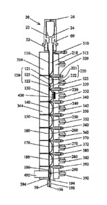
The sample processing device is shown in Fig. 1 and may include a transparent
flexible device
10 capable of being configured into a plurality of segments, such as 16, 110,
120, 130, 140, 150,
CA 03024774 2018-11-19
WO 2017/198863 PCT/EP2017/062191
17
160, 170, 180, and/or 190, and being substantially flattened by compression.
In an embodiment,
a device may have at least two segments. In an embodiment, a device may have
at least three
segments. The flexible device can provide operational functionality between
approximately 2-
105 C, compatibility with samples, targets and reagents, low gas permeability,
minimal
.. fluorescence properties, and/or resilience during repeated compression and
flexure cycles. The
device may be made of a variety of materials, examples of which include but
are not limited to:
polyolefins such as polypropylene or polyethylene, polyurethane, polyolefin co-
polymers
and/or other materials providing suitable characteristics.
In an additional embodiment, a filter can be embedded in a device segment. In
one embodiment,
a filter can be formed by stacking multiple layers of flexible filter
material. The uppermost
layer of the filter that directly contacts a sample may have a pore size
selected for filtration; the
bottom layer of the filter may include a material with much larger pore size
to provide a
support structure for the uppermost layer when a pressure is applied during
filtration. In this
preferred embodiment, the filter may be folded to form a bag, with the edges
of its open end
firmly attached to the device wall. The segment with the filter bag may be
capable of being
substantially flattened by compressing the exterior of the device.
In a specific embodiment, the sample pre-treatment compartment does not
include a filter.
Moreover, the sample inlet of the device can be adapted to receive a cell-
depletion module
including a viral infected cell marker immobilization matrix or media that
binds to viral cell
markers in the sample, allowing a depleted sample to flow through the matrix
or media and
flow into the sample inlet of the device. Such a module can be used to carry
out the cell
depletion method with manual mixing of the media/sample.
In exemplary embodiments, one or more reagents can be stored either as dry
substance and/or
as liquid solutions in device segments. In embodiments where reagents may be
stored in dry
format, liquid solutions can be stored in adjoining segments to facilitate the
reconstitution of
the reagent solution. Examples of typical reagents include: lysis reagent,
elution buffer, wash
buffer, DNase inhibitor, RNase inhibitor, proteinase inhibitor, chelating
agent, neutralizing
reagent, chaotropic salt solution, detergent, surfactant, anticoagulant,
germinant solution,
isopropanol, ethanol solution, antibody, nucleic acid probes, peptide nucleic
acid probes, and
phosphothioate nucleic acid probes. In embodiments where one of the reagents
is a chaotropic
salt solution, a preferred component is guanidinium isocyanate or guanidinium
hydrochloride
or a combination thereof In some embodiments, the order in which reagents may
be stored in
the device relative to the opening through which a sample is input, reflects
the order in which
the reagents can be used in methods utilizing the tube. In preferred
embodiments, a reagent
CA 03024774 2018-11-19
WO 2017/198863 PCT/EP2017/062191
18
includes a substance capable of specific binding to a preselected component of
a sample. For
example, a substance may specifically bind to nucleic acid, or a nucleic acid
probe may
specifically bind to nucleic acids having particular base sequences.
In a specific embodiment, in addition to the depletion surfaces discussed
above, the device can
also include a substrate such as a particle or a plurality of particles to
facilitate the selective
adsorption of nucleic acids. The particles are for example, of silica
particles, magnetic particles,
silica magnetic particles, glass particles, nitrocellulose colloid particles,
and magnetized
nitrocellulose colloid particles. In some embodiments where the particles can
be paramagnetic,
the particles can be captured by a magnetic field. Examples of reagents that
may permit the
.. selective adsorption of nucleic acid molecules to a functional group-coated
surface are
described, for example, in U.S. Pat. Nos. 5,705,628; 5,898,071; and 6,534,262.
Separation can
be accomplished by manipulating the ionic strength and polyalkylene glycol
concentration of
the solution to selectively precipitate, and reversibly adsorb, the nucleic
acids to a solid phase
surface. When these solid phase surfaces are paramagnetic microparticles, the
magnetic
particles, to which the target nucleic acid molecules have been adsorbed, can
be washed under
conditions that retain the nucleic acids but not other molecules. The nucleic
acid molecules
isolated through this process are suitable for: capillary electrophoresis,
nucleotide sequencing,
reverse transcription, cloning, transfection, transduction, microinjection of
mammalian cells,
gene therapy protocols, the in vitro synthesis of RNA probes, cDNA library
construction, and
the polymerase chain reaction (PCR) amplification. Several companies offer
magnetic-based
purification systems, such as QIAGEN's MagAttractTM, Cortex Biochem's
MagaZorbTM, and
Roche Life Science's MagNA Pure LCTM. These products use negatively charged
particles and
manipulate buffer conditions to selectively bind a variety of nucleic acids to
the particles, wash
the particles and elute the particles in aqueous buffers. Many of the products
used by these
.. companies use chaotropic salts to aid in the precipitation of nucleic acids
onto the magnetic
particles. Examples are described in U.S. Pat. Nos. 4,427,580; 4,483,920; and
5,234,809.
Preferred exemplary embodiments may include a linear arrangement of 2 or more
device
segments 110, 120, 130, 140, 150, 160, 170, 180, and/or 190 (Fig. 1). A linear
arrangement
facilitates moving the sample and resultant waste and target through the tube
in a controlled
manner. A sample, e.g., a whole blood sample, can be input through a first
opening 12 in a first
segment 110 of the device. Thereafter, waste from a processed sample can be
moved back
toward the first opening while the target is pushed towards the opposite end,
thereby
minimizing contamination of the target by reaction inhibitors that may have
become attached to
the device wall, and confining the target to a clean segment of the device
which can contain
CA 03024774 2018-11-19
WO 2017/198863 PCT/EP2017/062191
19
suitable reagents for further operations of the target. Some embodiments may
use a plurality of
segments, each containing at least one reagent. In some embodiments, these
segments may
contain reagents in the following order: the second segment can include
depletion particles
and/or a surface comprising an immobilized binding reagent and a dilution
buffer; the third
segment can be partitioned into two subsections, the first including
proteinase K and the second
including silica magnetic beads or other suitable particle; the reagent in the
fourth segment may
be either a lysis reagent; the reagent in the fifth segment may be either a
washing buffer; the
reagent in the sixth-eighth segments may be a wash buffer, a neutralization
reagent, a
suspension buffer, an elution reagent, or nucleic acid amplification and
detection reagents. In
some embodiments, the segments may be arranged continuously, while in other
embodiments,
these segments may be separated by another segment or segments in between.
In some embodiments, a method of extracting nucleic acids from biological
samples by using
the apparatus described in the previous paragraphs is contemplated. In certain
embodiments,
the sequence of events in such a test may include: 1) a biological sample
collected with a
collection tool, 2) a flexible device, which can include a plurality of
segments that may contain
the reagents required during the test, and in which the collected sample can
be placed using a
first opening in the device, 3) at least one substrate that may be set at a
controlled temperature
and/or other conditions to capture target organisms or nucleic acids during a
set incubation
period, 4) organisms or molecules, in the unprocessed sample, that may not
bind to the
substrate and could thus be removed by transferring liquid to a waste
reservoir, 5) storing waste,
in a waste reservoir, that can be segregated from the target by a clamp and/or
actuator
compressed against the device, 6) a wash buffer, released from another segment
of the device,
that can remove reaction inhibitors, 7) an elution reagent, from another
segment, that can
release the target bound to the substrate after incubation at a controlled
temperature, and 8)
nucleic acids that can be detected by techniques well known to those familiar
in the art or
collected through a second opening in the device. In exemplary embodiments the
flow of the
sample may be from the first opening towards the distal end of the device as
the test progresses
while the flow of waste may be towards the closed sample input opening of the
device, where a
waste chamber in the cap of the device receives the waste for storage.
Consequently,
undesirable contact between a processed sample and surfaces in a reaction
vessel that have
been touched by the unprocessed sample is avoided, thereby preventing reaction
inhibition due
to trace amounts of reaction inhibitors present in the unprocessed sample and
that might coat
the walls of the reaction vessel.
CA 03024774 2018-11-19
WO 2017/198863 PCT/EP2017/062191
Some embodiments may incorporate the use of a test tube 1, with a flexible
device 10 divided
into a plurality of segments, such as segments 16, 110, 120, 130, 140, 150,
160, 170, 180,
and/or 190, that may be transverse to the longitudinal axis of the device, and
which may
contain reagents, such as reagents 210, 221, 222, 230, 240, 250, 260, 270,
280, and/or 290; as
5 well as an analyzer, that may have a plurality of compression members,
such as actuators 312,
322, 332, 342, 352, 362, 372, 382, and/or 392, clamps, such as clamps 310,
320, 330, 340, 350,
360, 370, 380, and/or 390, and blocks, for example 314, 344, and/or 394
(others unnumbered
for simplicity); opposing the actuators and clamps, to process a sample.
Various combinations
of these actuators, clamps, and/or blocks may be used to effectively clamp the
device closed
10 thereby segregating fluid. In exemplary embodiments, at least one of the
actuators or blocks
may have a thermal control element to control the temperature of a device
segment for sample
processing. The sample processing apparatus can further have at least one
magnetic field source
430 capable of applying a magnetic field to a segment. The sample processing
apparatus can
further have a detection device 492, such as photometer or a CCD, to monitor a
reaction taking
15 place or completed within the device.
Fluid can be driven through a flow-channel by compressing the device with a
centrally-
positioned actuator, and its flanking clamps if any, to form a flow channel
with a gap of about 1
to about 500 um, preferably about 5 to about 500 um through each segment. The
adjacent
actuators gently compress the adjacent segments in liquid communication with
the flow-
20 channel to generate an offset inner pressure to ensure a substantially
uniform gap of the flow
channel. The two flanking actuators can then alternatively compress and
release pressure on the
device on their respective segments to generate flow at a controlled flow
rate. Optional flow,
pressure, and/or force sensors may be incorporated to enable closed-loop
control of the flow
behavior. The flow-channel process can be used in washing, enhancing the
substrate binding
efficiency, and detection.
A particle immobilization and re-suspension process can be used to separate
the particles from
the sample liquid. The magnetic field generated by a magnetic source 430 (FIG.
1) may be
applied to a segment containing a magnetic particle suspension to capture and
immobilize the
particles to the tube wall. An agitation process can be used during the
capturing process. In
another embodiment, a flow-channel can be formed in the segment with the
applied magnetic
field, and magnetic particles can be captured in the flow to increase the
capturing efficiency. To
resuspend immobilized particles, the magnetic field may be turned off or
removed, and an
agitation or flow-channel process can be used for re-suspension.
CA 03024774 2018-11-19
WO 2017/198863 PCT/EP2017/062191
21
In certain embodiments, nucleic acids extracted from the biological samples
may be further
processed by amplifying the nucleic acids using at least one method from the
group:
polymerase chain reaction (PCR), rolling circle amplification (RCA), ligase
chain reaction
(LCR), transcription mediated amplification (TMA), nucleic acid sequence based
amplification
(NASBA), and strand displacement amplification reaction (SDAR). In some
embodiments, the
nucleic acids extracted from the organism can be ribonucleic acids (RNA) and
their processing
may include a coupled reverse transcription and polymerase chain reaction (RT-
PCR) using
combinations of enzymes such as Tth polymerase and Taq polymerase or reverse
transcriptase
and Taq polymerase. In some embodiments, nicked circular nucleic acid probes
can be
circularized using T4 DNA ligase or AmpligaseTM and guide nucleic acids, such
as DNA or
RNA targets, followed by detecting the formation of the closed circularized
probes after an in
vitro selection process. Such detection can be through PCR, TMA, RCA, LCR,
NASBA or
SDAR using enzymes known to those familiar with the art. In exemplary
embodiments, the
amplification of the nucleic acids can be detected in real time by using
fluorescent-labeled
nucleic acid probes or DNA intercalating dyes as well as a photometer or
charge-coupled
device in the molecular analyzer to detect the increase in fluorescence during
the nucleic acid
amplification. These fluorescently-labeled probes use detection schemes well
known to those
familiar in the art (i.e., TaqManTm, molecular beaconsTM, fluorescence
resonance energy
transfer (FRET) probes, scorpionTM probes) and generally use fluorescence
quenching as well
as the release of quenching or fluorescence energy transfer from one reporter
to another to
detect the synthesis or presence of specific nucleic acids.
A real-time detection of a signal from a device segment can be achieved by
using a sensor 492
(FIG. 1), such as a photometer, a spectrometer, a CCD, connected to a block,
such as block 490.
In exemplary embodiments, pressure can be applied by an actuator 392 on the
device segment
190 to suitably define the device segment's shape. The format of signal can be
an intensity of a
light at certain wavelength, such as a fluorescent light, a spectrum, and/or
an image, such as
image of cells or manmade elements such as quantum dots. For fluorescence
detection, an
excitation of light from the optical system can be used to illuminate a
reaction, and emission
light can be detected by the photometer. To detect a plurality of signals
having specific
wavelengths, different wavelength signals can be detected in series or
parallel by dedicated
detection channels or a spectrometer.
Kits
In some embodiments, reagents, materials, and devices for carrying out the
presently disclosed
methods are included in a kit. In some embodiments, the kit includes
components for obtaining,
CA 03024774 2018-11-19
WO 2017/198863 PCT/EP2017/062191
22
storing, and/ or preparing sample. Such components include, e.g., sterile
needles and syringes,
depletion modules, EDTA-lined tubes, buffers (e.g., for binding nucleic acid
to, and elution
from a matrix), RNase inhibitors, and/ or DNase, etc.
In addition, the kit includes an assay processing device such as that
described above and
optionally, one or more components for obtaining, storing, and/or preparing
sample, including
but not limited to a depletion module as described hereinabove. In a specific
embodiment, the
assay processing device includes various reagents required to perform the
methods disclosed
herein stored within one or more segments of the device.
The kit can further include controls, e.g., a polynucleotide that is wild type
at the sequence to
be detected, or a polynucleotide that includes the sequence to be detected.
The kit can also include additional devices such as sample tubes or vials;
reaction containers
(e.g., tubes, multiwell plates, microfluidic chips or chambers, etc), as well
as directions for use
or reference to a website.
EXAMPLES
Example 1. Viral RNA Isolation and Detection from Whole Blood
RNA isolation and RNA sequence detection can be accomplished in a tube 1 (FIG.
1),
including a flexible device having nine segments separated by peelable seals
and containing
pre-packed reagents, and a cap, having a waste reservoir housed therein. Fluid
flow from one
segment or subsection of the device to another is controlled as described
herein by selective
.. engagement of one or more actuators and clamps operably connected within
one or more
segments or subsections of the device. The first segment of the device can
receive the whole
blood sample. The second segment contains 560 ug of Dynal ParticlesTM (Dynal
Biotech)
conjugated to anti-CD4 antibodies suspended in dilution buffer having 100 ul
of phosphate
buffered saline (PBS) (137 mM NaCl, 2.7 mM KC1, 4.3 mM Na2HPO4, 1.4 mM KH2PO4,
pH
7.3). The 3rd segment contains the quantification standard. The 4th segment is
partitioned into
two subsections, the 1st subsection containing 200 ug proteinase K, and the
2'd subsection
inc1uding250 ug Silica Magnetic particles, wherein the subsections are
separated by a peelable
seal. The 5th segment contains 200 ul of lysis buffer comprising chaotropic
salts which contain
4.7 M guanidinium hydrochloride, 10 mM urea, 10 mM Tris HC1, pH 5.7, and 2%
triton X-100..
The 6th segment contains 80 ul of wash buffer (including, e.g., 50% ethanol,
20 mM NaCl, 10
mM Tris HC1, pH 7.5). The 7th segment contains 80 ul of 20 mM 2-
morpholinoethanesulfonic
acid (MES) buffer, pH 5.3. The pH of the MES buffer is adjusted such that it
can be low
enough to avoid DNA elution from the particles. The 8th segment contains 40 ul
elution buffer
(e.g., 10 mM Tris HC1, pH 8.5, or any buffer suitable for PCR). The pH of the
elution buffer is
CA 03024774 2018-11-19
WO 2017/198863 PCT/EP2017/062191
23
adjusted such that it can be high enough to elute the DNA from the surface of
the particles into
the buffer. The 9th segment contains PCR reagents (which can contain 10 nmol
of each one of:
dATP, dCTP, and dGTP; 20 nmol dUTP, 2.5 mmol of KC1, 200 nmol of MgCl2, 1-5
units of
Taq DNA polymerase, 1-5 units of Tth DNA polymerase, 20-100 pmol of each of
the
oligonucleotide primers, and 6-25 pmol of TaqMan probe). The 10th segment may
contain a
divalent metal cofactor, such as MgCl2. The end of the 9th (or 10th) segment,
can be
permanently sealed or contain a pressure gate for collecting the products of
the amplification
reaction to confirm the results of a genotyping test by DNA sequencing or some
other test
known to those skilled in the art.
For viral RNA isolation and detection, 100 ul of whole blood is loaded into
the 1st segment.
The device can then be closed by a cap and inserted into an analyzer. Sample
processing can
include the following steps.
(a) Cell depletion and Sample Lysis. All clamps, except the first clamp, are
closed on the
device. The actuator operably connected with the 1st segment is used to adjust
the volume
of blood to retain about 100 ul in the segment, and then the 1st segment is
closed using the
associated segment clamp. The actuator operably connected with the 2'd
segment, in
whole or in part, compresses the first subsection of the 2'd segment to break
the peelable
seals between the 1st, 2ed, and 3'd segments and mix the anti-CD4 particles
with the
sample and quantification standard. Actuators operably connected with the
first
subsection can alternately compress the subsection to mix the particles with
the sample.
Then, a magnetic field can be generated by a magnetic source near the 3'd
segment to
capture the particles. By engaging the associated actuators and clamps in that
segment/subsection, the cell depleted sample is moved to the 4th segment, a
clamp is
closed above the 4th segment to prevent the depleted cells from mixing with
the
downstream depleted specimen, and then moved to the 5th segment to mix the
lysis buffer
with the cell depleted sample. The mixture in the 4th and 5th segments is
incubated at
50 C for 2 minutes.
(b) Nucleic Acid Capture. After lysis incubation, the actuators alternately
compress their
respective segments to agitate and incubate the mixture for 2 minutes at room
temperature to facilitate DNA binding to the particles. Then, a magnetic field
is generated
by a magnetic source near the segment to capture the particles in suspension.
The
actuators can alternately compress the segment to capture the particles. The
actuators and
clamps are sequentially opened and closed to move the unbound sample and waste
to the
waste reservoir.
CA 03024774 2018-11-19
WO 2017/198863 PCT/EP2017/062191
24
(c) Wash. A wash process follows the capture process in order to remove
residual debris and
reaction inhibitors from the particles and the segments that would be used for
further
sample processing. A dilution-based washing is used with the ethanol wash
buffer and a
flow-based washing is used with the MES wash buffer. Clamps and actuator first
open,
and then the actuator closes to move the ethanol buffer to the 6th segment,
followed by
the closing of the clamp. The magnetic field is removed; the actuator and at
least one
adjacent actuator is alternately compressed against their respective segments
to generate
flow to re-suspend the particles. The magnetic field is then turned on to
capture
substantially all the particles and the liquid is moved to a waste reservoir.
After
completing the first wash, the MES wash buffer is moved from one segment to
another
and the buffer is manipulated using the sequential application and release of
the
corresponding actuators and clamps to ensure an essentially laminar flow of
the wash
buffer through the flow channel. When the wash is completed, the actuators and
clamps
are closed and substantially all the waste is moved to the waste reservoir.
(d) Nucleic Acid Elution. The elution buffer is moved from the 8th segment
using a similar
process as mentioned before. The magnetic field can be removed and the
particles are re-
suspended in the elution buffer under flow between the fourth and fifth
segments. The
particle suspension is incubated at 95 C under stationary flow or agitation
conditions for
2 minutes. The magnetic field is turned on and substantially all the particles
are
immobilized, and the eluted nucleic acid solution can be moved to the 8th
segment by
sequentially opening and closing the actuators and clamps. The actuators can
compress
the 8th segment to adjust the volume of the eluted nucleic acid solution to 40
ul and a
clamp can then close against the device to complete the DNA extraction
process.
(e) Nucleic Acid Amplification and Detection. The nucleic acid solution can
then be
transferred to the 9th segment, mixed, and incubated with UNG 280 at 37 C for
1 minute
to degrade any contaminant PCR products that may have been present in the
biological
sample. After the incubation, the temperature may be increased to 95 C to
denature
nucleic acids and UNG for 2 minutes. The nucleic acid solution can then be
transferred to
the 10th segment, and mixed with RT-PCR reagents at 65 C for 10 minutes,
followed by
incubation at 60 C to initiate hot start PCR. A typical 2-temperature,
amplification assay
of 50 cycles of 95 C for 2 seconds and 60 C for 15 seconds can be conducted
by setting
the 8th segment at 95 C and the 9th segment at 60 C, and transferring the
reaction
mixture between the segments alternately by closing and opening the associated
actuators.
A typical 3-temperature, amplification assay of 50 cycles of 95 C for 2
seconds, 60 C
CA 03024774 2018-11-19
WO 2017/198863 PCT/EP2017/062191
for 10 seconds, and 72 C for 10 seconds can be conducted by setting the 8th
segment at
95 C, 9th segment at 72 C and the 10th segment at 60 C, and alternately
transferring the
reaction mixture among the segments by closing and opening the associated
actuators. A
detection sensor, such as a photometer, optically connected to the 9th chamber
can
5 monitor real-time fluorescence emission from the reporter dye through a
portion of the
device wall. After an assay is complete, the test results are reported.
Example 2. Offline CD4 Depletion and Analysis
Plasma dynamic range was determined using a Roche cobas0 6800/8800 HIV-
quantification
calibration panel. The panel components are provided in Table 1 below.
10 Table 1. Roche HIV-1 quantitation calibration panel
Material Material N Concentration Target Matrix
TR HIV 8E5 CAL PANEL PM1 5219612991 1.00E+07 Cp/mL HIV LAV 8E5 EDTA Plasma
TR HIV 8E5 CAL PANEL PM2 5219639991 1.00E+06 Cp/mL HIV LAV 8E5 EDTA Plasma
TR HIV 8E5 CAL PANEL PM3 5219647991 1.00E+05 Cp/mL HIV LAV 8E5 EDTA Plasma
TR HIV 8E5 CAL PANEL PM4 5219655991 1.00E+04 Cp/mL HIV LAV 8E5 EDTA Plasma
TR HIV 8E5 CAL PANEL PM5 5219663991 1.00E+03 Cp/mL HIV LAV 8E5 EDTA Plasma
TR HIV 8E5 CAL PANEL PM6 5219671991 1.00E+02 Cp/mL HIV LAV 8E5 EDTA Plasma
TR HIV 8E5 CAL PANEL PM7 6315801991 5.00E+01 Cp/mL HIV LAV 8E5 EDTA Plasma
Three replicates were tested on a cobas0 LIATO Analyzer for each Roche panel
member. To
run the tests on the cobas0 LIATO Analyzer, 100 uL of each panel member
material was
mixed with 100 uL dilution buffer (lx PBS, 0.1% BSA, 0.1% Sodium Azide), and
then the
15 diluted material was added to a cobas0 LIATO device pre-packaged with
assay reagents as
described below. The device was analyzed in a cobas0 LIATO Analyzer and upon
completion
of the assay, Ct and target input levels were used for dynamic range
regression analysis. The
results are shown in Figs. 2A-2B, which show 6 log linearity in plasma and no
inhibition of
HIV on the quantification standard performance of the assay.
20 For whole blood dynamic range, the Roche HIV-1 quantitation calibration
panel members (see
Table 2) were spiked with pooled normal human EDTA whole blood at 1:20 ratio.
For example,
to make 1 mL whole blood sample with HIV-1 spiked-in, 50 uL of a panel member
was
combined with 950 uL of normal human whole blood (Bioreclamation IVT, Cat #:
HMWBEDTA2), mixed by pipetting. Four test samples were prepared for whole
blood
25 dynamic range as shown in Table 2 below.
CA 03024774 2018-11-19
WO 2017/198863 PCT/EP2017/062191
26
Table 2. Whole blood dynamic range test samples
Sample Concentration Target Matrix
Sample 1 5.00E+05 Cp/mL HIV LAV 8E5 EDTA whole blood
Sample 2 5.00E+04 Cp/mL HIV LAV 8E5 EDTA whole blood
Sample 3 5.00E+03 Cp/mL HIV LAV 8E5 EDTA whole blood
Sample 4 5.00E+02 Cp/mL HIV LAV 8E5 EDTA whole blood
Three replicates were tested on the cobas0 LIATO Analyzer for each sample. To
run the tests
on the cobas0 LIATO Analyzer, 100 uL of a whole blood sample was mixed with
100 iut
dilution buffer, and then loaded into a cobas0 LIATO device prepackaged with
other assay
reagents packed. The device was analyzed in a cobas0 LIATO Analyzer and upon
completion
of the assay, Ct and target input levels were used for dynamic range
regression analysis. The
results are shown in Figs. 3A-3B, which show at least 4 log linearity in whole
blood and no
inhibition of HIV on the quantification standard performance of the assay. In
addition, the
limits of detection (LOD) of the assay in plasma vs. whole blood are shown in
Fig. 4.
For each patient blood sample, viral loads in plasma and whole blood were
evaluated, as well
as the viral load in whole blood after CD4 antibody cell depletion was
determined.
To separate plasma from whole blood, 10 mL of whole blood in the Vacutainer
Lavender blood
collection tube was centrifuged at 1200g for 10 min on Sorvall centrifuge at
room temperature.
The supernatant (plasma) was transferred to a labeled 50mL centrifuge tube. In
an eppendorf
tube, 80 uL dilution buffer was added followed by the addition of 100 uL of
whole blood
sample. Before adding blood sample to the Eppendorf tube, the whole blood
sample was mixed
by inverting the sample tube until blood becomes homogeneous, followed by
pipetting 3-5
times. Blood and dilution buffer were mixed by pipetting 6 times.
CD4 antibody particles were vortexed 15 seconds to resuspend the particles and
then twenty
(20) uL of CD4 antibody Dynabeads were added to the Eppendorf tube (100 to 500
ug, depends
on type of CD4 antibody particles). The mixture was vortexed 2 times (1
second/time) to mix
the sample with particles. The tube is loaded on the Rotator and incubated at
room temperature
with rotation for 10 minutes (Rotator was set at 60 tilt, and 10 rpm). The
tube was placed in a
magnetic rack for 1 minute and then all liquid (-200 uL) was transferred to a
cobas0 LiatO
device. RNA isolation and sequence detection in the depleted sample was
accomplished in a
tube as described in Example 1, except that the tube did not include particles
in the second
segment. Instead, the second segment included quantification standard.
CA 03024774 2018-11-19
WO 2017/198863 PCT/EP2017/062191
27
For each patient sample, 100 uL of plasma mixed with 100 uL of dilution
buffer, 100 uL of
EDTA whole blood mixed with 100 uL of dilution buffer, and 100 uL of EDTA
whole blood
with CD4 antibody cell depletion were loaded on to cobas0 LIATO device and
tested on a
cobas0 LIATO analyzer. Data were collected after test runs finished. Viral
load of each sample
type was calculated using equations generated in dynamic range regression for
plasma or whole
blood. Viral load correlation between plasma and whole blood, or plasma and
whole blood with
CD4 cell depletion were evaluated.
Following the protocol described above, one round of binding with CD4 magnetic
particles was
able to deplete 96% of CD4 positive cells from unfrozen whole blood samples.
The results are
shown in Table 3 and Figs. 5 and 6A-6B. Fig. 5 shows that CD4 depletion
treatment improved
viral load correlation in samples 3 and 5. Fig. 6A shows the viral load
correlation before CD4
depletion and Fig. 6B shows the viral load correlation after treatment.
Table 3. Viral load measurement values of 5 patient blood samples
Sample ID VL SD
Plasma WB WB/Particles Plasma WB WB/Particles
1 4.32 4.50 4.03 0.22 0.04 0.21
2 4.57 4.07 3.91 0.13 0.04 0.09
3 2.67 4.11 2.90 0.20 0.08 0.25
4 5.19 5.67 5.49 0.04 0.17 0.03
5 2.68 3.71 2.92 0.09 0.09 0.17
Out of the five patient samples tested, plasma viral load measurements of two
samples were
less than 1e3 copies/ml and direct whole blood viral load measurement of the
two patient
samples were higher than 1e3 copies/ml. Pre-treatment of the two patient whole
blood samples
with CD4 antibody particles improved whole blood and plasma viral load
correlation.
Following pre-treatment with anti-CD4 particles, the whole blood viral load of
the same two
samples previously studied were less than 1e3 copies/ml.
Example 3. Optimized Conditions for CD4 Depletion in a cobas0 LIATO Device
Various fluid flow driving mechanisms in a tube were evaluated based on the
desired degree of
cell lysis. As described above, in general, fluid flow is controlled in the
device shown in FIG. 1
by compressing selected segments of the tube with a centrally-positioned
actuator and its
flanking clamps to form a flow channel in the device with a gap (i.e., the
diameter of the flow
channel) of about 1 to about 500 um (0.001 - .5 mm), e.g., about 5 to 500 um
(0.005 - .5 mm).
CA 03024774 2018-11-19
WO 2017/198863 PCT/EP2017/062191
28
The adjacent actuators gently compress the adjacent segments in liquid
communication with the
flow channel to generate an offset inner pressure to ensure a substantially
uniform gap of the
flow channel. It was found that the degree of cell lysis can be modified by
varying the gap over
a shorter span of the pre-treatment compartment relative to that used to
achieve complete cell
lysis. Therefore, using this method, cell depletion without cell lysis could
be achieved without
the use of a filter positioned in the sample pretreatment compartment.
Three different fluid flow mechanisms were designed for use in the sample pre-
treatment
compartment (i.e., the portion of the device positioned between the sample
introduction port
and the segment used for nucleic acid capture):
a) conventional lysis fluid flow: low shear mixing with cell lysis;
b) harsh lysis fluid flow: high shear mixing with cell lysis; and
c) cell depletion fluid flow: low shear mixing without cell lysis.
The sample pre-treatment compartment includes a plurality of segments, e.g.,
up to 5 segments,
and for conventional lysis and harsh lysis, each of the segments of the
compartment were used
such that fluid mixing was performed over the full length of the fluid flow
channel in the
compartment. In contrast, in order to achieve cell depletion and the
subsequent nucleic acid
extraction of the cell-depleted sample, a subset of the segments in the
compartment can be used,
e.g., as few as 3 of the 5 segments or up to all of the segments of the
compartment can be used.
It was found that the fluid flow mechanism could be performed in fewer
segments and the
diameter of the flow channel in and between segments during the mixing/lysis
process was
modified relative to conventional and harsh lysis. The optimized conditions
for conventional
lysis, harsh lysis, and cell depletion are shown below:
Table 4. Relative Actuator Position, Lysis Type & Flow Channel Gap
Actuator Conventional Lysis Harsh Lysis (Generating Cell Depletion
(General Mixing) Shear Forces) without Lysis
PO Variable gap, 0-3 mm Variable gap, 0-3 mm Variable gap,
0.4-3 mm
P1 Variable gap, 0-3 mm Variable gap, 0-3 mm Narrow, fixed gap,
0-1 mm
P2 Variable gap, 0-3 mm Narrow, fixed gap, Variable gap,
0-.15 mm 0.4-3 mm
P3 Variable gap, 0-3 mm Variable gap, 0-3 mm Closed
P4 Variable gap, 0-3 mm Variable gap, 0-3 mm Closed
CA 03024774 2018-11-19
WO 2017/198863 PCT/EP2017/062191
29
In order to achieve conventional lysis (shown in column 2 of the table), the
actuators and
clamps were selectively engaged/disengaged to move liquid in the flow channel
throughout the
pre-treatment compartment. In order to achieve harsh lysis, the innermost
segment of the flow
channel (P2) was held to a narrower, fixed gap relative to the gaps flanking
the inner segment,
thereby subjecting the fluid in that inner segment of the flow path to shear
forces that generated
relatively harsh lysis conditions. The gap of the innermost segment was
substantially reduced
and held fixed relative to that in the flanking segments, i.e., 0.15 mm in P2
versus up to 3 mm
in PO-P1 and P3-P4. By contrast, in order to achieve cell depletion without
cell lysis, the entire
mixing step could be performed over a shorter fluid flow channel including
fewer segments of
.. the device (e.g., PO-P2), and the innermost segment was compressed to a gap
of up to 1.1 mm,
which is about one-third the size of the gap in the flanking segments, thereby
achieving a gentle
mixing that does not result in cell lysis. In one embodiment, the gap can be
reduced by 25-50%
of the diameter of the channel in the flanking segments, e.g., 33% of the
diameter.
The process is illustrated in Figs. 7A-7D. A portion of the sample processing
device is
illustrated in Fig. 7A, in which the tube 701 includes a sample pre-treatment
compartment 702
comprising a plurality of segments 703-707 in fluid communication with the
remainder of the
segments in the sample processing device 708. A fluid flow channel 709 spans
the pre-
treatment compartment into the remainder of the segments of the sample
processing device.
When the actuators (PO-P4) and clamps (CO-05) in communication with the pre-
treatment
compartment are not engaged with the tube, the fluid flow channel has a
diameter of about 5
mm, but when the tube is positioned in the device, the motors (not shown)
controlling the
position of the actuators and clamps drive the actuators and clamps against
the outer walls of
the tube, slightly compressing the tube such that the fluid flow channel has a
diameter of about
3 mm, which is designated as the fully open position of the compartment. Fig.
7B shows the
relative dimensions of the fluid flow channel during conventional lysis; the
fluid flow channel
spans P0-P5 and the channel has a diameter of up to 3 mm. Fig. 7C shows the
relative
dimensions of the fluid flow channel during harsh lysis; the fluid flow
channel spans P0-P5,
with the innermost segment, P2, reduced to a diameter of up to 0.15 mm.
Therefore, during
harsh lysis, the fluid flow is subjected to high pressure in P2 relative to
the flanking segments,
.. introducing high shear forces that lead to harsh cell lysis. In contrast,
Fig. 7D shows the relative
dimensions of the fluid flow channel during cell depletion; the fluid flow
channel spans PO-P2,
with the diameter of the channel in the innermost segment, Pl, being reduced
from up to 3 mm
(in PO and P2) to up to 1.1 mm. The innermost channel is not compressed to the
same degree as
CA 03024774 2018-11-19
WO 2017/198863 PCT/EP2017/062191
it is during harsh lysis, resulting in a more gentle mixing cycle that does
not lyze the cells in the
fluid.
In addition to the altered fluid flow mechanism, the speed of the motors
operatively connected
with each of the actuators and clamps was adjusted to avoid cell lysis during
cell depletion.
5 .. During conventional and harsh lysis, each of the motors engaged with the
actuators and clamps
rapidly engage and disengage the actuators and clamps at an equal rate.
However, in order to
achieve cell depletion without cell lysis, the motor speed during
disengagement was adjusted so
that the motor logarithmically decelerates relative to the rate of
acceleration. This is illustrated
in Figs. 8A-8B, wherein Fig. 8A shows the movement of the motor during
conventional and
10 harsh lysis and Fig. 8B show the movement of the motor during cell
depletion.
The methods described herein and in Example 1 were used to evaluate the viral
load of patient
samples in a sample processing device and the results are shown below:
Table 5. Viral Load Testing in a Device Configured to Perform Cell Depletion
Sample ID Sample type VL
Ave Log VL Ave VL (cp/mL)
1 Blood 3.14 1403
1 Blood/AB beads 2.36 244
2 Blood 3.24 1738
2 Blood/AB beads 2.43 368
3 Blood 3.59 4102
3 Blood/AB beads 2.40 349
The results shown in Table 5 are similar to those achieved using offline
processing, described
in Example 2.