Note: Descriptions are shown in the official language in which they were submitted.
1
STAND ALONE INTERBODY SPINAL SYSTEM
Priority Claim
[00011 This application claims priority to: (a) United States Provisional
Patent Application
No. 62/445,428, filed on January 12, 2017 and entitled "Stand Alone Interbody
Spinal
System", and (b) United States Provisional Patent Application No. 62/341,123,
filed on May
25, 2016 and entitled "Stand Alone Interbody Spinal System".
Technical Field
[0002] Embodiments of the invention are in the field of orthopedic implants.
Background
[0003] Fixation devices can be used to provide, for example, immobilization
and
stabilization of spinal segments in patients (e.g., humans, dogs, cats, and
other animals).
Fixation devices may be used to help fuse bone segments (e.g., vertebrae) in
the treatment of
instabilities or deformities of, for example, the cervical, thoracic, lumbar,
and/or sacral spine.
Such instabilities or deformities may include, for example, degenerative disc
disease (DDD);
.. spondylolisthesis; trauma (i.e., fracture or dislocation); spinal stenosis;
curvatures (i.e.,
scoliosis, kyphosis, and/or lordosis); tumor; pseudoarthrosis; and failed
previous fusions.
[0004] One such fixation device may include an interbody spacer implanted
using
techniques such as Anterior Lumbar Interbody Fusion (ALIF), Posterior Lumbar
Interbody
Fusion (PLIF), or Transforaminal Lumbar Interbody Fusion (TLIF) surgical
techniques. The
spacers used in these techniques are placed in the interdiscal space between
adjacent
vertebrae of the spine. Many times an exterior plate is used in conjunction
with the spacer to
hold the adjacent vertebrae while the fusion occurs.
[0005] Ideally, the spacer should stabilize the intervertebral space and allow
fusion of the
adjacent vertebrae. Moreover, during the time it takes for fusion to occur,
the interbody
spacer should have sufficient structural integrity to withstand the stress of
maintaining the
space without substantially degrading or deforming and have sufficient
stability to remain
securely in place prior to actual bone ingrowth fusion.
Date Regue/Date Received 2022-08-16
CA 03024894 2018-11-19
WO 2017/205623 PCT/US2017/034471
2
[0006] The degree or success of union, loads produced by weight bearing, and
activity
levels will, among other conditions, dictate the longevity of the implant.
Robust fixation
systems are needed to lessen risks associated with fixation and to promote
better outcomes
for patients.
Brief Description Of The Drawings
[0007] Features and advantages of embodiments of the present invention will
become
apparent from the appended claims, the following detailed description of one
or more
example embodiments, and the corresponding figures, in which:
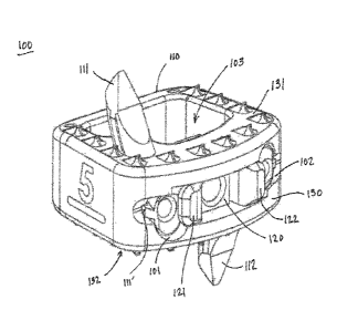
[0008] Figure 1 includes a perspective view of an embodiment of a standalone
interbody
cage and anchors.
[0009] Figure 2 includes a side view of an embodiment of a standalone
interbody cage and
anchors.
[0010] Figure 3 includes a top view of an embodiment of a standalone interbody
cage and
anchors.
[0011] Figure 4 includes a cross-sectional view of an embodiment of a
standalone
interbody cage and anchors.
[0012] Figure 5 includes a perspective view of an embodiment of a standalone
interbody
cage and anchors.
[0013] Figure 6 includes a perspective view of an embodiment of a standalone
interbody
cage.
[0014] Figure 7 includes a perspective view of an embodiment of a standalone
interbody
cage, anchors, and insertion tool.
[0015] Figures 8, 9, 10 include cross-sectional views of an embodiment of a
standalone
interbody cage, anchors, and insertion tool at various points of anchor
deployment within a
channel of the cage.
[0016] Figures 11, 12, 13 include cross-sectional views of an embodiment of a
standalone
interbody cage, anchors, and insertion tool at various points of anchor
deployment within a
channel of the cage.
[0017] Figures 14, 15, 16, 17, 18 include perspective views of an embodiment
of a
standalone interbody cage and withdrawal tool at various points of anchor
withdrawal from a
channel of the cage.
CA 03024894 2018-11-19
W02017/205623 PCT/US2017/034471
3
[0018] Figures 19, 20, and 21 include cross-sectional views of an embodiment
of a
standalone interbody cage and anchors.
[0019] Figures 22, 23, 24 include perspective views of an embodiment of a
standalone
interbody cage and anchors.
[0020] Figure 25 includes a perspective view of an embodiment of a standalone
interbody
cage and anchors.
[0021] Figure 26 includes a perspective view of an embodiment of a standalone
interbody
cage and anchors.
[0022] Figure 27 includes an insertion tool in an embodiment.
[0023] Figures 28, 29, 30 include various views of an embodiment of a
standalone
interbody cage and anchors.
Detailed Description
[0024] In the following description, numerous specific details are set forth.
However, it is
understood that embodiments of the invention may be practiced without these
specific details.
Well-known structures and techniques have not been shown in detail to avoid
obscuring an
understanding of this description. References to "one embodiment", "an
embodiment",
"example embodiment", "various embodiments" and the like indicate the
embodiment(s) so
described may include particular features, structures, or characteristics, but
not every
embodiment necessarily includes the particular features, structures, or
characteristics.
Further, some embodiments may have some, all, or none of the features
described for other
embodiments. Also, as used herein "first", "second", "third" and the like
describe a common
object and indicate that different instances of like objects are being
referred to. Such
adjectives are not intended to imply the objects so described must be in a
given sequence,
either temporally, spatially, in ranking, or in any other manner. Also, the
terms "coupled"
and "connected," along with their derivatives, may be used. In particular
embodiments,
"connected" may be used to indicate that two or more elements are in direct
physical contact
with each other and "coupled" may mean that two or more elements co-operate or
interact
with each other, but they may or may not be in direct physical contact.
[0025] Figures 1 to 6 and 11 are now discussed. Those figures depict an
orthopedic fusion
system 100 comprising: a cage 110; a curved first channel 101 coupling a
lateral wall 130 of
the cage to a superior surface 131 of the cage; a curved second channel 102
coupling the
CA 03024894 2018-11-19
W02017/205623 PCT/US2017/034471
4
lateral wall of the cage to an inferior surface 132 of the cage; a third
channel 103 coupling the
superior surface of the cage to the inferior surface of the cage; a curved
first anchor 111
configured to slide within the first channel; a curved second anchor 112
configured to slide
within the second channel; and a resilient member 120 comprising a resilient
first arm 121
that projects across a portion of the first channel and a resilient second arm
122 that projects
across a portion of the second channel. Figure 11 also shows first arm 121
projecting across
a portion of the first channel 101 and second arm 122 projecting across a
portion of the
second channel 102.
[0026] In an embodiment the first and second arms 121, 122 are monolithic with
one
another. For example, see Figure 4 showing member 120 as being a monolithic
structure
comprising a base that couples arms 121, 122 to each other. Structure 120 may
couple to
another retention member 123 (Figure 5) that is not necessarily monolithic
with the arms.
Thus, arms 121, 122 prevent structure 120 from advancing too far into channel
103 and
member 123 keeps member 120 from backing out of cage 110.
[0027] Figures 8 to 13 show various views and stages of implantation of
anchors. Figure
12 shows where in a first orientation the first anchor 111 directly deflects
the first arm 121
away from the first channel (first channel 101 is more easily seen in Figure
11) to allow the
first anchor 111 to pass within the first channel. In the first orientation
(Figure 12) the second
anchor 112 directly deflects the second arm 122 away from the second channel
(second
channel 102 is more easily seen in Figure 11) to allow the second anchor to
pass within the
second channel. Thus, in an embodiment the first and second anchors are
configured to
deploy into the first and second channels "simultaneously" (as define below)
with one
another.
[0028] Figure 7 depicts an insertion tool 140, the insertion tool comprising:
a first insertion
tool arm 141 configured to travel along a first arcuate path (see path taken
in Figures 8, 9, 10)
to drive the first anchor 111 along the first channel 101; a second insertion
tool arm 142
configured to travel along a second arcuate path (see path taken in Figures 8,
9, 10) to drive
the second anchor 112 along the second channel 102. Accordingly, the first and
second
insertion tool arms are configured to respectively travel along the first and
second arcuate
paths simultaneously with one another. As a result, a patient benefits because
simultaneous
anchor insertion saves procedure time. Figure 27 shows how an arcuate path
(including
CA 03024894 2018-11-19
W02017/205623
PCT/US2017/034471
undulating portions) may be due in part to a serpentine channel 149 on an arm,
such as arm
141. Not all paths for insertion arms must be arcuate (as used herein arcuate
means
"curved") and may be, for example, linear in other embodiments.
[0029] As shown in Figures 1, 2, 4, after anchor insertion the system may
enter into a
second orientation. In a second orientation: (a) a portion of the first arm
121 is lateral to a
proximal end of the first anchor 111 and prevents the first anchor from
backing out of the
first channel 101, and (b) the first anchor does not deflect the first arm
away from the first
channel.
[0030] Figure 10 shows (indirectly) how in a vertical plane 150 the first
anchor 111 is
completely surrounded by an interior wall of the first channel 101. Actually,
Figure 10
shows how anchor 111 is surrounded on two sides (top and bottom) by the
channel wall.
Figure 4 helps show how anchor 111 is surrounded on two additional sides (left
and right
sides) by the channel wall (with all four sides shown in Figures 10 and 4
amounting to
"surrounding" as used herein) . In an embodiment a horizontal axis 151 (Figure
11)
intersects the lateral wall and the first and second channels. In an
embodiment a horizontal
axis 151 (Figure 11) intersects the lateral wall and the first and second
channels and the
resilient member 120.
[0031] In Figure 1 the first anchor 111 includes a projection 111' configured
to abut a wall
of the first channel 101 to prevent a proximal portion (wherein the "proximal
end" of anchor
111 is the opposite end of anchor from the pointed distal tip projecting
superiorly in Figure 1)
of the first anchor 111 from passing through the first channel.
[0032] Figures 14 to 18 address an embodiment for anchor extraction or
withdrawal.
Figure 14 to 18 show a withdrawal tool 140', the withdrawal tool comprising a
first
withdrawal tool arm 141' configured to travel along an additional first
arcuate path 141" to
withdraw the first anchor 111 from the first channel 101. Figures 16 and 17
show a threaded
rod 145 that threads into the first anchor 111 to couple the anchor to the
withdrawal tool arm
141'. In an embodiment anchor 111 is threaded such that a tool (threaded rod
145 of Figure
16) with, for example, male threads can be threadably coupled to the anchor.
The arm 121 is
tapered so it is pushed away by rod 145 in a medial direction. In doing so the
resilient
member 121 no longer retains the anchor in the cage body and the anchor may be
CA 03024894 2018-11-19
WO 2017/205623 PCT/US2017/034471
6
removed. Thus, Figures 14-18 depict a system for reversing deployment of an
anchor in an
embodiment of a standalone interbody system. In an embodiment arm 121 is
tapered so it is
pushed away by articulating arm 141' in a medial direction. Regarding
deflection of resilient
arms, at times the arms have beveled surfaces and/or the anchors and/or
insertion/withdrawal
tools have beveled surfaces to deflect resilient arms away from the channel to
allow anchor
implantation or extraction.
[0033] In an embodiment resilient member 120 includes a threaded orifice to
couple to a
reciprocally threaded insertion tool 143 (Figure 11). Tool 143 may be rotated
via knob 144
(Figure 7) to couple insertion tool 140 to cage 110. In an embodiment the
resilient member
120 comprises a first material, such as Titanium or Nickle Titanium, and the
cage (e.g., wall
130) comprises a second material (e.g., Polyether ether ketone (PEEK)) that is
softer than the
first material. This can be critical for instances such as, for example, when
a physician is
manipulating cage 110. If the manipulation is particularly forceful the
threads being formed
in Titanium (instead of something relatively softer such as PEEK) helps resist
thread
stripping.
[0034] In an embodiment (e.g., Figure 10) the first anchor 111 includes an
arcuate outer
wall defining an arc 152 that extends along a majority of an overall length of
the first anchor.
Arc 152 has a single consistent radius of curvature. However, this does not
limit all
embodiments to anchors with arcs and further other anchors may be primarily
linear or have
curved surfaces that extend less than a majority of the overall length of the
anchors.
[0035] In an embodiment (e.g., Figure 1) the resilient member 120 directly
contacts an
outer surface of the lateral wall 130 of the cage. This allows a user to
visually verify that
arms 121, 122 have "snapped back" into position to prevent deployed anchors
from 111, 112
from "backing out" of vertebrae and cage 110. However, other embodiments
(e.g., Figure
19) have resilient members that are more interiorly located whereby such
visualization may
be more limited.
[0036] While many examples described herein have shown two channels for two
anchors
(e.g., Figure 1), other embodiments are not so limited and may include fewer
channels (e.g.,
1) or more channels (e.g., 3, 4, or more) for anchors. For example, while a
cervical implant
may have less "real estate" for such channels a lumbar implant may allow for,
as an example,
CA 03024894 2018-11-19
WO 2017/205623 = PCT/US2017/034471
=
7
two channels for superior facing anchors and two channels for inferior facing
anchors (e.g.,
Figure 25).
[0037] In an embodiment, orifice 104 (Figure 1) may go all the way through
(Figures 5 and
6) member 120 to constitute a channel. Such a channel may allow a physician to
inspect
channel 103, insert bone matrix or particulate into channel 103, and the like.
However, in
other embodiments orifice 104 may be sealed at one of its ends.
[0038] Embodiments above describe how upon insertion an anchor deforms a
resilient
member medially (due to, for example, tapered faces of resilient arms and/or
tapered faces of
the anchors) moving the member out of the channel or void in which it normally
resides.
After final deployment the resilient member "snaps back" laterally into
positon. The resilient
member now is located at least partially within a void of the anchor (or
lateral to the anchor)
thereby preventing "backing out" or withdrawal of the anchor. In other words,
in some
embodiments the resilient member is behind or lateral to the anchor after
deployment (e.g.,
Figure 1) but in other embodiments the arm may be deployed within a void of
the anchor. In
some embodiments arms may "snap back" in the same direction. For example, in
an
embodiment resilient arms may both be located to the left of channels and may
both deflect to
the left to allow anchor passage and "snap back" to the right. In some
embodiments more
than one arm may obstruct a portion of a channel.
[0039] In an embodiment the anchors include a guide on a side wall that mates
with a
channel in the cage (or vice versa in some embodiments). Anchors may include
teeth or
other gripping members to grip bone or tissue upon implantation. The cage body
(which may
include PEEK) may include apertures that retain radiopaque metal members (see,
e.g., 146 of
Figure 5) to allow for imaging of such metal members. For example, Tantalum
pins 146 may
be used to aid visualization of image transparent PEEK body 110. In an
embodiment portions
of the body 110 may be coated with a material, such as titanium to promote
tissue ingrowth.
[0040] The main cage body may have ramps or angled portions (see, e.g.,
element 153 of
Figure 10) that help project anchors in superior and inferior directions
respectively to deploy
into bone portions located superior and inferior to the spacer. In an
embodiment the anchors
are curved (see Figure 10). The curved nature of the anchors allows for a more
vertical
implantation into bone. For example, a flattened anchor portion 154 is
generally vertical in
CA 03024894 2018-11-19
W02017/205623 PCT/US2017/034471
8
Figure 10 illustrating an insertion path that generally has more than 45
degrees of rotation.
For example, from insertion (Figure 8) to final implantation (Figure 10) the
tip of the anchor
may rotate 45, 55, 65, 75, 85 degrees or more. This results in better purchase
with the
vertebrae. For example, in Figure 8 the distal tip of anchor 111 is generally
horizontal and in
Figure 10 is generally vertical constituting almost a 90 degree rotation. This
eases
implantation for the physician while still provide for secure bone purchase.
[0041] Figures 7-13 depict an insertion tool for the anchors. The insertion
tool allows for
simultaneous insertion of anchors into bone. The
anchors 111, 112 may deploy
simultaneously in superior and inferior directions. By "simultaneous" what is
meant is that at
some point in time both anchors are being deployed (e.g., Figure 12).
Simultaneous does not
necessarily require that each anchor move in lock step with each other (e.g.,
Figure 11 shows
one anchor further progressed than the other anchor) but in some embodiments
that is indeed
the cage. However, in other embodiments the anchors may be deployed
independently/non-
simultaneously of each other (e.g., one deployed and then another deployed).
For example,
the same tool shown in Figure 7 may be deployed with only a single anchor and
is so doing
only a single anchor is deployed regardless of arms 141, 142 both articulating
simultaneously. Another embodiment of tool 140 may include only a single arm
that still
advances along an arcuate path to project a single anchor along a superior or
inferior arcuate
path.
[0042] As shown in Figure 2, one anchor projects upwards and another anchor
projects
downwards. The anchors are not vertically aligned but are present in the same
horizontal
plane (e.g., a plane that intersects both channels), a plane that aligns with
the main axis of the
spacer. The anchors are equally offset from the vertical axis (e.g., a
vertical axis that bisects
the orifice of element 120). Due to this offset, multiple instances of the
body may be
employed in a multi-level fusion. In such a case, a first body may be inserted
into disc space
above a vertebra and a second body may be inserted into disc space below that
same vertebra.
Due to the offset of the anchors, even if the bodies are aligned vertically,
the upward
projecting anchor of the lower second body will not interfere with a downward
projecting
anchor of the upper first body. Embodiments include a set of multiple cages
for a multilevel
fusion as described above. Further, due to the offset between anchors each of
the anchor
channels may traverse more than 50% of the height of the body (e.g., start in
the bottom half
CA 03024894 2018-11-19
WO 2017/205623 PCT/US2017/034471
9
of the cage and traverse through the top half of the cage). If the body is
configured for
cervical fusion, the body is necessarily quite small (e.g., as opposed to
lumbar bodies) and
therefore "real estate" is limited. However, staggering of the anchors allows
for longer and
thicker anchors that have greater strength to accommodate both insertion but
also post-
operative loading.
[0043] An embodiment includes a set of anchors that come in varying lengths,
any of
which are compatible with either of the body channels simply by rotating the
nail 180 degrees
if switching between deployment in channels. Having an assortment of anchors
to choose
from allows a physician to use an anchor pair for a single body whereby the
anchors are equal
or unequal lengths. In an embodiment a physician may insert a relatively
smaller anchor
using the technique of Figures 7-13, then explant the smaller anchor using the
technique of
Figures 14-18, and then insert a relatively larger anchor using the technique
of Figures 7-13.
[0044] Embodiments are not limited to any one type of a spacer and may be used
for
cervical, thoracic, and lumbar spacers.
[0045] Figures 19, 20, 21 include an orthopedic fusion system comprising: a
cage; a curved
first channel (occupied by anchor 211) coupling a lateral wall 230 of the cage
to a superior
surface of the cage; a curved second channel (occupied by anchor 212) coupling
the lateral
wall of the cage to an inferior surface of the cage; a third channel 203
coupling the superior
surface of the cage to the inferior surface of the cage; a curved first anchor
211 configured to
slide within the first channel; a curved second anchor 212 configured to slide
within the
second channel; and a resilient member 220 comprising a resilient first arm
221 that projects
across a portion of the first channel and a resilient second arm 222 that
projects across a
portion of the second channel.
[0046] In such an embodiment an insertion tool may include an arm 241 that
couples to the
cage to force the resilient member towards channel 203 (Figure 20) thereby
flexing arms 221,
222 to move away from the channels. Those arms may later "snap back" behind
the anchors
or into recesses in the anchors to keep the anchors from "backing out" of the
bone. For
withdrawal of anchors the arm 241 may again be deployed to move the arms out
of a
restricting position and then hooks or other members may be used to withdraw
the anchors.
CA 03024894 2018-11-19
WO 2017/205623 PCT/US2017/034471
Figure 21 shows the resilient member in an unflexed state with resilient arms
abutting walls
of anchors to prevent anchor "back out".
[0047] Figures 22, 23, 24 include perspective views of an embodiment of a
standalone
interbody cage and anchors. Figure 22 shows a Titanium skeleton formed using,
for
example, additive manufacturing. Figures 23, 24 show the skeleton filled out
with PEEK
(e.g., after PEEK is injection molded into the cage). Figures 23, 24 include
an orthopedic
fusion system comprising: a cage; a curved first channel (occupied by anchor
311) (e.g.,
where channel may be milled within PEEK) coupling a lateral wall 330 of the
cage to a
superior surface of the cage; a curved second channel (occupied by anchor 312)
coupling the
lateral wall of the cage to an inferior surface of the cage; a third channel
303 coupling the
superior surface of the cage to the inferior surface of the cage; a curved
first anchor 311
configured to slide within the first channel; a curved second anchor 312
configured to slide
within the second channel; and a resilient member 320 comprising a resilient
first arm 321
that projects across a portion of the first channel and a resilient second arm
322 that projects
across a portion of the second channel.
[0048] In such an embodiment an insertion tool may include an arm that couples
to the
cage to force the resilient member towards channel 303 thereby flexing arms
321, 322 to
move away from the channels. Those arms may later "snap back" behind the
anchors or into
recesses in the anchors to keep the anchors from "backing out" of the bone.
For withdrawal
of anchors the arm may again be deployed to move the arms out of a restricting
position and
then hooks or other members may be used to withdraw the anchors.
[0049] Figure 25 includes a perspective view of an embodiment of a standalone
interbody
cage and anchors. Figure 25 includes an orthopedic fusion system comprising: a
cage;
curved channels 401, 401' coupling a lateral wall 430 of the cage to a
superior surface of the
cage; curved channels 402, 402' coupling the lateral wall of the cage to an
inferior surface of
the cage; a channel 403 coupling the superior surface of the cage to the
inferior surface of the
cage; curved anchors 411, 411' configured to slide within the channels 401,
401'; curved
anchors 412, 412' configured to slide within the channels 402, 402'. No
resilient member
analogous to member 320 (Figure 22) is present as not all embodiments require
such a
member. Member 407 (which couples anchors to each other in a pivotal manner
where
anchors pivot about member 407) may be resilient and include, for example,
nickel titanium.
CA 03024894 2018-11-19
W02017/205623 PCTRJS2017/034471
11
[0050] While many of the anchors shown thus far resemble nails and may have
cross-
sections that are generally cylindrical other embodiments may have more
flattened anchors
and the like. For example, Figure 26 shows two flattened anchors that deploy
simultaneously. In this embodiment the anchors are pivotally coupled to one
another via
resilient member 507 but they need not be in order to still simultaneously
deploy. Figure 26
shows two flattened channels configured to receive the flattened anchors.
Anchors may have
multiple tines (not shown) that share a common base and the like. Flattened
anchors may be
deployed offset from each other whereby flattened anchors are deployed in
channels offset
from each other (such as with Figure 1).
[0051] The designs of various resilient members described herein are such that
they may be
deformed yet still maintain mechanical integrity after cycling or repeated
deformation of the
members (which may be brought on due to insertion of the member in the device
and a
physician inserting anchors and then removing those anchors to later deploy
additional
anchors (possibly of a smaller or larger size than the initially deployed
anchors)).
[0052] Figures 28, 29, 30 include an orthopedic fusion system 600 comprising:
a cage 610;
a curved first channel 601 coupling a lateral wall 630 of the cage to a
superior surface of the
cage; a curved second channel 602 coupling the lateral wall of the cage to an
inferior surface
of the cage; a third channel 603 coupling the superior surface of the cage to
the inferior
surface of the cage; a curved first anchor 611 configured to slide within the
first channel; a
curved second anchor 612 configured to slide within the second channel.
Instead of a
resilient member analogous to member 120 of Figure 1, the embodiment of system
600
comprises barbs 691, 692 to wedge within portions of the material (e.g., PEEK)
that forms
channels 601, 602. Thus, resilient retention arms are not necessary in all
embodiments.
However, the embodiment of system 600 may be augmented with resilient arms. An
anchor
may be removed by coupling a hook member to recess 682 and then pulling the
anchor out of
the cage. An insertion tool may couple to orifice 620' to deploy or extract
the cage. Orifice
620' may be threaded to receive the insertion tool. The threads may be
composed from
PEEK, a metal liner, and the like.
[0053] The following examples pertain to further embodiments.
CA 03024894 2018-11-19
W02017/205623 PCT/US2017/034471
12
[0054] Example 1 includes an orthopedic fusion system comprising: a cage; a
curved first
channel coupling a lateral wall of the cage to a superior surface of the cage;
a curved second
channel coupling the lateral wall of the cage to an inferior surface of the
cage; a third channel
coupling the superior surface of the cage to the inferior surface of the cage;
a curved first
anchor configured to slide within the first channel; a curved second anchor
configured to
slide within the second channel; and a resilient member comprising a resilient
first arm that
projects across a portion of the first channel and a resilient second arm that
projects across a
portion of the second channel.
[0055] The "superior surface" does not necessarily mean it must be the "most"
superior
surface or highest surface of the cage. The "inferior surface" does not
necessarily mean it
must be the "most" inferior surface or lowest surface of the cage.
[0056] The portion of the channel that the arms project across may be, for
example, at a
proximal portion of the channel or distal to the proximal end of the channel
(where proximal
end is where the anchor initially inserts into the channel).
[0057] The lateral wall need not be completely flat. For example, the wall may
include a
ridge within but still constitute a single lateral wall. As used herein
lateral wall does not
necessarily mean lateral with regard to the patient but more generally means a
side wall. The
wall may face, for example, anterior or posterior when inserted into a
patient.
[0058] Example 2 includes the system of example 1 wherein the first and second
arms are
monolithic with one another.
[0059] Other embodiments may employ multiple resilient arms that are not
monolithic with
each other.
[0060] Example 3 includes the system of example I wherein in a first
orientation the first
anchor directly deflects the first arm away from the first channel to allow
the first anchor to
pass within the first channel.
[0061] This deflection may be aided by beveled surfaces on the arms, anchors,
and/or
insertion tools.
CA 03024894 2018-11-19
W02017/205623
PCT/US2017/034471
13
[0062] Example 4 includes the system of example 3 wherein in the first
orientation the
second anchor directly deflects the second arm away from the second channel to
allow the
second anchor to pass within the second channel.
[0063] Example 5 includes the system of example 3 wherein in a second
orientation: (a) a
portion of the first arm is lateral to a proximal end of the first anchor and
prevents the first
anchor from backing out of the first channel, and (b) the first anchor does
not deflect the first
arm away from the first channel.
[0064] Example 6 includes the system of example 5, wherein in a vertical plane
in the
second orientation the first anchor is completely surrounded by an interior
wall of the first
channel.
[0065] For example, see Figures 4 and 10 showing how in a vertical plane the
anchor is
surrounded in 360 degrees by interior wall of the channel. This is in contrast
to, for
example, Figure 28 where a slot joins the channel to prevent 360 degrees of
surrounding wall
in a vertical plane.
[0066] Example 7 includes the system of example 1 wherein the first anchor
includes a
projection configured to abut a wall of the first channel to prevent a
proximal portion of the
first anchor from passing through the first channel.
[0067] Examples of such projections include element 111' (Figure 1) and 692
(Figure 28).
Other embodiments may use recesses in the anchors that couple to resilient
members of the
cage to stop progress of the anchor.
[0068] Example 8 includes the system of example 1 comprising an insertion
tool, the
insertion tool comprising: a first insertion tool arm configured to travel
along a first arcuate
path to drive the first anchor along the first channel; a second insertion
tool arm configured to
travel along a second arcuate path to drive the second anchor along the second
channel.
[0069] An arcuate path need not maintain a single radius of curvature along
its entire path
but may indeed include such a single radius of curvature in some embodiments.
[0070] Example 9 includes the system of example 8 wherein the first and second
insertion
tool arms are configured to respectively travel along the first and second
arcuate paths
simultaneously with one another.
CA 03024894 2018-11-19
W02017/205623 PCT/US2017/034471
14
[0071] Example 10 includes the system of example 8 comprising a withdrawal
tool, the
withdrawal tool comprising a first withdrawal tool arm configured to travel
along an
additional first arcuate path to withdraw the first anchor from the first
channel.
[0072] Example 11 includes the system of example comprising a fourth channel
coupling
the lateral wall of the cage to the third channel.
[0073] Example 12 includes the system of example 1 comprising: a fourth
channel between
the first and second channels; a third anchor configured to slide within the
fourth channel.
[0074] Example 13 includes the system of example 1 wherein a horizontal axis
intersects
the lateral wall and the first and second channels.
[0075] Example 14 includes the system of example 1 wherein the first anchor
includes an
arcuate outer wall defining an arc that extends along a majority of an overall
length of the
first anchor.
[0076] Example 15 includes the system of example 14 wherein the arc has a
single
consistent radius of curvature.
[0077] Example 16 includes the system of example 1 wherein in a first
orientation the first
anchor directly deflects the first arm towards the second arm and away from
the first channel
to allow the first anchor to pass within the first channel.
[0078] Example 17 includes the system of example 1 wherein: the resilient
member
comprises a first material; the cage comprises a second material that is
softer than the first
material; and the resilient member directly contacts an outer surface of the
lateral wall of the
cage.
[0079] Example 18 includes the system of example 17 wherein: the first anchor
includes a
threaded orifice to couple to a reciprocally threaded withdrawal tool; and the
resilient
member includes an additional threaded orifice to couple to a reciprocally
threaded insertion
tool.
[0080] Example 19 includes the system of example 1 wherein the first and
second anchors
are configured to deploy into the first and second channels simultaneously
with one another.
CA 03024894 2018-11-19
WO 2017/205623 PCT/US2017/034471
[0081] Example 20 includes an orthopedic fusion system comprising: a cage; a
first
channel coupled to a lateral wall of the cage and projecting superiorly; a
second channel
coupled to the lateral wall of the cage and projecting inferiorly; a third
channel coupling a
superior surface of the cage to an inferior surface of the cage; a curved
first anchor
configured to slide within the first channel; a curved second anchor
configured to slide within
the second channel; and a resilient member comprising a resilient first arm
that projects
across a portion of the first channel and a resilient second arm that projects
across a portion
of the second channel.
[0082] Example 21 includes the system of example 20 wherein a horizontal axis
intersects
the lateral wall, the resilient member, and the first and second channels.
[0083] Example 22 includes the system of example 20 comprising an insertion
tool, the
insertion tool comprising: a first insertion tool arm configured to travel
along a first path to
drive the first anchor along the first channel; a second insertion tool arm
configured to travel
along a second path to drive the second anchor along the second channel.
[0084] Example 23 includes the system of example 8 wherein the insertion tool
comprises a
third insertion tool arm configured to drive the resilient member towards the
channel (103).
[0085] Example 24 includes the system of example 23 wherein the third
insertion tool arm
is configured to drive the first and second arms towards each other in
response to the third
insertion tool arm driving the resilient member towards the third channel.
[0086] Example 25 includes the system of example 3 wherein in the first
orientation the
second anchor is not included in the second channel.
[0087) Example 26 includes the system of example 1 wherein the first and
second anchors
are configured to deploy into the first and second channels asynchronously
from one another.
[0088] Example 27 includes the system of example 8 comprising a withdrawal
tool, the
withdrawal tool comprising: a first withdrawal tool arm configured to travel
along an
additional first arcuate path to withdraw the first anchor from the first
channel; a second
withdrawal tool arm configured to travel along an additional second arcuate
path to withdraw
the second anchor from the second channel,
CA 03024894 2018-11-19
WO 2017/205623 PCT/US2017/034471
16
[0089] Example 28 includes the system of example 1 wherein the first anchor
includes a
retention member configured to prevent a proximal portion of the first anchor
from passing
through the first channel.
[0090] Example 29 includes the system of example 1 wherein the first and
second anchors
are pivotally coupled to one another.
[0091] Example 30 includes an orthopedic fusion system comprising: a cage; a
curved first
channel coupling a lateral wall of the cage to a superior surface of the cage;
a curved second
channel coupling the lateral wall of the cage to an inferior surface of the
cage; a third channel
coupling the superior surface of the cage to the inferior surface of the cage;
a curved first
anchor configured to slide within the first channel; a curved second anchor
configured to
slide within the second channel.
[0092] Thus, not all embodiments require a resilient member.
[0093] Example 31 includes the system of example 30 wherein the cage includes
a vertical
plane that bisects the cage into left and right halves and the first channel
is included one of
the left and right halves and the second channel is included in another of the
left and right
halves.
[0094] Example 32 includes an orthopedic fusion system comprising: a cage; a
first
channel coupling a lateral wall of the cage to a superior surface of the cage;
a second channel
coupling the lateral wall of the cage to an inferior surface of the cage; a
third channel
coupling the superior surface of the cage to the inferior surface of the cage;
a first anchor
configured to slide within the first channel; a second anchor configured to
slide within the
second channel; and a resilient member comprising a resilient first arm that
projects across a
portion of the first channel and a resilient second arm that projects across a
portion of the
second channel.
[00951 Thus, not all embodiments require curved channels and/or curved
anchors.
[0096] Example 33 includes the system of example 32 wherein in a first
orientation the first
anchor directly deflects the first arm away from the first channel to allow
the first anchor to
pass within the first channel.
CA 03024894 2018-11-19
WO 2017/205623 PCT/US2017/034471
17
[0097] Example 34 includes the system of example 33 wherein in the first
orientation the
second anchor directly deflects the second arm away from the second channel to
allow the
second anchor to pass within the second channel.
[0098] Example 35 includes the system of example 32 wherein the first and
second anchors
are configured to deploy into the first and second channels simultaneously
with one another.
[0099] Example 36 includes the system of example 32 wherein the cage includes
a vertical
plane that bisects the cage into left and right halves and the first channel
is included one of
the left and right halves and the second channel is included in another of the
left and right
halves.
[0100] Example 37 includes an orthopedic fusion system comprising: a cage; a
curved first
channel coupling at least one side wall of the cage to a superior surface of
the cage; a curved
second channel coupling the at least one side wall of the cage to an inferior
surface of the
cage; a third channel coupling the superior surface of the cage to the
inferior surface of the
cage; a curved first anchor configured to slide within the first channel; a
curved second
anchor configured to slide within the second channel; and a resilient member
comprising a
resilient first arm that projects across a portion of the first channel and a
resilient second arm
that projects across a portion of the second channel.
[0101] Thus, in an embodiment the channels are not necessarily in the same
side wall but
may be included in two adjoining wall.
[0102] Embodiments are not limited to any one approach (anterior, posterior,
lateral),
[0103] An embodiment includes a kit with a cage and several anchors that have
the same
width but different lengths.
[0104] The foregoing description of the embodiments of the invention has been
presented
for the purposes of illustration and description. It is not intended to be
exhaustive or to limit
the invention to the precise forms disclosed. This description may include
terms, such as left,
right, top, bottom, over, under, upper, lower, first, second, etc. that are
used for descriptive
purposes only and are not to be construed as limiting. For example, terms
designating relative
vertical position refer to a situation where a side of a device is the "top"
surface of that
device; however the device may actually be in any orientation so that a "top"
side of a device
CA 03024894 2018-11-19
W02017/205623
PCT/US2017/034471
18
may be lower than the "bottom" side in a standard terrestrial frame of
reference and still fall
within the meaning of the term "top." Persons skilled in the art will
recognize various
equivalent combinations and substitutions for various components shown in the
Figures. It is
therefore intended that the scope of the invention be limited not by this
detailed description.