Note: Descriptions are shown in the official language in which they were submitted.
CA 03024963 2018-11-20
WO 2017/203531 PCT/IL2017/050584
AUTOMATED INSERTION DEVICE
FIELD OF THE INVENTION
The present invention relates to the field of interventional procedures, and
specifically to
devices, systems and methods for automated insertion of a medical tool into a
target within
the body of a subject.
BACKGROUND
Many routine treatments employed in modern clinical practice involve
percutaneous
insertion of medical tools, such as needles and catheters, for biopsy, drug
delivery and other
diagnostic and therapeutic procedures. The aim of an insertion procedure is to
place the tip
of an appropriate medical tool safely and accurately in a target region, which
could be a
lesion, tumor, organ or vessel. Examples of treatments requiring insertion of
such medical
tools include vaccinations, blood/fluid sampling, regional anesthesia, tissue
biopsy, catheter
insertion, cryogenic ablation, electrolytic ablation, brachytherapy,
neurosurgery, deep brain
stimulation and various minimally invasive surgeries.
Guidance and steering of needles in soft tissue is a complicated task that
requires good three-
dimensional coordination, knowledge of the patient's anatomy and a high level
of experience.
Therefore, image-guided automated (e.g., robotic) systems have been proposed
for
performing these functions. Among such systems are those described in U.S.
Patent No.
7,008,373 to Stoianovici, for "System and method for robot targeting under
fluoroscopy",
U.S. Patent No. 8,348,861 to Glozman et al, for "Controlled Steering of a
Flexible Needle",
U.S. Patent No. 8,663,130 to Neubach et al, for "Ultrasound Guided Robot for
Flexible
Needle Steering" and U.S. Patent Application No. US 15/027,439 to Glozman et
al, for
"Gripper for Robotic Image Guided Needle Insertion".
In recent years, body mounted automated devices have been introduced. Some of
these
devices are guiding devices that help in choosing the insertion point and in
aligning the needle
with the insertion point and with the target, and the physician then inserts
the needle
manually. Others are steering devices that also insert the needle towards the
target, as
1
CA 03024963 2018-11-20
WO 2017/203531 PCT/IL2017/050584
disclosed, for example, in U.S. Application Publication No. 2006/0229641 to
Gupta et al, for
"Guidance and Insertion System", U.S Application Publication No. 2009/0112119
to Kim,
for "Rotating Biopsy Device and Biopsy Robot", U.S. Application Publication
No.
2014/0371584 to Cleary et al, for "Patient Mounted MRI and CT Compatible Robot
for
Needle Guidance in Interventional Procedures", and U.S. Patent Application
Publication No.
2016/0249990 to Glozman et al, for "Needle Steering by Shaft Manipulation".
However, there is still a need for an automated insertion device which is
capable of steering
a medical tool into a target within the patient's body accurately and
reliably, and which
provides a large angular workspace for the medical tool while maintaining a
low-profile
workspace for the insertion device.
The disclosures of each of the publications mentioned in this section and in
other sections of
the specification, are hereby incorporated by reference, each in its entirety.
SUMMARY
The present disclosure describes new exemplary automated systems and devices
for insertion
of medical tools (e.g. needles) into a subject's body for diagnostic and/or
therapeutic
purposes.
In some implementations, an insertion system is disclosed, which includes an
insertion
device, a processor and a controller. The insertion system may be configured
to operate in
conjunction with an imaging system. The utilized imaging modality may be any
one of X-
ray fluoroscopy, CT, cone beam CT, CT fluoroscopy, MRI, ultrasound, or any
other suitable
imaging modality.
The processor may be configured, inter alia, to receive, process and show on a
display images
from an imaging system (e.g., CT, MRI), to calculate the optimal pathway for
the medical
tool (e.g., needle) from an entry point to the target while avoiding obstacles
en route, and to
provide instructions to steer the needle toward the target according to the
calculated optimal
pathway. In some implementations, needle steering is controlled in a closed-
loop manner,
i.e., the processor generates motion commands to the insertion device via the
controller and
2
CA 03024963 2018-11-20
WO 2017/203531 PCT/IL2017/050584
receives feedback regarding the actual location of the needle, which is then
used for real-time
pathway corrections. The optimal pathway, as well as pathway corrections, may
be calculated
and executed either on a two-dimensional plane or in the three-dimensional
space. In some
implementations, the entry point, the target and the obstacles, such as bones
or blood vessels,
are manually marked by the physician on one or more of the obtained images.
Automatic needle insertion and real-time steering has many advantages over
manual needle
insertion. For example, it obviates the need to withdraw and re-insert the
needle, as is often
required when the physician manually inserts the needle and fails to reach the
target, for
example, due to tissue movement as the needle is being inserted into the body.
Also,
automatic needle steering improves the accuracy of the procedure, which
enables reaching
small targets, thus allowing earlier detection of malignant neoplasms, for
example. In
addition, it provides increased safety for the patient, as there is a
significant lower risk of
human error. Further, such a procedure is safer for the medical personnel, as
it minimizes
their radiation exposure during the procedure. Since the automated device can
be controlled
from a remote site, even from outside of the hospital, there is no longer a
need for the
physician to be present in the procedure room.
In some implementations, the insertion device comprises at least one moveable
platform, two
piston mechanisms coupled to the at least one moveable platform, and an end
effector, to
which the medical tool is coupled, either directly or by means of an insertion
module. Each
piston mechanism may include a cylinder, a piston positioned, at least in
part, within the
cylinder, and a driving mechanism configured to propel the piston in and out
of the cylinder
in order to manipulate the end effector. In some implementations, the distal
ends of the two
pistons may be coupled to a common joint, and the proximal ends of the
cylinders may be
coupled either to a common shaft or each to a separate shaft. In some
implementations, the
cylinders, pistons, the pistons' common joint and the cylinders' shaft/s are
all located
substantially in a single plane, allowing larger angular movement and thus a
larger workspace
for the insertion device's end effector and medical tool. It can be
appreciated that the
cylinders, pistons, pistons' common joint and cylinders' shaft/s being located
substantially
in a single plane, may specifically refer to the axes (i.e., longitudinal
axes) of the cylinders,
pistons and cylinder shaft/s, and the line connecting between the pistons'
axes through the
common joint, all being located in a single plane. In some implementations,
the axis of the
3
CA 03024963 2018-11-20
WO 2017/203531 PCT/IL2017/050584
cylinders' common shaft (or the axes of the separate shafts) may be parallel
to the line
connecting between the pistons' axes through the common joint, such that the
axis (or axes)
of the cylinder shaft (or shafts), the line connecting between the pistons'
axes through the
common joint, and the axes of the cylinders and of the pistons, may
essentially form a trapeze
shape.
The piston and cylinder mechanisms are described and illustrated throughout
this disclosure
as motor driven linear actuator assemblies, with the activated rod being
called the "piston",
and the thrust tube or outer housing being termed the "cylinder", by analogy
with a fluid
operated device. However, it is to be understood that although electric motor
actuated devices
are generally understood to be the simplest and most controllable
implementations, it is
possible to implement the devices also using conventional pneumatic or
hydraulic cylinders
with their associated pistons. Therefore, the terms cylinders and pistons when
used
throughout this disclosure, and when claimed, are understood to include any
controllable
linear motion-generating devices.
In some implementations, the end effector may be coupled to one of the at
least one moveable
platforms of the insertion device via one or more gimbals. For example, the
end effector may
be coupled to the moveable platform by means of two gimbals; the first gimbal
being located
at its top end and the second gimbal being located at its bottom end. In some
implementations,
the first (top) gimbal may be coupled to the pistons' common joint via an
axial joint, and the
second (bottom) gimbal may be coupled to an extending arm member of the
moveable
platform via another axial joint, such that propulsion of the pistons in and
out of the cylinders
results in rotation of the gimbal/s while the cylinders, the pistons, the
pistons' common joint
and the cylinder shaft/s all remain in a single plane.
The combination of the extending arm and piston mechanisms distances the end
effector, and
thus the needle coupled to the end effector, from the metallic components of
the insertion
device (e.g., motors and gears), and thus minimizes imaging artifacts in the
area proximate
the needle, which is scanned, in image-guided procedures, to follow and
determine the
position of the needle during the insertion procedure.
4
CA 03024963 2018-11-20
WO 2017/203531 PCT/IL2017/050584
In some implementations, the insertion device may have several degrees of
freedom (DOF).
For example, the device may have five DOFs: forward-backward and left-right
linear
translations, front-back and left-right rotations, and longitudinal needle
translation toward
the subject's body. In some implementations, the device may comprise a Z
platform, an X
platform and a top assembly, the top assembly including the two piston
mechanisms. The Z
platform and the X platform may each include a portion of a driving mechanism,
such as a
ball screw mechanism, which propels the X platform along the Z axis, on top of
the Z
platform. The X platform and the top assembly may each include a portion of
another driving
mechanism, which may also be a ball screw mechanism, which propels the top
assembly
along the X axis, which may be perpendicular to the Z axis, on top of the X
platform. The
combination of the Z platform, the X platform and the top assembly thus
enables full planar
movement of the top assembly, and thus of the end effector coupled thereto. In
some
implementations, each piston mechanism of the top assembly may include a
cylinder and a
piston which is moveable in and out of the cylinder, for example via a ball
screw mechanism.
Controlling the pistons' movements provides the device with two rotational
DOFs. In some
implementations, longitudinal needle translation is enabled by means of an
insertion
mechanism, which may be coupled to the end effector or divided between the end
effector
and an insertion module which is coupleable to the end effector and which
includes the
needle.
Although a linear needle trajectory is generally preferred, a linear
trajectory may not always
be possible to plan, due to the location of the target (e.g., tumor, lesion),
the presence of
obstacles (e.g., bones, blood vessels), etc., thus the planned trajectory may
have a certain
degree of curvature. Further, even if the planned trajectory is linear, it may
not always be
possible to follow the planned linear trajectory due to movements of the
target and/or the
obstacles during the insertion procedure, for example. In such cases, the
needle trajectory
may be adjusted during the insertion procedure, as described, for example, in
abovementioned U.S. Patent No. 8,348,861.
In some implementations, the Remote Center of Motion (RCM) of the end effector
may be
virtual and located at the needle entry point on the body of the subject,
i.e., the virtual RCM
is not fixed by design, but changes according to the chosen entry point. Once
the needle entry
point is selected, the user may set the selected entry point as the virtual
RCM. The system's
CA 03024963 2018-11-20
WO 2017/203531 PCT/IL2017/050584
software can then determine, using a reverse kinematics algorithm, as
described, for example,
in abovementioned U.S. Patent No. 8,348,861, the linear movements required
from the X
platform and/or the top assembly, while the end effector is being rotated, in
order to maintain
the entry point as the virtual RCM. The virtual RCM being located at the
needle's entry point
prevents skin/tissue tearing if a linear trajectory is not possible to follow
and/or if the planned
trajectory (linear or otherwise) requires adjustment as the needle is being
inserted into the
patient's body.
In some implementations, the overall angular workspace of the needle may form
a cone
shape, with its vertex being the virtual RCM, i.e., at the selected needle
entry point.
There is thus provided in accordance with an exemplary implementation of the
devices
described in this disclosure, a an automated device for inserting a medical
tool into a body of
a subject, comprising:
(i) at least one moveable platform,
(ii) a first and a second piston mechanisms, each piston mechanism comprising:
a cylinder,
a piston, at least a portion of the piston being positioned within the
cylinder, and
a driving mechanism configured to controllably propel the piston in and out of
the
cylinder, and
(iii) an insertion mechanism configured to impart movement to the medical tool
in the
direction of the body of the subject,
wherein the distal ends of the pistons of the first and second piston
mechanisms are coupled
to a common joint.
In such an automated device, the axes of the cylinders and of the pistons, and
a line
connecting the points of coupling of the pistons with the common joint, may
all be located
substantially in a single plane. The axes may be the longitudinal axes of the
cylinders and of
the pistons.
Further, in such an automated device, the distal ends of the pistons of the
first and second
piston mechanisms may be coupled to the common joint via piston end joints,
each piston
end joint having at least one rotational degree of freedom. In either of the
above two devices,
6
CA 03024963 2018-11-20
WO 2017/203531 PCT/IL2017/050584
the proximal ends of the cylinders of the first and second piston mechanisms
may be coupled
to a single shaft, also located in the single plane. In that case, the
proximal ends of the
cylinders may be coupled to the single shaft via cylinder end joints, each
cylinder end joint
having at least one rotational degree of freedom.
Additionally, in alternative implementations of any of the above-described,
the at least one
moveable platform may comprise:
(i) a first platform adapted to move in a first linear direction, and
(ii) a second platform coupled to the first platform and adapted to move in a
second linear
direction substantially perpendicular to the first linear direction,
wherein the first and second piston mechanisms are coupled to the second
platform.
Furthermore, in any of these devices, the driving mechanism may comprise a
threaded shaft
and an internally threaded nut operatively coupled to the threaded shaft and
rigidly connected
to the piston, such that rotation of the threaded shaft results in linear
movement of the piston.
Still other example implementations of the above described devices may further
comprise an
end effector coupled to the common joint. The end effector may be coupled to
the common
joint via a first gimbal, and the first gimbal may be coupled to the common
joint via a
rotational joint.
In any of the above described devices, the second platform may further
comprise an
extending arm and a second gimbal coupled to the extending arm. At least a
first portion of
the insertion mechanism may then be coupled to the end effector. In the latter
case, the device
may further comprise an insertion module, the insertion module comprising the
medical tool
and at least a second portion of the insertion mechanism, the first portion of
the insertion
mechanism being configured for operative coupling to the first portion of the
insertion
mechanism.
In any of the above described devices the automated device may comprise a
virtual Remote
Center of Motion located at a selected entry point on the body of the subject,
and then, the
angular workspace of the medical tool should form a cone shape, the vertex of
the cone being
located at the virtual Remote Center of Motion.
7
CA 03024963 2018-11-20
WO 2017/203531 PCT/IL2017/050584
Further implementations involve devices as previously described, further
comprising at least
one registration element. The previously described devices may further
comprise a base
adapted for securing to the body of the subject. In the latter case, the base
may comprise a
printed circuit board, and the automated device may further comprise at least
one electrical
wire configured to connect the printed circuit board to at least one
additional printed circuit
board of the at least one moveable platform. The one or more of the at least
one electrical
wires may then comprise a flat flex cable.
Yet other implementations may involve an automated device according to any of
the above
mentioned implementations, further comprising one or more sensors configured
to be
coupled to one or more of the at least one moveable platform, the first piston
mechanism and
the second piston mechanism. In such a case, at least a first sensor of the
one or more sensors
may be configured to measure a parameter associated with the interaction
between the
medical tool and a bodily tissue. The first sensor may be a force sensor.
In any of the above described automated devices comprising sensors, at least a
second sensor
of the one or more sensors may be configured to monitor the movement of one or
more of
the at least one moveable platform, the first piston and the second piston.
There is further provided, according to additional implementations of this
disclosure, an
automated device for inserting a medical tool into a body of a subject,
comprising:
(i) a device base,
(ii) a first platform coupled to the device base and comprising a first
portion of a first driving
mechanism,
(iii) a second platform coupled to the first platform and comprising:
a second portion of the first driving mechanism, the first driving mechanism
being
configured to propel the second platform in a first linear direction, and
a first portion of a second driving mechanism,
(iv) a third platform coupled to the second platform and comprising:
a second portion of a second driving mechanism, the second driving mechanism
being
configured to propel the third platform in a second linear direction
substantially
perpendicular to the first linear direction, and
8
CA 03024963 2018-11-20
WO 2017/203531 PCT/IL2017/050584
first and second pistons connected to a common joint at their distal ends, and
(v) an end effector coupled to the common joint and configured for coupling
the medical tool
thereto.
In such automated devices, the axes of the first and second pistons and a line
connecting the
piston axes through the common joint, may be located substantially in a single
plane.
Such an automated device may further comprise an insertion module comprising
the medical
tool and configured to be coupled to the end effector. Additionally, in such
an automated
device, the end effector may comprise a first portion of a third driving
mechanism and the
insertion module may comprise a second portion of the third driving mechanism
operatively
coupleable to the first portion of the third driving mechanism, and the third
driving
mechanism may be configured to impart movement to the medical tool in the
direction of the
body of the subject.
In alternative further implementations, the automated device may further
comprise:
(vi) first and second cylinders, wherein at least a portion of the first
piston is positioned within
the first cylinder, and at least a portion of the second piston is positioned
within the second
cylinder,
(vii) a fourth driving mechanism configured to controllably propel the first
piston in and out
of the first cylinder, and
(viii) a fifth driving mechanism configured to controllably propel the second
piston in and
out of the second cylinder.
In such a configuration, the proximal ends of the first and second cylinders
may be coupled
to a single shaft, and the axes of the first and second cylinders and of the
single shaft may be
located in the single plane. Furthermore, in any of these automated devices,
the end effector
may be coupled to the common joint via a first gimbal, in which case the end
effector may
be further coupled to the second platform via a second gimbal.
In any of the above described devices the automated device may comprise a
virtual Remote
Center of Motion located at a selected entry point on the body of the subject.
9
CA 03024963 2018-11-20
WO 2017/203531 PCT/IL2017/050584
The previously described devices may further comprise a base adapted for
securing to the
body of the subject. In the latter case, the base may comprise a printed
circuit board, and the
automated device may further comprise at least one electrical wire configured
to connect the
printed circuit board to at least one additional printed circuit board coupled
to one or more of
the first, second and third platforms. The one or more of the at least one
electrical wires may
then comprise a flat flex cable.
Yet other implementations may involve an automated device according to any of
the above
mentioned implementations, further comprising one or more sensors configured
to be
coupled to one or more of the first platform, the second platform, the third
platform, the first
piston, the second piston and the end effector. In such a case, at least a
first sensor of the one
or more sensors may be configured to measure a parameter associated with the
interaction
between the medical tool and a bodily tissue. In that case, the at least first
sensor of the one
or more sensors may be configured to measure a parameter associated with the
interaction
between the medical tool and a bodily tissue. The first sensor may be a force
sensor.
In any of the above described automated devices comprising sensors, at least a
second sensor
of the one or more sensors may be configured to monitor the movement of one or
more of
the first platform, the second platform, the third platform, the first piston
and the second
piston.
Implementations of the systems and devices described above may further include
any of the
features described in the present disclosure, including any of the features
described
hereinabove in relation to other system and device implementations.
It is to be understood that the terms proximal and distal as used in this
disclosure have their
usual meaning in the clinical arts, namely that proximal refers to the end of
a device or object
closest to the person or machine inserting or using the device or object and
remote from the
patient, while distal refers to the end of a device or object closest to the
patient and remote
from the person or machine inserting or using the device or object.
It is also to be understood that although some examples used throughout this
disclosure relate
to systems and methods for insertion of a needle into a subject's body, this
is done for
CA 03024963 2018-11-20
WO 2017/203531 PCT/IL2017/050584
simplicity reasons alone, and the scope of this disclosure is not meant to be
limited to
insertion of a needle into the subject's body, but is understood to include
insertion of any
medical tool into the subject's body for diagnostic and/or therapeutic
purposes, including a
port, introducer, catheter (e.g., ablation catheter), cannula, surgical tool,
fluid delivery tool,
or any other such insertable tool.
In addition, the terms "user", "doctor", "physician", "clinician",
"technician", "medical
personnel" and "medical staff' are used interchangeably throughout this
disclosure and may
refer to any person taking part in the performed medical procedure.
BRIEF DESCRIPTION OF THE DRAWINGS
Some exemplary implementations of the methods and systems of the present
disclosure are
described with reference to the accompanying drawings. In the drawings, like
reference
numbers indicate identical or substantially similar elements.
Fig. 1 shows a schematic diagram of an exemplary system for inserting a
medical tool into
the body of a subject.
Fig. 2 shows a perspective view of an exemplary automated insertion device.
Fig. 3 shows an exploded view of an exemplary automated insertion device.
Fig. 4 shows a perspective view of an exemplary insertion device base.
Fig. 5 shows a perspective view of an exemplary robotic platform of an
automated insertion
device.
Figs. 6A-6B show perspective views of another exemplary robotic platform of an
automated
insertion device.
11
CA 03024963 2018-11-20
WO 2017/203531 PCT/IL2017/050584
Figs. 7A-7B show perspective views of an exemplary top assembly of an
automated insertion
device.
Fig. 7C shows a top view of a common joint to which the pistons of the top
assembly of Figs.
7A-7B are coupled.
Fig. 7D shows a longitudinal cross-sectional view of a piston mechanism of the
top assembly
of Figs. 7A-7B.
Fig. 8 shows an exploded view of an exemplary insertion assembly.
Fig. 9 shows a transverse cross-sectional view of an exemplary automated
insertion device,
demonstrating the interfaces between the different platforms.
Figs. 10A-10E depict an exemplary rotation range of an insertion assembly.
Fig. 11 depicts an overall angular workspace of an exemplary insertion
assembly.
DETAILED DESCRIPTION
Fig. 1 shows a schematic diagram of a system 10 for inserting a medical tool
(e.g., needle)
110 into the body 15 of a subject. The system includes an automated insertion
device 100,
configured for steering the needle during its insertion into the subject's
body 15. The needle
110 may be removably coupleable to the insertion device 100, such that the
insertion device
100 can be used repeatedly with new needles.
In some implementations, the system 10 may be configured to operate in
conjunction with
an imaging system, such that the insertion procedure is image-guided. The
utilized imaging
modality may be any one of X-ray fluoroscopy, CT, cone beam CT, CT
fluoroscopy, MRI,
ultrasound, or any other suitable imaging modality.
The insertion device 100 may be configured to be mounted directly on the
subject's body 15,
as shown in Fig. 1, or it may be configured to be coupled to a dedicated arm
or base which is
secured to the patient's bed, to a cart positioned adjacent the patient's bed
or to the imaging
12
CA 03024963 2018-11-20
WO 2017/203531 PCT/IL2017/050584
device, as described, for example, in abovementioned U.S. Patent Application
No. US
15/027,438, and in U.S. Patent Application No. 15/027,439, to Glozman et al,
for "Gripper
for Robotic Image Guided Needle Insertion", both of which are incorporated
herein by
reference in their entireties.
The system 10 further comprises a computer 130, including at least one
processor (not
shown) for image processing, calculation of the optimal needle insertion path,
etc., and a
display 131 on which the obtained images, the calculated insertion path, etc.,
can be
displayed. The computer 130 may be a personal computer (PC), a laptop, a
tablet, a
smartphone or any other processor-based device. The computer 130 may also
include a user
interface 132, which may be in the form of buttons, switches, keys, keyboard,
computer
mouse, joystick, touch-sensitive screen, etc. The display 131 and user
interface 132 may be
two separate components, or they may form together a single component, such as
a touch-
sensitive screen ("touch screen").
The computer 130 may be configured, inter alia, to receive, process and
visualize on the
display 131 images obtained from the imaging system (in DICOM format, for
example), to
calculate the optimal pathway for the medical tool, and to control needle
steering, which may
be executed in a closed-loop manner, i.e., the processor may generate motion
commands to
the insertion device 100 via the controller 120 and receive feedback regarding
the actual
location of the tool, which is then used for real-time pathway corrections. In
some
implementations, the optimal pathway may be calculated based on input from the
user, such
as the entry point, target and areas to avoid en route (also referred to as
"obstacles"), which
the user marks on at least one of the obtained images. In other
implementations, the processor
may be further configured to identify and mark the target, the obstacles and
the optimal entry
point. The optimal pathway may be calculated in a two-dimensional plane or in
a three-
dimensional space. In some implementations the needle path may be calculated
in a two-
dimensional plane, however, due to tissue movement, for example, the planned
path cannot
be followed and it is also not possible to adjust the needle path such that it
remains in the
same plane on which the original path was calculated, such that the real-time
pathway
corrections are executed in the three-dimensional space.
The system 10 further includes a controller 120, e.g., a robot controller,
which controls the
movement of the insertion device 100 and the steering of the medical tool 110
towards the
target (e.g., lesion or tumor) within the subject's body 15. Depending on the
planned
trajectory, needle steering may be carried out in a two-dimensional plane or
in a three-
13
CA 03024963 2018-11-20
WO 2017/203531 PCT/IL2017/050584
dimensional space. In some implementations, the controller 120 may be further
configured
to control the operation of sensors (not shown), such as a force sensor and/or
an acceleration
sensor, implemented in the system 10. Use of sensor/s for sensing parameters
associated with
the interaction between a medical tool and a bodily tissue, e.g., a force
sensor, and utilizing
the sensor data for guiding the insertion of the medical tool and/or for
initiating imaging, is
described, for example, in co-owned International Patent Application No.
PCT/IL2016/051013 to Shochat et al, for "Systems and Methods for Guiding
Insertion of a
Medical Tool", incorporated herein by reference in its entirety.
The controller 120 may be a separate component, as shown in Fig. 1.
Alternatively, at least
a portion of the controller 120 may be embedded within the insertion device
100, and/or
within the computer 130.
Fig. 2 shows a perspective view of an exemplary automated insertion device 20
having five
degrees of freedom (DOF): linear translation along the Z axis (front-back)
provided by a Z
platform 230, linear translation along the X axis (left-right) provided by an
X platform 240,
rotation about the X axis (forward-backward) Ri, rotation about the Z axis
(left-right) R2,
both rotations provided by a top assembly 250, and insertion, i.e.,
longitudinal needle
translation along the Y axis, provided by an insertion mechanism. The
insertion mechanism
may be part of an end effector (EEFF) 260, an insertion module (IM) 270
coupled to the
EEFF, or divided between the EEFF and the IM, as will be explained in detail
below.
The insertion device 20 may further comprise a base 220. In some
implementations, the
insertion device 20 may be attached to the subject's body directly, and
accordingly, the base
220 may be provided with straps (not shown in Fig. 2) and handles (or anchors)
222 for
connecting the straps to the base, with an adhesive layer (not shown) on the
bottom surface
of the base 220, or with any other suitable means for attaching the base to
the subject's body.
In other implementations, the insertion device 20 may be attached to the
subject's body via
a dedicated mounting pad 225. The mounting pad 225 may be attached to the
bottom of the
device base 220 or to the bottom of a sterile drape (not shown in Fig. 2)
which is used to
cover the insertion device 20, at least in part, or it may be positioned on
the subject's body
first and then the insertion device 20, or more specifically ¨ the base 220 of
the insertion
device, is coupled to the mounting pad. The mounting pad may be configured as
a cushion,
for example, to minimize any discomfort to the patient resulting from
attachment of the
insertion device to his/her body. In some implementations, if the insertion
procedure is image
14
CA 03024963 2018-11-20
WO 2017/203531 PCT/IL2017/050584
guided, the mounting pad 225 may include one or more fiducial markers (not
shown), which
form together an adjustable registration frame for determining the insertion
device's position
at any point during the procedure if the device 20 outside the scanned volume,
as described,
for example, in co-owned International Patent Application No.
PCT/IL2016/051396 to Roth
et al, for "Adjustable Registration Frame", which is hereby incorporated by
reference in its
entirety. In further implementations, the insertion device 20 may be attached
to the subject's
body by coupling the device to a dedicated mounting base (or cradle) (not
shown). Exemplary
attachment devices are disclosed in co-owned U.S. International Patent
Application No.
PCT/IL2017/050430 to Arnold et al, for "Devices and Methods for Attaching a
Medical
Device to a Subject", which is hereby incorporated by reference in its
entirety.
The insertion device 20 may further include at least one Printed Circuit Board
(PCB) 282 and
electrical cables/wires 283 to provide electrical connection between the
controller and the
motors and other electronic components of the insertion device. In some
implementations, at
least one of the electrical cables may be configured as a Flexible Flat Cable
(FFC), e.g., FFC
284. Such a cable takes up less space and provides greater flexibility and
easier cable
management. Further, in some implementation, a single FFC may be used to
provide
electrical connection between remote electronic components of the insertion
device. In such
a case, FFC 284, for example, may be folded and bent multiple times between
the different
platforms of the device 20, to electronically connect the base 220 with the
top assembly 250.
Thus, a single FFC 284 may be used instead of numerous round cables,
eliminating wire
coupling issues, taking up less space, and providing the flexibility required
in a complex
insertion device having several bases/platforms, each moving in a different
direction.
The insertion device 20 may further include fiducial markers (or registration
elements) 285
disposed at specific locations on the device, for registration of the device
to the image space,
in image guided procedures.
In some implementations, the insertion device 20 may include a housing (or
cover) 290,
which covers and protects, at least partially, the mechanical and electronic
components of the
device 20 from being damaged or otherwise compromised.
Fig. 3 is an exploded view of the exemplary insertion device 20 of Fig. 2,
designated by
numeral 30 in Fig. 3, showing the device base 320, the Z platform 330, the X
platform 340,
the top assembly 350, the end effector 360 and the insertion module 370. In
some
implementations, the device base 320 may include at least part of the
mechanism for
CA 03024963 2018-11-20
WO 2017/203531 PCT/IL2017/050584
attaching the insertion device 30 to the subject's body, such as one or more
strap anchors 322
to which one or more straps 325 are coupled by the user, or the straps 325
themselves, which
may be provided together with the base 320. In some implementations, the
device's cover
(not shown in Fig. 3) may also include at least part of the attachment
mechanism, such as the
strap anchors. The Z platform 330 may be coupled to the device base 320 (e.g.,
using screws),
and it may include at least part of the mechanism which enables the X platform
340 to move
linearly along the Z axis on top of the Z platform 330. The X platform 340 may
include the
complimentary part of the mechanism which enables it to move linearly along
the Z axis, as
well as at least part of the mechanism which enables the top assembly 350 to
move linearly
along the X axis on top of the X platform 340. The top assembly 350 may
include the
complimentary part of the mechanism which enables it to move linearly along
the X axis,
and it may further include the mechanism which enables the end effector 360 to
rotate. In
some implementations, the end effector 360 may be coupled to the top assembly
via one or
more gimbals 352 and 354. The end effector 360 may include a housing (or ¨
frame) 362 for
receiving the insertion module 370, and it may further include at least part
of the insertion
mechanism, as will be explained in detail below. The insertion module 370 may
include the
insertion mechanism in its entirety, or the complimentary part of the
insertion mechanism, in
case the end effector 360 includes part of the insertion mechanism, and it may
further include
the medical tool 310 to be inserted into the subject's body. Such a medical
tool may be a
needle (e.g., a biopsy needle), an introducer, a catheter etc. In some
implementations, the
medical tool 310 may be integral with the insertion module 370. In other
implementations,
the medical tool 310 may be separate from the insertion module 370, such that
it is coupled
to the insertion module 370 by a member of the medical staff prior to
commencing the
insertion procedure.
Fig. 4 shows a perspective view of an exemplary device base 40. The base may
include a
base plate 410 for attaching to the subject's body, either directly or via a
dedicated mounting
pad or a mounting base (both not shown in Fig. 4). The base plate 410 may
include a
dedicated area, such as in the form of a depression 412, for receiving and
coupling thereto
the Z platform (not shown in Fig. 4). The device base 40 may further include a
plurality of
anchors 420 to which straps/belts 425 may be coupled to secure the base 40
(and thus the
insertion device) to the subject's body. Alternatively, the straps/belts 425
may be coupled to
a mounting pad or mounting base to which the insertion device is then coupled.
The device
16
CA 03024963 2018-11-20
WO 2017/203531 PCT/IL2017/050584
base 40 may further include at least one Printed Circuit Board (PCB) 430,
which
accommodates a plurality of the device's electronic components, such as a CPU,
and
electrical wires, some of which provide an electrical connection between the
base PCB 430
and external components, such as cable 442 which may connect the base PCB 430
to the
controller (not shown in Fig. 4), and some of which connect between the base
PCB 430 and
other electronic components of the insertion device, such as FFC 444 which
provides
electrical connection between the base PCB 430 and the X platform PCB (not
shown in Fig.
4).
The device base 40 may further include one or more registration elements, such
as fiducial
markers 450, which are utilized in the process of registering the insertion
device to the image
space, in image guided procedures.
Fig. 5 shows a perspective view of an exemplary Z platform 50 of the insertion
device. The
Z platform 50 may be coupled to the device base (not shown in Fig. 5), such as
by using a
plurality of screws (not shown) and corresponding sockets 502, and it may
include at least
part of the driving mechanism which enables movement of the X platform (not
shown in Fig.
5) on top of the Z platform 50 and along the Z axis. In some implementations,
the driving
mechanism may include a ball screw (or - lead screw) mechanism. It can be
appreciated that
a ball screw mechanism is merely one example of a mechanism to propel the X
platform
along the Z axis, and other suitable propulsion mechanisms may be implemented
instead or
in addition.
In some implementations, the Z platform 50 may include a threaded shaft 512,
which is
rotated by a motor 514 (e.g., a brushless electric motor) via a pinion 516 and
gear 518, and
the X platform may include, coupled to its bottom surface, an internally
threaded nut (not
shown in Fig. 5), such that the rotation of the threaded shaft 512 is
transformed into linear
movement of the nut and therefore to linear movement of the X platform along
the Z axis. In
some implementations, the threaded shaft and nut may be provided preassembled
as an
integral unit, and the X platform may be secured to the threaded nut only
after the
preassembled shaft and nut (i.e., the ball screw mechanism) are secured to the
Z platform.
However, it should be noted, that in the present disclosure the shaft 512 is
referred to as being
part of the Z platform 50 and the nut is referred to as being part of the X
platform, since the
shaft remains stationary (though it does rotate) on the Z platform, whereas
the nut moves
together with the X platform, as one piece.
17
CA 03024963 2018-11-20
WO 2017/203531 PCT/IL2017/050584
The motor 514 may be provided with a rotational encoder, such as rotational
magnetic
encoder model IEM3-1024, manufactured by Faulhaber of Schonaich, Germany. The
encoder may be provided separately from the motor or it may be provided as an
integral part
of the motor such that both the motor and its encoder are designated by
numeral 514.
The Z platform 50 may further include one or more rails 520 which guide the X
platform's
movement along the Z axis, e.g., via carriages (not shown in Fig. 5) which are
attached to the
bottom surface of the X platform and are configured to couple with the rails
520 such that
they can move freely along the rails 520. A linear encoder, e.g., linear
magnetic encoder
model ID1101L manufactured by Posic Ltd. of Colombier, Switzerland, may be
used to
monitor the movement of the X platform along the Z axis. The encoder scale 525
may be
positioned adjacent at least one of the rails 520, and the encoder reader (not
shown in Fig. 5)
may be coupled to the bottom portion of the X platform. A limit switch may
also be utilized,
in order to limit the travel of the X platform and prevent it from reaching
the end of the rails,
which may disrupt the proper function of the insertion device or even cause
damage to the X
platform and/or the rails. The limit switch may include a sensor 540, such as
an opto-coupler
having a light (e.g., infrared) source and a light detector positioned
opposite each other, near
each end of at least one of the rails 520, and at least one sensor flag (not
shown in Fig. 5)
coupled to the bottom surface of the X platform, such that when the flag
passes between the
light source and a light detector and blocks the emitted light from reaching
the light detector,
an alert may be prompted and/or the movement of the X platform may be
automatically
stopped. It can be appreciated that the limit switch implemented in the
disclosed device is not
limited to an optical sensor, and other types of limit switches, such as limit
switches based
on proximity sensors (magnetic field, capacitance, etc.) may also be used.
Fig. 6A shows a bottom perspective view of an exemplary X platform 60 of the
insertion
device. The X platform 60 may include, coupled to its bottom surface, the
internally threaded
nut 602, which mates with the threaded shaft of the Z platform. Rotation of
the threaded shaft
by the motor and gears of the Z platform is transformed into linear movement
of the nut 602
and therefore of the X platform 60 along the Z axis. Also shown are the
carriages 604 which
mate with and slide along the rails of the Z platform so as to guide and
direct the linear
movement of the X platform 60 along the Z axis. The X platform 60 may further
include,
coupled to its bottom surface, the linear encoder reader 606, which operates
in conjunction
with the Z platform's encoder scale (not shown in Fig. 6A) to monitor the
movement of the
18
CA 03024963 2018-11-20
WO 2017/203531 PCT/IL2017/050584
X platform 60 along the Z axis, and the limit switch flag 608, which operates
in conjunction
with the Z platform's limit switch sensor (not shown in Fig. 6A) to limit the
travel of the X
platform and prevent it from reaching the end of the Z platform's rails.
Fig. 6B shows a top perspective view of the X platform 60. The X platform 60
may include
at least part of the driving mechanism which enables movement of the top
assembly (not
shown in Fig. 6B) on top of the X platform 60 and along the X axis. In some
implementations,
the driving mechanism may include a ball screw (or - lead screw) mechanism. It
can be
appreciated that a ball screw mechanism is merely one example of a mechanism
to propel the
top assembly along the X axis, and other suitable propulsion mechanisms may be
implemented instead or in addition.
In some implementations, the X platform 60 may include a threaded shaft 612,
which is
rotated by a motor 614 (e.g., a brushless electric motor) via a pinion 616 and
one or more
gears 618 and 619, and the top assembly may include, coupled to its bottom
surface, an
internally threaded nut (not shown in Fig. 6B), such that the rotation of the
threaded shaft
612 is transformed into linear movement of the nut and therefore of the top
assembly along
the X axis. In some implementations, the threaded shaft and the internally
threaded nut may
be provided preassembled as an integral unit, and the top assembly may be
secured to the
threaded nut after the preassembled shaft and nut (i.e., the ball screw
mechanism) is secured
to the X platform. However, it should be noted, that in the present disclosure
the shaft 612 is
referred to as being part of the X platform 60 and the nut is referred to as
being part of the
top assembly, since the shaft 612 moves together with the X platform, as one
piece, and the
nut moves together with the top assembly, as one piece.
The motor 614 may be provided with a rotational encoder, such as rotational
magnetic
encoder model IEM3-1024, manufactured by Faulhaber of Schonaich, Germany. The
encoder may be separate from the motor or it may be provided integrally with
the motor such
that both the motor and its encoder are designated by numeral 614.
The X platform 60 may further include one or more rails 622 which guide the
top assembly's
movement along the X axis, e.g., via carriages (not shown in Fig. 6B) which
are attached to
the bottom surface of the top assembly and are configured to couple with the
rails 622 such
that they can move freely along the rails 622.
The combination of the Z and X platforms enables full planar movement of the
top assembly.
19
CA 03024963 2018-11-20
WO 2017/203531 PCT/IL2017/050584
A linear encoder, such as linear magnetic encoder model ID 1101L, manufactured
by Posic
Ltd. of Colombier, Switzerland, may be used to monitor the movement of the top
assembly
along the X axis. The encoder scale 625 may be positioned adjacent at least
one of the rails
622, and the encoder reader (not shown in Fig. 6B) may be coupled to the
bottom portion of
the top assembly. A limit switch may also be utilized, in order to limit the
travel of the top
assembly and prevent it from reaching the end of the rails 622, which may
disrupt the proper
function of the insertion device or even cause damage to the top assembly
and/or the rails
622. The limit switch may include a sensor 644, such as an opto-coupler having
a light source
and a light detector positioned opposite each other, positioned near each end
of at least one
of the rails 622, and at least one sensor flag (not shown in Fig. 6B) coupled
to the bottom
surface of the top assembly. It can be appreciated that the limit switch
implemented in the
disclosed device is not limited to an optical sensor, and other types of limit
switches, such as
limit switches based on proximity sensors (magnetic field, capacitance, etc.)
may
alternatively be used.
The X platform 60 may further include at least one PCB 630 which accommodates
a plurality
of the X platform's electronic components, and electrical wires. In some
implementations,
FFC 650, which provides electrical connection between the PCB of the device
base and the
PCB of the top assembly, may be mechanically coupled to the X platform 60.
Fig. 7A shows a bottom perspective view of an exemplary top assembly 70 of the
insertion
device. The top assembly 70 may include a base 700 to the bottom surface of
which the
internally threaded nut 702, which mates with the threaded shaft of the X
platform, is coupled.
Rotation of the threaded shaft by the motor and gears of the X platform is
transformed into
linear movement of the nut 702 and therefore of the top base 700 and the
entire top assembly
70 along the X axis. Also shown are the carriages 704 which mate with and
slide along the
rails of the X platform, so as to guide and direct the linear movement of the
top assembly 70
along the X axis. The top assembly 70 may further include, coupled to its
bottom surface, the
linear encoder reader 706, which operates in conjunction with the X platform's
encoder scale
(not shown in Fig. 7A) to monitor the movement of the top assembly 70 along
the X axis,
and the limit switch flag 708, which operates in conjunction with the X
platform's limit
switch sensor (not shown in Fig. 7A) to limit the travel of the top assembly
70 and prevent it
from reaching the end of the X platform's rails.
CA 03024963 2018-11-20
WO 2017/203531 PCT/IL2017/050584
Fig. 7B shows a top perspective view of the top assembly 70. The top assembly
70 may
include a base portion 700 and an arm member 710 extending from the base
portion 700. The
extending arm 710 may include, coupled to its distal end, a bottom gimbal 715,
to which the
device's end effector (not shown in Fig. 7B) is coupled. The bottom gimbal 715
may be
coupled to the arm member 710 via an axial joint 712, to allow rotation of the
gimbal 715,
and thus of the end effector, while the arm 710 maintains its angular
position. In some
implementations, the top assembly arm 710 may include, coupled thereto, at
least one
registration element 717, which is utilized in the process of registering the
insertion device
to the image space, in image guided procedures. The registration elements 717
may comprise
tubes (or ¨ rods) made of carbon, for example. In some implementations, the
top assembly
70 may further include a force sensor (not shown) attached to the arm member
710, for
example, for measuring the forces exerted on the medical tool during its
insertion into the
subject's body. The real-time measurements of the force sensor may provide one
or more of:
a gating function, i.e., they may be used to define the optimal times/stages
for initiating
imaging of the region of interest, a monitoring and guidance function, i.e.,
they may be used
to monitor the progress of the insertion procedure and assist in verifying
that the needle is
following its preplanned trajectory, and a safety function, i.e., they may be
used to alert the
clinician, and preferably also prompt automatic halt of the insertion
procedure, upon
detecting that needle has hit/entered an obstacle, such as a bone, a blood
vessel, or the like,
all as described in abovementioned International Patent Application No.
PCT/IL2016/051013.
The top assembly 70 may further include piston mechanisms 720, positioned
above the top
assembly's base portion 700 and arm member 710. The arm member 710 and the
piston
mechanisms 720 distance the needle (not shown in Fig. 7B) from the metallic
components of
the device, such as the motors and the gears, and thus minimizes the
occurrence of imaging
artifacts in the area proximate the needle, which is scanned in order to
follow and determine
the position of the needle during the insertion procedure.
In some implementations, each piston mechanism 720 may include a cylinder 722
and a
piston 724 which is moveable in and out of the cylinder 722, for example via a
ball screw
mechanism. It can be appreciated that a ball screw mechanism is merely one
example of a
mechanism to propel the piston in and out of the cylinder, and other suitable
propulsion
mechanisms may be implemented.
21
CA 03024963 2018-11-20
WO 2017/203531 PCT/IL2017/050584
In some implementations, each piston mechanism 720 may include a motor 742,
which
rotates a threaded shaft (not shown in Fig. 7B) located within the cylinder
722, via a pinion
744 and a gear 746. The piston 724 may include at its proximal end, which is
located within
the cylinder 722, an internally threaded nut (not shown in Fig. 7B), which is
operatively
coupled to the threaded shaft, such that rotation of the threaded shaft is
transformed into
linear movement of the nut and therefore of the piston in and out of the
cylinder 722, as
required. The motor 742 may be provided with a rotational encoder 743 to
monitor its
rotation.
In some implementations, the distal end of each piston 724 may be coupled to a
separate joint
having at least two rotational DOFs, and both joints may be connected directly
to the end
effector (not shown in Fig. 7B). Such separate joints may be configured, for
example, as ball-
and-socket joints, as cardan joints, or as any other suitable joint. In other
implementations,
as shown from a perspective view in Fig. 7B and from a top view in Fig. 7C,
the distal end
joints 725 of the pistons 724 may be coupled to a common joint 726, which in
turn is coupled
to a top gimbal 728, to which the device's end effector (not shown in Figs. 7B
and 7C) is
coupled, in addition to its coupling to the bottom gimbal 715. In such
implementations, the
distal end joints 725 may have one rotational DOF and the common joint 726 may
have two
rotational DOFs. Further, in such implementations, the distal end joints 725
of the pistons
and the proximal end joints 723 of the cylinders, which may comprise cardan
joints, for
example, and provide each cylinder 722 with two DOFs, may be parallel to each
other, such
that the cylinders 722 and the pistons 724 may all be located on the same
plane. An axial
joint 727 connecting the top gimbal 728 to the common joint 726 allows the
cylinders 722,
the pistons 724 and the common joint 726 to all remain on the same plane as
the top gimbal
728 with the coupled end effector are being rotated. The cylinders 722 and the
pistons 724
all being located in a single plane allows also the horizontal axes of the
cardan joints 723 of
both cylinders 722 to be coupled to a single shaft (or - axle) 730, although
in some
implementations each cardan joint 723 may be coupled to a separate shaft. This
configuration, in which the cylinders 722, the pistons 724, the common joint
726 and the
shaft 730 are all located on the same plane, allows larger angular movement
and thus a larger
workspace of the end effector, without the limitations of ball-and-socket
joints, for example,
within a simple and compact design. It can be appreciated that although the
top gimbal 728
and bottom gimbal 715 shown in Figs. 7A-7C have two arms for coupling the end
effector
thereto, in some implementations either or both of the gimbals may have only
one arm for
22
CA 03024963 2018-11-20
WO 2017/203531 PCT/IL2017/050584
coupling the end effector thereto, or they each may have any other suitable
configuration
suitable for coupling the end effector thereto.
The top assembly 70 may further include one or more PCBs, for example, a PCB
719 may
be attached to the top assembly's base portion 700 and additional PCBs 729 may
be coupled
to each of the cylinders 722. Linear encoders, e.g., linear magnetic encoder
model ID1101L
manufactured by Posic Ltd. of Colombier, Switzerland, may be used to monitor
the
movement of the pistons 724 within the cylinders 722. The scales 7242 of the
linear encoders
may be coupled to the pistons 724, and the encoder readers 7244 may be coupled
to the
cylinders 722. Limit switches 7246 may also be utilized, in order to limit the
travel of the
piston 724 and prevent it from reaching the end of the threaded shaft.
Fig. 7D shows a cross-sectional view of one of the piston mechanisms of Figs.
7A-7B. As
described above, each piston mechanism 720 may include a cylinder 722 and a
piston 724
which is moveable in and out of the cylinder 722 via a ball screw mechanism.
Each piston
mechanism 720 may include a motor 742, which rotates a threaded shaft 748
located within
the cylinder 722, via a pinion 744 and gear 746. The piston 724 may include at
its proximal
end, which is located within the cylinder 722, an internally threaded nut 749,
which is
operatively coupled to the threaded shaft 748, such that rotation of the
threaded shaft 748 is
transformed into linear movement of the nut 749, and thus of the piston 724,
in and out of
the cylinder 722.
Fig. 8 shows an exploded view of an exemplary insertion assembly comprising an
end
effector 80 and an insertion module 85. The insertion module 85 may include
two flexible
strips 852 coupled together along their width, except in a region where they
envelop the
needle 855 at their center line. The flexible strips 852 may have perforations
8522 running
along at least a portion of their length and on either side of the needle
position along the
centerline. The insertion module 85 may further include rollers (not shown)
having
protrusions, such that the perforations 8522 of the strips 852 engage with the
protrusions on
the rollers, and as the rollers counter-rotate in the appropriate direction,
the double strip-
needle assembly is forced in a distal direction, i.e., towards the patient's
body. The strips 852
then peel away from the needle 855, and the needle 855 advances into the
patient's body.
The insertion module 85 may further include a needle head holder 858, which
secures
together the needle head 859 and the proximal end of the strips 852.
23
CA 03024963 2018-11-20
WO 2017/203531 PCT/IL2017/050584
The end effector 80 may comprise a frame 802 for receiving the insertion
module 85. Once
inserted into the frame 802, the insertion module 85 may be locked therein
using screws 804,
for example, or any other suitable securing mechanism, such as snap-fit
mechanism. The end
effector 80 may further include a motor assembly 810, which may include a
geared motor
812 (i.e., motor and planetary gear system) provided with a motor encoder (not
shown), a
bevel gear 814, and a PCB 816. The motor assembly 810 may actuate the
insertion
mechanism as follows: the geared motor 812 rotates the bevel gear 814, which
in turn rotates
a bevel gear 854 of the insertion module 85, to which it is coupled. The bevel
gear 854 of the
insertion module 85 then rotates the rollers of the insertion module 85, and
the counter-
rotation of the rollers pulls downwardly the coupled strips 852 via the
"timing belt-like"
mechanism comprised of the rollers' protrusions and the strips' perforations.
In some implementations, the end effector's frame 802 may include a dedicated
slot 8022 for
receiving the shaft 856 of the bevel gear 854 of the insertion module, such
that the bevel gear
854 remains outside the frame 802 after the insertion module 85 is inserted
therein, to enable
its coupling to the bevel gear 812 of the end effector's motor assembly 810.
The end effector 80 may further include one or more registration elements 808,
which may
be coupled to its frame 802.
Further details and embodiments of the exemplary insertion assembly are
disclosed in co-
owned International Patent Application No. PCT/IL2015/051158, to Galili et al,
for "Needle
Insertion Guide", incorporated herein by reference in its entirety.
In some implementations, the insertion module 85 is a disposable single-use
unit, and the end
effector 80 is reusable, i.e., it can be used repeatedly with new disposable
insertion modules
85. In such cases, the end effector 80 may be an integral unit of the
insertion device. In other
implementations, the end effector 80 may also be disposable and thus provided
separately
from the automated insertion device. In such cases the end effector 80 and the
insertion
module 85 may be provided as a single disposable unit.
Fig. 9 shows a transverse cross-sectional view of the insertion device 90,
demonstrating the
interfaces between the different platforms. As described hereinabove, the
movement of both
the X platform along the Z axis and the top assembly along the X axis may be
propelled via
a ball screw mechanism. In addition, the linear movement of both the X
platform and the top
assembly may be guided via a rail-carriage mechanism.
24
CA 03024963 2018-11-20
WO 2017/203531 PCT/IL2017/050584
Z-X platforms: The Z platform 930 may include a threaded shaft 932 and the X
platform 940
may include an internally threaded nut 948 which is operatively coupled to the
shaft 932.
Rotation of the shaft 932 by a motor 934 and gear/s (not shown in Fig. 9) is
transformed into
linear movement of the nut 948, and therefore of the X platform 940, along the
Z axis.
Further, the Z platform 930 may include one or more rails 936 and the X
platform 940 may
include one or more corresponding carriages 944 which are operatively coupled
to the rails
936 and can move freely (or ¨ slide) along the rails 936, to guide the linear
movement of the
X platform along the Z axis.
X platform ¨ top assembly: The X platform 940 includes a threaded shaft 942
and the top
assembly 950 includes an internally threaded nut 958, which is operatively
coupled to the
shaft 942. Rotation of the shaft 942 by a motor (not shown in Fig. 9) and
gears 945 and 946
is transformed into linear movement of the nut 958, and therefore of the top
assembly 950,
along the X axis. Further, the X platform 940 may include one or more rails
(not shown in
Fig. 9) and the top assembly 950 may include one or more corresponding
carriages (not
shown in Fig. 9), which are operatively coupled to the rails and can move
freely (or ¨ slide)
along the rails, to guide the linear movement of the top assembly along the X
axis. Also
shown in Fig. 9 are the top assembly's cylinders 952 with the threaded shafts
954 positioned
therein.
Figs. 10A-10D show the top assembly 1000 in four different states depicting an
exemplary
rotation range of the insertion assembly, i.e., the EEFF and the IM, of the
device.
Fig. 10A shows a side view of the top assembly 1000 with the insertion
assembly 1100 at its
maximal backward-directed rotation angle 01, i.e., the insertion assembly 1100
is maximally
rotated about the X axis away from the device. A backward angle is achieved by
propelling
both pistons 1010 forward, out of the cylinders 1020, causing the needle head
1112 to rotate
forward and the needle tip 1110 to point backward, i.e., toward the device. As
shown, the
manipulation of the needle, both its rotation and its insertion, is carried
out at the coupling
point/s of the top gimbal 1030 and the EEFF 1120, close to the patient's body,
unlike prior
art systems which generally manipulate the needle at the needle head. Since
there is no need
to generate the motion required for rotation and insertion of the full length
of the needle,
which could be considerable, the workspace required by the disclosed insertion
system is
significantly smaller than that of prior art systems. Further, the devices of
this disclosure are
capable of driving needles of variable lengths while the dimensions and
workspace of the
CA 03024963 2018-11-20
WO 2017/203531 PCT/IL2017/050584
driving mechanism do not depend on the length of the needles, as described in
abovementioned U.S. Patent Application No. 15/027,438.
Fig. 10B shows a side view of the top assembly 1000 with the insertion
assembly 1100 at its
maximal forward-directed rotation angle 02=45 , i.e., the insertion assembly
1100 is
maximally rotated about the X axis toward the device. A forward angle is
achieved by
propelling both pistons 1010 backward, into the cylinders 1020, causing the
needle head 1112
to rotate backward and the needle tip 1110 to point forward, i.e., away from
the device.
Fig. 10C shows a top view of the top assembly 1000 with the insertion assembly
1100 at its
maximal right rotation angle 03=45 (shown below in Fig. 10E), i.e., the
insertion assembly
1100 is maximally rotated to the right about the Z axis (the direction "right"
referring to the
page layout). A right angle is achieved by rotating both cylinders 1020a and
1020b to the
right. Such rotation is achieved by propelling the left piston 1010b further
out of the left
cylinder 1020b than the right piston 1010a is propelled out of the right
cylinder 1020b.
Rotation of the insertion assembly 1100 to the right causes the needle head
1112 to rotate to
the right, such that the needle tip 1110 points to the left.
Fig. 10D shows a top view of the top assembly 1000 with the insertion assembly
1100 at its
maximal left rotation angle 04=45 (shown below in Fig. 10E), i.e., the
insertion assembly
1100 is maximally rotated to the left about the Z axis. A left angle is
achieved by rotating
both cylinders 1020a, 1020b to the left. Such rotation is achieved by
propelling the right
piston 1010a further out of the right cylinder 1020a than the left piston
1010b is propelled
out of the left cylinder 1020b. Rotation of the insertion assembly 1100 to the
left causes the
needle head 1112 to rotate to the left, such that the needle tip 1110 points
to the right.
As shown in Figs. 10A-10D, as the insertion assembly 1100 is being rotated
throughout its
entire rotation range, the top assembly's arm member 1050 remains stationary
and the
cylinders 1020a and 1020b, the pistons 1010a and 1010b, the shaft 1025 and the
common
joint 1060 all remain on the same plane.
Fig. 10E demonstrates the maximal right and left rotation angles, 03 and 04
respectively, of
the insertion assembly of Figs. 10C and 10D. Also shown are the locations
1090A, 1090B
and 1090C of the top gimbal when the needle is parallel to the Y axis and when
the insertion
assembly reaches its maximal right and left rotation angles, respectively. The
top gimbal' s
trajectory between the maximal right and left rotation angles forms an arc on
the X-Y plane.
It can be appreciated that the top gimbal' s trajectory as the insertion
assembly is being rotated
26
CA 03024963 2018-11-20
WO 2017/203531 PCT/IL2017/050584
between the maximal forward angle and the maximal backward angle also forms an
arc, on
the Z-Y plane.
It is to be understood, that although in Figs. 10A-10E the maximal needle
rotation angles are
01= 02= 03= 04=45 , this is done for simplicity reasons alone. The maximal
rotation angles
01, 02, 03 and 04 are not necessarily equal to each other. Further, they are
not limited to 45 ,
and each may be higher or lower than 45 .
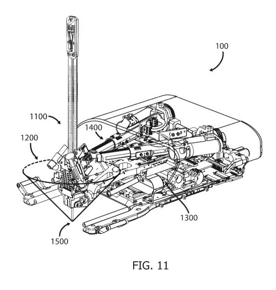
Fig. 11 depicts the overall workspace 1200 of the longitudinal axis of the
insertion assembly
1100 having two rotational degrees of freedom, the first depicted in Figs. 10A-
10B and the
second depicted in Figs. 10C-10E. In some implementations, the Remote Center
of Motion
(RCM) of the insertion assembly 1100 may be virtual and located at the
needle's entry point.
Although the rotation axis of the insertion assembly is located at the bottom
gimbal of the
device's top assembly, as shown in Fig. 10E, the location of the virtual RCM
is maintained
at the needle's entry point via linear movements of the X platform along the Z
axis and/or of
the top assembly along the X axis.
Once the needle entry point is selected, the user may set the selected entry
point as the virtual
RCM. The system's software can then determine, using a reverse kinematics
algorithm, as
described, for example, in abovementioned U.S. Patent No. 8,348,861, the
linear movements
required from the X platform and/or the top assembly, while the insertion
assembly is being
rotated, in order to maintain the entry point as the virtual RCM. The virtual
RCM being
maintained at the needle entry point prevents skin/tissue tearing in case a
linear needle
trajectory is not possible to follow and/or if the planned trajectory (linear
or otherwise)
requires adjustments as the needle is being inserted.
The workspace 1200 may form a cone shape, with its vertex 1500, being the
virtual RCM,
located at the needle's entry point. It can be appreciated that the insertion
assembly's
workspace is not necessarily symmetrical in all axis. If the maximal rotation
angles are
identical about all axis, e.g., as shown above in Figs. 10A-10E, then the
workspace is
symmetrical about all axis, as shown in Fig. 11, i.e., the transverse cross-
section of the formed
cone is a circle. However, if the maximal rotation angles about the X axis are
different from
the maximal rotation angles about the Z axis, e.g., 01= 02=45 and 03= 04=55 ,
the transverse
cross-section of the formed cone is an ellipse. In some implementations, the
maximal rotation
angles may differ in each direction. Further, the angular workspace is not
necessarily equal
27
CA 03024963 2018-11-20
WO 2017/203531 PCT/IL2017/050584
to the rotation about X axis and to the rotation about the Z axis, such that
the workspace may
be, for example, rectangular.
Although particular implementations have been disclosed herein in detail, this
has been done
by way of example for purposes of illustration only, and is not intended to be
limiting with
respect to the scope of the appended claims, which follow. In particular, it
is contemplated
that various substitutions, alterations, and modifications may be made without
departing from
the spirit and scope of the disclosure as defined by the claims. Other
aspects, advantages, and
modifications are considered to be within the scope of the following claims.
The claims
presented are representative of the implementations and features disclosed
herein. Other
unclaimed implementations and features are also contemplated. Accordingly,
other
implementations are within the scope of the following claims.
28