Note: Descriptions are shown in the official language in which they were submitted.
CA 03025095 2018-11-21
WO 2017/203470
PCT/IB2017/053097
1
ENHANCED INTRAUTERINE DEVICE
Cross Reference to Related Applications
[1] This disclosure is related to and claims the benefit of priority of
U.S.
Provisional Application No. 62/341,326, titled "Enhanced Intrauterine Device"
and filed on
May 25, 2016, which is hereby incorporated by this reference in its entirety.
Technical Field
[2] The present disclosure relates to a female contraceptive device and,
more
particularly (although not necessarily exclusively), to an enhanced
intrauterine device.
Background
[3] An intrauterine device ("IUD") may be inserted into a uterus to provide
long-
term contraception. IUDs may be made of metal, plastic, or other suitable
substances in
various shapes and sizes. An IUD can be inserted into the uterus through the
cervix and the
IUD may include arms that extend radially away from a body of the IUD (e.g.,
forming a "T"
shape) after insertion.
[4] Some IUDs include a substance that can be released into the uterus to
prevent fertilization. For example, an IUD may be wrapped with copper wire
such that it
releases a small amount of copper into the uterus, which may work as a
spermicide. In
another example, an IUD may release a synthetic hormone (e.g., levonorgestrel)
into the
uterus that thickens the cervical mucus so that sperm cannot reach an egg.
Additionally or
alternatively, the IUD may cause inflammation in the uterus that prevents the
egg from
implanting on the uterine wall.
[5] An IUD may provide contraception for years (e.g., a hormone IUD may
provide five years of contraception and a copper IUD may provide ten years of
contraception). But, the IUD can be pushed through the cervix and even
expelled
completely from the uterus without the user noticing. Thus, an IUD user may
periodically
check for the presence and proper placement of the IUD in the uterus.
[6] Some IUDs include removal strings that extend from the body of the IUD
in
the uterus, through the cervix, and into the vagina. The presence and proper
placement of
an IUD can be determined by locating the removal strings. To remove the IUD,
the removal
strings may be grasped by forceps and used to pull the entire IUD through the
cervix. But,
an IUD may break during the removal process (e.g., an arm may detach from a
body of the
CA 03025095 2018-11-21
WO 2017/203470
PCT/IB2017/053097
2
IUD) resulting in left over fragments of the IUD in the uterus that can
require a surgical
procedure to remove. Surgery may also be required if the removal strings are
drawn back
into the uterus such that the strings cannot be grasped for removal. Some
couples can
sense the removal strings during vaginal intercourse and find the removal
strings
uncomfortable. Others believe the strings may be linked to a higher risk of
infection.
Summary
[7] The present disclosure describes devices, systems, and methods for
female
contraception.
[8] In some aspects, an intrauterine device ("IUD") is provided. The IUD
may
include a body, an arm, a removal mechanism, and a communication circuit. The
body may
include a first end and a second end. The arm may extend radially from the
first end while
the body is inserted in a uterus. The removal mechanism may couple to the
second end for
removing the intrauterine device from the uterus. The communication circuit
may couple to
the body or the arm for responding to a first signal by transmitting a second
signal.
[9] In some aspects, a method is provided. The method may include
transmitting
a first signal to a communication circuit coupled to an IUD. The method may
further include
receiving a second signal from the communication circuit. The method may
further include
determining the IUD is positioned within a uterus based on the second signal.
[10] In some aspects, an IUD is provided with a body, an arm, a removal
mechanism, and a connector. The body may include a first end and a second end.
The arm
may, in a first position, extend radially from the first end of the body and
in a second
position be detached from the body. The removal mechanism may couple to the
second
end for removing the intrauterine device from a uterus. The connector may link
the arm to
the body such that the arm remains linked to the body in both the first and
second position
via the connector.
[11] In some aspects, an IUD is provided with a body, an arm, and a removal
mechanism. The body may include a first end and a second end. The arm may
extend
radially from the first end when the body is within a uterus. The removal
mechanism may
be coupled to the second end and include a magnetic portion for removing the
IUD from the
uterus.
[12] In some aspects, a method is provided. The method may include
inserting a
removal tool with a magnetic end through a cervix and into a uterus. The
method may
CA 03025095 2018-11-21
WO 2017/203470
PCT/IB2017/053097
3
further include docking the magnetic end with a magnetic portion of a removal
mechanism
of an IUD. The method may further include removing the IUD from the uterus
with the
removal tool.
Brief Description of the Drawings
[13] In the following detailed description, embodiments of the disclosure
are
described referring to the following figures:
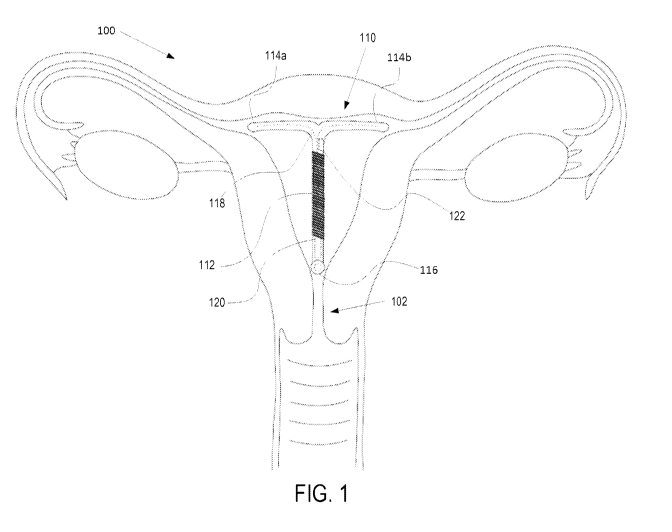
[14] FIG. 1 is a cross-sectional diagram of an example of a uterus with an
intrauterine device ("IUD") according to one aspect of the present disclosure.
[15] FIG. 2 is a perspective view of an example of a copper IUD with a
communication circuit according to one aspect of the present disclosure.
[16] FIG. 3 is a perspective view of an example of a hormone IUD with a
communication circuit according to one aspect of the present disclosure.
[17] FIG. 4 is a block diagram of a communication circuit of an IUD
communicatively coupled to an external device via a Near Field Communication
("NFC") field
according to one aspect of the present disclosure.
[18] FIG. 5 is a flow chart of an example of a process for detecting the
presence of
an IUD, with a communication circuit, within a uterus according to one aspect
of the present
disclosure.
[19] FIG. 6 is a perspective view of an example of a copper IUD with
connectors
coupling each arm to the body according to one aspect of the present
disclosure.
[20] FIG. 7 is a perspective view of an example of a hormone IUD with
connectors
coupling each arm to the body according to one aspect of the present
disclosure.
[21] FIG. 8 is a cross-sectional diagram of an example of a uterus with a
copper
IUD with arms extending from the body and with connectors linking each arm to
the body
according to one aspect of the present disclosure.
[22] FIG. 9 is a cross-sectional diagram of an example of the uterus and
IUD in FIG.
8 with arms that have detached from the body, but remain linked to the body
via the
connectors according to one aspect of the present disclosure.
[23] FIG. 10 is a perspective view of an example of a copper IUD with a
removal
mechanism with a magnetic portion according to one aspect of the present
disclosure.
[24] FIG. 11 is a perspective view of an example of a hormone IUD with a
removal
mechanism with a magnetic portion according to one aspect of the present
disclosure.
CA 03025095 2018-11-21
WO 2017/203470
PCT/IB2017/053097
4
[25] FIG. 12 is a side view of a removal tool according to one aspect of
the present
disclosure.
[26] FIG. 13A is a top view of a removal tool in a closed position
according to one
aspect of the present disclosure.
[27] FIG. 1313 is a side view of a removal tool in a close position
according to one
aspect of the present disclosure.
[28] FIG. 13C is a top view of a removal tool in an open position according
to one
aspect of the present disclosure.
[29] FIG. 13D is a side view of a removal tool in an open position
according to one
aspect of the present disclosure.
[30] FIG. 14 is a flow chart of an example of a process for removing an IUD
with a
removal mechanism with a magnetic portion according to one aspect of the
present
disclosure.
[31] FIGS. 15-20 are cross-sectional diagrams of an example of a uterus
with a
copper IUD being removed using the magnetic portion of a removal mechanism
according to
one aspect of the present disclosure.
Detailed Description
[32] Certain aspects and features relate to an enhanced intrauterine device
("IUD") that may be more efficient, comfortable, and safe. The IUD may include
a
communication circuit such that the IUD can be detected wirelessly. The IUD
may include
connectors that can link a body of the IUD with an arm of the IUD such that
the body and
arm remain linked if the arm detaches from the body. The IUD may include a
removal
mechanism with a magnetic portion such that the IUD may dock with a removal
tool
without the aid of removal strings.
[33] In some aspects, an IUD may include a communication circuit that can
transmit a signal for determining the presence of the IUD within a uterus. The
communication circuit may be embedded within the IUD or coupled to the surface
of the
IUD. In some aspects, the communication circuit may be coupled to a power
source. In
additional or alternative aspects, the communication circuit may be powered by
a signal
received by the communication circuit. The communication circuit may be
communicatively
coupled to a memory that may store data regarding the IUD. For example, the
data may
CA 03025095 2018-11-21
WO 2017/203470
PCT/IB2017/053097
include a manufacturer of the IUD, a date of production of the IUD, a
physician that inserted
the IUD, a date the IUD was inserted, and a mode of contraception used by IUD
(e.g., copper
or hormone). The communication circuit may also be communicatively coupled to
one or
more sensors that can measure characteristics (e.g., glucose level, body
temperature, or
iron level) of a user of the IUD. In some examples, the sensors may be on the
surface of the
IUD such that the sensors are exposed to bodily fluids. In additional or
alternative
examples, the sensors may be embedded within the IUD. In some aspects, the
memory may
store data based on the measurements from the sensors.
[34] In response to a first signal, the communication circuit may transmit
a second
signal indicating the presence of the IUD within the uterus. The first signal
may be transmit
by a device external to the uterus and the device may determine the IUD is
within the
uterus by receiving the second signal. The second signal may be modulated to
include the
data stored in the memory. Additionally or alternatively, the second signal
may be
modulated to include measurements from the sensors. In some aspects, the
device external
to the uterus may be a smartphone or another computing device that includes an
app for
monitoring the IUD. The device may allow a user to display information
regarding the IUD
as well as periodically remind the user to check the position of the IUD. In
additional or
alternative aspects, the communication circuit may be a near field
communication chip
("NFC"). In additional or alternative aspects, the communication circuit
may be
communicatively coupled to an antenna for receiving the first signal and
transmitting the
second signal. In some examples, the coil of copper wire around a copper IUD
can be used
as the antenna. In additional or alternative examples, an antenna (e.g.,
copper or silver
wire) may be coupled to the IUD. For example, a coil of silver wire may be
positioned
between the surface of the IUD and the hormone release mechanism of a hormone
IUD.
[35] In some aspects, a connector may link an arm of an IUD to a body of
the IUD
such that a link is maintained if the arm detaches from the body. In some
aspects, the arm
may be designed to detach from the body as the IUD is removed from the uterus.
In
additional or alternative aspects, the arm may detach from the body due to a
force applied
to a joint between the arm and the body. The connector may maintain a link
between a
detached arm and the body such that the detached arm may be removed with the
rest of
the IUD.
CA 03025095 2018-11-21
WO 2017/203470
PCT/IB2017/053097
6
[36] In some aspects, The connector may be flexible such that the arm can
bend
(e.g., forming a 'Y' shape IUD from a 'T' shape IUD) during a removal process.
The connector
may be a plastic string or any suitable material. In some aspects, the
connector may be a
portion of the removal strings that attach to an arm of the IUD, pass through
the body of
the IUD, and extend from the removal mechanism through the cervix. In
additional or
alternative aspects, the connector may attach to multiple arms and pass
through the center
of the IUD.
[37] In some aspects, a removal mechanism of the IUD may include a magnetic
portion for magnetically docking with a removal tool. In some aspects, the
magnetic portion
may replace the removal strings. In other aspects, the removal mechanism may
include
both a magnetic portion and removal strings.
[0001] These illustrative examples are given to introduce the reader to
the general
subject matter discussed here and are not intended to limit the scope of the
disclosed
concepts. The following sections describe various additional features and
examples with
reference to the drawings in which like numerals indicate like elements, and
directional
descriptions are used to describe the illustrative aspects but, like the
illustrative aspects,
should not be used to limit the present disclosure.
[38] FIG. 1 is a cross-sectional diagram of an example of a uterus 100 with
an IUD
110. The IUD 110 includes a body 112 with two arms 114a-b extending from one
end of the
body 112 to form a 'T' shape. On the other end of the body 112 is a removal
mechanism
116 with a magnetic portion. The IUD 110 further includes connectors 118 that
link the two
arms 114a-b to the body 112. The IUD 110 also includes a communication circuit
120 (e.g., a
near field communication ("NFC") chip) and a sensor 122 embedded in the IUD
110. Copper
wire 124 coils around the body 112. In some examples, the copper wire 124 can
coil around
both arms 114a-b.
[39] The IUD 110 may operate as a contraceptive in the uterus 100 by
releasing a
small amount of copper from the copper wire 124, which can act as a
spermicide. In
additional or alternative aspects, the IUD 110 may inflame the walls of the
uterus 100 and
prevent an egg from implanting therein. The copper wire 124 may also be
communicatively
coupled to the communication circuit 120 to act as an antenna. Although the
IUD 110 as
illustrated uses copper, other substances may be used (e.g., a hormone). In
some examples,
CA 03025095 2018-11-21
WO 2017/203470
PCT/IB2017/053097
7
a hormone IUD may include an antenna positioned between the surface of the IUD
and the
hormone release mechanism.
[40] The communication circuit 120 may respond to a first signal
transmitted by a
device external to the uterus 100 by transmitting a second signal. The second
signal may be
used by the device to determine that the IUD 110 is within the uterus 100. In
some aspects,
the second signal may include information about the IUD 110. For example, the
second
signal may include information related to a manufacturer of the IUD 110, a
date of
production of the IUD 110, a physician that inserted the IUD 110, a date the
IUD 110 was
inserted, and the mode of contraception used by IUD 110. In some aspects,
information
may be stored on a memory communicatively coupled to the communication circuit
120. In
additional or alternative aspects, the second signal may include information
about a user of
the IUD 110. For example, the second signal may include information measured
by the
sensor 122 such as a glucose level, body temperature, or iron level of the
user.
[41] Although the communication circuit 120 and sensor 122 are illustrated
as
embedded within the IUD 110, a communication circuit and a sensor may be
coupled to the
surface of an IUD. Furthermore, a communication circuit and a sensor may be
coupled to
any part of an IUD. In additional or alternative aspects, an IUD may include
more than one
sensor and some of the sensors may be embedded in the IUD and other sensors
can be
coupled to the surface of the IUD such that they are exposed to bodily fluids.
[42] The connectors 118 may link arms 114a-b to the body 112 such that if
the
arms 114a-b detach from the body 112 then the arm 114a-b may still be linked
to the body
112 via the connectors 118. For example, an arm 114a-b may detach from the
body 112 as
the IUD 110 is pulled through a cervix 102 during removal of the IUD 110. The
connectors
118 may maintain a link between the arm 114a-b and the body 112 such that the
arm 114a-
b is removed from the uterus 100 along with the rest of IUD 110. Although the
connectors
118 are illustrated as embedded in the IUD 110, in some examples, the
connectors 118 are
external to the IUD 110 and couple between a surface of the arms 114a-b and a
surface of
the body 112.
[43] The removal mechanism 116 may include a magnetic portion for
docking with
a removal tool. The magnetic portion may align a magnetic end of the removal
tool with the
removal mechanism 116. In some aspects, the magnetic coupling between the
magnetic
portion and the removal tool may allow the IUD 110 to be pulled through the
cervix 102 by
CA 03025095 2018-11-21
WO 2017/203470
PCT/IB2017/053097
8
pulling out the removal tool. In additional or alternative aspects, the
removal mechanism
116 may mechanically lock onto the removal tool after the removal tool has
docked with the
magnetic portion. As illustrated, the IUD 110 may be entirely housed by the
uterus 100. In
some aspects, removal strings may extend from the removal mechanism 116
through the
cervix 102 and into the vagina.
[44] FIGS. 2-3 are perspective views of an example of a copper IUD 200 and
hormone IUD 210, each with a communication circuit 208, 218. FIG. 2
illustrates an IUD 200
with copper wire 202, removal string 204, removal mechanism 206, communication
circuit
208, and sensor 209. In this example, the copper wire 202 is wrapped around a
body of IUD
200. In other examples, the copper wire 202 can be wrapped around a body and
two arms
of an IUD. The copper wire 202 can gradually release copper that may act as a
spermicide
while the IUD 200 is within a uterus. FIG. 3 illustrates an IUD 210 with a
hormone release
212, removal string 214, removal mechanism 216, communication circuit 218,
sensors 219,
and an antenna 220. While in the uterus, the hormone release 212 can release a
hormone
(e.g., levonorgestrel) that may thicken the cervical mucus so that sperm
cannot reach an
egg.
[45] The removal string 204, 214 may extend from the removal mechanism 206,
216 such that while the IUD 200, 210 is inserted in the uterus, the removal
string 204, 214
may extend through the cervix into the vagina. In some aspects, the removal
string 204, 214
and removal mechanism 206, 216 may be replaced by a removal mechanism with a
magnetic portion. The communication circuit 208, 218 can transmit a signal in
response to
receiving a signal. In FIG. 2, at least a portion of the copper wire 202
can be
communicatively coupled to the communication circuit 208 to act as an antenna.
In some
examples, as the copper as released into a uterus an impedance of the antenna
formed by
the copper wire may shift. The shift in the impedance of the coper wire can be
analyzed to
determine a remaining copper quantity. The IUD 200 can be scheduled to be
replaced in
response to the remaining copper quantity falling below a threshold value,
which can allow
for safer and longer use of the IUD 200. In FIG. 3, the antenna 220 may be
communicatively
coupled to the communication circuit. Although antenna 220 is depicted as
positioned
between the surface of the IUD 210 and the hormone release 212, an antenna may
be
positioned outside of a hormone release.
CA 03025095 2018-11-21
WO 2017/203470
PCT/IB2017/053097
9
[46] By transmitting a signal from within a uterus, the communication
circuit 208,
218 may indicate to a device external to the uterus (e.g., a smartphone) that
the IUD 200,
210 is within the uterus. In some aspects, the communication circuit 208, 218
may transmit
a modulated signal. The modulated signal may contain data indicating the IUD
200, 210 is
within the uterus. In additional or alternative aspects, the data may include
information
such as a manufacturer of the IUD 200, 210, a date of production of the IUD
200, 210, a
physician that inserted the IUD 200, 210, a date of the insertion of the IUD
200, 210, and the
mode of contraception used by the IUD 200, 210. The data may also include
information
regarding the status of the IUD 200, 210 such as an indication that an arm has
detached
from the body of the IUD 200, 210 or that the removal mechanism 206, 216 is
cooperating
with a removal tool. In additional or alternative aspects, the data may
include
measurements from a sensor 209, 219b communicatively coupled to the
communication
circuit 208, 218. For example, the data may include a glucose level, body
temperature, or
iron level of a user of the IUD.
[47] The communication circuit 208, 218 and the sensor 209, 219 may be
embedded within the IUD 200, 210 or coupled to the surface of the IUD 200,
210. In some
aspects, the communication circuit 208, 218 and the sensor 209, 219 may be
coupled to a
power source. In additional or alternative aspects, the communication circuit
208, 218 and
the sensor 209, 219 may be powered by the signal received by the communication
circuit
208, 218. The communication circuit 208, 218 may respond to all received
signals or only to
certain modulated signals. Although FIGS. 2-3 depict IUD 200, 210 with a
removal string
204, 214, the presence of the IUD 200, 210 may be detected without the removal
string 204,
214.
[48] In some aspects, the communication circuit 208, 218 may be
communicatively coupled to a memory that can store the data. The data may be
stored to
the memory prior to the IUD 200, 210 being inserted into the uterus or may be
written to
memory while within the uterus. For example, the communication circuit 208,
218 may
receive a signal from a device instructing information be stored to the
memory. In
additional or alternative aspects, a sensor 209, 219 may be communicatively
coupled to the
memory and may request a measurement be stored to memory. In some aspects, the
communication circuit 208 can be a NFC chip that can allow short-range (e.g.,
a distance of 4
cm) wireless communication.
CA 03025095 2018-11-21
WO 2017/203470
PCT/IB2017/053097
[49] FIG. 4 is a block diagram of an IUD 400 communicatively coupled to an
external device 422 via a NFC field 412. The IUD 400 includes an NFC circuit
402, processing
device 404 with memory 406, sensors 408a-c, and an antenna 410. The external
device
(e.g., a smart phone) 422 includes a processing device 424 communicatively
coupled to an
antenna 420.
[50] The NFC circuit 402 is communicatively coupled to the antenna 410 for
receiving signals transmit over the NFC field 412 by the external device 422.
The NFC circuit
is further communicatively coupled to the processing device 404, which is
communicatively
coupled to the sensors 408a-c. The signal may be used to power the NFC circuit
402 as well
as the processing device 404 and sensors 408a-c. In some aspects, a capacitor
may be
charged by the signal and used to provide extended power.
[51] The processing device 404 can include any number of processors
configured
for executing program code stored in the memory 406. Examples of the
processing device
404 can include a microprocessor, an application-specific integrated circuit
("ASIC"), a field-
programmable gate array ("FPGA"), or other suitable processor. In some
aspects, the
processing device 404 can be a dedicated processing device used for sending a
signal in
response to receiving a signal. For example, the processing device 404 can
instruct the NFC
circuit 402 to transmit a preset response signal. In other aspects, the
processing device 404
can perform additional functions such as evaluating measurements from the
sensors 408a-c
and modulating a response signal to include data related to the IUD 400 and
the health of
the user of the IUD 400.
[52] The processing device 404 can include (or be communicatively coupled
with)
a non-transitory computer-readable memory 406. The memory 406 can include one
or
more memory devices that can store program instructions. The program
instructions can
include for example, a signal response engine that is executable by the
processing device
304 to perform certain operations described herein.
[53] The operations can include determining that a received signal warrants
a
response. The operations can further include examining measurements from
sensors 408a-
c and data stored in memory 406 to determine information that should be
included in a
response signal. The operations can further include instructing the NFC
circuit 402 to
transmit a response signal that includes the information.
CA 03025095 2018-11-21
WO 2017/203470
PCT/IB2017/053097
11
[54] The memory 406 can also store information about the IUD 400 such as a
manufacturer of the IUD 400, a date of production of the IUD 400, a physician
that inserted
the IUD 400, a date the IUD 400 was inserted, and the mode of contraception
used by IUD
400. In additional or alternative aspects, the memory 406 can store
measurements taken by
the sensors 408a-c.
[55] In some examples, the sensors 408a-c can be embedded within the IUD
400
and on the surface of the IUD 400 such that the sensors 408a-c are exposed to
bodily fluids.
The sensors 408a-c embedded in the IUD 400 can measure characteristics of the
user of the
IUD 400 such as body temperature. The sensors 408a-c exposed to bodily fluids
can
measure characteristics of the user of the IUD 400 such as glucose level and
iron level.
[56] The processing device 424 in the external device 422 can receive a
signal
transmit by the IUD 400 via the antenna 420. The processing device 424 can use
the
received signal to determine the presence of the IUD 400. In some aspects, the
processing
device 424 can further use the signal to determine characteristics of the IUD
400 and the
health of the user of the IUD 400. In additional or alternative aspects, the
processing device
424 may instruct the external device 422 to display an analysis of the
information
determined from the received signal. In additional or alter aspects, the
processing device
424 may instruct the external device 422 to communicate with another device or
an
emergency medical service to provide assistance to the user of the IUD 400.
Although FIG. 4
illustrates the IUD 400 with an NFC circuit 402, any wireless communication
circuit may be
used to communicate over a wireless network with an external device.
[57] FIG. 5 is a flow chart of an example of a process for detecting the
presence of
an IUD, including a communication circuit, within a uterus.
[58] In block 502, a first signal is transmit to a communication circuit
coupled to
an IUD. The first signal may be a wireless signal transmitted by a device
(e.g., a smartphone)
separate from the IUD. In some aspects, the first signal may be received by an
antenna. In
some examples, the antenna may be copper wire for releasing copper by a copper
IUD. In
additional or alternative examples, the antenna may be positioned between a
surface of a
hormone IUD and a hormone release. In some aspects, the first signal powers
the
communication circuit and causes the communication circuit to transmit a
second signal in
response. In additional or alternative aspects, the first signal may include
instructions to
transmit a second signal.
CA 03025095 2018-11-21
WO 2017/203470
PCT/IB2017/053097
12
[59] In block 504, a second signal is received from the communication
circuit. The
second signal may be received by the device that transmitted the first signal
or a separate
device. In some aspects, the second signal is a short pulse. In additional or
alternative
aspects, the second signal may be a modulated to include data that indicates
the IUD is
within the uterus.
[60] The second signal may also be modulated to include data about the IUD
or
the user of the IUD. For example, the second signal may be modulated to
include data on a
manufacturer of the IUD, a date of production of the IUD, a physician that
inserted the IUD,
a date the IUD was inserted, and a mode of contraception used by the IUD. In
additional or
alternative examples, the second signal may include data about the current
status of the
IUD. For example, the data may indicate an arm has detached from a body of the
IUD or a
removal mechanism of the IUD is cooperating with a removal tool. In additional
or
alternative aspects, the second signal may include data about the condition of
the user of
the IUD, such as the glucose level, body temperature, or iron level of the
user.
[61] In block 506, the IUD may be determined to be positioned within the
uterus
based on the second signal. In some aspects, the device that received the
second signal
may be positioned such that receiving the signal allows the determination that
the IUD is
within the uterus. In additional or alternative aspects, a processing device
may determine
from data included in the second signal that the IUD is within the uterus.
[62] The processing device may determine additional information from the
data
included in the second signal. In some aspects, the processing device may
determine
medical action should be taken by the user and may display an analysis of the
data to the
user. For example, the processing device may determine a diabetic user has low
blood
sugar based on measurements transmitted as part of the data. The processing
device may
display the user's blood sugar level as well as a recommended insulin dosage.
In additional
or alternative aspects, the processing device may communicate to an additional
device (e.g.,
an insulin pump) and instruct the additional device to perform an action
(e.g., inject the user
with insulin). In additional or alternative aspects, the processing device may
determine that
emergency medical attention should be taken and may contact a hospital or
emergency
medical service provider.
[63] FIGS. 6-7 are perspective views of an example of a copper IUD 600 and
a
hormone IUD 610 with connectors 608, 618 linking each arm to a respective
body. Similar
CA 03025095 2018-11-21
WO 2017/203470
PCT/IB2017/053097
13
to the IUD 200 in FIG. 2, IUD 600 includes copper wire 602, a removal string
604, and a
removal mechanism 606. Similar to the IUD 210 in FIG. 3, IUD 610 includes a
hormone
release 612, removal string 614, and removal mechanism 616. The IUD 600, 610
also
includes connectors 608, 618 that link the each arm to the body.
[64] In some aspects, an arm may extend radially from a body of the IUD
600, 610
in a first position. And, the arm may be detached from the body in a second
position. The
connectors 608, 618 can maintain a link between the arm and the body in both
the first and
second position. FIGS. 8-9 depict the arms in a first and second position.
[65] FIG. 8 is a cross-sectional diagram of an example of a uterus 800 with
an IUD
810 with arms 804a in the first position (e.g. extending from the body 802a)
and with
connectors 818a coupling each arm 804a to the body 802a. FIG. 9 is a cross-
sectional
diagram of an example of the uterus 800 in FIG. 8 with the IUD 810 with arms
804b in the
second position (e.g., detached from the body 802b) but linked to the body
802b via the
connectors 818b. In some aspects, the arms 804a may be in the first position
while inserted
in the uterus 800 as illustrated in FIG. 8 with the removal mechanism 816a
entirely within
the uterus and the removal string 814a extending from the removal mechanism
816a
through the cervix and into the vagina. The arms 804a may move to a second
position
during removal of the IUD 810 from the uterus 800. FIG. 9 illustrates a
portion of the
removal process during which the removal mechanism 816b has been pulled into
the vagina
via the removal string 814b. The connectors 818b may cause the arms 804b to be
removed
with the IUD 810. Although FIGS. 8-9 depict a copper IUD 810, any suitable IUD
may be
used with the connectors 818a-b.
[66] Returning to FIGS. 6-7, although the connectors 608, 618 are depicted
as
individual links between the arms and the body. A single connector may couple
to one arm,
pass through a hole in the body of the IUD, and couple to a second arm. In
this example, the
hole may be smaller than an arm and may link the arms to the body such that
the arms can
be removed with the IUD while the arms are detached from the body. In
additional or
alternative aspects, the IUD 600, 610 may have one arm or more than two arms
and
connectors may link one or more of the arms to the body.
[67] In additional or alternative aspects, the arms of the IUD 600, 610 may
be
flexible such that the arms can bend to form a 'Y' shaped IUD. The connectors
608, 618 may
be a flexible material or possess sufficient slack to allow the arms to bend.
In some aspects,
CA 03025095 2018-11-21
WO 2017/203470
PCT/IB2017/053097
14
the connector 608, 618 may be an extension of the removal string 604, 614. For
example,
the removal string 604, 614 may attach to an arm, couple to the removal
mechanism 606,
616 and be configured to extend through a cervix while the IUD 600, 610 is
inserted in a
uterus. In additional or alternative aspects, the removal mechanism 606, 616
and removal
string 604, 614 may be replaced by a removal mechanism with a magnetic
portion.
[68] FIGS. 10-11 are perspective view of an example of a copper IUD 1000
and
hormone IUD 1010 with a removal mechanism 1006, 1016 with a magnetic portion.
IUD
1000 includes copper wiring 1002 wrapped around a body of the IUD 1000 and two
arms of
the IUD 1000. The IUD 1010 includes a hormone release 1012 coupled to a body
of the IUD
1010. The magnetic portion of the removal mechanism 1006, 1016 may be
generated using
an electromagnet or may have naturally magnetic properties. The copper IUD
1000 and
hormone IUD 1010 can have a communication circuit 1020 and a sensor 1022,
which can
include circuitry or insulation to prevent the magnetic portion of the removal
mechanism
1006, 1016 from interfering with communications or measurements respectively.
[69] In some aspects, the magnetic portion of the removal mechanism 1006,
1016
may have a strong enough magnetic force that the removal mechanism 1006, 1016
can
couple to a magnetic end of a removal tool. By coupling with the removal tool,
the IUD
1000, 1010 can be removed from a uterus as the removal tool is pulled out of
the uterus. In
additional or alternative aspects, an additional locking mechanism may be used
to couple
the removal tool to the IUD 1000, 1010.
[70] For example, the removal mechanism 1006, 1016 may include a first
portion
and a second portion. The second portion may be farther axially from the body
of the
intrauterine device 1000, 1010 than the first portion and may extend farther
radially (e.g.,
have a greater circumference) than the first portion. The removal tool may
include a loop
that can tighten around the first portion while the removal tool is docked
with the removal
mechanism 1006, 1016. Once tightened the loop may have a smaller diameter than
the
second portion such that the removal tool is coupled to the IUD 1000, 1010.
Although
removal mechanism 1006, 1016 is illustrated as having a spherical shape, and
suitable shape
may be used. In some aspects, the removal mechanism 1006, 1016 can reduce the
risk of
infection, reduce interference with sexual activities, and improve privacy by
reducing the
visibility to non-users than an IUD is being used.
CA 03025095 2018-11-21
WO 2017/203470
PCT/IB2017/053097
[71] FIG. 12 is a side view of an example of a removal tool 1200 for
removing an
IUD. In this example, the removal tool 1200 includes a body 1220 with a
grasping
mechanism 1210 on one end and a control mechanism 1230 on another end. The
body
1220 can be long enough to extend through a woman's cervix and into her
uterus. The
grasping mechanism 1210 can move between an open state and a closed state. In
the open
state, the grasping mechanism 1210 can create an opening. In some examples,
the grasping
mechanism 1210 can include one or more members that can fold radially outward
to form
the opening. In the closed position, the grasping mechanism can press radially
inward
closing the opening or grasping onto a removal mechanism of an IUD positioned
in the
opening. The control mechanism 1230 can be coupled to the grasping mechanism
1210
such that the control mechanism 1230 can control the state of the grasping
mechanism
1210. The control mechanism 1230 includes a handle that can move between an
extended
position and a depressed position. In the extended position, the control
mechanism 1230
causes the grasping mechanism 1210 to close. In the depressed position, the
control
mechanism 1230 causes the grasping mechanism 1210 to open.
[72] FIGS. 13A-D depict an example of the removal tool 1200 in a closed
position
and an open position. FIG. 13A depicts a top view of the grasping mechanism
1210 in a
closed position and FIG. 13C depicts a top view of the grasping mechanism 1210
in an open
position. A magnet 1350 is illustrated in FIG. 13C and can have an opposite
polarity of a
magnetic removal mechanism in an IUD. The magnet 1350 can be used to locate
and
position the IUD within the opening formed by the grasping mechanism 1210. In
some
examples, the magnet 1350 can be an electromagnet that receives power in
response to the
grasping mechanism 1210 moving to an open state. FIG. 13B depicts a side view
of a
portion of the removal tool 1200 in a closed position and FIG. 13 D depicts a
side view of a
portion of the removal tool 1200 in an open position.
[73] FIG. 14 is a flow chart of an example of a process for removing an IUD
with a
removal mechanism with a magnetic portion.
[74] In block 1402, a removal tool including a magnetic end is inserted
through a
cervix and into a uterus. In some aspects, the removal tool may include an
electromagnet
such that when power is applied removal tool, a magnetic force is generated at
the
magnetic end.
CA 03025095 2018-11-21
WO 2017/203470
PCT/IB2017/053097
16
[75] In block 1404, the magnetic end of the removal tool docks with the
magnetic
portion of the removal mechanism of the IUD. In some aspects, the magnetic
force
between the removal mechanism and the magnetic end may be strong enough to
couple
the removal tool with the removal mechanism. In additional or alternative
aspects, the
magnetic force may allow the removal tool to dock with the removal mechanism
such that
the removal tool can couple to the removal mechanism using a mechanical
locking
mechanism. For example, the removal tool may include a loop on the magnetic
end that
may tighten around the removal mechanism once the removal tool has docked with
the
removal mechanism.
[76] In block 1406, the IUD is removed from the uterus with the removal
tool. By
pulling the removal tool, while docked with the removal mechanism, through the
cervix the
IUD is pulled through the cervix.
[77] FIGS. 15-20 are cross-sectional diagrams of an example of a uterus
1500
during removal of a copper IUD 1502 using a removal tool 1608 docked to the
magnetic
portion of a removal mechanism 1506. As illustrated, the removal mechanism
1506 can be
coupled to one end of a body of the IUD 1502. The removal mechanism 1506
includes a
magnetic portion that can dock with a magnetic end of the removal tool 1608.
As the
removal tool 1608 is pulled out of the uterus 1500 and through the cervix, the
IUD 1502 is
pulled down through the uterus 1500.
[78] As the IUD 1502 is removed from the uterus 1500, the arms of the IUD
1502
may bend giving the IUD 1502 a 'Y' shape. In some aspects, connectors may link
the arms to
the body of the IUD 1502 such that the arms and body remain linked if the arms
detach
from the body. In additional or alternative aspects, the IUD 1502 may
include a
communication circuit that can transmit a wireless signal from inside the
uterus 1500 to a
device external to the uterus 1500. The signal may include an indication when
the removal
tool 1608 is docked with the IUD 1502.
[79] In some aspects, an enhanced intrauterine device is provided according
to
one or more of the following examples:
[80] Example #1: An intrauterine device include a body, at least one arm, a
removal mechanism, and a communication circuit. The body can include a first
end and a
second end. The at least one arm can extend radially from the first end while
the body is
inserted in a uterus. The removal mechanism can be coupled to the second end
for
CA 03025095 2018-11-21
WO 2017/203470
PCT/IB2017/053097
17
removing the intrauterine device from the uterus. The communication circuit
can be
coupled to at least one of the body and the at least one arm for responding to
a first signal
by transmitting a second signal.
[81] Example #2: The intrauterine device of Example #1, can further include
one
or more sensors communicatively coupled to the communication circuit for
measuring
health data of a user of the intrauterine device.
[82] Example #3: The intrauterine device of Example #2, can further feature
a
sensor of the one or more sensors being embedded within the intrauterine
device for
measuring a body temperature of the user.
[83] Example #4: The intrauterine device of Example #2, can further feature
a
sensor of the one or more sensors being exposed to bodily fluids for measuring
at least one
of a glucose level of the user and an iron level of the user.
[84] Example #5: The intrauterine device of Example #2, can further feature
a
sensor of the one or more sensors being powered by the first signal.
[85] Example #6: The intrauterine device of Example #1, can further include
further memory communicatively coupled to the communication circuit for
storing data.
The second signal can include at least a portion of the data.
[86] Example #7: The intrauterine device of Example # 6, can further
feature the
portion of the data including at least one of a manufacturer of the
intrauterine device, a
date of production of the intrauterine device, a physician that performed an
insertion of the
intrauterine device, a date of insertion of the intrauterine device, and a
type of the
intrauterine device.
[87] Example #8: The intrauterine device of Example #1, can further feature
the
communication circuit being a near field communication chip and being powered
by the first
signal.
[88] Example #9: The intrauterine device of Example #1, can further include
a
copper component coupled to at least one of the body and the at least one arm
for
releasing copper into the uterus.
[89] Example #10: The intrauterine device of Example #9, can further
feature the
copper component being further for operating as an antenna for receiving the
first signal
and for transmitting the second signal.
CA 03025095 2018-11-21
WO 2017/203470
PCT/IB2017/053097
18
[90] Example #11: The intrauterine device of Example #1, can further
include a
hormone component coupled to at least one of the body and the at least one arm
for
releasing a hormone into the uterus.
[91] Example #12: The intrauterine device of Example #11, can further
include an
antenna coupled to at least one of the body and the at least one arm for
receiving the first
signal and for transmitting the second signal.
[92] Example #13: The intrauterine device of Example # 1, can further
feature the
at least one arm in a first position being directly coupled to the body and in
a second
position being detached from the body. The intrauterine device can further
include at least
one connector for linking the at least one arm to the body such that the at
least one arm
remains linked to the body in both the first position and the second position
via the at least
one connector.
[93] Example #14: The intrauterine device of Example #13, can further
feature the
communication circuit being further for modulating the second signal to
indicate the at least
one arm is in the second position.
[94] Example #15: The intrauterine device of Example #1, can further
feature the
removal mechanism including a magnetic portion for magnetically docking to a
removal
tool.
[95] Example #16: The intrauterine device of Example #15, can further
feature the
communication circuit being for modulating the second signal to indicate the
magnetic
portion is docked to the removal tool.
[96] Example #17: A method can include transmitting a first signal to a
communication circuit coupled to an intrauterine device. The method can
further include
receiving a second signal from the communication circuit. The method can
further include
determining the intrauterine device is positioned within a uterus based on the
second
signal.
[97] Example #18: The method of Example #17, can further feature the
communication circuit being communicatively coupled to a memory that stores
data and
the second signal includes at least a portion of the data.
[98] Example #19: The method of Example #18, can further include
determining at
least one of a manufacturer of the intrauterine device, a date of production
of the
intrauterine device, a physician that performed an insertion of the
intrauterine device, a
CA 03025095 2018-11-21
WO 2017/203470
PCT/IB2017/053097
19
date of insertion of the intrauterine device, and a type of the intrauterine
device from the
second signal.
[99] Example #20: The method of Example #17, can further feature the
communication circuit being communicatively coupled to one or more sensors
that measure
health information associated with a user of the intrauterine device and the
second signal
includes at least a portion of the health information.
[100] Example #21: The method of Example #17, can further feature the
communication circuit being part of a near field communication chip.
[101] Example #22: The method of Example #17, can further feature the
intrauterine device including a copper component for releasing copper into the
uterus.
[102] Example #23: The method of Example #22, can further feature
transmitting
the first signal to the communication circuit further including transmitting
the first signal to
the copper component that operates as an antenna and is communicatively
coupled to the
communication circuit.
[103] Example #24: The method of Example #17, can further feature the
intrauterine device including a hormone component for releasing hormones into
the uterus.
[104] Example #25: The method of Example #17, can further feature the
intrauterine device including at least one arm that in a first position
extends from a body of
the intrauterine device and in a second position is detached from the body,
and at least one
connector for linking the at least one arm to the body such that the at least
one arm
remains linked to the body in both the first position and the second position
via the at least
one connector.
[105] Example #26: The method of Example #25, can further include
determining
the at least one arm is in the second position based on the second signal.
[106] Example #27: The method of Example #17, can further feature the
intrauterine device including a removal mechanism with a magnetic portion for
docking with
a removal tool for removing the intrauterine device from the uterus.
[107] Example #28: The method of Example #27, can further include
determining
that the magnetic portion is docked with the removal tool based on the second
signal.
[108] Example #29: The method of Example #17, can further include
displaying
information based on the second signal for making medical decisions.
CA 03025095 2018-11-21
WO 2017/203470
PCT/IB2017/053097
[109] Example #30: The method of Example #17, can further include
communicating information based on the second signal to another device to
request a user
of the intrauterine device receive medical assistance.
[110] Example #31: An intrauterine device can include a body, at least one
arm, a
removal mechanism, and at least one connector. The body can include a first
end and a
second end. The at least one arm can, in a first position, extend radially
from the first end
and, in a second position, be detached from the body. The removal mechanism
can be
coupled to the second end for removing the intrauterine device from a uterus.
The at least
one connector can link the at least one arm to the body such that the at least
one arm
remains linked to the body in both the first position and the second position
via the at least
one connector.
[111] Example #32: The intrauterine device of Example #31, can further
feature the
at least one arm in the first position being configured to move to the second
position in
response to removing the intrauterine device from the uterus.
[112] Example #33: The intrauterine device of Example #31, can further
include a
communication circuit coupled to at least one of the body and the at least one
arm for
responding to a first signal by transmitting a second signal.
[113] Example #34: The intrauterine device of Example #33, can further
include a
memory communicatively coupled to the communication circuit for storing data.
The
second signal can include at least a portion of the data.
[114] Example #35: The intrauterine device of Example # 34, can further
feature
the portion of the data including at least one of a manufacturer of the
intrauterine device, a
date of production of the intrauterine device, a physician that performed an
insertion of the
intrauterine device, a date of insertion of the intrauterine device, and a
type of the
intrauterine device.
[115] Example #36: The intrauterine device of Example #33, can further
include one
or more sensors communicatively coupled to the communication circuit for
measuring
health data associated with a user of the intrauterine device. The second
signal can include
at least a portion of the health data.
[116] Example #37: The intrauterine device of Example #36, can further
feature a
sensor of the one or more sensors being exposed to bodily fluids for measuring
at least one
of glucose level of the user and iron level of the user.
CA 03025095 2018-11-21
WO 2017/203470
PCT/IB2017/053097
21
[117] Example #38: The intrauterine device of Example #36, can further
feature the
communication circuit being a near field communication chip and at least one
of the
communication circuit and at least one sensor of the one or more sensors being
powered by
the first signal.
[118] Example #39: The intrauterine device of Example #33, can further
feature the
communication circuit being further for modulating the second signal to
indicate the at least
one arm is in the second position.
[119] Example #40: The intrauterine device of Example #33, can further
include a
copper component coupled to at least one of the body and the at least one arm
for
releasing copper into the uterus, and communicatively coupled to the
communication
circuit for operating as an antenna.
[120] Example #41: The intrauterine device of Example #31, can further
feature the
removal mechanism including a magnetic portion for magnetically docking with a
removal
tool.
[121] Example #42: The intrauterine device of Example #31, can further
include a
copper component coupled to at least one of the body and the at least one arm
for
releasing copper into the uterus.
[122] Example #43: The intrauterine device of Example #31, can further
include a
hormone component coupled to at least one of the body and the at least one arm
for
releasing a hormone into the uterus.
[123] Example #44: An intrauterine device including a body, at least one
arm, and a
removal mechanism. The body including a first end and a second end. The at
least one arm
extending radially from the first end when the body is within a uterus. The
removal
mechanism having a magnetic portion coupled to the second end for removing the
intrauterine device from the uterus.
[124] Example #45: The intrauterine device of Example #44, can further
feature the
removal mechanism being further for magnetically docking with a removal tool.
[125] Example #46: The intrauterine device of Example #45, can further
feature the
removal mechanism including a first portion and a second portion and the
second portion
being farther from the body than the first portion and the second portion
having a greater
circumference than the first portion for locking to the removal tool when a
loop attached to
CA 03025095 2018-11-21
WO 2017/203470
PCT/IB2017/053097
22
the removal tool is tightened around the first portion such that the loop has
a smaller
diameter than the second portion.
[126] Example #47: The intrauterine device of Example #44, can further
include a
communication circuit coupled to at least one of the body and the at least one
arm for
responding to a first signal by transmitting a second signal.
[127] Example #48: The intrauterine device of Example #47, can further
include
memory communicatively coupled to the communication circuit for storing data.
The
second signal can include at least a portion of the data.
[128] Example #49: The intrauterine device of Example #48, can further
feature the
portion of the data including at least one of a manufacturer of the
intrauterine device, a
date of production of the intrauterine device, a physician that performed an
insertion of the
intrauterine device, a date of insertion of the intrauterine device, and a
type of the
intrauterine device.
[129] Example #50: The intrauterine device of Example #47, can further
include one
or more sensors communicatively coupled to the communication circuit for
measuring
health data associated with a user of the intrauterine device. The second
signal can include
at least a portion of the health data.
[130] Example #51: The intrauterine device of Example #50, can further
feature a
sensor of the one or more sensors being exposed to bodily fluids for measuring
at least one
of glucose level of the user and iron level of the user.
[131] Example #52: The intrauterine device of Example #50, can further
feature the
communication circuit being a near field communication chip and at least one
of the
communication circuit and at least one sensor of the one or more sensors being
powered by
the first signal.
[132] Example #53: The intrauterine device of Example #47, can further
feature the
communication circuit being further for modulating the second signal to
indicate the
magnetic portion is docked with a removal tool.
[133] Example #54: The intrauterine device of Example #44, can further
feature the
at least one arm in a first position being directly coupled to the body and in
a second
position being detached from the body. The intrauterine device can further
include at least
one connector for linking the at least one arm to the body such that the at
least one arm
CA 03025095 2018-11-21
WO 2017/203470
PCT/IB2017/053097
23
remains linked to the body in both the first position and the second position
via the at least
one connector.
[134] Example #55: The intrauterine device of Example #44, can further
include a
copper component coupled to at least one of the body and the at least one arm
for
releasing copper into the uterus.
[135] Example #56: The intrauterine device of Example #44, can further
include a
hormone component coupled to at least one of the body and the at least one arm
for
releasing a hormone into the uterus.
[136] Example #57: A method can include inserting a removal tool including
a
magnetic end through a cervix and into a uterus. The method can further
include docking
the magnetic end with a magnetic portion of a removal mechanism of an
intrauterine device
within the uterus. The method can further include removing the intrauterine
device from
the uterus with the removal tool.
[137] Example #58: The method of Example #57, can further feature the
removal
mechanism including a first portion and a second portion and the second
portion being
farther from a body of the intrauterine device than the first portion and
having a greater
circumference than the first portion. Docking can include tightening a loop
attached to the
removal tool around the first portion such that the loop has a smaller
diameter than the
second portion.
[138] Example #59: The method of Example #57, can further feature docking
the
removal tool further including activating an indicator light for indicating
the removal tool
has coupled with the magnetic portion.
[139] Example #60: The method of Example #57, can further feature the
intrauterine device further including a communication circuit for responding
to a first signal
by transmitting a second signal.
[140] Example #61: The method of Example #60, can further feature the
second
signal being modulated to indicate that the removal tool is docked to the
removal
mechanism.
[141] Example #62: The method of Example #60, can further feature the
communication circuit being communicatively coupled to one or more sensors
that measure
health information associated with a user of the intrauterine device and the
second signal
includes at least a portion of the health information.
CA 03025095 2018-11-21
WO 2017/203470
PCT/IB2017/053097
24
[142] Example #63: The method of Example #60, can further feature
transmitting
the first signal to the communication circuit further including transmitting
the first signal to
a copper component that operates as an antenna and is communicatively coupled
to the
communication circuit.
[143] Example #64: The method of Example #57, can further feature the
intrauterine device including at least one arm that in a first position
extends from a body of
the intrauterine device and in a second position is detached from the body.
The
intrauterine device can further include at least one connector for linking the
at least one
arm to the body such that the at least one arm remains linked to the body in
both the first
position and the second position via the at least one connector.
[144] Example #65: The method of Example #57, can further feature the
intrauterine device including a copper component for releasing copper into the
uterus.
[145] Example #66: The method of Example #57, can feature the intrauterine
device including a hormone component for releasing hormones into the uterus.
[146] The foregoing description of certain examples, including illustrated
examples,
has been presented only for the purpose of illustration and description and is
not intended
to be exhaustive or to limit the disclosure to the precise forms disclosed.
Numerous
modifications, adaptations, and uses thereof will be apparent to those skilled
in the art
without departing from the scope of the disclosure.