Note: Descriptions are shown in the official language in which they were submitted.
CA 03025146 2018-11-21
WO 2017/190147
PCT/US2017/030447
THE IN VIVO USE OF CHONDROITINASE AND/OR HYALURONIDASE TO
ENHANCE DELIVERY OF AN AGENT
CROSS REFERENCE TO RELATED APPLICATIONS
[0001] This application claims priority to U.S. Provisional Application No.
62/329,593 filed
on April 29, 2016, U.S. Provisional Application No. 62/336,501, filed on May
13, 2016, and
U.S. Provisional Application No. 62/488,605, filed on April 21, 2017, each of
which is
incorporated by reference in their entirety.
FIELD
[0002] This disclosure relates to the delivery of an agent. This disclosure
further relates to
preventing and/or treating disease in a subject. Methods may include
administering a
chondroitinase polypeptide or a polynucleotide encoding a chondroitinase
polypeptide and an
agent. Methods may include administering a hyaluronidase polypeptide or a
polynucleotide
encoding a hyaluronidase polypeptide and an agent. Methods may include
administering a
chondroitinase polypeptide or a polynucleotide encoding a chondroitinase
polypeptide, a
hyaluronidase polypeptide or a polynucleotide encoding a hyaluronidase
polypeptide, and an
agent.
INTRODUCTION
[0003] Glycosaminoglycans (GAGs) are complex linear polysaccharides of the
extracellular
matrix (ECM). GAGs are characterized by repeating disaccharide structures of
an N-
substituted hexosamine and an uronic acid (in, e.g., hyaluronan (HA),
chondroitin sulfate
(CS), chondroitin (C), dermatan sulfate (DS), heparan sulfate (HS), heparin
(H)) or a
galactose (in, e.g., keratan sulfate (KS)). Except for hyaluronan, all exist
covalently bound to
core proteins. The GAGs with their core proteins are structurally referred to
as proteoglycans
(PGs).
[0004] Chondroitin sulfate proteoglycans (CSPGs) are major components of
extracellular
matrices and have diverse functional roles. For example, CSPGs are generally
secreted from
cells and are structural components of a variety of human tissues, including
cartilage, and are
known to be involved in certain cell processes, such as cell adhesion, cell
growth, receptor
binding, cell migration, and interaction with other extracellular matrix
constituents. These
other extracellular matrix constituents may be laminin, fibronectin, tenascin,
and/or collagen.
Hyaluronan is also one of the main components of the extracellular matrix,
especially in soft
connective tissues. In connective tissue, the water of hydration associated
with hyaluronan
1
CA 03025146 2018-11-21
WO 2017/190147
PCT/US2017/030447
creates spaces between tissues, thus creating an environment conducive to cell
movement and
proliferation. Hyaluronan plays a key role in biological phenomena associated
with cell
motility including rapid development, regeneration, repair, embryogenesis,
embryological
development, wound healing, angiogenesis, cell regulation, cell development,
cellular
differentiation, and cell migration. Hyaluronan production increases in
proliferating cells and
may have a role in tumorigenesis.
[0005] The extracellular matrix includes proteins and polysaccharide
molecules, assembled
in a dense, organized network in the extracellular space of most tissues. As
one of the main
components of the extracellular matrix, CSPGs and hyaluronan exert influence
on the
characteristics of the extracellular matrix by means of their viscous solution
forming
properties. There remains a need in the art for a means to traverse tissues
and extracellular
matrices to deliver an agent to a subject in a safe, cost effective, and
efficient manner.
SUMMARY
[0006] Aspects of the invention include methods for delivering agents to a
subject, wherein
the methods comprise administering to the subject a chondroitinase polypeptide
or a
polynucleotide encoding a chondroitinase polypeptide in an amount sufficient
to degrade a
chondroitin sulfate proteoglycan (CSPG), and administering the agent to the
subject. The
CSPG may be, for example, Aggrecan (CSPG1), Versican (CSPG2), Neurocan
(CSPG3),
CSPG4 (melanoma-associated chondroitin sulfate proteoglycan, NG2), CSPG5, SMC3
(CSPG6, Structural maintenance of chromosome 3), Brevican (CSPG7), CD44
(CSPG8,
cluster of differentiation 44), Phosphacan, or combinations thereof The agent
may illicit an
immune response or enhance an immune response in a subject.
[0007] Other aspects of the invention include methods of treating a disease or
disorder in a
subject, wherein the methods comprise administering to the subject a
chondroitinase
polypeptide or a polynucleotide encoding a chondroitinase polypeptide; and
administering to
the subject an agent.
[0008] In any of the methods described herein, the agent may be a
polynucleotide, a
polypeptide, a small molecule, or a combination thereof, for example. The
agent may
comprise a polynucleotide. The polynucleotide may encode a monoclonal
antibody. The
agent may comprise a polypeptide. The polypeptide may comprise a monoclonal
antibody.
The monoclonal antibody may be expressed in vivo. The agent may be
administered to the
subject via electroporation.
2
CA 03025146 2018-11-21
WO 2017/190147
PCT/US2017/030447
[0009] The chondroitinase polypeptide and the monoclonal antibody may be
encoded by the
same polynucleotide or separate polynucleotides. The polynucleotide encoding
the
chondroitinase polypeptide and the polynucleotide encoding the monoclonal
antibody may be
comprised within the same vector or separate vectors.
[00010] In any of the herein described methods, the chondroitinase
polypeptide or the
polynucleotide encoding the chondroitinase polypeptide may be administered to
the subject
prior to administration of the agent. The chondroitinase polypeptide or the
polynucleotide
encoding the chondroitinase polypeptide may be administered to the subject at
least about 15
minutes to about 24 hours prior to administration of the agent. The
chondroitinase
polypeptide or the polynucleotide encoding the chondroitinase polypeptide, and
the agent
may be administered to the subject concurrently. The chondroitinase
polypeptide or the
polynucleotide encoding the chondroitinase polypeptide, and the agent may be
administered
to the subject subcutaneously or intramuscularly. The chondroitinase
polypeptide or the
chondroitinase polypeptide encoded by the polynucleotide hydrolyzes CSPG and
leads to
disorganization of an extracellular matrix of the subject.
[00011] Other aspects of the invention include also administering a
hyaluronidase
polypeptide or a polynucleotide encoding a hyaluronidase polypeptide in any of
the herein
described methods in an amount sufficient to degrade a glycosaminoglycan. The
glycosaminoglycan may comprise hyaluronan. The hyaluronidase polypeptide or
the
polynucleotide encoding the hyaluronidase polypeptide may be administered at
the same time
as the chondroitinase. The hyaluronidase polypeptide or the polynucleotide
encoding the
hyaluronidase polypeptide may be administered to the subject prior to
administration of the
agent. The hyaluronidase polypeptide or the polynucleotide encoding the
hyaluronidase
polypeptide may be administered to the subject at least about 15 minutes to
about 24 hours
prior to administration of the agent. The hyaluronidase polypeptide or the
polynucleotide
encoding the hyaluronidase polypeptide, and the agent may be administered to
the subject
concurrently. The hyaluronidase polypeptide or the polynucleotide encoding the
hyaluronidase polypeptide, and the agent may be administered to the subject
subcutaneously
or intramuscularly.
[00012] The chondroitinase polypeptide or the polynucleotide encoding the
chondroitinase
polypeptide, and the agent may be co-formulated prior to administration. The
chondroitinase
polypeptide or the polynucleotide encoding the chondroitinase polypeptide, the
hyaluronidase
polypeptide or a polynucleotide encoding a hyaluronidase polypeptide, and the
agent may be
co-formulated prior to administration.
3
CA 03025146 2018-11-21
WO 2017/190147
PCT/US2017/030447
[00013] The disclosure provides for other aspects and embodiments that will
be
apparent in light of the following detailed description and accompanying
Figures.
BRIEF DESCRIPTION OF THE DRAWINGS
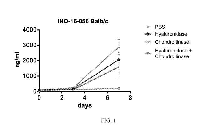
[00014] Figure 1 shows the levels (ng/ml) of hIgG measured by ELISA between
days
0-7 in groups 1 to 4 (Balb/c mice, 6-7 weeks). Group 1 (grey) PBS and pGX9214,
group 2
hyaluronidase pretreatment and pGX9214 (blue), group 3 chondroitinase and
pGX9214
(green), group 4 hyaluronidase/ chondroitinase pretreatment and pGX9214
(brown).
[00015] Figure 2 shows levels (ng/ml) of hIgG measured by ELISA between
days 0-7
in groups 1 to 4 (C57BL/6, 6-7 weeks). Group 1 (grey) PBS and pGX9214, group 2
hyaluronidase pretreatment and pGX9214 (blue), group 3 chondroitinase and
pGX9214
(green), group 4 hyaluronidase/ chondroitinase pretreatment and pGX9214
(brown).
[00016] Figure 3 shows levels (ng/ml) of hIgG measured by ELISA between
days 0-
21 in groups 1 to 4. Group 1 (grey) PBS and pGX9214, group 2 hyaluronidase
pretreatment
and pGX9214 (dark red), group 3 chondroitinase and pGX9214 (bright red), group
4
hyaluronidase/ chondroitinase pretreatment and pGX9214 (rose ¨ Hartley Guinea
pig).
[00017] Figure 4 shows levels (ng/ml) of hIgG measured by ELISA between
days 0-
21 in individual guinea pigs in groups 1 to 4. Figure 4A: Group 1 PBS. Figure
4B: group 2
hyaluronidase pretreatment with 400U/ml. Figure 4C: group 3 chondroitinase
(0.5U/m1).
Figure 4D: group 4 hyaluronidase 400U/m1 and chondroitinase 0.5U/m1
pretreatment.
[00018] Figure 5 shows chondroitinase enhances plasmid-encoded hlgG
expression in
Balb/c mice. (a) The levels of hIgG [ng/m11 measured by ELISA between days 0-7
in groups
1 and 2 (Balb/c mice, 6-7 weeks). Group 1 (grey) PBS and pGX9214, group 2
chondroitinase
pretreatment and pGX9214 (black). (b) Significant enhancement of hIgG by
chondroitinase at
day 7. Statistics performed by Mann Whitney test, P = 0.0079.
[00019] Figure 6 shows chondroitinase enhances plasmid-encoded hIgG expression
in
C57BL/6 mice. (a) The levels of hIgG [ng/m11 measured by ELISA between days 0-
7 in
groups 1 and 2 (Balb/c mice, 6-7 weeks). Group 1 (grey) PBS and pGX9214, group
2
chondroitinase pretreatment and pGX9214 (black). (b) Significant enhancement
of hIgG by
chondroitinase at day 7. Statistics performed by Mann Whitney test, P =
0.0079.
[00020] Figure 7 shows an investigation of the ability of different versions
of
chondroitinase to enhance DMAb expression. Graph represents hIgG levels (ng/
ml)
measured by ELISA at day 7 in groups 1-4 (Balb/c). Mice were treated with
either treated
with PBS (control group 1), Chondroitinase AC, clinical grade Chondroitinase
ABC or the
4
CA 03025146 2018-11-21
WO 2017/190147
PCT/US2017/030447
recombinant protein GALNS. All groups received an injection of pGX9203
followed by
electroporation. Statistics performed by Kruskal-Wallis test, **P = 0.0026.
[00021] Figure 8 shows increasing Chondroitinase ABC dose further enhances DNA-
based
protein expression. Balb/c mice were treated with increasing doses of
Chondroitinase ABC
30 minutes prior pDNA (pGX9207) injection and electroporation. Levels of hIgG
were
measured by ELISA. Statistics performed by Kruskal-Wallis test, *P = 0.0357.
[00022] Figure 9 shows chondroitinase administration results in enhanced
fluorescent
protein expression. (a) Left and right hindlimbs of Balb/c mice treated with
either
Chondroitinase or PBS into the skeletal muscle. Visualization of the reporter
protein
expression was performed by a fluorescence imager (Protein Simple). (b)
Arbitrary units
fluorescence intensity parallel reporter gene expression and were quantified
by using
AlphaView SA software. Statistics performed by Mann Whitney test, P = 0.0022.
[00023] Figure 10 shows representative histopathology of murine hindlimb
skeletal muscle
performed by H&E staining. Top panels show tissue treated with either PBS only
(control) or
Chondroitinase ABC only. Preatreated (30 min) muscles with either
chondroitinase or PBS
(control) before pDNA delivery are presented in the bottom figures. Results
were analyzed by
a slide scanner and CaseViewer software (3DHISTECH). Scale = 200 nm.
[00024] Figure 11 shows chondroitinase ABC enhances plasmid-encoded hIgG
expression
in New Zealand rabbits (9 weeks). (a) Presented are the levels of hIgG [ng/m11
measured by
ELISA between days 0 and 6 in group 2 (chondroitinase-pretreated, grey) and in
group 1
(PBS control, black). (b) Graph shows hIgG levels from day 6 measured by
ELISA. Statistics
performed by Mann Whitney test, P = 0.0043.
[00025] Figure 12 shows co-formulation of chondroitinase with pDNA (pGX9207)
in mice
(Balb/c; 14 mice per group). Graph shows hIgG levels [ng/m11 from day 6
measured by
ELISA. Statistics performed by Mann Whitney test, ****P < 0.0001.
[00026] Figure 13 shows chondroitinase administration results in enhanced
fluorescent
protein expression. (a) Left and right hindlimbs of Balb/c mice treated with
either
Chondroitinase or PBS into the skeletal muscle. Visualization of the reporter
protein
expression was performed by a fluorescence imaging system. (b) Arbitrary units
fluorescence
intensity parallel reporter gene expression and were quantified by using
AlphaView SA
software. Statistics performed by Mann Whitney test, **P = 0.0004.
[00027] Figure 14 shows co-formulation of chondroitinase with pDNA (pGX9207)
in
rabbits (New Zealand rabbits; 6 rabbits per group). (a) Graph shows serum hIgG
levels
[ng/m11 from day 0 to day 5 measured by ELISA. Comparison of groups 1, 2 and
4. (b)
CA 03025146 2018-11-21
WO 2017/190147
PCT/US2017/030447
Rabbit serum hIgG levels of all groups measured at day 5. Statistics performed
by Mann
Whitney test, ****P < 0.0001.
[00028] Figure 15 shows agarose gel electrophoresis of chondroitinase (2.5 U/
ml)/ pDNA
(pGX9207, 250 ng per well) coformulated samples. (a) Lanes with even numbers
present
pDNA samples containing Chondroitinase ABC, lanes with odd numbers refer to
PBS
negative controls. Lanes 1-2: No incubation of the sample before gel
electrophoresis, lanes 3-
4: incubation at RT (21 C) for 10 min, lanes 5-6: 6 C, 120 min, lanes 7-8: RT,
120 min, lanes
9-10: 6 C, 24 hrs, Lanes 11-12: RT, 24 hrs, lanes 13-14: 6 C, 10 min. M1 =
marker. (b)
Ladder for supercoiled DNA, 2-10 kb (New England Biolabs).
[00029] Figure 16 shows storage of chondroitinase/ pDNA coformulation for 24
hours
does not affect enhanced gene expression in Balb/c mice. Graph represents
serum hIgG levels
(ng/ ml) measured by ELISA at day 6. Mice were treated with coformulations
into the left
hindlimb skeletal muscle and electroporation was performed after 1 min of drug
injection.
Prior to treatments, coformulation samples were incubated at 10 min, 120 min
and 24 hours
at 4 C. Controls: Mice were pretreated with either chondroitinase or PBS 30
min before
pDNA administration and electroporation. Statistics performed by Kruskal-
Wallis test, ****P
= 0.0001.
[00030] Figure 17 shows the levels (ng/ml of hIgG measured by ELISA between
days 0-28
in groups 1, 2 and 3. Group 1 was treated with pDVSF-1 and hyaluronidase
pretreatment,
Group 2 was treated with pDVSF only, and Group 3 was treated with PBS only.
[00031] Figure 18 shows the anti-hIgG binding titers of groups 1 and 2.
[00032] Figure 19 shows the mean (+/- SEM) IFN-y (spots per million) response
in
PBMCs of groups 1-3 to the Influenza NP peptide pools in Example 2.
[00033] Figure 20 shows the anti-Influenza NP IgG binding titers for groups 1-
3 in
Example 2.
[00034] Figure 21 shows the P. aeruginosa acute pneumonia model.
Electroporation of
anti-PcrV or DMAb-antibody1-2 plasmid DNA yields expression of active IgG in
mice.
Potent protective activity observed for both anti-Pseudomonal DMAbs and DMAb-
antibody1-2 IgG.
[00035] Figure 22 shows IgG quantification of DMAb expression in serum. Serum
was
evaluated for DMAb expression prior to P. aeruginosa infection. Despite
similar survival
profiles, anti-PcrV DMAb expression was 5-fold greater than DMAb-antibody1-2.
6
CA 03025146 2018-11-21
WO 2017/190147
PCT/US2017/030447
[00036] Figure 23 shows the reduction of organ burden by anti-Pseudomonal
DMAbs.
DMAb-antibody1-2 and IgG reduces burden in the lung. Anti-PcrV and DMAb-
antibody1-2
significantly reduce systemic spread of bacteria. Tissues collected 24 hour
post-infection.
LOD = limit of detection; * = P<0.05 by Kruskal-Wallis and Dunn's multiple
comparison test
vs. control IgG DMAb.
[00037] Figure 24 shows the histology of acute pneumonia at 48 hours post-
infection
(H&E). A. Control IgG lungs exhibit areas of severe alveolar infiltrates
comprised of
neutrophils and macrophages and hemorrhage (10x). C. Mild pneumonia and
occasional
bronchiolar debris (10x). E. DMAb-antibody1-2 DNA group with mild alveolitis
(10x). B,
D, F. Insets at 40x from A. C. D., respectively.
[00038] Figure 25 shows that DMAb-antibody1-2 exhibits concentration dependent
protective activity. A. Animals were received DNA at 1, 2 or 3 sites prior to
EP. B.
Quantification of serum IgG. * indicates P<0.05 via Log-Rank test.
[00039] Figure 26 shows subtherapeutic dosages of DMAb-antibody1-2 and
meropenem
(MEM) exhibit enhanced activity against P. aeruginosa pneumonia. Animals were
received
DNA at 1, 2 or 3 sites prior to EP. B. Quantification of serum IgG. *
indicates P<0.05 via
Log-Rank test.
[00040] Figure 27 shows the mean (+/- SEM) IFN-y (spots per million) response
in
splenocytes to NP55 and NP147 peptide epitopes 14 days after pGX2013
immunization.
[00041] Figure 28 shows the anti-NP IgG binding titers for groups 7 and 14
days after
immunization with pGX2013.
[00042] Figure 29 shows in vitro expression of anti-MERS-CoV human IgG. (a)
Diagrammatic illustration of the anti-MERS-CoV antigen DMAb plasmid DNA pMERS.
CMV promoter situated upstream of the antibody heavy and light chain sequences
separated
by furin and 2A cleavage sites. (b & c) 293T cell cultures were transfected 1
pg/ml of pVax
or pMERS and culture supernatants harvested after 48 hours. (b) human IgG
levels in the
supernatant were assayed for by ELISA. (c) IgG binding to MERS CoV antigen was
measured by ELISA.
[00043] Figure 30 shows enhanced in vivo expression of DMAb in BALB/c mice
[00044] 6.25 to 100 pg of pMERS was administered into the TA muscle of BALB/c
mice
(4-8 mice per group), (a) by injection only (No EP), (b) injection with EP
(EP) and (c)
injection with EP into HYA-treated muscle (EP + HYA). (a-c) Serum human IgG
levels were
quantified (data points represent the Mean+/-SEM) by ELISA on day 6 after
pMERS
delivery. (d) MERS CoV antigen binding of reciprocal serum dilutions measured
by ELISA
7
CA 03025146 2018-11-21
WO 2017/190147
PCT/US2017/030447
on day 0 and day 6 after pMERS delivery. (e) BALB/c mice TA muscles were
harvested 72
hours after administration pRFP (25 pg) reporter gene with the protocol
employed in (a), (b)
or (c) or no treatment in inserts 1-4, respectively. Images illustrate
reporter gene expression.
Immunofluorescence images of sections of the TA muscle treated with pMERS or
pVax (100
pg) delivered with EP + HYA, and harvested 72 hours later (f). hIgG was
detected with anti-
human IgG followed by a FITC-labelled secondary antibody (green). DAPI stain
in blue.
Panel 1. No treatment. Panel 2. pVax. Panels 3 & 4. pMERS. Panels 1-3 display
a cross-
sectional image perpendicular to muscle fibers, and in Panel 4 the image is
along the muscle
fibers.
[00045] Figure 31 shows increased and sustained DMAb expression in Crl:Nu-
Foxnlnu
mice. 6.25 to 100 pg of pMERS was administered with EP into the HYA pretreated
TA
muscle of Crl:Nu-Foxnlnu mice (8 mice per group). Serum hIgG was quantified by
ELISA
on days 0 to 160 (a). (b) 100 pg of pMERS was administered as in (a) to 1
(left TA), 2 (right
and left TA), 3 (right and left TA, and left Quad) or 4 (right and left TA,
and left and right
Quad) muscle. Serum hIgG was quantified by ELISA on day 21.
[00046] Figure 32 shows in vivo DMAb expression in the New Zealand white
rabbit. (a)
Reporter gene expression in the rabbit TA muscle sections 72 hours after
delivery of pGFP
(0.2 mg) with EP in PBS- (top panel) or HYA- (bottom panel) treated muscle.
(b&c) 2 mg of
pMERS was administered with EP into the Quad muscle (pre-treated with PBS or
HYA) of
rabbits (6 per group). Serum hIgG was quantified on days 3, 5 and 6 (a), and
MERS CoV
antigen binding measured (b) by ELISA on day 6 after delivery. (c) 2 mg of
pMERS was
administered with EP at voltage setting of 20 to 65 V into the Quad muscle
(treated with
HYA) of rabbits (6 per group), and serum hIgG was quantified on day 5 after
delivery. (c &
d) Values are depicted as mean +/- SEM (n = 6/group). ****p < 0.0001, ***p <
0.001, **p <
0.01, and ns = non-significant. P values are from unpaired, two-tailed Mann-
Whitney tests.
[00047] Figure 33 shows In vivo DMAb expression in the rhesus macaques. 13.5
mg of
pMERS was administered with EP into the quad muscles (pre-treated with HYA) of
rhesus
macaques (5 per group). Serum hIgG was quantified (a) and MERS CoV antigen
binding
measured by ELISA on days 0 to 35 (b), and day 17 reciprocal serum dilution
(c) for each
rhesus macaque. (d) Antibody response to human IgG (ADA) was measured by
direct ELISA
in the serum of rhesus macaques. (e) The correlation of DMAb levels and anti-
human IgG
antibody binding levels in the serum is depicted. Plotted are data points
(hIgG (pg/ml) with
corresponding ADA (0D450nm)) after and including the peak hIgG (pg/ml) value
was
8
CA 03025146 2018-11-21
WO 2017/190147
PCT/US2017/030447
reached in each Rhesus macaque. P value and Spearman correlation coefficient
calculated
using GraphPad Prism 6 software.
[00048] Figure 34 shows In vivo expression kinetics of DMAb in BALB/c mice. (a-
c)
6.25 to 100 pg of pMERS was administered into the TA muscle of BALB/c mice (4-
8 mice
per group), (a) by injection only (No EP), (b) injection with EP (EP) and (c)
injection with EP
into HYA-treated muscle (EP + HYA). (a-c) Serum human IgG levels were
quantified (data
points represent the Mean+/-SEM) by ELISA on day 0 to 14 after pMERS delivery.
[00049] Figure 35 shows anti-antibody response to human IgG in pMERS treated
BALB/c
mice. Antibody response to human IgG (ADA) was measured by direct ELISA in the
serum
of BALB/c mice on days 0 to 14 after pMERS delivery with EP.
[00050] Figure 36 shows screen of pDNA delivery reagents reported to enhance
gene
expression. 100 pg of pMERS was administered into the TA muscle of BALB/c mice
(5
mice per group). The target TA muscle was pretreated 30 min before pMERS
delivery with
PBS, HYA, 7% Sucrose, Collagenase D, Elastase or MMP7 in groups 1, 2, 5, 7, 8,
and 9
respectively. pMERS was coformulated with Poly-L-Glutamic acid (Group 3) or
Tempol
(Group 4) or (OH)3D3 (Group 6), and there was no pretreatment. Serum human IgG
levels
were quantified (data points represent the mean+/-SEM) by ELISA on day 6 after
pMERS
delivery.
[00051] Figure 37 shows DMAb expression in rabbits. (a and b) 2 mg of pGX9207
was
administered with EP into the Quad muscle (pre-treated with HYA) of New
Zealand white
rabbits (6 per group). Serum hIgG was (a) and anti-human IgG (ADA) binding (b)
was
assayed by ELISA on days 0 to 10.
[00052] Figure 38 shows co-formulation study of HYA with pDNA in rabbits. Six
New
Zealand white rabbits per group. 2 mg pGX9207 in left quad muscle. Two sites.
HYA
(Intropharma) 200U per site. CELLECTRA0-5P pDNA/HYA co-formulated 5 minutes
before administration. Blood was sampled on day 5. Experiment number INO-16-
158b.
[00053] Figure 39 shows co-formulation of HYA with pDNA in rabbits with EP
delay. Six
New Zealand white rabbits per group. 2 mg pGX9207 in left quad muscle. Two
sites. HYA
(Intropharma) 200 U per site. CELLECTRA0-5P pDNA/HYA co-formulated 5 minutes
before administration. Blood was sampled on day 5. Experiment number INO-16-
158a.
[00054] Figure 40 shows co-formulation of HYA with pDNA in rhesus macaques.
pGX9207 (pMERS) delivered into quad muscle with CELLECTRA0-5P. 4 sites. 1 mg
pDNA per site. Intropharma (bovine testes purified HYA). pDNA /HYA co-
formulated 5
minutes before administration. Experiment number INO-16-194.
9
CA 03025146 2018-11-21
WO 2017/190147
PCT/US2017/030447
[00055] Figure 41 shows optimization of EP delay with Hylenex (human
recombinant
hyaluronidase). pGX9207 HYA co-formulation. Two Tx's in left quad. Day 5 hlgG
serum
levels are depicted. Experiment number INO-16-279.
[00056] Figure 42 shows DNA vaccine dose sparing effect of HYA co-formulation.
BALB/c mice. Day 0 influenza pNP IM CELLECTRA-3P, day 7 and 14 ELISA.
Experiment number INO-16-218.
[00057] Figure 43 shows augmentation of immune response to tumor antigen with
DNA
vaccine HYA formulation. B6 mice. Day 0 and 14 pmTERT IM CELLECTRAO-3P, day 21
IFN-gamma ELISpot against native mouse TERT peptide pools. Experiment number
IND-
17-018.
DETAILED DESCRIPTION
[00058] The present invention relates to compositions for and methods of
delivering an
agent to a subject. The methods may include administering to the subject an
agent, and a
chondroitinase polypeptide or a polynucleotide encoding a chondroitinase
polypeptide in an
amount sufficient to hydrolyze sulfate groups of CSPGs. The chondroitinase may
hydrolyze
the sulfate groups of CSPGs in the subject. This disorganization may lead to
disorganization
of the extracellular matrix and thereby facilitate the delivery of the agent.
Methods may
further include administering to the subject a hyaluronidase polypeptide or a
polynucleotide
encoding a hyaluronidase polypeptide.
[00059] The present invention also relates to administration of hyaluronidase
along with
an agent to a subject. The hyaluronidase may be administered as a polypeptide
or as a
nucleic acid encoding hyaluronidase or a fragment or variant thereof The
hyaluronidase may
facilitate delivery of the agent to the subject. As a result, the
hyaluronidase may enhance an
immune response in the subject and/or enhance expression of the agent in the
subject.
1) Definitions
[00060] Unless otherwise defined, all technical and scientific terms used
herein have
the same meaning as commonly understood by one of ordinary skill in the art.
In case of
conflict, the present document, including definitions, will control. Preferred
methods and
materials are described below, although methods and materials similar or
equivalent to those
described herein can be used in practice or testing of the present invention.
All publications,
patent applications, patents and other references mentioned herein are
incorporated by
reference in their entirety. The materials, methods, and examples disclosed
herein are
illustrative only and not intended to be limiting.
CA 03025146 2018-11-21
WO 2017/190147
PCT/US2017/030447
[00061] The terms "comprise(s)," "include(s)," "having," "has," "can,"
"contain(s),"
and variants thereof, as used herein, are intended to be open-ended
transitional phrases,
terms, or words that do not preclude the possibility of additional acts or
structures. The
singular forms "a," "an," and "the" include plural references unless the
context clearly
dictates otherwise. The present disclosure also contemplates other embodiments
"comprising," "consisting of," and "consisting essentially of," the
embodiments or elements
presented herein, whether explicitly set forth or not.
[00062] The term "about" as used herein as applied to one or more values of
interest,
refers to a value that is similar to a stated reference value. In certain
aspects, the term
"about" refers to a range of values that fall within 20%, 19%, 18%, 17%, 16%,
15%, 14%,
13%, 12%, 11%, 10%, 9%, 8%, 7%, 6%, 5%, 4%, 3%, 2%, 1%, or less in either
direction
(greater than or less than) of the stated reference value unless otherwise
stated or otherwise
evident from the context (except where such number would exceed 100% of a
possible
value).
[00063] "Antibody" may mean an antibody of classes IgG, IgM, IgA, IgD, or
IgE, or
fragments, fragments or derivatives thereof, including Fab, F(ab')2, Fd, and
single chain
antibodies, and derivatives thereof The antibody may be an antibody isolated
from the serum
sample of mammal, a polyclonal antibody, a monoclonal antibody, affinity
purified antibody,
or mixtures thereof which exhibits sufficient binding specificity to a desired
epitope or a
sequence derived therefrom. The antibody may be a synthetic antibody as
described herein.
[00064] "Antibody fragment" or "fragment of an antibody" as used
interchangeably
herein refers to a portion of an intact antibody comprising the antigen-
binding site or variable
region. The portion does not include the constant heavy chain domains (i.e.,
CH2, CH3, or
CH4, depending on the antibody isotype) of the Fc region of the intact
antibody. Examples
of antibody fragments include, but are not limited to, Fab fragments, Fab'
fragments, Fab'-SH
fragments, F(ab')2 fragments, Fd fragments, Fv fragments, diabodies, single-
chain Fv (scFv)
molecules, single-chain polypeptides containing only one light chain variable
domain, single-
chain polypeptides containing the three CDRs of the light-chain variable
domain, single-
chain polypeptides containing only one heavy chain variable region, and single-
chain
polypeptides containing the three CDRs of the heavy chain variable region.
[00065] "Fragment" as used herein means a nucleic acid sequence or a
portion thereof
that encodes a polypeptide capable of eliciting an immune response in a
mammal. The
fragments can be DNA fragments selected from at least one of the various
nucleotide
sequences that encode protein fragments set forth below. "Fragment" may also
refer to a
11
CA 03025146 2018-11-21
WO 2017/190147
PCT/US2017/030447
polypeptide sequence or a portion thereof that is capable of eliciting an
immune response in a
mammal.
[00066] "Immune response" as used herein means the activation of a host's
immune
system, e.g., that of a mammal, in response to the introduction of antigen.
The immune
response can be in the form of a cellular or humoral response, or both.
[00067] "Operably linked" as used herein means that expression of a gene is
under the
control of a promoter with which it is spatially connected. A promoter can be
positioned 5'
(upstream) or 3' (downstream) of a gene under its control. The distance
between the
promoter and a gene can be approximately the same as the distance between that
promoter
and the gene it controls in the gene from which the promoter is derived. As is
known in the
art, variation in this distance can be accommodated without loss of promoter
function.
[00068] A "peptide," "protein," or "polypeptide" as used herein can mean a
linked
sequence of amino acids and can be natural, synthetic, or a modification or
combination of
natural and synthetic.
[00069] "Polynucleotide" or "oligonucleotide" or "nucleic acid" as used
herein means
at least two nucleotides covalently linked together. A polynucleotide can be
single stranded
or double stranded, or can contain portions of both double stranded and single
stranded
sequence. The polynucleotide can be DNA, both genomic and cDNA, RNA, or a
hybrid.
The polynucleotide can contain combinations of deoxyribo- and ribo-
nucleotides, and
combinations of bases including uracil, adenine, thymine, cytosine, guanine,
inosine,
xanthine hypoxanthine, isocytosine, isoguanine, and synthetic or non-naturally
occurring
nucleotides and nucleosides. Polynucleotides can be obtained by chemical
synthesis methods
or by recombinant methods.
[00070] "Promoter" as used herein means a synthetic or naturally-derived
molecule
which is capable of conferring, activating, or enhancing expression of a
nucleic acid in a cell.
A promoter can comprise one or more specific transcriptional regulatory
sequences to further
enhance expression and/or to alter the spatial expression and/or temporal
expression of same.
A promoter can also comprise distal enhancer or repressor elements, which can
be located as
much as several thousand base pairs from the start site of transcription. A
promoter can be
derived from sources including viral, bacterial, fungal, plants, insects, and
animals. A
promoter can regulate the expression of a gene component constitutively or
differentially
with respect to the cell, the tissue, or organ in which expression occurs, or
with respect to the
developmental stage at which expression occurs, or in response to external
stimuli such as
physiological stresses, pathogens, metal ions, or inducing agents.
Representative examples of
12
CA 03025146 2018-11-21
WO 2017/190147
PCT/US2017/030447
promoters include the bacteriophage T7 promoter, bacteriophage T3 promoter,
SP6 promoter,
lac operator-promoter, tac promoter, SV40 late promoter, SV40 early promoter,
RSV-LTR
promoter, CMV IE promoter, SV40 early promoter or SV40 late promoter, and the
CMV IE
promoter.
[00071] "Subject" as used herein can mean a mammal. The mammal can be a
human,
chimpanzee, dog, cat, horse, cow, mouse, or rat.
[00072] "Treatment" or "treating," as used herein can mean protection of an
animal
from a disease through means of preventing, suppressing, repressing, or
completely
eliminating the disease. Preventing the disease can include administering a
composition of
the present invention to an animal prior to onset of the disease. Suppressing
the disease
involves administering a composition of the present invention to an animal
after induction of
the disease, but before its clinical appearance. Repressing the disease
involves administering
a composition of the present invention to an animal after clinical appearance
of the disease.
[00073] "Variant" as used herein with respect to a nucleic acid means (i) a
portion or
fragment of a referenced nucleotide sequence; (ii) the complement of a
referenced nucleotide
sequence or portion thereof; (iii) a nucleic acid that is substantially
identical to a referenced
nucleic acid or the complement thereof; or (iv) a nucleic acid that hybridizes
under stringent
conditions to the referenced nucleic acid, complement thereof, or a sequences
substantially
identical thereto.
[00074] "Variant" can further be defined as a peptide or polypeptide that
differs in
amino acid sequence by the insertion, deletion, or conservative substitution
of amino acids,
but retains at least one biological activity. Representative examples of
"biological activity"
include the ability to be bound by a specific antibody or to promote an immune
response.
Variant can also mean a protein with an amino acid sequence that is
substantially identical to
a referenced protein with an amino acid sequence that retains at least one
biological activity.
A conservative substitution of an amino acid, i.e., replacing an amino acid
with a different
amino acid of similar properties (e.g., hydrophilicity, degree and
distribution of charged
regions) is recognized in the art as typically involving a minor change. These
minor changes
can be identified, in part, by considering the hydropathic index of amino
acids, as understood
in the art (Kyte et al., I Mol. Biol. 1982, 157, 105-132). The hydropathic
index of an amino
acid is based on a consideration of its hydrophobicity and charge. It is known
in the art that
amino acids of similar hydropathic indexes can be substituted and still retain
protein function.
In one aspect, amino acids having hydropathic indexes of 2 are substituted.
The
hydrophilicity of amino acids can also be used to reveal substitutions that
would result in
13
CA 03025146 2018-11-21
WO 2017/190147
PCT/US2017/030447
proteins retaining biological function. A consideration of the hydrophilicity
of amino acids in
the context of a peptide permits calculation of the greatest local average
hydrophilicity of that
peptide, a useful measure that has been reported to correlate well with
antigenicity and
immunogenicity. Substitution of amino acids having similar hydrophilicity
values can result
in peptides retaining biological activity, for example immunogenicity, as is
understood in the
art. Substitutions can be performed with amino acids having hydrophilicity
values within 2
of each other. Both the hydrophobicity index and the hydrophilicity value of
amino acids are
influenced by the particular side chain of that amino acid. Consistent with
that observation,
amino acid substitutions that are compatible with biological function are
understood to
depend on the relative similarity of the amino acids, and particularly, the
side chains of those
amino acids, as revealed by the hydrophobicity, hydrophilicity, charge, size,
and other
properties.
[00075] A variant may be a nucleic acid sequence that is substantially
identical over
the full length of the full gene sequence or a fragment thereof The nucleic
acid sequence
may be 80%, 81%, 82%, 83%, 84%, 85%, 86%, 87%, 88%, 89%, 90%, 91%, 92%, 93%,
94%, 95%, 96%, 97%, 98%, 99%, or 100% identical over the full length of the
gene sequence
or a fragment thereof A variant may be an amino acid sequence that is
substantially identical
over the full length of the amino acid sequence or fragment thereof The amino
acid
sequence may be 80%, 81%, 82%, 83%, 84%, 85%, 86%, 87%, 88%, 89%, 90%, 91%,
92%,
93%, 94%, 95%, 96%, 97%, 98%, 99%, or 100% identical over the full length of
the amino
acid sequence or a fragment thereof
[00076] "Vector" as used herein means a nucleic acid sequence containing an
origin of
replication. A vector can be a viral vector, bacteriophage, bacterial
artificial chromosome, or
yeast artificial chromosome. A vector can be a DNA or RNA vector. A vector can
be a self-
replicating extrachromosomal vector, and preferably, is a DNA plasmid.
[00077] For the recitation of numeric ranges herein, each intervening
number there
between with the same degree of precision is explicitly contemplated. For
example, for the
range of 6-9, the numbers 7 and 8 are contemplated in addition to 6 and 9, and
for the range
6.0-7.0, the number 6.0, 6.1, 6.2, 6.3, 6.4, 6.5, 6.6, 6.7, 6.8, 6.9, and 7.0
are explicitly
contemplated.
2) Use of Chondroitinase to Enhance Agent Delivery
[00078] The present invention relates to administration of chondroitinase
along with an
agent to a subject. The chondroitinase may be administered as a polypeptide or
as a nucleic
acid encoding chondroitinase or a fragment or variant thereof The
chondroitinase may
14
CA 03025146 2018-11-21
WO 2017/190147
PCT/US2017/030447
facilitate delivery of the agent to the subject. As a result, the
chondroitinase may enhance an
immune response in the subject and/or enhance expression of the agent in the
subject.
a) Chondroitinase
[00079] A chondroitinase or fragment thereof may be administered to a
subject. The
chondroitinase may be any chondroitinase. For example, the chondroitinase may
be an N-
acetylgalactosamine-4-sulfatase, an N-acetylgalactosamine-6-sulfatase, or a
chondroitin ABC
lyase. The chondroitinase may be chondroitinase AC. The chondroitinase may be
recombinant chrondroitinase. The chondroitinase may catalyze the hydrolysis of
a
chondroitin sulfate proteoglycan (CSPG). The chondroitinase may hydrolyze the
4-sulfate
groups of the N-acetyl-D-galactosamine 4-sulfate units of chondroitin sulfate
and/or
dermatan sulfate. The chondroitinase may hydrolyze the 4-sulfate groups of N-
acetyl
glucosamine 4-sulfate. The chondroitinase may hydrolyze the 6-sulfate groups
of the N-
acetyl-D-galactosamine 6-sulfate units of chondroitin sulfate and of the D-
galactose 6-sulfate
units of keratin sulfate. The chondroitin ABC lyase may catalyze the
degradation of
polysaccharides containing 1,4-beta-D-hexosaminyl and 1,3-beta-D-glucuronosyl
or 1,3-
alpha-L-iduronosyl linkages to disaccharides containing 4-deoxy-beta-D-gluc-4-
enuronosyl
groups. The chondroitin ABC lyase may act on chondroitin 4-sulfate,
chondroitin 6-sulfate,
and dermatan sulfate.
[00080] The CSPG may be Aggrecan (CSPG1), Versican (CSPG2), Neurocan
(CSPG3), CSPG4 (melanoma-associated chondroitin sulfate proteoglycan, NG2),
CSPG5,
SMC3 (CSPG6, Structural maintenance of chromosome 3), Brevican (CSPG7), CD44
(CSPG8, cluster of differentiation 44), Phosphacan, and combinations thereof
[00081] By catalyzing the hydrolysis of CSPGs, constituents of the
extracellular matrix
(ECM), chondroitinase lowers the viscosity of the CSPGs and the extracellular
matrix,
thereby increasing tissue permeability. Administration of chondroitinase, or a
polynucleotide
encoding chondroitinase, may lead to hydrolysis of CSPGs, thereby leading to
disorganization of an extracellular matrix of the subject. Disorganization of
an extracellular
matrix of the subject may thereby facilitate delivery or administration of an
agent.
[00082] The chondroitinase may be a chondroitinase derived from a
bacterium. The
bacterium may be Flavobacterium heparinum, for example. The chondroitinase may
be
recombinantly produced in a bacterium.
[00083] In some embodiments, a chondroitinase polypeptide or fragment
thereof may
be administered.
CA 03025146 2018-11-21
WO 2017/190147
PCT/US2017/030447
[00084] In some embodiments, a polynucleotide encoding a chondroitinase
polypeptide or fragment thereof may be administered. The chondroitinase
polypeptide may
be expressed from the polynucleotide encoding the chondroitinase polypeptide
in vivo.
3) Use of Hyaluronidase
[00085] The methods may also comprise further administering to the subject
a
hyaluronidase polypeptide or a polynucleotide encoding a hyaluronidase
polypeptide in an
amount to degrade a glycosaminoglycan, such as hyaluronan. Administration of
chondroitinase and hyaluronidase may lead to an additive or synergistic effect
on agent
expression, immune response, or concentration of agent in the subject's serum,
for example.
The agent concentration detected in the serum of a subject exposed to
chondroitinase and
hyaluronidase combination treatment may be higher as compared to serum levels
of the agent
in a subject treated with a single chondroitinase or hyaluronidase enzyme, for
example.
[00086] The present invention also relates to administration of
hyaluronidase along
with an agent to a subject. The hyaluronidase may be administered as a
polypeptide or as a
nucleic acid encoding hyaluronidase or a fragment or variant thereof The
hyaluronidase may
facilitate delivery of the agent to the subject. As a result, the
hyaluronidase may enhance an
immune response in the subject and/or enhance expression of the agent in the
subject. The
method of administering hyaluronidase to a subject may or may not include
administration of
chondroitinase. The agent may be any agent as described herein. The use of
hyaluronidase
may be in conjunction with any agent, described timing of administration, mode
of
administration, method of treatment, or method of delivery, as described
herein.
[00087] The hyaluronidase polypeptide or the polynucleotide encoding the
hyaluronidase polypeptide and the agent may be administered to the subject
concurrently.
The hyaluronidase polypeptide or the polynucleotide encoding the hyaluronidase
polypeptide
may be administered concurrently with the chondroitinase polypeptide, or the
polynucleotide
encoding the chondroitinase polypeptide, and the agent. The methods of
delivery and times of
administration may be the same as, or similar, to those described herein. See
section 4 b), for
example.
[00088] The hyaluronidase polypeptide or the polynucleotide encoding the
hyaluronidase polypeptide and the agent may be administered to the subject
consecutively.
The hyaluronidase polypeptide or the polynucleotide encoding the hyaluronidase
polypeptide
and the chondroitinase polypeptide, or the polynucleotide encoding the
chondroitinase
polypeptide, and the agent may be administered to the subject consecutively.
The methods of
delivery and times of administration may be the same as, or similar, to those
described herein.
16
CA 03025146 2018-11-21
WO 2017/190147
PCT/US2017/030447
See section 4 b), for example. The chondroitinase polypeptide or the
polynucleotide
encoding the chondroitinase polypeptide, the hyaluronidase polypeptide or a
polynucleotide
encoding a hyaluronidase polypeptide, and the agent may be co-formulated prior
to
administration. The chondroitinase polypeptide or the polynucleotide encoding
the
chondroitinase polypeptide, and the hyaluronidase polypeptide or a
polynucleotide encoding
a hyaluronidase polypeptide, may be co-formulated prior to administration. Any
co-
formulation of chondroitinase polypeptide or the polynucleotide encoding the
chondroitinase
polypeptide, the hyaluronidase polypeptide or a polynucleotide encoding a
hyaluronidase
polypeptide, and the agconent may occur, for example, 1 minute, 5 minutes, 10
minutes, 15
minutes, 20 minutes, 45 minutes, 1 hour, 5 hours, 10 hours, 24 hours, 5 days,
7 days, 20 days,
30 day, 40 days, 50 days, 100 days, 200 days, 300 days, 1 year, 400 days, 1.5
years, or 2
years prior to administration. Any co-formulation of chondroitinase
polypeptide, or the
polynucleotide encoding the chondroitinase polypeptide, and the agent may
occur, for
example, 1 minute, 5 minutes, 10 minutes, 15 minutes, 20 minutes, 45 minutes,
1 hour, 5
hours, 10 hours, 24 hours, 5 days, 7 days, 20 days, 30 days, 40 days, 50 days,
100 days, 200
days, 300 days, 1 year, 400 days, 1.5 years, or 2 years prior to
administration. Any co-
formulation of hyaluronidase polypeptide, or a polynucleotide encoding a
hyaluronidase
polypeptide, and the agent may occur, for example, 1 minute, 5 minutes, 10
minutes, 15
minutes, 20 minutes, 45 minutes, 1 hour, 5 hours, 10 hours, 24 hours, 5 days,
7 days, 20 days,
30 day, 40 days, 50 days, 100 days, 200 days, 300 days, 1 year, 400 days, 1.5
years, or 2
years prior to administration. Any co-formulation of chondroitinase
polypeptide or the
polynucleotide encoding the chondroitinase polypeptide, and the hyaluronidase
polypeptide
or a polynucleotide encoding a hyaluronidase polypeptide, may occur, for
example, 1 minute,
minutes, 10 minutes, 15 minutes, 20 minutes, 45 minutes, 1 hour, 5 hours, 10
hours, 24
hours, 5 days, 7 days, 20 days, 30 day, 40 days, 50 days, 100 days, 200 days,
300 days, 1
year, 400 days, 1.5 years, or 2 years prior to administration days prior to
administration.
[00089] The chondroitinase and/or hyaluronidase and/or agent can be
administered via
electroporation (EP), such as by a method described in U.S. Patent No.
7,664,545, the
contents of which are incorporated herein by reference. The electroporation
can be by a
method and/or apparatus described in U.S. Patent Nos. 6,302,874; 5,676,646;
6,241,701;
6,233,482; 6,216,034; 6,208,893; 6,192,270; 6,181,964; 6,150,148; 6,120,493;
6,096,020;
6,068,650; and 5,702,359, the contents of which are incorporated herein by
reference in their
entirety. The electroporation may be carried out via a minimally invasive
device.
Electroporation may occur before or after administration of the chondroitinase
and/or
17
CA 03025146 2018-11-21
WO 2017/190147
PCT/US2017/030447
hyaluronidase and/or agent, for example. Electroporation may occur concomitant
with the
administration of the chondroitinase and/or hyaluronidase and/or agent, for
example. There
may be a delay between the administration of the chondroitinase,
hyaluronidase, agent, or any
co-formulation thereof, and EP. For example, EP may be administered 5 seconds,
10
seconds, 20 seconds, 30 seconds, 45 seconds, 1 minute, 2 minutes, 3 minutes, 4
minutes, or 5
minutes after the administration of chondroitinase, hyaluronidase, agent,
antigen, or any co-
formulation thereof
[00090] Hyaluronidase may refer to a polypeptide that degrades hyaluronic
acid.
"Hyaluronic acid" and "hyaluronan" are used herein interchangeably. Hyaluronan
is an
anionic, nonsulfated glycosaminoglycan. Hyaluronan is a polymer of
disaccharides, each
disaccharide comprising D-glucuronic acid and D-N-acetylglucosamine, linked
via
alternating 13 -1,4 and 13 -1,3 glycosidic bonds. Hyaluronan may comprise
thousands of
disaccharide repeats in length. Hyaluronan may have a molecular weight of
about 1 kDa to
about 5,000 kDa or more.
[00091] Hyaluronidases are a family of glycosaminoglycan
endoglucosaminidases,
wherein a glutamate residue in the hyaluronidase hydrolyzes the 13-1,4
linkages of hyaluronan
and chondroitin sulfates through an acid-base catalytic mechanism.
[00092] By catalyzing the hydrolysis of hyaluronan, a constituent of the
extracellular
matrix (ECM), hyaluronidase lowers the viscosity of hyaluronan and the
extracellular matrix,
thereby increasing tissue permeability. Administration of hyaluronidase, or a
polynucleotide
encoding hyaluronidase, may lead to hydrolysis of hyaluronan, thereby leading
to
disorganization of an extracellular matrix of the subject. Disorganization of
an extracellular
matrix of the subject may thereby facilitate delivery or administration of an
agent.
[00093] The hyaluronidase can be a hyaluronidase derived from a mammalian
origin, a
reptilian or hymenopteran hyaluronate glycanohydrolase, a hyaluronate
glycanohydrolase
from the salivary gland of the leech, or a bacterial origin. Bacterial
hyaluronidases may
include, for example, streptococcal, pneumococcal, and clostridial
hyaluronidases.
4) Agent
[00094] An agent may be administered to the subject. The agent may comprise
a
polypeptide, a polynucleotide, a small molecule, an antigen, or any
combination thereof The
agent may comprise a recombinant nucleic acid sequence encoding an antibody, a
fragment
thereof, a variant thereof, or a combination thereof, as detailed in, for
example,
PCT/US2014/070188, which is incorporated herein by reference. The agent may be
a DNA-
encoding monoclonal antibody (DMAb).
18
CA 03025146 2018-11-21
WO 2017/190147
PCT/US2017/030447
i) Polypeptide
[00095] In some embodiments, the agent comprises a polypeptide. The
polypeptide
may comprise an antibody, an antigen, an enzyme, or other protein, or any
combination
thereof The polypeptide may be derived from a mammalian, animal, bacterial, or
viral
origin. The polypeptide may be a heterologous polypeptide, i.e., derived from
different
sources or organisms. In some embodiments, the polypeptide comprises an
antibody. In
some embodiments, the antibody is a polyclonal antibody. In some embodiments,
the
antibody is a monoclonal antibody.
(1) Antibody
[00096] As described above, the polypeptide can comprise an antibody, a
fragment
thereof, a variant thereof, or a combination thereof The antibody can bind or
react with a
desired target molecule, which may be the antigen, which is described in more
detail below, a
ligand, including a ligand for a receptor, a receptor, including a ligand-
binding site on the
receptor, a ligand-receptor complex, and a marker, including a cancer marker.
[00097] The antibody may comprise a heavy chain and a light chain
complementarity
determining region ("CDR") set, respectively interposed between a heavy chain
and a light
chain framework ("FR") set which provide support to the CDRs and define the
spatial
relationship of the CDRs relative to each other. The CDR set may contain three
hypervariable regions of a heavy or light chain V region. Proceeding from the
N-terminus of
a heavy or light chain, these regions are denoted as "CDR1," "CDR2," and
"CDR3,"
respectively. An antigen-binding site, therefore, may include six CDRs,
comprising the CDR
set from each of a heavy and a light chain V region.
[00098] The proteolytic enzyme papain preferentially cleaves IgG molecules
to yield
several fragments, two of which (the F(ab) fragments) each comprise a covalent
heterodimer
that includes an intact antigen-binding site. The enzyme pepsin is able to
cleave IgG
molecules to provide several fragments, including the F(ab')2 fragment, which
comprises
both antigen-binding sites. Accordingly, the antibody can be the Fab or
F(ab')2 The Fab can
include the heavy chain polypeptide and the light chain polypeptide. The heavy
chain
polypeptide of the Fab can include the VH region and the CH1 region. The light
chain of the
Fab can include the VL region and CL region.
[00099] The antibody can be an immunoglobulin (Ig). The Ig can be, for
example,
IgA, IgM, IgD, IgE, and IgG. The immunoglobulin can include the heavy chain
polypeptide
and the light chain polypeptide. The heavy chain polypeptide of the
immunoglobulin can
19
CA 03025146 2018-11-21
WO 2017/190147
PCT/US2017/030447
include a VH region, a CHI region, a hinge region, a CH2 region, and a CH3
region. The
light chain polypeptide of the immunoglobulin can include a VL region and CL
region.
[000100] The antibody can be a polyclonal or monoclonal antibody. The
antibody can
be a chimeric antibody, a single chain antibody, an affinity matured antibody,
a human
antibody, a humanized antibody, or a fully human antibody. The humanized
antibody can be
an antibody from a non-human species that binds the desired antigen having one
or more
complementarity determining regions (CDRs) from the non-human species and
framework
regions from a human immunoglobulin molecule.
[000101] The antibody can be a bispecific antibody, a fragment thereof, a
variant
thereof, or a combination thereof The bispecific antibody can bind or react
with two
antigens, for example, two of the antigens described below in more detail. The
bispecific
antibody can be comprised of fragments of two of the antibodies described
herein, thereby
allowing the bispecific antibody to bind or react with two desired target
molecules, which
may include the antigen, which is described below in more detail, a ligand,
including a ligand
for a receptor, a receptor, including a ligand-binding site on the receptor, a
ligand-receptor
complex, and a marker, including a cancer marker.
[000102] The antibody can be a bifunctional antibody, a fragment thereof, a
variant
thereof, or a combination thereof The bifunctional antibody can bind or react
with the
antigen described below. The bifunctional antibody can also be modified to
impart an
additional functionality to the antibody beyond recognition of and binding to
the antigen.
Such a modification can include, but is not limited to, coupling to factor H
or a fragment
thereof Factor H is a soluble regulator of complement activation and thus, may
contribute to
an immune response via complement-mediated lysis (CML).
[000103] As described above, the antibody can be generated in the subject
upon
administration of the composition to the subject. The antibody may have a half-
life within
the subject. In some embodiments, the antibody may be modified to extend or
shorten its
half-life within the subject the subject. Such modifications are described
below in more
detail.
ii) Polynucleotide
[000104] In some embodiments, the agent comprises a polynucleotide. In some
embodiments, the agent is a polypeptide encoded by a polynucleotide, as
detailed above. The
polynucleotide may encode an antibody. In some embodiments, the antibody is a
monoclonal
antibody. The polynucleotide encoding a monoclonal antibody may facilitate in
vivo
expression and formation of the monoclonal antibody.
CA 03025146 2018-11-21
WO 2017/190147
PCT/US2017/030447
[000105] In some embodiments, the chondroitinase polypeptide and the
monoclonal
antibody are encoded by the same polynucleotide. In some embodiments, the
chondroitinase
polypeptide and the monoclonal antibody are encoded by separate
polynucleotides.
(1) Vector
[000106] One or more vectors may include a polynucleotide. In some
embodiments, the
polynucleotide encoding the chondroitinase polypeptide and the polynucleotide
encoding the
agent are comprised within the same vector. In some embodiments, the
polynucleotide
encoding the chondroitinase polypeptide and the polynucleotide encoding the
agent are
comprised within separate vectors. The one or more vectors can be capable of
expressing the
agent. The one or more vectors can be an expression construct, which is
generally a plasmid
that is used to introduce a specific gene into a target cell. Once the
expression vector is
inside the cell, the polypeptide that is encoded by the gene is produced by
the cellular-
transcription and translation machinery ribosomal complexes. The plasmid is
frequently
engineered to contain regulatory sequences that act as enhancer and promoter
regions and
lead to efficient transcription of the gene carried on the expression vector.
In on
embodiment, the vectors of the present invention can express large amounts of
stable
messenger RNA, and therefore polypeptides.
(a) Expression Vectors
[000107] The vector can be a circular plasmid or a linear nucleic acid. The
circular
plasmid and linear nucleic acid are capable of directing expression of a
particular nucleotide
sequence in an appropriate subject cell. The vector can have a promoter
operably linked to
the antigen-encoding nucleotide sequence, or the adjuvant-encoding nucleotide
sequence,
which may be operably linked to termination signals. The vector can also
contain sequences
required for proper translation of the nucleotide sequence. The vector
comprising the
nucleotide sequence of interest may be chimeric, meaning that at least one of
its components
is heterologous with respect to at least one of its other components. The
expression of the
nucleotide sequence in the expression cassette may be under the control of a
constitutive
promoter or of an inducible promoter, which initiates transcription only when
the host cell is
exposed to some particular external stimulus. In the case of a multicellular
organism, the
promoter can also be specific to a particular tissue or organ or stage of
development.
(b) Circular and Linear Vectors
[000108] The vector may be a circular plasmid, which may transform a target
cell by
integration into the cellular genome or exist extrachromosomally (e.g.,
autonomous
replicating plasmid with an origin of replication).
21
CA 03025146 2018-11-21
WO 2017/190147
PCT/US2017/030447
[000109] The vector can be pVAX, pcDNA3.0, or provax, or any other
expression
vector capable of expressing DNA encoding the agent, and enabling a cell to
translate the
sequence to an agent.
[000110] Also provided herein is a linear nucleic acid, or linear
expression cassette
("LEC"), that is capable of being efficiently delivered to a subject via
electroporation and
expressing one or more desired agents. The LEC may be any linear DNA devoid of
any
phosphate backbone. The DNA may encode one or more agents. The LEC may contain
a
promoter, an intron, a stop codon, and/or a polyadenylation signal. The
expression of the
agent may be controlled by the promoter. The LEC may not contain any
antibiotic resistance
genes and/or a phosphate backbone. The LEC may not contain other nucleic acid
sequences
unrelated to the desired agent gene expression.
[000111] The LEC may be derived from any plasmid capable of being
linearized. The
plasmid may be capable of expressing the agent. The plasmid can be pNP (Puerto
Rico/34)
or pM2 (New Caledonia/99). The plasmid may be WLV009, pVAX, pcDNA3.0, or
provax,
or any other expression vector capable of expressing DNA encoding the agent,
and enabling a
cell to translate the sequence to an agent.
[000112] The LEC can be perM2. The LEC can be perNP. perNP and perMR can be
derived from pNP (Puerto Rico/34) and pM2 (New Caledonia/99), respectively.
(c) Promoter, Intron, Stop Codon, and Polyadenylation Signal
[000113] The vector may have a promoter. A promoter may be any promoter
that is
capable of driving gene expression and regulating expression of the isolated
nucleic acid.
Such a promoter is a cis-acting sequence element required for transcription
via a DNA
dependent RNA polymerase, which transcribes the agent sequence described
herein.
Selection of the promoter used to direct expression of a heterologous nucleic
acid depends on
the particular application. The promoter may be positioned about the same
distance from the
transcription start in the vector as it is from the transcription start site
in its natural setting.
However, variation in this distance may be accommodated without loss of
promoter function.
[000114] The promoter may be operably linked to the nucleic acid sequence
encoding
the agent and signals required for efficient polyadenylation of the
transcript, ribosome
binding sites, and translation termination. The promoter may be operably
linked to the
nucleic acid sequence encoding the agent and signals required for efficient
polyadenylation of
the transcript, ribosome binding sites, and translation termination.
[000115] The promoter may be a CMV promoter, 5V40 early promoter, 5V40
later
promoter, metallothionein promoter, murine mammary tumor virus promoter, Rous
sarcoma
22
CA 03025146 2018-11-21
WO 2017/190147
PCT/US2017/030447
virus promoter, polyhedrin promoter, or another promoter shown effective for
expression in
eukaryotic cells.
[000116] The vector may include an enhancer and an intron with functional
splice donor
and acceptor sites. The vector may contain a transcription termination region
downstream of
the structural gene to provide for efficient termination. The termination
region may be
obtained from the same gene as the promoter sequence or may be obtained from
different
genes.
iii) Small Molecule
[000117] The agent may comprise a small molecule. Small molecules may
include, for
example, pharmaceuticals or drugs, organic compounds, organometallic
compounds,
antigens, hormones, vitamins, antibiotics, cofactors, cytokines, steroids,
carbohydrates,
sugars, alcohols, polyenes, alkaloids, glycosides, flavonoids, carboxylates,
pyrroles,
phenazines, fatty acids, amines, nucleobases and their derivatives (e.g.,
nucleotides and
nucleosides), amino acids, and cellular metabolites.
iv) Antigen
[000118] An antigen may be administered to the subject. "Antigen" can be
anything
that has the ability to generate an immune response in a subject. An antigen
may be a
polynucleotide, a polypeptide, or a combination thereof The polynucleotide can
also include
additional sequences that encode linker or tag sequences that are linked to
the antigen by a
peptide bond. An antigen can comprise a small molecule, as detailed above.
[000119] An antigen can be contained in a polynucleotide, a polypeptide, or
a fragment
thereof, or a variant thereof, or a combination thereof from any number of
organisms, for
example, a virus, a parasite, a bacterium, a fungus, or a mammal. The antigen
can be
associated with an autoimmune disease, allergy, or asthma. In other
embodiments, the
antigen can be associated with cancer, herpes, influenza, hepatitis B,
hepatitis C, human
papilloma virus (HPV), or human immunodeficiency virus (HIV).
[000120] An antigen may be recognized and bound by an antibody. Some
antigens can
induce a strong immune response. Other antigens can induce a weak immune
response. An
antigen may originate from within the body or from the external environment.
An antigen
can be a foreign antigen or a self-antigen.
[000121] In some embodiments, the antibody, as described above, may bind or
react
with the antigen.
[000122] In some embodiments, the chondroitinase polypeptide and the agent
comprising a polynucleotide are encoded by the same polynucleotide or separate
23
CA 03025146 2018-11-21
WO 2017/190147
PCT/US2017/030447
polynucleotides. In some embodiments, the chondroitinase polypeptide, the
agent
comprising a polynucleotide, and the antigen are comprised within the same
vector or
separate vectors.
[000123] The antigen can be anything that induces an immune response in a
subject.
Purified antigens are not usually strongly immunogenic on their own and are
therefore
combined with the adjuvant as described above. The immune response induced by
the
antigen can be boosted or increased when combined with the adjuvant. Such an
immune
response can be a humoral immune response and/or a cellular immune response.
In some
embodiments, the combination of the adjuvant and the antigen can boost or
increase a cellular
immune response in the subject.
[000124] The antigen can be a nucleic acid sequence, an amino acid
sequence, or a
combination thereof The nucleic acid sequence can be DNA, RNA, cDNA, a variant
thereof, a fragment thereof, or a combination thereof The nucleic acid
sequence can also
include additional sequences that encode linker or tag sequences that are
linked to the antigen
by a peptide bond. The amino acid sequence can be a protein, a peptide, a
variant thereof, a
fragment thereof, or a combination thereof
[000125] The antigen can be contained in a protein, a nucleic acid, or a
fragment thereof, or
a variant thereof, or a combination thereof from any number of organisms, for
example, a
virus, a parasite, a bacterium, a fungus, or a mammal. The antigen can be
associated with an
autoimmune disease, allergy, or asthma. In other embodiments, the antigen can
be associated
with cancer, herpes, influenza, hepatitis B, hepatitis C, human papilloma
virus (HPV), or
human immunodeficiency virus (HIV). Preferably, the antigen can be associated
with
influenza or HIV.
[000126] Some antigens can induce a strong immune response. Other antigens can
induce a
weak immune response. The antigen can elicit a greater immune response when
combined
with an adjuvant.
(1) Viral Antigens
[000127] The antigen can be a viral antigen, or fragment thereof, or variant
thereof The
viral antigen can be from a virus from one of the following families:
Adenoviridae,
Arenaviridae, Bunyaviridae, Caliciviridae, Coronaviridae, Filoviridae,
Hepadnaviridae,
Herpesviridae, Orthomyxoviridae, Papovaviridae, Paramyxoviridae, Parvoviridae,
Picornaviridae, Poxviridae, Reoviridae, Retroviridae, Rhabdoviridae, or
Togaviridae. The
viral antigen can be from papilloma viruses, for example, human papillomoa
virus (HPV),
24
CA 03025146 2018-11-21
WO 2017/190147
PCT/US2017/030447
human immunodeficiency virus (HIV), polio virus, hepatitis B virus, hepatitis
C virus,
smallpox virus (Variola major and minor), vaccinia virus, influenza virus,
rhinoviruses,
dengue fever virus, equine encephalitis viruses, rubella virus, yellow fever
virus, Norwalk
virus, hepatitis A virus, human T-cell leukemia virus (HTLV-I), hairy cell
leukemia virus
(HTLV-II), California encephalitis virus, Hanta virus (hemorrhagic fever),
rabies virus, Ebola
fever virus, Marburg virus, measles virus, mumps virus, respiratory syncytial
virus (RSV),
herpes simplex 1 (oral herpes), herpes simplex 2 (genital herpes), herpes
zoster (varicella-
zoster, a.k.a., chickenpox), cytomegalovirus (CMV), for example human CMV,
Epstein-Barr
virus (EBV), flavivirus, foot and mouth disease virus, chikungunya virus,
lassa virus,
arenavirus, lymphocytic choriomeningitis virus (LCMV), or cancer causing
virus.
(a) Hepatitis Antigen
[000128] The antigen may be a hepatitis virus antigen (i.e., hepatitis
antigen), or fragment
thereof, or variant thereof The hepatitis antigen can be an antigen or
immunogen from
hepatitis A virus (HAV), hepatitis B virus (HBV), hepatitis C virus (HCV),
hepatitis D virus
(HDV), and/or hepatitis E virus (HEV). In some embodiments, the hepatitis
antigen can be a
heterologous nucleic acid molecule(s), such as a plasmid(s), which encodes one
or more of
the antigens from HAV, HBV, HCV, HDV, and HEV. The hepatitis antigen can be
full-
length or immunogenic fragments of full-length proteins.
[000129] The hepatitis antigen can comprise consensus sequences and/or one or
more
modifications for improved expression. Genetic modifications, including codon
optimization, RNA optimization, and the addition of a highly efficient
immunoglobulin
leader sequence to increase the immunogenicity of the constructs, can be
included in the
modified consensus sequences. The consensus hepatitis antigen may comprise a
signal
peptide such as an immunoglobulin signal peptide such as an IgE or IgG signal
peptide, and
in some embodiments, may comprise an HA tag. The immunogens can be designed to
elicit
stronger and broader cellular immune responses than corresponding codon
optimized
immunogens.
[000130] The hepatitis antigen can be an antigen from HAV. The hepatitis
antigen can be a
HAV capsid protein, a HAV non-structural protein, a fragment thereof, a
variant thereof, or a
combination thereof
[000131] The hepatitis antigen can be an antigen from HCV. The hepatitis
antigen can be a
HCV nucleocapsid protein (i.e., core protein), a HCV envelope protein (e.g.,
El and E2), a
CA 03025146 2018-11-21
WO 2017/190147
PCT/US2017/030447
HCV non-structural protein (e.g., NS1, NS2, NS3, NS4a, NS4b, NS5a, and NS5b),
a
fragment thereof, a variant thereof, or a combination thereof
[000132] The hepatitis antigen can be an antigen from HDV. The hepatitis
antigen can be a
HDV delta antigen, fragment thereof, or variant thereof
[000133] The hepatitis antigen can be an antigen from HEV. The hepatitis
antigen can be a
HEV capsid protein, fragment thereof, or variant thereof
[000134] The hepatitis antigen can be an antigen from HBV. The hepatitis
antigen can be a
HBV core protein, a HBV surface protein, a HBV DNA polymerase, a HBV protein
encoded
by gene X, fragment thereof, variant thereof, or combination thereof The
hepatitis antigen
can be a HBV genotype A core protein, a HBV genotype B core protein, a HBV
genotype C
core protein, a HBV genotype D core protein, a HBV genotype E core protein, a
HBV
genotype F core protein, a HBV genotype G core protein, a HBV genotype H core
protein, a
HBV genotype A surface protein, a HBV genotype B surface protein, a HBV
genotype C
surface protein, a HBV genotype D surface protein, a HBV genotype E surface
protein, a
HBV genotype F surface protein, a HBV genotype G surface protein, a HBV
genotype H
surface protein, fragment thereof, variant thereof, or combination thereof The
hepatitis
antigen can be a consensus HBV core protein, or a consensus HBV surface
protein.
[000135] In some embodiments, the hepatitis antigen can be a HBV genotype A
consensus
core DNA sequence construct, an IgE leader sequence linked to a consensus
sequence for
HBV genotype A core protein, or a HBV genotype A consensus core protein
sequence.
[000136] In other embodiments, the hepatitis antigen can be a HBV genotype B
consensus
core DNA sequence construct, an IgE leader sequence linked to a consensus
sequence for
HBV genotype B core protein, or a HBV genotype B consensus core protein
sequence.
[000137] In still other embodiments, the hepatitis antigen can be a HBV
genotype C
consensus core DNA sequence construct, an IgE leader sequence linked to a
consensus
sequence for HBV genotype C core protein, or a HBV genotype C consensus core
protein
sequence.
[000138] In some embodiments, the hepatitis antigen can be a HBV genotype D
consensus
core DNA sequence construct, an IgE leader sequence linked to a consensus
sequence for
HBV genotype D core protein, or a HBV genotype D consensus core protein
sequence.
[000139] In other embodiments, the hepatitis antigen can be a HBV genotype E
consensus
core DNA sequence construct, an IgE leader sequence linked to a consensus
sequence for
HBV genotype E core protein, or a HBV genotype E consensus core protein
sequence.
26
CA 03025146 2018-11-21
WO 2017/190147
PCT/US2017/030447
[000140] In some embodiments, the hepatitis antigen can be a HBV genotype F
consensus
core DNA sequence construct, an IgE leader sequence linked to a consensus
sequence for
HBV genotype F core protein, or a HBV genotype F consensus core protein
sequence.
[000141] In other embodiments, the hepatitis antigen can be a HBV genotype G
consensus
core DNA sequence construct, an IgE leader sequence linked to a consensus
sequence for
HBV genotype G core protein, or a HBV genotype G consensus core protein
sequence.
[000142] In some embodiments, the hepatitis antigen can be a HBV genotype H
consensus
core DNA sequence construct, an IgE leader sequence linked to a consensus
sequence for
HBV genotype H core protein, or a HBV genotype H consensus core protein
sequence.
[000143] In still other embodiments, the hepatitis antigen can be a HBV
genotype A
consensus surface DNA sequence construct, an IgE leader sequence linked to a
consensus
sequence for HBV genotype A surface protein, or a HBV genotype A consensus
surface
protein sequence.
[000144] In some embodiments, the hepatitis antigen can be a HBV genotype B
consensus
surface DNA sequence construct, an IgE leader sequence linked to a consensus
sequence for
HBV genotype B surface protein, or a HBV genotype B consensus surface protein
sequence.
[000145] In other embodiments, the hepatitis antigen can be a HBV genotype C
consensus
surface DNA sequence construct, an IgE leader sequence linked to a consensus
sequence for
HBV genotype C surface protein, or a HBV genotype C consensus surface protein
sequence.
[000146] In still other embodiments, the hepatitis antigen can be a HBV
genotype D
consensus surface DNA sequence construct, an IgE leader sequence linked to a
consensus
sequence for HBV genotype D surface protein, or a HBV genotype D consensus
surface
protein sequence.
[000147] In some embodiments, the hepatitis antigen can be a HBV genotype E
consensus
surface DNA sequence construct, an IgE leader sequence linked to a consensus
sequence for
HBV genotype E surface protein, or a HBV genotype E consensus surface protein
sequence.
[000148] In other embodiments, the hepatitis antigen can be a HBV genotype F
consensus
surface DNA sequence construct, an IgE leader sequence linked to a consensus
sequence for
HBV genotype F surface protein, or a HBV genotype F consensus surface protein
sequence.
[000149] In still other embodiments, the hepatitis antigen can be a HBV
genotype G
consensus surface DNA sequence construct, an IgE leader sequence linked to a
consensus
sequence for HBV genotype G surface protein, or a HBV genotype G consensus
surface
protein sequence.
27
CA 03025146 2018-11-21
WO 2017/190147
PCT/US2017/030447
[000150] In other embodiments, the hepatitis antigen can be a HBV genotype H
consensus
surface DNA sequence construct, an IgE leader sequence linked to a consensus
sequence for
HBV genotype H surface protein, or a HBV genotype H consensus surface protein
sequence.
(b) Human Papilloma Virus (HPV) Antigen
[000151] The antigen may be a human papilloma virus (HPV) antigen, or fragment
thereof,
or variant thereof The HPV antigen can be from HPV types 16, 18, 31, 33, 35,
45, 52, and
58 which cause cervical cancer, rectal cancer, and/or other cancers. The HPV
antigen can be
from HPV types 6 and 11, which cause genital warts, and are known to be causes
of head and
neck cancer.
[000152] The HPV antigens can be the HPV E6 or E7 domains from each HPV type.
For
example, for HPV type 16 (HPV16), the HPV16 antigen can include the HPV16 E6
antigen,
the HPV16 E7 antigen, fragments, variants, or combinations thereof Similarly,
the HPV
antigen can be HPV 6 E6 and/or E7, HPV 11 E6 and/or E7, HPV 18 E6 and/or E7,
HPV 31
E6 and/or E7, HPV 33 E6 and/or E7, HPV 52 E6 and/or E7, or HPV 58 E6 and/or
E7,
fragments, variants, or combinations thereof
(c) RSV Antigen
[000153] The antigen may be an RSV antigen or fragment thereof, or variant
thereof The
RSV antigen can be a human RSV fusion protein (also referred to herein as "RSV
F", "RSV
F protein" and "F protein"), or fragment or variant thereof The human RSV
fusion protein
can be conserved between RSV subtypes A and B. The RSV antigen can be a RSV F
protein,
or fragment or variant thereof, from the RSV Long strain (GenBank AAX23994.1).
The
RSV antigen can be a RSV F protein from the RSV A2 strain (GenBank
AAB59858.1), or a
fragment or variant thereof The RSV antigen can be a monomer, a dimer or
trimer of the
RSV F protein, or a fragment or variant thereof The RSV antigen can be an
optimized
amino acid RSV F amino acid sequence, or fragment or variant thereof
[000154] The postfusion form of RSV F elicits high titer neutralizing
antibodies in
immunized animals and protects the animals from RSV challenge. The present
invention
utilizes this immunoresponse in the claimed vaccines. According to the
invention, the RSV F
protein can be in a prefusion form or a postfusion form.
[000155] The RSV antigen can also be human RSV attachment glycoprotein (also
referred to
herein as "RSV G", "RSV G protein" and "G protein"), or fragment or variant
thereof The
human RSV G protein differs between RSV subtypes A and B. The antigen can be
RSV G
28
CA 03025146 2018-11-21
WO 2017/190147
PCT/US2017/030447
protein, or fragment or variant thereof, from the RSV Long strain (GenBank
AAX23993).
The RSV antigen can be RSV G protein from: the RSV subtype B isolate H5601,
the RSV
subtype B isolate H1068, the RSV subtype B isolate H5598, the RSV subtype B
isolate
H1123, or a fragment or variant thereof The RSV antigen can be an optimized
amino acid
RSV G amino acid sequence, or fragment or variant thereof
[000156] In other embodiments, the RSV antigen can be human RSV non-structural
protein
1 ("NS1 protein"), or fragment or variant thereof For example, the RSV antigen
can be RSV
NS1 protein, or fragment or variant thereof, from the RSV Long strain (GenBank
AAX23987.1). The RSV antigen human can also be RSV non-structural protein 2
("NS2
protein"), or fragment or variant thereof For example, the RSV antigen can be
RSV NS2
protein, or fragment or variant thereof, from the RSV Long strain (GenBank
AAX23988.1).
The RSV antigen can further be human RSV nucleocapsid ("N") protein, or
fragment or
variant thereof For example, the RSV antigen can be RSV N protein, or fragment
or variant
thereof, from the RSV Long strain (GenBank AAX23989.1). The RSV antigen can be
human RSV Phosphoprotein ("P") protein, or fragment or variant thereof For
example, the
RSV antigen can be RSV P protein, or fragment or variant thereof, from the RSV
Long strain
(GenBank AAX23990.1). The RSV antigen also can be human RSV Matrix protein
("M")
protein, or fragment or variant thereof For example, the RSV antigen can be
RSV M protein,
or fragment or variant thereof, from the RSV Long strain (GenBank AAX23991.1).
[000157] In still other embodiments, the RSV antigen can be human RSV small
hydrophobic
("SH") protein, or fragment or variant thereof For example, the RSV antigen
can be RSV
SH protein, or fragment or variant thereof, from the RSV Long strain (GenBank
AAX23992.1). The RSV antigen can also be human RSV Matrix protein2-1 ("M2-1")
protein, or fragment or variant thereof For example, the RSV antigen can be
RSV M2-1
protein, or fragment or variant thereof, from the RSV Long strain (GenBank
AAX23995.1).
The RSV antigen can further be human RSV Matrix protein 2-2 ("M2-2") protein,
or
fragment or variant thereof For example, the RSV antigen can be RSV M2-2
protein, or
fragment or variant thereof, from the RSV Long strain (GenBank AAX23997.1).
The RSV
antigen human can be RSV Polymerase L ("L") protein, or fragment or variant
thereof For
example, the RSV antigen can be RSV L protein, or fragment or variant thereof,
from the
RSV Long strain (GenBank AAX23996.1).
[000158] In further embodiments, the RSV antigen can have an optimized amino
acid
sequence of NS1, NS2, N, P, M, SH, M2-1, M2-2, or L protein. The RSV antigen
can be a
29
CA 03025146 2018-11-21
WO 2017/190147
PCT/US2017/030447
human RSV protein or recombinant antigen, such as any one of the proteins
encoded by the
human RSV genome.
[000159] In other embodiments, the RSV antigen can be, but is not limited to,
the RSV F
protein from the RSV Long strain, the RSV G protein from the RSV Long strain,
the
optimized amino acid RSV G amino acid sequence, the human RSV genome of the
RSV
Long strain, the optimized amino acid RSV F amino acid sequence, the RSV NS1
protein
from the RSV Long strain, the RSV NS2 protein from the RSV Long strain, the
RSV N
protein from the RSV Long strain, the RSV P protein from the RSV Long strain,
the RSV M
protein from the RSV Long strain, the RSV SH protein from the RSV Long strain,
the RSV
M2-1 protein from the RSV Long strain, the RSV M2-2 protein from the RSV Long
strain,
the RSV L protein from the RSV Long strain, the RSV G protein from the RSV
subtype B
isolate H5601, the RSV G protein from the RSV subtype B isolate H1068, the RSV
G protein
from the RSV subtype B isolate H5598, the RSV G protein from the RSV subtype B
isolate
H1123, or fragment thereof, or variant thereof
(d) Influenza Antigen
[000160] The antigen may be an influenza antigen or fragment thereof, or
variant thereof
The influenza antigens are those capable of eliciting an immune response in a
mammal
against one or more influenza serotypes. The antigen can comprise the full
length translation
product HAO, subunit HAL subunit HA2, a variant thereof, a fragment thereof or
a
combination thereof The influenza hemagglutinin antigen can be a consensus
sequence
derived from multiple strains of influenza A serotype H1, a consensus sequence
derived from
multiple strains of influenza A serotype H2, a hybrid sequence containing
portions of two
different consensus sequences derived from different sets of multiple strains
of influenza A
serotype H1 or a consensus sequence derived from multiple strains of influenza
B. The
influenza hemagglutinin antigen can be from influenza B.
[000161] The influenza antigen can also contain at least one antigenic epitope
that can be
effective against particular influenza immunogens against which an immune
response can be
induced. The antigen may provide an entire repertoire of immunogenic sites and
epitopes
present in an intact influenza virus. The antigen may be a consensus
hemagglutinin antigen
sequence that can be derived from hemagglutinin antigen sequences from a
plurality of
influenza A virus strains of one serotype such as a plurality of influenza A
virus strains of
serotype H1 or of serotype H2. The antigen may be a hybrid consensus
hemagglutinin
antigen sequence that can be derived from combining two different consensus
hemagglutinin
CA 03025146 2018-11-21
WO 2017/190147
PCT/US2017/030447
antigen sequences or portions thereof Each of two different consensus
hemagglutinin
antigen sequences may be derived from a different set of a plurality of
influenza A virus
strains of one serotype such as a plurality of influenza A virus strains of
serotype Hl. The
antigen may be a consensus hemagglutinin antigen sequence that can be derived
from
hemagglutinin antigen sequences from a plurality of influenza B virus strains.
[000162] In some embodiments, the influenza antigen can be HI HA, H2 HA, H3
HA, H5
HA, or a BHA antigen. Alternatively, the influenza antigen can be a consensus
hemagglutinin antigen comprising a consensus HI amino acid sequence or a
consensus H2
amino acid sequence. The consensus hemagglutinin antigen may be a synthetic
hybrid
consensus HI sequence comprising portions of two different consensus HI
sequences, which
are each derived from a different set of sequences from the other. An example
of a consensus
HA antigen that is a synthetic hybrid consensus HI protein is a protein
comprising the U2
amino acid sequence. The consensus hemagglutinin antigen may be a consensus
hemagglutinin protein derived from hemagglutinin sequences from influenza B
strains, such
as a protein comprising the consensus BHA amino acid sequence.
[000163] The consensus hemagglutinin antigen may further comprise one or more
additional
amino acid sequence elements. The consensus hemagglutinin antigen may further
comprise
on its N-terminus an IgE or IgG leader amino acid sequence.The consensus
hemagglutinin
antigen may further comprise an immunogenic tag which is a unique immunogenic
epitope
that can be detected by readily available antibodies. An example of such an
immunogenic tag
is the 9 amino acid influenza HA Tag which may be linked on the consensus
hemagglutinin C
terminus.In some embodiments, consensus hemagglutinin antigen may further
comprise on
its N-terminus an IgE or IgG leader amino acid sequence and on its C terminus
an HA tag.
[000164] The consensus hemagglutinin antigen may be a consensus hemagglutinin
protein
that consists of consensus influenza amino acid sequences or fragments and
variants thereof
The consensus hemagglutinin antigen may be a consensus hemagglutinin protein
that
comprises non-influenza protein sequences and influenza protein sequences or
fragments and
variants thereof
[000165] Examples of a consensus HI protein include those that may consist of
the
consensus HI amino acid sequence or those that further comprise additional
elements such as
an IgE leader sequence, or an HA Tag or both an IgE leader sequence and an HA
Tag.
[000166] Examples of consensus H2 proteins include those that may consist of
the consensus
H2 amino acid sequence or those that further comprise an IgE leader sequence,
or an HA
Tag, or both an IgE leader sequence and an HA Tag.
31
CA 03025146 2018-11-21
WO 2017/190147
PCT/US2017/030447
[000167] Examples of hybrid consensus HI proteins include those that may
consist of the
consensus U2 amino acid sequence or those that further comprise an IgE leader
sequence, or
an HA Tag, or both an IgE leader sequence and an HA Tag.
[000168] Examples of hybrid consensus influenza B hemagglutinin proteins
include those
that may consist of the consensus BHA amino acid sequence or it may comprise
an IgE
leader sequence, or an HA Tag, or both an IgE leader sequence and an HA Tag.
[000169] The consensus hemagglutinin protein can be encoded by a consensus
hemagglutinin nucleic acid, a variant thereof or a fragment thereof Unlike the
consensus
hemagglutinin protein which may be a consensus sequence derived from a
plurality of
different hemagglutinin sequences from different strains and variants, the
consensus
hemagglutinin nucleic acid refers to a nucleic acid sequence that encodes a
consensus protein
sequence and the coding sequences used may differ from those used to encode
the particular
amino acid sequences in the plurality of different hemagglutinin sequences
from which the
consensus hemagglutinin protein sequence is derived. The consensus nucleic
acid sequence
may be codon optimized and/or RNA optimized. The consensus hemagglutinin
nucleic acid
sequence may comprise a Kozak's sequence in the 5' untranslated region. The
consensus
hemagglutinin nucleic acid sequence may comprise nucleic acid sequences that
encode a
leader sequence. The coding sequence of an N terminal leader sequence is 5' of
the
hemagglutinin coding sequence. The N-terminal leader can facilitate secretion.
The N-
terminal leader can be an IgE leader or an IgG leader. The consensus
hemagglutinin nucleic
acid sequence can comprise nucleic acid sequences that encode an immunogenic
tag. The
immunogenic tag can be on the C terminus of the protein and the sequence
encoding it is 3'
of the HA coding sequence. The immunogenic tag provides a unique epitope for
which there
are readily available antibodies so that such antibodies can be used in assays
to detect and
confirm expression of the protein. The immunogenic tag can be an HA Tag at the
C-terminus
of the protein.
(e) Human Immunodeficiency Virus (HIV) Antigen
[000170] The antigen may be an HIV antigen or fragment thereof, or variant
thereof HIV
antigens can include modified consensus sequences for immunogens. Genetic
modifications
including codon optimization, RNA optimization, and the addition of a high
efficient
immunoglobin leader sequence to increase the immunogenicity of constructs can
be included
in the modified consensus sequences. The novel immunogens can be designed to
elicit
32
CA 03025146 2018-11-21
WO 2017/190147
PCT/US2017/030447
stronger and broader cellular immune responses than corresponding codon
optimized
immunogens.
[000171] In some embodiments, the HIV antigen can be a subtype A consensus
envelope
DNA sequence construct, an IgE leader sequence linked to a consensus sequence
for Subtype
A envelope protein, or a subtype A consensus Envelope protein sequence.
[000172] In other embodimetns, the HIV antigen can be a subtype B consensus
envelope
DNA sequence construct, an IgE leader sequence linked to a consensus sequence
for Subtype
B envelope protein, or an subtype B consensus Envelope protein sequence.
[000173] In still other embodiments, the HIV antigen can be a subtype C
consensus envelope
DNA sequence construct, an IgE leader sequence linked to a consensus sequence
for subtype
C envelope protein, or a subtype C consensus envelope protein sequence.
[000174] In further embodiments, the HIV antigen can be a subtype D consensus
envelope
DNA sequence construct, an IgE leader sequence linked to a consensus sequence
for Subtype
D envelope protein, or a subtype D consensus envelope protein sequence.
[000175] In some embodiments, the HIV antigen can be a subtype B Nef-Rev
consensus
envelope DNA sequence construct, an IgE leader sequence linked to a consensus
sequence
for Subtype B Nef-Rev protein, or a Subtype B Nef-Rev consensus protein
sequence.
[000176] In other embodiments, the HIV antigen can be a Gag consensus DNA
sequence of
subtype A, B, C and D DNA sequence construct, an IgE leader sequence linked to
a
consensus sequence for Gag consensus subtype A, B, C and D protein, or a
consensus Gag
subtype A, B, C and D protein sequence.
[000177] In still other embodiments, the HIV antigen can be a MPol DNA
sequence or a
MPol protein sequence. The HIV antigen can be nucleic acid or amino acid
sequences of
Env A, Env B, Env C, Env D, B Nef-Rev, , Gag, or any combination thereof
(f) Lymphocytic Choriomeningitis Virus (LCMV) Antigen
[000178] The antigen may be an LCMV antigen or fragment thereof, or variant
thereof The
LCMV antigen can comprise consensus sequences and/or one or more modifications
for
improved expression. Genetic modifications, including codon optimization, RNA
optimization, and the addition of a highly efficient immunoglobulin leader
sequence to
increase the immunogenicity of constructs, can be included in the modified
sequences. The
LCMV antigen can comprise a signal peptide such as an immunoglobulin signal
peptide (e.g.,
IgE or IgG signal peptide), and in some embodiments, may comprise an HA tag.
The
33
CA 03025146 2018-11-21
WO 2017/190147
PCT/US2017/030447
immunogens can be designed to elicit stronger and broader cellular immune
responses than a
corresponding codon optimized immunogen.
[000179] The LCMV antigen can be an antigen from LCMV Armstrong. The LCMV
antigen can be an antigen from LCMV clone 13. The LCMV antigen can be a
nucleoprotein
(NP) from LCMV, a glycoprotein (GP; e.g., GP-1, GP-2, and GP-C) from LCMV, a L
protein
from LCMV, a Z polypeptide from LCMV, a fragment thereof, a variant thereof,
or a
combination thereof
(2) Parasite Antigens
[000180] The antigen can be a parasite antigen or fragment or variant thereof
The parasite
can be a protozoa, helminth, or ectoparasite. The helminth (i.e., worm) can be
a flatworm
(e.g., flukes and tapeworms), a thorny-headed worm, or a round worm (e.g.,
pinworms). The
ectoparasite can be lice, fleas, ticks, and mites.
[000181] The parasite can be any parasite causing the following diseases:
Acanthamoeba
keratitis, Amoebiasis, Ascariasis, Babesiosis, Balantidiasis,
Baylisascariasis, Chagas disease,
Clonorchiasis, Cochliomyia, Cryptosporidiosis, Diphyllobothriasis,
Dracunculiasis,
Echinococcosis, Elephantiasis, Enterobiasis, Fascioliasis, Fasciolopsiasis,
Filariasis,
Giardiasis, Gnathostomiasis, Hymenolepiasis, Isosporiasis, Katayama fever,
Leishmaniasis,
Lyme disease, Malaria, Metagonimiasis, Myiasis, Onchocerciasis, Pediculosis,
Scabies,
Schistosomiasis, Sleeping sickness, Strongyloidiasis, Taeniasis, Toxocariasis,
Toxoplasmosis, Trichinosis, and Trichuriasis.
[000182] The parasite can be Acanthamoeba, Anisakis, Ascaris lumbricoides,
Botfly,
Balantidium coli, Bedbug, Cestoda (tapeworm), Chiggers, Cochliomyia
hominivorax,
Entamoeba histolytica, Fasciola hepatica, Giardia lamblia, Hookworm,
Leishmania,
Linguatula serrata, Liver fluke, Loa loa, Paragonimus - lung fluke, Pinworm,
Plasmodium
falciparum, Schistosoma, Strongyloides stercoralis, Mite, Tapeworm, Toxoplasma
gondii,
Trypanosoma, Whipworm, or Wuchereria bancrofti.
(a) Malaria Antigen
[000183] The antigen may be a malaria antigen (i.e., PF antigen or PF
immunogen), or
fragment thereof, or variant thereof The antigen can be from a parasite
causing malaria. The
malaria causing parasite can be Plasmodium falciparum. The Plasmodium
falciparum
antigen can include the circumsporozoite (CS) antigen.
[000184] In some embodiments, the malaria antigen can be nucleic acid
molecules such as
plasmids which encode one or more of the P. falciparum immunogens CS, LSA1,
TRAP,
34
CA 03025146 2018-11-21
WO 2017/190147
PCT/US2017/030447
CelTOS, and Amal. The immunogens may be full length or immunogenic fragments
of full
length proteins. The immunogens comprise consensus sequences and/or
modifications for
improved expression.
[000185] In other embodiments, the malaria antigen can be a consensus sequence
of TRAP,
which is also referred to as SSP2, designed from a compilation of all full-
length Plasmodium
falciparum TRAP/SSP2 sequences in the GenBank database (28 sequences
total).Consensus
TRAP immunogens (i.e., ConTRAP immunogen) may comprise a signal peptide such
as an
immunoglobulin signal peptide such as an IgE or IgG signal peptide and in some
embodiments, may comprise an HA tag.
[000186] In still other embodiments, the malaria antigen can be CelTOS, which
is also
referred to as Ag2 and is a highly conserved Plasmodium antigen. Consensus
CelTOS
antigens (i.e., ConCelTOS immunogen) may comprise a signal peptide such as an
immunoglobulin signal peptide such as an IgE or IgG signal peptide and in some
embodiments, may comprise an HA tag.
[000187] In further embodiments, the malaria antigen can be Amal, which is a
highly
conserved Plasmodium antigen. The malaria antigen can also be a consensus
sequence of
Amal (i.e., ConAmaI immunogen) comprising in some instances, a signal peptide
such as an
immunoglobulin signal peptide such as an IgE or IgG signal peptide and in some
embodiments, may comprise an HA tag.
[000188] In some embodiments, the malaria antigen can be a consensus CS
antigen (i.e.,
Consensus CS immunogen) comprising in some instances, a signal peptide such as
an
immunoglobulin signal peptide such as an IgE or IgG signal peptide and in some
embodiments, may comprise an HA tag.
[000189] In other embodiments, the malaria antigen can be a fusion protein
comprising a
combination of two or more of the PF proteins set forth herein. For example,
fusion proteins
may comprise two or more of Consensus CS immunogen, ConLSA1 immunogen, ConTRAP
immunogen, ConCelTOS immunogen and ConAmal immunogen linked directly adjacent
to
each other or linked with a spacer or one or more amino acids in between. In
some
embodiments, the fusion protein comprises two PF immunogens; in some
embodiments the
fusion protein comprises three PF immunogens; in some embodiments the fusion
protein
comprises four PF immunogens; and in some embodiments the fusion protein
comprises five
PF immunogens. Fusion proteins with two Consensus PF immunogens may comprise:
CS
and LSA1; CS and TRAP; CS and CelTOS; CS and Amal; LSA1 and TRAP; LSA1 and
CelTOS; LSA1 and Amal; TRAP and CelTOS; TRAP and Amal; or CelTOS and Amal.
CA 03025146 2018-11-21
WO 2017/190147
PCT/US2017/030447
Fusion proteins with three Consensus PF immunogens may comprise: CS, LSA1 and
TRAP;
CS, LSA1 and CelTOS; CS, LSA1 and Amal; LSA1, TRAP and CelTOS; LSA1, TRAP and
Amal; or TRAP, CelTOS and Amal. Fusion proteins with four Consensus PF
immunogens
may comprise: CS, LSA1, TRAP and CelTOS; CS, LSA1, TRAP and Amal; CS, LSA1,
CelTOS and Amal; CS, TRAP, CelTOS and Amal; or LSA1, TRAP, CelTOS and Amal.
Fusion proteins with five Consensus PF immunogens may comprise CS or CS-alt,
LSA1,
TRAP, CelTOS and Amal.
[000190] In some embodiments, the fusion proteins comprise a signal peptide
linked to the N
terminus. In some embodiments, the fusion proteins comprise multiple signal
peptides linked
to the N terminus of each Consensus PF immunogen. In some embodiments, a
spacer may be
included between PF immunogens of a fusion protein. In some embodiments, the
spacer
between PF immunogens of a fusion protein may be a proteolyic cleavage site.
In some
embodiments, the spacer may be a proteolyic cleavage site recognized by a
protease found in
cells to which the vaccine is intended to be administered and/or taken up. In
some
embodiments, a spacer may be included between PF immunogens of a fusion
protein,
wherein the spacer is a proteolyic cleavage site recognized by a protease
found in cells to
which the vaccine is intended to be administered and/or taken up and the
fusion protein
comprises multiple signal peptides linked to the N terminus of each Consensus
PF
immunogens such that upon cleavage, the signal peptide of each Consensus PF
immunogen
translocates the Consensus PF immunogen to outside the cell.
(3) Bacterial Antigens
[000191] The antigen can be a bacterial antigen or fragment or variant thereof
The
bacterium can be from any one of the following phyla: Acidobacteria,
Actinobacteria,
Aquificae, Bacteroidetes, Caldiserica, Chlamydiae, Chlorobi, Chloroflexi,
Chrysiogenetes,
Cyanobacteria, Deferribacteres, Deinococcus-Thermus, Dictyoglomi,
Elusimicrobia,
Fibrobacteres, Firmicutes, Fusobacteria, Gemmatimonadetes, Lentisphaerae,
Nitrospira,
Planctomycetes, Proteobacteria, Spirochaetes, Synergistetes, Tenericutes,
Thermodesulfobacteria, Thermotogae, and Verrucomicrobia.
[000192] The bacterium can be a gram positive bacterium or a gram negative
bacterium.
The bacterium can be an aerobic bacterium or an anerobic bacterium. The
bacterium can be
an autotrophic bacterium or a heterotrophic bacterium. The bacterium can be a
mesophile, a
neutrophile, an extremophile, an acidophile, an alkaliphile, a thermophile, a
psychrophile, an
halophile, or an osmophile.
36
CA 03025146 2018-11-21
WO 2017/190147
PCT/US2017/030447
[000193] The bacterium can be an anthrax bacterium, an antibiotic resistant
bacterium, a
disease causing bacterium, a food poisoning bacterium, an infectious
bacterium, Salmonella
bacterium, Staphylococcus bacterium, Streptococcus bacterium, or tetanus
bacterium. The
bacterium can be a mycobacteria, Clostridium tetani,Yersinia pestis, Bacillus
anthracis,
methicillin-resistant Staphylococcus aureus (MRSA), or Clostridium difficile.
The bacterium
can be Mycobacterium tuberculosis.
(a) Mycobacterium tuberculosis Antigens
[000194] The antigen may be aMycobacterium tuberculosis antigen (i.e., TB
antigen or TB
immunogen), or fragment thereof, or variant thereof The TB antigen can be from
the Ag85
family of TB antigens, for example, Ag85A and Ag85B. The TB antigen can be
from the
Esx family of TB antigens, for example, EsxA, EsxB, EsxC, EsxD, EsxE, EsxF,
EsxH, Esx0,
EsxQ, EsxR, EsxS, EsxT, EsxU, EsxV, and EsxW.
[000195] In some embodiments, the TB antigen can be nucleic acid molecules
such as
plasmids which encode one or more of the Mycobacterium tuberculosis immunogens
from
the Ag85 family and the Esx family. The immunogens can be full-length or
immunogenic
fragments of full-length proteins. The immunogens can comprise consensus
sequences
and/or modifications for improved expression. Consensus immunogens may
comprise a
signal peptide such as an immunoglobulin signal peptide such as an IgE or IgG
signal peptide
and in some embodiments, may comprise an HA tag.
(4) Fungal Antigens
[000196] The antigen can be a fungal antigen or fragment or variant thereof
The fungus can
be Aspergillus species, Blastomyces dermatitidis, Candida yeasts (e.g.,
Candida albicans),
Coccidioides, Cryptococcus neoformans, Cryptococcus gattii, dermatophyte,
Fusarium
species, Histoplasma capsulatum, Mucoromycotina, Pneumocystis provecii,
Sporothrix
schenckii, Exserohilum, or Cladosporium.
b) Administration
[000197] The chondroitinase and agent can be formulated in accordance with
standard
techniques well known to those skilled in the pharmaceutical art.
Chondroitinase and agent
may be comprised in the same or separate compositions. The hyaluronidase and
agent can be
formulated in accordance with standard techniques well known to those skilled
in the
pharmaceutical art. Hyaluronidase and agent may be comprised in the same or
separate
37
CA 03025146 2018-11-21
WO 2017/190147
PCT/US2017/030447
compositions. Such compositions can be administered in dosages and by
techniques well
known to those skilled in the medical arts taking into consideration such
factors as the age,
sex, weight, and condition of the particular subject, and the route of
administration.
[000198] The chondroitinase and agent can be administered prophylactically
or
therapeutically. In prophylactic administration, the chondroitinase and agent
can be
administered in an amount sufficient to induce an immune response. In
therapeutic
applications, the chondroitinase and agent are administered to a subject in
need thereof in an
amount sufficient to elicit a therapeutic effect. The hyaluronidase and agent
can be
administered prophylactically or therapeutically. In prophylactic
administration, the
hyaluronidase and agent can be administered in an amount sufficient to induce
an immune
response. In therapeutic applications, the hyaluronidase and agent are
administered to a
subject in need thereof in an amount sufficient to elicit a therapeutic
effect. An amount
adequate to accomplish this is defined as a "therapeutically effective dose."
Amounts
effective for this use will depend on, e.g., the particular composition of the
vaccine regimen
administered, the manner of administration, the stage and severity of the
disease, the general
state of health of the patient, and the judgment of the prescribing physician.
[000199] The chondroitinase and agent, or the hyaluronidase and agent, for
example,
can be administered by methods well known in the art as described in Donnelly
et al. (Ann.
Rev. Immunol. 1997, 15, 617-648); U.S. Patent No. 5,580,859; U.S. Patent No.
5,703,055;
and U.S. Patent No. 5,679,647, the contents of all of which are incorporated
herein by
reference in their entirety. The polynucleotide can be complexed to particles
or beads that
can be administered to an individual, for example, using a vaccine gun. One
skilled in the art
would know that the choice of a pharmaceutically acceptable carrier, including
a
physiologically acceptable compound, depends, for example, on the route of
administration
of the expression vector.
[000200] The chondroitinase and agent can be delivered via a variety of
routes. The
hyaluronidase and agent can be delivered via a variety of routes. Typical
delivery routes
include parenteral administration, e.g., intradermal, intramuscular, adipose
tissue delivery, or
subcutaneous delivery. Other routes include oral administration, intranasal,
and intravaginal
routes. For a polynucleotide in particular, the chondroitinase and agent, or
the hyaluronidase
and agent, can be delivered to the interstitial spaces of tissues of an
individual (U.S. Patent
Nos. 5,580,859 and 5,703,055, the contents of all of which are incorporated
herein by
reference in their entirety). The chondroitinase and agent, or the
hyaluronidase and agent,
can also be administered to muscle, or can be administered via intradermal or
subcutaneous
38
CA 03025146 2018-11-21
WO 2017/190147
PCT/US2017/030447
injections, or transdermally, such as by iontophoresis. Epidermal
administration can also be
employed. Epidermal administration can involve mechanically or chemically
irritating the
outermost layer of epidermis to stimulate an immune response to the irritant
(U.S. Patent No.
5,679,647, the contents of which are incorporated herein by reference in its
entirety).
[000201] The chondroitinase and agent can be a liquid preparation such as a
suspension,
syrup or elixir. The chondroitinase and agent can also be a preparation for
parenteral,
subcutaneous, adipose tissue, intradermal, intramuscular or intravenous
administration (e.g.,
injectable administration), such as a sterile suspension or emulsion. The
hyaluronidase and
agent can be a liquid preparation such as a suspension, syrup or elixir. The
hyaluronidase and
agent can also be a preparation for parenteral, subcutaneous, adipose tissue,
intradermal,
intramuscular or intravenous administration (e.g., injectable administration),
such as a sterile
suspension or emulsion.
[000202] The chondroitinase and agent, or the hyaluronidase and agent, can
be
incorporated into liposomes, microspheres or other polymer matrices (U.S.
Patent No.
5,703,055; Gregoriadis, Liposome Technology,Vols. Ito III, 2nd ed. 1993), the
contents of
which are incorporated herein by reference in their entirety). Liposomes can
consist of
phospholipids or other lipids, and can be nontoxic, physiologically acceptable
and
metabolizable carriers that are relatively simple to make and administer.
[000203] The chondroitinase and agent, or the hyaluronidase and agent, can
be
administered via electroporation, such as by a method described in U.S. Patent
No.
7,664,545, the contents of which are incorporated herein by reference. The
electroporation
can be by a method and/or apparatus described in U.S. Patent Nos. 6,302,874;
5,676,646;
6,241,701; 6,233,482; 6,216,034; 6,208,893; 6,192,270; 6,181,964; 6,150,148;
6,120,493;
6,096,020; 6,068,650; and 5,702,359, the contents of which are incorporated
herein by
reference in their entirety. The electroporation may be carried out via a
minimally invasive
device.
[000204] The minimally invasive electroporation device ("MID") may be an
apparatus
for injecting the chondroitinase and agent described above and associated
fluid into body
tissue. The device may comprise a hollow needle, DNA cassette, and fluid
delivery means,
wherein the device is adapted to actuate the fluid delivery means in use so as
to concurrently
(for example, automatically) inject DNA into body tissue during insertion of
the needle into
the said body tissue. This has the advantage that the ability to inject the
DNA and associated
fluid gradually while the needle is being inserted leads to a more even
distribution of the fluid
39
CA 03025146 2018-11-21
WO 2017/190147
PCT/US2017/030447
through the body tissue. The pain experienced during injection may be reduced
due to the
distribution of the DNA being injected over a larger area.
[000205] The MID may inject the chondroitinase and agent into tissue
without the use
of a needle. The MID may inject the vaccine as a small stream or jet with such
force that the
vaccine pierces the surface of the tissue and enters the underlying tissue
and/or muscle. The
force behind the small stream or jet may be provided by expansion of a
compressed gas, such
as carbon dioxide through a micro-orifice within a fraction of a second.
Examples of
minimally invasive electroporation devices, and methods of using them, are
described in
published U.S. Patent Application No. 20080234655; U.S. Patent No. 6,520,950;
U.S. Patent
No. 7,171,264; U.S. Patent No. 6,208,893; U.S. Patent No. 6,009,347; U.S.
Patent No.
6,120,493; U.S. Patent No. 7,245,963; U.S. Patent No. 7,328,064; and U.S.
Patent No.
6,763,264, the contents of each of which are herein incorporated by reference.
[000206] The MID may comprise an injector that creates a high-speed jet of
liquid that
painlessly pierces the tissue. Such needle-free injectors are commercially
available.
Examples of needle-free injectors that can be utilized herein include those
described in U.S.
Patent Nos. 3,805,783; 4,447,223; 5,505,697; and 4,342,310, the contents of
each of which
are herein incorporated by reference.
[000207] A desired chondroitinase and agent, hyaluronidase and agent, in a
form
suitable for direct or indirect electrotransport may be introduced (e.g.,
injected) using a
needle-free injector into the tissue to be treated, usually by contacting the
tissue surface with
the injector so as to actuate delivery of a jet of the agent, with sufficient
force to cause
penetration into the tissue. For example, if the tissue to be treated is
mucosa, skin or muscle,
the agent is projected towards the mucosal or skin surface with sufficient
force to cause the
agent to penetrate through the stratum corneum and into dermal layers, or into
underlying
tissue and muscle, respectively.
[000208] Needle-free injectors are well suited to deliver to all types of
tissues,
particularly to skin and mucosa. In some embodiments, a needle-free injector
may be used to
propel a liquid that contains the chondroitinase and agent to the surface and
into the subject's
skin or mucosa. Representative examples of the various types of tissues that
can be treated
using the invention methods include pancreas, larynx, nasopharynx,
hypopharynx,
oropharynx, lip, throat, lung, heart, kidney, muscle, breast, colon, prostate,
thymus, testis,
skin, mucosal tissue, ovary, blood vessels, or any combination thereof
[000209] The MID may have needle electrodes that electroporate the tissue.
By pulsing
between multiple pairs of electrodes in a multiple electrode array, for
example set up in
CA 03025146 2018-11-21
WO 2017/190147
PCT/US2017/030447
rectangular or square patterns, provides improved results over that of pulsing
between a pair
of electrodes. Disclosed, for example, in U.S. Patent No. 5,702,359 entitled
"Needle
Electrodes for Mediated Delivery of Drugs and Genes" is an array of needles
wherein a
plurality of pairs of needles may be pulsed during the therapeutic treatment.
In that
application, which is incorporated herein by reference as fully set forth,
needles were
disposed in a circular array, but have connectors and switching apparatus
enabling a pulsing
between opposing pairs of needle electrodes. A pair of needle electrodes for
delivering
recombinant expression vectors to cells may be used. Such a device and system
is described
in U.S. Patent No. 6,763,264, the contents of which are herein incorporated by
reference.
Alternatively, a single needle device may be used that allows injection of the
DNA and
electroporation with a single needle resembling a normal injection needle and
applies pulses
of lower voltage than those delivered by presently used devices, thus reducing
the electrical
sensation experienced by the patient.
[000210] The MID may comprise one or more electrode arrays. The arrays may
comprise two or more needles of the same diameter or different diameters. The
needles may
be evenly or unevenly spaced apart. The needles may be between 0.005 inches
and 0.03
inches, between 0.01 inches and 0.025 inches; or between 0.015 inches and
0.020 inches.
The needle may be 0.0175 inches in diameter. The needles may be 0.5 mm, 1.0
mm, 1.5 mm,
2.0 mm, 2.5 mm, 3.0 mm, 3.5 mm, 4.0 mm, or more spaced apart.
[000211] The MID may consist of a pulse generator and a two or more-needle
vaccine
injectors that deliver the vaccine and electroporation pulses in a single
step. The pulse
generator may allow for flexible programming of pulse and injection parameters
via a flash
card operated personal computer, as well as comprehensive recording and
storage of
electroporation and patient data. The pulse generator may deliver a variety of
volt pulses
during short periods of time. For example, the pulse generator may deliver
three 15 volt
pulses of 100 ms in duration. An example of such a MID is the Elgen 1000
system by Inovio
Biomedical Corporation, which is described in U.S. Patent No. 7,328,064, the
contents of
which are herein incorporated by reference.
[000212] The MID may be a CELLECTRA (Inovio Pharmaceuticals, Plymouth
Meeting, PA) device and system, which is a modular electrode system, that
facilitates the
introduction of a macromolecule, such as a DNA, into cells of a selected
tissue in a body or
plant. The modular electrode system may comprise a plurality of needle
electrodes; a
hypodermic needle; an electrical connector that provides a conductive link
from a
programmable constant-current pulse controller to the plurality of needle
electrodes; and a
41
CA 03025146 2018-11-21
WO 2017/190147
PCT/US2017/030447
power source. An operator can grasp the plurality of needle electrodes that
are mounted on a
support structure and firmly insert them into the selected tissue in a body or
plant. The
macromolecules are then delivered via the hypodermic needle into the selected
tissue. The
programmable constant-current pulse controller is activated and constant-
current electrical
pulse is applied to the plurality of needle electrodes. The applied constant-
current electrical
pulse facilitates the introduction of the macromolecule into the cell between
the plurality of
electrodes. Cell death due to overheating of cells is minimized by limiting
the power
dissipation in the tissue by virtue of constant-current pulses. The CELLECTRA
device and
system is described in U.S. Patent No. 7,245,963, the contents of which are
herein
incorporated by reference.
[000213] The MID may be an Elgen 1000 system (Inovio Pharmaceuticals). The
Elgen
1000 system may comprise device that provides a hollow needle; and fluid
delivery means,
wherein the apparatus is adapted to actuate the fluid delivery means in use so
as to
concurrently (for example automatically) inject fluid, the described vaccine
herein, into body
tissue during insertion of the needle into the said body tissue. The advantage
is the ability to
inject the fluid gradually while the needle is being inserted leads to a more
even distribution
of the fluid through the body tissue. It is also believed that the pain
experienced during
injection is reduced due to the distribution of the volume of fluid being
injected over a larger
area.
[000214] In addition, the automatic injection of fluid facilitates
automatic monitoring
and registration of an actual dose of fluid injected. This data can be stored
by a control unit
for documentation purposes if desired.
[000215] It will be appreciated that the rate of injection could be either
linear or non-
linear and that the injection may be carried out after the needles have been
inserted through
the skin of the subject to be treated and while they are inserted further into
the body tissue.
[000216] Suitable tissues into which fluid may be injected include tumor
tissue, skin or
liver tissue but may be muscle tissue.
[000217] An apparatus for administration may further comprise needle
insertion means
for guiding insertion of the needle into the body tissue. The rate of fluid
injection is
controlled by the rate of needle insertion. This has the advantage that both
the needle
insertion and injection of fluid can be controlled such that the rate of
insertion can be
matched to the rate of injection as desired. It also makes the apparatus
easier for a user to
operate. If desired means for automatically inserting the needle into body
tissue could be
provided.
42
CA 03025146 2018-11-21
WO 2017/190147
PCT/US2017/030447
[000218] A user could choose when to commence injection of fluid. Ideally
however,
injection is commenced when the tip of the needle has reached muscle tissue
and the
apparatus may include means for sensing when the needle has been inserted to a
sufficient
depth for injection of the fluid to commence. This means that injection of
fluid can be
prompted to commence automatically when the needle has reached a desired depth
(which
will normally be the depth at which muscle tissue begins). The depth at which
muscle tissue
begins could for example be taken to be a preset needle insertion depth such
as a value of 4
mm which would be deemed sufficient for the needle to get through the skin
layer.
[000219] The sensing means may comprise an ultrasound probe. The sensing
means
may comprise a means for sensing a change in impedance or resistance. In this
case, the
means may not as such record the depth of the needle in the body tissue but
will rather be
adapted to sense a change in impedance or resistance as the needle moves from
a different
type of body tissue into muscle. Either of these alternatives provides a
relatively accurate and
simple to operate means of sensing that injection may commence. The depth of
insertion of
the needle can further be recorded if desired and could be used to control
injection of fluid
such that the volume of fluid to be injected is determined as the depth of
needle insertion is
being recorded.
[000220] An administration apparatus may further comprise: a base for
supporting the
needle; and a housing for receiving the base therein, wherein the base is
moveable relative to
the housing such that the needle is retracted within the housing when the base
is in a first
rearward position relative to the housing and the needle extends out of the
housing when the
base is in a second forward position within the housing. This is advantageous
for a user as
the housing can be lined up on the skin of a patient, and the needles can then
be inserted into
the patient's skin by moving the housing relative to the base.
[000221] As stated above, it is desirable to achieve a controlled rate of
fluid injection
such that the fluid is evenly distributed over the length of the needle as it
is inserted into the
skin. The fluid delivery means may comprise piston driving means adapted to
inject fluid at
a controlled rate. The piston driving means could for example be activated by
a servo motor.
However, the piston driving means may be actuated by the base being moved in
the axial
direction relative to the housing. It will be appreciated that alternative
means for fluid
delivery could be provided. Thus, for example, a closed container which can be
squeezed for
fluid delivery at a controlled or non-controlled rate could be provided in the
place of a
syringe and piston system.
43
CA 03025146 2018-11-21
WO 2017/190147
PCT/US2017/030447
[000222] Any type of injection may be used. It is however envisaged to be
particularly
useful in the field of electroporation and so it may further comprise means
for applying a
voltage to the needle. This allows the needle to be used not only for
injection but also as an
electrode during electroporation. This is particularly advantageous as it
means that the
electric field is applied to the same area as the injected fluid. There has
traditionally been a
problem with electroporation in that it is very difficult to accurately align
an electrode with
previously injected fluid and so users have tended to inject a larger volume
of fluid than is
required over a larger area and to apply an electric field over a higher area
to attempt to
guarantee an overlap between the injected substance and the electric field.
Using the present
invention, both the volume of fluid injected and the size of electric field
applied may be
reduced while achieving a good fit between the electric field and the fluid.
[000223] Administration of the chondroitinase polypeptide or the
polynucleotide
encoding the chondroitinase polypeptide and the agent may be concurrent or
consecutive.
i) Concurrent Administration
[000224] The chondroitinase polypeptide or the polynucleotide encoding the
chondroitinase polypeptide and the agent may be administered to the subject
concurrently.
As used herein, "concurrently" and "simultaneously" are used interchangeably.
For example,
the chondroitinase polypeptide or the polynucleotide encoding the
chondroitinase polypeptide
and the agent may be co-formulated. When co-formulated, the chondroitinase
polypeptide or
the polynucleotide encoding the chondroitinase polypeptide and the agent may
be
administered to a subject in a single step. When co-formulated, the
chondroitinase
polypeptide or the polynucleotide encoding the chondroitinase polypeptide and
the agent may
be administered to a subject in a single injection step, for example.
[000225] The hyaluronidase polypeptide or the polynucleotide encoding the
hyaluronidase polypeptide and the agent may be administered to the subject
concurrently. As
used herein, "concurrently" and "simultaneously" are used interchangeably. For
example, the
hyaluronidase polypeptide or the polynucleotide encoding the hyaluronidase
polypeptide and
the agent may be co-formulated. When co-formulated, the hyaluronidase
polypeptide or the
polynucleotide encoding the hyaluronidase polypeptide and the agent may be
administered to
a subject in a single step. When co-formulated, the hyaluronidase polypeptide
or the
polynucleotide encoding the hyaluronidase polypeptide and the agent may be
administered to
a subject in a single injection step, for example.
44
CA 03025146 2018-11-21
WO 2017/190147
PCT/US2017/030447
ii) Consecutive Administration
[000226] The chondroitinase polypeptide or the polynucleotide encoding the
chondroitinase polypeptide and the agent may be administered to the subject
consecutively.
As used herein, "consecutively" and "staggered over time" are used
interchangeably. In
some embodiments, the chondroitinase polypeptide or the polynucleotide
encoding the
chondroitinase polypeptide is administered to the subject prior to
administration of the agent.
The hyaluronidase polypeptide or the polynucleotide encoding the hyaluronidase
polypeptide
and the agent may be administered to the subject consecutively. As used
herein,
"consecutively" and "staggered over time" are used interchangeably. In some
embodiments,
the hyaluronidase polypeptide or the polynucleotide encoding the hyaluronidase
polypeptide
is administered to the subject prior to administration of the agent. The
hyaluronidase
polypeptide or the polynucleotide encoding the hyaluronidase polypeptide
and/or the
chrondroitinase polypeptide or the polynucleotide encoding the chrondroitinase
polypeptide
may be administered to the subject at least about 3 minutes, 5 minutes, 15
minutes, at least
about 20 minutes, at least about 25 minutes, at least about 30 minutes, at
least about 35
minutes, at least about 40 minutes, at least about 45 minutes, at least about
50 minutes, at
least about 55 minutes, at least about 1 hour, at least about 1.5 hours, at
least about 2 hours, at
least about 2.5 hours, at least about 3 hours, at least about 3.5 hours, at
least about 4 hours, at
least about 4.5 hours, at least about 5 hours, at least about 5.5 hours, at
least about 6 hours, at
least about 7 hours, at least about 8 hours, at least about 9 hours, at least
about 10 hours, at
least about 11 hours, at least about 12 hours, at least about 15 hours, at
least about 18 hours,
at least about 21 hours, or at least about 24 hours prior to administration of
the agent. The
hyaluronidase polypeptide or the polynucleotide encoding the hyaluronidase
polypeptide
and/or the chrondroitinase polypeptide or the polynucleotide encoding the
chrondroitinase
polypeptide may be administered to the subject less than about 24 hours, less
than about 21
hours, less than about 18 hours, less than about 15 hours, less than about 12
hours, less than
about 11 hours, less than about 10 hours, less than about 9 hours, less than
about 8 hours, less
than about 7 hours, less than about 6 hours, less than about 5.5 hours, less
than about 5 hours,
less than about 4.5 hours, less than about 4 hours, less than about 3.5 hours,
less than about 3
hours, less than about 2.5 hours, less than about 2 hours, less than about 1.5
hours, less than
about 1 hour, less than about 55 minutes, less than about 50 minutes, less
than about 45
minutes, less than about 40 minutes, less than about 35 minutes, less than
about 30 minutes,
less than about 25 minutes, less than about 20 minutes, less than about 10
minutes, less than
about 5 minutes, or less than about 15 minutes prior to administration of the
agent. The
CA 03025146 2018-11-21
WO 2017/190147
PCT/US2017/030447
hyaluronidase polypeptide or the polynucleotide encoding the hyaluronidase
polypeptide
and/or the chrondroitinase polypeptide or the polynucleotide encoding the
chrondroitinase
polypeptide may be administered to the subject at least about 5 minutes to
about 24 hours
prior to administration of the agent.
c) Methods
i) Method of Delivering an Agent
[000227] The present invention is also directed to a method of delivering
an agent to a
subject. The methods may comprise administering to the subject a
chondroitinase
polypeptide or a polynucleotide encoding a chondroitinase polypeptide in an
amount
sufficient to degrade glycosaminoglycans, and administering to the subject the
agent. The
methods may further comprise administering to the subject a polynucleotide
encoding an
antigen.
[000228] The present invention is also directed to a method of delivering
an agent to a
subject. The methods may comprise administering to the subject a hyaluronidase
polypeptide
or a polynucleotide encoding a hyaluronidase polypeptide in an amount
sufficient to degrade
glycosaminoglycans, and administering to the subject the agent. The methods
may further
comprise administering to the subject a polynucleotide encoding an antigen.
ii) Method of Treating a Disease or Disorder
[000229] The present invention is also directed to a method of treating a
disease or
disorder in a subject. The methods may comprise administering to the subject a
chondroitinase polypeptide or a polynucleotide encoding a chondroitinase
polypeptide, and
administering to the subject an agent. The methods may comprise administering
to the
subject a hyaluronidase polypeptide or a polynucleotide encoding a
hyaluronidase
polypeptide, and administering to the subject an agent.
[000230] Diseases may include, but are not limited to, cardiovascular
disease such as
coronary heart disease, atherosclerosis, hypertension, cardiac hypertrophy,
myocardial
infarction, ventricular or atrial fibrillation, and cardiomyopathy;
neurological disease such as
neuropathy or neurodegenerative disease; metabolic disease such as diabetes;
inflammatory
disorders including inflammatory bowel disease such as Crohn's disease and
ulcerative
colitis; dermatological disorders; autoimmune disease such as Alzheimer's
disease, multiple
sclerosis, psoriasis, systemic dermatomyositis, deterioration of immune
responses to antigens,
atherosclerosis, rheumatoid arthritis, and lupus erythematosus; osteoporosis;
osteoarthritis;
diseases caused by viral, bacterial, parasitic, or fungal infections; and
cancer. The viral
disease may be Middle East Respiratory Syndrome, for example. The subject
administered
46
CA 03025146 2018-11-21
WO 2017/190147
PCT/US2017/030447
the agent and chondroitinase can have an increased or boosted immune response
as compared
to the subject administered the agent. The increased immune response can be
used to treat
and/or prevent disease in the subject.
[000231] The disease can be cancer, for example, an HPV-associated cancer,
HBV-
associated cancer, ovarian cancer, prostate cancer, breast cancer, brain
cancer, head and neck
cancer, throat cancer, lung cancer, liver cancer, cancer of the pancreas,
kidney cancer, bone
cancer, melanoma, metastatic cancer, hTERT-associated cancer, FAP-antigen
associated
cancer, non-small cell lung cancer, blood cancer, esophageal squamous cell
carcinoma,
cervical cancer, bladder cancer, colorectal cancer, gastric cancer, anal
cancer, synovial
carcinoma, testicular cancer, recurrent respiratory papillomatosis, skin
cancer, glioblastoma,
hepatocarcinoma, stomach cancer, acute myeloid leukemia, triple-negative
breast cancer, and
primary cutaneous T cell lymphoma.
[000232] The method can further include reducing the size of an established
tumor or
lesion in the subject. The tumor can be reduced in size by about 50% to about
100%, about
60% to about 100%, about 70% to about 100%, about 80% to about 100%, about 90%
to
about 100%, about 50% to about 95%, about 60% to about 95%, about 70% to about
95%,
about 80% to about 95%, about 90% to about 95%, about 50% to about 90%, about
60% to
about 90%, about 70% to about 90%, or about 80% to about 90%, compared to
administering
the agent without chondroitinase and/or hyaluronidase, for example. The tumor
can be
reduced in size by about 80%, by about 81%, by about 82%, by about 83%, by
about 84%, by
about 85%, by about 86%, by about 87%, by about 88%, by about 89%, by about
90%, by
about 91%, by about 92%, by about 93%, by about 94%, by about 95%, by about
96%, by
about 97%,by about 98%, by about 99%, or by about 100%, compared to
administering the
agent without chondroitinase and/or hyaluronidase, for example.
[000233] The method can further include increasing tumor regression in the
subject as
compared to the subject administered the agent without chondroitinase and/or
hyaluronidase.
Administration of the agent with chondroitinase and/or hyaluronidase can
increase tumor
regression by about 40% to about 60%, about 45% to about 55%, or about 50%,
compared to
administering the agent without chondroitinase and/or hyaluronidase, for
example.
Administration of the agent with chondroitinase and/or hyaluridase can also
increase the rate
of tumor regression. Administration of the agent with chondroitinase and/or
hyaluronidase
can further achieve tumor regression in the subject of about 80% to about
100%, about 85%
to about 100%, about 90% to about 100%, about 95% to about 100%, about 80% to
about
95%, about 85% to about 95%, about 90% to about 95%, about 80% to about 90%,
or about
47
CA 03025146 2018-11-21
WO 2017/190147
PCT/US2017/030447
85% to about 90%, compared to administering the agent without chondroitinase
and/or
hyaluronidase. Tumor regression can be about 80%, about 81%, about 82%, about
83%,
about 84%, about 85%, about 86%, about 87%, about 88%, about 89%, about 90%,
about
91%, about 92%, about 93%, about 94%, about 95%, about 96%, about 97%, about
98%,
about 99%, or about 100% in the subject administered the agent with
chondroitinase and/or
hyaluronidase, compared to administering the agent alone. Tumor regression in
the subject
administered the agent with chondroitinase and/or hyaluronidase can further be
about 90% or
about 100%.
[000234] The method can further include preventing cancer or tumor growth
in the
subject administered the subject administered the agent with chondroitinase.
This prevention
can allow the subject administered the agent with chondroitinase to survive a
future cancer.
In other words, the agent with chondroitinase affords protection against
cancer to the subject
administered the agent with chondroitinase. The subject administered the agent
with
chondroitinase can have about 90% to about 100% survival of cancer, compared
to
administering the agent alone. The subject administered the agent with
chondroitinase can
have about 90%, about 91%, about 92%, about 93%, about 94%, about 95%, about
96%,
about 97%, about 98%, about 99%, or about 100% survival of cancer, compared to
administering the agent alone.
[000235] The present invention has multiple aspects, illustrated by the
following non-
limiting examples.
5) Examples
EXAMPLE 1
Methods for Examples 2 and 3
[000236] A total of 20 mice for either study INO-16-056 (Balb/c) or INO-16-
057
(C57BL/6) were divided into four groups of 5 female mice (6-7 weeks old) and
were
immunized with pGX9214, a DNA plasmid encoding human antibody (DMAb) reactive
to
Pseudomonas aeruginosa.
[000237] The left TA muscle of each mouse in experimental group 2 (see
table 1) was
pretreated with 30 ill hyaluronidase (400 Units/m1; purified from bovine
testes) 30 min before
pGX9214 delivery. Mice of group 3 received pretreatments with 30 ill
Chondroitinase AC
from Flavibacterium heparinum and mice of group 4 were given pretreatments
containing a
combination of hyaluronidase and chondroitinase at the same concentrations
into their left
TA muscle followed by pGX9214 plus EP after 30 min. Mice of the control group
(group 1)
were injected with PBS before DNA delivery and EP.
48
CA 03025146 2018-11-21
WO 2017/190147 PCT/US2017/030447
[000238] The plasmid DNA was administrated at a concentration of 0.1 mg
in 30 ill
SSC intramuscularly at the sites that received enzyme pretreatment (group 2, 3
and 4) or non-
pretreated sites (group 1), and electroporation was performed immediately
after injection.
Electroporation was applied to the site of injection using CELLECTRAO-3P (2 mm
electrodes) device (Inovio Pharmaceuticals, Inc.). Parameters were:
= Number of pulses = 2 sets of 2 pulses (2x2),
= Current Strength = 0.1 Amp,
= Maximum Voltage = 200V,
= Electroporation pulse duration = 52 milliseconds,
= Interval separating pulses = 0.2 seconds between pulses, and 3 seconds
between each
set of pulses.
[000239] On days 0, 3, 7 mice were bled (50 ill) and the levels of human
IgG kappa
(DMAb PseudoV2L2MD) were measured in the serum by ELISA.
[000240] For analysis of humoral responses against hIgG, an ELISA against
the hIgG
kappa standard protein was performed.
EXAMPLE 2
Chondroitinase Treatment of Muscle Prior to pMAb Vaccine Delivery ¨ Balb/c
[000241] The level of expression of DNA-encoded monoclonal antibody in
the serum of
Balb/c mice treated with pGX9214 (Pseudo V2L2MD) was determined. Pretreatment
of the
muscle delivery site with Chondroitinase AC from Flavobacterium heparinum,
similar to
hyaluronidase derived from bovine testis, significantly enhances systemic
serum levels of
hIgG detected.
Table 1: Experimental Details for Example 2
Group Animal Pre- Plasmid # of EP Device Injection
DNA dose Total DNA
Number Eartag # Treatment Injection & Inj Volume
per site dose/plas
(n/group) Site(s) / Method (uL) (mg)
mid (mg)
Location /
Tx
1.5 831-835 PBS pGX9214 1/TA, left site CELL., 3P 30
0.1 0.1
2.5 836-840 *Hyaluroni pGX9214 1/TA, left site CELL.,
3P 30 0.1 0.1
dase
(bovine
testes)
3.5 841-845 **Chondroi pGX9214 1/TA, left site CELL.,
3P 30 0.1 0.1
tinase AC
(F.
Heparinum
49
CA 03025146 2018-11-21
WO 2017/190147 PCT/US2017/030447
4.5 846-850 Chondroiti pGX9214 1/TA, left site CELL.,
3P 30 0.1 0.1
nase AC
(F.
Heparinum
)
Hyaluronid
ase
(bovine
testes)
[000242] * Hyaluronidase: Sigma Aldrich; catalog #H4272 ¨ 30 mg; 400U/m1;
4111/100[1.1 PBS.
[000243] ** Chondroitinase; Sigma Aldrich; catalog # C2780; 0.5U/m1;
dilute 1:10
from stock in PBS.
[000244] Treatment of enzymes was performed 30 minutes before DMAb
delivery.
[000245] Table 2: Plasmid Details for Example 2
Mfg. Date
Code Name R&D Lot#
pGX9214 Pseudo V2L2MD 0150827 9/1/2015
EXAMPLE 3
Chondroitinase Treatment of Muscle Prior to pDNA Vaccine Delivery ¨ C57BL/6
[000246] Similar to Balb/c mice, C57BL/6 mice treated with chondroitinase
and
pGX9214 show an increased level of expression of DNA-encoded monoclonal
antibody in
their serum in comparison to mice that received pGX9214 plus PBS only. The
hIgG levels
were comparable to mice pretreated with hyaluronidase before DNA injection and
EP, thus
showing that pretreatment of mouse TA muscle delivery site with chondroitinase
significantly enhances systemic serum levels of hIgG. See Figure 2.
Table 3: Experimental Details for Example 3
Group Animal Pre- Plasmid # of EP Device Injection
DNA Total
Number Eartag # Treatmen Injection & Inj Volume
dose per DNA
(n/group) t Site(s) / Method (uL) site
(mg) dose/plas
Location / mid (mg)
Tx
1.5 876-880 PBS pGX9214 1/TA, left CELL., 3P 30
0.1 0.1
site
2.5 881-885 *Hyaluron pGX9214 1/TA, left CELL., 3P
30 0.1 0.1
idase site
(bovine
testes)
3.5 886-890 **Chondr pGX9214 1/TA, left CELL., 3P
30 0.1 0.1
oitinase site
AC (F.
Heparinu
m)
CA 03025146 2018-11-21
WO 2017/190147 PCT/US2017/030447
4.5 891-895 Chondroiti pGX9214 1/TA, left CELL., 3P 30
0.1 0.1
nase AC site
(F.
He par/flu
m)+
Hyaluroni
dase
(bovine
testes)
[000247] * Hyaluronidase: Sigma Aldrich; catalog #H4272 - 30 mg; 400U/m1;
4111/100 1 PBS.
[000248] ** Chondroitinase; Sigma Aldrich; catalog # C2780; 0.5U/m1; dilute
1:10
from stock in PBS.
[000249] Treatment of enzymes was performed 30 minutes before DMAb
delivery.
Table 4: Plasmid Details for Example 2
Mfg. Date
Code Name R&D Lot#
pGX9214 Pseudo V2L2MD 0150827 9/1/2015
[000250] Summary of Examples 2 and 3 ¨ These studies demonstrate that the
administration of chondroitinase into the TA muscle of Balb/c mice, as well as
C57BL/6
mice, prior to delivery of a DMAb plus EP enhances the expression of hIgG in
the serum.
Figure 1 shows that there was a 13.5 fold increase in hIgG serum expression in
Balb/c mice at
the peak time point (day 7) in group 3 pretreated with chondroitinase (mean of
2904.53
ng/ml) compared to group 1 (mean of 214.86 ng/ m1). C57BL/6 that received the
same
treatment (study INO-16-057) showed an 8.8 fold increase in hIgG serum
expression at day 7
in group 3 pretreated with chondroitinase (mean of 5428.49 ng/ml) compared to
the control
group 1 (mean of 618.21 ng/ml). The hIgG serum expression levels of the groups
pretreated
with chondroitinase were comparable to the hIgG serum expression levels shown
in the
groups pretreated with hyaluronidase in both murine strains.
[000251] Chondroitinase pretreatment of the muscle may be used to enhance
DMAb
expression in mouse serum.
EXAMPLE 4
Methods for Example 5
[000252] Four groups of female Guinea pigs containing five animals each
were
administered pGX9207, a DNA plasmid encoding human antibody (DMAb) reactive to
Middle East Respiratory Syndrome corona virus (MERS-coV). The DNA injection
followed
by electroporation was performed in both quadriceps muscles in all groups.
51
CA 03025146 2018-11-21
WO 2017/190147 PCT/US2017/030447
[000253] Guinea pigs of group 3 were treated with 200 pi Chondroitinase
AC (0.5
Units/m1) into each muscle 90 minutes before DMAb delivery. Control groups
were either
pretreated with PBS (group 1) or hyaluronidase (group 2; 400 Units/ml;
purified from bovine
testes). To test if chondroitinase shows an additive effect for degrading the
cavy extracellular
matrix of the quadriceps muscles upon delivery with hyaluronidase, animals
were pretreated
with both enzymes, hyaluronidase and chondroitinase, 90 minutes before DMAb
delivery and
EP in group 4.
[000254] PGX 9207 was injected at a concentration of 0.45 mg in 200 p1
SSC
intramuscularly at the sites that received enzyme pretreatment (Group 2, 3 and
4) or non-
pretreated sites (Group 1), and EP was performed immediately after injection.
Electroporation was applied to the site of injection using CELLECTRAO-3P (8 mm
electrodes) device (Inovio Pharmaceuticals, Inc.).
[000255] The parameters were:
[000256] Number of pulses: 2 sets of 2 pulses (2x2);
[000257] Current strength: 0.2 Amp;
[000258] Maximum voltage: 200V;
[000259] Electroporation pulse duration: 52 milliseconds;
[000260] Interval separating pulses: 0.2 seconds between pulses, and 3
seconds
between each set of pulses.
[000261] On days 0, 3, 7, 10, 14, 17, and 21, Guinea pigs were bled (200
pi) and the
levels of human IgG kappa (DMAb MERS-hIgG) were measured in the serum by
ELISA.
Table 5: Experimental Details for Example 5
Group Animal Pre- Plasmid # of EP Device Injection
DNA dose Total DNA
Number Eartag # Treatment Injection & Inj Volume per
site dose/plas
(n/group) Site(s) / Method (uL)
(mg) mid (mg)
Location /
Tx
1.5 201-205 PBS pGX9207 1/quad CELL., 0.2 0.45 0.9
muscle, left 8mm,
and right site elongated
2.5 206-210 *Hyaluroni pGX9207 1/quad CELL., 0.2 0.45
0.9
dase muscle, left 8mm,
(bovine and right site elongated
testes)
3.5 211-215 **Chondroi pGX9207 1/quad CELL., 0.2 0.45
0.9
tinase AC muscle, left 8mm,
(F. and right site elongated
Heparinum
4.5 216-220 Chondroiti pGX9207 1/quad CELL., 0.2 0.45
0.9
nase AC muscle, left 8mm,
(F. and right site elongated
Heparinum
52
CA 03025146 2018-11-21
WO 2017/190147
PCT/US2017/030447
)
Hyaluronid
ase
(bovine
testes)
[000262] * Hyaluronidase: 400U/ml.
[000263] ** Chondroitinase: 0.5U/ml.
[000264] Treatment of enzymes was performed 90 minutes before DMAb
delivery.
Table 4: Plasmid Details for Example 2
Concentration Mfg. Date
Code Name R&D Lot#
(mg/ml)
pGX9207 MERS IgG Pooled of 8.7 mg/ml 2/26/2016
Below
0150803A 13.9 8/6/2015
0150730 10.6 8/3/2015
0150528 10.4 5/28/2015
0150728 8.45 7/28/2015
0150311B 5.6 3/11/2015
0163682 1
0160216 8.93 2/6/2016
EXAMPLE 5
Treatment with Chondroitinase Enhances Systemic DNA-based Antibody Expression
[000265] The
action of Chondroitinase AC derived from the bacterium Flavobacterium
heparanum enhances systemic serum levels of hIgG in Guinea pigs after
administration of
pGX9207 by injection and electroporation (EP).
[000266] The kinetics of serum hIgG levels in Hartley guinea pigs is
displayed in Figure
3. In each group, peak expression is observed on day 7. Hartley guinea pigs
treated with 0.5
Units/ml chondroitinase AC in 200 ill into both quadriceps muscles 90 minutes
before DMAb
delivery and EP had 8.6-fold higher levels of hIgG in their serum (day 7 mean
of 485.39
ng/ml) than those guinea pigs pretreated with the control reagent PBS (day 7
mean of 98.79
ng/ml; see Figure 3). Pretreatments with chondroitinase led to 1.74-fold
higher levels of
hIgG than pretreatment with hyaluronidase (day 7 mean of 278.41 ng/ml) used as
a positive
control. Simultaneous application of both enzymes 90 minutes before DMAb
delivery and
53
CA 03025146 2018-11-21
WO 2017/190147 PCT/US2017/030447
EP (mean of 849.57 ng/ml) suggest an additive effect (3-fold increase compared
to
hyaluronidase and 1.75-fold increase compared to chondroitinase).
[000267] IgG
concentrations detected in the guinea pig serum of animals exposed to a
combination of hyaluronidase and chondroitinase enzymes were higher when
compared to
levels in guinea pigs pretreated with a single enzyme. Example expression
kinetics of
pGX9207 are depicted in Figure 4.
EXAMPLE 6
Chondroitinase enhances plasmid-encoded protein expression in mice (INO-16-056
and
INO-16-057)
[000268] A total of 10 mice for either the INO-16-056 (Balb/c) or INO-16-057
(C57BL/6)
were divided into two groups of 5 female mice (6-7 weeks old) and received an
injection of
pGX9214 (PseudoV2L2MD (Lot # D150827)), a DNA plasmid encoding human antibody
(DMAb) reactive to Pseudomonas aeruginosa.
[000269] The left skeletal muscle of each mouse in experimental group 2 (Table
6) was
pretreated with 30 ul Chondroitinase AC (purified from Flavibacterium
heparinum; Sigma
Aldrich) 30 min before pGX9214 delivery. Mice of the control group (group 1)
were injected
with PBS before DNA delivery and EP.
Table 6: Experimental Details for Example 6
Group Animal Pre- Plasmid # of EP Device
Injection DNA dose Total DNA
Number Eartag # Treatment Injection & Inj
Volume per site dose/plas
(n/group) Site(s) / Method (uL) (mg)
mid (mg)
Location /
Tx
1.5 831-835 PBS pGX9214 1/TA, left CELL., 3P 30 0.1
0.1
side
2.5 841-845 *Chondroiti pGX9214 1/TA, left CELL., 3P
30 0.1 0.1
nase AC side
[000270] * Table 6 Protocol INO-16-056 ¨ Chondroitinase AC (Sigma
Aldrich; catalog
# C2780; 0.5U/m1; treatment of enzyme was performed 30 minutes before pDNA
delivery.
Mouse strain: Balb/c.
[000271] Table 7: Additional Experimental Details for Example 6
Group Animal Pre- Plasmid # of EP Device
Injection DNA dose Total DNA
Number Eartag # Treatment Injection & Inj
Volume per site dose/plas
(n/group) Site(s) / Method (uL) (mg)
mid (mg)
Location /
Tx
54
CA 03025146 2018-11-21
WO 2017/190147
PCT/US2017/030447
1.5 876-880 PBS pGX9214 1/TA, left CELL., 3P 30 0.1
0.1
side
2.5 886-890 *Chondroiti pGX9214 1/TA, left CELL., 3P
30 0.1 0.1
nase AC side
[000272] *
Table 7 Protocol INO-16-057¨ Chondroitinase AC (Sigma Aldrich; catalog
# C2780; 0.5U/m1; treatment of enzyme was performed 30 minutes before pDNA
delivery.
Mouse strain: C57BL/6.
[000273] The plasmid DNA was administrated at a concentration of 0.1 mg in 30
ill SSC
intramuscularly at the sites that received enzyme pretreatment (Group 2) or
non-pretreated
sites (Group 1), and electroporation was performed immediately after
injection.
Electroporation was applied to the site of injection using CELLECTRAO-3P (2 mm
electrodes) device (Inovio Pharmaceuticals, Inc.).
[000274] Parameters in mice for all experiments in this report were:
[000275] Number of pulses = 2 sets of 2 pulses (2x2),
[000276] Current Strength = 0.1 Amp,
[000277] Maximum Voltage = 200V,
[000278] Electroporation pulse duration = 52 milliseconds,
[000279] Interval separating pulses = 0.2 seconds between pulses, and 3
seconds between
each set of pulses.
[000280] On days 0, 3, 7 mice were bled (50 ill) and the levels of human IgG
kappa (DMAb
PseudoV2L2MD) were measured in the serum by ELISA.
[000281] Results INO-16-056:
[000282] The level of expression of DNA-encoded monoclonal antibody in the
serum of
Balb/c mice treated with pGX9214 (PseudoV2L2MD) is reported here. Pretreatment
of the
muscle delivery site with Chondroitinase AC from Flavobacterium heparinum,
significantly
enhances systemic serum levels of hIgG detected (Figure 5).
[000283] Results INO-16-057:
[000284] Similar to Balb/c mice, C57BL/6 mice treated with chondroitinase and
pGX9214
show an increased level of expression of DNA-encoded monoclonal antibody in
their serum
in comparison to mice that received pGX9214 plus PBS only (Figure 6).
[000285] Conclusion for studies INO-16-056 and INO-16-057:
CA 03025146 2018-11-21
WO 2017/190147
PCT/US2017/030447
[000286] These studies demonstrate that the administration of chondroitinase
into the
skeletal muscle of Balb/c mice as well as C57BL/6 mice prior to delivery of a
DMAb plus EP
enhances the expression of hIgG in the serum. Figure 5 shows that there was a
13.5 fold
increase in hIgG serum expression in Balb/c mice at the peak time point (day
7) in group 2
pretreated with chondroitinase (mean of 2904.53 ng/ml) compared to group 1
(mean of
214.86 ng/ m1). C57BL/6 that received the same treatment (study INO-16-057)
showed an 8.8
fold increase in hIgG serum expression at day 7 in group 2 pretreated with
chondroitinase
(mean of 5428.49 ng/ml) compared to the control group 1 (mean of 618.21
ng/ml).
[000287] In view of the foregoing, chondroitinase pretreatment of the muscle
can be used to
enhance pDNA expression.
EXAMPLE 7
Clinical grade Cho-ABC leads to increased levels of plasmid-encoded proteins
(INO-16-
083B and INO-16-097)
[000288] To eventually transfer the basic preclinical research of
chondroitinase into the
clinic, a clinical grade Chondroitinase ABC was tested. According to the
manufacturer, this
product is purified from Proteus vulgaris by cation exchange chromatography,
has low
endotoxin levels and shows no contaminants such as chondrosulfatases,
proteases,
heparinases and heparitinases. The enzyme shows similar activity to
Chondroitinase ABC
(Condoliase) from Seikagaku. Furthermore, we added GALNS, a human recombinant
chondroitinase (R&D system) to our assay. This enzyme, also known as lysosomal
NO Acetylgalactosamine-6-Sulfatase (GalNAc6S), hydrolyzes the 6-sulfate groups
of the N-
acetyl-D-galactosamine 6-sulfate units of chondroitin sulfate and of the D-
galactose 6-sulfate
units of keratan sulfate. In the clinic, this enzyme is applied for the
treatment of Morquio A
syndrome or mucopolysaccharidosis 4A, a lysosomal storage disorder. The
disease is
characterized by intracellular accumulation of keratan sulfate and chondroitin-
6-sulfate due to
the lack of GalNAc6S. Source of this protein is Spodoptera frugiperda, Sf 21
(baculovirus)-
derived Ala27-His522, with an N-terminal 6-His tag.
[000289] The objective was to determine the ability of Chondroitinase AC (F.
heparinum,
Sigma Aldrich) clinical grade Chondroitinase ABC (P. vulgaris, Amsbio) and
GALNS
(human recombinant chondroitinase, R&D systems) to enhance pDNA expression.
Subsequently, the effect of various doses of Chondroitinase ABC was examined.
[000290] INO-16-083B: In order to assess the effectiveness of the three
different types of
chondroitinases, Balb/c mice were split into 4 groups with 5 mice per group.
Each mouse
received an injection of 0.1 mg of plasmid DNA pGX9203 encoding for a Dengue
virus-
56
CA 03025146 2018-11-21
WO 2017/190147
PCT/US2017/030447
specific monoclonal antibody hIgG-DVSF-1 (Lot # D160222A) into the left
skeletal muscle
followed by electroporation. The parameters of the procedures were performed
as described
for study 1. Mice were treated with respective enzymes or PBS as a control 30
minutes before
DNA administration as shown in table 2.1. Blood samples were collected on day
0, 3 and 7
and serum human IgG lambda levels were measured by ELISA.
[000291] Data suggests, that both, Chondroitinase AC and Chondroitinase ABC
enzymes
enhance gene expression (Figure 7). However, the recombinant protein GALNS
does not.
[000292] INO-16-097: In order to decide on an optimal concentration for
following
experiments, the effect of various doses of the clinical grade Chondroitinase
ABC were
tested. The detailed experimental strategy is described in table 2.3. Four
different doses of
Chondroitinase ABC ranging from 0.1 U/ml to 5 U/ml were applied in Balb/c mice
(group 2-
5) prior to gene transfer of pGX9207 into the left skeletal muscle and
compared to the PBS
control group 1.
[000293] Increasing Chondroitinase ABC concentrations further enhanced the
expression of
the transgene with 2.5 U/ml as an optimal dose for our animal models (Figure
8).
[000294] For studies INO-16-083B and INO-16-097: When animals were pretreated
with the
respective chondroitinases, the clinical grade enzyme resulted in a similar
increase of gene
expression as Chondroitinase AC which was successfully shown in previous
experiments. By
applying a concentration of 2.5 U/ml before injection of pGX9207 we found a 4-
fold increase
in human serum IgG levels in comparison to the PBS control group. This
observation
confirmed that Chondroitinase ABC leads to a significant elevation of plasmid-
encoded
protein levels and provided an excellent starting point for further testing.
[000295] Table 8: Experimental details for Example 7.
Injection
Animal # of Injection EP Device
Volume Total DNA
Group DNA dose
Eartag Plasmid per & Inj per dose
/
per site
Pre-treatment site / Location Method treatment
plasmi
(mg) d
(mg)
(ul)
1.5 201-205 pGX9203 PBS 1/ TA, left site CELL., 3P 30
0.1 0.1
2.5
211-215 pGX9203 *Chondroitinase 1/ TA, left site CELL., 3P 30
0.1 0.1
AC
3.5 216-220 pGX9203 **Chondroitinase 1/ TA, left site CELL.,
3P 30 0.1 0.1
ABC
4.5 221-225 pGX9203 ***GALNS 1/ TA, left site CELL., 3P
30 0.1 0.1
[000296] Protocol INO-16-083B. *Chondroitinase AC (F. heparinum); Sigma
Aldrich;
catalog # C2780; 0.5U/m1; **Chondroitinase ABC (P. vulgaris); Amsbio, catalog
#
AMS.E1028-10; 0.5U/ml, recombinant Human GALNS Protein, R&D systems, catalog
57
CA 03025146 2018-11-21
WO 2017/190147
PCT/US2017/030447
#8269-SU-050; 20pg/ml. Enzyme treatments were performed 30min before pDNA
delivery;
mouse strain: Balb/c.
[000297] Table 9: Additional experimental details for Example 7.
Injection
Volume
per
Animal # of Injection EP
Device Total DNA
Group treatment DNA dose
Eartag Plasmid per & Inj dose
/
(ul) ¨ per site
Pre-treatment site / Location
Method plasmid (mg)
Enzyme (mg)
Conc.
(Dim!).
1.5 201-205 pGX9207 PBS 1/ TA, left site CELL., 3P 30 ¨
- 0.1 0.1
2.5
211-215 pGX9207 Chondroitinase 1/ TA, left site CELL., 3P 30 ¨
0.1 0.1 0.1
ABC
3. 5 216-220 pGX9207 Chondroitinase 1/ TA, left site CELL.,
3P 30 ¨ 0.2 0.1 0.1
ABC
4. 5 221-225 pGX9207 Chondroitinase 1/ TA, left site CELL.,
3P 30¨ 0.5 0.1 0.1
ABC
5. 5 351-355 pGX9207 Chondroitinase 1/TA, left site CELL.,
3P 30¨ 2.5 0.1 0.1
ABC
[000298] Protocol INO-16-097 Chondroitinase ABC (P. vulgaris); Amsbio, catalog
#
AMS.E1028-10; Enzyme treatments were performed 30min before pDNA delivery;
mouse
strain: Balb/c. pGX9207 (MERS IgG) ¨ Lot # 63682.
EXAMPLE 8
Cho-ABC administration results in enhanced protein expression (INO-16-188)
[000299] Previous experiments indicated that clinical grade Chondroitinase ABC
is
applicable for enhancing plasmid-dependent gene expression in mice. To further
confirm this
finding, the next step was to provide visual images of this enhanced gene
expression in
chondroitinase-treated skeletal mouse muscle. Therefore, a reporter gene and
measured
protein expression based on fluorescence.
[000300] For quantification of reporter gene expression enzyme (2.5 U/ ml; 30
min standard
pretreatment), PBS (control) and pDNA (; pGX9902, 50 pg dose per leg) were
delivered into
the left and right murine skeletal muscle by electroporation (table 10). After
72 hours murine
hindlimbs were dissected and skin was removed. Fluorescence intensity in the
treated mouse
muscles was measured by a fluorescence imaging system (ProteinSimple) and
quantified by
AlphaView SA.
[000301] The data reveals that reporter gene expression in mouse muscles upon
treatment
(30 min) with Chondroitinase ABC was enhanced (Figure 9).
[000302] Evidence was generated that shows Chondroitinase significantly
enhanced gene
expression by visualization of the protein after 72 hours of the treatments.
We achieved a 6.7-
58
CA 03025146 2018-11-21
WO 2017/190147 PCT/US2017/030447
fold improvement by applying the enzyme before treatment with the reporter
pDNA in
comparison to our control group. This finding of preclinical mouse experiments
further
assured that Chondroitinase ABC might potentially advance DNA-based
immunotherapies.
[000303] Table 10: Experimental details for Example 8.
Injection
# of Injection
Animal EP Device Volume
Total DNA
Group per
Eartag Plasmi site/
d Pre-treatment & Inj per DNA dose dose /
Location Method treatment per site (pg)
plasmid (mg)
(ul)
1.3 151-153 pGX9902 1/ TA, left andCELL.,
3P 30 50 0.1
PBS right site
2.3 154-156 pGX9902 Chondroitinase 1/ TA, left
andCELL., 3P 30 50 0.1
ABC right site
[000304] Protocol INO-16-188. Mice were pretreated with Chondroitinase ABC
(2.5 U/ ml)
or PBS (control) into the skeletal muscle on the left and right side 30 min
before pDNA
(pGX9902) injection. Mouse strain: Balb/c.
EXAMPLE 9
Histological analysis of Chondroitinase ABC treated muscles (INO-16-195)
[000305] To test if Chondroitinase ABC leads to morphological changes in
murine muscle
tissue, the histopathology of the treated hind limbs at the site of injection
was examined.
[000306] Four groups of mice received either a treatment with chondroitinase
(group 2 and
group 3) or with PBS (group 1 and group 4) in to their left and right skeletal
muscles of the
hind limb (table 11). Plasmid DNA (pGX9207 - MERS IgG (Lot# 163682)) was
delivered by
electroporation into the muscles of mice belonging to group 1 and 2. After 72
hours murine
hindlimbs were dissected and the skeletal muscles were isolated for
Haemotoxylin and Eosin
(H&E) staining. H&E staining as well as the scanning of the slides were
performed by a
contract research organization (Reveal Biosciences). After obtaining the
images of the whole
muscle, representative examples of the muscle histopathology were selected by
using the
software CaseViewer.
[000307] Experimental animals did not show any signs of changes in their
behavior and
appeared healthy during and after the experimental treatment procedures. After
completing
the analysis, evidence of spontaneous inflammation was not observed and no
tissue damage
or other apparent histological changes could be observed when mouse muscles
were treated
with only chondroitinase compared to the PBS control (figure 10). When pDNA
was
additionally delivered by electroporation infiltrating immune cells in both,
chondroitinase and
PBS pretreated mice was observed.
59
CA 03025146 2018-11-21
WO 2017/190147
PCT/US2017/030447
[000308] These data show that electroporation, but not the enzyme causes
reversibly changes
in the tissue morphology, indicating that the administration of the enzyme not
to be mediating
serious adverse events (SAE).
[000309] A slightly higher number of infiltrating cells in the chondroitinase
plus pDNA/EP
group is likely to be due to the higher pDNA transfection efficiency.
[000310] Table 11: Additional experimental details for Example 9.
Injection
# of Injection
Animal EP Device
Volume Total DNA
Group per
Eartag Plasmi site/
d & Inj per Enzyme DNA dose
dose /
Pre-treatment Location Method treatment conc.
(U/ per site (pg) -- plasmid (mg)
(ul) ml)
1.3 326-330 pGX9207 1/ TA, left and
PBS 50
CELL., 3P 30 0.1
right site
2.3 336-340 pGX9207 Chondroitinase 1/ TA, left
andCELL., 3P 30 2.5 50 0.1
ABC right site
3.3 341-345 No plasmid Chondroitinase 1/ TA, left
andCELL., 3P 30 2.5 50 0.1
ABC right site
1/ TA, left and
4.3 346-350 No plasmid PBS 50
CELL., 3P 30 0.1
right site
EXAMPLE 10
Cho-ABC enhances plasmid-encoded protein expression in New Zealand rabbits
(INO-
16-157)
[000311] To further study the effects of chondroitinase in DNA-based
immunotherapy, an
investigation was conducted into whether intramuscular pretreatment with
Chondroitinase
ABC was potent in enhancing protein expression in the rabbit.
[000312] 2 mg of pGX9207 (MERS IgG (Lot# 63682)) was administered to 12 New
Zealand
rabbits (group 1 and group 2, animal age: 9 weeks, table 12). Electroporation
was applied to
the site of injections in the skeletal muscle using CELLECTRAO-device.
[000313] Parameters in rabbits were for all experiments in this report:
[000314] = Number of pulses = 3 pulses
[000315] = Current Strength = 0.5 Amp,
[000316] = Electroporation pulse duration = 52 milliseconds,
[000317] = Interval separating pulses = 1 second between pulses
[000318] Animals of group 2 were pretreated with chondroitinase into the same
site as
pDNA delivery and animals of group 1 served as PBS controls. Bleeds were
performed at day
0, 3, 5 and 6. Human IgG levels were measured by ELISA.
[000319] The data demonstrated significant enhancement of hIgG serum levels in
groups
whose muscles were pretreated with the enzyme compared to the PBS treatment.
CA 03025146 2018-11-21
WO 2017/190147
PCT/US2017/030447
[000320] The significant improvement of DNA-based gene expression in the
rabbit by
injecting the matrix-degrading enzyme (4.9-fold in comparison to the control)
is consistent
with mouse data and further consolidates the potency of the use of
Chondroitinase ABC in
plasmid DNA-based gene therapy.
[000321] Table 12. Experimental detail for Example 10.
Injection
# of Injection
Animal EP Device Volume
Total DNA
Group per
Eartag Plasmi site/
d & Inj per Enzyme DNA dose
dose /
Pre-treatment Location Method treatment conc.
(U/ per site (pg) plasmid (mg)
(ml) ml)
1, 2, 3, CELL., 5P,
1. 6 pGX9207 2/ left quad1
4, 5, 6 PBS !M 1
2
21, 22,
CELL , 5P,
2.6 23, 24, pGX9207 Chondroitinase 2/ left quad IM 1
2.5 1 2
25, 26 ABC
EXAMPLE 11
Co-formulation of Cho ABC with pDNA in mice (INO-16-99B, INO-16-201, INO-16-
188)
[000322] Previous experiments showed that the accurate pretreatment timing of
chondroitinase is not essential for optimal gene expression. No significant
differences in mice
were seen when we pretreated them 5 min, 15 min, 30 min, 2 hrs, 24 hrs or 48
hrs before
pDNA delivery and EP. This raised the question whether it is possible to co-
formulate
Chondroitinase ABC with pDNA without negatively impacting gene expression.
[000323] To this purpose 14 mice were treated with the enzyme (2.5 U/ ml) and
pDNA
(pGX9702, MERS IgG (Lot #s: 63682 and 72883)) in one single injection (group
3). EP was
performed 1 min after injection due to the necessary reaction time of the
chondroitinase. Two
control groups were included in this experiment: mice of group 1 were
pretreated with PBS
and mice of group received an injection of the enzyme (group 2). Delivery of
the pDNA by
electroporation was performed in both groups after 30 min.
[000324] Parameters in mice for all co-formulation experiments in this report
were:
[000325] = Number of pulses = 2 sets of 2 pulses (2x2),
[000326] = Current Strength = 0.1 Amp,
[000327] = Maximum Voltage = 200V,
[000328] = Electroporation pulse duration = 52 milliseconds,
[000329] = Interval separating pulses = 0.2 seconds between pulses, and
3 seconds
between each set of pulses.
[000330] On days 0, 3, 6 mice were bled (50 .1) and the levels of human IgG
kappa (DMAb
PseudoV2L2MD) were measured in the serum by ELISA.
61
CA 03025146 2018-11-21
WO 2017/190147
PCT/US2017/030447
[000331] Animals that received the co-formulated drug, showed a high
significant increase
of hIgG in their serum in comparison to the PBS control group (group 1).
Notably, there was
no significant difference in protein production between animals that underwent
the standard
procedure (30 min pretreatment; group 2) and the animals that received the
novel gene
transfer procedure (group 3).
[000332] The results show that a co-formulation of chondroitinase with pDNA is
feasible.
Gene expression in animals that received the single injection (enzyme/ pDNA)
was 5.6-fold
higher than the expression levels in the PBS control group. The enhanced
protein expression
levels were equivalent to the original tissue pretreatment procedure. Co-
formulation
techniques and methods may be less complicated, less technically challenging
and time-
saving.
[000333] Table 13: Experimental detail for Example 11
Timepoint of Injection
Animal enzyme # of Injection EP Device
Volume Total DNA
Group DNA dose
Eartag Plasmid Enzyme or PBS adminis- per & Inj
per dose /
per site
tration or co- site / Location Method
treatment (m plasmid (mg)
g)
delivery (ul)
437-
1.14 442, pGX9207 PBS 30min before 1/ TA, left site
CELL., 3P 30 0.1 0.1
250-257 DMAb/EP
413-
2. 14
418, pGX9207 Chondroitinase 30min before 1/ TA, left site
CELL., 3P 30 0.1 0.1
266-273 ABC DMAb/EP
425- DMAb co-
3.14 430, pGX9207 Chondroitinase delivery with 1/ TA,
left site CELL., 3P 30 0.1 0.1
282-289 ABC enzyme, lmin
EP delay
EXAMPLE 12
Co-formulation of Chondroitinase with pDNA results in enhanced fluorescent
protein
expression (INO-16-188)
[000334] The next subsequent step was to validate observations with a
fluorescence reporter
plasmid.
[000335] To visualize reporter gene expression, chondroitinase (2.5 U/ml), PBS
(control)
and pDNA (pGX9902) were delivered as a single injection into the left and
right murine
skeletal muscle by electroporation (table 14). Dissection of the hindlimbs was
performed 72
hours after treatments. Fluorescence intensity in the treated mouse muscles
was measured by
an imager (FluorChem M system, ProteinSimple) and quantified by AlphaView SA.
[000336] Consistent to previous data, coformulation of Chondroitinase ABC and
pDNA
leads to enhanced gene expression in comparison to the PBS control. No
significant
62
CA 03025146 2018-11-21
WO 2017/190147
PCT/US2017/030447
differences in gene expression could be seen between group 3 (coformulation)
and group 2
(30 min standard pretreatment).
[000337] Visualization of gene expression by using a reporter plasmid confirms
that
coformulation of enzyme and pDNA in a single injection is beneficial in the
mouse model.
[000338] Table 14: Experimental detail for Example 12.
Timepoint of Injection
Animal enzyme # of Injection EP Device
Volume Total DNA
Group DNA dose
Eartag Plasmid Enzyme or PBS adminis- per & Inj
per dose /
per site
tration or co- site / Location Method
treatment (mg) plasmid (mg)
delivery (ul)
1.3 151-153 pGX9902 PBS 30min before 1/ TA, left
andCELL., 3P 30 0.1 0.1
DMAb/EP right site
2.3 154-156 pGX9902 Chondroitinase 30min before
1/ TA, left andCELL., 3P 30 0.1 0.1
ABC DMAb/EP right site
3.14 160-162 pGX9202 Enzyme/ pDNA1/ TA, left site
CELL., 3P 30 0.1 0.1
Chondroitinase CoF, 1min EP
ABC delay
[000339] Protocol INO-16-188. Mice of group 3 received a single injection of
Chondroitinase ABC (2.5 U/ml) and pDNA (pGX9902) into the skeletal muscle on
the left
and right side 1 min before EP. Mice of control groups were treated after the
standard
protocol: they either were pretreated with chondroitinase or PBS 30 min before
pDNA
delivery and EP. Mouse strain: Balb/c. CoF = coformulation.
EXAMPLE 13
Co-formulatioin of Cho ABC with pDNA in rabbit (INO-16-180)
[000340] To assess the effect of coformulated Chondroitinase ABC/ pDNA on gene
expression of large muscles, an experiment was carried out on the rabbit
skeletal muscle. In
addition, whether a delay of electroporation is advantageous in this model was
tested. Finally,
whether a 1 hour incubation of the enzyme with the pDNA has an impact on gene
expression
in vivo was studied.
[000341] To this purpose 3 groups of rabbits (New Zealand rabbits, 6 animals
per group, age
12 weeks) were injected with the enzyme/ pDNA (pGX9207) coformulation into the
left
skeletal muscle (two injections; table 15). EP was immediately performed after
enzyme/
pDNA delivery in group 3. In group 4 and 5 electroporation was delayed and
performed 1
min after enzyme/ pDNA administration. Animals of group 5 received a mixture
of
chondroitinase and pDNA which was incubated on ice 1 hour prior treatments.
Animals of
group 1 served as PBS controls and animals of group 2 were pretreated
according to our 30
min standard protocol. Electroporation was applied to the site of injections
in the skeletal
63
CA 03025146 2018-11-21
WO 2017/190147
PCT/US2017/030447
muscle using CELLECTRAO-device. Bleeds were performed at day 0, 4 and 5. Human
IgG
levels were measured by ELISA.
[000342] By measuring human IgG levels in the rabbit serum, protein production
was
significantly increased in rabbits that were administered with the
coformulation in
comparison to the control (figure 14). Furthermore, the 1 min EP delay was
associated with
enhanced human IgG serum levels, and equivalent expression upon coformulation
1 hour
before delivery with coformulation several minutes before delivery was shown.
[000343] Results demonstrate the ability to co-formulate chondroitinase with
pDNA to
enhance gene expression. Further advancement of this new protocol by adding an
EP delay of
1 min led to a 6.41-fold increase of expression in comparison to the control.
After an
incubation of the enzyme/ pDNA coformulation for 1 hour, equivalent gene
expression levels
in comparison to immediate injection after formulation was achieved.
[000344] Coformulating enzyme and pDNA improved protein production in this
preclinical
study.
[000345] Table 15: Experimental detail for Example 13.
Timepoint of Injection
Animal enzyme # of Injection EP Device
Volume Total DNA
Group DNA dose
Eartag Plasmid Enzyme or PBS adminis- per & Inj per
er site p dose /
tration or co- site / Location Method
treatment plasmid (mg)
(mg)
delivery (ul)
25, 26,
CELL. 5P
1. 6 27, 28, pGX9907 PBS 30min before 2/ left quad
IM 1 1
29, 30 DMAb/EP
2.6
1, 2, 3 5
6' pGX9907 Chondroitinase 30min before 2/ left quad
CELL. P 1 2 1
ABC DMAb/EP
7, 8, 9,
Enzyme/ pDNA CELL. 5P
1
3. 6 10, 11, pGX9907 2/ left quad
1 2
12
Chondroitinase CoF, immediate
IM
ABC EP
13, 14, Enzyme/ pDNA
CELL. 5P
4. 6 15, 16, pGX9907 Chondroitinase CoF, 1min EP 2/ left
quad IM 1 2 1
'
17, 18 ABC delay
19,20, Enzyme/ pDNA
Chondroitinase CoF, 1 hour
1
21 CELL. 5P
5. 6
22 2'3 pGX9907 ABC incubation of 2/ left quad IM 1
2
'
24 reagent, 1min
EP delay,
[000346]
EXAMPLE 14
Co-formulation of Cho ABC with pDNA - in vitro pDNA stability test (INO-16-
252A)
[000347] To further test the possibility of combining chondroitinase and pDNA
in one shot,
pDNA stability upon coformulation under different conditions was evaluated.
The purpose of
stability testing is to provide evidence on how the quality of the product
varies with time
under the influence of environmental factors, such as temperature, and to
establish a shelf life
for the future drug product and recommended storage conditions.
64
CA 03025146 2018-11-21
WO 2017/190147 PCT/US2017/030447
[000348] To test stability and quality of pDNA when incubated with
chondroitinase,
molecular weight and conformation of the pDNA was examined by agarose gel
electrophoresis. Seven samples contained pDNA (pGX9207, 250 ng) with
Chondroitinase
ABC (2.5 U/ ml, table 9.1). Seven further samples served as controls and
consisted of pDNA
and PBS. Samples of groups 1-2 were tested by gel electrophoresis (TAE, 1 %
agarose,
EmbiTec) after formulation, groups 3-4 were incubated at room temperature (RT,
defined as
21 C) for 10 min. Incubation at RT was performed in a PCR cycle. Further
incubations were
performed as followed: 5-6: 6 C, 120 min, 7-8: RT, 120 min, 9-10: 6 C, 24 hrs,
11-12: RT,
24 hrs, and13-14: 6 C, 10 min. After gel electrophoresis the gel was analyzed
by Gene Sys
software (no binning, EDR, 120 ms exposure).
[000349] The construct pGX9207 has a supercoiled conformation and a molecular
weight of
5171 bp. All DNA samples (enzyme-containing and control samples) in the
original
conformation with the original weight. No differences could be seen between
the bands and
samples, respectively (figure 15).
[000350] As the bands for all samples represent a supercoiled DNA with the
original
molecular weight this initial pDNA stability approach indicates that
chondroitinase has no
impact on plasmid structure and drug quality respectively.
[000351] Table 16: Experimental detail for Example 14.
DNA
Enzyme Total volume
Plasmid Enzyme or PBS Incubation time concentration
concentration of CoF in gel
in CoF
1. pGX9207 PBS No incubation time
500 ng/10u1 2.5 Wm! 5u1
2. pGX9207 CHO-ABC No incubation time
500 ng/10u1 2.5 Wm! 5u1
3. pGX9207 PBS 10 min, RT
500 ng/10u1 2.5 Wm! 5u1
4. pGX9207 CHO-ABC 10 min, RT
500 ng/10u1 2.5 Wm! 5u1
5. pGX9207 PBS 120 min, 6 C
500 ng/10u1 2.5 Wm! 5u1
6. pGX9207 CHO-ABC 120 min, 6 C
500 ng/10u1 2.5 Wm! 5u1
7. pGX9207 PBS 120 min, RT
500 ng/10u1 2.5 Wm! 5u1
8. pGX9207 CHO-ABC 120 min, RT
500 ng/10u1 2.5 Wm! 5u1
9. pGX9207 PBS 24 hours, 6 C 500
ng/10u1 2.5 Wm! 5u1
10. pGX9207 CHO-ABC 24 hours, 6 C 500
ng/10u1 2.5 Wm! 5u1
11. pGX9207 PBS 24 hours, RT
500 ng/10u1 2.5 Wm! 5u1
12. pGX9207 CHO-ABC 24 hours, RT
500 ng/10u1 2.5 Wm! 5u1
13. pGX9207 PBS 10 min, 6 C
500 ng/10u1 2.5 Wm! 5u1
CA 03025146 2018-11-21
WO 2017/190147
PCT/US2017/030447
14. pGX9207 CHO-ABC 10 min, 6 C 500 ng/10u1 2.5 Wm!
5u1
EXAMPLE 15
Co-formulation of Cho ABC with pDNA ¨ in vivo pDNA stability study (INO-16-
237)
[000352] Whether chondroitinase coformulation with pDNA can be prepared up to
24 hrs
prior to in vivo delivery was investigated. Therefore, the effect of co-
formulations made 10,
120 minutes or 24 hours before delivery had on gene expression in mice was
tested.
[000353] Coformulations of enzyme (2.5 U/ ml) and pDNA (pGX9207, 0.1 mg), were
prepared 10 min, 120 min, and 24 hours prior drug injection (intramuscular)
and
electroporation (1 min delayed, table 10.1). As controls two groups of mice
received either
chondroitinase (group 2) or PBS (group 1) 30 min pretreatment. Blood was
collected at day
0, 3, 6 and serum hIgG levels were determined by ELISA.
[000354] No decline of gene expression in mice did occur when enzyme/ pDNA
coformulations were stored for 24 hours.
[000355] This data demonstrates that our reagents containing both,
Chondroitinase ABC and
pDNA, can be stored for at least 24 hours. See Fig. 16.
[000356] Table 17: Experimental detail for Example 15.
Total DNA
Animal # of Injection per
Group # Plasmid Enzyme Incubation time DNA
dose per dose/
eartag # site / Location
site (mg) plasmid (mg)
1. 8 301-308 pGX9207 PBS 30 min
1/ TA, left site 0.1
0.1
pretreatment
2. 8 309-316 pGX9207 CHO ABC 30 pretreatmentmin
1/ TA, left site 0.1
0.1
min, on ice;
3.8 325-332 pGX9207 CHO ABC CoF, 1 min
EP 1/ TA, left site 0.1 0.1
delay
120 min, on ice;
4 8 333-340 pGX9207 CHO ABC CoF, 1 min
EP 1/ TA, left site 0.1 0.1
delay
341, 342,
24 hours, on ice
5.8 346'343 347 345 CHO ABC pGX9207 CoF, 1 min
EP 1/ TA, left site 0.1 0.1
348', 349 delay'
EXAMPLE 16
Methods for Examples 17 and 18
[000357] Hyaluronidase Treatment on Systemic Monoclonal Antibody Expression
[000358] Nine guinea pigs were divided into two groups of 3 female Hartley
Guinea pigs (6
weeks old) and were immunized with pGX9203, a DNA plasmid encoding human
antibody
((DMAb) reactive to Dengue virus, while the control group received PBS only.
66
CA 03025146 2018-11-21
WO 2017/190147
PCT/US2017/030447
[000359] The leg muscles of experimental group I (see table below) were
pretreated with
0.2 ml hyaluronidase ((0.4 Unites/m1) purified from bovine testes) at 6
separate sites three
hours before pGX9203 delivery. The pGX 9203 delivery was performed by
injection of 0.33
mg of plasmid in 200 ul SSC intramuscularly at the sites that received
hyaluronidase
pretreatment (Group 1) or non-pretreated sites (Group 2), and electroporation
was performed
immediately after injection. Electroporation was applied to the sited of
injection using
CELLECTRAO -3P (5 mm electrodes) device (Inovio Pharmaceuticals, Inc.).
Parameters
were:
= Number of pulses = 2 sets of 2 pulses (2x2),
= Current Strength = 0.1 Amp,
= Maximum Voltage = 200V,
= Electroporation pulse duration = 52 milliseconds, and
= Interval separating pulses = 0.2 seconds between pulses, and 3 seconds
between
each set of pulses.
[000360] On days 0, 7, 10, 14, 17, 21, and 24, Guinea pigs were bled (250 ul)
and the levels
of human IgG lambda (DMAb DVSF-1) were measured in the serum by ELISA.
[000361] For analysis of humoral responses against hIgG, an ELISA against the
hIgG kappa
standard protein was performed.
[000362] Table 1.
Group Plasmid Animal # of EP Injection DNA Total DNA
Injection
Number Eartag Sites/ Device Volume dose dose/plasmid
(n/Group) Location/ and per per (mg)
Tx
Injection Treatment site
Method (mL) (mg)
*1/3 pGX9203 793, 6/Quad Elongated 0.2
0.33 2
793, and TA 3P
794 muscles
2/3 PGX9203 795, 6/Quad Elongated 0.2
0.33 2
796, and TA 3P
797 muscles
3/3 pbs 798, N/A N/A 0.1 0 0
799,
800
67
CA 03025146 2018-11-21
WO 2017/190147
PCT/US2017/030447
*Group 1 was treated with 0.2 hyaluronidase (0.4) Unites/m1) in quad and TA
muscles on
each flank 3 hours before DMAb delivery, Sigma: H4272-30MG Lot#SLBM1476V.
[000363] Table 2.
Code Name R&D Lot # Mfg. Date
PGX9203 DVSF-1 in pVAX D150408A August 8, 2015 ¨
[000364] The level of expression of DNA-encoded monoclonal antibody in the
serum of
guinea pigs treated with pGX9203 (pDVSF-1) is reported. Pretreatment of the
muscle
delivery site with hyaluronidase significantly enhances systemic serum levels
of hIgG
detected. See figures 17 and 18. The addition of hyaluronidase (HYA)
pretreatment of the
leg muscle of Guinea pigs to a dNAb plus EP immunization regimen enhances the
expression
of hIgG in the serum. Figure 17 shows that there was an 18 fold increase in
hIgG serum
expression at the peak time point (day 14) in Group 1 pretreated with HYA
(mean of 1.700
ng/ml) compared to Group 2 (mean of 92 ng/ml). The host humoral response
raised against
hIgG lambda was minimal in both Groups 1 (16.66 binding titer) and 2 (216.66
binding titer
detected on day 28. Hyaluronidase pretreatment of the muscle may be utilized
to enhance
DMAb expression in the serum.
EXAMPLE 17
[000365] Hyaluronidase Treatment of Muscle Prior to pDNA Vaccine Delivery
[000366] Two groups of 4 female Hartley Guinea pigs (10 weeks old) were
immunized with
pGX2013, a DNA plasmid encoding Influenza NP PR8, while the control group
received
PBS only. The left TA muscle of experimental group 1 (see table 3 below) was
pretreated
with approximately 0.2 ml hyaluronidase ((about 0.4 Units/m1) purified from
bovine testes)
about 2 hours before vaccine delivery. Immunizations were performed on days 1
and 15. The
vaccine delivery was performed by injection of about 20 lig of plasmid in
approximately 200
ill SSC intramuscularly in TA, and electroporation was performed immediately
after
injection. Electroporation was applied to the site of injection using
CELLECTRA -3P (5 mm
electrodes) device (Inovio Pharmaceuticals, Inc.). Parameters were:
- Number of pulses = 2 sets of 2 pulses (2x2),
- Current Strength = 0.1 Amp,
- Maximum Voltage = 200 V,
68
CA 03025146 2018-11-21
WO 2017/190147
PCT/US2017/030447
- Electroporation pulse duration = 52 milliseconds, and
- Interval separating pulses = 0.2 seconds between pulses, and 3 seconds
between
each set of pulses.
[000367] 14 days after the first and 7 days after the second immunizations
Guinea pigs were
bled (about 3 mls), and antigen specific cellular responses were measured in
the harvested
PBMC populations by a IFNy ELISpot, which used overlapping peptides spanning
the
Influenza NP PR8 antigen. The assay was performed according to a modified
ELISpot
protocol.
[000368] Briefly, theELISpot protocol used is as follows: 3 mls of peripheral
blood was
drawn and transferred immediately into EDTA+ (Lavender cap) tubes on ice.
Blood was
diluted 1:1 with Balanced Salts solution. Blood was slowly layered over 4.5 ml
Ficoll density
gradient in a 15 ml tube. Cells were spun at 2000 rpm, 30 mins, room
temperature, with no
brake. Buffy coat was harvested and diluted in R10 to 15 mls. Cells were then
pelleted via
centrifugation at 1500 rpm for 5 min at 4 C. Cells were washed twice and
passed through
another 70 lam screen, counted and diluted to 1 x 10⁶ viable cells per ml in
RPMI media with
10% (v/v) FBS and 2 xantibiotic¨antimycotic. Viability was determined by
trypan blue
staining. Wells of 2 x 96-well Millipore IP plates (Millipore, Billerica, MA)
were coated with
100 !al primary anti-IFN-y antibody solution (5 jig/ml in PBS, pH 7.4, X-D11
(Stock at 1.78
mg/ml) for 24 h at 4 C. Non-specific binding was blocked with 200 !al of
blocking buffer 2 h
at room temperature. After blocking and washing, 1 x 10⁵ splenocytes in 100
!al of RPMI
were mixed with 50 !al stimulant in triplicate. After incubation in humidified
5% CO2 at 37
C for 18 h, cells were removed by washing and 100 !al of biotinylated
secondary anti-IFN-y
antibody (2 jig/ml, N-G3) in blocking buffer was added to each well. Following
a 2 hr
incubation and washing, alkaline phosphatase-conjugated streptavidin (SEL002,
R&D
Systems Inc., Minneapolis, MN) was diluted1:100 in blocking buffer and wells
were
incubated with 100 !al for 1 h at room temperature. Following washes, wells
were incubated
for 20 minutes at room temperature with 100 IA of BCIP/NBT detection reagent
(SEL002, R
& D Systems). The NP peptide pool used is the influenza nucleocapsid protein
(Influenza A
virus (A/Puerto Rico/8.34(H1N1)), which was split into pools of approximately
40 15-mers.
Each NP peptide pool (1-3) was diluted 1 in 66 in Guinea Pig R10, 30 ul in 2
ml. The spot-
forming units (SFU's) were counted and analyzed by ImmunoSpot Analyzers
(Cellular
Technology Ltd. in Shaker Heights, OH).
69
CA 03025146 2018-11-21
WO 2017/190147
PCT/US2017/030447
[000369] SFU's per million splenocytes for each individual Guinea pig and the
mean +/-
SEM of each group of mice were calculated. The statistical difference between
a pair of
immunized groups was assessed using a two-tailed unpaired t test that
generated a specific P-
value. P-values <0.05 were considered to be statistically different, and
therefore significant.
[000370] For analysis of humoral responses the plasma fraction was collected
from the
whole blood preparations. An ELISA against Influenza NP antigen was performed
and
binding titers determined.
[000371] Table 3: Experimental Details for Example 2
Group Animal Plasmid # of Injection EP Device Injection DNA
Number Eartag # Site(s) / & Inj Volume dose /
(n/group) Location / Tx Method (uL) plasmid
*1. 190, 191, pGX2013 Left TA Elongated 200 20 lig
4 192, 150 muscle 3P
2. 798**, pGX2013 Left TA
Elongated 200 20 lig
4 799, 176, muscle 3P
469
3. 187, 188, PBS Left TA
Elongated 200 0
3 189 muscle 3P
* treated with approximately 0.2 ml Hyaluronidase (about 0.4 Units/.t1) in TA
muscle 2 hours
before vaccine delivery
[000372] Table 4: Plasmid Details for Example 2
Code Name R&D Lot#
pGX2013 Influenza NP D141202B
[000373] Figure 19 shows IFN-y responses in PBMCs after stimulation with over-
lapping
peptide pools spanning the lengths of the Influenza NP PR8 antigen, as
detected by ELISpot
analysis 14 days after first (prime) or 7 days after second immunization
(boost). Plasma IgG
binding titers against Influenza NP (IMR-274) antigen were determined by ELISA
and are
shown in Figure 20.
[000374] This Example demonstrates that the addition of hyaluronidase (HYA)
pretreatment of the TA muscle to a pDNA plus EP immunization regimen enhances
the
elicited host immune response. Figure 19 shows that there was about a 3.5-fold
increase in
IFN-y spots per million in the pNP vaccine group pretreated with HYA (about
922
CA 03025146 2018-11-21
WO 2017/190147
PCT/US2017/030447
SFU/million) compared to the pNP vaccine group not pretreated (about 258
SFU/million);
these responses were measured 14 days after the prime immunization. This
increase was
approximately 3-fold 7 days after boost (about 3012 vs. about 1032
SFU/million). Figure 20
demonstrates that the humoral response was also increased in the pNP vaccine
HYA
pretreated group compared to the pNP vaccine group not pretreated with HYA.
EXAMPLE 18
[000375] DNA-Delivery Of Monospecific And Bispecific Monoclonal Antibodies
Targeting Pseudomonas aeruginosa Protect Mice From Lethal Pneumonia
[000376] The opportunistic bacterial pathogen Pseudomonas aeruginosa is often
multi-drug
resistant and associated with poor clinical outcomes. Growing drug resistance
and the lack of
novel mechanism antibiotics in development require alternative antimicrobial
strategies
including pathogen-specific monoclonal antibodies (mAbs). MAbs targeting the
P.
aeruginosa type III secretion protein PcrV (V2L2-MD) and the Psi
exopolysaccharide (EPS),
each conferring potent individual and synergistic combined protective
activities in preclinical
infection models, are components of bispecific clinical candidate antibodyl-1.
DNA delivery
of such mAbs could have significant advantages in clinical applications. Use
of these mAb
DNA sequences was explored to determine the feasibility of an alternative mAb
delivery
strategy by plasmid DNA delivery (DMAb) via electroporation, which was
engineered for
expressing both antibody heavy and light chains of full length human IgG1 in
vivo.
[000377] Intramuscular injection (IM) sites consisted of both tibialis
anterior muscles and
the right biceps femoris muscle. Injections were administered parallel to the
musculature.
One hour prior to electroporation, hyaluronidase was delivered by IM
injections, at 8U in
304 per site. 100 lig in 304 of each DMAb was delivered by IM injections at
all three
exact sites previously injected with hyaluronidase, then immediately
electroporated. Mice
were challenged intranasally with P. aeruginosa 5 days after electroporation
procedure.
DMAb in vivo IgG expression level was evaluated prior to infection.
[000378] Anti-PcrV monospecific V2L2-MD and bispecific antibody1-1 heavy and
light
chain sequences were cloned into plasmid pGX001, resulting in DMAb-V2L2-MD and
DMAb-antibody1-2. Each candidate was confirmed for expression in HEK293T cells
before
intramuscular injection followed by electroporation (IM-EP) in BALB/c mice. In
vivo
antibody expression was monitored up to 7 days post IM-EP as was the
functional activity of
the expressed mAbs (anti-PcrV anti-cytotoxic activity). DMAb-V2L2MD and DMAb-
antibody1-2 were also evaluated in a P. aeruginosa acute pneumonia model.
71
CA 03025146 2018-11-21
WO 2017/190147
PCT/US2017/030447
[000379] Serum antibody concentrations from both DMAb-V2L2-MD and DMAb-
antibody1-2 treated animals at day 7 post IM-EP correlated with measured ex
vivo anti-
cytotoxic activity, indicating that both V2L2-MD and antibody 1-1 were
expressed and
functional. In addition, in an acute murine P. aeruginosa lung infection
model, both DMAb-
V2L2MD and DMAb-antibody1-2 exhibited significant in vivo protective activity
compared
to a control IgG DMAb (90% and 100% survival vs. 0%, respectively; P<0.0001).
[000380] DMAb-V2L2-MD and DMAb-antibody1-2 are shown in vivo to prevent
lethality
in a murine lung infection model. In addition, our results suggest that DNA
delivery of full
length IgG mAbs may be a feasible platform strategy for preventing serious
bacterial
infections, and possibly adaptable for prophylaxis against other infectious
agents for which
highly potent mAbs have been identified and characterized.
EXAMPLE 19
[000381] Dose Sparing Effect Associated with Incorporation of Hyaluronidase in
Vaccine Formulation
[000382] To determine whether a co-formulation of a pDNA vaccine with
hyaluronidase has
a dose sparing effect on the host immune response elicited, a total of 12
groups of BALB/c
mice (6 per group) were treated with doses between 10 and 0.125 ug of pGX2013
(pDNA
encoding for influenza nucleoprotein (NP)). Groups 1-6 received pGX2013 co-
formulated
with hyaluronidase (200U/m1), and groups 7-12 with SSC buffer only. Group 13
received
SSC only (no vaccine). Details are presented in table 1. Immunization were
performed on day
0. The vaccine delivery was performed by injection of 30 ul formulation
intramuscularly (TA
leg muscle), and electroporation was performed immediately or 60 seconds after
injection in
the without HYA and with HYA groups, respectively. Electroporation was applied
to the site
of injection using CELLECTRAO-3P (3 mm electrodes) device (Inovio
Pharmaceuticals,
Inc.). Parameters were:
[000383] Number of pulses = 2 sets of 2 pulses (2x2),
[000384] Current Strength = 0.1 Amp,
[000385] Maximum Voltage = 200V,
[000386] Electroporation pulse duration = 52 milliseconds,
[000387] Interval separating pulses = 0.2 seconds between pulses, and 3
seconds between
each set of pulses.
72
CA 03025146 2018-11-21
WO 2017/190147 PCT/US2017/030447
[000388] On days 7 and 14 mice were bleed and serum harvested. Humoral
responses were
detected using an ELISA against A/PR8/34 (H1N1) influenza virus nucleoprotein
recombinant antigen. End-point binding titers were plotted.
[000389] On day 14 mice were sacrificed and spleens harvested. Antigen
specific cellular
responses to the A/PR8/34 (H1N1) influenza virus nucleoprotein H2d-restricted
epitopes
NP55-69 (class II) and NP147-155 (class I) were measured by a IFNy ELISpot.
The spot-
forming units (SFU's) were counted and analyzed by ImmunoSpot Analyzers
(Cellular
Technology Ltd. in Shaker Heights, OH). SFU's per million splenocytes for each
individual
mouse and the mean +/- SEM of each group of mice were calculated and graphed.
The
statistical difference between a pair of immunized groups was assessed using a
two-tailed
unpaired t test that generated a specific P-value. P-values <0.05 were
considered to be
statistically different, and therefore significant.
TABLE 5.
DNA
Group Number Animal # of Injection
EP Device & Inj. Injection
dose /
Eartag Plasmid Site(s) / Location / Volume
plasm
(n/group) Tx Method
(uL) id
(ug)
301,302
1.* ,303'
6 304305 pGX2013 1/left TA CELLECTRA-3P 30 10
,
,306
307,308
2.* , 31 309'
31 pGX2013 1/left TA CELLECTRA-3P 30 5
6 0, 1
,312
313,314
3.* ,315'
316317 pGX2013 1/left TA CELLECTRA-3P 30 1
6 ,
,318
319,320
322323 pGX2013 1/left TA CELLECTRA-3P 30
0.5
6 ,
,324
325,326
5.* ,327,
6 328,329 pGX2013 1/left TA CELLECTRA-3P 30
0.25
,330
331,332
6.* ,333'
6 334335 pGX2013 1/left TA CELLECTRA-3P 30
0.125
,
,336
337,338
340,341 pGX2013 1/left TA CELLECTRA-3P 30 10
6
,342
73
CA 03025146 2018-11-21
WO 2017/190147 PCT/US2017/030447
343,344
6 346347 pGX2013 1/left TA CELLECTRA-3P
30 5
,
,348
349,350
6 352353 pGX2013 1/left TA CELLECTRA-3P
30 1
,
,354
355,356
10.
,357, pGX2013 1/left TA CELLECTRA-3P
30 0.5
6
358,359
,360
361,362
11. ,363,
364365 pGX2013 1/left TA CELLECTRA-3P 30 0.25
6 ,
,366
367,368
12.
,369, pGX2013 1/left TA CELLECTRA-3P 30 0.125
6
370,371
,372
373,374
13.
,375
6 (neg control none 1/left TA CELLECTRA-3P
30 0
376,377
for ELISPOT)
,378
* receive 200U/m1 Hyaluronidase (Intrapharma) co-formulated with pDNA
[000390] Table 6: Plasmid Details for Example 4
Code Name R&D Lot#
pGX2013 Influenza NP D160926A
[000391] ELISpot IFN-y responses in splenocyte populations were enumerated
after
stimulation with PR8 nucleoprotein NP55 (CD4+ T cell) and NP147 (CD8+ T cell)
peptide
epitopes (Fig. 27). Serum IgG binding titers against PR8 nucleoprotein
recombinant antigen
were determined by ELISA (Fig. 28).
[000392] The additive effect on the host immune response elicited in BALB/c
mice by co-
formulation the pDNA vaccine with hyaluronidase is shown. The dose sparing
effect of this
delivery strategy is demonstrated here. Groups of BALB/c mice were immunized
with 0.125
to 10 ug pNP vaccine delivered with or without hyaluronidase. Figure 27 shows
equivalent
cellular immune responses in groups immunized with pNP doses above 0.5 ug,
however at
lower doses there was a significant decrease in the CD8+ T cell responses to
NP147 at 0.25
and 0.125 ug doses and the CD4+ T cell responses to NP55 at 0.125 ug in the
groups
74
CA 03025146 2018-11-21
WO 2017/190147
PCT/US2017/030447
immunized without hyaluronidase. The dose sparing effect of hyaluronidase co-
formulation
was also observed for humoral responses to the pNP vaccine at day 14 (Fig.
28). Furthermore
at day 7 after immunization we could detect significant anti-NP antigen
binding titers in the
serum of the mice treated with higher doses of pNP with hyaluronidase, but not
in mice
without hyaluronidase.
[000393] A dose sparing effect is associated with the incorporation of
hyaluronidase in the
pDNA vaccine formulation. Furthermore, this co-formulation may be associated
with an
accelerated immune response at higher doses. See also figure 42.
EXAMPLE 20
[000394] Materials and Methods for Examples 21 ¨ 24
[000395] DNA-encoded monoclonal antibody construction and in vitro expression.
Plasmid
DNA-encoded monoclonal antibody (DMAb) constructs were engineered as
previously
described14, 15. pMERS was generated by use of synthetic oligonucleotides with
several
modifications encoding the light (VL) and heavy (VH) chains for the full-
length anti-MERS
envelope glycoprotein monoclonal antibody, and the final sequence was cloned
into a human
CMV driven promoter expression system. The resulting modified and enhanced
immunogens
were codon and RNA optimized, followed by cloning into the pVaxl expression
vector by
GenScript (Picastaway, NJ) with subsequent large-scale production of these
constructs. The
VH and VL genes were inserted between the BamH1 and Xhol restriction sites. To
confirm
in vitro DMAb expression, human embryonic kidney 293T cells (ATCC) were
transfected
with 3 pg per 1 x 10⁶ cells of pMERS using Lipofectamine0 3000 transfection
reagent
(Invitrogen, Carlsbad, CA). 48 hours later culture supernatants were harvested
an MERS-
CoV binding antibodies and hIgG levels were measured by ELISA (described
below).
[000396] Animals. Female BALB/c and Crl:Nu-Foxneu mice (7-8 weeks old) were
purchased from Charles River Laboratories (Wilmington, MA). Female New Zealand
White
rabbits (10-12 weeks old, 2 to 2.5 kg) were purchased from Charles River
Laboratories.
Rhesus macaques 2.35 to 4.20 kg were purchased from WWP, Inc. (Miami, FL), and
placed
in quarantine for 33 days before study start. Mice were group housed, and
rabbits and rhesus
macaques were individually housed with ad libitum access to food and water.
All animals
were housed and handled at BTS Research (San Diego, CA) according to the
standards of the
Institutional Animal Care and Use Committee (IACUC).
[000397] Intramuscular pDNA delivery. Purified plasmid DNA was formulated in
saline-
sodium citrate (SSC) for subsequent administration into animals. For
pretreatments, animals
received an intramuscular pre-injection of 200U/m1 Hyaluronidase purified from
bovine
CA 03025146 2018-11-21
WO 2017/190147
PCT/US2017/030447
testes (Sigma) in lxDPBS (Thermofisher, MA). Mice received 30 pl into TA
muscle, rabbit
and rhesus macaque received lml into the quad muscle. After 30 minutes plasmid
DNA was
injected at the same site followed by immediate IM electroporation treatment.
pDNA delivery
into mouse TA muscle was assisted with the CELLECTRAO 3P, and the CELLECTRAO
5P
was used for treatments of rabbits and rhesus macaques.
[000398] Human IgG quantification ELISA. 96-well assay plates (Thermo
ScientificTM
NuncTM) were coated with 100 pl/well 10 pg/ml goat anti-huIgG Fc fragment
antibody
(Bethyl, TX) in lx DPBS (Thermofischer, MA) overnight at 4 C. Next day plates
were
washed with 0.2% (v/v)TWEEN in 1xPBS wash buffer and blocked with 10%(v/v)FBS
in
lxDPBS for lhr at room temperature. The serum samples were diluted in 1% (v/v)
FBS in
0.2% (v/v) TWEEN-1xPBS and 100 pl of this mix were added to the assay plate
after another
washing step. Additionally standard dilutions of purified human kappa light
chain (Bethyl,
TX) were prepared as 1:2 serial dilutions starting at 500 ng/ml in dilution
buffer was prepared
and added in duplicates to each assay plate. Samples and standard were
incubated for lhr at
room temperature. After washing, the plates were incubated with a 1:10,000
dilution of goat
anti-human IgG kappa light chain HRP (Bethyl, TX) for lhr at room temperature.
For
detection 100 pl/well SureBlue Substrate solution (KPL, MD) was added to the
washed
plates. The reaction was stopped by adding 100 pl/well of TMB Stop Solution
(KPL, MD)
after 6min to the assay plates. The O.D. were read at 450nm. The serum-level
expression was
interpolated from the standard curve using a sigmoidal four parameter logistic
curve fit for
log of the concentration.
[000399] Antigen Binding ELISA. Assay plates were coated with 100 pl/well 1
pg/ml
MERS-CoV Spike protein 51 (SinoBiological, China) in lxDPBS (Thermofisher, MA)
overnight at 4 C. Plates were washed with 1xPBS buffer with 0.05% TWEEN.
250u1/well of
3% (w/v) BSA in 1xPBS with 0.05% TWEEN were added and incubated for lhr at
room
temperature. Serum samples were diluted in 1% (w/v) BSA in 1xPBS with 0.05%
TWEEN.
After washing the assay plates were filled with 100 pl/well 1%BSA PBS/TWEEN
buffer. For
antigen binding of reciprocal serum dilutions 1:3 50 pl serial dilutions were
performed with
pre-diluted serum samples on assay plates. Plates were incubated lhr at room
temperature.
After washing 100 pl of 1:10000 diluted goat anti-human IgG heavy and light
chain monkey-
adsorbed antibody (bethyl, TX) was added and incubated for lhr at
roomtemperature. For
development the SureBlue/TMB Stop Solution (KPL, MD) was used and O.D. was
recorded
at 450nm.
76
CA 03025146 2018-11-21
WO 2017/190147
PCT/US2017/030447
[000400] ADA ELISA. Assay plates were coated overnight at 4 C with 100 ul/well
0.3
ug/m1MERS DMAb purified from in-vitro transfected cell culture supernatant.
Plates were
washed with 1xPBS 0.05% TWEEN wash buffer and 250111/well 3%BSA/PBS/TWEEN
blocking buffer was added and incubated for thr at room temperature. Serum
samples were
pre-diluted 1:25 in 1%BSA/PBS/TWEEN dilution buffer. After washing the assay
plates,
100u1/well of pre-diluted samples were added and incubated for 2hr at room
temperature. For
detection of ADA responses in rhesus macaques 100 ul/well 1:10000 goat-anti
human
lambda light chain HRP antibody (Bethyl, TX) was added and incubated for thr
at room
temperature. Sufficient cross-reactivity of this detection antibody to rhesus
macaque IgG and
low cross-reactivity to the coating protein pMERS was confirmed before (data
not shown).
For ADA detection in mouse samples goat anti- mouse IgG Peroxidase (Sigma),
for rabbit
goat anti-rabbit IgG (Sigma) was diluted 1:10000. SureBlue/TMB Stop Solution
were used
for plate development and O.D. values were recorded at 450nm.
EXAMPLE 21
[000401] Design, in vitro, and in vivo Characterization of DMAbs and Delivery
Enhancement Strategies
[000402] DMAbs are defined as DNA plasmids encoding the light and heavy
immunoglobulin (Ig) chains of a monoclonal antibody. Specifically, the DNA
cassettes
contain cDNAs for the coding sequences of the variable light (VL) and heavy
(VH) Ig chains
of the full length mAbs which have been optimized for expression and cloned
into the pVaxl
mammalian expression vector. For efficient separation of the heavy and light
chain to permit
the formation of a full length antibody from a single open-reading frame, a
furin cleavage site
and a 2A self-processing peptide were included in the design (Fig. 29a).
[000403] For these delivery optimization studies a plasmid DNA construct
(pMERS) which
encodes a human anti-Middle Eastern Respiratory Syndrome (MERS)-coronavirus
(CoV)
mAb was selected. pMERS was generated by use of synthetic oligonucleotides
with several
modifications encoding the light (VL) and heavy (VH) chains for the full-
length anti-MERS
envelope glycoprotein monoclonal antibody, the final sequence was cloned into
a human
CMV driven promoter expression system. Immunogloubulin heavy and light chain
leader
sequences were incorporated in order to improve expression. The resulting
modified and
enhanced immunogens were codon-and RNA optimized, and cloned into the pVaxl
expression vector (Fig. 29a).
[000404] Production of mAbs from pMERS transfected cells was initially
confirmed in vitro.
Human 293T cells were transfected with the pMERS or the empty pVax plasmid.
The levels
77
CA 03025146 2018-11-21
WO 2017/190147
PCT/US2017/030447
of secreted antibody were quantified in the supernatant 48 hours after
transfection by
enzyme-linked immunosorbent assay (ELISA), Fig. 29b. To confirm the production
of
functional antibodies, anti-MERS antibody binding levels were measured against
the MERS-
CoV antigen by ELISA in the harvested supernatants (Fig. 29c). In summary,
data
demonstrated functional MERS-CoV antigen binding antibodies were secreted from
transfected cells in vitro.
[000405] The in vivo expression of the anti-MERS DMAb following intramuscular
delivery
in the BALB/c mouse was investigated. Delivery of naked pDNA doses between
6.25 and
100 pg into the tibalis anterior (TA) muscle failed to produce serum human IgG
(hIgG) levels
above background (Fig. 30a). With the goal to increase the systemic hIgG
levels in the serum,
gene delivery strategies were studied which have been demonstrated to enhance
in vivo gene
transfer. Previous DMAb studies demonstrated enhanced expression with the
employment of
in vivo EP as a delivery aide. Electroporation (EP) is an established in vivo
delivery aide for
pDNA, increasing gene expression over 100 fold to naked pDNA delivery alone.
The theory
behind EP is that it increases the permeability of the cell membrane to permit
efficient
passage of large molecules (e.g pDNA) into the cell. At the low electrical
parameters
employed, the EP effect on the cell is transient, and the cell to remains
fully functional. Here,
the CELLECTRAO-3P EP was used to target the TA muscle of the BALB/c mouse. The
application of EP at the TA muscle delivery site immediately after DMAb
injection led to
increased serum hIgG levels (average 670 ng/ml for 50 pg pMERS dose at day 6
after
delivery, Fig. 30b). Pharmacokinetic analysis of serum hIgG levels revealed
peak expression
at day 6 post treatment (Fig. 34). hIgG levels rapidly decreased after day 6,
which coincided
with the elicitation of a host immune response directed against the foreign
human IgG protein
(Fig. 35).
[000406] The use of hyaluronidase (HYA) in in vivo pDNA + EP delivery
protocols has
been previously demonstrated to enhance gene expression. HYA catalyzes the
hydrolysis of
hyaluronan in the ECM, allowing for a transient increase in tissue
permeability. This allows
for greater dispersion of the injected pDNA across the muscle tissue, and an
increase in the
number of myocytes accessible for transfection. HYA was therefore added to the
delivery
protocol with the aim of further increasing hIgG serum concentration.
Pretreatment of the TA
muscle with 200U/m1 of HYA 30 minutes before gene delivery with
electroporation resulted
significantly higher levels (mean 2160 ng/ml (100 pg dose of pMERS on day 6
after
delivery)) of serum hIgG (Fig. 30c). Delivery of pMERS using HYA pretreatment
in the
absence of EP was not associated with enhanced serum hIgG concentration (data
not shown).
78
CA 03025146 2018-11-21
WO 2017/190147
PCT/US2017/030447
The in vivo production of functional antibody (serum binding to MERS CoV
antigen) by
ELISA was performed. Reciprocal serum dilution binding was plotted on day 6
after pMERS
delivery (Fig. 30d). Additionally, other reagents were studied to determine
whether they
enhanced intracellular gene delivery such as vitamin D, sucrose and polymers,
or possessing
ECM structure modifying capabilities such as elastase and collagenase, and
determined
whether their inclusion in the DMAb delivery protocol could significantly
enhance hIgG
levels. The addition of these reagents to the EP + pMERS delivery protocol
failed to
significantly enhance serum hIgG levels (Fig. 36). Furthermore, there was no
increase in
serum hIgG levels upon the addition of any of these reagents to the EP + HYA
pMERS
delivery protocol (data not shown).
[000407] The striking effect of HYA pretreatment of the delivery muscle on
gene expression
was observed visually upon delivery of reporter gene (pRFP) to mouse TA muscle
(Fig. 30e).
Additionally, a strong immunofluorescence signal upon assay for hIgG
expression in
myocytes at the site of treatment (Fig. 300 was observed, confirming
production of the
antibody by the muscle cells at the site of pMERS delivery. In summary, data
presented
above delineates a DMAb delivery protocol which includes EP and HYA that can
be adopted
to enhance the expression of functional hIgG in the BALB/c mouse model.
EXAMPLE 22
[000408] Sustained DMAb Expression in Immunodeficient Mice
[000409] Studies in BALB/c or B6 mice successfully demonstrated in vivo
expression of
DMAb and the importance of delivery aides in enhancing tissue transfection and
consequently systemic hIgG levels. However, the reaction of the host immune
system against
foreign hIgG protein negatively impacted expression kinetics (Figs. 34 and
35). To avoid the
negative effect of the host immune response on DMAb expression, we
investigated the PK of
serum hIgG levels in immunodeficient BALB/c nude mice. In the absence of
functional
adaptive immune system hIgG serum levels did not crash after day 6, but
continued to rise
and plateau between days 21 and 35 (Fig. 31a). This resulted in higher
concentrations of
serum hIgG observed in nude mice than BALB/c mice (12.5 pg/ml vs 2.2 pg/ml
mean peak
in at 100 pg pMERS dose). DMAb expression was also sustained. Duration of
expression
analysis revealed hIgG serum levels of between 0.77 pg/ml (12.5 pg dose group)
and 3.84
pg/ml (100 pg dose group) at 160 days after delivery (Fig. 31a).
[000410] It was hypothesized that increasing the number of myocytes
transfected would
result in higher the systemic hIgG levels. Thus targeting multiple muscle
sites would be
advantageous. 100 pg per site of pMERS was delivered to 1, 2, 3 or 4 muscles.
Fig. 31b
79
CA 03025146 2018-11-21
WO 2017/190147
PCT/US2017/030447
demonstrates increasing the number of muscle tissue sites targeted is
advantageous.
Delivering pMERS to the left and right TA and Quad muscles (4 sites in total)
resulted in
28.8 pg/ml of hIgG detected in the serum on day 21. Delivering doses above 100
pg to a
single TA muscle failed to further increase serum hIgG levels. In summary data
in this
section indicates in the absence of an anti-hIgG response, systemic levels DNA-
encoded
monoclonal antibody are increased and sustained. Furthermore, targeting
multiple delivery
sites is advantageous over increasing the local dose.
EXAMPLE 23
[000411] Sustained DMAb Expression in Immunodeficient Mice
[000412] A first step in the scaling up process was from the mouse
(approximately 20 g in
weight with a peripheral blood total volume of 2 ml) to the rabbit
(approximately 2.5 kg in
weight with a peripheral blood total volume of 140 ml), a species which is
phylogenetically
closer to primates than rodents. Furthermore the large size of the rabbit
quadriceps muscle is
compatible with the CELLECTRA0-5P, the human intramuscular EP device currently
being
used in clinical trials.
[000413] The pMERS expression kinetics was determined in terms of serum human
IgG in
the New Zealand white rabbit. Rabbits were treated with a total of 2 mg pMERS
delivered
with CELLECTRA0-5P EP into HYA-pretreated quadriceps muscle. Robust serum hIgG
levels peaked on day 6 (Fig. 37a), before rapidly falling to background at day
seven. This
rapid loss of serum hIgG coincided with a strong anti-drug antibodies (ADA)
developing
between days 6 and 7 (Fig. 37b). However, a robust and consistent systemic
expression at this
early time point (day 6) in this model using the CELLECTRAO 5P delivery system
was
obtained. The effect of hyaluronidase pretreatment of the muscle delivery site
on hIgG
expression was then demonstrated. Enhanced levels of serum hIgG was associated
with HYA
pretreatment of muscle compared to PBS pretreatment (Fig. 32a), 770 vs. 122
ng/ml (p <
0.0001) on day 6, respectively (Fig. 32b). Furthermore, the resulting hIgG was
functional as
determined by MERS CoV antigen binding (Fig. 32c).
[000414] Electrical settings for the CELLECTRAO 5P IM EP device were
previously
optimized for delivery and immunogenicity of DNA vaccines, not DMAb plasmid.
The
optimal EP electrical parameters in terms of voltage for DMAb delivery in the
context of
HYA pretreatment was investigated. The hIgG serum levels associated with pMERS
IM
delivery at 20, 35, 50 and 65 volts was analyzed. To perform this voltage
course the 5P array
and pulse pattern settings were transferred to the BTS-12 box ¨ a device
capable of delivering
a wide range of voltages. The 65 volts setting was comparable to the voltage
delivered by the
CA 03025146 2018-11-21
WO 2017/190147
PCT/US2017/030447
CELLECTRAO-5P device in rabbit quad muscle. Results clearly demonstrated a
significant
loss in hIgG serum levels associated with a decrease in voltage (Fig. 32d).
Due to the
potential for an increase in tissue damage and a decrease in treatment
tolerability, higher
voltages were not investigated.
[000415] In conclusion, the presence of robust serum levels of functional hIgG
at an early
time point (day 6) after pMERS delivery with our optimized delivery protocol
supported the
advancement of the DMAb platform to larger animals.
EXAMPLE 24
[000416] Application of DMAb Delivery Optimizations to Nonhuman Primates
[000417] With a comparable physiology and immune system to the human, the
nonhuman
primate provides an exceptional model to study the translational potential of
an antibody drug
candidate into the clinic. Applying the DMAb delivery optimizations delineated
in smaller
animal models, the levels of hIgG in the serum of Rhesus macaques (Maccaca
mulatto) were
assessed. pMERS was delivered with CELLECTRAO 5P EP into HYA-pretreated quad
muscles of 5 Rhesus macaques. Systemic levels of hIgG were detected in the
serum of all the
NHPs (Fig. 33a). Serum hIgG Levels peaked between days 11 and 21 with a range
of 1.30 to
4.97 pg/ml. To confirm the production of functional antibodies, serum antibody
binding to
MERS CoV antigen was confirmed by ELISA. MERS antigen binding values displayed
a
comparable kinetic profile to hIgG levels in the serum (Fig. 33b), and
reciprocal serum
dilution binding for each rhesus macaque was plotted on day 17 (Fig. 33c).
Together these
results confirmed the production of robust levels of human anti-MERS-CoV
antibodies in
NHPs.
[000418] To determine whether the elicitation of an ADA response to foreign
hIgG impacted
the pharmacokinetics of DMAb expression in NHPs, serum antibody binding to
pMERS
encoded purified protein hIgG by ELISA was assayed. An ADA response to hIgG in
other
experimental animal models was detected (Fig. 35 and 37b). In all NHPs we
detected the
elicitation of antibodies against DMAb (Fig. 33d), however, in four out of the
five NHPs
these ADA's were delayed compared to previous observations in other animals.
The
magnitude of the ADA correlated inversely with the hIgG serum levels (Spearman
r = -
0.5811 (p = 0.0072), Fig. 33e). This strongly suggested the host immune
response against the
"non-self' human IgG negatively impacted DMAb expression levels in rhesus
macaques.
[000419] In summary, the development of a delivery protocol which can be
applied to
significantly enhance the systemic expression of DNA-based monoclonal
antibodies
delivered to the muscle of small and large preclinical animal models is
delineated. As shown
81
CA 03025146 2018-11-21
WO 2017/190147
PCT/US2017/030447
here, the combined use of EP and hyaluronidase at the site of delivery permits
one to achieve
systemic serum hIgG levels in the pg/ml range in nonhuman primates.
EXAMPLE 25
[000420] Co-formulation of Hyaluronidase with pDNA in Rabbits and Rhesus
macaques
[000421] Data show a pDNA/hyaluronidase (HYA) co-formulation can be delivered
into the
rabbit muscle without a loss of expression compared to standard HYA
pretreatment protocol.
See Figure 38. An EP delay of 60 seconds after injection of pDNA/HYA is
associated with
increased DMAb expression. See Figure 39.
[000422] Data also shows a pDNA /HYA co-formulation can be delivered into the
NHP
muscle without a loss of expression compared to standard HYA pretreatment
protocol. An
EP delay of 60 seconds after injection of pDNA/HYA is associated with
increased DMAb
expression. See figure 40.
EXAMPLE 26
[000423] Optimization of EP Delay
[000424] Experiments were conducted to optimize EP delay after co-formulation.
Data
shows that a 20-second EP delay after co-formulation injection may be optimal.
See figure
41. Accordingly, significantly enhanced gene expression upon the incorporation
of a time
delay between drug delivery and EP compared to tissue pre-treatment with HYA
protocols.
Equivalent levels of gene expression were observed without the time delay when
compared to
tissue pre-treatment with HYA protocols.
EXAMPLE 27
[000425] Augmentation of Immune Response to Tumor Antigen with DNA Vaccine
HYA Formulation
[000426] It was hypothesized that the inclusion of HYA to the vaccine
formulation will
enhance the host immune response elicited to a vaccine encoding a tumor
associated self-
antigen. The experimental results are shown in figure 43.
[000427] Various changes and modifications to the disclosed embodiments will
be apparent
to those skilled in the art. Such changes and modifications, including without
limitation those
relating to the chemical structures, substituents, derivatives, intermediates,
syntheses,
compositions, formulations, or methods of use of the invention, may be made
without
departing from the spirit and scope thereof
[000428] For reasons of completeness, various aspects of the invention are set
out in the
following numbered clauses:
82
CA 03025146 2018-11-21
WO 2017/190147
PCT/US2017/030447
[000429] Clause 1: A method of delivering an agent to a subject, the method
comprising:
administering to the subject a chondroitinase polypeptide or a polynucleotide
encoding a chondroitinase polypeptide in an amount sufficient to degrade a
chondroitin
sulfate proteoglycan (CSPG); and
administering the agent to the subject.
[000430] Clause 2: The method of clause 1, wherein the CSPG is selected from
the group
consisting of Aggrecan (CSPG1), Versican (CSPG2), Neurocan (CSPG3), CSPG4
(melanoma-associated chondroitin sulfate proteoglycan, NG2), CSPG5, SMC3
(CSPG6,
Structural maintenance of chromosome 3), Brevican (CSPG7), CD44 (CSPG8,
cluster of
differentiation 44), Phosphacan, and combinations thereof
[000431] Clause 3: A method of treating a disease or disorder in a subject,
the method
comprising:
administering to the subject a chondroitinase polypeptide or a polynucleotide
encoding a chondroitinase polypeptide; and
administering to the subject an agent.
[000432] Clause 4: The method of any one of clauses 1-3, wherein the agent is
selected from
the group consisting of a polynucleotide, a polypeptide, and a small molecule.
[000433] Clause 5: The method of clause 4, wherein the agent comprises a
polynucleotide.
[000434] Clause 6: The method of clause 5, wherein the polynucleotide encodes
a
monoclonal antibody.
[000435] Clause 7: The method of clause 4, wherein the agent comprises a
polypeptide.
[000436] Clause 8: The method of clause 7, wherein the polypeptide comprises a
monoclonal antibody.
[000437] Clause 9: The method of clause 6, wherein the monoclonal antibody is
expressed
in vivo.
[000438] Clause 10: The method of clause 6, wherein the chondroitinase
polypeptide and
the monoclonal antibody are encoded by the same polynucleotide or separate
polynucleotides.
[000439] Clause 11: The method of clause 6, wherein the polynucleotide
encoding the
chondroitinase polypeptide and the polynucleotide encoding the monoclonal
antibody are
comprised within the same vector or separate vectors.
83
CA 03025146 2018-11-21
WO 2017/190147
PCT/US2017/030447
[000440] Clause 12: The method of any one of the above clauses, wherein the
chondroitinase polypeptide or the polynucleotide encoding the chondroitinase
polypeptide is
administered to the subject prior to administration of the agent.
[000441] Clause 13: The method of clause 12, wherein the chondroitinase
polypeptide or
the polynucleotide encoding the chondroitinase polypeptide is administered to
the subject at
least about 15 minutes to about 24 hours prior to administration of the agent.
[000442] Clause 14: The method of any one of clauses 1-11, wherein the
chondroitinase
polypeptide or the polynucleotide encoding the chondroitinase polypeptide, and
the agent are
administered to the subject concurrently.
[000443] Clause 15: The method of any one of the above clauses, wherein the
chondroitinase polypeptide or the polynucleotide encoding the chondroitinase
polypeptide,
and the agent are administered to the subject subcutaneously or
intramuscularly.
[000444] Clause 16: The method of any one of the above clauses, wherein the
chondroitinase
polypeptide or the chondroitinase polypeptide encoded by the polynucleotide
hydrolyzes
CSPG and leads to disorganization of an extracellular matrix of the subject.
[000445] Cause 17: The method of any one of the above clauses, further
comprising
administering a hyaluronidase polypeptide or a polynucleotide encoding a
hyaluronidase
polypeptide in an amount sufficient to degrade a glycosaminoglycan.
[000446] Clause 18: The method of clause 17, wherein the glycosaminoglycan
comprises
hyaluronan.
[000447] Clause 19: The method of clause 17, wherein the hyaluronidase
polypeptide or the
polynucleotide encoding the hyaluronidase polypeptide is administered to the
subject prior to
administration of the agent.
[000448] Clause 20: The method of clause 19, wherein the hyaluronidase
polypeptide or the
polynucleotide encoding the hyaluronidase polypeptide is administered to the
subject at least
about 15 minutes to about 24 hours prior to administration of the agent.
[000449] Clause 21: The method of clause 17, wherein the hyaluronidase
polypeptide or the
polynucleotide encoding the hyaluronidase polypeptide, and the agent are
administered to the
subject concurrently.
[000450] Clause 22: The method of clause 17, wherein the hyaluronidase
polypeptide or the
polynucleotide encoding the hyaluronidase polypeptide, and the agent are
administered to the
subject subcutaneously or intramuscularly.
[000451] Clause 23: The method of clause 17, wherein the hyaluronidase is
administered at
the same time as the chondroitinase.
84
CA 03025146 2018-11-21
WO 2017/190147
PCT/US2017/030447
[000452] Clause 24: The method of any one of the above clauses, wherein the
agent is
administered to the subject via electroporation.
[000453] Clause 25: The method of clause 14, wherein the chondroitinase
polypeptide or
the polynucleotide encoding the chondroitinase polypeptide, and the agent are
co-formulated
prior to administration.
[000454] Clause 26: The method of clause 17, wherein the chondroitinase
polypeptide or
the polynucleotide encoding the chondroitinase polypeptide, the hyaluronidase
polypeptide or
a polynucleotide encoding a hyaluronidase polypeptide, and the agent are co-
formulated prior
to administration.
[000455] Clause 27: The method of clause 17, wherein the hyaluronidase
polypeptide or the
polynucleotide encoding the hyaluronidase polypeptide, and the chondroitinase
polypeptide
or the polynucleotide encoding the chondroitinase polypeptide, are co-
formulated prior to
administration.
[000456] Clause 28: A method of delivering an agent to a subject, the method
comprising:
administering to the subject a hyaluronidase polypeptide or a polynucleotide
encoding
a hyaluronidase polypeptide in an amount sufficient to degrade a
glycosaminoglycan; and
administering the agent to the subject.
[000457] Clause 29: The method of clause 29, wherein the glycosaminoglycan
comprises
hyaluronan.
[000458] Clause 30: A method of treating a disease or disorder in a subject,
the method
comprising:
administering to the subject a hyaluronidase polypeptide or a polynucleotide
encoding
a hyaluronidase polypeptide; and
administering to the subject an agent.
[000459] Clause 31: The method of any one of clauses 28-30, wherein the agent
is selected
from the group consisting of a polynucleotide, a polypeptide, and a small
molecule.
[000460] Clause 32: The method of clause 31, wherein the agent comprises a
polynucleotide.
[000461] Clause 33: The method of clause 32, wherein the polynucleotide
encodes a
monoclonal antibody.
[000462] Clause 34: The method of clause 31, wherein the agent comprises a
polypeptide.
[000463] Clause 35: The method of clause 36, wherein the polypeptide comprises
a
monoclonal antibody.
CA 03025146 2018-11-21
WO 2017/190147
PCT/US2017/030447
[000464] Clause 36: The method of clause 33, wherein the monoclonal antibody
is expressed
in vivo.
[000465] Clause 37: The method of clause 33, wherein the hyaluronidase
polypeptide and
the monoclonal antibody are encoded by the same polynucleotide or separate
polynucleotides.
[000466] Clause 38: The method of clause 33, wherein the polynucleotide
encoding the
hyaluronidase polypeptide and the polynucleotide encoding the monoclonal
antibody are
comprised within the same vector or separate vectors.
[000467] Clause 39: The method of any one of the above clauses, wherein the
hyaluronidase
polypeptide or the polynucleotide encoding the hyaluronidase polypeptide is
administered to
the subject prior to administration of the agent.
[000468] Clause 40: The method of clause 39, wherein the hyaluronidase
polypeptide or the
polynucleotide encoding the hyaluronidase polypeptide is administered to the
subject at least
about 15 minutes to about 24 hours prior to administration of the agent.
[000469] Clause 41: The method of clause 39, wherein the hyaluronidase
polypeptide or the
polynucleotide encoding the hyaluronidase polypeptide is administered to the
subject one
hour prior to administration of the agent.
[000470] Clause 42: The method of any one of clauses 28-38, wherein the
hyaluronidase
polypeptide or the polynucleotide encoding the hyaluronidase polypeptide, and
the agent are
administered to the subject concurrently.
[000471] Clause 43: The method of any one of the above clauses, wherein the
hyaluronidase
polypeptide or the polynucleotide encoding the hyaluronidase polypeptide, and
the agent are
administered to the subject subcutaneously or intramuscularly.
[000472] Clause 44: The method of any one of the above clauses, wherein the
hyaluronidase
polypeptide or the hyaluronidase polypeptide encoded by the polynucleotide
hydrolyzes
hyaluronan and leads to disorganization of an extracellular matrix of the
subject.
[000473] Clause 45: The method of any one of the above clauses, wherein the
agent is
administered to the subject via electroporation.
[000474] Clause 46: The method of clause 28 or clause 30, wherein the
hyaluronidase
polypeptide or the polynucleotide encoding the hyaluronidase polypeptide, and
the agent are
co-formulated prior to administration.
[000475] Clause 47: The method of any one of the above claims, further
comprising
electroporation.
86
CA 03025146 2018-11-21
WO 2017/190147
PCT/US2017/030447
[000476] It is understood that the foregoing detailed description and
accompanying
examples are merely illustrative and are not to be taken as limitations upon
the scope of the
invention, which is defined solely by the appended claims and their
equivalents.
87