Note: Descriptions are shown in the official language in which they were submitted.
CA 03025213 2018-11-22
WO 2017/201625
PCT/CA2017/050635
SYSTEM FOR TREATING UNWANTED TISSUE
Cross-Reference to Related Applications
[0001] This application claims priority from US Application No. 62/341229
filed
25 May 2016 and US Application No. 62/468869 filed 8 March 2017. For purposes
of the
United States, this application claims the benefit under 35 U.S.C. 119 of US
Application
No. 62/341229 filed 25 May 2016 entitled CLOSED LOOP CONTROL OF
TREATMENT FOR EMPHYSEMA and US Application No. 62/468869 filed
8 March 2017 entitled SYSTEM FOR TREATING UNWANTED TISSUE, each one of
which is hereby incorporated herein by reference for all purposes.
Field
[0002] The invention relates to the medical field and in particular to the
treatment of
unwanted tissues. The invention has example application in treating lung
diseases such as
chronic obstructive pulmonary disease (COPD), one example of which is
emphysema.
Background
[0003] There are a variety of medical conditions for which treatment can
beneficially
include destroying or affecting a non-desired tissue. Such treatments should
ideally avoid
harming normal tissues adjacent to the non-desired tissue. For example, some
lung
conditions can benefit from treatments that involve destroying or affecting
diseased lung
tissue. Some of these treatments involve heating the lung tissue.
[0004] Background information on lung disease can be found in medical
textbooks, such
as "Pulmonary Pathophysiology" by Dr. John B. West, ISBN 0-683-08934-X.
Emphysema is a disease that damages the alevioli (air sacs) in a patient's
lungs. Affected
air sacs can rupture. This alters the distribution of air spaces in the lungs
and reduces the
surface area of the lungs available to take up oxygen. The lung damage caused
by
emphysema can trap stale air in the lungs and reduce the flow of fresh, oxygen-
rich air
into the lungs. In a patient suffering from emphysema, diseased parts of the
patient's lungs
cannot easily ventilate through the bronchi and trachea, thus preventing the
lungs from
fully deflating and inflating. Air trapped inside the lungs can prevent the
diaphragm from
moving up and down naturally.
[0005] Some prior art approaches to heating diseased tissue within the lung
involve
1
CA 03025213 2018-11-22
WO 2017/201625
PCT/CA2017/050635
inserting an ablation device through the trachea and bronchi into the diseased
area (for
example, see Brannan et al. US 2016/0184013). This approach has various
shortcomings:
only a small part of the lung is accessible, precise mapping of the diseased
area is
required, and the ablation device must be accurately guided to a precise
location. It would
be beneficial to provide a system that can automatically heat tissues in
diseased areas
without having to locate the diseased areas precisely. It would also be
beneficial to be able
to heat all diseased parts of the lung without excessively heating the healthy
parts or the
surrounding tissue.
[0006] Armitage, US4269199 discloses a method for inducing local hyperthermia
in
treatment of a tumor by short wave diathermy. The method involves moving an
induction
coil over the portion of the body containing the tumor such that the axis of
the coil
constantly transects different portions of the tumor.
[0007] Turner, U54798215 discloses a combined hyperthermia treatment and non-
invasive thermometry apparatus.
[0008] Leveen, US5010897 discloses an apparatus for the deep heating of
cancers. The
apparatus employs two single turn coaxial coils which rotate synchronously in
planes
which are parallel to each other with the central axis of each coil lying in
exactly the same
line which is perpendicular to the plane of the coil. The summated magnetic
field of the
rotating coils continuously heats a tumor.
[0009] Evans, US5503150 discloses an apparatus and method for noninvasively
locating
and heating a volume of tissue that include the ability to detect temperature
changes in the
volume of tissue.
[0010] Kasevich, US6181970 discloses medical systems and instruments which
utilize
microwave energy to provide heat treatment and diagnostic imaging of tissue.
[0011] Barry et al., U58585645 discloses treating locations in a patient's
lung using high
temperature vapor delivered through the inner lumen of a catheter.
[0012] Turnquist et al., U52011/0054431 discloses devices and methods to non-
invasively
heat bodily tissues and fluid using emitted energy and non-invasively measure
the
resulting temperature changes in the target and surrounding fluid and tissue
to detect
and/or treat for various physical conditions, such as, for example,
vesicoureteral reflux.
2
CA 03025213 2018-11-22
WO 2017/201625
PCT/CA2017/050635
[0013] Lichtenstein et al., US patent 8444635 which is hereby incorporated
herein by
reference discloses a system that exposes undesired tissue to a scanning
focused
microwave beam. US8444635 explains that the system is particularly useful for
heating
tissues in which the undesired tissue has reduced blood flow. The undesired
tissues will
heat up relatively rapidly while surrounding healthy tissues will be cooled by
the blood
flow. This differential heating effect is particularly strong in the lungs
because healthy
lung tissue has low density and high blood flow. US8444635 provides as an
example
application treating emphysema.
[0014] Vertikov et al., US8467858 describes devices and techniques for
thermotherapy
based on optical imaging.
[0015] There remains a need for apparatus and methods useful for controlling
and/or
delivering hyperthermy treatments.
Summary
[0016] This invention has a number of aspects. These aspects include, without
limitation:
= Apparatus useful for selectively heating tissues within a patient;
= Control systems for hyperthermy apparatus;
= Methods for controlling apparatus for selectively heating tissues within
a patient;
= Methods for treating a patient which include selective heating of tissues
within the
patient.
An example and non-limiting application of methods and apparatus as described
herein is
treatment of diseased lung tissues, for example, lung tissues affected by
emphysema or
other forms of COPD.
[0017] Innovations described herein include:
= Apparatus and methods useful in providing closed-loop control of
temperature in
tissues of a patient;
= Apparatus and methods useful for planning delivery of electromagnetic
radiation to
heat target tissues in a patient;
= Apparatus and methods useful for heating tissues in patients with
compensation
and/or accommodation for differential perfusion;
= Apparatus and methods useful for heating tissues in patients which
include novel
3
CA 03025213 2018-11-22
WO 2017/201625
PCT/CA2017/050635
feature combinations;
= Medical methods for treatment of emphysema and/or COPD.
These innovations may be applied individually or in any combinations.
[0018] Further aspects and example embodiments are illustrated in the
accompanying
drawings and/or described in the following description.
Enumerated Example Embodiments
[0019] The following enumerated example embodiments illustrate various non-
limiting aspects of the invention.
1. Medical thermal ablation apparatus useful for treatment of emphysema or
COPD,
the apparatus comprising:
a plurality of electromagnetic signal applicators, the plurality of
electromagnetic signal applicators adapted to deliver electromagnetic energy
to
lung tissues for differential heating of diseased and healthier portions of
the lung
tissues, the plurality of electromagnetic signal applicators comprising a
first set of
two or more first electromagnetic signal applicators positionable on one side
of a
body to be treated and at a second set of at least one second electromagnetic
signal
applicators positionable on a second side of the body to be treated opposed to
the
first side such that the body is between the first and second electromagnetic
signal
applicators (or any other aspect herein) wherein the first and second
electromagnetic signal applicators;
a heating energy signal generator;
a selector circuit connected to receive an output signal from the heating
energy
signal generator and to selectively apply the output signal between any of a
plurality of pairs of the electromagnetic signal applicators, the pairs of the
electromagnetic signal applicators each comprising one of the first
electromagnetic
signal applicators and one of the second electromagnetic signal applicators;
a controller connected to control the selector circuit, the controller
operable to
switch from applying the output signal from a currently selected one of the
pairs of
electromagnetic signal applicators to a different one of the pairs of
electromagnetic
signal applicators at spaced apart times.
4
CA 03025213 2018-11-22
WO 2017/201625
PCT/CA2017/050635
2. The medical thermal ablation apparatus according to aspect 1 (or any
other aspect
herein) (or any other aspect herein) wherein the electromagnetic signal
applicators
each comprises an electrode.
3. The medical thermal ablation apparatus according to aspect 2 (or any
other aspect
herein) comprising an impedance matching network between the heating energy
signal generator and the electrodes.
4. The medical thermal ablation apparatus according to aspect 3 (or any
other aspect
herein) wherein the impedance matching network comprises a plurality of
settings,
each of the settings provides impedance matching for at least one of the
plurality of
pairs of electrodes, each of the pairs of electrodes correspond to one of the
settings
and the controller is connected to control the impedance matching network to
switch the impedance matching network to the setting corresponding to the
currently selected one of the pairs of electrodes.
5. The medical thermal ablation apparatus according to any one of aspects 2
to 4 (or
any other aspect herein) wherein the controller is configured to switch from
applying the output signal from the currently selected one of the pairs of
electrodes
to a different one of the pairs of electrodes at a frequency of 100 Hz or
less.
6. The medical thermal ablation apparatus according to any one of aspects 2
to 5 (or
any other aspect herein) wherein the electrode selector circuit comprises a
first
switch or network of switches switchable to connect a first output of the heat
energy signal generator to one of the first electrodes.
7. The medical thermal ablation apparatus according to any one of aspects 2
to 6 (or
any other aspect herein) wherein the second electrodes comprise a plurality of
second electrodes and the electrode selector circuit comprises a second switch
or
network of switches switchable to connect a second output of the heat energy
signal generator to one of the plurality of second electrodes.
CA 03025213 2018-11-22
WO 2017/201625
PCT/CA2017/050635
8. The medical thermal ablation apparatus according to aspect 7 (or any
other aspect
herein) wherein one of the first and second outputs of the heat energy signal
generator is a ground potential.
9. The medical thermal ablation apparatus according to any one of aspects 1
to 8 (or
any other aspect herein) wherein the heating energy signal generator comprises
a
racliofrequency (RF) signal generator.
10. The medical thermal ablation apparatus according to aspect 9 (or any
other aspect
herein) wherein the RF signal generator is operable to output a signal having
a
frequency of at least 1 MHz.
11. The medical thermal ablation apparatus according to aspect 10 (or any
other aspect
herein) wherein the frequency is in the range of about 10 MHz to about 100
MHz.
12. The medical thermal ablation apparatus according to any one of aspects
1 to 11 (or
any other aspect herein) (or any other aspect herein) wherein the controller
is
connected to receive a temperature signal indicative of a temperature of
tissue at
one or more locations within the body and is configured to apply feedback
control
to regulate heating energy delivered into the body from the heat energy signal
generator based at least in part on the temperature signal.
13. The medical thermal ablation apparatus according to any one of aspects
1 to 12 (or
any other aspect herein) (or any other aspect herein) wherein the controller
is
configured to apply time domain modulation to the output signal of the heat
energy
signal generator.
14. The medical thermal ablation apparatus according to any one of aspects
1 to 13 (or
any other aspect herein) (or any other aspect herein) wherein the controller
is
configured to control the heat energy signal generator to emit the output
signal as a
pulsed signal and the controller is configured to control widths of the
pulses.
6
CA 03025213 2018-11-22
WO 2017/201625
PCT/CA2017/050635
15. The medical thermal ablation apparatus according to any one of aspects
12 to 14
(or any other aspect herein) further comprising a subcutaneous and/or invasive
temperature sensor and the temperature signal comprises an output signal from
the
subcutaneous and/or invasive temperature sensor.
16. The medical thermal ablation apparatus according to aspect 15 (or any
other aspect
herein) wherein the subcutaneous and/or invasive temperature sensor comprises
a
thermistor.
17. The medical thermal ablation apparatus according to any one of aspects
12 to 16
(or any other aspect herein) wherein the controller comprises a thermal model
of at
least a portion of the body, the thermal model correlating temperature at one
of the
locations to temperature of a location of interest and the controller is
configured to
apply the thermal model using the temperature signal as an input and to
regulate
the heating energy based at least in part on an output of the thermal model.
18. The medical thermal ablation apparatus according to aspect 17 (or any
other aspect
herein) wherein the thermal model models comprise some or all of: thermal
conductivities of different tissue types in the body, distributions of the
different
tissue types in the body, geometries of the electromagnetic energy
applicators, and
blood circulation in the body.
19. The medical thermal ablation apparatus according to any one of aspects
12 to 18
(or any other aspect herein) wherein the temperature signal is derived from a
non-
contact temperature measurement.
20. The medical thermal ablation apparatus according to any one of aspects
12 to 19
(or any other aspect herein) wherein the temperature signal comprises a signal
derived from processing a magnetic resonance imaging (MRI) signal.
21. The medical thermal ablation apparatus according to any one of aspects
2 to 20 (or
7
CA 03025213 2018-11-22
WO 2017/201625
PCT/CA2017/050635
any other aspect herein) wherein the electrodes of at least one of the first
and
second sets of electromagnetic signal applicators are arranged in an array.
22. The medical thermal ablation apparatus according to aspect 21 (or any
other aspect
herein) wherein the array is shaped to generally conform with a projection of
a
lung within the body.
23. The medical thermal ablation apparatus according to aspect 21 or 22 (or
any other
aspect herein) wherein the array is a two-dimensional array.
24. The medical thermal ablation apparatus according to aspect 1 (or any
other aspect
herein) wherein the first and second sets of electromagnetic signal
applicators
respectively comprise first and second two-dimensional arrays of electrodes.
25. The medical thermal ablation apparatus according to aspect 24 (or any
other aspect
herein) wherein the two-dimensional arrays of electrodes are each made up of
an
equal number of electrodes.
26. The medical thermal ablation apparatus according to aspect 24 or 25 (or
any other
aspect herein) wherein each electrode of the first array of electrodes is
positioned
directly opposite a corresponding electrode of the second array of electrodes.
27. The medical thermal ablation apparatus according to any one of aspects
24 to 26
(or any other aspect herein) wherein the first array of electrodes comprises a
first
column of electrodes axially spaced apart along the body and a second column
of
electrodes axially spaced apart along the body.
28. The medical thermal ablation apparatus according to any one of aspects
24 to 27
(or any other aspect herein) wherein the first and second arrays of electrodes
have
configurations that are mirror images of one another.
29. The medical thermal ablation apparatus according to aspect 27 or 28 (or
any other
8
CA 03025213 2018-11-22
WO 2017/201625
PCT/CA2017/050635
aspect herein) wherein each of the first and second columns of electrodes is
made
up of three to seven electrodes.
30. The medical thermal ablation apparatus according to any one of aspects
24 to 29
(or any other aspect herein) wherein the first array of electrodes comprises
at least
four columns of electrodes with the electrodes of each column of electrodes
axially
spaced apart along the body.
31. The medical thermal ablation apparatus according to any one of aspects
12 to 30
(or any other aspect herein) wherein the controller is configured to regulate
the
heating energy to raise a temperature at one of the one or more locations to a
temperature of at least 50 C and to maintain the temperature at 50 C or higher
for a
selected time period.
32. The medical thermal ablation apparatus according to any one of aspects
12 to 31
(or any other aspect herein) wherein the controller is configured to regulate
the
heating energy to prevent the temperature at one of the one or more locations
from
exceeding a safe temperature threshold.
33. The medical thermal ablation apparatus according to aspect 32 (or any
other aspect
herein) wherein the safe temperature threshold is lower than 50 C.
34. The medical thermal ablation apparatus according to aspect 32 or 33 (or
any other
aspect herein) wherein the controller is configured to discontinue application
of the
heating energy if the temperature at the one location exceeds the safe
temperature
threshold.
35. The medical thermal ablation apparatus according to aspect 32 or 33 (or
any other
aspect herein) wherein the controller is configured to modulate application of
heating energy from the heating energy signal generator if the temperature at
the
one location is rising toward the safe temperature threshold at a rate faster
than a
temperature rise threshold and/or is closer to the safe temperature threshold
than a
9
CA 03025213 2018-11-22
WO 2017/201625
PCT/CA2017/050635
safety margin.
36. The medical thermal ablation apparatus according to any one of aspects
2 to 35 (or
any other aspect herein) wherein the apparatus comprises shields located
between
one or more of the electrodes and the body.
37. The medical thermal ablation apparatus according to aspect 36 (or any
other aspect
herein) wherein the shields are movable relative to the electrodes.
38. The medical thermal ablation apparatus according to aspect 36 or 37 (or
any other
aspect herein) wherein the shields have a spatially-varying electrical
impedance.
39. The medical thermal ablation apparatus according to any one of aspects
2 to 38 (or
any other aspect herein) wherein the apparatus comprises a source of an
electrically conductive fluid connected to supply the electrically conductive
fluid
to outlets at the electrodes.
40. The medical thermal ablation apparatus according to any one of aspects
2 to 39 (or
any other aspect herein) wherein the electrodes of the first set of
electromagnetic
signal applicators are different in area from the electrodes of the second set
of
electromagnetic signal applicators.
41. The medical thermal ablation apparatus according to any one of aspects
2 to 40 (or
any other aspect herein) wherein at least some of the electrodes comprise
bladders
connected to a supply of an electrically-conductive fluid.
42. The medical thermal ablation apparatus according to aspect 41 (or any
other aspect
herein) wherein the apparatus comprises one or more pumps connected to
evacuate
the electrically-conductive fluid and the controller is configured to operate
the one
or more pumps to evacuate the electrically-conductive fluid from one or more
of
the bladders, when the electrically conductive fluid has been evacuated from
the
one or more bladders operate a MRI machine to acquire MRI data from the body.
CA 03025213 2018-11-22
WO 2017/201625
PCT/CA2017/050635
43. The medical thermal ablation apparatus according to aspect 42 (or any
other aspect
herein) wherein the controller is configured to process the MRI data to obtain
information characterizing temperatures at one or more locations within the
body.
44. The medical thermal ablation apparatus according to aspect 1 (or any
other aspect
herein) wherein the electromagnetic signal applicators each comprises a coil.
45. The medical thermal ablation apparatus according to any one of aspects
1 to 44 (or
any other aspect herein) wherein the electromagnetic signal applicators are
mounted to move relative to the body.
46. The medical thermal ablation apparatus according to any one of aspects
1 to 45 (or
any other aspect herein) wherein the electromagnetic signal applicators are
mounted to a frame that is rotatable relative to the body and the apparatus
comprises a motor connected to drive rotation of the frame.
47. The medical thermal ablation apparatus according to aspect 46 (or any
other aspect
herein) wherein the electromagnetic signal applicators are mounted for axial
movement relative to the body and the apparatus comprises one or more
actuators
coupled to move the electromagnetic signal applicators axially while the frame
is
being rotated such that the electromagnetic signal applicators are moved
helically
relative to the body.
48. The medical thermal ablation apparatus according to any one of aspects
1 to 44 (or
any other aspect herein) wherein at least one of the first and second
electromagnetic signal applicators is stationary and the apparatus comprises
an
actuator controlled by the controller and operable to move the body relative
to the
at least one of the first and second electromagnetic signal applicators.
49. The medical thermal ablation apparatus according to any one of aspects
1 to 48 (or
any other aspect herein) comprising bias means for biasing one or more of the
11
CA 03025213 2018-11-22
WO 2017/201625
PCT/CA2017/050635
electromagnetic signal applicators toward the body.
50. The medical thermal ablation apparatus according to aspect 49 (or any
other aspect
herein) wherein the bias means comprises an inflatable chamber.
51. The medical thermal ablation apparatus according to any one of aspects
49 to 50
(or any other aspect herein) wherein the one or more of the electromagnetic
signal
applicators is flexible and the bias means is adapted to flex the one or more
of the
electromagnetic signal applicators to conform to a concave surface.
52. The medical thermal ablation apparatus according to aspect 50 (or any
other aspect
herein) comprising a source of a pressurized cool fluid in fluid communication
with the inflatable chamber.
53. Medical thermal ablation apparatus useful in the treatment of emphysema
or
COPD, the apparatus comprising:
a heating energy signal generator;
one or more electromagnetic energy signal applicators connected to receive an
output signal from the heating energy signal generator and operative to couple
electromagnetic energy from the signal generator into tissues of a body, the
one or
more electromagnetic energy signal applicators comprising one or more signal
applicators selected from the group consisting of: electrodes; coils and
antennas;
and
a controller connected to receive a connected to receive a temperature signal
indicative of a temperature of the tissue at one or more locations within the
body
wherein the controller is configured to apply feedback control to regulate
heating
energy delivered into the body from the heat energy signal generator based at
least
in part on the temperature signal.
54. The medical thermal ablation apparatus according to aspect 53 (or any
other aspect
herein) wherein the controller is configured to apply time domain modulation
to
the heat energy signal generator.
12
CA 03025213 2018-11-22
WO 2017/201625
PCT/CA2017/050635
55. The medical thermal ablation apparatus according to any one of aspects
53 to 54
(or any other aspect herein) wherein the controller is configured to control
the heat
energy signal generator to emit the output signal as a pulsed signal and the
controller is configured to control widths of pulses in the pulsed signal.
56. The medical thermal ablation apparatus according to any one of aspects
53 to 55
(or any other aspect herein) further comprising a subcutaneous and/or invasive
temperature sensor wherein the temperature signal comprises an output signal
from
the subcutaneous and/or invasive temperature sensor.
57. The medical thermal ablation apparatus according to aspect 56 (or any
other aspect
herein) wherein the subcutaneous and/or invasive temperature sensor comprises
a
thermistor.
58. The medical thermal ablation apparatus according to aspect 56 or 57 (or
any other
aspect herein) wherein the subcutaneous and/or invasive temperature sensor is
deployed in a fine needle.
59. The medical thermal ablation apparatus according to any one of aspects
53 to 58
(or any other aspect herein) wherein the controller comprises a thermal model
of at
least a portion of the body, the thermal model correlating temperature at one
of the
locations to temperature of a location of interest and the controller is
configured to
apply the thermal model using the temperature signal as an input and to
regulate
the heating energy based at least in part on an output of the thermal model.
60. The medical thermal ablation apparatus according to aspect 59 (or any
other aspect
herein) wherein the thermal model comprises some or all of: thermal
conductivities
of different tissue types in the body, distributions of the different tissue
types in the
body, geometries of the electromagnetic energy applicators, and blood
circulation
in the body.
13
CA 03025213 2018-11-22
WO 2017/201625
PCT/CA2017/050635
61. The medical thermal ablation apparatus according to any one of aspects
53 to 55
(or any other aspect herein) wherein the temperature signal is derived from a
non-
contact temperature measurement.
62. The medical thermal ablation apparatus according to aspect 61 (or any
other aspect
herein) wherein the temperature signal comprises a signal derived from
processing
a magnetic resonance imaging (MRI) signal.
63. The medical thermal ablation apparatus according to any one of aspects
53 to 62
(or any other aspect herein) wherein the one or more signal applicators are
controllable to alter a direction of electrical fields and the controller is
configured
to periodically control the one or more signal applicators to alter the
direction.
64. The medical thermal ablation apparatus according to aspect 63 (or any
other aspect
herein) wherein the signal applicator comprises an antenna and at least one
actuator coupled to movably position the antenna (or any other aspect herein)
wherein the controller is configured to move the antenna to alter the
direction of
the electrical fields.
65. The medical thermal ablation apparatus according to aspect 63 (or any
other aspect
herein) wherein the signal applicator comprises a plurality of pairs of
electrodes
and an electrode selector circuit and the controller is configured to operate
the
electrode selector circuit to apply an output of the heating energy signal
generator
across different ones of the pairs of electrodes at different times.
66. The medical thermal ablation apparatus according to aspect 63 (or any
other aspect
herein) wherein the signal applicator comprises at least one pair of
electrodes and
at least one actuator operable to move the at least one pair of electrodes
relative to
a subject and the controller is connected to control the at least one
actuator.
67. The medical thermal ablation apparatus according to aspect 63 (or any
other aspect
herein) wherein the signal applicator comprises a plurality of pairs of coils
and a
14
CA 03025213 2018-11-22
WO 2017/201625
PCT/CA2017/050635
selector circuit and the controller is configured to operate the selector
circuit to
apply an output of the heating energy signal generator to the coils of one of
the
pairs of coils at a time such that different ones of the pairs of coils are
carrying the
output signal from the heating energy signal generator at different times.
68. The medical thermal ablation apparatus according to aspect 63 (or any
other aspect
herein) wherein the signal applicator comprises at least one pair of coils and
at
least one actuator operable to move the at least one pair of coils relative to
a
subject and the controller is connected to control the at least one actuator.
69. Use of the apparatus according to any one of aspects 1 to 68 (or any
other aspect
herein) in the treatment of emphysema or COPD.
70. A method for controlling a medical thermal ablation apparatus, the
apparatus
useful for treatment of emphysema or COPD, the method comprising:
applying a signal from a heating energy signal generator across a pair of
electromagnetic signal applicators, the electromagnetic signal applicators
adapted
to deliver electromagnetic energy to lung tissues for differential heating of
diseased
and healthier portions of the lung tissues, the pair of electromagnetic signal
applicators comprising one electromagnetic signal applicator of a first set of
two
or more first electromagnetic signal applicators positionable on one side of a
body
to be treated and another electromagnetic signal applicator of a second set of
at
least one second electromagnetic signal applicators positionable on a second
side
of the body to be treated opposed to the first side;
at spaced apart times switching the signal so that the signal is applied
across a
different pair of the electromagnetic signal applicators, each different pair
of the
electromagnetic signal applicators comprising one of the first electromagnetic
signal applicators and one of the second electromagnetic signal applicators .
71. The method according to aspect 70 (or any other aspect herein) wherein
the
electromagnetic signal applicators each comprises an electrode and the method
comprises matching an impedance of the heating energy signal generator to an
CA 03025213 2018-11-22
WO 2017/201625
PCT/CA2017/050635
impedance presented by each pair of the electromagnetic signal applicators.
72. The method according to aspect 71 (or any other aspect herein)
comprising storing
settings for an impedance matching network in a data store and, in conjunction
with switching the signal to apply the signal across the different pair of the
electromagnetic signal applicators, configuring the impedance matching network
according to one of the settings corresponding to the different pair of the
electromagnetic signal applicators .
73. The method according to any one of aspects 70 to 72 (or any other
aspect herein)
wherein the electromagnetic signal applicators are flexible and the method
comprises forming at least one of the electromagnetic signal applicators to
conform
to a concave surface.
74. The method according to aspect 73 (or any other aspect herein) wherein
forming
the one of the electromagnetic signal applicators comprises inflating a
chamber
adjacent to the one of the electromagnetic signal applicators.
75. The method according to any one of aspects 70 to 74 (or any other
aspect herein)
wherein switching the signal is performed at 100 Hz or less.
76. The method according to any one of aspects 70 to 75 (or any other
aspect herein)
wherein the signal comprises a radiofrequency (RF) signal.
77. The method according to aspect 76 (or any other aspect herein) wherein
the RF
signal has a frequency of at least 1 MHz.
78. The method according to aspect 76 (or any other aspect herein) wherein
the RF
signal has a frequency in the range of about 10 MHz to about 100 MHz.
79. The method according to any one of aspects 70 to 78 (or any other
aspect herein)
comprising regulating an output of the heating energy signal generator based
at
16
CA 03025213 2018-11-22
WO 2017/201625
PCT/CA2017/050635
least in part on a temperature signal.
80. The method according to aspect 79 (or any other aspect herein) wherein
regulating
the output of the heating energy signal generator comprises applying a
feedback
control algorithm.
81. The method according to aspect 79 or 80 (or any other aspect herein)
wherein the
signal comprises a pulsed signal and regulating the output of the heating
energy
signal generator comprises applying time domain modulation to the pulsed
signal.
82. The method according to aspect 81 (or any other aspect herein) wherein
the time
domain modulation comprises pulse width modulation.
83. The method according to any one of aspects 70 to 82 (or any other
aspect herein)
wherein the first and second sets of electromagnetic signal applicators each
comprises a two dimensional array of electrodes.
84. The method according to aspect 83 (or any other aspect herein) wherein
the two
dimensional arrays of electrodes are shaped to conform generally to lungs of a
human.
85. The method according to any one of aspects 70 to 84 (or any other
aspect herein)
comprising setting a controller to regulate the heating energy signal
generator to
raise a temperature at a location to a threshold temperature and to maintain
the
temperature at the threshold temperature or higher for a selected time period.
86. The method according to aspect 85 (or any other aspect herein) wherein
the
threshold temperature is at least 50 C.
87. The method according to any one of aspects 70 to 86 (or any other
aspect herein)
comprising setting the controller to regulate the heating energy signal
generator to
prevent the temperature at a location from exceeding a safe temperature
threshold.
17
CA 03025213 2018-11-22
WO 2017/201625
PCT/CA2017/050635
88. The method according to aspect 87 (or any other aspect herein) wherein
the safe
temperature threshold is lower than 50 C.
89. A method for treating a lung disease such as emphysema or COPD, the
method
comprising:
applying electromagnetic energy to tissues of a patient's lung between first
and
second electromagnetic signal applicators on opposing sides of the patient's
lung;
continuing to apply the electromagnetic energy at a power level such that one
or more areas of diseased tissue within the lung is heated to a temperature at
least
equal to a treatment temperature threshold while areas of healthier tissues of
the
lung are cooled by circulating blood such that temperatures of the areas of
healthier
tissues are .maintained below a safe temperature threshold that is lower than
the
treatment temperature threshold.
90. The method according to aspect 89 wherein the treatment temperature
threshold is
at least 50 C.
91. The method according to aspect 89 or 90 wherein applying the
electromagnetic
energy comprises matching an impedance of a source of the electromagnetic
energy to an impedance presented by the first and second electromagnetic
signal
applicators.
92. The method according to any one of aspects 89 to 91 comprising changing
an
orientation of the patient relative to vertical during the method.
93. The method according to any one of aspects 89 to 92 comprising
monitoring a
temperature at a first location within the one or more areas of diseased
tissue and
controlling the application of the electromagnetic energy based on the
monitored
temperature of the first location.
94. The method according to any one of aspects 89 to 93 comprising
monitoring a
18
CA 03025213 2018-11-22
WO 2017/201625
PCT/CA2017/050635
temperature at a second location within the one or more areas of healthier
tissue
and controlling the application of the electromagnetic energy based on the
monitored temperature of the second location.
95. The method according to any one of aspects 89 to 94 comprising forming
at least
one of the electromagnetic signal applicators to conform to a concave surface
of
the patient.
96. The method according to aspect 95 wherein forming the electromagnetic
signal
applicator comprises inflating an inflatable chamber adjacent to the
electromagnetic signal applicator.
97. The method according to any one of aspects 89 to 97 comprising flowing
a liquid
between the electromagnetic signal applicators and the patient while applying
the
electromagnetic energy.
98. The method according to aspect 97 wherein the liquid is electrically
conductive.
99. The method according to aspect 98 wherein the liquid comprises a saline
solution.
100. The method according to any one of aspects 89 to 99 comprising supplying
chilled
air for the patient to breathe while applying the electromagnetic energy.
101. The method according to any one of aspects 89 to 100 comprising actively
cooling
one or more of the electromagnetic signal applicators while applying the
electromagnetic energy.
102. The method according to any one of aspects 89 to 101 comprising, while
applying
the electromagnetic energy changing a field direction of the electromagnetic
energy.
103. The method according to aspect 102 wherein changing the field direction
of the
19
CA 03025213 2018-11-22
WO 2017/201625
PCT/CA2017/050635
electromagnetic energy comprises moving the first and/or second
electromagnetic
signal applicators relative to the patient.
104. The method according to aspect 103 wherein moving the first and/or second
electromagnetic signal applicators relative to the patient comprises moving
the first
and/or second electromagnetic signal applicators along a helical path relative
to the
patient.
105. The method according to aspect 102 wherein the first electromagnetic
signal
applicator is one of a first set of one or more electromagnetic signal
applicators and
the second electromagnetic signal applicators is one of a second set of two or
more
electromagnetic signal applicators and changing the field direction of the
electromagnetic energy comprises switching to apply the electromagnetic energy
between a pair made up of one of the first set of electromagnetic signal
applicators
and one of the second set of electromagnetic signal applicators other than the
second electromagnetic signal applicator.
106. The method according to aspect 105 wherein the second set of
electromagnetic
signal applicators comprises an array of electromagnetic signal applicators
that
includes a first ow of the electromagnetic signal applicators spaced apart
along the
patient's body adjacent to a first one of the patient's lungs and a second
column of
the electromagnetic signal applicators spaced apart along the patient's body
adjacent to a second one of the patient's lungs.
107. The method according to aspect 107 wherein the array of electromagnetic
signal
applicators comprises a plurality of columns of the electromagnetic signal
applicators spaced apart along the patient's body adjacent to each one of the
patient's lungs, each of the columns comprising a plurality of the
electromagnetic
signal applicators.
108. The method according to any of aspects 89 to 107 comprising while the one
or
more areas of diseased tissue within the lung is heated to a temperature at
least
CA 03025213 2018-11-22
WO 2017/201625
PCT/CA2017/050635
equal to the treatment temperature threshold deflating the patient's lung and
subsequently reflating the patient's lung.
109. The method according to any of aspects 89 to 108 wherein the
electromagnetic
signal applicators comprise electrodes and applying the electromagnetic energy
to
the tissues of the patient's lung comprises dielectric heating of the lung
tissues.
110. The method according to aspect 109 comprising while applying the
electromagnetic energy moving a shield located between one of the electrodes
and
the patient.
111. The method according to aspect 110 wherein the shield has a spatially
varying
electrical impedance.
112. The method according to any of aspects 89 to 108 wherein the
electromagnetic
signal applicators comprise coils and applying the electromagnetic energy to
the
tissues of the patient's lung comprises inductively coupling the energy to the
tissues.
113. The method according to any one of aspects 89 to 112 wherein the
electromagnetic
energy comprises radiofrequency energy.
114. The method according to aspect 113 wherein the radiofrequency energy has
a
frequency of at least 1 MHz.
115. The method according to aspect 113 wherein the radiofrequency energy has
a
frequency in the range of about 10 MHz to about 100 MHz.
116. The method according to any one of aspects 89 to 115 comprising applying
the
electromagnetic energy to the entire lung of the patient.
117. Apparatus having any new and inventive feature, combination of features,
or sub-
21
CA 03025213 2018-11-22
WO 2017/201625
PCT/CA2017/050635
combination of features as described anywhere herein.
118. Methods having any new and inventive steps, acts, combination of steps
and/or
acts or sub-combination of steps and/or acts as described anywhere herein.
Brief Description of the Drawings
[0020] The accompanying drawings illustrate non-limiting example embodiments
of the
invention.
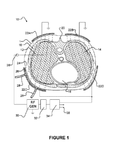
[0021] Figure 1 is a cross section of a patient's chest being exposed to an
electromagnetic
field.
[0022] Figure 2 is a view of the electrodes on the patient's back.
[0023] Figures 3A, 3B, 3C and 3D (collectively, Figure 3) are cross sectional
views of the
patient's chest being exposed to an electromagnetic field, showing an
alternative electrode
arrangement.
[0024] Figure 4 is a side view of the patient, showing a method of electrode
switching.
[0025] Figures 5A and 5B (collectively, Figure 5) are cross sectional views of
a patient's
chest showing the electrodes being supported by an inflatable vest. Figure 5A
illustrates a
deflated vest. Figure 5B shows an inflated vest.
[0026] Figure 6 is a cross section of a patient's chest being exposed to an
electromagnetic
field being generated by coils.
[0027] Figures 7A and 7B (collectively, Figure 7) are cross sectional views of
a patient's
chest showing a pair of electrodes being actuated to move in a helical path
around a
patient's thorax as electromagnetic energy is being delivered.
[0028] Figure 8 is a flow chart showing an exemplary method of treating
unwanted tissues
in a patient.
Detailed Description
[0029] Throughout the following description, specific details are set forth in
order to
provide a more thorough understanding of the invention. However, the invention
may be
practiced without these particulars. In other instances, well known elements
have not been
shown or described in detail to avoid unnecessarily obscuring the invention.
Accordingly,
22
CA 03025213 2018-11-22
WO 2017/201625
PCT/CA2017/050635
the specification and drawings are to be regarded in an illustrative, rather
than a restrictive
sense.
[0030] Methods and apparatus according to certain embodiments of the invention
may be
applied to selectively heat a diseased area of tissue in a patient while
minimizing heating
of other tissues in the patient. Heating may be achieved by exposing the
diseased tissues to
an electromagnetic field to cause dielectric or eddy current heating. The
electromagnetic
field may comprise radiofrequency (RF) energy. In some embodiments the RF
energy
comprises microwave radiation.
[0031] By application of electromagnetic energy, selected diseased tissues may
be heated
to temperatures above a threshold temperature. For example, diseased tissues
may be
heated to temperatures in the range of about 55 degrees C to about 65 degrees
C. The
exact temperature to which diseased tissues are heated is often not critical.
In many cases,
heating to a slightly lower maximum temperature can be compensated for by
maintaining
the temperature for a longer duration. It is desirable to avoid heating of
healthy tissues
because overheating healthy tissues can damage the healthy tissues. The
maximum
temperature to which healthy tissue can be subjected without lasting damage is
not known.
[0032] Certain embodiments of the invention are advantageously applied to
treat diseased
tissues that have reduced blood flow as compared to nearby healthier tissues.
In such cases
the diseased area(s) may be heated rapidly while the healthier tissues will be
cooled by the
blood flow and will therefore experience reduced increase in temperature as
compared to
the diseased tissues.
[0033] Emphysema is an example of a condition for which diseased area(s) have
reduced
blood flow. Certain embodiments of the invention can be particularly effective
for treating
emphysema because of the low mass (density) of the lungs and the high blood
flow in
healthy tissues within the lungs.
[0034] In some cases the diseased tissues are tissues in the lungs of a
patient. For example,
the patient may suffer from emphysema. For such treatments electromagnetic
energy may
be applied to heat diseased areas to temperatures of about 50 degrees C or
more. While
this is done the temperatures of surrounding healthier lung tissue may be kept
below a
threshold temperature. The inventors estimate that healthy tissues in the
lungs and organs
in the vicinity of the lungs should not be subjected to temperatures in excess
of about 40
23
CA 03025213 2018-11-22
WO 2017/201625
PCT/CA2017/050635
degrees C or about 45 degrees C.
[0035] Figure 1 illustrates apparatus 10 according to an example embodiment of
the
invention being applied to treat diseased tissues within lungs 12 and 14 of a
patient P.
Lungs 12 and 14 are surrounded by rib cage 16 inside the patient's body 1. To
heat
diseased tissues within lungs 12 and 14 while minimizing heat to adjacent
organs like
heart 18 and spine 20, a plurality of electrodes 22 (Figure 1 shows four
electrodes
individually identified as 22A, 22B, 22C and 22D. In some embodiments
apparatus 10
includes additional electrodes 22. The additional electrodes 22 may, for
example be
located on one or both sides of the plane of the cross-section of Figure 1.
[0036] Electrodes 22 are dimensioned and placed to create an electric field 24
covering as
much of lungs 12 and 14 as possible while minimizing penetration of electric
field 24 into
adjacent organs. Fortunately, the human anatomy allows such a placement.
[0037] To improve electrical coupling of electromagnetic energy to body 1
while cooling
the surface of body 1, a saline solution 26 may optionally be introduced by
tubes 28
between body 1 and electrodes 22. Such a liquid coupling can greatly improve
the
consistency of the coupling of the RF energy delivered by way of some or all
of electrodes
22 into body 1. In alternative embodiments electrodes 22 comprise baths of
electrically-
conductive fluid such as, for example, saline solution. Saline solution 26
may, for
example, comprise about 1 wt% NaCl in water. In some other embodiments an
electrically-conductive gel is provided between electrodes 22 and body 1.
[0038] RF generator 30 supplies RF energy to electrodes 22 via an impedance
matching
network 32 and electrode selector circuit 34. The RF energy is applied between
two or
more of electrodes 22 via wires 36.
[0039] In some example embodiments the RF generator 30 has a maximum power
output
in the range of about lkW to about 5 kW. In some example embodiments, the RF
energy
output by RF generator 30 has a frequency or frequencies in the range of about
1 MHz to
about 100MHz or about 10 MHz to about 100MHz.
[0040] It is optional but generally desirable to choose frequencies for
electric field 24 in
the industrial scientific and medical (ISM) bands of the spectrum. Such
frequency choices
may reduce or avoid interference between the RF energy generated by RF
generator 30
and other signals such as communications signals. For example, RF generator 30
may
24
CA 03025213 2018-11-22
WO 2017/201625
PCT/CA2017/050635
have an output frequency of 13.56 MHz or 27 MHz.
[0041] Impedance matching network 32 is provided to match the output impedance
of RF
generator 30 to the impedance of body 1. This facilitates the efficient
delivery of energy
into body 1. Impedance matching networks are well known in the art.
[0042] In an example embodiment impedance matching network 32 may comprise an
LC
circuit such as a capacitor connected in series between one output terminal of
RF
generator 30 and electrode selector 38 followed by an inductor connected in
parallel with
electrode selector 38. The values of the capacitor and inductor may be
determined after
measuring the resistance and capacitance between pairs of electrodes 22 on
body 1. For
example, impedance matching network 34 may match a pure resistive impedance
(e.g. 50
Ohms) of RF generator 30 to a complex impedance of a human or animal body.
[0043] Because the impedance presented by different patients may differ very
significantly (e.g. the size of a patient can have a significant effect on the
spacing of a pair
of electrodes located on either side of a patient and whether or not a gel or
conductive
solution is provided can significantly affect impedance at the electrode-body
interfaces) it
can be desirable to provide an adjustable impedance matching network. The
impedance
matching network may be adjustable to provide a best matching of impedance for
each of
a plurality of electrode pairs.
[0044] In some embodiments the impedance matching network is self-adjusting
(i.e. auto-
tuning) to maximize delivery of power into the body. Technologies that can be
used to
auto-tune the matching network for optimal power delivery (based for example
on
measurements of reflected radiation) are described for example in: US patent
Nos.
US5364392, US9028482 and US 9192422 as well as other publications known to
those of
skill in the art.
[0045] To avoid resistive currents going through body 1, and for electrical
safety, it is
desirable to provide capacitive coupling between electrodes 22 and body 1. For
example,
one can coat electrodes 22 with a very thin layer of an insulating material.
For example, a
thin layer of KaptonTM tape may be applied between electrodes 22 and body 1.
[0046] Diseased tissues within one or both lungs 12, 14 may be heated by
applying the
output of RF generator 30 between two of electrodes 22 located on either side
of the lung
to be treated. Heating may be continued for sufficient time to raise the
diseased tissues to
CA 03025213 2018-11-22
WO 2017/201625
PCT/CA2017/050635
temperatures above a threshold temperature for a time sufficient to achieve a
desired
treatment outcome.
[0047] In order to minimize heating of adjacent organs the direction of
electromagnetic
field 24 may be changed periodically. This may be achieved by applying the
output of RF
generator 30 between different pairs of electrodes 22. Different pairs of
electrodes 22 may
be selected such that the electric field changes direction but always passes
through the
portion(s) of lungs 12, 14 containing the diseased tissue to be treated. When
this is done,
the diseased lung tissues will be heated continuously while surrounding
tissues will be
heated only intermittently. In apparatus 10, electrode selector 38 switches
the output of RF
generator 30 to be applied between different pairs of electrodes 22. The
switching
frequency can be low. For example, electrode selector 38 may switch electrodes
once
every few seconds. In some non-limiting examples, electrode selector 38
switches
electrodes to use a different pair of electrodes deliver of heating energy
once every 30 to
300 seconds. In some non-limiting examples electrode selector 38 switches
electrodes to
use a different pair of electrodes at a frequency of 100 Hz or less.
[0048] In some cases the different pairs of electrodes 22 are selected such
that a direction
of alignment of the electric field within tissues of the patient is changed
through an angle
of at least 15 degrees (at least 10 degrees, at least 20 degrees and at least
25 degrees are
also options) at least every few seconds (e.g. at least every 1 to 30
seconds). In some cases
the different pairs of electrodes 22 are selected such that the direction of
alignment of the
electric field does not remain in the same plane for more than a few seconds.
This may be
facilitated by providing a two dimensional array of electrodes 22 adjacent
each of the
patient's lungs on at least one side of the patient.
[0049] Pairs of electrodes may be selected such that a volume of tissue (e.g.
lung tissue)
that includes diseased areas to be treated lies between electrodes of the
selected pairs. By
alternating applying heating energy using different ones of the selected pairs
of electrodes
the diseased areas within the volume of tissue may be heated consistently
while
surrounding tissues may be heated only some of the time. In some embodiments,
for each
lung, heating energy is delivered by way of one selected pair of electrodes at
a time. In
some embodiments delivery of heating energy is rotated among three or four or
more
selected pairs of electrodes. In such embodiments any one selected pair of
electrodes may
be active approximately 1/N of the time where N is the number of selected
pairs of
26
CA 03025213 2018-11-22
WO 2017/201625
PCT/CA2017/050635
electrodes being used to apply heating energy to a particular lung or other
volume of
tissue.
[0050] In some embodiments an array of electrodes that substantially covers an
area of a
patient's lung is provided on a patient's chest and back. The electrode arrays
may be
mirror images of one another. Each of the electrode arrays may be shaped to
conform to a
shape of the patient's lung. In some embodiments each of the arrays is two
dimensional
and comprises plural columns each containing plural electrodes and plural rows
each
containing plural electrodes. . In some embodiments such arrays are provided
for one of a
patient's lungs. In some embodiments such arrays are provided for both of a
patient's
lungs. Such arrays may be applied as described herein to deliver heating
energy to tissues
of either or both of the patient's lungs.
[0051] The electrodes of a pair of electrodes may be energized with opposite
polarities. In
some embodiments one electrode of a pair is grounded and the other electrode
is
connected to an output of RF signal generator 30. In some embodiments one
electrode of a
pair is connected to one output terminal of an RF signal generator and the
other electrode
is connected to another output terminal of the RF signal generator 30.
[0052] Healthier tissues of lungs 12, 14 may be protected from being heated to
damaging
temperatures by the fact that healthy lung tissue has much larger blood
circulation than
diseased tissue. When a non-contact heat source, such as radio-frequency (RF)
energy, is
directed at the lung the heat will be carried away from the healthy tissue by
the blood flow
while the diseased parts of the lung will heat up.
[0053] This works because the mass of the lungs is low (usually about 1 kg in
an adult
human) while blood flow through the lung is high (usually about 5 kg/minute or
about 5
liters per minute in an adult human). The blood flow tends to equalize the
temperature of
healthy parts of the lung with the rest of the body which effectively acts as
a heat-sink
having a mass of tens of kilograms. This is 10 to 100 times larger than the
effective heat
sink mass for diseased portions of the lungs which is typically less than
about one
kilogram. When lungs are exposed to a form of energy causing heating, such as
RF
energy, the temperature rise of lung tissues will be inversely proportional to
the effective
heat-sinking mass. Therefore, diseased tissues that have poor blood
circulation will be
heated to temperatures significantly higher than healthier tissues that have
normal blood
27
CA 03025213 2018-11-22
WO 2017/201625
PCT/CA2017/050635
circulation. Based on this, heating energy may be applied to cause the
diseased areas of
lung tissue to be heated to temperatures in the range of 50-70 degrees C while
healthy lung
areas will only heat up a few degrees above normal body temperature.
[0054] To assist in keeping down the temperature of healthier parts of lungs
12, 14,
patient P may be breathing chilled air during the procedure. The diseased
parts of lungs
12, 14 will not get a sufficient amount of chilled air to keep them cool.
Cooling may also
be facilitated by means of an aerosol of liquefied air.
[0055] Methods as described herein may be implemented in ways that provide the
advantage that the location(s) of diseased area(s) does not need to be
precisely known in
advance. Heating energy can be directed at the whole lung, but only the
diseased areas will
have their temperatures raised significantly.
[0056] Treatment methods as described herein may be applied to achieve various
desired
outcomes. For example, in some cases a single treatment in which a diseased
tissue is
heated to above a threshold temperature may be sufficient to achieve a desired
outcome.
For example, the desired outcome may be a reduction of the volume of diseased
tissue. A
single treatment may achieve sufficient volume reduction via fibrosis,
ablation or other
processes. In other cases the treatment may be repeated two or more times over
the course
of hours, days, weeks or months to achieved a desired reduction of volume of
diseased
tissues or other desired outcome.
[0057] Some embodiments optionally exploit the fact that when diseased lung
tissue is
heated to a temperature in the vicinity of about 60 degrees C the diseased
lung tissue may
lose the ability to expand back after lungs are collapsed (pneumothorax). This
may result
from temperature-induced damage to the surfactant layer and other
physiological reasons.
A treatment method may comprise heating diseased lung tissue (e.g. tissue
affected by
COPD or emphysema) in a lung, collapsing the lung and then re-inflating the
lung.
[0058] Heating the lung may be performed quickly (e.g. in seconds or minutes).
Collapsing the lung may be performed by inserting a hypodermic needle into the
pleural
space and allowing air to leak into the pleural space. Supplying the lung with
pure oxygen
will speed up the collapse as it oxygen fully absorbed in the blood. The lung
may be kept
in a collapsed state for long enough to allow the diseased area(s) to collapse
into a small
volume. The lung may be re-inflated by evacuating the pleural space. This may
be done,
28
CA 03025213 2018-11-22
WO 2017/201625
PCT/CA2017/050635
for example via the same needle used to collapse the lung. The procedure can
be done on
one lung at a time. The patient can breathe with the remaining lung.
Collapsing and
inflating lungs is done routinely in pulmonary medicine and need not be
detailed here.
[0059] This treatment may cause the areas affected by emphysema to collapse
and stay
collapsed so that these areas are prevented from interfering with normal
operation of the
healthy parts of the lung. this may achieve results similar to those that can
be achieved by
surgically removing the diseased lung tissues without the risks of surgery.
Other
mechanisms may exist that do not require pneumothorax: the heated diseased
area can lose
volume through ablation, fibrosis or other mechanisms and allow healthy lung
tissue to fill
the voids.
[0060] The heating process may be performed open-loop (i.e. based on a
previous
experimental calibration of power and duration), or using sensing or closed
loop control.
In some embodiments apparatus 10 includes a controller that automatically
controls one or
more of: the power output of RF generator 30, the electrodes between which the
output of
RF generator 30 is applied, a duty cycle of RF generator 30 and a duration of
a period
during which RF generator 30 applies heating energy to a body 1 based at least
in part on
real time measurements of temperature(s) at one or more locations in tissues
in a patient.
[0061] Temperature sensing may be performed using one or more sensors 36
placed in the
patient's body and/or any suitable non-contact temperature sensing technology.
In an
example embodiment temperature of tissues within a patient is sensed using
small
temperature sensors such as thermistors, For example, a prototype embodiment
used
miniature glass encased thermistors such as DigikeyTM part number 495-5820-ND
to
measure temperatures of lung tissues. Other example ways to measure
temperatures of
tissues include:
= hypodermic temperature sensors (these may for example comprise an
electronic
temperature sensor carried in a very fine gage needle (e.g. a needle about
0.6mm in
diameter);
= processing data obtained by a magnetic resonance imaging (MRI) system or
other
external imaging system capable of temperature monitoring;
= thermocouples;
= a bronchoscope equipped with a thermistor or other temperature sensor;
29
CA 03025213 2018-11-22
WO 2017/201625
PCT/CA2017/050635
= solid-state temperature sensors;
= and the like.
[0062] A controller may implement any of various control algorithms. For
example a
controller of system 10 may implement a PID control loop. A controller may
implement
simple algorithms such as shutting off or reducing the power output of RF
generator 30
when a desired temperature has been reached (e.g. a temperature in the range
of about 55-
65 degrees C). In some embodiments the controller both modulates the power
output of
RF generator 30 as the temperature of a tissue is raised toward a desired
temperature and
shuts of delivery of power by RF generator 30 when the desired temperature has
been
reached. Feedback control can prevent the target temperature from being
exceeded.
[0063] Embodiments that apply open-loop temperature control may optionally
calculate a
current temperature within a tissue of interest based on a mathematical model
of the heat
absorbed in the tissue and the cooling rate of the tissue. An output of the
model may be
applied to control power output of RF generator 30 and/or to stop RF generator
30 from
further raising temperature of tissues after the model predicts that a
threshold temperature
has been reached.
[0064] In some embodiments one or more temperature sensors are applied to
sense
temperatures of non-targeted tissues. For example non-targeted organs
identified as being
likely to heat up the most, or as being the organs most sensitive to heat, may
be identified
and the temperatures within these organs may be monitored during treatment.
[0065] In an example embodiment a simple temperature sensor installed in a
hypodermic
needle provides accurate temperature measurements when the needle is inserted
into the
organ. A controller for apparatus 10 may be configured to discontinue
treatment if a
temperature of a non-targeted tissue exceeds a safe temperature threshold
and/or to
modulate application of heating energy from RF generator 30 if the temperature
of the
non-targeted tissue is rising toward or close to the safe temperature
threshold.
[0066] Non-target temperature sensors which sense temperature of non-target
tissues may
be used on their own or combined with temperature sensors that measure
temperature of
targeted tissues. In some embodiments the same temperature sensor (e.g. an MRI-
based
temperature sensor or another non-contact temperature sensor) may monitor
temperatures
within both targeted tissues and non-targeted tissues.
CA 03025213 2018-11-22
WO 2017/201625
PCT/CA2017/050635
[0067] Some embodiments modify the system described in U58444635 to include a
temperature sensor, a controller connected to receive a temperature signal
from the
temperature sensor and configured to control delivery of radiation to heat
tissues in a
patient by a closed loop control algorithm.
[0068] In some cases it can be undesirable to place a temperature sensor in
target tissues.
For example, inserting a temperature sensor into certain areas of lung tissue
could risk
puncturing the lung. In some embodiments a model of the patient's anatomy may
be used
to estimate how temperature at a specific point in a targeted tissue and/or at
a specific
point in a non-targeted tissue relates to temperature at an alternative
location in the patient.
The alternative location may be selected to be a location at which a
temperature sensor
may be placed with lower risk and/or reduced adverse consequences. The other
location
may comprise one or more of muscle surrounding the lungs, exhaled air
temperature,
blood temperature at a certain location or the like.
[0069] A thermal model of the patient's anatomy may be generated from pre-
operative
images. Known thermal conductivities of different tissue types may be combined
with
known distributions of those tissue types in the patient, known geometries of
electrodes,
coils or other structures to be used to deliver heating energy to the tissues
and a circulation
model to estimate how temperatures at the alternative location(s) correlate to
temperatures
at the locations of interest. Temperatures measured at the alternative
location(s) can then
be used as proxies for temperatures at the locations of interest using the
correlations
determined using the model.
[0070] In some implementations the patient's orientation is taken into
consideration.
Lower parts of the lung typically contain more blood due to the effect of
gravity than parts
of the lung at higher elevations. This is called 'differential perfusion'. The
parts of the
lung that contain the most blood can vary with patient orientation. The amount
of blood at
a location to be treated can affect the rate at which the temperature of
tissue at that
location increases when electromagnetic energy is delivered to the tissue.
[0071] In some embodiments a patient is moved into different postures (e.g. by
rotating
and/or tilting the patient and or rolling the patient over) as treatment is
delivered.
Apparatus according to some embodiments of the invention may provide a couch,
chair,
bed or other patient support that moves by tilting rotating or the like in
coordination with
31
CA 03025213 2018-11-22
WO 2017/201625
PCT/CA2017/050635
the delivery of treatments. In some embodiments motions of the patient support
are
controlled by a controller that also controls application of heating energy to
the patient.
[0072] Apparatus according to some embodiments provides instructions (e.g. on
a display)
to change the posture of the patient at selected points during a treatment.
[0073] Apparatus according to some embodiments estimates an effect of
differential
perfusion on properties of tissues in different parts of the lung (or other
part of the
anatomy). Such estimates may be based for example on information regarding the
patient's anatomy (e.g. from pre-operative images). A profile for delivering
energy to
target tissues may take into account differential perfusion by increasing or
decreasing the
delivered energy depending on whether the target tissues are in a part of the
lung at which
the target tissues are expected to experience more rapid temperature rise as a
result of
differential perfusion (e.g. energy may be decreased where the target tissue
is at a higher
elevation and so the target tissue is depleted of blood) or the target tissues
are expected to
experience slower temperature rise as a result of differential perfusion (e.g.
energy may be
increased where the target tissue is at a lower elevation and so the target
tissue contains a
large amount of blood). Some embodiments of the apparatus provide a user
interface that
includes a control that a user may use to indicate a posture of the patient
during a
treatment. Compensation for differential perfusion may be based at least in
part on the
indicated posture.
[0074] It is generally desirable to apply electromagnetic energy to a
patient's tissues such
that the electric fields 24 within the tissues are generally uniform. Electric
field 24
uniformity can be affected by various factors including:
= The sizes, shapes and positions of the electrodes;
= Impedance of the interface between the electrodes and the patient's body;
= The frequency or frequencies present in the electromagnetic energy being
delivered by way of the electrodes;
= Where the electrodes are of different sizes, which electrode has the
highest
voltages applied to it (tissues near high voltage will tend to be heated to
higher
temperatures heat more quickly because the rate of heating relates to density
of
field lines).
Some embodiments manipulate one or more of these factors to achieve a desired
electric
32
CA 03025213 2018-11-22
WO 2017/201625
PCT/CA2017/050635
field distribution in the patient. For example:
= electrodes may be constructed by choice of material and/or coating to
have a
spatially-varying resistivity.
= shields and/or waveguides may be interposed between the electrodes and
the
body of the patient.
= the electrodes (and/or shields and/or waveguides if present) may be moved
as
treatments are delivered.
Features such as one or more of the above may be applied, for example, to
achieve a
generally uniform distribution of electric field in a lung or other volume of
tissue to be
treated.
[0075] In some embodiments an arrangement of electrodes 22 is designed or
customized
using knowledge of a patient's anatomy and the geometry of the target tissues.
For
example, MRI and/or computed tomography CT images may be processed to identify
regions of different consistency in the patient (e.g. fat tissue / muscle /
bone). Working
from known average electrical properties of these materials one can design a
treatment
plan that specifies one or more of:
= electrode arrangements;
= electrode switching sequence and/or timing;
= RF signal characteristics (power, frequency etc.);
The treatment plan may help to target the correct target tissue(s), achieve
sufficiently
uniform heating, and avoid excessive heating of critical tissue (heart, for
example). In
some embodiments an electrode pattern that comprises arrays of electrodes
dimensioned to
overlie a patient's lungs on two sides (e.g. chest and back) of a patient's
body is generated
by analysis of the patient's anatomy and a set of electrodes customized for
the patient is
fabricated by printing, cutting or other computer-controlled fabrication
process using the
electrode pattern.
[0076] Figure 2 shows an example arrangement of electrodes 22 on one side of a
patient P
(e.g. the patient's back) a similar arrangement of electrodes may be provided
on an
opposing side of the patient (e.g. the patient's chest). In this and some
other embodiments
a separate set of electrodes is provided overlying each of the patient's
lungs. Here,
electrodes 22AA through 22AC are provided over the patient's left lung and
electrodes
33
CA 03025213 2018-11-22
WO 2017/201625
PCT/CA2017/050635
22BA through 23BC are provided over the patient's right lung.
[0077] In this example, electromagnetic energy may be delivered to target lung
tissues of
the patient P by connecting the output of an RF generator 30 between a pair of
electrodes
22 which includes one electrode on the patient's chest and another electrode
on the
patient's back. The pair of electrodes 22 may be directly opposed to one
another in some
cases and offset from one another in others.
[0078] The electrode arrangement of Figure 2 may be varied in different ways
including,
for example:
= Replacing some or all of the illustrated electrodes with more electrodes,
which may
be smaller than the depicted electrodes in some cases.
= Dividing the illustrated electrodes to provide more columns of
electrodes. The
columns may, for example be arranged generally parallel to the patient's spine
on
one or both sides of the patient. For example, each of the illustrated
electrodes 22
may be replaced by a row of two or three electrodes. An exemplary embodiment
is
shown in Figures 3A to 3D where each of the electrodes 22A, 22B, 22C, and 22D
has been replaced by two electrodes.
= Dividing the illustrated electrodes to include more rows of electrodes.
[0079] An electrode selection circuit 34 as shown for example in Figure 1 may
apply
heating energy (e.g. an output from an RF signal generator) to different ones
of the
electrode pairs at different times (electrode switching).
[0080] Figure 4 illustrates an example of electrode switching. Figure 4 is a
side view of
patient P in which electrodes 22AA through 22CC are shown. Electrodes 22CA
through
22CC are on an opposite side of patient P from electrodes 22AA to 22AC. Figure
4 shows
that electrodes 22AA to 22CC provide 9 pairs of electrodes 22 wherein the
electrodes of
each pair include one electrode on one side of patient P and another electrode
on an
opposing side of patient P such that patient P is sandwiched between the
electrodes of the
pair.
[0081] The direction of an electric field 24 produced in patient P depends on
which pair of
electrodes 22 is being used to deliver heating energy. For example consider
the three pairs
of electrodes involving electrode 22CC. The electromagnetic field can be
directed as
shown by field lines 24, 25 and 26 by respectively pairing electrode 22CC with
electrodes
34
CA 03025213 2018-11-22
WO 2017/201625
PCT/CA2017/050635
22AA, 22AB and 22AC.
[0082] Figure 4 shows an example situation in which electrode selection
circuit 34
comprises electronically controlled switches or commutators 46 and 48. The
impedance
matching network may be constructed to provide a balanced output (balanced
relative to
ground potential) where a balanced configuration is desired. In the
illustrated embodiment
this is achieved by providing transformer 50.
[0083] Switches 46 and 48 may comprise, for example, electro-mechanical
relays, electro-
mechanical commutators, solid state switches such as RF FET transistors or RF
relays or
the like.
[0084] As shown in Figure 4, different pairs of electrodes 22 may have
electrode-to-
electrode spacings that are significantly different. Some embodiments include
mechanisms
to compensate for different energy densities in body tissues that may result
when heating
energy is switched among different electrode pairs. Such compensation may, for
example,
take one or more of the following forms:
= A controller may automatically set power output of RF generator 30 to
different
values depending on which pair of electrodes is being driven.
= Some electrodes may be split into plural sections. Different ones of the
sections or
different combinations of the sections may be used depending on which other
electrode the electrode is paired with.
= Pulse width modulation or other time domain compensation may be applied
depending on which pair of electrodes 22 is being driven.
= A larger number of electrodes 22 may be provided such that more different
pairs of
electrodes 22 that produce similar energy densities when driven are available
for
selection.
= An impedance matching network may be tuned or switched to match the
impedance presented by different pairs of electrodes.
= combinations of any two or more of the above.
= etc.
[0085] Electrodes 22 for use in applying heating energy to a patient may have
any of a
wide variety of forms including stick-on electrodes, electrodes mounted on a
belt or the
like, electrodes 22 supported by clothing such as a vest or the like. An
exemplary vest 58
CA 03025213 2018-11-22
WO 2017/201625
PCT/CA2017/050635
is shown in Figure 5. Vest 58 may be inflatable. Figure 5A shows vest 58 prior
to
inflation. Figure 5B shows vest 58 that is inflated. In some embodiments some
or all
electrodes 22 comprise bladders containing an electrically-conductive liquid.
Such
electrodes can be advantageous where apparatus as described herein
incorporates or is
used in conjunction with a MRI system. During times when it is desired to
deliver energy
to the patients' tissues the bladders may be filled with the electrically-
conductive fluid.
During times when it is desired to obtain MRI information the electrically-
conductive
fluid may be withdrawn from the bladders.
[0086] Ideally the electrodes are provided in a way that simplifies applying
the electrodes
to the bodies of patients such that the electrodes are in close contact with
the patients'
bodies.
[0087] In some embodiments, some or all of electrodes 22 have one or more of
the
following features:
= the electrodes are stretchable in length and/or width (for example, the
electrodes
may be made from an electrically-conductive stretchable fabric or a woven or
non-
woven conductive mesh or a sheet of a stretchable conductive plastic);
= the electrodes are bendable;
= the electrodes are attached to or are attachable to a vest, belt or other
clothing
article (for example using an adhesive or a hook and loop fabric coupling, or
a clip,
removable fastener, or the like);
= the electrodes are designed to be made smaller, for example by cutting or
tearing
off to a size suitable for a particular patient;
= the electrodes are made up of plural smaller electrodes, optionally
connections
between the smaller electrodes can be made or broken to adjust the sizes of
the
electrodes to suit individual patients.
[0088] Electrodes 22 may be held in place on a patient P, for example, by one
or more of:
= an adhesive (which may comprise a self-adhesive and/or a separately-
applied
adhesive) and/or gel;
= an article of clothing to which the electrodes are attached or integrated
into;
= an article of clothing such as, for example, a stretchy and/or inflatable
vest or shirt
worn over top of the electrodes;
36
CA 03025213 2018-11-22
WO 2017/201625
PCT/CA2017/050635
= etc.
[0089] Where it is desirable to hold an electrode 22 against a part of a
patient's body 1
that may be concave in form (e.g. the spine, areas around breasts etc.) a
formed member
such as a bendable support or inflatable chamber (which may be part of an
inflatable
article of clothing such as a vest) or the like may be provided to hold the
electrode against
the concave part of the patient's anatomy.
[0090] Where some or all electrodes 22 are provided on a support such as an
article of
clothing (e.g. a vest) or patient furniture such as a treatment couch, bed
table or chair, the
support may include passages in which a cool fluid is contained and/or
circulating. The
cool fluid may help to keep the patient cool. If the support is inflatable the
passages
containing the cool fluid may be the same as or different from chambers that
can be
pressurized to inflate the support. In some embodiments valves are provided
such that
circulation of the cool fluid may be inhibited in parts of the support that
are in close
proximity to a target tissue.
[0091] As described below, some embodiments provide coils instead of or in
addition to
electrodes. In such embodiments the coils may be supported against a patient
in the same
or similar ways as described above for electrodes. This is best illustrated in
Figure 6.
[0092] The innovations described herein may be applied in contexts which apply
heating
energy to body tissues in different ways. For example, electromagnetic energy
can be
coupled into a body to heat tissues by:
= Placing at least one pair of electrodes on either side of the body such
that at least a
portion of the body is between the electrodes (effectively forming a capacitor
in
which the tissues of the body forms a dielectric) and deliver RF energy across
the
electrodes. Dielectric losses within the tissue will generate heat.
= Place at least a part of the body inside a coil or sandwiched between
several coils
to form an inductor, drive a RF signal across the inductor and use the losses
of this
inductor (mainly eddy current losses) to generate heat in tissues of the body.
Eddy
current heating results where eddy currents in the patient's tissues are
induced by a
changing magnetic field.
= Radiate electromagnetic energy into the body from an antenna, as
disclosed for
example in US patent 8444635. Heating by radiating electromagnetic radiation
into
37
CA 03025213 2018-11-22
WO 2017/201625
PCT/CA2017/050635
the body is mainly suitable for high frequencies such as microwave
frequencies.
Closed-loop temperature control as described herein and/or switching the
direction of
electromagnetic field lines to reduce heating of non-targeted tissues may be
provided in
embodiments which apply any of these heating methods.
[0093] In order to control which areas are heated when using lower RF
frequencies (e.g. 1
MHz to 100MHz) the electrodes or coils that apply the RF energy to the body
should be
placed on opposing sides of the body. Placing electrodes or coils just on one
side of the
body will create uneven heating, with most heat generated near the electrodes
or coils.
[0094] Various example embodiments are described herein in which
electromagnetic
energy is applied to a lung or other structure by way of electrodes. Other
corresponding
embodiments may be provided by replacing the electrodes with coils.
[0095] For eddy current and mainly magnetic field induced heating, electrodes
can be
replaced by RF coils, as shown in Figure 6. The polarity of coils 52 and 54 is
selected to
create magnetic field lines 56 going through the lungs 12, 14. Multiple coils
can be used in
a coil switching arrangement similar to the electrode switching arrangements
disclosed
elsewhere herein. The magnetic field can be further directed by using ferrite
blocks.
[0096] Instead of providing fixed electrodes or coils apparatus may provide
electrodes or
coils that are movable relative to a patient P. For example: One or more pairs
of electrodes
may be carried on an actuator operative to move the pairs of electrodes
relative to a
patient. The pairs of electrodes may each include first and second electrodes
that are
respectively movable over first and second faces of the patient (e.g. chest
and back of the
patient). For example, one pair of electrodes 22 may be actuated to move in a
helical path
around a patient's thorax as electromagnetic energy is delivered by way of the
electrodes
22, as shown in Figures 7A and 7B. As another example, one or more pairs of
electrodes
may be fixed in at least one dimension and the patient may be moved in the
dimension
relative to the fixed electrodes.
[0097] Apparatus according to some embodiments includes or is used in
conjunction with
a Faraday cage or shielded room to reduce electromagnetic interference with
other
equipment. In some embodiment, shielding is provided by a wire mesh cage made
up of
wires spaced apart by a few centimeters or less. The cage may be integrated
into walls or
other structures of a room.
38
CA 03025213 2018-11-22
WO 2017/201625
PCT/CA2017/050635
Example
[0098] A method as described herein was tested on rats. It was found out that
miniature
thermistors work well as direct temperature sensors while the thermocouples
tested did not
perform well. It is believed that the electric field interfered with the low
level (under one
mV), signals from thermocouples but not with the higher level (volts) signal
from the
thermistors. By the way of example, a suitable thermistor is model H1744 made
by the US
Sensor company (http://www.ussensor.com/). This thermistor has an outside
diameter of
0.43mm.
[0099] The system was tested on several rats with induced emphysema in one of
the lungs.
The parameters used were:
= RF power of 100W at 13.56 MHz.
= Series C parallel L matching network with saline irrigated electrodes.
[0100] Reflected power was under 5%. Each electrode was approximately 25x50mm,
coated with 25ium thick KaptonTM tape. The tape does not attenuate the
capacitive currents
much because it is very thin, therefore allowing high capacitance between
electrode and
the body. Rats were shaved in the areas of contact with the electrodes.
[0101] Heating time was about 100 seconds. The healthy lung reached about 41
degrees
C, while the areas with emphysema reached about 55 degrees C. All rats
survived the
treatment. Subsequent autopsy verified scar tissue in the areas of induced
emphysema.
[0102] In the tests conducted on rats the dielectric heating was more
effective than the
magnetic field induced heating, but there may be unique benefits to each one
of them.
Interpretation of Terms
[0103] Unless the context clearly requires otherwise, throughout the
description and the
claims:
= "comprise", "comprising", and the like are to be construed in an
inclusive sense, as
opposed to an exclusive or exhaustive sense; that is to say, in the sense of
"including, but not limited to";
= "connected", "coupled", or any variant thereof, means any connection or
coupling,
either direct or indirect, between two or more elements; the coupling or
connection
between the elements can be physical, logical, or a combination thereof;
39
CA 03025213 2018-11-22
WO 2017/201625
PCT/CA2017/050635
= "herein", "above", "below", and words of similar import, when used to
describe
this specification, shall refer to this specification as a whole, and not to
any
particular portions of this specification;
= "or", in reference to a list of two or more items, covers all of the
following
interpretations of the word: any of the items in the list, all of the items in
the list,
and any combination of the items in the list;
= the singular forms "a", "an", and "the" also include the meaning of any
appropriate
plural forms.
= "electromagnetic signal applicator" is a generic term that encompasses
electrodes
(e.g. which may be used to apply electric fields for dielectric heating),
coils, (e.g.
which may be used to apply magnetic fields for eddy current heating, and
antennas
(e.g. which may be used to apply microwaves to heat tissues).
[0104] Words that indicate directions such as "vertical", "transverse",
"horizontal",
"upward", "downward", "forward", "backward", "inward", "outward", "vertical",
"transverse", "left", "right", "front", "back", "top", "bottom", "below",
"above", "under",
and the like, used in this description and any accompanying claims (where
present),
depend on the specific orientation of the apparatus described and illustrated.
The subject
matter described herein may assume various alternative orientations.
Accordingly, these
directional terms are not strictly defined and should not be interpreted
narrowly.
[0105] Certain embodiments of the invention incorporate control systems or
controllers.
Such controllers or control systems may be implemented using specifically
designed
hardware, configurable hardware, programmable data processors configured by
the
provision of software (which may optionally comprise "firmware") capable of
executing
on the data processors, special purpose computers or data processors that are
specifically
programmed, configured, or constructed to perform one or more steps in a
method as
explained in detail herein and/or combinations of two or more of these.
Examples of
specifically designed hardware are: logic circuits, application-specific
integrated circuits
("ASICs"), large scale integrated circuits ("LSIs"), very large scale
integrated circuits
("VLSIs"), and the like. Examples of configurable hardware are: one or more
programmable logic devices such as programmable array logic ("PALs"),
programmable
logic arrays ("PLAs"), and field programmable gate arrays ("FPGAs")). Examples
of
programmable data processors are: microprocessors, digital signal processors
("DSPs"),
CA 03025213 2018-11-22
WO 2017/201625
PCT/CA2017/050635
embedded processors, graphics processors, math co-processors, general purpose
computers, server computers, cloud computers, mainframe computers, computer
workstations, and the like. For example, one or more data processors in a
control circuit
for a device may implement methods as described herein by executing software
instructions in a program memory accessible to the processors.
[0106] For example, while processes or blocks are presented in a given order,
alternative
examples may perform routines having steps, or employ systems having blocks,
in a
different order, and some processes or blocks may be deleted, moved, added,
subdivided,
combined, and/or modified to provide alternative or subcombinations. Each of
these
processes or blocks may be implemented in a variety of different ways. Also,
while
processes or blocks are at times shown as being performed in series, these
processes or
blocks may instead be performed in parallel, or may be performed at different
times.
[0107] Software and other modules may reside on servers, workstations,
personal
computers, tablet computers, embedded controllers, process controllers and
other devices
suitable for the purposes described herein.
[0108] The invention may also be provided in the form of a program product.
The
program product may comprise any non-transitory medium which carries a set of
computer-readable instructions which, when executed by a data processor, cause
the data
processor to execute a method of the invention. Program products according to
the
invention may be in any of a wide variety of forms. The program product may
comprise,
for example, non-transitory media such as magnetic data storage media
including floppy
diskettes, hard disk drives, optical data storage media including CD ROMs,
DVDs,
electronic data storage media including ROMs, flash RAM, EPROMs, hardwired or
preprogrammed chips (e.g., EEPROM semiconductor chips), nanotechnology memory,
or
the like. The computer-readable signals on the program product may optionally
be
compressed or encrypted.
[0109] In some embodiments, the invention may be implemented in software. For
greater
clarity, "software" includes any instructions executed on a processor, and may
include (but
is not limited to) firmware, resident software, microcode, and the like. Both
processing
hardware and software may be centralized or distributed (or a combination
thereof), in
whole or in part, as known to those skilled in the art. For example, software
and other
41
CA 03025213 2018-11-22
WO 2017/201625
PCT/CA2017/050635
modules may be accessible via local memory, via a network, via a browser or
other
application in a distributed computing context, or via other means suitable
for the purposes
described above.
[0110] Where a component (e.g. an electrode, oscillator, switch, controller,
temperature
sensor, software module, processor, assembly, device, circuit, etc.) is
referred to above,
unless otherwise indicated, reference to that component (including a reference
to a
"means") should be interpreted as including as equivalents of that component
any
component which performs the function of the described component (i.e., that
is
functionally equivalent), including components which are not structurally
equivalent to the
disclosed structure which performs the function in the illustrated exemplary
embodiments
of the invention.
[0111] Specific examples of systems, methods and apparatus have been described
herein
for purposes of illustration. These are only examples. The technology provided
herein can
be applied to systems other than the example systems described above. Many
alterations,
modifications, additions, omissions, and permutations are possible within the
practice of
this invention. This invention includes variations on described embodiments
that would be
apparent to the skilled addressee, including variations obtained by: replacing
features,
elements and/or acts with equivalent features, elements and/or acts; mixing
and matching
of features, elements and/or acts from different embodiments; combining
features,
elements and/or acts from embodiments as described herein with features,
elements and/or
acts of other technology; and/or omitting combining features, elements and/or
acts from
described embodiments.
[0112] Methods according to the examples described herein may be varied. For
example,
while elements are at times shown as being performed sequentially, they may
instead be
performed simultaneously or in different sequences.
[0113] It is therefore intended that the following appended claims and claims
hereafter
introduced are interpreted to include all such modifications, permutations,
additions,
omissions, and sub-combinations as may reasonably be inferred. The scope of
the claims
should not be limited by the preferred embodiments set forth in the examples,
but should
be given the broadest interpretation consistent with the description as a
whole.
42