Language selection

Search

Patent 3025603 Summary

Third-party information liability

Some of the information on this Web page has been provided by external sources. The Government of Canada is not responsible for the accuracy, reliability or currency of the information supplied by external sources. Users wishing to rely upon this information should consult directly with the source of the information. Content provided by external sources is not subject to official languages, privacy and accessibility requirements.

Claims and Abstract availability

Any discrepancies in the text and image of the Claims and Abstract are due to differing posting times. Text of the Claims and Abstract are posted:

  • At the time the application is open to public inspection;
  • At the time of issue of the patent (grant).
(12) Patent Application: (11) CA 3025603
(54) English Title: DEVICE FOR TREATMENT OF DYSPHAGIA
(54) French Title: DISPOSITIF POUR LE TRAITEMENT DE LA DYSPHAGIE
Status: Deemed Abandoned
Bibliographic Data
(51) International Patent Classification (IPC):
  • A61B 1/267 (2006.01)
  • A61N 1/05 (2006.01)
  • A61N 1/36 (2006.01)
(72) Inventors :
  • MULROONEY, CONOR (United Kingdom)
(73) Owners :
  • PHAGENESIS LIMITED
(71) Applicants :
  • PHAGENESIS LIMITED (United Kingdom)
(74) Agent: SMART & BIGGAR LP
(74) Associate agent:
(45) Issued:
(86) PCT Filing Date: 2017-05-25
(87) Open to Public Inspection: 2017-11-30
Examination requested: 2022-05-25
Availability of licence: N/A
Dedicated to the Public: N/A
(25) Language of filing: English

Patent Cooperation Treaty (PCT): Yes
(86) PCT Filing Number: PCT/GB2017/051482
(87) International Publication Number: WO 2017203263
(85) National Entry: 2018-11-26

(30) Application Priority Data:
Application No. Country/Territory Date
1609425.2 (United Kingdom) 2016-05-27

Abstracts

English Abstract

The present invention defines a device for the treatment of dysphagia comprising a flexible member mounting an imaging device for insertion into a patient and one or more electrodes proximate the imaging device for imparting electrical stimulation to the patient.


French Abstract

La présente invention concerne un dispositif pour le traitement de la dysphagie qui comprend un élément flexible sur lequel est monté un dispositif d'imagerie à insérer dans le corps d'un patient et une ou plusieurs électrodes à proximité du dispositif d'imagerie pour appliquer une stimulation électrique au patient.

Claims

Note: Claims are shown in the official language in which they were submitted.

CLAIMS
1. A device for the treatment of dysphagia comprising a flexible member
mounting an
imaging device for insertion into a patient and one or more electrodes
proximate the
imaging device for imparting electrical stimulation to
the patient.
2. A device for the treatment of dysphagia according to claim 1 further
comprising a sleeve
movable relative to the flexible member, wherein the one or more electrodes
are mounted
on the sleeve.
3. A device for the treatment of dysphagia according to claim 1 further
comprising a dog leg
structure extending from the flexible member beyond the imaging device.
4. A device for the treatment of dysphagia according to claim 3, further
comprising a sleeve
movable relative to the dogleg
structure.
5. A device for the treatment of dysphagia according to claim 4, wherein the
one or more
electrodes are mounted on the sleeve or the dogleg structure.
6. A device for the treatment of dysphagia comprising a flexible member
mounting an
imaging device for insertion into a patient and a sleeve movable relative to
the flexible
member, wherein the sleeve mounts one or more electrodes and is configured
such that
the one or more electrodes are freely movable relative to the imaging device
along the
flexible member.
7. A device for the treatment of dysphagia comprising a first flexible member
mounting an
imaging device for insertion into a patient and a second flexible member
extending from
the first flexible member beyond the imaging device in parallel plane thereto
and one or
more electrodes for imparting electrical stimulation to the patient.
8. A device for the treatment of dysphagia according to claim 7 further
comprising a sleeve
movable relative to the second flexible member, wherein the one or more
electrodes are
mounted on the sleeve and the sleeve is configured such that the one or more
electrodes
are freely movable relative to the imaging device along the second flexible
member.
9. A device for the treatment of dysphagia according to claim 7 or claim 8,
wherein the first
flexible member and second flexible member together form a dogleg structure.
10. A device for the treatment of dysphagia substantially as described with
reference to,
and/or as shown in, the figures.
11. An endoscope comprising a camera at one end thereof and one or more
electrodes
proximate the imaging device.
3

Description

Note: Descriptions are shown in the official language in which they were submitted.


CA 03025603 2018-11-26
WO 2017/203263
PCT/GB2017/051482
DEVICE FOR TREATMENT OF DYSPHAGIA
FIELD
The present invention relates to a device for treatment of dysphagia.
BACKGROUND
The use of electrical pharyngeal stimulation to treat dysphagia is known.
Various devices
have been described in the literature to deliver such stimulation. One of the
requirements for
effective pharyngeal stimulation is that the electrodes should be placed in
contact with patient
tissue at a certain location within the pharyngeal region. Given that the
internal anatomy of
patients can vary this can present some technical challenges.
One of the possible approaches to reduce the challenge of correct positioning
of stimulating
electrodes is to have a means to visualise the stimulating catheter containing
those
electrodes during its placement and/or its use. This can be done using an
endoscope such
that the endoscope camera provides visual information to direct passage or
final positioning
of the treatment catheter. A problem with this approach however is that
passage of two
catheters simultaneously in a patient can be problematic. It is frequently the
case that both
nostrils cannot be used to pass catheters due to anatomical limitations, and
oral passage of
an endoscope or electrical stimulation catheter carries with it the risk of
the patient biting and
damaging the medical equipment or harming themselves. This presents an
additional
technical challenge.
Last but not least a limitation of the current state of the art is that the
effect of stimulation on
swallow function cannot be visualised in real time. This may mean that
critical information
relating to the responsiveness of the patient to the applied stimulation may
be lost and
opportunities to further optimise stimulation conditions may also be lost.
SUMMARY
The present invention seeks to overcome these technical challenges or
limitations by
integrating the functionality of an endoscope with the functionality of an
electrical stimulation
catheter. This may take a number of different forms and the embodiments below
are given by
way of example only and should not be considered limiting.
A first embodiment of the invention provides a device for the treatment of
dysphagia
comprising a flexible member mounting an imaging device for insertion into a
patient and one
or more electrodes proximate the imaging device for imparting electrical
stimulation to the
patient.
Four particular advantages of this first embodiment are that; i) only a single
device needs to
be inserted into the patient to both image a target area of tissue and impart
electrical
stimulation to the target area of tissue, ii) that it allows the imaging
device to direct the
passage of the electrodes, iii) that final positioning of the electrodes can
be judged relative to
the patients' anatomy visible via the imaging device, and iv) that the
flexible member may be
used to actively bring the electrodes in contact with target tissues to ensure
good electrical
contact.
The one or more electrodes may be mounted on a sleeve, wherein the sleeve is
positioned
around the flexible member and movable relative thereto.
The provision of a sleeve located around the flexible member such that the
sleeve is freely
moveable along the length of the flexible member, and located on the sleeve
one or more
electrodes designed to deliver electrical stimulation. Within the body of the
sleeve there are
current carrying means to connect the electrodes to a source of electrical
stimulation external
to the patient.
1

CA 03025603 2018-11-26
WO 2017/203263
PCT/GB2017/051482
An additional advantage of the sleeve is that the electrodes can be moved
relative to the
terminal end containing the camera and as such the two functions of
visualisation and
stimulation are capable of independent positioning optimisation.
The device may further comprise a dog leg structure extending from the
flexible member
beyond the imaging device.
Three additional advantages provided by the dog leg structure are; that the
target tissues can
be seen within the field of view, that the electrode position can be changed
before or during
treatment without movement of the imaging device or field of view, and that
the effect of the
stimulation can be assessed through visualisation of the specific area being
stimulated and
the behaviour of these tissues. These behaviours might include for example
muscular
contraction, protection of the airways or clearing of residues.
FIGURES
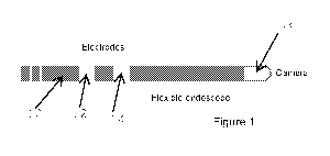
Figure 1 shows a first embodiment of a device for treating dysphagia;
Figure 2 shows a second embodiment of a device for treating dysphagia; and
Figure 3 shows a third embodiment of a device for treating dysphagia.
DESCRIPTION
Embodiments of the invention will now be described by way of reference to the
figures.
In a first embodiment of the invention there is a catheter (10), in the form
of a flexible
endoscope, with electrodes (12, 14) designed to deliver electrical stimulation
to target tissues.
The electrodes (12, 14) are integrated into the body of the endoscope (10) at
a fixed position
proximal an imaging device (16) part of the endoscope (10). The imaging device
(16) part of
the endoscope is positioned at an end of the endoscope (10). Within the body
of the flexible
endoscope (10) there are current carrying means to connect the electrodes (12,
14) to a
source of electrical stimulation external to the patient.
In a second embodiment there is a catheter in the form of a flexible endoscope
(100) having
an imaging device (108) at one end thereof with a sleeve (102) located on it
such that the
sleeve (102) is freely moveable along the length of the endoscope (100), and
located on the
sleeve (102) are one or more electrodes (104, 106) designed to deliver
electrical stimulation
to target tissues. Within the body of the sleeve (102) there are current
carrying means to
connect the electrodes (104, 106) to a source of electrical stimulation
external to the patient.
In a third embodiment there is a catheter in the form of a flexible endoscope
(200) with an
extended structure (202) distal to an imaging device (204) located at an end
of the flexible
endoscope (200), the extended structure (202) incorporating one or more
electrodes (206,
208) designed to deliver electrical stimulation to target tissues. The
electrodes (206, 208) in
this extended structure (202) may be either fixed and integrated into said
extended structure
(202), or may be disposed on a sleeve (210 - as shown in figure 4) that is
freely moveable
along the length of the extended structure (202), such movement being
controllable
externally. In the latter case the electrodes (206, 208) can then be moved
relative to the
position, and additionally within the field of view, of the imaging device
(204). Within the body
of the flexible endoscope (200) or the body of the sleeve (210) there are
current carrying
means to connect the electrodes to a source of electrical stimulation external
to the patient.
2

Representative Drawing
A single figure which represents the drawing illustrating the invention.
Administrative Status

2024-08-01:As part of the Next Generation Patents (NGP) transition, the Canadian Patents Database (CPD) now contains a more detailed Event History, which replicates the Event Log of our new back-office solution.

Please note that "Inactive:" events refers to events no longer in use in our new back-office solution.

For a clearer understanding of the status of the application/patent presented on this page, the site Disclaimer , as well as the definitions for Patent , Event History , Maintenance Fee  and Payment History  should be consulted.

Event History

Description Date
Deemed Abandoned - Conditions for Grant Determined Not Compliant 2024-09-13
Letter Sent 2024-03-19
Notice of Allowance is Issued 2024-03-19
Inactive: Approved for allowance (AFA) 2024-03-15
Inactive: Q2 passed 2024-03-15
Amendment Received - Voluntary Amendment 2023-11-01
Amendment Received - Response to Examiner's Requisition 2023-11-01
Examiner's Report 2023-07-04
Inactive: Report - No QC 2023-06-08
Letter Sent 2022-06-28
Inactive: Submission of Prior Art 2022-06-28
Request for Examination Received 2022-05-25
All Requirements for Examination Determined Compliant 2022-05-25
Request for Examination Requirements Determined Compliant 2022-05-25
Common Representative Appointed 2020-11-07
Common Representative Appointed 2019-10-30
Amendment Received - Voluntary Amendment 2018-12-11
Inactive: Notice - National entry - No RFE 2018-12-06
Inactive: Cover page published 2018-12-03
Application Received - PCT 2018-11-30
Inactive: IPC assigned 2018-11-30
Inactive: First IPC assigned 2018-11-30
National Entry Requirements Determined Compliant 2018-11-26
Application Published (Open to Public Inspection) 2017-11-30

Abandonment History

Abandonment Date Reason Reinstatement Date
2024-09-13

Maintenance Fee

The last payment was received on 2024-05-13

Note : If the full payment has not been received on or before the date indicated, a further fee may be required which may be one of the following

  • the reinstatement fee;
  • the late payment fee; or
  • additional fee to reverse deemed expiry.

Please refer to the CIPO Patent Fees web page to see all current fee amounts.

Fee History

Fee Type Anniversary Year Due Date Paid Date
Basic national fee - standard 2018-11-26
MF (application, 2nd anniv.) - standard 02 2019-05-27 2019-04-24
MF (application, 3rd anniv.) - standard 03 2020-05-25 2020-05-11
MF (application, 4th anniv.) - standard 04 2021-05-25 2021-05-17
MF (application, 5th anniv.) - standard 05 2022-05-25 2022-05-19
Request for examination - standard 2022-05-25 2022-05-25
MF (application, 6th anniv.) - standard 06 2023-05-25 2023-05-25
MF (application, 7th anniv.) - standard 07 2024-05-27 2024-05-13
Owners on Record

Note: Records showing the ownership history in alphabetical order.

Current Owners on Record
PHAGENESIS LIMITED
Past Owners on Record
CONOR MULROONEY
Past Owners that do not appear in the "Owners on Record" listing will appear in other documentation within the application.
Documents

To view selected files, please enter reCAPTCHA code :


To view images, click a link in the Document Description column. To download the documents, select one or more checkboxes in the first column and then click the "Download Selected in PDF format (Zip Archive)" or the "Download Selected as Single PDF" button.

List of published and non-published patent-specific documents on the CPD .

If you have any difficulty accessing content, you can call the Client Service Centre at 1-866-997-1936 or send them an e-mail at CIPO Client Service Centre.


Document
Description 
Date
(yyyy-mm-dd) 
Number of pages   Size of Image (KB) 
Claims 2023-11-01 1 21
Claims 2018-11-26 1 45
Abstract 2018-11-26 1 52
Description 2018-11-26 2 128
Representative drawing 2018-11-26 1 7
Drawings 2018-11-26 1 26
Cover Page 2018-12-03 1 34
Fees 2024-07-04 1 192
Maintenance fee payment 2024-05-13 44 1,804
Notice of National Entry 2018-12-06 1 207
Reminder of maintenance fee due 2019-01-28 1 111
Commissioner's Notice - Application Found Allowable 2024-03-19 1 580
Courtesy - Acknowledgement of Request for Examination 2022-06-28 1 424
Examiner requisition 2023-07-04 4 188
Amendment / response to report 2023-11-01 8 248
Patent cooperation treaty (PCT) 2018-11-26 1 37
Patent cooperation treaty (PCT) 2018-11-26 1 48
National entry request 2018-11-26 3 60
International search report 2018-11-26 2 51
Amendment / response to report 2018-12-11 2 78
Request for examination 2022-05-25 5 112