Sélection de la langue

Search

Sommaire du brevet 3025603 

Énoncé de désistement de responsabilité concernant l'information provenant de tiers

Une partie des informations de ce site Web a été fournie par des sources externes. Le gouvernement du Canada n'assume aucune responsabilité concernant la précision, l'actualité ou la fiabilité des informations fournies par les sources externes. Les utilisateurs qui désirent employer cette information devraient consulter directement la source des informations. Le contenu fourni par les sources externes n'est pas assujetti aux exigences sur les langues officielles, la protection des renseignements personnels et l'accessibilité.

Disponibilité de l'Abrégé et des Revendications

L'apparition de différences dans le texte et l'image des Revendications et de l'Abrégé dépend du moment auquel le document est publié. Les textes des Revendications et de l'Abrégé sont affichés :

  • lorsque la demande peut être examinée par le public;
  • lorsque le brevet est émis (délivrance).
(12) Demande de brevet: (11) CA 3025603
(54) Titre français: DISPOSITIF POUR LE TRAITEMENT DE LA DYSPHAGIE
(54) Titre anglais: DEVICE FOR TREATMENT OF DYSPHAGIA
Statut: Réputée abandonnée
Données bibliographiques
(51) Classification internationale des brevets (CIB):
  • A61B 1/267 (2006.01)
  • A61N 1/05 (2006.01)
  • A61N 1/36 (2006.01)
(72) Inventeurs :
  • MULROONEY, CONOR (Royaume-Uni)
(73) Titulaires :
  • PHAGENESIS LIMITED
(71) Demandeurs :
  • PHAGENESIS LIMITED (Royaume-Uni)
(74) Agent: SMART & BIGGAR LP
(74) Co-agent:
(45) Délivré:
(86) Date de dépôt PCT: 2017-05-25
(87) Mise à la disponibilité du public: 2017-11-30
Requête d'examen: 2022-05-25
Licence disponible: S.O.
Cédé au domaine public: S.O.
(25) Langue des documents déposés: Anglais

Traité de coopération en matière de brevets (PCT): Oui
(86) Numéro de la demande PCT: PCT/GB2017/051482
(87) Numéro de publication internationale PCT: WO 2017203263
(85) Entrée nationale: 2018-11-26

(30) Données de priorité de la demande:
Numéro de la demande Pays / territoire Date
1609425.2 (Royaume-Uni) 2016-05-27

Abrégés

Abrégé français

La présente invention concerne un dispositif pour le traitement de la dysphagie qui comprend un élément flexible sur lequel est monté un dispositif d'imagerie à insérer dans le corps d'un patient et une ou plusieurs électrodes à proximité du dispositif d'imagerie pour appliquer une stimulation électrique au patient.


Abrégé anglais

The present invention defines a device for the treatment of dysphagia comprising a flexible member mounting an imaging device for insertion into a patient and one or more electrodes proximate the imaging device for imparting electrical stimulation to the patient.

Revendications

Note : Les revendications sont présentées dans la langue officielle dans laquelle elles ont été soumises.


CLAIMS
1. A device for the treatment of dysphagia comprising a flexible member
mounting an
imaging device for insertion into a patient and one or more electrodes
proximate the
imaging device for imparting electrical stimulation to
the patient.
2. A device for the treatment of dysphagia according to claim 1 further
comprising a sleeve
movable relative to the flexible member, wherein the one or more electrodes
are mounted
on the sleeve.
3. A device for the treatment of dysphagia according to claim 1 further
comprising a dog leg
structure extending from the flexible member beyond the imaging device.
4. A device for the treatment of dysphagia according to claim 3, further
comprising a sleeve
movable relative to the dogleg
structure.
5. A device for the treatment of dysphagia according to claim 4, wherein the
one or more
electrodes are mounted on the sleeve or the dogleg structure.
6. A device for the treatment of dysphagia comprising a flexible member
mounting an
imaging device for insertion into a patient and a sleeve movable relative to
the flexible
member, wherein the sleeve mounts one or more electrodes and is configured
such that
the one or more electrodes are freely movable relative to the imaging device
along the
flexible member.
7. A device for the treatment of dysphagia comprising a first flexible member
mounting an
imaging device for insertion into a patient and a second flexible member
extending from
the first flexible member beyond the imaging device in parallel plane thereto
and one or
more electrodes for imparting electrical stimulation to the patient.
8. A device for the treatment of dysphagia according to claim 7 further
comprising a sleeve
movable relative to the second flexible member, wherein the one or more
electrodes are
mounted on the sleeve and the sleeve is configured such that the one or more
electrodes
are freely movable relative to the imaging device along the second flexible
member.
9. A device for the treatment of dysphagia according to claim 7 or claim 8,
wherein the first
flexible member and second flexible member together form a dogleg structure.
10. A device for the treatment of dysphagia substantially as described with
reference to,
and/or as shown in, the figures.
11. An endoscope comprising a camera at one end thereof and one or more
electrodes
proximate the imaging device.
3

Description

Note : Les descriptions sont présentées dans la langue officielle dans laquelle elles ont été soumises.

CA 03025603 2018-11-26
WO 2017/203263
PCT/GB2017/051482
DEVICE FOR TREATMENT OF DYSPHAGIA
FIELD
The present invention relates to a device for treatment of dysphagia.
BACKGROUND
The use of electrical pharyngeal stimulation to treat dysphagia is known.
Various devices
have been described in the literature to deliver such stimulation. One of the
requirements for
effective pharyngeal stimulation is that the electrodes should be placed in
contact with patient
tissue at a certain location within the pharyngeal region. Given that the
internal anatomy of
patients can vary this can present some technical challenges.
One of the possible approaches to reduce the challenge of correct positioning
of stimulating
electrodes is to have a means to visualise the stimulating catheter containing
those
electrodes during its placement and/or its use. This can be done using an
endoscope such
that the endoscope camera provides visual information to direct passage or
final positioning
of the treatment catheter. A problem with this approach however is that
passage of two
catheters simultaneously in a patient can be problematic. It is frequently the
case that both
nostrils cannot be used to pass catheters due to anatomical limitations, and
oral passage of
an endoscope or electrical stimulation catheter carries with it the risk of
the patient biting and
damaging the medical equipment or harming themselves. This presents an
additional
technical challenge.
Last but not least a limitation of the current state of the art is that the
effect of stimulation on
swallow function cannot be visualised in real time. This may mean that
critical information
relating to the responsiveness of the patient to the applied stimulation may
be lost and
opportunities to further optimise stimulation conditions may also be lost.
SUMMARY
The present invention seeks to overcome these technical challenges or
limitations by
integrating the functionality of an endoscope with the functionality of an
electrical stimulation
catheter. This may take a number of different forms and the embodiments below
are given by
way of example only and should not be considered limiting.
A first embodiment of the invention provides a device for the treatment of
dysphagia
comprising a flexible member mounting an imaging device for insertion into a
patient and one
or more electrodes proximate the imaging device for imparting electrical
stimulation to the
patient.
Four particular advantages of this first embodiment are that; i) only a single
device needs to
be inserted into the patient to both image a target area of tissue and impart
electrical
stimulation to the target area of tissue, ii) that it allows the imaging
device to direct the
passage of the electrodes, iii) that final positioning of the electrodes can
be judged relative to
the patients' anatomy visible via the imaging device, and iv) that the
flexible member may be
used to actively bring the electrodes in contact with target tissues to ensure
good electrical
contact.
The one or more electrodes may be mounted on a sleeve, wherein the sleeve is
positioned
around the flexible member and movable relative thereto.
The provision of a sleeve located around the flexible member such that the
sleeve is freely
moveable along the length of the flexible member, and located on the sleeve
one or more
electrodes designed to deliver electrical stimulation. Within the body of the
sleeve there are
current carrying means to connect the electrodes to a source of electrical
stimulation external
to the patient.
1

CA 03025603 2018-11-26
WO 2017/203263
PCT/GB2017/051482
An additional advantage of the sleeve is that the electrodes can be moved
relative to the
terminal end containing the camera and as such the two functions of
visualisation and
stimulation are capable of independent positioning optimisation.
The device may further comprise a dog leg structure extending from the
flexible member
beyond the imaging device.
Three additional advantages provided by the dog leg structure are; that the
target tissues can
be seen within the field of view, that the electrode position can be changed
before or during
treatment without movement of the imaging device or field of view, and that
the effect of the
stimulation can be assessed through visualisation of the specific area being
stimulated and
the behaviour of these tissues. These behaviours might include for example
muscular
contraction, protection of the airways or clearing of residues.
FIGURES
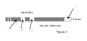
Figure 1 shows a first embodiment of a device for treating dysphagia;
Figure 2 shows a second embodiment of a device for treating dysphagia; and
Figure 3 shows a third embodiment of a device for treating dysphagia.
DESCRIPTION
Embodiments of the invention will now be described by way of reference to the
figures.
In a first embodiment of the invention there is a catheter (10), in the form
of a flexible
endoscope, with electrodes (12, 14) designed to deliver electrical stimulation
to target tissues.
The electrodes (12, 14) are integrated into the body of the endoscope (10) at
a fixed position
proximal an imaging device (16) part of the endoscope (10). The imaging device
(16) part of
the endoscope is positioned at an end of the endoscope (10). Within the body
of the flexible
endoscope (10) there are current carrying means to connect the electrodes (12,
14) to a
source of electrical stimulation external to the patient.
In a second embodiment there is a catheter in the form of a flexible endoscope
(100) having
an imaging device (108) at one end thereof with a sleeve (102) located on it
such that the
sleeve (102) is freely moveable along the length of the endoscope (100), and
located on the
sleeve (102) are one or more electrodes (104, 106) designed to deliver
electrical stimulation
to target tissues. Within the body of the sleeve (102) there are current
carrying means to
connect the electrodes (104, 106) to a source of electrical stimulation
external to the patient.
In a third embodiment there is a catheter in the form of a flexible endoscope
(200) with an
extended structure (202) distal to an imaging device (204) located at an end
of the flexible
endoscope (200), the extended structure (202) incorporating one or more
electrodes (206,
208) designed to deliver electrical stimulation to target tissues. The
electrodes (206, 208) in
this extended structure (202) may be either fixed and integrated into said
extended structure
(202), or may be disposed on a sleeve (210 - as shown in figure 4) that is
freely moveable
along the length of the extended structure (202), such movement being
controllable
externally. In the latter case the electrodes (206, 208) can then be moved
relative to the
position, and additionally within the field of view, of the imaging device
(204). Within the body
of the flexible endoscope (200) or the body of the sleeve (210) there are
current carrying
means to connect the electrodes to a source of electrical stimulation external
to the patient.
2

Dessin représentatif
Une figure unique qui représente un dessin illustrant l'invention.
États administratifs

2024-08-01 : Dans le cadre de la transition vers les Brevets de nouvelle génération (BNG), la base de données sur les brevets canadiens (BDBC) contient désormais un Historique d'événement plus détaillé, qui reproduit le Journal des événements de notre nouvelle solution interne.

Veuillez noter que les événements débutant par « Inactive : » se réfèrent à des événements qui ne sont plus utilisés dans notre nouvelle solution interne.

Pour une meilleure compréhension de l'état de la demande ou brevet qui figure sur cette page, la rubrique Mise en garde , et les descriptions de Brevet , Historique d'événement , Taxes périodiques et Historique des paiements devraient être consultées.

Historique d'événement

Description Date
Réputée abandonnée - les conditions pour l'octroi - jugée non conforme 2024-09-13
Lettre envoyée 2024-03-19
Un avis d'acceptation est envoyé 2024-03-19
Inactive : Approuvée aux fins d'acceptation (AFA) 2024-03-15
Inactive : Q2 réussi 2024-03-15
Modification reçue - modification volontaire 2023-11-01
Modification reçue - réponse à une demande de l'examinateur 2023-11-01
Rapport d'examen 2023-07-04
Inactive : Rapport - Aucun CQ 2023-06-08
Lettre envoyée 2022-06-28
Inactive : Soumission d'antériorité 2022-06-28
Requête d'examen reçue 2022-05-25
Toutes les exigences pour l'examen - jugée conforme 2022-05-25
Exigences pour une requête d'examen - jugée conforme 2022-05-25
Représentant commun nommé 2020-11-07
Représentant commun nommé 2019-10-30
Modification reçue - modification volontaire 2018-12-11
Inactive : Notice - Entrée phase nat. - Pas de RE 2018-12-06
Inactive : Page couverture publiée 2018-12-03
Demande reçue - PCT 2018-11-30
Inactive : CIB attribuée 2018-11-30
Inactive : CIB en 1re position 2018-11-30
Exigences pour l'entrée dans la phase nationale - jugée conforme 2018-11-26
Demande publiée (accessible au public) 2017-11-30

Historique d'abandonnement

Date d'abandonnement Raison Date de rétablissement
2024-09-13

Taxes périodiques

Le dernier paiement a été reçu le 2024-05-13

Avis : Si le paiement en totalité n'a pas été reçu au plus tard à la date indiquée, une taxe supplémentaire peut être imposée, soit une des taxes suivantes :

  • taxe de rétablissement ;
  • taxe pour paiement en souffrance ; ou
  • taxe additionnelle pour le renversement d'une péremption réputée.

Veuillez vous référer à la page web des taxes sur les brevets de l'OPIC pour voir tous les montants actuels des taxes.

Historique des taxes

Type de taxes Anniversaire Échéance Date payée
Taxe nationale de base - générale 2018-11-26
TM (demande, 2e anniv.) - générale 02 2019-05-27 2019-04-24
TM (demande, 3e anniv.) - générale 03 2020-05-25 2020-05-11
TM (demande, 4e anniv.) - générale 04 2021-05-25 2021-05-17
TM (demande, 5e anniv.) - générale 05 2022-05-25 2022-05-19
Requête d'examen - générale 2022-05-25 2022-05-25
TM (demande, 6e anniv.) - générale 06 2023-05-25 2023-05-25
TM (demande, 7e anniv.) - générale 07 2024-05-27 2024-05-13
Titulaires au dossier

Les titulaires actuels et antérieures au dossier sont affichés en ordre alphabétique.

Titulaires actuels au dossier
PHAGENESIS LIMITED
Titulaires antérieures au dossier
CONOR MULROONEY
Les propriétaires antérieurs qui ne figurent pas dans la liste des « Propriétaires au dossier » apparaîtront dans d'autres documents au dossier.
Documents

Pour visionner les fichiers sélectionnés, entrer le code reCAPTCHA :


Pour visualiser une image, cliquer sur un lien dans la colonne description du document. Pour télécharger l'image (les images), cliquer l'une ou plusieurs cases à cocher dans la première colonne et ensuite cliquer sur le bouton "Télécharger sélection en format PDF (archive Zip)" ou le bouton "Télécharger sélection (en un fichier PDF fusionné)".

Liste des documents de brevet publiés et non publiés sur la BDBC .

Si vous avez des difficultés à accéder au contenu, veuillez communiquer avec le Centre de services à la clientèle au 1-866-997-1936, ou envoyer un courriel au Centre de service à la clientèle de l'OPIC.


Description du
Document 
Date
(aaaa-mm-jj) 
Nombre de pages   Taille de l'image (Ko) 
Revendications 2023-11-01 1 21
Revendications 2018-11-26 1 45
Abrégé 2018-11-26 1 52
Description 2018-11-26 2 128
Dessin représentatif 2018-11-26 1 7
Dessins 2018-11-26 1 26
Page couverture 2018-12-03 1 34
Taxes 2024-07-04 1 192
Paiement de taxe périodique 2024-05-13 44 1 804
Avis d'entree dans la phase nationale 2018-12-06 1 207
Rappel de taxe de maintien due 2019-01-28 1 111
Avis du commissaire - Demande jugée acceptable 2024-03-19 1 580
Courtoisie - Réception de la requête d'examen 2022-06-28 1 424
Demande de l'examinateur 2023-07-04 4 188
Modification / réponse à un rapport 2023-11-01 8 248
Traité de coopération en matière de brevets (PCT) 2018-11-26 1 37
Traité de coopération en matière de brevets (PCT) 2018-11-26 1 48
Demande d'entrée en phase nationale 2018-11-26 3 60
Rapport de recherche internationale 2018-11-26 2 51
Modification / réponse à un rapport 2018-12-11 2 78
Requête d'examen 2022-05-25 5 112