Note: Descriptions are shown in the official language in which they were submitted.
CA 03025756 2018-11-26
WO 2017/217934
PCT/SG2017/050300
1
PRL3 Antibody
Field of the Invention
The present invention relates to humanised antibodies that bind PRL3.
Background to the Invention
Cancer is fundamentally a disease of disordered gene expression leading to
multistep
progression towards metastasis (1), the major cause of cancer-related deaths
(2).
Accumulating evidence indicates that protein tyrosine phosphatases (PTPs) play
important roles in driving metastatic progression (3). We identified
phosphatase of
regenerating liver-3 (PRL-3; also known as PTP4A3) in 1998 as a member of the
PRL
family of dual-specificity PTPs (4), which consists of three members: PRL-1,
PRL-2, and
PRL-3. In 2001, the Vogelstein group characterized PRL-3 as a metastasis-
associated
phosphatase specifically and highly upregulated in metastatic colorectal
cancer samples,
but not primary cancers and normal colorectal epithelia (5). PRL-3 was also
identified as
the most significant predictor of metastatic recurrence in patients with uveal
melanoma in
a recent independent global gene expression study (6). Clinically, elevated
PRL-3 mRNA
expression levels have been shown to correlate with higher metastatic
potential and poor
prognosis of multiple cancer types, including colorectal, gastric, breast,
ovarian, and lung
cancers (7).
PRLs are localized to the cytoplasmic face of the plasma membrane and
endosomes via
their prenylated C-termini (8). Mounting evidence suggests that PRL-3 promotes
multiple
stages of malignant transformation, including cellular proliferation,
epithelial-
mesenchymal transition (EMT), invasion, motility, angiogenesis, and survival
(9).
Molecularly, PRL-3 has been shown to activate the PI3K/Akt pathway indirectly
through
down-regulation of PTEN (10), and activate oncogenic ERK and SRC signaling via
constitutive activation of multiple upstream receptor tyrosine kinases (11-
13).
PRL-3 was first linked with GC progression in 2004 when it was found that
higher PRL-3
levels correlated with increased GC invasiveness and metastasis (14). Since
then, PRL-3
has been reported to be overexpressed in up to 70% of primary gastric
carcinomas, with
higher PRL-3 expression correlated to shorter post-operative survival at all
tumor stages
in GC patients (15,16). This prognostic potential of PRL-3 is particularly
important as GC
ranks as the third leading cause of cancer mortality worldwide with more than
700,000
gastric cancer-related deaths annually (2), largely due to delayed detection
and the
CA 03025756 2018-11-26
WO 2017/217934
PCT/SG2017/050300
2
asymptomatic nature of the disease in its early stages, coupled to the high
rate of
recurrence after treatment (17). Despite high failure rates, radical surgery
remains the
standard form of therapy for GC, and adjuvant chemotherapy is often considered
pre-
and/or post-resection (18,19). Nonetheless, overall survival with chemotherapy
remains
poor and is accompanied with undesirable side effects due to non-specific
targeting of
other actively dividing, noncancerous cells (17). To this end, targeted
therapy using
tumor-specific biological agents has emerged as the focus of anti-cancer drug
development due to their potential to selectively inhibit specific molecules
involved in the
growth and survival of cancer cells, whilst sparing normal cells. Current
antibody
therapies only target extracellular (cell-surface or secreted) proteins since
antibodies are
generally believed to be too large to enter cells, leaving a large pool of
intracellular
therapeutic targets, such as phosphatases, kinases, and transcription factors,
untapped
by antibody therapies. In GC, for example, the HER2/neu receptor antagonist
trastuzumab (Herceptin) has been approved to target the 13-20% of GCs
expressing cell-
surface HER2/neu receptors, particularly metastatic gastric or
gastroesophageal junction
adenocarcinoma (20,21). However, despite moderate responses, patients often
develop
resistance to trastuzumab (22), hindering its efficacy. Alternative targeted
therapies for
GC are thus desperately needed and actively being sought after.
In 2008, we reported a novel approach of antibody therapy, targeting
intracellular PRL-1
and PRL-3 oncoantigens (23). In that report, we showed that anti-PRL-3
antibodies
inhibited experimental metastasis of cancer cells expressing PRL-3 (but not
PRL-1) whilst
anti-PRL-1 antibodies inhibited cancer cells expressing PRL-1 (but not PRL-3),
thus
establishing a stringent requirement for specific antibody-antigen recognition
for
therapeutic efficacy when targeting such intracellular oncoproteins. Following
this, in
2011, we validated the feasibility and efficacy of this new concept, targeting
additional
endogenous and exogenous intracellular 'tumor-specific antigens' with antibody
therapies
or vaccinations in wild type C57BL/6 and transgenic spontaneous breast tumor
MMTV-
PyMT mice (24). We and Ferrone proposed three possible mechanisms for the
antitumor
activity of intracellular tumor antigen (TA)-specific antibodies, including
antibody
penetration into cells, antibody binding to externalized antigen and/or
antibody recognition
of MHC-bound antigen-derived peptides (25,26).
Following the success of murine and, more recently, chimeric (27) anti-PRL-3
antibodies
in targeting PRL-3-expressing tumors, we herein translate our approach into a
more
clinically-relevant setting with regards to four key aspects: 1) the use of
PRL-3 humanized
CA 03025756 2018-11-26
WO 2017/217934
PCT/SG2017/050300
3
antibodies (PRL3-zumab) instead of mouse or chimeric antibodies; 2) targeting
of human
cancer cell lines instead of mouse cancer cell lines; 3) the development of
more clinically-
relevant orthotopic gastric tumor models instead of mouse tail vein metastatic
models;
and 4) the identification of a potential surrogate biomarker for monitoring of
PRL3-zumab
therapeutic efficacy. We demonstrate the first example of a new class of
humanized
antibody to block gastric tumorigenesis. Our findings reveal the potential of
targeting
intracellular oncoproteins with antibody therapy, ushering in a new era of
cancer
therapeutics.
Summary of the Invention
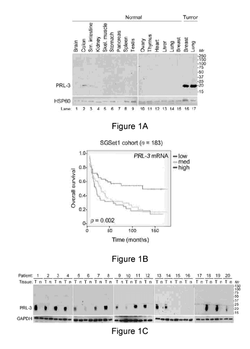
Off-target effects are major clinical concerns for cancer therapies. We
generated a first-in-
class humanized antibody (PRL3-zumab) against tumor-specific intracellular PRL-
3, an
oncogenic phosphatase upregulated in multiple human cancers. We focused on
gastric
cancer (GC), providing independent evidence that elevated PRL-3 mRNA levels
significantly correlate with shortened overall survival of GC patients. PRL-3
protein was
overexpressed in 85% of fresh-frozen GC tumors, but not in patient-matched
normal
gastric tissues examined. Using human GC cell lines, we established clinically
relevant
orthotopic gastric tumor models and demonstrated that PRL3-zumab specifically
blocked
growth of PRL-3-positive (PRL-3+), but not PRL-3-negative (PRL-3-) tumors. PRL-
3-
zumab had better therapeutic efficacy as a monotherapy than in combination
with 5-
fluorouracil (5-FU), or 5-FU alone. PRL3-zumab was specifically enriched in
PRL-3+
tumor tissues and promoted immune cell recruitment to PRL-3+ tumor
microenvironments. Unexpectedly, we found secreted PRL-3 oncoprotein in 62% of
multiple types of human cancer urines and in 100% of cancer urines derived
from PRL-3+,
but not PRL-3- tumor-bearing mice. Furthermore, urinary PRL-3 levels were
significantly
reduced after effective treatment with PRL3-zumab. The Urinary PRL-3 could be
considered as a potential diagnostic and a surrogate biomarker for therapeutic
response
monitoring of PRL3-zumab therapy in multiple cancer types in future.
We also investigated the mechanism of action (MOA) to address how PRL-3
antibody
could possibly bind to its intracellular PRL-3 antigen, and conclude that
indeed
'Intracellular oncoprotein' can be re-localized to the cell surface as
'Extracellular
oncoprotein' in cancer, thus follow a rational basis for tumor elimination via
antibody
conventional pathways against Extracellular Oncotargets.
CA 03025756 2018-11-26
WO 2017/217934
PCT/SG2017/050300
4
Consistently, we found that PRL3-zumab blocks tumors expressing PRL-3
'Intracellular
antigen, requiring host Fcy11/111 receptor interaction, full antibody
activities, and increased
M1 (but not M2) macrophages, B lymphocytes, natural killer cells to enhance
host
immunity. These results suggest the MOA of antibody targeting 'Intracellular
oncoprotein'
is indeed following the similar principles of targeting 'Extracellular
Oncoprotein' via
classical antibody-dependent cell cytotoxicity (ADCC) or phagocytotic (ADCP)
pathways
to eliminate tumors.
Finally, using 110 precious fresh-frozen human tumors or their matched normal
tissues,
we further showed that PRL-3 is an excellent tumor-specific oncotarget broadly
overexpressed on an average 7E3(:)/o from 9 different human cancer types:
liver, lung,
colon, breast, stomach, bladder, prostate, AML, and kidney patient tumor
samples, but
not in matched normal tissues. PRL-3 may therefore be a useful biomarker of
cancer, and
a near-universal target for cancer therapy. PRL-3 may therefore provide a
useful
biomarker for solid cancers.
The present invention is concerned with antibodies, or antigen binding
fragments, that
bind to PRL3. Heavy and light chain polypeptides are also disclosed. The
antibodies,
antigen binding fragments and polypeptides may be provided in isolated and/or
purified
form and may be formulated into compositions suitable for use in research,
therapy and
diagnosis. In particular, the invention is concerned with humanized antibodies
that bind
PRL3, and in particular PRL3 antagonist antibodies.
In some cases, the antibodies of the invention inhibit a function of PRL3. In
some cases,
the antibodies inhibit a protein tyrosine phosphatase (PTP) function of PRL3.
In some
cases, the antibodies induce ADCC and/or ADCP. In some cases, the antibodies
are
capable of binding to Fc receptors, such as FcRII and/or FcRIII. In some
cases, binding
of the antibody to the cell leads to the recruitment of immune cells to the
cell, such as B
cells, NK cells, or macrophages, preferably M1 macrophages.
In one aspect of the present invention an antibody, or antigen binding
fragment, is
provided, the amino acid sequence of the antibody may comprise the amino acid
sequences i) to iii), or the amino acid sequences iv) to vi), or preferably
the amino acid
sequences i) to vi):
i) KASQSVEDDGENYMN (SEQ ID NO:4)
ii) AASNLES (SEQ ID NO:5)
iii) QQSNEDPFT (SEQ ID NO:6)
CA 03025756 2018-11-26
WO 2017/217934
PCT/SG2017/050300
iv) GYTFTNYYMH (SEQ ID NO:1)
v) WIYPGNVNTYYNEKFRG (SEQ ID NO:2)
vi) EEKNYPWFAY (SEQ ID NO:3)
or a variant thereof in which one or two or three amino acids in one or more
of the
5 sequences (i) to (vi) are replaced with another amino acid.
The antibody, or antigen binding fragment, may comprise at least one light
chain variable
region incorporating the following CDRs:
LC-CDR1: KASQSVEDDGENYMN (SEQ ID NO:4)
LC-CDR2: AASNLES (SEQ ID NO:5)
LC-CDR3: QQSNEDPFT (SEQ ID NO:6)
The antibody, or antigen binding fragment, may comprise at least one heavy
chain
variable region incorporating the following CDRs:
HC-CDR1: GYTFTNYYMH (SEQ ID NO:1)
HC-CDR2: WIYPGNVNTYYNEKFRG (SEQ ID NO:2)
HC-CDR3: EEKNYPWFAY (SEQ ID NO:3)
The antibody may comprise at least one light chain variable region
incorporating the
CDRs shown in Figure 7. The antibody may comprise at least one heavy chain
variable
region incorporating the CDRs shown in Figure 7.
The antibody may comprise at least one light chain variable region (VL)
comprising the
one of the amino acid sequences shown in Figure 7 or an amino acid sequence
having at
least 70%, more preferably one of at least 75%, 80%, 85%, 86%, 87%, 88%, 89%,
90%,
91%, 92%, 93%, 94%, 95%, 96%, 97%, 98%, 99%, or 100%, sequence identity to one
of
the amino acid sequences of the VL chain amino acid sequence shown in Figure
7. The
antibody may have a VL chain amino acid sequence having least 70%, more
preferably
one of at least 75%, 80%, 85%, 86%, 87%, 88%, 89%, 90%, 91%, 92%, 93%, 94%,
95%,
96%, 97%, 98%, 99%, or 100% sequence identity to one of the amino acid
sequences
shown in Figure 7 and comprise the following CDR sequences:
LC-CDR1: KASQSVEDDGENYMN (SEQ ID NO:4)
LC-CDR2: AASNLES (SEQ ID NO:5)
LC-CDR3: QQSNEDPFT (SEQ ID NO:6)
CA 03025756 2018-11-26
WO 2017/217934
PCT/SG2017/050300
6
The antibody may comprise at least one heavy chain variable region (VH)
comprising the
one of the amino acid sequences shown in Figure 7 or an amino acid sequence
having at
least 70%, more preferably one of at least 75%, 80%, 85%, 86%, 87%, 88%, 89%,
90%,
91%, 92%, 93%, 94%, 95%, 96%, 97%, 98%, 99%, or 100%, sequence identity to one
of
amino acid sequences of the VH chain amino acid sequence shown in Figure 7.
The
antibody may have a VH chain amino acid sequence having least 70%, more
preferably
one of at least 75%, 80%, 85%, 86%, 87%, 88%, 89%, 90%, 91%, 92%, 93%, 94%,
95%,
96%, 97%, 98%, 99%, or 100% sequence identity to one of the amino acid
sequences
shown in Figure 7 and comprise the following CDR sequences:
HC-CDR1: GYTFTNYYMH (SEQ ID NO:1)
HC-CDR2: WIYPGNVNTYYNEKFRG (SEQ ID NO:2)
HC-CDR3: EEKNYPWFAY (SEQ ID NO:3)
The antibody may comprise at least one light chain variable region comprising
one of the
amino acid sequences shown in Figure 7 (or an amino acid sequence having at
least
70%, more preferably one of at least 75%, 80%, 85%, 90%, 95%, 96%, 97%, 98%,
99%
or 100%, sequence identity to one of the amino acid sequences of the VL chain
amino
acid sequence shown in Figure 7) and at least one heavy chain variable region
comprising one of the amino acid sequence shown in Figure 7 (or an amino acid
sequence having at least 70%, more preferably one of at least 75%, 80%, 85%,
86%,
87%, 88%, 89%, 90%, 91%, 92%, 93%, 94%, 95%, 96%, 97%, 98%, 99%, or 100%,
sequence identity to one of the amino acid sequences of the VH chain amino
acid
sequence shown in Figure 7).
The antibody may bind PRL3. The antibody may optionally have amino acid
sequence
components as described above. The antibody may be an IgA, IgD, IgE, IgM or
IgM,
preferably an IgG. In one embodiment an in vitro complex, optionally isolated,
comprising
an antibody, or antigen binding fragment, as described herein, bound to PRL3
is
provided.
In one aspect of the present invention an isolated heavy chain variable region
polypeptide
is provided, the heavy chain variable region polypeptide comprising the
following CDRs:
HC-CDR1: GYTFTNYYMH (SEQ ID NO:1)
HC-CDR2: WIYPGNVNTYYNEKFRG (SEQ ID NO:2)
HC-CDR3: EEKNYPWFAY (SEQ ID NO:3)
CA 03025756 2018-11-26
WO 2017/217934
PCT/SG2017/050300
7
In one aspect of the present invention an antibody, or antigen binding
fragment, is
provided, the antibody, or antigen binding fragment, comprising a heavy chain
and a light
chain variable region sequence, wherein:
the heavy chain comprises a HC-CDR1, HC-CDR2, HC-CDR3, having at least
85% overall sequence identity to HC-CDR1 sequence (SEQ ID NO:1), HC-CDR2
sequence (SEQ ID NO:2), HC-CDR3 sequence (SEQ ID NO:3), respectively, and
the light chain comprises a LC-CDR1, LC-CDR2, LC-CDR3õ having at least 85%
overall sequence identity to LC-CDR1 sequence (SEQ ID NO:4), LC-CDR2
sequence (SEQ ID NO:5), LC-CDR3 sequence (SEQ ID NO:6), respectively.
In some embodiments the degree of sequence identity may be one of 86%, 87%,
88%,
89%, 90%, 91%, 92%, 93%, 94%, 95%, 96%, 97%, 98%, 99%, or 100%.
In another aspect of the present invention an antibody, or antigen binding
fragment,
optionally isolated, is provided comprising a heavy chain and a light chain
variable region
sequence, wherein:
the heavy chain sequence has at least 85% sequence identity to a heavy chain
sequence set out in Figure 7, and
the light chain sequence has at least 85% sequence identity to a light chain
sequence set out in Figure 7
In some embodiments the degree of sequence identity may be one of 86%, 87%,
88%,
89%, 90%, 91%, 92%, 93%, 94%, 95%, 96%, 97%, 98%, 99%, or 100%.
In some embodiments the antibody, antigen binding fragment, or polypeptide
further
comprises variable region heavy chain framework sequences between the CDRs
according to the arrangement HCFR1:HC-CDR1:HCFR2:HC-CDR2:HCFR3:HC-
CDR3:HCFR4. The framework sequences may be derived from human consensus
framework sequences.
In some cases, the antibody, antigen binding fragment, or polypeptide
comprises a heavy
chain sequence selected from:
VQSGAEVKKPGASVKVSCKASGYTFTNYYMHVVV (SEQ ID NO: 29);
WIYPGNVNTYYNEKFR (SEQ ID NO: 30);
ASTAYMELSSLRSE (SEQ ID NO: 31); and/or
ASEEKNYPWFAYWGQGTLVT (SEQ ID NO: 32).
CA 03025756 2018-11-26
WO 2017/217934
PCT/SG2017/050300
8
In one aspect of the present invention an isolated light chain variable region
polypeptide,
optionally in combination with a heavy chain variable region polypeptide as
described
herein, is provided, the light chain variable region polypeptide comprising
the following
CDRs:
LC-CDR1: KASQSVEDDGENYMN (SEQ ID NO:4)
LC-CDR2: AASNLES (SEQ ID NO:5)
LC-CDR3: QQSNEDPFT (SEQ ID NO:6)
In some embodiments the antibody, antigen binding fragment, or polypeptide
further
comprises variable region light chain framework sequences between the CDRs
according
to the arrangement LCFR1:LC-CDR1:LCFR2:LC-CDR2:LCFR3:LC-CDR3:LCFR4. The
framework sequences may be derived from human consensus framework sequences.
In some cases, the antibody, antigen binding fragment, or polypeptide
comprises a light
chain sequence selected from:
QSPSSLSASVGDRVT (SEQ ID NO: 26);
KASQSVEDDGENYMNVVYQQK (SEQ ID NO: 27); and/or
SGSGSGTDFTLTISSLQPEDFATYYCQQSNEDPFT (SEQ ID NO: 28).
In some cases, the antibody, antigen binding fragment, or polypeptide
comprises 2, 3, 4,
5, 6, or all of the amino acid sequences selected from:
VQSGAEVKKPGASVKVSCKASGYTFTNYYMHVVV;
WIYPGNVNTYYNEKFR;
ASTAYMELSSLRSE;
ASEEKNYPWFAYWGQGTLVT;
QSPSSLSASVGDRVT;
KASQSVEDDGENYMNVVYQQK; and/or
SGSGSGTDFTLTISSLQPEDFATYYCQQSNEDPFT.
The antibody may comprise at least one light chain variable region (VL) and/or
a heavy
chain variable region (VH) comprising the one of the amino acid sequences
shown in
Figure 7 or an amino acid sequence having at least 70%, more preferably one of
at least
75%, 80%, 85%, 86%, 87%, 88%, 89%, 90%, 91%, 92%, 93%, 94%, 95%, 96%, 97%,
98%, 99%, or 100%, sequence identity to one of the amino acid sequences shown
in
Figure 7, and comprise the following CDR sequences:
LC-CDR1: KASQSVEDDGENYMN (SEQ ID NO:4)
LC-CDR2: AASNLES (SEQ ID NO:5)
CA 03025756 2018-11-26
WO 2017/217934
PCT/SG2017/050300
9
LC-CDR3: QQSNEDPFT (SEQ ID NO:6)
HC-CDR1: GYTFTNYYMH (SEQ ID NO:1)
HC-CDR2: WIYPGNVNTYYNEKFRG (SEQ ID NO:2)
HC-CDR3: EEKNYPWFAY (SEQ ID NO:3)
and contain at least one of the following sequences:
VQSGAEVKKPGASVKVSCKASGYTFTNYYMHVVV;
WIYPGNVNTYYNEKFR;
ASTAYMELSSLRSE;
ASEEKNYPWFAYWGQGTLVT;
QSPSSLSASVGDRVT;
KASQSVEDDGENYMNVVYQQK; and/or
SGSGSGTDFTLTISSLQPEDFATYYCQQSNEDPFT.
The antibody may comprise at least one light chain variable region (VL) and/or
a heavy
chain variable region (VH) comprising the one of the amino acid sequences
shown in
Figure 7 or an amino acid sequence having at least 70%, more preferably one of
at least
75%, 80%, 85%, 86%, 87%, 88%, 89%, 90%, 91%, 92%, 93%, 94%, 95%, 96%, 97%,
98%, 99%, or 100%, sequence identity to one of the amino acid sequences shown
in
Figure 7.
The antibody may comprise at least on light chain variable region (VH)
selected from:
SEQ ID NO: 16, SEQ ID NO: 17, SEQ ID NO: 18, SEQ ID NO: 19, SEQ ID NO: 20, SEQ
ID NO: 21, SEQ ID NO: 22, SEQ ID NO: 23, SEQ ID NO: 24 or SEQ ID NO: 25, or an
amino acid sequence having at least 70%, more preferably one of at least 75%,
80%,
85%, 86%, 87%, 88%, 89%, 90%, 91%, 92%, 93%, 94%, 95%, 96%, 97%, 98%, 99%, or
100%, sequence identity to amino acid sequence SEQ ID NO: 16, SEQ ID NO: 17,
SEQ
ID NO: 18, SEQ ID NO: 19, SEQ ID NO: 20, SEQ ID NO: 21, SEQ ID NO: 22, SEQ ID
NO: 23, SEQ ID NO: 24 or SEQ ID NO: 25.
Preferably, the antibody comprises a light chain variable region (VH) selected
from: SEQ
ID NO: 16, SEQ ID NO: 17, SEQ ID NO: 18, SEQ ID NO: 19, SEQ ID NO: 20, SEQ ID
NO: 21 or SEQ ID NO: 22, or an amino acid sequence having at least 70%, more
preferably one of at least 75%, 80%, 85%, 86%, 87%, 88%, 89%, 90%, 91%, 92%,
93%,
94%, 95%, 96%, 97%, 98%, 99%, or 100%, sequence identity to amino acid
sequence
SEQ ID NO: 16, SEQ ID NO: 17, SEQ ID NO: 18, SEQ ID NO: 19, SEQ ID NO: 20, SEQ
ID NO: 21 or SEQ ID NO: 22.
CA 03025756 2018-11-26
WO 2017/217934
PCT/SG2017/050300
The antibody may comprise at least on light chain variable region (VL)
selected from: SEQ
ID NO: 7, SEQ ID NO: 8, SEQ ID NO: 9, SEQ ID NO: 10, SEQ ID NO: 11, SEQ ID NO:
12, SEQ ID NO: 13, SEQ ID NO: 14, or SEQ ID NO: 15, or an amino acid sequence
5 having at least 70%, more preferably one of at least 75%, 80%, 85%, 86%,
87%, 88%,
89%, 90%, 91%, 92%, 93%, 94%, 95%, 96%, 97%, 98%, 99%, or 100%, sequence
identity to amino acid sequence SEQ ID NO: 7, SEQ ID NO: 8, SEQ ID NO: 9, SEQ
ID
NO: 10, SEQ ID NO: 11, SEQ ID NO: 12, SEQ ID NO: 13, SEQ ID NO: 14, or SEQ ID
NO: 15.
Preferably, the antibody comprises a light chain variable region (VL) selected
from: SEQ
ID NO: 7, SEQ ID NO: 8, SEQ ID NO: 9, SEQ ID NO: 10, SEQ ID NO: 11 or SEQ ID
NO:
12, or an amino acid sequence having at least 70%, more preferably one of at
least 75%,
80%, 85%, 86%, 87%, 88%, 89%, 90%, 91%, 92%, 93%, 94%, 95%, 96%, 97%, 98%,
99%, or 100%, sequence identity to amino acid sequence SEQ ID NO: 7, SEQ ID
NO: 8,
SEQ ID NO: 9, SEQ ID NO: 10, SEQ ID NO: 11 or SEQ ID NO: 12.
The antibody may comprise the following CDR sequences:
LC-CDR1: KASQSVEDDGENYMN (SEQ ID NO:4)
LC-CDR2: AASNLES (SEQ ID NO:5)
LC-CDR3: QQSNEDPFT (SEQ ID NO:6)
HC-CDR1: GYTFTNYYMH (SEQ ID NO:1)
HC-CDR2: WIYPGNVNTYYNEKFRG (SEQ ID NO:2)
HC-CDR3: EEKNYPWFAY (SEQ ID NO:3)
and contain at least one of the following sequences:
VQSGAEVKKPGASVKVSCKASGYTFTNYYMHVVV;
WIYPGNVNTYYNEKFR;
ASTAYMELSSLRSE;
ASEEKNYPWFAYWGQGTLVT;
QSPSSLSASVGDRVT;
KASQSVEDDGENYMNVVYQQK; and/or
SGSGSGTDFTLTISSLQPEDFATYYCQQSNEDPFT;
and be capable of binding to PRL3, and antagonising a biological function of
PRL3.
In some embodiments, the antibody, or antibody binding fragment, may further
comprise
a human constant region. For example selected from one of IgG1, IgG2, IgG3 and
IgG4.
CA 03025756 2018-11-26
WO 2017/217934
PCT/SG2017/050300
11
In some embodiments, the antibody, or antibody binding fragment, may further
comprise
a murine constant region. For example, selected from one of IgG1, IgG2A, IgG2B
and
IgG3.
The antibody is preferably a whole antibody, or an antibody or antibody
fragment that
includes an Fc domain. The antibody or antibody fragment may include one or
both of a
CH1 and a CH2 domain. Preferably, the antibody includes a CH2 domain. The
antibody
may contain both a CH1 and a CH2 domain. Preferably, the antibody is not a
Fab',
F(ab)'2 fragment, and/or is not an scFv and/or is not a minibody. Preferably,
the antibody
is an IgG immunoglobulin.
In some aspects, the individual to be treated is immunocompetent. The
individual may
have been determined to be immunocompetent. The individual may have been
determined to produce NK cells, and/or B cells. The individual may be treated
to
stimulate the production and/or activation of NK cells and/or B cells, such as
through the
administration of cytokines, or by stopping the administration of agents known
to reduce
the production and/or activation of NK cells and/or B cells.
In another aspect of the present invention, a composition, e.g. a
pharmaceutical
composition or medicament, is provided. The composition may comprise an
antibody,
antigen binding fragment, or polypeptide as described herein and at least one
pharmaceutically-acceptable carrier, excipient, adjuvant or diluent.
In another aspect of the present invention an isolated nucleic acid encoding
an antibody,
antigen binding fragment, or polypeptide as described herein is provided. The
nucleic
acid encode a sequence set out in Figure 7, or a coding sequence which is
degenerate
as a result of the genetic code, or may have a nucleotide sequence having at
least 70%
identity thereto, optionally one of 75%, 80%, 85%, 86%, 87%, 88%, 89%, 90%,
91%,
92%, 93%, 94%, 95%, 96%, 97%, 98%, 99%, or 100%.
The antibody may bind to PRL3. The antibody may bind to an epitope comprising
the
amino acid sequence KAKFYN and/or HTHKTR. The antibody may be capable of
binding both sequences.
CA 03025756 2018-11-26
WO 2017/217934
PCT/SG2017/050300
12
In one aspect of the present invention there is provided a vector comprising a
nucleic acid
described herein. In another aspect of the present invention, there is
provided a host cell
comprising the vector. For example, the host cell may be eukaryotic, or
mammalian, e.g.
Chinese Hamster Ovary (CHO), or human or may be a prokaryotic cell, e.g. E.
coll.
In one aspect of the present invention a method for making an antibody, or
antigen
binding fragment or polypeptide as described herein is provided, the method
comprising
culturing a host cell as described herein under conditions suitable for the
expression of a
vector encoding the antibody, or antigen binding fragment or polypeptide, and
recovering
the antibody, or antigen binding fragment or polypeptide.
In another aspect of the present invention an antibody, antigen binding
fragment or
polypeptide is provided for use in therapy, or in a method of medical
treatment. In
another aspect of the present invention an antibody, antigen binding fragment
or
polypeptide as described herein is provided for use in the treatment of a T-
cell
dysfunctional disorder. In another aspect of the present invention, the use of
an antibody,
antigen binding fragment or polypeptide as described herein in the manufacture
of a
medicament or pharmaceutical composition for use in the treatment of a T-cell
dysfunctional disorder is provided.
In another aspect of the present invention a method is provided, the method
comprising
contacting a sample containing, or suspected to contain, PRL3 with an antibody
or
antigen binding fragment, as described herein, and detecting the formation of
a complex
of antibody, or antigen binding fragment, and PRL3.
In another aspect of the present invention a method of diagnosing a disease or
condition
in a subject is provided, the method comprising contacting, in vitro, a sample
from the
subject with an antibody, or antigen binding fragmentõ as described herein,
and
detecting the formation of a complex of antibody, or antigen binding fragment,
and PRL3.
In a further aspect of the present invention the use of an antibody, or
antigen binding
fragment, as described herein, for the detection of PRL3 in vitro is provided.
In another
aspect of the present invention the use of an antibody, or antigen binding
fragment, as
described herein, as an in vitro diagnostic agent is provided.
In methods of the present invention the antibody, antigen binding fragment or
polypeptide
may be provided as a composition as described herein.
CA 03025756 2018-11-26
WO 2017/217934
PCT/SG2017/050300
13
In any aspect of the present invention the antibody preferably specifically
binds PRL3
over other PRL phosphatases, such as PRL1 or PRL2.
The antibody may be an IgG. It may have a molecular weight of about 140 to
160kDa,
preferably about 150kDa.
In some embodiments the antibody may be PRL3-ZUMAB.
Also disclosed herein is the use of a humanised antibody or antigen binding
fragment as
disclosed herein for the manufacture of a medicament for the treatment of
cancer.
In other aspects, there is provided a humanised antibody or antibody binding
fragment for
use in a method of treating cancer. The antibody may be useful for inhibiting
tumor
formation, and/or inhibiting metastasis of tumor. The antibody may be useful
for reducing
the size of tumors. A treated individual may for example show a 1%, 2%, 3%,
4%, 5%,
6%, 7%, 8%, 9%, 10%, 15%, 20%, 25%, 30%, 40%, 50%, 60%, 70%, 80%, 90%, 100%
or more decrease in tumour size of a particular tumour, or decrease in tumour
number, or
both, compared to an individual who has not been treated, or compared to that
same
individual prior to treatment.
Also provided are method of treating cancer comprising administering a
humanised
antibody or antibody binding fragment as disclosed herein.
The cancer may be a PRL3 expressing or overexpressing cancer. The cancer may
be
gastric cancer.
The humanised antibody or antibody binding fragment may be administered
intravenously. It may be administered at a location distant to the cancer to
be treated.
In some methods the patient has not previously received chemotherapy,
particularly
antimetabolite therapy, such as 5-FU. In some cases, the patient has not
previously
received such therapy before, or has not received such treatment for their
cancer, such
as for their gastric cancer. In some cases, the antibody is not co-
administered with
another agent (i.e. antibody monotherapy). In some cases, the antibody is not
co-
administered with 5-FU.
CA 03025756 2018-11-26
WO 2017/217934
PCT/SG2017/050300
14
In some methods, the patient has been determined not to have an impaired
immune
system. In particular, the patient may have been determined to have a white
blood cell
count within normal range. In particular, the patient may have been determined
to not
have leukopenia. The patient may have been determined to have neturophil,
lymphocyte,
monocyte, red blood cell or platelet counts within normal range. The patient
may have a
white blood count, neturophil, lymphocyte, monocyte, red blood cell or
platelet count that
is not significantly different to a control, such as the count from an
individual known to not
have an impaired immune system, or to established normal values. For example,
the
patient may be determined to have between about 4,500 and about 10,000 white
blood
cells per microliter of blood.
In some aspects, the invention provides a method for selecting a patient for
treatment
with a humanised anti-PRL3 antibody or antibody fragment, the method
comprising
determining, in a sample of urine from the patient, the presence of PRL3. In
some cases,
the method involves determining the level of PRL3 in a urine sample from the
patient. In
some cases, the patient may have gastric, nasopharyngeal, bladder, lung,
breast or
prostate cancer.
In some cases, the individual has a family history of PRL3 overexpressing
cancer, or has
been identified as having a likelihood of developing a PRL3 overexpressing
cancer. In
some cases, the individual has a PRL3 overexpressing cancer, and is considered
to be at
risk of metastasis of that cancer.
In another aspect, provided herein are method involving determining the
cellular
localisation of PRL3. An increased proportion of cellular PRL3 on the cell
surface may
indicate that the individual has cancer. Provided herein is a method
comprising
determining the cellular localisation of PRL3 in a cell, wherein expression of
PRL3 at the
cell surface indicates that the cell is cancerous.
Methods include methods for the diagnosis of cancer, wherein the presence of,
or an
increase in PRL3 on the surface of a cell may indicate that the individual has
cancer. In
some cases, the amount of PRL3 in the cell is the same as a non-cancerous
control
sample, but the localisation of that PRL3 may be changed as compared to the
non-
cancerous control.
CA 03025756 2018-11-26
WO 2017/217934
PCT/SG2017/050300
Other methods include a method for determining whether or not a cell is
cancerous, the
method comprising determining the presence of PRL3 at the surface of the cell.
An
increase in the level or proportion of PRL3 as compared to a control cell may
indicate that
the individual is, or will become, cancerous.
5
Methods may involve the selection of an individual for an anti-cancer therapy,
based on
the cellular localisation of PRL3 in the sample. In some cases, the methods
involve
administration of an anti-cancer therapy to an individual so selected.
10 In some cases, the method may comprise determining the cellular
localisation of PRL3 in
two or more samples from the patient, taken at two or more time points. A
change in the
amount of PRL3 on the surface of the cell may indicate an increase or decrease
in the
cancer in the individual. An increase in cell surface PRL3 over time may
indicate that the
individual has developed cancer, or the cancer has worsened. A decrease in
cell surface
15 PRL3 over time may indicate that the cancer has reduced, or that the
therapy has
resulted in treatment of the cancer. An increased or unchanged level of PRL3
at the cell
surface may indicate that additional or alternative anti-cancer therapy is
required. The
level of PRL3 at the cell surface may therefore be used to select an
individual for a
further, or alternative, anti-cancer therapy.
An increase of 2-fold, 3-fold, 4-fold, 5-fold, 6-fold, 7-fold, 8-fold, 9-fold
or 10-fold more
PRL3 at the cell surface may indicate that the individual has cancer, and/or
the cell is
cancerous, or that the individual should be selected for treatment. The level
of PRL3 at
the cell surface may be compared to a control.
The sample may be a blood sample or a serum sample. The sample may be a urine
sample. The cancer may be a sample of the tumor or of the tissue surrounding
the
tumor. The method may involve obtaining the sample, or the method may be
performed
on a sample previously obtained from the individual.
Methods of diagnosis and detection may be performed in vitro, or ex vivo, and
in some
cases do not involve the step of obtaining a sample from an individual.
Description
Antibodies
CA 03025756 2018-11-26
WO 2017/217934
PCT/SG2017/050300
16
Antibodies according to the present invention preferably bind to PRL3 (the
antigen),
optionally with a Kd in the range 5pM to 8pM, preferably 6-7pm, preferably
about 6.3pM.
In some cases, the antibodies have an off rate of approximately 7 x 10⁻⁵s-1.
For example,
between about 1x10⁻⁵s-1 and 1x10⁻⁶s-1.
In some embodiments, antibodies according to the present invention bind to
PRL3, but
not to PRL1 or PRL2.
Antibodies according to the present invention may be provided in isolated
form.
By "antibody" we include a fragment or derivative thereof, or a synthetic
antibody or
synthetic antibody fragment.
In view of today's techniques in relation to monoclonal antibody technology,
antibodies
can be prepared to most antigens. The antigen-binding portion may be a part of
an
antibody (for example a Fab fragment) or a synthetic antibody fragment (for
example a
single chain Fv fragment [ScFv]). Suitable monoclonal antibodies to selected
antigens
may be prepared by known techniques, for example those disclosed in
"Monoclonal
Antibodies: A manual of techniques ", H Zola (CRC Press, 1988) and in
"Monoclonal
Hybridoma Antibodies: Techniques and Applications ", J G R Hurrell (CRC Press,
1982).
Chimaeric antibodies are discussed by Neuberger et al (1988, 8th International
Biotechnology Symposium Part 2, 792-799).
Monoclonal antibodies (mAbs) are useful in the methods of the invention and
are a
homogenous population of antibodies specifically targeting a single epitope on
an
antigen. Thus, mAbs binding PRL3 may be useful in the treatment of cancer.
Antigen binding fragments of antibodies, such as Fab and Fab2 fragments may
also be
provided as can genetically engineered antibodies and antibody fragments. The
variable
heavy (VH) and variable light (VL) domains of the antibody are involved in
antigen
recognition, a fact first recognised by early protease digestion experiments.
Further
confirmation was found by "humanisation" of rodent antibodies. Variable
domains of
rodent origin may be fused to constant domains of human origin such that the
resultant
antibody retains the antigenic specificity of the rodent parented antibody
(Morrison et al
(1984) Proc. Natl. Acad. Sd. USA 81, 6851-6855).
CA 03025756 2018-11-26
WO 2017/217934
PCT/SG2017/050300
17
Antibodies and antibody binding fragments according to the invention have been
humanised. Humanized antibodies are antibodies from non-human species whose
protein sequences have been modified to increase their similarity to antibody
variants
produced naturally in humans. The process of "humanisation" is usually applied
to
monoclonal antibodies developed for administration to humans. The process of
"humanisation" can be necessary when the process of developing a specific
antibody
involves generation in a non-human immune system, such as mice, as such
antibodies
may be immunogenic when administered to human patients. Humanisation may
involve
substitution of selective amino acids in the Fab portion of the molecule.
Alternatively,
humanisation may involve insertion of the appropriate CDR coding segments into
a
human antibody scaffold.
That antigenic specificity is conferred by variable domains and is independent
of the
constant domains is known from experiments involving the bacterial expression
of
antibody fragments, all containing one or more variable domains. These
molecules
include Fab-like molecules (Better et al (1988) Science 240, 1041); Fv
molecules (Skerra
et al (1988) Science 240, 1038); single-chain Fv (ScFv) molecules where the VH
and VL
partner domains are linked via a flexible oligopeptide (Bird et al (1988)
Science 242, 423;
Huston et al (1988) Proc. Natl. Acad. Sd. USA 85, 5879) and single domain
antibodies
(dAbs) comprising isolated V domains (Ward et al (1989) Nature 341, 544). A
general
review of the techniques involved in the synthesis of antibody fragments which
retain their
specific binding sites is to be found in VVinter & Milstein (1991) Nature 349,
293- 299.
By "ScFv molecules" we mean molecules wherein the VH and VL partner domains
are
covalently linked, e.g. by a flexible oligopeptide.
Fab, Fv, ScFv and dAb antibody fragments can all be expressed in and secreted
from E.
coli, thus allowing the facile production of large amounts of the said
fragments.
Whole antibodies, and F(ab')2 fragments are "bivalent". By "bivalent" we mean
that the
said antibodies and F(ab')2 fragments have two antigen combining sites. In
contrast, Fab,
Fv, ScFv and dAb fragments are monovalent, having only one antigen combining
site.
Synthetic antibodies which bind to PRL3 may also be made using phage display
technology as is well known in the art.
CA 03025756 2018-11-26
WO 2017/217934
PCT/SG2017/050300
18
Antibodies may be produced by a process of affinity maturation in which a
modified
antibody is generated that has an improvement in the affinity of the antibody
for antigen,
compared to an unmodified parent antibody. Affinity-matured antibodies may be
produced
by procedures known in the art, e.g., Marks et al.,Rio/Technology 10:779-783
(1992);
Barbas etal. Proc Nat. Acad. Sci. USA 91:3809-3813 (1994); Schier etal. Gene
169:147-
155 (1995); Yelton etal. J. lmmunol. 155:1994-2004 (1995); Jackson eta,'., J.
lmmunol.
154(7):331 0-159 (1995); and Hawkins eta,', J. Mol. Biol. 226:889-896 (1992).
Antibodies according to the present invention preferably exhibit specific
binding to PRL3.
An antibody that specifically binds to a target molecule preferably binds the
target with
greater affinity, and/or with greater duration than it binds to other targets.
In one
embodiment, the extent of binding of an antibody to an unrelated target is
less than about
10% of the binding of the antibody to the target as measured, e.g., by a
radioimmunoassay (RIA).
Antibodies according to the present invention preferably have a dissociation
constant (Kd)
of one of 1pM, 100nM,
'inM or 100pM. Binding affinity of an antibody for
its target is often described in terms of its dissociation constant (Kd).
Binding affinity can
be measured by methods known in the art, such as by a radiolabeled antigen
binding
assay (RIA) performed with the Fab version of the antibody and antigen
molecule.
Antibodies according to the present invention may be "antagonist" antibodies
that inhibit
or reduce a biological activity of the antigen to which it binds. Blocking of
PRL3 may
inhibit or reduce a phosphatase activity of PRL3. In some cases, the antibody
binds to,
but does not necessarily affect an activity of, PRL3.
In certain methods, the antibody is PRL3-ZUMAB, or a variant of PRL3-ZUMAB.
PRL3-
ZUMAB comprises the following CDR sequences:
Light chain:
LC-CDR1: (SEQ ID NO: 4)
LC-CDR2: (SEQ ID NO:5)
LC-CDR3: (SEQ ID NO:6)
Heavy chain:
HC-CDR1: (SEQ ID NO:1)
HC-CDR2: (SEQ ID NO:2)
HC-CDR3: (SEQ ID NO:3)
CA 03025756 2018-11-26
WO 2017/217934
PCT/SG2017/050300
19
CDR sequences determined by Kabat definition.
The structure of an antibody molecule which has a CDR as described herein will
generally be of a heavy or light chain sequence of an antibody molecule or
substantial
portion thereof in which the CDR is located at a location corresponding to the
CDR of
naturally occurring VH and VL antibody variable domains encoded by rearranged
immunoglobulin genes. The structures and locations of immunoglobulin variable
domains
may be determined by reference to Kabat, E.A. eta!, Sequences of Proteins of
Immunological Interest. 4th Edition. US Department of Health and Human
Services. 1987,
and updates thereof. A number of academic and commercial on-line resources are
available to query this database. For example, see Martin, A.C.R. Accessing
the Kabat
Antibody Sequence Database by Computer PROTEINS: Structure, Function and
Genetics, 25 (1996), 130-133 and the associated on-line resource, currently at
the world
wide web address bioinf.org.uk/abs/simkab.html.
Antibodies according to the present invention may comprise the CDRs of PRL3-
ZUMAB
or one of SEQ ID NOs 1-6. In an antibody according to the present invention
one or two
or three or four of the six CDR sequences may vary. A variant may have one or
two
amino acid substitutions in one or two of the six CDR sequences.
Amino acid sequences of the VH and VL chains of ant-PRL3-ZUMAB clones are
shown in
Figure 7.
The light and heavy chain CDRs may also be particularly useful in conjunction
with a
number of different framework regions. Accordingly, light and/or heavy chains
having LC-
CDR1-3 or HC-CDR1-3 may possess an alternative framework region. Suitable
framework regions are well known in the art and are described for example in
M. Lefranc
& G. Lefranc (2001) "The lmmunoglobulin FactsBook", Academic Press,
incorporated
herein by reference.
In this specification, antibodies may have VH and/or VL chains comprising an
amino acid
sequence that has a high percentage sequence identity to one or more of the VH
and/or
VL amino acid sequences of Figure 7.
For example, antibodies according to the present invention include antibodies
that bind
CA 03025756 2018-11-26
WO 2017/217934
PCT/SG2017/050300
PRL3 and have a VH chain that comprises an amino acid sequence having at least
70%,
more preferably one of at least 75%, 80%, 85%, 86%, 87%, 88%, 89%, 90%, 91%,
92%,
93%, 94%, 95%, 96%, 97%, 98%, 99%, or 100%, sequence identity to the VH chain
amino acid sequence of one or the amino acid sequences shown in Figure 7.
5
Antibodies according to the present invention may be detectably labelled or,
at least,
capable of detection. For example, the antibody may be labelled with a
radioactive atom
or a coloured molecule or a fluorescent molecule or a molecule which can be
readily
detected in any other way. Suitable detectable molecules include fluorescent
proteins,
10 luciferase, enzyme substrates, and radiolabels. The binding moiety may
be directly
labelled with a detectable label or it may be indirectly labelled. For
example, the binding
moiety may be an unlabelled antibody which can be detected by another antibody
which
is itself labelled. Alternatively, the second antibody may have bound to it
biotin and
binding of labelled streptavidin to the biotin is used to indirectly label the
first antibody.
Although a variety of antibody fragments are described herein, the antibody is
preferably
a whole antibody, containing an antibody binding fragment (Fab), and a
crystallisable
fragment (Fc). The antibody may consist of two heavy chains and two light
chains. It
comprises a variable fragment (Fv), which provides the antigen specificity of
the antibody,
and a constant domain.
Antibody fragments according to the invention preferably include a CH2 domain.
The
CH2 domain of an antibody plays an important role in mediating effector
functions and
preserving antibody stability. Accordingly, the antibody fragments of the
present
invention are preferably not a Fab', F(ab)'2, seFy or minibody.
Antibodies and fragments according to the invention are preferably able to
interact with
Fey (Fe-gamma) receptors, preferably Fey!! (0D32) and Fey!!! (CD16) receptors.
Methods of detection
Antibodies, or antigen binding fragments, described herein may be used in
methods that
involve the binding of the antibody or antigen binding fragment to PRL3. Such
methods
may involve detection of the bound complex of antibody, or antigen binding
fragment, and
PRL3. As such, in one embodiment a method is provided, the method comprising
contacting a sample containing, or suspected to contain, PRL3 with an antibody
or
CA 03025756 2018-11-26
WO 2017/217934
PCT/SG2017/050300
21
antigen binding fragment as described herein and detecting the formation of a
complex of
antibody, or antigen binding fragment, and PRL3.
Suitable method formats are well known in the art, including immunoassays such
as
sandwich assays, e.g. ELISA. The method may involve labelling the antibody, or
antigen
binding fragment, or PRL3, or both, with a detectable label, e.g. fluorescent,
luminescent
or radio- label.
Methods of this kind may provide the basis of a method of diagnosis of a
disease or
condition requiring detection and or quantitation of PRL3. Such methods may be
performed in vitro on a patient sample, or following processing of a patient
sample. Once
the sample is collected, the patient is not required to be present for the in
vitro method of
diagnosis to be performed and therefore the method may be one which is not
practised
on the human or animal body.
Such methods may involve determining the amount of PRL3 present in a patient
sample.
The method may further comprise comparing the determined amount against a
standard
or reference value as part of the process of reaching a diagnosis. Other
diagnostic tests
may be used in conjunction with those described here to enhance the accuracy
of the
diagnosis or prognosis or to confirm a result obtained by using the tests
described here.
Detection in a sample of PRL3 may be used for the purpose of diagnosis a
cancerous
condition in the patient, diagnosis of a predisposition to a cancerous
condition or for
providing a prognosis (prognosticating) of a cancerous condition. The
diagnosis or
prognosis may relate to an existing (previously diagnosed) cancerous
condition, which
may be benign or malignant, may relate to a suspected cancerous condition or
may relate
to the screening for cancerous conditions in the patient (which may be
previously
undiagnosed).
A sample may be taken from any tissue or bodily fluid. The sample may comprise
or may
be derived from: a quantity of blood; a quantity of serum derived from the
individual's
blood which may comprise the fluid portion of the blood obtained after removal
of the
fibrin clot and blood cells; a tissue sample or biopsy; or cells isolated from
said individual.
CA 03025756 2018-11-26
WO 2017/217934
PCT/SG2017/050300
22
Methods according to the present invention are preferably performed in vitro.
The term "in
vitro" is intended to encompass experiments with cells in culture whereas the
term "in
vivo" is intended to encompass experiments with intact multi-cellular
organisms.
Therapeutic applications
Antibodies, antigen binding fragments and polypeptides according to the
present
invention and compositions comprising such agents may be provided for use in
methods
of medical treatment. Treatment may be provided to subjects having a disease
or
condition in need of treatment. The disease or condition may be cancer,
including
metastatic cancer.
Administration of an antibody, antigen binding fragment or polypeptide is
preferably in a
"therapeutically effective amount", this being sufficient to show benefit to
the individual.
The actual amount administered, and rate and time-course of administration,
will depend
on the nature and severity of the disease being treated. Prescription of
treatment, e.g.
decisions on dosage etc, is within the responsibility of general practitioners
and other
medical doctors, and typically takes account of the disorder to be treated,
the condition of
the individual patient, the site of delivery, the method of administration and
other factors
known to practitioners. Examples of the techniques and protocols mentioned
above can
be found in Remington's Pharmaceutical Sciences, 20th Edition, 2000, pub.
Lippincott,
VVilliams & Wilkins.
The methods and compositions described here suitably enable an improvement in
a
measurable criterion in an individual to whom the treatment is applied,
compared to one
who has not received the treatment.
For this purpose, a number of criteria may be designated, which reflect the
progress of
cancer or the well-being of the patient. Useful criteria may include tumour
size, tumour
dimension, largest dimension of tumour, tumour number, presence of tumour
markers
(such as alpha-feto protein), degree or number of metastates, etc.
Thus, as an example, a treated individual may show a decrease in tumour size
or number
as measured by an appropriate assay or test. A treated individual may for
example show
a 1%, 2%, 3%, 4%, 5%, 6%, 7%, 8%, 9%, 10%, 15%, 20%, 25%, 30%, 40%, 50%, 60%,
70%, 80%, 90%, 100% or more decrease in tumour size of a particular tumour, or
decrease in tumour number, or both, compared to an individual who has not been
treated.
CA 03025756 2018-11-26
WO 2017/217934
PCT/SG2017/050300
23
The term proliferative disorder is used herein in a broad sense to include any
disorder
that requires control of the cell cycle. In particular, a proliferative
disorder includes
malignant and pre-neoplastic disorders. The methods and compositions described
here
are especially useful in relation to treatment or diagnosis of adenocarcinomas
such as:
small cell lung cancer, and cancer of the kidney, uterus, prostrate, bladder,
ovary, colon
and breast. For example, malignancies which may be treatable include acute and
chronic
leukemias, lymphomas, myelomas, sarcomas such as Fibrosarcoma, myxosarcoma,
liposarcoma, lymphangioendotheliosarcoma, angiosarcoma, endotheliosarcoma,
chondrosarcoma, osteogenic sarcoma, chordoma, lymphangiosarcoma,
synovioma, mesothelioma, leimyosarcoma, rhabdomyosarcoma, colon carcinoma,
ovarian cancer, prostate cancer, pancreatic cancer, breast cancer, squamous
cell
carcinoma, basal cell carcinoma, adenocarcinoma, sweat gland carcinoma,
sebaceous 5
gland carcinoma, papillary carcinoma, papillary adenocarcinomas,
cystadenocarcinoma,
medullary carcinoma, bronchogenic carcinoma, choriocarcinoma, renal cell
carcinoma,
hepatoma, bile duct carcinoma seminoma, embryonal carcinoma, cervical cancer,
testicular tumour, lung carcinoma, small cell lung carcinoma, bladder
carcinoma,
epithelial carcinoma, glioma, astrocytoma, ependymoma, pinealoma, 10
hemangioblastoma, acoustic neuoma, medulloblastoma, craniopharyngioma,
oligodendroglioma, menangioma, melanoma, neutroblastoma and retinoblastoma.
For the purposes of this document, the term "cancer" can comprise any one or
more of
the following: acute lymphocytic leukemia (ALL), acute myeloid leukemia (AML),
adrenocortical cancer, anal cancer, bladder cancer, blood cancer, bone cancer,
brain
tumor, breast cancer, cancer of the female genital system, cancer of the male
genital
system, central nervous system lymphoma, cervical cancer, childhood
rhabdomyosarcoma, childhood sarcoma, chronic lymphocytic leukemia (CLL),
chronic
myeloid leukemia (CML), colon and rectal cancer, colon cancer, endometrial
cancer,
endometrial sarcoma, esophageal cancer, eye cancer, gallbladder cancer,
gastric cancer,
gastrointestinal tract cancer, hairy cell leukemia, head and neck cancer,
hepatocellular
cancer, Hodgkin's disease, hypopharyngeal cancer, Kaposi's sarcoma, kidney
cancer,
laryngeal cancer, leukemia, leukemia, liver cancer, lung cancer, malignant
fibrous
histiocytoma, malignant thymoma, melanoma, mesothelioma, multiple myeloma,
myeloma, nasal cavity and paranasal sinus cancer, nasopharyngeal cancer,
nervous
system cancer, neuroblastoma, non-Hodgkin's lymphoma, oral cavity cancer,
oropharyngeal cancer, osteosarcoma, ovarian cancer, pancreatic cancer,
parathyroid
CA 03025756 2018-11-26
WO 2017/217934
PCT/SG2017/050300
24
cancer, penile cancer, pharyngeal cancer, pituitary tumor, plasma cell
neoplasm, primary
CNS lymphoma, prostate cancer, rectal cancer, respiratory system,
retinoblastoma,
salivary gland cancer, skin cancer, small intestine cancer, soft tissue
sarcoma, stomach
cancer, stomach cancer, testicular cancer, thyroid cancer, urinary system
cancer, uterine
sarcoma, vaginal cancer, vascular system, Waldenstrom's macroglobulinemia and
VVilms'
tumor.
The treatment may result in an alleviation of the symptoms of the cancer, or
may result in
the complete treatment of the cancer. The treatment may slow the progression
of the
cancer, or may prevent the worsening of the symptoms of the cancer.
Formulating pharmaceutically useful compositions and medicaments
Antibodies, antigen binding fragments and polypeptides according to the
present
invention may be formulated as pharmaceutical compositions for clinical use
and may
comprise a pharmaceutically acceptable carrier, diluent, excipient or
adjuvant.
In accordance with the present invention methods are also provided for the
production of
pharmaceutically useful compositions, such methods of production may comprise
one or
more steps selected from: isolating an antibody, antigen binding fragment or
polypeptide
as described herein; and/or mixing an isolated antibody, antigen binding
fragment or
polypeptide as described herein with a pharmaceutically acceptable carrier,
adjuvant,
excipient or diluent.
For example, a further aspect of the present invention relates to a method of
formulating
or producing a medicament or pharmaceutical composition for use in the
treatment of a T-
cell dysfunctional disorder, the method comprising formulating a
pharmaceutical
composition or medicament by mixing an antibody, antigen binding fragment or
polypeptide as described herein with a pharmaceutically acceptable carrier,
adjuvant,
excipient or diluent.
Cancer
A cancer may be any unwanted cell proliferation (or any disease manifesting
itself by
unwanted cell proliferation), neoplasm or tumor or increased risk of or
predisposition to
the unwanted cell proliferation, neoplasm or tumor. The cancer may be benign
or
malignant and may be primary or secondary (metastatic). A neoplasm or tumor
may be
any abnormal growth or proliferation of cells and may be located in any
tissue. Examples
CA 03025756 2018-11-26
WO 2017/217934
PCT/SG2017/050300
of tissues include the adrenal gland, adrenal medulla, anus, appendix,
bladder, blood,
bone, bone marrow, brain, breast, cecum, central nervous system (including or
excluding
the brain) cerebellum, cervix, colon, duodenum, endometrium, epithelial cells
(e.g. renal
epithelia), gallbladder, oesophagus, glial cells, heart, ileum, jejunum,
kidney, lacrimal
5 glad, larynx, liver, lung, lymph, lymph node, lymphoblast, maxilla,
mediastinum,
mesentery, myometrium, nasopharynx, omentume, oral cavity, ovary, pancreas,
parotid
gland, peripheral nervous system, peritoneum, pleura, prostate, salivary
gland, sigmoid
colon, skin, small intestine, soft tissues, spleen, stomach, testis, thymus,
thyroid gland,
tongue, tonsil, trachea, uterus, vulva, white blood cells.
Tumors to be treated may be nervous or non-nervous system tumors. Nervous
system
tumors may originate either in the central or peripheral nervous system, e.g.
glioma,
medulloblastoma, meningioma, neurofibroma, ependymoma, Schwannoma,
neurofibrosarcoma, astrocytoma and oligodendroglioma. Non-nervous system
cancers/tumors may originate in any other non-nervous tissue, examples include
melanoma, mesothelioma, lymphoma, myeloma, leukemia, Non-Hodgkin's lymphoma
(NHL), Hodgkin's lymphoma, chronic myelogenous leukemia (CML), acute myeloid
leukemia (AML), myelodysplastic syndrome (M DS), cutaneous T-cell lymphoma
(CTCL),
chronic lymphocytic leukemia (CLL), hepatoma, epidermoid carcinoma, prostate
carcinoma, breast cancer, lung cancer, colon cancer, ovarian cancer,
pancreatic cancer,
thymic carcinoma, NSCLC, haematologic cancer and sarcoma.
In particularly preferred aspects, the cancer is a PRL3 expressing cancer. In
some
cases, the cancer is a PRL3 overexpressing cancer. That is, the cancer is
associated
with, or caused by, overexpression of PRL3. The PRL3 need not be functional in
the
cancer, but could instead be a label or artefact of the cancer cell. In
particularly preferred
aspects, the cancer is gastric cancer, nasopharyngeal cancer, bladder cancer,
lung
cancer, breast cancer or prostate cancer. The cancer may be acute myeloid
leukemia,
colon cancer or ovarian cancer. In some cases, the cancer is a metastatic
cancer.
Simultaneous or Sequential Administration
Compositions may be administered alone or in combination with other
treatments, either
simultaneously or sequentially dependent upon the condition to be treated.
CA 03025756 2018-11-26
WO 2017/217934
PCT/SG2017/050300
26
In this specification an antibody, antigen binding fragment or polypeptide of
the present
invention and an anti-infective agent or chemotherapeutic agent (therapeutic
agent) may
be administered simultaneously or sequentially.
In some embodiments, treatment with an antibody, antigen binding fragment or
polypeptide of the present invention may be accompanied by chemotherapy.
Simultaneous administration refers to administration of the antibody, antigen
binding
fragment or polypeptide and therapeutic agent together, for example as a
pharmaceutical
composition containing both agents (combined preparation), or immediately
after each
other and optionally via the same route of administration, e.g. to the same
artery, vein or
other blood vessel.
Sequential administration refers to administration of one of the antibody,
antigen binding
fragment or polypeptide or therapeutic agent followed after a given time
interval by
separate administration of the other agent. It is not required that the two
agents are
administered by the same route, although this is the case in some embodiments.
The
time interval may be any time interval.
Chemotherapy
Chemotherapy refers to treatment of a cancer with a drug or with ionising
radiation (e.g.
radiotherapy using X-rays or y-rays). In preferred embodiments chemotherapy
refers to
treatment with a drug. The drug may be a chemical entity, e.g. small molecule
pharmaceutical, antibiotic, DNA intercalator, protein inhibitor (e.g. kinase
inhibitor), or a
biological agent, e.g. antibody, antibody fragment, nucleic acid or peptide
aptamer,
nucleic acid (e.g. DNA, RNA), peptide, polypeptide, or protein. The drug may
be
formulated as a pharmaceutical composition or medicament. The formulation may
comprise one or more drugs (e.g. one or more active agents) together with one
or more
pharmaceutically acceptable diluents, excipients or carriers.
A treatment may involve administration of more than one drug. A drug may be
administered alone or in combination with other treatments, either
simultaneously or
sequentially dependent upon the condition to be treated. For example, the
chemotherapy
may be a co-therapy involving administration of two drugs, one or more of
which may be
intended to treat the cancer.
CA 03025756 2018-11-26
WO 2017/217934
PCT/SG2017/050300
27
The chemotherapy may be administered by one or more routes of administration,
e.g.
parenteral, intravenous injection, oral, or intratumoural.
The chemotherapy may be administered according to a treatment regime. The
treatment
regime may be a pre-determined timetable, plan, scheme or schedule of
chemotherapy
administration which may be prepared by a physician or medical practitioner
and may be
tailored to suit the patient requiring treatment.
The treatment regime may indicate one or more of: the type of chemotherapy to
administer to the patient; the dose of each drug or radiation; the time
interval between
administrations; the length of each treatment; the number and nature of any
treatment
holidays, if any etc. For a co-therapy a single treatment regime may be
provided which
indicates how each drug is to be administered.
Chemotherapeutic drugs may be selected from:
= alkylating agents such as cisplatin, carboplatin, mechlorethamine,
cyclophosphamide, chlorambucil, ifosfamide;
= purine or pyrimidine anti-metabolites such as azathiopurine or
mercaptopurine;
= alkaloids and terpenoids, such as vinca alkaloids (e.g. vincristine,
vinblastine,
vinorelbine, vindesine), podophyllotoxin, etoposide, teniposide, taxanes such
as
paclitaxel (TaxolTm), docetaxel;
= topoisomerase inhibitors such as the type I topoisomerase inhibitors
camptothecins irinotecan and topotecan, or the typelltopoisomerase inhibitors
amsacrine, etoposide, etoposide phosphate, teniposide;
= antitumor antibiotics (e.g. anthracyline antibiotics) such as dactinomycin,
doxorubicin (AdriamycinTm), epirubicin, bleomycin, rapamycin;
= antibody based agents, such as anti-TIM-3 antibodies, anti-VEGF, anti-
TNFa,
anti-IL-2, antiGpIlb/111a, anti-CD-52, anti-CD20, anti-RSV, anti-
HER2/neu(erbB2),
anti-TNF receptor, anti-EGFR antibodies, monoclonal antibodies or antibody
fragments, examples include: cetuximab, panitumumab, infliximab, basiliximab,
bevacizumab (Avastine), abciximab, daclizumab, gemtuzumab, alemtuzumab,
rituximab (Mabthera0), palivizumab, trastuzumab, etanercept, adalimumab,
nimotuzumab
= EGFR inihibitors such as erlotinib, cetuximab and gefitinib
= anti-angiogenic agents such as bevacizumab (Avastine
CA 03025756 2018-11-26
WO 2017/217934
PCT/SG2017/050300
28
Further chemotherapeutic drugs may be selected from: 13-cis-Retinoic Acid, 2-
Chlorodeoxyadenosine, 5-Azacitidine 5-Fluorouracil, 6-Mercaptopurine, 6-
Thioguanine,
Abraxane, Accutane , Actinomycin-D Adriamycin , Adrucil , Afinitor0, Agrylin ,
Ala-
Corte, Aldesleukin, Alemtuzumab, ALIMTA, Alitretinoin, Alkaban-AQ0, Alkeran ,
All-
transretinoic Acid, Alpha Interferon, Altretamine, Amethopterin, Amifostine,
Aminoglutethimide, Anagrelide, Anandron , Anastrozole, Arabinosylcytosine,
Aranesp ,
Aredia0, Arimidex , Aromasine, Arranon , Arsenic Trioxide, Asparaginase, ATRA
Avastin , Azacitidine, BCG, BCNU, Bendamustine, Bevacizumab, Bexarotene,
BEXXAR , Bicalutamide, BiCNU, Blenoxane , Bleomycin, Bortezomib, Busulfan,
Busulfex , Calcium Leucovorin, Compathe, Comptosar0, Camptothecin-11,
Capecitabine, CaracTM, Carboplatin, Carmustine, Casodex , 00-5013, CCI-779,
CCNU,
CDDP, CeeNU, Cerubidine , Cetuximab, Chlorambucil, Cisplatin, Citrovorum
Factor,
Cladribine, Cortisone, Cosmegen , CPT-11, Cyclophosphamide, Cytadren ,
Cytarabine
Cytosar-U , Cytoxan , Dacogen, Dactinomycin, Darbepoetin Alfa, Dasatinib,
Daunomycin, Daunorubicin, Daunorubicin Hydrochloride, Daunorubicin Liposomal,
DaunoXome , Decadron, Decitabine, Delta-Cortef0, Deltasone0, Denileukin,
Diftitox,
DepoCytTM, Dexamethasone, Dexamethasone Acetate, Dexamethasone Sodium
Phosphate, Dexasone, Dexrazoxane, DHAD, DIC, Diodex, Docetaxel, Doxil ,
Doxorubicin, Doxorubicin Liposomal, DroxiaTM, DTIC, DTIC-Dome , Durolone ,
EligardTM, EllenceTM, EloxatinTM, Elspar0, EmcytO, Epirubicin, Epoetin Alfa,
Erbitux,
Erlotinib, Erwinia L-asparaginase, Estramustine, Ethyol Etopophos , Etoposide,
Etoposide Phosphate, Eulexin , Everolim us, Evista0, Exemestane, Faslodex ,
Femora , Filgrastim, Floxuridine, Fludara , Fludarabine, Fluoroplex0,
Fluorouracil,
Fluoxymesterone, Flutamide, Folinic Acid, FUDR , Fulvestrant, Gefitinib,
Gemcitabine,
Gemtuzumab ozogamicin, GleevecTM, Gliadel Wafer, Goserelin, Granulocyte -
Colony
Stimulating Factor, Granulocyte Macrophage Colony Stimulating Factor,
Herceptin 0,
Hexadrol, Hexalen , Hexamethylmelamine, HMM, Hycamtin , Hydrea , Hydrocort
Acetate , Hydrocortisone, Hydrocortisone Sodium Phosphate, Hydrocortisone
Sodium
Succinate, Hydrocortone Phosphate, Hydroxyurea, Ibritumomab, Ibritumomab
Tiuxetan,
Idamycin , Idarubicin, Ifex0, IFN-alpha, Ifosfamide, IL-11, IL-2, Imatinib
mesylate,
Imidazole Carboxamide, Interferon alfa, Interferon Alfa-2b (PEG Conjugate),
Interleukin -
2, Interleukin-11, Intron A (interferon alfa-2b), Iressa0, Irinotecan,
Isotretinoin,
Ixabepilone, lxempraTM, Kidrolase, Lanacort0, Lapatinib, L-asparaginase, LCR,
Lenalidomide, Letrozole, Leucovorin, Leukeran, LeukineTM, Leuprolide,
Leurocristine,
Leustatin TM, Liposomal Ara-C, Liquid Pred , Lomustine, L-PAM, L-Sarcolysin,
Lupron ,
CA 03025756 2018-11-26
WO 2017/217934
PCT/SG2017/050300
29
Lupron Depot , Matulane0, Maxidex, Mechlorethamine, Mechlorethamine
Hydrochloride,
Medralone0, Medrole, Megacee, Megestrol, Megestrol Acetate, Melphalan,
Mercaptopurine, Mesna, MesnexTM, Methotrexate, Methotrexate Sodium,
Methylprednisolone, Meticortene, Mitomycin, Mitomycin-C, Mitoxantrone, M-
Prednisole,
MTC, MTX, Mustargene, Mustine, Mutamycine, Mylerane, MylocelTM, Mylotarge,
Nave!bine , Nelarabine, Neosar0, NeulastaTM, Neumegae, Neupogene, Nexavar0,
Nilandrone, Nilutamide, NipentO, Nitrogen Mustard, Novaldexe, Novantronee,
Octreotide, Octreotide acetate, Oncospar0, Oncovine, Ontak0, OnxalTM,
Oprevelkin,
Oraprede, Orasonee, Oxaliplatin, Paclitaxel, Paclitaxel Protein-bound,
Pamidronate,
Panitumumab, Panretine, Paraplatine, Pediaprede, PEG Interferon, Pegaspargase,
Pegfilgrastim, PEG-INTRONTm, PEG-L-asparaginase, PEMETREXED, Pentostatin,
Phenylalanine Mustard, Platinole, Platinol-AQ0, Prednisolone, Prednisone,
Prelone ,
Procarbazine, PROCRITO, Proleukine, Prolifeprospan 20 with Carmustine Implant
Purinethole, Raloxifene, Revlimide, Rheumatrex0, Rituxane, Rituximab, Roferon-
A0
(Interferon Alfa-2a), Rubex0, Rubidomycin hydrochloride, Sandostatine
Sandostatin
LARO, Sargramostim, Solu-Cortef0, Solu-Medrole, Sorafenib, SPRYCELTM, STI-571,
Streptozocin, 5U11248, Sunitinib, SutentO, Tamoxifen, Tarceva0, Targretine,
Taxo10,
Taxoteree, Temodar0, Temozolomide, Temsirolimus, Teniposide, TESPA,
Thalidomide,
Thalomide, TheraCys0, Thioguanine, Thioguanine Tabloid , Thiophosphoamide,
Thioplex0, Thiotepa, TICE , Toposar0, Topotecan, Toremifene, Torisele,
Tositumomab,
Trastuzumab, Treanda0, Tretinoin, TrexallTm, Trisenox0, TSPA, TYKERBO, VCR,
VectibixTM, Velbane, Velcade0, VePeside, Vesanoide, ViadurTM, Vidaza0,
Vinblastine,
Vinblastine Sulfate, Vincasar Pfse, Vincristine, Vinorelbine, Vinorelbine
tartrate, VLB,
VM-26, Vorinostat, VP-16, Vumone, Xeloda0, Zanosar0, ZevalinTM, Zinecarde,
Zoladex0, Zoledronic acid, Zolinza, Zometa0.
Routes of administration
Antibodies, antigen binding fragments, polypeptides and other therapeutic
agents,
medicaments and pharmaceutical compositions according to aspects of the
present
invention may be formulated for administration by a number of routes,
including but not
limited to, parenteral, intravenous, intra-arterial, intramuscular,
intratumoural and oral.
Antibodies, antigen binding fragments, polypeptides and other therapeutic
agents, may
be formulated in fluid or solid form. Fluid formulations may be formulated for
administration by injection to a selected region of the human or animal body.
CA 03025756 2018-11-26
WO 2017/217934
PCT/SG2017/050300
In preferred aspects, the antibody is administered systemically. Intravenous
administration is particularly contemplated.
In some cases, the antibody is applied at a location distant to cancerous
cells, or distant
5 to a known location of cancerous cells. In such cases, the antibodies may
migrate within
the body to the cancerous cells, such as migrating to a tumor.
In some aspects, the antibody is administered at the location of cancerous
cells, such as
applied directly to the tumor, or applied to a site of tumor resection.
Administration may
10 occur during resection surgery, or may occur after resection surgery.
The tumor may be
a primary cancer, or a metastatic cancer.
Administration may be performed with the intention of preventing a tumor
regrowing at a
site of tumor resection, or it may be performed with the intention of
preventing cancerous
15 cells forming at locations other than the resected tumor.
Dosage regime
Multiple doses of the antibody, antigen binding fragment or polypeptide may be
provided.
One or more, or each, of the doses may be accompanied by simultaneous or
sequential
20 administration of another therapeutic agent.
Multiple doses may be separated by a predetermined time interval, which may be
selected to be one of 1, 2, 3, 4, 5, 6, 7, 8, 9, 10, 11, 12, 13, 14, 15, 16,
17, 18, 19, 20, 21,
22, 23, 24, 25, 26, 27, 28, 29, 30, or 31 days, or 1, 2, 3, 4, 5, or 6 months.
By way of
25 example, doses may be given once every 7, 14, 21 or 28 days (plus or
minus 3, 2, or 1
days).
Kits
In some aspects of the present invention a kit of parts is provided. In some
embodiments
30 the kit may have at least one container having a predetermined quantity
of the antibody,
antigen binding fragment or polypeptide. The kit may provide the antibody,
antigen
binding fragment or polypeptide in the form of a medicament or pharmaceutical
composition, and may be provided together with instructions for administration
to a
patient in order to treat a specified disease or condition. The antibody,
antigen binding
fragment or polypeptide may be formulated so as to be suitable for injection
or infusion to
a tumor or to the blood.
CA 03025756 2018-11-26
WO 2017/217934
PCT/SG2017/050300
31
In some embodiments the kit may further comprise at least one container having
a
predetermined quantity of another therapeutic agent (e.g. anti-infective agent
or
chemotherapy agent). In such embodiments, the kit may also comprise a second
medicament or pharmaceutical composition such that the two medicaments or
pharmaceutical compositions may be administered simultaneously or separately
such
that they provide a combined treatment for the specific disease or condition.
The
therapeutic agent may also be formulated so as to be suitable for injection or
infusion to a
tumor or to the blood.
Subjects
The subject to be treated may be any animal or human. The subject is
preferably
mammalian, more preferably human. The subject may be a non-human mammal, but
is
more preferably human. The subject may be male or female. The subject may be a
patient. A subject may have been diagnosed with a disease or condition
requiring
treatment, or be suspected of having such a disease or condition.
Subject or Patient Selection
In some aspects, the patient has been selected for treatment with a humanised
anti-PRL3
antibody or antibody fragment. In some cases, the patient has been determined
to have
a PRL3 expressing cancer. In some cases, the cancer is a PRL3 overexpressing
cancer.
In some cases, the patient is determined to have a functioning or active
immune system,
for example as indicated by the patient having a normal white blood cell
count. In some
methods, the patient has been determined not to have an impaired immune
system. In
particular, the patient may have been determined to have a white blood cell
count within
normal range. In particular, the patient may have been determined to not have
leukopenia. The patient may have been determined to have neturophil,
lymphocyte,
monocyte, red blood cell or platelet counts within normal range. The patient
may have a
white blood count, neturophil, lymphocyte, monocyte, red blood cell or
platelet count that
is not significantly different to a control, such as the count from an
individual known to not
have an impaired immune system, or to established normal values. For example,
the
patient may be determined to have between about 4,500 and about 10,000 white
blood
cells per microliter of blood.
Some chemotherapeutic agents are associated with a decrease in white blood
cell count,
so in some cases, a patient is selected for treatment only if they have not
received
CA 03025756 2018-11-26
WO 2017/217934
PCT/SG2017/050300
32
chemotherapy, or a particular chemotherapeutic agent in the past. In some
cases, the
patient has not received chemotherapeutic treatment for their cancer in the
past. In some
cases, the patient has not received antimetabolite chemotherapy. In some
cases, the
patient has not received thymidylate synthase inhibitor therapy. In some
cases, the
patient has not received 5-FU therapy.
The data provided herewith show that PRL3 found within the cells of a tumor or
cancer
may be present at adequate levels within the patient's urine to enable
detection.
Moreover, the inventors have found that PRL3 may be detected in urine at a
very early
stage in the development of the cancer. Thus, in some cases, the patient is
selected for
treatment based on detection or quantification of PRL3 in a sample of bodily
fluid
obtained from the patient, such as a sample of urine, saliva, blood or plasma,
or any other
bodily fluid, including breast milk. Preferably, the bodily fluid is urine.
The presence or
absence of the oncoprotein may involve an immunoassay, such as an ELISA or
western
blot based method. In some cases, PRL3 is detected in exosomes in the sample.
Cancers detectable by the methods disclosed herein include gastric cancer,
bladder
cancer, lung cancer, breast cancer, stomach cancer, nasopharyngeal cancer,
prostate
cancer (such as prostatic adenocarcinoma or prostatic hyperplasia,
particularly being
prostatic hyperplasia). The cancer may be distant from the source of the
sample. The
cancer may be one that is difficult and/or invasive to access for example to
sample or
biopsy. Thus, in one aspect disclosed herein, the patient may be diagnosed as
having
cancer through the detection of PRL3 in a sample of bodily fluid obtained from
the patient,
and then selected for treatment with humanised anti-PRL3 antibody. The cancer
may be
a solid cancer. As demonstrated herein, PRL3 is associated with a wide range
of
cancers.
As explained here, detection may involve determining the cellular localisation
of PRL3,
wherein an increase in cell surface PRL3 may indicate that the individual has
cancer, or
that the cell is cancerous.
Methods for the determination of the cellular localisation of PRL3 will be
readily
appreciated by those of skill in the art. In some cases, immunoassays are used
to detect
the target (e.g. PRL3) in a sample from the individual. Immunoassays use
antibodies
with specific affinity for the target molecule in conjunction with a
detectable molecule. In
some cases, the antibody is conjugated to the detectable molecule. The
detectable
CA 03025756 2018-11-26
WO 2017/217934
PCT/SG2017/050300
33
molecule may be referred to as a label. The detectable molecule produces a
detectable
signal when the antibody is bound to the target molecule. The detectable
signal may be a
quantifiable signal. In some cases, an aptamer is used instead of, or together
with, the
antibody. Suitable methods include immunohistochemistry, such as in situ
hybridization,
fluorescence activated cell sorting (FACS) or flow cytometry. Methods may
utilise a
binding agent such as an antibody or aptamer that binds to PRL3, such as
PRL3zumab.
The methods may involve exposing the sample to the binding agent, such that
cell
surface PRL3 is bound by the binding agent, allowing detection of the binding
agent.
Protein Expression
Molecular biology techniques suitable for the producing polypeptides according
to the
invention in cells are well known in the art, such as those set out in
Sambrook et al.,
Molecular Cloning: A Laboratory Manual, New York: Cold Spring Harbor Press,
1989
The polypeptide may be expressed from a nucleotide sequence. The nucleotide
sequence may be contained in a vector present in a cell, or may be
incorporated into the
genome of the cell.
A "vector" as used herein is an oligonucleotide molecule (DNA or RNA) used as
a vehicle
to transfer exogenous genetic material into a cell. The vector may be an
expression
vector for expression of the genetic material in the cell. Such vectors may
include a
promoter sequence operably linked to the nucleotide sequence encoding the gene
sequence to be expressed. A vector may also include a termination codon and
expression enhancers. Any suitable vectors, promoters, enhancers and
termination
codons known in the art may be used to express polypeptides from a vector
according to
the invention. Suitable vectors include plasmids, binary vectors, viral
vectors and artificial
chromosomes (e.g. yeast artificial chromosomes).
In this specification the term "operably linked" may include the situation
where a selected
nucleotide sequence and regulatory nucleotide sequence (e.g. promoter and/or
enhancer) are covalently linked in such a way as to place the expression of
the nucleotide
sequence under the influence or control of the regulatory sequence (thereby
forming an
expression cassette). Thus a regulatory sequence is operably linked to the
selected
nucleotide sequence if the regulatory sequence is capable of effecting
transcription of the
nucleotide sequence. Where appropriate, the resulting transcript may then be
translated
into a desired protein or polypeptide.
CA 03025756 2018-11-26
WO 2017/217934
PCT/SG2017/050300
34
Any cell suitable for the expression of polypeptides may be used for producing
peptides
according to the invention. The cell may be a prokaryote or eukaryote.
Suitable
prokaryotic cells include E.coli. Examples of eukaryotic cells include a yeast
cell, a plant
cell, insect cell or a mammalian cell. In some cases the cell is not a
prokaryotic cell
because some prokaryotic cells do not allow for the same post-translational
modifications
as eukaryotes. In addition, very high expression levels are possible in
eukaryotes and
proteins can be easier to purify from eukaryotes using appropriate tags.
Specific
plasmids may also be utilised which enhance secretion of the protein into the
media.
Methods of producing a polypeptide of interest may involve culture or
fermentation of a
cell modified to express the polypeptide. The culture or fermentation may be
performed
in a bioreactor provided with an appropriate supply of nutrients, air/oxygen
and/or growth
factors. Secreted proteins can be collected by partitioning culture
media/fermentation
broth from the cells, extracting the protein content, and separating
individual proteins to
isolate secreted polypeptide. Culture, fermentation and separation techniques
are well
known to those of skill in the art.
Bioreactors include one or more vessels in which cells may be cultured.
Culture in the
bioreactor may occur continuously, with a continuous flow of reactants into,
and a
continuous flow of cultured cells from, the reactor. Alternatively, the
culture may occur in
batches. The bioreactor monitors and controls environmental conditions such as
pH,
oxygen, flow rates into and out of, and agitation within the vessel such that
optimum
conditions are provided for the cells being cultured.
Following culture of cells that express the polypeptide of interest, that
polypeptide is
preferably isolated. Any suitable method for separating polypeptides/proteins
from cell
culture known in the art may be used. In order to isolate a
polypeptide/protein of interest
from a culture, it may be necessary to first separate the cultured cells from
media
containing the polypeptide/protein of interest. If the polypeptide/protein of
interest is
secreted from the cells, the cells may be separated from the culture media
that contains
the secreted polypeptide/protein by centrifugation. If the polypeptide/protein
of interest
collects within the cell, it will be necessary to disrupt the cells prior to
centrifugation, for
example using sonification, rapid freeze-thaw or osmotic lysis. Centrifugation
will
produce a pellet containing the cultured cells, or cell debris of the cultured
cells, and a
supernatant containing culture medium and the polypeptide/protein of interest.
CA 03025756 2018-11-26
WO 2017/217934
PCT/SG2017/050300
It may then be desirable to isolate the polypeptide/protein of interest from
the supernatant
or culture medium, which may contain other protein and non-protein components.
A
common approach to separating polypeptide/protein components from a
supernatant or
5 culture medium is by precipitation. Polypeptides/proteins of different
solubilities are
precipitated at different concentrations of precipitating agent such as
ammonium sulfate.
For example, at low concentrations of precipitating agent, water soluble
proteins are
extracted. Thus, by adding increasing concentrations of precipitating agent,
proteins of
different solubilities may be distinguished. Dialysis may be subsequently used
to remove
10 ammonium sulfate from the separated proteins.
Other methods for distinguishing different polypeptides/proteins are known in
the art, for
example ion exchange chromatography and size chromatography. These may be used
as an alternative to precipitation, or may be performed subsequently to
precipitation.
Once the polypeptide/protein of interest has been isolated from culture it may
be
necessary to concentrate the protein. A number of methods for concentrating a
protein of
interest are known in the art, such as ultrafiltration or lyophilisation.
Medicaments and pharmaceutical compositions according to aspects of the
present
invention may be formulated for administration by a number of routes,
including but not
limited to, parenteral, intravenous, intra-arterial, intramuscular,
intratumoural, oral and
nasal. The medicaments and compositions may be formulated for injection.
Administration is preferably in a "therapeutically effective amount", this
being sufficient to
show benefit to the individual. The actual amount administered, and rate and
time-course
of administration, will depend on the nature and severity of the disease being
treated.
Prescription of treatment, e.g. decisions on dosage etc., is within the
responsibility of
general practitioners and other medical doctors, and typically takes account
of the
disorder to be treated, the condition of the individual patient, the site of
delivery, the
method of administration and other factors known to practitioners. Examples of
the
techniques and protocols mentioned above can be found in Remington's
Pharmaceutical
Sciences, 20th Edition, 2000, pub. Lippincott, VVilliams & VVilkins.
Sequence Identity
CA 03025756 2018-11-26
WO 2017/217934
PCT/SG2017/050300
36
Alignment for purposes of determining percent amino acid or nucleotide
sequence identity
can be achieved in various ways known to a person of skill in the art, for
instance, using
publicly available computer software such as ClustalW 1.82. T-coffee or
Megalign
(DNASTAR) software. When using such software, the default parameters, e.g. for
gap
penalty and extension penalty, are preferably used. The default parameters of
ClustalW
1.82 are: Protein Gap Open Penalty = 10.0, Protein Gap Extension Penalty =
0.2, Protein
matrix = Gonnet, Protein/DNA ENDGAP = -1, Protein/DNA GAPDIST = 4.
The invention includes the combination of the aspects and preferred features
described
except where such a combination is clearly impermissible or expressly avoided.
The section headings used herein are for organizational purposes only and are
not to be
construed as limiting the subject matter described.
Aspects and embodiments of the present invention will now be illustrated, by
way of
example, with reference to the accompanying figures. Further aspects and
embodiments
will be apparent to those skilled in the art. All documents mentioned in this
text are
incorporated herein by reference.
Throughout this specification, including the claims which follow, unless the
context
requires otherwise, the word "comprise," and variations such as "comprises"
and
"comprising," will be understood to imply the inclusion of a stated integer or
step or group
of integers or steps but not the exclusion of any other integer or step or
group of integers
or steps.
It must be noted that, as used in the specification and the appended claims,
the singular
forms "a," "an," and "the" include plural referents unless the context clearly
dictates
otherwise. Ranges may be expressed herein as from "about" one particular
value, and/or
to "about" another particular value. When such a range is expressed, another
embodiment includes from the one particular value and/or to the other
particular value.
Similarly, when values are expressed as approximations, by the use of the
antecedent
"about," it will be understood that the particular value forms another
embodiment.
Controls
In some cases, the method involves comparing cellular localisation of
oncoprotein in a
sample from an individual to one or more control samples.
CA 03025756 2018-11-26
WO 2017/217934
PCT/SG2017/050300
37
The comparison may not require the analysis of the control sample to be
simultaneously
or sequentially performed with the analysis of the sample from the individual.
Instead, the
comparison may be made with results previously obtained from a control sample,
such as
results stored in a database.
The control sample may be a sample obtained from the individual prior to the
onset of
cancer, or prior to the observation of symptoms associated with cancer, or
prior to the
administration of anti-cancer therapy.
The control sample may be a sample obtained from another individual, such as
an
individual who does not have cancer. The individual may be matched to the
individual
according to one or more characteristics, for example, sex, age, medical
history, ethnicity,
weight or expression of a particular marker. The control sample may have been
obtained
from the bodily location, or be of the same tissue or sample type as the
sample obtained
from the individual.
The control sample may be a collection of samples, thereby providing a
representative
value across a number of different individuals or tissues.
In some cases, the control may be a reference sample or reference dataset. The
reference may be a sample that has been previously obtained from a subject
with a
known degree of suitability for a particular treatment. The reference may be a
dataset
obtained from analyzing a reference sample.
Controls may be positive controls in which the target molecule is known to be
present, or
expressed at high level, or negative controls in which the target molecule is
known to be
absent or expressed at low level.
Controls may be samples of tissue that are from subjects who are known to
benefit from
the treatment. The tissue may be of the same type as the sample being tested.
For
example, a sample of tumor tissue from a subject may be compared to a control
sample
of tumor tissue from a subject who is known to be suitable for the treatment,
such as a
subject who has previously responded to the treatment.
CA 03025756 2018-11-26
WO 2017/217934
PCT/SG2017/050300
38
In some cases the control may be a sample obtained from the same individual as
the test
sample, but from a time when the subject known to be healthy, such as a time
when the
subject was known to be free from cancer. Thus, a sample of cancerous tissue
from a
subject may be compared to a non-cancerous tissue sample.
In some cases, the control is a cell culture sample.
Brief Description of the Figures
Embodiments and experiments illustrating the principles of the invention will
now be
discussed with reference to the accompanying figures in which:
Figure 1. PRL-3 is a novel oncotarget highly expressed in gastric tumors. (A)
Western blot of PRL-3 in various normal tissues of FVB/wild-type mice (lanes 1
to 15) and
spontaneous breast and metastatic lung tumors from FVB/MMTV-PyMT mice (lanes
16
and 17). Blots were probed with PRL3-zumab antibody. HSP70, loading control.
(B)
Kaplan-Meier survival analysis of PRL-3 mRNA expression in the SGSet1 GC
patient
cohort. n = 183; p = 0.002, log-rank test. (C) Full western blot analysis of
PRL-3 in 20
pairs of human primary gastric tumors (T) versus patient-matched normal tissue
(n) from
GC patients. Mr, relative molecular mass (kDa).
Figure 2. PRL3-zumab specifically blocks PRL-34orthotopic gastric tumors. (A)
Western blot for endogenous PRL-3 in 22 human GC cell lines. Tumorigenic PRL-
3+ and
PRL-3- cell lines selected for subsequent animal models are indicated in red
with an
asterisk (*). Mr, relative molecular mass (kDa). (B) Outline of the
experimental orthotopic
GC model in Balb/C nude mice. (C) PRL3-zumab treatment inhibits PRL-3+ SNU-484
orthotopic gastric tumor growth. Panels a-b, mice appearance at the end of the
experiment (Day 28). Arrows highlight abdominal distention in untreated mice.
Panels c-d,
excised stomachs with tumor areas framed with a black line. Bar, 10 mm. (D)
Mean
gastric tumor volumes in untreated and PRL3-zumab-treated groups at Day 28. n
= 8 per
group; p = 0.01, t-test; data representing mean S.D. (E) Kaplan Meier
survival analysis
of untreated (red lines) and PRL3-zumab-treated (black lines) groups of mice.
n = 4 per
group; p = 0.006, log-rank test. p-values < 0.05 were considered statistically
significant.
Figure 3. PRL3-zumab has no therapeutic effect on PRL-3-orthotopic gastric
tumors. (A) PRL3-zumab treatment could not block PRL-3- MKN45 orthotopic
gastric
CA 03025756 2018-11-26
WO 2017/217934
PCT/SG2017/050300
39
tumor growth. Panels a-b, mice appearance at the end of the experiment (Day
56).
Panels c-d, excised stomachs with tumor areas framed with a black line. Bar,
10 mm. (B)
Mean gastric tumor volumes in untreated and PRL3-zumab-treated groups at Day
56. n =
per group; p = 0.4, t-test; data representing mean S.D. (C) Kaplan Meier
survival
5 analysis of untreated (red lines) and PRL3-zumab-treated (black lines)
groups of mice. n
= 4 per group; p = 0.3, log-rank test. (D) Summary of PRL3-zumab treatment
outcomes in
orthotopic models of 4 human GC cell lines. p-values < 0.05 were considered
statistically
significant. (E) Mean gastric tumor volumes fat day 28 from MKN45-PRL3
orthotopic
gastric tumor growth. N = 4 (untreated) or 5 (treated)' p=0.00002, t-test'
data represeting
mean SEM.
Figure 4. PRL3-zumab is more effective as a monotherapy rather than in
combination therapy with 5-fluorouracil (5-FU), or 5-FU alone. Four treatment
groups
were used to treat PRL-3+ SNU-484 orthotopic tumors: PBS control (Group 1),
PRL3-
zumab monotherapy (Group 2), PRL3-zumab + 5-FU combination therapy (Group 3),
or
5-FU monotherapy (Group 4). (A) Excised mice stomachs from each treatment
group at
Day 28, with orthotopic tumor areas framed with a black line. Bar, 10 mm.
Right panel,
mean gastric tumor volumes in each group at Day 28. n = 5 per group; p-values
indicated
for each group when compared to Group 1, t-test; data representing mean S.D.
(B)
Representative images of Giemsa-stained blood smears from treated mice groups
before
the start of therapy (Day 0) and at the end of the experiment (Day 28). White
blood cells
(WBCs) are stained blue. Bar, 40 pm. Right panel, mean WBC count from blood
smears
from each mouse at Day 28. n = 5 per group; p-values indicated for each group
when
compared to Group 1, t-test; data representing mean S.D. p-values <0.05 were
considered statistically significant. (C) Haematological profiles of mice
groups at the end
of the various treatment regimens (Day 28). Values highlighted in red indicate
outliers
from the normal reference range for BALB/c nude mice (35).
Figure 5. Intracellular PRL-3 oncoprotein can be secreted into cell culture
media and is
present in 62% of cancer urines, but not in normal urines. (A) Western
blotting of PRL-3
in matched lysates and conditioned culture media of the indicated GC cell
lines. CANX,
calnexin. (B) Summary of % PRL-3 positivity in urine samples from all cancer
patients
and normal individuals studied. (C-F) Representative western blots for PRL-3
in the
urines of (C) normal individuals and GC patients, (D) nasopharyngeal cancer
patients, (E)
bladder cancer patients, and (F) lung cancer patients. Mr, relative molecular
mass (kDa).
CA 03025756 2018-11-26
WO 2017/217934
PCT/SG2017/050300
Figure 6. Effective PRL3-zumab treatment results in a loss of urinary PRL-3,
and
mechanistically involves intra-tumoral accumulation and recruitment of immune
effectors. (A) Western blotting for PRL-3 protein in matched urine and tumor
samples
from untreated or PRL3-zumab-treated mice harboring PRL-3+ SNU484 or PRL-3-
MKN45
5 orthotopic gastric tumors. Upper panels, excised stomachs at Day 28 (SNU-
484) or Day
56 (MKN45). (B) Orthotopic SNU-484 and MKN45 tumor tissue cryo-sections from
mice
subject to various treatments were analyzed by immunohistochemistry for PRL3-
zumab
(panels a-f; bar, 20 pm), or immunofluorescence for B cells (panels e-l) and
NK cell
markers (panels m-r; bar, 50 pm). Green, CD45R/B220 and 0D335/Nkp46 staining
of B
10 and NK cell markers, respectively; blue, DAPI nuclear stain. (C) Model
depicting the
proposed mechanism of action of PRL3-zumab on PRL-3+ cancer cells: 1) PRL-3
antigens, externalized via unconventional secretion (exosomal PRL-3), or
spontaneous
leakage from necrotic PRL-3+ tumor cells (free PRL-3), act as a bait for 2)
PRL3-zumab
binding and immune complex accumulation within tumor niches, subsequently
resulting in
15 3) recruitment and activation of effector NK and B cells for anti-tumor
effects.
Figure 7. Humanised Antibody Sequences showing location of CDRs. (A) Heavy
chain
sequences (B) Light chain sequences.
20 Figure 8. Sequences for murine antibody clones (A) clone #223 and (B)
clone #318
Figure 9. Human PRL3 sequence
Figure 10. PRL-3zumab sequence analysis. (A) light chain sequence alignment of
25 humanized sequences identifying CDR regions (grey boxes) and identifying
important
domain sequences (clear boxes). (B) heavy chain sequence alignment of
humanized
sequences identifying CDR regions (grey boxes) and identifying important
domain
sequences (clear boxes).
30 Figure 11. PRL-3 is not expressed in normal adult human tissues yet
strongly
expressed in human gastric tumors. (A) immunohistochemistry of (a) multiple
normal
human tissues from various organs and (b) matched gastric tumor and normal
stomach
tissues from a GC patient for PRL-3 expression. Bar 50um.
35 Figure 12. PRL3-zumab specifically binds to PRL-3, but not closely-
related PRL-1
or PRL-2. (A-C) human isoforms of PRL-1, PRL-2 and PRL-3 proteins were used
for
CA 03025756 2018-11-26
WO 2017/217934
PCT/SG2017/050300
41
analysis of PRL3-zumab specificity. (a) Western blotting of recombinant GST-
PRL1,
GST-PRL2, and GST-PRL3 probed with PRL3-zumab or anti-GST antibodies. (b)ELISA
for PRL3-zumab using recombinant GST-PRL1, GST-PRL-2 and GST-PRL-3 proteins.
(c) lmmunofluorsecence staining of Chinese Hamster Ovary (CHO) cells
overexpressing
GFP-PRL1, GFP-PRL2 or GFP-PRL3 cells with PRL3-zumba. Bar 40um.
Figure 13. PRL3-zumab inhibits the tumor growth of PRL-34 orthotopic gastric
tumors in mice. 8 week old male BALB/C nude mice were implanted with PRL-3-
positive
NUGC-4 or IM-95 cell lines to induce orthotopic gastric tumors. At the end of
the
experiment, visible tumors (outlined in black) were measured and volumes
compared. (a)
Stomachs with IM-95 tumors from untreated and PRL3-zumab-treated mice.
Rightmost
panel, chart indicating the mean tumor volume of IM-95 tumors in untreated and
PRL3-
zumab treated mice. p = 0.008, t-test n=6, data representing mean S.D. Bar
10mm (b)
Stomachs with NUGC-4 tumors from untreated and PRL3-zumab treated mice.
Rightmost panel, mean tumor volumes of NUGC-4 tumors in untreated and PRL3-
zumab-
treated mice. p=0.003, t-test; n=4, data representing mean S.D. bar, 10mm.
(C) PRL3-
zumab, but not human IgG isotype control, suppresses PRL3 positive gastric
tumor
growth in vivo. Eight week old male BALB/C nude mice were implanted with PRL3
positive SNU-484 tumors in untreated, human IgG-treated (hIgG), and PRL3-zumab-
treated mice. P<0.001, one-way ANOVA; n=4 per group, data representing mean
SEM.
*** p<0.001, Tukey's post-hoc test (untreated vs treated groups).
Figure 14. Postoperative PRL3-zumab therapy suppresses recurrence of PRL-34
tumors. (A) Xenograft tumors formed by PRL-3+ SNU-484 cells were grown for 3
weeks
before tumor resection. Mice were subsequently divided into placebo
(untreated) or
PRL3-zumab (treated) groups, and treated bi-weekly for 7 weeks to monitor
tumor
regrowth. Panel a, tumor-bearing mice appearance at the end of 3 weeks. Panel
b, mice
appearance after surgical resection of tumors, with dissected tumors shown in
lower
panel. Panels c-d, mice appearance 7 weeks after resection and treatment.
Panel e,
dissected tumors which recurred at resection sites. Panel f, no tumor
recurrence in
treated mice. Bar, 10 mm. (B) Kaplan Meier recurrence-free survival analysis
of untreated
(n = 10) and treated (n = 8) groups of mice. P<0.001, log-rank test.
Figure 15. PRL3-zumab inhibits local and metastatic abdominal tumors formed by
PRL-3+ HCT116 colorectal cancer cells implanted within the stomach. HCT116-
1uc2
cells were implanted into the gastric subserosa layer of mice stomachs to
mimic
CA 03025756 2018-11-26
WO 2017/217934
PCT/SG2017/050300
42
secondary colorectal cancer metastasis to the gastric niche. PRL3-zumab
treatment
reduced growth of HCT116-1uc2 tumors in the gastric niche. (A) IVIS imaging of
global in
vivo tumor growth over 3 weeks post-inoculation. (B) Mice from (A) were
analyzed for
whole-animal IVIS intensity changes over time. n = 4 per group; p < 0.001, two-
way
ANOVA. (C) Tumor burden in excised stomachs at the end of week 3. (D) Stomachs
from
(C) were analyzed for differences in IVIS intensity. n = 4 per group; p =
0.01, t-test; data
representing mean SEM. (E) Metastatic tumor burden within abdominal walls at
the end
of week 3. (F) Stomachs from (E) were analyzed for differences in IVIS
intensity. n = 4
per group; p= 0.0003, t-test; data representing mean SEM.
Figure 16. Exosome-associated PRL-3 is present in the urines of bladder cancer
patients. Purified exosome fractions from bladder cancer patient urine samples
were
analyzed with antibodies against PRL3 0D63 exosome marker.
Figure 17. Clinical characteristics of SGSet1 patient cohort.
Figure 18. Univariate and multivariate Cox regression analysis of PRL-3
expression
in SGSet1 patient cohort.
Figure 19. PRL3 oncoprotein may be secreted out of cancer cells and act as
bait for
PRL3-zumab. (A) Analysis of PRL3 protein expression in intracellular protein
pools (cell
lysate) and extracellular protein pools (concentrated conditioned media) after
culturing
gastric cancer (GC) cells in serum-free media for 48h. For extracellular
protein analysis,
conditioned media (50mL) from five dishes of GC cells were first cleared of
dead cells
and cellular debris, followed by centrifugal concentration (final volume - 0.2
mL). (B)
Orthotopic SNU-484 and MKN45 tumor tissue cryo-sections from mice subject to
various
treatments were analysed by immunohistochemistry for PRL3-zumab using anti-
human
IgG antibodies. Bar 20p<pM.
Figure 20. PRL-3 is highly upregulated on the surface of tumor cells in vivo,
but not
cultured cancer cells in vitro. (a) Experimental outline for cytometry
analysis of cell-
surface profiles of in vitro cultured cells and ex vivo tumor cells. (b)
Representative
histograms of cell-surface staining with control (clear), PRL3-zumab (pink),
or cetuximab
(CTX; grey) antibodies. The positive gate (%pos) was determined after
subtracting
background signals inferred from control staining. (c) % cell-surface positive
population
for different antibodies tested as in (b). Data representing mean SEM. (d)
Western blot
CA 03025756 2018-11-26
WO 2017/217934
PCT/SG2017/050300
43
for EGFR and PRL-3, showing altered levels of the proteins in cultured cells
versus
tumors. GAPDH, loading control.
Figure 21. PRL3-zumab suppression of orthotopic liver tumors requires host
Fcy11/111 receptor engagement. (a) Outline of orthotopic liver tumor model.
(b) Western
blot of PRL-3 expression in six human cancer cell lines. GAPDH, loading
control. (c) Mice
carrying orthotopic PRL-3+ MHCC-LM3 liver tumors had reduced tumor burden
after 5
weeks of bi-weekly 100 ug/dose PRL3-zumab administration (Treated) compared to
placebo (Untreated). Bar, 10 mm. (d) Mean liver tumor volumes in untreated and
treated
groups at Day 35. p = 0.0001, t-test; data representing mean SEM. (e) Kaplan
Meier
survival analysis of untreated (red lines) and treated (black lines) groups of
mice. p =
0.002, log-rank test. (f) Cartoon depicting domain architecture of PRL3-zumab
versus
PRL3-minibody, and their ability to engage Fc receptors (FcR) on host immune
cells. The
2.42G2 monoclonal antibody (mAb) functions as an FcR-blocker, preventing
intact IgG
from binding FcRs.
Figure 22: Interactions with host Fcy11/111 receptors are essential for PRL3-
zumab-
induced recruitment of NK cells, B cells, and M1 macrophages into the tumor
niche. Orthotopic MHCC-LM3 liver tumor tissue cryo-sections from mice subject
to
various treatments were analyzed by immunofluorescence with antibodies against
(a)
F4/80 (pan- macrophage), (b) CD206 (M2 macrophages), (c) CD86 (M1
macrophages),
(d) CD45/B220 (B cells), or (e) CD335 (NK cells). Tumor infiltration scores
were
calculated as described in Materials and Methods. *p < 0.05, one-way ANOVA;
data
representing mean SEM. (g) Excised livers from mice treated with placebo
(Untreated),
PRL3-zumab alone, 2.4G2 mAb, PRL3-zumab + 2.4G2 mAb combination therapy, human
IgG, or PRL3-minibody at Day 35 were photographed and tumor volumes measured.
Orthopic tumor areas framed with black lines. Bar, 10 mm. Mean liver tumor
volumes in
each group at Day 35. p = 0.003, one-way ANOVA; data representing mean SEM.
Figure 23: PRL-3 is a general oncotarget frequently overexpressed in multiple
human
tumors. (a-e) Full western blot analysis of PRL-3 in tumor (T) versus patient-
matched
normal tissues (n) pairs of (a) liver tumors, (b) lung tumors, (c) colon
tumors, (d) breast
tumors, and (e) kidney tumors. (f-j) Full western blot analysis of (f) kidney
tumors, (g)
bladder tumors, (h) acute myeloid leukemia (AML), (i) stomach tumors, and (j)
prostate
tumros in additional patient samples without matched normal tissues. GAPDH,
loading
control. Relative molecular masses (in kDa) are indicated on the right of each
result set.
CA 03025756 2018-11-26
WO 2017/217934
PCT/SG2017/050300
44
Figure 24: in vitro assays analysing if PRL3-zumab could directly inhibit PRL-
3+ cancer
cells.
Figure 25: Efficacy of the (scFv-CH3)2 PRL3-minibody was against orthotopic
PRL-3+
SNU-484 gastric tumors.
Examples
Example 1 Generation of PRL3-zumab
The PRL3-zumab construct was engineered from a previously characterized murine
anti-
PRL-3 antibody clone. We engaged two independent contract research
organizations
(CROs) from US to humanise or clone, using a proprietary modification of the
method
described by Queen et al (60).
Briefly, the Complementarity Determining Regions (CDR) of the heavy (IgG1) and
light
(kappa) chains of the mouse antibody were grafted onto "acceptor" human
sequence
frameworks, where the framework is defined as the segment of the variable
regions
excluding the CDRs. The choice of human acceptor frameworks was made by
aligning
the mouse framework sequences against a database of human framework sequences
to
find the closest human homolog for each chain (typically 65-70% sequence
identity).
In addition to grafting the CDRs from the mouse sequence, about three amino
acid
positions from the mouse sequence (in addition to the CDRs) were also grafted
into the
human acceptor sequence. This preserved the original murine anti-PRL-3
antibody's
CDR, which specifically recognizes an epitope within a C-terminal region
conserved
between both mouse and human PRL-3, but not PRL-1 or PRL-2.
We invited Sapidyne Instruments Inc. (700 W Diamond St Boise, Idaho 83705) to
test
PRL3-zumab affinity binding to PRL-3 antigen. Binding affinity analysis, using
a kinetic
exclusion assay (Drake et al., 2004), characterized purified PRL3-zumab to be
tight
binder with a Kd of 6.29 pM to purified human PRL-3, with an on rate (Kon) and
off rate
(Koff) of approximately 1 x 10⁷ M-1s-1 and 7 x 10⁻⁵ s-1, respectively (Table
1).
CA 03025756 2018-11-26
WO 2017/217934
PCT/SG2017/050300
Results PRL3-zumab 95% confidence
interval
Kd 6.29 pM 5.38 pM to 7.31
pM
Concentration Calculated for: PRL-3
Activity 40% 28.9% to 52.3%
Hill coefficient 1.77 1.51 t02
On rate (M-1s-1) 1.06 x10⁷ 8.38 x10⁶ to 1.34 x10⁷
Off rate (s-1) 6.68 x10⁻⁵
Table 1: Summary of KinExA PRL3-zumab binding affinity analysis.
Example 2: Use of PRL-zumab to treat dastric cancer
5 Material and Methods
Preparation of tissue and cell lysates
Multiple normal mouse organs were harvested from FVB/wild-type mice, whereas
breast
and metastatic lung tumors were dissected from the isogenic FVB/MMTV-PyMT mice
strain ¨ a well-established spontaneous model of metastatic breast cancer
driven by
10 transgenic overexpression of mammary-specific Polyoma virus middle T
oncogene (28).
For tissues, excised samples (5 mm3) were suspended in RIPA lysis buffer
(Sigma)
containing a protease-phosphatase inhibitor cocktail (Pierce), and disrupted
completely
with a tissue homogenizer (Polytron). Lysates were clarified by centrifugation
at 13,000 x
g for 40 min at 4 C. For cell lines, 5 x 10⁶ cells were lysed in RIPA lysis
buffer containing
15 a protease-phosphatase inhibitor and clarified as described above.
Protein concentrations
were estimated using a bicinchoninic assay kit (Pierce). After addition of 2x
Lamelli
buffer, samples were boiled and used immediately for western blotting or
stored at -20 C
till use.
20 Western blotting
200 pg of lysates were resolved in separate wells of 12% SDS-polyacrylamide
gels and
transferred to nitrocellulose membranes before blocking and probing with the
indicated
primary antibodies at a 1:1,000 dilution overnight at 4 C. After thorough
washing with
TBS-T buffer (20 mM Tris pH 7.6, 140 mM NaCI, 0.2% Tween-20), the membrane was
25 incubated with the respective horseradish peroxidase (HRP)-conjugated
secondary
antibodies at a 1:5,000 dilution for 1 h, washed with TBS-T, and visualized
using a
chemiluminescent substrate (Pierce).
CA 03025756 2018-11-26
WO 2017/217934
PCT/SG2017/050300
46
Cell culture
The 22 human GC cell lines studied were obtained from the following sources:
MKN7,
MKN74, NUGC-3, OCUM-1 (Health Science Research Resources Bank); YCC-1, YCC-3,
YCC-7, YCC-17 cells (Yonsei Cancer Centre); AGS, CRL-5822, KATO-Ill, SNU-1,
SNU-5
(American Type Culture Collection, ATCC); HGC27, N UGC-4, 0E19 (Sigma-
Aldrich);
MKN28, MKN45 (RIKEN BioResource Center); IM-95, SCH (Japanese Collection of
Research Bioresources Cell Bank); SNU-484, SNU-719 (Korean Cell Line Bank).
CHO
cells were purchased from ATCC. The generation of CHO cells stably expressing
GFP-
tagged PRL-1, PRL-2 or PRL-3 fusion proteins have been previously described
(23).
Luciferase-expressing HCT116-1uc2 human adenocarcinoma cells (Caliper Life
Sciences)
were established by stably transducing !antivirus containg luciferase 2 gene
under the
control of human ubiquitin C promotor (pGL4 1uc2) into parental HCT116 cells
(ATCC).
Cell lines were cultured in RMPI-1640 medium (Gibco) supplemented with 10%
heat-
inactivated fetal bovine serum (Hyclone) and 1% penicillin-streptomycin (Life
Technologies) and maintained in a 37 C incubator supplemented with 5% CO2.
Analysis of PRL-3 mRNA expression
We analyzed a publically-available GC microarray dataset (G5E15459) from the
Gene
Expression Omnibus (GEO) database, consisting of 200 primary gastric cancer
specimens profiled on Affymetrix Human Genome U133 Plus 2.0 Genechip arrays.
Data
pre-processing was carried out using the raffyPLM' R package (v2.15). Outliers
were
excluded, giving a total of 185 tumor samples available for downstream
analyses
(SGset1; patient characteristics are provided in Figure 17). Survival
analyses, with overall
survival as the outcome metric, were performed to compare tumors (n = 183; 2
samples
missing survival data) with "low", "medium" and "high" expression of the
respective genes,
i.e., "low" and "high" expression groups correspond to samples with lower than
the 33.3
percentile and greater than the 66.7 percentile expression levels
respectively, while the
middle percentile was classified as "medium".
Preparation of recombinant GST-tagged proteins
The preparation of recombinant GST-PRL-1, GST-PRL-2, and GST-PRL-3 fusion
proteins have been described previously (53).
ELISA
ELISA assays was performed as described previously (53). Briefly, 96-well
plates coated
overnight with GST-PRL-1 (20 ng), GST-PRL-2 (20 ng) or GST-PRL-3 (1 ng, 20 ng)
were
CA 03025756 2018-11-26
WO 2017/217934
PCT/SG2017/050300
47
blocked with 3% bovine serum albumin in PBS-0.05% Tween-20 prior to incubation
with
200 ng PRL3-zumab for 2 h at 37 C. After extensive washing, HRP¨conjugated
anti-
mouse antibody (Pierce) was added for 1 h at 37 C. Colorimetric development
was
performed using a Turbo-TMB substrate (Pierce) and stopped by acidification
with 2M
H2SO4. Absorbance was measured at 450 nm using a plate reader (Dynatech).
Animal models and treatments
Eight-week old male Balb/C nude mice obtained from the Biological Resource
Centre
(A*STAR, Singapore) were used for all animal models in this study. Mice were
anesthetized with 2.5% avertin (100 p1/10 g body weight) i.p Orthotopic
gastric cancer
model: Abdomen of anesthetized mice was opened in layers by a 1 cm midline
incision
starting from 0.5 cm below the xiphoid sternum. The stomach was taken out
through the
abdominal incision by surgical forceps, and cancer cells were injected into
the subserosa
layer. Subsequently, the stomach was placed back and the abdomen was sutured
back in
layers. The cell numbers required to induce orthotropic gastric tumors for
each cell line
and the duration of experiments were confirmed after preliminary experiments:
3 x 10⁶
cells for SNU-484 tumors or 5 x 10⁶ cells for IM-95, NUGC-4 and MKN-45 tumors.
The
treatment regime commenced on day 2 post-inoculation of cancer cells in the
gastric
subserosa layer. Mice were administered intravenous (i.v.) with 100 pg of PRL3-
zumab
(Wuxi Pharmatech) in 100 uL PBS twice a week, for a total of eight times (SNU-
484 and
NUGC-4 tumors) or ten times (1M-95 and MKN45 tumors). PBS was used as a
control in
"untreated" mice. Because of the different growth rates of the individual
tumors, the
duration of experiments were as follows: 4 weeks for SNU-484 and NUGC-4
tumors, 8
weeks for MKN-45 tumors, and 12 weeks for IM-95 tumors. Tumor volumes were
calculated using the formula: volume = 0.4 x tumor length x tumor width x
tumor width.
Xenograph tumor model: 3 x 10⁶ SNU-484 cells in 150p1 of PBS were injected to
both
flanks of anesthetized mice. After 3 weeks, the resultant tumors (5-10 mm)
were
surgically removed under anesthesia and mice were divided into 2 groups,
receiving
either PRL3-zumab (100 pg in 100 pL PBS, i.v.) or PBS (100 pL) biweekly after
tumor
removal. Tumor relapse was analyzed weekly in both untreated and treated group
up till 7
weeks post-resection. Tumor growth was monitored carefully in both groups.
Secondary
gastric metastasis model: 3 x 10⁶ HCT116-1uc2 cells were directly implanted
into the
gastric serosa of anesthesized mice as described. Mice were divided into
treated (PRL3-
zumab, 100 ug in 100 uL PBS) or untreated (100 uL PBS) groups, and tumor
growth in
vivo at weeks 1, 2 and 3 post-implantation was monitored by IVIS imaging under
2%
CA 03025756 2018-11-26
WO 2017/217934
PCT/SG2017/050300
48
isofluorane anaesthesia 15 minutes after intraperitoneal injection of 150
mg/kg luciferin
(Caliper Life Sciences).
Analysis of mice blood samples
WBC staining of mouse blood smears: After smearing a thin layer from a drop of
fresh
mouse blood on a glass slide, slides were baked at 37 C for 1 h before
flooding with
modified Wright Giemsa stain (Sigma) for 1 min followed by washing with
deionized water
for 3 min. After drying, the stained slide was observed under microscope, with
WBCs
stained blue. Estimation of total WBC was performed under light microscopy
(Olympus)
by counting in ten visual fields in each slide and calculation using the
equation WBCs/p1=
(total number of counted WBC/number of fields) x 2000. Full blood counts:
Hematological
analysis of mice samples was conducted by Quest Laboratories (Singapore).
Antibodies
HSP70 (cat# EXOAB-Hsp70A) antibodies were purchased from System Biosciences,
Inc.
Calnexin (cat# 2679) antibody was purchased from Cell Signaling. CD63 (cat# sc-
15363)
antibody was purchased from Santa Cruz Biotechnoloy. GAPDH (clone MAB374)
antibody was purchased from Millipore. B cell marker (CD45/CD220, clone RA3-
6B2) and
NK cell marker (CD335/Nkp46, clone 29A1.4) were purchased from BD Pharmingen.
lmmunofluorescence imaging
Preparation of cell slides: Cells were seeded directly onto glass coverslips
and grown for
48 h. After washing twice with PBSCM (PBS pH 7.0, 1 mM MgCl2, 1 mM CaCl2),
cells
were fixed in 3% paraformaldehyde for 20 min at room temperature (RT), washed
and
permeabilized for 15 min with PBS-0.1% saponin (Sigma). Preparation of tissue
section
slides: Fresh-frozen specimens of SNU-484 and MKN45 orthotopic gastric tumors
were
sectioned into 10 pm slices using a cryostat (Leica) at -18 C. The slides were
fixed with
4% paraformaldehyde for 20 min, washed with PBS-0.05% Tween-20, and blocked in
PBS-FDB (PBS pH 7.0, 2% BSA, 5% goat serum, 5% fetal bovine serum) for 1 h at
RT.
Slides were subsequently incubated with the indicated primary antibodies at a
1:200
dilution at RT for 4 h, washed, and incubated for 2 h with the corresponding
fluorochrome-conjugated secondary antibodies (Life Technologies). Washed
slides were
mounted with a DAPI-containing anti-fade mounting reagent (Vector
Laboratories) and
sealed using nail polish. Confocal imaging was performed with an LSM 510
confocal
microscope (Zeiss AG).
CA 03025756 2018-11-26
WO 2017/217934
PCT/SG2017/050300
49
lmmunohistochemistry for PRL3-zumab
pm-thick cryosection slides were fixed with 4% formalin for 20 min and
incubated with
1% H202-PBS in the dark for 5 min. Washed slides were then blocked in PBS
containing
10% goat serum and 1% BSA (Sigma) for 1 h at RT. Subsequently, slides were
washed
5 four times in PBS-0.05% Tween-20 with gentle shaking and incubated with
goat anti-
human labeled polymer-HRP (Dako) for 2 h before washing extensively and
incubating
with substrate-chromogen solution (Dako) for 10-20 min in the dark. Mounted
slides were
examined using a brightfield microscope (Olympus) and representative images
were
captured.
Statistical analysis
For human studies, the log-rank test was used to assess the significance of
the Kaplan-
Meier analysis of overall GC patients' survival, based on PRL-3 mRNA
expression
grouping. Univariate and multivariate analyses were performed using Cox
proportional
hazards regression. For mouse studies, the log-rank test was used to assess
significant
differences of the Kaplan-Meier analysis of overall survival between
'untreated' and
'treated' mice groups. The Student's t-test was used to calculate statistical
significant
differences in orthotopic tumor volumes. SPSS software v19.0 (IBM) was used
for
statistical calculations. In all instances, p values <0.05 were considered
significant.
Results
PRL-3 is a tumor-specific target
A pertinent challenge in the development of anti-cancer targeted therapy is
the
identification of 'tumor-specific antigens' that are exclusively expressed in
tumors, but not
in normal tissues, so as to avoid undesirable off-target effects. We first
screened normal
murine tissues from all major organs by western blotting for endogenous PRL-3.
In these
full blots, a single -20 kDa endogenous protein corresponding to PRL-3's
predicted
molecular weight was detected (Fig. 1A). We did not observe any non-specific
bands,
confirming PRL-3 antibody did not cross-react with other molecules (27).
Although PRL-3
protein was weakly detected in normal colon (Fig. 1A, lanes 2), it was
undetectable in 14
other major normal murine tissues examined (Fig. 1A, lanes 1, 3-15), including
breast and
lung tissues (Fig. 1A, lanes 14-15). In contrast, PRL-3 was abundantly
expressed in
spontaneously-developed breast and lung tumors (Fig. 1A, lanes 16-17) from
MMTV-
PyMT mice (28). Importantly, PRL-3 protein was also undetectable in 15 major
normal
Human Organs examined by immunohistochemistry (Fig. S1A). Furthermore, in
patient-
matched tissue samples, PRL-3 was undetectable in noncancerous gastric tissues
(Fig.
CA 03025756 2018-11-26
WO 2017/217934
PCT/SG2017/050300
11B, panel a), but highly expressed in gastric tumor sections (Fig. 11B, panel
b), again,
showing tumor-specific upregulation. Taken together with published literature
on the high
frequency of PRL-3 overexpression in cancers (29), and the recent observation
that PRL-
3-conditional knockout mice appear grossly normal (30), the specific
expression of PRL-3
5 in cancerous tissues but not in normal tissues validates PRL-3 as an
appropriate tumor-
specific target.
PRL-3 oncoprotein is overexpressed in 85% of gastric tumors examined
Over the past decade, a number of studies have demonstrated that elevated PRL-
3
10 expression is a negative prognostic factor for gastric cancer
(14,31,32). We further
studied the clinical significance of elevated PRL-3 mRNA levels in an
independent cohort
of 185 GC patients (clinical characteristics given in Figure 17). Kaplan-Meier
survival
analyses revealed that elevated PRL-3 mRNA levels in tumors were associated
with
shorter overall survival (p = 0.002; Fig. 1B). In multivariate Cox analysis,
high PRL-3
15 mRNA expression was also significantly associated with higher tumor
grades (Figure 18).
Next, we examined the levels of PRL-3 protein using 20 matched, fresh-frozen
biopsy
tissue sample pairs (tumor vs adjacent normal tissue) from GC patients
admitted to the
National University Hospital of Singapore. Western blots clearly showed
endogenous
PRL-3 overexpressed in 17/20 (85%) gastric tumors (T; Fig. 10), but not in any
of the
20 matched normal gastric tissues (n; Fig. 10), validating the tumor-
specific expression of
PRL-3. Notably, PRL-3 protein appeared as a broad band between 20 to 25 kDa in
these
blots, suggesting potential post-translational modifications of PRL-3 (-20
kDa) in human
tumor samples that are yet to be defined. Collectively, our clinical data
characterize PRL-
3 oncoprotein overexpression as a common phenomenon in human GC correlating
with
25 disease severity, reaffirming its suitability as a candidate for
targeted therapy.
Generation of a novel PRL-3-targeting humanized antibody, PRL3-zumab
We previously demonstrated the high efficacy of murine and chimeric PRL-3
antibodies
against tumors expressing intracellular PRL-3 in both nude and wild type
057BLJ6 mice
30 (24,27). In these studies, mice receiving PRL-3 monoclonal antibodies
gained weight
continuously and displayed normal activities, suggesting minimal off-target
side effects.
To translate these early findings towards a clinical application in humans, we
generated a
humanized monoclonal anti-PRL-3 antibody referred to as PRL3-zumab'. Similar
to its
predecessor, engineered PRL-3-zumab specifically recognized PRL-3 and did not
cross-
35 react with the PRL-3 homologues PRL-1 or PRL-2 by western blotting,
ELISA, and
CA 03025756 2018-11-26
WO 2017/217934
PCT/SG2017/050300
51
immunofluorescence analysis (Figs. 12A-C). Subsequently, we used PRL3-zumab
for all
further experiments described in this report.
PRL3-zumab specifically blocks growth of PRL-3-positive (PRL-3+) but not PRL-3-
negative (PRL-3-) orthotopic gastric tumors
Human cancer cells growing in their natural (orthotopic) locations in mouse
tumor models
replicate human disease with high fidelity. More importantly, tumor responses
to therapy
have been shown to vary dramatically depending on whether cancer cells are
implanted
in a subcutaneous versus orthotopic location (33), highlighting the
requirement of
choosing the right model for assaying therapeutic efficacies of anti-tumor
agents. To
establish a relevant preclinical orthotopic mouse model to examine the
efficacy of PRL3-
zumab against gastric tumors, we first screened a panel of 22 human GC cell
lines for
PRL-3 protein expression status, and subsequently tested their tumorigenic
capacity
within the subserosa layer of stomachs in mice. PRL-3 protein was detected in
13 out of
22 (59%) human GC cell lines analyzed (Fig. 2A). However, only a subset of GC
cell lines
grew well in culture and formed orthotopic tumors within manageable time
frames (<2
months). Based on these criterion, three PRL-3+ cell lines (SNU-484, NUGC-4
and IM-95)
and one PRL-3- cell line (MKN45) were selected for developing orthotopic GC
models to
assess the anti-tumor efficacy of PRL3-zumab. Cells from these lines were
inoculated
into the subserosa layer of the stomach, and subsequently treated following
the protocol
outlined in Fig. 2B. At the end of the experiment, stomachs were harvested
from mice and
analyzed for gastric tumor burden.
We first studied the effect of PRL3-zumab treatment on the SNU-484 GC cell
line, which
served as an excellent PRL-3+ orthotopic gastric tumor model due to its high
expression
of PRL-3 protein (Fig. 2A, lane 1), rapid growth in cultures, and reproducible
gastric tumor
formation within 3-4 weeks. Over the course of the experiment, untreated mice
developed
pronounced abdominal distention (Fig. 20, panel a, arrows) and displayed
reduced
physical activity and food intake, whereas PRL3-zumab treated mice appeared
grossly
normal (Fig. 20, panel b) and maintained normal physical activity with regular
food intake
patterns. Upon dissection, orthotopic tumor formation was visibly reduced in
the PRL3-
zumab-treated group compared to the untreated group (Fig. 20, panels c-d).
Measurement of tumor volume revealed a significant, 20-fold reduction of tumor
burden in
the PRL3-zumab treated group (0.23 0.25 cm3) compared to the untreated group
(4.08
1.52 cm3; p = 0.01; Fig. 2D). In line with reduced tumor burden, Kaplan-Meier
analysis
revealed a significantly longer survival time in PRL3-zumab-treated mice
compared to
CA 03025756 2018-11-26
WO 2017/217934
PCT/SG2017/050300
52
untreated mice with a median survival time of 7 versus 4.5 weeks, respectively
(p=
0.006; Fig. 2E), confirming that mice carrying PRL-3+ SNU-484 gastric tumors
responded
effectively to PRL3-zumab anti-tumor therapy. To validate this finding,
orthotopic GC
mouse models were generated using two additional PRL-3+ GC cell lines, IM-95
and
NUGC-4 (Fig. 2A, lanes 2 and 22 respectively). Similar to SNU-484 orthotropic
tumors,
PRL3-zumab treatment significantly suppressed the growth of gastric tumors
formed by
either PRL-3+ IM-95 cells (p= 0.008; Fig. 513A) or PRL-3+ N UGC-4 cells (p=
0.03; Fig.
513B).
In striking contrast, gastric tumors formed by MKN45, a PRL-3- GC cell line
(Fig. 2A, lane
4), showed no response to PRL3-zumab treatment, with pronounced abdominal
distention (Fig. 3A, panels a-b) and orthotopic tumor formation (Fig. 3A,
panels c-d)
present in mice from both treated and untreated groups. No difference in mean
orthotopic
tumor volume was found between treated (0.17 0.20 cm3) and untreated (0.13
0.19
cm3) groups (p = 0.4; Fig. 3B). Kaplan-Meier survival analysis revealed no
significant
difference in overall survival between untreated and treated groups, with
median survival
at 9.25 weeks in the untreated group versus 10 weeks in PRL3-zumab treated
group (p=
0.3; Fig 30). The results from PRL3-zumab treatment of orthotopic tumors
derived from
these four cell lines (summarized in Fig. 3D) cement a fundamental principle
we
previously proposed regarding PRL-3 antibody therapy (24) ¨ only PRL-3+ tumors
respond to PRL3-zumab therapy, while tumors lacking PRL-3 oncoprotein
expression do
not.
PRL3-zumab is more effective as a monotherapy than a combination therapy with
5-
fluorouracil (5FU) or 5-FU alone
Since 5-FU is a chemotherapeutic drug used as first line treatment of gastric
cancer (17),
we studied whether PRL3-zumab may be more effective in combination with 5-FU
in
inhibiting orthotopic tumor growth. We tested four treatment protocols: PBS
control
(Group 1), PRL3-zumab monotherapy (Group 2), PRL3-zumab + 5-FU combination
therapy (Group 3), or 5-FU monotherapy (Group 4). According to the treatment
protocol,
bi-weekly doses of PRL3-zumab (100 pg/dose) or 5-FU (30 mg/kg/dose) were
administered individually, or in combination, intravenously into groups of
nude mice
carrying orthotopic PRL-3+ SNU-484 gastric tumors. During the course of the
experiment,
we observed a reduction in overall animal activity in 5-FU-treated mice
(Groups 3 and 4).
Analysis of tumor volumes indicated that PRL3-zumab monotherapy (Group 2) had
the
highest therapeutic efficacy, with the lowest mean tumor volume of 0.67 0.59
cm3,
CA 03025756 2018-11-26
WO 2017/217934
PCT/SG2017/050300
53
followed by the PRL3-zumab + 5-FU combination treatment (Group 3; 1.49 0.27
cm3),
the 5-FU monotherapy (Group 4; 1.76 0.52 cm3), and, finally, the PBS control
(Group 1;
3.98 0.60 cm3; Fig. 4A). These results suggest that PRL3-zumab is more
effective at
reducing gastric tumors when used without the chemotherapeutic agent, 5-FU.
Previously, we emphasized a critical role of the host immune system in the
efficacy of
PRL-3 antibody therapy (24). In light of the known side effects of 5-FU
treatment in
causing a non-specific reduction in the numbers of white blood cells (WBC)
(34), we
investigated whether the reduction in therapeutic efficacy observed might be
due to this
phenomenon. In whole blood smears, we found a 5-fold reduction in peripheral
WBC
counts after 5-FU treatment (Groups 3 and 4) compared to control (Group 1) or
PRL3-
zumab monotherapy (Group 2; Fig. 4B). To validate these results, we performed
full
blood counts of mice samples to analyze the haemotological effects of the
different
treatment regimens at the end of the experiment (Day 28). Whereas mice
receiving
PRL3-zumab had a general haematological profile within the normal range for
the BALB/c
nude strain (35), those receiving 5-FU in combination with PRL3-zumab, or 5-FU
alone,
displayed reduced neutrophil, lymphocyte, and monocyte counts, together with
marked
reductions in red blood cell and platelet counts (Fig. 40). Taken together,
our results
suggest that the reduction in immune function as a result of 5-FU treatment
may account
for the reduced efficacy of PRL3-zumab when used in combination with 5-FU, and
supports our previous finding that PRL-3 antibody therapy requires a stronger
immune
system.
Postoperative PRL3-zumab therapy suppresses recurrence of PRL-3+ tumors
Although surgery is the cornerstone in the treatment of GC, nearly 80% of
patients die
within a short period of time largely due to locoregional recurrence and/or,
to a lower
extent, distant metastasis (36). In light of PRL3-zumab's ability to suppress
PRL-3+ GC
growth in vivo, we investigated if PRL3-zumab also had efficacy as a
postoperative
adjuvant treatment to suppress tumor recurrence. Using PRL-3+ SNU-484 GC
cells, we
first established xenograft tumors (between 5-10 mm width) in both flanks of
nude mice
over the course of 3 weeks (Fig. 19, panel a). The resulting solid tumors were
then
completely removed via careful surgery (Fig. 19, panel b), and mice were
divided into 2
groups for bi-weekly injections with control antibody (untreated), or PRL3-
zumab
(treated). Local tumor recurrence was then monitored weekly. By 7 weeks post-
surgery,
the untreated group had developed large local tumors at resection sites (Fig.
19, panel c).
In contrast, over the same period, no visible tumor growth was observed at
resection sites
CA 03025756 2018-11-26
WO 2017/217934
PCT/SG2017/050300
54
in mice receiving PRL3-zumab therapy (Fig. 19, panel d). This was confirmed
upon
dissection ¨ whereas large solid tumors could be harvested from the untreated
group
(Fig. 19, panel e), no solid tumors were found in the PRL3-zumab-treated group
at
resection sites (Fig. 19, panel f). Collectively, these results show that PRL3-
zumab has
efficacy in suppressing postoperative local tumor recurrence, suggesting a
possible
avenue for clinical translation of this drug as an adjuvant therapy.
PRL3-zumab suppresses growth of secondary PRL-3+ tumor metastasis in the
stomach
The presence of metastasis in the stomach is a rare condition (37-39) which is
almost
invariably associated with poor prognosis (40,41). To address if PRL3-zumab
could block
metastatic tumor formation, we developed an experimental model of colorectal
cancer
metastasis to the stomach using PRL-3+ HCT116-1uc2 colorectal cancer cells
surgically
injected into the gastric subserosa layer of mice. We used HCT116-1uc2 cells
for two
primary reasons: 1) gastric metastasis from colon cancer has been described in
humans
(38,39,42), and 2) HCT116-Luc2 constitutively express firefly luciferase,
allowing
monitoring of tumor growth using an In Vivo Imaging System (IVIS). In 2
separate
experimental replicates, whereas PRL-3+ HCT116-1uc2 tumors grew rapidly in
untreated
mice, PRL3-zumab-treated mice had much reduced PRL-3+ HCT116-1uc2 tumor growth
over the same period (Fig 14A). Upon dissection, heavy tumor burden was
observed in
stomachs of untreated mice (Fig. 14B, panels a, a'). In contrast, PRL3-zumab
treated
mice had much lower stomach tumor burden (Fig. 14B, panels b, b'). In
addition, the
extensive metastatic dissemination to abdominal walls was seen in untreated
mice (Fig.
14B, panels c, c') was also greatly reduced in treated mice (Fig. 14B, panels
d, d').
Collectively, these results suggested that PRL3-zumab could reduce growth and
metastases of PRL-3+ HCT116-1uc2 colorectal cancer tumors in and around the
gastric
niche.
Intracellular PRL-3 oncoprotein can be secreted out and is present in 62% of
cancer
urines, but not in normal urines
Having demonstrated the antitumor efficacy of PRL3-zumab in various cancer
models,
we next sought a simple method to identity PRL-3+ cancer patients for PRL3-
zumab
therapy. Previously, we reported that anti-PRL-3 antibodies could be
internalized by PRL-
3+ tumor cells in vitro (23). However, it was unclear how, and where, antibody
recognition
of "intracellular" PRL-3 antigens occurred. Herein, we report a previously
unrecognized
natural phenomenon that PRL-3 protein can be secreted and detected in
concentrated
culture media from corresponding PRL-3+, but not PRL-3-, cancer cell lines in
vitro (Fig.
CA 03025756 2018-11-26
WO 2017/217934
PCT/SG2017/050300
5A, lanes 1-4). To rule out non-specific contamination by dead cells or
cellular debris, we
detected the ER-localized protein, calnexin (CANX), as a control, exclusively
in lysates
(Fig. 5A, lanes 5-8) but not in conditioned media (Fig. 5A, lanes 1-4).
5 Since PRL-3 has promising cancer biomarker potential based on microarray
and
histological studies (7), we proceeded to investigate if "secreted" PRL-3
might have
clinical relevance as a biomarker by analyzing urine samples from both healthy
individuals and cancer patients. A total of 15 urine samples from healthy
individuals and
199 urine samples from cancer patients were analyzed by western blot to detect
PRL-3
10 protein. Encouragingly, PRL-3 was readily detected in an average of 62%
(123 out of
199) of urine samples from patients with different types of cancer (Fig. 5B),
yet
completely absent in normal urine samples (Fig. 5C, lanes 1-7). Specifically,
urinary PRL-
3 protein was detected in up to 14/16 (88%) of gastric cancer patients (Fig.
5C, lanes 8-
23), 12/17 (70%) of nasopharyngeal cancer patients (Fig. 5D), 30/67 (45%) of
bladder
15 cancer patients (Fig. 5E), 56/85 (66%) of lung cancer patients (Fig.
5F), 8/10 (80%) of
breast cancer patients, and 3/4 (75%) of prostate cancer patients (data not
shown). Our
results from these 214 urine samples identify PRL-3 as a common cancer-
specific urinary
protein.
20 Since PRL-3 protein does not have a sequence peptide for classical
secretion via the
ER/Golgi pathway, we considered whether it might be secreted via non-classical
exosome secretion. Exosomes are cell-membrane and/or endosomal-derived
vesicles
between 50 and 150 nm present in many biological fluids and cell culture media
(43). We
performed exosome fractionation of urine samples from patients with different
types of
25 cancer, using tetraspanin 0D63 as a control exosomal marker (44).
Surprisingly, we
detected exosomal PRL-3 exclusively in urine from patients with bladder cancer
(Fig. 16),
but not from other types of cancers (data not shown). Thus, urinary PRL-3
exists as
cancer-specific marker comprising of at least two forms ¨ a soluble, 'free'
form (urines
from multiple cancer patients), and an exosome-associated form (urines from
Bladder
30 cancers patients only).
Urinary PRL-3 could be a potential surrogate biomarker for therapeutic
response
monitoring of PRL3-zumab therapy
Since PRL-3 could be frequently detected in urine samples from cancer
patients, we
35 questioned if urinary PRL-3 was reflective of the presence of genuine
PRL-3+ tumors in
vivo. Due to the difficulty in obtaining clinical matched tumor-urine samples
to validate this
CA 03025756 2018-11-26
WO 2017/217934
PCT/SG2017/050300
56
relationship, we instead used PRL-3+ SNU-484 and PRL-3- MKN45 orthotopic
gastric
mouse models to compare the expression of PRL-3 in matched tumor-urine pairs.
In
addition, each orthotopic model was sub-divided into 2 groups ¨ untreated, or
PRL3-
zumab (treated) ¨ to elucidate the relationship between PRL3-zumab therapy and
urinary
PRL-3 expression. In untreated PRL-3+ SNU-484 tumor-bearing mice, PRL-3
protein was
highly abundant in urine samples (Fig. 6A, odd lanes 1-9). However, urinary
PRL-3 was
no longer detectable in all mice after PRL3-zumab treatment, in line with a
decrease in
intratumoral expression of PRL-3 (Fig. 6A, even lanes 2-10). Importantly, the
loss of
urinary PRL-3 signal from PRL3-zumab treated mice corresponded with stomach
tumor
shrinkage in each case (Fig. 6A, upper panels), suggesting that urinary PRL-3
could be
useful as a surrogate biomarker of PRL3-zumab therapeutic efficacy. In
contrast, we did
not detect urinary PRL-3 in mice carrying PRL-3- MKN45 orthotopic tumors,
regardless of
PRL3-zumab therapy or not (Fig. 6A, lanes 11-12). Thus, urinary PRL-3 is
specifically
detected in mice carrying PRL-3+ but not PRL-3- cancers, and diminishes upon
treatment
with PRL3-zumab in parallel with reduced tumor burden.
Increased B and NK cell infiltration in PRL-3+ tumors following PRL3-zumab
treatment
An important consideration in clinical antibody development is the bio-
distribution of
antibody between tumor and normal (or non-antigen expressing) tissues in vivo
(46). In
light of this, we explored the distribution of PRL3-zumab at tumor sites in
our orthotopic
model. Following PRL3-zumab treatment, we detected enrichment of PRL3-zumab
within
PRL-3+ SNU484 tumors (Fig. 6B, panels b-c) but not in PRL-3- MKN45 tumors
(Fig. 6B,
panel f). As a control, no signals were seen in untreated mice (Fig. 6B, panel
a) or 5-FU
alone (Fig. 6B, panel d). These results indicated a specific accumulation of
PRL3-zumab
in the microenvironment of PRL-3-expressing tumors. Recognition of antibodies
by
immune effector cells occurs via immunoglobulin receptors (FcRs), which bind
the Fc
portion of antibodies, resulting in recruitment and activation of these
effector cells (47). To
determine whether accumulation of PRL3-zumab in tumor tissues resulted in
infiltration of
immune cells, immunofluorescence was performed on PRL-3+ SNU-484 gastric tumor
sections using specific antibodies against B cells and NK cells, two FcR-
bearing immune
cell types suggested to be critical for the efficacy of intracellular antibody
therapy (26). In
PRL-3+ SNU-484 orthotopic tumor sections, compared to untreated tumor sections
(Fig.
6B, panels g and m), the numbers of infiltrating B cells and NK cells were
visibly higher in
PRL3-zumab-treated tumors (Fig. 6B, panels h and n). Strikingly, we
consistently
observed a lack of B or NK cell infiltration in mice subjected to combination
therapy with
PRL3-zumab and 5-FU (Fig. 6B, panels i and o) and in 5-FU treated mice (Fig.
6B,
CA 03025756 2018-11-26
WO 2017/217934
PCT/SG2017/050300
57
panels j and p), likely due to the decrease in lymphocyte population upon 5-FU
administration (Fig. 40). In PRL-3- MKN45 tumor sections, no differences in B
cells or NK
cell infiltration was observed, regardless of PRL3-zumab treatment (Fig. 6B,
panels k-I
and q-r). Based on these findings, we propose a novel mechanism for how PRL3-
zumab
and PRL-3 antigen might interact to elicit therapeutic effects in vivo (Fig.
60): 1) PRL-3
antigens, externalized via unconventional secretion (exosomal PRL-3), or
spontaneous
leakage from necrotic or apoptotic PRL-3+ tumor cells (free PRL-3), act as a
bait for 2)
PRL3-zumab binding and immune complex accumulation within tumor niches,
subsequently resulting in 3) recruitment and activation of effector NK and B
cells, for anti-
tumor effects.
Possible mechanism of action for PRL3-zumab suppression of PRL-3+ tumors
Studies on autoimmune pathologies have shown that autoantibodies can bind
specific
intracellular antigens and accumulate within the cytoplasm and nuclear
compartments of
antigen-expressing cells (47). Likewise, we have observed that anti-PRL-3
antibodies can
be internalized by PRL-3+ tumor cells in vitro (4). However, the mode of
antibody uptake
remains poorly defined. Here, we uncover two new findings by which
intracellular PRL-3
antigens might engage antibodies for specific binding and tumor suppression:
1)
Intracellular PRL-3 oncoprotein can be secreted out. In tumor cells, several
classically
"intracellular" proteins have been reported to be externalized via secretion
and/or cell
surface relocalization, thereby making them accessible to therapeutic
intervention using
antibodies (48, 49). We investigated if PRL-3 might likewise be externalized
as a target
antigen for PRL3-zumab binding by comparing PRL-3 intracellular and
extracellular PRL-
3 expression in three PRL-3+ cell lines: SNU-484, NUGC-4, IM-95, and one PRL-
3¨ cell
line, MKN45. PRL-3 expression was compared with the non-secreted, ER-anchored
protein, calnexin, as a control. PRL-3 protein was detected both in
intracellular protein
fractions (cell lysates) of PRL-3+ GC cells (Figure 6A, lanes 1-3), as well as
extracellular
protein pools (concentrated conditioned media) of PRL-3+ GC cells (Figure 6A,
lanes 5-
7), but not PRL-3¨ GC cell lines (Figure 6A, lanes 4, 8). In contrast,
calnexin was
exclusively present in intracellular pools of both PRL-3+ and PRL-3¨PRL GC
cells (Figure
6A, lanes 1-4), but not in extracellular pools (Figure 6A, lanes 5-8). This
observation ruled
out non-specific contamination by dead cells or cellular debris, and
characterizes PRL-3
as a novel secreted protein. 2) Externalized PRL-3 may serve as bait for PRL3-
zumab
binding. We next investigated the tumor sections from treated orthotopic GC
mice and
analyzed the distribution of PRL3-zumab within tumor niche. As a control, no
signals were
seen in untreated mice (Figure 6B, leftmost panels). Following treatments,
PRL3-zumab
CA 03025756 2018-11-26
WO 2017/217934
PCT/SG2017/050300
58
was enriched within the microenviroment of PRL-3+ SNU-484 tumors, but not
those
receiving 5-FU monotherapy, or PRL-3¨ MKN45 tumors (Figure 6B). These results
indicated a specific accumulation of PRL3-zumab in the microenvironment of PRL-
3+
tumors, but not PRL-3¨ tumors.
Discussion
This study further demonstrates the previously unrecognized potential of tumor-
specific
intracellular oncoproteins as viable molecular targets for cancer targeted
immunotherapies with minimal side-effects. Our results characterized PRL-3 as
an
excellent tumor-specific oncotarget and demonstrated the specific antitumor
efficacy of
PRL3-zumab in a clinically relevant setting, using human gastric cancer cell
lines to
generate orthotopic tumor models. PRL3-zumab specifically inhibited the growth
of
orthotopic PRL-3+ (but not PRL-3-) gastric tumors, establishing the
suitability of PRL3-
zumab for treatment of PRL-3+ gastric cancer. In addition, secreted urinary
PRL-3 can be
used as biomarker for diagnostic and treatment response monitoring.
To create a clinically-relevant orthotopic GC model using human GC cell lines,
we
employed immunodeficient nude mice but not severely immunocompromised mice
strains, such as NOD/SCID, BALB/c-RAG2null, or their derivatives (48). These
latter
strains have little or no endogenous immune system intact, creating a gap in
translating
research findings to immunocompetent human patients. The use of a more
clinically
relevant mouse model also overcomes the limitations of in vitro drug screening
in culture
dishes, which are unable to recapitulate the complex interactions within the
body and are
poorly predictive of in vivo toxicity (49). Indeed, anti-PRL-3 antibodies have
been shown
to lack anticancer efficacy in immunocompromised SCID mice(24), or when
directly
added to PRL-3+ cancer cells in vitro (27), indicating the importance of the
interaction of
the therapeutic agent with immune effectors for successful treatment. Here, we
demonstrated the therapeutic efficacy of PRL3-zumab in inhibiting primary and
metastatic
gastric tumor growth, as well as its value for postoperative adjuvant therapy
to prevent
cancer relapse. Furthermore, we extend these findings by demonstrating
accumulation of
PRL3-zumab and increased infiltration of B and NK cells in PRL-3+ tumor niches
upon
PRL3-zumab treatment, reinforcing the involvement of these key immune
effectors in
PRL3-zumab's antitumor activity. PRL-3 was recently shown to promote secretion
of
ULBP2, an NKG2D ligand, resulting in reduced tumor recognition and cytolysis
by NK
cells (50). This finding suggests that increased NK cell infiltration into PRL-
3+ tumor
niches observed in PRL3-zumab-treated mice might be synergistically
accompanied by
CA 03025756 2018-11-26
WO 2017/217934
PCT/SG2017/050300
59
increased NK cytolytic activity, resulting in more efficient immune targeting
of PRL-3+
tumors.
The discovery of PRL-3 in secreted form lends weight to the specific antibody-
antigen
interaction required for immune cell recruitment to PRL-3+ tumor sites and
PRL3-zumab's
antitumor efficacy. Interestingly, although soluble PRL-3 was detected in
urines from
multiple cancer patients, we detected exosome-associated PRL-3 only in the
urines of
bladder cancer patients, but not in urines from patients with other
malignancies. A likely
explanation for this is the physical exclusion limit imposed by renal
glomerular filtration,
which only allows passage of proteins smaller than 70 kDa from the plasma into
the
Bowman's capsule for urinary excretion (45). Our results indirectly indicate
that PRL-3
can be secreted from tumor cells in at least two forms in vivo: 1) Firstly, as
a soluble,
filterable form present in multiple types of cancer urines. Such 'free PRL-3'
may leak out
into body fluids during tumor necrosis, apoptosis, or tumor cell lysis, and
with its low
molecular weight of 20-25 kDa, likely passes through the glomeruli and get
excreted in
urine. 2) Secondly, as rexosomal PRL-3', exclusively found in urines of
bladder cancer
patients since bladder cancer cells with unhindered access to the bladder
urinary system
could shed such PRL-3-containing exosomes directly into urine pool. However,
circulating
exosomes from other cancer tissues (such as gastric, liver, lung) cannot pass
through
glomerular filtration, yet budding exosomes from PRL-3+ cancer cells could
serve as
anchor points within tumor areas for PRL3-zumab recognition in vivo to
initiate for
cascade immune response (Fig. 60).
Recently a large number of FDA-approved cancer drugs were shown to have poor
target
selectivity (51). In our study, more than 400 clinical cancer samples were
studied for
expression of PRL-3 at either mRNA level or protein level in tumor tissues
and/or cancer
urines. On average, PRL-3 oncoprotein was overexpressed in 62 % of the
multiple types
of cancers (gastric, liver, lung, nasopharyngeal, kidney, breast, colon,
bladder) examined.
VVith such a high PRL-3 tumor positivity, the development of PRL3-zumab-
targeted
therapy against tumor-specific PRL-3 is an exciting step towards the
personalized
medicine. By maximizing therapeutic benefits, whilst minimizing off-target
side effects
(PRL-3 is not expressed at detectable levels in most normal adult tissues),
PRL3-zumab
justifies clinical validation and development as a precision anti-cancer drug.
We summarize herein five key findings regarding PRL3-zumab cancer therapy: 1)
PRL3-
zumab specifically recognizes PRL-3 tumor-specific antigen. PRL3-zumab is
highly
CA 03025756 2018-11-26
WO 2017/217934
PCT/SG2017/050300
specific ¨ it does not cross-react with its two homologues (PRL-1 or PRL-2)
those share
>75% amino acid sequence identity. Furthermore, PRL3-zumab specifically
recognizes
PRL-3 antigen in tumor tissues but not in normal tissues, suggesting low
toxicity and
minimal off-target side effects. 2) PRL3-zumab specifically inhibits the
growth of PRL-
5 3+ orthotopic gastric tumors and prevents postoperative PRL-3+ tumor
relapse.
PRL-3 protein expression within tumors is an absolute requirement for a
therapeutic
response, indicating the necessity for specific antigen-antibody recognition
for tumor
inhibition. 3) PRL3-zumab is more effective as a monotherapy than in
combination
with chemotherapy. Collectively, our results indicate that PRL3-zumab
treatment
10 outcome depends on the host immune system, as chemotherapy-induced
immunosuppression (34) reduces the therapeutic efficacy of PRL3-zumab. 4) PRL3-
zumab should have broad utility in multiple PRL-3 positive cancers. Although
our
results here focus on multiple GC models as a case study for PRL3-zumab
efficacy, PRL-
3 has also been extensively linked to multiple cancer types of tumor
metastasis and poor
15 prognosis, with higher PRL-3 expression associated with shorter overall
survival (7).
Based on the principle that PRL3-zumab exerts its effects only upon
recognizing PRL-3
antigen, it is envisaged to have efficacy in targeting most, if not all, PRL-3-
positive
cancers in immunologically uncompromised patients, opening a new therapeutic
avenue
in general cancer therapy. 5) Urinary PRL-3 could be a potential novel
biomarker for
20 cancer diagnosis and therapeutic response monitoring. We detected
urinary PRL-3
in an average of 62% of multiple human cancer patients. The close correlation
between
tumor and urinary PRL-3 expression observed in mouse models indicated that
urinary
PRL-3 expression could have utility as a prospective diagnostic biomarker for
PRL-3-
targeted cancer therapies (including PRL3-zumab) in a variety of human
malignancies. In
25 addition, our data suggests that urinary PRL-3 could possibly function
as a surrogate
biomarker, providing a fast and simple qualitative method for clinicians to
infer PRL3-
zumab therapeutic efficacy. Although the biomarker value of urinary PRL-3 will
require
further validation, the potential for the development of such a 'companion
diagnostic' for
PRL3-zumab would accelerate its drug development process by allowing for
robust
30 hypothesis testing in early clinical trials (52).
Herein, we demonstrate PRL3-zumab as the first humanized antibody against
intracellular oncotarget to block PRL-3+ human cancers. Collectively, our
results here and
elsewhere (23,24,27) challenge the dogma that intracellular oncoantigens are
35 inaccessible to therapeutic antibodies for anti-cancer effects. We
suggest other
intracellular oncoproteins could also have tremendous potential to serve as
targeted
CA 03025756 2018-11-26
WO 2017/217934
PCT/SG2017/050300
61
immunotherapy A myriad of candidate tumor-specific intracellular oncoantigens
should
now be reconsidered for their potential as viable molecular targets for future
clinical trials.
References
1. D. Hanahan, R. A. Weinberg, Hallmarks of cancer: the next generation. Cell
144, 646-674
(2011).
2. J. Ferlay, I. Soerjomataram, R. Dikshit, S. Eser, C. Mathers, M. Rebelo, D.
M. Parkin, D.
Forman, F. Bray, Cancer incidence and mortality worldwide: sources, methods
and major patterns
in GLOBOCAN 2012. Int J Cancer 136, E359-E386 (2015).
3. S. G. Julien, N. Dube, S. Hardy, M. L. Tremblay, Inside the human cancer
tyrosine
phosphatome. Nat Rev Cancer 11, 35-49 (2011).
4. Q. Zeng, W. Hong, Y. H. Tan, Mouse PRL-2 and PRL-3, two potentially
prenylated protein
tyrosine phosphatases homologous to PRL-1. Biochem Biophys Res Commun 244, 421-
427
(1998).
5. S. Saha, A. Bardelli, P. Buckhaults, V. E. Velculescu, C. Rago, B. S.
Croix, K. E. Romans, M. A.
Choti, C. Lengauer, K. W. Kinzler, B. Vogelstein, A phosphatase associated
with metastasis of
colorectal cancer. Science 294, 1343-1346 (2001).
6. C. Laurent, F. Valet, N. Planque, L. Silveri, S. Maacha, 0. Anezo, P. Hupe,
C. Plancher, C.
Reyes, B. Albaud, A. Rapinat, D. Gentien, J. Couturier, X. Sastre-Garau, L.
Desjardins, J. P.
Thiery, S. Roman-Roman, B. Asselain, E. Barillot, S. Piperno-Neumann, S.
Saule, High PTP4A3
phosphatase expression correlates with metastatic risk in uveal melanoma
patients.
Cancer Res 71, 666-674 (2011).
7. D. C. Bessette, D. Qiu, C. J. Pallen, PRL PTPs: mediators and markers of
cancer progression.
Cancer and Metastasis Reviews 27, 231-252 (2008).
8. Q. Zeng, X. Si, H. Horstmann, Y. Xu, W. Hong, C. J. Pallen, Prenylation-
dependent association
of protein-tyrosine phosphatases PRL-1, -2, and -3 with the plasma membrane
and the early
endosome. J Biol Chem 275, 21444-21452 (2000).
9. A. Q. 0. Al-Aidaroos, Q. Zeng, PRL-3 phosphatase and cancer metastasis. J
Cell Biochem 111,
1087-1098 (2010).
10. H. Wang, S. Y. Quah, J. M. Dong, E. Manser, J. P. Tang, Q. Zeng, PRL-3
down-regulates
PTEN expression and signals through PI3K to promote epithelial-mesenchymal
transition. Cancer
Res 67, 2922-2926 (2007).
11. A. Q. 0. Al-Aidaroos, H. F. Yuen, K. Guo, S. D. Zhang, T. H. Chung, W. J.
Chng, Q. Zeng,
Metastasis-associated PRL-3 induces EGFR activation and addiction in cancer
cells. J Clin Invest
123, 3459-3471 (2013).
12. M. W. Zimmerman, K. E. McQueeney, J. S. Isenberg, B. R. Pitt, K. A.
Wasserloos, G. E.
Homanics, J. S. Lazo, Protein-tyrosine phosphatase 4A3 (PTP4A3) promotes
vascular endothelial
growth factor signaling and enables endothelial cell motility. J Biol Chem
289, 5904-5913 (2014).
CA 03025756 2018-11-26
WO 2017/217934
PCT/SG2017/050300
62
13. C. D. Walls, A. Iliuk, Y. Bai, M. Wang, W. A. Tao, Z. Y. Zhang,
Phosphatase of Regenerating
Liver 3 (PRL3) Provokes a Tyrosine Phosphoproteome to Drive Prometastatic
Signal
Transduction. Mol Cell Proteomics 12, 3759-3777 (2013).
14. U. A. Miskad, S. Semba, H. Kato, H. Yokozaki, Expression of PRL-3
phosphatase in human
gastric carcinomas: close correlation with invasion and metastasis.
Pathobiology 71, 176-184
(2004).
15. Z. R. Li, Z. Wang, B. H. Zhu, Y. L. He, J. S. Peng, S. R. Cai, J. P. Ma,
W. H. Zhan, Association
of tyrosine PRL-3 phosphatase protein expression with peritoneal metastasis of
gastric carcinoma
and prognosis. Surg Today 37, 646-651 (2007).
16. N. Dai, A. P. Lu, C. C. Shou, J. Y. Li, Expression of phosphatase
regenerating liver 3 is an
independent prognostic indicator for gastric cancer. World J Gastroenterol 15,
1499-1505 (2009).
17. M. Orditura, G. Galizia, V. Sforza, V. Gambardella, A. Fabozzi, M. M.
Laterza, F. Andreozzi, J.
Ventriglia, B. Savastano, A. Mabilia, E. Lieto, F. Ciardiello, F. De Vita,
Treatment of gastric cancer.
World J Gastroenterol 20, 1635-1649 (2014).
18. J. S. Macdonald, S. R. Smalley, J. Benedetti, S. A. Hundahl, N. C. Estes,
G. N. Stemmermann,
D. G. Haller, J. A. Ajani, L. L. Gunderson, J. M. Jessup, J. A. Martenson,
Chemoradiotherapy after
surgery compared with surgery alone for adenocarcinoma of the stomach or
gastroesophageal
junction. N Engl J Med 345, 725-730 (2001).
19. D. Cunningham, W. H. Allum, S. P. Stenning, J. N. Thompson, C. J. H. Van
de Velde, M.
Nicolson, J. H. Scarffe, F. J. Lofts, S. J. Falk, T. J. Iveson, D. B. Smith,
R. E. Langley, M. Verma,
S. Weeden, Y. J. Chua, MAGIC Trial Participants, Perioperative chemotherapy
versus surgery
alone for resectable gastroesophageal cancer. N Engl J Med 355, 11-20 (2006).
20. M. Terashima, K. Kitada, A. Ochiai, W. Ichikawa, I. Kurahashi, S.
Sakuramoto, H. Katai, T.
Sano, H. Imamura, M. Sasako, ACTS-GC Group, Impact of expression of human
epidermal growth
factor receptors EGFR and ERBB2 on survival in stage II/III gastric cancer.
Clin Cancer Res 18,
5992-6000 (2012).
21. Y. Y. Janjigian, D. Werner, C. Pauligk, K. Steinmetz, D. P. Kelsen, E.
Jager, H. M.
Altmannsberger, E. Robinson, L. J. Tafe, L. H. Tang, M. A. Shah, S. E. Al-
Batran, Prognosis of
metastatic gastric and gastroesophageal junction cancer by HER2 status: a
European and USA
International collaborative analysis. Ann Oncol 23, 2656-2662 (2012).
22. S. Shimoyama, Unraveling trastuzumab and lapatinib inefficiency in gastric
cancer: Future
steps (Review). Molecular and clinical oncology 2, 175-181(2014).
23. K. Guo, J. P. Tang, C. P. B. Tan, H. Wang, Q. Zeng, Monoclonal antibodies
target intracellular
PRL phosphatases to inhibit cancer metastases in mice. Cancer Biol Ther 7, 750-
757 (2008).
24. K. Guo, J. Li, J. P. Tang, C. P. B. Tan, C. W. Hong, A. Q. 0. Al-Aidaroos,
L. Varghese, C.
Huang, Q. Zeng, Targeting intracellular oncoproteins with antibody therapy or
vaccination. Sci
Transl Med 3, 99ra85 (2011).
25. S. Ferrone, Hidden immunotherapy targets challenge dogma. Science
translational medicine
3, 99ps38 (2011).
CA 03025756 2018-11-26
WO 2017/217934
PCT/SG2017/050300
63
26. C. W. Hong, Q. Zeng, Awaiting a new era of cancer immunotherapy. Cancer
Res 72, 3715-
3719 (2012).
27. K. Guo, J. P. Tang, L. Jie, A. Q. 0. Al-Aidaroos, C. W. Hong, C. P. B.
Tan, J. E. Park, L.
Varghese, Z. Feng, J. Zhou, W. J. Chng, Q. Zeng, Engineering the first
chimeric antibody in
targeting intracellular PRL-3 oncoprotein for cancer therapy in mice.
Oncotarget 3, 158-171
(2012).
28. C. T. Guy, R. D. Cardiff, W. J. Muller, Induction of mammary tumors by
expression of
polyomavirus middle T oncogene: a transgenic mouse model for metastatic
disease. Mol Cell Biol
12, 954-961 (1992).
29. H. Wang, L. A. Vardy, C. P. Tan, J. M. Loo, K. Guo, J. Li, S. G. Lim, J.
Zhou, W. J. Chng, S. B.
Ng, H. X. Li, Q. Zeng, PCBP1 Suppresses the Translation of Metastasis-
Associated PRL-3
Phosphatase. Cancer Cell 18, 52-62 (2010).
30. M. W. Zimmerman, G. E. Homanics, J. S. Lazo, Targeted deletion of the
metastasis-
associated phosphatase Ptp4a3 (PRL-3) suppresses murine colon cancer. PloS one
8, e58300
(2013).
31. Z. Wang, Y. L. He, S. R. Cai, W. H. Zhan, Z. R. Li, B. H. Zhu, C. Q. Chen,
J. P. Ma, Z. X.
Chen, W. Li, L. J. Zhang, Expression and prognostic impact of PRL-3 in lymph
node metastasis of
gastric cancer: its molecular mechanism was investigated using artificial
microRNA interference.
Int J Cancer 123, 1439-1447 (2008).
32. Z. Wang, S. R. Cai, Y. L. He, W. H. Zhan, C. Q. Chen, J. Cui, W. H. Wu, H.
Wu, W. Song, C.
H. Zhang, J. J. Peng, X. H. Huang, High expression of PRL-3 can promote growth
of gastric
cancer and exhibits a poor prognostic impact on patients. Ann Surg Oncol 16,
208-219 (2009).
33. C. Wilmanns, D. Fan, C. A. O'Brian, C. D. Bucana, I. J. Fidler, Orthotopic
and ectopic organ
environments differentially influence the sensitivity of murine colon
carcinoma cells to doxorubicin
and 5-fluorouracil. Int J Cancer 52, 98-104 (1992).
34. L. Rasmussen, A. Arvin, Chemotherapy-induced immunosuppression. Environ
Health Perspect
43, 21-25 (1982).
35. BALB/c Nude Mouse Hematology. Charles River Laboratories International,
Inc. (2012)
36. T. Sano, M. Sasako, S. Yamamoto, A. Nashimoto, A. Kurita, M. Hiratsuka, T.
Tsujinaka, T.
Kinoshita, K. Arai, Y. Yamamura, K. Okajima, Gastric cancer surgery: morbidity
and mortality
results from a prospective randomized controlled trial comparing D2 and
extended para-aortic
lymphadenectomy--Japan Clinical Oncology Group study 9501. J Clin Oncol 22,
2767-2773
(2004).
37. L. S. Menuck, J. R. Amberg, Metastatic disease involving the stomach. Am J
Dig Dis 20, 903-
913 (1975).
38. L. K. Green, Hematogenous metastases to the stomach. A review of 67 cases.
Cancer 65,
1596-1600 (1990).
CA 03025756 2018-11-26
WO 2017/217934
PCT/SG2017/050300
64
39. Oda, H. Kondo, T. Yamao, D. Saito, H. Ono, T. Gotoda, H. Yamaguchi, S.
Yoshida, T.
Shimoda, Metastatic tumors to the stomach: analysis of 54 patients diagnosed
at endoscopy and
347 autopsy cases. Endoscopy 33, 507-510 (2001).
40. T. Saito, T. lizuka, H. Kato, H. Watanabe, Esophageal carcinoma metastatic
to the stomach. A
clinicopathologic study of 35 cases. Cancer 56, 2235-2241 (1985).
41. G. Gallino, F. Belli, G. Bonfanti, A. Ditto, S. Andreola, G. Tragni, P. P.
Massone, E. Civelli, M.
Vitellaro, E. Leo, N. Cascinelli, Surgical treatment of gastric metastases
from cutaneous
melanoma: experience of the National Cancer Institute of Milan. Tumori 87, 229-
231 (2001).
42. G. Davis, R. Zollinger, Metastatic melanoma of the stomach. Am 99, 94-96
(1960).
43. A. V. Vlassov, S. Magdaleno, R. Setterquist, R. Conrad, Exosomes: current
knowledge of their
composition, biological functions, and diagnostic and therapeutic potentials.
Biochim Biophys Acta
1820, 940-948 (2012).
44. M. S. Pols, J. Klumperman, Trafficking and function of the tetraspanin
CD63. Exp Cell Res
315, 1584-1592 (2009).
45. J. P. Caulfield, M. G. Farquhar, The permeability of glomerular
capillaries to graded dextrans.
Identification of the basement membrane as the primary filtration barrier. J
Cell Biol 63, 883-903
(1974).
46. A. M. Scott, J. D. Wolchok, L. J. Old, Antibody therapy of cancer. Nat Rev
Cancer 12, 278-287
(2012).
47. F. Nimmerjahn, J. V. Ravetch, Fc-receptors as regulators of immunity. Adv
Immuno196, 179-
204 (2007).
48. R. Ito, T. Takahashi, I. Katano, M. Ito, Current advances in humanized
mouse models. Cell Mol
Immuno19, 208-214 (2012).
49. J. M. McKim, Building a tiered approach to in vitro predictive toxicity
screening: a focus on
assays with in vivo relevance. Comb Chem High Throughput Screen 13, 188-206
(2010).
50. W. H. Leung, Q. P. Vong, W. Lin, D. Bouck, S. Wendt, E. Sullivan, Y. Li,
R. Bari, T. Chen, W.
Leung, PRL-3 Mediates the Protein Maturation of ULBP2 by Regulating the
Tyrosine
Phosphorylation of HSP60. J Immunol 194, 2930-2941 (2015).
51. C. Rubio-Perez, D. Tamborero, M. P. Schroeder, A. A. Antolin, J. Deu-Pons,
C. Perez-Llamas,
J. Mestres, A. Gonzalez-Perez, N. Lopez-Bigas, In silico prescription of
anticancer drugs to
cohorts of 28 tumor types reveals targeting opportunities. Cancer Cell 27, 382-
396 (2015).
52. J. S. de Bono, A. Ashworth, Translating cancer research into targeted
therapeutics. Nature
467, 543-549 (2010).
53. J. Li, K. Guo, V. W. C. Koh, J. P. Tang, B. Q. Gan, H. Shi, H. X. Li, Q.
Zeng, Generation of
PRL-3- and PRL-1-specific monoclonal antibodies as potential diagnostic
markers for cancer
metastases. Clin Cancer Res 11, 2195-2204 (2005).
CA 03025756 2018-11-26
WO 2017/217934
PCT/SG2017/050300
Example 3: Investigation of PRL3zumab Mechanism of Action
In this study, we mainly focus on the molecular mechanism of action (MOA) to
address
how PRL-3 antibody could possible bind to its intracellular PRL-3 antigen
using 'seed and
soil' Liver orthotopic tumor model. We investigate from different aspects to
reach an
5 important conclusion that indeed 'Intracellular oncoprotein' has higher
rate in vivo than in
vitro to be re-localized to the cell surface as 'Extracellular oncoprotein',
thus follow a
rational basis for tumor elimination via antibody conventional pathways
against
Extracellular Oncotargets. Consistently, we mechanistically found that PRL3-
zuamb
blocks tumors expressing PRL-3 'Intracellular antigen required: 1. host Fcyl
I/III receptor
10 interaction, as both Fcy11/111 blockers abolished treatment efficacy. 2.
full antibody
activities, mini-body lacking Fc-fragments (CH1 and Ch2 domains) dismiss
treatment
efficacy, 3. Increase M1 (but not M2) macrophages, B lymphocytes, natural
killer cells to
enhance host immunity. These results suggest the MOA of antibody targeting
'Intracellular oncoprotein' is indeed following the similar principles of
targeting
15 'Extracellular Oncoprotein' via classical antibody-dependent cell
cytotoxicity (ADCC) or
phagocytotic (ADCP) pathways to eliminate tumors. Finally, using 110 precious
fresh-
frozen human tumors or their matched normal tissues, we further showed that
PRL-3 is
an excellent tumor-specific oncotarget broadly overexpressed on an average
7E3(:)/o from
9 different human cancer types: liver, lung, colon, breast, stomach, bladder,
prostate,
20 AML, and kidney patient tumor samples, but not in matched normal
tissues. These
findings warrant PRL3-zumab clinical trials as the next frontier of targeted
immunotherapy
for most 'hard- to- treat' cancers.
Materials and Methods
25 Cell lines. The human HCC cell lines Hep3B2.1, HepG2, Huh-7, PLC, SNU449
were
purchased from American Type Cell Culture (Manassas, VA, USA). The murine HCC
cell
line Hep53.4 was purchased from CLS Cell Lines Service GmbH (Eppelheim,
Germany).
All cell lines were cultured in their recommended media. The MHCC-LM3 human
HCC
cancer cell line was routinely maintained in Dr Kam-Man Hui laboratory
(National Cancer
30 Center, Singapore).
Western blotting. For patient tissues, a 5 mm3 piece were suspended in RIPA
lysis
buffer (Sigma) containing a protease-phosphatase inhibitor cocktail (Pierce),
and
disrupted completely with a tissue homogenizer (Polytron). Lysates were
clarified by
35 centrifugation at 13,000 x g for 40 min at 4 C. For cell lines, 5 x 10⁶
cells were lysed in
RIPA lysis buffer containing a protease-phosphatase inhibitor and clarified as
described
CA 03025756 2018-11-26
WO 2017/217934
PCT/SG2017/050300
66
above. Tissue lysates (40 pg) or cell lysates (200 pg) were resolved in 12%
SDS-
polyacrylamide gels and transferred to nitrocellulose membranes before
blocking and
probing with murine anti-PRL-3 34 or anti-GAPDH antibodies (clone MAB374,
Milipore)
overnight at 4 C. After thorough washing with TBS-T buffer (20 mM Tris pH 7.6,
140 mM
NaCI, 0.2% Tween-20), the membrane was incubated with the respective
horseradish
peroxidase (HRP)-conjugated secondary antibodies at a 1:5,000 dilution for 1
h, washed
with TBS-T buffer, and visualized using a chemiluminescent substrate (Pierce).
Animal models and treatments. Eight-week-old male BALB/c nude mice obtained
from
the Biological Resource Centre (A*STAR, Singapore) were used for all animal
models in
this study. Mice were anesthetized with 2.5% avertin (100 pl per 10 g body
weight).
Abdomens of anesthetized mice were opened in layers by a 1-cm midline incision
starting
just below the xiphoid sternum. The left lobe of liver was taken out through
the abdominal
incision and 3 x 10⁶ MHCC-LM3 liver cancer cells were inoculated into the
subcapsular
layer. Livers were returned back into the abdominal cavity and abdominal wall
sutured
back in layers. The treatment regime commenced on Day 5 post-inoculation of
cancer
cells. For tumor growth/volume experiments, treated mice were administered
i.v. bi-
weekly, for 5 weeks, with 100 pg each of PRL3-zumab, human IgG isotype control
(catalog BE0092; Bio X Cell), or PRL3-minibody. Where indicated, co-treatment
was
performed by co-administration of 100 ug anti-CD16/32 antibody (clone 2.4G2;
Bio X
Cell). All antibodies were diluted into 100 pL (final) of PBS for injection.
Final tumor
volumes were calculated using the formula: volume = 0.4 x tumor length x tumor
width x
tumor width. For survival studies, treated mice were administered i.v. with
100 pg of
PRL3-zumab diluted in 100 pl PBS twice a week, for a total of 10 times.
Untreated mice
were administered i.v. with an equivalent volume of placebo (buffer alone) as
a control.
When mice displayed reduced physical activity and appeared ill, they were
euthanized
and recorded as a "death" event in survival analysis.
Cell isolation. Tumor cells. Orthotopic MHCC-LM3 liver tumors were harvested
and
gently dissociated using a MACS tissue dissociation kit (130-095-929; Miltenyi
Biotec)
according to the manufacturer's instructions. The kit is optimized for high
yield of tumor
cells, while preserving important cell surface epitopes. Isolated tumor cells
were
subsequently counted, resuspended in RPM I, and kept on ice till analysis.
Cultured cells.
MHCC-LM3 PRL-3+ liver cancer cells, exponentially growing at 80% confluence in
full
RPM! media (RPM! supplemented with 10% FBS and 1% antibiotics) in T-75 flasks,
were
washed once with PBS and incubated with non-ezymatic cell dissociation buffer
(C5914;
CA 03025756 2018-11-26
WO 2017/217934
PCT/SG2017/050300
67
Sigma) for 5 minutes to dislodge the adherent cells into suspension. Cells
were washed
once with PBS, counted, resuspended in full RPM! media, and kept on ice till
analysis.
Cell surface labeling and flow cytometry analysis. 4 x 10⁵ cells were
incubated with 2
pg of either cetuximab (anti-EGFR, chimeric Ab), herceptin (anti-HER2,
humanized Ab),
PRL3-zumab (anti-PRL-3, humanized Ab) in a total volume of 100 pl for 30 min
at 4 C. A
seperate tube without any added primary antibody served as a negative control.
After
incubation, 1 mL PBS was added to each sample, centrifuged, and the cell
pellet
resuspended in 100 pL PBS containing 1.5 pL anti-human-FITC antibody. After a
15 min
incubation at 4 C, the cells were washed with PBS as previously, and finally
resuspended
in 200 pL PBS. Cells were passed through a cell strainer to obtain single cell
suspensions, and immediately acquired on a BD FACSCanto II flow cytometer
equipped
with 2 lasers (488 nm and 633 nm) using FACS Diva software. Data was stored as
FCS3
files and analyzed using Flowing Software version 2.5.1. Live cells were gated
based on
FSC and SSC. Single cells were gated using FSC and SSC width. Single antibody
stained cells (secondary alone) and unstained control cells were used for
compensation.
Preparation of recombinant GST-tagged proteins and ELISA. The preparation of
recombinant GST-PRL-1, GST-PRL-2, and GST-PRL-3 fusion proteins and ELISA
assay
have been described previously (59). Briefly, 96-well ELISA plates coated with
the
indicated antigen amounts were blocked with 3% bovine serum albumin prior to
incubation with 0.5 ng or 1 ng of PRL-3 minibody for 2 h at 37 C. After
extensive washing
and secondary antibody incubation, colorimetric development was performed
using a
Turbo-TMB substrate (Pierce) and stopped by acidification with 2M H2504.
Absorbance
was measured at 450 nm using a plate reader (Dynatech).
Immunofluorescence imaging. Fresh-frozen specimens of MHCC orthotopic liver
tumors were sectioned into 10 pm slices using a cryostat (Leica) at 16 C. The
slides were
fixed with 4% paraformaldehyde for 20 min, washed with PBS-0.05% Tween-20, and
blocked in PBS-FDB (PBS pH 7.0, 2% BSA, 5% goat serum, 5% fetal bovine serum)
for 1
h at RT. Slides were subsequently incubated with the indicated primary
antibodies at a
1:200 dilution 4 C overnight, washed, and incubated for 2 h with the
corresponding
fluorochrome-conjugated secondary antibodies (Life Technologies). Washed
slides were
mounted with a DAPI-containing anti-fade mounting reagent (Vector
Laboratories) and
sealed using nail polish. Confocal imaging was performed with an LSM 800
confocal
microscope (Zeiss AG). Representative images (n = 3) of tumor infiltrating
lymphocytes
CA 03025756 2018-11-26
WO 2017/217934
PCT/SG2017/050300
68
(TILs) in the tumor area adjacent to tumor capsule (junction of normal and
tumor tissue)
were taken. Total numbers of immune cells (green) and DAPI positive cells
(blue) were
analysed by Image J software, and the percentage of TILs determined by taking
the ratio
of immune cells to DAPI. Averaging the result of 3 images represents the data
of 1
sample.
Results
PRL-3 intracellular oncoprotein' can be identified as 'extracellular
oncoprotein' in vivo.
PRL-3 antibodies have shown efficacy against PRL-3-expressing xenograft
tumors,
metastatic lung tumors, and orthotopic gastric tumors. To understand these
unconventional antibody therapies against an Intracellular oncoprotein, how
could PRL3-
antibody bridge intracellular PRL-3 with FcyR on immune cells? A possible
hypothesis is
that some portion of PRL-3 itself might be flipped over to expose at the cell
surface in vivo
to trigger cycling effects, thereby permitting direct PRL3-zumab binding, like
other cell
surface (extracellular) antigens. To test this, we prepared single cell
suspensions from
solid liver tumors using gentle enzymatic dissociation, and compared the cell
surface
expression of these ex vivo tumor cells versus in vitro cultured cells using a
flow
cytometry approach (Fig. 20a). Cytometric analysis of these un-permeabilized
cell pools
revealed major antigen-specific differences between them. Cetuximab, an anti-
epidermal
growth factor receptor (EGFR) chimeric antibody, showed dramatically lower
surface
expression of EGFR in ex vivo tumors compared to cultured cells where high
amount of
growth factors were artificially added into, whereas the reverse was true for
PRL-3
(representative flow histograms in Fig. 20b). Quantification revealed a 3-fold
reduction
decrease in surface EGFR staining in ex vivo tumor cells (T) relative to
cultured cells (CC;
Fig. 20c, columns 3 vs 4). In contrast, PRL-3 expression, as analysed with
PRL3-zumab
staining, increased approximately 7-fold in ex vivo tumor cells where In vivo,
cancer cells
are under hypoxic stress and serum deprivation, conditions that might enhance
the
abilities of cancer cells to externalize intracellular PRL-3 proteins compared
to cultured
cells condition (CC; Fig. 20c, columns 5 vs 6). To validate if these changes
seen in flow
cytometry might be due to changes in total cellular levels of these antigens,
we performed
Western blotting of lysates in parallel. In agreement with the earlier
cytometric
observation, compared to cultured cells, total levels of EGFR were notably
downregulated
in ex vivo tumor cells (Fig. 20d). In contrast, PRL-3 expression clearly
increased in
tumors relative to original cells (Fig. 20d). However, since the increase in
total PRL-3
levels was much smaller compared to the increase in PRL-3 surface levels, we
reason
that the latter observation might be mainly attributed to a re-localization of
intracellular
CA 03025756 2018-11-26
WO 2017/217934
PCT/SG2017/050300
69
PRL-3. To validate this, transmission electron microscopy (TEM) of cultured
MHCC cells
and MHCC tumors was performed, where a marked increase in anti-PRL-3
immunogold
staining on the extracellular leaflet of the cell membrane in MHCC tumors
compared to
MHCC cells was observed, without a significant difference on the intracellular
leaflet of
the cell membrane.
PRL3-zumab Displays Therapeutic Efficacy in an Fc-dependent manner in Seed &
Soil'
Orthotopic Liver Tumor Models in mice
These higher levels of rextracellular PRL-3 antigens in vivo could then be
recognized by
PRL-3 antibody to recruit immunocytes and to follow classical ADCC and ADCP of
antibody therapeutics against traditional extracellular oncoproteins.
Orthotopic tumor
models, wherein human cancer cells ("seeds") are allowed to grow in their
natural
locations ("soils"), replicate human disease with high fidelity. We recently
reported that
PRL3-zumab could suppress PRL-3-expressing orthotopic Gastric tumors formed by
human gastric cancer cells (8). Additionally, we showed that PRL3-zumab could
also
block PRL-3-expressing tumor relapse after resection.
In this study, to better recapitulate clinically-relevant therapeutic HOC
therapeutic
responses 16, we established an orthotopic HOC model to test the ability for
PRL3-
zumab to suppress Liver tumors within their natural niche (Fig. 21a). In a
panel of six
human (1 mouse?) liver cancer cell lines screened for PRL-3 protein expression
status
(Fig. 21b), only PRL-3+ MHCC-LM3 cells robustly formed liver tumors within a
reasonable
timeframe (6 weeks), and were selected for subsequent treatment experiments.
Similar to
orthotopic gastric tumors 6, orthotopic liver tumor formation was visibly
reduced in PRL3-
zumab-treated mice compared to untreated mice (Fig. 21c). Measurement of tumor
volume revealed a significant, 7-fold reduction in mean tumor burden in
treated mice
compared to untreated mice (Fig. 21d; 0.30 0.36 vs 2.41 1.20 cm3, P =
0.0001). To
study if treatment would have longer-term effects on mice survival, we treated
mice with
PRL3-zumab for four weeks, stopped treatment, and monitored the time taken
till
appearance of morbid characteristics ("death" event). Following this treatment
schedule,
treated mice had a significantly longer overall survival compared to untreated
mice, with a
median survival time of 12 weeks versus 8 weeks, respectively (Fig. 21e;
Kaplan-Meier
survival analysis, P = 0.002). Collectively, our findings established that
PRL3-zumab
retained therapeutic efficacy in this clinically-relevant HCC model, with
significant tumor
burden reduction and longer survival.
CA 03025756 2018-11-26
WO 2017/217934
PCT/SG2017/050300
To understand the molecular mechanism(s) involved, we first performed in vitro
assays to
analyse if PRL3-zumab could directly inhibit PRL-3+ cancer cells. Despite
profound
suppression of PRL-3+ tumors in vivo, PRL3-zumab treatment did not inhibit PRL-
3+ nor
PRL-3¨ cancer cell growth in vitro, even at high doses of 50 mg/mL (Fig. 24).
In contrast,
5 cisplatin treatment resulted in a dose-dependent, nonspecific cell
killing of both PRL-3+
and PRL-3¨ cells (Fig. 24). This finding re-affirmed that PRL3-zumab, like
other
therapeutic antibodies, required specific host factors for anti-tumor effects
15. In
conventional antibody therapy, Fc receptors (FcR) on immune cells bind to the
constant
(Fe) region of antigen-antibody complexes, resulting in their recruitment and
activation of
10 effector pathways for target antigen/cell clearance via antibody-
dependent cell-mediated
cytotoxicity (ADCC) or phagocytosis (ADCP) 17. To investigate the involvement
of host
FcRs in PRL3-zumab's mechanism of action, we designed 2 complementary
experiments
(Fig. 200, namely 1) co-treatment of mice inoculated with PRL-3+ orthotopic
liver tumors
with PRL3-zumab and anti-CD16/32 antibody (2.4G2 mAb), a potent inhibitor of
IgG FeR-
15 mediated immune clearance by blocking the binding site of Fey!! and
Fey!!! receptors 18,
and 2) substitution of intact PRL3-zumab with an engineered (seFv-CH3)2 PRL3-
minibody lacking the CH1 and CH2 domains shown to be essential for binding to
Fc
receptors 19,20. Upon blockage of Fey11/111 receptors using the 2.4G2 mAb,
there was a
complete loss of PRL3-zumab treatment efficacy, resulting in mean tumor
volumes
20 without significant difference from untreated, 2.4G2 mAb, or isotype-
matched control
(IgG)-treated mice (Fig. 20g). Likewise, liver tumors treated with (seFv-CH3)2
PRL3-
minibody were also devoid of therapeutic response (Fig. 20g). Notably, a
similar lack in
therapeutic efficacy of the (seFv-CH3)2 PRL3-minibody was also evident against
orthotopic PRL-3+ SNU-484 gastric tumors (Fig. 25), illustrating that this was
not a tissue-
25 specific defect. Furthermore, deletion of PRL3-zumab's CH1 and CH2
domains did not
affect the resulting minibody's binding to PRL-3, as evidenced by Western
blotting,
ELISA, and immunofluorescence (data not shown), indicating that the loss of
therapeutic
effect was not due to potential antigen binding defects from antibody
miniaturization.
Taken together, our results establish that the interaction between the Fc
domain of PRL3-
30 zumab and host Fey11/111 receptors are essential for anti-tumor effects
of PRL3-zumab.
PRL3-zumab recruits B lymphocytes, natural killer cells, and M1 macrophage to
PRL-3-
expressing tumor niches for cancer cells killing in vivo
Fc-FeR interactions are important in tumor cell clearance via ADCC and ADCP.
Whereas
35 NK cells are the major effectors of ADCC, macrophages are the effectors
of ADCP, the
latter being increasingly recognized as the major mechanism of action behind
most
CA 03025756 2018-11-26
WO 2017/217934
PCT/SG2017/050300
71
antibodies approved to treat cancer 21. Tumor-associated macrophages (TAMs)
are an
important element of the tumor stoma which can play dynamic, opposing roles
during
tumorigenesis, varying between immune-stimulatory and tumoricidal activities
(M1
macrophages) to immuno-suppression and pro-metastatic activities (M2
macrophages)
22. To determine whether PRL3-zumab promoted the infiltration and accumulation
of
macrophages and other immune cells within the tumor niche, immunofluorescence
was
performed on PRL-3+ MHCC liver tumor sections using various antibodies
specific to
different macrophage subtypes: M1 macrophages (0D86), pan-macrophage (F4/80),;
M2
macrophages(0D206), B cells (0D45/B220), and NK cells (0D335). Interestingly,
a
significant increase in 0D86+ M1 macrophages was evident (Fig. 21c; F(5,12) =
7.127, p
<0.0053), whereas no significant differences were observed between group means
for
accumulation of F4/80+ macrophages and 0D206+ M2 macrophages (Figs. 2a,b),.
Similarly, a significant accumulation in B cells (Fig. 21d; F(5,12) = 40.14, P
< 0.001) and
NK cells (Fig. 21e; F(5,12) = 7.386, P < 0.0046) across all treatment groups
was also
observed. Remarkably, combination therapy with PRL3-zumab and 2.4G2 mAb
resulted
in a reversal in this PRL3-zumab induced accumulation (Figs. 2c-e),
establishing that
PRL3-zumab promoted the specific accumulation of these cells in an FcR-
dependent
manner. Taken together, our results establish that interaction between the Fc
domain of
PRL3-zumab and Fcyl I/III receptors was essential for recruitment of
tumoricidal M1
macrophages, B cells, and NK cells, and that these correlated closely with
anti-tumor
efficacy in vivo (Fig. 21f).
PRL-3, a novel oncotarget, is frequently overexpressed in multiple human
cancers; PRL3-
zumab will serve for urgent unmet medical needs to treat these multiple PRL-3
positive
human cancers
We previously demonstrated the value of PRL-3 as a novel gastric cancer
oncotarget,
where PRL-3 expression was detected in 85% of fresh frozen gastric tumor
tissues, but
not in patient-matched normal gastric tissues 6. Since elevated PRL-3
transcript
expression has been described in many other tumor types 2, we sought to
broadly
characterize PRL-3 protein expression in hard-to-obtain 110 fresh-frozen
patient tumor
samples from 9 different cancer types, particularly aggressive malignancies
with unmet
medical needs. In these randomly-allocated fresh-frozen samples from our
clinical
collaborators, we detected robust PRL-3 expression in 16/20 liver tumors (80%;
Fig. 23a),
9/10 lung tumors (90%; Fig. 23b), 7/10 colon tumors (70%; Fig. 23c), 9/10
breast tumors
(90%; Fig. 23d), 13/18 kidney tumors (72%; Figs. 23e,f), 19/28 bladder tumors
(68%; Fig.
23g), 6/12 AML samples (50%; Fig. 23h), 5/6 stomach tumors (83%; Fig. 23i),
and 4/4
CA 03025756 2018-11-26
WO 2017/217934
PCT/SG2017/050300
72
prostate tumors (100%; Fig. 23j). For liver, lung, colon, breast and kidney
tumors, we
managed to obtain fresh-frozen, patient-matched non-cancerous tissues from the
same
organs, which allowed precious insight into the specificity of PRL-3
expression.
Remarkably, PRL-3 was not detected in any of the matched normal tissues,
despite high
expression in corresponding matched tumors (Figs. 4a-e). In summary, these
results
demonstrate that PRL-3 is a broad, tumor-associated oncotarget expressed on
average
2-'78% in a variety of 9 tumor types (Table 1), and highlights PRL-3 as an
excellent
oncotarget in multiple cancer types, particularly those with urgent, unmet
medical needs.
PRL3-zumab will serve as an urgent need for novel cancer therapy.
No. of patient samples
Tumor type PRL-3 PRL-3¨ Total cyo pRL-3
Liver 16 4 20 80
Lung 9 1 10 90
Colon 7 3 10 70
Breast 9 1 10 90
Stomach 5 1 6 83
Bladder 19 9 28 68
Prostate 4 0 4 100
AML 6 6 12 50
Kidney 7 3 10 70
Table 1: Summary of PRL3 expression across different tumor types
Discussion
This study builds on our previous work, providing conclusive evidence on the
molecular of
action to further dissect how possible antibody could target 'Intracellular
oncoprotein' and
the future therapeutic value of PRL3-zumab against multiple PRL-3-positive
human
cancer types. Our finding of PRL-3 is a tumor-associated oncotarget present at
>78%
frequency in 110 randomly-analyzed fresh-frozen human cancer samples, and
having
demonstrated the significant therapeutic benefit of PRL3-zumab in orthotopic
liver and
lung tumors in this and previous study on orthotopic stomach tumor models
(ref.), we
again demonstrate PRL3-zumab as a breakthrough immunotherapy candidate for
these
acute malignancies with urgent, unmet medical needs, in addition to other
cancers in
general.
CA 03025756 2018-11-26
WO 2017/217934
PCT/SG2017/050300
73
The pathophysiologic complexity of HCC, which includes underlying functional
liver
insufficiency, has made medical treatment of HCC challenging. The recurrence
of HCC,
post-transplant, also remains a clinically relevant problem. Previous efforts
to identify
specific molecular changes involved in HCC progression have yielded few
practical hits,
particularly due to the diverse etiology of HCC: more than 90% of HCC develops
from
cirrhosis, which in turn is caused by diverse factors including alcoholism,
infection with
hepatitis B or C, or the build-up of fat in the liver. Testament to the
heterogeneity of HCC,
at least five major Phase III trials of novel, molecular-targeted agents
against advanced
liver cancer have failed in the past six years 32. Sorafenib was the original
therapy that
demonstrated an improvement in mortality for advanced HCC with extended median
survival of 2.8 months 12. Yet, treatment of sorafenib in patients with
advanced HCC and
liver dysfunction (Child-Pugh B patients) resulted in worse survival outcomes
33. Thus,
there is a pressing need to discover novel molecularly-targeted drugs with
both high
therapeutic efficacy and low toxicity for HCC patients. Here, PRL-3
overexpression was
detected in 80% of randomly-analyzed liver cancer patient samples, providing
the first
clinical evidence that PRL-3 protein could be a common marker of this morbid
disease.
Notably, as most major human organs lack PRL-3 protein expression 6, PRL3-
zumab has
been proven to be well-tolerated in nonhuman primate toxicology studies, with
a high no-
observed-adverse-event-level (NOAEL) dose of 36 mg/kg (unpublished
observations).
The anti-tumor efficacy of PRL3-zumab in orthotopic mouse models reported here
lends
strong support for commencing early trials for PRL3-zumab in PRL-3+ HCC
patients as a
safe, effective treatment modality.
To address how antibodies could recognize Intracellular oncoproteins,
previously, we
provided three possible models on the mechanism of action (MOA), including
antibodies
can be uptake by the cancer cells..(CBT, 2008). In this study, we consolidated
the MOA
by providing evidences on how an 'intracellular oncoprotein' can be
externalized to be
'extracellular oncoprotein', thus, following classical pathways of cancer
cells killing
effects, a mechanistic explanation for the safe and efficient anti-tumor
effects of PRL3-
zumab, underpinned by the specific and consistent upregulation of PRL-3 in
tumors but
not normal tissues. Indeed, cell-surface relocalization of tumor-associated
intracellular
antigens provides novel opportunities for therapeutic intervention.
Inflamentry, tumor
necrosis, tumor cell killing lysates, apoptosis, may also contribute to
intracellular proteins
to leak into tumor microenvironments and trigger immune responses in vivo.
Besides
PRL-3, tumor-associated, in fact, cell-surface relocation of other
intracellular proteins
have also been described heat-shock protein 70 (HSP70), heat-shock protein 90
CA 03025756 2018-11-26
WO 2017/217934
PCT/SG2017/050300
74
(HSP90), glucose-regulated protein 78 (GRP78), actin, cytokeratins, vimentin,
nucleolin,
nucleosomes, estrogen receptor-alpha variant 36 (ER-a36), and feto-acinar
pancreatic
protein (FAPP) (5). Since considerable efforts have been invested in
identifying new
antigen targets that are suitable for antibody-based therapies in cancer, our
results here
reaffirm that cell-surface relocalization of classically "intracellular"
cytosolic and nuclear
proteins during malignant progression might be a common tumor-specific
phenomenon
worth more attention, and form a basis for increasingly innovative and
rational drug
design to reduce cancer morbidity and mortality.
We understand the challenges to reflect an in vivo tumor cell killing event in
cell culture
system where artificial conditions limited cell types, in 10% FBS culture
media, impossible
to mimic in vivo complexity. We realize drugs that kill cancer cells in dishes
likely due to
drugs' own toxicities. Nevertheless, we show that despite PRL3-zumab treatment
of PRL-
3+ cancer cells in vitro did not result in any suppression of cell growth,
tumors derived
from these cells could be potently suppressed by PRL3-zumab in vivo.
Our findings herein provide two explanations for this phenomenon. Firstly,
amount of
rextracellular PRL-3' is insignificant in culture system to perform ADCC in
vitro, but get
highly upregulated on tumor cells to levels sufficient to trigger PRL3-zumab-
mediated
cancer cell killing effects. Secondly, unlike in vivo systems, in vitro
systems fail to
recapitulate the complex host factors essential for induction of immune-
mediated tumor
cell killing by PRL3-zumab.
These results provide sound evidence that the in vivo environment plays an
important
part in influencing the druggability of target proteins and their therapeutic
responses, a
phenomenon which could be overlooked in assays based on simplified culture
conditions.
In this vein, we found that Fc-host FcyR interactions were essential for anti-
tumor effects
of PRL3-zumab, blockage of FcyR in host cells resulted in a complete loss of
PRL3-
zumab anti-tumor effects, concomitant with reduced infiltration of B cells, NK
cells and M1
macrophages which are important to participate in ADCC and ADCP. Macrophages
are
one of the major populations of tumour-infiltrating immune cells, and are
generally
advantageous for tumor growth and metastasis. This is primarily because
macrophage-
polarizing events during tumour progression promote tumor escape by inducing a
differentiation from an M1 to an M2 phenotype, as observed in advanced
cancers. M1
cells have high microbicidal activity, immuno-stimulatory functions and tumour
cytotoxicity. Recent meta-analysis studies have identified a significant
correlation
CA 03025756 2018-11-26
WO 2017/217934
PCT/SG2017/050300
between increased tumoricidal M1 infiltration and favourable survival in lung
23 and
gastric 24 cancer patients. Importantly, PRL3-zumab treatment resulted in a
specific
increase in Ml, but not M2, macrophage accumulation. Whether this reflects a
reversal in
M1/M2 polarization towards an anti-tumor phenotype, or the specific promotion
of M1
5 macrophage recruitment, requires further study. Interestingly, this
enhancement of M1
tumoricidal activity by PRL3-zumab ranks it alongside other cutting-edge TAM-
targeted
anti-tumor strategies, such as targeting the NF-kB and STAT1 pathways, as well
as
treatment with cytokines (e.g. GM-CSF, IFN-g, IL-12) to promote M1 TAM
polarization 25.
Besides M1 macrophages, PRL3-zumab also promoted the infiltration of NK cells
and B
10 cells, and anti-tumor effects, in an FcyR-dependent manner. Whereas NK
cells are well-
known as major effectors of ADCC, little is known about the functional role of
B cells in
the anti-tumor response. Previously, we implicated an anti-tumor role for B
cells when we
found that anti-PRL-3 mAbs failed to suppress PRL-3+ tumors in genetically
engineered
mice strains (muMT mice) deficient in B cell maturation and activation .
Studies have
15 shown that tumor-associated B cells can contribute to cancer
immunosurveillance and
suppress metastasis. Higher infiltration of B cells into primary human breast
and ovarian
tumors have been found to correlate with better prognosis . Chemotherapy has
been
shown to promote anti-tumor B cell activation and intratumoral accumulation,
in a manner
correlating with better anti-tumor response. Conversely, B cell depletion
impairs T-cell-
20 dependent anti-tumor cytotoxic responses and promotes tumor growth.
Interestingly, in a
retrospective analysis of lymphoma patients receiving high-dose chemotherapy
with
subsequent autologous transplantation, it was noted that depletion of B cells
during a
high-dose chemotherapy regimen resulted in a significantly higher incidence of
solid
tumors. Taken together, we hypothesize that B cells play important, but under-
recognized
25 roles in mechanistic efficacy of general anti-tumor therapy,
The important evidences of Fc function of antibodies and Fc-receptors,
together with key
immunocytes recruitments in this study, we propose that the mechanism of
action of
PRL3-zumab mainly involves binding cell surface PRL-3, followed by anti-tumor
30 clearance via classical ADCC or ADCP, akin to other receptor-targeting
antibodies such
as trastuzumab and rituximab. Our unconventional antibody targets
'Intracellular
Oncoprotein' warrants further study on a large intracellular treasure of
potential cancer-
specific therapeutic targets tapped with antibody therapies since both 'infra-
cellular and
extra-cellular oncoproteins' follow immune-mediated tumour cell killing
through ADCC
35 and/or ADCP. Our pioneer novel cancer treatments will await a new era of
cancer
immunotherapies to benefit cancer patients soon.
CA 03025756 2018-11-26
WO 2017/217934
PCT/SG2017/050300
76
References for Example 2
1. Lazo JS, Sharlow ER. Drugging Undruggable Molecular Cancer Targets. Annu
Rev Pharmacol
Toxicol. 2016;56:23-40.
2. Guzinska-Ustymowicz K, Pryczynicz A. PRL-3, an emerging marker of
carcinogenesis, is
strongly associated with poor prognosis. Anticancer Agents Med Chem.
2011;11(1):99-108.
3. Al-Aidaroos AQO, Zeng Q. PRL-3 phosphatase and cancer metastasis. J Cell
Biochem.
2010;111(5):1087-98.
4. Guo K, Tang JP, Tan CPB, Wang H, Zeng Q. Monoclonal antibodies target
intracellular PRL
phosphatases to inhibit cancer metastases in mice. Cancer Biol Ther.
2008;7(5):750-7.
5. Guo K, Tang JP, Jie L, Al-Aidaroos AQO, Hong CW, Tan CPB et al. Engineering
the first
chimeric antibody in targeting intracellular PRL-3 oncoprotein for cancer
therapy in mice.
Oncotarget. 2012;3(2):158-71.
6. Thura M, Al-Aidaroos AQO, Yong WP, Kono K, Gupta A, Lin YB et al. PRL3-
zumab, a first-in-
class humanized antibody for cancer therapy. JCI insight. 2016;1(9):e87607.
7. Guo K, Li J, Tang JP, Tan CPB, Hong CW, Al-Aidaroos AQO et al. Targeting
intracellular
oncoproteins with antibody therapy or vaccination. Science translational
medicine.
2011;3(99):99ra85.
8. Wang Y, Wang X, Ferrone CR, Schwab JH, Ferrone S. Intracellular antigens as
targets for
antibody based immunotherapy of malignant diseases. Molecular oncology.
2015;9(10):1982-93.
9. Hong CW, Zeng Q. Awaiting a new era of cancer immunotherapy. Cancer Res.
2012;72(15):3715-9.
10. Ferlay J, Soerjomataram I, Ervik M, Dikshit R, Eser S, Mathers C et al.
GLOBOCAN 2012 v1.1,
Cancer Incidence and Mortality Worldwide: IARC CancerBase No. 11 [Internet].
2014 [cited
31/03/2017]. Available from: http://globocan.iarc.fr
11. Raza A, Sood GK. Hepatocellular carcinoma review: current treatment, and
evidence-based
medicine. World journal of gastroenterology. 2014;20(15):4115-27.
12. Bruix J, Raoul JL, Sherman M, Mazzaferro V, Bolondi L, Craxi A et al.
Efficacy and safety of
sorafenib in patients with advanced hepatocellular carcinoma: subanalyses of a
phase III trial. J
Hepatol. 2012;57(4):821-9.
13. Zhao \NB, Li Y, Liu X, Zhang LY, Wang X. Evaluation of PRL-3 expression,
and its correlation
with angiogenesis and invasion in hepatocellular carcinoma. Int J Mol Med.
2008;22(2):187-92.
14. Xu Y, Zhu M, Zhang S, Liu H, Li T, Qin C. Expression and prognostic value
of PRL-3 in human
intrahepatic cholangiocarcinoma. Pathol Oncol Res. 2010;16(2):169-75.
15. Hong CW, Zeng Q. Tapping the treasure of intracellular oncotargets with
immunotherapy.
FEBS Lett. 2014;588(2):350-5.
16. He L, Tian DA, Li PY, He )0(. Mouse models of liver cancer: Progress and
recommendations.
Oncotarget. 2015;6(27):23306-22.
17. Nimmerjahn F, Ravetch JV. Fc-receptors as regulators of immunity. Adv
Immunol.
2007;96:179-204.
CA 03025756 2018-11-26
WO 2017/217934
PCT/SG2017/050300
77
18. Kurlander RJ, Ellison DM, Hall J. The blockade of Fc receptor-mediated
clearance of immune
complexes in vivo by a monoclonal antibody (2.4G2) directed against Fc
receptors on murine
leukocytes. J Immunol. 1984;133(2):855-62.
19. Canfield SM, Morrison SL. The binding affinity of human IgG for its high
affinity Fc receptor is
determined by multiple amino acids in the CH2 domain and is modulated by the
hinge region. J
Exp Med. 1991;173(6):1483-91.
20. Greenwood J, Clark M, Waldmann H. Structural motifs involved in human IgG
antibody effector
functions. Eur J Immunol. 1993;23(5):1098-104.
21. Weiskopf K, Weissman IL. Macrophages are critical effectors of antibody
therapies for cancer.
mAbs. 2015;7(2):303-10.
22. Mills CD. M1 and M2 Macrophages: Oracles of Health and Disease. Crit Rev
Immunol.
2012;32(6):463-88.
23. Wu P, Wu D, Zhao L, Huang L, Chen G, Shen G et al. Inverse role of
distinct subsets and
distribution of macrophage in lung cancer prognosis: a meta-analysis.
Oncotarget.
2016;7(26):40451-60.
24. Wang XL, Jiang JT, Wu CP. Prognostic significance of tumor-associated
macrophage
infiltration in gastric cancer: a meta-analysis. Genet Mol Res. 2016;15(4)
25. Tang X, Mo C, Wang Y, Wei D, Xiao H. Anti-tumour strategies aiming to
target tumour-
associated macrophages. Immunology. 2013;138(2):93-104.
26. Wang W, Erbe AK, Hank JA, Morris ZS, Sonde! PM. NK Cell-Mediated Antibody-
Dependent
Cellular Cytotoxicity in Cancer Immunotherapy. Frontiers in immunology.
2015;6:368.
27. DiLillo DJ, Yanaba K, Tedder TF. B cells are required for optimal CD4+ and
CD8+ T cell tumor
immunity: therapeutic B cell depletion enhances B16 melanoma growth in mice. J
Immunol.
2010;184(7):4006-16.
28. Quan N, Zhang Z, Demetrikopoulos MK, Kitson RP, Chambers WH, Goldfarb RH
et al.
Evidence for involvement of B lymphocytes in the surveillance of lung
metastasis in the rat. Cancer
Res. 1999;59(5):1080-9.
29. Martinet L, Garrido I, Filleron T, Le Guellec S, Bellard E, Fournie JJ et
al. Human solid tumors
contain high endothelial venules: association with T- and B-lymphocyte
infiltration and favorable
prognosis in breast cancer. Cancer Res. 2011;71(17):5678-87.
30. Montfort A, Pearce 0, Maniati E, Vincent BG, Bixby L, Bohm S et al. A
Strong B-cell Response
Is Part of the Immune Landscape in Human High-Grade Serous Ovarian Metastases.
Clin Cancer
Res. 2017;23(1):250-62.
31. Tarella C, Passera R, Magni M, Benedetti F, Rossi A, Gueli A et al. Risk
factors for the
development of secondary malignancy after high-dose chemotherapy and
autograft, with or
without rituximab: a 20-year retrospective follow-up study in patients with
lymphoma. J Clin Oncol.
2011;29(7):814-24.
32. Scudellari M. Drug development: try and try again. Nature.
2014;516(7529):54-6.
CA 03025756 2018-11-26
WO 2017/217934
PCT/SG2017/050300
78
33. DA Fonseca LG, Barroso-Sousa R, Bento ADSA, Blanco BP, Valente GL, Pfiffer
TEF et al.
Safety and efficacy of sorafenib in patients with Child-Pugh B advanced
hepatocellular carcinoma.
Molecular and clinical oncology. 2015;3(4):793-6.
34. Li J, Guo K, Koh VWC, Tang JP, Gan BQ, Shi H et al. Generation of PRL-3-
and PRL-1-
specific monoclonal antibodies as potential diagnostic markers for cancer
metastases. Clin Cancer
Res. 2005;11(6):2195-204.