Note: Descriptions are shown in the official language in which they were submitted.
CA 03025975 2018-10-26
WO 2017/187272
PCT/IB2017/000663
OPTOGENETIC VISUAL RESTORATION USING CHRIMSON
CROSS-REFERENCE TO RELATED APPLICATIONS
[0 1 ] This application claims the benefit of priority of U.S. Provisional
Application No.
62/329,692, filed on April 29, 2016, the contents of which are incorporated
herein by
reference in its entirety.
SEQUENCE LISTING
[02] The instant application contains a Sequence Listing which has been
submitted
electronically in ASCII format and is hereby incorporated by reference in its
entirety.
Said ASCII copy, created on April 28, 2017, is named 12295_0006-00304.txt and
is
31 bytes in size.
FIELD
[03] The present disclosure provides, among other things, compositions and
methods
for altering conductance across membranes, cell activity, and cell function,
and relates
to the use of exogenous light-activated ion channels in cells and subjects.
More
particularly, an aspect of an embodiment of the present invention relates to a
method
for reactivating retinal ganglion cells (RGCs) in mammals comprising
administering
to a mammal an effective amount of a Chrimson polypeptide. In some
embodiments,
the method may include a light stimuli level inducing RGCs response below the
radiation safety limit. In some embodiments, the Chrimson polypeptide is fused
to a
fluorescent protein. In some embodiments the fluorescent protein is tdTomato
(tdT) or
green fluorescent protein (GFP).
BACKGROUND OF THE INVENTION
[04] The retina is composed of photoreceptors, which are highly specialized
neurons
that are responsible for photosensitivity of the retina by phototransduction,
i.e. the
conversion of light into electrical and chemical signals that propagate a
cascade of
events within the visual system, ultimately generating a representation of
world. In the
vertebrate retina, phototransduction is initiated by activation of light-
sensitive receptor protein, rhodopsin.
[05] Photoreceptor loss or degeneration, such as in case of retinitis
pigmentosa (RP) or
macular deneneration (MD), severely compromises, if not completely inhibits,
phototransduction of visual information within the retina. Loss of
photoreceptor cells
and/or loss of a photoreceptor cell function are the primary causes of
diminished
visual acuity, diminished light sensitivity, and blindness.
[06] Several therapeutic approaches dedicated to retinal degenerative diseases
are
currently in development, including gene therapy, stem cell therapy,
optogenetics, and
retinal prostheses (Scholl et al., 2016, Science Translational Medicine, 8
(368),
368rv6).
[07] For example it has been proposed to restore photosensitivity of the
retina of a
subject by controlling activity of defined populations of neurons without
affecting
other neurons in the brain by gene- and neuroengineering technology termed
optogenetics. In contrast to traditional gene therapy that attempts to replace
or repair a
1
CA 03025975 2018-10-26
WO 2017/187272
PCT/IB2017/000663
defective gene or bypass the genetic defect through correction of the protein
deficiency or dysfunction, optogenetic approaches to therapy can be used to
endow
normally non-photosensitive cells in the retina with the ability to respond to
light, thus
restoring useful vision to the patient. Unlike retinal chip implants that
provide extracellular electrical stimulation to bipolar or ganglion cells,
optogenetics-
based therapies stimulate the cells from inside the cell.
[08] Optogenetics (Deisseroth. Nat Methods 8 (1): 26-9, 2011) refers to the
combination of genetics and optics to control well-defined events within
specific cells
of living tissue. Optogenetics involves the introduction into cells of light-
activated
channels that allow manipulation of neural activity with millisecond precision
while
maintaining cell-type resolution through the use of specific targeting
mechanisms. It
includes the discovery and insertion into cells of genes that confer light
responsiveness; it also includes the associated technologies for delivering
light deep
into organisms as complex as mammals, for targeting light-sensitivity to cells
of
interest, and for assessing specific readouts, or effects, of this optical
control.
[09] For example W02007024391, W02008022772 or W02009127705 describe the
use of opsin genes derived from plants and microbial organisms (e.g.
archaebacteria,
bacteria, and fungi) encoding light-activated ion channels and pumps (e.g.
channelrhodopsin-2 [ChR2]; halorhodopsin [NpHR]), engineered for expression in
mammalian neurons and which can be genetically targeted into specific neural
populations using viral vectors. When exposed to light with appropriate
wavelength,
action potentials can be triggered in opsin-expressing neurons conferring
thereby light
sensitivity to these cells.
[010] In recent years, a number of channelrhodopsins have been engineered for
neuroscientific applications, derived from four channelrhodopsin genes
from Chlamydomonas reinhardtii or Volvox carteri. However, those natural
channelrhodopsins have only blue-green (430-550 nm) spectral peaks, and
engineered
red-shifted channelrhodopsins such as C1V1 and ReaChR have peak wavelength
sensitivity in the green (-545 nm) (Mattis et al., Nature Methods, 2011 Dec
18;9(2):159-72 ; Lin et al., Nature Neuroscience, 2013 Oct;16(10):1499-508).
[011] In 2014, Klapoetke et al., Nat Methods, 11(3), 338-346 have therefore
sought to
overcome these limitations through exploring natural channelrhodopsin genetic
diversity, aiming to discover new opsins possessing unique features not found
in
previously described channelrhodopsins. W02013071231 thus discloses new
channelrhodopsins, Chronos and Chrimson, which have different activation
spectra
from one another and from the state of the art (e.g., ChR2/VChR1), and allow
multiple
and distinct wavelengths of light to be used to depolarize different sets of
cells in the
same tissue, by expressing channels with different activation spectra
genetically
expressed in different cells, and then illuminating the tissue with different
colors of
light. More particularly, Chrimson is 45 nm red-shifted relative to any
previous
channelrhodopsin; this could be important for situations where red light would
be
preferred, as red light is more weakly scattered by tissue and absorbed less
by blood
than the blue to green wavelengths required by other channelrhodopsin
variants.
[012] Opsins are often fused to fluorescent proteins to facilitate
visualization in opsin-
expressing cells and thus to monitor their intracellular localization. It has
further being
shown that some types of fluorescent protein used can in certain conditions
modulate
opsin cellular localisation. For example, Arrenberg et al. (2009,
PNAS,106(42),17968-
73) have observed that fusion proteins containing the identical opsin but
different
2
CA 03025975 2018-10-26
WO 2017/187272
PCT/IB2017/000663
fluorescent tags (i.e. red fluorescent protein mCherry or yellow fluorescent
protein
YFP) are sometimes distributed in different cellular compartments.
[013] However this observation was not confirmed with tdTomato fluorescent
tag, as no
apparent difference in expression level or membrane localization was seen in
transgenic animals expressing channelrhodopsin-2 fused to tdTomato (Madisen et
al.
2012, Nat Neurosci., 15(5): 793-802). Moreover, no improvements have been
reported to date on the activity of the opsins that are associated with this
change in
localization or expression level of the fusion protein.
SUMMARY OF THE INVENTION
[014] In one embodiment, this disclosure shows that the Chrimson protein, and
more
particularly one special mutant thereof called Chrimson R (ChrR), fused to a
tdTomato (tdT) fluorescent protein or green fluorescent protein (GFP) is more
effective in responding to light stimuli compared to Chrimson protein alone.
In some
embodiments of the method, the fluorescent protein increases the expression
level,
more particularly the protein level at the plasma membrane, of the fused
Chrimson
protein for a given number of cells compared with the expression level of the
Chrimson protein alone/unfused. In some other embodiments of the method the
fluorescent protein increases the cellular trafficking of the fused Chrimson
to the
plasma membrane compared with the cellular trafficking of the Chrimson protein
alone/unfused. In some embodiments of the method, the expression level and/or
cellular trafficking of the fused Chrimson protein is increased through
enhanced
solubility, trafficking, and/or protein conformation of the Chrimson protein.
[015] In an aspect, the present disclosure encompasses a polynucleotide
sequence
encoding Chrimson protein and a fluorescent protein.
[016] In another aspect, the present disclosure encompasses a polynucleotide
sequence
encoding Chrimson protein fused to a fluorescent protein.
[017] In another aspect, the present disclosure encompasses a composition
comprising a
vector. The vector comprises a polynucleotide sequence encoding a polypeptide,
the
polypeptide comprising at least one Chrimson protein and a fluorescent
protein.
[018] In still another aspect, the present disclosure encompasses a
composition
comprising a vector comprising a polynucleotide sequence encoding a
polypeptide,
the polypeptide comprising Chrimson protein fused to a fluorescent protein.
[019] In still yet another aspect, the present disclosure encompasses a method
of treating
or preventing neuron mediated disorders in a subject wherein the method
comprises
administering to the cell (i.e., the neuron) a composition comprising a
vector. The
vector comprises a polynucleotide sequence encoding a polypeptide, the
polypeptide
comprising at least one Chrimson protein and a fluorescent protein.
Preferably, the
vector of the administered composition comprises a polynucleotide sequence
encoding
a polypeptide, the polypeptide comprising Chrimson protein fused to a
fluorescent
protein.
[020] In still yet another aspect, the present disclosure encompasses a method
of
restoring sensitivity to light in an inner retinal cell. The method comprises
administering to the cell a composition comprising a vector. The vector
comprises a
polynucleotide sequence encoding a polypeptide, the polypeptide comprising at
least
one Chrimson protein and a fluorescent protein. Preferably, the vector of the
3
CA 03025975 2018-10-26
WO 2017/187272
PCT/IB2017/000663
administered composition comprises a polynucleotide sequence encoding a
polypeptide, the polypeptide comprising Chrimson protein fused to a
fluorescent
protein.
[021] In a different aspect, the present disclosure encompasses a method of
restoring
vision to a subject. The method comprises identifying a subject with loss of
vision due
to a deficit in light perception or sensitivity; administering a composition
comprising a
vector to the eye?, the vector comprising a polynucleotide sequence encoding a
polypeptide, the polypeptide comprising at least one Chrimson protein and a
fluorescent protein; activating the polypeptide with light; and measuring
light
sensitivity in the subject, wherein increased light sensitivity indicates
vision
restoration.
[022] In another aspect, the present disclosure encompasses a method of
restoring vision
to a subject wherein the method comprises identifying a subject with loss of
vision due
to a deficit in light perception or sensitivity; administering a composition
comprising a
vector to the eye, the vector comprising a polynucleotide sequence encoding a
polypeptide, the polypeptide comprising at least one Chrimson protein fused to
a
fluorescent protein; activating the polypeptide with light; and measuring
light
sensitivity in the subject, wherein increased light sensitivity indicates
vision
restoration.
[023] In other aspects, the present disclosure encompasses a method of
treating or
preventing retinal degeneration in a subject. The method comprises identifying
a
subject with retinal degeneration due to loss of photoreceptor function;
administering
a composition comprising a vector to the eye, the vector comprising a
polynucleotide
sequence encoding a polypeptide, the polypeptide comprising at least one
Chrimson
protein and a fluorescent protein; and measuring light-sensitivity in the
subject,
wherein increased sensitivity to light indicates treatment of retinal
degeneration.
[024] In still another aspects, the present disclosure encompasses a method of
treating or
preventing retinal degeneration in a subject wherein the method comprises
identifying
a subject with retinal degeneration due to loss of photoreceptor function;
administering a composition comprising a vector, the vector comprising a
polynucleotide sequence encoding a polypeptide, the polypeptide comprising at
least
one Chrimson protein fused to a fluorescent protein; and measuring light-
sensitivity in
the subject, wherein increased sensitivity to light indicates treatment of
retinal
degeneration.
[025] In certain aspects, the present disclosure encompasses a method of
restoring
photoreceptor function in a human eye. The method comprises administering an
effective amount of composition comprising a vector, the vector comprising a
polynucleotide sequence encoding a polypeptide, the polypeptide comprising at
least
one Chrimson protein and a fluorescent protein.
[026] In another aspect, the present disclosure encompasses a method of
restoring
photoreceptor function in a human eye said method comprises administering an
effective amount of composition comprising a vector, the vector comprising a
polynucleotide sequence encoding a polypeptide, the polypeptide comprising at
least
one Chrimson protein fused to a fluorescent protein.
[027] In yet other aspects, the present disclosure encompasses a method of
depolarizing
an electrically active cell. The method comprises administering to the cell a
composition comprising a vector, the vector comprising a polynucleotide
sequence
4
CA 03025975 2018-10-26
WO 2017/187272
PCT/IB2017/000663
encoding a polypeptide, the polypeptide comprising at least one Chrimson
protein and
a fluorescent protein.
[028] In yet another aspect, the present disclosure encompasses a method of
depolarizing
an electrically active cell said method comprises administering to the cell a
composition comprising a vector, the vector comprising a polynucleotide
sequence
encoding a polypeptide, the polypeptide comprising at least one Chrimson
protein
fused to a fluorescent protein.
[029] In some embodiments of the method of the present disclosure, the vector
is an
adenoassociated virus (AAV) vector. In some embodiments of the method, the
vector
is an AAV2.7m8 vector or an AAV2 vector. In some embodiments the method
further
comprises the use of a CAG promoter.
[030] In some embodiment the vector is administered by injection, preferably
is injected
intravitreally.
[031] In some embodiments of the method, the effective amount of the Chrimson
protein
is expressed for a long term. In some embodiments of the method, the
expression of
the Chrimson protein is persistent after at least 11 months post injection. In
some
embodiments of the method, the expression of the Chrimson protein is
persistent after
at least 2 months post injection.
[032] In some embodiments of the method, the subject is a mammal. In some
embodiments, the subject is a human. hi some embodiments, the mammal is a
mouse.
In some embodiments of the method, the mouse is rdl. In some embodiments of
the
method the mammal is a rat. In some embodiments of the method, the rat is
P2311. In
some embodiments of the method, the mammal is a human or non-human primate. In
some embodiments of the method, the non-human primate is a cynomolgus macaque.
[033] The following disclosure also provides the following additional
embodiments:
[034] Embodiment 1 provides a method for reactivating retinal ganglion cells
(RGCs) in
mammals comprising administering to a mammal a vector expressing an effective
amount of Chrimson protein fused to a fluorescent protein.
[035] Embodiment 2 provides a method of treating or preventing neuron mediated
disorders in a subject wherein the method comprises administering to a neuron
a
composition comprising a vector expressing an effective amount of Chrimson
protein
fused to a fluorescent protein.
[036] Embodiment 3 provides a method of restoring sensitivity to light in an
inner retinal
cell wherein the method comprises administering to an inner retinal cell a
composition
comprising a vector expressing an effective amount of Chrimson protein fused
to a
fluorescent protein.
[037] Embodiment 4 provides a method of restoring vision to a subject wherein
the
method comprises administering to the subject a composition comprising a
vector
expressing an effective amount of Chrimson protein fused to a fluorescent
protein.
[038] Embodiment 5 provides a method of restoring vision to a subject wherein
the
method comprises identifying a subject with loss of vision due to a deficit in
light
perception or sensitivity and administering to the subject a composition
comprising a
vector expressing an effective amount of Chrimson protein fused to a
fluorescent
protein.
CA 03025975 2018-10-26
WO 2017/187272
PCT/IB2017/000663
[039] Embodiment 6 provides a method of treating or preventing retinal
degeneration in
a subject wherin the method comprises identifying a subject with retinal
degeneration
due to loss of photoreceptor function and administering to the subject a
composition
comprising a vector expressing an effective amount of Chrimson protein fused
to a
fluorescent protein.
[040] Embodiment 7 provides a method of restoring photoreceptor function in a
human
eye wherein the method comprises identifying a subject with loss of vision due
to a
deficit in light perception or sensitivity and administering to the subject a
composition
comprising a vector expressing an effective amount of Chrimson protein fused
to a
fluorescent protein.
[041] Embodiment 8 provides a method of depolarizing an electrically active
cell
wherein the method comprises administering to the cell a composition
comprising a
vector expressing an effective amount of Chrimson protein fused to a
fluorescent
protein.
[042] Embodiment 9 provides a method according to any one of embodiments 1
through
8, wherein a light stimuli level inducing RGCs response is below radiation
safety
limit.
[043] Embodiment 10 provides a method according to any one of embodiments 1
through 8, wherein the Chrimson protein is Chrimson 88 or Chrimson R.
[044] Embodiment 11 provides a method of embodiment 10, wherein the
fluorescent
protein is selected from Td-Tomato (TdT) protein and green fluorescent protein
(GFP).
[045] Embodiment 12 provides a method of embodiment 11, wherein the Chrimson
protein fused to the tdT protein is more effective in responding to light
stimuli
compared with Chrimson protein alone.
[046] Embodiment 13 provides a method of embodiment 10, wherein the
fluorescent
protein increases the expression level of the fused Chrimson protein for a
given
number of cells compared with the expression level of the Chrimson protein
alone.
[047] Embodiment 14 provides a method of embodiment 13, wherein the expression
level of the fused Chrimson protein is increased through enhanced solubility,
trafficking, and/or protein conformation of the Chrimson protein.
[048] Embodiment 15 provides a method according to any one of embodiments 1
through 8, wherein the vector is an adenoassociated virus (AAV) vector.
[049] Embodiment 16 provides a method of embodiment 15 wherein the AAV vector
is
selected from AAV2 vector and AAV2.7m8 vector.
[050] Embodiment 17 provides a method of embodiment 16, wherein the AAV vector
is
AAV2.7m8 vector.
[051] Embodiment 18 provides a method according to any one of embodiments 1
through 8, wherein the vector comprises a CAG promoter.
[052] Embodiment 19 provides a method according to any one of embodiments 1
through 8, wherein the vector is injected intravitreally.
[053] Embodiment 20 provides a method according to any one of embodiments 1
through 8, wherein an effective amount of the Chrimson protein fused to a
fluorescent
protein is expressed long term.
6
CA 03025975 2018-10-26
WO 2017/187272
PCT/IB2017/000663
[054] Embodiment 21 provides a method of embodiment 20, wherein the expression
of
the Chrimson protein fused to a fluorescent protein is persistent after at
least 2 months
post administration, or at least 11 months post administration.
[055] Embodiment 22 provides a composition comprising one or more of the
vectors
according to any one of embodiments 1 through 21.
[056] Embodiment 23 provides a composition comprising one or more
polynucleotides
encoding one or more Chrimson proteins and one or more fluorescent proteins,
fused
or separately.
[057] Embodiment 24 provides composition according to any one of claims 22
and23,
for use in one or more of the methods of any one of claims lthrough 21.
[058] Embodiment 25 provides for the use of any one of the compositions of
claims 22
and 23 to reactivate retinal ganglion cells (RGCs) in mammals, treat or
prevent neuron
mediated disorders in a subject, restore sensitivity to light in an inner
retinal cell, treat
or prevent retinal degeneration in a subject, restore photoreceptor function,
and/or
depolarize an electrically active cell.
BRIEF DESCRIPTION OF THE DRAWINGS
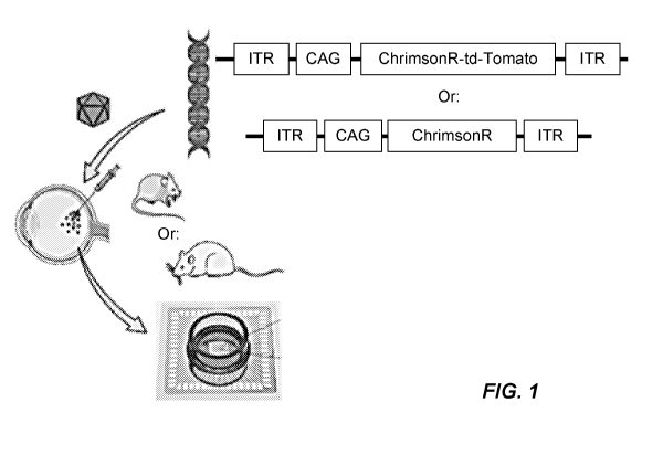
[059] FIG. 1: In vivo methods in rdl mice.
[060] FIGs. 2A through 2D : Degenerated rdl mice retinas respond to light at a
wavelight matching ChrimsonR spectral sensitivity and to duration below 10ms.
FIG.
2A ¨ Eye fundus of a ChrR-tdT expressing rdl mouse at 2 month post injection.
FIG.
2B. TdT fluorescence of a rdl mouse retina mounted on a MEA chip. FIG. 2C-
Spectral sensitivity of ChrR expressing mice retina (n=1 retina, 188
electrodes). FIG.
2D- Added firing rate in response to stimuli of increasing duration at 590 nm
at 1e17
photons.cm-2s-1. All recordings are done in presence of a mix of L-AP4, CNQX
and
CCP.
[061] FIGs. 3A through 3C : Chrimson R is more efficient when fused with tdT
in rdl
mice. FIG. 3A. Comparison between retinas infected with ChrR or ChrR-tdT was
more effective in responding to light stimuli. FIG. 3B. Raw data, raster plot
and
average PSTH (from top to bottom, respectively) of a responding RGC of a ChrR-
tdT
expressing retina. FIG. 3C. Intensity plot of retinas expressing ChrR (n=4
retinas, 27
cells) or ChrR-tdT (n=6 retinas, 548 cells), showing levels of activation at
differentstimuli intensities.
[062] FIGs. 4A through 4G : Expression if Chrimson R in Ganglion cells.
Expression of
ChrR-tdT in Retinal Ganglion Cells (RGCs) of rdl mice. Expression of ChrR-tdT
after in-vivo AAV infection was largely restricted to RGCs. FIG. 4A, FIG. 4B
and
FIG. 4C- Projection of a confocal stack showing membrane located expression in
two
examples of RGCs. FIG. 4A- Image of endogeneous tdTomato, no immunological
amplification. FIG. B. Image of the labelling for our custom made ChrR
antibody.
FIG. 4C- Overlay of both images (FIG. 4A and FIG. 4B), magenta and cyan for
tdTomato and ChrR antibody, respectively. Images taken with a 40x objective.
Expression of ChrR-tdT is enriched in RGCs membranes. FIG. 4D and FIG. 4E-
Projections of three optic slices showing cell body of two RGCs (see inset in
FIG. 4C), taken with a 60x objective. FIG. 4F and FIG. 4G- 3D surface plot of
fluorescence intensity for cell bodies in FIG. 4D and FIG. 4E-, respectively.
Peaks,
7
CA 03025975 2018-10-26
WO 2017/187272
PCT/IB2017/000663
indicating highest fluorescence intensity, are concentrated at, or near, the
cells
membranes.
[063] FIGs. 5A through 5D : Chrimson R long term expression. Multielectrode
Array
recording rd 1 mice 10 months after injection. FIG. 5A- Image of a retina
expressing
ChrR-tdT showing that expression is persistent at 10 months post injection.
FIG. 5B-
example of the activity measured on one electrode, top-light stimulus in red,
middle-
raster plots of the same cell responses for 10 repetitions of the flash,
bottom - average
PTSH (bin size: SOms). FIG. 5C- Added firing rate in response to flashes of
increasing intensity (n=4 retinas, 308 electrodes). FIG. 5D- Added firing rate
in
response to flashes of increasing durations at 590nm at 1e17 photons.cm-2.s-I.
All
recordings are done in presence of a mix of L-AP4, CNQX and CPP.
[064] FIGs. 6A through 6B : Chrimson R reactivates P23H retinas.
Multielectrode Array
recording on another degenerative rodent model: P23H rats. FIG. 6A-
Fluorescence
image of a P23H retina on the array of multielectrode, at 1 month post
injection. FIG.
6B- Added firing rate in response to stimuli of increasing intensities at
590nm at 1e'7
photons.cm-2.s-1 (n=2 retinas, 91 electrodes). All records are done in
presence of a mix
of L-AP4, CNQX and CPP.
[065] FIG. 7 : In vivo methods in non human primate. 4 different strategies
were tested
for ChrR expression in non-human primates (macaca fascicularis). 2 different
constructions : ChrimsonR (ChrR) or the fused protein ChrimsonR-td-Tomato
(ChrR-
tdT), both under the CAG promoter. 2 different viral capsids : the wild type
AAV2,
and the mutant AAV2-7m8 (Dalkara et al. 2013, Science Translational Medicine,
5(189):189ra76). Injections of a single viral does (5x10" vg/eye) was
performed two
months before MEA (512 array, MCS) or patch clamp (see poster Chaffiol et al.,
abstract 599 - B0072) recordings. All recordings were done in presence of
synaptic
blockers (LAP4 50 M and CPP 10 M).
[066] FIGs. 8A through 8C : Chrimson R is expressed in the pen i fovea after
In vivo
injection of the constructs. In vivo injection of the constructs leads to
expression in
RGCs of the pen-fovea! ring. FIG. 8A- Infrared image of a retinal explant, an
asterisk
indicates the depression of the fovea! pit. The black dots are the electrodes
of the
MEA array. FIG. 8B- Fluorescent image of the same retinal piece, infected with
the
AAV2.7m8-ChrR-tdT construct. Expression is restricted to the pen-fovea! ring.
FIG.
8C- Spectral sensitivity of the retina explant displayed in FIG. 8A & FIG. 8B.
Response averaged over 10 repetitions and across all responsive electrodes.
Shape of
the spectrum and presence of synaptic blockers indicate that ChrR in the RGCs
is the
source of the recorded activity.
[067] FIGs. 9A through 9G : Identification of the test construct leading to
the most
efficient transduction. Transduction is evaluated as the number of responsive
electrodes and the sensitivity of the light evoked response. FIG. 9A- Example
of one
electrode responses to light flashes at 4 different intensities. FIG. 9B-
Overview of the
complete set of experiments for the 4 constructs. Active electrodes:
electrodes where
action potentials are detected. Responding electrodes: electrodes where firing
rate was
increased by light stimuli. FIG. 9C-, FIG. 9D and FIG. 9E- Population
responses for
each responsive retina for the different constructs. Each colored line
represents
individual electrode responses, averaged over 10 repetitions. Each row of
graphs
represent responses from one retina, each column responses of different
retinas to a
same light stimulus (intensity on the top in photons/cm2/sec). FIG. 9F-
Average added
firing rate for each responsive retina at different light intensity,
spontaneous firing rate
8
CA 03025975 2018-10-26
WO 2017/187272
PCT/IB2017/000663
is subtracted FIG. 9G- Detail of F to better visualize response threshold. All
stimuli
were done at 600 nm.
[068] FIGs. 10A through 10D : Response to perifoveal RGCs stimuli of
increasing
duration in a retina infected with AAV2.7m8-ChR-tdT. Response of pen-fovea!
RGCs
to stimuli of increasing duration in a retina infected with AAV2.7m8-ChrR-tdT.
FIG.
10A- Responses to light stimuli of increasing duration, each line represent a
single
electrode spike density function average over 10 repetitions per stimuli. FIG.
10B-
Average firing rate for all the duration tested. FIG. 10C- Fraction of active
sites at
different stimulus duration for 4 different activity threshold. FIG. 10D- Time
to first
spike, average over 10 stimuli repetitions for all tested duration. Red dot
mark the
median value, edges of the box the 25th and 75th percentiles of the data, and
whiskers
the rest except for outliers plotted individually. The important drop of the
median
between 1 and 5 msec stimuli indicate that most recordings site start to
respond for
these duration. All stimuli were were presented at 600 +1- 20nm, at an
intensity of
2x10¹⁷ photons.cm-2.5-1.
[069] FIG. 11: Effect of tdTomato on ChrimsonR mRNA levels. Amplification
curves
of ChrimsonR in a RT-qPCR reaction. The Y-axis represents the delta Rn value
corresponding to an experimental reaction minus the Rn value of the baseline
signal.
This parameter reliably calculates the magnitude of the specific signal
generated from
a given set of PCR primers. Magenta and purple traces represent ChrimsonR;
Yellow
and orange traces represent ChrimsonR-tdTomato; Dark and light blue traces are
the
non-transfected controls. The experiment was repeated 3 times and each
experiment
was run on 2 plates yielding 6 total repetitions. Each sample was run in
triplicates on
each plate.
[070] FIGs. 12A through 12B : Level of ChrimsonR protein upon transfection of
HEK293 cells with pssAAV-CAG-ChrimsonR-tdTomato, pssAAV-CAG-ChrimsonR
and pssAAV-CAG-ChrimsonR-GFP plasmids.
[071] FIG. 13 : Effect of tdTomato on the number of cells expressing
ChrimsonR.
Percentage of ChrimsonR-positive cells is represented for cells transfected
with
plasmid 479 (ChrimsonR-tdTomato) and 480 (ChrimsonR) compared to non-
transfected controls. Percentage of fluorescent cells was determined by using
a
threshold value to eliminate background fluorescence. It is important to note
that the
number of cells does not indicate the intensity of fluorescence per cell.
Based on this
cell counting method there is no statistically significant difference between
the
percentage of ChrimsonR-expressing cells after transfection with the two
constructs.
Error bars represent SEM within this experiment and the experiment was
repeated 3
times with technical duplicates for each condition.
[072] FIGs. 14A through 14B : Effect of tdTomato on the subcellular
localization of
ChrimsonR in HEK 293T cells. Images of transfected HEK 293T cells; obtained by
maximum projections of confocal z stacks. Cells nuclei are shown in blue
(DAPI) and
Chrimson R is shown in white. FIG. 14A shows localisation of Chrimson R-
dtTomato; FIG. 14B shows distribution of ChrimsonR alone. Scale bars 20 m.
[073] FIGs. 15A through 15B : Effect of tdTomato on the subcellular
localization of
ChrimsonR in HEK 293T cells after AAV infection. Images of transfected HEK
293T
cells; obtained by maximum projections of confocal z stacks. Cells nuclei are
shown
in blue (DAPI) and Chrimson R is shown in white. FIG. 15A shows localisation
of
Chrimson R-dtTomato; FIG. 15B shows distribution of ChrimsonR alone. Scale
bars
20 m.
9
CA 03025975 2018-10-26
WO 2017/187272
PCT/IB2017/000663
DETAILED DESCRIPTION
[074] In this disclosure, the use of the singular includes the plural, the
word "a" or "an"
means "at least one", and the use of "or" means "and/or", unless specifically
stated
otherwise. Furthermore, the use of the term "including", as well as other
forms, such
as "includes" and "included", is not limiting. Also, terms such as "element"
or
"component" encompass both elements and components comprising one unit and
elements or components that comprise more than one unit unless specifically
stated
otherwise.
[075] As used herein, the term "about," when used in conjunction with a
percentage or
other numerical amount, means plus or minus 10% of that percentage or other
numerical amount. For example, the term "about 80%," would encompass 80% plus
or
minus 8%.
[076] All documents, or portions of documents, cited in this application,
including, but
not limited to, patents, patent applications, articles, books, and treatises,
are hereby
expressly incorporated herein by reference in their entirety for any purpose.
In the
event that one or more of the incorporated literature and similar materials
defines a
term in a manner that contradicts the definition of that term in this
application, this
application controls.
[077] The terms "protein", polypeptide and "peptide" as used herein are
interchangeable, unless instructed to the contrary.
[078] As used herein, the term fusion protein or protein fused to
another refers to
a protein construct or a chimeric protein. It is meant a single protein
molecule
containing two or more proteins or fragments thereof, covalently linked via
peptide
bond within their respective peptide chains, without additional chemical
linkers. One
protein can be fused to another protein either at the N-terminus or the C-
terminus
thereof. The fusion protein can further comprise linker moity resulting from
genetic
construction.
[079] As used herein, and unless otherwise indicated, the terms "treat,"
"treating,"
"treatment" and "therapy" contemplate an action that occurs while a patient is
suffering from a disorder, e.g. a neuron mediated disorder or ocular
disorders, that
reduces the severity of one or more symptoms or effects of said disorder. As
used
herein, and unless otherwise indicated, the terms "prevent," "preventing," and
"prevention" contemplate an action that occurs before a patient begins to
suffer from a
disorder, e.g. neuron mediated disorder or ocular disorder, that delays the
onset of,
and/or inhibits or reduces the severity of said disorder. It will be
understood that a
treatment may be a prophylactic treatment or may be a treatment administered
following the diagnosis of a disease or condition. A treatment of the
invention may
reduce or eliminate a symptom or characteristic of a disorder, disease, or
condition or
may eliminate the disorder, disease, or condition itself. It will be
understood that a
treatment of the invention may reduce or eliminate progression of a disease,
disorder
or condition and may in some instances result in the regression of the
disease,
disorder, or condition. In some embodiments of the invention one or more light-
activated ion channels polypeptide of the invention may be expressed in a cell
population and used in methods to treat a disorder or condition.
CA 03025975 2018-10-26
WO 2017/187272
PCT/IB2017/000663
[080] As used herein, and unless otherwise specified, a "therapeutically
effective
amount" of a compound is an amount sufficient to provide any therapeutic
benefit in
the treatment or management of a neuron mediated disorder or ocular disorder,
or to
delay or minimize one or more symptoms associated with a disorder, e.g. a
neuron
mediated disorder or ocular disorders. A therapeutically effective amount of a
compound means an amount of the compound, alone or in combination with one or
more other therapies and/or therapeutic agents that provide any therapeutic
benefit in
the treatment or management of a disorder, e.g. a neuron mediated disorder or
ocular
disorders. The term "therapeutically effective amount" can encompass an amount
that
alleviates a neuron mediated disorder or ocular disorder, improves or reduces
an
ocular disorder, improves overall therapy, or enhances the therapeutic
efficacy of
another therapeutic agent.
[081] As used herein, "patient" or "subject" includes mammalian organisms
which are
suffering or are susceptible to suffer from disorder as described herein, such
as human
and non-human mammals, for example, but not limited to, rodents, mice, rats,
non-
human primates, companion animals such as dogs and cats as well as livestock,
e.g.,
sheep, cow, horse, etc.
[082] Transfection of retinal neurons with nucleic acid (e.g. vector) encoding
Chrimson
polypeptide of the Invention provides retinal neurons, preferably bipolar
cells and/or
ganglion cells, with photosensitive membrane channels. Thus, it is possible to
measure, with a light stimulus, the transmission of a visual stimulus to the
animal's
visual cortex, the area of the brain responsible for processing visual signals
which
constitutes a form of vision, as intended herein. Such vision may differ from
forms of
normal human vision and may be referred to as a sensation of light, also
termed "light
detection" or "light perception." Thus, the term "vision" as used herein is
defined as
the ability of an organism to usefully detect light as a stimulus. " Vision"
is intended
to encompass the following: (i) Light detection or perception, i.e. the
ability to discern
whether or not light is present (ii) Light projection, i.e. the ability to
discern the
direction from which a light stimulus is coming; (iii) Resolution, i.e. the
ability to
detect differing brightness levels (i.e., contrast) in a grating or letter
target; and (iv)
Recognition, i.e. the ability to recognize the shape of a visual target by
reference to the
differing contrast levels within the target. Thus, "vision" includes the
ability to simply
detect the presence of light, preferably red light, more preferably with light
having a
wavelength between about 365 nm and about 700 nm, between about 530 nm and
about 640 nm, and in some embodiments, a peak activation may occur upon
contact
with light having a wavelength of about 590 nm.
[083] As used herein, "Functional derivatives" encompass "mutants," "variants"
and
"fragments" regardless of whether the terms are used in the conjunctive or the
alternative herein. Preferred variants are single amino acid conservative
substitution
variants, though conservative substitution of 2, 3, 4 or 5 residues, for
example, is also
intended. In some embodiments, the Functional derivatives has at least 70%
homology to the full length amino acid sequence of the original polypeptide,
preferably at least 75%, more preferably at least 80% homology, more
preferably at
least 85% homology, more preferably at least 90% homology, more preferably at
least
95% homology, more preferably at least 99% homology, more preferably 100%
homology. The percent homology is determined with regard to the length of the
relevant amino acid sequence. Therefore, if a polypeptide of the present
invention is
comprised within a larger polypeptide, the percent homology is determined with
regard only to the portion of the polypeptide that corresponds to the
polypeptide of the
11
CA 03025975 2018-10-26
WO 2017/187272
PCT/IB2017/000663
present invention and not the percent homology of the entirety of the larger
polypeptide. "Percent homology" with reference to polypeptide sequences,
refers to
the percentage of identical amino acids between at least two polypeptide
sequences
aligned using the Basic Local Alignment Search Tool (BLAST) engine. See
Tatusova
et al. (1999) ibid. The BLAST engine is provided to the public by the National
Center
for Biotechnology Information (NCBI), Bethesda, Md. According to specific
embodiments, the functional derivative is a polypeptide which comprises an
amino
acid sequence which has at least 70% homology to the full length sequence of
the
original polypeptide and wherein it only differs from its parent polypeptide
by a
substitution at one or more position(s). Said substitution is preferably
conservative
substitution >> or semi conservative . In addition, or alternatively, the
Functional
derivatives has at least 70% identity to the full length amino acid sequence
of the
original polypeptide, preferably at least 75% identity, more preferably at
least 80%
identity, more preferably at least 85% identity, more preferably at least 90%
identity,
more preferably at least 95% identity, more preferably at least 99% identity,
more
preferably 100% identity. Methods of determining sequence identity or homology
are
known in the art.
[084] As used herein, the term "conservative substitution" generally refers to
amino acid
replacements that preserve the structure and functional properties of a
protein or
polypeptide. Such functionally equivalent (conservative substitution) peptide
amino
acid sequences include, but are not limited to, additions or substitutions of
amino acid
residues within the amino acid sequences encoded by a nucleotide sequence that
result
in a silent change, thus producing a functionally equivalent gene product.
Conservative amino acid substitutions may be made on the basis of similarity
in
polarity, charge, solubility, hydrophobicity, hydrophilicity, and/or the
amphipathic
nature of the residues involved. For example: nonpolar (hydrophobic) amino
acids
include alanine, leucine, isoleucine, valine, proline, phenylalanine,
tryptophan, and
methionine; polar neutral amino acids include glycine, serine, threonine,
cysteine,
tyrosine, asparagine, and glutamine; positively charged (basic) amino acids
include
arginine, lysine, and histidine; and negatively charged (acidic) amino acids
include
aspartic acid and glutamic acid.
[085] The invention in some aspects relates to the expression in cells of
light-activated
ion channel polypeptides that can be activated by contact with one or more
pulses of
light, which results in strong depolarization of the cell. Light-activated
channel
polypeptides of the invention, also referred to herein as light-activated ion
channels
can be expressed in specific cells, tissues, and/or organisms and used to
control cells
in vivo, ex vivo, and in vitro in response to pulses of light of a suitable
wavelength.
[086] As used herein, the term "ion channel" means a transmembrane polypeptide
that
forms a pore, which when activated opens, permitting ion conductance through
the
pore across the membrane.
[087] According to the present invention, the light-activated ion channel
polypeptide
comprises Chrimson protein, or functional derivatives thereof, and a
fluorescent
protein.
[088] According to the present invention, the light-activated ion channel
polypeptide
comprises Chrimson protein, or functional derivatives thereof, fused to a
fluorescent
protein.
[089] According to special embodiment said Chrimson protein is selected in the
group
consisting in protein ChR88 (also referred to herein as Chrimson88 -SEQ ID NO
:1) or
12
CA 03025975 2018-10-26
WO 2017/187272
PCT/IB2017/000663
functional derivatives thereof, and K176R substituted Chrimson88 protein (also
referred to herein as Chrimson88 protein with K176R substitution or ChrimsonR -
SEQ ID NO: 2) or functional derivatives thereof.
[090] According to the present invention, the light-activated ion channel
polypeptide
comprises (i) protein ChR88 (SEQ ID NO: 1) or functional derivatives thereof
and (ii)
a fluorescent protein.
[091] According to preferred embodiment the light-activated ion channel
polypeptide of
the invention comprises (i) protein ChrimsonR ( SEQ ID NO: 2) or functional
derivatives thereof and (ii) a fluorescent protein.
[092] According to special embodiment, the light-activated ion channel
polypeptide of
the invention consists of protein ChR88 ( SEQ ID NO: 1) or functional
derivatives
thereof and fluorescent protein, both protein being expressed as independent
proteins.
[093] According to another embodiment the light-activated ion channel
polypeptide of
the invention consists of protein ChrimsonR (SEQ ID NO: 2) or functional
derivatives
thereof and fluorescent protein, both protein being expressed as independent
proteins.
[094] According to preferred embodiment, the light-activated ion channel
polypeptide of
the invention consists of protein ChR88 (SEQ ID NO: 1) or functional
derivatives
thereof fused to fluorescent protein.
[095] According to more preferred embodiment the light-activated ion channel
polypeptide of the invention consists of protein ChrimsonR (SEQ ID NO: 2) or
functional derivatives thereof fused to fluorescent protein.
[096] Light-activated ion channel polypeptides of the Invention are strongly
activated by
contact with red light, preferably with light having a wavelength between
about 365
nm and about 700 nm, between about 530 nm and about 640 nm, and in some
embodiments, a peak activation may occur upon contact with light having a
wavelength of about 590 nm.
[097] Contacting an excitable cell that includes a light-activated ion channel
polypeptide
of the invention with a light in the activating range of wavelengths strongly
depolarizes the cell. Exemplary wavelengths of light that may be used to
depolarize a
cell expressing a light-activated ion channel polypeptide of the invention,
include
wavelengths from at least about 365 nm, 385 nm, 405 nm, 425 nm, 445 nm, 465
nm,
485 nm, 505 nm, 525 nm, 545 nm, 565 nm, 585 nm; 590 nm, 605 nm, 625 nm, 645
nm, 665 nm, 685 nm; and 700 nm, including all wavelengths therebetween. In
some
embodiments, light-activated ion channel polypeptides of the invention have a
peak
wavelength sensitivity in of 590 nm, and may elicit spikes up to 660 nm.
[098] Light-activated ion channel polypeptides of the invention can be used to
depolarize excitable cells in which one or more light-activated ion channels
of the
invention are expressed. In some embodiments, light-activated ion channel
polypeptides of the invention can be expressed in a sub-population of cells in
a cell
population that also includes one or more additional subpopulations of cells
that
express light-activated ion channels that are activated by wavelengths of
light that do
not activate a light-activated ion channel polypeptide of the invention.
[099] The peptide amino acid sequences that can be used in various embodiments
include the light-activated ion channel polypeptide described herein (SEQ ID
NOS: 1
or 2, or 5), as well as functionally equivalent polypeptides.
13
CA 03025975 2018-10-26
WO 2017/187272
PCT/IB2017/000663
[0100] Such functionally equivalent peptide amino acid sequences (conservative
substitutions) include, but are not limited to, additions or substitutions of
amino acid
residues within the amino acid sequences of the Invention, but that result in
a silent
change, thus producing a functionally equivalent polypeptide. Amino acid
substitutions may be made on the basis of similarity in polarity, charge,
solubility,
hydrophobicity, hydrophilicity, and/or the amphipathic nature of the residues
involved. For example: nonpolar (hydrophobic) amino acids include alanine,
leucine,
isoleucine, valine, proline, phenylalanine, tryptophan, and methionine; polar
neutral
amino acids include glycine, serine, threonine, cysteine, tyrosine,
asparagine, and
glutamine; positively charged (basic) amino acids include arginine, lysine,
and
histidine; and negatively charged (acidic) amino acids include aspartic acid
and
glutamic acid. Conservative amino acid substitutions may alternatively be made
on the
basis of the hydropathic index of amino acids. Each amino acid has been
assigned a
hydropathic index on the basis of its hydrophobicity and charge
characteristics. They
are: isoleucine (+4.5); valine (+4.2); leucine (+3.8); phenylalanine (+2.8);
cysteine/cystine (+2.5); methionine (+1.9); alanine (+1.8); glycine (-0.4);
threonine (-
0.7); serine (-0.8); tryptophan (-0.9); tyrosine (-1.3); proline (-1.6);
histidine (-3.2);
glutamate (-3.5); glutamine (-3.5); aspartate (-3.5); asparagine (-3.5);
lysine (-3.9); and
arginine (-4.5). The use of the hydropathic amino acid index in conferring
interactive
biological function on a protein is understood in the art (Kyte and Doolittle,
J. Mol.
Biol. 157:105-132, 1982). It is known that in certain instances, certain amino
acids
may be substituted for other amino acids having a similar hydropathic index or
score
and still retain a similar biological activity. In making changes based upon
the
hydropathic index, in certain embodiments the substitution of amino acids
whose
hydropathic indices are within +-2 is included, while in other embodiments
amino acid
substitutions that are within +-1 are included, and in yet other embodiments
amino
acid substitutions within +-0.5 are included.
[0101] Conservative amino acid substitutions may alternatively be made on the
basis of
hydrophilicity, particularly where the biologically functional protein or
peptide
thereby created is intended for use in immunological embodiments. In certain
embodiments, the greatest local average hydrophilicity of a protein, as
governed by
the hydrophilicity of its adjacent amino acids, correlates with its
immunogenicity and
antigenicity, i.e., with a biological property of the protein. The following
hydrophilicity values have been assigned to these amino acid residues:
arginine
(+3.0); lysine (+3.0); aspartate (+3.0+-1); glutamate (+3.0+-1); serine
(+0.3);
asparagine (+0.2); glutamine (+0.2); glycine (0); threonine (-0.4); proline (-
0.5+-1);
alanine (-0.5); histidine (-0.5); cysteine (-1.0); methionine (-1.3); valine (-
1.5); leucine
(-1.8); isoleucine (-1.8); tyrosine (-2.3); phenylalanine (-2.5) and
tryptophan (-3.4). In
making changes based upon similar hydrophilicity values, in certain
embodiments the
substitution of amino acids whose hydrophilicity values are within +-2 is
included, in
certain embodiments those that are within +-1 are included, and in certain
embodiments those within +-0.5 are included.
[0102] According to one preferred embodiment, the light-activated ion channel
polypeptide of the invention is a fusion protein between a chrimson
polypeptide (e.g.
protein ChR88 or functional derivatives thereof, or protein ChrimsonR or
functional
derivatives thereof) and a fluorescent protein. The use of fusion proteins in
which a
polypeptide or peptide, or a truncated or mutant version of peptide is fused
to an
unrelated protein, polypeptide, or peptide, and can be designed on the basis
of the
desired peptide encoding nucleic acid and/or amino acid sequences described
herein.
14
CA 03025975 2018-10-26
WO 2017/187272
PCT/IB2017/000663
In certain embodiments, a fusion protein may be readily purified by utilizing
an
antibody that selectively binds to the fusion protein being expressed.
[0103] In general, the retinal or retinal derivative necessary for the
functioning of the
light-activated ion channel polypeptide of the invention is produced by the
cell to be
transfected with said channel polypetide. However according to the invention,
it is
further disclosed a channelrhodopsin comprising a light-activated ion channel
polypeptide of the invention and a retinal or retinal derivative such as for
example 3,4-
dehydroretinal, 13-ethylretinal, 9-dm-retinal, 3-hydroxyretinal, 4-
hydroxyretinal,
naphthyl retinal; 3 ,7,11-trimethyl-dodeca-2,4,6, 8,10-pentaenal ; 3 ,7-
dimethyl-deca-
2,4,6,8-tetraenal ; 3,7-dimethyl-octa-2,4,6-trienal; as well as 6-7- or 8-9-
or 10-11
rotation-blocked retinals (W003084994).
[0104] While the desired peptide amino acid sequences described can be
chemically
synthesized (see, e.g., "Proteins: Structures and Molecular Principles"
(Creighton, ed.,
W. H. Freeman & Company, New York, N.Y., 1984)), large polypeptides sequences
may advantageously be produced by recombinant DNA technology using techniques
well-known in the art for expressing nucleic acids containing a nucleic acid
sequence
that encodes the desired peptide. Such methods can be used to construct
expression
vectors containing peptide encoding nucleotide sequences and appropriate
transcriptional and translational control signals. These methods include, for
example,
in vitro recombinant DNA techniques, synthetic techniques, and in vivo genetic
recombination (see, e.g., "Molecular Cloning, A Laboratory Manual", supra, and
"Current Protocols in Molecular Biology", supra). Alternatively, RNA and/or
DNA
encoding desired peptide encoding nucleotide sequences may be chemically
synthesized using, for example, synthesizers (see, e.g., "Oligonucleotide
Synthesis: A
Practical Approach" (Gait, ed., IRL Press, Oxford, United Kingdom, 1984)).
[0105] The peptide amino acid sequences that can be used in various
embodiments
include the light-activated ion channel polypeptide described herein (SEQ ID
NOS: 1
or 2, 5 or 6), as well as functionally equivalent peptides and functionally
derivatives
thereof, and their functional fragments. In fact, in some embodiments, any
desired
peptide amino acid sequences encoded by particular nucleotide sequences can be
used,
as is the use of any polynucleotide sequences encoding all, or any portion, of
desired
peptide amino acid sequences. The degenerate nature of the genetic code is
well-
known, and, accordingly, each light-activated channel polypeptide amino acid-
encoding nucleotide sequence is generically representative of the well-known
nucleic
acid "triplet" codon, or in many cases codons, that can encode the amino acid.
As
such, as contemplated herein, the channelrhodopsin peptide amino acid
sequences
described herein, when taken together with the genetic code (see, e.g.,
"Molecular Cell
Biology", Table 4-1 at page 109 (Darnell et al., eds., W. H. Freeman &
Company,
New York, NY, 1986)), are generically representative of all the various
permutations
and combinations of nucleic acid sequences that can encode such amino acid
sequences.
[0106] Some embodiments are isolated nucleic acid molecules comprising a
nucleotide
sequence that encodes a light-activated ion channel polypeptide of the
invention. In
some embodiments, the nucleotide sequence encodes polypeptide which comprises
(i)
protein ChR88 (SEQ ID NO: 1) or functional derivatives thereof, and (ii) a
fluorescent
protein. In another embodiments, the nucleotide sequence encodes polypeptide
which
comprises (i) protein ChrimsonR (SEQ ID NO: 2) or functional derivatives
thereof,
and (ii) a fluorescent protein.
CA 03025975 2018-10-26
WO 2017/187272
PCT/IB2017/000663
[0107] According to one special embodiment, the nucleotide sequence encodes
polypeptide which consists in protein ChR88 ( SEQ ID NO: 1) or functional
derivatives thereof, fused to fluorescent protein. According to preferred
embodiment,
the nucleotide sequence encodes polypeptide which comprises protein ChrimsonR
(SEQ ID NO: 2) or functional derivatives thereof fused to fluorescent protein.
[0108] According to certain special embodiments, the fluorescent protein of
the invention
is selected from tdTomato (tdT) fluorescent protein and green fluorescent
protein
(GFP).
[0109] TdTomato is a bright red fluorescent protein (tdTomato's excitation
peak 554nm,
peak of emission wavelength 581 nm) (Shaner NC et al., Nat Biotechnol, 22,
1567-
1572, 2004). The genomic sequence encoding tdTomato according to the invention
might show at least 84% identity with the synthetic construct tandem-dimer red
fluorescent protein gene, complete cds (Genbank Accession number A Y6 7 82 6
9).
According to a preferred embodiment, the encoded tdTomato protein moiety of
the
invention is a polypeptide having between about 70% and about 75%; or more
preferably between about 75% and about 80%; or more preferably between about
80%
and 90%; or even more preferably between about 90% and about 99% of amino
acids
that are identical to the amino acid sequence of SEQ ID NO:3 .
[0110] In other embodiments, the present invention provides for an isolated
nucleic acid
encoding a polypeptide having between about 70% and about 75%; or more
preferably
between about 75% and about 80%; or more preferably between about 80% and 90%;
or even more preferably between about 90% and about 99% of amino acids that
are
identical to the amino acid sequence of SEQ ID NO: 5 or fragments thereof
[0111] Nucleic acid of the invention may include additional sequences
including, but not
limited to one or more signal sequences (e.g. enhancers, polyadenylation
signals,
additional restriction enzyme sites, multiple cloning sites) and/or promoter
sequences,
or other coding segments, or a combination thereof The promoter can be
inducible or
constitutive, general or cell specific promoter. An example of cell-specific
promoter is
mG1u6-promoter specific of bipolar cells. Some embodiments are any of the
disclosed
methods wherein the promoter is a constitutive promoter. Some embodiments are
any
of the disclosed methods wherein the constitutive promoter includes, but is
not limited
to, a CMV promoter or CAG promoter (CAG promoter is hybrid cytomegalovirus
(CMV) immediate early enhancer fused to the chicken beta-actin promoter (CBA)
and
SV40 intron insertion; Alexopoulou et al., BMC Cell Biol. 2008; 9: 2; SEQ ID
NO :8). Some embodiments are any of the disclosed methods wherein the promoter
includes, but is not limited to, an inducible and/or a cell type-specific
promoter.
Selection of promoter, vectors, enhancers, polyadenylation sites is matter of
routine
design for those skilled in the art. Those elements are well described in
litterature and
are commercially available.
[0112] In certain embodiments, the invention concerns isolated nucleic acid
segments and
recombinant vectors which encode a protein or peptide that includes within its
amino
acid sequence an amino acid sequence of light-activated ion channel
polypeptide of
the invention or a functional portions or variant thereof, such as those
identified (e.g.
SEQ ID NOS: 5).
[0113] In certain embodiments, the invention concerns isolated nucleic acid
segments and
recombinant vectors which comprises the amino acide sequence SEQ ID NO :6 or
SEQ ID NO:7.
16
CA 03025975 2018-10-26
WO 2017/187272
PCT/IB2017/000663
[0114] Some embodimentsare recombinant nucleic acids comprising a nucleotide
sequence that encodes amino acids of (i) SEQ ID NO: 1 or SEQ ID NO:2 with (ii)
SEQ ID NO:3 or SEQ ID NO:4.
[0115] Some preferred embodiments are recombinant nucleic acids comprising a
nucleotide sequence that encodes amino acids of SEQ ID NO:5 or fragments
thereof.
[0116] Some preferred embodiments are recombinant nucleic acids comprising a
nucleotide sequence SEQ ID NO:6 or SEQ ID NO:7.
[0117] Some embodimentsare recombinant nucleic acids comprising a nucleotide
sequence that encodes amino acids of (i) SEQ ID NO: 1 or SEQ ID NO:2 ,
operably
linked to a heterologous promoter and (ii) a nucleotide sequence that encodes
amino
acids of SEQ ID NO:3 or SEQ ID NO:4, operably linked to a heterologous
promoter.
[0118] Some preferred embodiments are recombinant nucleic acids comprising a
nucleotide sequence that encodes amino acids of SEQ ID NO:5 or fragments
thereof,
operably linked to a heterologous promoter.
[0119] Some preferred embodiments are recombinant nucleic acids comprising a
nucleotide sequence SEQ ID NO:6 or SEQ ID NO:7, operably linked to a
heterologous promoter.
[0120] Some preferred embodiments are recombinant nucleic acids comprising a
nucleotide sequence SEQ ID NO:6 or SEQ ID NO:7, operably linked to CAG
heterologous promoter (SEQ ID NO :8).
[0121] According to another aspect, the invention relates to a nucleic acid
expression
vector that includes a nucleic acid sequence that encodes any of the
aforementioned
light-activated ion channel polypeptides.
[0122] As used herein, the term "nucleic acid expression vector" refers to a
nucleic acid
molecule capable of transporting between different genetic environments
another
nucleic acid to which it has been operatively linked. The term " vector " also
refers to
a virus or organism that is capable of transporting the nucleic acid molecule.
One type
of vector is an episome, i.e., a nucleic acid molecule capable of extra-
chromosomal
replication. Some useful vectors are those capable of autonomous replication
and/or
expression of nucleic acids to which they are linked. Vectors capable of
directing the
expression of genes to which they are operatively linked are referred to
herein as
"expression vectors". Expression vectors and methods of their use are well
known in
the art. Non-limiting examples of suitable expression vectors and methods for
their
use are provided herein. In preferred embodiment, the vector is suitable for
gene
therapy, more particularly for virus-mediated gene transfer. Examples of
viruses
suitable for gene therapy are retroviruses, adenoviruses, adeno-assoiated
viruses
(AAV), lentiviruses, poxviruses (e.g. MVA), alphaviruses, herpesviruses.
However,
gene therapy further encompasses non-viral methods suc as use of naked DNA,
liposomes associated with nucleic acids. Vectors useful in some methods of the
invention can genetically insert light-activated ion channel polypeptides into
dividing
and non-dividing cells and can insert light-activated ion channel polypeptides
to cells
that are in vivo, in vitro, or ex vivo cells.
[0123] In some preferred embodiments, the nucleic acid expression vector
comprising the
gene for a light-activated ion channel of the invention is selected among AAV
viral
vectors. According to preferred embodiment said AAV viral vector is an AAV2
and
more preferably is AAV2-7m8 viral vector (WO 2012/145601).
17
CA 03025975 2018-10-26
WO 2017/187272
PCT/IB2017/000663
[0124] Some aspects of the invention include methods of treating a disorder or
condition
in a cell, tissue, or subject using light-activated ion channels polypeptide
of the
invention. Treatment methods of the invention may include administering to a
subject
in need of such treatment, a therapeutically effective amount of a light-
activated ion
channel polypeptide of the invention to treat the disorder.
[0125] Administration of a light-activated ion channel polypeptide of the
invention may
include administration pharmaceutical composition that includes effective
amount of
at least one light-activated ion channels polypeptide of the invention.
Administration
of a light-activated ion channel polypeptide of the invention may include
administration pharmaceutical composition that includes a cell, wherein the
cell
expresses the light-activated ion channel of the invention. Administration of
a light-
activated ion channel polypeptide of the invention may include administration
of
effective amount of a pharmaceutical composition that includes a vector,
wherein the
vector comprises a nucleic acid sequence encoding the light-activated ion
channel
polypeptide of the invention and the administration of the vector results in
expression
of the light-activated ion channel polypeptide in a cell in the subject.
[0126] In some embodiments, are methods of treating or preventing a neuron
mediated
disorder, comprising: (a) delivering to a target cell a nucleic acid
expression vector
that encodes a light-activated ion channel polypeptides of the invention,
expressible in
said target cell, said vector comprising an open reading frame encoding the
light-
activated ion channel polypeptides of the invention, operatively linked to a
promoter
sequence, and optionally, a transcriptional regulatory sequence; and (b)
expressing
said vector in said target cell, wherein the expressed light-activated ion
channel
polypeptides activates said target cell upon exposure to light.
[0127] In some embodiments, the expressed light-activated ion channel
polypeptide
consists in protein ChR88 ( SEQ ID NO: 1) or functional derivatives thereof,
fused to
fluorescent protein.
[0128] According to preferred embodiment, the expressed light-activated ion
channel
polypeptide consists in ChrimsonR ( SEQ ID NO: 2) or functional derivatives
thereof
fused to fluorescent protein.
[0129] In preferred embodiments, the expressed light-activated ion channel
polypeptide
consists in protein ChR88 (SEQ ID NO: 1) or functional derivatives thereof,
fused to
fluorescent protein selected in the group consisting in tdTomato (tdT)
fluorescent
protein or green fluorescent protein (GFP).
[0130] According to preferred embodiment, the expressed light-activated ion
channel
polypeptide consists in ChrimsonR (SEQ ID NO: 2) or functional derivatives
thereof
fused to fluorescent protein selected in the group consisting in tdTomato
(tdT)
fluorescent protein (SEQ ID NO:3) or green fluorescent protein (GFP) (SEQ ID
NO:4).
[0131] As used herein, and unless otherwise indicated, the term neuron
mediated
disorders for which the present methods and compositions may be used include,
but
are not limited to, neuronal dysfunctions, disorders of the brain, the central
nervous
system, the peripheral nervous system, neurological conditions, disorders of
memory
and learning disorders, cardiac arrhythmias, Parkinson's disease, ocular
disorders, ear
disorders, spinal cord injury, among others.
[0132] As used herein, and unless otherwise indicated, the term ocular
disorders for which
the present methods and compositions may be used to improve one or more
18
CA 03025975 2018-10-26
WO 2017/187272
PCT/IB2017/000663
parameters of vision include, but are not limited to, developmental
abnormalities that
affect both anterior and posterior segments of the eye. Anterior segment
disorders
include, but are not limited to, glaucoma, cataracts, corneal dystrophy,
keratoconus.
Posterior segment disorders include, but are not limited to, blinding
disorders caused
by photoreceptor degeneration, dysfunctioning, loss and/or death. Retinal
disorders
include retinitis pigmentosa (RP), macular deneneration (MD), congenital
stationary
night blindness, age-related macular degeneration and congenital cone
dystrophies.
[0133] A target cell according to certain embodiments of the invention may be
an
excitable cell or a non-excitable cell. It is preferably a cell in which a
light-activated
ion channel polypeptide of the invention may be expressed and may be used in
methods of the invention. It includes prokaryotic and eukaryotic cells. Target
cells
include but are not limited to mammalian cells. Examples of cells in which a
light-
activated ion channel polypeptide of the invention may be expressed are
excitable
cells, which include cells able to produce and respond to electrical signals.
[0134] Non-limiting examples of target cells according to the invention
include neuronal
cells (neurons), nervous system cells, cardiac cells, circulatory system
cells, visual
system cells, auditory system cells, secretory cells (such as pancreatic
cells, adrenal
medulla cells, pituitary cells, etc.), endocrine cells, or muscle cells. In
some
embodiments, a target cell used in conjunction with the invention may be a
healthy
normal cell, which is not known to have a disease, disorder or abnormal
condition. In
some embodiments, a target cell used in conjunction with methods and channels
of the
invention may be an abnormal cell, for example, a cell that has been diagnosed
as
having a disorder, disease, or condition, including, but not limited to a
degenerative
cell, a neurological disease-bearing cell, a cell model of a disease or
condition, an
injured cell, etc. In some embodiments of the invention, a cell may be a
control cell.
[0135] According to one special embodiment, light-activated ion channel
polypeptide of
the invention may be expressed in cells from culture, cells in solution, cells
obtained
from subjects, and/or cells in a subject (in vivo cells). Light-activated ion
channels
may be expressed and activated in cultured cells, cultured tissues (e.g.,
brain slice
preparations, etc.), and in living subjects, etc.
[0136] In a preferred embodiment, the target cell is mammalian cell and is an
electrically
excitable cell. Preferably, it is a photoreceptor cell, a retinal rod cell, a
retinal cone
cell, a retinal ganglion cell (RGC), an amacrine cell, a biporal neuron, a
ganglion cell,
a spiral ganglion neurons (SGNs), a cochlear nucleus neuron, a multipolar
neuron, a
granule cell, a neuron, or a hippocampal cell.
[0137] Some embodiments are methods of restoring light sensitivity to a
retina,
comprising: (a) delivering to a target retinal neuron a nucleic acid
expression vector
that encodes a light-activated ion channel polypeptides of the invention,
expressible in
said target retinal neuron, said vector comprising an open reading frame
encoding the
light-activated ion channel polypeptides of the invention, operatively linked
to a
promoter sequence, and optionally, a transcriptional regulatory sequence; and
(b)
expressing said vector in said target retinal neuron, wherein the expressed
light-
activated ion channel polypeptides renders said retinal neuron photosensitive,
thereby
restoring light sensitivity to said retina or a portion thereof
[0138] One embodiment is a method of restoring light sensitivity to a retina
wherein the
expressed light-activated ion channel polypeptide consists in protein ChR88
(SEQ ID
NO: 1) or functional derivatives thereof, fused to fluorescent protein.
19
CA 03025975 2018-10-26
WO 2017/187272
PCT/IB2017/000663
[0139] One preferred embodiment is a method of restoring light sensitivity to
a retina
wherein the expressed light-activated ion channel polypeptide consists in
ChrimsonR (SEQ ID NO: 2) or functional derivatives thereof fused to
fluorescent
protein.
[0140] One preferred embodiment is method of restoring light sensitivity to a
retina
wherein the expressed light-activated ion channel polypeptide consists in
protein
ChR88 (SEQ ID NO: 1) or functional derivatives thereof, fused to fluorescent
protein
selected in the group consisting in tdTomato (tdT) fluorescent protein or
green
fluorescent protein (GFP).
[0141] One preferred embodiment is method of restoring light sensitivity to a
retina
wherein the expressed light-activated ion channel polypeptide consists in
ChrimsonR
(SEQ ID NO: 2) or functional derivatives thereof fused to fluorescent protein
selected
in the group consisting in tdTomato (tdT) fluorescent protein (SEQ ID NO:3) or
green
fluorescent protein (GFP) (SEQ ID NO:4).
[0142] Some embodiments, are methods of restoring photosensitivity to a retina
of a
subject suffering from vision loss or blindness in whom retinal photoreceptor
cells are
degenerating or have degenerated and died, said method comprising: (a)
delivering to
a target retinal neuron a nucleic acid expression vector that encodes a light-
activated
ion channel polypeptides of the invention, expressible in said target retinal
neuron,
said vector comprising an open reading frame encoding the light-activated ion
channel
polypeptides of the invention, operatively linked to a promoter sequence, and
optionally, a transcriptional regulatory sequence; and (b) expressing said
vector in said
target retinal neuron, wherein the expressed light-activated ion channel
polypeptide
renders said retinal neuron photosensitive, thereby restoring photosensitivity
to said
retina or a portion thereof.
[0143] Some embodiments, are methods of restoring photosensitivity to a retina
of a
subject suffering from vision loss or blindness in whom retinal photoreceptor
cells are
degenerating or have degenerated and died wherein the expressed light-
activated ion
channel polypeptide consists in protein ChR88 (SEQ ID NO: 1) or functional
derivatives thereof, fused to fluorescent protein.
[0144] Some embodiments are methods of restoring photosensitivity to a retina
of a
subject suffering from vision loss or blindness in whom retinal photoreceptor
cells are
degenerating or have degenerated and died wherein the expressed light-
activated ion
channel polypeptide consists in ChrimsonR (SEQ ID NO: 2) or functional
derivatives
thereof fused to fluorescent protein.
[0145] Some preferred embodiments are methods of restoring photosensitivity to
a retina
of a subject suffering from vision loss or blindness in whom retinal
photoreceptor cells
are degenerating or have degenerated and died wherein the expressed light-
activated
ion channel polypeptide consists in protein ChR88 (SEQ ID NO: 1) or functional
derivatives thereof, fused to fluorescent protein selected in the group
consisting in
tdTomato (tdT) fluorescent protein or green fluorescent protein (GFP).
[0146] Some preferred embodiments are methods of restoring photosensitivity to
a retina
of a subject suffering from vision loss or blindness in whom retinal
photoreceptor cells
are degenerating or have degenerated and died wherein the expressed light-
activated
ion channel polypeptide consists in ChrimsonR (SEQ ID NO: 2) or functional
derivatives thereof fused to fluorescent protein selected in the group
consisting in
CA 03025975 2018-10-26
WO 2017/187272
PCT/IB2017/000663
tdTomato (tdT) fluorescent protein (SEQ ID NO:3) or green fluorescent protein
(GFP)
(SEQ ID NO:4).
[0147] In some embodiments, the target neuron in said methods of treating a
neuronal
disorder, or of restoring light sensitivity to a retina, or of restoring
photosensitivity to a
retina of a subject suffering from vision loss or blindness in whom retinal
photoreceptor cells are degenerating or have degenerated and died is a retinal
neuron.
[0148] Some embodiments are any of the disclosed methods, wherein the
expressed light-
activated ion channel polypeptide having the amino acid sequence of all or
part of
SEQ ID NOS: 5 , or a biologically active fragment thereof that retains the
biological
activity of the encoded light-activated channel polypeptide or a biologically
active
conservative amino acid substitution variant of SEQ ID NOS: 5 or of said
fragment.
[0149] Some embodiments are any of the disclosed methods, wherein the
expressed light-
activated ion channel polypeptide is encoded by nucleic acid sequence SEQ ID
NOS:
6.
[0150] Another aspect of the invention is the use of far-red (660 nm) light to
perform non-
invasive transcranial and/or transdural stimulation to modulate neural
circuits.
[0151] Working operation of certain aspects of the invention was demonstrated
by
genetically expressing light-activated ion channel polypeptides of the
invention in
excitable cells, illuminating the cells with suitable wavelengths of light,
and
demonstrating rapid depolarization of the cells in response to the light, as
well as rapid
release from depolarization upon cessation of light. Depending on the
particular
implementation, methods of the invention allow light control of cellular
functions in
vivo, ex vivo, and in vitro.
[0152] In non-limiting examples of methods of the invention, light-activated
ion channel
polypeptides of the invention and derivatives thereof are used in mammalian
cells
without need for any kind of chemical supplement, and in normal cellular
environmental conditions and ionic concentrations.
[0153] Light-activated ion channel polypeptides of the invention have been
found to be
suitable for expression and use in mammalian cells without need for any kind
of
chemical supplement, and in normal cellular environmental conditions and ionic
concentrations. Light-activated ion channel polypeotides of the invention have
been
found to activate at wavelengths of light in a range of 365 nm to 700 nm, with
an
optimal activation from light ranging from 530 nm to 640 nm, and a peak
optimal
activation at a wavelength of 590 nm.
[0154] An effective amount of a light-activated ion channel polypeptide or of
nucleic acid
expression vector is an amount that increases the level of the light-activated
ion
channel in a cell, tissue or subject to a level that is beneficial for the
subject. An
effective amount may also be determined by assessing physiological effects of
administration on a cell or subject, such as a decrease in symptoms following
administration. Other assays will be known to one of ordinary skill in the art
and can
be employed for measuring the level of the response to a treatment. The amount
of a
treatment may be varied for example by increasing or decreasing the amount of
the
light-activated ion channel polypeptide or nucleic acid expression vector
administered,
by changing the therapeutic composition in which the light-activated ion
channel
polypeptide or nucleic acid expression vector is administered, by changing the
route of
administration, by changing the dosage timing, by changing the activation
amounts
and parameters of a light-activated ion channel of the invention, and so on.
The
21
CA 03025975 2018-10-26
WO 2017/187272
PCT/IB2017/000663
effective amount will vary with the particular condition being treated, the
age and
physical condition of the subject being treated; the severity of the
condition, the
duration of the treatment, the nature of the concurrent therapy (if any), the
specific
route of administration, and the like factors within the knowledge and
expertise of the
health practitioner. For example, an effective amount may depend upon the
location
and number of cells in the subject in which the light-activated ion channel
polypeptide
is to be expressed. An effective amount may also depend on the location of the
tissue
to be treated. These factors are well known to those of ordinary skill in the
art and can
be addressed with no more than routine experimentation. It is generally
preferred that
a maximum dose of a composition to increase the level of a light-activated ion
channel
polypeptide, and/or to alter the length or timing of activation of a light-
activated ion
channel polypeptide in a subject (alone or in combination with other
therapeutic
agents) be used, that is, the highest safe dose or amount according to sound
medical
judgment. It will be understood by those of ordinary skill in the art,
however, that a
patient may insist upon a lower dose or tolerable dose for medical reasons,
psychological reasons or for virtually any other reasons.
[0155] A light-activated ion channel polypeptide of the invention (for
example, ChR88 ,
or ChrimsonR fused with tdT or GFP, or a derivative thereof) may be
administered
using art-known methods. In some embodiments a nucleic acid that encodes a
light-
activated ion channel polypeptide of the invention is administered to a
subject and in
certain embodiments a light-activated ion channel polypeptide is administered
to a
subject. The manner and dosage administered may be adjusted by the individual
physician or veterinarian, particularly in the event of any complication. The
absolute
amount administered will depend upon a variety of factors, including the
material
selected for administration, whether the administration is in single or
multiple doses,
and individual subject parameters including age, physical condition, size,
weight, and
the stage of the disease or condition. These factors are well known to those
of ordinary
skill in the art and can be addressed with no more than routine
experimentation.
[0156] Pharmaceutical compositions that deliver light-activated ion channels
polypeptide
or nucleic acid expression vector of the invention may be administered alone,
in
combination with each other, and/or in combination with other drug therapies,
or other
treatment regimens that are administered to subjects. A pharmaceutical
composition
used in the foregoing methods preferably contain an effective amount of a
therapeutic
compound that will increase the level of a light-activated ion channel
polypeptide to a
level that produces the desired response in a unit of weight or volume
suitable for
administration to a subject.
[0157] The dose of a pharmaceutical composition that is administered to a
subject to
increase the level of light-activated ion channel polypeptide in cells of the
subject can
be chosen in accordance with different parameters, in particular in accordance
with the
mode of administration used and the state of the subject. Other factors
include the
desired period of treatment. In the event that a response in a subject is
insufficient at
the initial doses applied, higher doses (or effectively higher doses by a
different, more
localized delivery route) may be employed to the extent that patient tolerance
permits.
The amount and timing of activation of a light-activated ion channel of the
invention
(e.g., light wavelength, length of light contact, etc.) that has been
administered to a
subject can also be adjusted based on efficacy of the treatment in a
particular subject.
Parameters for illumination and activation of light-activated ion channels
that have
been administered to a subject can be determined using art-known methods and
without requiring undue experimentation.
22
CA 03025975 2018-10-26
WO 2017/187272
PCT/IB2017/000663
[0158] Various modes of administration will be known to one of ordinary skill
in the art
that can be used to effectively deliver a pharmaceutical composition to
increase the
level of a light-activated ion channel polypeptide of the invention in a
desired cell,
tissue or body region of a subject. Methods for administering such a
composition or
other pharmaceutical compound of the invention may be topical, intravenous,
oral,
intracavity, intrathecal, intrasynovial, buccal, sublingual, intranasal,
transdermal,
intravitreal, subretinal, subcutaneous, intramuscular and intradermal
administration.
The invention is not limited by the particular modes of administration
disclosed
herein. Standard references in the art (e.g., Remington's Pharmaceutical
Sciences, 18th
edition, 1990) provide modes of administration and formulations for delivery
of
various pharmaceutical preparations and formulations in pharmaceutical
carriers.
Other protocols which are useful for the administration of a therapeutic
compound of
the invention will be known to one of ordinary skill in the art, in which the
dose
amount, schedule of administration, sites of administration, mode of
administration
(e.g., intra-organ) and the like vary from those presented herein.
[0159] Administration of a cell or vector to increase light-activated ion
channel
polypeptide levels in a mammal other than a human; and administration and use
of
light-activated ion channels of the invention. e.g. for testing purposes or
veterinary
therapeutic purposes, is carried out under substantially the same conditions
as
described above. It will be understood by one of ordinary skill in the art
that this
invention is applicable to both human and animals. Thus this invention is
intended to
be used in husbandry and veterinary medicine as well as in human therapeutics.
[0160] In some aspects of the invention, methods of treatment using a light-
activated ion
channel polypeptide of the invention are applied to cells including but not
limited to a
neuronal cell, a nervous system cell, a neuron, a cardiac cell, a circulatory
system cell,
a visual system cell, an auditory system cell, a muscle cell, or an endocrine
cell, etc.
[0161] Disorders and conditions that may be treated using methods of the
invention
include, injury, brain damage, degenerative to neurological conditions (e.g.,
Parkinson's disease, Alzheimer's disease, seizure, vision loss, hearing loss,
etc.
[0162] In some embodiments, methods and light-activated ion channels
polypeptide of the
invention may be used for the treatment of visual system disorders, for
example to
treat vision reduction or loss. A light-activated ion channel polypeptide of
the
invention or vector encoding such polypeptide may be administered to a subject
who
has a vision reduction or loss and the expressed light-activated ion channel
can
function as light-sensitive cells in the visual system, thereby permitting a
gain of
visual function in the subject.
[0163] Clinical applications of the disclosed methods and compositions include
(but are
not limited to) optogenetic approaches to therapy such as: restoration of
vision by
introduction of light-activated ion channels polypeptide of the invention in
post-
receptor neurons in the retina for ocular disorder gene-therapy treatment of
age-
dependent macular degeneration, diabetic retinopathy, and retinitis
pigmentosa, as
well as other conditions which result in loss of photoreceptor cells; control
of cardiac
function by using light-activated ion channels polypeptide of the invention
incorporated into excitable cardiac muscle cells in the atrioventricular
bundle (bundle
of His) to control heart beat rhythm rather than an electrical pacemaker
device;
restoration of dopamine-related movement dysfunction in Parkinsonian patients;
amelioration of depression; recovery of breathing after spinal cord injury;
provide
23
CA 03025975 2018-10-26
WO 2017/187272
PCT/IB2017/000663
noninvasive control of stem cell differentiation and assess specific
contributions of
transplanted cells to tissue and network function.
[0164] Similarly, sensorineural hearing loss may be treated through optical
stimulation of
downstream targets in the auditory nerve (see Hernandez et al., 2014, J. Clin.
Invest,
124(3), 1114-1129 or Darrow et al., 2015,Brain Res., 1599, 44-56). According
to
special embodiment, the invention relates to methods of treating conductive
hearing
loss by the use of optical cochlear cochlear implants comprising: (a)
delivering to
cochlea a nucleic acid expression vector that encodes a light-activated ion
channel
polypeptides of the invention, expressible in said cochlea, said vector
comprising an
open reading frame encoding the light-activated ion channel polypeptides of
the
invention, operatively linked to a promoter sequence, and optionally, a
transcriptional
regulatory sequence; (b) expressing said vector in said cochlea, wherein the
expressed
light-activated ion channel polypeptides renders said cochlea photosensitive,
and (c)
use of a cochlear implant with flashes.
[0165] Some embodiments are methods of treating conductive hearing loss by the
use of
optical cochlear implants wherein the expressed light-activated ion channel
polypeptide consists in protein ChR88 (SEQ ID NO: 1) or functional derivatives
thereof, fused to fluorescent protein.
[0166] Some preferred embodiments are methods of treating conductive hearing
loss by
the use of optical cochlear implants wherein the expressed light-activated ion
channel
polypeptide consists in ChrimsonR (SEQ ID NO: 2) or functional derivatives
thereof
fused to fluorescent protein.
[0167] Some preferred embodimentsare methods of treating conductive hearing
loss by
the use of optical cochlear implants wherein the expressed light-activated ion
channel
polypeptide consists in protein ChR88 (SEQ ID NO: 1) or functional derivatives
thereof, fused to fluorescent protein selected in the group consisting in
tdTomato (tdT)
fluorescent protein or green fluorescent protein (GFP).
[0168] Some preferred embodiments are methods of treating conductive hearing
loss by
the use of optical cochlear implants wherein the expressed light-activated ion
channel
polypeptide consists in ChrimsonR (SEQ ID NO: 2) or functional derivatives
thereof
fused to fluorescent protein selected in the group consisting in tdTomato
(tdT)
fluorescent protein (SEQ ID NO:3) or green fluorescent protein (GFP) (SEQ ID
NO:4).
[0169] The present invention in some aspects, includes preparing nucleic acid
sequences
and polynucleotide sequences; expressing in cells and membranes polypeptides
encoded by the prepared nucleic acid and polynucleotide sequences;
illuminating the
cells and/or membranes with suitable light, and demonstrating rapid
depolarization of
the cells and/or a change in conductance across the membrane in response to
light, as
well as rapid release from depolarization upon cessation of light. The ability
to
controllably alter voltage across membranes and cell depolarization with light
has
been demonstrated. The present invention enables light-control of cellular
functions in
vivo, ex vivo, and in vitro, and the light activated ion channels of the
invention and
their use, have broad-ranging applications for drug screening, treatments, and
research
applications, some of which are describe herein.
[0170] In illustrative implementations of this invention, the ability to
optically perturb,
modify, or control cellular function offers many advantages over physical
24
CA 03025975 2018-10-26
WO 2017/187272
PCT/IB2017/000663
manipulation mechanisms. These advantages comprise speed, non-invasiveness,
and
the ability to easily span vast spatial scales from the nanoscale to
macroscale.
[0171] The reagents use in the present invention (and the class of molecules
that they
represent), allow, at least: currents activated by light wavelengths not
useful in
previous light-activated ion channels, light activated ion channels that when
activated,
permit effectively zero calcium conductance, and different spectra from older
molecules (opening up multi-color control of cells).
[0172] The following Examples section provides further details regarding
examples of
various embodiments. It should be appreciated by those of skill in the art
that the
techniques disclosed in the examples that follow represent techniques and/or
compositions discovered by the inventors to function well. However, those of
skill in
the art should, in light of the present disclosure, appreciate that many
changes can be
made in the specific embodiments which are disclosed and still obtain a like
or similar
result without departing from the spirit and scope of the invention. These
examples are
illustrations of the methods and systems described herein and are not intended
to limit
the scope of the invention. Non-limiting examples of such include, but are not
limited
to those presented below.
EXAMPLES
EXAMPLE 1:. Validation in rdl and P23H degenerative rodent models
[0173] Retinal dystrophies are associated with dysfunction and degeneration of
retinal
cells which impairs the flow of visual information, and ultimately leads to
severe loss
of vision and blindness. Retinitis pigmentosa (RP) is the most common type of
retinal
dystrophy and is responsible for loss of vision in one in 4,000 people
worldwide. RP
results from alteration in any of more than 60 genes inherited as autosomal
dominant
(30%-40% of cases), autosomal recessive (50%-60%), or X-linked (5%-15%).
[0174] In most common forms of RP, rod photoreceptors degenerate first
followed by
cones. Thus, primary symptoms of RP are usually night blindness and peripheral
field
loss leading to tunnel vision. All RP conditions are progressive and the
pattern of sight
deterioration varies amongst patients, however, the ultimate outcome is
blindness.
There is no treatment of RP.
[0175] Since RP results from multiple types of mutation in several genes, that
a
significant proportion of RP is dominant, and that time course of the disease
is highly
variable, a retinal optogenetic therapeutic approach is of potential interest.
In this
regard, retinal ganglion cells (RGCs) appear as an attractive target for the
following
reasons: 1) RGCs are firing cells whose axons directly project and carry
visual
information to visual cortical centers, 2) RGCs remained preserved in the
macular
region of RP patients even with advanced retinal degenerations 3) Retinal
nerve fiber
layer thickness is either reduced, increased or normal in RP patients 4)
Clinical criteria
for RGC optogenetic therapy can be readily assessed using OCT and scanning
laser
polarimetry. Photoreceptor degeneration leading to similar alteration of the
retinal
tissue is occurring in more complex retinal diseases such as age-related
macular
degeneration.
[0176] Optogenetic therapy of RGCs using channelrhodopsin-2 has proven to
provide
light-induced retinal electrical activity, visual evoked potentials and visual
function, in
rodent models of RP and normal monkey . In addition, since RGCs are closest to
the
CA 03025975 2018-10-26
WO 2017/187272 PCT/IB2017/000663
vitreo-retinal surface, they are amenable to AAV infection with intravitreal
injection, a
major advantage from a surgical standpoint.
[0177] If ectopic expression of ChannelRhodopsin2 in retinal ganglion cells
was shown to
restore vision in blind rdl mice, concerns on phototoxicity were raised by the
required
high excitation threshold in the blue wavelength range.
[0178] In this study, we investigated the use of ChrimsonR (ChrR), a red-
shifted opsin, as
radiation safety limits are much higher in the red light range. ChrimsonR is
an
enhanced form of the microbial opsin CnChR1 also named Chrimson or Chrimson
88,
which was isolated from Chlamydomonas noctigama (Klapoetke et al., 2014,
supra).
Chrimson excitation spectrum is red-shifted by 45 nm relative to previous
channelrhodopsins. ChrimsonR is K176R mutant of Chrimson, which exhibits a
similar excitation spectrum but a better tetaoff value (15.8 ms vs 21.4 ms).
We have
here investigated the use of ChrR for restoring vision in two degenerative
models:
blind rdlmice and blind P23H rats.
[0179] During this study, we have compared further the functional efficacy of
ChrR and
the construct ChrimsonR-tdTomato (ChrR-tdT).
Methods (FIG. 1):
Gene delivery
[0180] Virus batches used for mice experiments:
vol. injected
prod nb production name titer (vg/ml) ( 1) vg / eye
433 AAV2.7m8-ssCAG-ChrimsonR 2.25E+13 2 4.50E+10
AAV2.7m8-ssCAG-ChrimsonR-
432 tdTomato 1.54E+13 2 3.08E+10
[0181] Viral suspensions for GS030_NC_PHAR_007 Study were ready-to-use clear
colourless liquids formulated in PBS + 0.001% Pluronic0 F68, in sterile 2-ml
Eppendorf tube. Viral suspensions were made by dilutions from the stock viral
suspensions with PBS + 0.001% Pluronic F68.
[0182] Viral suspensions were stored at 5+3 C until use.
[0183] All experiments were done in accordance with the National Institutes of
Health
Guide for Care and Use of Laboratory Animals. The protocol was approved by the
Local Animal Ethics Committees and conducted in accordance with Directive
2010/63/EU of the European Parliament.
[0184] 4-week-old mice were anesthetized with isoflurane and intravitreal
injection was
performed bilaterally. In brief, pupils were dilated using tropicamide and the
sclera
was perforated using a needle near the limbus. A Hamilton syringe was then
used to
deliver 2 ul through a blunt injector into the eye.
Details of mouse injection and animal allocation:
26
CA 03025975 2018-10-26
WO 2017/187272
PCT/IB2017/000663
Animal ID Injection date MEA date .. Expression
time
8090D 23/01/2015 19/02/2015 27
8090G 23/01/2015 19/02/2015 27
c' 8100D 23/01/2015 10/03/2015 46
23040D 19/02/2015 01/04/2015 41
23040G 19/02/2015 01/04/2015 41
23030G 19/02/2015 25/03/2015 34
8750D 23/01/2015 18/02/2015 26
8740D 23/01/2015 20/02/2015 28
8740G 23/01/2015 20/02/2015 28
23010G 19/02/2015 07/04/2015 47
g 23010D 19/02/2015 07/04/2015 47
.r.
-= 23020G 19/02/2015 13/04/2015 53
23020D 19/02/2015 13/04/2015 53
Retinal preparation
[0185] Mice were sacrificed ¨5 weeks (27 to 53 days, average: 38 days) or 11
months
after AAV injection by CO2 inhalation followed by cervical dislocation. Animal
eyeballs were isolated and dissected to remove the cornea and lens while
keeping the
retina attached to the sclera. This eye cup was conserved in a light tight
container
filled with Ames' solution (Sigma-Aldrich, St Louis, MO). Retina pieces
(typically
half a retina) were then isolated and use for multielectrode array recording.
MEA Recordings
[0186] Multi-Electrode Array (MEA) recordings were obtained from ex-vivo mouse
retina. The retinal fragments were placed on a cellulose membrane pre-
incubated with
polylysine (0.1%, Sigma) overnight. Once on a micromanipulator, the retinal
piece
was gently pressed against a MEA (MEA256 100/30 iR-ITO; Multi-Channel Systems,
Reutlingen, Germany), with RGCs facing the electrode array. With the ChR-tdT
construct, the fluorescence of tdTomato in the retinal piece on the electrode
array was
checked prior to recordings on the Nikon Eclipse Ti inverted microscope
(Nikon,
Dusseldorf, Germany) used to deliver the different light stimulations on the
MEA
system. The retina was continuously perfused with Ames' medium (Sigma-Aldrich,
St
Louis, MO) bubbled with 95% 02 and 5 % CO2 at 34 C at a rate of 1-2 ml/minute
during experiments. A selective group III metabotropic glutamate receptor
agonist, L-
(+)-2-Amino-4-phosphonobutyric acid (L-AP4, 50 }AM, Tocris Bioscience,
Bristol,
UK) was freshly diluted and bath applied through the perfusion system 10
minutes
prior to recordings. Full field light stimuli were applied with a Polychrome V
monochromator (Olympus, Hamburg, Germany) set to 600nm (+/- 15nm), driven by a
5TG2008 stimulus generator (MCS). Output light intensities were calibrated to
range
from 1.37 x 10¹⁴ to 6.78 x 101' photons.cm2.sec-1. For each light intensity,
10
repetitions of a 2-s flash is presented with 5-second interval between each
stimulus.
We also recorded responses to stimuli of variable durations using the
polychrome (at
27
CA 03025975 2018-10-26
WO 2017/187272
PCT/IB2017/000663
the maximum light intensity, 6.78 x 10¹⁶ photons.cm2.sec-1) or using the
source of a
fluorescence microscope (X-cite, Lumen Dynamics) projecting on a digital
micromirror display (DMD, Vialux, resolution 1024x768) coupled with a 600 +/-
20
nm chromatic filter. Calibration indicated a light intensity of 2 x 10¹⁷
photons.cm2.sec-
at the level of the retina. Single electrode activity was averaged over
stimulus
repetitions using an averaged spike density function (20msec Gaussian standard
deviation). Responsive electrodes are then averaged for each single retina.
Immunohistochemistry and imaging
[0187] Tissues were fixed for 30 min in 4% paraformaldehyde at room
temperature.
Saturation and permeabilization was done in a solution of PBS, bovine serum
albumin
(5%), Triton (0.5%) and Tween (0.25%) for one hour at room temperature.
Incubation
was done overnight at 4 C in a diluted saturation solution (BSA 2.5%, Triton
0.25%,
Tween 0.125%) with the primary antibody: 1/200 tdTomato. After four 20-min
washes in PBS, tissues were incubated with secondary antibodies 1 h at room
temperature. After five more PBS washes, tissues were mounted in vectashield
and
imaged using a confocal microscope (Olympus, Tokyo, Japan) equipped with 20X
and
63X objectives.
RESULTS
Localization of transfected cells
[0188] Five weeks after injection of ChrR-tdT, expression of the optogenetic
protein,
ChR, was readily visible thanks to tdTomato fluorescence. Its expression was
found to
be concentrated along large blood vessel present in the ganglion cells layer,
as well as
the optic disk (see FIG. 2A).
MEA recordings
[0189] To assess efficacy of ChrR and ChrR-tdT at a population level and
without
affecting cell integrity, we recorded transfected RGCs with a multielectrode
array
system (FIG. 2B). In order to avoid biasing the success rate of recordings
toward the
construct including the fluorescent reporter, tdTomato, the tissue
fluorescence was
examined after positioning the retinal piece on the electrode array (FIG. 2B).
In
addition, inhibition of potential light response originating from residual
photoreceptors
(Farber et al., 1994) was ensured through blockade of glutamate signalling
(See
Method Section).
[0190] For the two different conditions, animals were tested on one or two
eyes.
Recording was validated when a sufficient number of electrodes showed
spontaneous
RGC activity (FIG. 3A). This number of active electrodes range from 237 to
101. The
ability to record spontaneous activity from a large number of electrodes is
the
hallmark of excellent experimental tissue conditions: 1) healthy retina and
RGCs, and
2) adequate contact of the electrodes with the retinal tissue. Then, visual
stimulation
was generated at high light intensity in order to activate the microbial
opsin, ChrR. In
28
CA 03025975 2018-10-26
WO 2017/187272
PCT/IB2017/000663
6 of 7 eyes injected with ChrR-tdT and 4 of 6 eyes injected with ChrR
construct, light-
induced responses could be recorded (FIG. 3A-B). In responding retinas, the
percentage of active electrodes recording an electrical activity upon light
stimulation
was determined. It reached 47% and 2% for ChrR-tdT and ChrR construct,
respectively (FIG. 3A). These results suggest that ChrR-tdT is much more
effective
than ChrR construct to transform RGCs of rdl mice into photosensitive cells.
Sensitivity to variable light intensity
[0191] 600nm-light flashes were applied on the retinal tissue for 2 sec with a
light
intensity increasing from 1.37 x 10¹⁴ to 6.78 x 10¹⁶ photons.cm2.sec-1. FIG.s
2C the
recorded responses with ChrR-tdT and ChrR constructs, respectively. Each line
on the
graph represents the plotted activity recorded at the responsive electrodes,
where a
light-elicited response was recorded at least for the highest light intensity.
[0192] These figures clearly illustrate that responses generated by the ChrR-
tdT construct
(FIG. 3C) were significantly greater in amplitude than ChrR at all intensities
including
the highest one. These recordings also show that the induced activity is
mainly
transient, with high peak values compared to the sustained amplitude. Finally,
activation threshold seems to be lower with the ChrR-tdT construct, with first
noticeable activity at 2.34 x 10¹⁵ photons.cm2.sec-1. Measuring the responses
as the
maximum added firing rate due to light stimulation, it confirms a lower
threshold of
response in ChrR-tdT expressing retina at 2.34 x 10¹⁵ photons.cm2.sec-1 and an
activation at 8.82 x 10¹⁵ photons.cm2.sec-1 for ChrR construct (FIG. 3C).
These
observations indicate that the ChrR construct induced optogenetic responses
with a
higher intensity threshold and with lower spiking frequencies for a given
intensity than
those generated by the ChrR-tdT construct.
Wavelength sensitivity
[0193] In order to confirm the known light sensitivity of ChrimsonR, as well
as to attest
that the evoked activity is due only to ChrimsonR activity, we performed light
stimulation over a full range of wavelengths (400 to 650 nm, FIG. 2C). As
expected
from published data (Klapoetke et al., 2014), peak firing was reached at 577 -
598 nm,
consistent with a light sensitivity linked to ChrimsonR activation only.
Expression profile
[0194] Expression in the retina was largely confined to cells of the ganglion
cells layer,
the innermost layer of the retina. Most of the cells expressing ChrR-tdT were
retinal
ganglion cells (RGCs) as indicated by their axons labelled by tdTomato (FIG.
4A-C).
A close examination of cells expressing ChrR-tdT (FIG. 4D-E) revealed an
enrichment of tdTomato fluorescence at, or near, the plasma membrane. Such a
building-up of fluorescence at the cell membrane also occurred in cells with a
29
CA 03025975 2018-10-26
WO 2017/187272
PCT/IB2017/000663
relatively weak expression level. Finally, we had the opportunity to test a
polyclonal
antibody against ChrR (FIG. 4). ChrR antibody labelling confirmed that
tdTomato-
associated fluorescence is a good proxy for ChrimsonR localization.
[0195] When rd 1 mouse retina expressing ChrR-tdT were recorded 11 months
after the
viral vector injection (AAV2-7m8-ChrR-tdT), RGCs still produced major
responses to
light stimulation (Fig.5) in areas with tdTomato expression (Fig. 5A). The
sensitivities
to light were similar to those recorded after 1 month of expression although
lower
amplitude responses were reached (Fig. 5C). These lower amplitude responses
were
attributed to the RGC degeneration occurring after the photoreceptor loss,
which has
been reported in animal models of retinitis pigmentosa and patients. Finally,
the
amplitudes of the responses were reaching a plateau at 20ms in agreement with
observations obtained at 1 month post injection (Fig 5D). Therefore, these
results
indicated that the viral vector AAV2-7m8-ChrR-tdT can induce a long lasting
expression of ChrR-tdT to drive the light response of RGCs in blind rdl
animals.
[0196] To further demonstrate the potential of ChrR-tdT expression in
reactivating RGCs
in different neurodegenerative models of photoreceptors, the viral vector
(AAV2.7m8-
ssCAG-ChrimsonR-tdTomato) was also injected intravitreally in P23H rats. MEA
recording provided similar results in terms of RGC response amplitudes with
respect
to applied light intensities (FIG. 6). These results confirmed the interest
for
ChrR-TdT in photoreactivating RGCs following the loss of photoreceptors.
ANALYSIS:
[0197] This study demonstrated the potential of ChrR for the reactivation of
retinal
ganglion cells in a blind retina of two different models of retinal
degeneration. The
data suggested that ChrR-TdT was much more potent than ChrR. ChrR-TdT could be
activated at safe levels of light. These results paved the way for further
preclinical
investigation of ChrR-TdT expression and function in the non-human primate
retina
(see below).
EXAMPLE 2: Activation of retinal ganglion cell populations in non-human
primates below safety radiation limits
[0198] In the study above, we had shown that ChrimsonR (ChrR), a red-shifted
opsin, can
induce light activation of retinal ganglion cells (RGCs) in blind rodents (rdl
mice and
P23H rats). Furthermore, we had observed that the extended form ChrR fused to
the
fluorescent protein TdTomato appeared to provide a greater functional efficacy
in
terms of the number of cells responding to light and their response
amplitudes. It is
well established that, in contrast with rodents, AAV2 transduces only a ring
of
parafoveal RGCs in non-human primates (Yin et al., 2011). AAV2-7m8, extends
beyond the foveal ring and leads to islands of expression in peripheral
regions
(Dalkara et al., 2013). A similar pattern of transduction with AAV2 vector is
anticipated in humans.
[0199] Therefore, to further assess the translational potential of this
therapeutic
intervention, we assessed here in non-human primates whether an intravitreal
injection
CA 03025975 2018-10-26
WO 2017/187272
PCT/IB2017/000663
of AAV vectors driving expression of ChrR, or ChrR fused to the fluorescent
protein
tdTomato (ChrR-tdT), can result in sufficient optogenetic protein expression
to allow
direct photoactivatation of RGCs.
METHODS (SEE FIG. 7):
Gene delivery to the primate retina
Virus batches used:
# lot Diluted solution
Nom prod vg/ml vol vg
AAV2.7m8-ssCAG-
432 ChrimsonR-tdTomato 5x10e+12 400
2,00E+12
AAV2.7m8-ssCAG-
433 ChrimsonR 5x10e+12 400
2,00E+12
AAV2-ssCAG-ChrimsonR-
434 tdTomato 5x10e+12 400 2,00E+12
435 AAV2-ssCAG-ChrimsonR 5x10e+12 400
2,00E+12
[0200] The viral suspensions for GS030 study were ready-to-use clear
colourless liquids
formulated in PBS + 0.001% Pluronic0 F68, in sterile 2 ml Eppendorf tube. The
viral
suspensions were made by dilutions from the stock viral suspensions with PBS +
0.001% Pluronic0 F68.
[0201] The viral suspensions were stored at 5 3 C until use.
[Viral dose/ eye Injection route Right eye ! T Left eye
5E11 vg Intravitreal AAV2-7m8-
ChrimsonR AAV2-7m8-ChrimsonR-tdTomato
5E11 vg Intravitreal AAV2-7m8-
ChrimsonR AAV2-7m8-ChrimsonR-tdTomato
5E11 vg Intravitreal AAV2-7m8-
ChrimsonR1AAV2-7m8-ChrimsonR-tdTomato
5E11 vg Intravitreal AAV2-7m8-
ChrimsonR AAV2-7m8-ChrimsonR-tdTomato
15E11 vg Intravitreal AAV2-ChrimsonR AAV2-ChrimsonR-tdTomato
5E11 vg Intravitreal AAV2-ChrimsonR AAV2-ChrimsonR-tdTomato
15E11 vg Intravitreal AAV2-ChrimsonR AAV2-ChrimsonR-tdTomato
5E11 vg [Intravitreal AAV2-ChrimsonR AAV2-ChrimsonR-tdTomato
Primate Retina isolation and preservation
[0202] Two months (+/- 5 days) after AAV injection, primates received a lethal
dose of
pentobarbital. Eyeballs were removed and placed in sealed bags for transport
with
CO2 independent medium (ThermoFisher scientific), after puncturation of the
eye
with a sterile 20-gauge needle. Retina were then isolated and conserved as
retinal
explants in an incubator for 12 to 36 hours prior to recording. Hemi-foveal
retinal
fragments were transferred on polycarbonate transwell (Corning) in Neurobasal
+ B27
medium for conservation in the cell culture incubator.
31
CA 03025975 2018-10-26
WO 2017/187272
PCT/IB2017/000663
MEA Recordings
[0203] Multitude Electrode Array (MEA) recordings were obtained from ex-vivo
hemi-
fovea retina. These retinal fragments were placed on a cellulose membrane pre-
incubated with polylysine (0.1%, Sigma) overnight. Once on a micromanipulator,
the
retinal piece was gently pressed against a MEA (MEA256 100/30 iR-ITO; Multi-
Channel Systems, Reutlingen, Germany), the retinal ganglion cells facing the
electrodes. tdTomato fluorescence, when available, was checked prior to
recordings
with a Nikon Eclipse Ti inverted microscope (Nikon, Dusseldorf, Germany)
mounted
under the MEA system. The retina was continuously perfused with Ames medium
(Sigma-Aldrich, St Louis, MO) bubbled with 95% 02 and 5 % CO2 at 34 C at a
rate
of 1-2 ml/minute during experiments. AMPA/kainate glutamate receptor
antagonist,
6-cyano-7-nitroquinoxaline-2,3-dione (CNQX, 25 M, Sigma-Aldrich), NMDA
glutamate receptor antagonist, [3H]3-(2-carboxypiperazin-4-y1) propy1-1-
phosphonic
acid (CPP, 10 pM, Sigma- Aldrich) and a selective group III metabotropic
glutamate
receptor antagonist, L-(+)-2-Amino-4-phosphonobutyric acid (L-AP4, 50 jiM,
Tocris
Bioscience, Bristol, UK) were freshly diluted and bath applied through the
perfusion
system 10 minutes prior to recordings. Full field light stimuli were applied
with a
Polychrome V monochromator (Olympus, Hamburg, Germany) driven by a STG2008
stimulus generator (MCS). Output light intensities were calibrated to range
from 1.37
x 1014 to 6.78 x 10¹⁶ photons.cm2.sec-1. For each light intensity, the
stimulus was
applied for 2 seconds with 10-second interval between the 10 repetitions. The
light
spectrum sensitivity was generated by applying stimuli of 10 nm wavelength
bandwidths from 400 to 650 nm in 10 nm steps for 2 seconds 10 times. The order
of
the tested wavelength bandwidths was randomized in order to prevent any
adaptation
of the retina. To define the minimum time required for eliciting a response,
light
stimuli were achieved with duration from 1 to 2,000 msec at the maximal light
intensity, with 10 repetitions every 5 s.
RESULTS
Localization of transfected cells
[0204] Previous studies on gene delivery following intravitreal injection of
an AAV2
vector had shown the restriction of transfected cells to the foveal area
especially the
perifoveal ring of retinal ganglion cells (RGCs) (Dalkara et al., 2013).
Therefore,
when the retina was dissected out for recording RGCs, expression of tdTomato
was
examined in the retina with a greater attention toward this area. The fovea
was cut in
two halves for MEA recording. FIG. 8 illustrates the area with cells
expressing
tdTomato in the perifoveal ring on a flat-mounted retina, black dots represent
electrodes of the MEA recording system. When the construct did not include
tdTomato, the retina was similarly dissected and the foveal area similarly
dissected out
based on its identification using the yellow coloration of macular pigments.
MEA recordings
[0205] To assess efficacy of the different constructs at a large population
level and
without affecting cell integrity, we recorded transfected RGCs with a
multielectrode
32
CA 03025975 2018-10-26
WO 2017/187272
PCT/IB2017/000663
array system (MBA). In all the 16 recorded NHP retina, we were able to record
spontaneous activity from perifoveal RGCs (FIG. 8B). The number of "active"
electrodes, where RGC spikes were spontaneously recorded, was consistently
high
(152 electrodes on average) with the exception of one AAV2.7m8-ChrimsonR
experiment (only active 40 electrodes). The ability to record spontaneous
activity from
a large number of electrodes is the hallmark of excellent experimental
conditions: 1)
healthy retina and RGCs, and 2) adequate contact of the electrodes with the
retinal
tissue. When a light pulse was applied on the retina, an increase in spiking
activity
was measured on many electrodes (FIG. 8A). These electrodes were named
responsive
electrodes. Surprisingly, there were great differences among retina in terms
of cells
showing a light-evoked activity (FIG. 8B). Indeed, all retina (n=4) injected
with
AAV2.7m8-ChrR-TdT had responsive electrodes whereas all other groups had
retina
without responsive electrodes (AAV2.7m8-ChrR: 1/4, AAV2-ChrR-TdT: 2/4, AAV2-
ChrR: 0/4). It is worth mentioning that in case of an absence of the
fluorescent marker
TdTomato to localize the transfected cells, the retina was repositioned
multiple times
on the electrode array to increase the sampling area when no light response
was
measured.
Light sensitivity
[0206] To detect a light response, light flashes were applied on the retinal
tissue for 2 sec
at 600 nm with a light intensity increasing from 1.37 x 10¹⁴ to 6.78 x 10¹⁶
photons.cm2.sec-1. FIG. 9A illustrates responses to different light
intensities in a RGC
from an eye injected with AAV2.7m8-ChrR-tdT. These light responses were then
represented by spike rates with 50-msec bin widths (FIG. 9C). These responses
not
only displayed a strong sustained component but also often a transient
component.
FIG.s 9C-E represents the MEA recorded light responses for the different
constructs
under increasing light intensity. The amplitude of the responses increased
with
increasing light intensity although some variability was observed among the 4
different retinas with this best construct.
[0207] With the AAV2.7m8-ChrR-tdT construct, not only all retinas were light
sensitive,
but most retina showed higher response amplitudes (FIG. 9C). Furthermore, RGCs
showed greater light sensitivities compared to other treatment groups (FIG. 9C-
E).
Two retinas displayed spike histograms of light responses at 2.34 x 10¹⁵
photons.cm2.sec-1 (FIG. 9C). At the highest light intensity tested, spiking
frequencies
at some electrodes were close to 400 Hz. FIG.s 9F-G provide graphs showing the
amplitude of light response according to light intensities for various AAV
constructs.
Curves represent the average difference in cell firing rate during 2-sec
stimuli minus
the spontaneous firing rate. These two graphs are presented with two different
Y axis
scales in order to thoroughly show the full range of electrical response
intensities
while having a better illustration of response amplitude at low light levels.
When
ranking the different constructs according to their respective response
amplitude, three
retina transfected with AAV2.7m8-ChrR-tdT were much more sensitive than any
other transfected retina. Of the two responsive AAV2-ChrR-tdT retinas, one
came in
the fourth position; the second responsive one being at a similar level than
the sole
responsive retina expressing AAV2.7m8-ChrR or the fourth retina expressing
AAV2.7m8-ChrR-tdT. Therefore, AAV2.7m8- ChrR-tdT appeared as the most
powerful construct with many more responsive retinas, greater sensitivities
and with
generally the highest amplitudes of electrical response.
33
CA 03025975 2018-10-26
WO 2017/187272
PCT/IB2017/000663
Action spectrum
[0208] The light-induced electrical response at different wavelengths was
measured for all
retina displaying optogenetic light response. In this case, the action
spectrum was
established by quantifying the firing rate during the stimulus. When averaging
the
different action spectra measured for individual cells, we obtained an action
spectrum
of a single retina, which, by the way, was quite consistent with the ones
obtained for
mice above. FIG. 8C shows the spectrum of a retina injected with AAV2.7m8-ChrR-
tdT. The peak of activity is reached at the peak of sensitivity of ChrimsonR
(575 nm).
Variable duration stimuli
[0209] In order to determine the required stimulation duration to evoke a
spiking
behaviour, we applied stimuli of variable duration (from 0.2 msec to 2,000
msec) at
high light intensity (using DMD as a source, 1.34 x 10¹⁸ photons.cm2.sec-1).
FIG. 10
illustrates the data obtained for one retina injected with AAV2.7m8-ChrR-tdT.
Light
responses are displayed as a measured instantaneous firing rate for all
responsive cells
at all tested duration. The 2 second stimuli are used to define active
electrodes based
on an increased firing rate during the stimulations. Then, from all these
active
electrodes, responses to shorter stimuli were analyzed to examine the increase
in
spiking frequency during a window extending over the stimuli and 50 ms beyond.
As
can be seen on FIG. 10A-B, some cells displayed an increase in firing rate for
stimuli
as short as 0.4 msec. The number of responsive electrodes, as well as the
instantaneous firing rate increased continuously for longer stimuli up to 50
ms. For
longer stimuli, if the number of responsive cells does not change, the peak of
instantaneous firing rates starts to decrease (FIG. 10A). To define the best
stimulation
parameters in a clinical setting, we assessed two important factors: the
fraction of
active sites for a given stimulation duration (FIG. 10C), and the average time
to first
spikes (FIG. 10D). The selected duration is expected to trigger activity in a
sufficient
number of potentially active cells with a fast dynamic (time to first spike).
The
fraction of active sites was defined for 4 different threshold values (5 ¨ 20
¨ 50 ¨ 100
Hz) of instantaneous firing rate. An electrode will be considered activated if
the
instantaneous firing rate during stimulation is higher than the considered
threshold
(the spontaneous firing rate was subtracted). FIG. 10C illustrates that, the
added firing
rate exceeded 5 Hz on more than 60 % of the electrodes for 1 ms stimulus. In
order to
obtain a similar fraction of electrodes (roughly 70 %) with an activity level
above 100
Hz, stimuli of 10 ms are needed. We completed the analysis by measuring the
average
time to first spike for all sites and all durations. For this particular
analysis, the
spontaneous activity was not subtracted and it becomes very difficult to
determine an
accurate activation threshold for short duration eliciting no or a very low
added
spiking behavior. The long median values (-200 msec) correspond in fact to the
low
spontaneous spiking rates of the cells (-5 Hz) (0.2-1ms, FIG. 10D). For longer
stimuli
duration (4 ¨ 10 msec), the median values for the average time to first spike
reached a
plateau. These data indicate that, at this particular light intensity, 10 ms
will provide
fast response kinetics at a high rate of activity in more than half of the
responsive
cells. Therefore, these characteristics are compatible with at least a video
rate
activation of the retinal ganglion cells indicating thereby that AAV2.7m8-ChrR-
tdT
would provide an expression adequate for visual perception.
34
CA 03025975 2018-10-26
WO 2017/187272
PCT/IB2017/000663
ANALYSIS
[0210] The capacity of three constructs (AAV2.7m8-ChrR-tdT, AAV2.7m8-ChrR, and
AAV2-ChrR-tdT) to turn light-insensitive RGCs into photoactivable cells
following
intravitreal injection was investigated in the macaque monkey.
[0211] First, our data reproduced previous findings showing a specific
infection of RGCs
within the perifoveal ring following intravitreal administration of AAV2.
However,
and in line with Dalkara et al. (2013), infection rate was apparently stronger
with
AAV2.7m8 than with conventional AAV2. MEA were used to characterize functional
response of RGCs to 600 nm light in flat-mounted retina two months after
intravitreal
injection. Results clearly established that AAV2-7m8-ChrR-tdT is the best
candidate
out of the four tested constructs, both regarding level of expression and
functional
activity. In this regard, 3 out of 4 retinas expressing ChrR-tdT produced
large
photocurrents and high frequencies of firing in response to illumination. Only
one out
of four retinas treated with AAV2.7m8-ChrR responded to light indicating that
fusion
of ChrR with tdTomato markedly enhances the function of the optogenetic
protein.
[0212] In this study, we have established the light intensity range requested
to evoke
stimulation of ChrR-tdT-engineered RGCs. Analysis of photocurrents evoked by
ChrR
in RGCs at different light intensity provides valuable information on the
kinetics of
ChrR activation and inactivation. A 10 msec stimulation was shown to recruit a
large
number of responsive cells generating a high spiking rate with a fast
kinetics. Action
spectrum of the optogenetic protein was established and showed that maximal
response of ChrimsonR-tdTomato construct was at about 575 nm wavelength. Taken
together, these results allow selecting AAV2.7m8-ChrR-tdT as a candidate for
restoring vision in patients.
EXAMPLE 3: Role of the fluorescent protein tdTomato in expression and
localization of
the optogenetic protein ChrimsonR
[0213] In non-human primates and retinitis pigmentosa-bearing rd 1 mice,
AAV2.7m8-
CAG-ChrimsonR-tdTomato was substantially more potent than a similar construct
lacking tdTomato (AAV2.7m8-CAG-ChrimsonR). Thus, we aimed at understanding
the underlying mechanism. To do so, in vitro studies in HEK293 cells were
conducted
focusing on expression and trafficking of ChrimsonR alone or fused with
tdTomato.
METHODS
[0214] Human HEK293 cells were seeded in 24-well plates in a DMEM medium
supplemented with 10% fetal calf serum. Cells were used at 10 to 70%
confluence and
between passage 3 and 20. Cell transfection of pssAAV-CAG-ChrimsonR-tdTomato,
pssAAV-CAG-ChrimsonR and pssAAV-CAG-ChrimsonR-GFP plasmids was
achieved using jetPrime as a transfection agent (1 I of jetPrime0 mixed to
0.5 g
of plasmid DNA in 50 p,1 buffer solution).
[0215] ChrimsonR, ChrimsonR-tdTomato and ChrimsonR-GFP mRNA expression was
examined by RT-PCR, and actin house-keeping gene mRNA expression ran in
parallel. Cell level of fluorescence corresponding to ChrimsonR protein amount
was
evaluated by immunochemistry. An anti-ChrimsonR antibody belonging to and
CA 03025975 2018-10-26
WO 2017/187272
PCT/IB2017/000663
provided by GenSight was used at 1:1,000 dilution. A secondary anti-mouse
antibody
coupled to Alexafluor was used for immunofluorescence quantitation.
HEK 293T Cell Culture
[0216] HEK 293T (ATCC CRL3216TM) cells were maintained between 10% and 70%
confluence in DMEM medium (Invitrogen, Waltham, USA) supplemented with 10%
FBS (Invitrogen), 1% penicillin/streptomycin (Invitrogen).
Transfections and infections
[0217] Transfection of cells with pssAAV-CAG-ChrimsonR-tdTomato (plasmid 479),
and pssAAV-CAG-ChrimsonR (plasmid 480) was done using jetPrime0 as a
transfection reagent (http://www.polyplus-
transfection.com/products/jetprime/). A 24-
well plate was prepared with a glass coverslip at the bottom of each well.
Glass
coverslips were coated with Poly-D-Lysine and Laminin. HEK 293T cells were
plated
one day prior to transfection in these 24-well plates, at a density of 100,000
cells per
well. One I of jetPrime was mixed with 0.5 fig of plasmid DNA 479 or 480 in
50 I
buffer solution. 51.5 1 transfection mix was added to the cells and media was
changed 4-6 hours after transfection. Cells were then incubated 24 hours after
transfection prior to analysis.
[0218] For infections, cells were prepared as described above (plated one day
prior to
transfection in 24-well plates, at a density of about 100,000 cells per well).
The next
day, cells in one well were trypsinized and counted to determine the exact
number of
cells/well to calculate MOT. Cells are then infected with at a MOT of 500,000
with
AAV2-7m8-CAG-ChrimsonR-tdTomato (IDV Jot 768) or with AAV2-7m8-CAG-
ChrimsonR (1DV Jot 752). 24-hours post-infection cells were fixed with 4% PFA.
RT-qPCR
[0219] RNA was extracted from cellular lysates with the Nucleosping RNA kit
(Macherey-Nagel). Briefly, cells were lysed using the provided reagents, and
lysate
was filtered to remove cell debris. RNA was linked to a silica membrane.
Contaminating DNA was degraded by nebulization and by the action of a DNAse.
RNA was washed and eluted in RNAse free water. The RNA concentration and
purity
was assayed by UV spectrometry using Nanodrop. One g was deposited on a 1%
agarose gel in the presence of 1 kb size marker to assess RNA quality. RNA was
then
treated with a second DNAse: TURBOS DNAse (2U of TURBO DNAse per reaction
is added and followed by a 20-30 min incubation at room temperature (RT)) and
1 ng
of RNA was used for RT-qPCR. Reverse transcription was done using the
universal
oligo dT primers. Specific qPCR was done with primers matching parts of
ChrimsonR
sequence (Primer Actin Forward: GCTCTTTTCCAGCCTTCCTT (SEQ ID NO:9),
Primer Actin Rev: CTTCTGCATCCTGTCAGCAA(SEQ ID NO:10), Primer
ChrimsonR, Forward: ACACCTACAGGCGAGTGCTT(SEQ ID NO:11), Primer
ChrimsonR Rev: TCCGTAAGAAGGGTCACACC (SEQ ID NO:12). Standardization
was done against the actin encoding housekeeping gene. Relative analysis
method was
used (a standard range with an equimolar mixture of the reverse transcript
samples
was prepared and diluted sequentially in 1:10 increments). Each dilution of
the
standard was dispatched in triplicates on the qPCR plate before mixing with
the
above-mentioned primers. Relative expression analysis was conducted
subsequently.
The RT-qPCR was repeated two times (on two 96-well plates) and each
transfection
condition was tested in triplicates.
36
CA 03025975 2018-10-26
WO 2017/187272
PCT/IB2017/000663
Immunohistochernistry
[0220] Cells were rinsed with PBS and fixed with 4% PFA for 10 minutes at room
temperature. Blocking buffer (PBS with 1% Triton X-100, 0.5% Tween 20 and 10%
BSA blocking buffer) was added for 15 minutes at Room Temperature. Cells were
then incubated at RT for 2 hours with mouse polyclonal antibody directed
against
ChrimsonR (0.59 mg/mL) diluted at 1:1,000 in blocking buffer (10%BSA, 1%
Triton
X-100, 0.5% Tween). Three PBS washes were performed. Cells were then incubated
with secondary anti-mouse antibodies coupled to AlexaFluor 488 (A-31571 Thermo
fisher produced in donkey, dilution 1:500) for 1 hour at RT. The experiment
was done
3 times in 3 replicates.
Array Scan imaging and quantification
[0221] HEK 293T cells were transfected or infected as described above.
Antibodies
against ChrimsonR were applied to treated and control wells as described
above. Cell
nuclei were stained with Hoechst nuclear dye for 5 min then washed and imaged
on
the Cellomics Array Scan VTI. Images were obtained from far-red and blue
channel
with the 10x zoom using the Hamamatsu ORCA-ER digital camera. In order to
determine the exposure time, wells with or without labelling were used as
control.
Once the acquisition was complete, images were analysed with the software
Cellomics
View. Each parameter (Thresholding, Segmentation, Object border) was set
manually,
to ensure that the automatic cell count reflects the particularity of the
cells. The
automated fluorescent cell count and nuclei count across 25 fields were
averaged to
obtain the percentage of fluorescent cells for each transfection condition.
The number
of fluorescent cells over the number of nuclei was plotted as percentage of
fluorescent
cells using Graphpad prism software. The experiment was done 3 times and each
sample was represented in duplicates.
Confocal microscopy
[0222] Confocal microscopy was performed with an Olympus FV1000 laser-scanning
confocal microscope. Images were sequentially acquired, line-by-line, in order
to
reduce excitation and emission cross talk, and step size was defined according
to the
Nyquist¨Shannon sampling theorem. Exposure settings that minimized
oversaturated
pixels in the final images were used. Twelve bit images from each coverslip
were then
processed with FIJI, and Z-sections were projected on a single plane using
maximum
intensity under Z-project function and finally converted to 8-bit RGB colour
mode.
The experiment was repeated 3 times with 3 replicates per condition. At least
3 images
were acquired for each coverslip.
RESULTS
RT-qPCR
[0223] RNA extracted from transfected cells and quantified using RT-qPCR (FIG.
11).
Interestingly, we detected higher amounts of ChrimsonR mRNA within the cells
transfected with ChrimsonR (480) compared to ChrimsonR-tdTomato (479).
Assuming that the transfection was similar between plasmids encoding ChrimsonR
and ChrimsonR-tdTomato, this would in principal lead to higher-level
expression of
ChrimsonR. However, the amount of mRNA present inside the cells does not
directly
reflect the protein expression levels. Post-translational steps define the
overall protein
levels and protein localization within the cell. Therefore, in a next set of
experiments
37
CA 03025975 2018-10-26
WO 2017/187272
PCT/IB2017/000663
HEK cells were transfected with either ChrimsonR or ChrimsonR-tdTomato and
protein expression was tracked by microscopy.
[0224] FIG. 11 shows raw data of RT-PCR for pssAAV-CAG-ChrimsonR-tdTomato,
pssAAV-CAG-ChrimsonR and pssAAV-CAG-ChrimsonR-GFP plasmids. Actin gene
mRNA expression was similar regardless of construct tested. It appears that
the
expression of ChrimsonR-tdTomato is slightly lower than the one of ChrimsonR
alone
and ChrimsonR-GFP.
[0225] In contrast, the level of ChrimsonR protein was higher when using
pssAAV-CAG-
ChrimsonR-tdTomato and pssAAV-CAG-ChrimsonR-GFP rather than pssAAV-
CAG-ChrimsonR plasmid (FIG. 12). FIG. 12A shows a fluorescence image of
HEK293 cells transfected with pssAAV-CAG-ChrimsonR-tdTomato, and pssAAV-
CAG-ChrimsonR, respectively. Cell nucleus appear in blue (DAPI staining).
[0226] In FIG. 11B, shows that, out of 50,000 analyzed cells, the level of
ChrimsonR was
higher when ChrimsonR was fused to tdTomato or GFP.
[0227] FIG. 12 presents the level of ChrimsonR protein upon transfection of
HEK293
cells with pssAAV-CAG-ChrimsonR-tdTomato, pssAAV-CAG-ChrimsonR and
pssAAV-CAG-ChrimsonR-GFP plasmids.
Array Scan imaging and quantcation
[0228] Array scan was used to count the total number of cells (based on their
nuclei) as
well as the fluorescent cells after anti-ChrimsonR antibody labelling of
samples
transfected with ChrimsonR (480) versus ChrimsonR-tdTomato (479) plasmid. The
difference between the number of cells expressing ChrimsonR fused or not to
tdTomato was not significant (FIG. 13). Thus, according to this counting
method, a
same number of cells was transfected and expressed ChrimsonR regardless of the
presence or not of tdTomato. However, the percentage of fluorescent cells does
not
convey information about the localisation of the fluorescence. Since only
ChrimsonR
expressed at the membrane can lead to change in membrane potential upon light
activation, using confocal microscopy we next investigated the differences in
subcellular localisation of ChrimsonR in the presence and absence of tdTomato.
Confocal microscopy
[0229] Transfected/infected cells were labelled with antibodies against
ChrimsonR and
with DAPI as described in Materials and Methods. Coverslips were then mounted
and
observed with the confocal microscope. Z-stacks acquired using the same
parameters
were max-projected to obtain representative images of the distribution of
ChrimsonR
in HEK cells. Our data show that the subcellular localisation of ChrimsonR
versus
ChrimsonR-tdTomato is significantly different. ChrimsonR remains in the peri-
nuclear region in what seems to be the endoplasmic reticulum (FIG.s 14 and
15).
ChrimsonR-tdTomato on the other hand, is widely distributed across the cell
with no
accumulation in pen-nuclear areas (FIG.s 14 and 15). Of note, we did not
perform any
anti-endoplasmic reticulum staining however staining patterns with ER markers
such
as KDEL (SEQ ID NO:13) in HEK cells were shown to label a similar area (Wu et
al.
Biochem J, 464, 13-22, 2014).
38
CA 03025975 2018-10-26
WO 2017/187272
PCT/IB2017/000663
ANALYSIS
[0230] Transcription analysis by RT-qPCR indicated that mRNA levels are
slightly higher
for cells transfected with ChrimsonR expressing plasmid (480) compared to
ChrimsonR-tdTomato expressing plasmid (479). However, the percentage of cells
expressing ChrimsonR protein in fusion with tdTomato or not was similar after
transfection. Confocal microscopic observation of subcellular localization of
the
optogene showed that ChrimsonR-tdTomato had a different cellular distribution
pattern compared to ChrimsonR alone. Whilst ChrimsonR-tdTomato was widely
distributed within the cell, ChrimsonR alone essentially accumulated in the
endoplasmic reticulum (ER), which might indicate alteration in its release
from the ER
and subsequent insertion into the membrane. ChrimsonR is a fairly insoluble
protein
whilst tdTomato is a large and soluble protein (Shaner et al., Nat Methods, 2,
905-909,
2005). Thus, these data suggest that tdTomato might actually improve the
solubility of
the optogenetic protein and promote the release of ChrimsonR from the ER when
it is
included as a fusion protein at the C-terminal end of ChrimsonR.
[0231] The following sequences are disclosed in this disclosure:
[0232] <210> SEQ ID NO: 1 Chrimson 88
[0233] <211> 350
[0234] <212> PRT
[0235] <213> Artificial Sequence
[0236]
[0237] <220>
[0238] <221> source
[0239] <223> /note="Description of Artificial Sequence: Synthetic
[0240] polypeptide"
[0241]
[0242] <400> 1
[0243] Met Ala Glu Leu Ile Ser Ser Ala Thr Arg Ser Leu Phe Ala Ala Gly
[0244] 1 5 10 15
[0245]
[0246]
[0247] Gly Ile Asn Pro Trp Pro Asn Pro Tyr His His Glu Asp Met Gly Cys
[0248] 20 25 30
[0249]
[0250]
[0251] Gly Gly Met Thr Pro Thr Gly Glu Cys Phe Ser Thr Glu Trp Trp Cys
[0252] 35 40 45
[0253]
[0254]
39
CA 03025975 2018-10-26
WO 2017/187272
PCT/IB2017/000663
[0255] Asp Pro Ser Tyr Gly Leu Ser Asp Ala Gly Tyr Gly Tyr Cys Phe Val
[0256] 50 55 60
[0257]
[0258]
[0259] Glu Ala Thr Gly Gly Tyr Leu Val Val Gly Val Glu Lys Lys Gln Ala
[0260] 65 70 75 80
[0261]
[0262]
[0263] Trp Leu His Ser Arg Gly Thr Pro Gly Glu Lys Ile Gly Ala Gln Val
[0264] 85 90 95
[0265]
[0266]
[0267] Cys Gln Trp Ile Ala Phe Ser Ile Ala Ile Ala Leu Leu Thr Phe Tyr
[0268] 100 105 110
[0269]
[0270]
[0271] Gly Phe Ser Ala Trp Lys Ala Thr Cys Gly Trp Glu Glu Val Tyr Val
[0272] 115 120 125
[0273]
[0274]
[0275] Cys Cys Val Glu Val Leu Phe Val Thr Leu Glu Ile Phe Lys Glu Phe
[0276] 130 135 140
[0277]
[0278]
[0279] Ser Ser Pro Ala Thr Val Tyr Leu Ser Thr Gly Asn His Ala Tyr Cys
[0280] 145 150 155 160
[0281]
[0282]
[0283] Leu Arg Tyr Phe Glu Trp Leu Leu Ser Cys Pro Val Ile Leu Ile Lys
[0284] 165 170 175
[0285]
[0286]
[0287] Leu Ser Asn Leu Ser Gly Leu Lys Asn Asp Tyr Ser Lys Arg Thr Met
[0288] 180 185 190
[0289]
CA 03025975 2018-10-26
WO 2017/187272
PCT/IB2017/000663
[0290]
[0291] Gly Leu Ile Val Ser Cys Val Gly Met Ile Val Phe Gly Met Ala Ala
[0292] 195 200 205
[0293]
[0294]
[0295] Gly Leu Ala Thr Asp Trp Leu Lys Trp Leu Leu Tyr Ile Val Ser Cys
[0296] 210 215 220
[0297]
[0298]
[0299] Ile Tyr Gly Gly Tyr Met Tyr Phe Gin Ala Ala Lys Cys Tyr Val Glu
[0300] 225 230 235 240
[0301]
[0302]
[0303] Ala Asn His Ser Val Pro Lys Gly His Cys Arg Met Val Val Lys Leu
[0304] 245 250 255
[0305]
[0306]
[0307] Met Ala Tyr Ala Tyr Phe Ala Ser Trp Gly Ser Tyr Pro Ile Leu Trp
[0308] 260 265 270
[0309]
[0310]
[0311] Ala Val Gly Pro Glu Gly Leu Leu Lys Leu Ser Pro Tyr Ala Asn Ser
[0312] 275 280 285
[0313]
[0314]
[0315] Ile Gly His Ser Ile Cys Asp Ile Ile Ala Lys Glu Phe Trp Thr Phe
[0316] 290 295 300
[0317]
[0318]
[0319] Leu Ala His His Leu Arg Ile Lys Ile His Glu His Ile Leu Ile His
[0320] 305 310 315 320
[0321]
[0322]
[0323] Gly Asp Ile Arg Lys Thr Thr Lys Met Glu Ile Gly Gly Glu Glu Val
[0324] 325 330 335
41
CA 03025975 2018-10-26
WO 2017/187272
PCT/IB2017/000663
[0325]
[0326]
[0327] Glu Val Glu Glu Phe Val Glu Glu Glu Asp Glu Asp Thr Val
[0328] 340 345 350
[0329]
[0330]
[0331] <210> SEQ ID NO: 2 Chrimson R
[0332] <211>350
[0333] <212> PRT
[0334] <213> Artificial Sequence
[0335]
[0336] <220>
[0337] <221> source
[0338] <223> /note="Description of Artificial Sequence: Synthetic
[0339] polypeptide"
[0340]
[0341] <400> 2
[0342] Met Ala Glu Leu Ile Ser Ser Ala Thr Arg Ser Leu Phe Ala Ala Gly
[0343] 1 5 10 15
[0344]
[0345]
[0346] Gly Ile Asn Pro Trp Pro Asn Pro Tyr His His Glu Asp Met Gly Cys
[0347] 20 25 30
[0348]
[0349]
[0350] Gly Gly Met Thr Pro Thr Gly Glu Cys Phe Ser Thr Glu Trp Trp Cys
[0351] 35 40 45
[0352]
[0353]
[0354] Asp Pro Ser Tyr Gly Leu Ser Asp Ala Gly Tyr Gly Tyr Cys Phe Val
[0355] 50 55 60
[0356]
[0357]
[0358] Glu Ala Thr Gly Gly Tyr Leu Val Val Gly Val Glu Lys Lys Gln Ala
[0359] 65 70 75 80
42
CA 03025975 2018-10-26
WO 2017/187272
PCT/IB2017/000663
[0360]
[0361]
[0362] Trp Leu His Ser Arg Gly Thr Pro Gly Glu Lys Ile Gly Ala Gin Val
[0363] 85 90 95
[0364]
[0365]
[0366] Cys Gin Trp Ile Ala Phe Ser Ile Ala Ile Ala Leu Leu Thr Phe Tyr
[0367] 100 105 110
[0368]
[0369]
[0370] Gly Phe Ser Ala Trp Lys Ala Thr Cys Gly Trp Glu Glu Val Tyr Val
[0371] 115 120 125
[0372]
[0373]
[0374] Cys Cys Val Glu Val Leu Phe Val Thr Leu Glu Ile Phe Lys Glu Phe
[0375] 130 135 140
[0376]
[0377]
[0378] Ser Ser Pro Ala Thr Val Tyr Leu Ser Thr Gly Asn His Ala Tyr Cys
[0379] 145 150 155 160
[0380]
[0381]
[0382] Leu Arg Tyr Phe Glu Trp Leu Leu Ser Cys Pro Val Ile Leu Ile Arg
[0383] 165 170 175
[0384]
[0385]
[0386] Leu Ser Asn Leu Ser Gly Leu Lys Asn Asp Tyr Ser Lys Arg Thr Met
[0387] 180 185 190
[0388]
[0389]
[0390] Gly Leu Ile Val Ser Cys Val Gly Met Ile Val Phe Gly Met Ala Ala
[0391] 195 200 205
[0392]
[0393]
[0394] Gly Leu Ala Thr Asp Trp Leu Lys Trp Leu Leu Tyr Ile Val Ser Cys
43
CA 03025975 2018-10-26
WO 2017/187272
PCT/IB2017/000663
[0395] 210 215 220
[0396]
[0397]
[0398] Ile Tyr Gly Gly Tyr Met Tyr Phe Gin Ala Ala Lys Cys Tyr Val Glu
[0399] 225 230 235 240
[0400]
[0401]
[0402] Ala Asn His Ser Val Pro Lys Gly His Cys Arg Met Val Val Lys Leu
[0403] 245 250 255
[0404]
[0405]
[0406] Met Ala Tyr Ala Tyr Phe Ala Ser Trp Gly Ser Tyr Pro Ile Leu Trp
[0407] 260 265 270
[0408]
[0409]
[0410] Ala Val Gly Pro Glu Gly Leu Leu Lys Leu Ser Pro Tyr Ala Asn Ser
[0411] 275 280 285
[0412]
[0413]
[0414] Ile Gly His Ser Ile Cys Asp Ile Ile Ala Lys Glu Phe Trp Thr Phe
[0415] 290 295 300
[0416]
[0417]
[0418] Leu Ala His His Leu Arg Ile Lys Ile His Glu His Ile Leu Ile His
[0419] 305 310 315 320
[0420]
[0421]
[0422] Gly Asp Ile Arg Lys Thr Thr Lys Met Glu Ile Gly Gly Glu Glu Val
[0423] 325 330 335
[0424]
[0425]
[0426] Glu Val Glu Glu Phe Val Glu Glu Glu Asp Glu Asp Thr Val
[0427] 340 345 350
[0428]
[0429]
44
CA 03025975 2018-10-26
WO 2017/187272
PCT/IB2017/000663
[0430] <210> SEQ ID NO: 3 TdTomato
[0431] <211>475
[0432] <212> PRT
[0433] <213> Artificial Sequence
[0434]
[0435] <220>
[0436] <221> source
[0437] <223> /note="Description of Artificial Sequence: Synthetic
[0438] polypeptide"
[0439]
[0440] <400> 3
[0441] Val Ser Lys Gly Glu Glu Val Ile Lys Glu Phe Met Arg Phe Lys Val
[0442] 1 5 10 15
[0443]
[0444]
[0445] Arg Met Glu Gly Ser Met Asn Gly His Glu Phe Glu Ile Glu Gly Glu
[0446] 20 25 30
[0447]
[0448]
[0449] Gly Glu Gly Arg Pro Tyr Glu Gly Thr Gln Thr Ala Lys Leu Lys Val
[0450] 35 40 45
[0451]
[0452]
[0453] Thr Lys Gly Gly Pro Leu Pro Phe Ala Trp Asp Ile Leu Ser Pro Gln
[0454] 50 55 60
[0455]
[0456]
[0457] Phe Met Tyr Gly Ser Lys Ala Tyr Val Lys His Pro Ala Asp Ile Pro
[0458] 65 70 75 80
[0459]
[0460]
[0461] Asp Tyr Lys Lys Leu Ser Phe Pro Glu Gly Phe Lys Trp Glu Arg Val
[0462] 85 90 95
[0463]
[0464]
CA 03025975 2018-10-26
WO 2017/187272
PCT/IB2017/000663
[0465] Met Asn Phe Glu Asp Gly Gly Leu Val Thr Val Thr Gin Asp Ser Ser
[0466] 100 105 110
[0467]
[0468]
[0469] Leu Gin Asp Gly Thr Leu Ile Tyr Lys Val Lys Met Arg Gly Thr Asn
[0470] 115 120 125
[0471]
[0472]
[0473] Phe Pro Pro Asp Gly Pro Val Met Gin Lys Lys Thr Met Gly Trp Glu
[0474] 130 135 140
[0475]
[0476]
[0477] Ala Ser Thr Glu Arg Leu Tyr Pro Arg Asp Gly Val Leu Lys Gly Glu
[0478] 145 150 155 160
[0479]
[0480]
[0481] Ile His Gin Ala Leu Lys Leu Lys Asp Gly Gly His Tyr Leu Val Glu
[0482] 165 170 175
[0483]
[0484]
[0485] Phe Lys Thr Ile Tyr Met Ala Lys Lys Pro Val Gin Leu Pro Gly Tyr
[0486] 180 185 190
[0487]
[0488]
[0489] Tyr Tyr Val Asp Thr Lys Leu Asp Ile Thr Ser His Asn Glu Asp Tyr
[0490] 195 200 205
[0491]
[0492]
[0493] Thr Ile Val Glu Gin Tyr Glu Arg Ser Glu Gly Arg His His Leu Phe
[0494] 210 215 220
[0495]
[0496]
[0497] Leu Gly His Gly Thr Gly Ser Thr Gly Ser Gly Ser Ser Gly Thr Ala
[0498] 225 230 235 240
[0499]
46
CA 03025975 2018-10-26
WO 2017/187272
PCT/IB2017/000663
[0500]
[0501] Ser Ser Glu Asp Asn Asn Met Ala Val Ile Lys Glu Phe Met Arg Phe
[0502] 245 250 255
[0503]
[0504]
[0505] Lys Val Arg Met Glu Gly Ser Met Asn Gly His Glu Phe Glu Ile Glu
[0506] 260 265 270
[0507]
[0508]
[0509] Gly Glu Gly Glu Gly Arg Pro Tyr Glu Gly Thr Gin Thr Ala Lys Leu
[0510] 275 280 285
[0511]
[0512]
[0513] Lys Val Thr Lys Gly Gly Pro Leu Pro Phe Ala Trp Asp Ile Leu Ser
[0514] 290 295 300
[0515]
[0516]
[0517] Pro Gln Phe Met Tyr Gly Ser Lys Ala Tyr Val Lys His Pro Ala Asp
[0518] 305 310 315 320
[0519]
[0520]
[0521] Ile Pro Asp Tyr Lys Lys Leu Ser Phe Pro Glu Gly Phe Lys Trp Glu
[0522] 325 330 335
[0523]
[0524]
[0525] Arg Val Met Asn Phe Glu Asp Gly Gly Leu Val Thr Val Thr Gin Asp
[0526] 340 345 350
[0527]
[0528]
[0529] Ser Ser Leu Gin Asp Gly Thr Leu Ile Tyr Lys Val Lys Met Arg Gly
[0530] 355 360 365
[0531]
[0532]
[0533] Thr Asn Phe Pro Pro Asp Gly Pro Val Met Gin Lys Lys Thr Met Gly
[0534] 370 375 380
47
CA 03025975 2018-10-26
WO 2017/187272
PCT/IB2017/000663
[0535]
[0536]
[0537] Trp Glu Ala Ser Thr Glu Arg Leu Tyr Pro Arg Asp Gly Val Leu Lys
[0538] 385 390 395 400
[0539]
[0540]
[0541] Gly Glu Ile His Gln Ala Leu Lys Leu Lys Asp Gly Gly His Tyr Leu
[0542] 405 410 415
[0543]
[0544]
[0545] Val Glu Phe Lys Thr Ile Tyr Met Ala Lys Lys Pro Val Gln Leu Pro
[0546] 420 425 430
[0547]
[0548]
[0549] Gly Tyr Tyr Tyr Val Asp Thr Lys Leu Asp Ile Thr Ser His Asn Glu
[0550] 435 440 445
[0551]
[0552]
[0553] Asp Tyr Thr Ile Val Glu Gln Tyr Glu Arg Ser Glu Gly Arg His His
[0554] 450 455 460
[0555]
[0556]
[0557] Leu Phe Leu Tyr Gly Met Asp Glu Leu Tyr Lys
[0558] 465 470 475
[0559]
[0560]
[0561] <210> SEQ ID NO: 4 GFP
[0562] <211>238
[0563] <212> PRT
[0564] <213> Artificial Sequence
[0565]
[0566] <220>
[0567] <221> source
[0568] <223> /note="Description of Artificial Sequence: Synthetic
[0569] polypeptide"
48
CA 03025975 2018-10-26
WO 2017/187272
PCT/IB2017/000663
[0570]
[0571] <400>4
[0572] Met Ser Lys Gly Glu Glu Leu Phe Thr Gly Val Val Pro Ile Leu Val
[0573] 1 5 10 15
[0574]
[0575]
[0576] Glu Leu Asp Gly Asp Val Asn Gly His Lys Phe Ser Val Ser Gly Glu
[0577] 20 25 30
[0578]
[0579]
[0580] Gly Glu Gly Asp Ala Thr Tyr Gly Lys Leu Thr Leu Lys Phe Ile Cys
[0581] 35 40 45
[0582]
[0583]
[0584] Thr Thr Gly Lys Leu Pro Val Pro Trp Pro Thr Leu Val Thr Thr Phe
[0585] 50 55 60
[0586]
[0587]
[0588] Ser Tyr Gly Val Gin Cys Phe Ser Arg Tyr Pro Asp His Met Lys Gin
[0589] 65 70 75 80
[0590]
[0591]
[0592] His Asp Phe Phe Lys Ser Ala Met Pro Glu Gly Tyr Val Gin Glu Arg
[0593] 85 90 95
[0594]
[0595]
[0596] Thr Ile Phe Phe Lys Asp Asp Gly Asn Tyr Lys Thr Arg Ala Glu Val
[0597] 100 105 110
[0598]
[0599]
[0600] Lys Phe Glu Gly Asp Thr Leu Val Asn Arg Ile Glu Leu Lys Gly Ile
[0601] 115 120 125
[0602]
[0603]
[0604] Asp Phe Lys Glu Asp Gly Asn Ile Leu Gly His Lys Leu Glu Tyr Asn
49
CA 03025975 2018-10-26
WO 2017/187272
PCT/IB2017/000663
[0605] 130 135 140
[0606]
[0607]
[0608] Tyr Asn Ser His Asn Val Tyr Ile Met Ala Asp Lys Gin Lys Asn Gly
[0609] 145 150 155 160
[0610]
[0611]
[0612] Ile Lys Val Asn Phe Lys Ile Arg His Asn Ile Glu Asp Gly Ser Val
[0613] 165 170 175
[0614]
[0615]
[0616] Gin Leu Ala Asp His Tyr Gin Gin Asn Thr Pro Ile Gly Asp Gly Pro
[0617] 180 185 190
[0618]
[0619]
[0620] Val Leu Leu Pro Asp Asn His Tyr Leu Ser Thr Gin Ser Ala Leu Ser
[0621] 195 200 205
[0622]
[0623]
[0624] Lys Asp Pro Asn Glu Lys Arg Asp His Met Val Leu Leu Glu Phe Val
[0625] 210 215 220
[0626]
[0627]
[0628] Thr Ala Ala Gly Ile Thr His Gly Met Asp Glu Leu Tyr Lys
[0629] 225 230 235
[0630]
[0631]
[0632] <210> SEQ ID NO: 5 ChrimsonR tdT
Fusion protein comprising 350 aa from Chrimson R/ 6aa from linker and 475 aa
from TdT
[0633] <211> 831
[0634] <212> PRT
[0635] <213> Artificial Sequence
[0636]
[0637] <220>
[0638] <221> source
CA 03025975 2018-10-26
WO 2017/187272
PCT/IB2017/000663
[0639] <223> /note="Description of Artificial Sequence: Synthetic
[0640] polypeptide"
[0641]
[0642] <400> 5
[0643] Met Ala Glu Leu Ile Ser Ser Ala Thr Arg Ser Leu Phe Ala Ala Gly
[0644] 1 5 10 15
[0645]
[0646]
[0647] Gly Ile Asn Pro Trp Pro Asn Pro Tyr His His Glu Asp Met Gly Cys
[0648] 20 25 30
[0649]
[0650]
[0651] Gly Gly Met Thr Pro Thr Gly Glu Cys Phe Ser Thr Glu Trp Trp Cys
[0652] 35 40 45
[0653]
[0654]
[0655] Asp Pro Ser Tyr Gly Leu Ser Asp Ala Gly Tyr Gly Tyr Cys Phe Val
[0656] 50 55 60
[0657]
[0658]
[0659] Glu Ala Thr Gly Gly Tyr Leu Val Val Gly Val Glu Lys Lys Gln Ala
[0660] 65 70 75 80
[0661]
[0662]
[0663] Trp Leu His Ser Arg Gly Thr Pro Gly Glu Lys Ile Gly Ala Gln Val
[0664] 85 90 95
[0665]
[0666]
[0667] Cys Gln Trp Ile Ala Phe Ser Ile Ala Ile Ala Leu Leu Thr Phe Tyr
[0668] 100 105 110
[0669]
[0670]
[0671] Gly Phe Ser Ala Trp Lys Ala Thr Cys Gly Trp Glu Glu Val Tyr Val
[0672] 115 120 125
[0673]
51
CA 03025975 2018-10-26
WO 2017/187272
PCT/IB2017/000663
[0674]
[0675] Cys Cys Val Glu Val Leu Phe Val Thr Leu Glu Ile Phe Lys Glu Phe
[0676] 130 135 140
[0677]
[0678]
[0679] Ser Ser Pro Ala Thr Val Tyr Leu Ser Thr Gly Asn His Ala Tyr Cys
[0680] 145 150 155 160
[0681]
[0682]
[0683] Leu Arg Tyr Phe Glu Trp Leu Leu Ser Cys Pro Val Ile Leu Ile Arg
[0684] 165 170 175
[0685]
[0686]
[0687] Leu Ser Asn Leu Ser Gly Leu Lys Asn Asp Tyr Ser Lys Arg Thr Met
[0688] 180 185 190
[0689]
[0690]
[0691] Gly Leu Ile Val Ser Cys Val Gly Met Ile Val Phe Gly Met Ala Ala
[0692] 195 200 205
[0693]
[0694]
[0695] Gly Leu Ala Thr Asp Trp Leu Lys Trp Leu Leu Tyr Ile Val Ser Cys
[0696] 210 215 220
[0697]
[0698]
[0699] Ile Tyr Gly Gly Tyr Met Tyr Phe Gin Ala Ala Lys Cys Tyr Val Glu
[0700] 225 230 235 240
[0701]
[0702]
[0703] Ala Asn His Ser Val Pro Lys Gly His Cys Arg Met Val Val Lys Leu
[0704] 245 250 255
[0705]
[0706]
[0707] Met Ala Tyr Ala Tyr Phe Ala Ser Trp Gly Ser Tyr Pro Ile Leu Trp
[0708] 260 265 270
52
CA 03025975 2018-10-26
WO 2017/187272
PCT/IB2017/000663
[0709]
[0710]
[0711] Ala Val Gly Pro Glu Gly Leu Leu Lys Leu Ser Pro Tyr Ala Asn Ser
[0712] 275 280 285
[0713]
[0714]
[0715] Ile Gly His Ser Ile Cys Asp Ile Ile Ala Lys Glu Phe Trp Thr Phe
[0716] 290 295 300
[0717]
[0718]
[0719] Leu Ala His His Leu Arg Ile Lys Ile His Glu His Ile Leu Ile His
[0720] 305 310 315 320
[0721]
[0722]
[0723] Gly Asp Ile Arg Lys Thr Thr Lys Met Glu Ile Gly Gly Glu Glu Val
[0724] 325 330 335
[0725]
[0726]
[0727] Glu Val Glu Glu Phe Val Glu Glu Glu Asp Glu Asp Thr Val Ala Ala
[0728] 340 345 350
[0729]
[0730]
[0731] Pro Val Val Ala Val Ser Lys Gly Glu Glu Val Ile Lys Glu Phe Met
[0732] 355 360 365
[0733]
[0734]
[0735] Arg Phe Lys Val Arg Met Glu Gly Ser Met Asn Gly His Glu Phe Glu
[0736] 370 375 380
[0737]
[0738]
[0739] Ile Glu Gly Glu Gly Glu Gly Arg Pro Tyr Glu Gly Thr Gin Thr Ala
[0740] 385 390 395 400
[0741]
[0742]
[0743] Lys Leu Lys Val Thr Lys Gly Gly Pro Leu Pro Phe Ala Trp Asp Ile
53
CA 03025975 2018-10-26
WO 2017/187272
PCT/IB2017/000663
[0744] 405 410 415
[0745]
[0746]
[0747] Leu Ser Pro Gin Phe Met Tyr Gly Ser Lys Ala Tyr Val Lys His Pro
[0748] 420 425 430
[0749]
[0750]
[0751] Ala Asp Ile Pro Asp Tyr Lys Lys Leu Ser Phe Pro Glu Gly Phe Lys
[0752] 435 440 445
[0753]
[0754]
[0755] Trp Glu Arg Val Met Asn Phe Glu Asp Gly Gly Leu Val Thr Val Thr
[0756] 450 455 460
[0757]
[0758]
[0759] Gin Asp Ser Ser Leu Gin Asp Gly Thr Leu Ile Tyr Lys Val Lys Met
[0760] 465 470 475 480
[0761]
[0762]
[0763] Arg Gly Thr Asn Phe Pro Pro Asp Gly Pro Val Met Gin Lys Lys Thr
[0764] 485 490 495
[0765]
[0766]
[0767] Met Gly Trp Glu Ala Ser Thr Glu Arg Leu Tyr Pro Arg Asp Gly Val
[0768] 500 505 510
[0769]
[0770]
[0771] Leu Lys Gly Glu Ile His Gin Ala Leu Lys Leu Lys Asp Gly Gly His
[0772] 515 520 525
[0773]
[0774]
[0775] Tyr Leu Val Glu Phe Lys Thr Ile Tyr Met Ala Lys Lys Pro Val Gin
[0776] 530 535 540
[0777]
[0778]
54
CA 03025975 2018-10-26
WO 2017/187272
PCT/IB2017/000663
[0779] Leu Pro Gly Tyr Tyr Tyr Val Asp Thr Lys Leu Asp Ile Thr Ser His
[0780] 545 550 555 560
[0781]
[0782]
[0783] Asn Glu Asp Tyr Thr Ile Val Glu Gin Tyr Glu Arg Ser Glu Gly Arg
[0784] 565 570 575
[0785]
[0786]
[0787] His His Leu Phe Leu Gly His Gly Thr Gly Ser Thr Gly Ser Gly Ser
[0788] 580 585 590
[0789]
[0790]
[0791] Ser Gly Thr Ala Ser Ser Glu Asp Asn Asn Met Ala Val Ile Lys Glu
[0792] 595 600 605
[0793]
[0794]
[0795] Phe Met Arg Phe Lys Val Arg Met Glu Gly Ser Met Asn Gly His Glu
[0796] 610 615 620
[0797]
[0798]
[0799] Phe Glu Ile Glu Gly Glu Gly Glu Gly Arg Pro Tyr Glu Gly Thr Gin
[0800] 625 630 635 640
[0801]
[0802]
[0803] Thr Ala Lys Leu Lys Val Thr Lys Gly Gly Pro Leu Pro Phe Ala Trp
[0804] 645 650 655
[0805]
[0806]
[0807] Asp Ile Leu Ser Pro Gin Phe Met Tyr Gly Ser Lys Ala Tyr Val Lys
[0808] 660 665 670
[0809]
[0810]
[0811] His Pro Ala Asp Ile Pro Asp Tyr Lys Lys Leu Ser Phe Pro Glu Gly
[0812] 675 680 685
[0813]
CA 03025975 2018-10-26
WO 2017/187272
PCT/IB2017/000663
[0814]
[0815] Phe Lys Trp Glu Arg Val Met Asn Phe Glu Asp Gly Gly Leu Val Thr
[0816] 690 695 700
[0817]
[0818]
[0819] Val Thr Gin Asp Ser Ser Leu Gin Asp Gly Thr Leu Ile Tyr Lys Val
[0820] 705 710 715 720
[0821]
[0822]
[0823] Lys Met Arg Gly Thr Asn Phe Pro Pro Asp Gly Pro Val Met Gin Lys
[0824] 725 730 735
[0825]
[0826]
[0827] Lys Thr Met Gly Trp Glu Ala Ser Thr Glu Arg Leu Tyr Pro Arg Asp
[0828] 740 745 750
[0829]
[0830]
[0831] Gly Val Leu Lys Gly Glu Ile His Gin Ala Leu Lys Leu Lys Asp Gly
[0832] 755 760 765
[0833]
[0834]
[0835] Gly His Tyr Leu Val Glu Phe Lys Thr Ile Tyr Met Ala Lys Lys Pro
[0836] 770 775 780
[0837]
[0838]
[0839] Val Gin Leu Pro Gly Tyr Tyr Tyr Val Asp Thr Lys Leu Asp Ile Thr
[0840] 785 790 795 800
[0841]
[0842]
[0843] Ser His Asn Glu Asp Tyr Thr Ile Val Glu Gin Tyr Glu Arg Ser Glu
[0844] 805 810 815
[0845]
[0846]
[0847] Gly Arg His His Leu Phe Leu Tyr Gly Met Asp Glu Leu Tyr Lys
[0848] 820 825 830
56
CA 03025975 2018-10-26
WO 2017/187272
PCT/IB2017/000663
[0849]
[0850]
[0851] <210> SEQ ID NO: 6 ChrimsonR-tdTomato fusion protein encoding gene
[0852] <211> 2496
[0853] <212> DNA
[0854] <213> Artificial Sequence
[0855]
[0856] <220>
[0857] <221> source
[0858] <223> /note="Description of Artificial Sequence: Synthetic
[0859] polynucleotide"
[0860]
[0861] <400> 6
[0862] atggctgagc tgatcagcag cgccaccaga tctctgtttg ccgccggagg catcaaccct 60
[0863]
[0864] tggcctaacc cctaccacca cgaggacatg ggctgtggag gaatgacacc tacaggcgag
120
[0865]
[0866] tgcttcagca ccgagtggtg gtgtgaccct tcttacggac tgagcgacgc cggatacgga
180
[0867]
[0868] tattgcttcg tggaggccac aggcggctac ctggtcgtgg gagtggagaa gaagcaggct 240
[0869]
[0870] tggctgcaca gcagaggcac accaggagaa aagatcggcg cccaggtctg ccagtggatt 300
[0871]
[0872] gctttcagca tcgccatcgc cctgctgaca ttctacggct tcagcgcctg gaaggccact
360
[0873]
[0874] tgcggttggg aggaggtcta cgtctgttgc gtcgaggtgc tgttcgtgac cctggagatc 420
[0875]
[0876] ttcaaggagt tcagcagccc cgccacagtg tacctgtcta ccggcaacca cgcctattgc 480
[0877]
[0878] ctgcgctact tcgagtggct gctgtettgc cccgtgatcc tgatcagact gagcaacctg
540
[0879]
[0880] ageggcctga agaacgacta cagcaagcgg accatgggcc tgatcgtgtc ttgcgtggga 600
[0881]
[0882] atgatcgtgt teggcatgge cgcaggactg gctaccgatt ggctcaagtg gctgctgtat 660
[0883]
57
CA 03025975 2018-10-26
WO 2017/187272
PCT/IB2017/000663
[0884] atcgtgtctt gcatctacgg cggctacatg tacttccagg ccgccaagtg ctacgtggaa 720
[0885]
[0886] gccaaccaca gcgtgcctaa aggccattgc cgcatggtcg tgaagctgat ggcctacgct
780
[0887]
[0888] tacttcgcct cttggggcag ctacccaatc ctctgggcag tgggaccaga aggactgctg
840
[0889]
[0890] aagctgagcc cttacgccaa cagcatcggc cacagcatct gcgacatcat cgccaaggag 900
[0891]
[0892] ttttggacct tcctggccca ccacctgagg atcaagatcc acgagcacat cctgatccac
960
[0893]
[0894] ggcgacatcc ggaagaccac caagatggag atcggaggcg aggaggtgga agtggaagag 1020
[0895]
[0896] ttcgtggagg aggaggacga ggacacagtg gcggcaccgg tagtagcagt gagtaagggc 1080
[0897]
[0898] gaggaagtga tcaaagagtt catgcggttt aaggtgagaa tggaaggaag catgaacggc 1140
[0899]
[0900] cacgagttcg aaattgaggg agaaggagag ggacggccct acgagggcac ccagacagcc 1200
[0901]
[0902] aagctgaaag tgacaaaggg cgggcctctg ccattcgctt gggacatcct gagcccacag 1260
[0903]
[0904] tttatgtacg gctccaaggc ctatgtgaaa catccagctg acattcccga ttataagaaa 1320
[0905]
[0906] ctgagcttcc ccgaggggtt taagtgggaa agagtgatga acttcgagga cggaggcctg 1380
[0907]
[0908] gtgactgtga cccaggacag ctccctgcag gatgggaccc tgatctacaa ggtgaaaatg 1440
[0909]
[0910] agagggacaa attttccccc tgatggacct gtgatgcaga agaaaactat gggatgggag 1500
[0911]
[0912] gcctccaccg aaaggctgta tccacgcgac ggggtgctga aaggagaaat ccaccaggct 1560
[0913]
[0914] ctgaagctga aagatggggg acattacctg gtggagttca agacaatcta catggccaag 1620
[0915]
[0916] aaacctgtgc agctgccagg ctactattac gtggacacaa aactggatat cacttcacac 1680
[0917]
[0918] aacgaggact acactattgt ggagcagtat gaacggagcg aggggagaca ccatctgttc 1740
58
CA 03025975 2018-10-26
WO 2017/187272
PCT/IB2017/000663
[0919]
[0920] ctgggccatg ggactggaag taccggctca gggtctagtg gaaccgcctc aagcgaggat 1800
[0921]
[0922] aacaatatgg ctgtgatcaa agagttcatg aggtttaagg tgcgcatgga gggcagcatg 1860
[0923]
[0924] aatgggcacg aatttgagat tgaaggagag ggcgaaggga ggccttacga gggcacacag 1920
[0925]
[0926] actgccaagc tgaaagtgac caagggagga ccactgcctt tcgcttggga tatcctgtct 1980
[0927]
[0928] cctcagttta tgtacggaag taaggcctat gtcaagcatc ccgctgacat tcctgattac 2040
[0929]
[0930] aagaaactgt cttteccaga gggctttaag tgggagagag tgatgaattt tgaagatgga 2100
[0931]
[0932] ggcctggtga ccgtgacaca ggactcctct ctgcaggatg gcactctgat ctacaaagtc 2160
[0933]
[0934] aaaatgcgcg gcaccaattt tccacccgat gggcccgtga tgcagaagaa aacaatgggg 2220
[0935]
[0936] tgggaggcca gcactgaacg gctgtatcct agagacggag tgctgaaggg cgaaatccac 2280
[0937]
[0938] caggccctga agctgaaaga cggcggccac tacctggtgg agttcaaaac catctacatg 2340
[0939]
[0940] gccaagaaac cagtgcagct gcccggctat tactatgtgg acaccaagct ggatatcaca 2400
[0941]
[0942] tcccacaatg aagactacac cattgtggaa cagtatgaga ggtctgaagg acgccaccat 2460
[0943]
[0944] ctgtttctgt acggcatgga tgagctgtat aagtaa 2496
[0945]
[0946]
[0947] <210> SEQ ID NO: 7 Nucleotide sequence of rAAV2.7m8-CAG-ChrimsonR-
tdTomato vector genome
[0948] <211>4166
[0949] <212> DNA
[0950] <213> Artificial Sequence
[0951]
[0952] <220>
59
CA 03025975 2018-10-26
WO 2017/187272 PCT/IB2017/000663
[0953] <221> source
[0954] <223> /note="Description of Artificial Sequence: Synthetic
[0955] polynucleotide"
[0956]
[0957] <400> 7
[0958] cctgcaggca gctgcgcgct cgctcgctca ctgaggccgc ccgggcaaag cccgggcgtc 60
[0959]
[0960] gggcgacctt tggtcgcccg gcctcagtga gcgagcgagc gcgcagagag ggagtggcca
120
[0961]
[0962] actccatcac taggggttcc tgcggccgca cgcgtcgtgg tacctctggt cgttacataa
180
[0963]
[0964] cttacggtaa atggcccgcc tggctgaccg cccaacgacc cccgcccatt gacgtcaata 240
[0965]
[0966] atgacgtatg ttcccatagt aacgccaata gggactttcc attgacgtca atgggtggag
300
[0967]
[0968] tatttacggt aaactgccca cttggcagta catcaagtgt atcatatgcc aagtacgccc
360
[0969]
[0970] cctattgacg tcaatgacgg taaatggccc gcctggcatt atgcccagta catgacctta 420
[0971]
[0972] tgggactttc ctacttggca gtacatctac tcgaggccac gttctgcttc actctcccca 480
[0973]
[0974] tctccccccc cctccccacc cccaattttg tatttattta ttttttaatt attttgtgca
540
[0975]
[0976] gcgatggggg cggggggggg gggggggcgc gcgccaggcg gggcggggcg gggcgagggg 600
[0977]
[0978] cggggcgagg cggagaggtg cggcggcagc caatcagagc ggcgcgctcc gaaagtttcc 660
[0979]
[0980] ttttatggcg aggcggcggc ggcggcggcc ctataaaaag cgaagcgcgc ggcgggcggg 720
[0981]
[0982] agcgggatca gccaccgcgg tggcggccta gagtcgacga ggaactgaaa aaccagaaag 780
[0983]
[0984] ttaactggta agtttagtct ttttgtcttt tatttcaggt cccggatccg gtggtggtgc
840
[0985]
[0986] aaatcaaaga actgctcctc agtggatgtt gcctttactt ctaggcctgt acggaagtgt
900
[0987]
CA 03025975 2018-10-26
WO 2017/187272
PCT/IB2017/000663
[0988] tacttctgct ctaaaagctg cggaattgta cccgcggccg atccaccggt cgcatcgata
960
[0989]
[0990] ttctgagccg ccaccatggc tgagctgatc agcagcgcca ccagatctct gtttgccgcc 1020
[0991]
[0992] ggaggcatca acccttggcc taacccctac caccacgagg acatgggctg tggaggaatg 1080
[0993]
[0994] acacctacag gcgagtgctt cagcaccgag tggtggtgtg acccttctta cggactgagc 1140
[0995]
[0996] gacgccggat acggatattg cttcgtggag gccacaggcg gctacctggt cgtgggagtg 1200
[0997]
[0998] gagaagaagc aggcttggct gcacagcaga ggcacaccag gagaaaagat cggcgcccag 1260
[0999]
[01000] gtctgccagt ggattgcttt cagcatcgcc atcgccctgc tgacattcta cggcttcagc 1320
[01001]
[01002] gcctggaagg ccacttgcgg ttgggaggag gtctacgtct gttgcgtcga ggtgctgttc 1380
[01003]
[01004] gtgaccctgg agatcttcaa ggagttcagc agccccgcca cagtgtacct gtctaccggc 1440
[01005]
[01006] aaccacgcct attgcctgcg ctacttcgag tggctgctgt cttgccccgt gatcctgatc 1500
[01007]
[01008] agactgagca acctgagcgg cctgaagaac gactacagca agcggaccat gggcctgatc 1560
[01009]
[01010] gtgtcttgcg tgggaatgat cgtgttcggc atggccgcag gactggctac cgattggctc 1620
[01011]
[01012] aagtggctgc tgtatatcgt gtcttgcatc tacggcggct acatgtactt ccaggccgcc 1680
[01013]
[01014] aagtgctacg tggaagccaa ccacagcgtg cctaaaggcc attgccgcat ggtcgtgaag 1740
[01015]
[01016] ctgatggcct acgcttactt cgcctcttgg ggcagctacc caatcctctg ggcagtggga 1800
[01017]
[01018] ccagaaggac tgctgaagct gagcccttac gccaacagca tcggccacag catctgcgac 1860
[01019]
[01020] atcatcgcca aggagttttg gaccttcctg gcccaccacc tgaggatcaa gatccacgag 1920
[01021]
[01022] cacatcctga tccacggcga catccggaag accaccaaga tggagatcgg aggcgaggag 1980
61
CA 03025975 2018-10-26
WO 2017/187272
PCT/IB2017/000663
[01023]
[01024] gtggaagtgg aagagttcgt ggaggaggag gacgaggaca cagtggcggc accggtagta 2040
[01025]
[01026] gcagtgagta agggcgagga agtgatcaaa gagttcatgc ggtttaaggt gagaatggaa 2100
[01027]
[01028] ggaagcatga acggccacga gttcgaaatt gagggagaag gagagggacg gccctacgag 2160
[01029]
[01030] ggcacccaga cagccaagct gaaagtgaca aagggcgggc ctctgccatt cgcttgggac 2220
[01031]
[01032] atcctgagcc cacagtttat gtacggctcc aaggcctatg tgaaacatcc agctgacatt 2280
[01033]
[01034] cccgattata agaaactgag cttccccgag gggtttaagt gggaaagagt gatgaacttc 2340
[01035]
[01036] gaggacggag gcctggtgac tgtgacccag gacagctccc tgcaggatgg gaccctgatc 2400
[01037]
[01038] tacaaggtga aaatgagagg gacaaatitt ccccctgatg gacctgtgat gcagaagaaa 2460
[01039]
[01040] actatgggat gggaggcctc caccgaaagg ctgtatccac gcgacggggt gctgaaagga 2520
[01041]
[01042] gaaatccacc aggctctgaa gctgaaagat gggggacatt acctggtgga gttcaagaca 2580
[01043]
[01044] atctacatgg ccaagaaacc tgtgcagctg ccaggctact attacgtgga cacaaaactg 2640
[01045]
[01046] gatatcactt cacacaacga ggactacact attgtggagc agtatgaacg gagcgagggg 2700
[01047]
[01048] agacaccatc tgttcctggg ccatgggact ggaagtaccg gctcagggtc tagtggaacc 2760
[01049]
[01050] gcctcaagcg aggataacaa tatggctgtg atcaaagagt tcatgaggtt taaggtgcgc 2820
[01051]
[01052] atggagggca gcatgaatgg gcacgaattt gagattgaag gagagggcga agggaggcct 2880
[01053]
[01054] tacgagggca cacagactgc caagctgaaa gtgaccaagg gaggaccact gcctttcgct 2940
[01055]
[01056] tgggatatcc tgtctcctca gtttatgtac ggaagtaagg cctatgtcaa gcatcccgct 3000
[01057]
62
CA 03025975 2018-10-26
WO 2017/187272
PCT/IB2017/000663
[01058] gacattcctg attacaagaa actgtctttc ccagagggct ttaagtggga gagagtgatg 3060
[01059]
[01060] aattttgaag atggaggcct ggtgaccgtg acacaggact cctctctgca ggatggcact 3120
[01061]
[01062] ctgatctaca aagtcaaaat gcgcggcacc aatittccac ccgatgggcc cgtgatgcag 3180
[01063]
[01064] aagaaaacaa tggggtggga ggccagcact gaacggctgt atcctagaga cggagtgctg 3240
[01065]
[01066] aagggcgaaa tccaccaggc cctgaagctg aaagacggcg gccactacct ggtggagttc 3300
[01067]
[01068] aaaaccatct acatggccaa gaaaccagtg cagctgcccg gctattacta tgtggacacc 3360
[01069]
[01070] aagctggata tcacatccca caatgaagac tacaccattg tggaacagta tgagaggtct 3420
[01071]
[01072] gaaggacgcc accatctgtt tctgtacggc atggatgagc tgtataagta aagaagcttg 3480
[01073]
[01074] cctcgagcag cgctgctcga gagatctacg ggtggcatcc ctgtgacccc tccccagtgc 3540
[01075]
[01076] ctctcctggc cctggaagtt gccactccag tgcccaccag ccttgtccta ataaaattaa 3600
[01077]
[01078] gttgcatcat tttgtctgac taggtgtcct tctataatat tatggggtgg aggggggtgg 3660
[01079]
[01080] tatggagcaa ggggcaagtt gggaagacaa cctgtagggc ctgcggggtc tattgggaac 3720
[01081]
[01082] caagctggag tgcagtggca caatcttggc tcactgcaat ctccgcctcc tgggttcaag 3780
[01083]
[01084] cgattctcct gcctcagcct cccgagttgt tgggattcca ggcatgcatg accaggctca 3840
[01085]
[01086] gctaattttt gtttttttgg tagagacggg gtttcaccat attggccagg ctggtctcca 3900
[01087]
[01088] actcctaatc tcaggtgatc tacccacctt ggcctcccaa attgctggga ttacaggcgt 3960
[01089]
[01090] gaaccactgc tcccttccct gtccttctga ttttgtaggt aaccacgtgc ggaccgagcg 4020
[01091]
[01092] gccgcaggaa cccctagtga tggagttggc cactccctct ctgcgcgctc gctcgctcac 4080
63
CA 03025975 2018-10-26
WO 2017/187272
PCT/IB2017/000663
[01093]
[01094] tgaggccggg cgaccaaagg tcgcccgacg cccgggcttt gcccgggcgg cctcagtgag 4140
[01095]
[01096] cgagcgagcg cgcagctgcc tgcagg 4166
[01097]
[01098]
[01099] <210> SEQ ID NO: 8
[01100] <211>783
[01101] <212> DNA
[01102] <213> Artificial Sequence
[01103]
[01104] <220>
[01105] <221> source
[01106] <223> inote="Description of Artificial Sequence: Synthetic
[01107] polynucleotide"
[01108]
[01109] <400>8
[01110] cgttacataa cttacggtaa atggcccgcc tggctgaccg cccaacgacc cccgcccatt
60
[01111]
[01112] gacgtcaata atgacgtatg ttcccatagt aacgccaata gggactttcc attgacgtca
120
[01113]
[01114] atgggtggag tatttacggt aaactgccca cttggcagta catcaagtgt atcatatgcc
180
[01115]
[01116] aagtacgccc cctattgacg tcaatgacgg taaatggccc gcctggcatt atgcccagta
240
[01117]
[01118] catgacctta tgggactttc ctacttggca gtacatctac tcgaggccac gttctgcttc
300
[01119]
[01120] actctcccca tctccccccc cctecccacc cccaattttg tatttattta ttttttaatt
360
[01121]
[01122] attttgtgca gcgatggggg cggggggggg gggggggcgc gcgccaggcg gggeggggcg
420
[01123]
[01124] gggcgagggg cggggcgagg cggagaggtg cggeggcagc caatcagagc ggcgcgctcc
480
[01125]
[01126] gaaagtttcc ttttatggcg aggcggcggc ggcggcggcc ctataaaaag cgaagcgcgc
540
64
CA 03025975 2018-10-26
WO 2017/187272
PCT/IB2017/000663
[01127]
[01128] ggcgggcggg agcgggatca gccaccgcgg tggcggccta gagtcgacga ggaactgaaa 600
[01129]
[01130] aaccagaaag ttaactggta agtttagtct ttagtatt tatttcaggt cccggatccg 660
[01131]
[01132] gtggtggtgc aaatcaaaga actgctcctc agtggatgtt gcctttactt ctaggcctgt
720
[01133]
[01134] acggaagtgt tacttctgct ctaaaagctg cggaattgta cccgcggccg atccaccggt
780
[01135]
[01136] cgc 783
SEQ ID NO8CAG promoter : Underlined sequences denote the 3 components of the
promoter:
cytomegalovirus immediate early enhancer, chicken beta-actin promoter and 5V40
intron
insertion respectively.
CGTTACATAACTTACGGTAAATGGCCCGCCTGGCTGACCGCCCAACGACCCCCGCCCATTGACG
TCAATAATGACGTATGTTCCCATAGTAACGCCAATAGGGACTTTCCATTGACGTCAATGGGTGG
AGTATTTACGGTAAACTGCCCACTTGGCAGTACATCAAGTGTATCATATGCCAAGTACGCCCCC
TATTGACGTCAATGACGGTAAATGGCCCGCCTGGCATTATGCCCAGTACATGACCTTATGGGAC
TTTCCTACTTGGCAGTACATCTACTCGAGGCCACGTTCTGCTTCACTCTCCCCATCTCCCCCCC
CCTCCCCACCCCCAATTTTGTATTTATTTATTTTTTAATTATTTTGTGCAGCGATGGGGGCGGG
GGGGGGGGGGGGGCGCGCGCCAGGCGGGGCGGGGCGGGGCGAGGGGCGGGGCGAGGCGGAGAGG
TGCGGCGGCAGCCAATCAGAGCGGCGCGCTCCGAAAGTTTCCTTTTATGGCGAGGCGGCGGCGG
CGGCGGCCCTATAAAAAGCGAAGCGCGCGGCGGGCGGGAGCGGGATCAGCCACCGCGGTGGCGG
CCTAGAGTCGACGAGGAACTGAAAAACCAGAAAGTTAACTGGTAAGTTTAGTCTTTTTGTCTTT
TATTTCAGGTCCCGGATCCGGTGGTGGTGCAAATCAAAGAACTGCTCCTCAGTGGATGTTGCCT
TTACTTCTAGGCCTGTACGGAAGTGTTACTTCTGCTCTAAAAGCTGCGGAATTGTACCCGCGGC
CGATCCACCGGTCGC
[01137] <210> SEQ ID NO: 9
[01138] <211>20
[01139] <212> DNA
[01140] <213> Artificial Sequence
[01141]
[01142] <220>
[01143] <221> source
[01144] <223> /note="Description of Artificial Sequence: Synthetic
[01145] primer"
[01146]
[01147] <400>9
[01148] gctettttcc agecttectt 20
[01149]
[01150]
CA 03025975 2018-10-26
WO 2017/187272
PCT/IB2017/000663
[01151] <210> 10
[01152] <211>20
[01153] <212> DNA
[01154] <213> Artificial Sequence
[01155]
[01156] <220>
[01157] <221> source
[01158] <223> /note="Description of Artificial Sequence: Synthetic
[01159] primer"
[01160]
[01161] <400> SEQ ID NO: 10
[01162] cttctgcatc ctgtcagcaa 20
[01163]
[01164]
[01165] <210> SEQ ID NO: 11
[01166] <211>20
[01167] <212> DNA
[01168] <213> Artificial Sequence
[01169]
[01170] <220>
[01171] <221> source
[01172] <223> /note="Description of Artificial Sequence: Synthetic
[01173] primer"
[01174]
[01175] <400> SEQ ID NO: 11
[01176] acacctacag gcgagtgctt 20
[01177]
[01178]
[01179] <210> SEQ ID NO: 12
[01180] <211>20
[01181] <212> DNA
[01182] <213> Artificial Sequence
[01183]
[01184] <220>
[01185] <221> source
66
CA 03025975 2018-10-26
WO 2017/187272
PCT/IB2017/000663
[01186] <223> /note="Description of Artificial Sequence: Synthetic
[01187] primer"
[01188]
[01189] <400> 12
[01190] tccgtaagaa gggtcacacc 20
[01191]
[01192]
[01193] <210> SEQ ID NO: 13
[01194] <211>4
[01195] <212> PRT
[01196] <213> Artificial Sequence
[01197]
[01198] <220>
[01199] <221> source
[01200] <223> /note="Description of Artificial Sequence: Synthetic
[01201] peptide"
[01202]
[01203] <400> 13
[01204] Lys Asp Glu Leu
67