Note: Descriptions are shown in the official language in which they were submitted.
CA 03026222 2018-11-30
WO 2017/209772 PCT/US2016/035905
FREE FLOATING PATIENT INTERFACE FOR LASER SURGERY SYSTEM
CROSS-REFERENCES TO RELATED APPLICATIONS
[0001] This application claims the benefit of priority as a Continuation-in-
Part to U.S. Application
Serial No. 14/190,827, titled Free Floating Patient Interface for Laser
Surgery System, filed 26
February 2014 (which in turn claims the benefit of priority to U.S.
provisional application No.
61/780,881 filed on March 13, 2013), as well as claims the benefit of priority
as a Continuation-in-
Part to U.S. Application Serial No. 14/575,884, titled Laser Eye Surgery
System, filed 18 December
2014, which claims the benefit of priority to U.S. application 14/191,095,
titled Laser Eye Surgery
System, filed 26 February 2014 (which in turn claims the benefit of priority
to U.S. Provisional
Application Serial No. 61/780,736 filed on March 13, 2013), all of which
applications are hereby
incorporated by reference in their entirety. Full Paris Convention priority is
hereby expressly
reserved.
BACKGROUND AND FIELD OF INVENTION
[0002] Laser eye surgery systems have become ubiquitous and varied in purpose.
For example, a
laser eye surgery system may be configured to reshape the anterior surface of
the cornea via ablation
to effect a refractive correction.
[0003] A laser eye surgery system may also be configured to create a corneal
flap to expose an
underlying portion of the cornea such that the underlying portion can be
reshaped via ablation and
then recovered with the flap. More recently-developed laser eye surgery
systems may be configured
to create one or more incisions in the cornea or limbus to reshape the cornea,
create one or more
incisions in the cornea to provide access for a cataract surgical instrument
and/or to provide access
for implantation of an intraocular lens, incise a capsulotomy in the anterior
lens capsule to provide
access for removal of a cataractous lens, segment a cataractous lens, and/or
incise a capsulotomy in
the posterior lens capsule opening.
[0004] Many laser eye surgery systems generate a series of laser beam pulses
via a laser beam
source. The laser beam pulses propagate along an optical path to the patient's
eye. The optical path
typically includes controllable elements such as scanning mechanisms and/or
focusing mechanisms
to control the direction and/or location of the emitted laser beam pulses
relative to the patient.
-1-
CA 03026222 2018-11-30
WO 2017/209772 PCT/US2016/035905
[0005] Some laser eye surgery systems are configured to track eye movement
(e.g., change of
viewing direction of the eye) such that control over the direction and/or
location of the emitted laser
beam pulses can be accomplished so as to account for the eye movement. For
example, a laser eye
surgery system may optically track a feature in the eye, such as a natural
feature or a fiduciary
marker added to the eye, so as to track movement of the eye.
[0006] In contrast, other laser eye surgery systems may be configured to
inhibit eye movement. For
example, a contact lens may be employed that directly contacts the anterior
surface of the cornea so
as to restrain eye movement. Such restraint, however, may cause associated
patient discomfort
and/or anxiety.
[0007] Beyond eye movement, many laser eye surgery systems are configured to
inhibit relative
movement between the patient and the laser eye surgery system. For example, a
laser eye surgery
system may include some sort of substantial patient restraint feature such as
a dedicated support
assembly (e.g., chair or bed), which can include restraint features configured
to inhibit movement of
the patient relative to the support assembly. Such a dedicated support
assembly may include a
positioning mechanism by which the patient can be moved to suitably position
the patient's eye
relative to the optical path of the laser eye surgery system. Additionally, a
laser eye surgery system
may be configured to rigidly support components that determine the location of
the optical path of
the laser pulses so as to substantially prevent movement of the optical path
relative to the dedicated
support assembly, thereby also inhibiting relative movement of the patient's
eye relative to the
emitted laser pulses. A dedicated support assembly and rigid support of
optical path components,
however, can add significant complexity and related cost to a laser eye
surgery system.
Additionally, the use of rigid support of optical path components and a
dedicated patient support
assembly can fail to preclude the possibility of some level of significant
relative movement between
the patient and the laser eye surgery system.
[0008] Thus, laser surgery systems with improved characteristics with respect
to patient movement,
and related methods, may be beneficial.
SUMMARY
[0009] Accordingly, to obviate one or more problems due to limitations and
disadvantages of the
related art, this disclosure provides patient interface assemblies and related
methods that can be used
in suitable laser surgery systems such as, for example, laser eye surgery
systems. In many
embodiments, a patient interface assembly is configured to accommodate
relative movement of a
-2-
CA 03026222 2018-11-30
WO 2017/209772 PCT/US2016/035905
patient while maintaining alignment between a scanned electromagnetic
treatment beam and the
patient. By accommodating movement of the patient, additional system
complexity and related cost
associated with attempting to restrain movement of the patient can be avoided.
Additionally,
accommodation of movement of the patient can be employed to increase ease of
use of a laser
surgery system, such as by configuring the laser surgery system to be
supported by a repositionable
cart that can be moved adjacent to an existing patient support assembly (e.g.,
a non-dedicated patient
support assembly such as a bed).
[0010] Thus, in one aspect, a method of accommodating patient movement in a
laser surgery system
is provided. The method includes using a first support assembly to support a
scanner so as to
accommodate relative translation between the scanner and the first support
assembly parallel to a
first direction. The scanner is operable to controllably scan an
electromagnetic radiation beam and
configured to be coupled with a patient so that the scanner moves in
conjunction with movement of
the patient. A second support assembly is used to support the first support
assembly so as to
accommodate relative translation between the first support assembly and the
second support
assembly parallel to a second direction that is transverse to the first
direction. A base assembly is
used to support the second support assembly so as to accommodate relative
translation between the
second support assembly and the base assembly parallel to a third direction
that is transverse to each
of the first and second directions. The electromagnetic radiation beam is
propagated in a direction
that is fixed relative to the base assembly. The first support assembly is
used to support a first
reflector configured to reflect the electromagnetic radiation beam so as to
propagate parallel to the
first direction and to the scanner. The second support assembly is used to
support a second reflector
configured to reflect the electromagnetic radiation beam so as to propagate
parallel to the second
direction and to be incident on the first reflector. Relative translation
between the scanner and the
first assembly, between the first assembly and the second assembly, and
between the second
assembly and the base assembly is used to accommodate three-dimensional
relative translation
between the scanner and the base assembly.
[0011] In many embodiments of the method, the scanner has particular
operational characteristics
relative to the electromagnetic radiation beam. For example, the scanner can
be operable to scan the
electromagnetic radiation beam in at least two dimensions. The scanner can be
operable to focus the
electromagnetic radiation beam to a focal point. The scanner can be operable
to scan the focal point
in three dimensions.
-3-
CA 03026222 2018-11-30
WO 2017/209772 PCT/US2016/035905
[0012] In many embodiments of the method, the second direction is
perpendicular to the first
direction and the third direction is perpendicular to each of the first and
second directions. One of
the first, second, and third directions can be vertically oriented. For
example, the third direction can
be vertically oriented and each of the first and second directions can be
horizontally oriented. The
method can include inhibiting at least one of (1) gravity-induced movement of
the scanner in the
vertical direction and (2) transfer of gravity-induced force to the patient.
[0013] In many embodiments of the method, the electromagnetic radiation beam
includes a series of
laser pulses. The laser pulses can be configured to modify eye tissue.
[0014] The method can include using the base assembly to support a third
reflector. The third
reflector can be configured to reflect the electromagnetic radiation beam to
propagate parallel to the
third direction and to be incident on the second reflector.
[0015] The method can include monitoring one or more relative positions
between components. For
example, the method can include monitoring a relative position of at least one
of the group
consisting of (1) between the scanner and the first support assembly, (2)
between the first support
assembly and the second support assembly, and (3) between the second support
assembly and the
base assembly.
[0016] The method can include inhibiting relative movement during positioning
of the scanner
relative to the patient between at least one of (1) the scanner and the first
support assembly, (2) the
first support assembly and the second support assembly, and (3) the second
support assembly and the
base assembly. Such inhibiting relative movement during positioning of the
scanner relative to the
patient can be used to ensure that adequate relative movement ranges are
available after the scanner
is positioned relative to the patient.
[0017] In another aspect, a patient interface assembly for a laser eye surgery
system is provided.
The patient interface assembly includes an eye interface device, a scanner, a
first support assembly, a
second support assembly, a base assembly, a beam source, a first reflector,
and a second reflector.
The eye interface is configured to interface with an eye of a patient. The
scanner is coupled with the
eye interface and operable to scan an electromagnetic radiation beam in at
least two dimensions in an
eye interfaced with the eye interface device. The scanner and the eye
interface move in conjunction
with movement of the eye. The first support assembly supports the scanner so
as to accommodate
relative translation between the scanner and the first support assembly
parallel to a first direction.
The second support assembly supports the first support assembly so as to
accommodate relative
translation between the first support assembly and the second support assembly
parallel to a second
-4-
CA 03026222 2018-11-30
WO 2017/209772 PCT/US2016/035905
direction that is transverse to the first direction. The base assembly
supports the second support
assembly so as to accommodate relative translation between the second support
assembly and the
base assembly parallel to a third direction. The third direction is transverse
to each of the first and
second directions. The beam source generates the electromagnetic radiation
beam and outputs the
electromagnetic radiation beam so as to propagate in a fixed direction
relative to the base assembly.
The first reflector is supported by the first support assembly and configured
to reflect the
electromagnetic radiation beam to propagate parallel to the first direction
and propagate to the
scanner. The second reflector is supported by the second support assembly and
configured to reflect
the electromagnetic radiation beam to propagate parallel to the second
direction and to be incident
on the first reflector. Relative translation between the scanner and the first
assembly, between the
first assembly and the second assembly, and between the second assembly and
the base assembly
accommodates three-dimensional relative translation between the eye interface
and the base
assembly.
[0018] The patient interface assembly can include an objective lens assembly
disposed between the
scanner and the eye interface. For example, the electromagnetic radiation beam
can propagate from
the scanner to pass through the objective lens assembly and then from the
objective lens assembly
through the eye interface.
[0019] In many embodiments of the patient interface assembly, the
electromagnetic radiation beam
is focused to a focal point. The scanner can be operable to scan the focal
point in three dimensions
in an eye interfaced with the eye interface device.
[0020] In many embodiments of the patient interface assembly, the scanner
includes a z-scan device
and an xy-scan device. The z-scan device can be operable to change a depth of
the focal point in the
eye. The xy-scan device can be operable to scan the focal point in two
dimensions transverse to the
propagation direction of the electromagnetic radiation beam.
[0021] In many embodiments of the patient interface assembly, the second
direction is perpendicular
to the first direction and the third direction is perpendicular to each of the
first and second directions.
One of the first, second, and third directions can be vertically oriented. The
patient interface
assembly can include a counter-balance mechanism coupled with the scanner and
configured to
inhibit at least one of (1) gravity-induced movement of the eye interface in
the vertical direction and
(2) transfer of gravity-induced force to an eye coupled with the eye interface
device. The third
direction can be vertically oriented and each of the first and second
directions can be horizontally
oriented.
-5-
CA 03026222 2018-11-30
WO 2017/209772 PCT/US2016/035905
[0022] The patient interface assembly can include at least one sensor to
monitor relative position
between components of the patient interface assembly. For example, the patient
interface assembly
can include at least one sensor configured to monitor relative position of at
least one of the group
consisting of between the scanner and the first support assembly, between the
first support assembly
and the second support assembly, and between the second support assembly and
the base assembly.
[0023] In many embodiments of the patient interface assembly, the
electromagnetic radiation beam
includes a series of laser pulses. The laser pulses can be configured to
modify eye tissue.
[0024] The patient interface assembly can include at least one device (e.g.,
one or more solenoid
brake assemblies, one or more detent mechanisms, or any other suitable
mechanism configured to
selectively inhibit relative movement between components coupled for relative
movement)
configured to inhibit relative movement during positioning of the scanner
relative to the patient
between at least one of (1) the scanner and the first support assembly, (2)
the first support assembly
and the second support assembly, and (3) the second support assembly and the
base assembly. Such
a device(s) can be used to ensure that adequate relative movement ranges are
available after the
scanner is positioned relative to the patient.
[0025] In many embodiments, the patient interface assembly includes a third
reflector supported by
the base assembly. The third reflector is configured to reflect the
electromagnetic radiation beam to
propagate parallel to the third direction and to be incident on the second
reflector.
[0026] In another aspect, a method of accommodating patient movement in a
laser surgery system is
provided. The method includes using a using a first support assembly to
support a scanner so as to
accommodate relative movement between the scanner and the first support
assembly so as to
accommodate patient movement. The scanner is operable to controllably scan an
electromagnetic
radiation beam and configured to be coupled with a patient so that the scanner
moves in conjunction
with movement of the patient. The method further includes using a beam source
to generate the
electromagnetic radiation beam. The method further includes propagating the
electromagnetic
radiation beam from the beam source to the scanner along an optical path
having an optical path
length that changes in response to patient movement.
[0027] The method can include further acts related to the optical path. For
example, the method can
include using a second support assembly to support the first support assembly
so as to accommodate
relative movement between the first support assembly and the second support
assembly so as to
accommodate patient movement. The method can include using the first support
assembly to
support a first reflector configured to reflect the electromagnetic radiation
beam so as to propagate to
-6-
CA 03026222 2018-11-30
WO 2017/209772 PCT/US2016/035905
the scanner along a portion of the optical path. The method can include using
a base assembly to
support the second support assembly so as to accommodate relative movement
between the second
support assembly and the base assembly so as to accommodate patient movement.
The method can
include using the second support assembly to support a second reflector
configured to reflect the
electromagnetic radiation beam to propagate along a portion of the optical
path so as to be incident
on the first reflector. The method can include using the base assembly to
support a third reflector
configured to reflect the electromagnetic radiation beam to propagate along a
portion of the optical
path so as to be incident on the second reflector.
[0028] The method can include the use of relative translation and/or relative
rotation between optical
path related components. For example, the relative movement between the
scanner and the first
support assembly can be a translation in a first direction. The relative
movement between the first
support assembly and the second support assembly can be a translation in a
second direction that is
transverse to the first direction. The relative movement between the second
support assembly and
the base assembly can be a translation in a third direction that is transverse
to each of the first and
second directions. The second direction can be perpendicular to the first
direction. The third
direction can be perpendicular to each of the first and second directions. At
least one of (1) the
relative movement between the scanner and the first support assembly, (2) the
relative movement
between the first support assembly and the second support assembly, and (3)
the relative movement
between the second support assembly and the base assembly can be a relative
rotation.
[0029] The method can include inhibiting at least one of (1) gravity-induced
movement of the
scanner in the vertical direction and (2) transfer of gravity-induced force to
the patient. One of the
first, second, and third directions can be vertically oriented. For example,
the third direction can be
vertically oriented and each of the first and second directions can be
horizontally oriented.
[0030] The scanner can be operable to scan any suitable electromagnetic
radiation beam in any
suitable fashion. For example, the scanner can be operable to scan the
electromagnetic radiation
beam in at least two dimensions. The scanner can be operable to focus the
electromagnetic radiation
beam to a focal point and scan the focal point in three dimensions. The
scanner can be configured to
be coupled with an eye of the patient and to controllably scan a focal point
of the electromagnetic
radiation beam within a tissue of the eye. The electromagnetic radiation beam
can include a series of
laser pulses configured to modify eye tissue.
[0031] The method can include monitoring relative position and/or relative
orientation between
optical path related components. For example, the method can include
monitoring at least one of a
-7-
CA 03026222 2018-11-30
WO 2017/209772 PCT/US2016/035905
relative position and a relative orientation of at least one of the group
consisting of (1) between the
scanner and the first support assembly, (2) between the first support assembly
and the second
support assembly, and (3) between the second support assembly and the base
assembly.
[0032] The method can include inhibiting relative movement between optical
path related
components during positioning of the scanner relative to the patient. For
example, the method can
include inhibiting relative movement during positioning of the scanner
relative to the patient
between at least one of (1) the scanner and the first support assembly, (2)
the first support assembly
and the second support assembly, and (3) the second support assembly and the
base assembly.
[0033] In another aspect, a patient interface assembly for a laser eye surgery
system is provided.
The patient interface assembly includes an eye interface device, a scanner, a
first support assembly,
and beam source. The eye interface device is configured to interface with an
eye of a patient. The
scanner is configured to be coupled with the eye interface device and operable
to scan an
electromagnetic radiation beam in at least two dimensions in an eye interfaced
with the eye interface
device. The scanner and the eye interface device move in conjunction with
movement of the eye.
The first support assembly supports the scanner so as to accommodate relative
movement between
the scanner and the first support assembly parallel so as to accommodate
movement of the eye. The
beam source generates the electromagnetic radiation beam. The electromagnetic
radiation beam
propagates from the beam source to the scanner along an optical path having an
optical path length
that varies in response to movement of the eye.
[0034] The patient interface assembly can include additional optical path
related components. For
example, the patient interface assembly can include a second support assembly
that supports the first
support assembly so as to accommodate relative movement between the first
support assembly and
the second support assembly so as to accommodate movement of the eye. The
patient interface
assembly can include a first reflector supported by the first support assembly
and configured to
reflect the electromagnetic radiation beam to propagate to the scanner along a
portion of the optical
path. The patient interface assembly can include a base assembly that supports
the second support
assembly so as to accommodate relative movement between the second support
assembly and the
base assembly so as to accommodate movement of the eye. The patient interface
assembly can
include a second reflector supported by the second support assembly and
configured to reflect the
electromagnetic radiation beam to propagate along a portion of the optical
path so as to be incident
on the first reflector. The patient interface assembly can include a third
reflector supported by the
-8-
CA 03026222 2018-11-30
WO 2017/209772 PCT/US2016/035905
base assembly and configured to reflect the electromagnetic radiation beam to
propagate along a
portion of the optical path so as to be incident on the second reflector.
[0035] The patient interface assembly can employ relative translation and/or
relative rotation
between optical path related components. For example, the relative movement
between the scanner
and the first support assembly can be a translation in a first direction. The
relative movement
between the first support assembly and the second support assembly can be a
translation in a second
direction that is transverse to the first direction. The relative movement
between the second support
assembly and the base assembly can be a translation in a third direction that
is transverse to each of
the first and second directions. The second direction can be perpendicular to
the first direction. The
third direction can be perpendicular to each of the first and second
directions. At least one of (1) the
relative movement between the scanner and the first support assembly, (2) the
relative movement
between the first support assembly and the second support assembly, and (3)
the relative movement
between the second support assembly and the base assembly can be a relative
rotation.
[0036] The patient interface assembly can include a counter-balance mechanism
configured to
inhibit at least one of (1) gravity-induced movement of the scanner in the
vertical direction and (2)
transfer of gravity-induced force to eye of the patient. The third direction
can be vertically oriented
and each of the first and second directions can be horizontally oriented.
[0037] The scanner of the patient interface assembly can be operable to scan
any suitable
electromagnetic radiation beam in any suitable fashion. For example, the
scanner can be operable to
scan the electromagnetic radiation beam in at least two dimensions. The
scanner can be operable to
focus the electromagnetic radiation beam to a focal point and scan the focal
point in three
dimensions. The scanner can be configured to be coupled with an eye of the
patient and to
controllably scan a focal point of the electromagnetic radiation beam within a
tissue of the eye. The
electromagnetic radiation beam can include a series of laser pulses configured
to modify eye tissue.
The scanner can include a z-scan device and an xy-scan device. The z-scan
device can be operable
to change a depth of the focal point in the eye. The xy-scan device can be
operable to scan the focal
point in two dimensions transverse to the propagation direction of the
electromagnetic radiation
beam.
[0038] The patient interface assembly can include other suitable optical path
related components.
For example, the patient interface assembly can include at least one sensor
configured to monitor
relative position of at least one of the group consisting of (1) between the
scanner and the first
support assembly, (2) between the first support assembly and the second
support assembly, and (3)
-9-
CA 03026222 2018-11-30
WO 2017/209772 PCT/US2016/035905
between the second support assembly and the base assembly. The patient
interface assembly can
include an objective lens assembly disposed between and coupled with the
scanner and the eye
interface device. The electromagnetic radiation beam can propagate from the
scanner to pass
through the objective lens assembly and then from the objective lens assembly
through the eye
interface device. The patient interface assembly can include at least one
device (e.g., one or more
solenoid brake assemblies, one or more detent mechanisms, or any other
suitable mechanism
configured to selectively inhibit relative movement between components coupled
for relative
movement) configured to inhibit relative movement during positioning of the
scanner relative to the
patient between at least one of (1) the scanner and the first support
assembly, (2) the first support
assembly and the second support assembly, and (3) the second support assembly
and the base
assembly. Such a device(s) can be used to ensure that adequate relative
movement ranges are
available after the scanner is positioned relative to the patient.
[0039] For a fuller understanding of the nature and advantages of the present
invention, reference
should be made to the ensuing detailed description and accompanying drawings.
Other aspects,
objects and advantages of the invention will be apparent from the drawings and
detailed description
that follows.
[0040] This summary and the following detailed description are merely
exemplary, illustrative, and
explanatory, and are not intended to limit, but to provide further explanation
of the invention as
claimed. Additional features and advantages of the invention will be set forth
in the descriptions that
follow, and in part will be apparent from the description, or may be learned
by practice of the
invention. The objectives and other advantages of the invention will be
realized and attained by the
structure particularly pointed out in the written description, claims and the
appended drawings.
INCORPORATION BY REFERENCE
[0041] All publications, patents, and patent applications mentioned in this
specification are herein
incorporated by reference in their entirety to the same extent as if each
individual publication, patent,
or patent application was specifically and individually indicated to be
incorporated by reference.
BRIEF DESCRIPTION OF THE DRAWINGS
[0042] The novel features of the invention are set forth with particularity in
the appended claims. A
better understanding of the features and advantages of the present invention
will be obtained by
-10-
CA 03026222 2018-11-30
WO 2017/209772 PCT/US2016/035905
reference to the following detailed description that sets forth illustrative
embodiments, in which the
principles of the invention are utilized, and the accompanying drawings of
which:
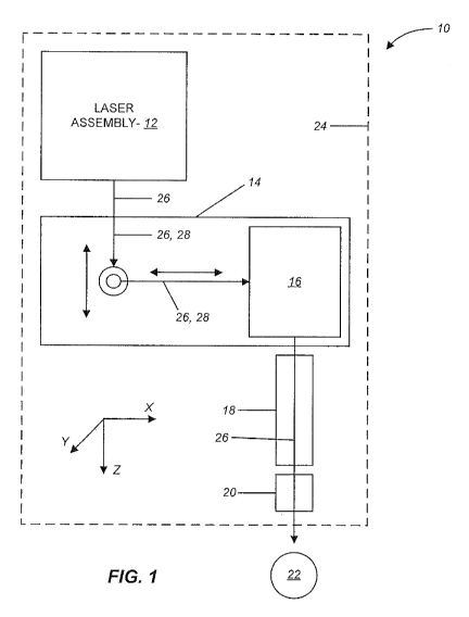
[0043] FIG. 1 is a schematic diagram of a laser surgery system, in accordance
with many
embodiments, in which a patient interface device is coupled to a laser
assembly by way of a scanner
and free-floating mechanism that supports the scanner.
[0044] FIG. 2 shows an isometric view of a patient interface assembly, in
accordance with many
embodiments, that includes a scanner supported by a free-floating mechanism.
[0045] FIG. 3 is a simplified block diagram of acts of a method, in accordance
with many
embodiments, for accommodating patient movement in a laser surgery system.
[0046] FIG. 4 is a simplified block diagram of optional acts, in accordance
with many
embodiments, that can be accomplished in the method of FIG. 3.
[0047] FIG. 5 schematically illustrates relative movements that can be used in
a patient interface
assembly, in accordance with many embodiments, that includes a scanner
supported by a free-
floating mechanism.
[0048] FIG. 6A is a simplified block diagram of acts of another method, in
accordance with many
embodiments, for accommodating patient movement in a laser surgery system.
[0049] FIG. 6B is a simplified block diagram of optional acts, in accordance
with many
embodiments, that can be accomplished in the method of FIG. 6A.
[0050] FIG. 7 is a schematic diagram of a laser surgery system, in accordance
with many
embodiments, in which an eye interface device is coupled to a laser assembly
by way of a scanner
and free-floating mechanism that supports the scanner.
[0051] FIG. 8 is a schematic diagram of another laser surgery system, in
accordance with many
embodiments, in which an eye interface device is coupled to a laser assembly
by way of a scanner
and free-floating mechanism that supports the scanner.
[0052] FIG. 9 is another schematic diagram of the laser surgery system, in
accordance with many
embodiments, in which an eye interface device is coupled to a laser assembly
by way of a scanner
and free-floating mechanism that supports the scanner.
DETAILED DESCRIPTION
[0053] In the following description, various embodiments of the present
invention will be described.
For purposes of explanation, specific configurations and details are set forth
in order to provide a
-11-
CA 03026222 2018-11-30
WO 2017/209772 PCT/US2016/035905
thorough understanding of the embodiments. It will also, however, be apparent
to one skilled in the
art that the present invention may be practiced without the specific details.
Furthermore, well-
known features may be omitted or simplified in order not to obscure the
embodiment being
described.
[0054] The drawings and related descriptions of the embodiments have been
simplified to illustrate
elements that are relevant for a clear understanding of these embodiments,
while eliminating various
other elements found in conventional laser eye surgery systems. Those of
ordinary skill in the art
may thus recognize that other elements and/or steps are desirable and/or
required in implementing
the embodiments that are claimed and described. But, because those other
elements and steps are
well-known in the art, and because they do not necessarily facilitate a better
understanding of the
embodiments, they are not discussed. This disclosure is directed to all
applicable variations,
modifications, changes, and implementations known to those skilled in the art.
As such, the
following detailed descriptions are merely illustrative and exemplary in
nature and are not intended
to limit the embodiments of the subject matter or the uses of such
embodiments. As used in this
application, the terms "exemplary" and "illustrative" mean "serving as an
example, instance, or
illustration." Any implementation described as exemplary or illustrative is
not meant to be construed
as preferred or advantageous over other implementations. Further, there is no
intention to be bound
by any expressed or implied theory presented in the preceding background,
brief summary, or the
following detailed description.
[0055] Patient interface assemblies and related methods for use in laser
surgery systems are
provided. While described herein as used in laser eye surgery systems, the
patient interface
assemblies and methods described herein can be used in any other suitable
laser surgery system. In
many embodiments, a free-floating patient interface_assembly is configured to
accommodate
movement of a patient relative to the laser surgery system while maintaining
alignment between an
electromagnetic treatment beam emitted by the laser surgery system and the
patient.
[0056] Referring now to the drawings in which like numbers reference similar
elements, FIG. 1
schematically illustrates a laser surgery system 10, in accordance with many
embodiments. The
laser surgery system 10 includes a laser assembly 12, a free-floating
mechanism 14, a scanning
assembly 16, an objective lens assembly 18, and a patient interface device 20.
The patient interface
device 20 is configured to interface with a patient 22. The patient interface
device 20 is supported
by the objective lens assembly 18. The objective lens assembly 18 is supported
by the scanning
assembly 16. The scanning assembly 16 is supported by the free-floating
mechanism 14. The free-
-12-
CA 03026222 2018-11-30
WO 2017/209772 PCT/US2016/035905
floating mechanism 14 has a portion having a fixed position and orientation
relative to the laser
assembly 12.
[0057] In many embodiments, the patient interface device 20 is configured to
interface with an eye
of the patient 22. For example, the patient interface device 20 can be
configured to be vacuum
coupled to an eye of the patient 22 such as described in U.S. Publication No.
US 2014-0128821 Al
(U.S. Patent Application serial number: 14/068,994, entitled "Liquid Optical
Interface for Laser Eye
Surgery System", filed October 31, 2013). The laser surgery system 10 can
further optionally
include a base assembly 24 that can be fixed in place or repositionable. For
example, the base
assembly 24 can be supported by a support linkage that is configured to allow
selective repositioning
of the base assembly 24 relative to a patient and secure the base assembly 24
in a selected fixed
position relative to the patient. Such a support linkage can be supported in
any suitable manner such
as, for example, by a fixed support base or by a movable cart that can be
repositioned to a suitable
location adjacent to a patient. In many embodiments, the support linkage
includes setup joints with
each setup joint being configured to permit selective articulation of the
setup joint and can be
selectively locked to prevent inadvertent articulation of the setup joint,
thereby securing the base
assembly 24 in a selected fixed position relative to the patient when the
setup joints are locked.
[0058] Eye Interface Examples
[0059] Certain older methods to measure the force on the eye 22 of the patient
interface device 20
utilized three load cells. The slow response time (approx.. 1/2 sec.) made
this less than effective for
docking the patient to the system and monitoring the force during the
procedure. Plus, the load cells
were used both to precisely locate the patient interface and measure the force
on the patient's eye.
Hence the load cells were mounted in a statically indeterminate manner and as
a result hysteresis
was a problem. These flaws made the load cell assembly unsuitable as a monitor
for patient safety.
[0060] In many embodiments, the force sensor here uses a
microelectromechanical system (MEMS)
device. It utilizes the piezo resistive properties of the silicon device to
convert the applied load into
an electrical signal in the range of tens of millivolts. By preloading the
force sensor in compression,
the force sensor assembly can measure an appropriate range of axial and
lateral forces exerted on the
patient's eye. This force sensor assembly separates the functions of load
sensing and precisely
locating the patient so that hysteresis is not an issue. The response time is
on the order of tens of
microseconds and can be used to accurately measure and monitor the forces on a
patient's eye while
docking and during the procedure. As an added benefit, the force sensors are
packaged in low profile
Surface Mount Technology (SMT) package so that the force sensor assembly is
thinner than the
-13-
CA 03026222 2018-11-30
WO 2017/209772 PCT/US2016/035905
original load cell assembly by approximately 8 mm, improving the clearance
between the system
and the patient. The force sensor assembly has been designed to limit the load
that can be applied to
the force sensor effectively preventing an overload condition from ever
occurring.
[0061] Laser Assembly Examples
[0062] In many embodiments, the laser assembly 12 is configured to emit an
electromagnetic
radiation beam 26. The beam 26 can include a series of laser pulses of any
suitable energy level,
duration, and repetition rate.
[0063] In many embodiments, the laser assembly 12 incorporates femtosecond
(FS) laser
technology. By using femtosecond laser technology, a short duration (e.g.,
approximately 10-13
seconds in duration) laser pulse (with energy level in the micro joule range)
can be delivered to a
tightly focused point to disrupt tissue, thereby substantially lowering the
energy level required as
compared to laser pulses having longer durations.
[0064] The laser assembly 12 can produce laser pulses having a wavelength
suitable to treat and/or
image tissue. For example, the laser assembly 12 can be configured to emit an
electromagnetic
radiation beam 26 such as emitted by any of the laser surgery systems
described in U.S. Publication
Nos. US 2014-0163534 Al and US 2011-0172649 Al ( co-pending U.S. Patent
Application serial
number 14/069,042, entitled "Laser Eye Surgery System", filed October 31,
2013; U.S. Patent
Application serial number 12,987,069, entitled "Method and System For
Modifying Eye Tissue and
Intraocular Lenses", filed January 7, 2011). For example, the laser assembly
12 can produce laser
pulses having a wavelength from 1020 nm to 1050 nm. For example, the laser
assembly 12 can have
a diode-pumped solid-state configuration with a 1030 (+/- 5) nm center
wavelength. As another
example, the laser assembly 12 can produce laser pulses having a wavelength
320 nm to 430 nm.
For example, the laser assembly 12 can include an Nd:YAG laser source
operating at the 3rd
harmonic wavelength, 355 nm. The laser assembly 12 can also include two or
more lasers of any
suitable configuration.
[0065] The laser assembly 12 can include control and conditioning components.
For example, such
control components can include components such as a beam attenuator to control
the energy of the
laser pulse and the average power of the pulse train, a fixed aperture to
control the cross-sectional
spatial extent of the beam containing the laser pulses, one or more power
monitors to monitor the
flux and repetition rate of the beam train and therefore the energy of the
laser pulses, and a shutter to
allow/block transmission of the laser pulses. Such conditioning components can
include an
adjustable zoom assembly and a fixed optical relay to transfer the laser
pulses over a distance while
-14-
CA 03026222 2018-11-30
WO 2017/209772 PCT/US2016/035905
accommodating laser pulse beam positional and/or directional variability,
thereby providing
increased tolerance for component variation.
[0066] In many embodiments, the laser assembly 12 has a fixed position
relative to the base
assembly 24. The beam 26 emitted by the laser assembly 12 propagates along a
fixed optical path to
the free-floating mechanism 14. The beam 12 propagates through the free-
floating mechanism 14
along a variable optical path 28, which delivers the beam 26 to the scanning
assembly 16. In many
embodiments, the beam 26 emitted by the laser assembly 12 is collimated so
that the beam 26 is not
impacted by patient movement induced changes in the length of the optical path
between the laser
assembly 12 and the scanning assembly 16. The scanning assembly 16 is operable
to scan the beam
26 (e.g., via controlled variable deflection of the beam 26) in at least one
dimension. In many
embodiments, the scanner is operable to scan the beam in two dimensions
transverse to the direction
of propagation of the beam 26 and is further operable to scan the location of
a focal point of the
beam 26 in the direction of propagation of the beam 26. The scanned beam is
emitted from the
scanning assembly 16 to propagate through the objective lens assembly 18,
through the interface
device 20, and to the patient 22.
[0067] The free-floating mechanism 14 is configured to accommodate a range of
movement of the
patient 22 relative to the laser assembly 12 in one or more directions while
maintaining alignment of
the beam 24 emitted by the scanning assembly 16 with the patient 22. For
example, in many
embodiments, the free-floating mechanism 14 is configured to accommodate a
range movement of
the patient 22 in any direction defined by any combination of unit orthogonal
directions (X, Y, and
Z).
[0068] The free-floating mechanism 14 supports the scanning assembly 16 and
provides the variable
optical path 28, which changes in response to movement of the patient 22.
Because the patient
interface device 20 is interfaced with the patient 22, movement of the patient
22 results in
corresponding movement of the patient interface device 20, the objective lens
assembly 18, and the
scanning assembly 16. The free-floating mechanism 14 can include, for example,
any suitable
combination of a linkage that accommodates relative movement between the
scanning assembly 16
and the laser assembly 12 and optical components suitably tied to the linkage
so as to form the
variable optical path 28.
[0069] FIG. 2 shows an free floating assembly 16 to illustrate an example
embodiment of a suitable
combination of a linkage that accommodates relative movement between the
scanning assembly 16
and the laser assembly 12 and optical components suitably tied to the linkage
so as to form the
-15-
CA 03026222 2018-11-30
WO 2017/209772 PCT/US2016/035905
variable optical path 28. The free floating assembly 16 includes an eye
interface device 20, the
objective lens assembly 18, the scanning assembly 16, and the free-floating
mechanism 14. The
free-floating mechanism 14 includes a first support assembly 32, a second
support assembly 34, and
a base assembly 36. The eye interface device 20 is coupled with and supported
by the objective lens
assembly 18. The objective lens assembly 18 is coupled with and supported by
the scanning
assembly 16. The combination of the interface device 20, the objective lens
assembly 18, and the
scanning assembly 16 form a unit that moves in unison in conjunction with
movement of the patient.
[0070] The first support assembly 32 includes a first end frame 38, a second
end frame 40, and
transverse rods 42, 44, which extend between and couple to the end frames 38,
40. The transverse
rods 42, 44 are oriented parallel to a first direction 46. The scanning
assembly 16 is supported by
the transverse rods 42, 44 and slides along the rods 42, 44 in response to
patient movement parallel
to the first direction 46. The transverse rods 42, 44 form part of a linear
bearing accommodating
patient movement parallel to the first direction 46.
[0071] The second support assembly 34 includes a first end frame 48, an
intermediate frame 50,
transverse rods 52, 54, a second end frame 56, and vertical rods 58, 60. The
transverse rods 52, 54
extend between and couple to the first end frame 48 and to the intermediate
frame 50. The
transverse rods 52, 54 are oriented parallel to a second direction 62, which
is at least transverse to
and can be orthogonal to the first direction 46. Each of the first and second
directions 46, 62 can be
horizontal. The first support assembly 32 is supported by the transverse rods
52, 54 and slides along
the rods 52, 54 in response to patient movement parallel to the second
direction 62. The transverse
rods 52, 54 form part of a linear bearing accommodating patient movement
parallel to the second
direction 62. The vertical rods 58, 60 extend between and couple to the
intermediate frame 50 and
to the second end frame 56. The vertical rods 58, 60 are oriented parallel to
a third direction 64,
which is at least transverse to each of first and second directions 46, 62,
and can be orthogonal to at
least one of the first and second directions 46, 62. The vertical rods 58, 60
form part of a linear
bearing accommodating relative movement between the second support assembly 34
and the base
assembly 36 parallel to the third direction 64, thereby accommodating patient
movement parallel to
the third direction 64.
[0072] First, second, and third reflectors 66, 68, 70 (e.g., mirrors) are
supported by the free-floating
mechanism 14 and configured to reflect the electromagnetic radiation beam 26
to propagate along a
variable optical path 28. The first reflector 66 is mounted to the first
support assembly 32 (to second
end frame 42 in the illustrated embodiment). The second reflector 68 is
mounted to the second
-16-
CA 03026222 2018-11-30
WO 2017/209772 PCT/US2016/035905
support assembly 34 (to intermediate frame 50 in the illustrated embodiment).
The third reflector 70
is mounted to the base assembly 36. In operation, the beam 26 emitted by the
laser assembly is
deflected by the third reflector 70 so as to propagate parallel to the third
direction 64 and be incident
upon the second reflector 68. The second reflector 68 deflects the beam 26 so
as to propagate
parallel to the second direction 62 and be incident upon the first reflector
66. The first reflector 66
deflects the beam 26 so as to propagate parallel to the first direction 46 and
into the scanning
assembly 16, which then controllably scans and outputs the scanned beam
through the objective lens
assembly 18 and the eye interface device 20. By propagating the beam 26
parallel to the third
direction 64 from the third reflector 70 to the second reflector 68, the
length of the corresponding
portion of the variable optical path 28 can be varied so as to accommodate
relative movement of the
patient relative to the third direction 64. By propagating the beam 26
parallel to the second direction
62 from the second reflector 68 to the first reflector 66, the length of the
corresponding portion of
the variable optical path 28 can be varied so as to accommodate relative
movement of the patient
relative to the second direction 62. By propagating the beam 26 parallel to
the first direction 46
from the first reflector 66 to the scanning assembly 16, the length of the
corresponding portion of the
variable optical path 28 can be varied so as to accommodate relative movement
of the patient
relative to the first direction 46.
[0073] In the illustrated embodiment, the free-floating mechanism 14 further
includes a first
solenoid brake assembly 72, a second solenoid brake assembly 74, and a third
solenoid brake
assembly 76. The solenoid brake assemblies 72, 74, 76 are operable to
selectively prevent
inadvertent articulation of the free-floating mechanism 14 during initial
positioning of the scanning
assembly 16 relative to a patient's eye. For example, in the absence of any
mechanism for
preventing inadvertent articulation of the free-floating mechanism 14,
movement of the scanning
assembly 16 may induce inadvertent articulation of the free-floating mechanism
14, especially when
a user induces movement of the scanning assembly 16 through contact with, for
example, the
objective lens assembly 18 to move the objective lens assembly 18 into a
suitable location relative to
the patient. When the laser surgery system 10 is supported by a support
linkage mechanism that
includes setup joints, preventing inadvertent articulation of the free-
floating mechanism 14 can be
used to ensure that the initial positioning of the laser surgery system 10
occurs via articulation of the
setup joints instead of via articulation of the free-floating mechanism 14.
[0074] The first solenoid brake assembly 72 is configured to selectively
prevent inadvertent
movement between the scanning assembly 16 and the first support assembly 32.
Engagement of the
-17-
CA 03026222 2018-11-30
WO 2017/209772 PCT/US2016/035905
first solenoid brake assembly 72 prevents movement of the scanning assembly 16
along the
transverse rods 42, 44, thereby preventing relative movement between the
scanning assembly 16 and
the first support assembly 32 parallel to the first direction 46. When the
first solenoid brake
assembly 72 is not engaged, the scanning assembly 16 is free to slide along
the transverse rods 42,
44, thereby permitting relative movement between the scanning assembly 16 and
the first support
assembly 32 parallel to the first direction 46. In many embodiments, the free-
floating mechanism 14
includes a detent mechanism and/or an indicator that is configured to permit
engagement of the first
solenoid brake assembly 72 when the scanning assembly 16 is centered relative
to its range of travel
along the transverse rods 42, 44, thereby ensuring equal range of travel of
the scanning assembly 16
in both directions parallel to the first direction 46 when the first solenoid
brake assembly 72 is
disengaged following positioning of the objective lens assembly 18 relative to
the patient.
[0075] The second solenoid brake assembly 74 is configured to selectively
prevent inadvertent
movement between the first support assembly 32 and the second support assembly
34. Engagement
of the second solenoid brake assembly 74 prevents movement of the first
support assembly 32 along
the transverse rods 52, 54, thereby preventing relative movement between the
first support
assembly 32 and the second support assembly 34 parallel to the second
direction 62. When the
second solenoid brake assembly 74 is not engaged, the first support assembly
32 is free to slide
along the transverse rods 52, 54, thereby permitting relative movement between
the first support
assembly 32 and the second support assembly 34 parallel to the second
direction 62. In many
embodiments, the free-floating mechanism 14 includes a detent mechanism and/or
an indicator that
is configured to permit engagement of the second solenoid brake assembly 74
when the first support
assembly 32 is centered relative to its range of travel along the transverse
rods 52, 54, thereby
ensuring equal range of travel of the first support assembly 32 in both
directions parallel to the
second direction 62 when the second solenoid brake assembly 74 is disengaged
following
positioning of the objective lens assembly 18 relative to the patient.
[0076] The third solenoid brake assembly 76 is configured to selectively
prevent inadvertent
movement between the second support assembly 34 and the base assembly 36.
Engagement of the
third solenoid brake assembly 76 prevents movement of the base assembly 36
along the vertical
rods 58, 60, thereby preventing relative movement between the second support
assembly 34 and the
base assembly 36 parallel to the third direction 64. When the third solenoid
brake assembly 76 is not
engaged, the base assembly 36 is free to slide along the vertical rods 58, 60,
thereby permitting
relative movement between the second support assembly 34 and the base assembly
36 parallel to the
-18-
CA 03026222 2018-11-30
WO 2017/209772 PCT/US2016/035905
third direction 64. In many embodiments, the free-floating mechanism 14
includes a detent
mechanism and/or an indicator that is configured to permit engagement of the
third solenoid brake
assembly 76 when the base assembly 36 is centered relative to its range of
travel along the vertical
rods 58, 60, thereby ensuring equal range of travel of the base assembly 36 in
both directions parallel
to the third direction 64 when the third solenoid brake assembly 76 is
disengaged following
positioning of the objective lens assembly 18 relative to the patient.
[0077] In an optional embodiment, the third reflector 70 is omitted and the
incoming beam 26 is
directed to propagate parallel to the third direction 64 and be incident on
the second reflector 68.
Each of the reflectors 66, 68, 70 can be adjustable in position and/or in
orientation and thereby can
be adjusted to align the corresponding portions of the variable optical path
28 with the first, second,
and third directions 46, 62, and 64, respectively. Accordingly, the use of the
third reflector 70 can
provide the ability to align the portion of the variable optical path 28
between the third reflector 70
and the second reflector 68 so as to be parallel to the third direction 64 and
thereby compensate for
relative positional and/or orientation variability between the laser assembly
12 and the free-floating
mechanism 14.
[0078] In the illustrated embodiment of the free floating assembly 16, the
first and second directions
46, 62 can be horizontal and the third direction 64 can be vertical. The free-
floating mechanism 14
can also include a counter-balance mechanism coupled with the scanner and
configured to inhibit
gravity-induced movement of the eye interface device 20 and/or inhibit the
transfer of gravity-
induced forces from the eye interface device 20 to an eye coupled with the eye
interface device 20.
For example, a counter-balance mechanism can be employed to apply a counter-
balancing vertical
force to the second assembly 34, thereby inhibiting or even preventing gravity-
induced relative
movement between the second assembly 34 and the base assembly 36 and/or
inhibiting the transfer
of gravity-induced forces from the eye interface device 20 to an eye coupled
with the eye interface
device 20.
[0079] Other suitable variations of the free floating assembly 16 are
possible. For example, the
scanning assembly 16 can be slidably supported relative to a first support
assembly via a vertically-
oriented linear bearing. The first support assembly can be slidably supported
relative to a second
support assembly via a first horizontally-oriented linear bearing. The second
support assembly can
be slidably supported relative to a base assembly via a second horizontally-
oriented linear bearing
that is oriented transverse (e.g., perpendicular) to the first horizontally-
oriented linear bearing. In
such a configuration, a counter-balancing mechanism can be used to apply a
counter-balancing force
-19-
CA 03026222 2018-11-30
WO 2017/209772 PCT/US2016/035905
to the scanning assembly 16, thereby inhibiting or even preventing gravity-
induced relative
movement of the scanning assembly 16 and the eye interface device 20 and/or
inhibiting or even
preventing the transfer of gravity-induced force from the eye interface device
20 to an eye coupled
with the eye interface device 20. The free floating assembly 16 can also
incorporate one or more
sensors configured to monitor relative position 1) between the scanning
assembly 16 and the first
support assembly 32, 2) between the first support assembly 32 and the second
support assembly 34,
and/or 3) between the second support assembly 34 and the base assembly 36.
[0080] FIG. 3 is a simplified block diagram of acts of a method 100, in
accordance with many
embodiments, of accommodating patient movement in a laser surgery system. Any
suitable device,
assembly, and/or system described herein can be used to practice the method
100. The method 100
includes using a first support assembly (e.g., first support assembly 32) to
support a scanner (e.g.,
scanning assembly 16) so as to accommodate relative translation between the
scanner and the first
support assembly parallel to a first direction (e.g., direction 46). The
scanner is operable to
controllably scan an electromagnetic radiation beam (e.g., beam 26) and
configured to be coupled
with a patient so that the scanner moves in conjunction with movement of the
patient (act 102). A
second support assembly (e.g., second support assembly 34) is used to support
the first support
assembly so as to accommodate relative translation between the first support
assembly and the
second support assembly parallel to a second direction (e.g., direction 62)
that is transverse to the
first direction (act 104). A base assembly (e.g., base assembly 36) is used to
support the second
support assembly so as to accommodate relative translation between the second
support assembly
and the base assembly parallel to a third direction (e.g., direction 64) that
is transverse to each of the
first and second directions (act 106). The electromagnetic radiation beam is
propagated in a
direction that is fixed relative to the base assembly (act 108). The first
support assembly is used to
support a first reflector (e.g., first reflector 66) configured to reflect the
electromagnetic radiation
beam so as to propagate parallel to the first direction and to the scanner
(act 110). The second
support assembly is used to support a second reflector (e.g., second reflector
68) configured to
reflect the electromagnetic radiation beam so as to propagate parallel to the
second direction and to
be incident on the first reflector (act 112). Relative translation between the
scanner and the first
assembly, between the first assembly and the second assembly, and between the
second assembly
and the base assembly is used to accommodate three-dimensional relative
translation between the
scanner and the base assembly (act 114).
-20-
CA 03026222 2018-11-30
WO 2017/209772 PCT/US2016/035905
[0081] FIG. 4 is a simplified block diagram of additional aspects and/or
optional acts that can be
accomplished as part of the method 100. For example, the method 100 can
include using the base
assembly to support a third reflector (e.g., third reflector 70) configured to
reflect the
electromagnetic radiation beam to propagate parallel to the third direction
and to be incident on the
second reflector (act 116). The method 100 can include operating the scanner
to scan the
electromagnetic radiation beam in at least two dimensions (act 118). The
method 100 can include
focusing the electromagnetic radiation beam to a focal point (act 120). The
method 100 can include
operating the scanner to scan the focal point in three dimensions (act 122).
The method 100 can
include using a counter-balance mechanism to inhibit gravity-induced movement
of the scanner
and/or to inhibit transfer of gravity-induced force to an eye coupled with the
scanner (act 124). The
method 100 can include monitoring a relative position of at least one of the
group consisting of (1)
between the scanner and the first support assembly, (2) between the first
support assembly and the
second support assembly, and (3) between the second support assembly and the
base assembly (act
126). The method 100 can include inhibiting relative movement during
positioning of the scanner
relative to the patient between at least one of (1) the scanner and the first
support assembly, (2) the
first support assembly and the second support assembly, and (3) the second
support assembly and the
base assembly (act 128).
[0082] FIG. 5 schematically illustrates relative movements that can be used in
the free-floating
mechanism 14 that can be used to accommodate patient movement, in accordance
with many
embodiments. The free-floating mechanism 14 includes the first reflector 66,
the second
reflector 68, and the third reflector 70. In many embodiments, the free-
floating mechanism 14
includes a linkage assembly (not shown) that is configured to permit certain
relative movement
between the scanning assembly 16 and the first reflector 66, between the first
reflector 66 and the
second reflector 68, and between the second reflector 68 and the third
reflector 70 so as to
consistently direct the electromagnetic radiation beam 26 to the scanning
assembly 16 while
accommodating three-dimensional relative movement between the patient
interface device 20 and
the laser assembly used to generate the electromagnetic radiation beam 26. For
example, similar to
the embodiment of the free-floating mechanism 14 illustrated in FIG. 2, a free-
floating mechanism
14 can be configured such that the scanning assembly 16 is supported by a
first support assembly
such that the scanner is free to translate relative to the first support
assembly parallel to the first
direction 46, thereby maintaining the location and orientation of the beam 26
between the first
reflector 66 and the scanning assembly 16. Likewise, the first support
assembly can be supported by
-21-
CA 03026222 2018-11-30
WO 2017/209772 PCT/US2016/035905
a second support assembly such that the first support assembly is free to
translate relative to the
second support assembly parallel to a second direction 62, thereby maintaining
the location and
orientation of the beam 26 between the second reflector 68 and the first
reflector 66. And the second
support assembly can be supported by a base assembly such that the second
support assembly is free
to translate relative to the base assembly parallel to a third direction 64,
thereby maintaining the
location and orientation of the beam 26 between the third reflector 70 and the
second reflector 68.
[0083] The free-floating mechanism 14 can also employ one or more relative
rotations so as to
maintain the location and orientation of path segments of the beam 26. For
example, the scanning
assembly 16 can be supported by a first support assembly such that the scanner
is free to undergo a
rotation 78 relative to the first support assembly about an axis coincident
with the path segment of
the beam 26 between the first reflector 66 and the scanning assembly 16,
thereby maintaining the
location and orientation of the beam 26 between the first reflector 66 and the
scanning assembly 16.
Likewise, the first support assembly can be supported by a second support
assembly such that the
first support assembly is free to undergo a rotation 80 relative to the second
support assembly about
an axis coincident with the path segment of the beam 26 between the second
reflector 68 and the first
reflector 66, thereby maintaining the location and orientation of the beam 26
between the second
reflector 68 and the first reflector 66. And the second support assembly can
be supported by a base
assembly such that the second support assembly is free to undergo a rotation
82 relative to the base
assembly about an axis coincident with the path segment of the beam 26 between
the third reflector
70 and the second reflector 68, thereby maintaining the location and
orientation of the beam 26
between the third reflector 70 and the second reflector 68.
[0084] The free-floating mechanism 14 can also employ any suitable combination
of relative
translations and relative rotations so as to maintain the location and
orientation of path segments of
the beam 26. For example, with respect to the configuration illustrated in
FIG. 5, the free-floating
mechanism 14 can employ relative translation parallel to the second direction
62, relative translation
parallel to the third direction 64, and relative rotation 82, thereby allowing
three-dimensional
movement of the patient interface 20 relative to the laser assembly used to
generate the
electromagnetic radiation beam 26, and thereby accommodating patient movement.
[0085] FIG. 6A is a simplified block diagram of acts of a method 200, in
accordance with many
embodiments, of accommodating patient movement in a laser surgery system. Any
suitable device,
assembly, and/or system described herein can be used to practice the method
200. The method 200
includes using a first support assembly to support a scanner so as to
accommodate relative
-22-
CA 03026222 2018-11-30
WO 2017/209772 PCT/US2016/035905
movement between the scanner and the first support assembly so as to
accommodate patient
movement. The scanner is operable to controllably scan an electromagnetic
radiation beam and
configured to be coupled with a patient so that the scanner moves in
conjunction with movement of
the patient (act 202). The method 200 includes using a beam source to generate
the electromagnetic
radiation beam (act 204). The method 200 includes propagating the
electromagnetic radiation beam
from the beam source to the scanner along an optical path having an optical
path length that changes
in response to patient movement (act 206).
[0086] FIG. 6B is a simplified block diagram of additional aspects and/or
optional acts that can be
accomplished as part of the method 200. For example, the method 200 can
include using a second
support assembly to support the first support assembly so as to accommodate
relative movement
between the first support assembly and the second support assembly so as to
accommodate patient
movement (act 208). The method 200 can include using the first support
assembly to support a first
reflector configured to reflect the electromagnetic radiation beam so as to
propagate to the scanner
along a portion of the optical path (act 210). The method 200 can include
using a base assembly to
support the second support assembly so as to accommodate relative movement
between the second
support assembly and the base assembly so as to accommodate patient movement
(act 212). The
method 200 can include using the second support assembly to support a second
reflector configured
to reflect the electromagnetic radiation beam to propagate along a portion of
the optical path so as to
be incident on the first reflector (act 214). The method 200 can include using
the base assembly to
support a third reflector configured to reflect the electromagnetic radiation
beam to propagate along
a portion of the optical path so as to be incident on the second reflector
(act 216). The method 200
can include monitoring at least one of a relative position and a relative
orientation of at least one of
the group consisting of (1) between the scanner and the first support
assembly, (2) between the first
support assembly and the second support assembly, and (3) between the second
support assembly
and the base assembly (act 218). The method 200 can include inhibiting
relative movement during
positioning of the scanner relative to the patient between at least one of (1)
the scanner and the first
support assembly, (2) the first support assembly and the second support
assembly, and (3) the second
support assembly and the base assembly (act 220).
[0087] FIG. 7 schematically illustrates a laser surgery system 300, in
accordance with many
embodiments. The laser surgery system 300 includes the laser assembly 12, the
free-floating
mechanism 14, the scanning assembly 16, the objective lens assembly 18, the
patient interface 20,
communication paths 302, control electronics 304, control panel/graphical user
interface (GUI) 306,
-23-
CA 03026222 2018-11-30
WO 2017/209772 PCT/US2016/035905
and user interface devices 308. The control electronics 304 includes processor
310, which includes
memory 312. The patient interface 20 is configured to interface with a patient
22. The control
electronics 304 is operatively coupled via the communication paths 302 with
the laser assembly 12,
the free-floating mechanism 14, the scanning assembly 16, the control
panel/GUI 306, and the user
interface devices 308.
[0088] The free-floating mechanism 14 can be configured as illustrated in FIG.
2 to include, for
example, the first reflector 66, the second reflector 68, and the third
reflector 70. Accordingly, the
free-floating mechanism 14 can be configured to accommodate movement of the
patient 22 relative
to the laser assembly 12 in any direction resulting from any combination of
three orthogonal unit
directions.
[0089] The scanning assembly 16 includes a z-scan device 314 and an xy-scan
device 316. The
laser surgery system 300 is configured to focus the electromagnetic radiation
beam 26 to a focal
point that is scanned in three dimensions. The z-scan device 314 is operable
to vary the location of
the focal point in the direction of propagation of the beam 26. The xy-scan
device 316 is operable to
scan the location of the focal point in two dimensions transverse to the
direction of propagation of
the beam 26. Accordingly, the combination of the z-scan device 314 and the xy-
scan device 316 can
be operated to controllably scan the focal point of the beam in three
dimensions, including within a
tissue of the patient 22 such as within an eye tissue of the patient 22. As
described above with
respect to free floating assembly 16, the scanning assembly 16 is supported by
the free-floating
mechanism 14, which accommodates patient movement induced movement of the
scanning device
relative to the laser assembly 12 in three dimensions.
[0090] The patient interface 20 is coupled to the patient 22 such that the
patient interface 20, the
objective lens 18, and the scanning assembly 16 move in conjunction with the
patient 22. For
example, in many embodiments, the patient interface 20 employs a suction ring
that is vacuum
attached to an eye of the patient 20. The suction ring can be coupled with the
patient interface 20,
for example, using vacuum to secure the suction ring to the patient interface
20.
[0091] The control electronics 304 controls the operation of and/or can
receive input from the laser
assembly 12, the free-floating assembly 14, the scanning assembly 16, the
patient interface 20, the
control panel/GUI 306, and the user interface devices 308 via the
communication paths 302. The
communication paths 302 can be implemented in any suitable configuration,
including any suitable
shared or dedicated communication paths between the control electronics 304
and the respective
system components.
-24-
CA 03026222 2018-11-30
WO 2017/209772 PCT/US2016/035905
[0092] The control electronics 304 can include any suitable components, such
as one or more
processor, one or more field-programmable gate array (FPGA), and one or more
memory storage
devices. In many embodiments, the control electronics 304 controls the control
panel/GUI 306 to
provide for pre-procedure planning according to user specified treatment
parameters as well as to
provide user control over the laser eye surgery procedure.
[0093] The control electronics 304 can include a processor/controller 310 that
is used to perform
calculations related to system operation and provide control signals to the
various system elements.
A computer readable medium 312 is coupled to the processor 310 in order to
store data used by the
processor and other system elements. The processor 310 interacts with the
other components of the
system as described more fully throughout the present specification. In an
embodiment, the
memory 312 can include a look up table that can be utilized to control one or
more components of
the laser system surgery system 300.
[0094] The processor 310 can be a general purpose microprocessor configured to
execute
instructions and data, such as a Pentium processor manufactured by the Intel
Corporation of Santa
Clara, California. It can also be an Application Specific Integrated Circuit
(ASIC) that embodies at
least part of the instructions for performing the method in accordance with
the embodiments of the
present disclosure in software, firmware and/or hardware. As an example, such
processors include
dedicated circuitry, ASICs, combinatorial logic, other programmable
processors, combinations
thereof, and the like.
[0095] The memory 312 can be local or distributed as appropriate to the
particular application.
Memory 312 can include a number of memories including a main random access
memory (RAM)
for storage of instructions and data during program execution and a read only
memory (ROM) in
which fixed instructions are stored. Thus, the memory 312 provides persistent
(non- volatile) storage
for program and data files, and may include a hard disk drive, flash memory, a
floppy disk drive
along with associated removable media, a Compact Disk Read Only Memory (CD-
ROM) drive, an
optical drive, removable media cartridges, and other like storage media.
[0096] The user interface devices 308 can include any suitable user input
device suitable to provide
user input to the control electronics 304. For example, the user interface
devices 308 can include
devices such as, for example, a touch-screen display/input device, a keyboard,
a footswitch, a
keypad, a patient interface radio frequency identification (RFID) reader, an
emergency stop button,
and a key switch.
-25-
CA 03026222 2018-11-30
WO 2017/209772 PCT/US2016/035905
[0097] Any suitable laser surgery system can be suitably modified to employ an
electromagnetic
beam scanner that is supported by a free-floating mechanism as disclosed
herein. For example, co-
pending United States provisional patent application serial number 14/069,042
filed October 31,
2013 (published as U.S. Publication No. US 2014-0163534 Al), describes a laser
eye surgery system
that includes beam scanning components that form part of a shared optical
assembly used to scan a
treatment beam, an optical coherence tomography (OCT) measurement beam, and an
alignment
beam. Using the approaches described herein, such beam scanning components can
be supported
from a free-floating mechanism so as to accommodate patient movement as
described herein.
[0098] FIG. 8 is a schematic diagram of a laser surgery system, in accordance
with many
embodiments, in which an eye interface device is coupled to a laser assembly
by way of a scanner
and free-floating mechanism that supports the scanner. The mechanism shown in
FIG. 8 could be
used in lieu of the assembly shown in FIG. 2, for example working in
conjunction with the laser
assembly 12 from FIG. 1. Thus, FIG. 8 shows an assembly 400 example embodiment
of a suitable
combination of a linkage that accommodates relative movement between the
scanning assembly 16
and the laser assembly 12 and optical components suitably tied to the linkage
so as to form the
variable optical path 28 (from FIG. 1). Such free-floating head mechanism
could be used to move in
unison with the movement of a patient.
[0099] FIG. 8 shows another example free-floating head mechanism assembly 400
with three
degrees of freedom of movement about three axes x, y and z. Thus, the system
includes a base
assembly 410 upon which components are attached. The base assembly is stable
relative to a floating
scanning assembly 440 and objective lens assembly 420 which are attached to
the base assembly 410
but are able to move in three degrees of freedom according to the embodiments
described here.
[00100] The base assembly 410 is shown as a framework of parts that are
arranged to support
the components of the system here. The base assembly 410 could be made of any
number of things
including metal such as aluminum or steel, it could be made of plastics or
composites, or a
combination of things. The example base assembly 410 in FIG. 8 generally has
two flat platforms
412 that are held apart by various struts 414. The example is not intended to
be limiting and any
arrangement of support structure could be used.
[00101] Regarding the relative motion of the floating scanning assembly
440 and objective
lens assembly 420 relative to the base assembly 410, the first axis of
movement is a z axis which is
made possible using a z axis spring mechanism 430 and vertical z axis bearings
432. The z axis
-26-
CA 03026222 2018-11-30
WO 2017/209772 PCT/US2016/035905
bearings 432 allow the floating scanning assembly 440 and objective lens
assembly 420 to move
vertically, up and down, relative to the base assembly 410.
[00102] Such a bearing system may include rollers and a linear track or
rail system that keeps
the floating scanning assembly 440 and objective lens assembly 420 from
shifting off of a smooth
and direct movement in any particular axis. In such an assembly, a roller, or
multiple rollers are
configured to contact a track or rail. Each of the two, the roller assembly
and track, are attached to
either the base assembly or the floating scanning assembly 440 and lens
assembly 440. Thus, as the
rollers and track interact, the floating scanning assembly 440 and lens
assembly 440 movement,
relative to the base assembly 410 is forced into a linear direction, according
to the orientation of the
bearing track, in this example, that is along the z axis. As discussed herein,
a combination of such
bearings, can allow for the floating scanning assembly 440 and lens assembly
440 to move about
more than one axis and more than one degree of freedom, relative to the base
assembly 410,
depending on how many axes are configured.
[00103] It should be noted that the example of these roller and track
bearings in FIG. 8 is
merely exemplary and any kind of bearings could be used.
[00104] To complement the vertical bearings 432 in the example embodiment
shown in FIG.
8, a system of springs 430 are shown that help keep any vertical movement of
the floating scanning
assembly 440 and objective lens assembly 420 from happening unless acted upon
by a force other
than gravity, such as a user or operator positioning the floating scanning
assembly 440 and objective
lens assembly 420. In FIG. 8, the example mechanism shown includes two z axis
springs 430 but it
should be noted that any arrangement of multiple or one spring could be used.
These springs 430 can
counteract the force of gravity, which accelerates the floating scanning
assembly 440 and objective
lens assembly 420 toward the earth. Thus, the z axis is the only axis that
needs additional assistance
to counteract gravity, hence the springs.
[00105] In the example embodiment in FIG. 8, the z axis springs 430 are
shown as metal
tapes wound around spring loaded bearing spools. When the floating scanning
assembly 440 and
objective lens assembly 420 is moved by an outside force such as a user or
operator in the vertical
dimension, or z axis, the metal tapes coil or uncoil respectively and the
spring tension within the
coils keep the floating scanning assembly 440 and objective lens assembly 420
from free falling due
to gravity. It should be noted that the wound tape spring example is not
intended to be limiting. Any
kind of spring mechanism or other mechanism could be used with similar effect.
For example, a
hydraulic piston system could be used to counteract gravity, a coiled wire
spring system could be
-27-
CA 03026222 2018-11-30
WO 2017/209772 PCT/US2016/035905
used, a pulley system could be used, a geared system could be used, a magnetic
system could be
used, etc. Additionally a locking mechanism could be used to hold the floating
scanning assembly
440 and objective lens assembly 420 in place, relative to the base assembly,
after it is positioned.
[00106] The free-floating head mechanism 400 also includes bearings in the
horizontal x axis
434 and the horizontal y axis 436 as well as the z axis as discussed. Such x
axis and y axis bearings
keep the floating scanning assembly 440 and objective lens assembly 420 from
slipping or shifting,
relative to the base assembly 410, in the x and y axis directions. Used in
combination, these bearings
allow for the floating scanning assembly 440 and objective lens assembly 420
to move in a
horizontal plane, for example.
[00107] It should be noted that the horizontal bearings do not necessarily
include springs such
as those used for the z axis because generally there is not a force acting
upon the horizontal plane as
there is in the vertical plane with gravity. But the bearings in any degree of
freedom could include a
brake or lock mechanism to keep the floating scanning assembly 440 and
objective lens assembly
420 locked into a certain position, for any or all of the three axes. Such a
brake could be a pin and
hole, either spring loaded or not. A lock could be a gear mechanism with a
latch that holds the gear.
A lock could be a stopper on a spring or piston as well. It could be a
solenoid brake, either manually
operated or magnetically. Any of various locks could be used in any one or
combination of the axes.
[00108] The combination of the three axis bearing arrangement as shown in
FIG. 8 allows for
the entire floating scanning assembly 440 and objective lens assembly 420 to
be moved, by an
operator or user, relative to the base assembly 410 in any position of the
three axes: up and down,
left and right, and in and out, and thus any position in three dimensional
space, within the boundaries
of the bearing tracks. Thus, the range of motion is only limited by the
physical length of the bearing
rails or tracks in each direction. If the z axis bearing rail or track has a
total length of 12 inches, the
range of motion of the floating scanning assembly 440 and objective lens
assembly 420 relative to
the base assembly 410 would be 12 inches. The x axis bearing could have the
same length or
different length. The y axis bearing could have the same length or different
length. The combination
of the three axis bearings would define the range of motion within the three
degrees of freedom for
the floating scanning assembly 440 and objective lens assembly 420. In
combination, these three
axes allow for the floating scanning assembly 440 and objective lens assembly
420 to be positioned
in any three dimensional coordinate within the range of the system.
[00109] Referring now to FIG. 9, the free-floating head mechanism 400 is
depicted with the
three axes of movement for the floating scanning assembly 440 and objective
lens assembly 420
-28-
CA 03026222 2018-11-30
WO 2017/209772 PCT/US2016/035905
relative to the base assembly 410 from FIG. 8, as shown with arrows. The
vertical z axis 462, the
horizontal y axis 466 and the horizontal x axis 464 are all shown which are
the three axes in this
example that the floating scanning assembly 440 and objective lens assembly
420 are able to move
relative to the base assembly 410 from FIG. 8.
[00110] It should be noted that the combination of the three axis system
as described here is
merely exemplary. Fewer axes could be used, or additional axes could be used.
In certain example
embodiments, additional axes of rotations could be added for example, with a
rotating or pivoting
bearing assembly or assemblies attached to the base assembly 410 as well. Such
an embodiment is
not shown, but could be configured to add one, two, or three more degrees of
freedom to the
movement of the assembly 400.
[00111] Certain example embodiments may include motors, attached to or in
communication
with the bearings. Such motors could actuate movement of the optical scanning
module 440 and lens
assembly 420 relative to the base assembly 410. Such motors could be
configured to allow the
movement of the assembly in a remote fashion, using wired or wireless
transmitters. Example
embodiments include motors that could be directed by a program that receives
feedback regarding
patient input, and directs the assembly to move to counteract such patient
movement. It should be
noted that the operation of such motors could be via a remote control device
or local operation.
Wireless or wired control could be utilized to move the system. Wireless
control could be via WiFi
or cellular or Bluetooth Low Energy systems, or any number of other
communication mechanisms.
[00112] Referring again to FIG. 8, as the overall system is designed to
direct beams of
energy, as shown in FIG. 1, to its intended target, through the floating
scanning assembly 440 and
the lens portion 420. Any movement of the floating scanning assembly 440 and
objective lens
assembly 420 relative to the base assembly and potentially the source of the
beam must be
compensated for. An arrangement of mirrors can be used, in certain example
embodiments, to direct
such an energy beam into the floating scanning assembly 440 and objective lens
assembly 420 no
matter where in the floating scanning assembly 440 and objective lens assembly
420 are, relative to
the base assembly 410. Thus, the mirrors could be arranged to move to keep the
beam directed into
the floating scanning assembly 440 and objective lens assembly 420 or be fixed
to the various
portions of the base assembly 410 in order to maintain the beam direction into
the floating scanning
assembly 440 and objective lens assembly 420. Such a beam source, such as a
laser, could be
mounted to the base assembly or some other structure attached to or nearby the
base assembly. Any
-29-
CA 03026222 2018-11-30
WO 2017/209772 PCT/US2016/035905
kind of system could be used, as described in US application 14/191,095 Laser
Eye Surgery Systems
or other such systems.
[00113] The horizontal y axis fixed mirror 450 works in conjunction with
the horizontal y axis
floating mirror and x axis fixed mirror 452. Finally, the horizontal x axis
floating mirror, and the
vertical z axis fixed mirror 454 direct the beam into the floating scanning
assembly 440 and lens
portion 420. These mirrors keep the energy beam aimed at the floating scanning
assembly 440 and
lens assembly 420, no matter where the floating scanning assembly 440 and
objective lens assembly
420 is moved on its three axis bearing system, relative to the base assembly
410.
[00114] It should also be noted that the bearings, springs and mirrors are
shown in
representative places on the base assembly 410 of the assembly. These
components could be moved
to other parts of the base assembly 410, oriented in different ways than are
shown in the example of
FIG. 8. Additional mirrors could be used, for example in embodiments with more
than three degrees
of freedom.
[00115] In certain example embodiments, a patient support structure, such
as for example a
bed or gurney support, could be coupled to the base assembly 410 or base
assembly support structure
and be used to accommodate for patient movement relative to the system such as
that discussed in
FIG. 3, 4, 5 and 6A. An arrangement of motors in communication with a computer
that can sense
patient movement, provide a feedback loop and move the patient support
structure accordingly, to
compensate and keep the patient in place of the energy beam coming through the
floating scanning
assembly 440 and objective lens assembly 420. Such a patient support structure
could also be moved
manually or through direction of a user into such motors.
[00116] Conclusion
[00117] Other variations are within the spirit of the present invention.
Thus, while the
invention is susceptible to various modifications and alternative
constructions, certain illustrated
embodiments thereof are shown in the drawings and have been described above in
detail. It should
be understood, however, that there is no intention to limit the invention to
the specific form or forms
disclosed, but on the contrary, the intention is to cover all modifications,
alternative constructions,
and equivalents falling within the spirit and scope of the invention, as
defined in the appended
claims.
[00118] The use of the terms "a" and "an" and "the" and similar referents
in the context of
describing the invention (especially in the context of the following claims)
are to be construed to
cover both the singular and the plural, unless otherwise indicated herein or
clearly contradicted by
-30-
CA 03026222 2018-11-30
WO 2017/209772 PCT/US2016/035905
context. The terms "comprising," "having," "including," and "containing" are
to be construed as
open-ended terms (i.e., meaning "including, but not limited to,") unless
otherwise noted. The term
"connected" is to be construed as partly or wholly contained within, attached
to, or joined together,
even if there is something intervening. Recitation of ranges of values herein
are merely intended to
serve as a shorthand method of referring individually to each separate value
falling within the range,
unless otherwise indicated herein, and each separate value is incorporated
into the specification as if
it were individually recited herein. All methods described herein can be
performed in any suitable
order unless otherwise indicated herein or otherwise clearly contradicted by
context. The use of any
and all examples, or exemplary language (e.g., "such as") provided herein, is
intended merely to
better illuminate embodiments of the invention and does not pose a limitation
on the scope of the
invention unless otherwise claimed. No language in the specification should be
construed as
indicating any non-claimed element as essential to the practice of the
invention.
[00119] While preferred embodiments of the present invention have been
shown and
described herein, it will be obvious to those skilled in the art that such
embodiments are provided by
way of example only. Numerous variations, changes, and substitutions will now
occur to those
skilled in the art without departing from the invention. It should be
understood that various
alternatives to the embodiments of the invention described herein may be
employed in practicing the
invention. It is intended that the following claims define the scope of the
invention and that methods
and structures within the scope of these claims and their equivalents be
covered thereby.
[00120] The foregoing description, for purpose of explanation, has been
described with
reference to specific embodiments. However, the illustrative discussions above
are not intended to
be exhaustive or to limit the invention to the precise forms disclosed. Many
modifications and
variations are possible in view of the above teachings. The embodiments were
chosen and described
in order to best explain the principles of the invention and its practical
applications, to thereby
enable others skilled in the art to best utilize the invention and various
embodiments with various
modifications as are suited to the particular use contemplated
-31-