Note: Descriptions are shown in the official language in which they were submitted.
CA 03026617 2018-12-05
1
Description
Title of Invention: NON-INVASIVE DIAGNOSTIC IMAGING AGENT
FOR HEART DISEASE
Technical Field
[0001]
The present invention relates to a non-invasive
diagnostic imaging agent for a heart disease.
Background Art
[0002]
In chronic heart failure, left ventricular
remodeling such as ventricular hypertrophy and fibrosis
progresses in the heart. It has been reported that the
activation of the renin-angiotensin-aldosterone system
(RAAS) is associated with this process. RAAS is a
regulatory mechanism of the circulatory system that
regulates blood pressure in the body. In addition to
this RAAS of the systemic circulatory system, data has
recently been reported, indicating that the RAAS or
aldosterone synthesis system is present at the heart
locally.
[0003]
For example, non-Patent Literature 1 states that
blood collected by catheterization of the heart showed
CA 03026617 2018-12-05
2
that aldosterone was synthesized or secreted in the human
failing heart.
[0004]
Non-Patent Literature 2 discloses that a study using
an autopsied heart revealed an increased gene expression
of an aldosterone synthase CYP11B2. This literature
further discloses that the increased expression of the
CYP11B2 gene is related to myocardial fibrosis or cardiac
dysfunction.
[0005]
Attempts have been made to develop various compounds
that selectively inhibit CYP11B2 such that cardiovascular
diseases are treated by selectively inhibiting CYP11B2
(Patent Literatures 1 to 3 and non-Patent Literatures 3
to 5).
[0006]
Although not intended for cardiovascular diseases,
various radioactive compounds that exhibit high
selectivity for CYP11B2 have also been developed for the
purpose of visualizing a lesion in the adrenal gland and
enabling diagnostic imaging of primary aldosteronism
(Patent Literatures 4 to 11 and non-Patent Literature 6).
Citation List
Patent Literature
[0007]
CA 03026617 2018-12-05
3
Patent Literature 1: Japanese Patent Laid-Open (Kohyo) No.
2013-512271
Patent Literature 2: Japanese Patent Laid-Open (Kohyo) No.
2011-520799
Patent Literature 3: Japanese Patent Laid-Open (Kohyo) No.
2014-526539
Patent Literature 4: Japanese Patent Laid-Open (Kohyo) No.
2 013-534 911
Patent Literature 5: Japanese Patent Laid-Open (Kokai) No.
2014-129315
Patent Literature 6: Japanese Patent Laid-Open (Kokai) No.
2015-093831
Patent Literature 7: Japanese Patent Laid-Open (Kokai) No.
2015-093832
Patent Literature 8: Japanese Patent Laid-Open (Kokai) No.
2015-093833
Patent Literature 9: Japanese Patent Laid-Open (Kokai) No.
2015-110563
Patent Literature 10: Japanese Patent Laid-Open (Kokai)
No. 2015-193545
Patent Literature 11: International Publication No. WO
2015/199205
Non-Patent Literature
[0008]
Non-Patent Literature 1: Mizuno, Y. et al., Circulation,
vol. 103, p. 72-7 (2001)
CA 03026617 2018-12-05
4
Non-Patent Literature 2: Satoh, M. et al., Clinical
Science, vol. 102, p. 381-386 (2002)
Non-Patent Literature 3: Grombein, C. M., European
Journal of Medicinal Chemistry, vol. 89, p. 597-605
(2015)
Non-Patent Literature 4: Grombein, C. M., European
Journal of Medicinal Chemistry, vol. 90, p. 788-796
(2015)
Non-Patent Literature 5: Martin, R. E. et al., Journal of
Medicinal Chemistry, vol. 58, p. 8054-8065 (2015)
Non-Patent Literature 6: Abe, T., Journal of Clinical
Endocrinol Metabolism, vol. 101, p. 1008-1015 (2016)
Summary of Invention
[0009]
There might be a possibility that the detection of
expression of CYP11B2 in the heart leads to the
evaluation of course of progression of heart diseases
such as heart failure. However, no non-invasive method
exists that can detect such lesions at preset.
[0010]
One example of the method for detecting a change at
molecular level in vivo is nuclear medicine diagnosis
using single-photon emission computed tomography (SPECT)
and positron emission tomography (PET). Patent
Literatures 4 to 11 and non-Patent Literature 6 have
reported tracers for SPECT or PET that exhibit high
CA 03026617 2018-12-05
selectivity for CYP11B2. However, these tracers target
the adrenal gland and have not yet been found to be
suitable for imaging in the heart.
[0011]
5 No finding has been obtained as to whether or not
the local expression of CYP11B2 in the heart of heart
disease patients is increased to a level that permits
visualization by nuclear medicine diagnosis.
[0012]
The present invention has been made in light of the
circumstances described above and is directed to
providing a technique for detecting a lesion of a heart
disease in a non-invasive manner.
[0013]
The present invention provides a non-invasive
diagnostic imaging agent for a heart disease, comprising
a radioactively labeled compound capable of binding to an
aldosterone synthase or a salt thereof as an active
ingredient.
[0014]
According to the present invention, a lesion of a
heart disease can be detected in a non-invasive manner.
Brief Description of Drawings
[0015]
The object mentioned above and other objects,
features and advantages of the present invention will
CA 03026617 2018-12-05
6
become more apparent upon reading the following preferred
embodiments and accompanying drawings.
[0016]
Figure 1 represents views showing a result of
immunostaining of CYP11B2 using a heart tissue section of
an ischemic heart disease model rat. Figure 1(a) is an
overall view of the section, Figure 1(b) is an enlarged
view of a non-ischemic site, and each of Figures 1(c) and
1(d) is an enlarged view of an ischemia reperfusion site.
Figure 2 represents views showing a result of
autoradiogram of an individual given 6-chloro-5-fluoro-1-
(2_ [18F,
ifluoroethyl)-2-[5-(imidazol-1-ylmethyl)pyridin-3-
yl]benzimidazole (Compound [18F] 1) . Figure 2(a)
represents views showing a series of autoradiograms of
sections arranged from the base of the heart toward the
cardiac apex, and Figure 2(b) is an enlarged view after
rotation by 90 degrees of the autoradiogram surrounded
with the broken line in Figure 2(a).
Figure 3 represents views showing results of
staining a section adjacent to the heart tissue section
shown in Figure 2(b). Figure 3(a) is a view showing a
result of HE staining, Figure 3(b) is a view showing a
result of Masson trichrome staining, and Figure 3(c) is a
view showing a result of immunostaining of CYP11B2.
Figure 4 represents views showing results of in
vitro autoradiography using 6-chloro-5-fluoro-1-(2-
P8F]fluoroethyl)-2-[5-(imidazol-1-ylmethyl)pyridin-3-
CA 03026617 2018-12-05
7
yl]benzimidazole (Compound [18F] 1). Figure 4(a) is a
view showing a result of immersing a heart tissue section
of a normal rat in a solution containing 6-chloro-5-
fluoro-1- (2-['8F]fluoroethyl)-2-[5-(imidazol-1-
ylmethyl)pyridin-3-yl]benzimidazole (Compound [18F] 1).
Each of Figures 4(b) to 4(d) is a view showing a result
of immersing a heart tissue section of an ischemic heart
disease model rat in a solution containing 6-chloro-5-
fluoro-1- (2-['8F]fluoroethyl)-2-[5-(imidazol-1-
ylmethyl)pyridin-3-yl]benzimidazole (Compound [18F] 1).
Figure 4(e) is a view showing a result of immersing a
heart tissue section of the normal rat in a solution
containing 6-chloro-5-fluoro-1-(2-['8F]fluoroethyl)-2-[5-
(imidazol-1-ylmethyl)pyridin-3-yl]benzimidazole (Compound
[18F] 1) and 5 ilmol/L 6-chloro-5-fluoro-1-(2-
fluoroethyl)-2-[5-(imidazol-1-ylmethyl)pyridin-3-
yl]benzimidazole (Compound 1). Each of Figures 4(f) to
4(h) is a view showing a result of immersing a heart
tissue section of the ischemic heart disease model rat in
a solution containing 6-chloro-5-fluoro-1-(2-
['8F]fluoroethyl)-2-[5-(imidazol-1-ylmethyl)pyridin-3-
yl]benzimidazole (Compound [18F] 1) and 5 mol/L 6-
chloro-5-fluoro-1-(2-fluoroethyl)-2-[5-(imidazol-1-
ylmethyl)pyridin-3-yl]benzimidazole (Compound 1).
Figure 5 represents views showing results of in
vitro autoradiography using 1-(2-fluoroethyl)-2-[5-
{(imidazol-1-y1)methyllpyridin-3-y1]-6-
CA 03026617 2018-12-05
8
[1231] iodobenzimidazole (Compound [1231] 2) . Figure 5(a)
is a view showing a result of immersing a heart tissue
section of a normal rat in a solution containing 1-(2-
fluoroethyl)-2-[5-{(imidazol-1-y1)methyl}pyridin-3-y1]-6-
[1231] iodobenzimidazole (Compound [1231] 2). Figure 5(b)
represents views showing results of immersing heart
tissue sections of an ischemic heart disease model rat in
a solution containing 1-(2-fluoroethyl)-2-[5-{(imidazol-
1-y1)methyl}pyridin-3-yl] -6-['23I]iodobenzimidazole
(Compound [1231] 2) . Figure 5(c) is a view showing a
result of immersing a heart tissue section of the normal
rat in a solution containing 1-(2-fluoroethyl)-2-[5-
{(imidazol-1-y1)methyllpyridin-3-y1]-6-
[123i] iodobenzimidazole (Compound [1231] 2) and 5 mol/L 1-
(2-fluoroethyl)-2-[5-{(imidazol-1-y1)methyl}pyridin-3-
y1]-6-iodobenzimidazole (Compound 2). Figure 5(d)
represents views showing results of immersing heart
tissue sections of the ischemic heart disease model rat
in a solution containing 1-(2-fluoroethyl)-2-[5-
Himidazol-1-y1)methyllpyridin-3-y1]-6-
[123i] iodobenzimidazole (Compound [1231] 2) and 5 mol/L 1-
(2-fluoroethyl)-2-[5-{(imidazol-1-y1)methyl}pyridin-3-
y1]-6-iodobenzimidazole (Compound 2).
Figure 6 represents views showing results of
autoradiography and staining of a heart tissue section of
an ischemic heart disease model rat using 1-(2-
fluoroethyl)-2-[5-{(imidazol-1-y1)methyl}pyridin-3-y1]-6-
CA 03026617 2018-12-05
9
[123I]iodobenzimidazole (Compound [123I] 2). Figure 6(a)
is a view showing a result of autoradiography, Figure
6(b) is a view showing a result of Masson trichrome
staining, and Figure 6(c) is a view showing a result of
immunostaining of CY911B2.
Figure 7 represents views showing comparison of
results of autoradiography using 6-chloro-5-fluoro-1-(2-
[18F] fluoroethyl)-2-[5-(imidazol-1-ylmethyl)pyridin-3-
yl]benzimidazole (Compound [18F] 1) and 1-(2-
fluoroethyl)-2-[5-{(imidazol-1-y1)methyl}pyridin-3-y11-6-
[123i] iodobenzimidazole (Compound [123I] 2). Figure 7(a)
is a view showing an example of region of interest (ROI)
established on an autoradiogram of a section immersed in
a solution containing 6-chloro-5-fluoro-1-(2-
[18F] fluoroethyl)-2-[5-(imidazol-1-ylmethyl)pyridin-3-
yl]benzimidazole (Compound [AF] 1) . Figure 7(b) is a bar
graph showing a ratio of ischemia reperfusion site signal
intensity/non-ischemic site signal intensity of 6-chloro-
5-fluoro-1-(2-[AF]fluoroethyl)-2-[5-(imidazol-1-
ylmethyl)pyridin-3-yl]benzimidazole (Compound [16F] 1) on
an individual basis. Figure 7(c) is a view showing an
example of ROI established on an autoradiogram of a
section immersed in a solution containing 1-(2-
fluoroethyl)-2-[5-{(imidazol-1-y1)methyl}pyridin-3-y1]-6-
2 5 [1231] iodobenzimidazole (Compound [123I] 2). Figure 7(d)
is a bar graph showing a ratio of ischemia reperfusion
site signal intensity/non-ischemic site signal intensity
CA 03026617 2018-12-05
of 1-(2-fluoroethyl)-2-[5-1(imidazol-1-y1)methyllpyridin-
3-y11-6-[123I]iodobenzimidazole (Compound [1231] 2) on an
individual basis.
Figure 8 represents views showing results of
5 autoradiography and staining with 1-(2-fluoroethyl)-2-[5-
{(imidazol-1-y1)methyllpyridin-3-y11-6-
[1231] iodobenzimidazole (Compound [1231] 2) using the
hearts of a normal rat and rats I day, 3 days and 1 week
after ischemic heart disease model rat preparation.
10 Figure 8(a) is a bar graph showing a ratio of ischemia
reperfusion site signal intensity/non-ischemic site
signal intensity, and Figure 8(b) represents views
showing results of Masson trichrome staining.
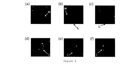
Figure 9 represents views displaying a SPECT image
with 1-(2-fluoroethyl)-2-[5-{(imidazol-1-
y1)methyl}pyridin-3-y1]-6- [123I]iodobenzimidazole
(Compound [1231] 2), which is superimposed with a computed
tomography image. Figure 9(a) is a short axis image of a
normal rat, Figure 9(b) is a horizontal long axis image
of the normal rat, Figure 9(c) is a vertical long axis
image of the normal rat, Figure 9(d) is a short axis
image of an ischemic heart disease model rat, Figure 9(e)
is a horizontal long axis image of the ischemic heart
disease model rat, and Figure 9(f) is a vertical long
axis image of the ischemic heart disease model rat.
Figure 10 represents views showing results of
immunostaining of CYP11B2 using the respective heart
CA 03026617 2018-12-05
11
tissue sections of a myocarditis model rat and a normal
rat. Figure 10(a) is an overall view of the heart tissue
section of the myocarditis model rat, Figure 10(b) is an
enlarged view of Figure 10(a), Figure 10(c) is an overall
view of the heart tissue section of the normal rat, and
Figure 10(d) is an enlarged view of Figure 10(c).
Figure 11 represents views showing results of in
vitro autoradiography with 2-[5-{(1H-imidazol-1-
yl)methyl}pyridin-3-yl] -6-[123I]iodo-l-methyl-1H-
benz[d]imidazole (Compound [1231] 3). Figure 11(a) is a
view showing a result of autoradiography of a heart
tissue section of a normal rat, Figure 11(b) is a view
showing a result of autoradiography of a heart tissue
section of a myocarditis model rat, Figure 11(c) is a
view showing a result of Masson trichrome staining of a
heart tissue section of the normal rat, Figure 11(d) is a
view showing a result of Masson trichrome staining of a
heart tissue section of the myocarditis model rat, Figure
11(e) is a view showing a result of immunostaining of
CYP11B2 of a heart tissue section of the normal rat, and
Figure 11(f) is a view showing a result of immunostaining
of CYP11B2 of a heart tissue section of the myocarditis
model rat.
Figure 12 represents views showing results of
immunostaining of CYP11B2 using the respective heart
tissue sections of a hypertensive heart disease model rat
and a normal rat. Figure 12(a) is an overall view of the
CA 03026617 2018-12-05
12
heart tissue section of the hypertensive heart disease
model, Figure 12(b) is an enlarged view of Figure 12(a),
Figure 12(c) is an overall view of the heart tissue
section of the normal rat, and Figure 12(d) is an
enlarged view of Figure 12(c).
Figure 13 represents views showing results of in
vitro autoradiography with 2-[5-{(1H-imidazol-1-
yl)methy1}pyridin-3-y11-6-[1231] iodo-l-methyl-1H-
benz[d]imidazole (Compound [1231] 3). Figure 13(a) is a
view showing a result of autoradiography of a heart
tissue section of a normal rat, Figure 13(b) is a view
showing a result of autoradiography of a heart tissue
section of a hypertensive heart disease model rat, Figure
13(c) is a view showing a result of Masson trichrome
staining of a heart tissue section of the normal rat,
Figure 13(d) is a view showing a result of Masson
trichrome staining of a heart tissue section of the
hypertensive heart disease model rat, Figure 13(e) is a
view showing a result of immunostaining of CYP11B2 of a
heart tissue section of the normal rat, and Figure 13(f)
is a view showing a result of immunostaining of CYP11B2
of a heart tissue section of the hypertensive heart
disease model rat.
Figure 14 represents views showing results of in
vitro autoradiography using 2-[5-{(1H-imidazol-1-
yl)methyl}pyridin-3-y1]-6-[1231] iodo-l-methyl-1H-
benz[d]imidazole (Compound [1231] 3). Figure 14(a) is a
CA 03026617 2018-12-05
13
view showing a result of immersing a heart tissue section
of a normal rat in a solution containing 2-[5-{(1H-
imidazol-1-yl)methyl}pyridin-3-yl] -6-[123I]iodo-1-methyl-
1H-benz[d]imidazole (Compound [123I] 3). Figure 14(b)
represents views showing results of immersing heart
tissue sections of an ischemic heart disease model rat in
a solution containing 2-[5-{(1H-imidazol-1-
yl)methyl}pyridin-3-yl] -6-[123I]iodo-l-methy1-1H-
benz[d]imidazole (Compound [123I] 3).
Description of Embodiments
[0017]
The present invention provides a non-invasive
diagnostic imaging agent for a heart disease, comprising
a radioactively labeled compound capable of binding to an
aldosterone synthase or a salt thereof as an active
ingredient. The diagnostic imaging agent of the present
invention can visualize a site having the advanced
fibrosis of the heart.
[0018]
In the present invention, the "non-invasive
diagnostic imaging agent" refers to one which is used in
nuclear medicine diagnosis and is preferably used in
positron emission tomography (PET) or single-photon
emission computed tomography (SPECT).
[0019]
CA 03026617 2018-12-05
14
In the present invention, the "heart disease"
includes an ischemic heart disease and a non-ischemic
heart disease and is preferably a disease caused by the
fibrosis of the heart. One example thereof is heart
failure.
In the present invention, the "ischemic heart
disease" is not limited as long as the ischemic heart
disease is a heart disease caused by myocardial ischemia.
Examples thereof include coronary heart diseases such as
angina pectoris, myocardial infarction, acute coronary
syndrome, and ischemic heart failure.
In the present invention, examples of the "non-
ischemic heart disease" include myocarditis, hypertensive
heart disease, dilated cardiomyopathy, hypertrophic
cardiomyopathy, and non-ischemic heart failure.
[0020]
In the present invention, the "radioactively labeled
compound" is not limited as long as the radioactively
labeled compound is a compound labeled with a
radioisotope for use in nuclear medicine diagnosis.
Examples of the radioisotope include carbon-11, fluorine-
18, chlorine-34m, bromine-76, iodine-123 and iodine-124.
The non-invasive diagnostic imaging agent of the present
invention can be used as a diagnostic imaging agent for
positron emission tomography in the case of using a
positron-emitting radionuclide such as carbon-11,
fluorine-18, chlorine-34m or iodine-124 as the
CA 03026617 2018-12-05
radioisotope, and can be used as a diagnostic imaging
agent for single-photon emission computed tomography in
the case of using iodine-123 as a radioactive halogen
atom.
5 [0021]
In the present invention, the phrase "capable of
binding to an aldosterone synthase" means being capable
of binding to human CYP11B2.
[0022]
10 In the present invention, the "radioactively labeled
compound capable of binding to an aldosterone synthase"
is not limited as long as the radioactively labeled
compound is capable of binding to human CYP11B2.
Examples thereof include radioactively labeled compounds
15 having affinity for adrenocortical adenoma or
adrenocortical carcinoma as described in U.S. Patent
Application Publication No. 2005/0033060 and Japanese
Patent Laid-open (Kohyo) No. 2009-539822, and
radioactively labeled compounds having CYP11B2
selectivity as described in Japanese Patent Laid-open
(Kohyo) No. 2013-534911, Japanese Patent Laid-Open
(Kokai) No. 2014-129315, Japanese Patent Laid-Open
(Kokai) No. 2015-093831, Japanese Patent Laid-Open
(Kokai) No. 2015-093832, Japanese Patent Laid-Open
(Kokai) No. 2015-093833, Japanese Patent Laid-Open
(Kokai) No. 2015-110563, Japanese Patent Laid-Open
(Kokai) No. 2015-193545, and International Publication No.
CA 03026617 2018-12-05
16
WO 2015/199205. Alternatively, the radioactively labeled
compound capable of binding to an aldosterone synthase
may be a compound derived from a compound described in
Japanese Patent Laid-open (Kohyo) No. 2013-512271,
Japanese Patent Laid-open (Kohyo) No. 2011-520799,
Japanese Patent Laid-open (Kohyo) No. 2011-525894,
Japanese Patent Laid-open (Kohyo) No. 2012-526774,
Japanese Patent Laid-open (Kohyo) No. 2013-510896,
Japanese Patent Laid-open (Kohyo) No. 2014-526539,
Japanese Patent Laid-open (Kohyo) No. 2014-527077, or
Japanese Patent Laid-open (Kohyo) No. 2014-533736 by the
replacement of one constituent element with the
radioisotope mentioned above.
[0023]
In the present invention, the radioactively labeled
compound is preferably a compound represented by the
following general formula (1) described in International
Publication No. WO 2015/199205.
[0024]
CA 03026617 2018-12-05
17
X3 X1
R4
X2
R3
( 1 )
R2
R5
X \
R1 N/
[0025]
In the general formula (1), Ri represents a hydrogen
atom or CO2Ra. R2 represents a hydrogen atom, a halogen
atom or CO2Ra. R3 represents a hydrogen atom or a
hydroxyalkyl group having 1 to 10 carbon atoms. R4
represents a hydrogen atom, a hydroxy group or an alkoxy
group having 1 to 10 carbon atoms. R5 represents a chain
alkyl group having 1 to 5 carbon atoms in which a
hydrogen atom is optionally replaced with a halogen atom,
a cyclic alkyl group having 3 to 5 carbon atoms in which
a hydrogen atom is optionally replaced with a halogen
atom, a hydroxyalkyl group having 1 to 5 carbon atoms, or
an o-, p- or m-halobenzyl group. A represents CH or a
nitrogen atom. X1 and X3 each independently represent a
hydrogen atom or a halogen atom. X2 represents a
hydrogen atom, a halogen atom or a nitrile group. At
least one of Xi, X2 and X3 is a halogen atom.
[0026]
CA 03026617 2018-12-05
18
In the radioactively labeled compound of the general
formula (1), the "CO2Ra" is a carboxylic acid ester group.
Each Ra is independently an alkyl group having 1 to 10
carbon atoms. The alkyl group may be linear or branched
and is preferably an alkyl group having 1 to 5 carbon
atoms (methyl group, ethyl group, n-propyl group,
isopropyl group, n-butyl group, isobutyl group, tert-
butyl group, n-pentyl group, isopentyl group, or
neopentyl group), more preferably an alkyl group having 1
to 3 carbon atoms (methyl group, ethyl group, n-propyl
group, or isopropyl group). The "CO2Ra" is particularly
preferably a "carboxylic acid methyl ester group," in
which Ra is a methyl group.
[0027]
In the radioactively labeled compound of the general
formula (1), the "halogen atom" is a fluorine atom, a
chlorine atom, a bromine atom or an iodine atom.
[0028]
In the compound of the general formula (1), the
"hydroxyalkyl group" is a group represented by -(CH2)õ,01-1.
In the general formula (1), for example, m for R3 is an
integer of 1 to 10, preferably an integer of 1 to 3. In
the general formula (1), m for R5 is an integer of 1 to 5,
preferably an integer of 1 to 3.
[0029]
In the radioactively labeled compound of the general
formula (1), the "alkoxy group" is a group in which a
CA 03026617 2018-12-05
19
linear or branched alkyl group is bonded to an oxygen
atom. Examples thereof preferably include a methoxy
group, an ethoxy group, a propoxy group, and an
isopropoxy group, among which a methoxy group is more
preferred.
[0030]
In the radioactively labeled compound of the general
formula (1), the "chain alkyl group" is a non-cyclic
alkyl group and may be linear or branched. Examples
thereof preferably include a methyl group, an ethyl group,
a propyl group, an isopropyl group, a n-butyl group, an
isobutyl group, a sec-butyl group, a tert-butyl group, a
n-pentyl group, an isopentyl group, a neopentyl group,
and a tert-pentyl group. In these chain alkyl groups,
one or two or more hydrogen atoms may be replaced with
halogen atom(s) and are preferably replaced with fluorine
atom(s). Specific examples thereof include a
fluoromethyl group, a 1-fluoroethyl group, a 1,1-
difluoroethyl group, a 1,1,1-trifluoroethyl group, and a
1-fluoropropyl group.
[0031]
In the radioactively labeled compound of the general
formula (1), the "cyclic alkyl group" includes a
cyclopropyl group, a cyclobutyl group, and a cyclopentyl
group. In these cyclic alkyl groups, one or two or more
hydrogen atoms may be replaced with halogen atom(s).
[0032]
CA 03026617 2018-12-05
In the radioactively labeled compound of the general
formula (1), the "halobenzyl group" is a benzyl group in
which a hydrogen atom at position 2, 3, or 4 of the
benzene ring is replaced with a halogen atom. A benzyl
5 group in which the hydrogen atom at position 2 is
replaced with a halogen atom is an o-halobenzyl group. A
benzyl group in which the hydrogen atom at position 3 is
replaced with a halogen atom is a m-halobenzyl group. A
benzyl group in which the hydrogen atom at position 4 is
10 replaced with a halogen atom is a p-halobenzyl group.
Among them, a p-halobenzyl group is preferred.
[0033]
In the radioactively labeled compound of the general
formula (1), any one of R2, R5, and X2 contains a
15 radioactive halogen atom. Preferably, the radioactively
labeled compound of the general formula (1) has the
following configuration (a), (b), (c) or (d):
(a) a radioactive halogen atom is used as a halogen atom
in R2;
20 (b) R5 is a group represented by -(CH2)nX4, and a
radioactive halogen atom is used as a halogen atom of X4;
(c) R5 is a p-halobenzyl group, and a radioactive halogen
atom is used as a halogen atom introduced at position 4
of the benzyl group; and
(d) a radioactive halogen atom is used as a halogen atom
of X2. .
[0034]
CA 03026617 2018-12-05
21
In this context, the "radioactive halogen atom"
refers to any of fluorine-18, chlorine-34m, bromine-76,
iodine-123 and iodine-124.
[0035]
More preferably, in the general formula (1), R3 is a
hydrogen atom; R4 is a hydrogen atom or an alkoxy group
having 1 to 10 carbon atoms; R5 is a chain alkyl group
having 1 to 5 carbon atoms in which a hydrogen atom is
optionally replaced with a halogen atom, a cyclic alkyl
group having 3 to 5 carbon atoms, or an o-, p- or m-
halobenzyl group; X2 is a halogen atom; and X3 is a
hydrogen atom. R5 is more preferably a methyl group, an
ethyl group, a propyl group, an isopropyl group, a group
represented by -(CH2)nX4 (wherein n represents an integer
of 1 to 5, and X4 represents a halogen atom), a
cyclopropyl group, or a p-halobenzyl group.
[0036]
Further preferably, in the general formula (1), R2
is a hydrogen atom or a halogen atom.
[0037]
Further preferably, in the general formula (1), R5
is a methyl group, an ethyl group, a group represented by
-(CH2)nX4, or a cyclopropyl group. In the group
represented by -(CH2)nX4, n is preferably an integer of 1
to 3, more preferably 2 or 3, furthermore preferably 2.
X4 is preferably a fluorine atom.
[0038]
CA 03026617 2018-12-05
22
One specific embodiment of the compound according to
the present invention is a compound represented by the
general formula (2).
[0039]
XII
= X12
R12 ( 2)
N
''X14
[0040]
In the general formula (2), R12 represents a hydrogen
atom, a halogen atom or CO2Ra. XII represents a hydrogen
atom or a halogen atom. X12 represents a halogen atom.
X14 represents a hydrogen atom, a halogen atom or a
hydroxy group. n represents an integer of 1 to 5. In
the radioactively labeled compound of the general formula
(2), the "CO2Ra" is as defined in the general formula (1).
[0041]
The compound represented by the general formula (2)
is a compound of the general formula (1) wherein R1 is a
hydrogen atom; R2 is a hydrogen atom, a halogen atom, or
CO2Ra (wherein Ra is an alkyl group having 1 to 10 carbon
atoms); each of R3 and R4 is a hydrogen atom; R5 is a
chain alkyl group having 1 to 5 carbon atoms, or a group
CA 03026617 2018-12-05
23
represented by -(Cl2)nXi4; A is CH; Xi is a hydrogen atom
or a halogen atom; X2 is a halogen atom; and X3 is a
hydrogen atom. In the general formula (2), R12 may
represent a hydrogen atom or CO2Ra and is preferably a
hydrogen atom. X14 is preferably a hydrogen atom or a
fluorine atom, and n is preferably an integer of 1 to 3.
[0042]
In the general formula (2), R12, X12, or X14 is a
radioactive halogen atom. Preferably, X12 or X14 is a
radioactive halogen atom. More preferably, R12 is a
hydrogen atom, and X12 or X14 is a radioactive halogen
atom. In this case, still more preferably, n is an
integer of 1 to 3. The radioactive halogen atom is as
defined above.
[0043]
The compounds represented by the general formulas
(1) and (2) can be obtained by methods described in
International Publication No. WO 2015/199205.
[0044]
In the present invention, the "salt" may be one that
is pharmaceutically acceptable. The salt can be, for
example, a salt derived from an inorganic acid such as
hydrochloric acid, hydrobromic acid, sulfuric acid,
nitric acid, and phosphoric acid, or an organic acid such
as acetic acid, trifluoroacetic acid, maleic acid,
succinic acid, mandelic acid, fumaric acid, malonic acid,
pyruvic acid, oxalic acid, glycolic acid, salicylic acid,
CA 03026617 2018-12-05
24
pyranosidyl acids (glucuronic acid, galacturonic acid,
etc.), a-hydroxy acids (citric acid, tartaric acid, etc.),
amino acids (aspartic acid, glutamic acid, etc.),
aromatic acids (benzoic acid, cinnamic acid, etc.), and
sulfonic acids (p-toluenesulfonic acid, ethanesulfonic
acid, etc.).
[0045]
The non-invasive diagnostic imaging agent of the
present invention is a formulation containing the
radioactively labeled compound described above or the
salt thereof in a form suitable for administration into a
living body. This non-invasive diagnostic imaging agent
is preferably administered through a parenteral route,
i.e., by injection, and is more preferably an aqueous
solution. Such a composition may appropriately contain
an additional component such as a pH adjuster, or a
pharmaceutically acceptable solubilizer, stabilizer or
antioxidant.
Examples
[0046]
Hereinafter, the present invention will be described
in more detail by way of Examples. However, the present
invention is not limited by the contents thereof.
In Examples given below, the names of compounds
subjected to experiments were defined as follows.
CA 03026617 2018-12-05
Compound 1: 6-chloro-5-fluoro-1-(2-fluoroethyl)-2-[5-
(imidazol-1-ylmethyl)pyridin-3-yl]benzimidazole
Compound [18F] 1: 6-chloro-5-fluoro-1-(2-
['8F]fluoroethyl)-2-[5-(imidazol-1-ylmethyl)pyridin-3-
5 yl]benzimidazole (a compound represented by the general
formula (2) wherein R12 represents a hydrogen atom; XII
represents a fluorine atom; X12 represents a chlorine
atom; X14 represents fluorine-18; and n represents an
integer of 2)
10 Compound 2: 1-(2-fluoroethyl)-2-[5-{(imidazol-1-
y1)methyl}pyridin-3-y1]-6-iodobenzimidazole
Compound [123I] 2: 1-(2-fluoroethyl)-2-[5-{(imidazol-1-
y1)methyl}pyridin-3-yl] -6-[123I]iodobenzimidazole (a
compound represented by the general formula (2) wherein
15 R12 represents a hydrogen atom; Xli represents a hydrogen
atom; X12 represents iodine-123; X14 represents a fluorine
atom; and n represents an integer of 2)
Compound 3: 2-[5-{(1H-imidazol-1-yl)methyl}pyridin-3-y1]-
6-iodo-1-methy1-1H-benz[d]imidazole
20 Compound [1231] 3: 2-[5-{(1H-imidazol-1-yl)methyl}pyridin-
3-y1]-6-[123I]iodo-1-methy1-1H-benz[d]imidazole (a
compound represented by the general formula (2) wherein
R12 represents a hydrogen atom; XII represents a hydrogen
atom; X12 represents iodine-123; XN represents a hydrogen
25 atom; and n represents an integer of 1)
[0047]
CA 03026617 2018-12-05
26
Compound 1 was synthesized according to the method
for synthesizing Compound 100 in International
Publication No. WO 2015/199205.
Compound [1-8F' 1 1 was synthesized according to the
method for synthesizing Compound HEIF,
j 100 in
International Publication No. WO 2015/199205, and a
compound having 95% or higher radiochemical purity under
the TLC conditions disclosed therein was used.
Compound 2 was synthesized according to the method
for synthesizing Compound 604 in International
Publication No. WO 2015/199205.
Compound [1231] 2 was synthesized according to the
method for synthesizing Compound [123I] 604 in
International Publication No. WO 2015/199205, and a
compound having 95% or higher radiochemical purity under
the TLC conditions disclosed therein was used.
Compound 3 was synthesized according to the method
for synthesizing Compound 607 in International
Publication No. WO 2015/199205.
Compound [1231] 3 was synthesized according to the
method for synthesizing Compound [123I' j 607 in
International Publication No. WO 2015/199205, and a
compound having 95% or higher radiochemical purity under
the TLC conditions disclosed therein was used.
[0048]
(Example 1) CYP11B2 expression evaluation in ischemic
heart disease model rat heart
CA 03026617 2018-12-05
27
The chest of each Wistar rat (male) was opened under
isoflurane anesthesia, and the left coronary artery was
ligated for 30 minutes, followed by reperfusion. The
chest was closed to prepare an ischemic heart disease
model rat. Approximately 1 week after the operation, the
rat was sacrificed under anesthesia, and the heart was
taken out thereof. Sections of 5 pm in thickness were
prepared from the base side of the heart. Immunostaining
was carried out using the prepared sections to confirm
the expression and distribution of CPY11B2. As an anti-
CYP11B2 antibody, one prepared according to the method
described in Ogishima T et al., Endocrinology, 1992, vol.
130, pp. 2971-7 was used. As a secondary antibody, HRP
Labelled Polymer Anti-Rabbit (manufactured by
Dako/Agilent Technologies, Inc.) was used. CYP11B2
expression sites were detected by applying DAB+ (3,3'-
diaminobenzidine tetrahydrochloride) substrate kit
(manufactured by Dako/Agilent Technologies, Inc.) to the
HRP bound to the secondary antibody.
[0049]
The result is shown in Figure 1. Figure 1 is a view
showing a result of immunostaining of CYP11B2. Figure
1(a) is an overall view of the section, Figure 1(b) is a
20-fold enlarged view of a non-ischemic site (ROIa in
Figure 1(a)), Figure 1(c) is a 20-fold enlarged view of
an ischemia reperfusion site (ROIb in Figure 1(a)), and
Figure 1(d) is a 20-fold enlarged view of an ischemia
CA 03026617 2018-12-05
28
reperfusion site (ROIc in Figure 1(a)). As shown in
Figure 1, it was confirmed that CYP11B2 was expressed at
the ischemia reperfusion site. It was also confirmed
that CYP11B2 was not expressed or was low in expression
at the non-ischemic site.
[0050]
(Example 2) Ex vivo autoradiography using ischemic heart
disease model rat
Each ischemic heart disease model rat was prepared
in the same way as in Example 1. Compound [18F] 1 was
administered thereto (approximately 50 MBq/rat) between 1
and 3 weeks after the operation. 20 minutes after the
administration, the rat was sacrificed under anesthesia,
and the heart was taken out thereof. Frozen sections of
20 m in thickness were prepared from the base of the
heart toward the cardiac apex. The prepared sections
were exposed to an imaging plate (BAS-SR2040,
manufactured by Fujifilm Corp.) for 2 hours.
Autoradiograms were obtained using a fluoro image
analyzer (FLA-7000, manufactured by GE Healthcare Corp.).
HE staining, Masson trichrome staining, and the same
immunostaining of CYP11B2 as in Example 1 were each
carried out using sections adjacent to the section on the
base side of the heart.
[0051]
The results of an individual given Compound [18F] 1
are shown in Figures 2 and 3. Figure 2 shows the results
CA 03026617 2018-12-05
29
of autoradiogram of the individual given Compound [18F] 1.
Figure 2(a) shows the respective autoradiograms of
sections arranged from the base of the heart toward the
cardiac apex. Figure 2(b) is an enlarged view after
rotation by 90 degrees of the autoradiogram surrounded
with the broken line in Figure 2(a). Figure 3 shows the
results of staining a section adjacent to the section
shown in Figure 2(b). Figure 3(a) shows the result of HE
staining, Figure 3(b) shows the result of Masson
trichrome staining, and Figure 3(c) shows the result of
immunostaining of CYP11B2.
As illustrated in Figure 2, the local accumulation
of Compound [18F] 1 was confirmed. From the results of
staining in Figure 3, inflammatory reaction, fibrosis,
and CYP11B2 expression were also confirmed at a position
corresponding to the accumulation site of Compound [18F]
1.
[0052]
(Example 3) In vitro autoradiography using ischemic heart
disease model rat
Each ischemic heart disease model rat was prepared
in the same way as in Example 1. The rat was sacrificed
under isoflurane anesthesia between 1 and 3 weeks after
the operation. Then, the heart was harvested, and 5 m
sections were prepared and preserved at -80 C until they
were used. The sections were brought back to room
temperature from -80 C, dried for 30 minutes or longer,
CA 03026617 2018-12-05
then immersed in phosphate-buffered saline for 30 minutes,
and subsequently immersed in phosphate-buffered saline
containing 1 w/v% bovine serum albumin for 30 minutes for
hydrophilization. Each phosphate-buffered saline
5 containing 1 w/v% bovine serum albumin and further
containing Compound [18F] 1 (radioactivity concentration:
approximately 40 kBq/mL) or Compound [1231] 2
(radioactivity concentration: approximately 10 kBq/mL)
was prepared, and the hydrophilized sections were
10 immersed therein at room temperature for 30 minutes.
Then, the sections were washed by immersing for 5 minutes
each in phosphate-buffered saline containing 1 w/v%
bovine serum albumin, phosphate-buffered saline, and
phosphate-buffered saline. The sections thus washed were
15 thoroughly dried and then exposed to light for
approximately 3 hours as to Compound [18F] 1 and for
approximately 16 hours as to Compound [123I] 2 on an
imaging plate (BAS-SR2040, manufactured by Fujifilm
Corp.). Autoradiograms were obtained using a fluoro
20 image analyzer (FLA-7000, manufactured by GE Healthcare
Corp.).
Also, autoradiograms were obtained by immersing the
sections in a solution containing Compound [18F] 1
together with 5 pmol/L of Compound 1, or a solution
25 containing Compound [1231] 2 together with 5 gmol/L of
Compound 2 in the same way as above.
CA 03026617 2018-12-05
31
The same experiment as above was conducted using the
heart harvested from a normal rat.
[0053]
The results of Compound [18F] I are shown in Figure 4.
Each of Figures 4(a) and 4(e) shows the section of the
normal rat, and each of Figures 4(b) to 4(d) and 4(f) to
4(h) shows the section of the ischemic heart disease
model rat. Figures 4(a) to 4(d) resulted from immersing
in a solution containing Compound [1-8F] 1, and Figures
4(e) to 4(h) resulted from immersing in a solution
containing Compound [18F]
1 and Compound 1 (5 gmol/L). As
shown in Figure 4, the accumulation of Compound ['8F] 1
in a lesion region was confirmed. The accumulation was
inhibited by the addition of an excessive amount of a
non-labeled compound, suggesting that the binding is
specific.
[0054]
The results of Compound [1231] 2 are shown in Figure
5. Each of Figures 5(a) and 5(c) shows the section of
the normal rat, and each of Figures 5(b) to 5(d) shows
the section of the ischemic heart disease model rat.
Figures 5(a) and 5(b) resulted from immersing in a
solution containing Compound [1231] 2, and Figures 5(c)
and 5(d) resulted from immersing in a solution containing
Compound [1231] 2 and Compound 2 (5 gmol/L). As shown in
Figure 5, the accumulation of Compound [1231] 2 in a
lesion region was confirmed. The accumulation was
CA 03026617 2018-12-05
32
inhibited by the addition of an excessive amount of a
non-labeled compound, suggesting that the binding is
specific.
[0055]
The section of Figure 5(b) was also subjected to
staining. The results are shown in Figure 6. Figure
6(a) shows a result of autoradiography. Figure 6(b)
shows a result of Masson trichrome staining, and Figure
6(c) shows a result of immunostaining of CYP11B2. As
illustrated in Figure 6, the accumulation of Compound
[1231] 2 was in good agreement with a fibrosis region.
The expression of CYP11B2 at an ischemia reperfusion site
was also confirmed.
[0056]
ROI was established at an ischemia reperfusion site
and a non-ischemic site as to the autoradiography of
Compound [18F] 1 and Compound [123I] 2, and the ratio of
signal intensity was compared between these sites. The
results are shown in Figure 7 (n = 4). Figure 7(a) is
one example of an autoradiogram of the section immersed
in Compound [18F] 1. Figure 7(c) is one example of an
autoradiogram of the section immersed in Compound [123I] 2.
In both Figures 7(a) and 7(c), ROI of the ischemia
reperfusion site is surrounded with the solid line, and
ROI of the non-ischemic site is surrounded with the
broken line. Figure 7(b) is a bar graph that shows a
ratio of ischemia reperfusion site signal intensity/non-
CA 03026617 2018-12-05
33
ischemic site signal intensity of Compound [18F] 1 in
each rat. Figure 7(d) is a bar graph that shows a ratio
of ischemia reperfusion site signal intensity/non-
ischemic site signal intensity of Compound [1231] 2 in
each rat. As shown in Figure 7, the increased
accumulation of Compound [18F' j 1 and Compound [1231] 2 at
the ischemia reperfusion site compared with the non-
ischemic site was observed. Higher accumulation of
Compound [1231] 2 than that of Compound [18F] 1 was also
observed.
[0057]
(Example 4) Study on influence of elapsed time after
ischemic heart disease model rat preparation on compound
accumulation
Each ischemic heart disease model rat was prepared
in the same way as in Example 1. The rat was sacrificed
under isoflurane anesthesia 1 day (3 rats), 3 days (4
rats) or 1 week (4 rats) after the operation. Then, the
heart was harvested, and 5 m-thick sections were
prepared and preserved at -80 C until they were used.
The sections were brought back to room temperature from
-80 C, dried for 30 minutes or longer, then immersed in
phosphate-buffered saline for 30 minutes, and
subsequently immersed in phosphate-buffered saline
containing 1 w/v% bovine serum albumin for 30 minutes for
hydrophilization. Phosphate-buffered saline containing 1
w/v% bovine serum albumin and further containing Compound
CA 03026617 2018-12-05
34
[1231] 2 (radioactivity concentration: approximately 10
kBq/mL) was prepared, and the hydrophilized sections were
immersed therein at room temperature for 30 minutes.
Then, the sections were washed by immersing for 5 minutes
each in phosphate-buffered saline containing 1 w/v%
bovine serum albumin, phosphate-buffered saline, and
phosphate-buffered saline. The sections thus washed were
thoroughly dried and then exposed to light for
approximately 16 hours on an imaging plate (BAS-SR2040,
manufactured by Fujifilm Corp.). Autoradiograms were
obtained using a fluoro image analyzer (FLA-7000,
manufactured by GE Healthcare Corp.).
The same experiment as above was conducted using the
hearts harvested from normal rats (two rats).
[0058]
The results are shown in Figure 8. Figure 8(a) is a
bar graph that shows a ratio of ischemia reperfusion site
signal intensity/non-ischemic site signal intensity of
Compound [1231] 2 in autoradiography using the hearts of
the normal rat and the rats 1 day, 3 days and 1 week
after ischemic heart disease model rat preparation.
Figure 8(b) shows the results of Masson trichrome
staining using sections adjacent to the sections used in
Figure 8(a). As shown in Figure 8(a), the accumulation
of Compound [1231] 2 was increased with elapsed time after
the operation. As shown in Figure 8(b), the acceleration
CA 03026617 2018-12-05
of fibrosis was also confirmed with elapsed time after
the operation.
[0059]
(Example 5) SPECT imaging experiment using ischemic heart
5 disease model rat
Each ischemic heart disease model rat was prepared
in the same way as in Example 1. Compound [123I] 2 was
administered thereto (approximately 100 MBq/rat) in one
week after the operation. Static imaging for
10 approximately 8 minutes was started at 150 minutes after
the administration using a SPECT apparatus for animals
(FX3000, manufactured by TriFoil Imaging). Data
collection was carried out in an energy window of 143 to
175 key, and the collected data was reconstituted by OSEM
15 (Ordered Subset Expectation Maximization) to obtain
images. Computed tomography imaging was carried out in
order to identify the position of the heart. The same
experiment as above was conducted using a normal rat.
[0060]
20 The results are shown in Figure 9. Figures 9(a) to
9(c) show the results of SPECT imaging based on Compound
[123I] 2 using the normal rat. In Figures 9(a) to 9(c),
the arrow H depicts the heart, and the arrow L depicts
the liver. Figures 9(d) to 9(f) show the results of
25 SPECT imaging based on Compound [1231i
j 2 using the
ischemic heart disease model rat. In Figures 9(d) to
9(f), the arrow i depicts an ischemia reperfusion site.
CA 03026617 2018-12-05
36
Each of Figures 9(a) and 9(d) is a short axis image, each
of Figures 9(b) and 9(e) is a horizontal long axis image,
and each of Figures 9(c) and 9(f) is a vertical long axis
image. All the SPECT images of Figure 9 were displayed
in a manner superimposed with a computed tomography image.
As shown in Figure 9, the accumulation of Compound [12311
2, which was not observed in the heart of the normal rat,
was confirmed in the heart of the ischemic heart disease
model rat.
[0061]
(Example 6) CYP11B2 expression evaluation in myocarditis
model rat heart
Porcine heart cardiac myosin (Sigma-Aldrich Co. LLC)
was diluted to 5 mg/mL using a phosphate buffer solution
(solution A). 100 mg of Mycobacterium tuberculosis H37Ra
(Difco Laboratories Ltd.) was added to 10 mL of Freund's
Adjuvant, Complete (Sigma-Aldrich Co. LLC) and mixed
therewith (solution B). Solution A and solution B were
mixed at a ratio of 1:1 until becoming uniform (solution
C). Solution C was administered at 50 L each to the
right and left hind footpads of each Lewis rat (male, 7
weeks old, Charles River Laboratories Japan, Inc.) under
isoflurane anesthesia. After raising up to 21 days after
the immunization, the rat was sacrificed under anesthesia,
and the heart was taken out thereof. Sections of 5 pm in
thickness were prepared. Immunostaining was carried out
using the prepared sections to confirm the expression and
CA 03026617 2018-12-05
37
distribution of CPY11B2. As an anti-CYP11B2 antibody,
one prepared according to the method described in
Ogishima T et al., Endocrinology, 1992, vol. 130, pp.
2971-7 was used. As a secondary antibody, HRP Labelled
Polymer Anti-Rabbit (manufactured by Dako/Agilent
Technologies, Inc.) was used. 0YP11B2 expression sites
were detected by applying DAB+ (3,3'-diaminobenzidine
tetrahydrochloride) substrate kit (manufactured by
Dako/Agilent Technologies, Inc.) to the HRP bound to the
secondary antibody. The same experiment as above was
conducted using the heart harvested from a conventionally
raised Lewis rat (normal rat).
[0062]
The results are shown in Figure 10. Figure 10 is a
view showing the results of immunostaining of CYP11B2.
Figure 10(a) is an overall view of the heart tissue
section of the myocarditis model, and Figure 10(b) is a
40-fold enlarged view of the lesion site in Figure 10(a).
Figure 10(c) is an overall view of the heart tissue
section of the normal rat, and Figure 10(d) is a 40-fold
enlarged view of the normal site in Figure 10(c). As
shown in Figure 10, it was confirmed that 0YP11B2 was
expressed in the heart of the myocarditis model rat. It
was also confirmed that 0YP11B2 was not expressed or was
low in expression in the heart of the normal rat.
[0063]
CA 03026617 2018-12-05
38
(Example 7) In vitro autoradiography using myocarditis
model rat
Each myocarditis model rat was prepared in the same
way as in Example 6. The heart was harvested, and 5 m-
thick sections were prepared and preserved at -80 C until
they were used. The sections were brought back to room
temperature from -80 C, dried for 30 minutes or longer,
then immersed in phosphate-buffered saline for 30 minutes,
and subsequently immersed in phosphate-buffered saline
containing 1 w/v% bovine serum albumin for 30 minutes for
hydrophilization. Phosphate-buffered saline containing 1
w/v% bovine serum albumin and further containing Compound
[123I] 3 (radioactivity concentration: approximately 10
kBq/mL) was prepared, and the hydrophilized sections were
immersed therein at room temperature for 30 minutes.
Then, the sections were washed by immersing for 5 minutes
each in phosphate-buffered saline containing 1 w/v%
bovine serum albumin, phosphate-buffered saline, and
phosphate-buffered saline. The sections thus washed were
thoroughly dried and then exposed to light for
approximately 16 hours as to Compound [123I] 3 on an
imaging plate (BAS-SR2040, manufactured by Fujifilm
Corp.). Autoradiograms were obtained using a fluoro
image analyzer (FLA-7000, manufactured by GE Healthcare
Corp.). The same experiment as above was conducted using
the heart harvested from a normal rat. Masson trichrome
staining and the same immunostaining of CYP11B2 as in
CA 03026617 2018-12-05
39
Example 6 were each carried out using sections adjacent
to the sections used in autoradiography.
[0064]
The results of in vitro autoradiography of Compound
[123I] 3 are shown in Figure 11. Figure 11(a) shows the
result of autoradiography of the heart tissue section of
the normal rat, and Figure 11(b) shows the result of
autoradiography of the heart tissue section of the
myocarditis model rat. Figure 11(c) shows the result of
Masson trichrome staining of the heart tissue section of
the normal rat, and Figure 11(d) shows the result of
Masson trichrome staining of the heart tissue section of
the myocarditis model rat. Figure 11(e) shows the result
of immunostaining of CYP11B2 of the heart tissue section
of the normal rat, and Figure 11(f) shows the result of
immunostaining of CYP11B2 of the heart tissue section of
the myocarditis model rat. As illustrated in Figure 11,
the increased accumulation of Compound [123I] 3 in the
heart tissue section of the myocarditis model rat
compared with the heart tissue section of the normal rat
was confirmed. The accumulation of Compound [1231] 3 was
in good agreement with a fibrosis region, and the
expression of CYP11B2 in the fibrosis region was also
confirmed.
[0065]
(Example 8) CYP11B2 expression evaluation in hypertensive
heart disease model rat heart
CA 03026617 2018-12-05
Each DIS/Eis (Dahl-Iwai S) rat (male, Japan SLC,
Inc.) was fed with 8% NaC1 diet (Oriental Yeast Co.,
Ltd.) from the age of 5 weeks, raised up to the age of 11
weeks, and then sacrificed under anesthesia, and the
5 heart was taken out thereof. Sections of 5 m in
thickness were prepared. Immunostaining was carried out
using the prepared sections to confirm the expression and
distribution of CPY11B2. As an anti-CYP11B2 antibody,
one prepared according to the method described in
10 Ogishima T et al., Endocrinology, 1992, vol. 130, pp.
2971-7 was used. As a secondary antibody, HRP Labelled
Polymer Anti-Rabbit (manufactured by Dako/Agilent
Technologies, Inc.) was used. CYP11B2 expression sites
were detected by applying DAB+ (3,3'-diaminobenzidine
15 tetrahydrochloride) substrate kit (manufactured by
Dako/Agilent Technologies, Inc.) to the HRP bound to the
secondary antibody. The same experiment as above was
conducted using the heart harvested from a DIS/Eis rat
raised with a NaCl-free diet (normal rat).
20 [0066]
The results are shown in Figure 12. Figure 12 is a
view showing the results of immunostaining of CYP11B2.
Figure 12(a) is an overall view of the heart tissue
section of the hypertensive heart disease model rat, and
25 Figure 12(b) is a 40-fold enlarged view of the lesion
site in Figure 12(a). Figure 12(c) is an overall view of
the heart tissue section of the normal rat, and Figure
CA 03026617 2018-12-05
41
12(d) is a 40-fold enlarged view of the normal site in
Figure 12(c). As shown in Figure 12, it was confirmed
that CYP11B2 was expressed in the heart of the
hypertensive heart disease model rat. It was also
confirmed that CYP11B2 was not expressed or was low in
expression in the normal rat.
[0067]
(Example 9) In vitro autoradiography using hypertensive
heart disease model rat
Each hypertensive heart disease model rat was
prepared in the same way as in Example 8. The heart was
harvested, and 5 pm-thick sections were prepared and
preserved at -80 C until they were used. The sections
were brought back to room temperature from -80 C, dried
for 30 minutes or longer, then immersed in phosphate-
buffered saline for 30 minutes, and subsequently immersed
in phosphate-buffered saline containing 1 w/v% bovine
serum albumin for 30 minutes for hydrophilization.
Phosphate-buffered saline containing 1 w/v% bovine serum
albumin and further containing Compound [123I] 3
(radioactivity concentration: approximately 10 kBq/mL)
was prepared, and the hydrophilized sections were
immersed therein at room temperature for 30 minutes.
Then, the sections were washed by immersing for 5 minutes
each in phosphate-buffered saline containing 1 w/v%
bovine serum albumin, phosphate-buffered saline, and
phosphate-buffered saline. The sections thus washed were
CA 03026617 2018-12-05
42
thoroughly dried and then exposed to light for
approximately 16 hours as to Compound [1231] 3 on an
imaging plate (BAS-SR2040, manufactured by Fujifilm
Corp.). Autoradiograms were obtained using a fluoro
image analyzer (FLA-7000, manufactured by GE Healthcare
Corp.). The same experiment as above was conducted using
the heart harvested from a normal rat. Masson trichrome
staining and the same immunostaining of CYP11B2 as in
Example 8 were each carried out using sections adjacent
to the sections used in autoradiography.
[0068]
The results of in vitro autoradiography of Compound
[1231] 3 are shown in Figure 13. Figure 13(a) shows the
result of autoradiography of the heart tissue section of
the normal rat, and Figure 13(b) shows the result of
autoradiography of the heart tissue section of the
hypertensive heart disease model rat. Figure 13(c) shows
the result of Masson trichrome staining of the heart
tissue section of the normal rat, and Figure 13(d) shows
the result of Masson trichrome staining of the heart
tissue section of the hypertensive heart disease model
rat. Figure 13(e) shows the result of immunostaining of
CYP11B2 of the heart tissue section of the normal rat,
and Figure 13(f) shows the result of immunostaining of
CYP11B2 of the heart tissue section of the hypertensive
heart disease model rat. As illustrated in Figure 13,
the increased accumulation of Compound [1231] 3 in the
CA 03026617 2018-12-05
43
heart tissue section of the hypertensive heart disease
model rat compared with the heart tissue section of the
normal rat was confirmed. The accumulation of Compound
[12311 3 was in good agreement with a fibrosis region, and
the expression of CYP11B2 in the fibrosis region was also
confirmed.
[0069]
(Example 10) In vitro autoradiography using ischemic
heart disease model rat
Each ischemic heart disease model rat was prepared
in the same way as in Example 1. The rat was sacrificed
under isoflurane anesthesia in one week after the
operation. Then, the heart was harvested, and 5 m-thick
sections were prepared and preserved at -80 C until they
were used. The sections were brought back to room
temperature from -80 C, dried for 30 minutes or longer,
then immersed in phosphate-buffered saline for 30 minutes,
and subsequently immersed in phosphate-buffered saline
containing 1 w/v% bovine serum albumin for 30 minutes for
hydrophilization. Phosphate-buffered saline containing 1
w/v% bovine serum albumin and further containing Compound
[1231] 3 (radioactivity concentration: approximately 10
kBq/mL) was prepared, and the hydrophilized sections were
immersed therein at room temperature for 30 minutes.
Then, the sections were washed by immersing for 5 minutes
each in phosphate-buffered saline containing 1 w/v%
bovine serum albumin, phosphate-buffered saline, and
CA 03026617 2018-12-05
44
phosphate-buffered saline. The sections thus washed were
thoroughly dried and then exposed to light for
approximately 16 hours on an imaging plate (BAS-SR2040,
manufactured by Fujifilm Corp.). Autoradiograms were
obtained using a fluoro image analyzer (FLA-7000,
manufactured by GE Healthcare Corp.). The same
experiment as above was conducted using the heart
harvested from a normal rat.
[0070]
The results are shown in Figure 14. Figure 14(a)
resulted from immersing of the section of the normal rat
in a solution containing Compound [1231] 3, and Figure
14(b) resulted from immersing of the section of the
ischemic heart disease model rat in a solution containing
Compound [1231] 3. As shown in Figure 14, the increased
accumulation of Compound [1231] 3 in a lesion region
compared with the normal rat was confirmed.
[0071]
The results described above suggested that the
radioactively labeled compound capable of binding to an
aldosterone synthase achieves nuclear medicine diagnosis
of the myocardial remodeling process such as the
progression of fibrosis in heart disease patients.
[0072]
This application claims the priority based on
Japanese Patent Application No. 2016-115806 filed on June
CA 03026617 2018-12-05
10, 2016, the disclosure of which is incorporated herein
by reference in its entirety.