Note: Descriptions are shown in the official language in which they were submitted.
CA 03026650 2018-12-05
WO 2017/214421
PCT/US2017/036587
DESCRIPTION
SYSTEMS AND METHODS FOR AUTOMATED CORONARY PLAQUE
CHARACTERIZATION AND RISK ASSESSMENT USING INTRAVASCULAR
OPTICAL COHERENCE TOMOGRAPHY
CROSS-REFERENCE TO RELATED APPLICATIONS
This application claims priority to U.S. Provisional Patent Application Serial
No.
62/347,379 filed June 8, 2016, the entire contents of wich are incorporated
herein by reference.
BACKGROUND INFORMATION
This invention was made with government support under Grant no. EB007507
awarded
by the National Institutes of Health. The government has certain rights in the
invention.
Atherosclerosis and plaque rupture leading to myocardial infarction remain the
leading
cause of death worldwide [1]. Inflammation and underlying cellular and
molecular
mechanisms [2-4] contribute to atherogenesis from initiation through
progression, plaque
rupture and ultimately, thrombosis. The vulnerable plaque, recently defined by
Virmani [5] as
"thin-cap fibroatheroma", results from inflammation and is characterized as
having a thin
fibrous cap typically less than 65 thick, increased infiltration of
macrophages with
decreased smooth muscle cells, and an increased lipid core size compared to
stable plaques [6-
8].
Several cellular and molecular events that lead to rupture of thin-cap
fibroatheromas
are now understood and being utilized to develop novel imaging approaches.
Accumulations
of macrophages in thin-cap fibroatheromas over-express matrix
metalloproteinases (MMPs)
[9-12] which are believed to contribute to vulnerability of thin-cap
fibroatheromas and
increased thrombogenicity [13-15]. Macrophages are an important early cellular
marker that
indicates the risk of plaque rupture in the coronary, cerebral, and peripheral
circulations. Since
plaque vulnerability is related to cellular composition as well as anatomical
structure,
developing a diagnostic method that can simultaneously reveal both composition
and structure
is desirable to identify vulnerable plaques and would allow in vivo monitoring
of
cardiovascular disease in longitudinal studies in response to cardiovascular
interventions.
Intravascular OCT (IVOCT) is a recently developed catheter-based method for
high-
resolution intravascular imaging. Of the cardiovascular imaging modalities,
IVOCT is the only
approach that provides sufficient spatial resolution to image thin-cap
fibroatheromas.
- 1 -
CA 03026650 2018-12-05
WO 2017/214421
PCT/US2017/036587
However, risk of plaque rupture cannot be easily assessed by only IVOCT
images.
Two-photon luminescence (TPL) microscopy uses nonlinear optical properties of
tissue and
has been utilized to image plaque components such as endothelial cells, smooth
muscle cells
[16], elastin fibers [17,18], oxidized LDL [19] and lipid droplets [20] based
on their
.. endogenous autofluorescence. More recently, it has been reported that
macrophages loaded
with nanoparticles can be detected by TPL microscopy [21,22]. Fiber-based OCT
[23,24] and
TPL microscopy [25-28] has been reported respectively using photonic crystal
fibers to
transmit broadband light for achieving higher spatial resolution or to
transmit ultrashort pulses
for system size minimization. However, a combined fiber-based OCT-TPL system
has not
been previously realized.
Determining arterial plaque composition however can significantly improve
early
diagnosis of atherosclerosis. Early detection of vulnerable plaque can lead to
earlier
management of risk factors, improving future clinical outcome, and can give
rise to more
targeted treatments. Coronary atherosclerotic plaque is generally composed of
lipid rich,
.. fibrous, or calcified tissues. Calcified plaques are linked to stable
lesions, while lesions with
high amounts of fibrous and/or lipid tissue are linked to unstable thin-capped
fibroatheroma
(TCFA) lesions. TCFAs are particularly risky, being responsible for the
majority of acute
coronary events, such as plaque ruptures (Fujii et al, 2015). Plaque tissue
characterization can
also help guide stent placement. Metallic stents placed adjacent to lipid
plaques, for example,
have displayed non-optimal healing responses while those adjacent to calcified
plaques have a
higher chance of stent thrombosis or in-stent restenosis (Ughi et al). Thus
plaque composition
can be particularly predictive of disease and interventional outcome.
Furthermore, quantitative
characterization of plaque morphologies can advance the understanding of
atherosclerosis
mechanisms, uncover new diagnostic criteria, and hasten development and
testing of new
therapies.
Current standards for clinical plaque classification rely on intravascular
ultrasound (IVUS)
or computed tomography (CT) scans. The current industry standard for
quantitative coronary
angiography in terms of plaque characterization, IVUS, has not been able to
consistently
identify fibrous or lipid unstable plaques (Jang et al). This limitation is
linked to IVUS's axial
resolution of ¨100 jim, which makes detection of unstable plaque problematic
as these lesions
are often under 100 jim in thickness, such as TCFAs measured at <65 mm.
- 2 -
CA 03026650 2018-12-05
WO 2017/214421
PCT/US2017/036587
Intravascular optical coherence tomography (IVOCT) however, typically has a 10
mm axial
resolution, allowing for the detection of a larger range of plaque sizes.
IVOCT uses broadband
interferometry from a catheter-mounted light source to generate images based
on the refractive
indices and reflectivity of sample material. In the case of the coronary
artery, backscattered
light from the arterial wall is interfered with light at a controlled path
length to generate images
at various tissue depths of up to 2 mm, making it ideally suited to radially
imaging arteries.
Additionally, IVOCT can deliver this micron-level resolution in real-time,
making it a great
tool for noninvasive catheter-based intravascular imaging, in vivo.
Currently, the majority of IVOCT plaque classification is formed on a ground
truth that is
built visually, with regions of pixels being classified into fibrous, lipid,
and calcified tissue one
at a time by human experts trained to read OCT images. However, expert
analysis of OCT
images is prone to mischaracterization. Experiments conducted by Manfrini et
al have shown
that "misinterpretation by experts] occurred in 28 OCT images [overall] (41%);
21 fibrous-
cap atheromas (31%), 6 fibrocalcific plaques (9%), and 1 fibrous plaque (1%)"
(Manfrini et
al). Such misinterpretations and dependence on human experts represent one of
the most
significant barriers to the medical community when making recommendations for
IVOCT over
IVUS or CT scans for diagnosis and represents a lack of fidelity in the IVOCT
field.
Accordingly, the existing plaque classification techniques include many
shortcomings, and
improved systems and methods are desired.
SUMMARY
Exemplary embodiments of the present disclosure include an automated
algorithmic
method to classify the plaque tissue present in the coronary artery that is
based on IVOCT
images co-registered with histology for validation. The described powerful
algorithmic method
for a tissue classification system based on histology, the clinical gold
standard, as its ground
truth can bridge the gap between the potential of IVOCT and clinical
acceptance.
Exemplary embodiments of the present disclosure include systems and methods
for an
automated coronary plaque characterization and risk assessment using
intravascular optical
coherence tomography and a smart-algorithm. Particular embodiments may
incorporate optical
coherence tomography systems and methods as disclosed in U.S. Patent
Publications
2014/0268168 and 2016/0078309, incorporated by reference herein.
- 3 -
CA 03026650 2018-12-05
WO 2017/214421
PCT/US2017/036587
Exemplary embodiments include a system comprising: an imaging device
comprising
an optical coherence tomography light source, wherein the imaging device is
configured to
obtain an image of intravascular tissue comprising plaque; and a non-
transitory computer
readable medium configured to: analyze a pixel of the image with a first
neural network
configured to classify the plaque as a first tissue type of a plurality of
tissue types; analyze the
pixel of the image with a second neural network configured to classify the
plaque as a second
tissue type of the plurality of tissue types; and analyze the pixel of the
image with a third neural
network configured to classify the plaque as a third tissue type of the
plurality of tissue types.
In certain embodiments, histological data from the plurality of tissue types
is analyzed
to characterize tissue types of pixels selected to train the first, second and
third neural networks.
In particular embodiments, the first tissue type is lipid plaque, the second
tissue type is a calcific
plaque, and the third tissue type is a fibrous plaque. In some embodiments,
the non-transitory
computer readable medium is configured to optimize the first, second and third
neural networks
by evaluating a plurality of features of the image with nodes of the first,
second and third neural
networks to calculate sensitivity and specificity of the plurality of features
using a receiver
operating characteristic (ROC) curve. In specific embodiments, the plurality
of features
comprise one or more of the following Gray Level Co-Occurrence Matrix (GLCM)
features:
contrast, energy, correlation, homogeneity, entropy, and maximum probability.
In certain embodiments, the plurality of features comprise one or more of the
following
two-dimensional image statistics: mean value, variance, skewness, kurtosis,
and energy. In
particular embodiments, the optical coherence tomography light source is
configured as a swept
source optical coherence tomography light source. In some embodiments, the
optical
coherence tomography light source is configured as a broadband optical
coherence tomography
light source. In specific embodiments, the imaging device further comprises a
short pulsed
excitation light source. In certain embodiments, the short pulsed excitation
light source is a
two photon luminescence light source.
In particular embodiments, the imaging device further comprises a photonic
crystal
fiber configured to simultaneously: enable single-mode propagation of a first
wavelength from
the optical coherence tomography light source to a sample site; enable single-
mode propagation
of a second wavelength from the short-pulsed light source to the sample site;
transmit an
optical coherence tomography signal from the sample site, wherein the optical
coherence
tomography signal is generated from the first wavelength; and transmit an
emission signal from
- 4 -
CA 03026650 2018-12-05
WO 2017/214421
PCT/US2017/036587
the sample site, wherein the emission signal is induced by the second
wavelength from the
short-pulsed light source.
Specific embodiments further comprise a first dichroic element, and in some
embodiments, the first dichroic element is configured to direct the first and
second wavelengths
to the sample path. Certain embodiments comprise a second dichroic element,
and in particular
embodiments, the second dichroic element is configured to direct two photon
luminescence
toward a photon counting detector. Specific embodiments comprise a balanced
detector, and
in certain embodiments, the balanced detector is configured to minimize a non-
interfering OCT
component. Particular embodiments comprise a photon counting detector, and in
some
embodiments the photon counting detector is a photomultiplier tube or an
avalanche photo
diode. In certain embodiments, the photon counting detector is configured to
detect two-photon
luminescence.
Particular embodiments include a method of characterizing coronary plaque, the
method comprising: obtaining an image of a sample site using an optical
coherence tomography
light source emitting light from an optical fiber, wherein the image comprises
intravascular
tissue comprising plaque; analyzing quantitative data of a pixel of the image
with a first neural
network configured to classify the plaque as a first tissue type of a
plurality of tissue types,
wherein the first neural network comprises a first plurality of nodes and
reads a first plurality
of features; analyzing quantitative data of the pixel of the image with a
second neural network
configured to classify the plaque as a second tissue type of the plurality of
tissue types, wherein
the second neural network comprises a second plurality of nodes and reads a
second plurality
of features; and analyzing quantitative data of the pixel of the image with a
third neural network
configured to classify the plaque as a third tissue type of the plurality of
tissue types, wherein
the third neural network comprises a third plurality of nodes and reads a
third plurality of
features.
In certain embodiments, histological data from the plurality of tissue types
is analyzed
to characterize tissue types of pixels selected to train the first, second and
third neural networks.
IN particular embodiments, the first tissue type is lipid plaque, the second
tissue type is a
calcific plaque, and the third tissue type is a fibrous plaque. In some
embodiments, the
quantitative data includes classifying features comprising one or more of the
following:
contrast, energy, correlation, homogeneity, entropy, and maximum probability.
In some
embodiments, the quantitative data includes classifying features comprising
one or more of the
- 5 -
CA 03026650 2018-12-05
WO 2017/214421
PCT/US2017/036587
following: mean value, variance, skewness, kurtosis, and energy. Specific
embodiments
include optimizing the first, second and third neural networks by calculating
a receiver
operating characteristic (ROC) curve which plots a true positive versus a
false positive rate for
a plurality of classifying features of the image. Some embodiments further
comprise
calculating an area under each receiver operating characteristic (ROC) curve
for each of the
plurality of classifying features. Some embodiments have the ability to create
features by
optimally weighting different portions of the input image. Such embodiments do
not rely on
pre-formed quantitative values or features.
Certain embodiments further comprise ranking the plurality of classifying
features by
the area under each receiver operating characteristic (ROC) curve for each of
the plurality of
classifying features. Particular embodiments further comprise calculating a
sensitivity and a
specificity of the classifying features for the first, second and third neural
networks. In some
embodiments, the sensitivity is a proportion of known plaque type data points
that are correctly
classified by each of the first, second and third neural networks. In specific
embodiments, the
specificity is a ratio of correct classifications to total classifications for
a certain category of
plaque tissue types for each of the first, second and third neural networks.
In certain
embodiments, each of the first, second and third neural networks is optimized
by selecting a
combination of nodes and classifying features for each of the first, second
and third neural
networks that result in the highest value of a sum of the specificity and
sensitivity.
Particular embodiments include a system comprising: an imaging device
comprising an
optical coherence tomography light source, wherein the imaging device is
configured to obtain
an image of intravascular tissue; and a non-transitory computer readable
medium configured
to analyze a pixel of the image with a first neural network configured to
classify the
intravascular tissue in the image as a first tissue type of a plurality of
tissue types. In certain
embodiments, a non-transitory computer readable medium configured to perform
certain steps
may do so via a computer processor or other hardware configured to read the
non-transitory
computer readable medium. In some embodiments, histological data from a
plurality of tissue
types is analyzed to characterize tissue types of pixels selected to train the
first neural network.
In particular embodiments, the non-transitory computer readable medium is
configured to
analyze the pixel of the image with a second neural network configured to
classify the
intravascular tissue in the image as a second tissue type of the plurality of
tissue types. In some
embodiments, the non-transitory computer readable medium is configured to
analyze the pixel
- 6 -
CA 03026650 2018-12-05
WO 2017/214421
PCT/US2017/036587
of the image with a third neural network configured to classify the
intravascular tissue in the
image as a third tissue type of the plurality of tissue types.
In certain embodiments, histological data from the plurality of tissue types
is analyzed
to characterize tissue types of pixels selected to train the first, second and
third neural networks.
.. In particular embodiments, the first tissue type is lipid plaque, the
second tissue type is a calcific
plaque, and the third tissue type is a fibrous plaque. In some embodiments,
the non-transitory
computer readable medium is configured to optimize the first, second and third
neural networks
by evaluating a plurality of features of the image with nodes of the first,
second and third neural
networks to calculate sensitivity and specificity of the plurality of features
using a receiver
.. operating characteristic (ROC) curve. In specific embodiments, the
plurality of features
comprise one or more of the following Gray Level Co-Occurrence Matrix (GLCM)
features:
contrast, energy, correlation, homogeneity, entropy, and maximum probability.
In certain
embodiments, the plurality of features comprise one or more of the following
two-dimensional
image statistics: mean value, variance, skewness, kurtosis, and energy.
In particular embodiments, the optical coherence tomography light source is
configured
as a swept source optical coherence tomography light source. In some
embodiments, the
optical coherence tomography light source is configured as a broadband optical
coherence
tomography light source. In specific embodiments, the imaging device further
comprises a
short pulsed excitation light source. In certain embodiments, the short pulsed
excitation light
source is a two photon luminescence light source.
In particular embodiments, the imaging device further comprises a photonic
crystal
fiber configured to simultaneously: enable single-mode propagation of a first
wavelength from
the optical coherence tomography light source to a sample site; enable single-
mode propagation
of a second wavelength from the short-pulsed light source to the sample site;
transmit an
optical coherence tomography signal from the sample site, wherein the optical
coherence
tomography signal is generated from the first wavelength; and transmit an
emission signal from
the sample site, wherein the emission signal is induced by the second
wavelength from the
short-pulsed light source. Some embodiments further comprisea first dichroic
element, and in
specific embodiments the first dichroic element is configured to direct the
first and second
.. wavelengths to the sample path.
- 7 -
CA 03026650 2018-12-05
WO 2017/214421
PCT/US2017/036587
Certain embodiments further comprise a second dichroic element, and in
particular
embodiments the second dichroic element is configured to direct two photon
luminescence
toward a photon counting detector. Some embodiments further comprise a
balanced detector,
and in specific embodiments the balanced detector is configured to minimize a
non-interfering
OCT component. Specific embodiments further comprise a photon counting
detector. In
certain embodiments the photon counting detector is a photomultiplier tube or
an avalanche
photo diode. In particular embodiments, the photon counting detector is
configured to detect
two-photon luminescence.
Certain embodiments include a method of characterizing coronary plaque, where
the
method comprises: obtaining an image of a sample site using an optical
coherence tomography
light source emitting light from an optical fiber, wherein the image comprises
intravascular
tissue comprising plaque; analyzing quantitative data of a pixel of the image
with a first neural
network configured to classify the plaque as a first tissue type of a
plurality of tissue types,
wherein the first neural network comprises a first plurality of nodes and
reads a first plurality
of features; analyzing quantitative data of the pixel of the image with a
second neural network
configured to classify the plaque as a second tissue type of the plurality of
tissue types, wherein
the second neural network comprises a second plurality of nodes and reads a
second plurality
of features; and analyzing quantitative data of the pixel of the image with a
third neural network
configured to classify the plaque as a third tissue type of the plurality of
tissue types, wherein
the third neural network comprises a third plurality of nodes and reads a
third plurality of
features.
In particular embodiments, histological data from the plurality of tissue
types is
analyzed to characterize tissue types of pixels selected to train the first,
second and third neural
networks. In some embodiments, the first tissue type is lipid plaque, the
second tissue type is
a calcific plaque, and the third tissue type is a fibrous plaque. In specific
embodiments, the
quantitative data includes classifying features comprising one or more of the
following:
contrast, energy, correlation, homogeneity, entropy, and maximum probability.
In certain
embodiments, the plurality of features comprise one or more of the following
two-dimensional
image statistics: mean value, variance, skewness, kurtosis, and energy.
Particular embodiments
further comprise optimizing the first, second and third neural networks by
calculating a
receiver operating characteristic (ROC) curve which plots a true positive
versus a false positive
rate for a plurality of classifying features of the image.
- 8 -
CA 03026650 2018-12-05
WO 2017/214421
PCT/US2017/036587
Some embodiments further comprise calculating an area under each receiver
operating
characteristic (ROC) curve for each of the plurality of classifying features.
Specific
embodiments further comprise ranking the plurality of classifying features by
the area under
each receiver operating characteristic (ROC) curve for each of the plurality
of classifying
features. Certain embodiments further comprise calculating a sensitivity and a
specificity of
the classifying features for the first, second and third neural networks. In
particular
embodiments, the sensitivity is a proportion of known plaque type data points
that are correctly
classified by each of the first, second and third neural networks. In some
embodiments, the
specificity is a ratio of correct classifications to total classifications for
a certain category of
plaque tissue types for each of the first, second and third neural networks.
In specific
embodiments, each of the first, second and third neural networks is optimized
by selecting a
combination of nodes and classifying features for each of the first, second
and third neural
networks that result in the highest value of a sum of the specificity and
sensitivity.
Certain embodiments include a system comprising: an imaging device comprising
an
optical coherence tomography light source, wherein the imaging device is
configured to obtain
an image of intravascular tissue; and a non-transitory computer readable
medium configured
to analyze a pixel of the image with a first neural network configured to
classify the
intravascular tissue in the image as a first tissue type of a plurality of
tissue types. In particular
embodiments, histological data from a plurality of tissue types is analyzed to
characterize tissue
types of pixels selected to train the first neural network. In some
embodiments, the non-
transitory computer readable medium is configured to analyze the pixel of the
image with a
second neural network configured to classify the intravascular tissue in the
image as a second
tissue type of the plurality of tissue types. In specific embodiments, the non-
transitory
computer readable medium is configured to analyze the pixel of the image with
a third neural
network configured to classify the intravascular tissue in the image as a
third tissue type of the
plurality of tissue types.
In some enbodiments, to further improve discrimination between the three
classified
tissue types, fibrous, calcium and lipid, individual A-scans in IVOCT images
undergo pre-
processing and classification. First individual A-scans are delimited to
signal from the start of
the lumen boundary to where the signal is attenuated. Additionally a the
region of steepest
signal decay is also isolated from each A- scan. In an exemplary embodiment,
this can be
accomplished by using panning windows which apply an algorithm approximating
rate of
- 9 -
CA 03026650 2018-12-05
WO 2017/214421
PCT/US2017/036587
change in signal intensity. Rapid exemplary embodiments can have an algorithm
where slope
is calculated between intensity values at the end points of a window. Other
embodiments may
apply first or second derivative algorithms. Window sizes must be optimized to
measure
change in slope with precision without being too computationally expensive.
.. Analysis of A-scans is conducted by extracting statistical signal features
and features derived
from a gaussian fit. Statistical features in an exemplary embodiment would
include area
under the entire A-scan signal and corresponding region of interest, and the
starting and
ending points of the region of steepest signal decay. For gaussian analysis,
entire A-scans and
the isolated region of steepest signal decay are mirrored to create a
symmetric signal
distribution. This mirrored distribution is fitted to a gaussian function of
the following
equation:
f (x) ae 2e2
Variables a, b, and c from the equation above are collected as features for
each
mirrored distribution. Additionally, Goodness of Fit (GOF) to the gaussian is
calculated as a
.. feature for each mirrored signal. The statistical and gaussian features can
be fed into a
classifier, like Linear Discriminant Analysis in an exemplary embodiment, to
classify each A-
scan as corresponding to Lipid, Fibrous, or Calcium tissue. This
classification is then used to
threshold the outputs of a neural network.
Neural network outputs have a threshold applied to them to generate a
classification
into a certain tissue type. After pixels from B-scans are fed into the neural
networks and
outputs generated, the A-scans these pixels exist within are determined and
registered. Neural
network outputs then have a threshold applied to them to bias classification
towards the
classification determined in A-scan processing. For example, If an A-scan was
classified as
Lipid in the preprocessing stage, then the very hard to meet or high
thresholds would be
.. applied to Calcium and Fibrous neural network outputs, while the Lipid
network outputs
would only have to exceed a threshold of 0.5. This would make classification
into a category
other than Lipid for any pixels in this A-scan only possible in high
unambiguous cases.
Certain embodiments include a method of improving discrimination between
superficial lipid and calcium versus fibrous tissue and lipid, calcium
tissues, and connective
tissue, the method comprising: (1) creating a database of a-scans
characteristic of each fibrous,
calcium, lipid, and connective tissue based on histology and user input; (2)
parsing individual
- 10 -
CA 03026650 2018-12-05
WO 2017/214421
PCT/US2017/036587
a-scans one at a time from a b-scans; (3) delimiting a tissue region; (4)
identifying an index of
an initiation of a signal decay region; (5) identifying an index of a
termination of the signal
decay region; (6) calculating a goodness-of-fit (GOP) to a Gaussian function;
(7) extracting a
denominator coefficient in the Gaussian function; (8) calculating an area
under a signal decay
region; (9) calculating an area under a total delimited tissue region; and
(10) inputting statistics
from steps (4) and (5) into a linear discrimination analysis (LDA) trained on
the database to
classify an a-scan as fibrous, calcium or lipid.
Particular embodiments further comprise biasing thresholds on a neural network
based
on a-scan classification obtained in step (10) above. In some embodiments,
delimiting a tissue
region comprises sampling from a start of a lumen to a point where an
intensity is five percent
of a maximum intensity. In specific embodiments, identifying an index of an
initiation of a
signal decay region comprises: using a panning window algorithm where slope is
calculated
between intensity values at end points of a window; and determining a signal
decay region i
when five consecutive windows show a negative slope. In certain embodiments,
identifying
an index of a termination of the signal decay region comprises identifying
five consecutive
windows with positive slope one in the signal decay region.
In the following, the term "coupled" is defined as connected, although not
necessarily
directly, and not necessarily mechanically.
The use of the word "a" or "an" when used in conjunction with the term
"comprising"
in the claims and/or the specification may mean "one," but it is also
consistent with the meaning
of "one or more" or "at least one." The term "about" means, in general, the
stated value plus
or minus 5%. The use of the term "or" in the claims is used to mean "and/or"
unless explicitly
indicated to refer to alternatives only or the alternative are mutually
exclusive, although the
disclosure supports a definition that refers to only alternatives and
"and/or."
The terms "comprise" (and any form of comprise, such as "comprises" and
"comprising"), "have" (and any form of have, such as "has" and "having"),
"include" (and any
form of include, such as "includes" and "including") and "contain" (and any
form of contain,
such as "contains" and "containing") are open-ended linking verbs. As a
result, a method or
device that "comprises," "has," "includes" or "contains" one or more steps or
elements,
possesses those one or more steps or elements, but is not limited to
possessing only those one
or more elements. Likewise, a step of a method or an element of a device that
"comprises,"
-11 -
CA 03026650 2018-12-05
WO 2017/214421
PCT/US2017/036587
"has," "includes" or "contains" one or more features, possesses those one or
more features, but
is not limited to possessing only those one or more features. Furthermore, a
device or structure
that is configured in a certain way is configured in at least that way, but
may also be configured
in ways that are not listed.
Other objects, features and advantages of the present invention will become
apparent
from the following detailed description. It should be understood, however,
that the detailed
description and the specific examples, while indicating specific embodiments
of the invention,
are given by way of illustration only, since various changes and modifications
within the spirit
and scope of the invention will be apparent to those skilled in the art from
this detailed
description.
BRIEF DESCRIPTION OF THE DRAWINGS
The patent or application file contains at least one drawing executed in
color. Copies
of this patent or patent application publication with color drawing(s) will be
provided by the
Office upon request and payment of the necessary fee.
The following drawings form part of the present specification and are included
to
further demonstrate certain aspects of the present disclosure. The invention
may be better
understood by reference to one of these drawings in combination with the
detailed description
of specific embodiments presented herein.
FIG. 1 shows a schematic of a method according to an exemplary embodiment.
FIG. 2 shows an image obtained from an IV-OCT system.
FIG. 3 shows graphs for feature selection and network architecture
optimization.
FIG. 4 shows a schematic of a feature and node optimized neural network
(FANNON)
optimization process.
FIG. 5 shows a schematic of an optical coherence tomography system according
to
exemplary embodiments.
FIG. 6 shows a perspective view of patient interface module of the embodiment
of FIG.
5.
FIG. 7 shows a schematic view of the catheter of FIG. 6.
FIG. 8 shows a partial section view of the distal end of catheter of FIG. 7.
FIG. 9 shows an optical coherence tomography and short-pulsed laser system
according to exemplary embodiments.
- 12 -
CA 03026650 2018-12-05
WO 2017/214421
PCT/US2017/036587
DETAILED DESCRIPTION OF ILLUSTRATIVE EMBODIMENTS
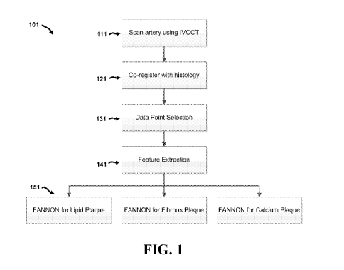
Referring now to FIG. 1, an overview of an exemplary method 101 comprises
various
steps performed to classify different types of tissue observed in
intravascular optical coherence
tomography images. An outline of exemplary methods and systems will be
presented initially,
followed by more detailed discussion of specific features and elements. As
shown in the
exemplary embodiment of FIG. 1, method 101 comprises a first step 111 of
scanning an artery
to obtain intravascular optical coherence tomography images. An example of one
such image
201 is provided in FIG. 2. In image 201, a catheter 211 with a light source
221 is used to image
arterial tissue 231. It is understood that not all components of catheter 211
are labeled in FIG.
2 for purposes of clarity. As previously noted, in particular embodiments such
images may by
obtained using optical coherence tomography systems and methods as disclosed
in U.S. Patent
Publications 2014/0268168 and 2016/0078309, incorporated by reference herein.
In the embodiment disclosed in FIG. 1, method 101 then co-registers image data
with
histological data in step 121, as described in more detail below. Image data
point selection is
performed in step 131, followed by feature extraction in step 141, also
further discussed below.
Feature and Node Optimized Neural Networks (FANONN) for different types of
tissue (e.g.
lipid plaque, fibrous plaques, and calcific plaques) can then be used to
classify the tissue in the
image in step 151. In exemplary embodiments, image data point selection may be
manually
selected by a user (e.g. "point-and-click" selection), or via sampling from
regions of interest in
B-scans of the tissue. It is understood that the classification techniques
disclosed herein may
be applied to other tissue types (including non-diseased tissues) as well.
Referring now to FIG. 3 graphs are provided for feature selection and network
architecture optimization for fibrous-optimized and lipid-optimized features
and architecture.
As shown in the graph on the left side of FIG. 3, sensitivity and specificity
is highest for a
neural network with features selected to optimize for classification of a
specific tissue. Lipid
sensitivity and specificity is worse with a network running on features
optimized for fibrous
plaque. Likewise, fibrous sensitivity and specificity is worse with a network
running on
features optimized for lipid plaque.
The graph on the right side of FIG. 3 illustrates that sensitivity and
specificity is highest
for a neural network with number of nodes (e.g. neurons) optimized for
classification of a
specific tissue type. Lipid sensitivity and specificity is worse with a
network with number of
- 13 -
CA 03026650 2018-12-05
WO 2017/214421
PCT/US2017/036587
nodes optimized for fibrous plaque. Likewise, fibrous tissue sensitivity and
specificity is worse
with a network with number of nodes optimized for lipid tissue. Accordingly,
tissue
classification is dependent on both the specific features used and the number
of nodes
comprising the network.
As previously mentioned, exemplary embodiments co-register intravascular OCT
image data with histological data. In one example, IVOCT imaging was conducted
on 10
human hearts (from 3 women and 7 men) collected within 24 hours of death. The
age at death
was 65 11
years. Imaging was conducted on 14 coronary arteries (n=10, left anterior
descending artery [LAD]; n=4 right coronary artery [RCA]). From these artery
scans, image
data points were extracted.
IVOCT imaging was conducted using a 1310 nm swept source laser (HSL-1000,
Santec, Hackensack, NJ) with a bandwidth of 80 nm scanning, a repetition rate
of 34 kHz, and
a measured free-space axial resolution of 20 tim with a 2.8 mm scan depth. The
IVOCT signal
was sampled with a linear k-space clock to allow real-time OCT image
acquisition and display.
Per artery, 100 cross-section images (B-scans) were collected. A fluoroscopy
system (GE
Medical Systems) and a chamber designed to maintain the tissue at 37 C were
used. Left and
right coronary 6F guide catheters were sewn into the coronary ostia, 0.014
inch guide-wire
access to the coronary arteries was gained under fluoroscopic guidance, and a
stent was
deployed 80 mm from the guide catheter tip as a fiduciary marker. IVOCT
pullbacks were
acquired from the stent to the guide catheter (80 mm total pullback length).
The left anterior
descending (LAD) artery and right coronary artery (RCA) were imaged. Following
imaging,
the RCA and LAD were perfusion-fixed with formahn at 100 mmHg. Histology cross-
sections
were taken from the same 14 coronary arteries and 10 human hearts with 100
histology slices
at the same depth as 100 cross-section B-scans for each artery.
To conduct histology after IVOCT imaging, LADs and RCAs were perfusion-fixed
with 10% neutral-buffered formalin, excised from each heart, individually
radiographed on a
Faxitron MX-20 (Faxitron Bioptics LLC, Tucson AZ), and decalcified overnight
with Cal-Rite
(Richard Allen Scientific) if necessary. The arterial segments were sliced
into 2-3 mm thick
rings and further processed on a Tissue-Tek Vacuum Infiltration Processor
(Sakura Finetek
USA, Torrance, CA) for standard paraffin-embedded sections. An average of 25
rings were
generated from each artery. Serial tissue sections (5 lam thick) were cut at
150-iim intervals
and stained with hematoxylin and eosin (H&E), modified Movat's pentachrome,
and Von
- 14 -
CA 03026650 2018-12-05
WO 2017/214421
PCT/US2017/036587
Kossa. Anti-CD68 (Dako North America, Inc, Carpinteria, CA) and anti-a-smooth
muscle cell-
actin (Sigma-Aldrich, St. Louis, MO) antibodies were used in
immunohistochemical studies to
identify macrophages and smooth muscle cells, respectively.
In this embodiment, histology rings were then matched to respective IVOCT
frames.
Co-registration was performed between IVOCT images and histological sections
based on the
following: (1) two fiducial landmarks¨a stent deployed at the distal end of
the pullback and
the sewn-in guide catheter at the proximal edge¨that were visible in IVOCT
images,
fluoroscopy, and radiography before histopathological processing, and (2) the
physical position
of IVOCT images in the pullbacks measured against the estimated distance in
microns from
the fiducial landmarks in the tissue sections.
Classification was automated based on a series of quantifiable image features
acquired
using an IVOCT scan of the coronary artery. Extraction of image data for
classification of
plaque required reading specific quantitative measures from the images, known
as quantitative
features. The quantitative feature set was created using two-dimensional
windowed image
.. statistics along with Gray Level Co-Occurrence Matrix (GLCM) textural
features and are
explained herein.
In this embodiment, the two-dimensional windowed image statistics are
determined by
generating a square window around a pixel of interest and calculating the
following statistics:
(1) Mean Value
(2) Variance
(3) Skewness
(4) Kurtosis
(5) Energy
These measures are calculated for both the horizontal and vertical averages
within the
square window with both image intensity and attenuation data. The intensity is
defined as the
backscattered light from the tissue measured in decibels. The attenuation data
represents how
the backscattered light intensity decays as a function of radial distance from
the light source.
The GLCM is a method for texture analysis and characterization based on the
spatial
relationship between pixels. In this method, image texture is characterized by
determining the
frequency with which pairs of pixels with certain values and a pre-defined
spatial relationship
occur. In exemplary embodiments, specific GLCM textural features include:
- 15 -
CA 03026650 2018-12-05
WO 2017/214421
PCT/US2017/036587
(1) Contrast
(2) Energy
(3) Correlation
(4) Homogeneity
(5) Entropy
(6) Max Probability
Each of these textural features is again calculated with intensity and
attenuation. The
optimization process for the algorithm to classify each tissue type selects
from these windowed
and GLCM features. Additional discussion of GLCM can be found in Yang,
Xiaofeng, et al.
"Ultrasound GLCM texture analysis of radiation-induced parotid-gland injury in
head-and-
neck cancer radiotherapy: an in vivo study of late toxicity." Medical physics
39.9 (2012): 5732-
5739, incorporated by reference herein.
In exemplary embodiments, a classification technique uses an optimized neural
network
to classify plaque tissue from a set of images. A neural network has the
ability to sort a dataset
into many different classes. In the embodiment disclosed herein, three
different classes of tissue
types are identified: lipid, calcium, and fibrous plaque. It is undertood that
different
embodiments may include different classes of tissue types.
A set of quantitative image features is provided to the network as a basis for
judgment
and using these features, the neural network will make decisions as to what
class to sort a pixel
into.
There are several design considerations associated with the use of these
quantitative
features, however. First, the sensitivity and specificity of a neural network
can change based
on the features that are provided to it. All of the available features to be
inputted into the neural
network are called candidate features. For example, if one has 300 candidate
features to choose
from, it might be found that the neural network functions best with a specific
set of 150 of those
features instead of the full 300. In order to best classify data, the best
features should be selected
amongst a pool of candidates. Having either too few or too many features than
optimum can
be damaging to the resulting sensitivity and specificity of the method.
IVOCT expert imaging technicians typically use different features to classify
different
types of plaque. For example, when looking for fibrous plaque, imaging
technicians will
typically look for high backscattering and homogeneity whereas when searching
for calcium
- 16 -
CA 03026650 2018-12-05
WO 2017/214421
PCT/US2017/036587
plaque an expert might look at signal quality and delineation of tissue
borders. Accordingly,
it is not optimal to use a single network with a single set of features to
classify all types of
tissue.
As previously mentioned in the discussion of FIG. 3, a set of features that
work best for
.. sorting fibrous plaque will not be the best features to use for sorting
calcific plaque.
Furthermore, the number of nodes comprising the neural network affects its
performance with
a given set of features. The optimal set of features and network structure are
interdependent
because the inputted features affect the optimal distribution of weights
associated with the
connections between nodes in the network and this can have an impact on
sensitivity and
.. specificity. Therefore, in order to construct an optimized network, one
must optimize not only
the features selected to classify the tissue but also the structure of a
network based on the
features used.
Accordingly, exemplary embodiments of the present disclosure utilize a
multiple-pass,
co-optimized classification system for each tissue type. The method maximizes
the sensitivity
.. and specificity for each type of tissue. The classification system first
gathers the quantitative
image features associated with the IVOCT image data along with the truth data
from co-
registered histology slides of the tissue. Each type of tissue is handled
individually. In the
embodiment disclosed herein, a first network is optimized to detect fibrous
plaque, then another
network is optimized to detect calcific plaque, and a third network is
optimized to detect lipid
.. plaque. It is understood, that for additional tissue classes, additional
networks can be
constructed.
Referring now to FIG. 4, the feature and node optimized neural network
(FANNON)
optimization process 401 for each tissue begins by using each feature
individually to evaluate
the data with a neural network to sort each tissue type. The process begins by
initializing a
neural network with one node in step 411. The resulting sensitivity and
specificity of each
feature method is calculated using a receiver operating characteristic (ROC)
curve which plots
the true positive vs. false positive rates of the classifier. The greater the
area under the ROC
curve, the greater the sensitivity and specificity of the neural network based
on the feature. In
step 421, the features are all ranked according to the area under the ROC
curves of the neural
.. networks they serve as inputs to, from greatest sensitivity and specificity
to least sensitivity
and specificity.
- 17 -
CA 03026650 2018-12-05
WO 2017/214421 PCT/US2017/036587
After the rank features step in 421, the classification system uses an
increasing number
of features from the ranked feature list, starting from 1 to the number of
candidate features, and
records the sensitivity and specificity of each group of features in step 431.
This process is
repeated for a range of neural network architectures, varying the number of
nodes involved. In
step 441, the best combination of number of features and nodes used is
selected based on the
sum of sensitivity and specificity of the network to detect the specific type
of tissue involved.
The best network for each tissue type has a unique feature set and a unique
number of nodes
paired together, creating a Feature and Node Optimized Neural Network (FANONN)
that is
used to optimally classify each plaque type.
Results
The FANONN classification algorithm of exemplary embodiments has been
demonstrated to sort plaque tissue as fibrous, calcium, or lipid plaque as
verified by histology
analysis with sensitivities and specificities listed in the table below:
*Mile
Y.Pt* riiic
Accuracy ROt ()N. erlap Accuraw,::Accuracy
itrotig,95 SI 962
- -
Calm* 72 57 597
Oti'l 1.$941% ig)PM i*E
":1!
The data presented in the table above compares results using FANNON techniques
disclosed herein to studies in literature that attempt to automate the plaque
classification
process using IVOCT. The accuracy for each technique is the average of
sensitivity and
specificity, where the sensitivity is the proportion of the known plaque type
data points that the
algorithm correctly classifies and the specificity is the ratio of correct
classifications to total
classifications for a certain category of plaque.
Using accuracy as a reported metric, the direct comparison to current
literature studies
helps show the power and novelty of the techniques disclosed herein. It should
also be noted
- 18 -
CA 03026650 2018-12-05
WO 2017/214421
PCT/US2017/036587
that the typical current approaches [113, 1231 to automated plaque
classification are limited in
that they are not co-registered with histology, making their classification
ground truth weaker.
In addition to this primary classification ability, exemplary embodiments can
further
classify lipid lesions as the particularly high-risk TCFA type of lesion with
100% sensitivity
and 100% specificity. Taken with the classification of lipid plaque as the
limiting factor, the
algorithm can detect TCFA lesions with 94% accuracy.
Discussion and Conclusion
The described classification techniques and systems can characterize arterial
plaque
tissue in the coronary artery into fibrous, calcium, or lipid plaque without
any human input
better than other reported methods. Other groups have conducted similar
studies to automate
the characterization of coronary plaque with similar motivations but have not
had the same
degree of success. Specific groups in the field include Ughi et al. who have
achieved accuracies
of 89.5%, 72%, and 79.5%, and Athanasiou et al. who have achieved accuracies
of 81%, 87%,
and 71% accuracies in automated characterization of fibrous, calcium, and
lipid plaque,
respectively. The current leading studies by Ughi and Athanasiou use human
observers as their
ground truth which makes their classification technique inherently less
accurate. In contrast,
exemplary embodiment disclosed herein use histology as the ground truth for
training which
improves accuracy and stability.
Exemplary embodiments of the present disclosure achieve high accuracy through
not
only the use of histology as the reference truth but also through the
classification techniques
disclosed herein. Exemplary embodiments achieve improved results by treating
each
individual plaque type individually and allowing the creation of a tailored
neural network
structure to optimally classify each type. Such techniques provide for
improved results for each
plaque type and can be expanded to as many tissue types as desired.
The FANONN classification method disclosed herein not only classifies plaque
tissue
composition with high accuracy but can also provide risk analysis of the
tissue after
classification. Of the classified lipid plaque points in an artery, the
classification method can
identify plaque lesions as TCFA which are known to be indicative of unstable
plaque and lead
to a majority of acute coronary event such as plaque ruptures (Fujii et al,
2015). Such plaque
ruptures can occlude a blood vessel, leading to heart attack or stroke. Unlike
previous attempts
to classify TCFA lesions via IVUS imaging (Swada), the FANONN smart algorithm
paired
- 19 -
CA 03026650 2018-12-05
WO 2017/214421
PCT/US2017/036587
with the micron-level resolution of IVOCT has both the physical resolution and
machine
intelligence required to accurately classify these risk-prone plaques. This
ability of the
classification method makes it very powerful but also special in that no other
group in the world
can provide automated analysis with a higher degree of accuracy.
Referring now to FIGS. 5-8, and in particular FIG. 5, an exemplary embodiment
of an
optical coherence tomography system 500 is shown. System 500 can be used to
obtain images
of tissue for analysis and classification as described herein. In this
embodiment, system 500
comprises an optical coherence tomography light source 510, a splitter 515,
optical circulator
520, coupler 525 and balanced detector 530. Splitter 515 is configured to
direct light from
OCT light source 510 to a reference path 511 and a sample path 521. In the
embodiment
shown, sample path 521 is directed through patient interface module 502 and
catheter 501,
while reference path 511 is directed to a fiber reflector 512 via a photonic
crystal fiber 513.
A perspective view of patient interface module 502 is shown in FIG. 6, while a
schematic view of catheter 501 is shown in FIG. 7, and a partial section view
of the distal end
of catheter 501 is shown in FIG. 8. As shown in FIG. 7, catheter 501 comprises
a bead 535
and an optical connector 534 near proximal end 532. In this embodiment,
patient interface
module 502 is configured to control catheter 501 via a torque cable 509 (shown
in FIG. 8) that
transmits torque from patient interface module 502 to distal end 531 of
cathether 501,
In certain embodiments, patient interface module 502 can be configured to
provide 100
mm of linear stroke to catheter 501 at variable translation speeds up to 50 mm
per second in
two directions (e.g. push forward or pull back). In addition, patient
interface module 502 can
be configured to rotate an imaging port 533 at speeds up to 3,600 revolutions
per minute and
obtain 1,000 A-scans per rotation.
In certain embodiments, catheter 501 can be a sterile, single-use disposable
catheter
with a 3.2 F crossing profile and monorail design compatible with a 6F guide
catheter and a
0.014 inch guide wire. In particular embodiments, catheter 501 may comprise a
stationary
outer sheath 551 with an imaging port 557, a rotating and translating torque
cable 509 and
optics assembly 552. In specific embodiments, catheter 501 comprises an
optical fiber through
its length, with an optic assembly (e.g. a ferrule, gradient index [GRIN]
lens, and prism) near
imaging port 557 and distal end 531 of catheter 501. In addition, catheter 501
may comprise a
radiopaque marker 553 on the outer assembly near distal end 531, as well as a
radiopaque
- 20 -
CA 03026650 2018-12-05
WO 2017/214421
PCT/US2017/036587
marker 554 on the inner assembly near imaging port 557. Catheter 501 may
further comprise
a guidewire exit port 558 near distal end 531. It is understood that the
dimensions shown in
FIGS. 7 and 8 are merely exemplary, and that other embodiments may comprise
configurations
with dimensions different from those shown in this embodiment.
As previously mentioned, certain embodiments may incorporate optical coherence
tomography systems and methods as disclosed in U.S. Patent Publications
2014/0268168 and
2016/0078309 (incorporated by reference herein) to acquire images for
analysis. Referring
now to FIG. 9, one exemplary embodiment of such an apparatus 50 comprises an
optical
coherence tomography light source 100, a splitter 200, a short pulsed (e.g.
two-photon
luminescence) excitation light source 300, a first dichroic element 400 and a
second dichroic
element 450. It is understood that other embodiments may comprise an apparatus
with a
different combination of components or fewer components than those shown in
FIG. 9.
In this embodiment, optical coherence tomography light source 100 is
configured to
emit a first wavelength 110 and splitter 200 is configured to direct first
wavelength 110 to a
reference path 210 and a sample path 220. In certain embodiments, optical
coherence
tomography light source 100 can be configured as a swept source optical
coherence
tomography light source or a broadband optical coherence tomography light
source. In
particular embodiments, sample path 220 can be directed through a photonic
crystal fiber. In
the embodiment shown, two-photon luminescence excitation light source 300 is
configured to
emit a second wavelength 320.
During operation, apparatus 50 can be positioned such that sample path 220 and
second
wavelength 320 are directed to a sample site 280 (e.g. via first dichroic
element 400 as well as
other components in FIG. 9).
In certain exemplary embodiments, sample site 280 may comprise nanoparticles
260
and in specific embodiments, nanoparticles 260 may be configured as nanorods.
In particular
embodiments, nanoparticles 260 may be configured as nanorods comprising gold
with a surface
plasmon resonance of approximately 756 nm. In certain embodiments, the
configuration of the
nanorods can be selected according to the procedures established in the
Example Section 4
provided below.
Apparatus 50 further comprises a photon counting detector 350 configured to
detect
two-photon luminescence (TPL) and a balanced detector 250 configured to
minimize a non-
- 21 -
CA 03026650 2018-12-05
WO 2017/214421
PCT/US2017/036587
interfering OCT component. In specific embodiments, photon counting detector
350 can be
configured as one or more photomultiplier tubes (PMTs). In other embodiments,
photon
counting detector 350 can be configured as an avalanche photo diode.
In a particular embodiments, components of the system illustrated in FIG. 9
can be
incorporated into a catheter-based system that utilizes a photonic crystal
fiber (PCF) to enable
the propagation of light in sample path 220 and second wavelength 320 from TPL
excitation
light source 300 to sample site 280. The PCF allows single-mode transmission
of both OCT
and TPL excitation light. Single-mode transmission is required in OCT imaging
to insure the
modal interference does not occur. Single mode transmission is required for
TPL imaging to
insure the pulse duration of TPL excitation light is not broadened due to
modal dispersion. In
specific embodiments the catheter can be inserted into a blood vessel to
obtain intravascular
images utilizing system 50.
During operation, system 50 provides the benefits of both OCT and TPL imaging
technologies in a single system. In exemplary embodiments, the components of
system 50
function according to established principles in OCT and TPL fields.
Accordingly, while an
overview of the individual OCT and TPL will be provided, it is understood that
exemplary
embodiments may utilize various combinations of parameters according to
environmental
conditions or other factors. For example, OCT light source 100 can produce
near-infrared
light, and the use of relatively long wavelength light allows deeper
penetration into the
scattering medium such as an arterial wall. In a particular embodiment OCT
light source 100
can be configured to provide light at a wavelength of approximately 1310 nm.
As light in sample path 220 is directed at sample site 280, a small portion of
this light
that reflects from sub-surface features of sample site 280 is collected.
During operation, a
significant portion of light in sample path 220 is not reflected but, rather,
backscatters from the
sample. Although backscattered light contributes background that obscures an
image in
conventional imaging, this light can be used beneficially in OCT systems via
interferometry.
For example, balanced detector 250 can be used to record the optical path
length of received
photons, allowing rejection of most photons that multiply scatter in the
tissue before detection.
This can allow recording three-dimensional images of thick samples to be
constructed by
rejecting background signal while collecting light directly reflected from
regions of interest in
sample site 280. In exemplary embodiments, OCT imaging is generally limited to
one to two
- 22 -
CA 03026650 2018-12-05
WO 2017/214421
PCT/US2017/036587
millimeters below the surface in biological tissue in sample site 280. At
greater depths, the
proportion of light that escapes without scattering is typically too small for
detection.
During operation of system 50, TPL light source 300 and photon counting
detector 350
are also utilized consistent with established principles in two-photon
luminescence microscopy.
In certain embodiments, TPL light source 300 can be configured as a tunable
femtosecond laser
producing excitation energy of second wavelength 320 at 760-1040 nm with a
maximum pulse
energy of 6 nJ-5 0, a pulse width of 100 fs-1 ps, and a repetition rate of 500
kHz-80 MHz. In
particular embodiments, TPL light source 300 may also be configured to produce
a spot size
of 10-30 [tm with a spot area of approximately 78-706.8 p..m2 and a pixel
dwell time of 20 [Is.
In addition, TPL light source 300 may also be configured to produce 10-1600
pulses per pixel,
with an average power on sample of 500-2500 mW, an instantaneous power of
0.0625-5 MW
and an instantaneous power density of 2E-4-16E-3 MW/ m2.
In the embodiment shown in FIG. 5, first dichroic element 400 can be
positioned to
direct second wavelength 320 to sample site 280 via a photonic crystal fiber
(PCF). In
particular embodiments, the PCF can have a large sized mode field diameter (20
vm) (LMA-
20) available from NKT Photonics. In certain embodiments, the PCF may be
configured as a
double-clad fiber, and in specific embodiments, may be a double-clad high NA
fiber such as a
model number DC-165-16-Passive Fiber available from Crystal Fibre. Exemplary
double-clad
photonic crystal fibers may comprise a large-mode area, single-mode core
embedded in a high-
NA multimode fiber structure. Such fibers can allow a single-mode beam to be
propagated
forward in the fiber and at the same time scattered light or two-photon
luminescence may be
collected and propagated backwards for detection. The use of a double-clad
fiber instead of a
single-clad photonic crystal fiber can increase the two-photon luminescence
detection
efficiency with a high-NA inner cladding (compared to the low-NA core). It is
understood that
the particular specifications of components are presented for purposes of
example only, and
that other embodiments may comprise components with different specifications
than those
described herein.
During operation of system 50, second wavelength 320 can provide excitation
energy
to nanoparticles 260, which can emit luminescence 270 that is directed to
photon counting
detector 350 via second dichroic element 450. In exemplary embodiments, the
outputs from
the photon counting detector 350 and balanced detector 250 can be configured
to be combined
- 23 -
CA 03026650 2018-12-05
WO 2017/214421
PCT/US2017/036587
in a single display that allows a user to visualize the results of both OCT
and TPL imaging
ov erl ay ed.
* * * * * * * * * * * * * * *
All of the devices, systems and/or methods disclosed and claimed herein can be
made
and executed without undue experimentation in light of the present disclosure.
While the
devices, systems and methods of this invention have been described in terms of
particular
embodiments, it will be apparent to those of skill in the art that variations
may be applied to
the devices, systems and/or methods in the steps or in the sequence of steps
of the method
described herein without departing from the concept, spirit and scope of the
invention. All
such similar substitutes and modifications apparent to those skilled in the
art are deemed to be
within the spirit, scope and concept of the invention as defined by the
appended claims.
- 24 -
CA 03026650 2018-12-05
WO 2017/214421
PCT/US2017/036587
REFERENCES:
The contents of the following references are incorporated by reference herein:
[1] Yusuf S, Reddy S. Ounpuu S, Anand S, "Global burden of cardiovascular
diseases: part I:
general considerations, the epidemiologic transition, risk factors, and impact
of
urbanization," Circulation 104, 2746-2753 (2001)
[2] Libby P, Ridker PM, Maseri A, "Inflammation and Atherosclerosis,"
Circulation
105,1135-1143 (2002)
[3] Libby P, Theroux P, "Pathophysiology of coronary artery disease,"
Circulation 111, 3481-
8 (2005)
[4] Lucas AR, Korol R, Pepine CJ, "Inflammation in atherosclerosis: some
thoughts about
acute coronary syndromes," Circulation 113, e728-732 (2006)
[5] Virmani R, Burke AP, Kolodgie FD, Farb A, "Pathology of the Thin-Cap
Fibroatheroma:
A Type of Vulnerable Plaque," J Intery Cardiol 16(3), 267-272 (2003)
[6] Davies MJ, Richardson PD, Woolf N, Katz DR, Mann J, "Risk of thrombosis
in human
atherosclerotic plaques: role of extracellular lipid, macrophage, and smooth
muscle cell
content," Br Heart J 69, 377-381 (1993)
[7] Stary HC, Chandler AB, Dinsmore RE, "A definition of advanced types of
atherosclerotic
lesions and a histological classification of atherosclerosis: a report from
the Committee on
Vascular Lesions of the Council on Arteriosclerosis," Circulation 92, 1355-
1374 (1995)
[8] Jonasson L, Holm J, Skalli 0, Bondjers G, Hansson GK, "Regional
accumulations of T
cells, macrophages, and smooth muscle cells in the human atherosclerotic
plaque,"
Arteriosclerosis 6, 131-138 (1986)
[9] Johnson JL, George SJ, Newby AC, Jackson CL, "Divergent effects of matrix
metalloproteinases 3, 7, 9, and 12 on atherosclerotic plaque stability in
mouse
brachiocephalic arteries," Proc Natl Acad Sci 102, 15575-15580 (2005)
[10] Henney AM, Wakeley PR, Davies MJ, Foster K, Hembry R, Murphy G, Humphries
S,
"Localization of stromelysin gene expression in atherosclerotic plaques by in
situ
hybridization," Proc Natl Acad Sci 88, 8154-8158 (1991)
[11] Galis ZS, Sukhova GK, Lark MW, Libby P, "Increased expression of matrix
metalloproteinases and matrix degrading activity in vulnerable regions of
human
atherosclerotic plaques," J Clin Invest 94, 2493-2503 (1994)
- 25 -
CA 03026650 2018-12-05
WO 2017/214421
PCT/US2017/036587
[12] Nikkari ST, O'Brien KD, Ferguson M, Hatsukami T, Welgus HG, Alpers CE,
Clowes
AW, "Interstitial collagenase (MMP-1) expression in human carotid
atherosclerosis,"
Circulation 92,1393-1398 (1995)
[13] Libby P, Geng YJ, Aikawa M, Schoenbeck U, Mach F, Clinton SK, Sukhova GK,
Lee,
RT, "Macrophages and atherosclerotic plaque stability," Curr Opin Lipidol 7,
330-335
(1996)
[14] Taubman MB, Fallon JT, Schecter AD, Giesen P, Mendlowitz M, Fyfe BS,
Marmur JD,
Nemerson Y, "Tissue factor in the pathogenesis of atherosclerosis," Thromb
Haemost 78,
200-204 (1997)
[15] Kolodgie FD, Virmani R, Burke AP, Farb A, Weber DK, Kutys R, Finn AV,
Gold HK,
"Pathologic assessment of the vulnerable human coronary plaque," Heart 90,
1385-1391
(2004)
[16] van Zandvoort M, Engels W, Douma K, Beckers L, Oude Egbrink M, Daemen M,
Slaaf
DW, "Two-photon microscopy for imaging of the (atherosclerotic) vascular wall:
a proof
of concept study," J Vasc Res 41, 54-63 (2004)
[17] Zoumi A, Lu XA, Kassab GS, Tromberg BJ, "Imaging coronary artery
microstructure
using secondharmonic and two-photon fluorescence microscopy," Biophys J 87,
2778-
2786 (2004)
[18] Boulesteix T, Pena AM, Pages N, Godeau G, Sauviat MP, Beaurepaire E,
Schanne-Klein
MC, "Micrometer scale ex vivo multiphoton imaging of unstained arterial wall
structure,"
Cytometry Part A 69A, 20-26 (2006)
[19] Le TT, Langohr IM, Locker MJ, Sturek M, Cheng JX, "Label-free molecular
imaging of
atherosclerotic lesions using multimodal nonlinear optical microscopy," J
Biomed Opt
12(5), 0540071-05400710 (2007)
[20] Lilledahl MB, Haugen OA, de Lange Davies C, Svaasand LO,
"Characterization of
vulnerable plaques by multiphoton microscopy," J Biomed Opt 12(4), 0440051-
04400512
(2007)
[21] Wang T , Mancuso JJ, Sapozhnikova V, Dwelle J, Ma LL, Willsey B, Kazmi
SM, Qiu J,
Li X, Asmis R, Johnston KP, Feldman MD, Milner TE, "Dual-wavelength multi-
frequency photothermal wave imaging combined with OCT for macrophage and lipid
detection in atherosclerotic plaques", J Biomed Opt 17(3), 0360091-03600910
(2012)
[22] Wang T, Mancuso JJ, Kazmi SM, Dwelle J, Sapozhnikova V. Willsey B, Ma LL,
Qiu J,
Li X, Dunn AK, Johnston KP, Feldman MD, Milner TE, "Combined two-photon
- 26 -
CA 03026650 2018-12-05
WO 2017/214421
PCT/US2017/036587
luminescence microscopy and OCT for macrophage detection in the
hypercholesterolemic
rabbit aorta using plasmonic gold nanorose", Lasers Surg Med 44(1), 49-59
(2012)
[23] Xue P, Fujimoto JG, "Ultrahigh resolution optical coherence tomography
with
femtosecond Ti:sapphire laser and photonic crystal fiber," Chinese Science
Bulletin
53(13), 1963-1966 (2008)
[24] Ryu SY, Choi HY, Na JH, Choi ES, Yang GH, Lee BH, "Optical coherence
comography
implemented by photonic crystal fiber," Opt Quant Electron 37(13-15), 1191-
1198 (2005)
[25] Fu L, Gu M, "Double-clad photonic crystal fiber coupler for compact
nonlinear optical
microscopy imaging," Opt Lett 31, 1471-1473 (2006)
[26] Liu G, Kieu K, Wise FW, Chen Z, "Multiphoton microscopy system with a
compact fiber-
based femtosecond-pulse laser and handheld probe," J Biophoton 4, 34-39
(2011).
[27] Fu L, Jain A, Xie H, Cranfield C, Gu M, "Nonlinear optical endoscopy
based on a double-
clad photonic crystal fiber and a MEMS mirror," Opt Exp 14, 1027-1032 (2006)
[28] Wu Y, Xi J, Cobb MJ, Li X, "Scanning fiber-optic nonlinear endomicroscopy
with
miniature aspherical compound lens and multimode fiber collector," Opt Left
34, 953-955
(2009)
[29] Kim, EH, Dave, DP, Milner, TE. "Fiber-optic spectral polarimeter using a
broadband swept
laser source," Optics Communications, 249 351-356 (2005)
[30] Park J, Estrada A, Sharp K, Sang K, Schwartz JA, Smith DK, Coleman C,
Payne JD,
Korgel BA, Dunn AK, Tunnell JW, "Two-photon-induced photoluminescence imaging
of
tumors using near-infrared excited gold nanoshells," Opt Exp 16(3), 1590-1599
(2008)
[31] Available at http: //s al es . hamamatsu. com/assets/pdf/parts_H/m-
h7422e.pdf
[32] V. L. Roger, A. S. Go, D. M. Lloyd-Jone, R. J. Adams, J. D. Berry, T. M.
Brown, M. R.
Carnethon, S. Dai, G. de Simone, E. S. Ford, C. S Fox, H. J. Fullerton, C.
Gillespie, K. J.
Greenlund, S. M. Hailpern, J. A. Heit, P. M .Ho, V. J. Howard, B. M. Kissela,
S. J. Kittner,
D. T. Lackland, J. H. Lichtman, L. D. Lisabeth, D. M. Makuc, G. M. Marcus, A.
Marelli,
D. B. Matchar, M. M. McDermott, J. B. Meigs, C. S. Moy, D. Mozaffarian, M. E.
Mussolino, G. Nichol, N. P. Paynter, W. D. Rosamond, P. D. Sorlie, R. S.
Stafford, T. N.
Turan, M. B. Turner, N. D. Wong and J. Wylie-Rosett, "Heart disease and stroke
statistics
- 2011 update: a report from the American Heart Association," Circulation
123(4), e18-
e209 (2011).
[33] E. Falk, P. K. Shah and V. Fuster, "Coronary plaque disruption,"
Circulation 92(3), 657-
671 (1995).
[34] F. D. Kolodgie, R. Virmani, A. P. Burke, A. Farb, D. K. Weber, R. Kutys,
A. V. Finn and
- 27 -
CA 03026650 2018-12-05
WO 2017/214421
PCT/US2017/036587
H. K. Gold, "Pathologic assessment of the vulnerable human coronary plaque,"
Heart
90(12), 1385-1391 (2004).
[35] N. B. Hao, M. H. Lu, Y. H. Fan, Y. L Cao, Z. R. Zhang, and S. M. Yang,
"Macrophages in
tumor microenvironments and the progression of tumors," Clin. Dev. Immunol.
2012,
948098-948108 (2012).
[36] B. Ruffell, N. I. Affara, and L. M. Coussens. "Differential macrophage
programming in the
tumor microenvironment," Trends Immunol. 33(3), 119-126 (2012).
[37] R. Shukla, V. Bansal, M. Chaudhary, A. Basu, R. R. Bhonde, and M. Sastry,
"Biocompatibility of gold nanoparticles and their endocytotic fate inside the
cellular
compartment: a microscopic overview," Langmuir 21(23), 10644-10654 (2005).
[38] M. M. Janat-Amsbury, A. Ray, C. M. Peterson, and H. Ghandehari, "Geometry
and surface
characteristics of gold nanoparticles influence their biodistribution and
uptake by
macrophages," Eur. J. Pharm. Biopharm. 77(3), 417-423 (2011).
[39] S. Lal, S. E.Clare, and N. J. Halas, "Nanoshell-enabled photothermal
cancer therapy:
impending clinical impact," Acc. Chem. Res. 41(12), 1842-1851 (2008).
[40] X. Ji, R. Shao, A. M. Elliott, R. J. Stafford, E. Esparza-Coss, G. Liang,
X. P. Luo, K. Park,
J. T. Markert, and C. Li, "Bifunctional Gold Nanoshells with a
Superparamagnetic Iron
Oxide-Silica Core Suitable for Both MR Imaging and Photothermal Therapy," J.
Phys.
Chem. C 111(17), 6245-6251 (2007).
[41] S. E. Skrabalak, L. Au, X. Lu, X. Li, and Y. Xia, "Gold nanocages for
cancer detection and
treatment," Nanomedicine (Lond) 2(5), 657-668 (2007).
[42] M. Longmire, P. L. Choyke, and H. Kobayashi, "Clearance properties of
nano-sized
particles and molecules as imaging agents: considerations and caveats,"
Nanomedicine
(Lond) 3(5), 703-717 (2008).
[43] L. L. Ma, M. D. Feldman , J. M. Tam, A. S. Paranjape, K. K. Cheruku, T.
A. Larson, J. 0.
Tam, D. R. Ingram, V. Paramita, J. W. Villard, J. T. Jenkins, T. Wang, G. D.
Clarke, R.
Asmis, K. Sokolov, B. Chandrasekar, T. E. Milner, and K. P. Johnston, "Small
multifunctional nanoclusters (nanoroses) for targeted cellular imaging and
therapy," ACS
Nano 3(9), 2686-2696 (2009).
[44] T. Wang, J. J. Mancuso, S. M. Kazmi, J. Dwelle, V. Sapozhnikova, B.
Willsey, L. L. Ma,
J. Qiu, X. Li, A. K. Dunn, K. P. Johnston, M. D. Feldman, and T. E. Milner,
"Combined
two-photon luminescence microscopy and OCT for macrophage detection in the
hypercholesterolemic rabbit aorta using plasmonic gold nanorose," Lasers Surg.
Med.
44(1), 49-59 (2012).
- 28 -
CA 03026650 2018-12-05
WO 2017/214421
PCT/US2017/036587
[45] T. S. Hauck, A. A. Ghazani, and W. C. W. Chan, "Assessing the effect of
surface chemistry
on gold nanorod uptake, toxicity, and gene expression in mammalian cells,"
Small 4(1),
153-159 (2008).
[46] T. Niidome, M. Yamagata, Y. Okamoto, Y. Akiyama, H. Takahashi, T. Kawano,
Y.
Katayama, and Y. Niidome, "PEG-modified gold nanorods with a stealth character
for in
vivo applications," J. Control Release 114(3), 343-347 (2006).
[47] A. Mooradian, "Photoluminescence of metals," Phys. Rev. Left. 22(5), 185-
187 (1969).
[48] J. Zheng, C. Zhang, and R. M. Dickson, "Highly fluorescent, water-
soluble, size-tunable
gold quantum dots," Phys. Rev. Lett. 93(7), 077402-077405 (2004).
[49] G. Wang, T. Huang, R. W. Murray, L. Menard, and R. G. Nuzzo, "Near-IR
luminescence
of monolayer-protected metal clusters," J. Am. Chem. Soc. 127(3), 812-813
(2005).
[50]J. P. Wilcoxon, J. E.Martin, F. Parsapour, B. Wiedenman, and D. F. Kelley,
"Photoluminescence from nanosize gold clusters," J. Chem. Phys. 108(21), 9137-
9143
(1998).
[51] Y. Fang, W. Chang, B. Willingham, P. Swanglap, S. Dominguez-Medina, and
S. Link,
"Plasmon emission quantum yield of single gold nanorods as a function of
aspect ratio,"
ACS Nano 6(8), 7177-7184 (2012).
[52] P. K. Jain, X. Huang, I. H. El-Sayed, and M. A. El-Sayed, "Review of some
interesting
surface plasmon resonance-enhanced properties of noble metal nanoparticles and
their
applications to biosystems," Plasmonics 2(3), 107-118 (2007).
[53] M. A. El-Sayed, "Some interesting properties of metals confined in time
and nanometer
space of different shapes," Acc. Chem. Res. 34(4), 257-264 (2001).
[54] C. Sonnichsen, T. Franzl, T. Wilk, G. von Plessen, J. Feldmann, 0.
Wilson, and P.
Mulvaney, "Drastic reduction of plasmon damping in gold nanorods," Phys. Rev.
Lett. 88,
077402-077405 (2002).
[55] M. B. Mohamed, V. Volkov, S. Link, and M. A. El-Sayed, "The 'lightning'
gold nanorods:
fluorescence enhancement of over a million compared to the gold metal," Chem.
Phys. Left.
317(6), 517-523 (2000).
[56] S. Link, M. B. Mohamed, and M. A. El-Sayed, "Simulation of the optical
absorption spectra
of gold nanorods as a function of their aspect ratio and the effect of the
medium dielectric
constant," J. Phys. Chem. B 106(16), 3073-3077 (1999).
[57] S. S. Verma and J. S. Sekhon, "Influence of aspect ratio and surrounding
medium on
localized surface plasmon resonance (LSPR) of gold nanorod," J. Optics 41(2),
89-93
(2012).
- 29 -
CA 03026650 2018-12-05
WO 2017/214421
PCT/US2017/036587
[58] P. K. Jain, X. Huang, I. H. El-Sayed and M. A. El-Sayed, "Noble metals on
the nanoscale:
optical and photothermal properties and some applications in imaging, sensing,
biology,
and medicine," Acc. Chem. Res. 41(12), 1578-1586 (2008).
[59] E. T. Castellano, R. C. Gamez , M. E. Gomez, and D. H. Russell,
"Longitudinal surface
plasmon resonance based gold nanorod biosensors for mass spectrometry,"
Langmuir
26(8), 6066-6070 (2010).
[60] H. Wang, T. B. Huff, D. A. Zweifel, W. He, P. S. Low, A. Wei, and J. X.
Cheng, "In vitro
and in vivo two-photon luminescence imaging of single gold nanorods," Proc.
Natl. Acad.
Sci. USA 102(44), 15752-15756 (2005).
[61] L. Tong, Q. Wei, A. Wei, and J. X. Cheng, "Gold nanorods as contrast
agents for biological
imaging: optical properties, surface conjugation and photothermal effects,"
Photochem.
Photobiol. 85(1), 21-32 (2009).
[62] T. Y. Ohulchanskyy, I. Roy, K. T. Yong, H. E. Pudavar, and R. N. Prasad,
"High-resolution
light microscopy using luminescent nanoparticles," WIREs Nanomed.
Nanobiotechnol.
2(2), 162-175 (2010).
[63] D. Nagesha, G. S. Laevsky, P. Lampton, R. Banyal, C. Warner, C. DiMarzio,
and S.
Sridhar, "In vitro imaging of embryonic stem cells using multiphoton
luminescence of gold
nanoparticles," Int. J. Nanomedicine 2(4), 813-819 (2007).
[64] Y. Zhang, J. Yu, D. J. S. Birch, and Y. Chen, "Gold nanorods for
fluorescence lifetime
imaging in biology," J. Biomed. Opt. 15(2), 0205041-0205043 (2010).
[65] C. L. Chen, L. R. Kuo, C. L. Chang, Y. K. Hwu, C. K. Huang, S. Y. Lee, K.
Chen, S. J.
Lin, J. D. Huang, and Y. Y. Chen, "In situ real-time investigation of cancer
cell
photothermolysis mediated by excited gold nanorod surface plasmons,"
Biomaterials
31(14), 4104-4112 (2010).
[66] H. Okamoto and K. Imura, "Near-field imaging of optical field and plasmon
wavefunctions
in metal nanoparticles," J. Mater. Chem. 16(40), 3920-3928 (2006).
[67] K. Imura, T. Nagahara, and H. Okamoto, "Near-field two-photon-induced
photoluminescence from single gold nanorods and imaging of plasmon modes," J.
Phys.
Chem. B 109(27), 13214-13220 (2005).
[681W. H. Ni, X. S. Kou, Z. Yang, and J. F. Wang, "Tailoring longitudinal
surface plasmon
wavelengths, scattering and absorption cross sections of gold nanorods," ACS
Nano 2(4),
677-686 (2008).
[69] C. Xu and W. W. Webb, "Measurement of two-photon excitation cross
sections of
molecular fluorophores with data from 690 to 1050 nm," JOSA B 13(3), 481-491
(1996).
- 30 -
CA 03026650 2018-12-05
WO 2017/214421
PCT/US2017/036587
[70] R. Gans, "Form of ultramicroscopic particles of silver," Ann. Phys.
47(10), 270-284 (1915).
[71] M. A. Albota, C. Xu, and W. W. Webb, "Two-photon fluorescence excitation
cross sections
of biomolecular probes from 690 to 960 nm," Appl. Opt. 37(31), 7352-7356
(1998).
[72] G.T. Boyd, Z. H. Yu, and Y. R. Shen, "Photoinduced luminescence from the
noble metals
and its enhancement on roughened surfaces," Phys. Rev. B 33(12), 7923-7936
(1986).
[73] S. Eustis and M. A. El-Sayed, "Aspect ratio dependence of the enhanced
fluorescence
intensity of gold nanorods: experimental and simulation study," J. Phys. Chem.
B 109(34),
16350-16356 (2005).
[74] M. Guerrisi and R. Rosei, "Splitting of the interband absorption edge in
Au", Phys. Rev. B
12(2), 557-563 (1975).
[75] X. Huang, S. Neretina, and M. A. El-Sayed, "Gold nanorods: from synthesis
and properties
to biological and biomedical applications," Adv. Mater. 21(48), 4880-4910
(2009).
[76] K. S. Lee and M. A. El-Sayed, "Dependence of the enhanced optical
scattering efficiency
relative to that of absorption for gold metal nanorods on aspect ratio, size,
end-cap shape
and medium refractive index," J. Phys. Chem. B 109(43), 20331-20338 (2005).
[77] C. SOnnichsen and A. P. Alivisatos, "Gold nanorods as novel nonbleaching
plasmon-based
orientation sensors for polarized single-particle microscopy," Nano Left.
5(2), 301-304
(2005).
[78] S. Link, C. Burda, B. Nikoobakht, and M. A. El-Sayed, "Laser-induced
shape changes of
colloidal gold nanorods using femtosecond and nanosecond laser pulses" J.
Phys. Chem. B
104(26), 6152-6163 (2000).
[79] A. Bouhelier, R. Bachelot, G. Lerondel, S. Kostcheev, P. Royer, G. P.
Wiederrecht,
"Surface plasmon characteristics of tunable photoluminescence in single gold
nanorods,"
Phys. Rev. Lett. 95(26), 2674051-2674054 (2005).
[80] R. E. Hummel, Electronic Properties of Materials, 37-61, 4th ed.
(Springer, New York,
2011).
[81] R. Rosei, and P. Winsemius, "Splitting of the interband absorption edge
in Au," Phys. Rev.
B 12(2), 557-563 (1975).
[82] Tearney GJ, Regar E, Akasaka T et al. Consensus standards for
acquisition, measurement,
and reporting of intravascular optical coherence tomography studies: a report
from the
International Working Group for Intravascular Optical Coherence Tomography
Standardization and Validation. J Am Coll Cardiol 2012;59:1058-72.
[83] Tearney GJ, Yabushita H, Houser SL et al. Quantification of macrophage
content in
atherosclerotic plaques by optical coherence tomography. Circulation
2003;107:113-9.
-31 -
CA 03026650 2018-12-05
WO 2017/214421
PCT/US2017/036587
[84] MacNeill BD, Jang IK, Bouma BE et al. Focal and multi-focal plaque
macrophage
distributions in patients with acute and stable presentations of coronary
artery disease. J
Am Coll Cardiol 2004;44:972-9.
[85] Tahara S, Morooka T, Wang Z et al. Intravascular optical coherence
tomography
detection of atherosclerosis and inflammation in murine aorta. Arterioscler
Thromb Vasc
Biol 2012;32:1150-7.
[86] Raffel OC, Tearney GJ, Gauthier DD, Halpern EF, Bouma BE, Jang IK.
Relationship
between a systemic inflammatory marker, plaque inflammation, and plaque
characteristics determined by intravascular optical coherence tomography.
Arterioscler
Thromb Vasc Biol 2007;27:1820-7.
[87] Raffel OC, Merchant FM, Tearney GJ et al. In vivo association between
positive coronary
artery remodelling and coronary plaque characteristics assessed by
intravascular optical
coherence tomography. Eur Heart J 2008;29:1721-8.
[88] Chia S, Raffel OC, Takano M, Tearney GJ, Bouma BE, Jong IK. Comparison of
coronary
plaque characteristics between diabetic and non-diabetic subjects: An in vivo
optical
coherence tomography study. Diabetes Res Clin Pract 2008;81:155-60.
[89] Tanaka A, Tearney GJ, Bouma BE. Challenges on the frontier of
intracoronary imaging:
atherosclerotic plaque macrophage measurement by optical coherence tomography.
J
Biomed Opt 2010;15:011104.
[90] Ali ZA, Roleder T, Narula J et al. Increased thin-cap neoatheroma and
periprocedural
myocardial infarction in drug-eluting stent restenosis: multimodality
intravascular
imaging of drug-eluting and bare-metal stents. Circ Cardiovascular Intery
2013;6:507-17.
[91] Cilingiroglu M, Oh JH, Sugunan B et al. Detection of vulnerable plaque in
a murine
model of atherosclerosis with optical coherence tomography. Catheter
Cardiovasc Intery
2006;67:915-23.
[92] Virmani R, Kolodgie FD, Burke AP, Farb A, Schwartz SM. Lessons from
sudden
coronary death - A comprehensive morphological classification scheme for
atherosclerotic lesions. Arterioscler Thromb Vasc Biol 2000;20:1262-1275.
[93] Bornstein P, Sage H. Structurally distinct collagen types. Annu Rev
Biochem
1980;49:957-1003.
[94] Falk E, Nakano M, Bentzon JF, Finn AV, Virmani R. Update on acute
coronary
syndromes: the pathologists' view. Eur Heart J 2013;34:719-28.
- 32 -
CA 03026650 2018-12-05
WO 2017/214421
PCT/US2017/036587
[95] Tavakoli S, Zamora D, Ullevig S, Asmis R. Bioenergetic profiles diverge
during
macrophage polarization: implications for the interpretation of "F-FDG PET
imaging of
atherosclerosis. J Nucl Med 2013;54:1661-7.
[96] Mourant JR, Freyer JP, Hielscher AH, Eick AA, Shen D, Johnson TM.
Mechanisms of
light scattering from biological cells relevant to noninvasive optical-tissue
diagnostics.
Appl Opt 1998;37:3586-3593.
[97] van der Meer FJ, Faber DJ, Baraznji Sassoon DM, Aalders MC, Pasterkamp G,
van
Leeuwen TG. Localized measurement of optical attenuation coefficients of
atherosclerotic plaque constituents by quantitative optical coherence
tomography. IEEE
Trans Med Imaging 2005;24:1369-76.
[98] Wang T, Mancuso JJ, Kazmi SM et al. Combined two-photon luminescence
microscopy
and OCT for macrophage detection in the hypercholesterolemic rabbit aorta
using
plasmonic gold nanorose. Lasers Surg Med 2012;44:49-59.
[99] Phipps JE, Sun Y, Saroufeem R, Hatami N, Fishbein MC, Marcu L.
Fluorescence lifetime
imaging for the characterization of the biochemical composition of
atherosclerotic
plaques. J Biomed Opt 2011;16:096018.
[100] van Soest G, Regar E, Goderie TP et al. Pitfalls in plaque
characterization by OCT:
image artifacts in native coronary arteries. J Am Coll Cardiol Img 2011;4:810-
3.
[101] Nadra I, Mason JC, Philippidis P et al. Proinflammatory activation of
macrophages by
basic calcium phosphate crystals via protein kinase C and MAP kinase pathways:
a
vicious cycle of inflammation and arterial calcification? Circ Res
2005;96:1248-56.
[102] Rajamaki K, Lappalainen J, Oorni K et al. Cholesterol crystals activate
the NLRP3
inflammasome in human macrophages: a novel link between cholesterol metabolism
and
inflammation. PLoS One 2010;5:e11765.
[103] Marcu L, Jo JA, Fang Q et al. Detection of rupture-prone atherosclerotic
plaques by
time-resolved laser-induced fluorescence spectroscopy. Atherosclerosis
2009;204:156-64.
[104] Motz JT, Fitzmaurice M, Miller A et al. In vivo Raman spectral pathology
of human
atherosclerosis and vulnerable plaque. J Biomed Opt 2006;11:021003.
[105] Maldonado N, Kelly-Arnold A, Vengrenyuk Y et al. A mechanistic analysis
of the
role of microcalcifications in atherosclerotic plaque stability: potential
implications for
plaque rupture. Am J Physiol Heart Circ Physiol 2012;303:H619-28.
[106] Bostrom K, Watson KE, Horn S, Wortham C, Herman IM, Demer LL. Bone
morphogenetic protein expression in human atherosclerotic lesions. J Clin
Invest
1993;91:1800-9.
- 33 -
CA 03026650 2018-12-05
WO 2017/214421
PCT/US2017/036587
[107] Manfrini 0, Mont E, Leone 0 et al. Sources of error and interpretation
of plaque
morphology by optical coherence tomography. Am J Cardiol 2006;98:156-9.
[108] Rieber J, Meissner 0, Babaryka G et al. Diagnostic accuracy of optical
coherence
tomography and intravascular ultrasound for the detection and characterization
of
atherosclerotic plaque composition in ex-vivo coronary specimens: a comparison
with
histology. Coron Artery Dis 2006;17:425-30.
[109] Kume T, Akasaka T, Kawamoto T et al. Assessment of coronary arterial
plaque by
optical coherence tomography. Am J Cardiol 2006;97:1172-5.
[110] Virmani R, Kolodgie FD, Burke AP, Farb A, Schwartz SM. Lessons from
sudden
coronary death - A comprehensive morphological classification scheme for
atherosclerotic lesions. Arterioscler Thromb Vasc Biol 2000;20:1262-1275.
[111] Tearney GJ, Yabushita H, Houser SL, et al. Quantification of macrophage
content in
atherosclerotic plaques by optical coherence tomography. Circulation
2003;107:113-9.
[112] MacNeill BD, Jang 1K, Bouma BE, et al. Focal and multi-focal plaque
macrophage
distributions in patients with acute and stable presentations of coronary
artery disease. J
Am Coll Cardiol 2004;44:972-9.
[113] Athanasiou LS, Bourantas CV, Rigas G et al. Methodology for fully
automated
segmentation and plaque characterization in intracoronary optical coherence
tomography
images. J Biomed Opt 2014; 19: 026009.
[114] Chen, T. C. et al. Spectral Domain Optical Coherence Tomography and
Glaucoma.
International ophthalmology clinics 2008; 48.4: 29-45. PMC. Web.
[115] Christensen, C. M., Jerome G. H., and Hwang, J. The Innovator's
Prescription: A
Disruptive Solution for Health Care. New York: McGraw-Hill, 2009. Print.
[116] Choma, M. A., et al. Sensitivity advantage of swept source and Fourier
domain
optical coherence tomography. Optics express 2003; 11.18: 2183-2189.
[117] Fujii K, Hao H, Shibuya M et al. Accuracy of OCT, Grayscale IVUS, and
Their
Combination for the Diagnosis of Coronary TCFA: An Ex Vivo Validation Study.
JACC
Cardiovasc Imaging 2015; 8:451-60.
[118] Jang, Ik-Kyung et al. Visualization of Coronary Atherosclerotic Plaques
in Patients
Using Optical Coherence Tomography: Comparison with Intravascular Ultrasound.
Journal of the American College of Cardiology 2002; 39.4: 604-09. Web.
[119] Manfrith 0, Mont E, Leone 0 et al. Sources of error and interpretation
of plaque
morphology by optical coherence tomography. The American journal of cardiology
2006;
98:156-9.
- 34 -
CA 03026650 2018-12-05
WO 2017/214421
PCT/US2017/036587
[120] Standaert, Michael. "Cybersecurity: The Age of the Megabreach." MIT
Technology
Review 2016; 119.2: 69-77. Web.
[121] Swada, T. et al. Feasibility of combined use of intravascular ultrasound
radiofrequency data analysis and optical coherence tomography for detecting
thin-cap
fibroatheroma. European Heart Journal 2008; 29: 1136-1146. Web.
[122] Tearney, Guillermo J et al. Consensus Standards for Acquisition,
Measurement, and
Reporting of Intravascular Optical Coherence Tomography Studies. Journal of
the
American College of Cardiology 2012; 59.12: 1058-072. Web.
[123] Ughi GJ, Adriaenssens T, Sinnaeve P, Desmet W, D'Hooge J.
Automated tissue characterization of in vivo atherosclerotic plaques by
intravascular
optical coherence tomography images. Biomed Opt Express 2013;4:1014-30.
- 35 -