Note: Descriptions are shown in the official language in which they were submitted.
CA 03027341 2018-12-11
WO 2018/011795
PCT/IL2017/050780
1
PROTEIN SIGNATURES FOR DISTINGUISHING BETWEEN BACTERIAL
AND VIRAL INFECTIONS
RELATED APPLICATION
This application claims the benefit of priority of U.S. Provisional Patent
Application No. 62/360,418 filed July 10, 2016, the contents of which are
incorporated
herein by reference in their entirety
FIELD AND BACKGROUND OF THE INVENTION
The present invention, in some embodiments thereof, relates to the
identification
of signatures and determinants associated with bacterial and viral infections.
More
specifically it was discovered that certain protein determinants are
differentially
expressed in a statistically significant manner in subjects with bacterial and
viral
infections.
Antibiotics are the world's most prescribed class of drugs with a 25-30
billion
$US global market. Antibiotics are also the world's most misused drug with a
significant
fraction of all drugs (40-70%) being wrongly prescribed.
One type of antibiotics misuse is when the drug is administered in case of a
non-
bacterial disease, such as a viral infection, for which antibiotics is
ineffective. For
example, according to the USA center for disease control and prevention CDC,
over 60
Million wrong antibiotics prescriptions are given annually to treat flu in the
US. The
health-care and economic consequences of the antibiotics over-prescription
include: (i)
the cost of antibiotics that are unnecessarily prescribed globally, estimated
at >$10
billion annually; (ii) side effects resulting from unnecessary antibiotics
treatment are
reducing quality of healthcare, causing complications and prolonged
hospitalization
(e.g. allergic reactions, Antibiotics-associated diarrhea, intestinal yeast
etc.) and (iii) the
emergence of resistant strains of bacteria as a result of the overuse.
Resistance of microbial pathogens to antibiotics is increasing world-wide at
an
accelerating rate ("CDC - Get Smart: Fast Facts About Antibiotic Resistance"
2013;
"European Surveillance of Antimicrobial Consumption Network (ESAC-Net)" 2014;
"CDC - About Antimicrobial Resistance" 2013; "Threat Report 2013 I
Antimicrobial
Resistance I CDC" 2013), with a concomitant increase in morbidity and
mortality
associated with infections caused by antibiotic resistant pathogens ("Threat
Report 2013
CA 03027341 2018-12-11
WO 2018/011795
PCT/IL2017/050780
2
I Antimicrobial Resistance I CDC" 2013). At least 2 million people are
infected with
antibiotic resistant bacteria each year in the US alone, and at least 23,000
people die as
a direct result of these infections ("Threat Report 2013 I Antimicrobial
Resistance I
CDC" 2013). In the European Union, an estimated 400,000 patients present with
resistant bacterial strains each year, of which 25,000 patients die ("WHO
Europe -Data
and Statistics" 2014). Consequently, the World Health Organization has warned
that
therapeutic coverage will be insufficient within 10 years, putting the world
at risk of
entering a "post-antibiotic era", in which antibiotics will no longer be
effective against
infectious diseases ("WHO I Antimicrobial Resistance" 2013). The CDC considers
this
phenomenon "one of the world's most pressing health problems in the 21st
century"
("CDC - About Antimicrobial Resistance" 2013).
Antibiotics under-prescription is not uncommon either. For example up to 15%
of adult bacterial pneumonia hospitalized patients in the US receive delayed
or no Abx
treatment, even though in these instances early treatment can save lives and
reduce
complications.
Technologies for infectious disease diagnostics have the potential to reduce
the
associated health and financial burden associated with antibiotics misuse.
Ideally, such a
technology should: (i) accurately differentiate between a bacterial and viral
infections;
(ii) be rapid (within minutes); (iii) be able to differentiate between
pathogenic and non-
pathogenic bacteria that are part of the body's natural flora; (iv)
differentiate between
mixed co-infections and pure viral infections and (v) be applicable in cases
where the
pathogen is inaccessible (e.g. sinusitis, pneumonia, otitis-media, bronchitis,
etc).
Circulating host-proteins are routinely used to support diagnosis of infection
(for
example IL-6, PCT and CRP). However, these markers are sensitive to inter-
patient
variability, including time from symptom onset, clinical syndrome, and
pathogen
species [1-6]. For example, multiple studies found that procalcitonin is
valuable for
guiding antimicrobial therapy duration and for predicting disease severity [7-
9],
however its diagnostic accuracy for detecting bacterial etiology in cases such
as sepsis
and pneumonia has been challenged [1,10-13]. Elevated CRP levels are
suggestive of a
bacterial infection [14], but similar levels may be observed in patients with
some viral
strains (e.g., adenovirus and influenza) [15], and inflammatory diseases.
Combinations
of these proteins resulted in limited-to-moderate diagnostic improvement over
CA 03027341 2018-12-11
WO 2018/011795
PCT/IL2017/050780
3
individual proteins, presumably since they share biological pathways, and are
thus
inherently sensitive to the same factors.
To overcome this the present inventors have previously developed a multi-
protein signature for distinguishing between bacterial and viral infections
[16]. The
signature includes both viral- and bacterial-induced proteins (TRAIL [TNF-
related
apoptosis-inducing ligand], CRP [C-reactive protein], 1P-10 [Interferon gamma-
induced
protein-10] ¨ TCP signature). When tested in a heterogeneous group of
patients, in a
clinical study that included 1002 subjects presenting with various acute
infection
conditions, the TCP signature demonstrated sensitivity of 92% 4 and
specificity of
89% 3 [16].
Correct identification of bacterial patients is of high importance as these
patients
require antibiotic treatment and in some cases more aggressive management
(hospitalization, additional diagnostic tests etc). Misclassification of
bacterial patients
increases the chance of morbidity and mortality. Therefore, increasing the
sensitivity of
a diagnostic test that distinguishes between bacterial and viral infections is
desired, even
at a cost of reduced specificity.
Additional background art includes US Patent Application No. 20080171323,
W02011/132086 and W02013/117746.
SUMMARY OF THE INVENTION
According to an aspect of some embodiments of the present invention there is
provided a method of distinguishing between an infective exacerbation state
and a non-
infective exacerbation state of chronic obstructive pulmonary disease (COPD)
in a
subject, the method comprising measuring the amount of at least two
polypeptides
selected from the group consisting of TNF-related apoptosis-inducing ligand
(TRAIL),
C-reactive protein (CRP), Interferon gamma-induced protein 10 (IP10),
Interleukin 6
(IL-6) and Procalcitonin (PCT) in a sample derived from the subject, wherein
the
amount is indicative of the exacerbation state of COPD.
According to an aspect of some embodiments of the present invention there is
provided a method of distinguishing between sepsis and non-infective systemic
inflammatory response syndrome (SIRS) comprising measuring the amount of at
least
two polypeptides selected from the group consisting of TNF-related apoptosis-
inducing
CA 03027341 2018-12-11
WO 2018/011795
PCT/IL2017/050780
4
ligand (TRAIL), C-reactive protein (CRP), Interferon gamma-induced protein 10
(IP10), Interleukin 6 (IL-6) and Procalcitonin (PCT) in a sample derived from
the
subject, wherein the amount is indicative of sepsis or non-infective SIRS.
According to an aspect of some embodiments of the present invention there is
provided a method of ruling in sepsis in a subject suspected of having in
infection
comprising:
(a) measuring the amount of at least two polypeptides selected from the
group consisting of TNF-related apoptosis-inducing ligand (TRAIL), C-reactive
protein
(CRP), Interferon gamma-induced protein 10 (IP10), Interleukin 6 (IL-6) and
Procalcitonin (PCT) in a sample derived from the subject;
(b) measuring the respiratory rate of the subject;
(c) analyzing the mental state of the subject; and
(d) measuring the blood pressure of the subject;
wherein when each of steps provide a result which is indicative of sepsis,
sepsis
is ruled in.
According to an aspect of some embodiments of the present invention there is
provided a method of analyzing biological data, the method comprising:
obtaining biological data containing at least expression levels of TNF-related
apoptosis-inducing ligand (TRAIL), C-reactive protein (CRP), Interferon gamma-
induced protein-10 (IP-10) and Interleukin 6 (IL-6) in the blood of a subject;
applying a non-linear multinomial logistic regression to expression levels of
the
TRAIL, the CRP, the IP-10 to provide a calculated score;
calculating a distance between a segment of a curved line and an axis defined
by
a direction, the distance being calculated at a point over the curved line
defined by a
coordinate 6 along the direction; and
correlating the distance to the presence of, absence of, or likelihood that
the
subject has, a bacterial infection;
wherein at least 90% of the segment is between a lower bound line f(6)-co and
an
upper bound line f(6)+61, wherein the 46) equals 1/(1+exp(-6)), wherein the
coordinate
6, once calculated, equals a0+a1X+a2Y, wherein the X is a value of the
calculated score,
and the Y is a value of the IL-6 in pg/ml, wherein each of the go and the E 1
is less than
CA 03027341 2018-12-11
WO 2018/011795
PCT/IL2017/050780
0.5, and wherein ao is from about 2.75 to about 3.40, ai is from about 4.5 to
about 5.5,
and a2 is from about 0.0044 to about 0.0055.
According to an aspect of some embodiments of the present invention there is
provided a method of analyzing biological data, the method comprising:
obtaining biological data containing at least expression levels of TNF-related
apoptosis-inducing ligand (TRAIL), C-reactive protein (CRP), Interferon gamma-
induced protein-10 (IP-10) and Procalcitonin (PCT) in the blood of a subject;
applying a non-linear multinomial logistic regression to expression levels of
the
TRAIL, the CRP, the IP-10 to provide a calculated score;
calculating a distance between a segment of a curved line and an axis defined
by
a direction, the distance being calculated at a point over the curved line
defined by a
coordinate 6 along the direction; and
correlating the distance to the presence of, absence of, or likelihood that
the
subject has, a bacterial infection;
wherein at least 90% of the segment is between a lower bound line f(6)-co and
an
upper bound line f(6)+61, wherein the 46) equals 1/(1+exp(-6)), wherein the
coordinate
6, once calculated, equals a0+a1X+a2Y, wherein the X is a value of the
calculated score,
and the Y is a value of the PCT in g/L, wherein each of the go and the E 1 is
less than
0.5, and wherein ao is from about 2.70 to about 3.30, al is from about 4.55 to
about 5.60,
and a2 is from about 0.176 to about 0.215.
According to an aspect of some embodiments of the present invention there is
provided a method of analyzing biological data, the method comprising:
obtaining biological data containing expression levels of C-reactive protein
(CRP), Interleukin 6 (IL-6), and TNF-related apoptosis-inducing ligand (TRAIL)
in the
blood of a subject;
calculating a distance between a segment of a curved line and an axis defined
by
a direction, the distance being calculated at a point over the curved line
defined by a
coordinate 6 along the direction; and
correlating the distance to the presence of, absence of, or likelihood that
the
subject has, a bacterial infection;
wherein at least 90% of the segment is between a lower bound line f(6)-co and
an
upper bound line f(6)+61, wherein the 46) equals 1/(1+exp(-6)), wherein the
coordinate
CA 03027341 2018-12-11
WO 2018/011795
PCT/IL2017/050780
6
6, once calculated, equals a0+a1X+a2Y+ a3Z, wherein the X is a value of the
CRP in
g/ml, the Y is a value of the IL-6 in pg/ml and the Z is a value of the TRAIL
in pg/ml,
wherein each of the go and the E 1 is less than 0.5, and wherein ao is from
about -1.05 to
about -0.85, al is from about 0.025 to about 0.032, a2 is from about 0.004 to
about 0.006,
and a3 is from about -0.022 to about -0.017.
According to an aspect of some embodiments of the present invention there is
provided a method of analyzing biological data, the method comprising:
obtaining biological data containing expression levels of C-reactive protein
(CRP), Procalcitonin (PCT), and TNF-related apoptosis-inducing ligand (TRAIL)
in the
blood of a subject;
calculating a distance between a segment of a curved line and an axis defined
by
a direction, the distance being calculated at a point over the curved line
defined by a
coordinate 6 along the direction; and
correlating the distance to the presence of, absence of, or likelihood that
the
subject has, a bacterial infection;
wherein at least 90% of the segment is between a lower bound line f(6)-co and
an
upper bound line f(6)+61, wherein the 46) equals 1/(1+exp(-6)), wherein the
coordinate
6, once calculated, equals a0+a1X+a2Y+ a3Z, wherein the X is a value of the
CRP in
g/ml, the Y is a value of the PCT in g/L and the Z is a value of the TRAIL in
pg/ml,
wherein each of the go and the E 1 is less than 0.5, and wherein ao is from
about -0.60 to
about -0.48, al is from about 0.024 to about 0.31, a2 is from about 0.13 to
about 0.16,
and a3 is from about -0.025 to about -0.019.
According to an aspect of some embodiments of the present invention there is
provided a method of analyzing biological data, the method comprising:
obtaining biological data containing expression levels of Interferon gamma-
induced protein 10 (IP-10), Procalcitonin (PCT), and TNF-related apoptosis-
inducing
ligand (TRAIL) in the blood of a subject;
calculating a distance between a segment of a curved line and an axis defined
by
a direction, the distance being calculated at a point over the curved line
defined by a
coordinate 6 along the direction; and
correlating the distance to the presence of, absence of, or likelihood that
the
subject has, a bacterial infection;
CA 03027341 2018-12-11
WO 2018/011795
PCT/IL2017/050780
7
wherein at least 90% of the segment is between a lower bound line f(6)-60 and
an
upper bound line f(6)+61, wherein the 46) equals 1/(1+exp(-6)), wherein the
coordinate
6, once calculated, equals a0+a1X+a2Y+ a3Z, wherein the X is a value of the IP-
10 in
g/ml, the Y is a value of the PCT in g/L and the Z is a value of the TRAIL in
pg/ml,
wherein each of the co and the E 1 is less than 0.5, and wherein ao is from
about 1.42 to
about 1.75, al is from about 0.00024 to about 0.00031, a2 is from about 0.23
to about
0.29, and a3 is from about -0.038 to about -0.030.
According to an aspect of some embodiments of the present invention there is
provided a method of analyzing biological data, the method comprising:
obtaining biological data containing at least expression levels of TNF-related
apoptosis-inducing ligand (TRAIL), C-reactive protein (CRP), Interferon gamma-
induced protein-10 (IP-10), Interleukin 6 (IL-6) and Procalcitonin (PCT) in
the blood of
a subject;
applying a non-linear multinomial logistic regression to expression levels of
the
TRAIL, the CRP, the IP-10 to provide a calculated score;
calculating a distance between a segment of a curved line and an axis defined
by
a direction, the distance being calculated at a point over the curved line
defined by a
coordinate 6 along the direction; and
correlating the distance to the presence of, absence of, or likelihood that
the
subject has, a bacterial infection;
wherein at least 90% of the segment is between a lower bound line 46)-60 and
an
upper bound line f(6)+61, wherein the 46) equals 1/(1+exp(-6)), wherein the
coordinate
6, once calculated, equals a0+a1X+a2Y+ a3Z, wherein the X is a value of the
calculated
score, the Y is a value of the IL-6 in pg/L and the Z is a value of the PCT in
g/ml,
wherein each of the co and the 61 is less than 0.5, and wherein ao is from
about -3.48 to
about -2.84, ai is from about 4.40 to about 5.39, a2 is from about 0.0041 to
about 0.0051,
and a3 is from about 0.14 to about 0.18.
According to an aspect of some embodiments of the present invention there is
provided a method of analyzing biological data, the method comprising:
obtaining biological data containing expression levels of C-reactive protein
(CRP), Interleukin 6 (IL-6), Procalcitonin (PCT), and TNF-related apoptosis-
inducing
ligand (TRAIL) in the blood of a subject;
CA 03027341 2018-12-11
WO 2018/011795
PCT/IL2017/050780
8
calculating a distance between a segment of a curved line and an axis defined
by
a direction, the distance being calculated at a point over the curved line
defined by a
coordinate 6 along the direction; and
correlating the distance to the presence of, absence of, or likelihood that
the
subject has, a bacterial infection;
wherein at least 90% of the segment is between a lower bound line f(6)-co and
an
upper bound line f(6)+61, wherein the 46) equals 1/(1+exp(-6)), wherein the
coordinate
6, once calculated, equals a0+a1X+a2Y+a3Z+a4T, wherein the X is a value of the
CRP in
g/ml, the Y is a value of the IL-6 in pg/ml, the Z is a value of the PCT in
g/L and the
T is a value of the TRAIL in pg/ml, wherein each of the go and the El is less
than 0.5, and
wherein ao is from about -1.13 to about -0.92, al is from about 0.025 to about
0.031, a2 is
from about 0.0045 to about 0.0055, a3 is from about 0.098 to about 0.13 and a4
is from
about -0.021 to about -0.016.
According to an aspect of some embodiments of the present invention there is
provided a method of analyzing biological data, the method comprising:
obtaining biological data containing expression levels of Interleukin 6 (IL-
6),
Interferon gamma-induced protein-10 (IP-10), Procalcitonin (PCT), and TNF-
related
apoptosis-inducing ligand (TRAIL) in the blood of a subject;
calculating a distance between a segment of a curved line and an axis defined
by
a direction, the distance being calculated at a point over the curved line
defined by a
coordinate 6 along the direction; and
correlating the distance to the presence of, absence of, or likelihood that
the
subject has, a bacterial infection;
wherein at least 90% of the segment is between a lower bound line f(6)-co and
an
upper bound line f(6)+61, wherein the 46) equals 1/(1+exp(-6)), wherein the
coordinate
6, once calculated, equals a0+a1X+a2Y+a3Z+a4T, wherein the X is a value of the
IL-6 in
pg/ml, the Y is a value of the IP-10 in pg/ml, the Z is a value of the PCT in
g/L and the
T is a value of the TRAIL in pg/ml, wherein each of the go and the El is less
than 0.5, and
wherein ao is from about 1.029 to about 1.258, al is from about 0.0049 to
about 0.0060,
a2 is from about 0.00013 to about 0.00017, a3 is from about 0.19 to about 0.24
and a4 is
from about -0.033 to about -0.027.
CA 03027341 2018-12-11
WO 2018/011795
PCT/IL2017/050780
9
According to an aspect of some embodiments of the present invention there is
provided a method of analyzing biological data, the method comprising:
obtaining biological data containing expression levels of C-reactive protein
(CRP), Interleukin 6 (IL-6), Interferon gamma-induced protein-10 (IP-10),
Procalcitonin
(PCT), and TNF-related apoptosis-inducing ligand (TRAIL) in the blood of a
subject;
calculating a distance between a segment of a curved line and an axis defined
by
a direction, the distance being calculated at a point over the curved line
defined by a
coordinate 6 along the direction; and
correlating the distance to the presence of, absence of, or likelihood that
the
subject has, a bacterial infection;
wherein at least 90% of the segment is between a lower bound line f(6)-co and
an
upper bound line f(6)+61, wherein the 46) equals 1/(1+exp(-6)), wherein the
coordinate
6, once calculated, equals a0+a1X+a2Y+a3Z+a4T+a5W, wherein the X is a value of
the
CRP in g/ml, wherein the Y is a value of the IL-6 in pg/ml, the Z is a value
of the IP-10
in pg/ml, the T is a value of the PCT in g/L and the W is a value of the
TRAIL in
pg/ml, wherein each of the go and the E 1 is less than 0.5, and wherein ao is
from about -
3.08 to about -2.52, al is from about 0.10 to about 0.13, a2 is from about
0.038 to about
0.047, a3 is from about 0.008 to about 0.010, a4 is from about -0.17 to about -
0.13 and a5
is from about 0.0044 to about 0.0054.
According to an aspect of some embodiments of the present invention there is
provided a method of diagnosing an infection type in a subject comprising
measuring
the amount of at least two polypeptides selected from the group consisting of
TRAIL,
CRP, IP10, IL-6 and PCT in a sample derived from the subject, wherein the
sample is
derived from the subject no more than two days following symptom onset,
wherein the
amount is indicative of the infection type.
According to an aspect of some embodiments of the present invention there is
provided a method of diagnosing an infection in a subject comprising measuring
the
amount of each of the polypeptides TRAIL, CRP, IP10 and at least one
additional
polypeptide selected from the group consisting of IL-6 and PCT in a sample
derived
from the subject, wherein the amount is indicative of the infection.
According to an aspect of some embodiments of the present invention there is
provided a method of diagnosing an infection in a subject comprising measuring
the
CA 03027341 2018-12-11
WO 2018/011795
PCT/IL2017/050780
amount of each of the polypeptides TRAIL, CRP and IL-6 in a sample derived
from the
subject, wherein the amount is indicative of the infection.
According to an aspect of some embodiments of the present invention there is
provided a kit for diagnosing an infection comprising:
(i) an antibody which specifically detects TRAIL;
(ii) an antibody which specifically detects IP10:
(iii) an antibody which specifically detects CRP; and
(iv) at least one additional antibody which specifically detects IL-6 or
PCT.
According to an aspect of some embodiments of the present invention there is
provided a kit for diagnosing an infection comprising:
(i) an antibody which specifically detects TRAIL;
(ii) an antibody which specifically detects IL-6:
(iii) an antibody which specifically detects CRP; and
(iv) at least one additional antibody which specifically detects IP10 or
PCT.
According to some embodiments of the invention, step (a) is effected prior to
steps (b), (c) and (d).
According to some embodiments of the invention, step (a) is effected following
steps (b), (c) and (d).
According to some embodiments of the invention, the at least two polypeptides
comprises each of TNF-related apoptosis-inducing ligand (TRAIL), C-reactive
protein
(CRP) and Interferon gamma-induced protein 10 (IP10).
According to some embodiments of the invention, the at least two polypeptides
comprises each of TNF-related apoptosis-inducing ligand (TRAIL), C-reactive
protein
(CRP), Interferon gamma-induced protein 10 (IP10), Interleukin 6 (IL-6) and
Procalcitonin (PCT).
According to some embodiments of the invention, when the amount of TRAIL is
below a predetermined level, the amount of CRP is above a predetermined level,
the
amount of IP-10 is below a predetermined level, the amount of PCT is above a
predetermined level and the amount of IL-6 is above a predetermined level, the
subject
is diagnosed as having sepsis.
CA 03027341 2018-12-11
WO 2018/011795
PCT/IL2017/050780
11
According to some embodiments of the invention, the at least two polypeptides
comprises each of TNF-related apoptosis-inducing ligand (TRAIL), C-reactive
protein
(CRP) and Interferon gamma-induced protein 10 (IP10).
According to some embodiments of the invention, the at least two polypeptides
comprises each of TNF-related apoptosis-inducing ligand (TRAIL), C-reactive
protein
(CRP), Interferon gamma-induced protein 10 (IP10), Interleukin 6 (IL-6) and
Procalcitonin (PCT).
According to some embodiments of the invention, the sample is derived from
the subject no more than one day following symptom onset.
According to some embodiments of the invention, the at least two polypeptides
comprises each of TNF-related apoptosis-inducing ligand (TRAIL), C-reactive
protein
(CRP) and Interferon gamma-induced protein 10 (IP10).
According to some embodiments of the invention, the at least two polypeptides
comprises each of TNF-related apoptosis-inducing ligand (TRAIL), C-reactive
protein
(CRP) and Procalcitonin (PCT).
According to some embodiments of the invention, the at least two polypeptides
comprises each of TNF-related apoptosis-inducing ligand (TRAIL), C-reactive
protein
(CRP), Interferon gamma-induced protein 10 (IP10), Interleukin 6 (IL-6) and
Procalcitonin (PCT).
According to some embodiments of the invention, the level of the IL-6 and the
PCT are taken into account when their concentration passes a threshold level,
and are
not taken into account otherwise.
According to some embodiments of the invention, the diagnosing is effected
using an algorithm in which the weight of the IL-6 and the PCT increase as
their
concentration increases.
According to some embodiments of the invention, the method comprises
measuring the amount of each of the polypeptides TRAIL, CRP, IP10, IL-6 and
PCT in
the sample.
According to some embodiments of the invention, when the amount of TRAIL is
below a predetermined level, the amount of CRP is above a predetermined level,
the
amount of IP-10 is below a predetermined level and the amount of IL-6 is above
a
predetermined level, the infection is a bacterial infection or when the amount
of TRAIL
CA 03027341 2018-12-11
WO 2018/011795
PCT/IL2017/050780
12
is below a predetermined level, the amount of CRP is above a predetermined
level, the
amount of IP-10 is below a predetermined level and the amount of PCT is above
a
predetermined level, the infection is a bacterial infection.
According to some embodiments of the invention, the amount of TRAIL is
below a predetermined level, the amount of CRP is above a predetermined level,
the
amount of IP-10 is below a predetermined level, the amount of PCT is above a
predetermined level and the amount of IL-6 is above a predetermined level, the
infection is a bacterial infection.
According to some embodiments of the invention, the sample is derived from
the subject no more than two days following symptom onset.
According to some embodiments of the invention, when the amount of TRAIL is
below a predetermined level, the amount of CRP is above a predetermined level
and the
amount of IL-6 is above a predetermined level, the infection is a bacterial
infection.
According to some embodiments of the invention, when the amount of TRAIL is
above a predetermined level, the amount of CRP is below a predetermined level
and the
amount of IL-6 is below a predetermined level, the infection is a viral
infection.
According to some embodiments of the invention, the method further comprises
measuring the amount of IP10 or PCT.
According to some embodiments of the invention, the method further comprises
measuring the amount of IP10 and PCT.
According to some embodiments of the invention, the method further comprises
measuring the amount of at least one polypeptide set forth in Table 2.
According to some embodiments of the invention, the infection is a viral
infection, a bacterial infection or a mixed infection.
According to some embodiments of the invention, the infection is sepsis.
According to some embodiments of the invention, no more than 20 polypeptides
are measured.
According to some embodiments of the invention, the no more than 5
polypeptides which are differentially expressed in a statistically signficant
manner in
subjects with a bacterial infection compared to subjects with a viral
infection are
measured.
CA 03027341 2018-12-11
WO 2018/011795
PCT/IL2017/050780
13
According to some embodiments of the invention, the sample is whole blood or
a fraction thereof.
According to some embodiments of the invention, the blood fraction sample
comprises cells selected from the group consisting of lymphocytes, monocytes
and
granulocytes.
According to some embodiments of the invention, the blood fraction sample
comprises serum or plasma.
According to some embodiments of the invention, the kit comprises antibodies
which specifically detect the TRAIL, the IP10, the CRP, the IL-6 and the PCT.
According to some embodiments of the invention, the antibodies are attached to
a detectable moiety.
According to some embodiments of the invention, the antibodies are attached to
a solid support.
According to some embodiments of the invention, the kit comprises antibodies
that specifically detect no more than 10 polypeptides.
According to some embodiments of the invention, the kit comprises antibodies
that specifically detect no more than 5 polypeptides.
According to some embodiments of the invention, each of the additional
antibodies comprise a detectable label selected from the group consisting of a
radioactive label, a fluorescent label, a chemiluminescent label, a
colorimetric label and
an enzyme.
According to some embodiments of the invention, the enzyme is horseradish
peroxidase or alkaline phosphatase.
According to some embodiments of the invention, the each of the antibodies are
monoclonal antibodies.
According to some embodiments of the invention, the applying the non-linear
multinomial logistic regression comprises calculating a value of probabilistic
classification function which, once calculated, equals about
exp()/(1+exp()+exp(i)),
wherein =b0+b1P+b2P 5+113P2+114Q b5R-Fb 6R 5 and
i=co+ciP+c2P 5+c3P2+c4Q+c5R+c6R 5, wherein the P is a value of the CRP, the Q
is a
value of the IP-10, and the R is a value of the TRAIL, and wherein bo is from
about 4.96
to about 6.1, b1 is from about -0.07 to about -0.05, b2 is from about 1.33 to
about 1.64, b3
CA 03027341 2018-12-11
WO 2018/011795
PCT/IL2017/050780
14
is from about 0.000031 to about 0.000039, b4 is from about 0.007 to about
0.010, b5 is
from about 0.055 to about 0.071, b6 is from about 1.62 to about 1.98, co is
from about -
0.93 to about -0.75, c1 is from about -0.054 to about -0.044, c2 is from about
1.02 to
about 1.25, c3 is from about -0.000057 to about -0.000046, c4 is from about
0.0080 to
about 0.0098, c5 is from about 0.036 to about 0.045 and c6 is from about 0.054
to about
0.066.
Unless otherwise defined, all technical and/or scientific terms used herein
have
the same meaning as commonly understood by one of ordinary skill in the art to
which
the invention pertains. Although methods and materials similar or equivalent
to those
described herein can be used in the practice or testing of embodiments of the
invention,
exemplary methods and/or materials are described below. In case of conflict,
the patent
specification, including definitions, will control. In addition, the
materials, methods, and
examples are illustrative only and are not intended to be necessarily
limiting.
BRIEF DESCRIPTION OF THE SEVERAL VIEWS OF THE DRAWINGS
The patent or application file contains at least one drawing executed in
color.
Copies of this patent or patent application publication with color drawing(s)
will be
provided by the Office upon request and payment of the necessary fee.
Some embodiments of the invention are herein described, by way of example
only, with reference to the accompanying drawings. With specific reference now
to the
drawings in detail, it is stressed that the particulars shown are by way of
example and for
purposes of illustrative discussion of embodiments of the invention. In this
regard, the
description taken with the drawings makes apparent to those skilled in the art
how
embodiments of the invention may be practiced.
In the drawings:
FIG. 1: Clinical study workflow.
FIG. 2: Distribution of age and gender of the infectious disease patients
enrolled
in the clinical study (N=948).
FIG. 3: Distribution of physiological systems of the infectious disease
patients
enrolled in the clinical study.
FIG. 4: Distribution of major clinical syndromes of the infectious disease
patients enrolled in the clinical study.
CA 03027341 2018-12-11
WO 2018/011795
PCT/IL2017/050780
FIG. 5: Distribution of maximal body temperatures of the infectious disease
patients enrolled in the clinical study.
FIG. 6: Distribution of time from initiation of symptoms of the infectious
disease patients enrolled in the clinical study.
FIG. 7: Pathogen isolated from infectious disease patients enrolled in the
clinical
study
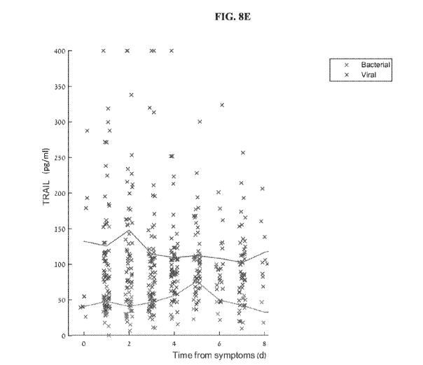
FIGs. 8A-F. Protein temporal dynamics ¨Protein serum levels measured in
patients at different times after symptom onset are depicted in blue 'x'
(viral), and red 'x'
(bacterial). Average serum levels are depicted by solid lines. The dynamics of
the
following proteins are shown: (A) CRP; (B) IL-6; (C) IP-10; (D) PCT; (E)
TRAIL; (F)
TCP signature.
FIG. 9. Temporal dynamics of bacterially induced biomarkers. Average levels of
IL-6, PCT, and CRP measured at different times after symptom onset from serum
samples of bacterially infected patients.
FIGs. 10A-B. Fuzzy OR models surface plot. The output of the Fuzzy OR model
is a likelihood of a bacterial infection as a function of TCP signature (y-
axis) and IL-6
concentrations in pg/ml. A. This example depicts the formula "Fuzzy OR
formula#5"
presented in section "Using Fuzzy OR model to generate improved signatures for
distinguishing between bacterial and viral patients" below, using IL-6 cutoff
of 250
pg/ml and hill coefficient of 10. B. The surface plot depicted in the figure
corresponds
to the following formula using different IL-6 cutoffs as indicated:
FIGs. 11A-D. Fuzzy OR model results - combined score of the TCP signature
and IL-6 using the hill-function, when applying different IL-6 cutoffs and
hill
coefficients as indicated (respectively): (A) 250 pg/ml and 6; (B) 250 pg/ml
and 10; (C)
350 pg/ml and 6; (D) 350 pg/ml and 10. The X axis represents the TCP signature
score
(ranging from 0 to 1, equivalent to 0-100%), and the Y axis represents the IL-
6
concentration in pg/ml. The color represents the combined score (likelihood of
bacterial
infection), wherein white represents a score of 1 and black represents a score
of 0
CA 03027341 2018-12-11
WO 2018/011795
PCT/IL2017/050780
16
(equivalent to 100% and 0% respectively). The surface lines represent round
scores
(e.g., 0.95, 0.9, 0.85). Overlaid on the plot are actual values of 378
bacterial (red) and
570 viral (blue) patients.
FIGs. 12A-G: Cutoff dependent models. (A) Illustration of a quadrary
separation
pattern that can separate between bacterial, viral and mixed (bacterial-viral
co-
infection), generated by applying a single TRAIL and PCT cutoffs as indicated.
(B)
TRAIL and PCT levels of 378 bacterial (blue) and 570 viral (orange) patients.
Dashed
lines represent an example of TRAIL cutoff of 75 pg/ml, and an example of PCT
cutoff
of 0.5 t.g/L. Diagnostic labels were determined by panel of experts as
described in the
Examples section. (C) Illustration of the different diagnostic labels (viral,
bacterial,
mixed and healthy), generated by applying TRAIL and PCT cutoffs. TRAIL cutoff
1
(low levels) is used to rule in bacterial infections and TRAIL cutoff 2 (high
levels) is
used to rule in viral infections. Integration of TRAIL and PCT cutoffs
generates
different diagnostic results: (i) a pure bacterial infection is indicated in
cases wherein
PCT is lower than PCT cutoff 1 AND TRAIL is lower than TRAIL cutoff 1; OR in
cases wherein PCT is higher than PCT cutoff 1 AND TRAIL is lower than TRAIL
cutoff 2; (ii) a pure viral infection is indicated in cases wherein PCT is
lower than PCT
cutoff 1 AND TRAIL is higher than TRAIL cutoff 2; (iii) mixed bacterial-viral
co-
infection is indicated in cases wherein PCT is higher than PCT cutoff 1 AND
TRAIL is
higher than TRAIL cutoff 2; (iv) healthy (or non-infectious) condition is
indicated in
cases wherein PCT is lower than PCT cutoff 1 AND TRAIL is higher than TRAIL
cutoff 1 but is lower than TRAIL cutoff 2. (D) TRAIL and PCT levels of 378
bacterial
(blue), 570 viral (orange), and 109 non-infectious (control; black) patients.
Dashed lines
represent an example of TRAIL cutoff 1 of 50 pg/ml, TRAIL cutoff 2 of 100
pg/ml, and
an example of PCT cutoff of 0.5 t.g/L. Diagnostic labels were determined by
panel of
experts as described in the Examples section. (E) A classifier for
distinguishing between
bacterial and viral patients based on the PCT/TRAIL ratio.
TRAIL and PCT levels of 378 bacterial (blue), 570 viral (orange) patients are
presented. Diagnostic labels were determined by panel of experts as described
in the
Examples section. The cutoff for separating between bacterial and viral
patients is
represented by the red line that equals PCT/TRAIL=0.05. This classifier will
label a
patient as bacterial in case PCT/TRAIL>0.05 and as viral in case
PCT/TRAIL<0.05. (F)
CA 03027341 2018-12-11
WO 2018/011795
PCT/IL2017/050780
17
A classifier for distinguishing between bacterial and viral patients based on
the
PCT/TRAIL ratio. TRAIL and PCT levels of 378 bacterial (blue), 570 viral
(orange)
patients are presented. Diagnostic labels were determined by panel of experts
as
described in the Examples section. The cutoff for separating between bacterial
and viral
patients is represented by the red line that equals PCT/TRAIL=0.02. This
classifier will
label a patient as bacterial in case PCT/TRAIL>0.02 and as viral in case
PCT/TRAIL<0.02. (G) A classifier for distinguishing between bacterial and
viral
patients based on the PCT/TRAIL ratio. TRAIL and PCT levels of 378 bacterial
(blue),
570 viral (orange) patients are presented. Diagnostic labels were determined
by panel of
experts as described in the Examples section. The cutoff for separating
between
bacterial and viral patients is represented by the red line that equals
PCT/TRAIL=0.01.
This classifier will label a patient as bacterial in case PCT/TRAIL>0.01 and
as viral in
case PCT/TRAIL<0.01.
FIG. 13 is a schematic illustration of geometrical objects that can be used
for
determining a likelihood, according to some embodiments of the present
invention;
FIG. 14 is a flowchart diagram of a method suitable for analyzing biological
data
obtained from a subject, according to some embodiments of the present
invention;
FIGs. 15A-D a schematic illustrations of a procedure for obtaining a smooth
version of a segment of a curved object, according to some embodiments of the
present
invention;
FIG. 16 is a schematic illustration of a block diagram of a system for
analyzing
biological data, according to some embodiments of the present invention; and
FIGs. 17A and 17B are schematic illustrations of a block diagram of a system
for analyzing biological data, in embodiments of the invention in which the
system
comprises a network interface (FIG. 17A) and a user interface (FIG. 17B).
DESCRIPTION OF SPECIFIC EMBODIMENTS OF THE INVENTION
The present invention, in some embodiments thereof, relates to the
identification
of signatures and determinants associated with infections. The signatures may
be used to
distinguish between bacterial and viral infections and also to distinguish
between sepsis
and non-infectious systemic inflammatory response syndrome (SIRS) and to
distinguish
CA 03027341 2018-12-11
WO 2018/011795
PCT/IL2017/050780
18
between an infective exacerbation state and a non-infective exacerbation state
in
patients with chronic obstructive pulmonary disease (COPD).
Before explaining at least one embodiment of the invention in detail, it is to
be
understood that the invention is not necessarily limited in its application to
the details set
forth in the following description or exemplified by the Examples. The
invention is
capable of other embodiments or of being practiced or carried out in various
ways.
Methods of distinguishing between bacterial and viral infections by analyzing
protein determinants have been disclosed in International Patent Application
W02013/117746, to the present inventors. Seeking to expand the number and type
of
determinants that can aid in accurate diagnosis, the present inventors have
now carried
out additional experiments and have identified other determinants that can be
used for
this aim.
Correct identification of bacterial patients is of high importance as these
patients
require antibiotic treatment and in some cases more aggressive management
(hospitalization, additional diagnostic tests etc). Misclassification of
bacterial patients
increases the chance of morbidity and mortality. Therefore, increasing the
sensitivity of
a biomarker or diagnostic test that distinguishes between bacterial and viral
infections
may be desired, even though specificity may be reduced.
Whilst reducing the present invention to practice, the present inventors noted
that
the markers PCT and IL-6 increase the sensitivity of a previously disclosed
signature ¨
TRAIL, CRP and IP-10 (referred to herein as the TCP signature). More
specifically, the
present inventors have shown that PCT and IL-6 provide a temporal dynamic
pattern
that complements the TCP signature which is particularly useful in diagnosing
infections
at a very early stage.
In some embodiments, the TCP signature is calculated as one or more
probabilistic classification functions which receive the values of the
expression of the
TRAIL, CRP and IP-10, and output a score. Based on the type of the respective
probabilistic classification function, the score can represent the likelihood
that the
subject has, a viral infection, a bacterial infection or has no infection. A
probabilistic
classification function that returns a score that represents the likelihood
that the subject
has a viral infection can be calculated as exp(i)/(1+exp()+exp(i)), a
probabilistic
classification function that returns a score that represents the likelihood
that the subject
CA 03027341 2018-12-11
WO 2018/011795
PCT/IL2017/050780
19
has a bacterial infection can be calculated as exp()/(1+exp()+exp(i)), and a
probabilistic classification function that returns a score that represents the
likelihood that
the subject has no infection can be calculated as 1/(1+exp()+exp(i)), where
=b0+b1P+b2P 5+b3P2+b4Q+b5R+b6R 5 and 1=co+c1P+c2P 5+c3P2+c4Q+c5R+c6R 5,
where P is a value of CRP, Q is a value of IP-10, and R is a value of TRAIL,
and where
110 is from about 4.96 to about 6.1, b1 is from about -0.07 to about -0.05, b2
is from about
1.33 to about 1.64, b3 is from about 0.000031 to about 0.000039, b4 is from
about 0.007
to about 0.010, b5 is from about 0.055 to about 0.071, b6 is from about 1.62
to about
1.98, co is from about -0.93 to about -0.75, c1 is from about -0.054 to about -
0.044, c2 is
from about 1.02 to about 1.25, c3 is from about -0.000057 to about -0.000046,
c4 is from
about 0.0080 to about 0.0098, c5 is from about 0.036 to about 0.045 and c6 is
from about
0.054 to about 0.066. More preferred values for the parameters bo,.., b6 and
co,.., c6 are
provided in Table 3, below.
Furthermore, the present inventors predict that the TCP signature together
with
PCT and/or IL-6 is useful for distinguishing between additional diseases
states such as
between a non-infective exacerbation state and an infective exacerbation state
in chronic
obstructive pulmonary disease (COPD) patients and between sepsis and non-
infective
systemic inflammatory response syndrome (SIRS).
Thus, according to a first aspect of the present invention there is provided a
method of diagnosing an infection in a subject comprising measuring the amount
of
each of the polypeptides TRAIL, CRP, IP10 and at least one additional
polypeptide
selected from the group consisting of IL-6 and PCT in a sample derived from
the
subject, wherein the amount is indicative of the infection type.
According to another aspect of the present invention there is provided a
method
of diagnosing an infection in a subject comprising measuring the amount of
each of the
polypeptides TRAIL, CRP and IL-6 in a sample derived from the subject, wherein
the
amount is indicative of the infection.
The methods disclosed herein are used to identify subjects with an infection
or a
specific infection type. By type of infection it is meant to include bacterial
infections,
viral infections, mixed infections, no infection (i.e., non-infectious). More
specifically,
some methods of the invention are used to distinguish subjects having a
bacterial
infection, a viral infection, a mixed infection (i.e., bacterial and viral co-
infection),
CA 03027341 2018-12-11
WO 2018/011795
PCT/IL2017/050780
patients with a non-infectious disease and healthy individuals. Some methods
of the
present invention can also be used to monitor or select a treatment regimen
for a subject
who has a an infection, and to screen subjects who have not been previously
diagnosed
as having an infection, such as subjects who exhibit risk factors developing
an infection.
Some methods of the present invention are used to identify and/or diagnose
subjects
who are asymptomatic for an infection. "Asymptomatic" means not exhibiting the
traditional signs and symptoms.
Thus, the infection type may be a bacterial infection, a viral infection or a
mixed
infection.
In various aspects the method distinguishes a virally infected subject from
either
a subject with non-infectious disease or a healthy subject; a bacterially
infected subject,
from either a subject with non-infectious disease or a healthy subject; a
subject with an
infectious disease from either a subject with an non-infectious disease or a
healthy
subject; a bacterially infected subject from a virally infected subject; a
mixed infected
subject from a virally infected subject; a mixed infected subject from a
bacterially
infected subject and a bacterially or mixed infected and subject from a
virally infected
subject.
A mixed infected subject refers to a subject having a bacterial and viral co-
infection.
The infection may be an acute or chronic infection.
A chronic infection is an infection that develops slowly and lasts a long
time.
Viruses that may cause a chronic infection include Hepatitis C and HIV. One
difference
between acute and chronic infection is that during acute infection the immune
system
often produces IgM+ antibodies against the infectious agent, whereas the
chronic phase
of the infection is usually characteristic of IgM-/IgG+ antibodies. In
addition, acute
infections cause immune mediated necrotic processes while chronic infections
often
cause inflammatory mediated fibrotic processes and scaring (e.g. Hepatitis C
in the
liver). Thus, acute and chronic infections may elicit different underlying
immunological
mechanisms.
As used herein, the term "infection" refers to a state caused by an infectious
agent of viral or bacterial origin. The bacterial infection may be the result
of gram-
positive, gram-negative bacteria or atypical bacteria.
CA 03027341 2018-12-11
WO 2018/011795
PCT/IL2017/050780
21
The term "Gram-positive bacteria" are bacteria that are stained dark blue by
Gram staining. Gram-positive organisms are able to retain the crystal violet
stain
because of the high amount of peptidoglycan in the cell wall.
The term "Gram-negative bacteria" are bacteria that do not retain the crystal
violet dye in the Gram staining protocol.
The term "Atypical bacteria" are bacteria that do not fall into one of the
classical
"Gram" groups. They are usually, though not always, intracellular bacterial
pathogens.
They include, without limitations, Mycoplasmas spp., Legionella spp.
Rickettsiae spp.,
and Chlamydiae spp.
In one embodiment, the level of the determinant may be used to rule in an
infection type. In another embodiment, the level of the determinant may be
used to rule
out an infection type.
By "ruling in" an infection it is meant that the subject has that type of
infection.
By "ruling out" an infection it is meant that the subject does not have that
type
of infection.
The subjects of this aspect of the present invention may present with a
variety of
pathogens including, but not limited to Adenovirus, Coronavirus, Parainfluenza
virus,
Influenza A virus, Influenza B virus, Respiratory syncytial virus A/B,
Chlamydophila
pneumoniae, Mycoplasma pneumoniae, Legionella pneumophila, Rota Virus,
Staphylococcus aureus, Streptococcus pneumoniae, Astrovirus, Enteric
Adenovirus,
Norovirus G I and G II, Bocavirus 1/2/3/4, Enterovirus, CMV virus, EBV virus,
Group
A Strep, or Escherichia coli.
In one embodiment, the method is used to distinguish between non-infective
Systemic inflammatory response syndrome (SIRS) and sepsis.
SIRS is a serious condition related to systemic inflammation, organ
dysfunction,
and organ failure. It is defined as 2 or more of the following variables:
fever of more
than 38 C (100.4 F) or less than 36 C (96.8 F); heart rate of more than 90
beats per
minute; respiratory rate of more than 20 breaths per minute or arterial carbon
dioxide
tension (PaCO2) of less than 32 mm Hg; abnormal white blood cell count
(>12,000/0_,
or < 4,000/0_, or >10% immature [band] forms). SIRS is nonspecific and can be
caused
by ischemia, inflammation, trauma, infection, or several insults combined.
Thus, SIRS
is not always related to infection.
CA 03027341 2018-12-11
WO 2018/011795
PCT/IL2017/050780
22
Sepsis is a life-threatening condition that is caused by inflammatory response
to
an infection. The early diagnosis of sepsis is essential for clinical
intervention before the
disease rapidly progresses beyond initial stages to the more severe stages,
such as
severe sepsis or septic shock, which are associated with high mortality.
Current
diagnostics are limited in their ability to distinguish between non-infective
SIRS and
sepsis. Therefore, there is a need for new biomarkers or combinations of
biomarkers
that can provide added value in the accurate and timely diagnosis of sepsis.
According to this embodiment, sepsis may be diagnosed as the presence of SIRS
criteria in the presence of a known infection.
Thus, according to one aspect, there is provided a method of distinguishing
between sepsis and non-infective systemic inflammatory response syndrome
(SIRS)
comprising measuring the amount of at least two polypeptides selected from the
group
consisting of TNF-related apoptosis-inducing ligand (TRAIL), C-reactive
protein
(CRP), Interferon gamma-induced protein 10 (IP10), Interleukin 6 (IL-6) and
Procalcitonin (PCT) in a sample derived from the subject, wherein said amount
is
indicative of sepsis or non-infective SIRS.
Particular combinations of polypeptides are described herein below.
According to this aspect the subject that is tested has been diagnosed with
SIRS.
The method that is carried out is used to determine if the SIRS is infective
(i.e. sepsis)
or non-infective.
In another embodiment, sepsis is diagnosed in a subject suspected of having an
infection and which fulfils each of the three criteria:
Respiratory rate greater or equal to_22/min
Altered mentation (e.g. a Glasgow coma score of less than 15)
Systolic blood pressure lower than or equal to 100mmHg.
Further criteria for diagnosing sepsis are disclosed in Singer et al. 2016,
315(8):801-810 JAMA.
Thus, according to another aspect of the present invention there is provided a
method of ruling in sepsis in a subject suspected of having in infection
comprising:
(a)
measuring the amount of at least two polypeptides selected from the
group consisting of TNF-related apoptosis-inducing ligand (TRAIL), C-reactive
protein
CA 03027341 2018-12-11
WO 2018/011795
PCT/IL2017/050780
23
(CRP), Interferon gamma-induced protein 10 (IP10), Interleukin 6 (IL-6) and
Procalcitonin (PCT) in a sample derived from the subject;
(b) measuring the respiratory rate of the subject;
(c) analyzing the mental state of the subject; and
(d) measuring the blood pressure of the subject;
wherein when each of steps are indicative of sepsis, sepsis is ruled in.
Particular combinations of polypeptides are described herein below.
It will be appreciated that steps (b), (c) and (d) may be carried out as part
of
determining the SOFA score (originally the Sepsis-related Organ Failure
Assessment;
Vincent J.L et al Intensive Care Med. 1996;22(7):707-710) of a subject.
In one embodiment, step (a) is carried out in order to confirm the subject has
an
infection. Only when subjects have a confirmed infection are steps (b), (c)
and (d)
carried out to confirm sepsis.
In another embodiment, the subject has a suspected infection, steps (b), (c)
ad
(d) are carried out to rule in sepsis; and step (a) is carried out to
corroborate the
diagnosis.
The present inventors contemplate analyzing the amount of at least two
polypeptides selected from the group consisting of TNF-related apoptosis-
inducing
ligand (TRAIL), C-reactive protein (CRP), Interferon gamma-induced protein 10
(IP10), Interleukin 6 (IL-6) and Procalcitonin (PCT) in a sample derived from
the
subject in order to confirm that the subject has an infection.
In another aspect, sepsis is ruled in a subject suspected of having an
infection
when his SOFA score is above 2.
In one embodiment, analyzing the amount of at least two polypeptides selected
from the group consisting of TNF-related apoptosis-inducing ligand (TRAIL), C-
reactive protein (CRP), Interferon gamma-induced protein 10 (IP10),
Interleukin 6 (IL-
6) and Procalcitonin (PCT) in a sample derived from the subject is carried out
in order
to confirm the subject has an infection. Only when subjects have a confirmed
infection
is the SOFA analysis carried out to diagnose sepsis (when the subject has a
SOFA score
of more than or equal to 2, the subject is diagnosed with sepsis).
Alternatively, a SOFA analysis is carried out to diagnose sepsis. Analyzing
the
amount of at least two polypeptides selected from the group consisting of TNF-
related
CA 03027341 2018-12-11
WO 2018/011795
PCT/IL2017/050780
24
apoptosis-inducing ligand (TRAIL), C-reactive protein (CRP), Interferon gamma-
induced protein 10 (IP10), Interleukin 6 (IL-6) and Procalcitonin (PCT) in a
sample
derived from the subject is carried out in order to confirm the subject has
the sepsis.
In another embodiment, the method is used to discriminate between bacterial
and viral etiologies in patients with chronic obstructive pulmonary disease
(COPD)
exacerbation.
In still another embodiment, the method is used to distinguish between an
infective exacerbation state and a non-infective exacerbation state of chronic
obstructive
pulmonary disease (COPD) in a subject.
Chronic obstructive pulmonary disease (COPD) is an obstructive, inflammatory
lung disease characterized by long-term poor airflow. The main symptoms
include
shortness of breath and cough with sputum production. COPD is a progressive
disease,
worsening over time.
An exacerbation of COPD may be defined as an event in the natural course of
the disease characterized by a change in the patient's baseline dyspnea,
cough, and/or
sputum that is beyond normal day-to-day variations. The exacerbation is
typically acute.
It may present with signs of increased work of breathing such as fast
breathing, a fast
heart rate, sweating, active use of muscles in the neck, a bluish tinge to the
skin, and
confusion or combative behavior in very severe exacerbations. Crackles may
also be
heard over the lungs on examination with a stethoscope.
Particular combinations of polypeptides are described herein below.
The subjects (e.g. children) may present with a particular clinical syndrome ¨
for
example, low respiratory tract infection (LRTI) infection, upper respiratory
tract
infection (URTI) or a serious bacterial infection (SBI) such as UTI (urinary
tract
infections), septic shock, bacteremia, pneumonia or meningitis.
"Measuring" or "measurement," or alternatively "detecting" or "detection,"
means assessing the presence, absence, quantity or amount (which can be an
effective
amount) of the determinant within a clinical or subject-derived sample,
including the
derivation of qualitative or quantitative concentration levels of such
determinants.
A "sample" in the context of the present invention is a biological sample
isolated
from a subject and can include, by way of example and not limitation, whole
blood,
serum, plasma, saliva, mucus, breath, urine, CSF, sputum, sweat, stool, hair,
seminal
CA 03027341 2018-12-11
WO 2018/011795
PCT/IL2017/050780
fluid, biopsy, rhinorrhea, tissue biopsy, cytological sample, platelets,
reticulocytes,
leukocytes, epithelial cells, or whole blood cells.
In a particular embodiment, the sample is a blood sample - e.g. serum or a
sample
comprising blood cells. In a particular embodiment, the sample is depleted of
red blood
cells.
In one embodiment, the sample is derived from the subject no more ten days
following symptom onset, no more than five days following symptom onset, no
more
than four days following symptom onset, no more than three days following
symptom
onset, no more than two days following symptom onset or preferably no more
than one
day following symptom onset.
The sample may be fresh or frozen.
A "subject" in the context of the present invention may be a mammal (e.g. a
human, dog, cat, horse, cow, sheep, pig or goat). According to another
embodiment, the
subject is a bird (e.g. chicken, turkey, duck or goose). According to a
particular
embodiment, the subject is a human. The subject can be male or female. The
subject
may be an adult (e.g. older than 18, 21, or 22 years or a child (e.g. younger
than 18, 21
or 22 years). In another embodiment, the subject is an adolescent (between 12
and 21
years), an infant (29 days to less than 2 years of age) or a neonate (birth
through the first
28 days of life).
The subject can be one who has been previously diagnosed or identified as
having an infection, and optionally has already undergone, or is undergoing, a
therapeutic intervention for the infection. Alternatively, a subject can also
be one who
has not been previously diagnosed as having an infection. For example, a
subject can
be one who exhibits one or more risk factors for having an infection.
According to a particular embodiment, the subject does not show signs of
having
had a heart attack (e.g. has a normal level of creatine kinase, troponin or
serum
myoglobin, and/or has a normal ECG or EKG).
In one embodiment, the subject is one which has undergone a trauma (e.g. car
accident or combat related trauma) and/or has undergone a surgical procedure.
As mentioned, in order to determine the type of infection, the amount of each
of
the following polypeptides are determined: TRAIL, CRP, IP10, together with
either IL-6
and/or PCT.
CA 03027341 2018-12-11
WO 2018/011795 PCT/IL2017/050780
26
Alternatively, in order to determine the type of infection, the amount of
TRAIL,
CRP and IL-6 are measured in a sample derived from the subject, wherein the
amount is
indicative of the infection type.
Other contemplated combinations are provided herein below:
TRAIL, CRP, 1P-10 and PCT;
TRAIL, 1P-10 and PCT;
TRAIL, CRP, 1P-10 and IL-6;
TRAIL, CRP, 1P-10, PCT and IL-6;
TRAIL, CRP and PCT;
TRAIL, CRP and IL-6;
TRAIL, CRP, PCT and IL-6
Information regarding the above mentioned polypeptides is provided in Table 1,
herein below.
Table I
Protein symbol Full Gene Name RefSeq DNA
RefSeq proteins
sequence
CRP C-reactive protein, NC 000001.11 NP 000558.2
pentraxin-related NT 004487.20
NC 018912.2
TRAIL Tumor necrosis factor NC 000003.12 NP 001177871.1
superfamily member NC 018914.2 NP 001177872.1
NT 005612.17 NP 003801.1
1P-10 Chemokine (C-X-C NC 000004.12 NP
001556.2
motif) ligand 10 NC 018915.2
NT 016354.20
Procalcitonin (PCT) Calcitonin-related NC
000011.10 NP 001029124.1
polypeptide alpha NC
018922.2 NP 001029125.1
NT 009237.19 NP 001732.1
IL-6 Interleukin 6 NC 000007.14 NP 000591.1
NT 007819.18
NC 018918.2
Exemplary ranges of the mentioned polypeptides in bacterial and viral patients
include without limitation:
CRP ¨ CRP levels of 0-40 t.g/m1 are usually indicative of a viral infection,
while
40-400 i.t.g/m1 are usually indicative of a bacterial infection. Bacterial
infection can
usually be ruled in if CRP levels are higher than 50, 60, 70 or more
preferably 80 i.t.g/ml,
and ruled out if CRP levels are lower than 30 and more preferably 20 .t.g/ml.
CA 03027341 2018-12-11
WO 2018/011795
PCT/IL2017/050780
27
TRAIL ¨TRAIL levels of 100-1000 pg/ml are usually indicative of a viral
infection, while 0-85 pg/ml are usually indicative of a bacterial infection.
Bacterial
infection can usually be ruled in if TRAIL levels are lower than 85 pg/ml, 70
pg/ml, 60
pg/ml or more preferably 50 pg/ml, and ruled out if TRAIL levels are higher
than 100
pg/ml.
1P-10 ¨ IP-10 levels of 300-2000 pg/ml are usually indicative of a viral
infection, while 160-860 pg/ml are usually indicative of a bacterial
infection. Viral
infection can usually be ruled in if IP10 levels are higher than 800 pg/ml,
and ruled out
if IP10 levels are lower than 300 pg/ml.
PCT ¨ PCT levels higher than 0.5 i.t.g/L are usually indicative of a bacterial
infection.
IL-6 ¨ IL-6 levels higher than 100 pg/ml are usually indicative of a bacterial
infection.
CRP: C-reactive protein; additional aliases of CRP include without limitation
RP11-419N10.4 and PTX1.
An exemplary amino acid sequence of human CRP is set forth below in SEQ ID
NO: 1.
The level of CRP typically increases in infections (as compared to non-
infectious diseases), with the level of CRP being higher in bacterial
infections as
opposed to viral infections.
Thus, when the level of CRP is above a predetermined level, it is indicative
that
the infection is a bacterial infection and a bacterial infection may be ruled
in (or a viral
infection may be ruled out).
When the level of CRP is below a predetermined level, it is indicative that
the
infection is a viral infection and a viral infection may be ruled in (or a
bacterial
infection may be ruled out).
TRAIL: The protein encoded by this gene is a cytokine that belongs to the
tumor necrosis factor (TNF) ligand family. The present invention contemplates
measuring either the soluble and/or the membrane form of this protein. In one
embodiment, only the soluble form of this protein is measured. Additional
names of the
gene include without limitations APO2L, TNF-related apoptosis-inducing ligand,
TNFSF10 and CD253. This protein binds to several members of the TNF receptor
CA 03027341 2018-12-11
WO 2018/011795
PCT/IL2017/050780
28
superfamily such as TNFRS F 10A/TRAILR 1, TNFRSF
10B/TRAILR2,
TNFRSF 10C/TRAILR3 , TNFRSF 10D/TRAILR4, and possibly also to
TNFRSF 1 1 B/OPG
Exemplary amino acid sequences of TRAIL are set forth in SEQ ID NOs: 4-8.
In a particular embodiment, TRAIL is the protein that is recognized by the
antibody of the kit R&D systems, Human TRAIL/TNFSF10 Quantikine ELISA Kit
catalog # DTRLOO.
The level of TRAIL increases in viral infections (as compared to non-
infectious
diseases), and decreases in bacterial infections (as compared to non-
infectious diseases).
Thus, when the level of TRAIL is above a predetermined level, it is indicative
that the infection is a viral infection and a viral infection may be ruled in
(or a bacterial
infection may be ruled out).
When the level of TRAIL is below a predetermined level, it is indicative that
the
infection is a bacterial infection and a bacterial infection may be ruled in
(or a viral
infection may be ruled out).
For example, a bacterial infection may be ruled out if the polypeptide
concentration of TRAIL determined is higher than a pre-determined first
threshold
value. Optionally, the method further includes determining if a subject has a
viral
infection (i.e., ruling in a viral infection). A viral infection is ruled in
if the polypeptide
concentration of TRAIL is higher than a pre-determined second threshold value.
In another specific embodiment the invention includes determining if a subject
does not have a viral infection (i.e. ruling out a viral infection). A viral
infection is
ruled out if the polypeptide concentration of TRAIL determined is lower than a
pre-
determined first threshold value. Optionally, the method further includes
determining if
a subject has a bacterial infection (i.e., ruling in a bacterial infection). A
bacterial
infection is ruled in if the polypeptide concentration of TRAIL is lower than
a pre-
determined second threshold value.
IP10: This gene encodes a chemokine of the CXC subfamily and ligand for the
receptor CXCR3. Additional names of the gene include without limitations:
CXCL10,
Gamma-IP10, INP10 and chemokine (C-X-C motif) ligand 10.
An exemplary amino acid sequence of human IP10 is set forth in SEQ ID NO:
16.
CA 03027341 2018-12-11
WO 2018/011795
PCT/IL2017/050780
29
In a particular embodiment, IP10 is the protein that is recognized by the
antibody of the kit (R&D systems, Human CXCL10/IP-10 Quantikine ELISA Kit
catalog # DIP100).
The level of IP10 increases in infections (as compared to non-infectious
diseases), with the level of IP10 being higher in viral infections as opposed
to bacterial
infections.
Thus, when the level of IP10 is above a predetermined level, it is indicative
that
the infection is a viral infection and a viral infection may be ruled in (or a
bacterial
infection may be ruled out).
When the level of IP10 is below a predetermined level, it is indicative that
the
infection is a bacterial infection and a bacterial infection may be ruled in
(or a viral
infection may be ruled out).
IL-6: This gene encodes a cytokine that functions in inflammation and the
maturation of B cells. In addition, the encoded protein has been shown to be
an
endogenous pyrogen capable of inducing fever in people with autoimmune
diseases or
infections. The protein is primarily produced at sites of acute and chronic
inflammation,
where it is secreted into the serum and induces a transcriptional inflammatory
response
through interleukin 6 receptor, alpha. The functioning of this gene is
implicated in a
wide variety of inflammation-associated disease states, including
susceptibility to
diabetes mellitus and systemic juvenile rheumatoid arthritis.
Exemplary amino acid sequences of human IL-6 is set forth in SEQ ID NOs: 23
and 24.
The data presented herein shows that the level of IL-6 increases in infections
(as
compared to non-infectious diseases), with the level of IL-6 being higher in
bacterial
infections as opposed to viral infections.
Thus, when the level of IL-6 is above a predetermined level, it is indicative
that
the infection is a bacterial infection and a bacterial infection may be ruled
in (or a viral
infection may be ruled out).
When the level of IL-6 is below a predetermined level, it is indicative that
the
infection is a viral infection and a viral infection may be ruled in (or a
bacterial
infection may be ruled out).
CA 03027341 2018-12-11
WO 2018/011795
PCT/IL2017/050780
PCT: Procalcitonin (PCT) is a peptide precursor of the hormone calcitonin, the
latter being involved with calcium homeostasis.
Exemplary amino acid sequences of human PCT are set forth in SEQ ID NOs:
19-22.
The level of PCT typically increases in infections (as compared to non-
infectious diseases), with the level of PCT being higher in bacterial
infections as
opposed to viral infections.
Thus, when the level of PCT is above a predetermined level, it is indicative
that
the infection is a bacterial infection and a bacterial infection may be ruled
in (or a viral
infection may be ruled out).
When the level of PCT is below a predetermined level, it is indicative that
the
infection is a viral infection and a viral infection may be ruled in (or a
bacterial
infection may be ruled out).
The concentrations of each of the above identified polypeptides may be
combined (e.g. by way of a pre-determined mathematical function) to compute a
score
and the score may be compared to a predetermined reference value as further
described
herein below.
Further information on generating pre-determined mathematical functions in
general and for CRP, IP10 and TRAIL in particular are provided in
International Patent
Application IL2015/050823, the contents of which are incorporated herein by
reference.
Statistical classification algorithms which may be used to calculate the score
include, but are not limited to Support Vector Machine (SVM), Logistic
Regression
(LogReg), Neural Network, Bayesian Network, and a Hidden Markov Model.
Alternatively, the integration of the different proteins into a single
predictive score
could be achieved by applying "Fuzzy OR model" analysis, as shown in the
Examples
section herein below.
In one embodiment, the level of PCT and/or IL-6 is taken into account in the
statistical classification together with the TRAIL, CRP, IP-10 signature (TCP
signature)
only when concentrations reaches a threshold level. Thus, for example only
when the
level of PCT is above 1, 1.5, 2, 2.5, 5, or 7.5 g/L, is PCT included in the
algorithm
together with the TCP signature. Similarly, only when the level of IL-6 is
above 100,
200, 240, 250, 280, 320, or 350 pgiml is IL-6 included in the algorithm
together with
CA 03027341 2018-12-11
WO 2018/011795
PCT/IL2017/050780
31
the TCP signature. It will be appreciated that the weight of PCT and 1L-6 in
the
algorithm may vary according to its concentration. For example, if the level
of PCT is
above 5 then its
relative weight may be higher with respect to the TCP signature
that if the level of PCT is below 5 p
A reference value can be relative to a number or value derived from population
studies, including without limitation, such subjects having the same
infection, subject
having the same or similar age range, subjects in the same or similar ethnic
group, or
relative to the starting sample of a subject undergoing treatment for an
infection. Such
reference values can be derived from statistical analyses and/or risk
prediction data of
populations obtained from mathematical algorithms and computed indices of
infection.
Reference determinant indices can also be constructed and used using
algorithms and
other methods of statistical and structural classification.
In one embodiment of the present invention, the reference value is the amount
(i.e. level) of determinants in a control sample derived from one or more
subjects who
do not have an infection (i.e., healthy, and or non-infectious individuals).
In a further
embodiment, such subjects are monitored and/or periodically retested for a
diagnostically relevant period of time ("longitudinal studies") following such
test to
verify continued absence of infection. Such period of time may be one day, two
days,
two to five days, five days, five to ten days, ten days, or ten or more days
from the
initial testing date for determination of the reference value. Furthermore,
retrospective
measurement of determinants in properly banked historical subject samples may
be used
in establishing these reference values, thus shortening the study time
required.
A reference value can also comprise the amounts of determinants derived from
subjects who show an improvement as a result of treatments and/or therapies
for the
infection. A reference value can also comprise the amounts of determinants
derived
from subjects who have confirmed infection by known techniques.
An example of a bacterially infected reference value index value is the mean
or
median concentrations of that determinant in a statistically significant
number of
subjects having been diagnosed as having a bacterial infection.
An example of a virally infected reference value is the mean or median
concentrations of that determinant in a statistically significant number of
subjects
having been diagnosed as having a viral infection.
CA 03027341 2018-12-11
WO 2018/011795
PCT/IL2017/050780
32
In another embodiment, the reference value is an index value or a baseline
value.
An index value or baseline value is a composite sample of an effective amount
of
determinants from one or more subjects who do not have an infection. A
baseline value
can also comprise the amounts of determinants in a sample derived from a
subject who
has shown an improvement in treatments or therapies for the infection. In this
embodiment, to make comparisons to the subject-derived sample, the amounts of
determinants are similarly calculated and compared to the index value.
Optionally,
subjects identified as having an infection, are chosen to receive a
therapeutic regimen to
slow the progression or eliminate the infection.
Additionally, the amount of the determinant can be measured in a test sample
and
compared to the "normal control level," utilizing techniques such as reference
limits,
discrimination limits, or risk defining thresholds to define cutoff points and
abnormal
values. The "normal control level" means the level of one or more determinants
or
combined determinant indices typically found in a subject not suffering from
an
infection. Such normal control level and cutoff points may vary based on
whether a
determinant is used alone or in a formula combining with other determinants
into an
index. Alternatively, the normal control level can be a database of
determinant patterns
from previously tested subjects.
The effectiveness of a treatment regimen can be monitored by detecting a
determinant in an effective amount (which may be one or more) of samples
obtained
from a subject over time and comparing the amount of determinants detected.
For
example, a first sample can be obtained prior to the subject receiving
treatment and one
or more subsequent samples are taken after or during treatment of the subject.
For example, the methods of the invention can be used to discriminate between
bacterial, viral and mixed infections (i.e. bacterial and viral co-
infections.) This will
allow patients to be stratified and treated accordingly.
In a specific embodiment of the invention a treatment recommendation (i.e.,
selecting a treatment regimen) for a subject is provided by identifying the
type
infection (i.e., bacterial, viral, mixed infection or no infection) in the
subject according
to the method of any of the disclosed methods and recommending that the
subject
receive an antibiotic treatment if the subject is identified as having
bacterial infection or
CA 03027341 2018-12-11
WO 2018/011795 PCT/IL2017/050780
33
a mixed infection; or an anti- viral treatment is if the subject is identified
as having a
viral infection.
In another embodiment, the methods of the invention can be used to prompt
additional targeted diagnosis such as pathogen specific PCRs, chest-X-ray,
cultures etc.
For example, a diagnosis that indicates a viral infection according to
embodiments of
this invention, may prompt the usage of additional viral specific multiplex-
PCRs,
whereas a diagnosis that indicates a bacterial infection according to
embodiments of this
invention may prompt the usage of a bacterial specific multiplex-PCR. Thus,
one can
reduce the costs of unwarranted expensive diagnostics.
In a specific embodiment, a diagnostic test recommendation for a subject is
provided by identifying the infection type (i.e., bacterial, viral, mixed
infection or no
infection) in the subject according to any of the disclosed methods and
recommending
a test to determine the source of the bacterial infection if the subject is
identified as
having a bacterial infection or a mixed infection; or a test to determine the
source of the
viral infection if the subject is identified as having a viral infection.
As well as measuring the polypeptide determinants mentioned herein above, the
present inventors contemplate measuring at least one, two, three, four, five,
six, seven,
eight, nine, ten or more additional (non-identical) determinants (polypeptide,
RNA or
other), wherein the at least one additional determinant is set forth in US
Patent
Application No. 20080171323, W02011/132086 and W02013/117746 and PCT
Application IL 2015/051024 and PCT Application IL 2015/051201 and Provisional
Application No. 62/302,849 the contents of each are incorporated herein by
reference.
Other polypeptide determinants contemplated by the present inventors are the
polypeptide counterparts of the RNA determinants described therein.
In one embodiment, at least of the additional determinants is set forth in
Table 2
herein below.
Table 2
Protein symbol Full Gene Name RefSeq DNA RefSeq proteins
sequence
IL1R/ IL1R1/ Interleukin 1 receptor, NC 000002.12 .. NP 000868.1
IL1RA type I NT 005403.18 NP 001275635.1
NC 018913.2
SAA/ SAA1 Serum amyloid Al NC 000011.10 NP 000322.2
NC 018922.2 NP 001171477.1
NT 009237.19 NP 954630.1
CA 03027341 2018-12-11
WO 2018/011795 PCT/IL2017/050780
34
TREM1 Triggering receptor NC 000006.12 NP 001229518.1
expressed on myeloid NT 007592.16 NP 001229519.1
cells 1 NC 018917.2 NP 061113.1
TREM2 Triggering receptor NC 000006.12 NP 001258750.1
expressed on myeloid NT 007592.16 NP 061838.1
cells 2 NC 018917.2
RSAD2 Radical S-adenosyl NC 000002.12 NP 542388.2
methionine domain NT 005334.17
containing 2 NC 018913.2
NGAL Lipocalin 2 NC 000009.12 NP 005555.2
NC 018920.2
NT 008470.20
MMP8 Matrix NC 000011.10 NP 001291370.1
metallopeptidase 8 NT 033899.9 NP 001291371.1
NC 018922.2 NP 002415.1
MX1 MX Dynamin-Like NC 000021.9 NP 001138397.1
GTPase 1 NT 011512.12 NP 001171517.1
NC 018932.2 NP 001269849.1
NP 002453.2
Neopterin 2-amino-6-(1,2,3- N/A N/A
trihydroxypropy1)-1H-
pteridin-4-one
IUPAC name
According to this aspect of the present invention, in order to distinguish
between
the different infection types, no more than 30 determinants (e.g. proteins
that are
differentially expressed in a statistically signficant manner in subjects with
a bacterial
infection compared to subjects with a viral infection) are measured, no more
than 25
determinants are measured, no more than 20 determinants are measured, no more
than
15 determinants are measured, no more than 10 determinants are measured, no
more
than 9 determinants are measured, no more than 8 determinants are measured, no
more
than 7 determinants are measured, no more than 6 determinants are measured, no
more
than 5 determinants are measured or even no more than 4 determinants are
measured.
Other determinants that may be measured according to aspects of the present
invention include pathogen (bacterial or viral) specific RNA or polypeptide
determinants. This may be carried out in order to aid in identification of a
specific
pathogen. The measurements may be effected simultaneously with the above
described
measurements or consecutively.
CA 03027341 2018-12-11
WO 2018/011795
PCT/IL2017/050780
Methods of measuring the levels of polypeptides are well known in the art and
include, e.g., immunoassays based on antibodies to proteins, aptamers or
molecular
imprints.
The polypeptide determinants can be detected in any suitable manner, but are
typically detected by contacting a sample from the subject with an antibody,
which
binds the determinant and then detecting the presence or absence of a reaction
product.
The antibody may be monoclonal, polyclonal, chimeric, or a fragment of the
foregoing,
as discussed in detail above, and the step of detecting the reaction product
may be
carried out with any suitable immunoassay. The sample from the subject is
typically a
biological sample as described above, and may be the same sample of biological
sample
used to conduct the method described above.
In one embodiment, the antibody which specifically binds the determinant is
attached (either directly or indirectly) to a signal producing label,
including but not
limited to a radioactive label, an enzymatic label, a hapten, a reporter dye
or a
fluorescent label.
Immunoassays carried out in accordance with some embodiments of the present
invention may be homogeneous assays or heterogeneous assays. In a homogeneous
assay the immunological reaction usually involves the specific antibody (e.g.,
anti-
determinant antibody), a labeled analyte, and the sample of interest. The
signal arising
from the label is modified, directly or indirectly, upon the binding of the
antibody to the
labeled analyte. Both the immunological reaction and detection of the extent
thereof
can be carried out in a homogeneous solution. Immunochemical labels, which may
be
employed, include free radicals, radioisotopes, fluorescent dyes, enzymes,
bacteriophages, or coenzymes.
In a heterogeneous assay approach, the reagents are usually the sample, the
antibody, and means for producing a detectable signal. Samples as described
above may
be used. The antibody can be immobilized on a support, such as a bead (such as
protein
A and protein G agarose beads), plate or slide, and contacted with the
specimen
suspected of containing the antigen in a liquid phase. The support is then
separated from
the liquid phase and either the support phase or the liquid phase is examined
for a
detectable signal employing means for producing such signal. The signal is
related to
the presence of the analyte in the sample.
CA 03027341 2018-12-11
WO 2018/011795
PCT/IL2017/050780
36
Means for producing a detectable signal include the use of radioactive labels,
fluorescent labels, or enzyme labels. For example, if the antigen to be
detected contains
a second binding site, an antibody which binds to that site can be conjugated
to a
detectable group and added to the liquid phase reaction solution before the
separation
step. The presence of the detectable group on the solid support indicates the
presence of
the antigen in the test sample. Examples of suitable immunoassays are
oligonucleotides,
immunoblotting, immunofluorescence methods,
immunoprecipitation,
chemiluminescence methods, electrochemiluminescence (ECL) or enzyme-linked
immunoas says.
Those skilled in the art will be familiar with numerous specific immunoassay
formats and variations thereof which may be useful for carrying out the method
disclosed herein. See generally E. Maggio, Enzyme-Immunoassay, (1980) (CRC
Press,
Inc., Boca Raton, Fla.); see also U.S. Pat. No. 4,727,022 to Skold et al.,
titled "Methods
for Modulating Ligand-Receptor Interactions and their Application," U.S. Pat.
No.
4,659,678 to Forrest et al., titled "Immunoassay of Antigens," U.S. Pat. No.
4,376,110
to David et al., titled "Immunometric Assays Using Monoclonal Antibodies,"
U.S. Pat.
No. 4,275,149 to Litman et al., titled "Macromolecular Environment Control in
Specific
Receptor Assays," U.S. Pat. No. 4,233,402 to Maggio et al., titled "Reagents
and
Method Employing Channeling," and U.S. Pat. No. 4,230,767 to Boguslaski et
al., titled
"Heterogeneous Specific Binding Assay Employing a Coenzyme as Label."The
determinant can also be detected with antibodies using flow cytometry. Those
skilled in
the art will be familiar with flow cytometric techniques which may be useful
in carrying
out the methods disclosed herein(Shapiro 2005). These include, without
limitation,
Cytokine Bead Array (Becton Dickinson) and Luminex technology.
Antibodies can be conjugated to a solid support suitable for a diagnostic
assay
(e.g., beads such as protein A or protein G agarose, microspheres, plates,
slides or wells
formed from materials such as latex or polystyrene) in accordance with known
techniques, such as passive binding. Antibodies as described herein may
likewise be
, ,
conjugated to detectable labels or groups such as radiolabels (e.g., 35S 121
1311)
,
enzyme labels (e.g., horseradish peroxidase, alkaline phosphatase), and
fluorescent
labels (e.g., fluorescein, Alexa, green fluorescent protein, rhodamine) in
accordance
with known techniques.
CA 03027341 2018-12-11
WO 2018/011795
PCT/IL2017/050780
37
Antibodies can also be useful for detecting post-translational modifications
of
determinant proteins, polypeptides, mutations, and polymorphisms, such as
tyrosine
phosphorylation, threonine phosphorylation, serine phosphorylation,
glycosylation (e.g.,
0-G1cNAc). Such antibodies specifically detect the phosphorylated amino acids
in a
protein or proteins of interest, and can be used in immunoblotting,
immunofluorescence,
and ELISA assays described herein. These antibodies are well-known to those
skilled
in the art, and commercially available. Post-translational modifications can
also be
determined using metastable ions in reflector matrix-assisted laser desorption
ionization-time of flight mass spectrometry (MALDI-TOF) (Wirth U. and Muller
D.
2002).
For determinant-proteins, polypeptides, mutations, and polymorphisms known
to have enzymatic activity, the activities can be determined in vitro using
enzyme
assays known in the art. Such assays include, without limitation, kinase
assays,
phosphatase assays, reductase assays, among many others. Modulation of the
kinetics
of enzyme activities can be determined by measuring the rate constant Km using
known
algorithms, such as the Hill plot, Michaelis-Menten equation, linear
regression plots
such as Lineweaver-Burk analysis, and Scatchard plot.
In particular embodiments, the antibodies of the present invention are
monoclonal antibodies.
Suitable sources for antibodies for the detection of determinants include
commercially available sources such as, for example, Abazyme, Abnova,
AssayPro,
Affinity Biologicals, AntibodyShop, Aviva bioscience, Biogenesis, Biosense
Laboratories, Calbiochem, Cell Sciences, Chemicon International, Chemokine,
Clontech, Cytolab, DAKO, Diagnostic BioSystems, eBioscience, Endocrine
Technologies, Enzo Biochem, Eurogentec, Fusion Antibodies, Genesis Biotech,
GloboZymes, Haematologic Technologies, Immunodetect, Immunodiagnostik,
Immunometrics, Immunostar, Immunovision, Biogenex, Invitrogen, Jackson
ImmunoResearch Laboratory, KMI Diagnostics, Koma Biotech, LabFrontier Life
Science Institute, Lee Laboratories, Lifescreen, Maine Biotechnology Services,
Mediclone, MicroPharm Ltd., ModiQuest, Molecular Innovations, Molecular
Probes,
Neoclone, Neuromics, New England Biolabs, Novocastra, Novus Biologicals,
Oncogene Research Products, Orbigen, Oxford Biotechnology, Panvera,
PerkinElmer
CA 03027341 2018-12-11
WO 2018/011795
PCT/IL2017/050780
38
Life Sciences, Pharmingen, Phoenix Pharmaceuticals, Pierce Chemical Company,
Polymun Scientific, Polysiences, Inc., Promega Corporation, Proteogenix,
Protos
Immunoresearch, QED Biosciences, Inc., R&D Systems, Repligen, Research
Diagnostics, Robo screen, Santa Cruz Biotechnology, Seikagaku America,
Serological
Corporation, Serotec, SigmaAldrich, StemCell Technologies, Synaptic Systems
GmbH,
Technopharm, Terra Nova Biotechnology, TiterMax, Trillium Diagnostics, Upstate
Biotechnology, US Biological, Vector Laboratories, Wako Pure Chemical
Industries,
and Zeptometrix. However, the skilled artisan can routinely make antibodies,
against
any of the polypeptide determinants described herein.
The presence of a label can be detected by inspection, or a detector which
monitors a particular probe or probe combination is used to detect the
detection reagent
label. Typical detectors include spectrophotometers, phototubes and
photodiodes,
microscopes, scintillation counters, cameras, film and the like, as well as
combinations
thereof. Those skilled in the art will be familiar with numerous suitable
detectors that
widely available from a variety of commercial sources and may be useful for
carrying
out the method disclosed herein. Commonly, an optical image of a substrate
comprising
bound labeling moieties is digitized for subsequent computer analysis. See
generally
The Immunoassay Handbook [The Immunoassay Handbook. Third Edition. 2005].
Traditional laboratory risk factors and additional clinical parameters may
also be
measured together with the above described polypeptides to further increase
the
accuracy of the signatures.
"Traditional laboratory risk factors" encompass biomarkers isolated or derived
from subject samples and which are currently evaluated in the clinical
laboratory and
used in traditional global risk assessment algorithms, such as absolute
neutrophil count
(abbreviated ANC), absolute lymphocyte count (abbreviated ALC), white blood
count
(abbreviated WBC), neutrophil % (defined as the fraction of white blood cells
that are
neutrophils and abbreviated Neu (%)), lymphocyte % (defined as the fraction of
white
blood cells that are lymphocytes and abbreviated Lym (%)), monocyte % (defined
as
the fraction of white blood cells that are monocytes and abbreviated Mon
(%)),Sodium
(abbreviated Na), Potassium (abbreviated K), Bilirubin (abbreviated Bili).
"Clinical parameters" encompass all non-sample or non-analyte biomarkers of
subject health status or other characteristics, such as, without limitation,
age (Age),
CA 03027341 2018-12-11
WO 2018/011795
PCT/IL2017/050780
39
ethnicity (RACE), gender (Sex), core body temperature (abbreviated
"temperature"),
maximal core body temperature since initial appearance of symptoms
(abbreviated
"maximal temperature"), time from initial appearance of symptoms (abbreviated
"time
from symptoms") or family history (abbreviated FamHX).
The patient medical background conditions such as chronic lung diseases and
diabetes may affect its immune response to infection that is reflected by
changes in
diagnostic accuracy of immune-based diagnostics (see Example 1, herein below).
Thus,
information regarding the patient background clinical conditions could
potentially be
integrated with protein biomarker classifiers predicted outcome in order to
improve
patient diagnosis.
As mentioned, the signature polypeptides described herein are particularly
useful at classifying early infections.
Thus, according to another aspect of the present invention there is provided a
method of diagnosing an infection in a subject comprising measuring the amount
of at
least two polypeptides selected from the group consisting of TRAIL, CRP, IP10,
IL-6
and PCT in a sample derived from the subject, wherein the sample is derived
from the
subject no more than two days following symptom onset, wherein the amount is
indicative of the infection.
In one embodiment, the sample is derived from the subject no more than one day
following symptom onset.
Exemplary symptoms include but are not limited to fever, nausea, headache,
rash and/or muscle soreness.
Exemplary pairs of polypeptides that may be analyzed for any of the aspects
described herein include:
TRAIL and CRP;
TRAIL and IP10;
TRAIL and IL-6;
TRAIL and PCT;
CRP and IP10;
CRP and IL-6;
CRP and PCT;
IP10 and IL-6;
CA 03027341 2018-12-11
WO 2018/011795
PCT/IL2017/050780
IP10 and PCT;
IL-6 and PCT;
MX1 and PCT or
MX1 and IL-6.
Exemplary triplets of polypeptides that may be analyzed include:
TRAIL, CRP and IP10;
TRAIL, CRP and IL6;
TRAIL, IP10 and PCT; or
TRAIL, CRP and PCT.
Exemplary quadruplets of polypeptides that may be analyzed include:
TRAIL, CRP, 1P-10, PCT;
TRAIL, CRP, 1P-10, IL-6; or
TRAIL, CRP, PCT, IL-6.
In another embodiment, each of the polypeptides are measured: TRAIL, CRP,
1P-10, PCT and IL-6.
For particular embodiments, when the amount of TRAIL is below a
predetermined level, the amount of CRP is above a predetermined level, the
amount of
IP-10 is below a predetermined level and the amount of IL-6 is above a
predetermined
level, the subject is diagnosed as having sepsis. Alternatively, when the
amount of
TRAIL is below a predetermined level, the amount of CRP is above a
predetermined
level, the amount of 1P-10 is below a predetermined level and the amount of
PCT is
above a predetermined level, the subject is diagnosed as having sepsis.
Alternatively,
when the amount of TRAIL is below a predetermined level, the amount of CRP is
above a predetermined level, the amount of 1P-10 is below a predetermined
level, the
amount of PCT is above a predetermined level and the amount of IL-6 is above a
predetermined level, the subject is diagnosed as having sepsis.
In other embodiments, when the amount of TRAIL is below a predetermined
level, the amount of CRP is above a predetermined level, and the amount of IL-
6 is
above a predetermined level, the subject is diagnosed as having sepsis.
Alternatively,
when the amount of TRAIL is below a predetermined level, the amount of CRP is
above a predetermined level, and the amount of PCT is above a predetermined
level, the
subject is diagnosed as having sepsis. Alternatively, when the amount of TRAIL
is
CA 03027341 2018-12-11
WO 2018/011795
PCT/IL2017/050780
41
below a predetermined level, the amount of CRP is above a predetermined level,
the
amount of PCT is above a predetermined level and the amount of IL-6 is above a
predetermined level, the subject is diagnosed as having sepsis.
As mentioned, the markers and combinations thereof may be measured to
distinguish between a non-infectious exacerbation state and an infectious
exacerbation
state of chronic obstructive pulmonary disease (COPD) in a subject. The method
comprises measuring the amount of at least two polypeptides selected from the
group
consisting of TNF-related apoptosis-inducing ligand (TRAIL), C-reactive
protein
(CRP), Interferon gamma-induced protein 10 (IP10), Interleukin 6 (IL-6) and
Procalcitonin (PCT) in a sample derived from the subject, wherein the amount
is
indicative of the exacerbation state of COPD.
It will be appreciated that if the exacerbation is due to an infection, the
levels of
the markers will change according to the infection type (viral/bacterial) as
described
herein above. If the exacerbation is not due to an infection type, the levels
of the
markers will be similar to a non-infectious subject.
According to a further aspect of the present invention there is provided a
method
of determining an infection type in subjects with trauma-induced or combat-
related
wounds comprising measuring the amount of each of the polypeptides TNF-related
apoptosis-inducing ligand (TRAIL), C-reactive protein (CRP), Interferon gamma-
induced protein 10 (IP10) and at least one additional polypeptide selected
from the
group consisting of Interleukin 6 (IL-6) and Procalcitonin (PCT) in a sample
derived
from the subject, wherein the amount is indicative of the infection type. In
yet another
embodiment, these signatures are specifically used to monitor post-surgery
patients.
Kits
Some aspects of the invention also include a determinant-detection reagent
such
as antibodies packaged together in the form of a kit. The kit may contain in
separate
containers antibodies (either already bound to a solid matrix or packaged
separately
with reagents for binding them to the matrix), control formulations (positive
and/or
negative), and/or a detectable label such as fluorescein, green fluorescent
protein,
rhodamine, cyanine dyes, Alexa dyes, luciferase, radiolabels, among others.
The
detectable label may be attached to a secondary antibody which binds to the Fc
portion
CA 03027341 2018-12-11
WO 2018/011795
PCT/IL2017/050780
42
of the antibody which recognizes the determinant. Instructions (e.g., written,
tape,
VCR, CD-ROM, etc.) for carrying out the assay may be included in the kit.
The kits of this aspect of the present invention may comprise additional
components that aid in the detection of the determinants such as enzymes,
salts, buffers
etc. necessary to carry out the detection reactions.
Thus, according to another aspect of the present invention, there is provided
a kit
for diagnosing an infection type comprising:
(i) an antibody which specifically detects TRAIL;
(ii) an antibody which specifically detects IP10:
(iii) an antibody which specifically detects CRP; and
(iv) at least one additional antibody which specifically detects IL-6 or
PCT.
According to still another aspect of the present invention there is provided a
kit
for diagnosing an infection type comprising:
(i) an antibody which specifically detects TRAIL;
(ii) an antibody which specifically detects IL-6:
(iii) an antibody which specifically detects CRP; and
(iv) at least one additional antibody which specifically detects IP10 or
PCT.
For example, determinant detection reagents (e.g. antibodies) can be
immobilized on a solid matrix such as a porous strip or an array to form at
least one
determinant detection site. The measurement or detection region of the porous
strip
may include a plurality of sites. A test strip may also contain sites for
negative and/or
positive controls. Alternatively, control sites can be located on a separate
strip from the
test strip. Optionally, the different detection sites may contain different
amounts of
immobilized detection reagents, e.g., a higher amount in the first detection
site and
lesser amounts in subsequent sites. Upon the addition of test sample, the
number of
sites displaying a detectable signal provides a quantitative indication of the
amount of
determinants present in the sample. The detection sites may be configured in
any
suitably detectable shape and are typically in the shape of a bar or dot
spanning the
width of a test strip.
Examples of "Monoclonal antibodies for measuring TRAIL", include without
limitation: Mouse, Monoclonal (55B709-3) IgG; Mouse, Monoclonal (2E5) IgG1 ;
Mouse, Monoclonal (2E05) IgG1 ; Mouse, Monoclonal (M912292) IgG1 kappa; Mouse,
CA 03027341 2018-12-11
WO 2018/011795
PCT/IL2017/050780
43
Monoclonal (IIIF6) IgG2b; Mouse, Monoclonal (2E1-1B9) IgGl; Mouse, Monoclonal
(RIK-2) IgGl, kappa; Mouse, Monoclonal M181 IgGl; Mouse, Monoclonal VI10E
IgG2b; Mouse, Monoclonal MAB375 IgGl; Mouse, Monoclonal MAB687 IgGl;
Mouse, Monoclonal HS501 IgGl; Mouse, Monoclonal clone 75411.11 Mouse IgGl;
Mouse, Monoclonal T8175-50 IgG; Mouse, Monoclonal 2B2.108 IgGl; Mouse,
Monoclonal B-T24 IgGl; Mouse, Monoclonal 55B709.3 IgG1 ; Mouse, Monoclonal D3
IgGl; Goat, Monoclonal C19 IgG; Rabbit, Monoclonal H257 IgG; Mouse, Monoclonal
500-M49 IgG; Mouse, Monoclonal 05-607 IgG; Mouse, Monoclonal B-T24 IgGl; Rat,
Monoclonal (N2B2), IgG2a, kappa; Mouse, Monoclonal (1A7-2B7), IgGl; Mouse,
Monoclonal (55B709.3), IgG and Mouse, Monoclonal B-S23* IgGl.
Soluble TRAIL and membrane TRAIL can be distinguished by using different
measuring techniques and samples. For example, Soluble TRAL can be measured
without limitation in cell free samples such as serum or plasma, using without
limitation
lateral flow immunoassay (LFIA), as further described herein below. Membrane
TRAIL
can be measured in samples that contain cells using cell based assays
including without
limitation flow cytometry, ELISA, and other immunoassays.
Lateral Flow Immunoassays (LFIA): This is a technology which allows rapid
measurement of analytes at the point of care (POC) and its underlying
principles are
described below. According to one embodiment, LFIA is used in the context of a
hand-
held device.
The technology is based on a series of capillary beds, such as pieces of
porous
paper or sintered polymer. Each of these elements has the capacity to
transport fluid
(e.g., urine) spontaneously. The first element (the sample pad) acts as a
sponge and
holds an excess of sample fluid. Once soaked, the fluid migrates to the second
element
(conjugate pad) in which the manufacturer has stored the so-called conjugate,
a dried
format of bio-active particles (see below) in a salt-sugar matrix that
contains everything
to guarantee an optimized chemical reaction between the target molecule (e.g.,
an
antigen) and its chemical partner (e.g., antibody) that has been immobilized
on the
particle's surface. While the sample fluid dissolves the salt-sugar matrix, it
also
dissolves the particles and in one combined transport action the sample and
conjugate
mix while flowing through the porous structure. In this way, the analyte binds
to the
particles while migrating further through the third capillary bed. This
material has one
CA 03027341 2018-12-11
WO 2018/011795
PCT/IL2017/050780
44
or more areas (often called stripes) where a third molecule has been
immobilized by the
manufacturer. By the time the sample-conjugate mix reaches these strips,
analyte has
been bound on the particle and the third 'capture' molecule binds the complex.
After a while, when more and more fluid has passed the stripes, particles
accumulate and the stripe-area changes color. Typically there are at least two
stripes:
one (the control) that captures any particle and thereby shows that reaction
conditions
and technology worked fine, the second contains a specific capture molecule
and only
captures those particles onto which an analyte molecule has been immobilized.
After
passing these reaction zones the fluid enters the final porous material, the
wick, that
simply acts as a waste container. Lateral Flow Tests can operate as either
competitive or
sandwich assays.
Different formats may be adopted in LFIA. Strips used for LFIA contain four
main components. A brief description of each is given before describing format
types.
Sample application pad: It is made of cellulose and/or glass fiber and sample
is
applied on this pad to start assay. Its function is to transport the sample to
other
components of lateral flow test strip (LFTS). Sample pad should be capable of
transportation of the sample in a smooth, continuous and homogenous manner.
Sample
application pads are sometimes designed to pretreat the sample before its
transportation.
This pretreatment may include separation of sample components, removal of
interferences, adjustment of pH, etc.
Conjugate pad: It is the place where labeled biorecognition molecules are
dispensed. Material of conjugate pad should immediately release labeled
conjugate
upon contact with moving liquid sample. Labeled conjugate should stay stable
over
entire life span of lateral flow strip. Any variations in dispensing, drying
or release of
conjugate can change results of assay significantly. Poor preparation of
labeled
conjugate can adversely affect sensitivity of assay. Glass fiber, cellulose,
polyesters and
some other materials are used to make conjugate pad for LFIA. Nature of
conjugate pad
material has an effect on release of labeled conjugate and sensitivity of
assay.
Nitrocellulose membrane: It is highly critical in determining sensitivity of
LFIA. Nitrocellulose membranes are available in different grades. Test and
control lines
are drawn over this piece of membrane. So an ideal membrane should provide
support
and good binding to capture probes (antibodies, aptamers etc.). Nonspecific
adsorption
CA 03027341 2018-12-11
WO 2018/011795
PCT/IL2017/050780
over test and control lines may affect results of assay significantly, thus a
good
membrane will be characterized by lesser non-specific adsorption in the
regions of test
and control lines. Wicking rate of nitrocellulose membrane can influence assay
sensitivity. These membranes are easy to use, inexpensive, and offer high
affinity for
proteins and other biomolecules. Proper dispensing of bioreagents, drying and
blocking
play a role in improving sensitivity of assay.
Adsorbent pad: It works as sink at the end of the strip. It also helps in
maintaining flow rate of the liquid over the membrane and stops back flow of
the
sample. Adsorbent capacity to hold liquid can play an important role in
results of assay.
All these components are fixed or mounted over a backing card. Materials for
backing card are highly flexible because they have nothing to do with LFIA
except
providing a platform for proper assembling of all the components. Thus backing
card
serves as a support and it makes easy to handle the strip.
Major steps in LFIA are (i) preparation of antibody against target analyte
(ii)
preparation of label (iii) labeling of biorecognition molecules (iv)
assembling of all
components onto a backing card after dispensing of reagents at their proper
pads (v)
application of sample and obtaining results.
Sandwich format: In a typical format, label (Enzymes or nanoparticles or
fluorescence dyes) coated antibody or aptamer is immobilized at conjugate pad.
This is
a temporary adsorption which can be flushed away by flow of any buffer
solution. A
primary antibody or aptamer against target analyte is immobilized over test
line. A
secondary antibody or probe against labeled conjugate antibody/aptamer is
immobilized
at control zone.
Sample containing the analyte is applied to the sample application pad and it
subsequently migrates to the other parts of strip. At conjugate pad, target
analyte is
captured by the immobilized labeled antibody or aptamer conjugate and results
in the
formation of labeled antibody conjugate/analyte complex. This complex now
reaches at
nitrocellulose membrane and moves under capillary action. At test line, label
antibody
conjugate/analyte complex is captured by another antibody which is primary to
the
analyte. Analyte becomes sandwiched between labeled and primary antibodies
forming
labeled antibody conjugate/analyte/primary antibody complex. Excess labeled
antibody
conjugate will be captured at control zone by secondary antibody. Buffer or
excess
CA 03027341 2018-12-11
WO 2018/011795
PCT/IL2017/050780
46
solution goes to absorption pad. Intensity of color at test line corresponds
to the amount
of target analyte and is measured with an optical strip reader or visually
inspected.
Appearance of color at control line ensures that a strip is functioning
properly.
Competitive format: Such a format suits best for low molecular weight
compounds which cannot bind two antibodies simultaneously. Absence of color at
test
line is an indication for the presence of analyte while appearance of color
both at test
and control lines indicates a negative result. Competitive format has two
layouts. In the
first layout, solution containing target analyte is applied onto the sample
application pad
and prefixed labeled biomolecule (antibody/aptamer) conjugate gets hydrated
and starts
flowing with moving liquid. Test line contains pre-immobilized antigen (same
analyte
to be detected) which binds specifically to label conjugate. Control line
contains pre-
immobilized secondary antibody which has the ability to bind with labeled
antibody
conjugate. When liquid sample reaches at the test line, pre-immobilized
antigen will
bind to the labeled conjugate in case target analyte in sample solution is
absent or
present in such a low quantity that some sites of labeled antibody conjugate
were
vacant. Antigen in the sample solution and the one which is immobilized at
test line of
strip compete to bind with labeled conjugate. In another layout, labeled
analyte
conjugate is dispensed at conjugate pad while a primary antibody to analyte is
dispensed
at test line. After application of analyte solution a competition takes place
between
analyte and labeled analyte to bind with primary antibody at test line.
Multiplex detection format: Multiplex detection format is used for detection
of
more than one target species and assay is performed over the strip containing
test lines
equal to number of target species to be analyzed. It is highly desirable to
analyze
multiple analytes simultaneously under same set of conditions. Multiplex
detection
format is very useful in clinical diagnosis where multiple analytes which are
inter-
dependent in deciding about the stage of a disease are to be detected. Lateral
flow strips
for this purpose can be built in various ways i.e. by increasing length and
test lines on
conventional strip, making other structures like stars or T-shapes. Shape of
strip for
LFIA will be dictated by number of target analytes. Miniaturized versions of
LFIA
based on microarrays for multiplex detection of DNA sequences have been
reported to
have several advantages such as less consumption of test reagents, requirement
of lesser
sample volume and better sensitivity.
CA 03027341 2018-12-11
WO 2018/011795
PCT/IL2017/050780
47
Labels: Any material that is used as a label should be detectable at very low
concentrations and it should retain its properties upon conjugation with
biorecognition
molecules. This conjugation is also expected not to change features of
biorecognition
probes. Ease in conjugation with biomolecules and stability over longer period
of time
are desirable features for a good label. Concentrations of labels down to le M
are
optically detectable. After the completion of assay, some labels generate
direct signal
(as color from gold colloidal) while others require additional steps to
produce analytical
signal (as enzymes produce detectable product upon reaction with suitable
substrate).
Hence the labels which give direct signal are preferable in LFA because of
less time
consumption and reduced procedure.
Gold nanoparticles: Colloidal gold nanoparticles are the most commonly used
labels in LFA. Colloidal gold is inert and gives very perfect spherical
particles. These
particles have very high affinity toward biomolecules and can be easily
functionalized.
Optical properties of gold nanoparticles are dependent on size and shape. Size
of
particles can be tuned by use of suitable chemical additives. Their unique
features
include environment friendly preparation, high affinity toward proteins and
biomolecules, enhanced stability, exceptionally higher values for charge
transfer and
good optical signaling. Optical signal of gold nanoparticles in colorimetric
LFA can be
amplified by deposition of silver, gold nanoparticles and enzymes.
Magnetic particles and aggregates: Colored magnetic particles produce color at
the test line which is measured by an optical strip reader but magnetic
signals coming
from magnetic particles can also be used as detection signals and recorded by
a
magnetic assay reader. Magnetic signals are stable for longer time compared to
optical
signals and they enhance sensitivity of LFA by 10 to 1000 folds.
Fluorescent and luminescent materials: Fluorescent molecules are widely used
in LFA as labels and the amount of fluorescence is used to quantitate the
concentration
of analyte in the sample. Detection of proteins is accomplished by using
organic
fluorophores such as rhodamine as labels in LFA.
Current developments in nanomaterial have headed to manufacture of quantum
dots which display very unique electrical and optical properties. These
semiconducting
particles are not only water soluble but can also be easily combined with
biomolecules
because of closeness in dimensions. Owing to their unique optical properties,
quantum
CA 03027341 2018-12-11
WO 2018/011795
PCT/IL2017/050780
48
dots have come up as a substitute to organic fluorescent dyes. Like gold
nanoparticles
QDs show size dependent optical properties and a broad spectrum of wavelengths
can
be monitored. Single light source is sufficient to excite quantum dots of all
different
sizes. QDs have high photo stability and absorption coefficients.
Upconverting phosphors (UCP) are characterized by their excitation in infra-
red
region and emission in high energy visible region. Compared to other
fluorescent
materials, they have a unique advantage of not showing any auto fluorescence.
Because
of their excitation in IR regions, they do not photo degrade biomolecules. A
major
advantage lies in their production from easily available bulk materials.
Although
difference in batch to batch preparation of UCP reporters can affect
sensitivity of
analysis in LFA, it was observed that they can enhance sensitivity of
analytical signal
by 10 to 100 folds compared to gold nanoparticles or colored latex beads, when
analysis
is carried out under same set of biological conditions.
Enzymes: Enzymes are also employed as labels in LFA. But they increase one
step in LFA which is application of suitable substrate after complete assay.
This
substrate will produce color at test and control lines as a result of
enzymatic reaction. In
case of enzymes, selection of suitable enzyme substrate combination is one
necessary
requirement in order to get a colored product for strip reader or
electroactive product for
electrochemical detection. In other words, sensitivity of detection is
dependent on
enzyme substrate combination.
Colloidal carbon: Colloidal carbon is comparatively inexpensive label and its
production can be easily scaled up. Because of their black color, carbon NPs
can be
easily detected with high sensitivity. Colloidal carbon can be functionalized
with a large
variety of biomolecules for detection of low and high molecular weight
analytes.
Detection systems: In case of gold nanoparticles or other color producing
labels,
qualitative or semi-quantitative analysis can be done by visual inspection of
colors at
test and control lines. The major advantage of visual inspection is rapid
qualitative
answer in "Yes" or "NO". Such quick replies about presence of an analyte in
clinical
analysis have very high importance. Such tests help doctors to make an
immediate
decision near the patients in hospitals in situations where test results from
central labs
cannot be waited for because of huge time consumption. But for quantification,
optical
strip readers are employed for measurement of the intensity of colors produced
at test
CA 03027341 2018-12-11
WO 2018/011795
PCT/IL2017/050780
49
and control lines of strip. This is achieved by inserting the strips into a
strip reader and
intensities are recorded simultaneously by imaging softwares.
Optical images of the strips can also be recorded with a camera and then
processed by using a suitable software. Procedure includes proper placement of
strip
under the camera and a controlled amount of light is thrown on the areas to be
observed.
Such systems use monochromatic light and wavelength of light can be adjusted
to get a
good contrast among test and control lines and background. In order to provide
good
quantitative and reproducible results, detection system should be sensitive to
different
intensities of colors. Optical standards can be used to calibrate an optical
reader device.
Automated systems have advantages over manual imaging and processing in terms
of
time consumption, interpretation of results and adjustment of variables.
In case of fluorescent labels, a fluorescence strip reader is used to record
fluorescence intensity of test and control lines. Fluorescence brightness of
test line
increased with an increase in nitrated seruloplasmin concentration in human
serum
when it was detected with a fluorescence strip reader. A photoelectric sensor
was also
used for detection in LFIA where colloidal gold is exposed to light emitting
diode and
resulting photoelectrons are recorded. Chemiluminescence which results from
reaction
of enzyme and substrate is measured as a response to amount of target analyte.
Magnetic strip readers and electrochemical detectors are also reported as
detection
systems in LFTS but they are not very common. Selection of detector is mainly
determined by the label employed in analysis.
Examples of "Monoclonal antibodies for measuring CRP", include without
limitation: Mouse, Monoclonal (108-2A2); Mouse, Monoclonal (108-7G41D2);
Mouse,
Monoclonal (12D-2C-36), IgG1 ; Mouse, Monoclonal (1G1), IgG1 ; Mouse,
Monoclonal
(5A9), IgG2a kappa; Mouse, Monoclonal (63F4), IgGl; Mouse, Monoclonal (67A1),
IgGl; Mouse, Monoclonal (8B-5E), IgGl; Mouse, Monoclonal (B893M), IgG2b,
lambda; Mouse, Monoclonal (Cl), IgG2b; Mouse, Monoclonal (C11F2), IgG; Mouse,
Monoclonal (C2), IgGl; Mouse, Monoclonal (C3), IgGl; Mouse, Monoclonal (C4),
IgGl; Mouse, Monoclonal (C5), IgG2a; Mouse, Monoclonal (C6), IgG2a; Mouse,
Monoclonal (C7), IgGl; Mouse, Monoclonal (CRP103), IgG2b; Mouse, Monoclonal
(CRP11), IgGl; Mouse, Monoclonal (CRP135), IgGl; Mouse, Monoclonal (CRP169),
IgG2a; Mouse, Monoclonal (CRP30), IgGl; Mouse, Monoclonal (CRP36), IgG2a;
CA 03027341 2018-12-11
WO 2018/011795
PCT/IL2017/050780
Rabbit, Monoclonal (EPR283Y), IgG; Mouse, Monoclonal (KT39), IgG2b; Mouse,
Monoclonal (N-a), IgGl; Mouse, Monoclonal (N1G1), IgGl; Monoclonal (P5A9AT);
Mouse, Monoclonal (S5G1), IgGl; Mouse, Monoclonal (SB78c), IgGl; Mouse,
Monoclonal (SB78d), IgG1 and Rabbit, Monoclonal (Y284), IgG.
Polyclonal antibodies for measuring determinants include without limitation
antibodies that were produced from sera by active immunization of one or more
of the
following: Rabbit, Goat, Sheep, Chicken, Duck, Guinea Pig, Mouse, Donkey,
Camel,
Rat and Horse.
Examples of detection agents, include without limitation: scFv, dsFv, Fab,
sVH,
F(ab')2, Cyclic peptides, Haptamers, A single-domain antibody, Fab fragments,
Single-
chain variable fragments, Affibody molecules, Affilins, Nanofitins,
Anticalins,
Avimers, DARPins, Kunitz domains, Fynomers and Monobody.
In particular embodiments, the kit does not comprise a number of antibodies
that
specifically recognize more than 50, 20 15, 10, 9, 8, 7, 6, 5 or 4
polypeptides.
In other embodiments, the array of the present invention does not comprise a
number of antibodies that specifically recognize more than 50, 20 15, 10, 9,
8, 7, 6, 5 or
4 polypeptides.
Some aspects of the present invention can also be used to screen patient or
subject populations in any number of settings. For example, a health
maintenance
organization, public health entity or school health program can screen a group
of
subjects to identify those requiring interventions, as described above, or for
the
collection of epidemiological data. Insurance companies (e.g., health, life or
disability)
may screen applicants in the process of determining coverage or pricing, or
existing
clients for possible intervention. Data collected in such population screens,
particularly
when tied to any clinical progression to conditions like infection, will be of
value in the
operations of, for example, health maintenance organizations, public health
programs
and insurance companies. Such data arrays or collections can be stored in
machine-
readable media and used in any number of health-related data management
systems to
provide improved healthcare services, cost effective healthcare, improved
insurance
operation, etc. See, for example, U.S. Patent Application No. 2002/0038227;
U.S.
Patent Application No. US 2004/0122296; U.S. Patent Application No. US 2004/
0122297; and U.S. Patent No. 5,018,067. Such systems can access the data
directly
CA 03027341 2018-12-11
WO 2018/011795
PCT/IL2017/050780
51
from internal data storage or remotely from one or more data storage sites as
further
detailed herein.
A machine-readable storage medium can comprise a data storage material
encoded with machine readable data or data arrays which, when using a machine
programmed with instructions for using the data, is capable of use for a
variety of
purposes. Measurements of effective amounts of the biomarkers of the invention
and/or
the resulting evaluation of risk from those biomarkers can be implemented in
computer
programs executing on programmable computers, comprising, inter alia, a
processor, a
data storage system (including volatile and non-volatile memory and/or storage
elements), at least one input device, and at least one output device. Program
code can be
applied to input data to perform the functions described above and generate
output
information. The output information can be applied to one or more output
devices,
according to methods known in the art. The computer may be, for example, a
personal
computer, microcomputer, or workstation of conventional design.
Each program can be implemented in a high level procedural or object oriented
programming language to communicate with a computer system. However, the
programs can be implemented in assembly or machine language, if desired. The
language can be a compiled or interpreted language. Each such computer program
can
be stored on a storage media or device (e.g., ROM or magnetic diskette or
others as
defined elsewhere in this disclosure) readable by a general or special purpose
programmable computer, for configuring and operating the computer when the
storage
media or device is read by the computer to perform the procedures described
herein.
The health-related data management system used in some aspects of the
invention may
also be considered to be implemented as a computer-readable storage medium,
configured with a computer program, where the storage medium so configured
causes a
computer to operate in a specific and predefined manner to perform various
functions
described herein.
The polypeptide determinants of the present invention, in some embodiments
thereof, can be used to generate a "reference determinant profile" of those
subjects who
do not have an infection. The determinants disclosed herein can also be used
to generate
a "subject determinant profile" taken from subjects who have an infection. The
subject
determinant profiles can be compared to a reference determinant profile to
diagnose or
CA 03027341 2018-12-11
WO 2018/011795
PCT/IL2017/050780
52
identify subjects with an infection. The subject determinant profile of
different infection
types can be compared to diagnose or identify the type of infection. The
reference and
subject determinant profiles of the present invention, in some embodiments
thereof, can
be contained in a machine-readable medium, such as but not limited to, analog
tapes
like those readable by a VCR, CD-ROM, DVD-ROM, USB flash media, among others.
Such machine-readable media can also contain additional test results, such as,
without
limitation, measurements of clinical parameters and traditional laboratory
risk factors.
Alternatively or additionally, the machine-readable media can also comprise
subject
information such as medical history and any relevant family history. The
machine-
readable media can also contain information relating to other disease-risk
algorithms
and computed indices such as those described herein.
The effectiveness of a treatment regimen can be monitored by detecting a
determinant in an effective amount (which may be one or more) of samples
obtained
from a subject over time and comparing the amount of determinants detected.
For
example, a first sample can be obtained prior to the subject receiving
treatment and one
or more subsequent samples are taken after or during treatment of the subject.
For example, the methods of the invention can be used to discriminate between
bacterial, viral and mixed infections (i.e. bacterial and viral co-
infections). This will
allow patients to be stratified and treated accordingly.
In a specific embodiment of the invention a treatment recommendation (i.e.,
selecting a treatment regimen) for a subject is provided by identifying the
type
infection (i.e., bacterial, viral, mixed infection or no infection) in the
subject according
to the method of any of the disclosed methods and recommending that the
subject
receive an antibiotic treatment if the subject is identified as having
bacterial infection or
a mixed infection; or an anti-viral treatment is if the subject is identified
as having a
viral infection.
Examples of antibiotic agents include, but are not limited to Daptomycin;
Gemifloxacin; Telavancin; Ceftaroline; Fidaxomicin; Amoxicillin; Ampicillin;
Bacampicillin; Carbenicillin; Cloxacillin; Dicloxacillin; Flucloxacillin;
Mezlocillin;
Nafcillin; Oxacillin; Penicillin G; Penicillin V; Piperacillin; Pivampicillin;
Pivmecillinam; Ticarcillin; Aztreonam; Imipenem; Doripenem; Meropenem;
Ertapenem;
Clindamycin; Lincomycin; Pristinamycin; Quinupristin; Cefacetrile
(cephacetrile);
CA 03027341 2018-12-11
WO 2018/011795
PCT/IL2017/050780
53
Cefadroxil (cefadroxyl); Cefalexin (cephalexin); Cefaloglycin (cephaloglycin);
Cefalonium (cephalonium); Cefaloridine (cephaloradine); Cefalotin
(cephalothin);
Cefapirin (cephapirin); Cefatrizine; Cefazaflur; Cefazedone; Cefazolin
(cephazolin);Cefradine (cephradine); Cefroxadine; Ceftezole; Cefaclor;
Cefamandole;
Cefmetazole; Cefonicid; Cefotetan; Cefoxitin; Cefprozil (cefproxil);
Cefuroxime;
Cefuzonam; Cefcapene; Cefdaloxime; Cefdinir; Cefditoren; Cefetamet; Cefixime;
Cefmenoxime; Cefodizime; Cefotaxime; Cefpimizole; Cefpodoxime; Cefteram;
Ceftibuten; Ceftiofur; Ceftiolene; Ceftizoxime; Ceftriaxone; Cefoperazone;
Ceftazidime;
Cefclidine; Cefepime; Cefluprenam; Cefoselis; Cefozopran; Cefpirome;
Cefquinome;
Fifth Generation; Ceftobiprole; Ceftaroline; Not Classified; Cefaclomezine;
Cefaloram;
Cefaparole; Cefcanel; Cefedrolor; Cefempidone; Cefetrizole; Cefivitril;
Cefmatilen;
Cefmepidium; Cefovecin; Cefoxazole; Cefrotil; Cefsumide; Cefuracetime;
Ceftioxide;
Azithromycin; Erythromycin; Clarithromycin; Dirithromycin; Roxithromycin;
Telithromycin; Amikacin; Gentamicin; Kanamycin; Neomycin; Netilmicin;
Paromomycin; Streptomycin; Tobramycin; Flumequine; Nalidixic acid; Oxolinic
acid;
Piromidic acid; Pipemidic acid; Rosoxacin; Ciprofloxacin; Enoxacin;
Lomefloxacin;
Nadifloxacin; Norfloxacin; Ofloxacin; Pefloxacin; Rufloxacin; B alofloxacin;
Gatifloxacin; Grepafloxacin; Levofloxacin; Moxifloxacin; Pazufloxacin;
Sparfloxacin;
Temafloxacin; To sufloxacin ; Besifloxacin; Clinafloxacin; Gemifloxacin;
Sitafloxacin;
Troy afloxacin ; Prulifloxacin; Sulfamethizole; Sulfamethoxazole;
Sulfisoxazole;
Trimethoprim-Sulfamethoxazole; Demeclocycline; Doxycycline; Minocycline;
Oxytetracycline; Tetracycline; Tigecycline; Chloramphenicol; Metronidazole;
Tinidazole; Nitrofurantoin; Vancomycin; Teicoplanin; Telavancin; Linezolid;
Cycloserine 2; Rifampin; Rifabutin; Rifapentine; B acitracin; Polymyxin B;
Viomycin;
Capreomycin.
If a viral infection is ruled in, the subject may be treated with an antiviral
agent.
Examples of antiviral agents include, but are not limited to Abacavir;
Aciclovir;
Acyclovir; Adefovir; Amantadine; Amprenavir; Ampligen; Arbidol; Atazanavir;
Atripla;
Balavir; Boceprevirertet; Cidofovir; Combivir; Dolutegravir; Darunavir;
Delavirdine;
Didano sine ; Docosanol; Edoxudine; Efavirenz; Emtricitabine; Enfuvirtide;
Entecavir;
Ecoliever; Famciclovir; Fomivirsen; Fosamprenavir; Foscarnet; Fosfonet; Fusion
inhibitor; Ganciclovir; Ibacitabine; Imunovir; Idoxuridine; Imiquimod;
Indinavir;
CA 03027341 2018-12-11
WO 2018/011795
PCT/IL2017/050780
54
Inosine; Integrase inhibitor; Interferon type III; Interferon type II;
Interferon type I;
Interferon; Lamivudine; Lopinavir; Loviride; Maraviroc; Moroxydine;
Methisazone;
Nelfinavir; Nevirapine; Nexavir; Oseltamivir; Peginterferon alfa-2a;
Penciclovir;
Peramivir; Pleconaril; Podophyllotoxin; Raltegravir; Reverse transcriptase
inhibitor;
Ribavirin; Rimantadine; Ritonavir; Pyramidine; S
aquinavir; Sofosbuvir;
StavudineTelaprevir; Tenofovir; Tenofovir disoproxil; Tipranavir;
Trifluridine; Trizivir;
Tromantadine; Truvada; traporved; Valaciclovir; Valganciclovir; Vicriviroc;
Vidarabine; Viramidine; Zalcitabine; Zanamivir; Zidovudine; RNAi antivirals;
inhaled
rhibovirons; monoclonal antibody respigams; neuriminidase blocking agents.
In another embodiment, the methods of the invention can be used to prompt
additional targeted diagnosis such as pathogen specific PCRs, chest-X-ray,
cultures etc.
For example, a diagnosis that indicates a viral infection according to
embodiments of
this invention, may prompt the usage of additional viral specific multiplex-
PCRs,
whereas a diagnosis that indicates a bacterial infection according to
embodiments of this
invention may prompt the usage of a bacterial specific multiplex-PCR. Thus,
one can
reduce the costs of unwarranted expensive diagnostics.
In a specific embodiment, a diagnostic test recommendation for a subject is
provided by identifying the infection type (i.e., bacterial, viral, mixed
infection or no
infection) in the subject according to any of the disclosed methods and
recommending
a test to determine the source of the bacterial infection if the subject is
identified as
having a bacterial infection or a mixed infection; or a test to determine the
source of the
viral infection if the subject is identified as having a viral infection.
Performance and Accuracy Measures of the Invention.
The performance and thus absolute and relative clinical usefulness of the
invention may be assessed in multiple ways as noted above. Amongst the various
assessments of performance, some aspects of the invention are intended to
provide
accuracy in clinical diagnosis and prognosis. The accuracy of a diagnostic or
prognostic
test, assay, or method concerns the ability of the test, assay, or method to
distinguish
between subjects having an infection is based on whether the subjects have, a
"significant alteration" (e.g., clinically significant and diagnostically
significant) in the
levels of a determinant. By "effective amount" it is meant that the
measurement of an
appropriate number of determinants (which may be one or more) to produce a
CA 03027341 2018-12-11
WO 2018/011795
PCT/IL2017/050780
"significant alteration" (e.g. level of expression or activity of a
determinant) that is
different than the predetermined cut-off point (or threshold value) for that
determinant
(s) and therefore indicates that the subject has an infection for which the
determinant (s)
is an indication. The difference in the level of determinant is preferably
statistically
significant. As noted below, and without any limitation of the invention,
achieving
statistical significance, and thus the preferred analytical, diagnostic, and
clinical
accuracy, may require that combinations of several determinants be used
together in
panels and combined with mathematical algorithms in order to achieve a
statistically
significant determinant index.
In the categorical diagnosis of a disease state, changing the cut point or
threshold
value of a test (or assay) usually changes the sensitivity and specificity,
but in a
qualitatively inverse relationship. Therefore, in assessing the accuracy and
usefulness of
a proposed medical test, assay, or method for assessing a subject's condition,
one
should always take both sensitivity and specificity into account and be
mindful of what
the cut point is at which the sensitivity and specificity are being reported
because
sensitivity and specificity may vary significantly over the range of cut
points. One way
to achieve this is by using the Matthews correlation coefficient (MCC) metric,
which
depends upon both sensitivity and specificity. Use of statistics such as area
under the
ROC curve (AUC), encompassing all potential cut point values, is preferred for
most
categorical risk measures when using some aspects of the invention, while for
continuous risk measures, statistics of goodness-of-fit and calibration to
observed
results or other gold standards, are preferred.
By predetermined level of predictability it is meant that the method provides
an
acceptable level of clinical or diagnostic accuracy. Using such statistics, an
"acceptable
degree of diagnostic accuracy", is herein defined as a test or assay (such as
the test used
in some aspects of the invention for determining the clinically significant
presence of
determinants, which thereby indicates the presence an infection type) in which
the AUC
(area under the ROC curve for the test or assay) is at least 0.60, desirably
at least 0.65,
more desirably at least 0.70, preferably at least 0.75, more preferably at
least 0.80, and
most preferably at least 0.85.
By a "very high degree of diagnostic accuracy", it is meant a test or assay in
which the AUC (area under the ROC curve for the test or assay) is at least
0.75, 0.80,
CA 03027341 2018-12-11
WO 2018/011795
PCT/IL2017/050780
56
desirably at least 0.85, more desirably at least 0.875, preferably at least
0.90, more
preferably at least 0.925, and most preferably at least 0.95.
Alternatively, the methods predict the presence or absence of an infection or
response to therapy with at least 75% total accuracy, more preferably 80%,
85%, 90%,
95%, 97%, 98%, 99% or greater total accuracy.
Alternatively, the methods predict the presence of a bacterial infection or
response to therapy with at least 75% sensitivity, more preferably 80%, 85%,
90%,
95%, 97%, 98%, 99% or greater sensitivity.
Alternatively, the methods predict the presence of a viral infection or
response to
viral therapy with at least 75% specificity, more preferably 80%, 85%, 90%,
95%, 97%,
98%, 99% or greater specificity.
Alternatively, the methods rule out the presence of a bacterial infection or
rule in
a viral infection with at least 75% NPV, more preferably 80%, 85%, 90%, 95%,
97%,
98%, 99% or greater NPV. Alternatively, the methods rule in the presence of a
bacterial
infection or rule out a viral infection with at least 75% PPV, more preferably
80%, 85%,
90%, 95%, 97%, 98%, 99% or greater PPV.
Alternatively, the methods predict the presence of a viral infection or
response to
therapy with at least 75% specificity, more preferably 80%, 85%, 90%, 95%,
97%,
98%, 99% or greater specificity. Alternatively, the methods predict the
presence or
absence of an infection or response to therapy with an MCC larger than 0.2,
0.3, 0.4,
0.5, 0.6, 0.7, 0.8, 0.9 or 1Ø
In general, alternative methods of determining diagnostic accuracy are
commonly
used for continuous measures, when a disease category has not yet been clearly
defined
by the relevant medical societies and practice of medicine, where thresholds
for
therapeutic use are not yet established, or where there is no existing gold
standard for
diagnosis of the pre-disease. For continuous measures of risk, measures of
diagnostic
accuracy for a calculated index are typically based on curve fit and
calibration between
the predicted continuous value and the actual observed values (or a historical
index
calculated value) and utilize measures such as R squared, Hosmer- Lemeshow P-
value
statistics and confidence intervals. It is not unusual for predicted values
using such
algorithms to be reported including a confidence interval (usually 90% or 95%
CI)
CA 03027341 2018-12-11
WO 2018/011795
PCT/IL2017/050780
57
based on a historical observed cohort's predictions, as in the test for risk
of future breast
cancer recurrence commercialized by Genomic Health, Inc. (Redwood City,
California).
In general, by defining the degree of diagnostic accuracy, i.e., cut points on
a
ROC curve, defining an acceptable AUC value, and determining the acceptable
ranges
in relative concentration of what constitutes an effective amount of the
determinants of
the invention allows for one of skill in the art to use the determinants to
identify,
diagnose, or prognose subjects with a pre-determined level of predictability
and
performance.
Furthermore, other unlisted biomarkers will be very highly correlated with the
determinants (for the purpose of this application, any two variables will be
considered
to be "very highly correlated" when they have a Coefficient of Determination
(R2) of
0.5 or greater). Some aspects of the present invention encompass such
functional and
statistical equivalents to the aforementioned determinants. Furthermore, the
statistical
utility of such additional determinants is substantially dependent on the
cross-
correlation between multiple biomarkers and any new biomarkers will often be
required
to operate within a panel in order to elaborate the meaning of the underlying
biology.
One or more of the listed determinants can be detected in the practice of the
present invention, in some embodiments thereof. For example, two (2), three
(3), four
(4), five (5), ten (10), fifteen (15), twenty (20), forty (40), or more
determinants can be
detected.
In some aspects, all determinants listed herein can be detected. Preferred
ranges
from which the number of determinants can be detected include ranges bounded
by any
minimum selected from between one and, particularly two, three, four, five,
six, seven,
eight, nine ten, twenty, or forty. Particularly preferred ranges include two
to five (2-5),
two to ten (2-10), two to twenty (2-20), or two to forty (2-40).
Construction of determinant Panels
Groupings of determinants can be included in "panels", also called
"determinant-
signatures", "determinant signatures", or "multi-determinant signatures." A
"panel"
within the context of the present invention means a group of biomarkers
(whether they
are determinants, clinical parameters, or traditional laboratory risk factors)
that includes
one or more determinants. A panel can also comprise additional biomarkers,
e.g.,
CA 03027341 2018-12-11
WO 2018/011795
PCT/IL2017/050780
58
clinical parameters, traditional laboratory risk factors, known to be present
or associated
with infection, in combination with a selected group of the determinants
listed herein.
As noted above, many of the individual determinants, clinical parameters, and
traditional laboratory risk factors listed, when used alone and not as a
member of a
multi-biomarker panel of determinants, have little or no clinical use in
reliably
distinguishing individual normal subjects, subjects at risk for having an
infection (e.g.,
bacterial, viral or co-infection), and thus cannot reliably be used alone in
classifying any
subject between those three states. Even where there are statistically
significant
differences in their mean measurements in each of these populations, as
commonly
occurs in studies which are sufficiently powered, such biomarkers may remain
limited
in their applicability to an individual subject, and contribute little to
diagnostic or
prognostic predictions for that subject. A common measure of statistical
significance is
the p-value, which indicates the probability that an observation has arisen by
chance
alone; preferably, such p-values are 0.05 or less, representing a 5% or less
chance that
the observation of interest arose by chance. Such p-values depend
significantly on the
power of the study performed.
Despite this individual determinant performance, and the general performance
of
formulas combining only the traditional clinical parameters and few
traditional
laboratory risk factors, the present inventors have noted that certain
specific
combinations of two or more determinants can also be used as multi-biomarker
panels
comprising combinations of determinants that are known to be involved in one
or more
physiological or biological pathways, and that such information can be
combined and
made clinically useful through the use of various formulae, including
statistical
classification algorithms and others, combining and in many cases extending
the
performance characteristics of the combination beyond that of the individual
determinants. These specific combinations show an acceptable level of
diagnostic
accuracy, and, when sufficient information from multiple determinants is
combined in a
trained formula, they often reliably achieve a high level of diagnostic
accuracy
transportable from one population to another.
The general concept of how two less specific or lower performing determinants
are combined into novel and more useful combinations for the intended
indications, is a
key aspect of some embodiments of the invention. Multiple biomarkers can yield
CA 03027341 2018-12-11
WO 2018/011795
PCT/IL2017/050780
59
significant improvement in performance compared to the individual components
when
proper mathematical and clinical algorithms are used; this is often evident in
both
sensitivity and specificity, and results in a greater AUC or MCC. Significant
improvement in performance could mean an increase of 1%, 2%, 3%, 4%, 5%, 8%,
10% or higher than 10% in different measures of accuracy such as total
accuracy, AUC,
MCC, sensitivity, specificity, PPV or NPV. Secondly, there is often novel
unperceived
information in the existing biomarkers, as such was necessary in order to
achieve
through the new formula an improved level of sensitivity or specificity. This
hidden
information may hold true even for biomarkers which are generally regarded to
have
suboptimal clinical performance on their own. In fact, the suboptimal
performance in
terms of high false positive rates on a single biomarker measured alone may
very well
be an indicator that some important additional information is contained within
the
biomarker results ¨ information which would not be elucidated absent the
combination
with a second biomarker and a mathematical formula.
On the other hand, it is often useful to restrict the number of measured
diagnostic
determinants (e.g., protein biomarkers), as this allows significant cost
reduction and
reduces required sample volume and assay complexity. Accordingly, even when
two
signatures have similar diagnostic performance (e.g., similar AUC or
sensitivity), one
which incorporates fewer proteins could have significant utility and ability
to reduce to
practice. For example, a signature that includes 5 proteins compared to 10
proteins and
performs similarly has many advantages in real world clinical setting and thus
is
desirable. Therefore, there is value and invention in being able to reduce the
number of
genes incorporated in a signature while retaining similar levels of accuracy.
In this
context similar levels of accuracy could mean plus or minus 1%, 2%, 3%, 4%,
5%, 8%,
or 10% in different measures of accuracy such as total accuracy, AUC, MCC,
sensitivity, specificity, PPV or NPV; a significant reduction in the number of
genes of a
signature includes reducing the number of genes by 2, 3, 4, 5, 6, 7, 8, 9, 10
or more than
genes.
Several statistical and modeling algorithms known in the art can be used to
both
assist in determinant selection choices and optimize the algorithms combining
these
choices. Statistical tools such as factor and cross-biomarker
correlation/covariance
analyses allow more rationale approaches to panel construction. Mathematical
CA 03027341 2018-12-11
WO 2018/011795
PCT/IL2017/050780
clustering and classification tree showing the Euclidean standardized distance
between
the determinants can be advantageously used. Pathway informed seeding of such
statistical classification techniques also may be employed, as may rational
approaches
based on the selection of individual determinants based on their participation
across in
particular pathways or physiological functions.
Ultimately, formula such as statistical classification algorithms can be
directly
used to both select determinants and to generate and train the optimal formula
necessary
to combine the results from multiple determinants into a single index. Often,
techniques
such as forward (from zero potential explanatory parameters) and backwards
selection
(from all available potential explanatory parameters) are used, and
information criteria,
such as AIC or BIC, are used to quantify the tradeoff between the performance
and
diagnostic accuracy of the panel and the number of determinants used. The
position of
the individual determinant on a forward or backwards selected panel can be
closely
related to its provision of incremental information content for the algorithm,
so the
order of contribution is highly dependent on the other constituent
determinants in the
panel.
Construction of Clinical Algorithms
Any formula may be used to combine determinant results into indices useful in
the practice of the invention. As indicated above, and without limitation,
such indices
may indicate, among the various other indications, the probability,
likelihood, absolute
or relative risk, time to or rate of conversion from one to another disease
states, or make
predictions of future biomarker measurements of infection. This may be for a
specific
time period or horizon, or for remaining lifetime risk, or simply be provided
as an index
relative to another reference subject population.
Although various preferred formula are described here, several other model and
formula types beyond those mentioned herein and in the definitions above are
well
known to one skilled in the art. The actual model type or formula used may
itself be
selected from the field of potential models based on the performance and
diagnostic
accuracy characteristics of its results in a training population. The
specifics of the
formula itself may commonly be derived from determinant results in the
relevant
training population. Amongst other uses, such formula may be intended to map
the
feature space derived from one or more determinant inputs to a set of subject
classes
CA 03027341 2018-12-11
WO 2018/011795
PCT/IL2017/050780
61
(e.g. useful in predicting class membership of subjects as normal, having an
infection),
to derive an estimation of a probability function of risk using a Bayesian
approach, or to
estimate the class-conditional probabilities, then use Bayes' rule to produce
the class
probability function as in the previous case.
Preferred formulas include the broad class of statistical classification
algorithms,
and in particular the use of discriminant analysis. The goal of discriminant
analysis is
to predict class membership from a previously identified set of features. In
the case of
linear discriminant analysis (LDA), the linear combination of features is
identified that
maximizes the separation among groups by some criteria. Features can be
identified for
LDA using an eigengene based approach with different thresholds (ELDA) or a
stepping algorithm based on a multivariate analysis of variance (MANOVA).
Forward,
backward, and stepwise algorithms can be performed that minimize the
probability of
no separation based on the Hotelling-Lawley statistic.
Eigengene-based Linear Discriminant Analysis (ELDA) is a feature selection
technique developed by Shen et al. (2006). The formula selects features (e.g.
biomarkers) in a multivariate framework using a modified eigen analysis to
identify
features associated with the most important eigenvectors. "Important" is
defined as
those eigenvectors that explain the most variance in the differences among
samples that
are trying to be classified relative to some threshold.
A support vector machine (SVM) is a classification formula that attempts to
find
a hyperplane that separates two classes. This hyperplane contains support
vectors, data
points that are exactly the margin distance away from the hyperplane. In the
likely
event that no separating hyperplane exists in the current dimensions of the
data, the
dimensionality is expanded greatly by projecting the data into larger
dimensions by
taking non-linear functions of the original variables (Venables and Ripley,
2002).
Although not required, filtering of features for SVM often improves
prediction.
Features (e.g., biomarkers) can be identified for a support vector machine
using a non-
parametric Kruskal-Wallis (KW) test to select the best univariate features. A
random
forest (RF, Breiman, 2001) or recursive partitioning (RPART, Breiman et al.,
1984) can
also be used separately or in combination to identify biomarker combinations
that are
most important. Both KW and RF require that a number of features be selected
from the
total. RPART creates a single classification tree using a subset of available
biomarkers.
CA 03027341 2018-12-11
WO 2018/011795
PCT/IL2017/050780
62
Other formula may be used in order to pre-process the results of individual
determinant measurements into more valuable forms of information, prior to
their
presentation to the predictive formula. Most notably, normalization of
biomarker
results, using either common mathematical transformations such as logarithmic
or
logistic functions, as normal or other distribution positions, in reference to
a
population's mean values, etc. are all well known to those skilled in the art.
Of
particular interest are a set of normalizations based on clinical-determinants
such as
time from symptoms, gender, race, or sex, where specific formula are used
solely on
subjects within a class or continuously combining a clinical-determinants as
an input.
In other cases, analyte-based biomarkers can be combined into calculated
variables
which are subsequently presented to a formula.
In addition to the individual parameter values of one subject potentially
being
normalized, an overall predictive formula for all subjects, or any known class
of
subjects, may itself be recalibrated or otherwise adjusted based on adjustment
for a
population's expected prevalence and mean biomarker parameter values,
according to
the technique outlined in D'Agostino et al., (2001) JAMA 286:180-187, or other
similar normalization and recalibration techniques. Such epidemiological
adjustment
statistics may be captured, confirmed, improved and updated continuously
through a
registry of past data presented to the model, which may be machine readable or
otherwise, or occasionally through the retrospective query of stored samples
or
reference to historical studies of such parameters and statistics. Additional
examples
that may be the subject of formula recalibration or other adjustments include
statistics
used in studies by Pepe, M.S. et al., 2004 on the limitations of odds ratios;
Cook, N.R.,
2007 relating to ROC curves. Finally, the numeric result of a classifier
formula itself
may be transformed post-processing by its reference to an actual clinical
population and
study results and observed endpoints, in order to calibrate to absolute risk
and provide
confidence intervals for varying numeric results of the classifier or risk
formula.
There are various ways (and formulations) to combine two biomarkers into one
predictive score. For example, using dual cutoffs ¨ one for each biomarker,
generates a
quadrary separation pattern that can separate between bacterial, viral and
mixed
(bacterial-viral co-infection) patients. For some biomarkers, adding another
cutoff also
enables the identification of healthy patients by generating a separation
pattern
CA 03027341 2018-12-11
WO 2018/011795
PCT/IL2017/050780
63
composed of six units. Alternatively, the separation between bacterial and
viral patients
could be based on the ratio between the two biomarkers. Using a defined cutoff
for the
ratio between the two biomarkers generates a line that separates between
bacterial and
viral zones.
Another way to combine two biomarkers is using statistical classification
algorithms that can generate various unique separation hyperplanes that
distinguish
between two groups of patients with high levels of accuracy in a cutoff
independent
manner. Importantly, cutoff independent models (generated for example using
statistical
classification algorithms) can provide a likelihood score (e.g., 90% chance
for bacterial
infection) compared to a binary result (bacterial or viral result only)
obtained using
defined cutoffs and a quadrary/six units separation patterns. Thus, it can
provide
additional clinical information that can guide correct patient management.
Examples for
statistical classification algorithms include Artificial Neural Networks
(ANN), Support
Vector Machines (SVM), Bayesian Networks (BN), K-Nearest Neighbor (KNN) and
Logistic Regression.
Thus, certain embodiments of this invention include combining two polypeptides
out of the list of polypeptides that includes for example CRP, TRAIL, PCT, IL-
6, 1P-10,
MX1, for distinguishing between bacterial and viral patients.
In one embodiment, the separation is based on applying dual cutoffs (one for
each biomarker) and generating a quadrary separation pattern.
In another embodiment, the separation is based on applying dual cutoffs for
one
biomarker and a single cutoff for the second biomarker and generating a six
unit
separation pattern.
In another embodiment, the separation is based on the ratio between the two
biomarkers using a defined cutoff.
In yet another embodiment, the combination of the two biomarkers is performed
in a cutoff independent manner using statistical classification algorithms.
Some determinants may exhibit trends that depends on the patient age (e.g. the
population baseline may rise or fall as a function of age). One can use an
'Age
dependent normalization or stratification' scheme to adjust for age related
differences.
Performing age dependent normalization, stratification or distinct
mathematical
formulas can be used to improve the accuracy of determinants for
differentiating
CA 03027341 2018-12-11
WO 2018/011795
PCT/IL2017/050780
64
between different types of infections. For example, one skilled in the art can
generate a
function that fits the population mean levels of each determinant as function
of age and
use it to normalize the determinant of individual subjects levels across
different ages.
Another example is to stratify subjects according to their age and determine
age specific
thresholds or index values for each age group independently.
In the context of the present invention the following statistical terms may be
used:
"TP" is true positive, means positive test result that accurately reflects the
tested-for activity. For example in the context of the present invention a TP,
is for
example but not limited to, truly classifying a bacterial infection as such.
"TN" is true negative, means negative test result that accurately reflects the
tested-for activity. For example in the context of the present invention a TN,
is for
example but not limited to, truly classifying a viral infection as such.
"FN" is false negative, means a result that appears negative but fails to
reveal a
situation. For example in the context of the present invention a FN, is for
example but
not limited to, falsely classifying a bacterial infection as a viral
infection.
"FP" is false positive, means test result that is erroneously classified in a
positive category. For example in the context of the present invention a FP,
is for
example but not limited to, falsely classifying a viral infection as a
bacterial infection.
"Sensitivity" is calculated by TP/(TP+FN) or the true positive fraction of
disease
subjects.
"Specificity" is calculated by TN/(TN+FP) or the true negative fraction of non-
disease or normal subjects.
"Total accuracy" is calculated by (TN + TP)/(TN + FP +TP + FN).
"Positive predictive value" or "PPV" is calculated by TP/(TP+FP) or the true
positive fraction of all positive test results. It is inherently impacted by
the prevalence
of the disease and pre-test probability of the population intended to be
tested.
"Negative predictive value" or "NPV" is calculated by TN/(TN + FN) or the
true negative fraction of all negative test results. It also is inherently
impacted by the
prevalence of the disease and pre-test probability of the population intended
to be
tested. See, e.g., O'Marcaigh AS, Jacobson RM, "Estimating The Predictive
Value Of
A Diagnostic Test, How To Prevent Misleading Or Confusing Results," Clin. Ped.
CA 03027341 2018-12-11
WO 2018/011795
PCT/IL2017/050780
1993, 32(8): 485-491, which discusses specificity, sensitivity, and positive
and negative
predictive values of a test, e.g., a clinical diagnostic test.
"MCC" (Matthews Correlation coefficient) is calculated as follows: MCC = (TP
* TN ¨ FP * FN) / {(TP + FN) * (TP + FP) * (TN + FP) * (TN + FN)}^0.5 where
TP,
FP, TN, FN are true-positives, false-positives, true-negatives, and false-
negatives,
respectively. Note that MCC values range between -1 to +1, indicating
completely
wrong and perfect classification, respectively. An MCC of 0 indicates random
classification. MCC has been shown to be a useful for combining sensitivity
and
specificity into a single metric (Baldi, Brunak et al. 2000). It is also
useful for
measuring and optimizing classification accuracy in cases of unbalanced class
sizes
(Baldi, Brunak et al. 2000).
Often, for binary disease state classification approaches using a continuous
diagnostic test measurement, the sensitivity and specificity is summarized by
a Receiver
Operating Characteristics (ROC) curve according to Pepe et al., "Limitations
of the
Odds Ratio in Gauging the Performance of a Diagnostic, Prognostic, or
Screening
Marker," Am. J. Epidemiol 2004, 159 (9): 882-890, and summarized by the Area
Under
the Curve (AUC) or c-statistic, an indicator that allows representation of the
sensitivity
and specificity of a test, assay, or method over the entire range of test (or
assay) cut
points with just a single value. See also, e.g., Shultz, "Clinical
Interpretation Of
Laboratory Procedures," chapter 14 in Teitz, Fundamentals of Clinical
Chemistry,
Burtis and Ashwood (eds.), 4th edition 1996, W.B. Saunders Company, pages 192-
199;
and Zweig et al., "ROC Curve Analysis: An Example Showing The Relationships
Among Serum Lipid And Apolipoprotein Concentrations In Identifying Subjects
With
Coronory Artery Disease," Clin. Chem., 1992, 38(8): 1425-1428. An alternative
approach using likelihood functions, odds ratios, information theory,
predictive values,
calibration (including goodness-of-fit), and reclassification measurements is
summarized according to Cook, "Use and Misuse of the Receiver Operating
Characteristic Curve in Risk Prediction," Circulation 2007, 115: 928-935.
"Accuracy" refers to the degree of conformity of a measured or calculated
quantity (a test reported value) to its actual (or true) value. Clinical
accuracy relates to
the proportion of true outcomes (true positives (TP) or true negatives (TN)
versus
misclassified outcomes (false positives (FP) or false negatives (FN)), and may
be stated
CA 03027341 2018-12-11
WO 2018/011795
PCT/IL2017/050780
66
as a sensitivity, specificity, positive predictive values (PPV) or negative
predictive
values (NPV), Matthews correlation coefficient (MCC), or as a likelihood, odds
ratio,
Receiver Operating Characteristic (ROC) curve, Area Under the Curve (AUC)
among
other measures.
A "formula," "algorithm," or "model" is any mathematical equation,
algorithmic,
analytical or programmed process, or statistical technique that takes one or
more
continuous or categorical inputs (herein called "parameters") and calculates
an output
value, sometimes referred to as an "index" or "index value". Non-limiting
examples of
"formulas" include sums, ratios, and regression operators, such as
coefficients or
exponents, biomarker value transformations and normalizations (including,
without
limitation, those normalization schemes based on clinical-determinants, such
as gender,
age, or ethnicity), rules and guidelines, statistical classification models,
and neural
networks trained on historical populations. Of particular use in combining
determinants
are linear and non-linear equations and statistical classification analyses to
determine
the relationship between levels of determinants detected in a subject sample
and the
subject's probability of having an infection or a certain type of infection.
In panel and combination construction, of particular interest are structural
and
syntactic statistical classification algorithms, and methods of index
construction,
utilizing pattern recognition features, including established techniques such
as cross-
correlation, Principal Components Analysis (PCA), factor rotation, Logistic
Regression
(LogReg), Linear Discriminant Analysis (LDA), Eigengene Linear Discriminant
Analysis (ELDA), Support Vector Machines (SVM), Random Forest (RF), Recursive
Partitioning Tree (RPART), as well as other related decision tree
classification
techniques, Shrunken Centroids (SC), StepAIC, Kth-Nearest Neighbor, Boosting,
Decision Trees, Neural Networks, Bayesian Networks, and Hidden Markov Models,
among others. Other techniques may be used in survival and time to event
hazard
analysis, including Cox, Weibull, Kaplan-Meier and Greenwood models well known
to
those of skill in the art. Many of these techniques are useful either combined
with a
determinant selection technique, such as forward selection, backwards
selection, or
stepwise selection, complete enumeration of all potential panels of a given
size, genetic
algorithms, or they may themselves include biomarker selection methodologies
in their
own technique. These may be coupled with information criteria, such as
Akaike's
CA 03027341 2018-12-11
WO 2018/011795
PCT/IL2017/050780
67
Information Criterion (AIC) or Bayes Information Criterion (BIC), in order to
quantify
the tradeoff between additional biomarkers and model improvement, and to aid
in
minimizing overfit. The resulting predictive models may be validated in other
studies,
or cross-validated in the study they were originally trained in, using such
techniques as
Bootstrap, Leave-One-Out (L00) and 10-Fold cross-validation (10-Fold CV). At
various steps, false discovery rates may be estimated by value permutation
according to
techniques known in the art. A "health economic utility function" is a formula
that is
derived from a combination of the expected probability of a range of clinical
outcomes
in an idealized applicable patient population, both before and after the
introduction of a
diagnostic or therapeutic intervention into the standard of care. It
encompasses
estimates of the accuracy, effectiveness and performance characteristics of
such
intervention, and a cost and/or value measurement (a utility) associated with
each
outcome, which may be derived from actual health system costs of care
(services,
supplies, devices and drugs, etc.) and/or as an estimated acceptable value per
quality
adjusted life year (QALY) resulting in each outcome.
The sum, across all predicted outcomes, of the product of the predicted
population size for an outcome multiplied by the respective outcome's expected
utility
is the total health economic utility of a given standard of care. The
difference between
(i) the total health economic utility calculated for the standard of care with
the
intervention versus (ii) the total health economic utility for the standard of
care without
the intervention results in an overall measure of the health economic cost or
value of the
intervention. This may itself be divided amongst the entire patient group
being
analyzed (or solely amongst the intervention group) to arrive at a cost per
unit
intervention, and to guide such decisions as market positioning, pricing, and
assumptions of health system acceptance. Such health economic utility
functions are
commonly used to compare the cost-effectiveness of the intervention, but may
also be
transformed to estimate the acceptable value per QALY the health care system
is
willing to pay, or the acceptable cost-effective clinical performance
characteristics
required of a new intervention.
For diagnostic (or prognostic) interventions of the invention, as each outcome
(which in a disease classifying diagnostic test may be a TP, FP, TN, or FN)
bears a
different cost, a health economic utility function may preferentially favor
sensitivity
CA 03027341 2018-12-11
WO 2018/011795
PCT/IL2017/050780
68
over specificity, or PPV over NPV based on the clinical situation and
individual
outcome costs and value, and thus provides another measure of health economic
performance and value which may be different from more direct clinical or
analytical
performance measures. These different measurements and relative trade-offs
generally
will converge only in the case of a perfect test, with zero error rate
(a.k.a., zero
predicted subject outcome misclassifications or FP and FN), which all
performance
measures will favor over imperfection, but to differing degrees.
"Analytical accuracy" refers to the reproducibility and predictability of the
measurement process itself, and may be summarized in such measurements as
coefficients of variation (CV), Pearson correlation, and tests of concordance
and
calibration of the same samples or controls with different times, users,
equipment and/or
reagents. These and other considerations in evaluating new biomarkers are also
summarized in Vasan, 2006.
"Performance" is a term that relates to the overall usefulness and quality of
a
diagnostic or prognostic test, including, among others, clinical and
analytical accuracy,
other analytical and process characteristics, such as use characteristics
(e.g., stability,
ease of use), health economic value, and relative costs of components of the
test. Any
of these factors may be the source of superior performance and thus usefulness
of the
test, and may be measured by appropriate "performance metrics," such as AUC
and
MCC, time to result, shelf life, etc. as relevant.
By "statistically significant", it is meant that the alteration is greater
than what
might be expected to happen by chance alone (which could be a "false
positive").
Statistical significance can be determined by any method known in the art.
Commonly
used measures of significance include the p-value, which presents the
probability of
obtaining a result at least as extreme as a given data point, assuming the
data point was
the result of chance alone. A result is often considered highly significant at
a p-value of
0.05 or less.
In the context of the present invention the following abbreviations may be
used:
Antibiotics (Abx), Adverse Event (AE), Arbitrary Units (A.U.), Complete Blood
Count
(CBC), Case Report Form (CRF), Chest X-Ray (CXR), Electronic Case Report Form
(eCRF), Food and Drug Administration (FDA),Good Clinical Practice (GCP),
Gastrointestinal (GI),Gastroenteritis (GE), International Conference on
Harmonization
CA 03027341 2018-12-11
WO 2018/011795
PCT/IL2017/050780
69
(ICH), Infectious Disease (ID), In vitro diagnostics (IVD), Lower Respiratory
Tract
Infection (LRTI), Myocardial infarction (MI), Polymerase chain reaction (PCR),
Per-
055 (P.0), Per-rectum (P.R), Standard of Care (SoC), Standard Operating
Procedure
(SOP), Urinary Tract Infection (UTI), Upper Respiratory Tract Infection
(URTI).
As used herein the term "about" refers to 10 %.
The terms "comprises", "comprising", "includes", "including", "having" and
their conjugates mean "including but not limited to".
The term "consisting of' means "including and limited to".
The term "consisting essentially of" means that the composition, method or
structure may include additional ingredients, steps and/or parts, but only if
the
additional ingredients, steps and/or parts do not materially alter the basic
and novel
characteristics of the claimed composition, method or structure.
As used herein, the singular form "a", "an" and "the" include plural
references
unless the context clearly dictates otherwise. For example, the term "a
compound" or
"at least one compound" may include a plurality of compounds, including
mixtures
thereof.
Throughout this application, various embodiments of this invention may be
presented in a range format. It should be understood that the description in
range format
is merely for convenience and brevity and should not be construed as an
inflexible
limitation on the scope of the invention. Accordingly, the description of a
range should
be considered to have specifically disclosed all the possible subranges as
well as
individual numerical values within that range. For example, description of a
range such
as from 1 to 6 should be considered to have specifically disclosed subranges
such as
from 1 to 3, from 1 to 4, from 1 to 5, from 2 to 4, from 2 to 6, from 3 to 6
etc., as well
as individual numbers within that range, for example, 1, 2, 3, 4, 5, and 6.
This applies
regardless of the breadth of the range.
Whenever a numerical range is indicated herein, it is meant to include any
cited
numeral (fractional or integral) within the indicated range. The phrases
"ranging/ranges
between" a first indicate number and a second indicate number and
"ranging/ranges
from" a first indicate number "to" a second indicate number are used herein
interchangeably and are meant to include the first and second indicated
numbers and all
the fractional and integral numerals therebetween.
CA 03027341 2018-12-11
WO 2018/011795
PCT/IL2017/050780
As used herein the term "method" refers to manners, means, techniques and
procedures for accomplishing a given task including, but not limited to, those
manners,
means, techniques and procedures either known to, or readily developed from
known
manners, means, techniques and procedures by practitioners of the chemical,
pharmacological, biological, biochemical and medical arts.
As used herein, the term "treating" includes abrogating, substantially
inhibiting,
slowing or reversing the progression of a condition, substantially
ameliorating clinical
or aesthetical symptoms of a condition or substantially preventing the
appearance of
clinical or aesthetical symptoms of a condition.
It is appreciated that certain features of the invention, which are, for
clarity,
described in the context of separate embodiments, may also be provided in
combination
in a single embodiment. Conversely, various features of the invention, which
are, for
brevity, described in the context of a single embodiment, may also be provided
separately or in any suitable subcombination or as suitable in any other
described
embodiment of the invention. Certain features described in the context of
various
embodiments are not to be considered essential features of those embodiments,
unless
the embodiment is inoperative without those elements.
Although the invention has been described in conjunction with specific
embodiments thereof, it is evident that many alternatives, modifications and
variations
will be apparent to those skilled in the art. Accordingly, it is intended to
embrace all
such alternatives, modifications and variations that fall within the spirit
and broad scope
of the appended claims.
All publications, patents and patent applications mentioned in this
specification
are herein incorporated in their entirety by reference into the specification,
to the same
extent as if each individual publication, patent or patent application was
specifically and
individually indicated to be incorporated herein by reference. In addition,
citation or
identification of any reference in this application shall not be construed as
an admission
that such reference is available as prior art to the present invention. To the
extent that
section headings are used, they should not be construed as necessarily
limiting.
Various embodiments and aspects of the present invention as delineated
hereinabove and as claimed in the claims section below find experimental
support in the
following examples.
CA 03027341 2018-12-11
WO 2018/011795
PCT/IL2017/050780
71
EXAMPLES
Reference is now made to the following examples, which together with the above
descriptions illustrate some embodiments of the invention in a non limiting
fashion.
Generally, the nomenclature used herein and the laboratory procedures utilized
in the present invention include molecular, biochemical, microbiological and
recombinant DNA techniques. Such techniques are thoroughly explained in the
literature. See, for example, "Molecular Cloning: A laboratory Manual"
Sambrook et
al., (1989); "Current Protocols in Molecular Biology" Volumes I-III Ausubel,
R. M., ed.
(1994); Ausubel et al., "Current Protocols in Molecular Biology", John Wiley
and Sons,
Baltimore, Maryland (1989); Perbal, "A Practical Guide to Molecular Cloning",
John
Wiley & Sons, New York (1988); Watson et al., "Recombinant DNA", Scientific
American Books, New York; Birren et al. (eds) "Genome Analysis: A Laboratory
Manual Series", Vols. 1-4, Cold Spring Harbor Laboratory Press, New York
(1998);
methodologies as set forth in U.S. Pat. Nos. 4,666,828; 4,683,202; 4,801,531;
5,192,659
and 5,272,057; "Cell Biology: A Laboratory Handbook", Volumes I-III Cellis, J.
E., ed.
(1994); "Culture of Animal Cells - A Manual of Basic Technique" by Freshney,
Wiley-
Liss, N. Y. (1994), Third Edition; "Current Protocols in Immunology" Volumes I-
III
Coligan J. E., ed. (1994); Stites et al. (eds), "Basic and Clinical
Immunology" (8th
Edition), Appleton & Lange, Norwalk, CT (1994); Mishell and Shiigi (eds),
"Selected
Methods in Cellular Immunology", W. H. Freeman and Co., New York (1980);
available immunoassays are extensively described in the patent and scientific
literature,
see, for example, U.S. Pat. Nos. 3,791,932; 3,839,153; 3,850,752; 3,850,578;
3,853,987; 3,867,517; 3,879,262; 3,901,654; 3,935,074; 3,984,533; 3,996,345;
4,034,074; 4,098,876; 4,879,219; 5,011,771 and 5,281,521; "Oligonucleotide
Synthesis" Gait, M. J., ed. (1984); "Nucleic Acid Hybridization" Hames, B. D.,
and
Higgins S. J., eds. (1985); "Transcription and Translation" Hames, B. D., and
Higgins
S. J., eds. (1984); "Animal Cell Culture" Freshney, R. I., ed. (1986);
"Immobilized
Cells and Enzymes" IRL Press, (1986); "A Practical Guide to Molecular Cloning"
Perbal, B., (1984) and "Methods in Enzymology" Vol. 1-317, Academic Press;
"PCR
Protocols: A Guide To Methods And Applications", Academic Press, San Diego, CA
(1990); Marshak et al., "Strategies for Protein Purification and
Characterization - A
Laboratory Course Manual" CSHL Press (1996); all of which are incorporated by
CA 03027341 2018-12-11
WO 2018/011795
PCT/IL2017/050780
72
reference as if fully set forth herein. Other general references are provided
throughout
this document. The procedures therein are believed to be well known in the art
and are
provided for the convenience of the reader. All the information contained
therein is
incorporated herein by reference.
Example 1
Methods
Patient recruitment: A total of 1057 patients were recruited to the study of
whom 948 had a suspected infectious disease and 109 had a non-infectious
disease
(control group). Informed consent was obtained from each participant or legal
guardian,
as applicable. Inclusion criteria for the infectious disease cohort included:
clinical
suspicion of an acute infectious disease, peak fever >37.5 C since symptoms
onset, and
duration of symptoms <12 days. Inclusion criteria for the control group
included:
clinical impression of a non-infectious disease (e.g., trauma, stroke and
myocardial
infarction), or healthy subjects. Exclusion criteria included: evidence of any
episode of
acute infectious disease in the two weeks preceding enrollment; diagnosed
congenital
immune deficiency; current treatment with immunosuppressive or
immunomodulatory
therapy; active malignancy, proven or suspected human immunodeficiency virus
(HIV)-
1, hepatitis B virus (HBV), or hepatitis C virus (HCV) infection. Importantly,
in order
to enable broad generalization, antibiotic treatment at enrollment did not
cause
exclusion from the study. An overview of study workflow is depicted in Figure
1.
Enrollment process and data collection: For each patient, the following
baseline variables were recorded: demographics, physical examination, medical
history
(e.g. main complaints, underlying diseases, chronically-administered
medications,
comorbidities, time of symptom onset, and peak temperature), complete blood
count
(CBC) obtained at enrollment, and chemistry panel (e.g. creatinine, urea,
electrolytes,
and liver enzymes). A nasal swab was obtained from each patient for further
microbiological investigation, and a blood sample was obtained for protein
screening
and validation. Additional samples were obtained as deemed appropriate by the
physician (e.g. urine and stool samples in cases of suspected urinary tract
infection
[UTI], and gastroenteritis [GI] respectively). Radiological tests were
obtained at the
discretion of the physician (e.g. chest X-ray for suspected lower respiratory
tract
CA 03027341 2018-12-11
WO 2018/011795
PCT/IL2017/050780
73
infection [LRTI]). All information was recorded in a custom electronic case
report form
(eCRF).
Establishing the reference standard: Currently, no single reference standard
exists for determining bacterial and viral infections in a wide range of
clinical
syndromes. Therefore, a rigorous reference standard was created following
recommendations of the Standards for Reporting of Diagnostic Accuracy (STARD)
[21]. First, a thorough clinical and microbiological investigation was
performed for each
patient as described above. Then, all the data collected throughout the
disease course
was reviewed by a panel of up to three physicians that assigned one of the
following
diagnostic labels to each patient: (i) bacterial; (ii) viral; (iii) no
apparent infectious
disease or healthy (controls); and (iv) indeterminate. Importantly, the panel
members
were blinded to the labeling of their peers to prevent group pressure or
influential
personality bias as recommended by NHS-HTA [22], and to the results of the
host-
proteins measurements.
Samples, procedures and sample processing: Venous blood samples were
stored at 4 C for up to 5 hours, subsequently fractionated into plasma, serum
and total
leukocytes, and stored at -80 C. Nasal swabs and stool samples were stored at
4 C for
up to 72 hours and subsequently transported to a certified service laboratory
for
multiplex PCRs. C-reactive protein (CRP) was measured from serum using either
Cobas-6000, Cobas-Integra-400/800, or Modular-Analytics-P800 (Roche).
Procalcitonin (PCT) was measured using either Elecsys BRAHMS PCT Kit or
LIAISON BRAHMS PCT Kit. Other host-determinants were measured using enzyme-
linked immunosorbent-as say (ELIS A).
Statistical analysis Primary analysis was based on area under the receiver
operating curve (AUC), Matthews correlation coefficient (MCC), sensitivity,
specificity, total accuracy. positive predictive value (PPV), and negative
predictive
value (NPV). These measures are defined as follows:
TP
Sensitivity = _______________________________
TP + FN
TN
Specificity = _______________________________
TN + FP
CA 03027341 2018-12-11
WO 2018/011795
PCT/IL2017/050780
74
TP +TN
total accuracy = _________________________________
TP+FN +TN +FP
TP
PPV = _______________
TP + FP
sensitivity = prevalence
= _____________________________________________________________
sensitivity = prevalence + (1¨ specificity) = (1 ¨ prevalence)
TN
NPV = ________________
TN + FN
specificity = (1 ¨ prevalence)
=
specificity = (1¨ prevalence) + (1¨ sensitivity) = (prevalence)
TPx TN ¨FP x FN
MCC = ______________________________
-µl(TP+FP)(TP+FN)(TN +FP)(TN +FN)
P, N, TP, FP, TN, FN are positives, negatives, true-positives, false-
positives, true-
negatives, and false-negatives, respectively. Unless mentioned otherwise,
positives and
negatives refer to patients with bacterial and viral infections, respectively.
Results
Patients characteristics: The studied group of acute infection patients
included
47% females and 53% males aged 1 month to 88 years. The patients presented
with a
variety of clinical syndromes affecting different physiological systems (e.g.,
respiratory,
urinal, central nervous system, systemic). Detailed characterization of
studied patients is
depicted in FIGs. 2-7.
Importantly, as improved identification of bacterial patients was the primary
end
goal of the inventors, the evaluated cohort was deliberately enriched with
hard to
diagnose patients, previously classified by individual proteins or the TCP
signature as
false negative or false positives. Therefore the performance measures of the
TCP in the
following sections are significantly lower than what would be expected in the
general
population.
CA 03027341 2018-12-11
WO 2018/011795
PCT/IL2017/050780
Generating new host-protein signatures with improved ability to identify
bacterial infected patients and distinguish them from viral patients:
The TCP signature predictive score is calculated using a non-linear
Multinomial
Logistic Regression model (MLR; Table 3). The model provides a viral or other
(including non-infectious) labels when the predictive score is between 0-35; a
bacterial
label when the predictive score is between 65-100; and an equivocal result
when the
predictive score is between 35-65. Its measures of accuracy on the studied
cohort are
summarized in Table 4.
Table 3. Non-linear Multinomial Logistic Regression coefficients of the TCP
signature
Class (viral) Class (bacterial)
Constant c0=-0.8388 b0=5.5123
CRP c1=-0.0487 b1=-0.0636
CRP^0.5 c2=1.1367 b2=1.4877
CRPA2 c3=-5.14E-05 b3=3.50E-05
IP-10 c4=0.0089 b4=0.0085
TRAIL c5=0.0408 b5=0.0646
TRAIL A0.5 c6=-0.6064 b6=-1.8039
Table 4. Measures of accuracy of the TCP (TRAIL-CRP-IP-10) signature in
distinguishing between bacterial (n=378) and viral (n=570) patients.
Total
AUC Sensitivity Specificity PPV NPV
accuracy
TCP signature 0.9 0.82 0.79 0.84 0.77 0.86
In trying to improve the sensitivity of the TCP signature we evaluated
different
ways to combine it with additional proteins such as IL-6 and PCT. We initially
evaluated the performance of PCT and IL-6 as individual classifiers in cutoff
independent (using logistic regression models; Table 5), and dependent manner
(Table
6).The logistic regression model used for calculating IL-6 or PCT measures of
accuracy
in a cutoff independent manner is:
CA 03027341 2018-12-11
WO 2018/011795
PCT/IL2017/050780
76
Table 5. Measures of accuracy of IL-6 and PCT in distinguishing between
bacterial (n=378) and viral (n=570) patients, calculated for a logistic
regression model
that optimized each biomarker performances in a cutoff independent manner.
Total Logistic regression
AUC MCC Sensitivity Specificity PPV NPV
accuracy coefficients
Constant protein
IL-6 0.72 0.34 0.67 0.68 0.66 0.57 0.75 -1.12 0.011
PCT 0.59 0.28 0.62 0.47 0.73 0.53 0.67 -0.977 0.488
Table 6. Measures of accuracy of IL-6 and PCT in distinguishing between
bacterial (n=378) and viral (n=570) patients, calculated for different protein
cutoffs as
indicated.
Sensitivity Specificity PPV NPV
IL-6
0.62 0.74 0.57 0.78
(25 pg/ml)
(50 pg/ml) 0.47 0.87 0.67 0.75
(100 pg/ml) 0.31 0.95 0.78 0.71
(200 pg/ml) 0.16 0.98 0.81 0.68
(300 pg/ml) 0.13 0.99 0.92 0.67
PCT
0.39 0.86 0.61 0.72
(0.5 ig/L)
PCT
0.32 0.94 0.74 0.71
(1 ig/L)
PCT
0.26 0.96 0.78 0.70
(1.5 ig/L)
PCT
0.22 0.98 0.87 0.69
(2 ig/L)
PCT
0.20 0.99 0.93 0.69
(2.5 ig/L)
Statistical classification algorithms
Both PCT and IL-6 were poor-medium classifiers in the studied cohort with a
maximal sensitivity of 0.68 (IL-6) and 0.47 (PCT). To test whether they are
able to
improve the sensitivity of the TCP signature requires: i) to mathematically
combine
them with the TCP model; and ii) to test the new combination on hundreds of
real world
clinical samples. To combine the TCP signature with IL-6 and PCT, several
statistical
approaches were applied in order to generate different unique separation
hyperplanes
that distinguish between bacterial and viral patients with higher levels of
accuracy. The
inventors first examined multiple statistical classification algorithms and
computational
models including Artificial Neural Networks (ANN), Support Vector Machines
(SVM),
Bayesian Networks (BN), K-Nearest Neighbor (KNN) and Logistic Regression.
Results
CA 03027341 2018-12-11
WO 2018/011795
PCT/IL2017/050780
77
using Logistic Regression are provided herein below. The performance (accuracy
levels) of the developed models using real world infectious disease clinical
samples
described above was evaluated (Tables 7-9). A set of quantitative parameters
(model
coefficients) that specifically define the hyperplane separating between two
patient
groups are provided in Tables 7-9. As clinically demonstrated, both IL-6 and
PCT (and
IL-6+PCT) were able to improve the sensitivity of the TCP signature and
comprising
proteins (Tables 7-9).
Table 7. Logistic regression models combining the TCP signature with either
PCT or IL-6 and their measures of accuracy in distinguishing between bacterial
(n=378)
and viral (n=570) patients.
Added AU M Total Sensiti Specifi PP NP
Logistic regression coefficients
protein C CC accuracy vity city V V
Const TCP Added
ant Signature protein
IL-6 0.9 0.6 0.7 0.8 3.095
(pg/ml) 0 4 0.83 0.82 0.84 7 7 4 5.0059
0.004961
PCT 0.9 0.6 0.7 0.8 3.001
(iig/L) 0 2 0.82 0.80 0.83 6 6 7 5.0839
0.19589
Table 8. Logistic regression models of protein/determinant triplets and their
measures of accuracy in distinguishing between bacterial (n=378) and viral
(n=570)
patients.
Deter Deter A M Tota P N
minan minan U C 1 Sensi SpeciP P Logistic regression
coefficients
2 3 C C
accu tivity ficity
t t V V
racy
Deter Deter Deter
Cons
minan minan minan
tant
ti t2 t3
IL-6 TRAIL 0. 0.6 O. O. - 0.0282 0.0052
0.84 0.83 0.85 0.95 0.0192
(pg/ml) (pg/ml) 91 6 79 88 67 342
196 9
PCT TRAIL 0. 0.6 O. O. - 0.0275 0.1465
0.83 0.84 0.82 0.53 0.0218
(ig/L) (pg/ml) 90 5 76 88 91 1
429 7
PCT TRAIL
(ig/L) (pg/ml) 0. 0.5 0. 0. 1.58 0.0002 0.2633
0.0341 0.78 0.77 0.79
86 4 71 84 67 77 2
24
IL-6 PCT 0. 0.6 O. O. 0.0046 0.1603
0.83 0.81 0.85 3A5 4.8972
(pg/ml) (iig/L) 91 4 78 87 277 7
73
CA 03027341 2018-12-11
WO 2018/011795
PCT/IL2017/050780
78
Table 9. Logistic regression models of protein/determinant quads and their
measures of accuracy in distinguishing between bacterial (n=378) and viral
(n=570)
patients.
Pro Pro Pro Pro A M Total Sens Spe P N
tein tein tein tein U C accur
itivi cific P P Logistic regression coefficients
1 2 3 4 C C acy ty ity V V
Co Pro Pro Pro Pro
nst tein tein tein tein
ant 1 2 3 4
CR IL- PC TR
6 T AIL 0. 0. 0. - 0.02 0.0 0.1 0.0
(lug/ (pg/ (p.g/ (pg/ 9 0. 7 8 1.0
787 049 098 189
ml) ml) L) ml) 1 66 0.84 0.84 0.84 7 9 278 4 72 2 6
IL- IP- PC TR
6 10 T AIL 0. 0. 0. 0.00 0.0 0.2 0.0
(pg/ (pg/ (p.g/ (pg/ 8 0. 7 8 1.1
552 001 166 303
ml) ml) L) ml) 7 54 0.79 0.79 0.79 1 5 437 58 55 4 4
Using Fuzzy OR model to generate improved signatures for distinguishing
between bacterial and viral patients
The inventors developed the models which incorporates the measurements of
PCT and or IL-6 with the results of the TCP model in a manner that allows a
significant
improvement in model specificity. These models are referred to as "Fuzzy Or
models."
The rationale behind the construction of the Fuzzy or Model is as follows: in
the
data the inventors found that both 1L-6 and PCT have relatively very low
sensitivity but
reasonable specificity, for distinguishing between bacterial and viral
etiologies. This is
particularly true for relatively high cutoffs (e.g., 1L-6 cutoff of 100 pg/ml
gives
sensitivity of 31% and specificity of 95%; PCT cutoff of 2 Rg/L gives
sensitivity of
22%, specificity of 98%; Tables 10-12). Accordingly, the inventors devised a
novel
model that incorporates the TCP signature with levels of IL-6, PCT or both. In
particular the new Fuzzy OR model gives low weight to low or medium levels of
1L-6
and PCT, because the inventors found that such low levels add limited
information
beyond the TCP signature. Accordingly, the Fuzzy OR model was devised such
that the
signature is by and large dominated by the likelihood of the TCP signature
(FIGs. 10A-
B).
However, as 1L-6 and PCT levels become higher (for example without limitation
100, 200, 240, 250, 280, 320, and 350 pg/m1 for 1L-6 and 1, 1.5, 2, 2.5, 5,
and 7.5 [ig/L
CA 03027341 2018-12-11
WO 2018/011795 PCT/IL2017/050780
79
for PCT), they start to increase the likelihood of bacterial infection. The
hill coefficient,
is applied to control over the slope of the transition between 'TCP signature
prediction to
1L-6 or PCT. The result of the combined model is a single likelihood score for
prediction of bacterial infections that has improved sensitivity compared to
the TCP
model (FIGs. 11A-D and Table 6).
Examples of such models are described in Fuzzy OR formulas #1-6, below:
Fuzzy OR formula#1
1 1
FuzzyOrProb = TCP
- Prob *
1 + (PCT)hpcT 1 + (_PCT)¨hpCT
cPCT) cPCT
were
TCPprob = TCP signature score / 100 (scale of 0-1)
hpcT = PCT model hill coefficient
CpCT = PCT model cutoff
Fuzzy OR formula#2
1 1
FuzzyOrProb = TCP
- Prob *
1+ (1L6)"IL6 1 + (1L)L6
cIL6
TCPprob = TCP signature score / 100 (scale of 0-1)
flm6 = IL-6 model hill coefficient
Cm6 = IL-6 model cutoff
Fuzzy OR formula#3
____________________________________ + ______________ * ________
(/L6)-h/L6\
FuzzyOrProb 1- 1 1
= TCPprob* 1+ (! -)'' 1 + L6
PCT)hPCT
c1L6 c1L6 1 (¨)
cPCT
1
PCT)¨
hpCT
cPCT
Fuzzy OR formula#4
In one preferred embodiment a very small constant (6) is added to the protein
concentrations to prevent numeric instability:
CA 03027341 2018-12-11
WO 2018/011795 PCT/IL2017/050780
1 1
FuzzyOrProb = TCP
- Prob *
1+ (PCT E)hpCT 1 + (PCT E+ \¨hpcT
CPCT CPCT )
TCPprob = TCP signature score / 100 (scale of 0-1)
hpcT = PCT model hill coefficient
CpCT = PCT model cutoff
Fuzzy OR formula#5
1 1
FuzzyOrProb = TCP
- Prob *
1 + (1L6 _____________________________________________________ + E),IL6 + 1 +
(1L6 E\¨hIL6
CIL6 ) CIL6 )
TCPprob = TCP signature score /100 (scale of 0-1)
flm6 = IL-6 model hill coefficient
Cm6 = IL-6 model cutoff
Fuzzy OR formula#6
-h/L6
E) ______________________________________ h/L6 ______________
FuzzyOrProb 1 1
= TCPprob*
+
1 + (11,6 1+ (1L6 + E)
CIL6 CIL6
1 1
hpCT
1 + (PCT E)hpCT 1 + (PCT
CPCT CPCT
The inventors further evaluated the performance (accuracy levels) of the
developed model in distinguishing between bacterial and viral patients (Tables
10-12),
and between bacterial and non-bacterial (viral plus non-infectious) patients
(Tables 13-
15). The Fuzzy OR models significantly improved the TCP signature sensitivity.
For
example, combining IL-6 and the TCP signature resulted in sensitivity levels
of 0.834-
0.888 depending on the cutoff applied (Table 10). Combining PCT and the TCP
signature resulted in sensitivity levels of 0.82-0.99 depending on the cutoff
applied
(Table 11). Combining both PCT and IL-6 with the TCP signature resulted in
sensitivity
levels of 0.855-0.995 depending on the cutoffs applied (Table 12). Similar
improvements were observed for distinguishing between bacterial and non-
bacterial
CA 03027341 2018-12-11
WO 2018/011795
PCT/IL2017/050780
81
(viral plus non-infectious) patients (Tables 13-15). The optimal cutoff of
either IL-6,
PCT or both depends on the balance between optimal sensitivity and specificity
and
may vary according to the desired use and/or clinical setting.
Table 10. Measures of accuracy in distinguishing between bacterial (n=378) and
viral (n=570) patients of different combinations of the TCP signature and IL-6
generated using Fuzzy OR analysis in different IL-6 model cutoffs as
indicated.
IL-6
cutoff Sensitivity Specificity %Equivocal PPV NPV
(pg/ml)
50 0.888 0.767 10.7% 0.72 0.91
100 0.859 0.845 12.3% 0.79 0.90
150 0.849 0.865 12.6% 0.81 0.90
200 0.846 0.875 12.7% 0.82 0.89
250 0.845 0.886 13.7% 0.83 0.89
300 0.844 0.898 13.9% 0.85 0.90
350 0.841 0.900 13.6% 0.85 0.89
400 0.840 0.900 13.8% 0.85 0.89
450 0.837 0.900 13.8% 0.85 0.89
500 0.834 0.904 14.0% 0.85 0.89
Table 11. Measures of accuracy in distinguishing between bacterial (n=378) and
viral (n=570) patients of different combinations of the TCP signature and PCT
generated using Fuzzy OR analysis in different PCT cutoffs as indicated.
PCT
cutoff Sensitivity Specificity %Equivocal PPV NPV
(1-1g/L)
0.1 0.992 0.023 0.1% 0.40 0.81
0.25 0.964 0.214 3.4% 0.44 0.90
0.5 0.881 0.757 11.6% 0.71 0.90
1 0.865 0.837 12.2% 0.78 0.90
1.5 0.859 0.860 13.5% 0.80 0.90
2 0.841 0.888 13.6% 0.83 0.89
2.5 0.841 0.896 13.9% 0.84 0.89
0.828 0.898 13.9% 0.84 0.89
7.5 0.827 0.900 14.1% 0.84 0.89
14.2%
0.820 0.902 0.85 0.88
CA 03027341 2018-12-11
WO 2018/011795 PCT/IL2017/050780
82
Table 12. Measures of accuracy in distinguishing between bacterial (n=378) and
viral (n=570) patients of different combinations of the TCP signature and PCT
and IL-6
generated using Fuzzy OR analysis in different PCT/IL-6 cutoffs as indicated.
IL-6 PCT
cutoff cutoff
(pg/ml) (pg/L) Sensitivity Specificity %Equivocal PPV NPV
50 0.1 0.995 0.021 0.1% 0.40 0.86
50 0.25 0.967 0.191 3.1% 0.44 0.90
50 0.5 0.922 0.657 8.6% 0.64 0.93
50 1.5 0.907 0.731 9.6% 0.69 0.92
50 2.5 0.901 0.762 10.5% 0.72 0.92
150 0.1 0.992 0.023 0.1% 0.40 0.81
150 0.25 0.964 0.211 3.2% 0.44 0.90
150 0.5 0.899 0.738 11.2% 0.70 0.92
150 1.5 0.880 0.826 12.1% 0.77 0.91
150 2.5 0.863 0.860 12.6% 0.81 0.90
250 0.1 0.992 0.023 0.1% 0.40 0.81
250 0.25 0.964 0.213 3.2% 0.44 0.90
250 0.5 0.897 0.750 11.5% 0.71 0.91
250 1.5 0.877 0.842 12.8% 0.79 0.91
250 2.5 0.859 0.877 13.4% 0.83 0.90
300 0.1 0.992 0.023 0.1% 0.40 0.81
300 0.25 0.964 0.214 3.1% 0.44 0.90
300 0.5 0.897 0.756 11.4% 0.71 0.92
300 1.5 0.877 0.854 13.0% 0.80 0.91
300 2.5 0.858 0.889 13.6% 0.84 0.90
500 0.1 0.992 0.023 0.1% 0.40 0.81
500 0.25 0.964 0.214 3.1% 0.44 0.90
500 0.5 0.893 0.757 11.3% 0.71 0.91
500 1.5 0.873 0.858 13.0% 0.81 0.91
500 2.5 0.855 0.894 13.5% 0.84 0.90
Table 13. Measures of accuracy in distinguishing between bacterial (n=378) and
non-bacterial (viral+non-infectious; n=679) patients of different combinations
of the
TCP signature and IL-6 generated using Fuzzy OR analysis in different IL-6
cutoffs as
indicated.
IL-6
cutoff Sensitivity Specificity %Equivocal PPV NPV
(pg/ml)
50 0.888 0.802 10.4% 0.72 0.93
CA 03027341 2018-12-11
WO 2018/011795
PCT/IL2017/050780
83
100 0.859 0.870 11.9% 0.79 0.92
150 0.849 0.886 12.1% 0.81 0.91
200 0.846 0.894 12.2% 0.82 0.91
250 0.845 0.903 13.2% 0.83 0.91
300 0.844 0.913 13.3% 0.84 0.91
350 0.841 0.915 13.1% 0.85 0.91
400 0.840 0.916 13.2% 0.85 0.91
450 0.837 0.916 13.2% 0.84 0.91
500 0.834 0.919 13.4% 0.85 0.91
Table 14. Measures of accuracy in distinguishing between bacterial (n=378) and
non-bacterial (viral+non-infectious; n=679) patients of different combinations
of the
TCP signature and PCT generated using Fuzzy OR analysis in different PCT
cutoffs as
indicated.
PCT
cutoff
(pg/L) Sensitivity Specificity %Equivocal PPV NPV
0.1 0.992 0.024 0.1% 0.36 0.84
0.25 0.964 0.236 3.3% 0.41 0.92
0.5 0.881 0.793 11.3% 0.70 0.92
1 0.865 0.861 11.8% 0.78 0.92
1.5 0.859 0.880 13.0% 0.80 0.92
2 0.841 0.904 13.1% 0.83 0.91
2.5 0.841 0.910 13.3% 0.84 0.91
0.828 0.912 13.3% 0.84 0.91
7.5 0.827 0.914 13.5% 0.84 0.91
0.820 0.917 13.6% 0.84 0.90
Table 15. Measures of accuracy in distinguishing between bacterial (n=378) and
non-bacterial (viral+non-infectious; n=679) patients of different combinations
of the
TCP signature and PCT and IL-6 generated using Fuzzy OR analysis in different
PCT/IL-6 cutoffs as indicated.
IL-6 PCT
cutoff cutoff Sensitivity Specificity %Equivocal PPV NPV
(Pg/m1) (1-1g/L)
50 0.1 0.995 0.022 0.1% 0.36 0.88
50 0.25 0.967 0.215 3.0% 0.41 0.92
50 0.5 0.922 0.706 8.6% 0.64 0.94
50 1.5 0.907 0.770 9.5% 0.69 0.94
CA 03027341 2018-12-11
WO 2018/011795
PCT/IL2017/050780
84
50 2.5 0.901 0.797 10.3% 0.72 0.93
150 0.1 0.992 0.024 0.1% 0.36 0.84
150 0.25 0.964 0.233 3.1% 0.41 0.92
150 0.5 0.899 0.776 10.9% 0.69 0.93
150 1.5 0.880 0.852 11.7% 0.77 0.93
150 2.5 0.863 0.880 12.1% 0.80 0.92
250 0.1 0.992 0.024 0.1% 0.36 0.84
250 0.25 0.964 0.234 3.1% 0.41 0.92
250 0.5 0.897 0.787 11.2% 0.70 0.93
250 1.5 0.877 0.865 12.3% 0.78 0.93
250 2.5 0.859 0.895 12.9% 0.82 0.92
300 0.1 0.992 0.024 0.1% 0.36 0.84
300 0.25 0.964 0.236 3.0% 0.41 0.92
300 0.5 0.897 0.792 11.1% 0.71 0.93
300 1.5 0.877 0.875 12.5% 0.80 0.93
300 2.5 0.858 0.905 13.1% 0.84 0.92
500 0.1 0.992 0.024 0.1% 0.36 0.84
500 0.25 0.964 0.236 3.0% 0.41 0.92
500 0.5 0.893 0.793 11.0% 0.71 0.93
500 1.5 0.873 0.879 12.5% 0.80 0.93
500 2.5 0.855 0.908 13.0% 0.84 0.92
Combining TCP signature with IL-6 and PCT improves the ability to diagnose
early infections
Interestingly, we find that one class of examples in which the contribution of
IL-
6 or PCT to the TCP signature, is particularly pronounced, is during early
bacterial
infections. Indeed, the rise in IL-6 levels in response to bacterial
infections can be
detected as early as the first day of symptoms onset and precedes the rise in
other
bacterially induced proteins (FIGs. 8A-9). Consequently, adding IL-6, PCT or
both to
the TCP signature significantly improved sensitivity (0.891, 0.886 and 0.913
respectively compared to 0.837; Table 16) without compromising specificity
when
applied on 107 patients 0-1 days from symptoms onset. Importantly, adding PCT
or IL-
6 (or both) to the TCP signature did not only improve sensitivity but also
reduced the
portion of patients with equivocal results, meaning that a higher number of
patients will
receive definitive diagnostic result (bacterial or viral) when using these
biomarkers in
combination using the Fuzzy OR models.
Table 16. Measures of accuracy in distinguishing between bacterial and viral
patients of different combinations of the TCP signature and PCT and IL-6
generated
CA 03027341 2018-12-11
WO 2018/011795 PCT/IL2017/050780
using Fuzzy OR analysis 0-1 days after symptoms onset (n=107). PCT cutoff =
2.5
1..t.g/L; IL-6 cutoff = 250 pg/ml; Hill coefficient = 10.
Days 0-1
n=107 Sensitivity
Specificity %Equivocal PPV NPV
TCP 0.837 0.878 0.140 0.9 0.86
TCP+IL-6 0.891 0.875 0.121 0.9 0.89
TCP+PCT 0.886 0.878 0.131 0.9 0.9
TCP+PCT+IL-6 0.913 0.875 0.121 0.9 0.91
Sub-group analysis
The present inventors further evaluated the ability of IL-6 and PCT to improve
the sensitivity and accuracy levels of the TCP signature in various patient
sub-groups
using the Fuzzy OR model. Patients were stratified according to several
categories (i.e.,
clinical syndrome; specific pathogen; and age), and measures of accuracy were
calculated for different combinations of IL-6, PCT and the TCP signature
generated
using the Fuzzy OR model (PCT cutoff = 2.5 i.t.g/L; IL-6 cutoff = 250 pg/ml;
Hill
coefficient = 10; Tables 17-19). Adding PCT and IL-6 was able to improve the
TCP
signature performance in various patient sub-groups (Tables 17-19). For
example, the
combined model improved the sensitivity of the TCP signature in identifying
patients
with serious bacterial infections (SBI), which are at high risk for adverse
outcomes
(0.92 vs 0.9; Table 19). This combination was also superior in various
clinical
syndromes, specific pathogens and age groups (Table 19).
Table 17. Measures of accuracy in distinguishing between bacterial and viral
patients of a combination of the TCP signature and PCT generated using Fuzzy
OR
analysis in various patient sub-groups. PCT cutoff = 2.5 i.t.g/L; Hill
coefficient = 10.
TCP signature TCP signature + PCT (2.5 pg/L)
Sensi Specifi %Equi ppv NP Sensit Specifi %Equ NP
tivity city vocal V ivity city ivocal v V
Syndrome
LRTI 0.89 0.95 0.13
0.97 0.82 0.89 0.95 0.11 0.97 0.82
SBI 0.90 1.00 0.12
1.00 0.63 0.91 1.00 0.10 1.00 0.66
Pathogen
Adenovirus 0.87 0.92 0.12 0.99 0.47 0.88 0.92 0.11 0.99 0.49
Parainfluen
0.87 1.00 0.10 1.00 0.36 0.88 1.00 0.09 1.00 0.38
za virus
CA 03027341 2018-12-11
WO 2018/011795
PCT/IL2017/050780
86
Influenza
0.87 0.97 0.09 0.99 0.60 0.88 0.97 0.08 0.99 0.62
A/B
RSV A/B 0.87 0.94 0.10 0.99 0.55 0.88 0.94 0.09
0.99 0.57
Enterovirus 0.87 1.00 0.10 1.00 0.29 0.88 1.00 0.08 1.00 0.30
CMV/EBV 0.87 0.91 0.09 0.96 0.71 0.88 0.91 0.09 0.97 0.72
Age
Infants (<3) 0.71 0.95 0.12 0.74 0.94 0.73 0.94
0.11 0.74 0.94
Children
0.82 0.93 0.13 0.79 0.94 0.84 0.93 0.12 0.80 0.95
(<18)
Adults
0.90 0.86 0.08 0.95 0.74 0.90 0.86 0.08 0.95 0.74
(>18)
Table 18. Measures of accuracy in distinguishing between bacterial and viral
patients of a combination of the TCP signature and IL-6 generated using Fuzzy
OR
analysis in various patient sub-groups. IL-6 cutoff = 250 pg/ml; Hill
coefficient = 10.
TCP signature TCP signature + IL-6 (250 pg/ml)
Sensit Specifi %Equ NPV Sensi Specifi %Equ NP
PPV . . PPV
ivity city ivocal tivity city ivocal V
Syndrome
LRTI 0.89 0.95 0.13 0.97 0.82 0.90 0.95 0.13 0.97 0.84
SBI 0.90 1.00 0.12 1.00 0.63 0.91 1.00 0.12 1.00 0.64
Pathogen
Adenovirus 0.87 0.92 0.12 0.99 0.47 0.88 0.92 0.11 0.99 0.49
Parainfluen
0.87 1.00 0.10 1.00 0.36 0.88 1.00 0.09 1.00 0.38
za virus
Influenza
0.87 0.97 0.09 0.99 0.60 0.88 0.97 0.08 0.99 0.62
A/B
RSV A/B 0.87 0.94 0.10 0.99 0.55 0.88 0.94 0.10
0.99 0.57
Enterovirus 0.87 1.00 0.10 1.00 0.29 0.88 1.00 0.09 1.00 0.30
CMV/EBV 0.87 0.91 0.09 0.96 0.71 0.89 0.91 0.09 0.97 0.73
Age
Infants (<3) 0.71 0.95 0.12 0.74 0.94 0.74 0.94
0.12 0.72 0.94
Children
0.82 0.93 0.13 0.79 0.94 0.83 0.92 0.13 0.78 0.94
(<18)
Adults
0.90 0.86 0.08 0.95 0.74 0.91 0.86 0.07 0.95 0.75
(>18)
Table 19. Measures of accuracy in distinguishing between bacterial and viral
patients of a combination of the TCP signature and IL-6 and PCT generated
using Fuzzy
OR analysis in various patient sub-groups. PCT cutoff = 2.5 i.t.g/L; IL-6
cutoff = 250
pg/ml; Hill coefficient = 10.
CA 03027341 2018-12-11
WO 2018/011795
PCT/IL2017/050780
87
TCP
TCP signature + PCT (2.5 jig/L) + IL-6
signature
(250 pg/ml)
Sensi Specifi %Equ ppv NP Sensiti Specifi %Equ NP
tivity city ivocal V vity city ivocal v V
Syndrome
LRTI 0.89 0.95 0.13 0.97 0.82 0.90 0.95 0.11 0.97 0.84
SBI 0.90 1.00 0.12 1.00 0.63 0.92 1.00 0.10 1.00 0.67
Pathogen
Adenovirus 0.87 0.92 0.12 0.99 0.47 0.89 0.92 0.11 0.99 0.51
Parainfluenza
0.87 1.00 0.10 1.00 0.36 0.89 1.00 0.08 1.00 0.40
virus
Influenza A/B 0.87 0.97 0.09 0.99 0.60 0.89 0.97
0.08 0.99 0.64
RSV A/B 0.87 0.94 0.10 0.99 0.55 0.89 0.94 0.09
0.99 0.59
Enterovirus 0.87 1.00 0.10 1.00 0.29 0.89 1.00 0.08 1.00 0.32
CMV/EBV 0.87 0.91 0.09 0.96 0.71 0.90 0.91 0.08 0.97 0.75
Age
Infants (<3) 0.71 0.95 0.12 0.74 0.94 0.75 0.93
0.11 0.72 0.94
Children (<18) 0.82 0.93 0.13 0.79 0.94 0.85 0.92
0.12 0.78 0.95
Adults (>18) 0.90 0.86 0.08 0.95 0.74 0.91 0.86
0.07 0.95 0.75
Combining two biomarkers to distinguish between bacterial and viral
infections
In certain clinical settings, for example at the point-of-care or in resource
limited
settings, it might be beneficial to use a smaller set of biomarkers. There are
various
ways (and formulations) to combine two biomarkers into one predictive score.
For
example, using dual cutoffs - one for each biomarker (e.g., one for TRAIL and
one for
PCT), generates a quadrary separation pattern that can separate between
bacterial, viral
and mixed (bacterial-viral co-infection) patients (FIG. 12A). Choosing the two
suitable
biomarkers as well as correct cutoffs is challenging and will affect the model
ability to
accurately separate between closely related data sets. For example, when the
inventors
combined TRAIL and PCT in this manner (TRAIL cutoff = 75 pg/ml, PCT cutoff =
0.5
i.t.g/L), the model presented good sensitivity (87%) but poor specificity
(64%;
nbacteria1=378, nviral=570, FIG. 12B). For some biomarkers (e.g., TRAIL),
adding another
cutoff also enables the identification of healthy patients by generating a
separation
pattern composed of six units (FIGs. 12C-D). In the examined data set this
model
CA 03027341 2018-12-11
WO 2018/011795
PCT/IL2017/050780
88
resulted in sensitivity of 61% and specificity of 52% in distinguishing
between bacterial
and viral patients nbactenal=378, nviral=570, FIG. 12D).
Alternatively, the separation between bacterial and viral patients could be
based
on the ratio between the two biomarkers. Using a defined cutoff for the ratio
between
the two biomarkers generates a line that separates between bacterial (below
the line) and
viral (above the line) zones (FIGs. 12E-G). The chosen cutoff will affect the
classifier
accuracy. For example, a classifier that used a PCT/TRAIL cutoff of 0.05
resulted in
sensitivity of 24% and specificity of 99% in distinguishing between bacterial
and viral
patients (nbactenal=378, nviral=570, FIG. 12E). A classifier that used a
PCT/TRAIL cutoff
of 0.02 resulted in sensitivity of 33% and specificity of 96% in
distinguishing between
bacterial and viral patients (nbactenal=378, nviral=570, FIG. 12F). A
classifier that used a
PCT/TRAIL cutoff of 0.01 resulted in sensitivity of 46% and specificity of 91%
in
distinguishing between bacterial and viral patients (nbactenal=378,
nviral=570, FIG. 12G).
Distinguishing between infectious and non-infectious patients
Distinguishing between infectious and non-infectious patients is a crucial
step in
many clinical scenarios, in order to guide correct patient management and
treatment.
Notable examples include: (i) distinguishing between SIRS and sepsis (which is
SIRS
of infective origin), and (ii) distinguishing between a non-
infectiveexacerbation state
and an infective exacerbation state in chronic obstructive pulmonary disease
(COPD).
In these two examples classifying the patient as infectious will require more
aggressive
management including antibiotic treatment, follow-up microbiological
diagnostics, and
even ICU admission.
Therefore, the inventors developed logistic regression models and evaluated
the
accuracy levels of single biomarker noted above (CRP, IL-6, lP-10, PCT,
TRAIL), and
their combinations, in distinguishing between infectious (including bacterial,
viral and
mixed bacterial-viral co infection patients, n=948) and non-infectious
patients (n=109).
CA 03027341 2018-12-11
WO 2018/011795 PCT/IL2017/050780
89
Table 20. Measures of accuracy of CRP, IL-6, IP-10, PCT, and TRAIL in
distinguishing between infectious (n=948) and non-infectious patients (n=109),
calculated for a logistic regression model that optimized each biomarker
performances in a cutoff independent manner.
Total Logistic regression
AUC Sensitivity Specificity PPV NPV
accuracy coefficients
Constant protein
CRP 0.92 0.85 0.84 0.90 0.99 0.40 -0.06985
0.21716
IL-6 0.86 0.78 0.77 0.92 0.99 0.31 1.0671
0.15485
IP-10 0.91 0.83 0.82 0.88 0.98 0.36 -0.71024
0.011946
PCT 0.62 0.58 0.57 0.64 0.93 0.15 1.7155
0.97969
TRAIL 0.50 0.62 0.62 0.65 0.94 0.16 1.8402
0.003669
Table 21. Logistic regression models of protein/determinant couples and their
measures of accuracy in distinguishing between infectious (n=948) and non-
infectious patients (n=109).
Protei Protei AU Total Sensiti Specifi PP NP
Logistic regression
n 1 n 2 C accuracy vity city V V coefficients
0.9 0.9 0.4 Const Protei Protei
CRP IL-6 4 0.87 0.86 0.92 9 4 ant n 1 n 2
0.9 0.9 0.5 0.2225 0.170 0.071
CRP IP-10 8 0.93 0.92 0.95 9 9 8 54 545
0.9 0.9 0.3 0.142 0.009
CRP PCT 2 0.84 0.83 0.92 9 8 -2.283 51 77
TRAI 0.9 0.9 0.4 0.0852 0.216 0.053
CRP L 4 0.86 0.86 0.92 9 2 3 3 308
0.9 0.9 0.4 0.231 0.020
IL-6 IP-10 5 0.89 0.89 0.92 9 8 -2.148 66 457
0.8 0.9 0.3 - 0.123 0.010
IL-6 PCT 7 0.80 0.79 0.90 9 3 1.5109 35 683
TRAI 0.8 0.9 0.3 0.8929 0.151 0.451
IL-6 L 9 0.85 0.86 0.80 7 9 4 43 66
0.9 0.9 0.4 0.2299 0.163 0.013
IP-10 PCT 2 0.85 0.85 0.87 8 0 8 11 4
TRAI 0.9 0.9 0.4 0.9076 0.011 0.534
IP-10 L 3 0.87 0.87 0.87 8 4 4 591 28
TRAI 0.7 0.9 0.1 0.1611 0.014 0.016
PCT L 6 0.63 0.61 0.79 6 9 6 205 65
0.9 0.9 0.4 1.109 0.006
CRP IL-6 4 0.87 0.86 0.92 9 4 1.1198 2 076
CA 03027341 2018-12-11
WO 2018/011795
PCT/IL2017/050780
Table 22. Logistic regression models of protein/determinant triplets and their
measures of accuracy in distinguishing between infectious (n=948) and non-
infectious patients (n=109).
A Total . P N
Prot Prot Prot Sensi Spec' Logistic regression
a. U ccurac . . . P P
em n 1 em n 2 em 3 C tivity ficity coefficients
Y V V
Cons Prote Prote Prote
tant in l in 2 in 3
IP- 0. 0. 0. 2.49 0.11 0.04 0.00
CRP IL-6 10 98 0.94 0.93 0.95 99 62 74 436 1151 9752
0. 0. 0. 0.22 0.17 0.07 0.00
CRP IL-6 PCT 94 0.87 0.86
0.92 99 43 151 059 155 363
TRA 0. 0. 0. 2.57 0.17 0.06 0.02
CRP IL-6 IL 95 0.87 0.86
0.94 99 44 06 875 0296 3434
IP- 0. 0. 0.
2.30 0.14 0.00 0.05
CRP 10 PCT 98 0.92 0.92 0.95 99 58 2 204 9768 8902
IP- TRA 0. 0. 0. 2.35 0.14 0.00 0.00
CRP 10 IL 98 0.93 0.93 0.94 99 59 99 368 9639 1109
TRA 0. 0. 0. 2.22 0.22 0.16 0.02
CRP PCT IL 94 0.86 0.85
0.94 99 41 25 86 562 0729
IP- 0. 0. 0.
1.60 0.11 0.01 0.27
IL-6 10 PCT 95 0.89 0.89
0.91 99 49 64 947 0603 096
IP- TRA 0. 0. 0. 1.17 0.11 0.01 0.00
IL-6 10 IL 96 0.91 0.91
0.90 99 53 42 749 1416 542
TRA 0. 0. 0. 0.52 0.15 0.55 0.01
IL-6 PCT IL 89 0.83 0.84 0.80 97 36 984 868 959 4242
IP- TRA 0. 0. 0.
0.05 0.01 0.45 0.01
10 PCT IL 93 0.88 0.88 0.87 98 46 898 3874 6 578
Table 23. Logistic regression models of protein/determinant quads and their
measures of accuracy in distinguishing between infectious (n=948) and non-
infectious
patients (n=109).
Pro Pro Pro Pro A Total Sens Spe P N
Logistic regression
tein tein tein tein U accur itivi =cific P P
coefficients
1 2 3 4 C acy ty ity V V
Co Pro Pro Pro Pro
nst tein tein tein tein
ant 1 2 3 4
CA 03027341 2018-12-11
WO 2018/011795
PCT/IL2017/050780
91
0. 0. 0. - 0.1 0.0 0.0 -
CR IL- IP- PC 9 9 6 2.4 154 411 097 0.1
P 6 10 T 8 0.94 0.94 0.95 9 4 547 7 99 58 513
0. 0. 0. - 0.1 0.0 0.0 0.0
CR IL- IP- TR 9 9 6 2.8 174 422 091 049
P 6 10 AIL 8 0.94 0.94 0.95 9 5 515 7 24 92 88
0. 0. 0. - 0.1 0.0 0.0 0.0
CR IL- PC TR 9 9 4 2.6 776 601 990 235
P 6 T AIL 5 0.87 0.86 0.94 9 4 168 8 35 76 94
0. 0. 0. - 0.0 0.0 0.0
CR IP- PC TR 9 9 5 2.3 0.1 096 627 012
P 10 T AIL 8 0.92 0.92 0.95 9 7 87 433 24 24 08
0. 0. 0. - 0.1 0.0 0.2 0.0
IL- IP- PC TR 9 9 4 1.2 136 113 703 053
6 10 T AIL 6 0.89 0.89 0.91 9 9 757 3 47 1 6
Table 24. Measures of accuracy of a logistic regression model combining
CRP, IL-6, IP-10, PCT, and TRAIL in distinguishing between infectious (n=948)
and
non-infectious patients (n=109).
A
Total Sensiti Specifi PP NP
U Logistic regression coefficients
accuracy vity city V V
C
Const TRA
ant CRP IL-6 IP-10 PCT IL
0.9 0.9 0.6
2.804 0.11 0.04 0.009 0.151 0.00
8 0.94 0.94 0.95 9 4 4 829 227 216 15 491
Example 2
Some embodiments of the present invention analyze the biological data by
calculating a value of a likelihood function using the expression levels. When
the value
of a likelihood function, as calculated using the expression levels obtained
from the
subject, is between a lower bound SLB and an upper bound SUB, wherein each of
the
lower and upper bounds is calculated using a combination 6 (e.g., a linear
combination)
of the expression levels, the value of the likelihood function can be used to
provide
information pertaining an infection the subject is suffering from.
The lower bound SLB and upper bound SUB can be viewed geometrically as two
curved objects, and the combination 6 of the expression levels, can be can be
viewed
geometrically as a non-curved object, as illustrated schematically in FIG. 13.
In this
geometrical representation, the value of the likelihood function is
represented by a
CA 03027341 2018-12-11
WO 2018/011795
PCT/IL2017/050780
92
distance d between the non-curved object 7C and a curved object S, where at
least a
segment SRO' of the curved object S is between the lower bound SLB and the
upper bound
SUB =
Generally, each of the curved objects S, SLB and SUB is a manifold in n
dimensions, where n is a positive integer, and the non-curved object 7C is a
hyperplane in
an n+1 dimensional space.
The concept of n-dimensional manifolds and hyperplanes in n+1 dimensions are
well known to those skilled in the art of geometry. For example, when n=1 the
first
curved object is a curved line, and the non-curved object 7C is a hyperplane
in 2
dimensions, namely a straight line defining an axis. When n=2, the first
curved object is
a curved surface, and the non-curved object 7C is a hyperplane in 3
dimensions, namely a
flat plane, referred to below as "a plane".
In the simplest case each of S, SLB and SUB is a curved line and 7C is a
straight axis
defined by a direction.
Thus, the present embodiments provide information pertaining to the infection
by
calculating distances between curved and non-curved geometrical objects.
FIG. 14 is a flowchart diagram of a method suitable for analyzing biological
data obtained from a subject, according to various exemplary embodiments of
the
present invention. It is to be understood that, unless otherwise defined, the
operations
described hereinbelow can be executed either contemporaneously or sequentially
in
many combinations or orders of execution. Specifically, the ordering of the
flowchart
diagrams is not to be considered as limiting. For example, two or more
operations,
appearing in the following description or in the flowchart diagrams in a
particular order,
can be executed in a different order (e.g., a reverse order) or substantially
contemporaneously. Additionally, several operations described below are
optional and
may not be executed.
The method begins at 10 and optionally and preferably continuous to 11 at
which
biological data containing, e.g., expression values of two or more
determinants in the
blood of a subject are obtained.
The method optionally and preferably continues to 12 at which background
and/or clinical data that relate to the subject are obtained. In some
embodiments of the
present invention the background data includes the age of the subject, in some
CA 03027341 2018-12-11
WO 2018/011795
PCT/IL2017/050780
93
embodiments of the present invention the background data includes the
ethnicity of the
subject, in some embodiments of the present invention the background data
includes the
gender of the subject, in some embodiments of the present invention the
clinical data
includes a syndrome that the subject is experiencing, in some embodiments of
the
present invention the clinical data includes a pathogen suspected as being
present in the
subject.
The method proceeds to 13 at which the distance d between a segment of the
curved object S (e.g., a curved line) and a non-curved object 7C (e.g., an
axis defined by a
direction) is calculated. The distance d is calculated at a point P(6) over
the curved line
S defined by a coordinate 6 along the direction. The direction is denoted
herein using
the same Greek letters as the coordinate, except that the direction is denoted
by
underlined Greek letters to indicate that these are vectors. Thus, when the
coordinate is
denoted 6, the direction is denoted 6.
The distance d is measured from S to the point P, perpendicularly to 7C. The
segment SRO' of S is above a region-of-interest 7cR01 defined in the non-
curved object 7C.
For example, when 7C is an axis, 7cRoi is a linear segment along the axis.
Thus, 7cRoi is the
projection of SRO' on 7C. For n=1, Sizoi is preferably a curved segment of
(the curve) S.
The coordinate 6 is optionally and preferably defined by a combination of
expression values of the determinants. For example, 6 can be a combination of
the
determinants, according to the following equation:
6 = ao + aiDi + a2D2 + +
where ao, are constant and predetermined coefficients, where each of the
variables
D1, D2, ... is an expression levels of one of the determinants or some score
pertaining to
one or more of the determinants, and where it, is a function that is nonlinear
with respect
to at least one of the expression levels.
The function it, is optional and may, independently, be set to zero (or,
equivalently, not included in the calculation of the respective coordinate).
When (1)=0
the coordinate 6 is a linear combination of the determinants.
The nonlinear function it, can optionally and preferably be expressed as a sum
of
powers of expression levels, for example, according to the following
equations:
(I) = Elq1X171
CA 03027341 2018-12-11
WO 2018/011795
PCT/IL2017/050780
94
where i is a summation index, q, and r, are sets of coefficients, X, e {D1,
D2, ...}, and yi
is a numerical exponent. Note that the number of terms in the nonlinear
function (I) does
not necessarily equals the number of the determinants, and that two or more
terms in the
sum may correspond to the same determinant, albeit with a different numerical
exponent.
One or more of the predetermined coefficients (at, qõ ri) typically depends on
the
respective type of the determinant, but can also depend on the background
and/or
clinical data obtained at 12. Thus, the calculation of the distance d can
optionally and
preferably be based on the background and/or clinical data, because the
location of the
coordinate 6 on 7C can depend on such data. For example, the coefficient a,
for a
particular determinant D, can be different when the subject has a particular
syndrome or
pathogen, than when the subject does not have this particular syndrome or
pathogen. In
this case, the location of the point P(6) on 7C is different for subjects with
the particular
syndrome or pathogen, than for subjects without the particular syndrome or
pathogen.
Since the location is different, the distance d can also be different.
Similarly, the
coefficient a, (hence also the location of the point P(6) on 7c) for a
particular determinant
D, can be different when the subject is of a particular age, gender and/or
ethnicity, than
when the subject is of a different age, gender and/or ethnicity.
The patient background and/or clinical data can be used for determining the
coefficients, in more than one way. In some embodiments of the present
invention, a
lookup table is used. Such a lookup table can include a plurality of entries
wherein each
entry includes a determinant, information pertaining to the background and/or
clinical
data, and a coefficient that is specific to the determinant and the background
and/or
clinical data of the respective entry. Relevant clinical data includes but is
not limited to
absolute neutrophil count (abbreviated ANC), absolute lymphocyte count
(abbreviated
ALC), white blood count (abbreviated WBC), neutrophil % (defined as the
fraction of
white blood cells that are neutrophils and abbreviated Neu (%)), lymphocyte %
(defined
as the fraction of white blood cells that are lymphocytes and abbreviated Lym
(%)),
monocyte % (defined as the fraction of white blood cells that are monocytes
and
abbreviated Mon (%)), Sodium (abbreviated Na), Potassium (abbreviated K),
Bilirubin
(abbreviated Bili). Other clinical parameters are described herein below.
CA 03027341 2018-12-11
WO 2018/011795
PCT/IL2017/050780
As used herein the term "patient background" refers to the history of diseases
or
conditions of the patient, or which the patient is prone to. For example, the
patient
medical background may include conditions such as chronic lung diseases and
diabetes
that affect its immune response to infection (see Example 1, herein below).
In some embodiments of the present invention, the coefficients are initially
selected based on the particular determinants, (without taking into account
the
background and/or clinical data), and thereafter corrected, e.g., by
normalization, based
on the background and/or clinical data. For example, the coefficients can be
normalized
according to the age of the subject. In these embodiments, the subject is
optionally and
preferably stratified according to the subject's age, and the coefficient for
the particular
determinant is normalized by an age-dependent normalization procedure. In some
embodiments, there are different coefficients, normalizations or
stratification when the
subject is an adult (e.g., older than 18, 21, or 22 years), than when the
subject is a child
(e.g., younger than 18, 21 or 22 years). In some embodiments, there are
different
coefficients, normalizations or stratifications when the subject is an adult
(e.g., older
than 18, 21, or 22 years), an adolescent (e.g., between 12 and 21 years), a
child (e.g.,
between 2 and 12 years), an infant (e.g., 29 days to less than 2 years of
age), and a
neonates (e.g., birth through the first 28 days of life). In some embodiments,
there are
different coefficients, normalizations or stratification when the subject is
older than 70,
65, 60, 55, 50, 40, 30, 22, 21, 18, 12, 2, 1 years than when the subject is
older than 3, 2
and/or 1 month. In some embodiments, there are different coefficients,
normalizations
or stratification when the subject is younger than 70, 65, 60, 55, 50, 40, 30,
22, 21, 18,
12, 2, 1 year, than when the subject is older than 3, 2 and/or 1 month.
The boundaries of 7cRoi are denoted herein omIN and 6mAx. These boundaries
preferably correspond to the physiologically possible ranges of the expression
values of
the determinants. The range of the expression values can be set by the
protocol used for
obtaining the respective determinants. Alternatively, the expression values of
one or
more of the determinants that are used in the calculation of 6 can be score
values, for
example, z-scored values, relative to a group of subjects previously diagnosed
with a
bacterial infection. These embodiments are particularly useful when the
distance d is
used for distinguishing between bacterial and viral infections. Still
alternatively, the
expression values of one or more of the determinants that are used in the
calculation of 6
CA 03027341 2018-12-11
WO 2018/011795
PCT/IL2017/050780
96
can be score values, for example, z-scored values, relative to a group of
subjects
previously diagnosed with an infection. These embodiments are particularly
useful
when the distance d is used for distinguishing between infectious and non-
infectious
subjects. Still alternatively, the expression values of one or more of the
determinants
that are used in the calculation of 6 can be score values, for example, z-
scored values,
relative to a group of subjects previously diagnosed with a mixed infection.
These
embodiments are particularly useful when the distance d is used for
distinguishing
between mixed infection and viral infection.
At least a major part of the segment Sizoi of curved object S is between two
curved objects referred to below as a lower bound curved object SLB and an
upper bound
curved object Sus.
As used herein "major part of the segment Sizoi" refers to a part of a
smoothed
version SRO' whose length (when n=1), surface area (when n=2) or volume (when
nA)
is 60% or 70% or 80% or 90% or 95% or 99% of a smoothed version of the length,
surface area or volume of SROI, respectively.
As used herein, "a smooth version of the segment Sizoi" refers to the segment
SRO', excluding regions of SRO' at the vicinity of points at which the
Gaussian curvature
is above a curvature threshold, which is X times the median curvature of SROI,
where X
is 1.5 or 2 or 4 or 8.
The following procedure can be employed for the purpose of determining
whether the major part of the segment Sizoi is between SLB and SUB. Firstly, a
smoothed
version of the segment Sizoi is obtained. Secondly, the length (when n=1),
surface area
(when n=2) or volume (when nA) A1 of the smoothed version of the segment Sizoi
is
calculated. Thirdly, the length (when n=1) surface area (when n=2) or volume
(when
nA) A2 of the part of the smoothed version of the segment Sizoi that is
between SLB and
SUB is calculated. Fourthly, the percentage of A2 relative to A1 is
calculated.
FIGs. 15A-D illustrates a procedure for obtaining the smooth version of SRO'.
For clarity of presentation, Sizoi is illustrated as a one dimensional
segment, but
the skilled person would understand that Sizoi is generally an n-dimensional
mathematical object. The Gaussian curvature is calculated for a sufficient
number of
sampled points on Sm. For example, when the manifold is represented as point
cloud,
the Gaussian curvature can be calculated for the points in the point cloud.
The median
CA 03027341 2018-12-11
WO 2018/011795
PCT/IL2017/050780
97
of the Gaussian curvature is then obtained, and the curvature threshold is
calculated by
multiplying the obtained median by the factor X. FIG. 15A illustrates Sizoi
before the
smoothing operation. Marked is a region 320 having one or more points 322 at
which
the Gaussian curvature is above the curvature threshold. The point or points
at which
with the Gaussian curvature is maximal within region 320 is removed and region
320 is
smoothly interpolated, e.g., via polynomial interpolation, (FIG. 15B). The
removal and
interpolation is repeated iteratively (FIG. 15C) until the segment Sizoi does
not contain
regions at which the Gaussian curvature is above the curvature threshold (FIG.
15D).
When n=1 (namely when S is a curved line), SLB is a lower bound curved line,
and SUB an upper bound curved line. In these embodiments, SLB and SUB can be
written
in the form:
Si_s = f(0)-co,
Sus = 46)+61
where 46) is a probabilistic classification function of the coordinate 6
(along the
direction 6) which represents the likelihood that the test subject has an
infection of a
predetermined type (e.g., a bacterial infection, or a viral infection or a
mixed infection).
Also contemplated, are embodiments in which 46) is a probabilistic
classification
function which represents the likelihood that the test subject has an
infection. In some
embodiments of the invention f(6)=1/(1+exp(-6)). In some embodiments of the
invention both SLB and SUB are positive for any value of 6 within 7cRoi=
In any of the above embodiments each of the parameters co and 61 is less than
0.5
or less than 0.4 or less than 0.3 or less than 0.2 or less than 0.1 or less
than 0.05.
The method preferably proceeds to 14 at which the calculated distance d is
correlated to the presence of, absence of, or likelihood that the subject has,
a disease or
condition corresponding to the type of the probabilistic function f. For
example, when
the probabilistic function f represents the likelihood that the test subject
has a bacterial
infection, the calculated distance d is correlated to the presence of, absence
of, or
likelihood that the subject has, a bacterial infection, when the probabilistic
function f
represents the likelihood that the test subject has a viral infection, the
calculated distance
d is correlated to the presence of, absence of, or likelihood that the subject
has, a viral
infection, and when the probabilistic function f represents the likelihood
that the test
CA 03027341 2018-12-11
WO 2018/011795
PCT/IL2017/050780
98
subject has a mixed infection, the calculated distance d is correlated to the
presence of,
absence of, or likelihood that the subject has, a mixed infection.
In various exemplary embodiments of the invention the correlation includes
determining that the distance d is the likelihood that the subject has the
respective
infection (bacterial, viral, mixed). The likelihood is optionally and
preferably compared
to a predetermined threshold coB, wherein the method can determine that it is
likely that
the subject has a bacterial infection when the likelihood is above coB, and
that it is
unlikely that the subject has a bacterial infection otherwise. Typical values
for coB
include, without limitation, about 0.2, about 0.3, about 0.4, about 0.5, about
0.6 and
about 0.7. Other likelihood thresholds are also contemplated.
In some embodiments of the present invention the method proceeds to 15 at
which the likelihood is corrected based on the background and/or clinical
data. Such a
correction can be executed in more than one way. For example, the method can
employ
different predetermined thresholds coB for different ages, ethnicities,
genders, syndromes,
and/or suspected pathogens. The method can alternatively or additionally
employ
different values for one or both the parameters co and ci for different ages,
ethnicities,
genders, syndromes, and/or suspected pathogens. The method can alternatively
or
additionally normalize the value of the probabilistic classification function
6, based on
the age, ethnicity, gender, syndrome, and/or suspected pathogen.
The method optionally and preferably continues to 16 at which an output of the
likelihood(s) is generated. The output can be presented as text, and/or
graphically and/or
using a color index. The output can optionally include the results of the
comparison to
the threshold coB. From 16 the method can optionally and preferably loops back
to 13
for repeating the analysis using a different set of coefficients for the
calculation of the
coordinate 6 and/or a different probabilistic classification function f. For
example, the
analysis can be initially executed using a set of coefficients and
probabilistic
classification function f that are selected for determining the presence of,
absence of, or
likelihood that the subject has, a bacterial infection or a mixed infection,
and then, in a
subsequent execution, the analysis can use a set of coefficients and
probabilistic
classification function f that are selected for determining the presence of,
absence of, or
likelihood that the subject has, a viral infection.
CA 03027341 2018-12-11
WO 2018/011795
PCT/IL2017/050780
99
In some embodiments of the present invention, when the method determines that
it is likely that the subject has a bacterial infection, the subject is
treated (17) for the
bacterial infection, as further detailed herein. In some embodiments of the
present
invention, when the method determines that it is likely that the subject has a
viral
infection, the subject is treated (17) for the viral infection, as further
detailed herein.
The method ends at 18.
Following are representative examples of coefficients that can be used for
defining the coordinate 6 according to some embodiments of the present
invention.
In some embodiments of the present invention the coordinate 6 is calculated as
a0+a1X+a2Y, wherein X is a value of the score of the TCP signature as further
detailed
hereinabove, and Y is a value of the expression level of IL-6 in pg/ml,
wherein ao is
from about 2.75 to about 3.40, al is from about 4.5 to about 5.5, and a2 is
from about
0.0044 to about 0.0055. In these embodiments the probabilistic classification
function f
represents the likelihood the subject has a bacterial infection. More
preferred values for
the parameters ao, al and a2 in these embodiments are provided in Table 7
In some embodiments of the present invention the coordinate ois calculated as
a0+a1X+a2Y, wherein X is a value of the calculated score of the TCP signature
as further
detailed hereinabove, and the Y is a value of the expression level of PCT in
g/L,
wherein ao is from about 2.70 to about 3.30, al is from about 4.55 to about
5.60, and a2 is
from about 0.176 to about 0.215. In these embodiments the probabilistic
classification
function f represents the likelihood the subject has a bacterial infection.
More preferred
values for the parameters ao, al and a2 in these embodiments are provided in
Table 7
In some embodiments of the present invention the coordinate 6 is calculated as
a0+a1X+a2Y+ a3Z, wherein X is a value of the expression level of CRP in g/ml,
Y is a
value of the expression level of IL-6 in pg/ml and Z is a value of the
expression level of
TRAIL in pg/ml, wherein ao is from about -1.05 to about -0.85, al is from
about 0.025 to
about 0.032, a2 is from about 0.004 to about 0.006, and a3 is from about -
0.022 to about -
0.017. In these embodiments the probabilistic classification function f
represents the
likelihood the subject has a bacterial infection. More preferred values for
the parameters
ao, ai, a2 and a3 in these embodiments are provided in Table 8.
In some embodiments of the present invention the coordinate 6 is calculated as
a0+a1X+a2Y+ a3Z, wherein X is a value of the expression level of CRP in g/ml,
Y is a
CA 03027341 2018-12-11
WO 2018/011795
PCT/IL2017/050780
100
value of the expression level of PCT in g/L and Z is a value of the
expression level of
TRAIL in pg/ml, wherein ao is from about -0.60 to about -0.48, al is from
about 0.024 to
about 0.31, a2 is from about 0.13 to about 0.16, and a3 is from about -0.025
to about -
0.019. In these embodiments the probabilistic classification function f
represents the
likelihood the subject has a bacterial infection. More preferred values for
the parameters
ao, al, a2 and a3 in these embodiments are provided in Table 8.
In some embodiments of the present invention the coordinate 6 is calculated as
a0+a1X+a2Y+ a3Z, wherein X is a value of the expression level of IP-10 in
g/ml, Y is a
value of the expression level of PCT in g/L and Z is a value of the
expression level of
TRAIL in pg/ml, wherein ao is from about 1.42 to about 1.75, al is from about
0.00024
to about 0.00031, a2 is from about 0.23 to about 0.29, and a3 is from about -
0.038 to
about -0.030. In these embodiments the probabilistic classification function f
represents
the likelihood the subject has a bacterial infection. More preferred values
for the
parameters ao, al, a2 and a3 in these embodiments are provided in Table 8.
In some embodiments of the present invention the coordinate 6 is calculated as
a0+a1X+a2Y+ a3Z, wherein X is a value of the calculated score of the TCP
signature as
further detailed hereinabove, Y is a value of the expression level of IL-6 in
pg/L and the
Z is a value of the expression level of PCT in g/ml, wherein ao is from about
-3.48 to
about -2.84, ai is from about 4.40 to about 5.39, a2 is from about 0.0041 to
about 0.0051,
and a3 is from about 0.14 to about 0.18. In these embodiments the
probabilistic
classification function f represents the likelihood the subject has a
bacterial infection.
More preferred values for the parameters ao, ai, a2 and a3 in these
embodiments are
provided in Table 8.
In some embodiments of the present invention the coordinate 6 is calculated as
a0+a1X+a2Y+a3Z+a4T, wherein X is a value of the expression level of CRP in
g/ml, Y
is a value of the expression level of IL-6 in pg/ml, Z is a value of the
expression level of
PCT in g/L and T is a value of the expression level of TRAIL in pg/ml,
wherein ao is
from about -1.13 to about -0.92, al is from about 0.025 to about 0.031, a2 is
from about
0.0045 to about 0.0055, a3 is from about 0.098 to about 0.13 and a4 is from
about -0.021
to about -0.016. In these embodiments the probabilistic classification
function f
represents the likelihood the subject has a bacterial infection. More
preferred values for
the parameters ao, ai, a2, a3 and a4 in these embodiments are provided in
Table 9.
CA 03027341 2018-12-11
WO 2018/011795
PCT/IL2017/050780
101
In some embodiments of the present invention the coordinate 6 is calculated as
a0+a1X+a2Y+a3Z+a4T, wherein X is a value of the expression level of IL-6 in
pg/ml, Y
is a value of the expression level of IP-10 in pg/ml, Z is a value of the
expression level
of PCT in g/L and T is a value of the expression level of TRAIL in pg/ml,
wherein ao is
from about 1.029 to about 1.258, ai is from about 0.0049 to about 0.0060, a2
is from
about 0.00013 to about 0.00017, a3 is from about 0.19 to about 0.24 and a4 is
from about
-0.033 to about -0.027. In these embodiments the probabilistic classification
function f
represents the likelihood the subject has a bacterial infection. More
preferred values for
the parameters ao, ai, a2, a3 and a4 in these embodiments are provided in
Table 9.
In some embodiments of the present invention the coordinate 6 is calculated as
a0+a1X+a2Y+a3Z+a4T+a5W, wherein X is a value of the expression level of CRP in
g/ml, wherein Y is a value of the expression level of IL-6 in pg/ml, Z is a
value of the
expression level of IP-10 in pg/ml, T is a value of the expression level of
PCT in g/L
and the W is a value of the expression level of TRAIL in pg/ml, wherein ao is
from about
-3.08 to about -2.52, al is from about 0.10 to about 0.13, a2 is from about
0.038 to about
0.047, a3 is from about 0.008 to about 0.010, a4 is from about -0.17 to about -
0.13 and a5
is from about 0.0044 to about 0.0054. In these embodiments the probabilistic
classification function f represents the likelihood the subject has an
infection. More
preferred values for the parameters ao, al, a2, a3, a4 and a5 in these
embodiments are
provided in Table 24.
In some embodiments, the method can be carried out using a system 330, which
optionally and preferably, but not necessarily, comprises a hand-held device.
The
system can comprise two or more compartments, wherein the levels of
determinants in
the blood is measured in one of the compartments (e.g. using an
immunohistochemical
method), and wherein an analysis of the obtained levels is executed in the
other
compartment to provide an output relating to the diagnosis.
A block diagram of representative example of system 330 in embodiments in
which system 330 comprises a hand-held device 331 is illustrated in FIG. 16.
System
330 can comprise a first compartment 332 having a measuring system 333
configured to
measure the expression value of the determinants in the blood of a subject.
Measuring
system 333 can perform at least one automated assay selected from the group
consisting
of an automated ELISA, an automated immunoassay, and an automated functional
CA 03027341 2018-12-11
WO 2018/011795
PCT/IL2017/050780
102
assay. System 330 can also comprise a second compartment 334 comprising a
hardware
processor 336 having a computer-readable medium 338 for storing computer
program
instructions for executing the operations described herein (e.g., computer
program
instructions for defining the first and/or second coordinates, computer
program
instructions for defining the curved line and/or plane, computer program
instructions for
calculating the first and/or distances, computer program instructions for
correlating the
calculated distance(s) to the presence of, absence of, or likelihood that the
subject has, a
bacterial and/or viral infection). Hardware processor 336 is configured to
receive
expression value measurements from first compartment 332 and execute the
program
instructions responsively to the measurements and output the processed data to
a display
device 340.
In some embodiments of the present invention system 330 communicates with a
communication network, as schematically illustrated in the block diagram of
FIG. 17A.
In these embodiments, system 330 can comprise computer-readable medium 338, as
further detailed hereinabove, and a hardware processor, such as, but not
limited to,
processor 336. Hardware processor 336 comprises a network interface 350 that
communicates with a communication network 352. Via interface 350, hardware
processor 336 receives expression value measurements from a measuring system,
such
as, but not limited to, measuring system 333, and executes the computer
program
instructions in computer-readable medium 338, responsively to the received
measurements. Hardware processor 336 can then output the processed data to
display
device 340.
In some embodiments of the present invention system 330 communicates with a
user, as schematically illustrated in the block diagram of FIG. 17B. In these
embodiments, system 330 can comprise computer-readable medium 338, as further
detailed hereinabove, and a hardware processor, such as, but not limited to,
processor
336. Hardware processor 336 comprises a user interface 354 that communicates
with a
user 356. Via interface 350, hardware processor 336 receives expression value
measurements from user 356. User 356 can obtain the expression value from an
external
source, or by executing at least one assay selected from the group consisting
of an
immunoassay and a functional assay, or by operating system 333 (not shown, see
FIGs.
16 and 17A). Hardware processor 336 executes the computer program instructions
in
CA 03027341 2018-12-11
WO 2018/011795
PCT/IL2017/050780
103
computer-readable medium 338, responsively to the received measurements.
Hardware
processor 336 can then output the processed data to display device 340.
Measuring the determinant levels is typically affected at the protein level as
further described herein below.
Methods of detecting expression and/or activity of proteins
Expression and/or activity level of proteins expressed in the cells of the
cultures
of some embodiments of the invention can be determined using methods known in
the
arts and typically involve the use of antibodies. Such methods may be referred
to an
immunoassays. Immunoassays may be run in multiple steps with reagents being
added
and washed away or separated at different points in the assay. Multi-step
assays are
often called separation immunoassays or heterogeneous immunoassays. Some
immunoassays can be carried out simply by mixing the reagents and sample and
making
a physical measurement. Such assays are called homogenous immunoassays or less
frequently non-separation immunoassays. The use of a calibrator is often
employed in
immunoassays. Calibrators are solutions that are known to contain the analyte
in
question, and the concentration of that analyte is generally known. Comparison
of an
assay's response to a real sample against the assay's response produced by the
calibrators makes it possible to interpret the signal strength in terms of the
presence or
concentration of analyte in the sample.
The antibody may be monoclonal, polyclonal, chimeric, or a fragment of the
foregoing, and the step of detecting the reaction product may be carried out
with any
suitable immunoassay.
Suitable sources for antibodies for the detection of the polypeptides include
commercially available sources such as, for example, Abazyme, Abnova,
AssayPro,
Affinity Biologicals, AntibodyShop, Aviva bioscience, Biogenesis, Biosense
Laboratories, Calbiochem, Cell Sciences, Chemicon International, Chemokine,
Clontech, Cytolab, DAKO, Diagnostic BioSystems, eBioscience, Endocrine
Technologies, Enzo Biochem, Eurogentec, Fusion Antibodies, Genesis Biotech,
GloboZymes, Haematologic Technologies, Immunodetect, Immunodiagnostik,
Immunometrics, Immuno star, Immunovision, Biogenex, Invitrogen, Jackson
ImmunoResearch Laboratory, KMI Diagnostics, Koma Biotech, LabFrontier Life
Science Institute, Lee Laboratories, Lifescreen, Maine Biotechnology Services,
CA 03027341 2018-12-11
WO 2018/011795
PCT/IL2017/050780
104
Mediclone, MicroPharm Ltd., ModiQuest, Molecular Innovations, Molecular
Probes,
Neoclone, Neuromics, New England Biolabs, Novocastra, Novus Biologicals,
Oncogene Research Products, Orbigen, Oxford Biotechnology, Panvera,
PerkinElmer
Life Sciences, Pharmingen, Phoenix Pharmaceuticals, Pierce Chemical Company,
Polymun Scientific, Polysiences, Inc., Promega Corporation, Proteogenix,
Protos
Immunoresearch, QED Biosciences, Inc., R&D Systems, Repligen, Research
Diagnostics, Robo screen, Santa Cruz Biotechnology, Seikagaku America,
Serological
Corporation, Serotec, SigmaAldrich, StemCell Technologies, Synaptic Systems
GmbH,
Technopharm, Terra Nova Biotechnology, TiterMax, Trillium Diagnostics, Upstate
Biotechnology, US Biological, Vector Laboratories, Wako Pure Chemical
Industries,
and Zeptometrix. However, the skilled artisan can routinely make antibodies,
against
any of the polypeptides described herein.
Polyclonal antibodies for measuring polypeptides include without limitation
antibodies that were produced from sera by active immunization of one or more
of the
following: Rabbit, Goat, Sheep, Chicken, Duck, Guinea Pig, Mouse, Donkey,
Camel,
Rat and Horse.
Examples of additional detection agents, include without limitation: scFv,
dsFv,
Fab, sVH, F(ab')2, Cyclic peptides, Haptamers, A single-domain antibody, Fab
fragments, Single-chain variable fragments, Affibody molecules, Affilins,
Nanofitins,
Anticalins, Avimers, DARPins, Kunitz domains, Fynomers and Monobody.
The detection agents may be labeled with a label and detected by inspection,
or a
detector which monitors a particular probe or probe combination is used to
detect the
detection reagent label. Typical detectors include spectrophotometers,
phototubes and
photodiodes, microscopes, scintillation counters, cameras, film and the like,
as well as
combinations thereof. Those skilled in the art will be familiar with numerous
suitable
detectors that widely available from a variety of commercial sources and may
be useful
for carrying out the method disclosed herein. Commonly, an optical image of a
substrate comprising bound labeling moieties is digitized for subsequent
computer
analysis. See generally The Immunoassay Handbook (Wild 2005).
Enzyme linked immunosorbent assay (ELISA): Performing an ELISA involves
at least one antibody with specificity for a particular antigen. The sample
with an
unknown amount of antigen is immobilized on a solid support (usually a
polystyrene
CA 03027341 2018-12-11
WO 2018/011795
PCT/IL2017/050780
105
microtiter plate) either non-specifically (via adsorption to the surface) or
specifically
(via capture by another antibody specific to the same antigen, in a "sandwich"
ELISA).
After the antigen is immobilized, the detection antibody is added, forming a
complex
with the antigen. The detection antibody can be covalently linked to an
enzyme, or can
itself be detected by a secondary antibody that is linked to an enzyme through
bioconjugation. Between each step, the plate is typically washed with a mild
detergent
solution to remove any proteins or antibodies that are a specifically bound.
After the
final wash step, the plate is developed by adding an enzymatic substrate to
produce a
visible signal, which indicates the quantity of antigen in the sample.
Enzymes commonly employed in this method include horseradish peroxidase
and alkaline phosphatase. If well calibrated and within the linear range of
response, the
amount of substrate present in the sample is proportional to the amount of
color
produced. A substrate standard is generally employed to improve quantitative
accuracy.
Western blot: This method involves separation of a substrate from other
protein
by means of an acrylamide gel followed by transfer of the substrate to a
membrane
(e.g., nylon or PVDF). Presence of the substrate is then detected by
antibodies specific
to the substrate, which are in turn detected by antibody binding reagents.
Antibody
binding reagents may be, for example, protein A, or other antibodies. Antibody
binding
reagents may be radiolabeled or enzyme linked as described hereinabove.
Detection
may be by autoradiography, colorimetric reaction or chemiluminescence. This
method
allows both quantitation of an amount of substrate and determination of its
identity by a
relative position on the membrane which is indicative of a migration distance
in the
acrylamide gel during electrophoresis.
Fluorescence activated cell sorting (FAGS): This method involves detection of
a substrate in situ in cells by substrate specific antibodies. The substrate
specific
antibodies are linked to fluorophores. Detection is by means of a cell sorting
machine
which reads the wavelength of light emitted from each cell as it passes
through a light
beam. This method may employ two or more antibodies simultaneously.
Automated Immunoassay: An automated analyzer applied to an immunoassay
(often called "Automated Immunoassay") is a medical laboratory instrument
designed to
measure different chemicals and other characteristics in a number of
biological samples
quickly, with minimal human assistance. These measured properties of blood and
other
CA 03027341 2018-12-11
WO 2018/011795
PCT/IL2017/050780
106
fluids may be useful in the diagnosis of disease. Many methods of introducing
samples
into the analyzer have been invented. This can involve placing test tubes of
sample into
racks, which can be moved along a track, or inserting tubes into circular
carousels that
rotate to make the sample available. Some analyzers require samples to be
transferred to
sample cups. However, the effort to protect the health and safety of
laboratory staff has
prompted many manufacturers to develop analyzers that feature closed tube
sampling,
preventing workers from direct exposure to samples. Samples can be processed
singly,
in batches, or continuously. Examples of automated immunoassay machines
include,
without limitation, ARCHITECT ci4100, ci8200 (2003), ci16200 (2007), ARCHITECT
i1000SR, ARCHITECT i2000õ i2000SR, i4000SR, AxSYM/AxSYM Plus, 1994 U.S.,
D52, AIMS, AtheNA, DSX, ChemWell, UniCel DxI 860i Synchron Access
Clinical System, UniCel DxC 680i Synchron Access Clinical System,
Access/Access 2
Immunoassay System, UniCel DxI 600 Access Immunoassay System, UniCel DxC 600i
Synchron Access Clinical System, UniCel DxI 800 Access Immunoassay System,
UniCel DxC 880i Synchron Access Clinical System, UniCel DxI 660i Synchron
Access
Clinical System, SPA PLUS (Specialist Protein Analyzer), VIDAS Immunoassay
Analyzer, BioPlex 2200, PhD System EVOLIS PR 3100TSC Photometer, MAGO
4S/2011 Mago Plus Automated EIA Processor, LIAISON XL/2010 LIAISON, ETI-
MAX 3000 Agility, Triturus, HYTEC 288 PLUSDSX, VITROS ECi Immunodiagnostic
System, VITROS 3600 Immunodiagnostic System, Phadia Laboratory System 100E,
Phadia Laboratory System 250, Phadia Laboratory System 1000, Phadia Laboratory
System 2500, Phadia Laboratory System 5000, cobas e 602/2010, cobas e411,
cobas
e601, MODULAR ANALYTICS E170, Elecsys 2010, Dimension EXL 200/2011,
Dimension Xpand Plus Integrated Chemistry System, Dimension RxL Max/Max
Suite Integrated Chemistry System,; Dimension RxL Integrated Chemistry System,
Dimension EXL with LM Integrated Chemistry System, Stratus CS Acute Care
Diagnostic System, IMMULITE 2000 XPi Immunoassay System, ADVIA Centaur CP
Immunoassay System, IMMULITE 2000, IMMULITE 1000, Dimension Vista 500
Intelligent Lab System, Dimension Vista 1500 Intelligent Lab System, AD VIA
Centaur XP, AIA-900, AIA-360, AIA-2000, AIA-600 II, AIA-1800. Measurements of
CRP, IP-10 and TRAIL can also be performed on a Luminex machine.
CA 03027341 2018-12-11
WO 2018/011795
PCT/IL2017/050780
107
Lateral Flow Immunoassays (LFIA): This is a technology which allows rapid
measurement of analytes at the point of care (POC) and its underlying
principles are
described below. According to one embodiment, LFIA is used in the context of a
hand-
held device.
The technology is based on a series of capillary beds, such as pieces of
porous
paper or sintered polymer. Each of these elements has the capacity to
transport fluid
(e.g., urine) spontaneously. The first element (the sample pad) acts as a
sponge and
holds an excess of sample fluid. Once soaked, the fluid migrates to the second
element
(conjugate pad) in which the manufacturer has stored the so-called conjugate,
a dried
format of bio-active particles (see below) in a salt-sugar matrix that
contains everything
to guarantee an optimized chemical reaction between the target molecule (e.g.,
an
antigen) and its chemical partner (e.g., antibody) that has been immobilized
on the
particle's surface. While the sample fluid dissolves the salt-sugar matrix, it
also
dissolves the particles and in one combined transport action the sample and
conjugate
mix while flowing through the porous structure. In this way, the analyte binds
to the
particles while migrating further through the third capillary bed. This
material has one
or more areas (often called stripes) where a third molecule has been
immobilized by the
manufacturer. By the time the sample-conjugate mix reaches these strips,
analyte has
been bound on the particle and the third 'capture' molecule binds the complex.
After a while, when more and more fluid has passed the stripes, particles
accumulate and the stripe-area changes color. Typically there are at least two
stripes:
one (the control) that captures any particle and thereby shows that reaction
conditions
and technology worked fine, the second contains a specific capture molecule
and only
captures those particles onto which an analyte molecule has been immobilized.
After
passing these reaction zones the fluid enters the final porous material, the
wick, that
simply acts as a waste container. Lateral Flow Tests can operate as either
competitive or
sandwich assays.
Different formats may be adopted in LFIA. Strips used for LFIA contain four
main components. A brief description of each is given before describing format
types.
Sample application pad: It is made of cellulose and/or glass fiber and sample
is
applied on this pad to start assay. Its function is to transport the sample to
other
components of lateral flow test strip (LFTS). Sample pad should be capable of
CA 03027341 2018-12-11
WO 2018/011795
PCT/IL2017/050780
108
transportation of the sample in a smooth, continuous and homogenous manner.
Sample
application pads are sometimes designed to pretreat the sample before its
transportation.
This pretreatment may include separation of sample components, removal of
interferences, adjustment of pH, etc.
Conjugate pad: It is the place where labeled biorecognition molecules are
dispensed. Material of conjugate pad should immediately release labeled
conjugate
upon contact with moving liquid sample. Labeled conjugate should stay stable
over
entire life span of lateral flow strip. Any variations in dispensing, drying
or release of
conjugate can change results of assay significantly. Poor preparation of
labeled
conjugate can adversely affect sensitivity of assay. Glass fiber, cellulose,
polyesters and
some other materials are used to make conjugate pad for LFIA. Nature of
conjugate pad
material has an effect on release of labeled conjugate and sensitivity of
assay.
Nitrocellulose membrane: It is highly critical in determining sensitivity of
LFIA. Nitrocellulose membranes are available in different grades. Test and
control lines
are drawn over this piece of membrane. So an ideal membrane should provide
support
and good binding to capture probes (antibodies, aptamers etc.). Nonspecific
adsorption
over test and control lines may affect results of assay significantly, thus a
good
membrane will be characterized by lesser non-specific adsorption in the
regions of test
and control lines. Wicking rate of nitrocellulose membrane can influence assay
sensitivity. These membranes are easy to use, inexpensive, and offer high
affinity for
proteins and other biomolecules. Proper dispensing of bioreagents, drying and
blocking
play a role in improving sensitivity of assay.
Adsorbent pad: It works as sink at the end of the strip. It also helps in
maintaining flow rate of the liquid over the membrane and stops back flow of
the
sample. Adsorbent capacity to hold liquid can play an important role in
results of assay.
All these components are fixed or mounted over a backing card. Materials for
backing card are highly flexible because they have nothing to do with LFIA
except
providing a platform for proper assembling of all the components. Thus backing
card
serves as a support and it makes easy to handle the strip.
Major steps in LFIA are (i) preparation of antibody against target analyte
(ii)
preparation of label (iii) labeling of biorecognition molecules (iv)
assembling of all
CA 03027341 2018-12-11
WO 2018/011795
PCT/IL2017/050780
109
components onto a backing card after dispensing of reagents at their proper
pads (v)
application of sample and obtaining results.
Sandwich format: In a typical format, label (Enzymes or nanoparticles or
fluorescence dyes) coated antibody or aptamer is immobilized at conjugate pad.
This is
a temporary adsorption which can be flushed away by flow of any buffer
solution. A
primary antibody or aptamer against target analyte is immobilized over test
line. A
secondary antibody or probe against labeled conjugate antibody/aptamer is
immobilized
at control zone.
Sample containing the analyte is applied to the sample application pad and it
subsequently migrates to the other parts of strip. At conjugate pad, target
analyte is
captured by the immobilized labeled antibody or aptamer conjugate and results
in the
formation of labeled antibody conjugate/analyte complex. This complex now
reaches at
nitrocellulose membrane and moves under capillary action. At test line, label
antibody
conjugate/analyte complex is captured by another antibody which is primary to
the
analyte. Analyte becomes sandwiched between labeled and primary antibodies
forming
labeled antibody conjugate/analyte/primary antibody complex. Excess labeled
antibody
conjugate will be captured at control zone by secondary antibody. Buffer or
excess
solution goes to absorption pad. Intensity of color at test line corresponds
to the amount
of target analyte and is measured with an optical strip reader or visually
inspected.
Appearance of color at control line ensures that a strip is functioning
properly.
Competitive format: Such a format suits best for low molecular weight
compounds which cannot bind two antibodies simultaneously. Absence of color at
test
line is an indication for the presence of analyte while appearance of color
both at test
and control lines indicates a negative result. Competitive format has two
layouts. In the
first layout, solution containing target analyte is applied onto the sample
application pad
and prefixed labeled biomolecule (antibody/aptamer) conjugate gets hydrated
and starts
flowing with moving liquid. Test line contains pre-immobilized antigen (same
analyte
to be detected) which binds specifically to label conjugate. Control line
contains pre-
immobilized secondary antibody which has the ability to bind with labeled
antibody
conjugate. When liquid sample reaches at the test line, pre-immobilized
antigen will
bind to the labeled conjugate in case target analyte in sample solution is
absent or
present in such a low quantity that some sites of labeled antibody conjugate
were
CA 03027341 2018-12-11
WO 2018/011795
PCT/IL2017/050780
110
vacant. Antigen in the sample solution and the one which is immobilized at
test line of
strip compete to bind with labeled conjugate. In another layout, labeled
analyte
conjugate is dispensed at conjugate pad while a primary antibody to analyte is
dispensed
at test line. After application of analyte solution a competition takes place
between
analyte and labeled analyte to bind with primary antibody at test line.
Multiplex detection format: Multiplex detection format is used for detection
of
more than one target species and assay is performed over the strip containing
test lines
equal to number of target species to be analyzed. It is highly desirable to
analyze
multiple analytes simultaneously under same set of conditions. Multiplex
detection
format is very useful in clinical diagnosis where multiple analytes which are
inter-
dependent in deciding about the stage of a disease are to be detected. Lateral
flow strips
for this purpose can be built in various ways i.e. by increasing length and
test lines on
conventional strip, making other structures like stars or T-shapes. Shape of
strip for
LFIA will be dictated by number of target analytes. Miniaturized versions of
LFIA
based on microarrays for multiplex detection of DNA sequences have been
reported to
have several advantages such as less consumption of test reagents, requirement
of lesser
sample volume and better sensitivity.
Labels: Any material that is used as a label should be detectable at very low
concentrations and it should retain its properties upon conjugation with
biorecognition
molecules. This conjugation is also expected not to change features of
biorecognition
probes. Ease in conjugation with biomolecules and stability over longer period
of time
are desirable features for a good label. Concentrations of labels down to 10-9
M are
optically detectable. After the completion of assay, some labels generate
direct signal
(as color from gold colloidal) while others require additional steps to
produce analytical
signal (as enzymes produce detectable product upon reaction with suitable
substrate).
Hence the labels which give direct signal are preferable in LFA because of
less time
consumption and reduced procedure.
Gold nanoparticles: Colloidal gold nanoparticles are the most commonly used
labels in LFA. Colloidal gold is inert and gives very perfect spherical
particles. These
particles have very high affinity toward biomolecules and can be easily
functionalized.
Optical properties of gold nanoparticles are dependent on size and shape. Size
of
particles can be tuned by use of suitable chemical additives. Their unique
features
CA 03027341 2018-12-11
WO 2018/011795
PCT/IL2017/050780
111
include environment friendly preparation, high affinity toward proteins and
biomolecules, enhanced stability, exceptionally higher values for charge
transfer and
good optical signaling. Optical signal of gold nanoparticles in colorimetric
LFA can be
amplified by deposition of silver, gold nanoparticles and enzymes.
Magnetic particles and aggregates: Colored magnetic particles produce color at
the test line which is measured by an optical strip reader but magnetic
signals coming
from magnetic particles can also be used as detection signals and recorded by
a
magnetic assay reader. Magnetic signals are stable for longer time compared to
optical
signals and they enhance sensitivity of LFA by 10 to 1000 folds.
Fluorescent and luminescent materials: Fluorescent molecules are widely used
in LFA as labels and the amount of fluorescence is used to quantitate the
concentration
of analyte in the sample. Detection of proteins is accomplished by using
organic
fluorophores such as rhodamine as labels in LFA.
Current developments in nanomaterial have headed to manufacture of quantum
dots which display very unique electrical and optical properties. These
semiconducting
particles are not only water soluble but can also be easily combined with
biomolecules
because of closeness in dimensions. Owing to their unique optical properties,
quantum
dots have come up as a substitute to organic fluorescent dyes. Like gold
nanoparticles
QDs show size dependent optical properties and a broad spectrum of wavelengths
can
be monitored. Single light source is sufficient to excite quantum dots of all
different
sizes. QDs have high photo stability and absorption coefficients.
Upconverting phosphors (UCP) are characterized by their excitation in infra-
red
region and emission in high energy visible region. Compared to other
fluorescent
materials, they have a unique advantage of not showing any auto fluorescence.
Because
of their excitation in IR regions, they do not photo degrade biomolecules. A
major
advantage lies in their production from easily available bulk materials.
Although
difference in batch to batch preparation of UCP reporters can affect
sensitivity of
analysis in LFA, it was observed that they can enhance sensitivity of
analytical signal
by 10 to 100 folds compared to gold nanoparticles or colored latex beads, when
analysis
is carried out under same set of biological conditions.
Enzymes: Enzymes are also employed as labels in LFA. But they increase one
step in LFA which is application of suitable substrate after complete assay.
This
CA 03027341 2018-12-11
WO 2018/011795
PCT/IL2017/050780
112
substrate will produce color at test and control lines as a result of
enzymatic reaction. In
case of enzymes, selection of suitable enzyme substrate combination is one
necessary
requirement in order to get a colored product for strip reader or
electroactive product for
electrochemical detection. In other words, sensitivity of detection is
dependent on
enzyme substrate combination.
Colloidal carbon: Colloidal carbon is comparatively inexpensive label and its
production can be easily scaled up. Because of their black color, carbon NPs
can be
easily detected with high sensitivity. Colloidal carbon can be functionalized
with a large
variety of biomolecules for detection of low and high molecular weight
analytes.
Detection systems: In case of gold nanoparticles or other color producing
labels,
qualitative or semi-quantitative analysis can be done by visual inspection of
colors at
test and control lines. The major advantage of visual inspection is rapid
qualitative
answer in "Yes" or "NO". Such quick replies about presence of an analyte in
clinical
analysis have very high importance. Such tests help doctors to make an
immediate
decision near the patients in hospitals in situations where test results from
central labs
cannot be waited for because of huge time consumption. But for quantification,
optical
strip readers are employed for measurement of the intensity of colors produced
at test
and control lines of strip. This is achieved by inserting the strips into a
strip reader and
intensities are recorded simultaneously by imaging softwares. Optical images
of the
strips can also be recorded with a camera and then processed by using a
suitable
software. Procedure includes proper placement of strip under the camera and a
controlled amount of light is thrown on the areas to be observed. Such systems
use
monochromatic light and wavelength of light can be adjusted to get a good
contrast
among test and control lines and background. In order to provide good
quantitative and
reproducible results, detection system should be sensitive to different
intensities of
colors. Optical standards can be used to calibrate an optical reader device.
Automated
systems have advantages over manual imaging and processing in terms of time
consumption, interpretation of results and adjustment of variables.
In case of fluorescent labels, a fluorescence strip reader is used to record
fluorescence intensity of test and control lines. Fluorescence brightness of
test line
increased with an increase in nitrated seruloplasmin concentration in human
serum
when it was detected with a fluorescence strip reader. A photoelectric sensor
was also
CA 03027341 2018-12-11
WO 2018/011795
PCT/IL2017/050780
113
used for detection in LFIA where colloidal gold is exposed to light emitting
diode and
resulting photoelectrons are recorded. Chemiluminescence which results from
reaction
of enzyme and substrate is measured as a response to amount of target analyte.
Magnetic strip readers and electrochemical detectors are also reported as
detection
systems in LFTS but they are not very common. Selection of detector is mainly
determined by the label employed in analysis.
Immunohistochemical analysis: Immunoassays carried out in accordance with
some embodiments of the present invention may be homogeneous assays or
heterogeneous assays. In a homogeneous assay the immunological reaction
usually
involves the specific antibody (e.g., anti- MX1 and CRP antibody), a labeled
analyte,
and the sample of interest. The signal arising from the label is modified,
directly or
indirectly, upon the binding of the antibody to the labeled analyte. Both the
immunological reaction and detection of the extent thereof can be carried out
in a
homogeneous solution. Immunochemical labels, which may be employed, include
free
radicals, radioisotopes, fluorescent dyes, enzymes, bacteriophages, or
coenzymes.
In a heterogeneous assay approach, the reagents are usually the sample, the
antibody, and means for producing a detectable signal. Samples as described
above may
be used. The antibody can be immobilized on a support, such as a bead (such as
protein
A and protein G agarose beads), plate or slide, and contacted with the
specimen
suspected of containing the antigen in a liquid phase.
According to a particular embodiment, the antibody is immobilized to a porous
strip to form a detection site. The measurement or detection region of the
porous strip
may include a plurality of sites, one for MX1 and one for CRP. A test strip
may also
contain sites for negative and/or positive controls.
Alternatively, control sites can be located on a separate strip from the test
strip.
Optionally, the different detection sites may contain different amounts of
antibodies,
e.g., a higher amount in the first detection site and lesser amounts in
subsequent sites.
Upon the addition of test sample, the number of sites displaying a detectable
signal
provides a quantitative indication of the amount of polypeptides present in
the sample.
The detection sites may be configured in any suitably detectable shape and are
typically
in the shape of a bar or dot spanning the width of a test strip.
CA 03027341 2018-12-11
WO 2018/011795
PCT/IL2017/050780
114
The support is then separated from the liquid phase and either the support
phase
or the liquid phase is examined for a detectable signal employing means for
producing
such signal. The signal is related to the presence of the analyte in the
sample. Means
for producing a detectable signal include the use of radioactive labels,
fluorescent
labels, or enzyme labels. For example, if the antigen to be detected contains
a second
binding site, an antibody which binds to that site can be conjugated to a
detectable
group and added to the liquid phase reaction solution before the separation
step. The
presence of the detectable group on the solid support indicates the presence
of the
antigen in the test sample. Examples of suitable immunoassays are
oligonucleotides,
immunoblotting, immunofluorescence methods,
immunoprecipitation,
chemiluminescence methods, electrochemiluminescence (ECL) or enzyme-linked
immuno as says.
Those skilled in the art will be familiar with numerous specific immunoassay
formats and variations thereof which may be useful for carrying out the method
disclosed herein. See generally E. Maggio, Enzyme-Immunoassay, (1980) (CRC
Press,
Inc., Boca Raton, Fla.); see also U.S. Pat. No. 4,727,022 to Skold et al.
titled "Methods
for Modulating Ligand-Receptor Interactions and their Application," U.S. Pat.
No.
4,659,678 to Forrest et al. titled "Immunoassay of Antigens," U.S. Pat. No.
4,376,110 to
David et al., titled "Immunometric Assays Using Monoclonal Antibodies," U.S.
Pat.
No. 4,275,149 to Litman et al., titled "Macromolecular Environment Control in
Specific
Receptor Assays," U.S. Pat. No. 4,233,402 to Maggio et al., titled "Reagents
and
Method Employing Channeling," and U.S. Pat. No. 4,230,767 to Boguslaski et
al., titled
"Heterogenous Specific Binding Assay Employing a Coenzyme as Label."
Antibodies can be conjugated to a solid support suitable for a diagnostic
assay
(e.g., beads such as protein A or protein G agarose, microspheres, plates,
slides or wells
formed from materials such as latex or polystyrene) in accordance with known
techniques, such as passive binding. Antibodies as described herein may
likewise be
, ,
conjugated to detectable labels or groups such as radiolabels (e.g., 35S 121
1311)
,
enzyme labels (e.g., horseradish peroxidase, alkaline phosphatase), and
fluorescent
labels (e.g., fluorescein, Alexa, green fluorescent protein, rhodamine) in
accordance
with known techniques.
CA 03027341 2018-12-11
WO 2018/011795
PCT/IL2017/050780
115
Examples of monoclonal antibodies for measuring CRP include without
limitation: Mouse, Monoclonal (108-2A2); Mouse, Monoclonal (108-7G41D2);
Mouse,
Monoclonal (12D-2C-36), IgG1 ; Mouse, Monoclonal (1G1), IgG1 ; Mouse,
Monoclonal
(5A9), IgG2a kappa; Mouse, Monoclonal (63F4), IgGl; Mouse, Monoclonal (67A1),
IgGl; Mouse, Monoclonal (8B-5E), IgGl; Mouse, Monoclonal (B893M), IgG2b,
lambda; Mouse, Monoclonal (Cl), IgG2b; Mouse, Monoclonal (C11F2), IgG; Mouse,
Monoclonal (C2), IgGl; Mouse, Monoclonal (C3), IgGl; Mouse, Monoclonal (C4),
IgGl; Mouse, Monoclonal (C5), IgG2a; Mouse, Monoclonal (C6), IgG2a; Mouse,
Monoclonal (C7), IgGl; Mouse, Monoclonal (CRP103), IgG2b; Mouse, Monoclonal
(CRP11), IgGl; Mouse, Monoclonal (CRP135), IgGl; Mouse, Monoclonal (CRP169),
IgG2a; Mouse, Monoclonal (CRP30), IgGl; Mouse, Monoclonal (CRP36), IgG2a;
Rabbit, Monoclonal (EPR283Y), IgG; Mouse, Monoclonal (KT39), IgG2b; Mouse,
Monoclonal (N-a), IgGl; Mouse, Monoclonal (N1G1), IgGl; Monoclonal (P5A9AT);
Mouse, Monoclonal (S5G1), IgGl; Mouse, Monoclonal (SB78c), IgGl; Mouse,
Monoclonal (SB78d), IgG1 and Rabbit, Monoclonal (Y284), IgG, Human C-Reactive
Protein/CRP Biot MAb (Cl 232024), Mouse IgG2B, Human C-Reactive Protein/CRP
MAb (Clone 232007), Mouse IgG2B, Human/Mouse/Porcine C-Reactive Protein/CRP
MAb (Cl 232026), Mouse IgG2A.
Antibodies for measuring CRP include monoclonal antibodies for measuring
CRP and polyclonal antibodies for measuring CRP.
Antibodies for measuring CRP also include antibodies that were developed to
target epitopes from the list comprising of: Human plasma derived CRP, Human
serum
derived CRP, Mouse myeloma cell line NSO-derived recombinant human C-Reactive
Protein/CRP (Phe17-Pro224 Accession # P02741).
As mentioned, the present invention also contemplates measuring determinants
at the RNA level.
Methods of analyzing the amount of RNA are known in the art and are
summarized infra:
Northern Blot analysis: This method involves the detection of a particular RNA
in a mixture of RNAs. An RNA sample is denatured by treatment with an agent
(e.g.,
formaldehyde) that prevents hydrogen bonding between base pairs, ensuring that
all the
RNA molecules have an unfolded, linear conformation. The individual RNA
molecules
CA 03027341 2018-12-11
WO 2018/011795
PCT/IL2017/050780
116
are then separated according to size by gel electrophoresis and transferred to
a
nitrocellulose or a nylon-based membrane to which the denatured RNAs adhere.
The
membrane is then exposed to labeled DNA probes. Probes may be labeled using
radio-
isotopes or enzyme linked nucleotides. Detection may be using autoradiography,
colorimetric reaction or chemiluminescence. This method allows both
quantitation of
an amount of particular RNA molecules and determination of its identity by a
relative
position on the membrane which is indicative of a migration distance in the
gel during
electrophoresis.
RT-PCR analysis: This method uses PCR amplification of relatively rare RNAs
molecules. First, RNA molecules are purified from the cells and converted into
complementary DNA (cDNA) using a reverse transcriptase enzyme (such as an
MMLV-RT) and primers such as, oligo dT, random hexamers or gene specific
primers.
Then by applying gene specific primers and Taq DNA polymerase, a PCR
amplification
reaction is carried out in a PCR machine. Those of skills in the art are
capable of
selecting the length and sequence of the gene specific primers and the PCR
conditions
(i.e., annealing temperatures, number of cycles and the like) which are
suitable for
detecting specific RNA molecules. It will be appreciated that a semi-
quantitative RT-
PCR reaction can be employed by adjusting the number of PCR cycles and
comparing
the amplification product to known controls.
RNA in situ hybridization stain: In this method DNA or RNA probes are
attached to the RNA molecules present in the cells. Generally, the cells are
first fixed to
microscopic slides to preserve the cellular structure and to prevent the RNA
molecules
from being degraded and then are subjected to hybridization buffer containing
the
labeled probe. The hybridization buffer includes reagents such as formamide
and salts
(e.g., sodium chloride and sodium citrate) which enable specific hybridization
of the
DNA or RNA probes with their target mRNA molecules in situ while avoiding non-
specific binding of probe. Those of skills in the art are capable of adjusting
the
hybridization conditions (i.e., temperature, concentration of salts and
formamide and the
like) to specific probes and types of cells. Following hybridization, any
unbound probe
is washed off and the bound probe is detected using known methods. For
example, if a
radio-labeled probe is used, then the slide is subjected to a photographic
emulsion
which reveals signals generated using radio-labeled probes; if the probe was
labeled
CA 03027341 2018-12-11
WO 2018/011795
PCT/IL2017/050780
117
with an enzyme then the enzyme-specific substrate is added for the formation
of a
colorimetric reaction; if the probe is labeled using a fluorescent label, then
the bound
probe is revealed using a fluorescent microscope; if the probe is labeled
using a tag
(e.g., digoxigenin, biotin, and the like) then the bound probe can be detected
following
interaction with a tag-specific antibody which can be detected using known
methods.
In situ RT-PCR stain: This method is described in Nuovo GJ, et al.
[Intracellular localization of polymerase chain reaction (PCR)-amplified
hepatitis C
cDNA. Am J Surg Pathol. 1993, 17: 683-90] and Komminoth P, et al. [Evaluation
of
methods for hepatitis C virus detection in archival liver biopsies. Comparison
of
histology, immunohistochemistry, in situ hybridization, reverse transcriptase
polymerase chain reaction (RT-PCR) and in situ RT-PCR. Pathol Res Pract. 1994,
190:
1017-25]. Briefly, the RT-PCR reaction is performed on fixed cells by
incorporating
labeled nucleotides to the PCR reaction. The reaction is carried on using a
specific in
situ RT-PCR apparatus such as the laser-capture microdissection PixCell I LCM
system
available from Arcturus Engineering (Mountainview, CA).
DNA microarrays/DNA chips:
The expression of thousands of genes may be analyzed simultaneously using
DNA microarrays, allowing analysis of the complete transcriptional program of
an
organism during specific developmental processes or physiological responses.
DNA
microarrays consist of thousands of individual gene sequences attached to
closely
packed areas on the surface of a support such as a glass microscope slide.
Various
methods have been developed for preparing DNA microarrays. In one method, an
approximately 1 kilobase segment of the coding region of each gene for
analysis is
individually PCR amplified. A robotic apparatus is employed to apply each
amplified
DNA sample to closely spaced zones on the surface of a glass microscope slide,
which
is subsequently processed by thermal and chemical treatment to bind the DNA
sequences to the surface of the support and denature them. Typically, such
arrays are
about 2 x 2 cm and contain about individual nucleic acids 6000 spots. In a
variant of
the technique, multiple DNA oligonucleotides, usually 20 nucleotides in
length, are
synthesized from an initial nucleotide that is covalently bound to the surface
of a
support, such that tens of thousands of identical oligonucleotides are
synthesized in a
small square zone on the surface of the support. Multiple oligonucleotide
sequences
CA 03027341 2018-12-11
WO 2018/011795
PCT/IL2017/050780
118
from a single gene are synthesized in neighboring regions of the slide for
analysis of
expression of that gene. Hence, thousands of genes can be represented on one
glass
slide. Such arrays of synthetic oligonucleotides may be referred to in the art
as "DNA
chips", as opposed to "DNA microarrays", as described above [Lodish et al.
(eds.).
Chapter 7.8: DNA Microarrays: Analyzing Genome-Wide Expression. In: Molecular
Cell Biology, 4th ed., W. H. Freeman, New York. (2000)].
Oligonucleotide microarray ¨ In this method oligonucleotide probes capable of
specifically hybridizing with the polynucleotides of some embodiments of the
invention
are attached to a solid surface (e.g., a glass wafer). Each oligonucleotide
probe is of
approximately 20-25 nucleic acids in length. To detect the expression pattern
of the
polynucleotides of some embodiments of the invention in a specific cell sample
(e.g.,
blood cells), RNA is extracted from the cell sample using methods known in the
art
(using e.g., a TRIZOL solution, Gibco BRL, USA). Hybridization can take place
using
either labeled oligonucleotide probes (e.g., 5'-biotinylated probes) or
labeled fragments
of complementary DNA (cDNA) or RNA (cRNA). Briefly, double stranded cDNA is
prepared from the RNA using reverse transcriptase (RT) (e.g., Superscript II
RT), DNA
ligase and DNA polymerase I, all according to manufacturer's instructions
(Invitrogen
Life Technologies, Frederick, MD, USA). To prepare labeled cRNA, the double
stranded cDNA is subjected to an in vitro transcription reaction in the
presence of
biotinylated nucleotides using e.g., the BioArray High Yield RNA Transcript
Labeling
Kit (Enzo, Diagnostics, Affymetix Santa Clara CA). For efficient hybridization
the
labeled cRNA can be fragmented by incubating the RNA in 40 mM Tris Acetate (pH
8.1), 100 mM potassium acetate and 30 mM magnesium acetate for 35 minutes at
94
C. Following hybridization, the microarray is washed and the hybridization
signal is
scanned using a confocal laser fluorescence scanner which measures
fluorescence
intensity emitted by the labeled cRNA bound to the probe arrays.
For example, in the Affymetrix microarray (Affymetrix , Santa Clara, CA)
each gene on the array is represented by a series of different oligonucleotide
probes, of
which, each probe pair consists of a perfect match oligonucleotide and a
mismatch
oligonucleotide. While the perfect match probe has a sequence exactly
complimentary
to the particular gene, thus enabling the measurement of the level of
expression of the
particular gene, the mismatch probe differs from the perfect match probe by a
single
CA 03027341 2018-12-11
WO 2018/011795
PCT/IL2017/050780
119
base substitution at the center base position. The hybridization signal is
scanned using
the Agilent scanner, and the Microarray Suite software subtracts the non-
specific signal
resulting from the mismatch probe from the signal resulting from the perfect
match
probe.
RNA sequencing: Methods for RNA sequence determination are generally
known to the person skilled in the art. Preferred sequencing methods are next
generation
sequencing methods or parallel high throughput sequencing methods. An example
of an
envisaged sequence method is pyrosequencing, in particular 454 pyrosequencing,
e.g.
based on the Roche 454 Genome Sequencer. This method amplifies DNA inside
water
droplets in an oil solution with each droplet containing a single DNA template
attached
to a single primer-coated bead that then forms a clonal colony. Pyrosequencing
uses
luciferase to generate light for detection of the individual nucleotides added
to the
nascent DNA, and the combined data are used to generate sequence read-outs.
Yet
another envisaged example is Illumina or Solexa sequencing, e.g. by using the
Illumina
Genome Analyzer technology, which is based on reversible dye-terminators. DNA
molecules are typically attached to primers on a slide and amplified so that
local clonal
colonies are formed. Subsequently one type of nucleotide at a time may be
added, and
non-incorporated nucleotides are washed away.
Subsequently, images of the
fluorescently labeled nucleotides may be taken and the dye is chemically
removed from
the DNA, allowing a next cycle. Yet another example is the use of Applied
Biosystems'
SOLiD technology, which employs sequencing by ligation. This method is based
on the
use of a pool of all possible oligonucleotides of a fixed length, which are
labeled
according to the sequenced position. Such oligonucleotides are annealed and
ligated.
Subsequently, the preferential ligation by DNA ligase for matching sequences
typically
results in a signal informative of the nucleotide at that position. Since the
DNA is
typically amplified by emulsion PCR, the resulting bead, each containing only
copies of
the same DNA molecule, can be deposited on a glass slide resulting in
sequences of
quantities and lengths comparable to IIlumina sequencing. A further method is
based
on Helicos' Heliscope technology, wherein fragments are captured by polyT
oligomers
tethered to an array. At each sequencing cycle, polymerase and single
fluorescently
labeled nucleotides are added and the array is imaged. The fluorescent tag is
subsequently removed and the cycle is repeated. Further examples of sequencing
CA 03027341 2018-12-11
WO 2018/011795
PCT/IL2017/050780
120
techniques encompassed within the methods of the present invention are
sequencing by
hybridization, sequencing by use of nanopores, microscopy-based sequencing
techniques, microfluidic Sanger sequencing, or microchip-based sequencing
methods.
The present invention also envisages further developments of these techniques,
e.g.
further improvements of the accuracy of the sequence determination, or the
time needed
for the determination of the genomic sequence of an organism etc.
According to one embodiment, the sequencing method comprises deep
sequencing.
As used herein, the term "deep sequencing" refers to a sequencing method
wherein the target sequence is read multiple times in the single test. A
single deep
sequencing run is composed of a multitude of sequencing reactions run on the
same
target sequence and each, generating independent sequence readout.
It will be appreciated that the expression level of the determinants described
herein can be an absolute expression level, a normalized expression and/or a
relative
expression level.
In general scientific context, normalization is a process by which a
measurement
raw data is converted into data that may be directly compared with other so
normalized
data. In the context of the present invention, measurements of expression
levels are
prone to errors caused by, for example, unequal degradation of measured
samples,
different loaded quantities per assay, and other various errors. More
specifically, any
assayed sample may contain more or less biological material than is intended,
due to
human error and equipment failures. Thus, the same error or deviation applies
to both
the polypeptide of the invention and to the control reference, whose
expression is
essentially constant. Thus, division of MX1 or CRP raw expression value by the
control
reference raw expression value yields a quotient which is essentially free
from any
technical failures or inaccuracies (except for major errors which destroy the
sample for
testing purposes) and constitutes a normalized expression value of the
polypeptide.
Since control reference expression values are equal in different samples, they
constitute
a common reference point that is valid for such normalization.
According to a particular embodiment, each of the polypeptide expression
values are normalized using the same control reference.
CA 03027341 2018-12-11
WO 2018/011795
PCT/IL2017/050780
121
Once the tests are carried out to determine the level of the determinants, a
subject specific dataset is optionally generated which contains the results of
the
measurements.
The subject-specific dataset may be stored in a computer readable format on a
non-volatile computer readable medium, and is optionally and preferably
accessed by a
hardware processor, such as a general purpose computer or dedicated circuitry.
As mentioned, the levels of the determinants (e.g. polypeptides) in the test
subjects blood are compared to the levels of the identical polypeptides in a
plurality of
subjects' blood, when the subjects have already been verified as having a
bacterial
infection, viral infection or non-bacterial/non-viral disease on the basis of
parameters
other than the blood level of the polypeptides. The levels of the polypeptides
of the
plurality of subjects together with their verified diagnosis can be stored in
a second
dataset, also referred to herein as the "group dataset" or "prediagnosed
dataset", as
further described herein below.
The phrase "non-bacterial/non-viral disease" refers to disease that is not
caused
by a bacteria or virus. This includes diseases such as acute myocardial
infarction,
physical injury, epileptic attack, inflammatory disorders etc, fungal
diseases, parasitic
diseases etc.
The phrase "viral infection" as used herein refers to a disease that is caused
by a
virus and does not comprise a bacterial component.
Methods of analyzing a dataset, for example, for the purpose of calculating
one
or more probabilistic classification function representing the likelihood that
a particular
subject has a bacterial infection, or the likelihood that a particular subject
has a viral
infection or the likelihood that a particular subject has a non-bacterial non-
viral disease,
may be performed as described in the Examples section below.
For example, diagnosis may be supported using PCR diagnostic assays such as
(i) Seeplex RV15 for detection of parainfluenza virus 1, 2, 3, and 4,
coronavirus
229E/NL63, adenovirus A/B/C/D/E, bocavirus 1/2/3/4, influenza virus A and B,
metapneumovirus, coronavirus 0C43, rhinovirus A/B/C, respiratory syncytial
virus A
and B, and Enterovirus, or (ii) Seeplex PB6 for detection of Streptococcus
pneumoniae, Haemophilus influenzae, Chlamydophila pneumoniae, Legionella
pneumophila, Bordetella pertussis, and Mycoplasma pneumoniae.
CA 03027341 2018-12-11
WO 2018/011795
PCT/IL2017/050780
122
Blood cultures, urine cultures and stool cultures may be analyzed for Shigella
spp., Campylobacter spp. and Salmonella spp.; serological testing (IgM and/or
IgG) for
cytomegalovirus (CMV), Epstein-Barr virus (EBV), Mycoplasma Pneumonia, and
Coxiella burnetii (Q-Fever).
Radiological tests (e.g. chest X-ray for suspected lower respiratory tract
infection [LRTI]) may be used to confirm chest infections.
Alternatively, or additionally at least one trained physician may be used to
establish the diagnosis.
Methods of determining the expression level of the polypeptides in the pre-
diagnosed subjects have been described herein above.
Preferably, the same method which is used for determining the expression level
of the polypeptides in the pre-diagnosed subjects is used for determining the
level of the
polypeptides in the test subject. Thus, for example if an immunoassay type
method is
used for determining the expression level of the polypeptides in the pre-
diagnosed
subjects, then an immunoassay type method should be used for determining the
level of
the polypeptides in the test subject.
It will be appreciated that, the type of blood sample need not be identical in
the
test subject and the pre-diagnosed subjects. Thus, for example, if a serum
sample is
used for determining the expression level of the polypeptides in the pre-
diagnosed
subjects, then a plasma sample may be used for determining the level of the
polypeptides in the test subject.
The additional dimensions of the datasets provides additional information
pertaining to the subject under analysis, to the other subjects and/or to
levels of
polypeptides other than CRP and MX1.
"Traditional laboratory risk factors" also referred to as "clinical data"
encompass
biomarkers isolated or derived from subject samples and which are currently
evaluated
in the clinical laboratory and used in traditional global risk assessment
algorithms.
Examples of same are provided herein above.
Preferably, at least one of the traditional laboratory risk factors of the
subject
under analysis is included in the subject specific dataset, and at least one
of the
traditional laboratory risk factors of one or more (more preferably all) of
the other
subjects is included in the group dataset. When the subject specific dataset
includes at
CA 03027341 2018-12-11
WO 2018/011795
PCT/IL2017/050780
123
least one of the traditional laboratory risk factors, the risk factors can be
included as a
separate entry. When the group dataset includes the risk factors, the risk
factors is
optionally and preferably included per subject. Thus, for example, a group
dataset entry
can be described by the tuple (S, G, D, L {R}), where S, G, D and L have been
introduced before and {R} is the at least one risk factor of subject S.
"Clinical parameters" encompass all non-sample or non-analyte biomarkers of
subject health status or other characteristics, such as, without limitation,
age (Age),
ethnicity (RACE), gender (Sex), core body temperature (abbreviated
"temperature"),
maximal core body temperature since initial appearance of symptoms
(abbreviated
"maximal temperature"), time from initial appearance of symptoms (abbreviated
"time
from symptoms"), pregnancy, or family history (abbreviated FamHX).
Preferably, at least one of the clinical parameters of the subject under
analysis is
included in the subject specific dataset, and at least one of the clinical
parameters of one
or more (more preferably all) of the other subjects is included in the group
dataset.
When the subject specific dataset includes at least one of the clinical
parameters, the
clinical parameters can be included as a separate entry. When the group
dataset includes
the clinical parameters, the clinical parameters is optionally and preferably
included per
subject. Thus, for example, a group dataset entry can be described by the
tuple (S, G, D,
L {C}), where S, G, D and L have been introduced before and {C} is the
clinical
parameter of subject S.
As used herein "blood chemistry" refers to the concentration, or
concentrations,
of any and all substances dissolved in, or comprising, the blood.
Representative
examples of such substances, include, without limitation, albumin, amylase,
alkaline
phosphatase, bicarbonate, total bilirubin, BUN, C-reactive protein, calcium,
chloride,
LDL, HDL, total cholesterol, creatinine, CPK, y-GT, glucose, LDH, inorganic
phosphorus, lipase, potassium, total protein, AST, ALT, sodium, triglycerides,
uric acid
and VLDL.
Once the diagnosis has been made, it will be appreciated that a number of
actions
may be taken.
Thus, for example, if a bacterial infection is ruled in, then the subject may
be
treated with an antibiotic agent.
CA 03027341 2018-12-11
WO 2018/011795
PCT/IL2017/050780
124
Examples of antibiotic agents include, but are not limited to Daptomycin;
Gemifloxacin; Telavancin; Ceftaroline; Fidaxomicin; Amoxicillin; Ampicillin;
Bacampicillin; Carbenicillin; Cloxacillin; Dicloxacillin; Flucloxacillin;
Mezlocillin;
Nafcillin; Oxacillin; Penicillin G; Penicillin V; Piperacillin; Pivampicillin;
Pivmecillinam; Ticarcillin; Aztreonam; Imipenem; Doripenem; Meropenem;
Ertapenem;
Clindamycin; Lincomycin; Pristinamycin; Quinupristin; Cefacetrile
(cephacetrile);
Cefadroxil (cefadroxyl); Cefalexin (cephalexin); Cefaloglycin (cephaloglycin);
Cefalonium (cephalonium); Cefaloridine (cephaloradine); Cefalotin
(cephalothin);
Cefapirin (cephapirin); Cefatrizine; Cefazaflur; Cefazedone; Cefazolin
(cephazolin);Cefradine (cephradine); Cefroxadine; Ceftezole; Cefaclor;
Cefamandole;
Cefmetazole; Cefonicid; Cefotetan; Cefoxitin; Cefprozil (cefproxil);
Cefuroxime;
Cefuzonam; Cefcapene; Cefdaloxime; Cefdinir; Cefditoren; Cefetamet; Cefixime;
Cefmenoxime; Cefodizime; Cefotaxime; Cefpimizole; Cefpodoxime; Cefteram;
Ceftibuten; Ceftiofur; Ceftiolene; Ceftizoxime; Ceftriaxone; Cefoperazone;
Ceftazidime;
Cefclidine; Cefepime; Cefluprenam; Cefoselis; Cefozopran; Cefpirome;
Cefquinome;
Fifth Generation; Ceftobiprole; Ceftaroline; Not Classified; Cefaclomezine;
Cefaloram;
Cefaparole; Cefcanel; Cefedrolor; Cefempidone; Cefetrizole; Cefivitril;
Cefmatilen;
Cefmepidium; Cefovecin; Cefoxazole; Cefrotil; Cefsumide; Cefuracetime;
Ceftioxide;
Azithromycin; Erythromycin; Clarithromyc in ; Dirithromycin; Roxithromycin;
Telithromycin; Amikacin; Gentamicin; Kanamycin; Neomycin; Netilmicin;
Paromomycin; Streptomycin; Tobramycin; Flumequine; Nalidixic acid; Oxolinic
acid;
Piromidic acid; Pipemidic acid; Rosoxacin; Ciprofloxacin; Enoxacin;
Lomefloxacin;
Nadifloxacin; Norfloxacin; Ofloxacin; Pefloxacin; Rufloxacin; B alofloxacin;
Gatifloxacin; Grepafloxacin; Levofloxacin; Moxifloxacin; Pazufloxacin;
Sparfloxacin;
Temafloxacin; To sufloxacin; Besifloxacin; Clinafloxacin; Gemifloxacin;
Sitafloxacin;
Troy afloxacin ; Prulifloxacin; Sulfamethizole; Sulfamethoxazole;
Sulfisoxazole;
Trimethoprim-Sulfamethoxazole; Demeclocycline; Doxycycline; Minocycline;
Oxytetracycline; Tetracycline; Tigecycline; Chloramphenicol; Metronidazole;
Tinidazole; Nitrofurantoin; Vancomycin; Teicoplanin; Telavancin; Linezolid;
Cycloserine 2; Rifampin; Rifabutin; Rifapentine; B acitracin; Polymyxin B;
Viomycin;
Capreomycin.
CA 03027341 2018-12-11
WO 2018/011795
PCT/IL2017/050780
125
If a viral infection is ruled in, the subject may be treated with an antiviral
agent.
Examples of antiviral agents include, but are not limited to Abacavir;
Aciclovir;
Acyclovir; Adefovir; Amantadine; Amprenavir; Ampligen; Arbidol; Atazanavir;
Atripla;
Balavir; Boceprevirertet; Cidofovir; Combivir; Dolutegravir; Darunavir;
Delavirdine;
Didanosine; Docosanol; Edoxudine; Efavirenz; Emtricitabine; Enfuvirtide;
Entecavir;
Ecoliever; Famciclovir; Fomivirsen; Fosamprenavir; Foscarnet; Fosfonet; Fusion
inhibitor; Ganciclovir; Ibacitabine; Imunovir; Idoxuridine; Imiquimod;
Indinavir;
Inosine; Integrase inhibitor; Interferon type III; Interferon type II;
Interferon type I;
Interferon; Lamivudine; Lopinavir; Loviride; Maraviroc; Moroxydine;
Methisazone;
Nelfinavir; Nevirapine; Nexavir; Oseltamivir; Peginterferon alfa-2a;
Penciclovir;
Peramivir; Pleconaril; Podophyllotoxin; Raltegravir; Reverse transcriptase
inhibitor;
Ribavirin; Rimantadine; Ritonavir; Pyramidine; S
aquinavir; Sofosbuvir;
StavudineTelaprevir; Tenofovir; Tenofovir disoproxil; Tipranavir;
Trifluridine; Trizivir;
Tromantadine; Truvada; traporved; Valaciclovir; Valganciclovir; Vicriviroc;
Vidarabine; Viramidine; Zalcitabine; Zanamivir; Zidovudine; RNAi antivirals;
inhaled
rhibovirons; monoclonal antibody respigams; neuriminidase blocking agents.
The information gleaned using the methods described herein may aid in
additional patient management options. For example, the information may be
used for
determining whether a patient should or should not be admitted to hospital. It
may also
affect whether or not to prolong hospitalization duration. It may also affect
the decision
whether additional tests need to be performed or may save performing
unnecessary tests
such as CT and/or X-rays and/or MRI and/or culture and/or serology and/or PCR
assay
for specific bacteria and/or PCR assays for viruses and/or perform procedures
such as
lumbar puncture.
Although the invention has been described in conjunction with specific
embodiments thereof, it is evident that many alternatives, modifications and
variations
will be apparent to those skilled in the art. Accordingly, it is intended to
embrace all
such alternatives, modifications and variations that fall within the spirit
and broad scope
of the appended claims.
All publications, patents and patent applications mentioned in this
specification
are herein incorporated in their entirety by reference into the specification,
to the same
extent as if each individual publication, patent or patent application was
specifically and
CA 03027341 2018-12-11
WO 2018/011795
PCT/IL2017/050780
126
individually indicated to be incorporated herein by reference. In addition,
citation or
identification of any reference in this application shall not be construed as
an admission
that such reference is available as prior art to the present invention. To the
extent that
section headings are used, they should not be construed as necessarily
limiting.
CA 03027341 2018-12-11
WO 2018/011795
PCT/IL2017/050780
127
REFERENCES
1. Tang BM, Eslick GD, Craig JC, McLean AS. Accuracy of procalcitonin
for sepsis diagnosis in critically ill patients: systematic review and meta-
analysis.
Lancet Infect Dis. 2007;7: 210-217. doi:10.1016/S1473-3099(07)70052-X.
2. Limper M, de Kruif MD, Duits AJ, Brandjes DPM, van Gorp ECM. The
diagnostic role of Procalcitonin and other biomarkers in discriminating
infectious from
non-infectious fever. J Infect. 2010;60: 409-416.
doi:10.1016/j.jinf.2010.03.016.
3. Engel MF, Paling FP, Hoepelman AIM, van der Meer V, Oosterheert JJ.
Evaluating the evidence for the implementation of C-reactive protein
measurement in
adult patients with suspected lower respiratory tract infection in primary
care: a
systematic review. Fam Pract. 2012;29: 383-393. doi:10.1093/fampra/cmr119.
4. Quenot J-P, Luyt C-E, Roche N, Chalumeau M, Charles P-E, Claessens
Y-E, et al. Role of biomarkers in the management of antibiotic therapy: an
expert panel
review II: clinical use of biomarkers for initiation or discontinuation of
antibiotic
therapy. Ann Intensive Care. 2013;3: 21. doi:10.1186/2110-5820-3-21.
5. van der Meer V, Neven AK, van den Broek PJ, Assendelft WJJ.
Diagnostic value of C reactive protein in infections of the lower respiratory
tract:
systematic review. BMJ. 2005;331: 26. doi:10.1136/bmj.38483.478183.EB.
6. Falk G, Fahey T. C-reactive protein and community-acquired pneumonia
in ambulatory care: systematic review of diagnostic accuracy studies. Fam
Pract.
2009;26: 10-21. doi:10.1093/fampra/cmn095.
7. Henriquez-Camacho C, Losa J. Biomarkers for sepsis. BioMed Res Int.
2014;2014: 547818. doi:10.1155/2014/547818
8. Schuetz P, Muller B, Christ-Crain M, Stolz D, Tamm M, Bouadma L, et
al. Procalcitonin to initiate or discontinue antibiotics in acute respiratory
tract infections.
Cochrane Database Syst Rev. 2012;9:
CD007498.
doi:10.1002/14651858.CD007498.pub2.
9. Reinhart K, Meisner M. Biomarkers in the critically ill patient:
procalcitonin. Crit Care Clin. 2011;27: 253-263.
doi:10.1016/j.ccc.2011.01.002.
10. Pierrakos C, Vincent J-L. Sepsis biomarkers: a review. Crit Care Lond
Engl. 2010;14: R15. doi:10.1186/cc8872.
CA 03027341 2018-12-11
WO 2018/011795
PCT/IL2017/050780
128
11. Musher DM, Roig IL, Cazares G, Stager CE, Logan N, Safar H. Can an
etiologic agent be identified in adults who are hospitalized for community-
acquired
pneumonia: results of a one-year study. J Infect. 2013;67: 11-18.
doi:10.1016/j jinf.2013 .03 .003 .
12. Musher DM, Bebko SP, Roig IL. Serum procalcitonin level, viral
polymerase chain reaction analysis, and lower respiratory tract infection. J
Infect Dis.
2014;209: 631-633. doi:10.1093/infdis/jit579.
13. Polzin A, Pletz M, Erbes R, Raffenberg M, Mauch H, Wagner S, et al.
Procalcitonin as a diagnostic tool in lower respiratory tract infections and
tuberculosis.
Eur Respir J. 2003;21: 939-943.
14. Woodhead M, Blasi F, Ewig S, Garau J, Huchon G, Ieven M, et al.
Guidelines for the management of adult lower respiratory tract infections -
Full version.
Clin Microbiol Infect. 2011;17: E1¨E59. doi:10.1111/j.1469-0691.2011.03672.x.
15. Kunze W, Beier D, Groeger K. Adenovirus Respiratory Infections In
Children. Do They Mimic Bacterial Infections? 2010; Available:
http://www(dot)webmedcentral(dot)com/article view/1098.
16. Oved K, Cohen A, Boico 0, Navon R, Friedman T, Etshtein L, et al. A
Novel Host-Proteome Signature for Distinguishing between Acute Bacterial and
Viral
Infections. PLoS ONE. 2015;10: e0120012. doi:10.1371/journal.pone.0120012.
17. Tilg H, Trehu E, Atkins MB, Dinarello CA, Mier JW. Interleukin-6 (IL-
6) as an anti-inflammatory cytokine: induction of circulating IL-1 receptor
antagonist
and soluble tumor necrosis factor receptor p55. Blood. 1994;83: 113-118.
18. Gabay C, Kushner I. Acute-Phase Proteins and Other Systemic
Responses to Inflammation. N Engl J Med. 1999;340: 448-454.
doi:10.1056/NEJM199902113400607.
19. Kurt ANC, Aygun AD, Godekmerdan A, Kurt A, Dogan Y, Yilmaz E.
Serum IL-lbeta, IL-6, IL-8, and TNF-alpha levels in early diagnosis and
management
of neonatal sepsis. Mediators Inflamm. 2007;2007: 31397.
doi:10.1155/2007/31397.
20. Reinhart K, Meisner M. Biomarkers in the Critically 111 Patient:
Procalcitonin. Crit Care Clin. 2011;27: 253-263.
doi:10.1016/j.ccc.2011.01.002.
CA 03027341 2018-12-11
WO 2018/011795
PCT/IL2017/050780
129
21. Bossuyt PM, Reitsma JB, Bruns DE, Gatsonis CA, Glasziou PP, Irwig
LM, et al. The STARD Statement for Reporting Studies of Diagnostic Accuracy:
Explanation and Elaboration. Ann Intern Med. 2003;138: W1¨W12.
22. Rutjes AWS, Reitsma JB, Coomarasamy A, Khan KS, Bossuyt PMM.
Evaluation of diagnostic tests when there is no gold standard. A review of
methods.
Health Technol Assess Winch Engl. 2007;11: iii, ix-51.
23. Zhang X, Liu D, Liu Y-N, Wang R, Xie L-X. The accuracy of presepsin
(sCD14-ST) for the diagnosis of sepsis in adults: a meta-analysis. Crit Care.
2015;19:
323. doi:10.1186/s13054-015-1032-4.
24. Salvi S. The silent epidemic of COPD in Africa. Lancet Glob Health.
2015;3: e6-7. doi:10.1016/S2214-109X(14)70359-6.
25. GOLD - the Global initiative for chronic Obstructive Lung Disease
[Internet]. [cited 15 Apr 2015]. Available:
http://www(dot)goldcopd(dot)org/guidelines-
global-strategy-for-diagnosis-management(dot)html.
26. WHO I The top 10 causes of death. In: WHO [Internet]. [cited 17 Mar
2015]. Available: http://www(dot)who(dot)int/mediacentre/factsheets/fs310/en/.
27. Global Problems, Smart Solutions. In: Cambridge University Press
[Internet]. [cited 15 Apr 2015].
Available:
http://www(dot)cambridge(dot)org/il/academic/subjects/economics/public-
economics-
and-public-polic y/glob al-problems- smart-solutions-costs-and-benefits.
28. Hoogendoorn M. Economic impact of COPD: Empirical and model-
based studies on the cost-effectiveness of treatment options. J Neurophysiol -
J
NEUROPHYSIOL. 2011.
29. Hurst JR, Vestbo J, Anzueto A, Locantore N, Miillerova H, Tal-Singer
R, et al. Susceptibility to exacerbation in chronic obstructive pulmonary
disease. N Engl
J Med. 2010;363: 1128-1138. doi:10.1056/NEJMoa0909883.
30. Kim V, Desai P, Newell JD, Make BJ, Washko GR, Silverman EK, et al.
Airway wall thickness is increased in COPD patients with bronchodilator
responsiveness. Respir Res. 2014;15: 84. doi:10.1186/s12931-014-0084-3.
31. Bhowmik A, Seemungal TA, Sapsford RJ, Wedzicha JA. Relation of
sputum inflammatory markers to symptoms and lung function changes in COPD
exacerbations. Thorax. 2000;55: 114-120.
CA 03027341 2018-12-11
WO 2018/011795
PCT/IL2017/050780
130
32. Molyneaux PL, Mallia P, Cox MJ, Footitt J, Willis-Owen SAG, Homola
D, et al. Outgrowth of the bacterial airway microbiome after rhinovirus
exacerbation of
chronic obstructive pulmonary disease. Am J Respir Crit Care Med. 2013;188:
1224-
1231. doi:10.1164/rccm.201302-03410C.
33. Taylor AE, Finney-Hayward TK, Quint JK, Thomas CMR, Tudhope SJ,
Wedzicha JA, et al. Defective macrophage phagocytosis of bacteria in COPD. Eur
Respir J. 2010;35: 1039-1047. doi:10.1183/09031936.00036709.
34. Sethi S, Evans N, Grant BJB, Murphy TF. New strains of bacteria and
exacerbations of chronic obstructive pulmonary disease. N Engl J Med.
2002;347: 465-
471. doi:10.1056/NEJMoa012561.
35. Lin C-L, Siu L-K, Lin J-C, Liu C-Y, Chian C-F, Lee C-N, et al.
MAnnose-binding lectin gene polymorphism contributes to recurrence of
infective
exacerbation in patients with copd. Chest. 2011;139: 43-51.
doi:10.1378/chest.10-0375.
36. Schappert SM, Rechtsteiner EA. Ambulatory medical care utilization
estimates for 2007. Vital Health Stat 13. 2011; 1-38.
37. De S, Williams GJ, Hayen A, Macaskill P, McCaskill M, Isaacs D, et al.
Value of white cell count in predicting serious bacterial infection in febrile
children
under 5 years of age. Arch Dis Child. 2014;99: 493-499.
doi:10.1136/archdischild-
2013-304754.
38. Verbakel JY, Van den Bruel A, Thompson M, Stevens R, Aertgeerts B,
Oostenbrink R, et al. How well do clinical prediction rules perform in
identifying
serious infections in acutely ill children across an international network of
ambulatory
care datasets? BMC Med. 2013;11: 10. doi:10.1186/1741-7015-11-10.
39. Launay E, Gras-Le Guen C, Martinot A, Assathiany R, Martin E,
Blanchais T, et al. Why Children with Severe Bacterial Infection Die: A
Population¨
Based Study of Determinants and Consequences of Suboptimal Care with a Special
Emphasis on Methodological Issues. PLoS ONE. 2014;9.
doi:10.1371/journal.pone.0107286.
40. Teillant A, Gandra S, Barter D, Morgan DJ, Laxminarayan R. Potential
burden of antibiotic resistance on surgery and cancer chemotherapy antibiotic
prophylaxis in the USA: a literature review and modelling study. Lancet Infect
Dis.
2015;15: 1429-1437. doi:10.1016/51473-3099(15)00270-4.
CA 03027341 2018-12-11
WO 2018/011795
PCT/IL2017/050780
131
41. Shapiro H. Practical Flow Cytometry [Internet]. 2005. Available:
http://onlinelibrary(dot)wiley(dot)com/doi/10.1002/0471722731(dot)fmatter/summa
ry
42. Wirth U., Muller D. Post-translational modification detection using
metastable ions in reflector matrix-assisted laser desorption/ionization-time
of flight
mass spectrometry. Proteomics. 2002;2: 1445-1451.
43. Wild D. The Immunoassay Handbook. Third Edition. 2005.