Note: Descriptions are shown in the official language in which they were submitted.
CA 03027437 2018-12-12
THREE-DIMENSIONAL IMAGING METHOD AND SYSTEM
TECHNICAL FIELD
The present application relates to the field of image processing, and more
particularly, relates to a three-dimensional imaging method and system by
continuously acquiring two-dimensional images.
BACKGROUND
Three-dimensional imaging is widely used in the medical field, and one of
which is to acquire a three-dimensional image by acquiring continuous
two-dimensional images and corresponding spaces. In such three-dimensional
imaging systems, the spatial position information is usually acquired by one
of the
following three methods: 1) using a mechanical scanning device to obtain the
position and information of each two-dimensional image; 2) applying a spatial
information sensor, such as electromagnetic sensor, optical sensor, etc.; 3)
using
the intrinsic correlation between successively acquired two-dimensional images
and the image size. These three methods have their own advantages and
disadvantages. Among them, the third method does not need any device other
than
the scan probe, so it is the most economical and practical method. However,
although the scan probe can achieve the three-dimensional imaging effect
without
any modification, its three-dimensional scanning range is limited, its
scanning
speed cannot be fast, and it can just scan in one direction without changing
the
scanning angle arbitrarily during the scanning process, which defects greatly
limit
the practicality.
CA 03027437 2018-12-12
SUMMARY
Aiming at the shortcomings of the above-mentioned three-dimensional
imaging technology, a three-dimensional imaging method and system are
proposed,
which can extract three-dimensional spatial information by using the image
information acquired by the probe itself, thereby realizing three-dimensional
imaging of the scanning target.
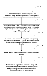
According to an aspect, a three-dimensional imaging method implemented by
using three-dimensional spatial information scanned by a constructed
two-dimensional scan probe is provided, comprising following steps:
Si) acquiring at least two orthogonal two-dimensional images at a current
position of a scanning target by the scan probe;
S2) moving the scan probe by a specific distance or rotating the scan probe by
a specific angle along a specific spatial direction, and acquiring at least
two
orthogonal two-dimensional images of the scanning target;
S3) comparing two groups of the two-dimensional images of a corresponding
direction obtained from step Si and step S2, and obtaining a movement distance
and a rotation angle of the scan probe along the corresponding direction;
S4) repeating steps Si to S3 until the scanning target is completely scanned;
S5) by means of the acquired two-dimensional images, as well as the
movement distance and the rotation angle of each image group relative to a
previous image group, reconstructing a three-dimensional image of the scanning
target.
In the three-dimensional imaging method of the present application, the
two-dimensional image is one of an ultrasonic image, an optical tomography
(OCT)
image, a photoacoustic image, and a terahertz image.
In the three-dimensional imaging method of the present application, one of
the two orthogonal two-dimensional images of the scanning target has a
relatively
2
CA 03027437 2018-12-12
low resolution to obtain the movement distance and rotation angle of the scan
probe in a plane corresponding to a clear image, not to obtain a final
three-dimensional imaging.
In the three-dimensional imaging method of the present application, the two
orthogonal two-dimensional images of the scanning target are different in
size.
In the three-dimensional imaging method of the present application, the two
orthogonal two-dimensional images of the scanning target are obtained in real
time
with a speed of at least 25 frames/second to ensure that a movement and
rotation of
the scan probe in an interval time between the images in two adjacent frames
mainly occur in one plane.
In the three-dimensional imaging method of the present application, the
rotation angle of the two-dimensional image is obtained by an accelerometer
provided on the scan probe.
According to another aspect, a three-dimensional imaging system by means of
a multi-directional two-dimensional scanning probe is provided, comprising at
least two probes for acquiring orthogonal images, an imaging device for
acquiring
the orthogonal images, a computing unit for analyzing a continuous movement
distance and rotation angle of the two-dimensional image probe, an
accelerometer
provided on the scan probe for acquiring the rotation angle of the two-
dimensional
orthogonal images.
In the three-dimensional imaging system by means of a multi-directional
two-dimensional scanning probe, the image acquired by the imaging device is
one
of an ultrasonic image, an optical tomography (OCT) image, a photoacoustic
image, and a terahertz image.
In the three-dimensional imaging system by means of a multi-directional
two-dimensional scanning probe, the probes for acquiring the orthogonal images
are different in size.
The three-dimensional imaging method and system proposed by the present
3
CA 03027437 2018-12-12
application constructs a three-dimensional image of a scanning target by using
a
three-dimensional imaging device according to image information and
three-dimensional spatial information obtained by the orthogonally constructed
scan probe, which not only simplifies a structural assembly of an imaging
system
and reduces the cost, but also improves an economic feasibility of the imaging
system.
BRIEF DESCRIPTION OF THE DRAWINGS
The present application will be further described below in conjunction with
the accompanying drawings and embodiments, in which:
Figure 1 is a schematic diagram showing the layout of the scan probes on
two planes according to the present application.
Figure 2 is a schematic diagram showing the formation of the two
two-dimensional images by using the two scan probes laying out in Figure 1.
Figure 3 is an explanatory diagram showing the movement of the scanning
target in different two-dimensional images.
Figure 4 is an explanatory diagram showing the rotation of the scanning
target in different two-dimensional images.
Figure 5 is an explanatory diagram of mutually orthogonal scan probes
formed by ultrasonic transducers.
Figure 6 is a schematic diagram of the ultrasonic scan probe transducer array
in the prior art.
Figure 7 is a schematic diagram of an ultrasonic transducer array having the
orthogonal scan probes of the present application.
Figure 8 is a schematic flow chart of a three-dimensional imaging method
according to an embodiment of the present application.
Figure 9 is a schematic diagram of an embodiment of the three-dimensional
4
CA 03027437 2018-12-12
imaging system proposed by the present application.
Figure 10 is a layout diagram of a transducer array composed of three
probes.
Figure 11 is a layout diagram of the present application which using two
OCT probes.
Figure 12 is a schematic diagram of the present application showing an
orthogonal scanning for acquiring two two-dimensional images by using two OCT
probes.
Figure 13 is a schematic diagram of the present application showing an
orthogonal scanning for acquiring two two-dimensional images by using one OCT
probe.
Figure 14 is a layout diagram of the present application which using a
photoacoustic imaging probe (including an ultrasound probe and a laser beam)
and
an orthogonal ultrasound probe.
DETAILED DESCRIPTION OF THE PREFERRED EMBODIMENT
Figure 1 is a schematic diagram showing the layout of the scan probes 101
on two planes. As there is only one probe in the existing scanning technology,
so
each time there is only one obtained two-dimensional image, which is not
helpful
for the three-dimensional imaging of the scanning target. Figure 2 is a
schematic
diagram showing the formation of the two two-dimensional images 102 by using
the two scan probes laying out in Figure 1. The two orthogonal probes acquire
a
group of two mutually orthogonal two-dimensional images by scanning the object
in each time. Figure 3 is an explanatory diagram showing the movement of the
scanning target in different two-dimensional images 103. A group of orthogonal
two-dimensional images that change position relative to the previous group of
orthogonal two-dimensional images can be obtained by moving the ultrasound
CA 03027437 2018-12-12
transducer array 105 with orthogonal probes 105. Figure 4 is an explanatory
diagram showing the rotation of the scanning target in different two-
dimensional
images 104. A group of orthogonal two-dimensional images at different angles
relative to the previous group of orthogonal two-dimensional images can be
obtained by rotating the scan probes 105 having an orthogonal transducer
array.
Figure 5 is an explanatory diagram of the transducer array in the orthogonal
scan
probes 105 of the present embodiment. Figure 6 is an explanatory diagram of
the
ultrasound probe transducer array 106 in the prior art, in which there is only
one
scan probe and only one two-dimensional image can be obtained in each
scanning.
Figure 7 is a schematic diagram of a transducer array 107 having the
orthogonal
scan probes according to the present embodiment. The ultrasound transducer
array
107 has two mutually orthogonal scan probes, and a group of two mutually
orthogonal two-dimensional images can be obtained by scanning the object in
each
time. Orthogonal two-dimensional images (104, 105) of different positions are
acquired by changing the position of the scan probe 107. Orthogonal
two-dimensional images (104, 105) at different locations, as well as distances
and
locations of adjacent groups are continuously obtained. Then the three-
dimensional
image of the relative scanning target is acquired by a three-dimensional
imaging
device.
Figure 8 is a schematic flow chart of a three-dimensional imaging method
using the spatial information acquired by a scan probe of the present
application.
Figure 9 is a schematic diagram of a three-dimensional imaging system provided
by the present application. Please refer to Figure 1-9, a three-dimensional
imaging
method using the spatial information acquired by the constructed scan probes
comprises the following steps.
Si) During each scanning, the ultrasonic transducer probe 107 constructed
by the orthogonal scan probes 105 acquires a group of at least two
two-dimensional images (103a, 103c) or (104a, 104c) of the scanning object at
the
6
CA 03027437 2018-12-12
=
current position. The ultrasonic transducer probe 107 is composed of two
orthogonal scan probes. The scanned two-dimensional images 103a and 103c are
orthogonal and the two-dimensional images 104a and 104c are orthogonal.
S2) The ultrasonic transducer scan probe 107 is moved by a specific distance
or rotated by a specific angle along a specific spatial direction, for
acquiring at
least two orthogonal two-dimensional images (103b,103d) or (104b,104d) of the
scanning target. In order to acquire a three-dimensional image of the scanning
target, the ultrasonic transducer scan probe 107 is moved by a specific
distance or
rotated by a specific angle along a specific spatial direction and
simultaneously
scans for acquiring a group of orthogonal two-dimensional images (103b,103d)
or
(104b,104d) after moving or rotating.
S3) The ultrasonic transducer scan probe 107 compares the two groups of
orthogonal two-dimensional images (103b,103d) and (103a,103c), or (104b,1034d)
and (104a,104c) of a corresponding direction obtained from steps Si and S2.
The
computing unit 202 for calculating the angle and position information of
spatial
information of different groups of orthogonal images is used to obtain the
movement distance and the rotation angle information of the scan probe 105 in
the
direction.
S4) The steps Si to S3 are repeated for continuously moving and rotating the
ultrasonic transducer probe 107 in a certain direction to scan and store the
two-dimensional images of the scanning object after moving and rotating; and
sending them to the three-dimensional imaging device 203 for three-dimensional
imaging until the scan probe traverses all position information of the
scanning
target.
S5) After receiving all the two-dimensional images of the scanning target
acquired in step S4 and the movement distance and rotation angle information
of
each image group relative to a previous image group, the three-dimensional
imaging device 203 reconstructs the three-dimensional image of the scanning
7
CA 03027437 2018-12-12
target.
In the present embodiment, referring to Figure 3 or Figure 4, one of the two
orthogonal two-dimensional images has a relatively low resolution, and a main
purpose of it is to obtain the movement distance and rotation angle of the
scan
probe 105 in a plane corresponding to a clear image, rather than for obtaining
a
final three-dimensional imaging of the three-dimensional imaging device 203.
According to the three-dimensional imaging method of the present
application, in the present embodiment, referring to Figure 3 or Figure 4, the
two
orthogonal two-dimensional images of the scanning target are obtained in real
time
with a speed of at least 25 frames/second to ensure that the movement and
rotation
of the ultrasonic transducer scan probe 107 in an interval time between the
images
in two adjacent frames mainly occur in one plane.
According to the three-dimensional imaging method of the present
application, in the present embodiment, the rotation angle of two adjacent
two-dimensional orthogonal images is obtained by an accelerometer 201a
provided
on the scan probe 201.
Referring to Figure 9, a three-dimensional imaging system by means of a
multi-directional two-dimensional scanning probe according to the present
application is provided, which comprises at least two probes 201 for acquiring
orthogonal images, a computing unit 202 for analyzing a continuous movement
distance and rotation angle of the two-dimensional image probe, an imaging
device
203 for acquiring the three-dimensional image.
According to the three-dimensional imaging system of the present
application, the scan probe 201 is orthogonally constructed by at least two
transducer arrays. On the one hand, the scan probe 201 continuously transmits
the
orthogonal images scanned in real time to the three-dimensional imaging device
203, and on the other hand transmits the angle information and the distance
position information to the computing unit 202. The computing unit 202
transmits
8
CA 03027437 2018-12-12
the angle information and the distance position information to the
three-dimensional imaging device 203 for three-dimensional imaging after
comparing and calculating the angle information and the distance position
information.
The scan probe 201 comprises a two-dimensional imaging device 201b and
an accelerometer for acquiring angle information of the scan probe 201. The
two-dimensional imaging device 201b is used for acquiring the two-dimensional
orthogonal images, and the accelerometer is used for acquiring the rotation
angle
information of the scan probe 201.
According to the three-dimensional imaging system 203 of the present
application, the acquired image is one of an ultrasonic image, an optical
tomography (OCT) image, a photoacoustic image, and a terahertz image.
According to the three-dimensional imaging system 203 of the present
application, the scan probes for acquiring orthogonal images are different in
size.
Figure 10 is a layout diagram of a transducer array composed of three probes
according to the imaging of the present application, in which probes 10B1 and
10B2 are orthogonal to probe10A. The image information formed by the probes
10B1 and 10B2 can complement with each other. If one of them cannot get a
image of a good quality, the other image can be used for related calculations.
The
information obtained in the two images can also be averaged to increase
accuracy.
Figure 11 is a layout diagram of the present application which using two
OCT probes according to the present application. Figure 12 is a schematic
diagram
of the present application showing an orthogonal scanning for acquiring two
two-dimensional images by using two OCT probes. Figure 13 is a schematic
diagram of the present application showing an orthogonal scanning for
acquiring
two two-dimensional images by using one OCT probe. That is, one OCT probe
scans a two-dimensional image at one position, and then scans at an orthogonal
position to obtain two orthogonal two-dimensional images.
9
CA 03027437 2018-12-12
Figure 14 is a layout diagram of the present application which using a
photoacoustic imaging probe (including a main ultrasound probe 141 and a laser
beam 142) and an auxiliary orthogonal ultrasound probe 143. The auxiliary
ultrasonic transducer array 143 for measuring the movement and rotation
information of the scan probe operates in an ultrasound imaging mode, that is,
transmitting and receiving ultrasound for imaging, thereby acquiring the
movement
and rotation of the main ultrasound probe 141 in the corresponding plane. The
main ultrasound probe 141 operating in the ultrasound mode can obtain more
information measurement of movement and rotation in the corresponding plane,
and can also operate in the photoacoustic imaging mode.
The above is only a preferred embodiment of the present application, but the
scope of the present application is not limited thereto. Any changes or
substitutions
that can be easily thought by any person skilled in the art within the
technical
scope disclosed by the present application are within the scope of the present
application. Therefore, the protection scope of the present application should
be
determined by the scope of the claims.
