Note: Descriptions are shown in the official language in which they were submitted.
CA 03027595 2018-12-13
WO 2017/214736
PCT/CA2017/050744
BIOPRINTED MENISCUS IMPLANT AND METHODS OF USING SAME
CROSS REFERENCE TO RELATED APPLICATIONS
[0001] This
application claims priority benefit of the filing date of US Provisional
Patent
Application Serial No. 62/351,222, filed on June 16, 2016, the disclosure of
which application is
herein incorporated by reference in its entirety.
FIELD OF THE INVENTION
[0002] The
invention provides synthetic tissue structures and methods for their
fabrication and
use, including artificial meniscus implants, comprising precisely patterned
layers containing a
variable synthetic tissue fiber structure dispensed from a bioprinter.
BACKGROUND OF THE INVENTION
[0003] The
meniscus is one of the most commonly damaged areas of the knee joint, with a
mean
incidence of injury in the United States of 66 injuries per 100,000 people.
Complete or partial
removal of the meniscus relieves acute pain, but without adequate replacement,
meniscus
removal can lead to damage of the articular cartilage of the knee, leading to
osteoarthritis (OA).
The meniscus typically demonstrates poor healing potential, and none of the
currently available
meniscal replacement options meets the necessary load-bearing and
biomechanical requirements
of this unique tissue, while also successfully engrafting into the surrounding
tissue to provide a
long-term solution to meniscus injury.
[0004] The
tissue engineering art has long sought to fabricate viable synthetic
structures capable
of mimicking and/or replacing living organs and tissues using myriad materials
and methods.
Historically, cells and other biological materials were seeded into pre-formed
three-dimensional
scaffolds imparting a desired structure, with the scaffold preferably being
biodegradable or
otherwise removable. See, e.g. U.S. Patent No. 6,773,713. Despite decades of
development,
however, significant challenges remain with this approach in respect of
effective cell seeding and
growth, and the technique does not work for more complex physiological
structures involving
more complicated spatial arrangements of different cell types.
[0005] More
recently, 3D printing, a form of additive manufacturing (AM), has been applied
to
create three-dimensional objects directly from digital files, wherein the
object is built up layer-
- 1 -
CA 03027595 2018-12-13
WO 2017/214736
PCT/CA2017/050744
by-layer to achieve the desired three dimensional structure. Initial efforts
to adapt 3D printing
techniques to the creation of cellular constructs and tissues, termed 3D
bioprinting, also focused
on initial printing of scaffold materials independent of the direct seeding or
subsequent printing
of the cellular materials, consistent with the above convention. See, e.g.
U.S. Patent No.
6,139,574; No. 7,051,654; No. 8,691,274; No. 9,005,972 and No. 9,301,925.
Unfortunately,
however, the polymers typically employed to form the prior art scaffolds,
while generally
considered biocompatible, are not physiologically compatible. As such, cell
viability is sacrificed
with this approach in favor of the mechanical stability of the requisite
scaffold.
[0006] In
the meniscus implant art in particular, for example, Bakarich et al. described
a system
in which a combination of an alginate/acrylamide gel precursor solution and an
expoxy based
UV-curable adhesive were combined to form a printable matrix material. ACS
Appl. Mater.
Interfaces 6:15998-16006 (2014). The printable matrix material was used in a
3D bioprinting
process to deposit a 2D layer of the matrix material alone, after which UV
light was passed over
the layer for one to five minutes to solidify it before depositing another
layer on top. Due to the
non-physiologic nature of the acrylamide gel and epoxy-based UV-curable matrix
components,
however, living cells cannot be maintained in this matrix material during the
bioprinting process,
and the resulting scaffold is still non-conducive to cell growth,
differentiation and
communication.
[0007]
Alternative 3D bioprinting techniques have also been described emphasizing the
converse, wherein mechanical structure and printing fidelity are sacrificed in
favor of cell
viability. These bioprinting systems create synthetic tissues by depositing
cellular materials
within a biocompatible matrix, which is then cross-linked or otherwise
solidified after deposition
to create a solid or semi-solid tissue structure. See, e.g., U.S. Patent No.
9,227,339; No.
9,149,952; No. 8,931,880 and No. 9,315,043; U.S. Patent Publication No.
2012/0089238; No.
2013/0345794; No. 2013/0164339 and No. 2014/0287960. With all of these
systems, however,
the temporal delay between the deposition and crosslinking steps invariably
leads to a lack of
control over the geometry of the printed structure, as well as the cellular
and matrix composition
of the structure. Moreover, cellular viability is often still compromised in
any event by the
subsequent cross-linking or solidification event.
[0008] As
but one example of this problem, Markstedt et al. described a system in which
hydrogels, such as collagen, hyaluronic acid, chitosan and alginate were used
in combination
- 2 -
CA 03027595 2018-12-13
WO 2017/214736
PCT/CA2017/050744
with non-physiologic reinforcing fiber materials, such as nanofibrillated
cellulose, as a bio-ink
for 3D bioprinting. BioMacromolecules 16:1489-96 (2015). This bio-ink is
deposited as a 2D
layer of material, which is submerged in a divalent cation bath (CaCl2) to
crosslink for ten
minutes and solidify the first layer before depositing another layer on top.
Although living cells
were successfully incorporated into their bio-ink, a cell viability analysis
demonstrated that the
cell viability decreased significantly as a result of the cross-linking
process, from ¨95.3% before
embedding, to ¨69.9% after embedding and crosslinking. Furthermore, a
comparison to non-
printed controls revealed that the decrease in cell viability was likely due
to the preparation and
mixing of the bio-ink itself, rather than the actual 3D printing process.
[0009]
Accordingly, existing 3D bioprinting techniques and materials have failed to
satisfactorily resolve the technical conflict between structural integrity and
printing fidelity on
the one hand, and physiological compatibility and cellular viability on the
other. The current
invention addresses these and other unmet needs. All prior art references
listed herein are
incorporated by reference in their entirety.
SUMMARY OF INVENTION
[0010] The
present invention successfully resolves the previously conflicting objectives
in the
3D bioprinting art between structural integrity and cellular viability,
providing synthetic tissue
structures deposited in solidified form with improved cell growth and/or
survival characteristics
and physiological functionality, and without the need for cross-linking or
other subsequent
solidification steps. Aspects of the present invention include synthetic
tissue structures
comprising one or more layers deposited by a bioprinter, wherein each layer
comprises synthetic
tissue fiber(s) comprising a solidified biocompatible matrix optionally
comprising cells, and
optionally comprising one or more active agents, wherein at least one of the
matrix material, cell
type, cell density, and/or amount of an active agent varies in at least one
direction within the
layers. Preferably, at least one of said layers comprises a single continuous
synthetic tissue fiber
dispensed from the bioprinter having a variable composition.
[0011] In
specific embodiments, meniscus implants are provided comprising layers of
synthetic
tissue fiber(s) dispensed from a bioprinter as a solidified biocompatible
matrix optionally
comprising cells, and optionally comprising one or more active agents, wherein
at least one of
the matrix material, cell type, cell density, and/or amount of an active agent
varies in at least one
- 3 -
CA 03027595 2018-12-13
WO 2017/214736
PCT/CA2017/050744
direction within the layers. Preferably, at least one of said layers comprises
a continuous
synthetic tissue fiber dispensed from the bioprinter having a variable
composition. More
preferably, each of said layers comprises a continuous synthetic tissue fiber
having a variable
composition. Still more preferably, a meniscus implant comprises a reinforced
peripheral region,
and/or at least one anchor region, as described herein.
[0012] In
one aspect, the invention provides a synthetic tissue structure comprising a
plurality
of layers deposited by a bioprinter, each layer comprising synthetic tissue
fiber(s) comprising a
solidified biocompatible matrix optionally comprising cells, and optionally
comprising one or
more active agents, wherein at least one of said layers comprises a matrix
material varying in
type and/or amount in at least one direction. In some embodiments, each layer
comprises a
matrix material varying in type and/or amount in at least one direction.
[0013] In
another aspect, the invention provides a synthetic tissue structure comprising
a
plurality of layers, each layer comprising synthetic tissue fiber(s)
comprising a plurality of
mammalian cells dispensed from a bioprinter within a solidified biocompatible
matrix, wherein
at least one of said layers comprises a cell type and/or cell density varying
in at least one
direction. In some embodiments, each layer comprises a cell type and/or cell
density varying in
at least one direction.
[0014] In
another aspect, the invention provides a synthetic tissue structure comprising
a
plurality of layers deposited by a bioprinter, each layer comprising synthetic
tissue fiber(s)
comprising a solidified biocompatible matrix optionally comprising cells,
wherein at least one of
said layers comprises an active agent varying in type and/or amount in at
least one direction. In
some embodiments, each layer comprises an active agent varying in type and/or
amount in at
least one direction.
[0015] In
some embodiments, one or more synthetic tissue fibers are dispensed in a
desired
pattern or configuration to form a first layer, and one or more additional
layers are then
dispensed on top, having the same or a different pattern or configuration. In
certain
embodiments, a plurality of layers are stacked to form a three dimensional
structure that can be
used as an artificial meniscus implant. Preferably, at least one of said
layers comprises a single
continuous synthetic tissue fiber dispensed from the bioprinter having a
variable composition.
More preferably, each of said layers comprises a single continuous synthetic
tissue fiber having a
variable composition.
- 4 -
CA 03027595 2018-12-13
WO 2017/214736
PCT/CA2017/050744
[0016] In
some embodiments, a synthetic tissue structure comprises a number of
individual
layers that ranges from about 1 to about 250, such as about 2, 3, 4, 5, 6, 7,
8, 9, 10, 11, 12, 13,
14, 15, 16, 17, 18, 19, 20, 25, 30, 35, 40, 45, 50, 55, 60, 65, 70, 75, 80,
85, 90, 95, 100, 105, 110,
115, 120, 125, 130, 135, 140, 145, 150, 155, 160, 165, 170, 175, 180, 185,
190, 200, 205, 210,
215, 220, 225, 230, 235, 240 or about 245 individual layers. Any suitable
number of individual
layers can be incorporated to generate a tissue structure having desired
dimensions.
[0017] In
some embodiments, one or more individual fibers and/or layers are organized to
create
one or more zones within a tissue structure, wherein each zone has one or more
desired
properties (e.g., one or more mechanical and/or biological properties). As
used herein, the term
"region" refers to a portion of a tissue structure defined in an x-y plane
(e.g., an area or portion
of an individual layer, where each layer of the tissue structure defines an x-
y plane), whereas the
term "zone" refers to a portion of a tissue structure defined in the z-
direction and comprising at
least two contiguous regions from separate x-y planes, or layers (e.g., a
"macrolayer" that
comprises a plurality of individual "microlayers").
[0018] Zones
in accordance with embodiments of the invention can have any desired three
dimensional geometry, and can occupy any desired portion of a synthetic tissue
structure. For
example, in some embodiments, a zone can span an entire length, width, or
height of a synthetic
tissue structure. In some embodiments, a zone spans only a portion of a
length, width, or height
of a synthetic tissue structure. In some embodiments, a synthetic tissue
structure comprises a
plurality of different zones that are organized along a length, width, height,
or a combination
thereof, of the synthetic tissue structure. In one preferred embodiment, a
synthetic tissue
structure comprises three different zones that are organized along the height
of the synthetic
tissue structure, such that a path through the synthetic tissue structure from
the bottom to the top
would pass through all three zones.
[0019] In
some embodiments, a zone can comprise a number of layers that ranges from
about 2
to about 250, such as about 3, 4, 5, 6, 7, 8, 9, 10, 11, 12, 13, 14, 15, 16,
17, 18, 19, 20, 25, 30, 35,
40, 45, 50, 55, 60, 65, 70, 75, 80, 85, 90, 95, 100, 105, 110, 115, 120, 125,
130, 135, 140, 145,
150, 155, 160, 165, 170, 175, 180, 185, 190, 200, 205, 210, 215, 220, 225,
230, 235, 240 or
about 245 individual layers. In some embodiments, the individual layers within
a zone are
organized in a manner that confers one or more mechanical and/or biological
properties on the
zone. For example, in some embodiments, the individual layers within a zone
comprise one or
- 5 -
CA 03027595 2018-12-13
WO 2017/214736
PCT/CA2017/050744
more reinforcing materials that confer increased mechanical strength on the
zone. In some
embodiments, the individual layers within a zone comprise one or more
materials that confer
desirable cell growth properties on the zone. In some embodiments, the
individual layers within a
zone, or the plurality of individual compartments of a fiber structure passing
through the zone,
can be alternated in a manner that confers desirable properties on the zone.
For example, in some
embodiments, the individual layers or regions within a zone are alternated
such that the odd
numbered layers contain one or more reinforcing materials that confer
desirable mechanical
properties on the zone, and the even numbered layers contain one or more
materials that confer
desirable biological properties on the zone (e.g., softer materials that are
conducive to cell
migration, growth, viability, and the like). In some embodiments, a zone
comprises a plurality of
contiguous individual layers (e.g., about 2, 3, 4, 5, 6, 7, 8, 9 or about 10
or more contiguous
layers) that comprise one or more reinforcing materials that confer increased
mechanical strength
on the zone, which contiguous layers are alternated with another plurality of
contiguous
individual layers (e.g., about 2, 3, 4, 5, 6, 7, 8, 9 or about 10 or more
contiguous layers) that
comprise one or more materials that confer desirable biological properties on
the zone (e.g.,
softer materials that are conducive to cell migration, growth, viability, and
the like).
[0020] In
one aspect, an artificial meniscus implant comprises at least one basal zone,
at least
one interior zone, and at least one superficial zone, wherein at least one of
said zones comprises
a layer comprising a synthetic tissue fiber(s) comprising a solidified
biocompatible matrix,
wherein the matrix materials vary in type and/or amount between the center of
a layer and the
periphery of the layer. In some embodiments, one or more matrix materials at
or near the
periphery of the layer comprise a reinforced matrix material.
[0021]
Aspects of the invention also include artificial meniscus implants that
comprise one or
more anchor regions. As used herein, the term "anchor region" refers to a
region that comprises
one or more reinforced matrix materials. Artificial meniscus implants in
accordance with
embodiments of the invention can include any suitable number of anchor
regions, such as 1 to
12, such as 2, 3, 4, 5, 6, 7, 8, 9, 10 or 11 anchor regions. In some
embodiments, an artificial
meniscus implant comprises no anchor regions.
[0022] In
another aspect, artificial meniscus implants are provided comprising at least
one basal
zone, at least one interior zone, and at least one superficial zone, wherein
at least one zone
comprises a layer comprising at least one synthetic tissue fiber comprising a
plurality of
- 6 -
CA 03027595 2018-12-13
WO 2017/214736
PCT/CA2017/050744
mammalian cells dispensed from a bioprinter within a solidified biocompatible
matrix, wherein
at least one layer comprises a cell density that varies in at least one
direction. In some
embodiments, each of said layers comprises a cell density that varies in at
least one direction. In
some embodiments, the cell density ranges from 0 to about 100 x 106 cells/mL.
[0023] In
another aspect, artificial meniscus implants in accordance with embodiments of
the
invention include at least one basal zone, at least one interior zone, and at
least one superficial
zone, wherein at least one layer in one of said zones comprises a synthetic
tissue fiber(s)
comprising a solidified biocompatible matrix and at least one active agent,
wherein the at least
one active agent varies in type and/or amount between the center of the layer
and the periphery
of the layer.
[0024] In
some embodiments, the biocompatible matrix on the periphery of the layer may
comprise at least one active soluble agent that is released over time from the
matrix to encourage
host vascular cell ingrowth and chondrocyte cell ingrowth. Such bioactive
agents include, but are
not limited to: vascular endothelial growth factor (VEGF), fibroblast growth
factor (FGF),
insulin-like growth factor-1 (IGF-1), bone morphogenetic factors, hepatocyte
scatter factor,
urokinase plaminogen activator, transforming growth factor-I3 (TGF-I3),
platelet derived growth
factor (PDGF), or any combination thereof.
[0025] In
some embodiments, the biocompatible matrix on the periphery of the layer may
comprise at least one insoluble factor to encourage cell ingrowth. Non-
limiting examples of
such insoluble factors include: hyaluronic acid or sulfated hyaluronic acid,
fibronectin, fibrin,
and collagen I. Additional bioactive factors can be incorporated into the
matrix arranged in the
interior of the subject artificial meniscus implants to encourage collagen
deposition by
chondrocytes including. Non-limiting examples of such additional bioactive
factors include:
insulin, connective tissue-derived growth factor (CTGF), or a combination
thereof.
[0026] In
some embodiments, portions or regions of the periphery will comprise at least
one
active agent. In some embodiments, the entire periphery of a layer comprises
at least one active
agent. In some embodiments, the periphery comprises a plurality of active
agents. In some
embodiments the entire periphery of the layer includes an active agent that
reduces the host
inflammatory response, for example, via the inclusion of one or more steroid
compounds
contained within one of more microparticles to ensure sustained release over
an extended time
period.
- 7 -
CA 03027595 2018-12-13
WO 2017/214736
PCT/CA2017/050744
[0027] In
some embodiments, an artificial meniscus implant has an arcuate shape that has
an
anterior end, a posterior end, a middle section therebetween defining a curved
path between said
anterior and posterior ends, an internal side, and an external side. In some
embodiments, the cell
density increases in a radial manner from the internal side towards the
external side. In some
embodiments, the concentration of reinforced matrix materials increases in a
radial manner from
the internal side towards the external side. In some embodiments, the amount
of active agent
increases in a radial manner from the internal side towards the external side.
[0028] In
some embodiments, the basal zone comprises one or more layers comprising
randomly-oriented synthetic tissue fiber(s); the interior zone comprises one
or more layers
comprising circumferentially-oriented synthetic tissue fiber(s) and radially-
oriented synthetic
tissue fiber(s); and the superficial zone comprises one or more layers
comprising randomly-
oriented synthetic tissue fiber(s). In some embodiments, the circumferentially-
oriented synthetic
tissue fiber(s) has a first diameter and the radially-oriented synthetic
tissue fiber(s) has a second,
different diameter. In some embodiments, the circumferentially-oriented
synthetic tissue fiber(s)
and the radially-oriented synthetic tissue fiber(s) have the same diameter. In
some embodiments,
the synthetic tissue fiber(s) has a diameter that ranges from about 20 lam to
about 500 pm.
[0029] In
some embodiments, the circumferentially-oriented synthetic tissue fiber(s)
comprises a
first solidified biocompatible matrix, and the radially-oriented synthetic
tissue fiber(s) comprises
a second, different solidified biocompatible matrix. In some embodiments, the
circumferentially-
oriented synthetic tissue fiber(s) and the radially-oriented synthetic tissue
fiber(s) comprise the
same solidified biocompatible matrix.
[0030] In
some embodiments, the interior zone comprises a layer comprising a synthetic
tissue
fiber(s) that is configured to promote deposition of collagen fibers aligned
with a longitudinal
direction of the synthetic tissue fiber(s). In some embodiments, the interior
zone comprises a
layer comprising a circumferentially-oriented synthetic tissue fiber(s) that
is configured to
promote deposition of collagen fibers that are aligned with a longitudinal
direction of the
circumferentially-oriented synthetic tissue fiber(s). In some embodiments, the
interior zone
comprises a layer comprising a radially-oriented synthetic tissue fiber(s)
that is configured to
promote deposition of collagen fibers that are aligned with a longitudinal
direction of the
radially-oriented synthetic tissue fiber(s).
- 8 -
CA 03027595 2018-12-13
WO 2017/214736
PCT/CA2017/050744
[0031] The
solidified biocompatible matrix can comprise any of a wide variety of natural
or
synthetic polymers that support the viability of living cells, including,
e.g., alginate, laminin,
fibrin, hyaluronic acid, poly(ethylene) glycol based gels, gelatin, chitosan,
agarose, or
combinations thereof. In preferred embodiments, the solidified biocompatible
matrix comprises
alginate, or other suitable biocompatible polymers that can be instantaneously
solidified while
dispensing from the print head. In further preferred embodiments, the
solidified biocompatible
matrix comprises a homogeneous composition of alginate throughout the radial
cross section of
each synthetic tissue fiber.
[0032] In
particularly preferred embodiments, the solidified biocompatible matrix is
physiologically compatible, i.e., conducive to cell growth, differentiation
and communication. In
some such embodiments, the physiologically compatible matrix comprises
alginate in
combination with one or more of: collagen, fibronectin, thrombospondin,
glycosaminoglycans
(GAG), deoxyribonucleic acid (DNA), adhesion glycoproteins, elastin, and
combinations
thereof. In specific embodiments, the collagen is selected from the group
consisting of: collagen
I, collagen II, collagen III, collagen IV, collagen V, collagen VI, or
collagen XVIII. In specific
embodiments, the GAG is selected from the group consisting of: hyaluronic
acid, chondroitin-6-
sulfate, dermatan sulfate, chondroitin-4-sulfate, or keratin sulfate.
[0033] As
reviewed above, anchor regions can be generated by the incorporation of higher
strength materials into specific zones of an implant (i.e., suture points),
for example, stiffer
synthetic materials, including, but not limited to, polycaprolactone (PCL),
poly(lactic-co-glycolic
acid) (PLGA), polyurethane (PU) and any combination thereof. In some
embodiments, an anchor
region can contain double network hydrogels, generated by combining at least
two different
hydrogel materials, examples of which include, without limitation, alginate,
Gelatin methacrylol
(GelMA), methacryloyl polyethylene glycol (PEGMA), gellan gum, agarose,
polyacrylamide, or
any combination thereof. In addition, high strength fibers can be generated
from high
concentrations of biological polymers including, without limitation, collagen,
chitosan, silk
fibroin, or any combination thereof, and these biological polymers can be
incorporated into one
or more anchor regions. In some embodiments, an anchor region and/or a
reinforced peripheral
region of an implant comprises one or more layers of high strength material(s)
deposited in
alternation in the z-direction with one or more layers of softer matrix
materials containing, e.g.,
hydrogel material(s) conducive to cell survival and ingrowth described above.
In this way, the
- 9 -
CA 03027595 2018-12-13
WO 2017/214736
PCT/CA2017/050744
softer, cell compatible hydrogel materials provide one or more desirable
biological functions,
and the stiffer materials provide one or more desirable mechanical functions,
to generate a hybrid
structure with appropriate mechanical and biological functions.
[0034] In
some embodiments, the mammalian cells are selected from the group consisting
of:
fibroblasts, chondrocytes, fibrochondrocytes, primary human meniscus-derived
chondrocytes,
stem cells, bone marrow cells, embryonic stem cells, mesenchymal stem cells,
bone marrow-
derived mesenchymal stem cells, induced pluripotent stem cells, differentiated
stem cells, tissue-
derived cells, microvascular endothelial cells, and combinations thereof. In
preferred
embodiments, the cell viability within the synthetic living tissue structures
ranges from about
70% up to about 100%, such as about 75%, about 80%, about 85%, about 90%,
about 95%,
about 98%, about 99%, about 99.5%, or about 99.9% in comparison with cell
viability before
printing.
[0035] In
some embodiments, the meniscus implant further comprises an acellular sheath
positioned below the basal zone. In some embodiments, the meniscus implant
further comprises
an acellular sheath positioned above the superficial zone. In some
embodiments, the meniscus
implant comprises a first acellular sheath positioned below the basal zone and
a second acellular
sheath positioned about the superficial zone.
[0036] In
some embodiments, the meniscus implant further comprises at least one active
agent.
In some embodiments, the at least one active agent is selected from the group
consisting of:
TGF-131, TGF-132, TGF-133, BMP-2, BMP-4, BMP-6, BMP-12, BMP-13, basic
fibroblast growth
factor, fibroblast growth factor-1, fibroblast growth factor-2, platelet-
derived growth factor-AA,
platelet-derived growth factor-BB, platelet rich plasma, IGF-I, IGF-II, GDF-5,
GDF-6, GDF-8,
GDF-10, vascular endothelial cell-derived growth factor, pleiotrophin,
endothelin, nicotinamide,
glucagon like peptide-I, glucagon like peptide-II, parathyroid hormone,
tenascin-C, tropoelastin,
thrombin-derived peptides, laminin, biological peptides containing cell-
binding domains and
biological peptides containing heparin-binding domains, therapeutic agents,
and any
combinations thereof.
[0037] In
preferred embodiments, the bioprinter dispenses the solidified biocompatible
matrix
comprising the plurality of mammalian cells through a single orifice. In
particularly preferred
embodiments, the single orifice is comprised within a print head such as that
described and
- 10 -
CA 03027595 2018-12-13
WO 2017/214736
PCT/CA2017/050744
claimed in WO 2014/197999, the disclosure of which is herein incorporated by
reference in its
entirety.
BRIEF DESCRIPTION OF THE DRAWINGS
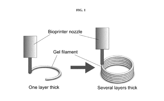
[0038] FIG.
1 is a schematic depiction of a layer-by-layer synthetic tissue fiber
deposition
process.
[0039] FIG.
2 is a schematic illustration of a knee joint, depicting a lateral and a
medial
meniscus. (Adapted from: The knee meniscus: structure-function,
pathophysiology, current
repair techniques, and prospects for regeneration. Biomaterials. 2011 October
; 32(30): 7411-
7431. doi:10.1016/j.biomaterials.2011.06.037, Eleftherios A. Makris, MD1,
Pasha Hadidi, BS1,
and Kyriacos A. Athanasiou, Ph.D., P.E.1).
[0040] FIG.
3 is a schematic illustration of a meniscus, depicting both a top view and a
cross
sectional view. The outer (red-red) region, central (white-red) region, and
the inner (white-white)
region are depicted. (Adapted from: The knee meniscus: structure-function,
pathophysiology,
current repair techniques, and prospects for regeneration. Biomaterials. 2011
October ; 32(30):
7411-7431. doi: 10.1016/j .biomaterials.2011.06.037, Eleftherios A. Makris,
MD1, Pasha Hadidi,
BS1, and Kyriacos A. Athanasiou, Ph.D., P.E.1).
[0041] FIG.
4, Panel A is a schematic illustration of a meniscus, depicting a cross
sectional
view. Circumferential and radial alignment of collagen fibers confer
biomechanical properties to
the meniscus. Panel B depicts a superficial zone, a lamellar zone, and an
interior (deep) zone.
Collagen fibers in the superficial and lamellar zones close to the meniscus
surface are randomly
oriented. Fibers deeper in the meniscus are oriented in both circumferential
and radial directions.
[0042] FIG.
5 is a force diagram that depicts the components of an axial load force F on
various
portions of a meniscus. The axial load force (F) perpendicular to the meniscus
surface and
horizontal force (f r) are created by compressing the femur (F f). F rebounds
due to the tibial
upgrade force (Ft), whereas f r leads to meniscal extrusion radially, which is
countered by the
pulling force from the anterior and posterior insertional ligaments.
Consequently, tensile hoop
stress is created along the circumferential directions during axial
compression, which is resisted
by the circumferentially-oriented collagen fibers. (Adapted from: The knee
meniscus: structure-
function, pathophysiology, current repair techniques, and prospects for
regeneration.
-11-
CA 03027595 2018-12-13
WO 2017/214736
PCT/CA2017/050744
Biomaterials. 2011 October ; 32(30): 7411-7431.
doi:10.1016/j.biomaterials.2011.06.037,
Eleftherios A. Makris, MD1, Pasha Hadidi, BS1, and Kyriacos A. Athanasiou,
Ph.D., P.E.1).
[0043] FIG.
6 provides images of two cell-free 3D meniscus-like structures with pre-
programmed zone-specific scaffold content and coordinated patterning of
printed synthetic tissue
fiber structures. Scale bar = lcm.
[0044] FIG.
7 is a series of microscope images that depict spontaneous collagen fiber
alignment
in small diameter fibers. Polymerised collagen fiber orientation in
microfluidic channels of
different diameters including; 30um (Panel a), 100um (Panel b), 400um (Panel
c) and no channel
(Panel d) (Lee et al., 2006).
[0045] FIG.
8 shows a series of images showing on-the-fly modulation of printed alginate-
based
fiber diameter using a 3D bioprinting system, as well as a graph comparing the
mean fiber
diameters. Panels A, B, and C depict alginate-based fibers of 3 different
diameters that were
generated by printing at 3 different pressure settings in the 3D bioprinting
system. Quantification
of width in multiple fibers demonstrates that the mean diameter at each
pressure setting is
consistent (graph, right).
[0046] FIG.
9 is an illustration of synthetic tissue fiber patterning in various layers of
a 3D
bioprinted meniscus. Synthetic tissue fiber structures of specific diameters
loaded with
extracellular matrix (ECM), e.g., collagen, are patterned in a manner that
recapitulates the micro-
patterning of collagen and the zonal architecture of the meniscus. The basal
and superficial zones
contain randomly oriented fibers printed in larger diameter fibers. The
interior zones contain
circumferential and radially-aligned collagens aligned within patterned fibers
of smaller
diameter.
[0047] FIG.
10 shows data from 2-photon imaging of collagen fibers in an engineered 3D
tissue.
Panel A: formaldehyde-fixed, H&E stained section of a 3D co-culture of primary
human airway
epithelial cells and fibroblasts after 90 day culture on an electrospun
gelatin (ESG) scaffold.
Panel B: 2-photon imaging of unstained sections demonstrating deposition of
fibrillar collagen
(purple) oriented parallel to the surface of the ESG scaffold, in a similar
direction to the
fibroblasts depositing the collagen. Panel C: Emission spectra of the
unstained tissues
demonstrates that non-centro-symmetric collagen fibers generate a specific 2nd
harmonic signal
(SHG) (Wadsworth et al., 2014).
- 12 -
CA 03027595 2018-12-13
WO 2017/214736
PCT/CA2017/050744
[0048] FIG.
11 is an illustration of a meniscal tissue with zone-specific cell and ECM
content.
"Red-Red bio-ink" and "White-white bio-ink" are used to generate tissues with
zonal
architecture. Desired cell types, (e.g., MSC-derived chondrocytes or primary
meniscus-derived
cells) are seeded at appropriate physiological densities into red-red and
white-white zones.
Specific ECM content of the scaffold is modified according to the tissue zone.
The "white-red"
zone in the central zone of the tissue contains a mixture of red-red and white-
white bio-inks and
cells. The bioprinting system facilitates control over both the cellular (cell
type and cell density)
and ECM content in any given zone of the meniscus implant.
DETAILED DESCRIPTION
[0049]
Aspects of the present invention include synthetic tissue structures
comprising one or
more layers deposited by a bioprinter, wherein each layer comprises synthetic
tissue fiber(s)
comprising a solidified biocompatible matrix optionally comprising cells, and
optionally
comprising one or more active agents, wherein at least one of the matrix
material, cell type, cell
density, and/or amount of an active agent varies in at least one direction
within the layers.
Preferably, at least one of said layers comprises a single continuous
synthetic tissue fiber
dispensed from the bioprinter having a variable composition. The term
"solidified" as used
herein refers to a solid or semi-solid state of material that maintains its
shape fidelity and
structural integrity upon deposition. The term "shape fidelity" as used herein
means the ability of
a material to maintain its three dimensional shape. In some embodiments, a
solidified material is
one having the ability to maintain its three dimensional shape for a period of
time of about 30
seconds or more, such as about 1, 10 or 30 minutes or more, such as about 1,
10, 24, or 48 hours
or more. The term "structural integrity" as used herein means the ability of a
material to hold
together under a load, including its own weight, while resisting breakage or
bending.
[0050] In
some embodiments, a solidified composition is one having an elastic modulus
greater
than about 15, 20 or 25 kilopascals (Oa), more preferably greater than about
30, 40, 50, 60, 70,
80 or 90 Oa, still more preferably greater than about 100, 110, 120 or 130 Oa.
Preferred elastic
modulus ranges include from about 15, 25 or 50 Pa to about 80, 100, 120 or 140
kPa.
[0051]
Additional aspects of the invention include artificial meniscus implants for
use in
repairing and/or replacing a damaged or diseased meniscal tissue in a
mammalian subject,
comprising synthetic tissue fiber(s) dispensed from a bioprinter as a
solidified biocompatible
- 13 -
CA 03027595 2018-12-13
WO 2017/214736
PCT/CA2017/050744
matrix optionally containing cells, and optionally containing one or more
active agents, wherein
at least one of the matrix material, cell type, cell density, and/or type
and/or amount of an active
agent varies in at least one direction within the fiber.
[0052] As
provided in FIG. 1, a solidified biocompatible matrix optionally containing a
plurality
of mammalian cells is dispensed from a bioprinter forming one or more
synthetic tissue fiber(s)
on a deposition surface, and ultimately forming a layer. As such, subsequent
cross-linking or
other solidification steps are unnecessary after dispensation of the already-
solidified matrix from
the printhead. Accordingly, a second layer can be rapidly deposited on top of
the first layer,
while maintaining the structural integrity of the first layer, and this
process can be continued to
deposit a plurality of layers, one on top of the next, until a three
dimensional structure having a
desired geometry is obtained.
[0053] The
solidified biocompatible matrix may advantageously comprise alginate, or any
other
suitable biocompatible polymer that can be instantaneously solidified while
dispensing from the
printhead. In a preferred embodiment, the alginate-based matrix is printed and
simultaneously
crosslinked at the time of printing by contacting with a divalent cation
crosslinking solution (e.g.,
a CaCl2 solution) before or upon dispensation from the printhead. In
particularly preferred
embodiments, the alginate-based biocompatible matrix further comprises one or
more
physiological materials, as described in more detail herein. In further
preferred embodiments,
the solidified biocompatible matrix comprises a homogeneous composition of
alginate
throughout the radial cross section of each synthetic tissue fiber.
[0054] In
some embodiments, a synthetic tissue fiber structure comprises a plurality of
individual compartments (organized along the length of the synthetic tissue
fiber) that are created
by sequentially depositing different matrix materials (e.g., natural and/or
synthetic polymers),
different cell types, different cell concentrations, and/or different types
and/or amounts of active
agents in each compartment of the same continuous synthetic tissue fiber
structure. For example,
in some embodiments, a synthetic tissue fiber structure comprises a first
compartment that
comprises a first matrix material, and a second compartment that comprises a
second matrix
material. In some embodiments, a synthetic tissue fiber structure comprises a
first compartment
that comprises a first cell type, and a second compartment that comprises a
second cell type. In
some embodiments, a synthetic tissue fiber structure comprises a first
compartment that
comprises a first cell concentration, and a second compartment that comprises
a second cell
- 14 -
CA 03027595 2018-12-13
WO 2017/214736
PCT/CA2017/050744
concentration. In some embodiments, a synthetic tissue fiber structure
comprises a first
compartment that comprises a first active agent, and a second compartment that
comprises a
second active agent. Any combination of matrix materials, cell types, cell
concentrations, and/or
types and/or amounts of active agents can be used in different compartment of
a subject synthetic
tissue fiber structure to achieve desired biomechanical properties and/or
biological activities.
[0055]
Synthetic tissue fiber structures in accordance with embodiments of the
invention can
include controlled patterning of different matrix materials (e.g., natural
and/or synthetic
polymers) and crosslinking techniques to create a desired cross-sectional
profile within a given
compartment. For example, in some embodiments, a synthetic tissue fiber
structure comprises a
compartment having a solid, tubular, or porous cross-sectional profile. Non-
limiting examples of
cross-sectional profiles that can be created in a synthetic tissue fiber
structure in accordance with
embodiments of the invention include those described in Jun, Yesl, et al.
"Microfluidic spinning
of micro-and nano-scale fibers for tissue engineering." Lab on a Chip 14.13
(2014): 2145-2160,
the disclosure of which is incorporated herein by reference in its entirety.
[0056] In
some embodiments, the resulting synthetic tissue fiber is patterned, using
software
tools, to form layers optionally containing a plurality of mammalian cells
and/or a plurality of
biocompatible matrix materials. In certain embodiments, a plurality of layers
is deposited in a
sequential manner to generate a multi-layered meniscus implant comprising a
plurality of zones.
In some embodiments, a meniscus implant comprises at least one basal zone, at
least one interior
zone, and at least one superficial zone, wherein the interior zone comprises
at least one layer
comprising at least one circumferentially-oriented synthetic tissue fiber, and
at least one radially-
oriented synthetic tissue fiber. Preferably, at least one of said layers
comprises a single
continuous synthetic tissue fiber dispensed from the bioprinter having a
variable composition.
[0057] One
advantage of the subject meniscus implants is that the matrix composition,
cell type,
cell density, and active agent type and/or concentration can be controlled at
any given point in
any portion of any layer of the implant, thereby facilitating the generation
of meniscus implants
more closely resembling the natural architecture of a meniscus tissue, and
that possess desirable
biomechanical properties, including, but not limited to, reinforced anchor
regions on the
periphery of the implant, circumferentially- and radially-oriented fiber
structures within the
meniscus implant, as well as specific cell types and cell densities within
specific regions and/or
zones of the implant.
- 15 -
CA 03027595 2018-12-13
WO 2017/214736
PCT/CA2017/050744
[0058]
Another advantage of the present invention is that one or more active agents
(described in
more detail herein) can be selectively added to different compartments of a
synthetic tissue fiber
to allow precise localization of an active agent within one or more layers of
a meniscus implant,
including, but not limited to, increased concentrations of appropriate active
agents on the
periphery of an acellular implant to encourage the ingrowth of endogenous
cells. The subject
meniscus implants are described in further detail below.
Meniscus Anatomy:
[0059] The
menisci are a pair of crescent-shaped fibrocartilages comprised of both a
medial and
a lateral component situated between the corresponding femoral condyle and
tibial plateau. (FIG.
2). The anterior and posterior insertional ligaments attach the menisci
firmly, and they fix the
meniscus to the tibial plateau well. Menisci are generally wedge-shaped, and
the lateral menisci
are approximately 32.4-35.7 mm in length, and approximately 26.6-29.3 mm wide,
while the
medial menisci are approximately 40.5-45.5 mm long and approximately 27 mm
wide. Each is a
glossy-white, complex tissue comprised of cells, specialized extracellular
matrix (ECM)
materials, and zone-specific innervation and vascularization. The menisci are
fully vascularized
at birth, however, over time the blood vessels retreat outwards until (in
humans) at 10 years of
age, approximately 10-30% of the meniscus at the periphery is vascularized.
The adult human
meniscus thus has two distinct zones, the outer, vascular/neural zone (red-red
zone), and the
inner completely avascular/aneural zone (white-white zone). These regions are
separated by the
narrow central (red-white) zone that contains features of both the outer (red-
red) and the inner
(white-white) zones (FIG. 3). Critically, the self-healing capacity of each
area is directly related
to blood supply, leaving the inner, white-white zone susceptible to trauma and
degenerative
lesions.
Meniscus cellular and biochemical composition
[0060] The
meniscus is a highly hydrated tissue comprising approximately 72% water, with
the
remaining 28% mostly comprising ECM and cells. Collagens make up most of the
ECM (75%)
followed by glycosaminoglycans (GAGs, 17%) DNA (2%), adhesion glycoproteins
(<1%) and
elastin (<1%). These ratios vary depending on the zone of the tissue, age, and
condition. The
- 16 -
CA 03027595 2018-12-13
WO 2017/214736
PCT/CA2017/050744
cellular component of the meniscus is zone-specific, comprising both
fibrochondrocytes and
chondrocyte-like cells.
[0061] The
composition of the meniscus differs in each zone. In the outer red-red zone,
the cells
are more fibroblast-like in morphology, with many processes. The ECM in this
zone is mainly
fibrillar collagen type-I (80%). The inner white-white zone has ECM closely
resembling hyaline
cartilage, with more collagen-II (42%), a reduced proportion of collagen-I
(28%) and a higher
GAG concentration. The cells in this zone are termed fibrochondrocytes, or
chondrocyte-like
cells. The superficial layers of the menisci have another distinct cell type
with potential stem
cell-like properties. The zone-specific ECM components of the meniscus are
generated by the
cells resident within the tissue, thus phenotypic markers for meniscal cells
can include ECM
protein expression or gene expression such as: COL1A1 (collagen-1), COL2A2
(collagen-2),
VCAN (versican), ACAN (aggrecan), CSPG4 (chondroitin-6-sulfate), Sox9 and Coll
Oa
(collagen-10a). Similar to the unique cell types in each meniscal zone, cell
density also varies in
each zone. Vascular (red-red) and avascular (white-red, white-white) zones
have avergage cell
densities of 12,820 cells/mm3 and 27,199 cells/mm3, respectively, and more
fibrochondrocytes
than fibroblast-like cells (Cengiz et al., 2015). The meniscus is highly
heterogenous, with zone-
specific variation in cell phenotype and ECM composition.
[0062] The
heterogeneous distribution of cell types and biochemical scaffold content of
the knee
meniscus is described in Table 1. The red-red zone is characterised by
fibroblast-like cells and a
collagen-I-predominant extracellular matrix (ECM), with trace amounts of
collagen-II. The
white-red and white-white zones contain fibrochondrocyte cells and a matrix
rich in collagen-II,
and a higher proportion of glycosaminoglycans (GAGs).
- 17 -
CA 03027595 2018-12-13
WO 2017/214736
PCT/CA2017/050744
Table 1:
Organic Red-Red zone Red-white zone White-White
component/Zone zone
Cells Vessels, Fibrochondrocytes/ Fibrochondrocytes
nerves, & Chondrocyte-like & superficial zone
fibroblast-like cells cells (stem cells)
cells
ECM (% total dry
wgt)
Total collagen >80% 70% 70%
Collagen-I >80% 28% 28%
Collagen-II <1% 42% 42%
Collagens-III,IV,V, <1% <1% <1%
VI,XVIII,
fibronectin,
thrombospondin
Elastin <0.6% <0.6% <0.6%
GAGs (% total
dry wgt)
Total GAGs 17% 30% 30%
Chondroitin-6- 10.2% 18% 18%
sulfate
Dermatan sulfate 3.4-5.1% 6.0-9.0% 6.0-9.0%
Chondroitin-4- 1.7-3.4% 3.0-6.0% 3.0-6.0%
sulfate
Keratin sulfate 2.6% 4.5% 4.5%
Collagen fiber patterning confers meniscal biomechanical properties
[0063] The
micro-anatomic geometry of the meniscus is closely associated with its
biomechanical properties. The hydrated nature of the meniscus (-72% water)
confers resistance
to compressive stress, as water is incompressible, however, the meniscus has
considerable tensile
strength which is conferred via the ordered arrangement of 1011m-diameter
collagen fibers
throughout the tissue (FIG. 4) (Baker et al., 2007). The surface and lamellar
zones of the
meniscus are made up of randomly oriented collagen fibers, whereas the fibers
deeper in the
meniscus are oriented in circumferential and radial directions. With normal
use, forces of several
times body weight arise within the knee, with the menisci transmitting 50-100%
of this load
through the dense network of circumferentially aligned collagen fibers (FIG.
4). This ordered
architecture engenders very high tensile properties in the fiber direction (50-
300 MPa) (Baker et
al., 2007). Tensile hoop stress is created in the circumferential direction
when the knee bears an
- 18 -
CA 03027595 2018-12-13
WO 2017/214736
PCT/CA2017/050744
axial load, and this stress tries to extrude the meniscus out of the knee
joint (FIG. 5). However,
the tensile strength of circumferentially-aligned collagen fibers and the firm
attachment at the
anterior and posterior insertional ligaments helps prevent extrusion of the
meniscus and
significantly reduces stress and protects the tibial cartilage. In contrast,
if the anterior or posterior
insertional ligaments or peripheral circumferential collagen fibers rupture,
the load transmission
mechanism changes, which damages the tibial cartilage. Compressive strength
has been
measured in fresh-frozen cadaveric human menisci, the axial and radial
compressive moduli at
12% strain were 83.4 kPa and 76.1 Oa, respectively, with tensile modulus
several orders of
magnitude greater (Chia & Hull, 2008).
[0064] The
goal of tissue engineering is to generate a structure that recapitulates the
function of
the native tissue. In the case of the meniscus, the challenge is to generate a
living tissue capable
of long-term engraftment into the knee joint, while also having the
biomechanical strength
necessary to withstand the considerable compressive forces that it is exposed
to during everyday
life. The meniscus is a surprisingly complex tissue with specific architecture
at the mm, lam and
nm scale, all of which contribute to the biomechanical function of the tissue.
To date, meniscal
engineering has been somewhat limited by the fabrication tools available to
researchers, such as
molding hydrogels using casts, or seeding cells onto prefabricated scaffolds.
These approaches
are not capable of generating the micro-scale architectures necessary to
recapitulate function. In
contrast, the meniscus implants described herein are able to achieve point to
point control over
matrix material(s), cell type, cell density, and active agent composition,
which facilitates the
generation of an implant that more closely resembles native structural
features of the meniscus.
[0065] The
meniscus is a heterogeneous tissue, with cells and ECM components distributed
in
specific zones. Zonal specificity is vital for conferring regenerative and
biomechanical function.
The subject artificial meniscus implants employ specific placement of
different matrix materials,
cell types, cell densities, and active agent compositions into precise regions
and/or zones of the
3D tissue, thus allowing for re-creation of the red-red, white-red, white-
white zonal architecture
of the meniscus (FIG. 6).
[0066] The
density of cells within the human meniscus has been demonstrated to vary in a
zone-
specific manner (approximately 13x106 cells/ml in the red-red zone, and 28x106
cells/ml in the
white-white and white-red zones (Cengiz et al., 2015)). Cell density plays a
vital role in
maintaining appropriate cell phenotype, ECM organization and corresponding
tissue
- 19 -
CA 03027595 2018-12-13
WO 2017/214736
PCT/CA2017/050744
biomechanics. In some embodiments, the subject meniscus implants comprise cell
densities
ranging from about 0 to about 100x106 cells/mL or more. As such, in some
embodiments, the
subject meniscus implants can have a cell density that varies from one
position within the
implant to another. For example, in certain embodiments, a meniscus implant
comprises a layer
having a cell density that varies in at least one direction. In other
embodiments, the subject
implants are acellular and designed for endogenous cell ingrowth.
[0067]
Collagen gives most tissues tensile strength, and multiple collagen fibrils
approximately
100 nm in diameter combine to generate strong coiled-coil fibers of
approximately 10 lam in
diameter. Biomechanical function of the meniscus is conferred via collagen
fiber alignment in
circumferential and radial directions (FIG. 4). In some embodiments, the
subject meniscus
implants comprise patterned collagen fibrils that are created by modulating
the diameter of the
synthetic tissue fiber structures that are used to create the implant.
[0068]
Previous studies have shown that microfluidic channels of different diameters
can direct
the polymerization of collagen fibrils to form fibers that are oriented along
the length of the
channels, but only at channel diameters of 100[tm or less (Lee et al., 2006)
(FIG. 7). Primary
endothelial cells grown in these oriented matrices were shown to align in the
direction of the
collagen fibers. In another study, Martinez et al, demonstrate that 500 um
channels within a
cellulose-bead scaffold can direct collagen and cell alignment (Martinez et
al., 2012). In some
embodiments, the subject meniscus implants comprise synthetic tissue fiber
structures that have
a diameter that ranges from about 20 lam to about 500 lam, such as about 50
lam, about 75 lam,
about 100 lam, about 125 lam, about 150 lam, about 175 lam, about 200 lam,
about 225 lam, about
250 lam, about 275 lam, about 300 iumõ about 325 lam, about 350 lam, about 375
lam, about 400
lam, about 425 lam, about 450 lam, or about 475 lam (FIG. 7). By modulating
the fiber diameter,
the orientation of the collagen fibers within the subject meniscus implants
can be controlled. As
such, the synthetic tissue fiber structures, and the collagen fibers within
them, can therefore be
patterned to produce meniscus implants with a physiologically accurate
arrangement of
circumferential and radially aligned collagen fibers, essential for conferring
necessary
biomechanical properties on the meniscus implants (FIG. 8).
[0069] The
meniscus is an intrinsically heterogeneous structure with zones of varying
composition and architecture. The subject meniscus implants comprise complex
biological
structures that comprise unique material compositions and architectures,
including, without
- 20 -
CA 03027595 2018-12-13
WO 2017/214736
PCT/CA2017/050744
limitation, fiber diameter, ECM composition, cell composition, and cell
density. The ability to
control these and other aspects of the synthetic tissue fiber structures that
are used to generate the
subject meniscus implants enables construction of the zonal architectures
found in native
meniscus tissue.
[0070] In
certain embodiments, the subject meniscus implants are generated using
automated
control systems that modulate one or more characteristics of the synthetic
tissue fiber(s) to
achieve, e.g., material switching within an individual fiber structure,
between separate fiber
structures, within or across a layer, within or across a zone, and essentially
at any point
throughout the structure. As a result, point to point control of the meniscus
implant composition
is achieved. Furthermore, key parameters, such as fiber diameter and layer
thickness, can also be
modulated as desired. This level of automated control is essential to
accurately recreating the
heterogeneous composition and morphology found in native knee menisci.
[0071] The
subject synthetic tissue fibers support the viable growth of a wide variety of
human
cells. The synthetic tissue fiber structures can be finely tuned to contain,
e.g., different ECM
proteins, GAGs and growth factors to optimize the matrix for specific cell
types. Computer-
controlled deposition of the synthetic tissue fiber structures enables precise
placement of cells
and matrix materials into specific locations to generate physiologically-
relevant heterogeneous
meniscus implants.
[0072] In
certain embodiments, the mechanical properties of a meniscus implant are
controlled
by modulating the patterning of collagen, and/or by modulating one or more
characteristics of
the matrix materials (e.g., alginate, collagen) that are used to generate the
synthetic tissue fiber
structures. For example, in some embodiments, one or more anchor regions, as
described above,
are placed about the periphery of an implant to facilitate attachment and/or
fixation, e.g., via
suturing or the like. Anchor regions can be generated by the incorporation of
higher strength
materials, for example, stiffer synthetic materials, including, but not
limited to, polycaprolactone
(PCL), poly(lactic-co-glycolic acid) (PLGA), polyurethane (PU) or any
combination thereof.
Anchor regions in accordance with embodiments of the invention can contain,
e.g., double
network hydrogels, generated by combining at least two different hydrogel
materials including,
but not limited to: alginate, Gelatin methacrylol (GelMA), methacryloyl
polyethylene glycol
(PEGMA), gellan gum, agarose, polyacrylamide, or any combination thereof. In
addition, high
strength fibers may be generated from high concentrations of biological
polymers, including, but
-21-
CA 03027595 2018-12-13
WO 2017/214736
PCT/CA2017/050744
not limited to: collagen, chitosan, silk fibroin, or any combination thereof,
and these may be
incorporated into one or more anchor regions.
[0073]
Artificial meniscus implants in accordance with embodiments of the invention
can
include from 0 to about 12 anchor regions, such as 1, 2, 3, 4, 5, 6, 7, 8, 9,
10 or 11 anchor
regions. Anchor regions in accordance with embodiments of the invention can
range in size from
about 5 mm2 to about 40 mm2, such as about 6, 7, 8, 9 or 10 mm2, or about 12,
14, 16, 18, 20, 22,
24, 26, 28, 30, 32, 34, 36 or 38 mm2.
[0074]
Anchor regions in accordance with embodiments of the invention can be
generated by the
incorporation of higher strength materials into suture points, for example,
stiffer synthetic
materials such as, e.g., polycaprolactone (PCL), poly(lactic-co-glycolic acid)
(PLGA),
polyurethane (PU), or any combination thereof. Anchor regions in accordance
with embodiments
of the invention can optionally contain double network hydrogels, generated by
combining at
least two different hydrogel materials, including but not limited to,
alginate, Gelatin methacrylol
(GelMA), methacryloyl polyethylene glycol (PEGMA), gellan gum, agarose,
polyacrylamide, or
any combination thereof. In addition, high strength fibers can be generated
from high
concentrations of biological polymers, including, but not limited to,
collagen, chitosan, silk
fibroin, or any combination thereof. In some embodiments, one or more of these
biological
polymers can be incorporated into one or more anchor regions. In some
embodiments, the entire
periphery of a layer of an artificial meniscus implant comprises a reinforced
matrix material. In
some embodiments, the periphery comprises a plurality of reinforced anchor
regions comprising
one or more reinforced matrix materials.
[0075] In
some embodiments, high strength fibers can be incorporated (e.g., patterned)
into one
or more reinforced peripheral regions of an artificial meniscus implant to
increase strength along
the periphery of the implant. In some embodiments, high strength fibers are
incorporated into the
entire periphery of the implant. Within an anchor region and/or a reinforced
peripheral region of
an artificial meniscus implant, layers of high strength material can be
alternated with layers of
softer material that is optimized for cell survival and ingrowth. Increased
strength within anchor
regions and/or reinforced peripheral region can be conferred by increasing the
concentration of a
fiber material, by increasing the infill density of the printed fibers, by
increasing the diameter of
the printed fibers, or by any combination thereof. In some embodiments, an
anchor region can be
colored by incorporating, e.g., a non-toxic dye into the printable anchor
material to act as a visual
- 22 -
CA 03027595 2018-12-13
WO 2017/214736
PCT/CA2017/050744
guide during surgery, thereby informing the surgeon of the location of the
reinforced areas of the
artificial meniscus implant that are adapted for placement of sutures.
[0076] In
the human meniscus, the correct orientation and alignment of collagen fibers
is crucial
to confer appropriate biomechanical properties to the tissue. As discussed
previously,
spontaneous collagen fiber orientation and subsequent cell alignment can be
directed by
restricting the cross-linking process to small diameter channels or fibers
less than approximately
100[tm (Lee et al., 2006) (Onoe et al., 2006). In certain embodiments, the
subject meniscus
implants comprise a layer wherein one or more synthetic tissue fiber
structures are configured to
promote deposition of collagen fibers that are aligned with a longitudinal
direction of the
synthetic tissue fiber. As such, in certain embodiments, a synthetic tissue
fiber(s) is deposited in
a radial and/or a circumferential orientation, and is configured to promote
deposition of collagen
fibers that are aligned with the radial and/or circumferential directional
orientation of the
synthetic tissue fiber(s). In this way, circumferential and/or radial
orientation of collagen fibers
can be achieved.
[0077] In
some embodiments, the diameter of a synthetic tissue fiber is modulated so
that
collagen fibers are aligned appropriately; e.g. the surface and periphery of
the meniscus contain
randomly-oriented (e.g., disordered) collagen fibers, whereas the inner
region(s) contain
circumferentially and radially-aligned fibers. An illustration of a non-
limiting example of the
synthetic tissue fiber orientation in each of a plurality of layers in a
subject meniscus implant is
shown in FIG. 9.
Meniscus injury and options for surgical repair
[0078]
Damage to the meniscus is very common in the knee joint. Meniscal lesions are
typically
categorized by distinct age groups. Meniscal injuries in younger human
patients (<40 years) are
usually caused by trauma or congenital meniscal diseases, whereas those in
older human patients
(>40 years) tend to be associated with degenerative tears. Meniscal injuries
can simply be
classified clinically into peripheral meniscal lesions and avascular meniscal
lesions. Numerous
surgical techniques have been developed to repair meniscal tears in the
vascular (red-red) zone
with high overall success rates in young patients with stable knees (63-91%).
Damage and
tearing in the avascular (white-white) zone of the meniscus are often
associated with a poor
prognosis following repair and consequently several different therapeutic
strategies have been
-23 -
CA 03027595 2018-12-13
WO 2017/214736
PCT/CA2017/050744
attempted with varied results. The most notable include the use of
parameniscal synovial tissue,
trephination of the peripheral meniscus rim with suture of the meniscus tear,
creation of vascular
access channels, and the use of mesenchymal stem cells and/or growth factors.
None of the
above techniques have been generally adopted, thus the main strategy of
orthopedic surgeons is
to perform a partial meniscectomy in cases of unrepairable or degenerative
meniscal injuries,
even though this treatment strategy does not prevent the development of knee
OA. A partial
meniscectomy can result in OA by decreasing the contact area between the
femoral condyle and
tibial platform. Altering the loading characteristics of the articular knee
cartilage can lead to
progressive degeneration of meniscus and articular cartilage via a vicious
cycle of damage,
inflammation and further tissue degeneration.
Artificial Meniscus Implants:
[0079] As
reviewed above, aspects of the invention include artificial meniscus implants
comprising at least one basal zone, at least one interior zone, and at least
one superficial zone,
wherein each of said zone comprises a layer comprising at least one synthetic
tissue fiber
dispensed from a bioprinter as a solidified biocompatible matrix optionally
comprising cells, and
optionally comprising one or more active agents, as described herein. In some
embodiments,
one or more of the matrix material, cell type, cell density, and/or type
and/or amount of an active
agent can vary can vary across at least one direction of a given layer. For
example, in some
embodiments, a layer of a meniscus implant can have a cell density that is
lower along a first
side, and increases (in a linear or non-linear manner) across the layer
towards the opposite side.
In certain embodiments, the cell density in a given layer can vary in two
directions. For example,
in some embodiments, the cell density in a given layer can increase (in a
linear or non-linear
manner) in both an x- and a y- direction across the layer. In certain
embodiments, the cell density
can vary from 0 to 100 x 106 cells per mL, or more.
[0080] In
some embodiments, at least one layer of the subject artificial meniscus
implant can
comprise at least one circumferentially and/or radially oriented synthetic
tissue fiber. The
circumferential and/or radial fiber(s) can have the same or different
diameters, the same or
different matrix materials, the same or different cell types, and the same or
different cell
densities. In certain embodiments, the diameter of a synthetic tissue fiber
can vary from 20 lam
to 500 pm.
- 24 -
CA 03027595 2018-12-13
WO 2017/214736
PCT/CA2017/050744
[0081] In
certain embodiments, a synthetic tissue fiber is configured to promote
deposition of
collagen fibers aligned with a longitudinal direction of the synthetic tissue
fiber. In certain
embodiments, a synthetic tissue fiber is configured to promote deposition of
randomly-oriented
collagen fibers. As provided in FIG. 10, collagen fibers in 3D engineered
tissues take on an
orientation dependent on one or more features of the scaffold materials used
to create the 3D
tissue. Similarly, aspects of the subject artificial meniscus implants can be
modulated to control
the orientation of the collagen fibers within the implant material.
[0082] In
certain embodiments, a subject meniscus implant is constructed, using
sequential
deposition of layers, as described above, such that the meniscus implant
comprises an inner,
central and outer zone, as provided in FIG. 11. In certain embodiments, the
cell type, cell
density, and/or matrix material present in any given zone can be controlled,
thereby creating a
meniscus implant that resembles the native architecture and biomechanical
characteristics of
natural meniscus tissue.
Biocompatible Matrix Materials:
[0083] The
solidified biocompatible matrix may comprise any of a wide variety of natural
or
synthetic polymers that support the viability of living cells, including,
e.g., alginate, laminin,
fibrin, hyaluronic acid, poly(ethylene) glycol based gels, gelatin, chitosan,
agarose, or
combinations thereof. In preferred embodiments, the solidified biocompatible
matrix comprises
alginate, or other suitable biocompatible polymers that can be instantaneously
solidified while
dispensing from the printhead. In further preferred embodiments, the
solidified biocompatible
matrix comprises a homogeneous composition of alginate throughout the radial
cross section of
each synthetic tissue fiber.
[0084] In
particularly preferred embodiments, the solidified biocompatible matrix is
physiologically compatible, i.e., conducive to cell growth, differentiation
and communication.
By "physiological matrix material" is meant a biological material found in a
native mammalian
tissue. Non-limiting examples of such physiological matrix materials include:
fibronectin,
thrombospondin, glycosaminoglycans (GAG) (e.g., hyaluronic acid, chondroitin-6-
sulfate,
dermatan sulfate, chondroitin-4-sulfate, or keratin sulfate), deoxyribonucleic
acid (DNA),
adhesion glycoproteins, and collagen (e.g., collagen I, collagen II, collagen
III, collagen IV,
collagen V, collagen VI, or collagen XVIII).
- 25 -
CA 03027595 2018-12-13
WO 2017/214736
PCT/CA2017/050744
Mammalian Cell Types:
[0085] Non-
limiting examples of mammalian cells types that can be used in the subject
meniscus
implants include: fibroblasts, chondrocytes, meniscus fibrochondrocytes, stem
cells, bone
marrow stromal (stem) cells, embryonic stem cells, mesenchymal stem cells,
induced pluripotent
stem cells, differentiated stem cells, tissue-derived cells, smooth muscle
cells, skeletal muscle
cells, epithelial cells, endothelial cells, myoblasts, chondroblasts,
osteoblasts, osteoclasts, and
any combinations thereof.
[0086] Cells
can be obtained from donors (allogenic) or from recipients (autologous). Cells
can
also be from established cell culture lines, or can be cells that have
undergone genetic
engineering and/or manipulation to achieve a desired genotype of phenotype. In
some
embodiments, pieces of tissue can also be used, which may provide a number of
different cell
types in the same structure. In one preferred embodiment, the artificial
meniscus implant
comprises patient-specific bone marrow-derived mesenchymal stem cells. In one
preferred
embodiment, the artificial meniscus implant comprises primary meniscal
chondrocytes. In one
preferred embodiment, the artificial meniscus implant comprises microvascular
endothelial cells.
In one preferred embodiment, the artificial meniscus implant comprises patient-
specific induced
pluripotent stem cell derived chondrocytes.
[0087] In
some embodiments, cells can be obtained from a suitable donor, either human or
animal, or from the subject into which the cells are to be implanted.
Mammalian species include,
but are not limited to, humans, monkeys, dogs, cows, horses, pigs, sheep,
goats, cats, mice,
rabbits, rats. In one embodiment, the cells are human cells. In other
embodiments, the cells can
be derived from animals such as, dogs, cats, horses, monkeys, or any other
mammal.
[0088]
Without being held to any particular theory, the number of cells seeded does
not limit the
final tissue (e.g., meniscus) produced, however, optimal cell density can
improve one or more
properties of the subject meniscus implants.
[0089] Cells
can be present anywhere within a meniscus implant, e.g., within the basal
zone,
within the interior zone, and/or within the superficial zone. In some
embodiments, to mimic
native meniscus fibrocartilaginous structure, different types of cells can be
spatially placed in
certain zones of the meniscus implant. For example, in some embodiments, one
or more
fibroblasts can be placed in a first region and/or in the individual layers of
the meniscus implant.
- 26 -
CA 03027595 2018-12-13
WO 2017/214736
PCT/CA2017/050744
In some embodiments, one or more chondrocytes can be placed in a first region
and/or in the
individual layers of the meniscus implant.
[0090] Cells
of a particular type, and having a particular density, can be placed into any
desired
zone of the subject meniscus implants. In some embodiments, one or more stem
cells, e.g., bone
marrow stem cells, can be placed within at least a portion of a subject
meniscus implant and/or
the individual layers thereof. In some embodiments, at least a portion of the
stem cells can be
differentiated to a chondrogenic phenotype. One of ordinary skill in the art
can readily perform
differentiating stem cells into a desired phenotype (e.g., a chondrogenic
phenotype) e.g., by
exposing the cells to art-recognized cell differentiation factors and/or
commercially-available
differentiation media.
[0091]
Appropriate growth conditions for mammalian cells are well known in the art
(Freshney,
R. I. (2000) Culture of Animal Cells, a Manual of Basic Technique. Hoboken
N.J., John Wiley &
Sons; Lanza et al. Principles of Tissue Engineering, Academic Press; 2nd
edition May 15, 2000;
and Lanza & Atala, Methods of Tissue Engineering Academic Press; 1st edition
October 2001).
Cell culture media generally include essential nutrients and, optionally,
additional elements such
as growth factors, salts, minerals, vitamins, etc., that may be selected
according to the cell
type(s) being cultured. Particular ingredients may be selected to enhance cell
growth,
differentiation, secretion of specific proteins, etc. In general, standard
growth media include
Dulbecco's Modified Eagle Medium, low glucose (DMEM), with 110 mg/L pyruvate
and
glutamine, supplemented with 10-20% fetal bovine serum (FBS) or calf serum and
100 U/ml
penicillin are appropriate as are various other standard media well known to
those in the art.
Growth conditions will vary dependent on the type of mammalian cells in use
and tissue desired.
[0092]
Additional sources of human cells include, but are not limited to, bone marrow-
derived
mesenchymal stem cells (MSCs) and primary human meniscus-derived chondrocytes.
MSCs are
attractive for regenerative medicine purposes as they can be isolated from
patients and readily
expanded for use as an autograft tissue replacement. Unlike donor allografts,
recipient-derived
MSC autografts have zero risk of inter-person disease transmission or immune-
mediated tissue
rejection. An additional advantage of MSCs is that the culture protocols to
differentiate MSCs
into fibrochondrocyte-like cells are well defined. Chemically defined medium
has been
demonstrated to induce a chondrogenic phenotype in cultured MSCs as well as
promote
-27 -
CA 03027595 2018-12-13
WO 2017/214736
PCT/CA2017/050744
deposition of fibrocartilaginous ECM by MFCs in pellet culture over a 10-week
period (Mauck
2006 & Brendon 2007).
[0093] The
de-differentiation of chondrocytes in 2D cell culture conditions has
encouraged
investigation into the effects of more complex physiological culture
conditions. Oxygen has a
fundamental effect on cell behavior and the cells of the avascular zone of the
meniscus are under
low oxygen conditions due to the lack of oxygenated blood supply. Several
studies have
investigated the effects of hypoxic growth conditions on chondrocyte
phenotype; bovine articular
chondrocytes grown in hypoxic (5% 02) culture were shown to re-express high
amounts of
collagen-II at the protein level compared to the same cells grown in normoxic
(21% 02)
conditions (Domm et al., 2002). The meniscus is under regular compressive
stress, and it has
been postulated that mechanical stimulation is necessary to trigger
appropriate chondrocyte
phenotype. Ultrasonic stimulation at a frequency of 1MHz was demonstrated to
increase the
deposition of ECM by chondrocytes in 2D and 3D cultures, but the effect was
transient only
lasting for 28 days (Hsu et al., 2006). Upton et al., isolated cells from the
inner and outer zones
of the meniscus and grew them in monolayers on flexible membranes. When
exposed to a biaxial
strain of 5% both populations of cells were shown to increase NO and total
protein expression
(Upton et al., 2006). For optimal meniscus biomechanical performance, hypoxic
culture and
mechanical strain can be utilized to maximize phenotypic differentiation of
MSC-derived
chondrocytes or primary meniscal cells in 3D cultures.
Active Agents:
[0094] In
some aspects, a meniscus implant in accordance with embodiments of the
invention
can comprise at least one active agent. Non-limiting examples of such active
agents include
TGF-131, TGF-132, TGF-133, BMP-2, BMP-4, BMP-6, BMP-12, BMP-13, basic
fibroblast growth
factor, fibroblast growth factor-1, fibroblast growth factor-2, platelet-
derived growth factor-AA,
platelet-derived growth factor-BB, platelet rich plasma, IGF-I, IGF-II, GDF-5,
GDF-6, GDF-8,
GDF-10, vascular endothelial cell-derived growth factor, pleiotrophin,
endothelin, nicotinamide,
glucagon like peptide-I, glucagon like peptide-II, parathyroid hormone,
tenascin-C, tropoelastin,
thrombin-derived peptides, laminin, biological peptides containing cell-
binding domains and
biological peptides containing heparin-binding domains, therapeutic agents,
and any
combinations thereof.
- 28 -
CA 03027595 2018-12-13
WO 2017/214736
PCT/CA2017/050744
[0095] The
term "therapeutic agents" as used herein refers to any chemical moiety that is
a
biologically, physiologically, or pharmacologically active substance that acts
locally or
systemically in a subject. Non-limiting examples of therapeutic agents, also
referred to as
"drugs", are described in well-known literature references such as the Merck
Index, the
Physician's Desk Reference, and The Pharmacological Basis of Therapeutics, and
they include,
without limitation, medicaments; vitamins; mineral supplements; substances
used for the
treatment, prevention, diagnosis, cure or mitigation of a disease or illness;
substances which
affect the structure or function of the body; or pro-drugs, which become
biologically active or
more active after they have been placed in a physiological environment. In
some embodiments,
one or more therapeutic agents can be used, which are capable of being
released from a meniscus
implant described herein into adjacent tissues or fluids upon implantation to
a subject. Examples
of therapeutic agents include, but are not limited to, antibiotics,
anesthetics, any therapeutic
agents that promote meniscus regeneration or tissue healing, or that reduce
pain, infection, or
inflammation, or any combination thereof.
[0096]
Additional active agents can include, but are not limited to, proteins,
peptides, nucleic
acid analogues, nucleotides, oligonucleotides, nucleic acids (DNA, RNA,
siRNA), peptide
nucleic acids, aptamers, antibodies or fragments or portions thereof, antigens
or epitopes,
hormones, hormone antagonists, growth factors or recombinant growth factors
and fragments
and variants thereof, cytokines, enzymes, antibiotics or antimicrobial
compounds, anti-
inflammation agent, antifungals, antivirals, toxins, prodrugs, small
molecules, drugs (e.g., drugs,
dyes, amino acids, vitamins, antioxidants) or any combination thereof.
[0097] Non-
limiting examples of antibiotics that are suitable for inclusion in a meniscus
implant
of the present invention include: aminoglycosides (e.g., neomycin),
ansamycins, carbacephem,
carbapenems, cephalosporins (e.g., cefazolin, cefaclor, cefditoren,
cefditoren, ceftobiprole),
glycopeptides (e.g., vancomycin), macrolides (e.g., erythromycin,
azithromycin), monobactams,
penicillins (e.g., amoxicillin, ampicillin, cloxacillin, dicloxacillin,
flucloxacillin), polypeptides
(e.g., bacitracin, polymyxin B), quinolones (e.g., ciprofloxacin, enoxacin,
gatifloxacin, ofloxacin,
etc.), sulfonamides (e.g., sulfasalazine, trimethoprim, trimethoprim-
sulfamethoxazole (co-
trimoxazole)), tetracyclines (e.g., doxycyline, minocycline, tetracycline,
etc.), chloramphenicol,
lincomycin, clindamycin, ethambutol, mupirocin, metronidazole, pyrazinamide,
thiamphenicol,
- 29 -
CA 03027595 2018-12-13
WO 2017/214736
PCT/CA2017/050744
rifampicin, thiamphenicl, dapsone, clofazimine, quinupristin, metronidazole,
linezolid, isoniazid,
fosfomycin, fusidic acid, or any combination thereof.
[0098] Non-
limiting examples of antibodies include: abciximab, adalimumab, alemtuzumab,
basiliximab, bevacizumab, cetuximab, certolizumab pegol, daclizumab,
eculizumab, efalizumab,
gemtuzumab, ibritumomab tiuxetan, infliximab, muromonab-CD3, natalizumab,
ofatumumab
omalizumab, palivizumab, panitumumab, ranibizumab, rituximab, tositumomab,
trastuzumab,
altumomab pentetate, arcitumomab, atlizumab, bectumomab, belimumab,
besilesomab,
biciromab, canakinumab, capromab pendetide, catumaxomab, denosumab,
edrecolomab,
efungumab, ertumaxomab, etaracizumab, fanolesomab, fontolizumab, gemtuzumab
ozogamicin,
golimumab, igovomab, imciromab, labetuzumab, mepolizumab, motavizumab,
nimotuzumab,
nofetumomab merpentan, oregovomab, pemtumomab, pertuzumab, rovelizumab,
ruplizumab,
sulesomab, tacatuzumab tetraxetan, tefibazumab, tocilizumab, ustekinumab,
visilizumab,
votumumab, zalutumumab, zanolimumab, or any combination thereof.
[0099] Non-
limiting examples of enzymes suitable for use in a meniscus implant as
described
herein include: peroxidase, lipase, amylose, organophosphate dehydrogenase,
ligases, restriction
endonucleases, ribonucleases, DNA polymerases, glucose oxidase, and laccase.
[00100] Additional non-limiting examples of active agents that are suitable
for use with the
subject meniscus implants include: cell growth media, such as Dulbecco's
Modified Eagle
Medium, fetal bovine serum, non-essential amino acids and antibiotics; growth
and morphogenic
factors such as fibroblast growth factor, transforming growth factors,
vascular endothelial growth
factor, epidermal growth factor, platelet derived growth factor, insulin-like
growth factors), bone
morphogenetic growth factors, bone morphogenetic-like proteins, transforming
growth factors,
nerve growth factors, and related proteins (growth factors are known in the
art, see, e.g., Rosen
& Thies, CELLULAR & MOLECULAR BASIS BONE FORMATION & REPAIR (R.G.
Landes Co., Austin, Tex., 1995); anti-angiogenic proteins such as endostatin,
and other naturally
derived or genetically engineered proteins; polysaccharides, glycoproteins, or
lipoproteins; anti-
infectives such as antibiotics and antiviral agents, chemotherapeutic agents
(i.e., anticancer
agents), anti-rejection agents, analgesics and analgesic combinations, anti-
inflammatory agents,
steroids, or any combination thereof.
- 30 -
CA 03027595 2018-12-13
WO 2017/214736
PCT/CA2017/050744
Methods for Repairing a Meniscal Defect:
[00101] Aspects of the invention include methods for repairing and/or
replacing at least a portion
of a meniscus in a subject. Any of the meniscus implants described herein can
be implanted into
a subject in need thereof in order to accomplish meniscus repair or
regeneration. Accordingly,
methods of repairing a meniscal defect or promoting meniscal regeneration in a
subject are also
provided herein. In one embodiment, a method comprises implanting a meniscus
implant as
described herein into a defect site in need of meniscus repair or
regeneration.
[00102] The term "subject" includes, but is not limited to, humans, nonhuman
primates such as
chimpanzees and other apes and monkey species; farm animals such as cattle,
sheep, pigs, goats
and horses; domestic mammals such as dogs and cats; laboratory animals
including rodents such
as mice, rats and guinea pigs, and the like. The term does not denote a
particular age or sex.
Thus, adult and newborn subjects, as well as fetuses, whether male or female,
are included. In
one embodiment, the subject is a mammal. In one embodiment, the subject is a
human subject.
[00103] In some embodiments, a method can comprise securing a meniscus
implant, or an anchor
region thereof, at a defect site, and/or securing one or more anchor regions
of a meniscus implant
to at least one anatomical structure within a subject. In some embodiments, a
method can further
comprise removing at least a portion of a defective meniscus from the subject.
[00104] In some embodiments, a method can further comprise systemically and/or
locally (e.g., to
the meniscus implant site) administering to the subject at least one active
agent described herein.
[00105] All patents and patent publications referred to herein are hereby
incorporated by
reference in their entirety.
[00106] Certain modifications and improvements will occur to those skilled in
the art upon a
reading of the foregoing description. It should be understood that not all
such modifications and
improvements have been included herein for the sake of conciseness and
readability, but are
properly within the scope of the following claims.
-31-